Software Reflexion Models: Bridging the Gap Between Source and High-Level Models
Upload
khangminh22Category
view
0download
0
���
�����
Bridging the gap between detection and
confirmation of B. anthracis in blood cultures
By
Suzanna Hawkey, BSc (Hons), MSc
��
A portfolio of research and development in a professional context
Submitted in partial fulfilment of the
Degree of Professional Doctorate in Biomedical Science
School of Pharmacy and Biomedical Sciences
Faculty of Science
University of Portsmouth
February
2015

i
Copyright
© Crown copyright 2014. Reproduced with the permission of the Controller of
Her Majesty’s Stationery Office/Queen’s Printer for Scotland and Public Health
England.
ii
Abstract
The spore forming bacterium, Bacillus anthracis is the aetiological agent of
anthrax. The 2001 US anthrax letter attacks and the 2009‐2010 outbreak of
injectional anthrax in the UK highlighted the importance of early detection and
confirmation of this agent, both for patient outcome and forensic investigations.
A reliable and consistent method was used in this study to safely simulate
blood cultures with B. anthracis and used to determine the time to positive
detection. This was performed with different strains and with varying
concentrations of inoculum. An inverse linear relationship was observed with
all strains and used to estimate the bacterial blood concentration of anthrax
patients based on data gathered from the literature and front‐line laboratories
in the UK.
The study explored a method to potentially reduce the turnaround times for the
confirmation of B. anthracis at the national reference laboratory. Serum
separator tubes were used to concentrate the bacteria from simulated blood
cultures. A simple wash step was performed prior to performing confirmatory
phenotypic tests and inactivation for rapid molecular detection. A comparison
of test results with and without serum separator tube processing was made for
B. anthracis and bacterial isolates referred during the outbreak of injectional
anthrax. Simulated mixed blood cultures of B. anthracis and possible common
contaminants were also tested. Compared to routine methods, confirmatory
phenotypic test results were achieved 24 hours sooner using the method. The
simple wash step and inactivation was sufficient to provide nucleic acid for
molecular confirmatory assays and genotyping. A new ‘sample to answer’
platform, the Biofire Filmarray® was also trialled and correctly identified B.
anthracis directly from simulated blood culture and provided results within one
hour.
iii
Aspects relating to potential biosafety concerns for processing B. anthracis blood
cultures were explored. The data generated suggests the aerosol risk is low for
B. anthracis. Viability of material on microscopy slides was examined and the
data supports the recommended use of alcohol fixation for slide preparation.
There has been no previous evidence reported for sporulation occurring in
blood culture bottles and the study findings suggest this is possible five days
post positive detection.
Interactive e‐learning modules have been produced to disseminate the study
outcome. The e‐learning is intended for front‐line laboratories to raise
awareness for the safe handling and laboratory identification of B. anthracis.
Table
Copyrig
Abstrac
List of T
List of F
Acknow
Dedicat
Declara
Chapter
1.1
1.2
1.2.1
1.2.2
1.2.3
1.2.4
1.2.5
1.2.6
1.2.7
1.3
1.3.1
1.3.2
1.3.3
1.4
1.4.1
1.4.2
1.5
e of C
ght
ct
Tables
Figures
wledgemen
tion
ation
r 1 Int
Ba
Cl
Pa
Sa
Ov
Conten
nts
troduction
acillus anthr
linical pres
Cutan
Gastro
Inhala
Injecti
Comp
Infect
Treatm
athogenesis
The sp
Capsu
Toxin
afety
Facilit
Condu
verview of
i
nts
n
racis
sentation
neous anth
ointestinal
ational ant
ional anthr
plications
tious dose
ment
s
pore
ule
ns
ty requirem
ucting rese
f laboratory
iv
hrax
l anthrax
thrax
rax
ments
earch with
y referral
h B. anthraccis
Page
i
ii
x
xii
xvi
xvii
xviii
1
1
8
9
11
14
16
19
20
21
24
26
28
30
32
33
36
37
number

1.5.1
1.5.2
1.5.3
1.6
1.6.1
1.6.2
1.6.3
1.6.4
1.6.5
1.6.6
Chapter
2.1
2.1.1
2.1.2
2.1.3
2.1.4
2.1.5
2.2
2.2.1
2.2.2
2.3
2.3.1
2.3.2
2.3.3
2.3.4
2.4
2.4.1
Ai
r 2 Blo
Int
B.
M
Re
Presu
Toxin
Confi
ims and sc
Backg
Aims
Ethics
The p
Safety
Traini
ood cultur
troduction
Bacter
Factor
Blood
Positi
B. ant
anthracis s
Study
Aims
aterials an
Simul
Huma
Conce
Huma
esults
Simul
umptive ide
n and antib
irmatory id
ope of pro
ground
s
process
y
ing
res
n
raemia
rs affecting
d culture m
ive blood c
thracis bact
simulated b
y design an
nd Methods
lated blood
an and hor
entration o
an anthrax
lated blood
v
entification
body detect
dentificatio
oject
g blood cu
methodolog
culture and
teraemia
blood cultu
nd method
s
d cultures
rse blood c
of inoculum
x cases
d cultures
n
tion
on
ltures
gy
d interpreta
ures
ology
comparison
m and TTP
ation
n
P
Page
37
40
41
45
45
47
47
48
49
49
51
51
51
53
55
59
60
63
63
65
65
65
69
70
71
72
72
number
2.4.2
2.4.3
2.4.4
2.5
Chapter
3.1
3.1.1
3.1.2
3.1.3
3.1.4
3.1.5
3.1.6
3.2
3.2.1
3.2.2
3.2.3
3.2.4
3.2.5
3.3
3.3.1
3.3.2
3.3.3
3.3.4
3.3.5
3.4
Di
r 3 Re
Int
M
Re
Di
Huma
Conce
Anthr
iscussion
educing tur
troduction
Manu
Autom
Other
B. ant
Study
Aims
aterials an
Presu
Biofir
SST p
Limit
Diagn
esults
Presu
Biofir
SST p
Limit
Diagn
iscussion
v
an and hor
entration o
rax cases
rnaround t
n
ual identifi
mated iden
r technolog
thracis iden
y design an
nd Methods
umptive ide
re Filmarra
processing
of detectio
nostic strat
umptive ide
re Filmarra
processing
of detectio
nostic strat
vi
rse blood c
of inoculum
times
cation
ntification
gies
ntification
nd method
s
entification
ay
on as deter
tegy trial
entification
ay
on as deter
tegy trial
comparison
m and TTP
ology
n methods
rmined by
n methods
rmined by
n
P
s
PCR
s
PCR
Page n
78
80
84
90
100
100
102
103
105
109
114
118
119
119
120
121
124
126
130
130
133
135
139
141
148
number
Chapter
4.1
4.2
4.2.2
4.2.3
4.3
4.4
4.5
Chapter
5.1
5.2
5.2.1
5.2.2
5.2.3
5.2.4
5.2.5
5.2.6
5.2.7
5.2.8
5.3
5.4
5.5
5.6
r 4 Bio
Int
Stu
Re
Di
Su
r 5 De
Int
Tr
Tr
Ou
Di
Su
osafety inv
troduction
udy design
Aims
Mater
esults
iscussion
ummary
evelopmen
troduction
raining ma
E‐lear
Focus
Other
Desig
Articu
Pre‐co
Anthr
Anthr
raining stra
utcomes
iscussion
ummary
v
vestigation
n
n and meth
rials and M
nt of trainin
n
aterial
rning
sing the tra
r suppleme
gn
ulate Story
ourse infor
rax – Blood
rax refresh
ategy
vii
ns
hodology
Methods
ng materia
aining mat
entary mat
yline softw
rmation m
d cultures m
her module
al
erial
terial
are and ter
odule
module
e
rms
Page
157
157
161
163
164
171
185
195
196
196
203
203
204
208
209
211
213
219
220
222
224
228
232
number
Chapter
6.1
6.2
6.3
6.3.1
Chapter
7.1
7.2
Referen
Append
Append
Append
Append
Append
Append
Append
Append
Append
Append
Append
Append
Append
Append
Append
Append
r 6 Co
Th
Sa
Tr
r 7 Re
Th
Cr
nces
dices
dix 1.1 Et
dix 1.2 Bl
dix 2.1 D
dix 2.2 V
dix 3.1 Sp
dix 3.2 Ti
dix 3.3 G
dix 3.4 M
dix 3.5 Cu
dix 4.1 H
dix 4.2 M
dix 5.1 E‐
dix 5.2 LM
dix 5.3 PH
dix 5.4 B.
onclusions
he Process
afety
raining
Futur
eflection
he study pr
ritical refle
thical revie
lood donor
ata
ariance in
pin protoco
ime point d
enotyping
Microscopy
ulture plat
HSE safety n
Microscopy
‐learning e
MS reports
HE and NH
anthracis ‘
v
and furthe
re aim
roposal
ection
ew‐ BSREC
r consent f
TVC
ol compari
data with a
g of ASC st
images
te images
note
images
evaluation
s
HS attenda
‘bench gui
viii
er work
C & UPR16
form
ison
and withou
rains
ance June 2
de’
6
ut SST pro
2014
ocessing
Page
233
233
236
237
238
240
241
245
258
281
282
285
286
288
291
294
295
296
300
302
304
306
308
311
312
number

ix
Appendix 6.1 Guidance documents available to front-line staff 310
Appendix 7.1 Poster – WAM 2012 314
Appendix 7.2 Poster – RUSI 2013 315
Appendix 7.3 Poster – EBSA 2014 316
Page number
x
List of Tables
Table 2‐1 B.anthracis strains used to inoculate blood cultures. 71
Table 2-2 TTP detection for simulated blood cultures. 74
Table 2-3 Summary of linear regression descriptives for human and
horse blood cultures. 80
Table 2-4 Anthrax cases describing bacteraemia and blood cultures. 86
Table 2-5 Reverse regression estimates of bacterial concentration for
anthrax patients. 89
Table 3-1 SST references and detail of protocols. 116
Table 3-2 Summary of relative centrifugal force and centifugation
duration time for different SST protocols. 122
Table 3-3 Bacterial strains used for diagnostic strategy 128
Table 3-4 Filmarray and PCR results for simulated blood cultures
containing human blood and 8 strains of B. anthracis. 133
Table 3-5 QUANDHIP live unknown sample results. 134
Table 3-6 Optical density for different centrifugation protocols. 136
Table 3-7 Transformed counts for three centriguation teatment groups. 137
Table 3-8 Concentration of B. anthracis with and without SST
processing. 138
Table 3-9 PCR results following three different extraction methods. 140
Table 3-10 Phenotypic test results with and without SST processing. 142
Table 3-11 Molecular test results with and without SST processing. 144
Table 3-12 Recorded timings for performing diagnostic strategy. 146
Table 4-1 Recovered CFU during processing blood cultures with
venting needles. 172
Table 4-2 Recovered CFU during processing blood cultures with safety
adaptors. 173
Table 4-3 Summary of aerosol data for calculation of spray factor. 173
Page number

xi
Table 4-4 Recovered CFU during processing blood cultures with 30 ml
added air. 175
Table 4-5 Viability of B. anthracis on microscopy slides following three
treatments. 176
Table 4-6 Viability of slides following heat treatment and staining for B.
endophyticus and B. anthracis. 177
Table 4-7 TVC of fluid before and after heat treatment and following 5
days storage. 178
Table 4-8 Duplicate counts for serum sampled at t=0 and t=5 days
storage in the fridge. 184
Page number

xii
List of Figures
Figure 1-1 The cycle of infection in anthrax. 3
Figure 1-2 Typical cutaneous anthrax black eschar. 11
Figure 1-3 Mediastinal widening on post anterior chest X-ray. 15
Figure 1-4 Severe skin and soft tissue involvement in injectional anthrax
case. 19
Figure 1-5 Thin-section electron micrograph of a B. anthracis spore. 28
Figure 1-6 Microscopic visualisation of B. anthracis capsule. 30
Figure 1-7 General effects of oedema toxin and lethal toxin on host
physiology. 32
Figure 1-8 Class III MSC in CL3 training laboratory at PHE Porton. 35
Figure 1-9 Use of class III MSC to handle known HG3 cultures. 36
Figure 1-10 B. anthracis colony morphology and phenotypic
confirmatory tests. 43
Figure 1-11 Simplified overview of isolation and indentification of B.
anthracis. 44
Figure 2-1 Relationship between the concentration of inoculum and
TTP detection for B. anthracis using horse blood. 73
Figure 2-2 Relationship between the concentration of inoculum and
TTP detection for different bacteria using horse blood. 73
Figure 2-3 Effect of head space atmosphere on growth. 76
Figure 2-4 Concentration of B. anthracis at different time points 78
Figure 2-5 Effect of blood type and TTP detection of B. anthracis 79
Figure 2-6 Relationship between concentration of inoculum and TTP
detection for nine strains of B. anthracis. 81
Figure 2-7 Linear regression for nine strains of B. anthracis overlayed
with human blood data. 82
Figure 2-8 Linear regression for nine strains of B. anthracis overlayed
with capped data. 83
Page number
xiii
Figure 2-9 Age and numbers of blood cultures for patients confirmed
with anthrax during 2009 – 2013 in the UK. 84
Figure 3-1 Images of B. anthracis stained using PMB and Azure blue
stains 130
Figure 3-2 Images of RedLine Alert™ test results 132
Figure 3-3 Diagnostric strategy 147
Figure 4-1 Air sampling positions for blood culture processing. 166
Figure 4-2 SEM of blood culture at TTP simulated with horse blood. 180
Figure 4-3 SEM of blood culture at TTP following SST processing
simulated with horse blood. 180
Figure 4-4 SEM of blood culture after 7 days storage simulated with
horse blood. 181
Figure 4-5 SEM of blood culture at TTP simulated with human blood. 181
Figure 4-6 SEM of blood culture at TTP following SST processing
simulated with horse blood. 182
Figure 4-7 SEM of blood culture after 5 days simulated with horse
blood. 182
Figure 4-8 SEM of blood culture after 5 days storage and SST
processing simulated with horse blood. 183
Figure 5-1 Story view of HG3 pre-course information module. 217
Figure 5-2 Screen shots of HG3 pre-course information module. 218
Figure 5-3 Training strategy. 223

xiv
List of Abbreviations
ACDP ‐Advisory Committee on Dangerous Pathogens
ASC ‐Anthrax Strain Collection
BC ‐Blood culture
BHI ‐Brain Heart Infusion
CBA ‐Columbia Blood agar
CL2 ‐Containment Level 2
CL3 ‐Containment Level 3
COP ‐Code of Practice
Ct ‐Cycle threshold
DEFRA ‐Department of Environment, Food and Rural Affairs
DNA ‐Deoxyribonucleic acid
FAO ‐Food and Agriculture Organization of the United Nations
FDA ‐Food and Drug Administration
HG ‐Hazard group
HPA ‐Health Protection Agency
HSE ‐Health and Safety Executive
LMS ‐Learning management system
MLVA ‐Multiple‐Locus Variable number tandem repeat Analysis
MSC ‐Microbiological Safety Cabinet
xv
MSC I ‐Class I Microbiological Safety Cabinet
MSC III ‐Class III Microbiological Safety Cabinet
NHS ‐National Health Service
OIE ‐Office of International Epizootics
PBS ‐Phosphate buffered saline
PCR ‐Polymerase chain reaction
PHE ‐Public Health England
PPE ‐Personal Protective Equipment
PWID ‐People who inject drugs
RIPL ‐Rare and Imported Pathogens Laboratory
SAPO ‐Specified Animal Pathogens Order
SEM ‐Scanning Electron Microscopy
SNP ‐Single Nucleotide Polymorphism
SST ‐Serum separator tube
TTP ‐Time to positive
UK ‐United Kingdom
US ‐Unites States
VNTR ‐Variable number tandem repeat
WHO ‐World Health Organisation
xvi
Acknowledgements
There are three main areas of this project and they have been influenced by
important people in my life, these areas are ‘the process’, ‘safety’ and ‘training’.
I would like to thank my father for inspiring me to always question and look to
see how processes work and, where efficiencies can be made. This has
influenced my ideas on how to reduce the turnaround times for the
confirmation of B. anthacis in blood cultures. I would like to thank my mother
for always encouraging me to pursue whatever I had an interest for and to be
considerate and care for others, which are reflected in the safety aspects of this
project. I would like to thank my grandmother for her wise words of wisdom
such as ‘you don’t get unless you ask’ and to always to treat people with respect
when teaching. This has influenced the idea in the project that by providing
laboratory staff with evidence they can make informed judgments and to use
their professionalism to handle suspected samples with confidence. The most
important acknowledgment is the support I have been given whilst conducting
this study part time. Here I would like to thank colleagues; Mr Allen Roberts as
my project supervisor, Professor Nigel Silman, and the Novel and Dangerous
Pathogens Training Team especially Clare Shieber. Also Dr. Tim Brooks, Dr.
Jane Osbourne and the Rare and Imported Pathogens Laboratory, Diagnostic
support, Jennie Latham, Debbie McKee, course delegates from across Public
Health England and the NHS, Laboratory staff who handle anthrax cases from
across the UK and participants of EU project ‘EQADeBa’ and ‘QUANDHIP’. I
would like to thank Dr. Graham Mills and Sarah Fouch from the University of
Portsmouth for the continued support, encouragement and guidance. Finally I
couldn’t have coped without the continued support from my husband who
enabled me to spend many hours at home working on the study whilst he
cooked, cleaned and offered encouragement.

xvii
Dedication
For my family, friends and lab workers everywhere
xviii
Declaration
I declare that whilst registered as a candidate for the award of Doctor of
Biomedical Science, I have not been registered for any other research award. The
results and conclusions embodied in this thesis are the work of Suzanna
Hawkey and have not been submitted for any other academic award.
Suzanna Hawkey
February 2015
Chap
This intr
anthracis
informed
the focus
decipheri
importan
detail un
reports a
relevance
microbiol
The chap
B1.1
The spore
disease o
an infecti
derived f
“coal like
skin in ca
B. anthra
bacillus, a
the enviro
pter 1
roduction
and impo
d experime
s has been
ing patho
nt and esse
less releva
and aspec
e of the
logy labor
pter conclud
Bacill
e forming
of animals
ious diseas
from the G
e”. This d
ases of cuta
acis is a
a member
onment wi
Int
aims to
ortant feat
ental design
to elucida
genesis an
ential area
ant to this s
cts of pat
body of
ratories for
des with th
lus an
bacterium
and huma
se docume
Greek word
description
aneous ant
Gram pos
of the Bac
ith a wide
troduc
provide a
tures relev
n and influ
ate the mol
nd the de
a of scient
study. Wh
thogenesis
f knowle
r diagnosis
he aims an
nthrac
m, B. anthra
ans, as pro
ented throu
d anthrako
n refers to
thrax.
sitive aero
cillus cereus
geographi
1
ction
an overvie
vant to thi
uenced lin
lecular eve
evelop th
tific discov
here possib
s have be
dge perti
s and deve
nd objective
is
acis is the a
ved by Ro
ughout his
os or anthr
the typica
obic, facu
s sub group
ic distribut
ew of the
is study, w
nes of enqu
ents during
erapies.
very, it is
ble, atypica
een discus
inent to
elopments
es of the st
aetiologica
obert Koch
story and t
rakites wh
al black es
ltative an
p of soil ba
tion.
e bacterium
which hav
uiry. In re
g infection
Though t
not cover
al presenta
ssed, to r
front‐line
s in diagno
tudy.
al agent of
in 1877. A
the term ‘a
hich means
schar form
naerobic, n
acilli and i
m Bacillus
ve directly
ecent years
n, to aid in
this is an
red in any
tions, case
reflect the
, clinical,
ostic tools.
anthrax, a
Anthrax is
anthrax’ is
s “coal” or
med on the
non‐motile
is found in
s
y
s
n
n
y
e
e
,
.
a
s
s
r
e
e
n

2
The B. cereus sub group exhibit species specific phenotypes, some of which are
related to pathogenicity. Despite close genetics and physiology (Koehler, 2009).
B. anthracis is one of the most genetically homogeneous pathogens described,
making strain discrimination particularly difficult although diversity can be
shown in variable‐number tandem repeat (VNTR) loci that exist in the
chromosome and both plasmids (Keim et al., 2000).
Anthrax is a zoonotic disease and predominantly seen in herbivorous animals
in endemic areas and human infection is incidental, associated with contact
with infected animals or contaminated animal products (World Health
Organization [WHO], Office of International Epizootics [OIE], & Food and
Agriculture Organization of the United Nations [FAO], 2008). The ability of the
bacterium to sporulate enables it to survive for long periods of time in the
environment and forms a significant part of the natural life cycle of the
organism (Figure 1‐1). Spores lay dormant in the environment, are ingested by
the animal, where they germinate into vegetative form in the nutrient rich
surroundings of the host. The vegetative bacteria multiply and express
virulence factors such as a protective capsule and exotoxins which cause
haemorrhage, oedema, and necrosis, the infection and toxaemia ultimately kill a
susceptible host. The infected, dying or dead animal sheds bacteria via bloody
discharges from orifices into the environment. The vegetative bacilli sporulate
in the presence of free oxygen in the air, permitting their survival in the
environment where they lay dormant once more. Biting flies and non‐biting
flies have been implicated in the transmission of anthrax in animals. Flies feed
on the contaminated blood of infected animals or carcases and deposit highly
contaminated faeces or vomit on adjacent vegetation, later consumed by
browsing animals (Von Terzi et al., 2014).

3
Experimental investigations have shown that vegetative bacteria are unable to
multiply within blow flies and bluebottle flies and do not survive past 7–9 days.
Evidence of spores within these flies were also observed and the length of time
excrements are infective probably depends on whether there is ingestion and
excretion of either spores or vegetative cells (Von Terzi et al., 2014).
Figure 1-1 The cycle of infection in anthrax.
The spore is key to the cycle, although transmission of vegetative bacteria can occur
from biting insects and consumption of uncooked contaminated meat, where vegetative
bacteria may still be present deep in the tissue, rather than the spore form (Koehler,
2002).
Historically, there have been major episodes of infection in animals across
Europe over the late middle ages and first reported in America in the early
1700s (Koehler, 2002). The natural outbreaks across Europe in domesticated
animals, during the late 1800s sparked an interest and the disease became a
focus of investigations in early microbiology.
4
Over a period of about fifty years much had been discovered by the early
microbiologists, demonstrating the infectivity of the disease, followed by
transmissibility, loss of infectious material upon filtration and the recognition
that a single agent could produce different manifestations. Among the eminent
scientists in the 19th century to work with B. anthracis, Robert Koch determined
B. anthracis as the specific causative agent of anthrax which provided
experimental support for the concept of the germ theory of infection and
principles of which remain the ‘Gold standard’ today.
Louis Pasteur also worked with B. anthracis and discovered a procedure to
produce live attenuated bacilli for use as an animal vaccine in 1881 and the
mechanism for the reduced virulence was only recently revealed in 1980 by
Mikesell, Ivins, Ristroph, & Dreier (1983). Pasteur’s method was shown to cure
the bacilli of the plasmid encoding the proteins that compose the two exotoxins.
The Pasteur vaccine was in widespread use for over 50 years but was replaced
by a more stable attenuated spore vaccine developed by Max Sterne in 1937.
Sterne’s non‐encapsulated toxigenic spore vaccine was effective in domesticated
animals and along with analogous vaccines in China and USSR, helped render
anthrax a controllable disease worldwide (Koehler, 2002). Sterne’s vaccine was
important in reducing infection in domesticated animals but there were initially
doubts over its safety for human use but in 1954 (UK) and 1970 (US), licensed
human vaccines became available.
Up until control of the disease in animals, natural outbreaks occurred, with
human infections occurring in rural endemic areas. Occupational exposure was
also widespread in industries working with contaminated animal products,
such as the textile, tanning and wool industries. In the UK imported wool from
the Middle East was the cause of external (cutaneous) and internal
(inhalational) anthrax and given the name Woolsorters disease.
5
Industrially related disease such as Woolsorters disease was well documented
and in the UK between 1896 and 1917 there were 537 cutaneous cases with 58
deaths (10.4%) and 56 inhalation cases with 55 deaths (98.2%) reported in the
woollen industry (Koehler, 2002). These findings occurred during a period
when control measures were introduced in the UK, becoming regulatory
requirements in 1897, controls included segregation of bails and downdraft
ventilation.
Improvements were later made and routine disinfection of animal hair was
introduced in 1919 and imported horsehair since 1921 (Heritage, 1999). Even
today, soil samples from suspected animal burial sites, historical horsehair
plaster and other environmental samples, taken from building renovations or
excavations are tested at Public Health England (PHE), Porton for the presence
of B. anthracis. The Health and Safety Executive (HSE), also offer guidance on
safe working for occupations with a risk of contact with contaminated animals
or their products, to make workers aware of the risks and control measures to
prevent infection (Great Britain & Health and Safety Executive [HSE], 1997).
Anthrax is not only an infamous animal disease which many early pioneering
microbiologists investigated, but also causes fear and terror as a potential
biological weapon. During the First World War we saw the first report of B.
anthracis being used in an aggressive context and offensive research stimulated
a new wave of interest into the bacterium, which lasted just over 50 years. In
the Second World War, the British began conducting experiments in response to
reports that the Germans were developing biological weapons. One of the most
well‐known experiments was conducted on Gruinard Island, off the coast of
Scotland. Here, scientists intentionally contaminated the island with B.
anthracis spores between 1942 and 1943, in preparation of using the so called N
bomb on Germany to reduce the meat stock and therefore reduce German food
6
resources (Spencer, 2003). During the offensive research programme both here
at Porton Down in the UK and at Fort Detrick in the US, the tripartite nature of
the exotoxin were elucidated and lead to the production of both the UK and US
human anthrax vaccines. In the late 1950s the offensive programme turned to
defensive and by 1972 with the introduction of the Biological Weapons
Convention offensive research on B. anthracis and other organisms stopped and
so work with the bacillus went out of fashion.
In 1979, interest was ignited once more when 96 people contracted anthrax in
the former USSR and the Soviet government declared the infections to be
caused by consumption of contaminated meat. There were suspicions about the
cause of infection by the US and other Western governments. The cases were
located near the Sverdlovsk military biological facility and the general theory at
the time was of accidental release of spores into the local environment (Spencer,
2003; Tucker, 2000). When the Soviet Union collapsed in 1992, American
scientists went to investigate the incident and collected as much information as
possible from the families of victims and tested retained pathological samples.
Their conclusion was that most of the victims had died from inhalation anthrax
cause by a mixture of up to four different strains, and therefore unlikely to be a
natural infection (Jackson et al., 1998). Spores are thought to have been released
from the military complex, compound 19, after a failure to activate air filters on
a freeze‐drier. The spores were spread downwind and in their path caused
human infection within 4 kilometres and animal infection up to 50 kilometres
from the facility (Spencer, 2003).
Terror was also sparked during the Gulf War in 1991 with claims of a biological
weapon programmes in Iraq and research took off once again, this time with
great advances in our understanding of molecular mechanisms, pathogenesis
and whole genome sequencing (Koehler, 2002). Then, followed a real terror
7
incident in 2001, when B. anthracis spores in letters were sent via the postal
system in the US. This occurred less than a month after the September 11th
terrorist aeroplane crashes into the New York’s World Trade Centre and the
Pentagon in Washington DC (Spencer, 2003). The letter attacks lead to twenty
two confirmed or suspected cases, eleven of which were cutaneous (7
confirmed and four suspected), and eleven inhalational anthrax leading to five
deaths (Inglesby et al., 2002). At least five letters were sent containing B.
anthracis spores, one reported as containing approximately two grams of
powder with a concentration in the region of 106 to 108 spores per gram
(Inglesby et al., 2002).
The WHO calculated back in 1970, that a release of 50 kg of dried anthrax
powder by aerosolisation, over two hours, on a city containing 500 000 people,
would almost certainly lead to a rapid breakdown in medical resources and the
civilian infrastructures (Spencer, 2003). The deliberate release of B. anthracis
spores in the 2001 US anthrax letter attacks prompted a huge amount of
funding for research in the US and elsewhere in the world, to look at new
treatment and therapies, rapid identification of both clinical and environmental
samples and emergency preparedness planning and response.
Since 2001, there has been three natural cases of inhalational anthrax associated
with contaminated skin hides used to make African Bongo drums, one in the
US in 2006 (Riley, 2007), two fatal cases of inhalational anthrax in the UK in
2006 and 2008 and one case of gastrointestinal anthrax (CDC, 2010).
Between August 2009 and September 2010 fourteen outbreaks were identified
in Bangladesh with 140 infected animals resulting in 234 suspected cutaneous
and 25 suspected gastrointestinal anthrax and 39 confirmed cases of cutaneous
anthrax being reported (Chakraborty et al., 2012). In Europe, cases of injectional
anthrax have occurred, the first reported in Norway in 2000 (Ringertz et al.,
2000), fol
five cases
There we
in Scotlan
(Grunow
Hansen, 2
with the o
The ancie
over the
naturally
This stud
notorious
detection
isolated.
C1.2
Human
acquired
however
and sub
contamin
presentat
entry occ
spores.
manifesta
llowed by
s in Englan
ere no case
nd, one in
w et al., 201
2013). Thi
other man
ent diseas
years bu
y, occupatio
dy contrib
s spore for
n in blood
Clinic
cases are
infection
elsewhere
sequently
nated anim
tions of an
curs via ab
These rou
ations of an
an outbre
nd and thre
es in 2011 b
n Wales, fo
12) and tw
is new form
ifestations
e of anthr
ut outbreak
onally and
butes to t
rming bact
cultures a
cal pre
rare in E
has been
e in endem
human
mal produ
nthrax rela
rasions, in
utes of en
nthrax, cut
eak 2009‐2
ee in Germ
but subseq
our in En
wo in Denm
m of anthra
s of this inf
rax has ca
ks and iso
d deliberate
the wider
terium B. a
and the la
esenta
Europe an
associate
mic countri
cases are
ucts (WH
ate to the e
ngestion of
ntry are a
taneous, in
8
2010 with
many.
quent cases
gland, one
mark (Russ
ax will be
fection in h
used deva
olated cas
ely.
body of
anthracis w
aboratory
ation
nd Northe
d with co
ies, sporad
associate
HO, OIE,
entry of sp
contamina
associated
ngestion an
forty seven
s occurred
e in Franc
sell, Peder
discussed
humans.
astating ou
ses in hum
knowledg
with respec
confirmati
ern Ameri
ontaminate
dic outbrea
ed with d
FAO, 20
pores into
ated meat
with the
nd inhalati
en cases in
d across Eu
ce, four in
rsen, Jense
in more de
utbreaks, i
mans do
ge surroun
ct to bacter
ion of ant
ica where
ed animal
aks occur i
direct con
008). Th
the body.
or inhalin
three ma
ional respe
n Scotland,
urope, one
Germany
n, Soes, &
etail along
in animals
still occur
nding the
raemia, its
thrax once
naturally
products,
in animals
ntact with
he clinical
Typically
g airborne
in clinical
ectively.
,
e
y
&
g
s
r
e
s
e
y
,
s
h
l
y
e
l
There ar
septicaem
of the bac
to the blo
other for
(WHO, O
In moder
2009–201
cases are
(Ringertz
inject dru
1.2.1
Cutaneou
through b
non‐inva
Cutaneou
all huma
endemic
gastrointe
course of
the appe
develops
fluid may
there is se
a painful
re two se
mia. Bacte
cteria via t
ood stream
rms of an
OIE, FAO, 2
rn times,
0, cases oc
e distinct f
z et al., 200
ugs (PWID
Cutan
us anthrax
breaches in
sive nature
us anthrax
an cases (S
areas of
estinal ma
f cutaneou
earance of
a ring of
y exude fro
econdary i
lymphade
erious com
raemia can
he lympha
m. Meningi
thrax whe
2008).
a new ma
ccurred in
from cutan
00), after th
) in Norwa
neous a
x occurs f
n the skin,
e of the org
is reported
pencer, 20
f the wo
ay be mor
us infection
a small p
vesicles ar
om the site
infection th
enitis may
mplications
n result fro
atic system
itis anthra
ere infectio
anifestation
the UK (B
neous anth
he first des
ay in 2000.
anthrax
from conta
infection
ganism.
d as the m
003), howe
orld from
re commo
n described
pimple or
round the
e and mark
he infected
occur in re
9
s of anth
om any of
m from prim
x is a serio
on can ca
n has eme
Booth, Hoo
hrax and a
scribed cas
act with c
is largely c
most commo
ever, under
eating c
on (Sirisan
d by the W
papule af
papule at
ked oedem
d area is ab
egional lym
rax, anthr
the forms
mary sites
ous compli
ause haem
erged, inje
od, Brooks
are termed
se of anthr
contaminat
confined to
on form re
r reporting
contaminat
nthana & B
WHO, OIE,
fter two to
three to fo
ma starts to
sent of pus
mph nodes
rax menin
s after diss
or direct in
ication foll
morrhagic m
ectional an
s, & Hart,
d injection
rax in in pe
ted anima
o the skin
esponsible
g of rural
ted meat
Brown, 20
, FAO (200
o three da
four days.
o develop a
s and pain
s.
ngitis and
semination
noculation
lowing the
meningitis
nthrax. In
2010). The
al anthrax
eople who
al material
due to the
for 90% of
disease in
suggests
002). The
08), details
ays which
Vesicular
and unless
n, although
d
n
n
e
s
n
e
x
o
l
e
f
n
s
e
s
h
r
s
h
10
The characteristic black eschar forms at five to seven days when the papule
ulcerates (Figure 1‐2) and after approximately ten days begins to resolve.
Full resolution takes approximately 6 weeks and is not reduced by treatment, in
a small proportion of untreated cases patients develop systemic anthrax with
hyperacute symptoms. Death occurs in < 1% of treated cases and
approximately 20% of untreated cutaneous anthrax cases (Spencer, 2003). In
2008, three related cases of anthrax were reported in France by Cinquetti et al.,
(2009), where the presentations were atypical in two of the three cases. A 56
year old male with type II diabetes mellitus, butchered a dead cow and later
presented at hospital with classical but extensive cutaneous anthrax with serous
discharge. Bacteriological cultures were negative, only being identified with a
specific molecular assay targeting B. anthracis and initial titre of antibodies were
negative and only weakly positive after ten days.
The severity of infection was likely related to either age or diabetes (Erkek,
Ayaslioglu, Beygo, & Ozluk, 2005). The second related case was the 59 year old
male farmer who helped the butcher, he was identified during epidemiological
enquiry and had presented at the hospital a week prior with a bullous necrotic
wound to the hand which had a bacteriological culture isolating Staphylococcus
aureus for which treatment was given. The treatment was effective for the
associated lymphangitis and adenopathy, however the wound persisted.
Further culture from the wound and molecular identification revealed the
presence of B. anthracis along with the S. aureus. Similar to the butcher,
antibodies were initially negative but were found to be positive after ten days.
In contrast, the third case was a 16 year old male apprentice farmer who had
also assisted during the carcase butchery. The apprentice was identified by
epidemiological enquiry and upon medical examination a wound with a
necrotic centre, in the same location of the hand as case two, was reported.
The appr
cultures w
biopsy of
three cas
These ca
occur wit
cause of
of identif
Figure 1-2
CDC Publi
1.2.2
Within th
infection,
lesions an
In huma
consump
as mild
commonl
rentice had
were negat
f the woun
ses had se
ses also sh
th co infec
negative c
fication.
Typical
ic Health Im
Gastro
he animal
, where th
nd infects t
ans, gastro
ption of un
gastroente
ly three to
d applied a
tive. Mole
nd and titr
rological r
how how
ctions and
cultures an
cutaneous
mage Library
ointest
host, gas
he organism
the lining o
ointestinal
ndercooked
eritis to s
seven day
1
antibiotic c
ecular iden
res of antib
responses
atypical p
prior use
nd highligh
anthrax bla
y No:2033
inal an
strointestin
m enters t
of the dige
anthrax
d meat from
evere fata
ys (WHO, O
11
cream for
ntification o
bodies wer
which cor
presentatio
of antibio
hts the use
ack eschar, a
nthrax
nal infectio
through br
estive syste
occurs vi
m infected
al infection
OIE, FAO,
three days
of B. anthra
re initially
rrelated w
on of cuta
otics. The
efulness of
arm
on is the m
reaches su
em.
ia the sam
d animals.
n with an
2008).
s and bacte
acis was ev
highly po
with clinica
aneous ant
latter was
f molecula
most likely
uch as sma
me route
Cases ma
n incubatio
eriological
vident in a
sitive. All
al features.
thrax may
the likely
r methods
y route of
all cuts or
following
ay present
on period
l
a
l
.
y
y
s
f
r
g
t
d
12
In rural endemic areas under reporting may be contributed by the distances of
suitable facilities for microbiological investigation, resources for
epidemiological investigation and the potential for severe cases leading to death
within two to three days to occur before even reaching medical attention
(WHO, OIE, FAO, 2008). Reports of cases are likely biased towards severe
disease presented upon hospitalisation of patients and mild gastroenteritis may
go unreported even in developed areas of the world.
Disease in the animal carcass would be evident to those butchering meat, as
animals will succumb to massive bacteraemia and manifestations of infection
should be visible. In the rural setting, people may eat the meat knowing it to be
contaminated or potentially more commonly sold to people unaware of the
infection risk (Sirisanthana & Brown, 2002). The risk of infection is dependent
on inactivation of bacteria with sufficient cooking and epidemiological
investigation of the three cutaneous cases reported in France revealed seven
people who had eaten the contaminated meat after cooking and none
developed gastrointestinal anthrax (Cinquetti et al., 2009). In Minnesota in 2000,
cooking of contaminated beef may have prevented human cases (MMWR,
2000).
Numerous cases have been reported from endemic areas such as Thailand,
India, Gambia, Uganda and Iran (Sirisanthana & Brown, 2002). During a large
outbreak in Uganda, gastroenteritis developed (within 15–72 hours) in most
(92%) of the 155 of those who feasted on an infected Asian ox (Ndyabahinduka,
Chu, Abdou, & Gaifuba, 1984 cited by Sirisanthana & Brown, 2002).
Nine deaths occurred, all in children, asymptomatic cases were reported in 12
adults and 134 symptomatic people were treated in hospital with antibiotics
and rehydration; all recovered.
13
Infection lower in the digestive tract causes intestinal anthrax and initial
symptoms include fever, nausea, vomiting and anorexia (Spencer, 2003; WHO,
OIE, FAO, 2008). Ulcerative lesions may occur in the stomach, mid‐jejunum,
terminal ilium, or caecum, these may lead to haemorrhage, obstruction,
perforation or a combination of these. As the disease progresses symptoms of
abdominal pain, haematemesis and bloody diarrhoea may present and some
cases are complicated with massive ascites which lead to shock, toxaemia,
sepsis and death (WHO, OIE, FAO, 2008).
Oropharyngeal anthrax in the upper part of the digestive system is rare, though
this may also be under reported. Initial symptoms include fever, sore throat,
dysphagia, toxaemia and regional lymphadenopathy in the neck. If left
untreated the mortality rate is very high and is approximately 50% if treated
(Turnbull, 1998; Spencer, 2003).
During an outbreak in 1982, in Northern Thailand, 76 cases of anthrax were
reported, 52 with cutaneous and 24 with oropharyngeal anthrax. The
oropharyngeal cases sought medical treatment complaining of painful neck
swelling and fever in all but one and the incubation period ranged from two to
144 hours (Sirisanthana & Brown, 2002). Lesions in the 24 oropharyngeal
anthrax patients have been described, being located on the tonsils, posterior
pharyngeal wall and the hard palate.
Early on, lesions were oedematous and after a week, a central whitish patch of
necrosis and ulceration was visible. Unfortunately three of the 24 patients died
despite hospital admission and antibiotic treatment (Sirisanthana,
Navachareon, Tharavichitkul, Sirisanthana, & Brown, 1984).
Oropharyngeal anthrax can result in bacteraemia, toxaemia, acute respiratory
distress syndrome followed by shock, coma and death (WHO, OIE, FAO, 2008).
1.2.3
B. anthra
respirator
reaching
Particles
spore for
phagocyt
mediastin
bacteria
informati
occupatio
2003; Car
reported
1900 and
of inhalat
The US a
al., 2007)
Until 200
The death
Early sy
typically
six week
dyspnoea
2008). Fo
USA, init
cough, n
Inhal
acis produ
ry route c
the correc
less than 5
rm of B.
tosed by
nal lymph
multiply
ion regard
onal expos
rter 2004).
cases in th
2005. In 1
tional anth
anthrax let
), and pro
01, the fata
h rate for t
mptoms a
characteri
ks after con
a, cyanosis
ollowing th
tial presen
nausea or
lational
uces spore
could theo
ct site and
5 μm can
anthracis i
alveolar m
nodes. Sp
and are
ding inhala
sure in th
Natural i
he US and
1979, the Sv
hrax at the
ters in 200
vided info
ality rate f
he 2001 US
are nonsp
ised as flu
ntact. As
s, disorien
he 2001 cas
ntation incl
vomiting,
1
l anthra
es and the
oretically b
d on other
penetrate
is typically
macrophag
pores germ
released,
ational ant
he textile,
infection i
d eight case
verdlovsk
cost of 64
01 resulted
ormation o
for inhalat
S cases wa
pecific and
u like symp
the disea
ntation, wi
ses associa
luded feve
changes i
14
ax
e infectiou
be as little
r host fact
into the lo
y 1‐2 μm
ges and t
minate in th
killing th
thrax had
tanning a
s rare and
es in the U
incident r
lives (Tuck
d in five fa
on sympto
tional anth
s 45% with
d include
ptoms and
ase progre
ith coma a
ated with t
er, chills, sw
in mental
us dose fo
e as one sp
tors (WHO
ower respi
in size.
transported
he macroph
he macrop
been gath
nd wool i
d there wer
UK (Holty
esulted in
ker, 2000).
atalities of
oms and d
hrax was 9
h treatmen
cough, fe
d may occu
sses patien
and death
the anthrax
weats, fatig
state and
or human
pore, depe
O, OIE, FA
iratory tra
Inhaled s
d to the
hages and
phages.
hered from
industries
re approxi
et al., 2006
the larges
f 11 cases
disease pr
95%, even
nt and inten
ever, mus
ur between
nts develo
h (WHO, O
x letter eve
gue, non‐p
d dyspnoe
ns via the
endant on
AO, 2008).
ct and the
spores are
hilar and
vegetative
Historical
m cases of
(Spencer,
imately 20
6) between
t outbreak
(Doolan et
rogression.
if treated.
nsive care.
scle pains,
n two and
op sudden
OIE, FAO,
ents in the
productive
a with an
e
n
.
e
e
d
e
l
f
,
0
n
k
t
.
.
,
d
n
,
e
e
n
incubatio
of inhala
mediastin
pleural ef
Mediastin
form of t
use of co
node invo
Figure 1-3
Departmen
A system
1900 and
disease
reported
cough, ch
inhalation
symptom
2447 arti
on ranging
ational an
nal lymph
ffusions pa
nal widen
the disease
omputerise
olvement (
Mediast
nt of Radiol
matic revie
d 2004 iden
and four
that patien
hest pain
nal anthra
ms. Interes
icles, 892
from four
nthrax is c
h nodes b
articularly
ing on po
e, though t
ed axial tom
(WHO, OIE
tinal wideni
logic Pathol
ew of atyp
ntified six
suspected
nts with at
or abnorm
ax should
tingly the
describing
1
r–six days
consistent
before bac
haemorrh
ost anterio
this may p
mography
E, FAO, 20
ing on post
logy, Armed
pical prese
suspected
d larynge
typical inh
mal lung
not be exc
systematic
g 5,527 ca
15
(Doolan et
with les
cteraemia,
hagic effusi
r chest X‐
present wit
y scans are
008).
anterior che
d Forces Inst
entations o
d to have p
eal/laryngo
alation ant
examinati
cluded in
c review b
ases of cu
t al., 2007).
ion devel
other ab
ions.
rays (Figu
th other di
useful to
est X-ray
titute of Pat
of inhalatio
primary na
opharynge
thrax were
ions (Holt
patients w
y Holty et
utaneous
. The clini
lopment w
bnormalitie
ure 1‐3) ty
iseases the
demonstr
thology
on anthrax
asal/nasop
eal disease
e less likely
ty et al., 2
who lack t
al., (2006)
anthrax, 9
ical course
within the
es include
ypifies this
erefore the
ate lymph
x between
pharyngeal
e. It was
y to have a
2006), and
the typical
identified
96 articles
e
e
e
s
e
h
n
l
s
a
d
l
d
s
describin
cases of t
1.2.4
In 2000,
Norway,
septic sho
clinic afte
The patie
site, only
patient w
was bloo
erythema
exploratio
with no
identifica
severe br
This wa
contamin
was injec
due to sp
1950’s acc
and infec
a feature
contamin
result in
ng 371 case
ypical inha
Inject
Rigertz do
who prese
ock and m
er four day
ent was no
y visible er
was admitte
ody and
atous area
on showed
pus or n
ation of B.
rain damag
s the firs
nated heroi
cting direc
pore form
counted fo
ction was m
of Clostrid
nation of i
n the spor
es of gastro
alation ant
tional a
ocumented
ented with
meningitis.
ys history o
n‐febrile, t
rythematou
ed to hosp
purulent
had sprea
d massive
necrosis. C
anthracis a
ge and died
st case te
in was pro
ctly into th
ming bacter
or most cas
more comm
dium and B
llicit drug
res enterin
1
ointestinal
thrax betw
anthrax
d a case o
h a severe
Initially,
of infection
there was n
us skin an
pital in a c
containin
ad substant
oedema o
Culture o
and two da
d.
ermed ‘inj
oposed as t
he muscle
ria in PW
ses of tetan
mon in the
Bacillus sp
gs, the inje
ng the bo
16
l anthrax a
ween 1900 a
x
of anthrax
soft tissue
the patien
n.
no pus, esc
d painful
oma with
ng large G
tially from
of the mus
f the wou
ays follow
jectional a
the cause o
(skin pop
WID have b
nus in New
US than in
p., can sur
ecting equ
ody via in
and 138 art
and 2005.
in an inje
e infection
nt had atte
char or pap
infiltrate.
cardiovasc
Gram pos
m the injecti
scle and su
und fluid
wing admis
anthrax’ b
of infection
pping). Hi
been docu
w York (Pa
n Europe.
rvive harsh
ipment or
ntravenous
ticles desc
ecting dru
at the inje
ended an
pules at th
After fou
cular shock
sitive bac
tion site an
subcutaneo
lead to a
ssion the p
by the au
n in the pa
istorically
umented an
almateer et
The bacte
h environm
r cutting a
s, intramu
cribing 223
ug user in
ection site,
outpatient
he injection
r days the
k, the CSF
illi. The
nd surgical
ous tissues
a tentative
patient had
uthor and
atient who
infections
nd by the
t al., 2013),
erial spore,
ments and
agents can
uscular or
3
n
,
t
n
e
F
e
l
s
e
d
d
o
s
e
,
,
d
n
r
17
subcutaneous inoculation. Entry via this route can deliver the spores directly
into the nutrient rich environment, providing perfect conditions for
germination and production of potent exotoxins that cause illness and death
(Palmateer et al., 2013). Following the isolated injectional anthrax case in the
Norwegian heroine user in 2000, an outbreak of anthrax in PWID occurred,
initially in Scotland during December 2009, and cases were subsequently
identified in England and Germany. Cases continued through 2010 and then
intermittently in 2012 with the latest cases in Scotland and Cambridgeshire in
2013. Genetic analysis using multilocus variable‐number tandem repeat
analysis (MLVA) for 31 markers and canonical single‐nucleotide polymorphism
(canSNP) genotyping was performed on the strains isolated in the outbreak and
were shown to belong to the so called Trans‐Eurasian group (Price et al., 2012).
The genetic analysis of the B. anthracis isolates from the injectional anthrax cases
and an extensive collection of diverse worldwide samples (Keim et al., 2004;
Van Ert et al., 2007) was performed, along with whole genome sequencing
techniques and used to determine a possible origin for the B. anthracis spores
responsible for the outbreak. The results support the possible idea that the
heroin was contaminated along the trafficking route and not at its origin
(Afghanistan) or destination (Scotland), (Price et al., 2012).
Strains isolated from the Norwegian case in 2000 and others in 2012 were also
analysed by Grunow et al. (2013) and were shown to share the same two highly
distinctive ‘heroin specific’ SNPs reported by Price et al. (2012), implying these
are likely to have come from the same trafficking route. Genetic analysis will be
explored further in Chapter Three.
A recent review of infections in PWID in the UK between 2000 and 2009
(Palmateer et al., 2013) document a total of 295 infections caused by the spore
18
forming bacteria Clostridium botulinum (157), C. Tetani (33), C. Novyi (92) and B.
anthracis (13) and highlights the need for health professionals to be alert to the
problem and to ensure rapid detection and dissemination of advice during
outbreaks.
The clinical presentation of injectional anthrax is different to cutaneous anthrax
and guidance from Health Protection Scotland and the Health Protection
Agency (2012) describe symptoms of severe soft tissue infection, including
necrotizing fasciitis, cellulitis and abscess particularly if associated with often
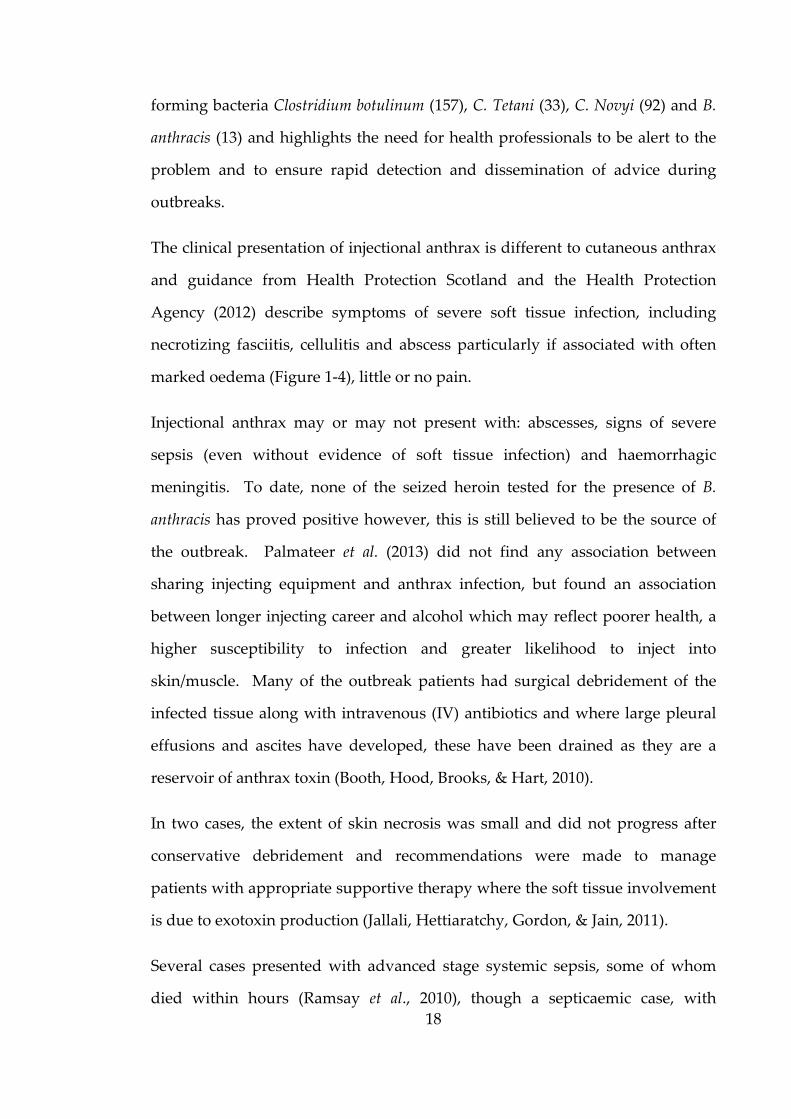
marked oedema (Figure 1‐4), little or no pain.
Injectional anthrax may or may not present with: abscesses, signs of severe
sepsis (even without evidence of soft tissue infection) and haemorrhagic
meningitis. To date, none of the seized heroin tested for the presence of B.
anthracis has proved positive however, this is still believed to be the source of
the outbreak. Palmateer et al. (2013) did not find any association between
sharing injecting equipment and anthrax infection, but found an association
between longer injecting career and alcohol which may reflect poorer health, a
higher susceptibility to infection and greater likelihood to inject into
skin/muscle. Many of the outbreak patients had surgical debridement of the
infected tissue along with intravenous (IV) antibiotics and where large pleural
effusions and ascites have developed, these have been drained as they are a
reservoir of anthrax toxin (Booth, Hood, Brooks, & Hart, 2010).
In two cases, the extent of skin necrosis was small and did not progress after
conservative debridement and recommendations were made to manage
patients with appropriate supportive therapy where the soft tissue involvement
is due to exotoxin production (Jallali, Hettiaratchy, Gordon, & Jain, 2011).
Several cases presented with advanced stage systemic sepsis, some of whom
died within hours (Ramsay et al., 2010), though a septicaemic case, with
bacteraem
therapy
Figure 1-4
injectiona
1.2.5
Severe a
dissemin
and enter
survive a
capsule p
fever, sho
Mortality
cases wa
positive b
died of ac
mia but no
and surgi
Severe
l anthrax ca
Comp
and often
ate from t
r the blood
and multi
preventing
ock and de
y associate
as 40% and
blood cultu
cute circul
o evidence
cal debrid
skin and
ase (Grunow
plication
fatal cas
the infectio
d stream, i
iply in the
phagocyto
eath.
ed with sy
d seven o
ures with B
atory colla
1
e of septic
dement (P
soft-tissue
w et al. 2012)
ns
ses of ant
on focus v
irrespectiv
e host pro
osis; bacter
ystemic an
of ten patie
B. anthraci
apse (Doola
19
shock sur
owell, Cro
involveme
).
thrax resu
via lymph
ve of route
otected by
raemia and
thrax infec
ents prese
is, four of t
an et al., 20
rvived foll
ozier, Hod
2011).
ent in ant
ult when
vessels to
e of entry.
y the poly
d toxaemia
ction in th
ented with
the seven b
007).
lowing IV
dgson, &
thrax infect
vegetative
o local lym
Vegetativ
y‐γ‐D‐glut
a ultimatel
he US anth
h severe se
bacteraem
antibiotic
Galloway,
tion in
e bacteria
mph nodes
ve bacteria
tamic acid
ly result in
hrax letter
epsis with
ic patients
c
,
a
s
a
d
n
r
h
s

In a revie
10 of 22 p
blood cu
Zealand
septic sta
bacteria (
spread of
to that s
initiated i
Anthrax
forms of
onset one
identified
gastrointe
infection.
signs, hy
The surv
younger
cases. A
multifoca
cerebral o
1.2.6
Human
experime
Manchur
ew of cutan
patients w
ltures but
white rabb
age of dise
(Levy et al.
f the bacilli
seen durin
infection (K
meningitis
f anthrax,
e to six day
d 70 pat
estinal (17
. These ca
perreflexia
viving pat
and show
long with
al subarach
oedema pa
Infect
infectious
entation w
ria. The d
neous anth
with severe
no fatal o
bits have a
ease by in
, 2014). In
i and cause
ng the bac
Kobiler, 20
s is a seve
typically
ys after the
ients with
7%), inhal
ases presen
a, and deli
tients gen
wed initial
haemorrh
hnoid and
athological
tious d
s doses
was conduc
doses are b
2
hrax cases
infection,
outcomes.
artificially
ntravenous
noculation
es lethality
cteraemic p
006).
ere complic
presentin
e onset of
h mening
lational (3
nted with
irium, stup
nerally pre
cerebrosp
hagic meni
d intrapare
l findings (
dose
have no
cted durin
based on 1
20
Doganay,
two with
Experime
created b
inoculatio
with this m
y in the rab
phase of i
cation and
g with ha
illness. A
goencepha
39%), and
symptom
por, or com
esented w
pinal fluid
ingitis, pat
enchymal h
(Lanska, 20
ot been e
ng the Sec
1 gram dry
Metan, &
septic sho
ental inves
acteraemia
on with ca
method allo
bbits with a
intranasal
d may follo
aemorrhag
recent revi
litis with
unknown
s of fever,
ma, 75% di
with cutane
results le
ients were
haemorrha
002).
established
ond Worl
y weight o
Alp, (2010
ock, all wit
stigations
a that rese
apsulated
ows haem
a time cou
and sub
ow any of
gic mening
iew of ant
h cutaneou
n (16%) s
, malaise, m
ied within
eous anth
ess severe
e also foun
ages, vasc
d, though
ld War in
of spores c
0) describe
th positive
with New
embles the
vegetative
atogenous
rse similar
cutaneous
f the other
gitis, with
thrax cases
us (29%),
sources of
meningeal
n 24 hours.
hrax, were
than fatal
nd to have
ulitis, and
h human
Unit 731,
containing
e
e
w
e
e
s
r
s
r
h
s
,
f
l
.
e
l
e
d
n
,
g

~109 CFU
which ar
epidemio
summari
50% of a
and the p
animals s
humans t
infection
was the m
mill in th
hour shif
the 2001 a
is no safe
been exam
incidence
al., 2014).
1.2.7
With the
antibiotic
more fav
Natural i
penicillin
cheap an
bacteria’s
U and repo
re far hig
ological da
sed by WH
nimals (LD
parenteral r
some mor
to anthrax
and expos
most comm
he USA wo
ft periods (
anthrax let
e level of c
mples of re
e of infecti
.
Treat
discovery
cs to treat i
ourable ou
isolates of
n and its u
nd effective
s ability to
orted as 10
gher than
ata. Infecti
HO, OIE, F
D50) for inh
route LD50
e susceptib
is conside
sure rate fo
mon presen
orkers wer
(Dahlgren,
tter events
contaminat
elatively la
ion; both o
ment
y of penicil
infectious
utcomes.
B. anthrac
use was wi
e treatmen
o produce
2
0 mg for s
n would b
ious dose
FAO, (2008
halational
0 of < 10 to
ible than o
ered mode
or exposed
ntation for
re found to
, 1960) cite
s in the US
tion (WHO
arge expos
occupation
llin by Ale
diseases, a
cis have a
idespread,
nt. Concer
penicillina
21
subcutaneo
be expect
data from
8), and sho
route to r
1010 spores
others to a
erate based
d industrial
r industria
o be inhali
d by (WHO
A, risk ass
O, OIE, FA
sed popula
nally and n
exander Fl
anthrax pa
high prob
especially
rns over p
ase (Barne
ous and 50
ted based
m in‐vivo ex
ow median
range from
s. These da
anthrax. T
d on the re
l workers.
al workers
ing 600‐130
O, OIE, FA
sessments c
AO, 2008), h
ations to B.
non‐occupa
leming in
atients cou
ability of b
y in develo
penicillin re
es, 1947; Tu
0 mg for o
on histo
xperiments
n lethal do
m ~ 104 to
ata are for
The suscep
elatively lo
Cutaneou
even thou
00 spores
AO, 2008).
concluded
however, t
. anthracis,
ationally (B
1928 and
uld be man
being susc
oping coun
esistance d
urnbull et
oral MID50
orical and
s has been
oses to kill
107 spores
a range of
ptibility of
ow rates of
us anthrax
ugh at one
over eight
Following
d that there
there have
but a low
Bennett, et
the use of
naged with
ceptible to
ntries, as a
due to the
al., 2004),
0
d
n
l
s
f
f
f
x
e
t
g
e
e
w
t
f
h
o
a
e
,
22
and the demonstration of inducible β–lactamase production in a number of
strains by Lightfoot, Scott & Turnbull (1990) has led to recommendations to use
alternative choices of antibiotics. Penicillin use is likely to continue in
developing endemic countries for both humans and animals where alternatives
are expensive (WHO, OIE, FAO, 2008). If penicillin is used, it should be
ensured that adequate doses are given to prevent sub‐inhibitory concentrations
leading to inducible β–lactamase production.
Susceptibility to a range of broad spectrum antibiotics has been demonstrated
in both in vitro and in vivo studies, these have informed the alternative choices
of ciprofloxacin and doxycycline for adults and where strains are susceptible,
oral amoxicillin can be used as an alternative. Neither ciprofloxacin nor
doxycycline are ideal for children less than eight years due to their side‐effects,
the severity of infection may however outweigh the side effects. The
therapeutic antibiotic regimes recommended for different forms of anthrax
reflect the severity of disease. Intravenous antibiotic therapy is not
recommended for mild cases but should be used with severe or potentially life
threatening cases, where patients are exhibiting systemic involvement such as
inhalational and gastrointestinal anthrax (WHO, OIE, FAO, 2008). In these
severe cases, a combination of antibiotics may be used especially with one that
has good central nervous system penetration, then returning to a single regimen
when the progression of symptoms cease (WHO, OIE, FAO, 2008). Supportive
care, in addition to antibiotic therapy for the treatment of shock should also be
considered.
Intubation, ventilator support or tracheotomy may be crucial in cases with
respiratory problems and should be implemented before obstruction is caused
by oedema (WHO, OIE, FAO, 2008).
23
Anthrax meningitis has an extremely high mortality and supportive care may
be required (such as respiratory support and anti‐oedema therapy for the
brain). A combination of supportive care and intravenous penicillin G with an
antibiotic which can rapidly penetrate into CSF such as rifampicin have been
used however, treatment failure rates are high with this complication of anthrax
(WHO, OIE, FAO, 2008).
Natural inhalation of B. anthracis is rare, but following the 1979 Sverdlovsk
incident and 2001 US anthrax letters, the threat from deliberate release resulting
in inhalational anthrax is proven. There is evidence that spores could be
detected in the lungs of rhesus macaques up to 100 days after initial exposure
(Henderson, Peacock, & Belton, 1956) and the UK and US use a 60 day duration
for prophylaxis due to the potential for latent germination of spores.
Experimental in vivo investigation of antibiotic treatment comparing prompt
prophylaxis and treatment after evidence of bacteraemia indicates that
development of immunity can prevent further infection due to latent spore
germination (Vietri et al., 2009). In the UK guidance, individuals will be
considered for vaccination on a case‐by‐case basis. If given post‐exposure
antibiotic prophylaxis duration can be reduced to four weeks and the
combination of vaccination to provide immunity to latent spore germination
appears effective (Doolan et al., 2007).
During the outbreak in 2009‐2010 and subsequent cases of injectional anthrax,
debridement of infected tissue in combination of IV antibiotics was
implemented.
The use of a negative pressure wound therapy device has been documented in
one of the cases which also had the complication of sepsis, NPWT proved to
give excellent wound healing results in this case (Powell, Crozier, Hodgson, &
Galloway, 2011).
Bactericid
become s
treatmen
remains a
Efforts h
treatmen
compone
explored.
preparati
a compon
Fifteen of
AIG supp
investiga
patients
have rece
appeared
injectiona
the treatm
Details o
fourth ed
though in
P1.3
The path
form spo
dal action
sterile afte
t. Even if
a problem
have been
ts to ove
ents, host
. Anthra
ion of antib
nent of the
f the cases
plied by th
ational new
(three inh
eived AIG
d to tolera
al and one
ment of sev
of suggeste
dition of A
ndividual c
Patho
hogenesis o
ores, the p
of antibio
er 24 hour
the bacter
and toxaem
made afte
rcome thi
cell inte
ax immun
bodies from
e U.S strat
s of injectio
he US Cent
w drug pro
halational,
intravenou
ate the an
e gastrointe
vere anthra
ed antibiot
Anthrax in
countries m
ogenes
of B. anthr
production
2
otics can cl
rs and blo
ria have be
mia can ul
er the 200
is problem
eraction an
ne globuli
m healthy
tegic natio
onal anthra
tres for Dis
otocol (Ra
15 injecti
usly in add
ntitoxin, a
estinal ant
ax patients
tic admini
n humans
may have s
sis
racis result
n of a po
24
lear infecti
ood cultur
een killed,
ltimately ca
01 US ant
m and ap
nd neutra
in (AIG)
, anthrax v
onal stockp
ax in Scotl
sease Contr
amsay et a
ional and
dition to an
nd 13 sur
thrax) and
s (Hendrick
istration r
and anim
specific gu
s from thr
oly‐γ‐D‐glu
ion, cutane
res are oft
, the prior
ause death
thrax lette
proaches
alising an
is an un
vaccinated
pile (Arten
and in 201
rol and Pre
l., 2010).
one gastr
ntimicrobi
rvived (tw
d therefore
ks et al., 20
egimens c
mals (WHO
idelines.
ree main fa
utamic aci
eous lesion
ten sterile
productio
h.
ers to look
targeting
ntibodies
nlicensed p
d, donors a
nstein & O
10 were tre
evention u
To date 1
rointestina
al drugs, a
wo inhalat
e may have
014).
can be fou
O, OIE, FA
actors, the
id capsule
ns usually
following
on of toxin
k at novel
the toxin
are being
polyclonal
and is now
pal, 2012).
eated with
under their
19 anthrax
l anthrax)
all patients
tional, ten
e a role in
und in the
AO, 2008),
e ability to
e and the
y
g
n
l
n
g
l
w
.
h
r
x
)
s
n
n
e
,
o
e
25
production of exotoxins. The ability of B. anthracis to produce capsule and
exotoxin are the two virulence factors required to cause disease, and their genes
are encoded on the two plasmids found in virulent strains. The virulence
plasmids are designated pX01 (encodes genes for exotoxin) and pX02 (encodes
genes for capsule) and form the basis for molecular confirmation of virulent
isolates of B. anthracis, their role in the identification and diagnosis of anthrax is
covered in section 1.5 and Chapter Three.
The virulence of B. anthracis is a combination of contributing factors involved
with growth, persistence in the host and toxicity after the organism has gained
entry via the routes discussed in section 1.2. The spore is key to the cycle of
infection (Figure 1‐1), and the natural infectious particle for all forms of anthrax.
Inside the host, spores typically enter macrophages by phagocytosis where they
germinate into the vegetative form. Early in pathogenesis, the germination of
spores within the macrophage enables them to emerge into the nutrient rich
environment of the host, allowing rapid replication of capsulated vegetative
bacteria. During infection, the protective poly‐γ‐D‐glutamic acid capsule
allows the bacteria to replicate and produce exotoxins whilst evading killing by
the innate immune system. In most infections it is advantageous for host
survival; in contrast anthrax ultimately results in death of susceptible hosts by
systemic involvement, sepsis with huge bacteraemia close to death (~ 108 CFU
ml‐1) and toxaemia. The end result leads to the dissemination of the organism
into the environment from decomposing carcasses in haemorrhagic exudates
and vegetative bacteria sporulate in the presence of air.
Both veterinarians and clinicians are recommended not to undertake post
mortems as there is an increased risk of exposure but also the problem of
containing the organism (WHO, OIE, FAO, 2008).

1.3.1
Spore for
vegetativ
in the ha
metabolic
desiccatio
disinfecta
Resistanc
laborator
environm
character
The dorm
species,
geograph
Sporulati
occurring
conducte
integrates
secreted
have been
The com
forming b
their part
the signa
likely to e
The s
rmation (sp
ve form, su
arsh envir
cally inac
on, gamm
ants.
ce poses co
ry setting
ment (Shar
rised by a r
mancy ha
resulting
hical sourc
ion is a com
g over app
ed with Bac
s a variety
factors ha
n identified
mplexity of
bacteria m
ticular nich
al transduc
ensure tha
spore
porulation
uch as follo
ronment o
ctive and
ma radiat
onsiderable
and po
rp & Rob
reservoir o
s greatly
in gene
es (Keim e
mplex dev
roximately
cillus subtil
y of signals
ve been d
d in B. anth
f control a
may allow t
he. Wilso
ction netw
t B. anthrac
2
n) occurs w
owing deat
outside of
able to
tion, ultra
e problem
otentially
berts, 2006
of dormant
influenced
etic homo
et al., 2009;
velopment
y eight hou
lis. In B. su
s to includ
documente
hracis (Bru
and variet
them to ini
on, Soyer, H
work in B.
cis sporula
26
when condi
th of the h
the host,
withstand
a violet
s when bo
large sca
6). The
spores in
d the evo
ogeneity
Pearson e
al process,
urs (Driks,
ubtilis, a sig
de nutrient
d and ma
nsing et al.
ty of trigg
itiate sporu
Hoch, & P
anthracis
ation does n
tions are n
ost entailin
typically
d extreme
light an
oth inactiva
ale decon
life cycle
the environ
olutionary
of isolate
t al., 2004;
, involving
2009) and
gnal transd
t levels, st
ny gene h
., 2005).
gers for sp
ulation wh
Perego (200
differs fro
not occur i
not favoura
ng a need
soil. The
es in tem
nd many
ating mate
ntamination
e of B. an
onment (soi
rate of t
es from
Van Ert et
g coordina
most work
duction net
tate of cell
homologue
porulation
hen advant
08) have sh
om B. subt
in host blo
able to the
to survive
e spore is
mperature,
chemical
erial in the
n of the
nthracis is
il).
this clonal
dispersed
t al., 2007).
ated stages
k has been
twork that
cycle and
es to these
n in spore
tageous to
hown that
tilis and is
od.
e
e
s
,
l
e
e
s
l
d
.
s
n
t
d
e
e
o
t
s
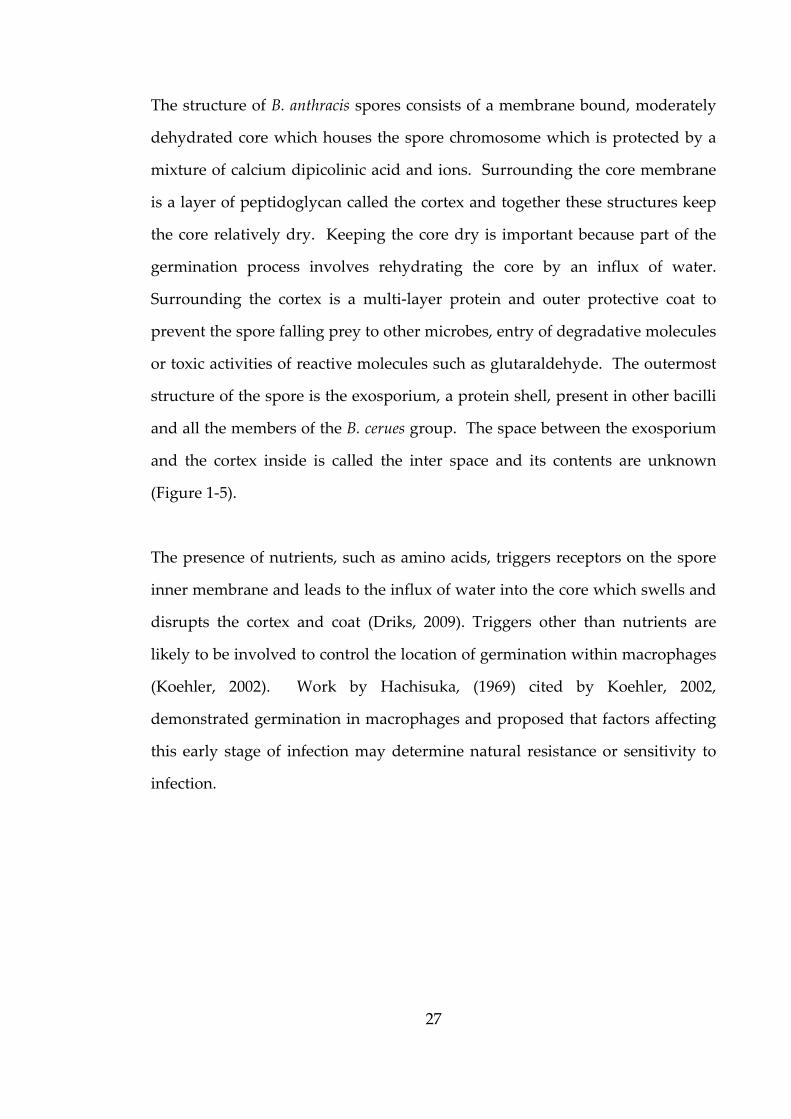
27
The structure of B. anthracis spores consists of a membrane bound, moderately
dehydrated core which houses the spore chromosome which is protected by a
mixture of calcium dipicolinic acid and ions. Surrounding the core membrane
is a layer of peptidoglycan called the cortex and together these structures keep
the core relatively dry. Keeping the core dry is important because part of the
germination process involves rehydrating the core by an influx of water.
Surrounding the cortex is a multi‐layer protein and outer protective coat to
prevent the spore falling prey to other microbes, entry of degradative molecules
or toxic activities of reactive molecules such as glutaraldehyde. The outermost
structure of the spore is the exosporium, a protein shell, present in other bacilli
and all the members of the B. cerues group. The space between the exosporium
and the cortex inside is called the inter space and its contents are unknown
(Figure 1‐5).
The presence of nutrients, such as amino acids, triggers receptors on the spore
inner membrane and leads to the influx of water into the core which swells and
disrupts the cortex and coat (Driks, 2009). Triggers other than nutrients are
likely to be involved to control the location of germination within macrophages
(Koehler, 2002). Work by Hachisuka, (1969) cited by Koehler, 2002,
demonstrated germination in macrophages and proposed that factors affecting
this early stage of infection may determine natural resistance or sensitivity to
infection.
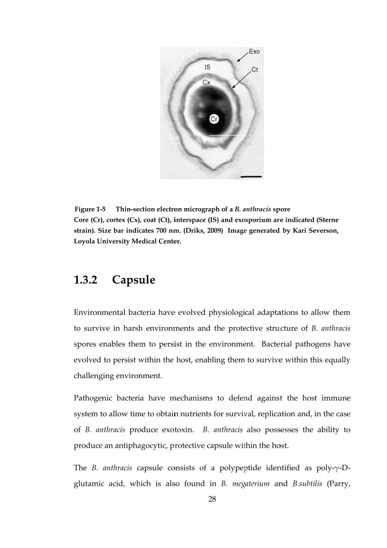
Core (Cr),
strain). Siz
Loyola Un
1.3.2
Environm
to surviv
spores en
evolved t
challengi
Pathogen
system to
of B. ant
produce a
The B. a
glutamic
Figure 1-5
cortex (Cx)
ze bar indic
niversity Me
Capsu
mental bac
ve in harsh
nables them
to persist w
ing environ
nic bacteri
o allow tim
thracis pro
an antipha
anthracis c
acid, wh
5 Thin-sec
, coat (Ct), i
cates 700 nm
edical Cente
ule
teria have
h environm
m to persi
within the
nment.
a have m
me to obtain
oduce exot
agocytic, pr
capsule co
hich is als
ction electro
2
interspace (
m. (Driks, 2
er.
e evolved p
ments and
ist in the e
host, enab
mechanisms
n nutrients
toxin. B.
rotective c
onsists of
so found i
on microgra
28
IS) and exo
2009) Image
physiologi
d the prote
environme
bling them
s to defen
s for surviv
anthracis
capsule wit
a polypep
in B. meg
aph of a B. a
osporium are
e generated
cal adapta
ective stru
ent. Bacte
to survive
nd against
val, replica
also poss
thin the ho
ptide iden
gaterium an
anthracis sp
e indicated
d by Kari Se
ations to a
ucture of B
erial patho
e within th
t the host
ation and, i
sesses the
ost.
ntified as
nd B.subti
pore
(Sterne
everson,
llow them
B. anthracis
gens have
his equally
t immune
in the case
ability to
poly‐γ‐D‐
lis (Parry,
m
s
e
y
e
e
o
‐
,
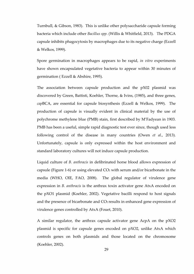
29
Turnbull, & Gibson, 1983). This is unlike other polysaccharide capsule forming
bacteria which include other Bacillus spp. (Willis & Whitfield, 2013). The PDGA
capsule inhibits phagocytosis by macrophages due to its negative charge (Ezzell
& Welkos, 1999).
Spore germination in macrophages appears to be rapid, in vitro experiments
have shown encapsulated vegetative bacteria to appear within 30 minutes of
germination ( Ezzell & Abshire, 1995).
The association between capsule production and the pX02 plasmid was
discovered by Green, Battisti, Koehler, Thorne, & Ivins, (1985), and three genes,
capBCA, are essential for capsule biosynthesis (Ezzell & Welkos, 1999). The
production of capsule is visually evident in clinical material by the use of
polychrome methylene blue (PMB) stain, first described by M’Fadyean in 1903.
PMB has been a useful, simple rapid diagnostic test ever since, though used less
following control of the disease in many countries (Owen et al., 2013).
Unfortunately, capsule is only expressed within the host environment and
standard laboratory cultures will not induce capsule production.
Liquid culture of B. anthracis in defibrinated horse blood allows expression of
capsule (Figure 1‐6) or using elevated CO2 with serum and/or bicarbonate in the
media (WHO, OIE, FAO, 2008). The global regulator of virulence gene
expression in B. anthracis is the anthrax toxin activator gene AtxA encoded on
the pXO1 plasmid (Koehler, 2002). Vegetative bacilli respond to host signals
and the presence of bicarbonate and CO2 results in enhanced gene expression of
virulence genes controlled by AtxA (Fouet, 2010).
A similar regulator, the anthrax capsule activator gene AcpA on the pXO2
plasmid is specific for capsule genes encoded on pXO2, unlike AtxA which
controls genes on both plasmids and those located on the chromosome
(Koehler, 2002).
B. anthrac
glutamic
reaction, c
1.3.3
The exoto
and at al
comprise
(LF, 89 k
individua
toxin (EF
Virulence
(Pezard, B
The cellu
spores (W
Figure 1-6
cis vegetativ
acid capsu
capsule is ob
Toxin
oxins relea
ll stages o
e the anthr
kDa) and
ally non‐to
+PA), majo
e is greatly
Berche, & M
ular bindin
Welkos et a
Microsc
ve cells gro
le stained
bserved as p
ns
ased by B. a
of infection
rax exotox
oedema fa
oxic but co
or virulenc
y attenuate
Mock, 199
ng moiety
al., 2001, 2
opic visuali
3
own in hor
with polyc
pink around
anthracis a
n. B. anth
xins, protec
actor (EF,
ombined fo
ce factors o
ed with m
1; Pezard,
PA, has
2002) wher
isation of B.
30
rse blood, c
chrome me
d the cells.
ppear to h
hracis prod
ctive antig
90 kDa).
orm lethal
of B. anthra
mutants lac
Weber, Sir
been foun
re it may
. anthracis c
cells are exp
ethylene blu
have wide r
duces thre
gen (PA, 83
These thr
l toxin (LF
acis.
cking singl
rard, Berch
nd on the
have a fun
capsule pressing po
ue for M'F
ranging ro
ee polypep
3 kDa), let
ree compo
F + PA) an
le toxin co
he, & Mock
surface of
nction in
oly-γ-D-
Fadyean
oles during
ptides that
thal factor
onents are
nd oedema
omponents
k, 1995).
f dormant
enhancing
g
t
r
e
a
s
t
g
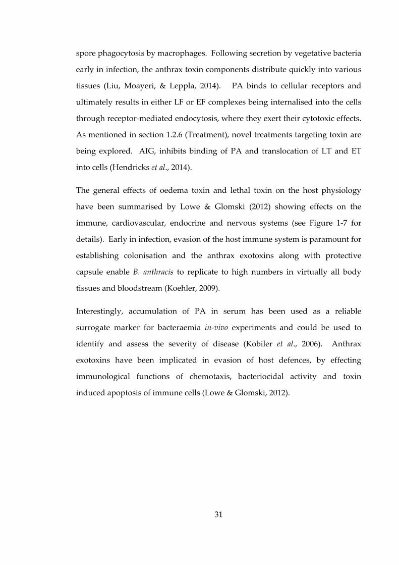
31
spore phagocytosis by macrophages. Following secretion by vegetative bacteria
early in infection, the anthrax toxin components distribute quickly into various
tissues (Liu, Moayeri, & Leppla, 2014). PA binds to cellular receptors and
ultimately results in either LF or EF complexes being internalised into the cells
through receptor‐mediated endocytosis, where they exert their cytotoxic effects.
As mentioned in section 1.2.6 (Treatment), novel treatments targeting toxin are
being explored. AIG, inhibits binding of PA and translocation of LT and ET
into cells (Hendricks et al., 2014).
The general effects of oedema toxin and lethal toxin on the host physiology
have been summarised by Lowe & Glomski (2012) showing effects on the
immune, cardiovascular, endocrine and nervous systems (see Figure 1‐7 for
details). Early in infection, evasion of the host immune system is paramount for
establishing colonisation and the anthrax exotoxins along with protective
capsule enable B. anthracis to replicate to high numbers in virtually all body
tissues and bloodstream (Koehler, 2009).
Interestingly, accumulation of PA in serum has been used as a reliable
surrogate marker for bacteraemia in‐vivo experiments and could be used to
identify and assess the severity of disease (Kobiler et al., 2006). Anthrax
exotoxins have been implicated in evasion of host defences, by effecting
immunological functions of chemotaxis, bacteriocidal activity and toxin
induced apoptosis of immune cells (Lowe & Glomski, 2012).

(Lowe &
present in
has effects
cardiovasc
Specific e
and card
blood pre
Vascular
also the l
internaliz
(composi
may refle
S1.4
The great
is laborat
Figure 1-7
Glomski, 2
n most cells
s on all four
cular and en
exotoxin ta
iac tissue)
essure and
damage a
likely caus
zation of
ing the blo
ect the dev
Safety
test risk to
tory acquir
7 General e
2012). Anth
which lead
r systems w
ndocrine sys
argeting o
may lead
d shock in t
and increa
se of haem
vegetative
ood brain b
elopment o
y
o the labor
red infectio
effects of oe
3
hrax toxins
d to diverse
whereas oede
stems.
of the card
to vascula
terminal st
sed perme
morrhage se
e bacteria
barrier) ar
of anthrax
ratory wor
on via the i
edema toxin
32
target con
effects thro
ema toxin h
diovascular
ar damage
tages of di
eability ind
een during
a by the
re potentia
meningiti
rker handli
inhalation
n and lethal
served and
oughout the
has known e
r system (b
e, cyanosis
sease (Low
duced by a
g disease.
brain mi
ated by an
is in system
ing highly
route (HSE
toxin on ho
d critical pa
e host. Leth
effects on im
both the v
s, reduced
we & Glom
anthrax ex
The adhe
icroendoth
nthrax exot
mic patient
pathogen
E, 2009).
ost physiolo
athways
hal toxin
mmune,
asculature
pulse and
mski, 2012).
xotoxins is
erence and
helial cells
toxins and
ts.
nic bacteria
ogy
e
d
.
s
d
s
d
a
Bacteria
(ACDP)
hazard g
and risk
prophyla
tularensis
are exam
3 laborato
B. anthra
chemical
(WHO, O
produce
disinfecta
due to ex
tularensis
100 CFU
aerosol ro
1.4.1
The use o
the risk
protection
equipmen
The com
provide p
containm
are classif
into hazar
roup 3 (H
of spread
axis or tre
, Yersinia
mples of AC
ory (CL3) i
acis spores
disinfecta
OIE, FAO,
spores are
ants, thoug
xtremely lo
and Bruce
ml‐1 and a
oute of infe
Facili
of enginee
of labora
n to all w
nt which o
mbination o
primary co
ment of the
fied by the
rd groups
G3). This
d in the
atment av
pestis, Bru
CDP HG 3
in the UK.
s are high
ants and c
, 2008). O
e relatively
gh they ma
ow infectio
ella spp. wh
are the cau
ection (Col
ity requ
ering contr
atory wor
orkers in
only protec
of enginee
ontainment
pathogen
3
e Advisor
and high
means the
communit
vailable. B
ucella spp.
pathogens
hly resista
can persis
Other hig
y easy to in
ay present
ous doses.
hich have d
use of man
llins, 1983)
uireme
rols such a
rkers inha
the labora
cts the user
ering contr
t, and the l
(HSE, 2009
33
ry Commit
hly pathog
ere is a po
ty; howev
Bacteria su
and Burkh
s and are h
ant to ext
st in the
ghly patho
nactivate u
t a serious
. Example
documente
ny laborato
).
nts
as microbio
aling infec
atory, as op
r.
rols and g
laboratory
9).
ttee on Da
enic bacte
otential to c
ver, there
uch as B.
holderia pse
handled in
tremes of
environme
ogenic bac
using temp
hazard to
es of which
ed infectio
ory acquire
ological sa
ctious aer
pposed to
good micr
y (Figure 1‐
angerous
eria are cla
cause seve
is usually
anthracis,
eudomallei
n a containm
temperatu
ent for m
cteria whic
perature or
o laborator
ch include
ous doses o
ed infectio
afety cabin
rosols and
personal
robiologica
‐8) acts as
Pathogens
assified as
ere disease
y effective
Fransicella
and mallei
ment level
ure, some
any years
ch do not
r chemical
ry workers
Francisella
of less than
ons via the
ets reduce
d provide
protective
al practice
secondary
s
s
e
e
a
i
l
e
s
t
l
s
a
n
e
e
e
e
e
y
34
There are also several important physical and engineering features of
containment Level 3 laboratories. These physical features are:
the laboratory is separate from other activities in the same building
an observation window, or alternative, is present so that occupants can
be seen
the laboratory contains its own equipment
floor and bench surfaces are resistant to acids, alkalis, solvents and
disinfectants, impervious to water and easy to clean
Able to seal the workplace to permit disinfection, typically formaldehyde
gaseous fumigation in the UK
maintaining the workplace at air pressure negative to atmosphere
single pass air extracted from the workplace through high efficiency
particulate absorption (HEPA) filters or equivalent
Validated room fumigations can be performed using biological indicators and
are typically used to clean up after spillage/release of HG3 organisms in the
open lab and before laboratory servicing to make the laboratory safe for
engineers to enter and service the laboratory and equipment.
Apart from the physical and engineering features, the staff and procedural
controls are important to ensure safe practices of work. The procedural controls
used include having a laboratory code of practice, disinfection policy, standard
operating procedures, and personal discipline to prevent infection via routes
other than inhalation.
Across the UK the most widely used microbiological safety cabinet (MSC) in a
diagnostic setting is the class I MSC (MSC I). This type of cabinet is open
fronted to allow the user to manipulate cultures within the cabinet whilst
protected by an inward flow of air within the range of 0.7–1.0 ms‐1. User
movements, the presence of equipment within the cabinet and external factors
35
can affect the air flow across the front of the cabinet therefore reducing the
protection afforded to the user from infectious aerosols. Surface
decontamination of items being removed from the cabinet, and the use of
double gloves, with the outer pair being removed both reduces the risk of
bringing contamination out of the cabinet. The risk assessment detailing the
handling of HG3 organisms at high titre and/or volume determines the
suitability of the class of MSC used. The MSC I is suitable for diagnostic work
involving low risk screening of low titre clinical samples, whereas research
conducted on known cultures at higher volume and or titre would necessitate
the use of the class III MSC (MSC III). The MSC III provides both product and
user protection, by passing air through an inlet HEPA filter and providing full
containment afforded by rubber gauntlets to manipulate cultures inside the
cabinet (Figure 1‐9). If a breach occurs across the gauntlet port holes the inward
airflow is three to five ms‐1, greater than three times that across the front of a
MSC I (personal measurement).
Figure 1-8 Class III MSC in CL3 training laboratory at PHE Porton

The safe
subseque
for handl
1.4.2
This stud
Centre fo
for all in
principle
species w
cultures.
higher th
this stud
Figure 1-9
ety aspect
ent chapter
ling culture
Condu
dy took pl
or Emergen
nvestigatio
s of reduci
were used
The time
han work a
dy was lim
9 Use of c
ts relating
rs where a
es in CL2 l
ucting
ace at PH
ncy Prepar
ons involv
ing risk we
for the det
taken and
at CL2. For
mited by th
class III MS
3
g to B. a
appropriat
laboratorie
researc
HE Porton,
redness an
ing ACDP
ere undert
tection of
d costs to u
r this reaso
hese facto
SC to handle
36
anthracis w
te inactiva
es are cons
ch with
formerly
nd Respons
P HG 3 b
taken and
aerosols d
undertake a
on the exte
rs and rep
e known HG
will be di
tion of ma
idered.
h B. ant
the Health
se (CEPR),
acteria. W
surrogate A
during the
any work i
ent of pract
presents ex
G3 cultures
iscussed f
aterial and
thracis
h Protectio
, using CL
Where po
ACDP HG
processing
in CL3 is l
ctical inves
xploratory
further in
d concerns
on Agency
L3 facilities
ssible, the
G 1 Bacillus
g of blood
longer and
tigation in
y work, in
n
s
y
s
e
s
d
d
n
n
some cas
trained, c
O1.5
The diag
cannot be
are not n
may be u
natural so
will be t
suspected
laborator
Rare and
reference
an over
confirma
1.5.1
Basic, pr
microsco
of B. anth
clinical m
5‐8 μm i
material
es identify
competent
Overv
gnosis of h
e taken ligh
necessarily
used for bi
ources or a
the first to
d, cultures
ries and sen
d Imported
e centre for
view of
tion of B. a
Presu
resumptive
pic and ty
hracis can
material or
in length
and very
ying the ne
staff follow
view o
highly path
htly in a co
endemic
oterrorist
a bioterror
o perform
s of poten
nt for confi
Pathogen
r B. anthrac
the meth
anthracis; fu
umptive
e identific
pical macr
be easily
culture. C
and typic
long chain
3
eed for furt
wing risk a
of lab
hogenic b
ountry suc
but becau
activities.
rist event,
m prelimin
ntial ACD
irmation a
ns Laborato
cis and F. tu
hods for
urther disc
e ident
cation of
roscopic ph
visualised
Cells appea
cally in sh
ns from lab
37
ther work.
assessment
orator
acteria, as
ch as the U
use of the p
Whether
the local N
ary testing
DP HG3 b
at national
ory, (RIPL)
ularensis. T
the pres
cussion is g
tificatio
B. anthra
henotypic
d upon dir
ar as squar
hort chains
boratory c
. The stud
ts and reco
ry refe
s a cause o
UK, even th
potential t
the source
NHS micro
g on clini
bacteria ar
reference c
) at PHE P
The followi
sumptive
given in Ch
on
acis can b
methods.
ect Gram
re ended ba
s (a few b
cultures (K
dy was con
ognized pr
erral
of human
hough thes
threat that
e of infectio
obiology la
ical sampl
re handle
centres.
Porton is th
ing section
identifica
hapter Thr
be made
The veget
stain micr
acilli, appr
bacilli) fro
Koehler, 20
nducted by
rocedures.
infection,
se diseases
the agent
on is from
aboratories
les. Once
d in CL3
he national
ns describe
ation and
ree.
using the
tative cells
roscopy of
roximately
om clinical
009; WHO,
y
,
s
t
m
s
e
3
l
e
d
e
s
f
y
l
,

38
OIE, FAO, 2008). The capsule may be visualised with M’Fadyean’s reaction
with poly‐chrome methylene blue stain, where the capsule appears as a pink
layer around blue cells (Figure 1‐6) or non‐specifically using Indian ink.
Capsule production occurs within the host and will be evident in direct
microscopy of clinical samples or following specific laboratory culture.
Refractile spores are visible under phase contrast microscopy and are 1‐1.5 μm
in diameter, located centrally or sub‐terminally, without cell body swelling.
B. anthracis is an environmental soil bacteria and can be easily grown in the
laboratory, the most common method being culture on agar containing blood.
When grown on horse or sheep blood agar at 37˚ C, colonies are typically 2‐5
mm diameter, irregular edge, non‐haemolytic, with white/grey opaque colour
and a rough ‘ground glass’ appearance (Parry, Turnbull, & Gibson, 1983).
Haemolysis on blood agar must not be confused with confluent growth of
bacteria, though it has been documented that B. anthracis can be weakly
haemolytic and haemolysis may be evident when different donor species of
blood are used in agar, such as human blood (WHO, OIE, FAO, 2008).
The long chains of cells observed in laboratory cultures give the characteristic
tenacity of colonies, which adhere to the agar surface and can be made to stand
up unaided, perpendicular to the plate surface when teased with a loop (Parry,
Turnbull, & Gibson, 1983, p. 109) as shown in Figure 1‐10a. When removed
from the surface the colony comes away in entirety and is hard to emulsify in
liquid broth.
Cultures from fresh clinical samples are likely to have a pure growth of B.
anthracis, particularly from normally sterile sites and skin biopsies (cutaneous
anthrax) prior to treatment. Older clinical samples and post mortem samples
are likely to be of mixed cultures and B. anthracis can be easily out competed by
putrifying bacteria; the presence of Staphylococcus aureus appears to outcompete

39
B. anthracis in clinical samples (personal experience). Specialist media
polymixin‐lysozyme‐EDTA‐thallous acetate agar (PLET) may be of use with soil
and other environmental samples (WHO, OIE, FAO, 2008) but I would question
its use with clinical samples because it permits the growth of many Gram
positive human commensal bacteria and B. anthracis colonies do not grow with
any of the distinctive colony characteristics as seen with growth on blood agar
(personal experience during outbreak 2009‐2010).
On PLET agar, B. anthracis colonies grow as small (approximately 2‐4 mm)
white colonies with a ‘mashed potato’ consistency after 48 hours incubation.
Other bacteria such as Staphylococcus spp. also grow as small white colonies and
therefore other media such as B. cereus selective agar may be more beneficial
and more readily available in routine laboratories. B. cereus selective agar can
be used to distinguish Staphylococcus aureus, Serratia marcescens and Proteus
vulgaris by colony morphology and colour and produces an egg yolk clearing
reaction. In contrast B. cereus colonies appear white with a rich peacock blue
colouration of the media caused by egg yolk precipitation and B. anthracis
colonies appear a pale blue colour without colouration of the media or egg yolk
clearing (Appendix 5.4 Bench guide).
The optimum culture temperature is 37˚C for both solid and liquid cultures and
growth at 42˚C has been shown to cause a loss of pXO1 plasmid (Mikesell,
Ivins, Ristroph, & Dreier, 1983). At 37˚C cell doubling times have been shown
to range between 30–60 minutes (Koehler, 2009).
B. anthracis is non‐motile and therefore turbidity of liquid cultures is not seen,
upon disturbing static cultures a pellicle (Koehler, 2009) or ‘wisp’ (personal
description) of growth is visible due to the long chains of cells.
The use of biochemical identification systems such as the API 50CHB used in
conjunction with the first twelve reactions of API20E (Logan, Carman, Melling,
& Berkel
rely on d
Biochemi
identifica
testing at
desorptio
many fro
for colon
microorg
sample sp
For the
would b
identified
1.5.2
The meas
prominen
This is d
anthrax,
antibody
al., 2004).
Serologic
of antibo
used in a
the delib
ley, 1985)
ata base pr
ical identif
ation shou
t specialis
on/ionizati
ont line mi
ny identific
ganism’s g
pectrum to
identificat
be require
d as B. cereu
Toxin
surement
nt feature
due to the
however
may be m
.
cal enzyme
ody to the
a retrospec
berate use
and more
rofiles may
fication sh
ld always
t laborator
on time‐of
icrobiology
cation. M
enus and
o an extens
tion of B.
ed, withou
us (Grunow
n and a
of an imm
of diagno
acute and
in an out
more appli
e immunoa
PA compo
ctive study
of anthrax
4
recently a
y identify i
hould not
be perfor
ries. The r
f‐flight ma
y laborator
MALDI‐TOF
species in
sive databa
anthracis,
ut this da
w et al., 20
antibod
mune respo
sis compa
d often fa
tbreak sett
icable than
assays (EIA
onent of bo
y to test se
x spores in
40
automated
isolates as
be relied
rmed and
recent add
ass spectro
ry identific
F MS can
n just a few
ase of spec
the addi
atabase is
12).
dy detec
onse in an
ared to cul
tal nature
ting, serolo
n treatmen
A) have be
oth toxins
erum from
n the US A
d identifica
B. cereus or
upon in is
isolates se
dition of m
ometry (M
cation repe
provide an
w minutes
tra from ba
tional bio
solates wo
ction
nthrax pati
lture and m
of inhala
ogical det
nt‐sensitive
en develop
and antib
m anthrax c
Anthrax le
ation syste
r B. anthra
solation an
ent for con
matrix assi
MALDI–TO
ertoire can
n identific
s by comp
acterial sp
oterrorism
ould be i
ients has n
molecular
ational and
tection of
e methods
ped for the
bodies. EIA
cases resu
etter attack
ems which
cis.
nd further
nfirmatory
isted laser
OF MS) to
n be useful
cation of a
paring the
ecies.
databases
incorrectly
not been a
detection.
d systemic
toxin and
(Quinn et
e detection
A has been
lting from
ks in 2001
h
r
y
r
o
l
a
e
s
y
a
.
c
d
t
n
n
m
1

(Quinn e
tests for a
The diag
weeks ap
than a w
Antimicr
anthracis
response
Serologic
resources
During th
ELISA fo
diagnosti
for PA an
1.5.3
A commo
reports o
considere
access to
inoculatio
Capsule p
horse blo
visualisat
Alternativ
t al., 2004)
any infecti
nostic reli
part and th
week after
obial trea
and could
(WHO, OI
cal assays
s to prepa
he 2009–2
or human
ic tests. De
nd was also
Confi
on phenoty
of resistan
ed in conju
o specific γ
on site on a
production
ood for a
tion usin
vely isola
). Factors
ion surrou
ability wil
he diagnost
the onse
tment of
result in i
IE, FAO, 2
s are typic
are the ass
010 Injecti
anti‐PA I
etection of
o performe
irmator
ypic test is
nce (WHO
unction w
γ‐phage, a
agar cultur
n can be in
a minimum
ng poly‐c
ates may
4
to consid
und the tim
ll be greate
tic value w
et of symp
anthrax w
insufficien
2008).
cally limit
say reagen
ional anth
IgG was u
toxin can
ed during 2
ry iden
s sensitivit
O, OIE, F
with other c
a clear zon
res (Figure
nduced in t
m of 5 h
chrome m
be grown
41
der when u
ming of sam
er if two o
will be grea
ptoms if o
will result
nt antigen t
ted to sp
nts and ar
hrax outbre
used in co
also be per
2009‐2010.
ntificati
ty to penici
AO, 2008
confirmato
ne of sensi
e 1‐10b).
the laborat
hours and
methylene
n on agar
using serol
mple collec
or more sa
ater if colle
only one s
in the r
to induce a
ecialist lab
re therefor
eak in the
ombination
rformed w
ion
illin, howe
) and thi
ory tests.
itivity can
tory by gro
d a blood
blue st
containin
logical con
ction and
amples are
ection is m
sample is
rapid clear
a detectabl
boratories
re not wid
e UK, a qu
n with con
with anti‐to
ever there
is result s
If laborato
n be observ
owing the b
film pre
tain (Fig
ng bicarbo
nfirmatory
treatment.
e taken 2‐4
made more
collected.
ring of B.
le immune
with the
dely used.
uantitative
nventional
oxin ELISA
have been
should be
ories have
ved at the
bacteria in
epared for
gure 1‐6).
onate and
y
.
4
e
.
.
e
e
.
e
l
A
n
e
e
e
n
r
.
d
42
incubated in an enriched CO2 atmosphere; capsulated bacteria grow as mucoid
colonies.
For many years isolation by culture was the only method to detect B. anthracis
and cultures could be confirmed with the phenotypic characteristics described.
This remains the gold standard for diagnosis but with the advent of sequence
data on the virulence plasmids and chromosome, specific molecular detection
assays have been developed. Polymerase chain reaction (PCR), has been used
to detect many pathogenic bacteria by designing assays targeting species
specific nucleic acid sequences. RIPL use an in‐house triple target assay specific
for sequences targeting the pX01, pX02 plasmids and a chromosomal target.
The presence of all three targets confirms the diagnosis of virulent B. anthracis.
The assay utilises fluorescent probe technology providing real‐time detection of
PCR product and gives a rapid and reliable result on the day samples are
received. This assay can be used for detecting B. anthracis in clinical samples
following nucleic acid extraction and also for confirming isolates following heat
inactivation. The thermal lysis protocol for culture heat inactivation is quick
(~15 minutes) and validated.
This simple rapid process involves a colony being emulsified in brain heart
infusion broth (BHI) and heated to 37˚ C to germinate any spores present (this
will not occur unless the suspension is in a nutrient broth), the temperature is
then raised to 99˚C to inactivate the vegetative bacteria.
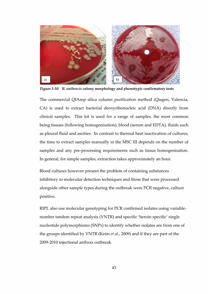
43
The commercial QIAmp silica column purification method (Qiagen, Valencia,
CA) is used to extract bacterial deoxyribonucleic acid (DNA) directly from
clinical samples. This kit is used for a range of samples, the most common
being tissues (following homogenisation), blood (serum and EDTA), fluids such
as pleural fluid and ascities. In contrast to thermal heat inactivation of cultures,
the time to extract samples manually in the MSC III depends on the number of
samples and any pre‐processing requirements such as tissue homogenisation.
In general, for simple samples, extraction takes approximately an hour.
Blood cultures however present the problem of containing substances
inhibitory to molecular detection techniques and those that were processed
alongside other sample types during the outbreak were PCR negative, culture
positive.
RIPL also use molecular genotyping for PCR confirmed isolates using variable‐
number tandem repeat analysis (VNTR) and specific ‘heroin specific’ single
nucleotide polymorphisms (SNPs) to identify whether isolates are from one of
the groups identified by VNTR (Keim et al., 2009) and if they are part of the
2009‐2010 injectional anthrax outbreak.
Figure 1-10 B. anthracis colony morphology and phenotypic confirmatory tests
a) b)
The WHO
in Figure
(WHO, 20
and genoty
Figure 1-1
O guidanc
1‐11 with
003) and add
yping indic
11 Simplifi
ce for the is
the additio
ditional det
cated by sha
ied overview
4
solation an
on of RIPL
tails of RIPL
aded boxed
w of isolatio
44
nd identifi
L procedur
L confirmat
text.
on and inde
ication of B
es.
tion of B. a
entification
B. anthracis
anthracis wi
of B. anthra
s is shown
ith PCR
acis
n
1.6 A
1.6.1
The 2009
UK, place
anthracis.
confirma
cultures
blood sam
large NH
facilities
of tissue
locally an
the outbr
RIPL for
other mo
comment
risks of
(Personal
Pathogen
Blood cu
suspected
& Gent, 2
cultures i
of bacter
Aims a
Backg
9–2010 out
ed a huge d
During
tion of wh
(26 sent; 1
mples for
HS and PH
for the de
samples.
nd subsequ
reak, howe
processin
ore commo
ted that ma
handling
l commun
ns Laborato
ultures are
d bioterror
2006). Cur
involve sim
ria, followe
and sc
ground
break and
demand on
the main
hich 21 w
10 confirm
molecular
HE routine
tection of
Many of
uent bacter
ever, many
ng and not
on causes
any labora
B. anthra
nication –
ory).
e one of
rist exposu
rrent recom
mple micro
ed by ove
4
cope o
d subseque
n RIPL for
outbreak
were positiv
med positiv
r and sero
e clinical m
Mycobacte
the anthr
rial isolate
y samples f
t cultured
of infectio
atories wer
acis even
Dr Tim
the clinic
ure to ACD
mmended p
oscopy pro
ernight cul
45
of pro
ent cases o
r the identi
(2009–201
ve; other
ve), 239 ti
ology testi
microbiolog
erium tuber
rax sample
es sent for
for bacteria
locally w
on were p
re reluctant
though th
Brooks, H
cal sample
DP hazard
procedures
oviding a ra
lture upon
ject
of B. anthr
fication an
10), 45 iso
sample ty
issue samp
ing (unpu
gy laborat
culosis and
es could h
confirmati
al culture w
hich may
resent. C
t to proces
he risks h
Head of R
es taken
group 3 b
s for proce
apid indica
n which fu
racis in PW
nd confirm
olates wer
ypes includ
ples and o
ublished da
tories will
d routine p
have been
ion. Initia
were sent
have high
Clinicians f
ss samples
had been
Rare and
from pati
bacteria (H
essing posi
ation of th
urther ide
WID in the
mation of B.
e sent for
ded blood
over 1,000
ata). Most
have CL3
processing
processed
ally during
directly to
hlighted if
from RIPL
due to the
explained
Imported
ients with
Heptonstall
itive blood
e presence
entification
e
.
r
d
0
t
3
g
d
g
o
f
L
e
d
d
h
l
d
e
n
46
may be made. This could lead to a delay of several days before referral to a
reference centre for confirmed identification due to three main factors.
First, is the delay in the realisation that a bacterial culture is a highly pathogenic
bacterium, second is the time taken to make a subculture onto an agar slope and
thirdly the time it takes for transport to the specialist laboratory for
confirmation. In 2006, a fatal case of anthrax in the UK had a delay of >2 weeks
occurring between blood culture isolation of B. anthracis and final confirmed
identification as the isolate went for Bacillus spp. typing and confirmation at
another reference centre. The blood culture as a sample type also hinders the
rapid identification of causative bacteria due to the presence of inhibitory
substances to molecular identification techniques. This study explores a
method to process blood cultures to enable the confirmation of virulent B.
anthracis using molecular detection. This method could be used alongside the
molecular identification of other sample types sent from a patient. The
processing method and molecular detection, together with confirmatory
phenotypic tests performed the same day, enables the gold standard culture
confirmation being made 24 hours sooner than at present. Increased awareness
of B. anthracis in blood cultures would also reduce the chance of unusual or
unexpected cases of anthrax which may occur in the future are not mistaken for
other Bacillus species often considered contaminants in blood cultures. The
front line diagnostic experience I gained whilst working at the Royal Cornwall
Hospital Trust and discussions with laboratory workers from front‐line
microbiology laboratories across the UK and internationally, have informed the
study to reflect genuine concerns.
1.6.2
The aims
detection
process,
methodo
method t
in blood c
Secondly
processin
microbiol
process a
e‐learning
hands o
identifica
laborator
1.6.3
The majo
to ethical
clinical an
cases dur
were Cal
isolates w
Aims
s of this
n and confi
(ii) safet
logies and
to potentia
cultures.
y investiga
ng to asse
logy labor
and safety
g modules
n practica
ation of cu
ry staff in f
Ethics
ority of the
l approval
nd biomed
ring the ou
dicott com
were used i
s
study wer
irmation o
ty and (
d explore
ally reduce
te the safe
ess wheth
ratories.
investigat
s on anthra
al training
ultures and
front‐line la
s
e study pro
l. No pati
dical scienc
utbreak. A
mpliant. La
instead of p
4
re to expl
of B. anthra
(iii) traini
the feasi
e turnaroun
ety surrou
her this m
Thirdly, b
tions to de
ax. The m
g is inten
d the safet
aboratorie
oject fell u
ient identif
ce professi
All staff we
aboratory r
patient ide
47
lore three
acis in bloo
ing. Fir
ibility of
nd times f
unding cur
method ca
bring toge
velop train
modules, in
nded to
ty around
s.
under diag
fiers have
ionals whe
ere trained
reference n
entifiers.
main are
od cultures
rstly to
using an
for confirm
rrent pract
n be ado
ether the
ning mater
n conjuncti
raise awa
processing
nostic eva
been disse
n gatherin
d in patient
numbers fo
eas involv
s which w
investigate
altered p
mation of B
tice and th
opted by
outcomes
rial, in the
ion with e
areness of
g blood cu
aluation wi
eminated
ng informa
t confiden
or clinical s
ved in the
were (i) the
e current
processing
B. anthracis
he altered
front line
from the
e format of
established
f anthrax,
ultures for
ith respect
outside of
ation about
tiality and
samples or
e
e
t
g
s
d
e
e
f
d
,
r
t
f
t
d
r
Ethical ap
the Unive
the appro
The dono
PHE Port
given to
thesis wa
practice d
associated
1.6.4
Blood cu
involvem
between
increasin
using a r
patient c
informati
injectiona
explore t
simulated
The aim
results as
without
simulated
pproval wa
ersity of P
oval is avai
or human b
ton for the
participan
as carried
described i
d guidance
The p
ultures a
ment in B.
time to po
g bacterial
ange of str
cases, bot
ion of pati
al anthrax
the feasibi
d blood cu
was to de
s conventi
serum sep
d blood cu
as sought f
ortsmouth
ilable in A
blood was
e purposes
nts is give
out in ac
in the PHE
e documen
process
re an im
anthracis
ositive dete
l load. The
rains and c
th docum
ents durin
. The sec
ility of us
ultures to re
termine if
ional testin
parator tu
ultures with
4
from the B
h for the fir
Appendix 1
collected
of simulat
en in App
ccordance
E policy fo
nts.
s
mportant
infection.
ection of si
e data gene
clinical iso
mented in
ng the 2009
cond aspe
sing serum
educe the
f their use
ng. A tri
ube proces
h ten strai
48
BioSciences
rst use of d
.1.
from healt
ting blood
endix 1.2.
with the
or conduct
sample t
The stu
imulated B
erated from
olates are r
the liter
9‐2010 outb
ect of the
m separato
turnaroun
can provi
ial of the
ssing was
ins of B. an
s Research
donor hum
thy unvacc
cultures a
The wor
principles
ting scienti
ype to d
udy explor
B. anthracis
m the simu
elated bac
rature an
break and
process in
or tubes in
nd times fo
de the sam
diagnostic
s conducte
nthracis, ten
Ethics Com
man blood,
cinated vol
and the con
rk describ
s of good
ific researc
determine
res the re
s blood cul
ulated bloo
ck to outco
nd from
subsequen
n the stud
n the pro
or confirma
me confirm
c strategy
ed, which
n isolates o
mmittee at
, a copy of
lunteers at
nsent form
bed in this
d scientific
ch and the
systemic
elationship
tures with
od cultures
ome in real
laboratory
nt cases of
dy was to
cessing of
atory tests.
matory test
with and
h involved
of bacteria
t
f
t
m
s
c
e
c
p
h
s
l
y
f
o
f
.
t
d
d
a
received
cultures.
1.6.5
A concer
processin
as oppose
current m
bottles, p
method t
Viability
was dete
recomme
Addition
to five d
laborator
processin
based risk
1.6.6
Trained,
the quali
microbiol
with mat
during the
Safety
n for safet
ng of blood
ed to with
methods f
production
to transfer
of materia
ermined to
ended ina
nally the ris
days was
ries. The d
ng of blood
k assessme
Train
competen
ity of dia
logy labor
erial devel
e outbreak
y
ty and pot
d cultures s
hin a micro
for proces
n of micros
r positive
al containin
o assess th
activation
sk of spore
assessed
data gener
d cultures t
ent.
ning
t, confiden
gnosis bu
atory. The
loped as a
4
as negativ
tential labo
suspected
obiological
ssing bloo
scopy slide
fluid into
ng B. anthr
he potenti
method
es in blood
to simul
rated wou
to include
nt laborato
ut also the
e aim was
direct outc
49
ve controls
oratory acq
to contain
safety cab
od culture
es and cul
serum se
racis on th
ial hazard
for B. a
d cultures le
ate this t
uld inform
serum sep
ory worke
e safety of
to supple
come of th
and five m
quired infe
B. anthraci
binet. The
s involvin
lture. In a
eparator tu
he surface o
to labora
anthracis w
eft at room
ypical situ
the suitab
parator tub
rs are key
f all peop
ment exist
e study.
mixed bact
ection surr
is on the o
aim was t
ng venting
addition, t
ubes was
of microsc
atory work
was not
m temperat
uation in
bility of al
bes and for
y to ensure
ple involve
ting trainin
teria blood
rounds the
pen bench
o examine
g positive
the altered
examined.
copy slides
kers if the
followed.
ture for up
front‐line
ltering the
r evidence
e not only
ed with a
ng courses
d
e
h
e
e
d
.
s
e
.
p
e
e
e
y
a
s
50
For NHS and PHE staff attending practical live HG3 agent courses there are
numerous health and safety aspects to be covered. An e‐learning module was
developed for the dissemination of specific laboratory and safety instructions
prior to staff attending the course. In addition, supplementary material was
produced for the live HG3 agent course and a specific e‐learning module was
developed to provide an anthrax refresher, to be undertaken post live HG3
agent course attendance. A third module was developed separate to the HG3
awareness course, for front‐line staff aimed to provide laboratory specific
instructions in the event of a suspected B. anthracis culture occurring.
By exploring the three aspects in this study, the altered method, confidence in
safety procedures and training material, will aid in the prompt identification
and timely referral of samples to RIPL for the performance of rapid
confirmatory tests. The combination of these factors is hoped to reduce the
turnaround times, therefore safely bridging the gap between detection and
confirmation of B. anthracis in blood cultures.
Chap
2.1
Many sit
not cause
pathogen
part of th
in immu
represent
Weinstein
presence
aetiology
directed a
the UK, a
for the y
52,345 rep
2.1.1
The term
interchan
difference
pter 2
Introd
es of the h
e adverse e
nic bacteria
he normal f
unocompro
ts one of th
n, 1997). T
of bacter
y is therefo
antimicrob
approxima
years 2008
ported in 2
Bacte
ms “bacter
ngeably in
es.
Blo
ductio
human bod
effects and
a. When b
flora found
omised p
he most im
The taking
ria in the
ore critical
bials are us
ately 100,00
– 2012 (P
2000 (PHLS
eraemia
raemia”, “
the litera
5
ood cu
on
dy are colo
d are often
bacteria ent
d elsewher
atients.
mportant s
of blood f
e blood (b
lly import
sed to clea
00 cases o
Public Hea
S, 2001).
a
“sepsis” a
ature and
51
ulture
onised by
beneficial
ter sterile s
re can caus
Invasion
sequelae o
for bacterio
bacteraemi
tant in tre
ar bacteria
f bacteraem
lth Englan
nd “septic
clarificatio
es
commensa
by preven
sites, even
se serious
of bacter
f infection
ological cu
ia), and id
atment to
effectively
mia were v
nd [PHE],
caemia” a
on is neces
al bacteria
nting colon
commens
disease, p
ria into t
n (Reimer,
ulture to di
dentificatio
ensure ap
y from the
voluntarily
2014) in c
are freque
ssary to ex
which do
nisation by
al bacteria
articularly
the blood
Wilson, &
iscover the
on of the
ppropriate
blood. In
y reported
contrast to
ently used
xplain the
o
y
a
y
d
&
e
e
e
n
d
o
d
e
52
The term bacteraemia describes simply the presence of bacteria in the blood.
Septicaemia is the presence of bacteria in the blood, which are actively dividing,
resulting in a systemic response leading to organ dysfunction (DH, 2007).
The blood stream can become infected by one of two mechanisms, the first
mechanism is via spread from a primary focus of infection, where a failure of
host defences has failed to localise the infection. The second mechanism is
where the bacteria gain entry directly into the blood stream, this can occur
where the hosts physical defences have been breached. Breaches can occur
following dental treatment, endoscopic procedures, surgery, intravenous
inoculation or contaminated intravascular medical devices such as catheters.
Bacteraemia can be described as transient, intermittent and continuous.
Normally bacteria are removed from the bloodstream within a few minutes,
and it is only when the host defences are overwhelmed or evaded by the
bacteria that systemic infection may become apparent. Transient bacteraemia
occur for periods of several minutes commonly caused by dental extraction and
urinary catheterisation and may occur with injection via contaminated needles
or drugs. Intermittent bacteraemia or recurrent transient bacteraemia usually
occurs early in a variety of systemic and localised infections. Severe infection,
where the host defences are overwhelmed, can cause continuous bacteraemia
and typically characteristic of intravascular infections such as infective
endocarditis, however immunosuppressed patients may have a non‐vascular
source (HPA BSOP 37). Mortality is related to the type of infecting organism
and the nature of any underlying disease (Reimer, Wilson, & Weinstein, 1997).
The presentation of bacteraemia in patients has been historically divided into
community‐acquired and healthcare associated infections. The infections
typically acquired in the community are predominantly due to micro‐
organisms that are capable of infecting otherwise healthy people or higher risk
groups d
and the e
patients
immunoc
medical d
The caus
strains, a
and aerob
are comm
Bacteraem
more than
Cases of h
isolation
B. pseudo
event, wh
(foreign
hazard) (
2.1.2
Several b
discussed
that may
tool, irres
method
antimicro
ue to their
elderly. In
with pre
compromis
devices.
ative organ
s a result o
bic opport
mon causes
mia may b
n one (poly
highly pat
of bacteria
omallei and
here there
travel, cli
HPA BSOP
Fac
blood cultu
d further in
y affect the
spective o
of collect
obial therap
r age (such
contrast, h
edisposing
sed and ha
nisms are
of selection
tunistic ba
s of healthc
be caused
ymicrobial
thogenic ba
a such as B
d mallei cou
are cases
inical labo
P 37).
ctors af
ure system
n section 2
e quality an
f the syste
tion, the
py, review
5
h as pneum
healthcare
g factors
aving inva
likely to b
n pressure
acteria and
care associ
by a sing
l).
acterial cau
B. anthracis
uld sugge
or clusters
oratory or
ffecting
ms are in r
2.1.3, howe
nd clinical
em used.
number a
wed by Myl
53
mococcal ba
associated
such as
sive proce
be opportun
from inten
Gram neg
iated bacte
gle bacteria
uses of bac
s, F. tularen
st the pos
s in the ab
r veterinar
g blood
routine us
ever, there
l value of
Quality an
and timin
lotte & Tay
acteraemia
d infections
underlyi
dures, suc
nistic and
nsive antib
gative gast
eraemia (St
al species
cteraemia a
nsis, Y. pest
sibility of
sence of an
ry work p
d cultur
se in the U
e are sever
blood cult
nd clinical
ng of sam
yara, (2000
a) common
s are assoc
ing disea
ch as impla
often mor
biotic use.
trointestin
tandards U
(monomic
are not com
tis, Brucella
a delibera
ny other r
posing an
res
UK and th
ral importa
tures as a
l factors in
mples and
0).
n in infants
ciated with
ase, being
antation of
re resistant
Anaerobic
al bacteria
Unit, 2014).
crobial) or
mmon and
a spp. and
ate release
isk factors
n infection
his will be
ant factors
diagnostic
nclude the
previous
s
h
g
f
t
c
a
.
r
d
d
e
s
n
e
s
c
e
s
54
In the UK, the Department of Health (DH) has provided a summary of best
practice to NHS trusts for the taking of blood cultures as part of the Saving
Lives tool originally launched in 2005 and updated in 2007. This guidance
provides recommendations, which aim to ensure that blood cultures are taken
for the correct indication; at the correct time; and using the correct technique in
order to prevent contamination of the sample and minimise the risk to patients
and staff (DH, 2007).
The guidance will be discussed in relation to factors affecting the quality and
clinical relevance of blood cultures.
2.1.2.1 Indication for taking blood cultures
The DH guidance recommends that blood cultures are only taken when
clinically needed to identify patients with bacteraemia, and not as a routine
sample. Clinical judgement is therefore required, and the following signs may
suggest bacteraemia: core temperature out of normal range; focal signs of
infection; abnormal heart rate (raised), blood pressure (low or raised) or
respiratory rate (raised); chills or rigors; raised or very low white blood cell
count; and new or worsening confusion. Bacteraemic patients often have < 1
CFU ml‐1 of blood (Hawkey & Lewis, 2004) and patients may have a transient,
intermittent or continuous bacteraemia. The likely underlying disease should
be considered for the number and timing of blood culture collections, for
example continuous bacteraemia associated with infective endocarditis would
normally only require a small number of samples compared to other conditions
where transient and intermittent bacteraemia may require repeated samples
over time and taken near fever spikes (Hawkey & Lewis, 2004).
2.1.2.2
The DH
times fro
lines/cann
risk of co
cause of
infection
from a pe
be ideall
positive c
collected
trained st
The guid
femoral v
disinfecti
(even wh
possible
should be
guidance
is not pos
possible b
2.1.3
As menti
affect the
irrespecti
Blood
guidance
om separat
nulae or si
ontaminatio
bacteraem
related to
eripheral v
ly taken b
culture (w
before th
taff should
dance reco
vein punct
ion. Once
hen gloved
for the bl
e removed
e details th
ssible to ac
backflow o
Blood
ioned in s
e quality
ive of the s
culture c
recommen
e sites. Bl
ites immed
on. As me
mia and blo
o central li
vein before
before ant
with the ex
he next do
d perform s
ommends
ture becau
the skin h
d) to avoi
lood cultu
d immedia
e type of a
ccurately ju
of blood cu
d cultur
section 2.1
and clinic
system use5
collection
nds that tw
lood shoul
diately abo
entioned, i
ood cultur
ine site. In
e sampling
timicrobial
ception of
ose. To e
sampling f
disinfectio
se of the d
as been di
id cross‐co
ure bottle
ately before
adaptor an
udge samp
ulture med
re meth
1.2, there
cal value
ed. 55
n
wo sets of
ld not be t
ove periph
intravascul
res can be
n these ca
g from the
l treatmen
f paediatric
ensure corr
for blood c
on of the
difficulty in
sinfected,
ontaminat
to be con
e collection
nd collectio
ple volume
dia into pat
hodolo
are severa
of blood
cultures a
taken from
heral lines,
lar medica
e used to i
ses sampli
e central lin
nt to incre
c patients)
rect collec
ultures.
sampling
n adequate
it should n
ion from
ntaminated
n and disi
on method
e and ther
ient veins.
gy
al importa
cultures a
are taken a
m existing p
, so as to r
al devices m
investigate
ing should
ne. Samp
ease the c
) and the s
ction of bl
g site and
e skin clea
not be touc
the collec
d, therefor
infected. F
d not to be
re is the po
.
ant factors
as a diagn
at separate
peripheral
reduce the
may be the
e potential
d be taken
les should
chances of
second set
lood, only
to avoid
ansing and
ched again
ctor. It is
re the cap
Finally the
used, as it
otential for
that may
nostic test,
e
l
e
e
l
n
d
f
t
y
d
d
n
s
p
e
t
r
y
,
56
The technical factors include the volume of sample, the media used,
neutralisation of antimicrobial agents, incubation time and temperature,
agitation of media and headspace atmosphere. These factors have been
extensively investigated and used to inform the development of blood culture
methodology, automated systems and their evaluation and the standard
operating procedures for blood cultures as a diagnostic test.
Manual blood cultures basically consisted of bottles containing broth media
with a partial vacuum in the head space which provides an anaerobic
environment. Bottles were vented to atmosphere to create an aerobic bottle and
once inoculated with patient blood and incubated, would require periodical
examination and subculture. These are too labour intensive to be practical for
most laboratories (Reimer, Wilson, & Weinstein, 1997).
A development in the manual blood culture bottle was the development of
“Castenada” bottles which incorporate a plastic paddle containing agar. The
agar on the plastic paddle indicated growth and required less manual handling
as subculturing was not required as colonies could be identified and used for
antimicrobial susceptibility testing performed (Reimer, Wilson, & Weinstein,
1997). Other variations of manual blood cultures were also used until the
development of automated systems in the 1970’s.
Components of human blood such as phagocytes, complement and antibodies
would decrease the yield of blood culture therefore culture media can minimise
the activity of these substances by dilution and by inclusion of specific
ingredients. A dilution factor of at least 1 in 15 – 30 has been shown necessary
to remove the antimicrobial effects of normal human blood (Salventi, Davies,
Randall, Whitaker, & Waters, 1979). A widely used ingredient is sodium
polyanethol sulfonate (SPS) which is a polyanionic anticoagulant substance that
can also inactivate aminoglycoside and polymixin antimicrobial agents (Mylotte
57
& Tayara, 2000). The addition of SPS can also allow a smaller dilution factor of
blood to media to produce the same affect and most dilution factors range from
1 in 5 ‐ 10. There is no one media available that supports the growth of the
theoretical range of bacteria which could be isolated from the blood.
Automated blood culture systems are widely used in clinical microbiology
laboratories with a range of blood culture bottle types developed to recover a
diverse range of bacteria. Commercially available blood culture bottles contain
varying proportions of supplements and anticoagulants, volumes, headspace
atmospheres and antimicrobial‐neutralising agents.
Automated systems on the market have evolved over time, they vary in
methodology but all have a growth indicator, a means of detecting the indicator
and a signalling system to alert the operator (Hawkey & Lewis, 2004). The two
main systems used in the UK, are the BacT/ALERT® system (BioMérieux, Marcy
L’Etoile, France) and BACTEC® system (Becton Dickinson, Sparks, MD).
Commercial blood culture bottles will contain nutritious media with SPS, other
additives such as activated charcoal to neutralise antimicrobials and may have a
predetermined atmosphere sealed with the media for the growth of anaerobes.
Typically, a blood culture set will consist of an aerobic and anaerobic bottle
which is usually inoculated with 10 ml of patient blood. Blood culture bottles
have also been developed for smaller volumes for paediatric samples, the
specific removal of antimicrobials and those specifically for promoting growth
of fastidious bacteria.
The automated system used in this study and therefore discussed in detail is the
BacT/ALERT® which uses the production of carbon dioxide as an indicator of
microbial growth. The carbon dioxide gas diffuses through a semi‐permeable
membrane in the base of BacT/ALERT® bottles and reacts with water to
generate hydrogen ions.
58
This causes a decrease in pH causing a colour change in the base of the bottle,
the incubator unit continuously monitors the bottles using light emitting and
sensing diodes that measure reflectance (each bottle is scanned every ten
minutes). These reflectance values are recorded and an algorithm detects
evidence for microbial growth and flags for positive detection when the
reflectance exceeds a predefined threshold or there is a sustained linear increase
in carbon dioxide or increase in the rate of carbon dioxide production (Reimer,
Wilson, & Weinstein, 1997).
The BacT/ALERT® blood culture bottles are made of shatterproof plastic and the
contents securely contained using a rubber stopper secured by a metal ring.
Upon inoculation with patient blood, the bottles are transferred to the
microbiology laboratory. Individual bar codes and bottle identifiers link the
patient to the bottles and are logged onto the system. Bottles are then loaded
into specified locations in the rocking incubator unit where the base of each
individual bottle can be monitored. Colorimetric detection of growth then
triggers the system to flag the bottle as positive and the operator can remove
the bottle and examine the individual bottle plots of the reflectance units as a
function of time of incubation.
Fluid is removed from positive bottles to perform microscopy and culture.
Fluid removal requires the use of either a blunt ended venting needle or a
safety adaptor. If there is clinical indication of a possible HG3 bacterial
infection, the laboratory should be informed so that blood culture bottles can be
processed at the appropriate containment level because the greatest risk to the
laboratory workers handling highly pathogenic bacteria is laboratory acquired
infection via the inhalation route (HSE, 2009).
2.1.4
Direct Gr
used to in
broad sp
following
unloaded
prompts
fastidious
identify t
though id
the use o
MALDI‐T
dependen
patient sa
In contra
culture i
treatmen
sensitive
factor, as
Blood cu
bacteria f
taking t
Contamin
patient’s
potential
microbiol
Posi
ram micro
nform the
pectrum a
g culture
d and disp
extended
s bacteria
the blood c
dentificatio
of automate
TOF. The
nt on bacte
ample.
ast, identi
isolates re
t of patien
method o
contamina
ulture bot
from the e
the samp
nation resu
bloodstre
contamin
logist base
itive bl
oscopy of t
clinician fo
antimicrob
identificat
posed of a
culture, f
may requi
culture isol
on protoco
ed identifi
time to po
erial growt
ification an
presents t
nts and is
f isolating
ation can o
ttles may
environme
ple and
ulting in is
eam is te
nants in bl
ed on clinic
5
lood cu
the positiv
or approp
bials. Tre
tion. Bott
and reporte
for exampl
ire extende
late to the
ols may v
cation syst
ositive (TT
th and the
nd antimi
the time
the topic
bacteria th
occur quite
y become
ent, with s
potentiall
solation of
ermed pse
lood cultu
cal informa
59
ulture a
ve blood c
riate initia
eatment m
tles negat
ed negativ
le indicati
ed incubat
species lev
ary betwe
tems and n
TP) detectio
numbers o
icrobial su
limiting s
of Chapte
herefore re
e easily.
cross‐con
skin organ
ly in th
f organism
eudobacter
ures is usu
ation.
and inte
ulture flui
al treatmen
may be su
ive at fiv
ve, unless
ons for Br
tion. It is
vel using s
en laborat
now the re
on on the a
of bacteria
usceptibilit
tep for th
r Three. B
esult interp
ntaminated
isms from
he microb
s originati
raemia. T
ually made
erpreta
id is perfo
nt of the pa
ubsequentl
ve days ar
clinical in
rucellosis
common p
standard pr
tories depe
ecent intro
automated
present in
ty testing
he promp
Blood cult
pretation i
d with sa
m the patie
biology l
ing from o
The signif
e by the
ation
ormed and
atient with
ly altered
re usually
nformation
and other
practice to
rocedures,
ending on
duction of
d system is
n the initial
of blood
t directed
tures are a
is a crucial
aprophytic
nt or staff
aboratory.
outside the
ficance of
consultant
d
h
d
y
n
r
o
,
n
f
s
l
d
d
a
l
c
f
.
e
f
t
Bacillus s
represent
environm
susceptib
recent pu
algorithm
coagulase
Bacillus sp
contamin
present in
cultures
identifica
from sixt
now not o
2.1.5
2.1.5.1
Much of
investiga
treatmen
Interestin
examined
infected
monitore
measurem
CFU ml‐1
species oth
t true b
mental con
bility testin
ublication
m. Blood c
e‐negative
pp. not ant
nants, with
n two or m
are report
ation or sus
teen front‐
often isola
B. an
In vivo
the scient
ation of an
t.
ngly a ser
d bacteraem
via the in
ed in the
ment of PA
1 with a c
her than B
acteraemia
ntaminants
ng, as this r
reviewing
cultures co
staphylo
thracis, or
h their ide
more blood
ted as eith
sceptibility
‐line labor
ted from b
nthracis
o studies
tific knowl
nimal mo
ries of ex
mia in New
ntranasal r
animals a
A levels. T
correlating6
B. anthracis
a. Thes
s without
represents
the probl
ontaining c
ococci, Co
viridans g
entification
d cultures p
her probab
y (Weinste
ratories in
blood cultu
s bacte
ledge of th
odels and
xperiments
w Zealand
route with
and whole
The data s
g rise in s60
s, isolated
se bacteri
further id
increased
em of con
common c
orynebacteri
group strep
n and susc
positive w
le or inter
ein & Doern
the UK, c
ures.
raemia
he disease
specific r
s by Kobi
d White rab
h B. anthra
e blood sa
shows bact
serum PA
in blood
ia are o
dentificatio
costs in tim
taminants,
ontaminat
ium spp.,
ptococci ar
eptibility b
within 48 ho
rmediate w
n, 2011). D
comment t
a
anthrax h
routes of
iler et al.
bbits and H
acis spores
amples qu
teraemia r
from 100–
cultures o
often disc
on or ant
me and res
, describes
ting bacter
, Micrococ
re reported
being und
ours. Oth
without un
Discussion
that Bacillu
has revolve
infection
(2006) p
Hartley gu
s. Bactera
uantified a
ranging fro
–105 ng m
only rarely
carded as
timicrobial
sources. A
s a sample
ria such as
ccus spp.,
d probable
dertaken if
erwise the
ndertaking
n with staff
us spp. are
ed around
and their
articularly
uinea pigs,
aemia was
along with
om 101–109
ml‐1 (log of
y
s
l
A
e
s
,
e
f
e
g
f
e
d
r
y
,
s
h
9
f
61
bacteremia and PA concentration exhibiting a coefficient of linearity r2 of 0.864).
The use of PA as a correlate for bacteraemia was also shown in the guinea pig
model (a coefficient of linearity r2 of 0.785), interestingly they describe an almost
simultaneous appearance of PA and bacteraemia in the guinea pigs, compared
to 8/18 cases in the rabbit model showing PA detection preceding bacteremia by
6 to 12 hours. The study also determined the average time to death of infected
animals was approximately 16 hours after the appearance of bacteremia and
suggests the use of PA as a marker for bacteraemia would be beneficial for
faster diagnosis and treatment of inhalational anthrax. EF and LF can also be
detected in the circulation but were detected at later stages of infection
(Edwards 2006). In addition to PA as a marker of systemic anthrax, three
unique protein markers specifically secreted by B. anthracis have been identified
for potential use as novel diagnostic markers of anthrax (Sela‐Abramovich et al
2009). The markers are a HtrA protease (BA3660), the BA1952 endopeptidase,
and a protein of unknown function (BA0796).
In addition to detection in the animal model, infected animal blood has been
used to simulate blood cultures. Bactec, (Beckton Dickinson) standard bottles
were inoculated with rabbit whole blood containing 103 CFU ml‐1 B. anthracis
and they were able to detect the three novel markers after 6 hours incubation of
bottles.
Weiss et al. (2011), report investigations of antimicrobial treatment in the guinea
pig and rabbit models where treatment was initiated at first signs of systemic
disease (determined by the presence of bacteraemia). Their results showed
antibiotic administration can cure rabbits at the systemic stage of disease (~ 105
CFU ml‐1 and toxaemia levels of 150 to 500 ng PA ml‐1) and combined with anti–
PA antibodies cured animals in more advanced disease (2 to 4 x10⁶ CFU ml‐1
and toxaemia > 1000 ng PA ml‐1). The combination of antibiotics and anti‐PA
62
antibody also conferred long term protected immunity against reestablishment
of disease.
Finally, Levy et al. (2014) established a new rabbit model by artificially creating
bacteremia by intravenously injecting B. anthracis encapsulated vegetative cells
into rabbits. This method of inoculation allowed haematogenous spread of the
bacilli resulting in bacteraemia that resembles the systemic stage of disease
(Kobiler, 2006). Inoculation with 107 CFU ml‐1 resulted in 105 CFU ml‐1
bacteraemia 5 hours post inoculation, reaching a maximum bacteraemia of 108
CFU ml‐1 after 24 hours and inoculation resulting in bacterial levels of less than
1 CFU ml‐1 (total dose of 10 CFU) was sufficient to cause lethality in rabbits.
The work conducted by Kobiler et al. (2006), Sela‐Abramovich et al. (2009),
Weiss et al. (2011) and Levy et al. (2014) was all conducted at the Israel Institute
for Biological Research and clearly describe the severity of disease based on the
PA concentration in the blood and bacteraemia in the rabbit and guinea pig
animal models and present some interesting lines of enquiry for future
diagnostics and finally, highlighting the therapeutic windows for curing the
systemic phase of anthrax.
2.1.5.2 Human cases
The Emerging Infections and Zoonoses Section at Public Health England Centre
for Infectious Disease Surveillance and Control, and the Health Protection
Division of PHE and Public Health Wales produce quarterly reports of
confirmed zoonotic infections for England and Wales, these include cases of B.
anthracis though these may not have been isolated from blood cultures.
Anthrax became a notifiable industrial disease under the Factories Act in 1895,
and in December 1960 became a notifiable disease under the Public Health Act.
Statistics
Departme
archive c
cases, th
necessari
related ca
2.2
2.2.1
The litera
and isola
laborator
cultures.
The earli
Wilson, &
various b
different
blood cul
the Publi
and spor
cases in 2
recorded
ent for En
ontains de
hough no
ily related
ase in Grea
B. an
cultu
Study
ature docu
ate identifi
ries, where
ier studies
& Weinste
bottle type
organism
lture isolat
ic Health (
radic, apar
2012 and 2
as part o
nvironment
etails of oc
clinical in
are the st
at Britain si
nthrac
ures
design
ments man
ication me
e there is a
reviewed
in (1997),
es, the eff
ms or direc
tes. Since
(Control of
rt from the
013. The ‘
6
of mandato
nt, Food an
cupation a
nformation
tatistics he
ince 1887.
cis sim
n and m
ny evaluat
ethods bein
a ready su
d for the B
typically c
fect of blo
ctly comp
e 1981, the
f Diseases)
e outbreak
gold stand
63
ory notific
nd Rural A
and other s
n is avail
eld on the
mulate
method
tions of aut
ng conduc
upply of la
BacT/ALER
compare r
ood volum
are differe
number o
) Act in th
k in PWID
dard’ samp
cation can
Affairs (DE
sources of
lable. In
e number
ed blo
ology
tomated bl
ted in fron
arge numb
RT® and BA
ecovery ra
mes, the t
ent identif
of anthrax
he UK, has
D 2009 – 20
ple type of
be found
EFRA) arch
infection f
nterestingly
and type
ood
lood cultur
nt‐line mic
bers of pat
ACTEC® b
ates of org
time to po
fication m
cases notif
been low
010 and su
blood cult
d from the
hive. The
for human
y but not
of animal
re systems
crobiology
ient blood
by Reimer,
ganisms in
ositive for
methods of
fied under
(21 cases)
ubsequent
tures from
e
e
n
t
l
s
y
d
,
n
r
f
r
)
t
m
64
actual patients suspected of anthrax would therefore not be possible and it
would not be practical to design the study around obtaining such samples.
An investigation to study the capability of the BacT/ALERT® to detect three
strains of B. anthracis in blood cultures was conducted at PHE Porton (then
HPA) by Hill & Spencer, (2007). The investigation determined the time to
positive detection of B. anthracis simulated blood cultures using sheep blood in
a range of different blood culture bottle types. Using the information from this
study, the decision was made to limit investigations in this study to only
standard aerobic blood culture bottles and determine the time to positive over a
larger range of concentrations.
A search of literature for simulating blood cultures using the BacT/ALERT®
system revealed a study conducted by Solomon and Jackson (1992),
investigating Brucella melitensis using volunteer human blood to simulate blood
cultures. The use of volunteer human blood was chosen only for a small
portion of this study due to the volume of blood required for all investigations.
It was decided to use commercially obtained defibrinated horse blood for the
majority of the study. Horses, swine, deer and humans are considered less
susceptible to anthrax than cattle or sheep, therefore horse blood was
considered as a suitable surrogate due to susceptibility of the animal.
The other consideration for the simulation of blood cultures is the inoculum of
bacterial cells. The spore is considered the infectious particle of anthrax
however, patients with bacteraemia would have vegetative bacteria circulating
in the blood stream, and therefore spores were not chosen to inoculate
simulated blood cultures.
2.2.2
Prelimina
of concep
studies.
by invest
informati
To
To
blo
To
ho
To
blo
To
ass
M2.3
2.3.1
To simul
prepare
documen
melitensis
blood cu
Aim
ary investi
pt and w
This sectio
tigating th
ion of bact
o examine t
o investigat
ood cultur
o compare
orse blood.
o compare
ood cultur
o gather inf
sess wheth
Mater
Simula
ate blood
reproducib
nted metho
s and Funk
ltures. Th
ms
igation of s
ere used
on describ
he growth
eraemia in
the method
te possible
es.
simulated
the growth
es.
formation
her time to
rials a
ated blo
cultures w
ble blood
ods descr
ke and Fun
he method
6
simulated
to inform
bes and dis
h of B. ant
n real cases
d for simu
e variation
blood cult
h of differe
about bloo
positive d
and M
ood cul
with B. anth
cultures.
ibed by S
nke‐Kisslin
d involves
65
blood cult
m the meth
scusses the
thracis in b
s, the aims
lating bloo
in bacteria
tures with
ent B. anthr
od cultures
detection is
Method
ltures
hracis the f
The met
Soloman &
ng (2004), f
two main
tures were
hods used
study rela
blood cult
were:
od cultures
al load ove
B. anthraci
racis strain
s from real
s a predicto
ds
following m
thod is ba
& Jackson
for Escheric
n steps, the
e conducte
d in the su
ating to ‘th
tures and
s
er time in s
is in huma
ns in simula
l anthrax c
or of progn
method w
ased on p
n (1992) fo
chia coli in
e preparat
d as proof
ubsequent
he process’
gathering
simulated
n and
ated horse
ases and
nosis.
as used to
previously
or Brucella
simulated
tion of the
f
t
’
g
o
y
a
d
e
66
inoculum and then inoculating the bottles. To perform the work within CL3
and MSC III, a safe method of inoculation was implemented to eliminate sharp
hazards.
2.3.1.1 Inoculum preparation
The BacT/ALERT® (Biomerieux) automated blood culture system recommends
10 ml human blood per bottle for the detection of human pathogens therefore
for this study 1 ml bacterial suspension and 9ml blood was chosen. Preliminary
work used defibrinated horse blood (TCS Biosciences Ltd, Buckingham) and
following ethical approval, a comparison was made with human blood from
healthy unvaccinated donors.
To prepare the B. anthracis cultures for inoculation of simulated blood cultures,
fresh culture was prepared by streaking a colony onto Columbia Horse Blood
agar (CBA) and incubating overnight at 37˚C. A colony from the fresh
overnight CBA plate was inoculated into 10 ml Brain Heart Infusion broth (BHI)
and incubated overnight at 37˚C. The overnight suspension (approximately 108
CFU ml‐1) was then diluted in BHI broth using 10‐fold serial dilutions to
prepare a range of suspensions containing 100 to 107 CFU.
To estimate the number of viable bacteria in the initial BHI broth culture, a total
viable count (TVC) was performed by plating 100 μl of appropriate dilutions to
duplicate CBA plates. Plates were incubated overnight at 37˚C and colonies
counted, counts of 30 – 300 colonies were accepted and used to calculate the
viable count of bacteria in the initial BHI broth culture.
BacT/ALERT® blood culture bottles consist of a contained plastic bottle with a
metal ring securing the rubber stopper. In practice, inoculation with patient
blood requires the use of sharps and venting needles are used to remove
cultures after incubation.

67
An alternative method to using sharps in the MSCIII was used by removing the
stopper and re‐securing it after inoculation using Biomerieux resealing caps
(BacT/ALERT® Reseals PN 259787, Biomerieux). The resealing caps also allow
easy removal of positive fluid in the MSC III. Plastic safety adaptors with
Luer® lock syringes were also used to remove small samples when required.
All bottles were held in specially designed racks to mitigate against spillage on
the bench and within the MSC III. Preliminary work with Bacillus atropheus, a
Hazard Group 1 surrogate for B. anthracis, enabled work to trial bottle
inoculation without the use of sharps. This work was conducted successfully
on the open bench and then the method was successfully transferred to the
MSC III for all manipulations of B. anthracis. To assess whether the removal of
the predetermined atmosphere within the head space of the bottle affected
growth, bottles were prepared using safety adaptors and Luer® lock syringes to
inoculate horse blood and suspensions of B. endophyticus and B. anthracis
(P12C16588). A further experiment with B. anthracis (Sterne) used blunt needles
to inoculate 1 ml of low titre bacterial suspension (101 CFU).
2.3.1.2 Blood culture bottle preparation for human blood
Each blood sample contained approximately 3 ml of fresh human whole blood
and was transported to the CL3 laboratory and inoculated quickly into
uncapped blood culture bottles in the MSC I before clotting. The volunteer
blood was pooled by distributing each tube of blood into several bottles. Four
aerobic bottles containing human blood were inoculated with B. anthracis in the
MSC III and a fifth bottle used as a negative control containing only human
blood. Subsequent human blood samples from volunteers were collected
directly into the blood culture bottles. To enable the pooling of the volunteer
blood, the whole contents of the volunteer blood cultures were distributed
randomly to new empty BacT/ALERT® bottles.
68
2.3.1.3 Inoculation of prepared bottles
Bottles prepared with either 9 ml of human or horse blood were transferred to
the MSC III where B. anthracis prepared suspensions were inoculated by
removing the stopper and transferring 1 ml of bacterial suspension with a
micropipette. Stoppers were then secured using resealing caps and Parafilm®
was applied around the bottle neck and surface decontaminated with
10,000ppm available chlorine solution with a ten minute contact time. Bottles
were then removed from the MSC III and logged and loaded onto the
BacT/ALERT® system and into the incubator unit inside the CL3 laboratory.
2.3.1.4 Enumeration of positive blood culture fluid
To estimate the total viable count (TVC) of positive blood culture fluid, 10‐fold
serial dilutions were performed and 100 μl of appropriate dilutions plated to
CBA media, in duplicate, and incubated overnight at 37˚C. Colonies were
counted and counts of 30 – 300 colonies were accepted and used to calculate the
number of bacteria in the sample. To assess the consistency of the TVC method,
triplicate aerobic blood culture bottles were inoculated with the same initial
concentration of B. anthracis and removed from the BacT/ALERT® after TTP
detection. Positive fluid was removed from each bottle and used to prepare
triplicate 10‐fold serial dilutions for each bottle and appropriate dilutions plated
in triplicate to CBA, plates were incubated overnight at 37 ˚C and counts
recorded.
2.3.1.5 Growth at different time points
Standard bottle inoculations were prepared as described in sections 2.3.1.1 and
2.3.1.2 with B. anthracis (P10C1297) and horse blood. A low concentration of
inoculum (101 CFU) was used to inoculate 15 aerobic bottles, these were used as
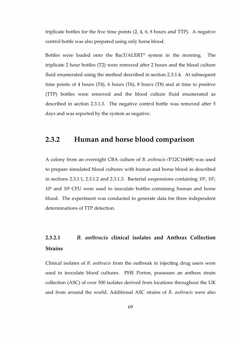
triplicate
control bo
Bottles w
triplicate
fluid enu
time poin
(TTP) bo
described
days and
2.3.2
A colony
to prepar
in section
104 and 1
blood. T
determin
2.3.2.1
Strains
Clinical i
used to
collection
and from
bottles for
ottle was a
were loade
2 hour bo
umerated u
nts of 4 ho
ottles wer
d in sectio
d was repor
Hum
y from an o
re simulate
ns 2.3.1.1, 2
106 CFU w
The experim
nations of T
B. a
isolates of
inoculate
n (ASC) of
m around t
r the five t
also prepar
ed onto th
ottles (T2) w
using the m
ours (T4),
e remove
n 2.3.1.3.
rted by the
man an
overnight
ed blood c
2.3.1.2 and
were used
ment was
TTP detecti
anthracis
B. anthrac
blood cult
over 500 i
the world.
6
time point
red using o
he BacT/A
were remo
method des
6 hours (T
ed and th
The nega
e system as
nd hors
CBA cultu
cultures wi
d 2.3.1.3. B
to inocula
conducted
ion.
s clinical
cis from th
tures. PH
isolates de
. Addition
69
ts (2, 4, 6, 8
only horse
ALERT® sy
oved after
scribed in
T6), 8 hour
he blood
ative contro
s negative.
se blood
ure of B. an
ith human
Bacterial su
ate bottles
d to genera
isolates
he outbreak
HE Porton
erived from
nal ASC str
8 hours an
blood.
ystem in
2 hours an
section 2.3
rs (T8) and
culture flu
ol bottle w
d comp
nthracis (P1
n and horse
uspensions
containing
ate data fo
s and An
k in injecti
n, possesse
m locations
rains of B.
nd TTP). A
the morn
nd the blo
3.1.4. At su
d at time t
uid enum
was remov
parison
12C16488)
e blood as
s containin
g human
or three ind
nthrax C
ing drug u
es an anth
s througho
. anthracis
A negative
ning. The
od culture
ubsequent
to positive
merated as
ved after 5
n
was used
described
ng 100, 102,
and horse
dependent
Collection
users were
hrax strain
out the UK
were also
e
e
e
t
e
s
5
d
d
,
e
t
n
e
n
K
o
used for
environm
were cho
(refer to A
2.3.3
A frozen
suspensio
2.3.1.2 u
inoculate
inoculatin
mental isola
osen by ph
Appendix
Con
n vial of B.
ons for blo
using hors
e blood cul
ng blood
ates from
hylogenetic
3.3 for furt
ncentra
. anthracis
ood cultur
se blood.
tures for a
7
cultures
the UK an
c relationsh
ther detail
ation of
strains or
re inoculat
The stra
range of c
70
and repre
nd USA. T
hip as dete
s).
f inocul
existing c
tion as des
ains shown
concentrati
esent a ran
The strains
ermined by
lum an
cultures w
scribed in
n in Table
ions (100 to
nge of cli
s used in t
y VNTR g
nd TTP
were used t
sections 2
e 2‐1 wer
o 106 CFU).
inical and
the project
genotyping
to prepare
2.3.1.1 and
re used to
d
t
g
e
d
o
Table 2‐1
2.3.4
Informati
National
anthracis
infections
informati
the UK, s
was inter
EIA resul
UK from
Reference
ASC 1
ASC 6
ASC 12
ASC 27
ASC 69
ASC 182
ASC 192
ASC 458
P10C001
P12C1297
P12C8461
P12C16488
B.anthracis
Hum
ion was ga
Library of
AND bact
s and revie
ion was ad
since 2006.
rrogated to
lts for antib
2009 to pr
Bacte
B. ant
B. ant
B. ant
B. ant
B. ant
B. ant
B. ant
B. ant
B. ant
B. ant
B. ant
8 B. ant
s strains use
man an
athered by
f Medicine
eraemia O
ews not pr
dditionally
The RIPL
o gather inf
body and t
resent.
erial isolate
thracis Vollu
thracis Stern
thracis ATC
thracis Envir
thracis New
thracis Paste
thracis Envir
thracis Clini
thracis Clini
thracis Clini
thracis Clini
thracis Clini
7
ed to inocul
nthrax c
y an electro
e®) databa
OR sepsis A
roviding s
y gathered
L laborator
formation
toxin for in
information
um (UK)
ne (UK), vac
C 10(7b)
ronmental,
w Hampshire
eur, pXO1 d
ronmental,
ical Scotland
ical Injection
ical Injection
ical Injection
ical Injection
71
late blood c
cases
onic literat
ase with th
AND huma
suitable inf
from refe
ry comput
of onset da
njectional a
n
ccine strain
Wales bovi
e
deficient
Landkey
d 2006
nal case 201
nal case 201
nal case 201
nal case 201
ultures
ture search
he search te
an (results
formation
rring front
er informa
ate, sample
anthrax ca
pXO2 defic
ine
10
12
12
12
h of MEDL
erms Anth
document
were exclu
t‐line labo
ation syste
e collection
ases diagno
cient
LINE (U.S.
hrax OR B.
ing in‐vivo
uded) and
ratories in
em (LIMS),
n date and
osed in the
.
.
o
d
n
,
d
e

2.4
2.4.1
Pilot wor
were inoc
(P10C129
in section
system a
work rep
bottle ino
was desc
time to d
inoculate
7.2 x10‐2 C
ml‐1 (Not
this study
not comp
The resul
of the me
of inocul
strains St
Bacillus s
occasion
to positiv
to B. anth
Res
Sim
rk for the
culated wi
97) ranging
n 2.3.1. Bot
and the tim
presents an
oculum an
cribed by S
detection
ed with a r
CFU ml‐1 (T
determine
y was 5.9
parable to t
lts from th
ethods des
um and T
taphylococc
spp. were u
a set of blo
ve results f
hracis (P12C
sults
mulated
project wa
ith 9ml hor
g from 5.9
ttles were
me to posi
n inverse lin
nd TTP det
Soloman &
determine
range of co
TTP 13.2 h
ed). The lo
x 10‐2 CFU
that determ
e pilot wor
cribed in s
TP detecti
us epidermi
used for in
ood culture
or bottles i
C1297). Da
7
d blood
as perform
rse blood a
x 10‐2 to 5.
loaded ont
itive detec
near relati
tection (Fi
& Jackson
ed by Hill
oncentratio
h), 7.2 x10‐3
owest conc
U with a T
mined by H
rk were us
section 2.3.
ion for diff
idis, Staphy
nvestigatio
es were sim
inoculated
ata are sho
72
d cultur
med and a
and 1ml su
.9 x106 CFU
to the BAC
tion record
ionship (r2=
igure 2‐1),
(1992) for
l & Spenc
on were 7.
CFU ml‐1 (
centration
TP detecti
Hill & Spen
sed to infor
. The relati
ferent bact
ylococcus au
ons detaile
mulated w
d with diffe
own in Figu
res
series of b
uspensions
U, using th
CT/Alert® C
ded. The r
= 0.998) be
an inverse
Brucella m
cer, (2007)
.2 x10‐1 CF
(TTP 15.7 h
inoculated
on of 17.0
ncer (2007)
rm the plan
ionship be
teria was m
ureus, Micr
ed in Chap
with these b
erent conce
ure 2‐2.
blood cultu
s of Bacillu
he method
Classic blo
results for
etween blo
e linear re
melitensis.
for aerob
FU ml‐1 (TT
h), and 7.2
d in the pil
hours, thi
.
nning and
etween con
made. Th
rococcus spp
pter Three
bacteria an
entrations
ure bottles
us anthracis
described
od culture
r this pilot
od culture
elationship
The mean
bic bottles
TP 11.8 h),
x10‐4 CFU
ot work in
is result is
d execution
ncentration
e bacterial
p. and two
e. On one
nd the time
compared
s
s
d
e
t
e
p
n
s
,
U
n
s
n
n
l
o
e
e
d
Figure 2-2
for differe
1e-2
Tim
e to
pos
itive
(h)
4
6
8
10
12
14
16
18
20
Figure 2-1
anthracis
2 Relationsh
ent bacteria
Bloo
1e-1 1e
1 Relations
(P12C1297)
hip between
using horse
od culture ino
+0 1e+1
ship betwe
using horse
7
n the conce
e blood.
oculum conce
1e+2 1e+3
(Tota
(To
een concent
e blood.
73
entration of
entration (CF
1e+4 1e+
al CFU)
tal CFU)
ration of in
inoculum a
FU ml-1)
+5 1e+6
noculum an
and TTP de
1e+7
CoRe95%95%
P
nd TTP det
etection
onetration vs Timegression% Confidence B% Prediction Ba
P12C1297
tection for
me
Band and
B.
74
2.4.1.1 TTP variation for simulated blood cultures
The reproducibility of preparing simulated aerobic blood cultures was tested by
determining the TTP detection with bottles initially inoculated with a range of
concentrations (100, 102, 104 and 106 total CFU) of B. anthracis (P10C0001). The
same overnight BHI culture was used to prepare three sets of suspensions. The
concentration of inoculum, TTP detection, mean TTP detection (decimal h) and
standard error are shown in Table 2‐2. The range in TTP for bottles inoculated
with 100 CFU is 0.3 decimal hours (18 minutes), 102 CFU is 0.4 decimal hours (24
minutes), 104 CFU is 0.3 decimal hours (18 minutes), and 106 CFU is 0.5 decimal
hours (30 minutes).
Table 2-2 TTP detection for simulated blood cultures.
Time to positive detection (TTP) in decimal hours for three sets of simulated blood
cultures prepared using three dilutions of the same broth culture of B. anthracis.
Inoculum
(Total CFU)
TTP
detection (h)
Mean TTP
detection (h)
Standard
error (h)
4.10 x100
4.93 x100
5.56 x100
13.0
12.8
12.7
12.8 0.09
4.10 x10²
4.93 x10²
5.56 x10²
11.0
11.2
10.8
11.0 0.12
4.10 x10⁴
4.93 x10⁴
5.56 x10⁴
8.8
8.7
8.5
8.7 0.09
4.10 x10⁶
4.93 x10⁶
5.56 x10⁶
6.0
5.7
5.5
5.7 0.14
75
2.4.1.2 Effect of head space atmosphere on growth
A method to remove the sharps hazard was chosen to prepare and inoculate
blood cultures. Studies were conducted to determine if the removal of the
blood culture rubber seal affected the growth of B. anthracis in aerobic blood
cultures due to the alteration of the atmosphere in the head space.
The mean time to positive flag for triplicate capped and uncapped aerobic
blood cultures inoculated with 1.5 x10³ CFU Bacillus endophyticus were, capped
8.3 ± 0.03 h (mean ± s.e), and uncapped 8.1 ± 0.10 h (mean ± s.e). The difference
in mean TTP detection between capped and uncapped bottles was 0.2 h. The
method was repeated using B. anthracis (P12C16488) inoculated with a range of
concentrations (5.7 x 100 to 5.7 x 10⁶ CFU) in both capped and uncapped bottles
and in replicate bottles inoculated with a low concentration 57 CFU (n=5 capped
and n=3 uncapped). A scatter plot of the data shows an association between
inoculum (Total CFU) and TTP detection (decimal hour), for the capped and
uncapped bottle data (Figure 2‐3). The mean TTP detection and standard error
for capped bottles was 13.7 ± 0.14 h (mean ± s.e) and uncapped bottles 14.6 ±
0.10 h (mean ± s.e) with a difference between the two treatment means of 0.9 h.
A possible factor contributing to the difference between capped and uncapped
bottles was the actual delivery of inoculum to capped bottles using the safety
adaptor and Luer® lock syringe.
To examine this possibility, B. anthracis (Sterne) was used to simulate capped
and uncapped bottles inoculated with a low concentration (1.04 x 101 CFU),
with addition of the bacterial suspension using a blunt ended needle and
syringe. B. anthracis (Sterne) at a low concentration was chosen to reduce the
infectious hazard for the procedure. The mean TTP detection for capped bottles
was 10.9 ± 0.05 h (mean ± s.e), and uncapped bottles was 11.3 ± 0.14 h (mean ±

76
Blood culture inoculum concentration (CFU ml-1)
1e+0 1e+1 1e+2 1e+3 1e+4 1e+5 1e+6 1e+7
Tim
e to
pos
itive
(h)
4
6
8
10
12
14
16
18Capped [low]Uncapped [low]CappedUncapped
s.e). The difference in mean TTP detection for the capped and uncapped bottles
using the blunt needle delivery was 0.4 h.
Figure 2-3 Effect of head space atmosphere on growth. Relationship between the
concentration of inoculum and TTP detection of B. anthracis (P12C16488) using
horse blood for capped and uncapped aerobic blood culture bottles.
2.4.1.3 Total viable count variation
To assess the variation of total viable counts (TVC) for estimating the
concentration of bacteria in positive blood cultures, an initial investigation was
conducted. The counts were log transformed to perform an Analysis of
Variance (ANOVA) statistical analysis using SPSS (IBM). The test statistic F=
0.463 and corresponding p‐value (p= 0.304) indicate no significant difference at
the 5% significance level (p<0.05) between the mean values for transformed
count data from triplicate dilutions of the same blood culture fluid (Appendix
2.2). There is high confidence in the consistency of TVC estimations of total
viable bacterial concentrations in the positive blood culture fluid.
(Total CFU)
77
2.4.1.4 Growth at different time points
In a front‐line laboratory blood culture bottles may flag positive during the
night and processed the following day. To consider this situation an experiment
was conducted to determine the concentration of B. anthracis at different time
points. B. anthracis (clinical isolate P12C1297) was initially used to determine
the concentration at time points T2 hours, T4 hours, T6 hours, T8 hours and
TTP. A second experiment using B. anthracis (Vollum) was conducted to
determine the concentration at time points T4, T8, TTP, 4 hours post TTP
(TTP+4) and 8 hours post TTP (TTP+8).
The initial concentration of B. anthracis (P10C1297) inoculated was 6.6 CFU and
1.3 CFU (Vollum) representing low bacteraemia. Triplicate bottles were
removed from the BacT/ALERT® incubator unit at different time points and the
blood culture fluid enumerated using TVC. The mean concentrations
(geometric mean with 95% confidence intervals) for triplicate bottles of B.
anthracis (P12C1297 and Vollum) removed at different time points are
graphically represented in Figure 2‐4. The data suggests that there is no
dramatic decrease in the bottle concentration after 4 hours and 8 hours post TTP
detection therefore not presenting a problem if bottles flag positive overnight.
The concentration of bacteria at time points 4 and 8 hours was used to
determine the generation time (doubling time) of B. anthracis in simulated
aerobic blood cultures. The generation time was calculated by dividing the
interval time by the number of generations which uses the Log (final count) –
Log (initial count) divided by Log (2) to express growth by binary fusion. The
calculated doubling time for B. anthracis was 28.81 minutes with 95% confidence
limits of 23.55 to 43.53 minutes (Vollum) and 28.14 minutes with 95%
confidence limits of 25.78 to 37.54 minutes (P12C1297).

(P12C1297
bottle geo
2.4.2
The meth
methods
volunteer
simulated
Porton fo
blood com
of blood u
between
the metho
Figure 2-4
Tot
al v
iabl
e co
unt (
CF
U m
l-1)
1e+0
1e+1
1e+2
1e+3
1e+4
1e+5
1e+6
1e+7
1e+8
1e+9
7 and Vollu
moetric mea
Hum
hod for sim
described
r human b
d blood cu
or inducin
mmercially
used in blo
human vo
ods describ
Concentra
2 hour 4 ho
um strain).
ans.
man and
mulating b
d by Solom
blood and
ultures with
ng capsule
y supplied
ood culture
olunteer blo
bed in sect
tion of B. an
Time
ur 6 hour 8 h
7
Error bars
d horse
blood cultu
man & Ja
d Funke a
h sheep blo
productio
d by TCS B
es affected
ood and d
tion 2.3.2.
nthracis at d
e point
hour TTP T
78
indicate th
e blood
ures is base
ackson (19
and Funke
ood. A com
on in B. a
Biosciences
d the growt
defibrinated
different tim
TTP +4 TTP + 8
he standard
d compa
ed on prev
992) for B
‐Kissling (
mmon bloo
nthracis is
Ltd. To d
th of B. ant
d horse blo
me points
P12Voll
error of tr
arison
viously do
Brucella me
(2004) for
od type us
s defibrina
determine
thracis, a co
ood was m
2C1297um
riplicate
ocumented
elitensis in
E. coli in
sed at PHE
ated Horse
if the type
omparison
made using
d
n
n
E
e
e
n
g
79
The relationship between the concentration of inoculum and TTP detection in
human and horse blood is presented in the scatter plot (Figure 2‐5), the data
points are for three independent determinations.
Relationship between the concentration of inoculum and TTP detection for B.
anthracis (P12C16488) using human and horse blood.
The scatter plot indicates an inverse linear relationship between initial
inoculum concentration and TTP detection for both human and horse blood
cultures. Linear regression was performed on the paired data for each
determination with human and horse blood cultures. A summary of the linear
regression line equation values for the human and horse blood is shown in
Table 2‐4. (line equation y= a ln(x)+yo). The coefficient r2 indicates a strong
association between the paired values and the regression line equation. The
coefficient r2 is a measure of the portion of variability in Y explained by the
regression. It is the proportion of variation in Y that can be attributed to X. The
coefficient r2 is a measure of the strength of the linear relationship between X
and Y and the residual or unexplained variability is 1‐ r2.
Blood culture inoculum concentration (CFU ml-1)
1e-1 1e+0 1e+1 1e+2 1e+3 1e+4 1e+5 1e+6 1e+7 1e+8
Tim
e to
pos
itive
(h)
4
6
8
10
12
14
16
18
Human blood 1 Human blood 2 Human blood 3 Horse blood 1 Horse blood 2 Horse blood 3
Figure 2-5 Effect of blood type and TTP detection of B. anthracis
(Total CFU)
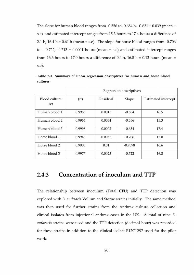
The slope
s.e) and e
2.1 h, 16.4
to – 0.72
from 16.6
s.e).
Table 2-3
cultures.
Blood cu
set
Human b
Human b
Human b
Horse blo
Horse blo
Horse blo
2.4.3
The rela
explored
was then
clinical is
anthracis
for these
work.
e for huma
estimated
4 h ± 0.61 h
2, ‐0.713 ±
6 hours to
Summary
ulture
t
lood 1
lood 2
lood 3
ood 1
ood 2
ood 3
Conc
ationship b
with B. an
n used for
solates fro
strains we
strains in
an blood ra
intercept r
h (mean ±
± 0.0004 ho
17.0 hours
of linear r
(r2)
0.9985
0.9966
0.9998
0.9948
0.9900
0.9977
centrat
between i
nthracis Vol
r further
om injectio
ere used an
n addition
8
anges from
ranges from
s.e). The
ours (mea
s a differen
regression d
R
Resi
0.0
0.0
0.0
0.0
0.
0.0
tion of
inoculum
llum and S
strains fro
onal anthra
nd the TTP
to the clin
80
m ‐0.556 to ‐
m 15.3 hou
slope for h
n ± s.e) an
nce of 0.4
descriptives
Regression d
idual
0015
0034
0002
0052
.01
0023
inoculu
(Total CF
Sterne strai
om the An
ax cases in
P detection
nical isolat
‐0.684 h, ‐0
urs to 17.4 h
horse blood
nd estimat
h, 16.8 h ±
s for human
descriptives
Slope
‐0.684
‐0.556
‐0.654
‐0.706
‐0.7098
‐0.722
um and
FU) and T
ins initially
nthrax cul
n the UK.
n (decimal
e P12C129
0.631 ± 0.03
hours a dif
d ranges fr
ted interce
± 0.12 hour
n and horse
s
Estimate
1
1
1
1
1
1
d TTP
TTP dete
y. The sam
lture colle
A total
hour) was
97 used fo
39 (mean ±
fference of
rom ‐0.706
ept ranges
rs (mean ±
e blood
ed intercept
16.5
15.3
17.4
17.0
16.6
16.8
ction was
me method
ection and
of nine B.
s recorded
r the pilot
±
f
6
s
±
s
d
d
d
t
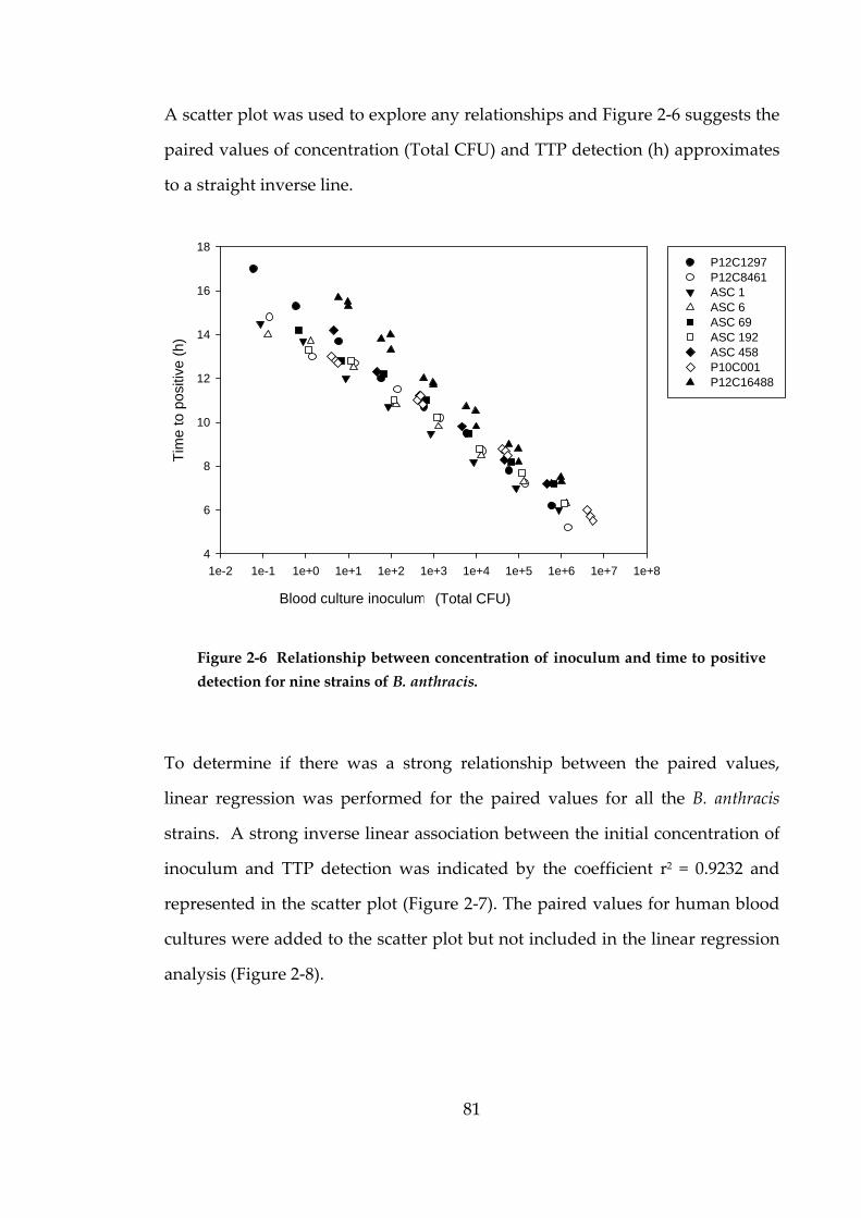
81
Blood culture inoculum concentration (CFU ml-1)
1e-2 1e-1 1e+0 1e+1 1e+2 1e+3 1e+4 1e+5 1e+6 1e+7 1e+8
Tim
e t
o p
ositi
ve (
h)
4
6
8
10
12
14
16
18P12C1297 P12C8461 ASC 1 ASC 6 ASC 69 ASC 192 ASC 458 P10C001 P12C16488
A scatter plot was used to explore any relationships and Figure 2‐6 suggests the
paired values of concentration (Total CFU) and TTP detection (h) approximates
to a straight inverse line.
To determine if there was a strong relationship between the paired values,
linear regression was performed for the paired values for all the B. anthracis
strains. A strong inverse linear association between the initial concentration of
inoculum and TTP detection was indicated by the coefficient r2 = 0.9232 and
represented in the scatter plot (Figure 2‐7). The paired values for human blood
cultures were added to the scatter plot but not included in the linear regression
analysis (Figure 2‐8).
(Total CFU)
Figure 2-6 Relationship between concentration of inoculum and time to positive
detection for nine strains of B. anthracis.
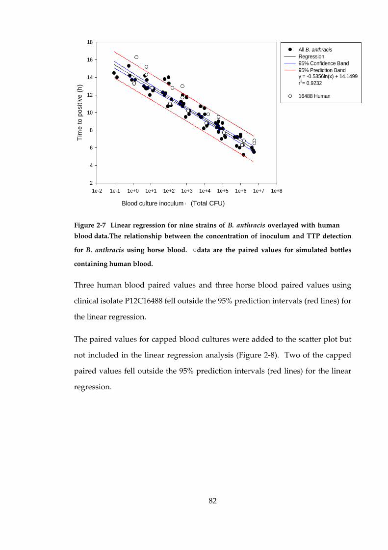
82
The relationship between the concentration of inoculum and TTP detection
for B. anthracis using horse blood. ○data are the paired values for simulated bottles
containing human blood.
Three human blood paired values and three horse blood paired values using
clinical isolate P12C16488 fell outside the 95% prediction intervals (red lines) for
the linear regression.
The paired values for capped blood cultures were added to the scatter plot but
not included in the linear regression analysis (Figure 2‐8). Two of the capped
paired values fell outside the 95% prediction intervals (red lines) for the linear
regression.
Blood culture inoculum concentration (CFU ml-1)
1e-2 1e-1 1e+0 1e+1 1e+2 1e+3 1e+4 1e+5 1e+6 1e+7 1e+8
Tim
e to
po
sitiv
e (
h)
2
4
6
8
10
12
14
16
18All B. anthracis Regression95% Confidence Band 95% Prediction Band y = -0.5356ln(x) + 14.1499r2= 0.9232
16488 Human
(Total CFU)
Figure 2-7 Linear regression for nine strains of B. anthracis overlayed with human
blood data.
83
Figure 2-8 Linear regression for nine strains of B. anthracis overlayed with capped
data. The relationship between the concentration of inoculum and TTP detection for
B. anthracis using horse blood. The red circles are the paired values for capped
bottles using horse blood.
Blood culture inoculum concentration (CFU ml-1)
1e-2 1e-1 1e+0 1e+1 1e+2 1e+3 1e+4 1e+5 1e+6 1e+7 1e+8
Tim
e t
o po
sitiv
e (
h)
2
4
6
8
10
12
14
16
18All B. anthracis Regression95% Confidence Band 95% Prediction Band y = -0.5356ln(x) + 14.1499r2= 0.9232
16488 Human Capped 16488
(Total CFU)
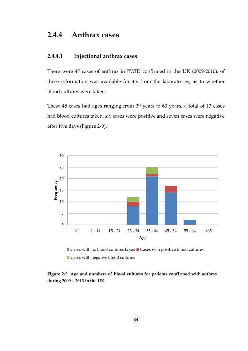
2.4.4
2.4.4.1
There we
these inf
blood cul
These 45
had blood
after five
Figure 2-9
during 200
0
5
10
15
20
25
30
Frequency
Anthr
Injecti
ere 47 case
ormation
ltures were
cases had
d cultures
days (Figu
9 Age and n
09 – 2013 in
<1
Cases with
Cases with
rax cas
ional anth
es of anthr
was availa
e taken.
d ages rang
taken, six
ure 2‐9).
numbers of
the UK.
1 ‐ 14 15
h no blood cu
h negative blo
8
es
hrax case
rax in PW
able for 45
ging from
cases were
f blood cultu
5 ‐ 24 25 ‐
ultures taken
ood cultures
84
s
WID confirm
5, from th
29 years t
e positive
ures for pat
34 35 ‐ 44
Age
n Cases wit
s
med in the
he laborato
to 60 years
and seven
tients confir
4 45 ‐ 54
th positive bl
e UK (2009
ories, as to
s, a total o
n cases wer
rmed with
55 ‐ 64
lood culture
9‐2010), of
o whether
of 13 cases
re negative
anthrax
>65
s
f
r
s
e
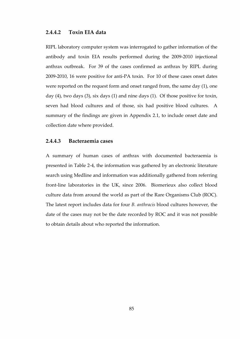
85
2.4.4.2 Toxin EIA data
RIPL laboratory computer system was interrogated to gather information of the
antibody and toxin EIA results performed during the 2009‐2010 injectional
anthrax outbreak. For 39 of the cases confirmed as anthrax by RIPL during
2009‐2010, 16 were positive for anti‐PA toxin. For 10 of these cases onset dates
were reported on the request form and onset ranged from, the same day (1), one
day (4), two days (3), six days (1) and nine days (1). Of those positive for toxin,
seven had blood cultures and of those, six had positive blood cultures. A
summary of the findings are given in Appendix 2.1, to include onset date and
collection date where provided.
2.4.4.3 Bacteraemia cases
A summary of human cases of anthrax with documented bacteraemia is
presented in Table 2‐4, the information was gathered by an electronic literature
search using Medline and information was additionally gathered from referring
front‐line laboratories in the UK, since 2006. Biomerieux also collect blood
culture data from around the world as part of the Rare Organisms Club (ROC).
The latest report includes data for four B. anthracis blood cultures however, the
date of the cases may not be the date recorded by ROC and it was not possible
to obtain details about who reported the information.

86
Table 2-4 Anthrax cases describing bacteraemia and blood cultures
Case Year Location and reference Description
1 2001 Barakat et al. (2002)
USA
11th case of bioterrorism‐related
inhalational anthrax reported in the United
States. Admitted 16th, BCs +ve 17th, died 21st
Nov. (BacT/Alert®) all 4 blood culture
bottles positive after 14 hours of incubation
(outcome – fatal).
2 2002 Tse, Yeung, Trendell‐
Smith, & Kwong,
(2002), Hong Kong
Cutaneous anthrax case, blood culture grew
E. coli (outcome ‐fatal E. coli septicaemia).
3 2006 Riley, (2007), Scottish
borders
Inhalational anthrax ‐Positive blood culture
, taken 7th July, positive 8th July, second set
of BCs negative (outcome – fatal)
4 2006 Walsh et al. (2007), USA Blood cultures taken from patient in
Pennsylvania, US and positive in <12 hours
in 4 of 4 bottles via the BacT/Alert® System
(outcome – survived).
5 2008 Hackney, London Inhalational anthrax, Blood cultures
positive but no further information
(outcome – fatal).
6 2009 Biomerieux ROC Time to detection 17 hours for aerobic and
anaerobic bottles (unknown outcome or
origin of information).
7 2009 Glasgow Royal
Infirmary, Scotland
2 cases, at least 1 case had positive blood
culture – no further details
8 2009 Southern General
Glasgow, Scotland
2 x BC set taken both negative (outcome‐
survived)
9 2009 Southern General
Glasgow, Scotland
Blood culture set taken 09/12/09 16:40,
received at lab 19:42, 10/12/09 aerobic bottle
positive 06:07 (TTP 10.42) anaerobic bottle
positive 07:57 (TTP 12.25) (outcome‐ fatal).
10 2009 Southern General
Glasgow
Blood culture set taken 07/12/09, aerobic
bottle positive 19:15 (TTP 9.64) and
anaerobic bottle positive 19:35 (TTP 9.97) 2
further sets taken both negative (outcome‐
survived).
87
Case Year Location and reference Description
11 2010 Southern General
Glasgow, Scotland
Blood culture set received at lab 04/02/10
21:19, aerobic bottle positive 5/02/10 05:25
(TTP 8.1) and anaerobic bottle positive 04:35
(TTP 7.27) (outcome‐ survived).
12 2010 Hairmyres Glasgow,
Scotland
BacT/ALERT® blood cultures taken
06/05/2010 12:30, received at laboratory
14:42 aerobic bottle +ve 8.3h , anaerobic
bottle +ve 7.7h (outcome‐ fatal).
13 2010 Ninewells, Scotland Blood culture negative
14 2010 Ninewells, Scotland Blood cultures taken 06/01/2010 and
positive 07/01/2010 (outcome‐ survived).
15 2010 Crosshouse, Scotland Positive blood culture – no further details
16 2010 London
Imperial, England
Positive blood culture – no further details
17 2010 Leicester, England 2 x sets of BC both negative
18 2010 Royal Infirmary
Edinburgh, Scotland
Positive blood culture – no further details
19 2010 Charing Cross
Hospital, England
2 cases, both positive blood cultures – no
further details
Jallali, Hettiaratchy, Gordon, & Jain, (2011)
20 2010 Knox et al. (2011)
Dumfries and
Galloway Royal
Infirmary
Negative blood cultures
21 2010 Powell, Crozier,
Hodgson, & Galloway
(2011)
General Hospital,
Glasgow, Scotland
Within 24 hours of collection, all 6 blood
culture bottles grew Gram positive
organisms suggestive of Bacillus anthracis.
(outcome – survived)
22 2011 Biomerieux ROC Time to positive 0.6 day for aerobic bottle
and 0.56 day for anaerobic bottle (unknown
outcome or origin of information)
88
Case Year Location and reference Description
23 2011 Biomerieux USA 2 sets of BacT/ALERT® blood cultures
positive in 13 hours. (outcome‐ survived)
24 2012 Micropath Wirral, Blood cultures negative at 5 days
25 2012 Victoria Infirmary
Blackpool, England
Aerobic bottle positive 12/8/12 (15:55)
Anaerobic bottle positive 13/8/12 (08:44)
(outcome – fatal)
26 2012 Victoria Infirmary
Blackpool, England
Aerobic bottle positive 04/9/12 (09:12)
Anaerobic bottle positive 04/9/12 (16:08)
(outcome – fatal)
27 2012 Grunow et al. (2012),
Germany
The blood culture taken and flagged
positive after 3.5 hours. (outcome –
survived, treatment as outpatient)
28 2012 Holzmann et al. (2012),
Germany
Blood cultures (Becton Dickinson) taken
from patient in Germany turned positive
after 53 minutes of incubation (outcome –
fatal).
29 2013 James Paget Norfolk Patient admitted 27/02/13, Blood cultures
taken 01/03/13 but negative
(outcome – survived)
30 2013 Russell, Pedersen,
Jensen, Soes, & Hansen,
2013
Danish case 1 Bacillus anthracis was found in
both aerobic blood cultures (Becton
Dickinson) no times given (outcome –
survived)
Danish case 2 aerobic blood cultures
positive (BacT/ALERT®) no times given
(outcome – survived)
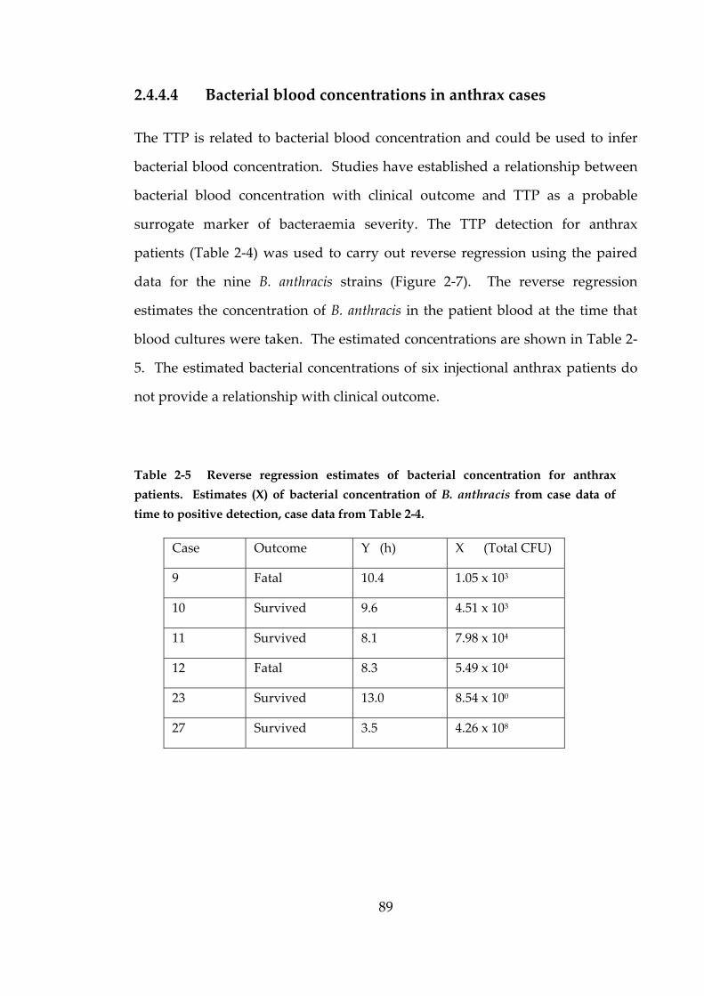
89
2.4.4.4 Bacterial blood concentrations in anthrax cases
The TTP is related to bacterial blood concentration and could be used to infer
bacterial blood concentration. Studies have established a relationship between
bacterial blood concentration with clinical outcome and TTP as a probable
surrogate marker of bacteraemia severity. The TTP detection for anthrax
patients (Table 2‐4) was used to carry out reverse regression using the paired
data for the nine B. anthracis strains (Figure 2‐7). The reverse regression
estimates the concentration of B. anthracis in the patient blood at the time that
blood cultures were taken. The estimated concentrations are shown in Table 2‐
5. The estimated bacterial concentrations of six injectional anthrax patients do
not provide a relationship with clinical outcome.
Table 2-5 Reverse regression estimates of bacterial concentration for anthrax
patients. Estimates (X) of bacterial concentration of B. anthracis from case data of
time to positive detection, case data from Table 2-4.
Case Outcome Y (h) X (Total CFU)
9 Fatal 10.4 1.05 x 10³
10 Survived 9.6 4.51 x 10³
11 Survived 8.1 7.98 x 10⁴
12 Fatal 8.3 5.49 x 10⁴
23 Survived 13.0 8.54 x 100
27 Survived 3.5 4.26 x 10⁸
2.5
A search
system re
examinin
which de
identifica
relevance
The stu
BacT/ALE
growth.
concentra
identified
concentra
type of b
for B. ant
the effec
preparati
culture of
calculated
triplicate
triplicate
concentra
The stand
0.09 and
minutes 4
bacterial
Discu
h of literat
evealed a
ng the gro
etailed sim
ation. Of t
e to investi
dy invest
ERT® auto
Pilot wo
ation of in
d. The va
ation of th
lood. The
thracis simu
ct of these
ion of bott
f B. anthrac
d concentr
dilution s
dilutions
ations (100,
dard error
0.14 hours
44 seconds
suspension
ussion
ture for sim
relevant s
wth of Br
mulating bl
these Funk
igations de
tigations
omated sy
ork show
noculum an
ariables con
he inoculu
e disparity
ulated bloo
e variables
tle inoculu
cis was use
ration of
series) was
were use
, 102, 104 a
r for the ge
s (decimal
s. The vari
ns was dee
9
n
mulating b
study cond
rucella meli
lood cultu
ke and Fun
escribed in
simulated
ystem to
wed an inv
nd TTP de
nsidered w
um, alterat
between p
od culture
s. To de
um and res
ed to prepa
the initial
s 4.10 x10⁶
ed to inoc
and 106 CFU
eometric m
l hour) and
iation in T
emed accep
90
blood cult
ducted by
itensis. Se
res to com
nke‐Kisslin
Chapter T
d aerobic
incubate,
verse line
etection, ho
were the p
tion of the
previously
s (Hill & S
etermine i
sulting TT
are triplica
BHI brot
6, 4.93 x10⁶
culate blo
U) and the
means of T
d equate t
TTP genera
ptable for t
tures using
Solomon
everal stud
mpare subs
ng (2004) w
Three.
blood c
monitor
ear relatio
owever sev
potential fo
e head spa
y reported
Spencer, 20
f there w
P detection
ate 10‐fold
th culture
6 and 5.56
od culture
e TTP dete
TTP detect
o 5 minute
ted from t
the purpos
g the BacT
and Jacks
dies were
sequent me
was chosen
cultures u
and aler
onship bet
veral varia
for variabi
ace atmosp
mean TTP
007) are lik
was variati
n, an over
serial dilu
(using TV
x106 CFU
es with a
ection was
tion ranged
tes 24 seco
three prepa
ses of this
T/ALERT®
son (1992),
identified
ethods for
n for their
using the
t positive
tween the
ables were
lity in the
phere and
P detection
kely due to
on in the
rnight BHI
tions. The
VC of the
ml‐1. The
a range of
recorded.
d between
onds and 8
arations of
study.
®
,
d
r
r
e
e
e
e
e
d
n
o
e
I
e
e
e
f
.
n
8
f
91
To safely simulate blood cultures in MSC III, within the CL3 laboratory, a
method was developed to remove the sharps hazard. This method required
the removal of the blood culture bottle rubber stopper and therefore released
the vacuum and predetermined atmosphere within the head space of the blood
culture bottle. BacT/ALERT® aerobic blood culture bottles are prepared with an
atmosphere of carbon dioxide in oxygen under vacuum. The effect of removing
the vacuum and predetermined atmosphere on the growth and therefore TTP
detection by the BacT/ALERT® system was investigated. The time to positive
detection for bottles inoculated with removal of the rubber stopper (uncapped)
and those without removal (capped) were tested with B. endophyticus and B.
anthracis clinical isolate (P12C16488). The difference in mean TTP detection for
the capped and uncapped bottles was 0.2 h for B. endophyticus and 0.9 h for B.
anthracis (P12C16488) which equates to 12 minutes and 54 minutes respectively.
The actual procedure used to inoculate the capped bottles may account for the
variation in TTP. The method used Luer® lock syringes and safety adaptors
and it was difficult to ensure the exact volume of inoculum was delivered to the
bottle. Further work was conducted to inoculate a low concentration of B.
anthracis (Sterne) into blood culture bottles without the removal of the rubber
stopper. Horse blood was inoculated using the safety adaptor and Luer® lock
syringe and the bacterial suspension was inoculated using a blunt needle and
syringe. The Sterne strain was chosen and a low concentration inoculated to
reduce the infectious hazard. The difference in mean TTP detection (n=5)
between capped and uncapped bottles with this method of inoculation was 0.4
h (decimal hour) which equates to 24 minutes. The effect of removing the
predetermined head space on resulting TTP detection was considered low and
24 minutes variation was more acceptable compared to 54 minutes initially
determined.
92
Without further testing to generate sufficient data for confidence in statistical
analysis it cannot be shown to have a significant difference.
Defibrinated horse blood was used to simulate blood cultures for the majority
of the study due to it being commercially available and the time required to
organise collection of volunteer blood. Human blood was collected from
healthy, unvaccinated volunteers to compare with the use of defibrinated horse
blood. No specific data on the susceptibility of horses to anthrax was found in
the literature, though it is generally accepted that horses and humans have a
lower susceptibility to anthrax compared to other animals, such as cattle and
sheep. Another possible factor which may alter the growth of the bacteria
would be the action of complement and macrophages present in the human
blood, within the blood culture bottle. The effect of these human blood
constituents should not, however, have a large effect because Sodium
polyanethol sulfonate (SPS) in blood culture media has anticoagulant properties
and also counter‐acts the bacterial inhibitors of human blood. SPS is known to
neutralize the bactericidal activity of fresh human serum and to inhibit
phagocytosis (Salventi et al., 1979).
The paired study data for the concentration of inoculum and TTP detection was
used to produce a scatter plot to perform linear regression. The slope of the line
for human blood was determined as ‐0.631 ± 0.039 (mean ± s.e; n=3) with an
estimated intercept of 16.4 hours ± 0.61 hours (mean ± s.e; n=3). The slope of the
line for horse blood was determined as ‐0.713 ± 0.0004 hours (mean ± s.e; n=3)
with an estimated intercept as 16.8 hours ± 0.12 hours (mean± s.e; n=3). From
this data, an initial concentration of 1 CFU in human blood (10 ml) gives an
estimated TTP detection of 16.4 hours and for every 10 fold increase in
concentration the estimated TTP detection decreases by 1.45 hours.
93
Similarly, an initial concentration of one CFU in horse blood (10 ml) gives and
estimated TTP detection of 16.8 hours and for every 10 fold increase in
concentration the estimated TTP detection decreases by 1.64 hours. The
difference in 1.64 hours and 1.45 hours is 0.19 hours, equivalent to 11 minutes
24 seconds. The effect of different blood as a culture media on the growth of B.
anthracis was considered low and without further testing to generate sufficient
data for confidence in statistical analysis it cannot be shown to have a
significant effect or difference due to chance alone. The choice of horse blood to
simulate aerobic blood cultures for the majority of investigations was justified
for the purposes of the exploratory investigations in this study. Future work
would collect human blood directly into blood culture bottles followed by blunt
ended needle inoculation of the bacterial suspension to more closely simulate
human blood cultures. This method would alleviate the potential factors of
blood and head space atmosphere on the growth of bacteria. In this situation,
the potential variation in preparing the bacterial suspension would be the only
remaining factor to consider.
The estimations of bacterial concentration in the blood have obvious limitations
due to the potential factors affecting the TTP detection data generated in this
study. The factors identified were the type of blood used to simulate the blood
cultures, the method of inoculation which altered the predetermined
atmosphere in the bottle head space both of which were shown to have minimal
effect. The inherent inaccuracy of the total viable count method to determine
bacterial concentration may also contribute to estimates derived from these
data. The TVC method determines only the concentrations of viable bacteria
and relies on the assumptions that the small sample taken from the population
of bacteria in suspension is representative of the whole population therefore the
culture is homogeneous, all viable cells will grow to produce a single colony
and the growth conditions allow for the growth of all the bacteria sampled.

94
The consistency of preparing dilutions for TVC was examined and shown to
have no significant difference between counts from triplicate dilutions.
The relationship between concentration and TTP detection for nine B. anthracis
strains (five ASC stains and four clinical isolates) was shown to follow an
inverse linear relationship. The linear regression for all strains (Figure 2‐8)
describes the association of concentration (Total CFU) and TTP detection (h) by
the BacT/ALERT® system by the line equation y= ‐0.5356 ln(x)+14.1499. Paired
values for all strains except three points for clinical isolate P12C16488, fell
within 95% prediction. When the paired values for human blood were added to
the scatter plot only three fell outside these limits. Similarly, the overlay for
capped paired data revealed only two falling outside the limits. This reinforces
the conclusion that the potential effect of blood type and head space
atmosphere on TTP detection was considered insignificant. Using the line
equation, a concentration of one CFU, the estimated TTP detection is 14.15
hours and for every tenfold increase in concentration the estimated time to
positive detection decreases by 1.23 hours. The inverse linear relationship
demonstrated by Soloman & Jackson (1992) for Brucella melitensis cannot be
compared to B. anthracis due to large differences in growth characteristics and
insufficient data provided in the literature. The relationship between
concentration and TTP detection for other bacteria used in this study were only
determined on one occasion. However, it can be clearly seen in Figure 2‐2, the
two Bacillus spp. and S. aureus have a similar slope to B. anthracis compared to
the other bacteria (S. epidermidis and Micrococcus spp.).
Several studies have established a relationship between bacterial blood
concentrations with clinical outcome of bacteraemic patients (Khatib, et al., 2005;
Willmann, et al., 2013; Hsu, Huang, Hsu and Liao., 2014; Huang, Sun and Yan,
2014). To examine whether this was the case with injectional anthrax patients,
95
the relationship between concentration and TTP detection for nine strains of B.
anthracis was used. The line equation determined in this study was used to
perform reverse regression with the time to positive detection data received
from referring hospitals during the outbreak of injectional anthrax in the UK, to
estimate the concentration of circulating B. anthracis cells at the time of sample
collection (Table 2‐6). Of the cases used to estimate bacterial blood
concentration, four cases survived anthrax and their estimated concentrations
ranged from low bacteraemia (8.54 CFU) to very high bacteraemia (4.26 x 10⁸
CFU) and two fatal cases had estimated concentrations in the order of 103 and
104 CFU. The small data set of six patients suggests bacterial blood
concentration does not have a relationship with clinical outcome for injectional
anthrax. The outcome of systemic anthrax in these patients will not have been
necessarily wholly dependent on the concentration of circulating bacteria in the
blood, but how long they had a bacteraemia. Several factors would have been
involved with either their favourable or fatal outcomes; one being prompt and
appropriate antibiotic treatment which can quickly clear bacteria in the blood,
although high levels of exotoxin would remain and can ultimately result in
shock and death.
For a bacteraemia to occur there would have to be sufficient bacteria in a focal
point of infection to then disseminate in to the circulatory system. At the focal
point, the bacteria would have been able to overcome the local immune
responses, aided by the presence of protective capsule and the action of the
exotoxins on preventing macrophages to signal immune responses. Once bacilli
disseminate from the focal point, the systemic immune responses would mop
up some of the bacteria with initial bacteraemia being low for several hours but
the production of exotoxins would then again enable the bacteria to multiply
without much problem until reaching a concentration of approximately 108
before death of the host. At this concentration there are sufficient bacteria in
96
the blood to perform direct microscopy, a reason why this diagnostic test is
performed on blood sampled in cases of sudden death occurring in livestock.
The high concentration of bacteria in the blood is not common among the many
bacterial species which cause bacteraemia, the only other being Yersinia pestis
which can also reach levels of 108 CFU ml‐1.
The linear regression analysis describes the relationship between concentration
and TTP detection assumes the x explanatory variable can be measured without
error. In this study it was determined by TVC, therefore not necessarily
without inherent limitations and examination of count variance provides
confidence in consistency for this study. It would be possible to use reverse
regression to estimate future unknown x values, to more reliably estimate
bacterial blood concentration, if this limitation was addressed. For this purpose,
standard inoculums could be prepared using optical density and quantified by
PCR. Blood cultures could be simulated with human blood inoculated with
blunt ended needles so the data generated would more closely represent the
real situation. These data could then be used to generate a calibration curve for
reverse regression estimations of future unknown bacterial blood
concentrations.
Blood cultures may flag positive on automated systems overnight in routine
front‐line clinical laboratories and therefore may not be processed directly after
being determined positive, due to limited out of hours staffing. To simulate this
situation, blood cultures were sampled at different time points and
concentration of bacteria determined by TVC. At time points 4 and 8 hours
post positive detection (TTP +4 and TTP +8 hour) did not show any significant
reduction in concentration.
Processing B. anthracis bottles after this time was not considered a problem
because the diagnostic tests to be performed were not dependant on growth
97
phase. Blood cultures would not be routinely removed from the automated
system before the time to positive detection however, time points at 2 hours, 4
hours and 8 hours post inoculation were taken to look at the doubling time of B.
anthracis in simulated blood cultures. The concentration at time points 4 and 8
hours were used to determine the doubling times of B. anthracis which were
28.81 minutes, 95%CI (23.55, 43.53 minutes) for Vollum and 28.14 minutes,
95%CI (25.78, 37.54 minutes) for clinical isolate P12C1297. It is presumed that
the favourable conditions in blood culture is represented by fast doubling
times, compared to in‐vivo recorded doubling times for B. anthracis. During the
final 10 to14 hours of bacteraemia, different experimental animals are
documented to have doubling times of approximately 115 minutes (rats), 95
minutes (sheep), 53 minutes (Guinea pigs) and 45 minutes (mice) summarised
in WHO, OIE, FAO, (2008).
Blood cultures are an important sample and diagnostic tool used to determine
the presence of bacteria in the blood. Isolation of bacteria from the blood
culture can enable identification and antimicrobial susceptibility testing to be
performed and the information used to appropriately treat the patient. The
Department of Health guidance for taking blood cultures recommends that this
sample should not be taken routinely but dependent upon clinical judgement,
to ensure quality and relevance of the test. However, blood cultures are one of
the recommended clinical samples to be taken from patients who have
suspected exposure to ACDP Hazard Group 3 bacteria from potential
bioterrorism events. In a potential bioterrorism event the isolation of bacteria
from the blood culture may not only aid the appropriate treatment of the
patient but also aid forensic purposes. The ‘gold standard’ for confirming
infection caused by ACDP Hazard Group 3 bacteria is isolation and
identification by several methods such as phenotypic and more commonly
specific genotypic characteristics. Genotyping of isolates is especially important
98
in outbreaks for epidemiological study but also forensic investigations in
bioterrorism events. Patients may present at hospital with different
manifestations of anthrax and the indication for blood cultures may not be
present depending on the route of infection and stage of disease. The recent
documented cases of injectional anthrax examined as part of this study revealed
that 24.4% of patients had blood cultures taken (11 of 45) and of those 54.5%
were positive (6 of 11). Negative blood cultures were likely due to
administration of antibiotic treatment prior to blood cultures being taken and
also implied, with subsequent negative blood cultures after initial positive
blood cultures (Table 2‐5. Case 10, 2009‐Southern General Glasgow).
Interrogation of the RIPL laboratory computer system revealed the number of
cases with positive toxin and antibody results. The 16 toxin positive cases, 10
reported onset dates on request forms. The suggestion by Kobiler et al. (2006)
that PA can be used as correlate for bacteraemia would indicate that 9 cases of
the 16 positive for PA could have been bacteraemic but without blood cultures
being taking this cannot be corroborated.
99
This chapter described the experiments performed to investigate the growth of
B. anthracis in blood cultures, the main points may be summarised as follows:
The use of resealing caps and safety adaptors provided a safe and
practical method to work with B. anthracis in blood cultures within a
research setting.
Modifications made to simulate blood cultures for the purposes of this
study by altering the head space atmosphere of aerobic blood cultures
and using horse blood did not result in disparate results.
There is a strong relationship between initial inoculation concentration in
simulated aerobic blood culture bottles and time to positive detection on
the BacT/ALERT® automated system.
Reverse regression could be used to estimate unknown concentrations of
bacteria in bacteraemic patients.
The conclusions of the study relating to the aspect of ‘the process’ for
simulating blood cultures, have informed subsequent study investigations
explored in Chapter Three. Here methods to potentially reduce the turnaround
times for the confirmation of B. anthracis in blood cultures continue the aspect of
‘the process’.
Chap
3.1
Turnarou
laborator
the labora
supporte
have a ba
automate
introduct
susceptib
prompted
only brie
potential
consider
(total test
potential
laborator
for impr
infections
therefore
integrated
pter 3
Introd
und times (
ry quality.
atories, a p
d by Pati
ackground
ed system
tion of
bility testin
d a pletho
efly comme
cost savin
a definitio
ting proce
errors (H
ries with in
roving the
s, laborato
able to co
d laborato
R
ductio
(TAT) have
The defin
problem hi
& Singh,
d in clinica
ms in this
rapid id
ng and auto
ora of pub
ent on TAT
ngs made f
on of TAT
ess) and ho
Hawkins, 2
nsight to c
e overall
ories routi
onduct app
ry comput
1
Reduci
on
e long been
nition used
ighlighted
(2014). M
al chemistr
s disciplin
dentificatio
omation in
blications.
T for the a
for its intro
to encomp
ow this ac
2012). An
causes of d
quality o
inely hand
propriately
ter systems
00
ing tu
n perceive
d by clinici
in a review
Many meth
ry because
ne compa
on meth
n microbiol
The majo
analytical a
oduction in
pass pre a
tually imp
nalysis of
delays in T
of diagno
dle large n
y sized ran
s to provid
urnaro
d by clinic
ians may h
w by Hawk
hods used
of the lon
ared to m
ods, pro
logy over t
ority of pu
aspect of t
nto routin
nd post‐an
pacts labor
outliers in
TAT and ca
stic servic
numbers o
ndomised
de TAT dat
ound t
cians as a m
however, d
kins (2012)
d to invest
nger histor
microbiolog
ompt ant
the past 20
ublished ev
the new te
ne use. Ma
nalytical ti
ratory repo
n TAT ca
an therefor
ces. For
of samples
control tr
ta.
times
measure of
differ from
), and later
igate TAT
y of using
gy. The
timicrobial
0 years has
valuations
est and the
any do not
ime points
orting and
n provide
re be used
common
s, and are
ials, using
f
m
r
T
g
e
l
s
s
e
t
s
d
e
d
n
e
g

101
Other patient orientated outcomes, such as changes in the use of antibiotics,
length of hospitalisation and most importantly, patient mortality have been
conducted (Barenfanger, Drake, & Kacich, 1999; Kerremans et al., 2008;
Lehmann, Herpichboehm, Kost, Kollef, & Stüber, 2010; Saito, 2009;
Schneiderhan, Grundt, Wörner, Findeisen, & Neumaier, 2013).
The TAT for blood cultures encompasses time points for; 1. sample collection, 2.
sample receipt at the laboratory, 3. loading onto the automated system, 4. time
to positive detection, 5. identification and antimicrobial testing and 6. final
report release. The replication of bacteria in blood cultures cannot be
performed more rapidly because bacteria are dynamic and vary in doubling
time. To reduce TAT, the period post positive detection involved with
identification could be reduced. A first consideration is the resources available
to the laboratory. Cheap, rapid presumptive identification methods such as
lateral‐flow immuno‐chromatographic tests and stains in addition to Gram
microscopy, could be used in conjunction with standard identification methods
and be particularly useful in resource poor countries. Compared to
conventional identification, more rapid technologies may provide either specific
detection for a range of common pathogens or unrestricted identification of any
possible pathogen. The direct testing of positive blood culture fluid for
molecular detection can be pathogen specific or unrestricted by using 16S rRNA
sequencing, MALDI‐TOF MS is unrestricted but depends on the library
coverage of pathogens. Peptide Nucleic Acid Fluorescence In Situ
Hybridization (PNA FISH) and array‐based nanoparticle technology both have
specific panels for pathogens. The hands on time for all of these methods
would have to be considered in respect to TAT and also additional alterations
to normal work flow for their incorporation.
For exam
impact th
for suffici
3.1.1
Blood cu
patients’
variety o
perform a
on the dir
antibiotic
The blood
horse blo
bacteria,
deficient
(FAA) in
agar plate
an atmos
the grow
will be u
methods
would in
tested on
biochemi
results in
mple, batch
he TAT for
ient numb
Man
ultures, as
blood to r
of means b
a direct blo
rect Gram
c and upon
d culture w
ood agar, c
a general
agar (CLE
ncubated in
es are rout
phere whi
th of other
used to id
in conjun
nvolve basi
n colonies
ical profile
n changes in
hing of sam
r individua
ers to batc
nual ide
a clinical
replicate a
by differen
ood cultur
result, clin
n further id
will be sub
chocolate a
media for
ED) and a
n an anaer
tinely incu
ich stimula
rs. The sub
dentify iso
nction with
c biochem
and susp
e panels w
n biochem
1
mples to p
al samples
ch.
entifica
l sample r
and the inc
nt automa
re microsco
nicians wil
dentificatio
bcultured o
agar (cont
Gram neg
an anaerob
robic atmo
ubated aero
ates the gro
bsequent g
olates usin
h the Gram
mical reactio
pensions o
which requi
mical tests, c
02
ractically p
dependin
ation
rely on th
crease in b
ated system
opy slide f
ll treat the
on may alt
onto standa
taining lys
gative baci
bic culture
osphere. T
obically in
owth of som
growth of b
ng a varie
m result.
ons such as
of the bac
ire further
chromogen
perform pr
ng on how
e presence
bacterial lo
ms. The l
for Gram s
patient wi
er the anti
ard laborat
ed blood)
illi such as
e on fastid
The blood
5% carbon
me bacteri
bacteria on
ety of stan
The meth
s oxidase a
teria may
incubatio
nic reaction
rocessing
long it tak
e of bacte
oad is dete
laboratory
staining. D
ith a broad
imicrobial
tory media
for more
s cysteine
dious anae
agar and
n dioxide t
ia and is re
n this range
ndard ide
hods routi
and catalas
y be inocu
on. Bacteri
ns and pH
steps, will
kes to wait
eria in the
ected by a
will then
Depending
d spectrum
treatment.
a such as a
fastidious
electrolyte
robic agar
chocolate
to provide
equired for
e of media
entification
inely used
se, directly
ulated into
ial growth
H.
l
t
e
a
n
g
m
.
a
s
e
r
e
e
r
a
n
d
y
o
h
The resu
confidenc
several s
API®, BD
panels.
Simple, r
detect sp
commonl
detection
Salmonell
conducte
flow test
which wa
use of su
shows pr
blood cul
3.1.2
There are
such as,
WalkAw
panels, au
provide
provide
Expert S
resistance
lts are use
ce of the id
suppliers o
D BBL™
rapid late
pecific bact
ly used fo
n of Plasm
a typhi a
ed by Casto
designed f
as used w
uch a devic
romise for
ltures parti
Auto
e several a
the Biom
ay® plus.
utomated
identificat
antimicrob
System™ (
e mechan
ed to enter
dentificatio
of manual
Crystal™
eral‐flow i
terial path
or blood c
modium an
antigen de
onguay‐Va
for the det
ith positiv
ce, with a
the rapid p
icularly in
omated
automated
merieux V
In cont
systems ar
tion result
bial suscep
(AES) to p
nisms bas
1
r a profile
on based o
l identifica
Identifica
immuno‐ch
hogens in
culture sa
ntigens in
etection in
anier et al. (
tection of S
ve blood cu
simple pr
presumpti
resource p
d identi
identificat
VITEK®, B
trast to th
re more ra
ts in five
ptibility te
provide r
sed on th
03
into a da
on strains w
ation pane
ation Syste
hromatogr
defined cl
ample type
the blood
n faecal
(2013), des
Salmonella
ultures. Th
rocessing s
ive identifi
poor count
ificatio
tion system
BD Phoeni
he overnig
apid, for ex
to eight h
esting, the
apid, accu
he antimi
atabase wh
within the
els, these
em and S
raphic test
inical sam
es and ex
d for mal
samples.
scribe the u
typhi antig
he authors
step of the
ication of S
tries.
n
ms based o
ix system
ght incuba
xample the
hours. Se
e VITEK®
urate reco
crobial m
hich then p
database.
include B
Siemens M
ts, are av
mples. The
xamples in
laria diag
An inv
use of a rap
gen in faeca
s conclude
e blood cu
S. typhi dir
on the man
m and Siem
ation of th
e VITEK®
everal sys
uses an
ognition of
minimum
provides a
There are
Biomerieux
Microscan®
vailable to
ey are not
nclude the
nosis and
vestigation
pid lateral‐
al samples
ed that the
lture fluid
rectly from
nual panel
mens The
he manual
which can
stems also
Advanced
f bacterial
inhibitory
a
e
x
®
o
t
e
d
n
‐
s
e
d
m
l
e
l
n
o
d
l
y
104
concentration results of isolates. The identification of bacteria will also inform
appropriate antimicrobial susceptibility testing on colonies of the bacteria
manually if automated systems are not routinely used. If there are doubts
about the identification of the bacteria, an agar slope culture of the isolate
would be sent to a reference laboratory. The reference laboratory identifies and
confirms identification of the isolate using specialist methods.
To reduce the TAT direct inoculation of rapid automated identification and
antimicrobial testing systems with positive blood culture fluid for common
bacterial causes of bacteraemia has been investigated (Hansen et al., 2002;
Putnam et al., 1997). This method is not a recommended protocol by the
manufacturers and has not become common practice in front‐line diagnostic
laboratories in the UK. The identification of the rarer highly pathogenic
bacteria in such rapid automated systems may be limited by the extent of their
databases (Lim et al., 2005).
A method, which is an alteration of the direct inoculation of rapid automated
identification previously mentioned, is the use of serum separator tubes (SST).
It has been shown that bacteria can be isolated from the gel plug of SST as a
method of concentrating bacteria from positive blood cultures (Barman,
Sengupta, & Singh, 2010; Beuving, van der Donk, Linssen, Wolffs, & Verbon,
2011a; Funke & Funke‐Kissling, 2004; Putnam et al., 1997). The same method
has been proposed by Steinburger‐Levy et al., (2007) for rapid antimicrobial
susceptibility determined by flow cytometry. The use of SST enables the
accurate preparation of McFarland standard suspensions for inoculation of
rapid identification panels, automated systems and for antimicrobial testing.
3.1.3
The use o
hampered
of many
Chapter
anticomp
Common
co‐purify
(Fredrick
soluble in
which us
SPS and D
the use o
Unfortun
blood an
extraction
of the DN
method t
(1998), w
however
and BAC
respectiv
investiga
species o
provide s
to be pe
amplified
Othe
of rapid m
d by the p
automate
Two, is
plementary
n extraction
y SPS and
ks and Rel
n water bu
se alcohol
DNA. Phe
of chlorof
nately, kits
nd tissues
n kit (ORC
NA extract
to remove
was benz
they foun
CTEC® m
ely follow
ations. Th
of bacteria
species spe
erformed,
d product a
er techn
molecular id
presence of
ed blood c
a polyani
y propertie
n methods
d DNA,
man, 1998
ut insolubl
precipitat
enol‐chloro
orm and
s such as t
s (Qiagen
CA Researc
tion to rem
SPS durin
yl alcoho
nd the un‐i
media cont
wing PCR
he 16S rRN
and conta
ecific signa
identificat
and compa
1
nologie
dentificatio
f sodium p
culture bo
ionic subs
es, howev
for bacter
which su
8). SPS is
le in alcoh
tion such
oform extr
commercia
the QIAmp
Corporat
ch Inc., Bo
move SPS (F
ng DNA ex
ol‐guarnidi
inoculated
tained DN
R amplifi
NA gene
ains hyper
ature seque
tion is m
ared to kno
05
es
on of bacte
polyanetho
ottles. SPS
stance use
ver SPS is
ial DNA fr
bsequently
a high m
hol similar
as phenol‐
raction is n
al extracti
p silica co
tion, Calif
thell, Was
Fredricks &
xtraction de
ine hydro
d blood cul
NA from
cation of
is highly
rvariable r
ences. Thi
ade by su
own seque
eria in bloo
l sulfonate
S, as prev
ed for its
s a potent
rom blood
y inhibits
molecular w
to DNA.
‐chloroform
not routine
on kits ar
lumn puri
fornia) an
hington) b
& Relman,
etailed by
ochloride
lture fluid
Streptoco
16S rRN
conserved
regions wh
is allows a
ubsequent
ence librari
od cultures
e (SPS), in
viously dis
anticoagu
t inhibitor
have been
the PCR
weight pol
Extraction
m, precipi
ely perform
re readily
ification m
nd Isoquic
both requir
, 1998). A
Fredricks
organic e
from BacT
occus and
NA gene
d between
hich can b
a single PC
sequencin
ies.
s has been
the media
scussed in
ulant and
r of PCR.
n shown to
R reaction
lyanion, is
n methods
itates both
med due to
available.
method for
k organic
re dilution
successful
& Relman
extraction,
T/ALERT®
d Bacillus
in their
n different
be used to
CR reaction
ng of the
n
a
n
d
.
o
n
s
s
h
o
.
r
c
n
l
n
,
®
s
r
t
o
n
e

106
16S rRNA PCR and sequencing may not be performed routinely in many
laboratories and the use of genus and species specific 16SrRNA gene primers
can be used to identify pathogens in blood cultures (Hansen, Beuving, Verbon,
& Wolffs, 2012). The PCR assays described in this investigation were also used
to detect antimicrobial susceptibility of the bacteria in 6 hours by using SST to
concentrate the bacteria for preparation of standard inoculum of micro‐broth
dilution plate and the minimum inhibitory concentration was determined by
PCR. An advantage of using genus and species specific 16SrRNA PCR allows
for specific common pathogens to be identified but also allow for broader genus
identification. The usefulness of PCR identification of blood cultures has to
consider; the potential for sterile blood culture media to contain bacterial DNA,
whether the method of extraction sufficiently removes SPS and if the laboratory
has the capability to perform PCR reactions on every blood culture without
batching samples.
A commercially available system which extracts, purifies and detects nucleic
acid from positive blood culture fluid is the blood culture identification panel
(BCID) Filmarray (BioFire Diagnostics, Utah). The BCID is capable of detecting
24 pathogens and 3 antibiotic resistance genes in one hour by using two stage
PCR, the first a multiplex PCR followed by individual single PCR reactions to
detect products from the first stage PCR. The system can only allow one test
panel per hour and at present individual panels are expensive, therefore this
system may not be cost and time effective in large diagnostic laboratories. The
blood culture Filmarray was shown to be an accurate rapid method compared
to conventional identification methods (Blaschke et al., 2012) and performed
well compared to MALDI‐TOF MS and the 3‐ 5 minute hands on time was
preferred to the processing of blood cultures for mass spectrometry (Rand &
Delano, 2014).

107
As discussed in Chapter One (section 1.5.2), MALDI–TOF MS has recently
become available to many front‐line microbiology laboratories to identify
cultures. The Biotyper (Bruker, Daltonics) and VITEK MS IVD (Biomerieux) are
available to front‐line laboratories and are being routinely used to identify
colonies grown on solid media. The use of MALDI‐TOF MS for the
identification of bacteria in positive blood cultures following processing to
remove non‐bacterial proteins present such as serum proteins and haemoglobin
have been extensively described (Christner et al., 2010; Ferreira, Sánchez‐Juanes,
Muñoz‐Bellido, & González‐Buitrago, 2011; Foster, 2013; Kroumova et al., 2011;
Martinez, Bauerle, Fang, & Butler‐Wu, 2014; Schmidt et al., 2012; Stevenson,
Drake, & Murray, 2010).
The general opinion is that the use of MALDI‐TOF MS can provide reliable
rapid results within an hour of a blood culture flagging positive, though it
depends on blood culture bottle type and protocol for preparation (Schmidt et
al., 2012). Identification of Gram negative bacteria gave better results than
Gram positive bacteria (Loonen, Jansz, Stalpers, Wolffs, & van den Brule, 2011)
and Streptococcus pneumoniae may be misidentified, therefore requiring
confirmation by an alternative method (Stevenson et al., 2010).
There are other direct identification methods which specifically target bacterial
species which commonly cause bacteraemia. Two commercially available tests
are the QuickFISH assays (AdvanDx) which uses Peptide nucleic acid
fluorescence in situ hybridization (PNA FISH) and the Verigene (Nanosphere)
Blood culture Gram‐Positive (BC‐GP) test which uses array‐based nanoparticle
technology and a single assay is capable of detecting 12 different Gram‐positive
organisms from positive blood cultures.

108
A recent study by Martinez, Bauerle, Fang and Butler‐Wu (2014), compares the
use of QuickFISH, BC‐GP and the Bruker MALDI‐TOF MS with the Bruker
Sepsityper processing of blood cultures. The performance observed for all three
methods was high and concordance with routine methods varied depending on
specific organisms for the QuickFISH and BC‐GP. There are differences in the
hands on time with each system, the BC‐GP was the only method to detect
antimicrobial resistance, none of the methods were capable of reliable
identification with polymicrobial blood cultures and implementation of any of
the systems would still require some routine culture work.
A new novel technology not yet commercially available is the use of a
colorimetric sensor array which is a promising tool to identify bacteria in blood
cultures. The artificial olfaction technology can accurately discriminate
complex mixtures of volatile organic compounds and has been used to identify
bacterial species in simulated blood cultures (Lim et al., 2014).
Next Generation Sequencing (NGS) is also another future possibility for blood
culture pathogen identification and the availability of bench‐top sequencers has
made adoption of whole genome sequencing possible for more than just
specialist research facilities. The analytical sensitivity and limit of detection of
bench top NGS for the detection of pathogens in clinical samples is currently
lacking (Frey et al., 2014). Extensive work for validation will be required and
the technology is currently expensive but it is envisaged by many, as the future
for pathogen detection.
Other means to identify bacteraemia from whole blood have been assessed and
a recent publication by Skvarc, Stubljar, Rogina, & Kaasch, (2013) provides a
comprehensive review. Nucleic acid amplification tests currently available to
directly test whole blood for the presence of bacteraemia include the
Lightcycler® Septifast test (Roche Molecular Systems, Switzerland), Sepsitest™
(Molzym
as C‐rea
typically
single bio
and Kaas
offer a gr
3.1.4
So far m
causes of
systems f
been desc
provide a
methods
used in sp
In additi
bacteraem
inactivati
not remo
laborator
CDC Atl
staff to v
level bio
bacterial
2014).
m, Germany
ctive prot
providing
omarkers h
sch. 2013),
reater value
B. an
ethods to
f bacteraem
for the ide
cribed in C
a score of
performe
pecialist la
ion, the r
mia may
ion method
oved from
ries. This
lanta camp
viable B. an
osecurity l
samples f
y) and VYO
tein or pr
g results b
have low s
however s
e to the dia
nthracis
reduce th
mia have b
entification
Chapter On
confidence
d in front
aboratories
apid meth
not suffi
ds for HG3
the CL3 l
issue has
pus labora
nthracis. It
laboratory
for MALD
1
OO® assay
rocalcitonin
before mic
sensitivity
several use
agnosis of
s ident
he TAT for
been discus
n of rarer
ne, the pro
e in the re
t‐line labo
s for B. anth
hods discu
iciently in
3 organism
laboratory
recently b
atories wit
t was repo
failed to
DI‐TOF MS
09
y (SIRS‐Lab
n can aid
crobiologic
and specif
ed in comb
sepsis (Silm
tificatio
r the ident
ssed. The
HG3 patho
oblem bein
esult. For
ratories an
hracis need
ussed for
nactivate
ms is requir
y for other
been highli
th the pot
rted that r
o ensure a
S was suffi
b, Germany
d in the d
al investig
ficity (Skva
bination as
man, 2013)
on
tification o
limitation
ogens such
ng the relia
this reason
nd specific
d to be cons
common
B. anthrac
red to ensu
r testing, s
ighted wit
tential exp
researchers
a new m
icient prio
y). Bioma
diagnosis
gations. T
arc, Stublja
s a signatu
).
of common
of many a
h as B. an
ance on da
n, both pre
c confirma
sidered.
bacterial
cis. Vali
ure viable m
such as PC
th an incid
posure of
s working
method to
or to remo
rkers such
of sepsis,
The use of
ar, Rogina,
re set may
n bacterial
automated
thracis has
atabases to
esumptive
atory tests
causes of
idation of
material is
CR in CL2
dent at the
laboratory
in a high‐
inactivate
val (CDC,
h
,
f
,
y
l
d
s
o
e
s
f
f
s
2
e
y
‐
e
,
110
A simple, presumptive identification method for B. anthracis used for many
years is the visualisation of the capsule using the M’Fadyean reaction,
previously described. The method is still used by veterinarians to
presumptively diagnose sudden death in cattle, however since the decline in
the disease in many countries, the quality of commercially available
polychrome methylene blue (PMB) has led to diagnosis failures (Owen et al,
2013).
The majority of front‐line laboratories are unlikely to hold PMB and this stain
improves with age, therefore old stocks of PMB have been disposed of due to
quality system requirements to prevent the use of out of date reagents. An
alternative staining method, recently proposed by Owen et al. (2013) is to use
commercially available pure azure blue to prepare stain when needed and the
prepared stain can be stored in the dark for a one year shelf life. The study
compared different commercially available stains and different preparations
using azure blue for the visualisation of capsule, from laboratory prepared
slides and in the field for two cases of anthrax in goats in Kabul, Afghanistan
and goats in South Africa used as controls for a vaccine efficacy study. There
has been no documented use of the azure blue stain preparation for direct
microscopy staining of B. anthracis blood culture, in the literature to date.
A simple rapid presumptive identification test is the RedLine Alert™ Test
(Tetracore, Rockville), it is an immuno‐chromatographic test for in‐vitro
qualitative presumptive identification of B. anthracis from suspect colonies on
solid agar. The test is not used in the UK, but is incorporated into identification
protocols in the US front‐line laboratories for non‐haemolytic Bacillus colonies
cultured on sheep blood agar plates and results used in conjunction with other
testing for the presumptive identification of Bacillus anthracis. The test detects a
protein in the S‐layer of vegetative cells of B. anthracis.

111
If test bacteria have the surface protein, a labelled monoclonal binds with target
antigen to form an antigen‐conjugate complex (Tetracore, kit insert). The kit
insert contains information of specificity and reproducibility studies conducted,
results show 100% expected results for non‐haemolytic colonies however,
haemolytic colonies of B. cereus and B. thuringiensis may cross react and produce
a positive test line.
The Bacillus cereus group contains the species B. cereus, B. thuringiensis, B.
mycoides, B. pseudomycoides, B. weihenstephanensis and B. anthracis and are
indistinguishable from each other using 16S rDNA sequence analysis (La Duc,
Satomi, Agata, and Venkateswaran, 2004). It has been demonstrated that there
is a close genetic relationship of B. cereus, B. anthracis, and B. thuringiensis using
multi locus enzyme electrophoresis which led to the proposal that these species
should be considered as one single species (Helgason et al., 2000), with B.
anthracis and B. cereus adapted to infection of mammals and B. thuringiensis to
insects (a well‐characterized insecticidal toxin producer). B. cereus strains have
later been classified into those that are closely, and those that are more distantly
related to B. anthracis based on DNA–DNA homology investigations (La Duc,
Satomi, Agata, and Venkateswaran, 2004). Amplified fragment length
polymorphisms (AFLP) was used for phylogenetic analysis by Hill et al. (2004)
to reveal extensive diversity within B. thuringiensis and B. cereus compared to
the monomorphic nature of B. anthracis, with B. anthracis falling into a single
branch.
Confirmatory identification of B. anthracis by molecular methods is performed
at PHE Porton using an in‐house triple target PCR assay specific for sequences
targeting the pX01, and pX02 plasmids together with a chromosomal target
(Antwerpen, Zimmermann, Bewley, Frangoulidis, & Meyer, 2008). The
incorporation of a chromosomal target allows for the differentiation between B.
112
anthracis, non‐anthracis Bacillus species harbouring anthrax‐specific virulence
plasmids, and plasmidless B. anthracis strains. Cultures of B. anthracis grown in
either broth or on solid agar are heat inactivated prior to use in PCR reactions
and clinical samples undergo DNA extraction using QIAmp silica column
purification methods for blood and tissues (Qiagen Corporation, California). At
present blood culture samples are also extracted with the QIAmp silica column
purification method and diluted 1:100 and 1:1000 prior to performing PCR to
reduce inhibition.
A problem with Bacillus anthracis is that it is one of the most genetically
homogeneous pathogens described, this makes strain discrimination
particularly difficult (Keim et al., 2000). Diversity can be shown in variable‐
number tandem repeat (VNTR) loci that exist in the chromosome and both
plasmids. Multiple‐locus VNTR analysis (MLVA) uses multiple alleles at
several marker loci and PCR amplification products from between eight and
twenty one VNTR regions in the genome can be used to genotype B. anthracis
(Keim et al., 2000; Lista et al., 2006). Molecular genotyping of PCR confirmed
isolates using VNTR and specific distinctive ‘heroin specific’ SNPs are used at
PHE Porton. Genotyping is performed on heat inactivated cultures or DNA
extracted from cultures using, the QIAmp silica column purification method.
A recent project was conducted at PHE Porton, to assist a military biomedical
scientist in assessing the Biofire Filmarray PCR system to determine whether
the biothreat panel could accurately and consistently identify agents in
diagnostic samples. The Biofire Filmarray was being considered for military
field deployment for several reasons, such as changes in conflict situations to a
focus towards contingency operations, more microbiology based situations
such as outbreaks or biological threats, to adapt and modernise military
microbiology capability and the need for transportable automated systems.

113
The current Food and Drug Administration (FDA) approved Filmarray panels
in the US are the respiratory and blood culture panels and those currently for
research purposes only are the biothreat panel and gastrointestinal panel. The
biothreat panel can simultaneously detect 27 targets for 17 pathogens including
3 targets for B. anthracis. Fifty five clinical samples from the injectional anthrax
outbreak know to be positive for B. anthracis were tested and of 55 specimens
(EDTA, pleural fluid, plasma and pericardial fluid), 46 were positive using the
Filmarray. The discrepancy for 9 samples identified as Bacillus sp. by the
Filmarray was thought to have resulted from sample degradation or low
bacterial load, shown by repeat PCR testing. The conclusions of the study
reported that the Filmarray was suitable for military environments and that it
would be useful as a screening tool to provide early diagnosis of acute
infections or rule out suspicion of a biological attack. The system is able to test a
variety of clinical specimens and environmental samples of soil and powders
however, it would not be able to process large numbers of samples, as the
system can only process an individual sample every hour.
Front‐line laboratories may use automated systems or manual methods on solid
agar to perform antimicrobial susceptibility testing for bacteria isolated in blood
cultures. For isolates of B. anthracis, antimicrobial susceptibility testing would
be performed using micro‐broth dilution to determine minimum inhibitory
concentrations (MIC) for a range of suitable antimicrobial agents. This method
has been used extensively by a research project at PHE Porton to determine
antimicrobial susceptibility of HG3 bacteria to a range of antibiotics and a
Masters project (Pearson, 2009) specifically examined B. anthracis antimicrobial
susceptibility testing. The project showed that quantitative PCR could be used
to determine end point detection of MIC following 4 hours incubation of
bacteria with antimicrobial agents compared to overnight culture and optical
density detection of MIC.
The mic
alternativ
provide d
a one yea
projects a
antimicro
microbro
used to r
Standard
processed
and antim
determin
Gronthou
2012) how
of HG3 b
cultures
inactivate
survival
therefore
3.1.5
Studies t
infections
HG3 bact
the low n
UK (2009
crobroth d
ves such as
dehydrated
ar shelf lif
antimicrob
obial susce
oth dilution
reduce the
d suspensi
d with SST
microbial
ne antimicr
ud, Stobbe
wever meth
bacteria su
described
e B. anthr
of Bacillus
are not ap
Stud
to investig
s with a lo
teria to inc
numbers of
9‐2010) res
dilution p
s MICRON
d antimicro
e. During
bial suscep
eptibility
n plate suc
e time for
ions of ba
T (as descr
testing. T
robial susc
eringh, & W
hods do no
ch as B. an
by Hans
acis vegeta
s spp. repo
ppropriate
dy desig
gate the r
ow inciden
clude B. an
f cases, eve
sulted in 5
1
plates are
NAUT syst
obial agen
g the EU, E
ptibility wa
testing.
ch as MICR
antimicrob
acteria cou
ribed in se
The princ
ceptibility
Wolffs, 201
ot necessar
nthracis. T
sen et al.
ative cells
orted in 0.
for use in
gn and
reduction
nce and hig
nthracis) pr
en though
52 cases an
14
prepared
tem (Merli
ts in a read
EQADeBA
as explored
The comb
RONAUT a
bial suscep
uld be pr
ection 3.1.2
iple of th
has been
11b; Hanse
rily consid
he sample
(2012) us
s and defi
.9% NaCl
this study
d metho
of TAT f
gh mortalit
resent a ch
the outbre
nd a furth
d manuall
in, Bornhei
dy prepare
and subse
d to look a
bination o
and quanti
ptibility tes
repared fr
2) for autom
e method
proposed
en, Beuvin
er the app
e preparati
ing 0.9%N
initely not
(Hugbo &
.
odology
or rapid
ty (such as
hallenge. T
eak of injec
er six ove
ly and co
im‐Hesel,
ed plate for
equent QU
at standard
of an off
itative PCR
sting of B
rom blood
mated ide
to includ
d (Beuving
ng, Verbon,
propriate in
ion of posi
NaCl is u
t spores d
& Akpan,
y
diagnostic
s rarely en
The main
ctional anth
er the follo
ommercial
Germany)
rmat, with
UANDHIP
disation of
the shelf
R could be
. anthracis.
d cultures
entification
de PCR to
g, Verbon,
, & Wolffs
nactivation
itive blood
unlikely to
due to the
1989) and
c tests for
ncountered
obstacle is
hrax in the
owing two
l
)
h
P
f
f
e
s
n
o
,
s
n
d
o
e
d
r
d
s
e
o
115
years, there were only 10 blood cultures received and several isolates from
blood cultures. For this reason, the potential to reduce turnaround times was
based on the analytical time period measured to perform the diagnostic testing
strategy for blood culture confirmation using SST processing. The start time
point of the analytical period in this study, was the removal of the blood culture
from the BacT/ALERT®, whereas in reality it would be receipt of the blood
culture itself. A mid‐analytical time point was deemed the time samples
would be ready for removal from the CL3 laboratory following inactivation
prior to PCR. The end time point of the analytical period is the time phenotypic
confirmatory tests are read.
As discussed in Chapter One, the identification of B. anthracis using manual
biochemical panels such as the API® CHB50 cannot be relied upon and
therefore are not worth using to test blood cultures either directly or after SST
processing. This would also be the case for automated systems, which have
evolved from the manual panel technology, as they also rely upon databases
which are likely to include only a few strains of B. anthracis. For these reasons,
such methods of identification from blood cultures were not explored in this
study.
Polychrome methylene blue (PMB) staining (M’Fadyean reaction) can be used
to directly visualise the presence of B. anthracis capsule from blood cultures, as a
presumptive identification method. The WHO Manual for Laboratory
Diagnosis of B. anthracis (WHO, 2003), describes the processing of blood culture
samples and the use of Loeffler’s PMB stain for M’Fadyean reaction directly on
clinical samples and blood culture fluid. Simulated blood cultures were used to
perform capsule staining using PMB for M’Fadyean reaction and the use of an
alternative azure blue stain preparation as described by Owen et al. (2013).

116
The Biofire Filmarray biothreat panel will be used with simulated blood
cultures for a range of B. anthracis strains and tested with direct inoculation of
positive blood culture fluid. The biothreat panel has not previously been used
for blood culture samples, unlike the FDA approved blood culture panel.
Serum separator tubes (SST) contain a gel which exhibits properties such that it
is semi‐solid under static conditions and becomes less viscous when a force is
applied. This property enables it to flow during centrifugation and is designed
with a specific density to fall between serum and cells. Publications retrieved
from a systematic literature search detail studies conducted using serum
separator tubes to concentrate bacteria from positive blood cultures (Table 3‐1).
Table 3-1 SST references and detail of protocols
Protocol ‐
reference
Volume
(ml)
Centrifugation
(first step)
Time for first
spin
(minutes)
Centrifugation
(second step)
Time for
second spin
(minutes)
A ‐ Steinberger‐
Levy et al. (2007)
2‐10 1400 x g 10 700 x g 15
B ‐ Putnam et al.
(1997)
5 800rpm Not given 3000rpm 10
C ‐ Steinbrukner
et al. (2001)
2 2500 x g 1 10000 x g 10
D ‐ Hansen et al.
(2002)
5 160 x g 5 650 x g 10
E – Funke and
Funke‐Kissling
(2004)
8.5 2000 x g 10 N/A N/A
F ‐ Barman,
Sengupta, and
Singh, 2010
5 2000 x g 10 N/A N/A
117
The principle for using SST is to concentrate the bacteria in the blood culture
fluid with the initial centrifugation step and then wash the bacteria from the
surface of the gel plug and either directly re‐suspend the bacteria to a required
standard inoculum or perform a subsequent centrifugation step to clean the
bacterial suspension. Five selected publications (Table 3‐1. A, C, D and E)
which describe the use of serum separator tubes to concentrate bacteria from
positive blood culture bottles differ in the volume of positive blood culture
fluid and centrifugation protocols for concentration of the bacteria to those that
will be examined in the study.
A volume of 5 ml positive blood culture fluid and centrifuged at 2000 x g for 10
minutes was used by Barman, Sengupta & Singh (2010) (Table 3‐1, F). A similar
method was used by Funke G., & Funke‐Kissling P. (2004), only differing in a
volume of 8.5ml positive blood culture fluid. A volume of 10ml spiked human
blood, centrifuged at 1700 x g for 10 minutes was used by Steinberger‐Levy et
al. (2007). Studies conducted by both Hansen et al. (2002) and Putnam et al.
(1997) both use differential centrifugation of 5 ml positive blood cultures in a
sterile vessel not a serum separator tube and differ in centrifugation protocols.
The study conducted by Steinberger‐Levy et al.(2007), was the only study that
was conducted using spiked blood cultures with the highly pathogenic bacteria,
Yersinia pestis. The other retrieved studies were conducted using real patient
blood cultures where processing using serum separator tubes was compared to
conventional gold standard methods. The different protocols will be assessed
and the chosen protocol for this study will be made based on the assessment
and practicalities for performing the work at CL3. Initial spiked blood culture
work performed by Funke, & Funke‐Kissling, (2004) to validate the use of
serum separator tubes, was used to inform the study methods.

3.1.6
Prelimina
of concep
and discu
methods
of B. anth
To
tes
To
B.
To
To
To
SS
To
sim
To
po
Aim
ary investi
pt and info
usses the
to reduce
hracis in blo
o explore th
sting of sim
o determin
anthracis in
o compare
o determin
o compare
ST processe
o compare
mulated bl
o compare
olymicrobia
s
gations of
rmed the m
study rela
the turnar
ood culture
he use of p
mulated B.
e if the Film
n simulate
protocols f
e the conce
three DNA
ed blood cu
the diagno
ood cultur
the diagno
al simulate
1
simulated
methods u
ating to s
round time
es. The aim
presumptiv
anthracis b
marray Bio
ed blood cu
for SST pro
entration o
A extractio
ultures
ostic strate
res with an
ostic strate
ed blood cu
18
d blood cul
used in this
speeding u
e for the id
ms were:
ve identific
blood cultu
othreat pan
ultures
ocessing of
of B. anthra
on methods
gy for B. an
nd without
gy for B. an
ultures wit
tures were
s study. T
up ‘the pr
dentificatio
cation meth
ures
nel could d
f blood cul
acis with SS
s and the li
nthracis con
t SST proce
nthracis con
th and with
e conducte
This section
rocess’ by
on and con
hods for di
directly ide
lture fluid
ST process
imit of det
nfirmation
essing
nfirmation
thout SST p
ed as proof
n describes
exploring
nfirmation
irect
entify
ing
ection of
n from
n from
processing
f
s
g
n
3.2
3.2.1
3.2.1.1
Microsco
Porton fo
of blood
another s
slides in 9
the MSC
covering
water. P
minimum
magnifica
The azur
Owen et a
of 95% m
blue was
3.2.1.2
Cultured
typically
recomme
used acco
but altere
Mate
Presu
Microsc
opy slides
or B. anthra
culture f
similar slid
95% alcoho
C III for st
slides wit
PMB stain
m of two m
ation on th
re blue sta
al. (2013), b
methanol (0
performed
RedLin
d colonies w
used in th
ended cultu
ording to t
ed for bloo
erials
umptive
copy
were prep
acis in a MS
fluid acros
de. A valid
ol for one m
taining in
th each sta
ing was p
minutes. Sl
he light m
ain prepar
being 10 m
0.23% fina
d by cover
ne Alert™
were tested
he UK, com
ure media
the manufa
od culture t
1
and M
e ident
pared follo
SC III. A b
ss a grou
dated meth
minute wa
the MSC
ain for one
performed
lides were
microscope
ration was
ml of 0.01%
al azure B
ing slides
™ Test
d using the
mpared to s
a for the te
acturer’s in
testing.
19
Metho
tificatio
owing a st
blood film
nd edge g
hod of fixin
as used for
C I. Gram
e minute, w
by coveri
e examinat
which is l
s made us
% KOH add
concentrat
for 5 minu
e RedLine
sheep bloo
est. The R
nstructions
ods
on meth
tandard m
m was made
glass micr
ng the slid
all slides b
m staining
with washi
ing the sli
ion under
located in
ing the m
ded to 0.03
tion). Stai
utes before
Alert™ Te
od agar use
RedLine Al
s to test co
hods
method use
e by sprea
roscopy sl
des by imm
being rem
was perf
ing in betw
ide with s
x 100 oil i
the CL3 l
method des
g azure bl
ining with
rinsing in
est on CBA
ed in the U
lert™ Test
olonies of B
ed at PHE
ding 10 μl
lide using
mersing the
oved from
formed by
ween with
stain for a
immersion
aboratory.
scribed by
lue in 3 ml
the azure
water.
A which is
US and the
t kits were
B. anthracis
E
l
g
e
m
y
h
a
n
.
y
l
e
s
e
e
s
Colonies
found no
from a fre
200 μl su
inoculatin
the recom
Direct tes
Test requ
for the t
suspensio
into the
lateral flo
A sample
after time
drop of t
mixed an
3.2.2
Simulated
were use
1000 μl
microtub
sample th
vacuum.
MCS III a
of isolates
ot to be B. a
esh overni
uspension
ng the who
mmended 1
sting of B.
uired altera
est, a dro
on buffer,
test well.
ow devices
e of simul
e to positi
the supern
nd left for s
Biofi
d blood cu
ed with th
of positive
be. Sample
he kit hyd
Positive
and injecte
s sent dur
anthracis an
ight culture
buffer, m
ole suspen
15 minutes
anthracis (P
ation of th
p of posit
gently mix
A further
s with bloo
lated blood
ve detectio
natant was
several min
ire Film
ultures wit
e Biofire F
e fluid of
es were eith
ration flui
blood cult
d into the
1
ring the in
nd B. anthr
e (CBA an
mixed gent
nsion into t
s waiting p
P12C1297)
he kit instru
tive blood
xed and le
r alteration
od culture s
d culture
on and cen
s then add
nutes befor
marray
th human
Filmarray
f each bot
her tested
id was take
ture samp
pouch, pou
20
njectional a
racis strains
d sheep bl
tly and le
the test we
period.
blood cult
uctions. In
d culture f
eft for seve
n was ma
samples (C
with B. an
ntrifuged
ded to 200
re inoculat
blood and
biothreat
ttle was re
the same d
en into the
les were a
uches wer
anthrax ou
s were test
lood agar)
ft for seve
ll. Results
tures with
nstead of a
fluid was
ral minute
de to facil
Castonguay
nthracis (St
at 500 x g
0 μl suspe
tion into th
d a range o
panel afte
emoved an
day or stor
e biothreat
added to s
e then surf
utbreak, w
ted. Briefly
was transf
eral minu
s were reco
the RedLi
a colony b
added to
es before in
litate the r
y‐Vanier et
terne) was
g for one m
ension buff
he test well
of B. anthra
er TTP det
nd transfe
red at ‐80˚C
t panel pou
sample bu
face decon
which were
y, a colony
ferred into
utes before
orded after
ne Alert™
being used
200 μl of
noculation
reading of
t al., 2013).
s removed
minute. A
fer, gently
l.
acis strains
tection. A
erred to a
C, for each
uch under
ffer in the
ntaminated
e
y
o
e
r
™
d
f
n
f
.
d
A
y
s
A
a
h
r
e
d
and trans
the instru
Simulated
of seven
QUANDH
Filmarray
was remo
using SS
(describe
HG3 bact
HG3 bact
3.2.3
Prelimina
optical de
To simul
BacT/ALE
detailed
minutes
measured
blood fro
centrifug
sferred int
ument the t
d blood cu
n unknow
HIP Nove
y biothreat
oved for te
ST (describ
d in sectio
terial targe
teria other
SST
ary work u
ensity of t
ate blood c
ERT® aerob
in Table 3
to determ
d with un‐
om the flu
gation proto
to the instr
test was st
ultures, ino
wn, live
ember 201
t panel. A
esting on t
bed in sec
on 3.2.5.1),
ets to exam
than B. an
proces
using non‐
the supern
cultures, 1
bic bottles
3‐2. The
ine the eff
‐inoculated
uid past t
ocol.
1
rument ou
tarted and
oculated ho
external
13 ring tri
After TTP d
the Filmarr
ction 3.3.4)
, these we
mine wheth
nthracis.
ssing
‐infectious
natant follo
10 ml defib
and 8.5 m
time for c
fect. The
d blood cu
the gel pl
21
utside of th
results we
orse blood
quality a
ial were a
detection a
ray. Blood
) and ther
ere then te
her the SST
material w
owing diffe
brinated ho
ml transferr
centrifugat
optical de
ulture med
lug was u
he MSC III
ere availabl
and 100 μ
ssurance
also used
a sample of
d cultures
rmolysates
ested with
T processin
was perfor
erent centr
orse blood
ed to a SST
tion was a
ensity of t
ia as a bla
used as an
I. Once lo
le within o
μl suspensi
samples
to test th
f blood cu
were also
s prepared
PCR for a
ng could b
rmed to m
rifugation
was inocu
T and cent
also reduc
the supern
ank. The r
an indicati
aded onto
one hour.
ion in PBS
from the
he Biofire
ulture fluid
processed
d for PCR
a range of
be used for
easure the
protocols.
ulated into
trifuged as
ed to five
natant was
removal of
ion of the
o
S
e
e
d
d
R
f
r
e
.
o
s
e
s
f
e
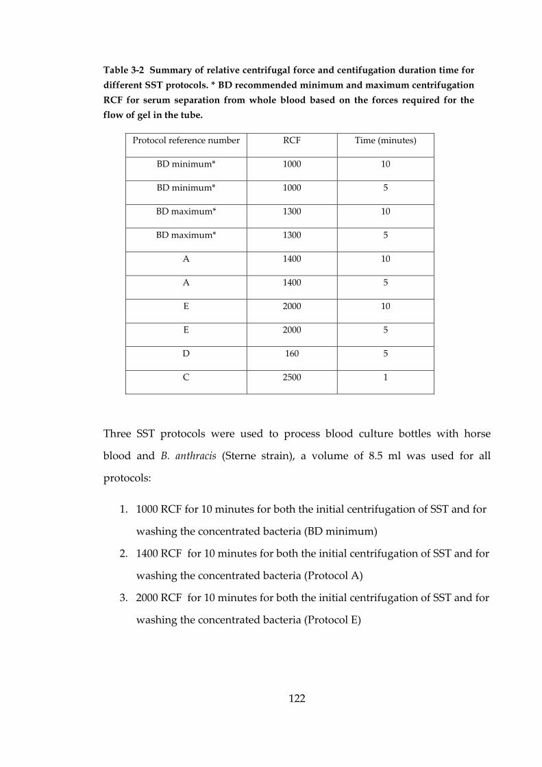
122
Table 3-2 Summary of relative centrifugal force and centifugation duration time for
different SST protocols. * BD recommended minimum and maximum centrifugation
RCF for serum separation from whole blood based on the forces required for the
flow of gel in the tube.
Protocol reference number RCF Time (minutes)
BD minimum* 1000 10
BD minimum* 1000 5
BD maximum* 1300 10
BD maximum* 1300 5
A 1400 10
A 1400 5
E 2000 10
E 2000 5
D 160 5
C 2500 1
Three SST protocols were used to process blood culture bottles with horse
blood and B. anthracis (Sterne strain), a volume of 8.5 ml was used for all
protocols:
1. 1000 RCF for 10 minutes for both the initial centrifugation of SST and for
washing the concentrated bacteria (BD minimum)
2. 1400 RCF for 10 minutes for both the initial centrifugation of SST and for
washing the concentrated bacteria (Protocol A)
3. 2000 RCF for 10 minutes for both the initial centrifugation of SST and for
washing the concentrated bacteria (Protocol E)
123
Blood culture fluid in SSTs underwent centrifugation for protocols one, two and
three and the supernatant removed. The concentrated bacteria on the gel plug
of the SST was washed with 0.5 ml phosphate buffered saline (PBS) and
transferred to a clean centrifuge tube and centrifuged described in protocols
one, two and three. The supernatant was removed and the pellet of bacteria
was re‐suspended in 1 ml PBS. The resulting concentrated, washed bacterial
cells were used for quantification with TVC using triplicate dilutions (section
2.3.1.4) and to prepare thermolysate samples (section 3.2.5.1) for PCR (section
3.3.5.5).
3.2.3.1 SST processing for different time points
Blood culture bottles containing horse blood and Bacillus anthracis (Vollum)
used for investigation of growth at different time points (as described in section
2.3.4), were also processed with SST. Following removal at different time
points (T4, T8, TTP, TTP+4 hours and TTP+8 hours), SST processing was
undertaken using 8.5ml volumes of positive blood culture fluid.
The SSTs were centrifuged at 1300 x g for 10 minutes and the supernatant
removed. The bacteria on the gel plug of the SST was washed with 0.5ml
phosphate buffer solution (PBS) and transferred to a clean centrifuge tube and
spun 1300 x g for 10 minutes. The supernatant was removed and the bacterial
pellet of bacteria was re‐suspended in 1 ml PBS. The resulting concentrated,
washed bacterial cells were quantified with TVC (section 2.3.1.4) and to prepare
thermolysate samples (section 3.3.5.1) for PCR (section 3.3.5.5).
3.2.4
3.2.4.1
During
concentra
molecula
undertak
50 μl of l
lysed usi
heating s
protocol
all viable
cultured
the rema
Themoly
3.2.4.2
To determ
after SST
Instagene
fluid from
strain) w
blood cul
The dilut
range of
determin
were then
Limit
Thermol
several in
ated wash
ar testing b
ken by pick
iquid sam
ng an App
amples at
ensures th
e vegetativ
on CBA m
aining the
sates were
Limit of
mine the lim
T processin
e and QIA
m blood
as used to
lture medi
tions were
f bacteria
ned by TV
n transferr
t of det
lysate pre
nvestigatio
hed bacte
by therma
king a colo
ples to 50
plied Biosy
37 °C for o
he germina
e cells. Af
media and
ermolysate
e stored at ‐
detection
mit of mol
ng, two dif
Amp silica
cultures c
make six,
ia (10 ml h
made to re
. The c
C. The un
red to a SST
1
tection
eparation
ons, colon
ria were
al lysis of
ony and ad
μl BHI bro
ystems® T
one minute
ation of an
fter heat tr
incubated
e samples
‐20˚C until
n
lecular det
fferent nuc
column p
containing
ten‐fold se
horse blood
epresent b
concentrati
ndiluted an
T and proc
24
as dete
n
nies, direc
used to
the bacter
dding it to
oth. The s
hermo cyc
e, followed
ny spores p
reatment,
at 37˚C fo
s were tr
l tested.
tection of B
cleic acid e
purification
horse blo
erial diluti
d and 40ml
blood cultu
ion of un
nd diluted
cessed usin
ermined
ct blood
prepare
ria (thermo
100 μl BHI
suspension
cler 2720, th
d by 95 °C f
present in t
10% of eac
or 48 hours
ransferred
B. anthracis
extraction m
n and ther
ood and B
ons in hors
l aerobic bl
ures contain
ndiluted b
d blood cu
ng the SST
d by PC
culture f
bacterial
olysates).
I broth or
n was then
the method
for 10 min
the sample
ch thermol
s to test fo
d to a cl
from bloo
methods w
rmal lysis
B. anthraci
se blood m
lood cultu
ning a con
blood cul
ulture fluid
T protocol p
CR
fluid and
DNA for
This was
by adding
thermally
d involved
nutes. This
e and kills
lysate was
r viability,
ean tube.
od cultures
were used,
. Positive
s (Vollum
mixed with
ure media).
ncentration
lture was
d samples
previously
d
r
s
g
y
d
s
s
s
,
.
s
,
e
m
h
.
n
s
s
y
125
described (section 3.3.4). The washed concentrated, bacteria resulting from the
neat and six dilutions were used for each of the three extraction methods. Each
extraction method was prepared in triplicate on the seven SST processed
samples.
3.2.4.3 Chelex–based resin DNA purification
Instagene extractions were performed using Biorad InstaGene Matrix (product
number 732‐6030) a Chelex–based resin for PCR‐ready DNA purification. For
each Instagene extraction, 20 μl of sample (positive blood culture fluid and
washed gel plug sample) was added to 180 μl Instagene matrix and mixed by
pipetting. The matrix was then incubated at 56˚C for 15 minutes, mixed and
then incubated at 99˚C for 8 minutes, centrifuged at 10,000 – 12,000 rpm in a
microcentrifuge. The supernatant was removed and 10% of the extraction
sterility tested as previously described for thermolysates, all extractions were
shown not to contain viable organisms and were later used for molecular
testing.
3.2.4.4 QIAmp silica column purification method
QIAmp silica column purification method for blood or body fluid was
performed according to manufacturer’s protocol. Briefly, 200 μl of sample was
added to 20 μl protease and 200 μl buffer AVL and mixed. These were then
incubated at 56˚C for 10 minutes, and 200 μl ethanol added, mixed and
transferred to the spin column. The spin column was centrifuged at 8,000 rpm
for 1 minute (using a standard bench top microfuge), columns were then
washed sequentially with AW1 and AW2 (with centrifugation at 8,000rpm for 1
minute and 14000 rpm for 3 minutes respectively) and a final elution of 100 μl
made. The eluted DNA extractions were sterility tested, using the method
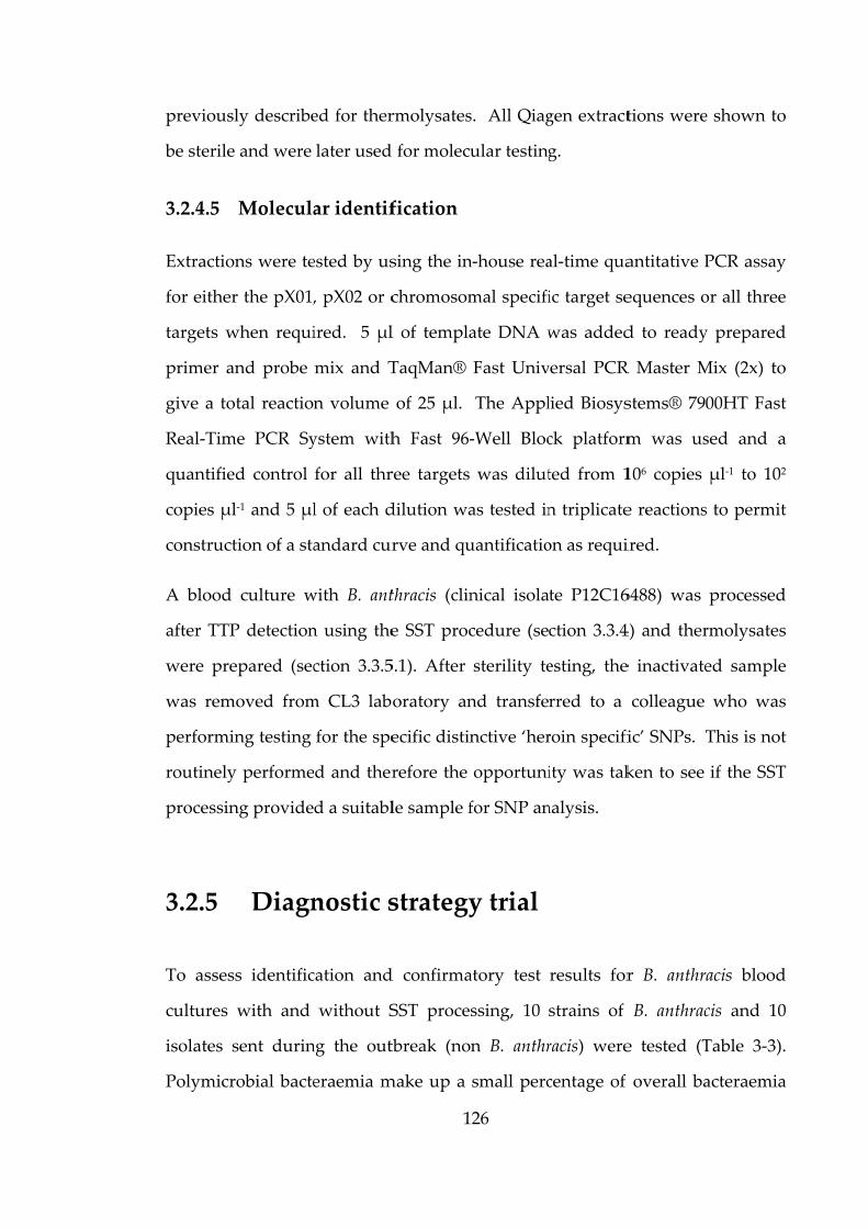
previousl
be sterile
3.2.4.5
Extraction
for either
targets w
primer an
give a to
Real‐Tim
quantifie
copies μl
construct
A blood
after TTP
were pre
was rem
performin
routinely
processin
3.2.5
To assess
cultures
isolates s
Polymicr
ly describe
and were
Molecula
ns were te
r the pX01,
when requi
nd probe
tal reactio
me PCR Sy
d control
l‐1 and 5 μl
tion of a sta
culture w
P detection
epared (sec
oved from
ng testing
y performe
ng provide
Diagn
s identific
with and
sent durin
robial bacte
ed for ther
later used
ar identif
ested by us
, pX02 or c
ired. 5 μl
mix and T
n volume
ystem with
for all thr
l of each d
andard cur
with B. ant
n using the
ction 3.3.5
m CL3 lab
for the spe
ed and ther
d a suitabl
nostic s
cation and
without S
ng the outb
eraemia m
1
rmolysates
for molecu
fication
sing the in
chromosom
l of templa
TaqMan®
of 25 μl.
h Fast 96‐
ree targets
dilution wa
rve and qu
thracis (clin
e SST proc
5.1). After
oratory an
ecific distin
refore the
le sample f
strategy
d confirma
SST proce
tbreak (no
make up a
26
s. All Qiag
ular testing
n‐house rea
mal specifi
ate DNA w
Fast Univ
The Appl
‐Well Bloc
was dilut
as tested in
uantificatio
nical isola
cedure (se
sterility te
nd transfe
nctive ‘her
opportuni
for SNP an
y trial
atory test
ssing, 10
n B. anthr
small perc
gen extract
g.
al‐time qua
ic target se
was added
versal PCR
lied Biosys
ck platform
ted from 1
n triplicate
on as requi
te P12C16
ction 3.3.4
esting, the
rred to a
roin specifi
ity was tak
nalysis.
results for
strains of
racis) were
centage of
tions were
antitative P
equences o
d to ready
R Master M
stems® 79
m was us
106 copies
e reactions
ired.
6488) was
4) and ther
e inactivate
colleague
ic’ SNPs. T
ken to see
r B. anthr
B. anthrac
e tested (T
overall ba
e shown to
PCR assay
or all three
y prepared
Mix (2x) to
00HT Fast
sed and a
μl‐1 to 102
s to permit
processed
rmolysates
ed sample
who was
This is not
if the SST
acis blood
cis and 10
Table 3‐3).
acteraemia
o
y
e
d
o
t
a
2
t
d
s
e
s
t
T
d
0
.
a
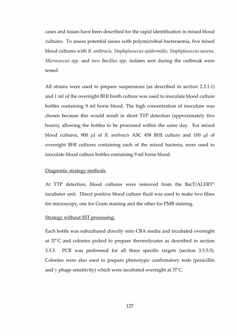
127
cases and issues have been described for the rapid identification in mixed blood
cultures. To assess potential issues with polymicrobial bacteraemia, five mixed
blood cultures with B. anthracis, Staphylococcus epidermidis, Staphylococcus aureus,
Micrococcus spp. and two Bacillus spp. isolates sent during the outbreak were
tested.
All strains were used to prepare suspensions (as described in section 2.3.1.1)
and 1 ml of the overnight BHI broth culture was used to inoculate blood culture
bottles containing 9 ml horse blood. The high concentration of inoculum was
chosen because this would result in short TTP detection (approximately five
hours), allowing the bottles to be processed within the same day. For mixed
blood cultures, 900 μl of B. anthracis ASC 458 BHI culture and 100 μl of
overnight BHI cultures containing each of the mixed bacteria, were used to
inoculate blood culture bottles containing 9 ml horse blood.
Diagnostic strategy methods
At TTP detection, blood cultures were removed from the BacT/ALERT®
incubator unit. Direct positive blood culture fluid was used to make two films
for microscopy, one for Gram staining and the other for PMB staining.
Strategy without SST processing:
Each bottle was subcultured directly onto CBA media and incubated overnight
at 37˚C and colonies picked to prepare thermolysates as described in section
3.3.5. PCR was performed for all three specific targets (section 3.5.5.5).
Colonies were also used to prepare phenotypic confirmatory tests (penicillin
and γ phage sensitivity) which were incubated overnight at 37˚C.
128
Table 3-3 Bacteria strains used for diagnostic strategy
Ten strains of B.anthracis (shaded) and ten other bacterial isolates referred for B.
anthracis identification in 2010 with ASC or clinical sample references. Five blood
cultures were simulated with a mix of B. anthracis and another bacteria (shaded). a
Identification made by 16S rRNA by PHE Colindale, b Presumptive identification
made with API 50CHB at PHE Porton.
Bacteria Reference
B. anthracis Sterne UK ASC 1
B. anthracis Vollum UK ASC 6
B. anthracis Wales Bovine ASC 27
B. anthracis ATCC 10( 7b) ASC 12
B. anthracis Clinical Scotland 2006 ASC 458
B. anthracis Clinical Hackney 2008 ASC 497
B. anthracis New Hampshire ASC 69
B. anthracis Clinical 2012 P12C1297
B. anthracis Landkey ASC 192
B. anthracis Pasteur ASC 182
Bacillus endophyticusa P10C1418
Non reactive Bacillusb P10C7386
Bacillus mycoidesb P10C0301
Bacillus cereusb P10C7449
Non reactive Bacillusb P10C7657
Brevibacillus laterosporusb P10C5932
Bacillus megateriumb P10C0141
Non reactive Bacillusb P10C7798
Non reactive Bacillusb P10C0040
Aneurinibacillus aneurinilyticusb P10C2307
ASC 458 & Staphylococcus epidermidis
ASC 458 & Staphylococcus aureus
ASC 458 & Micrococcus spp.
ASC 458 +P10C0141
ASC 458 +P10C1418
Strategy with SST processing:
8.5 ml positive blood culture fluid was processed using SST as described in
section 3.3.4 and the concentrated, washed bacterial sample was used to

129
inoculate CBA plates for culture and testing with penicillin and γ phage
sensitivity testing.
The same sample was also used to prepare thermolysates as described in
section 3.3.5. and PCR was performed for all three specific targets (section
3.5.5.5).
3.3
3.3.1
3.3.1.1
The routi
37˚C was
PMB as w
images w
B. anthrac
staining (h
using hors
blood).
a
c
Figure 3-1
Resu
Presum
Blood cu
ine metho
s used to p
well as azu
were captur
cis (P12C164
horse blood
se blood), d
Images of
ults
mptive
ulture mic
d for cultu
repare blo
ure blue sta
red (Figure
488), a) PM
d for 5 hour
d) Azure bl
B. anthracis
1
e identi
croscopy
uring B. an
ood films w
ain prepar
e 3‐2).
MB staining
rs), c) Azur
lue staining
s stained us
30
ificatio
nthracis in
which were
ration (sect
(horse bloo
e blue stain
g (simulated
b
d
sing PMB an
n meth
horse blo
e using com
tion 3.2.1.1
od for 5 ho
ning (simul
d blood cul
nd Azure bl
hods
ood for five
mmercially
1) and digi
ours), b)Azu
lated blood
lture using
lue stains
e hours at
y available
tal camera
ure blue
culture
human
t
e
a
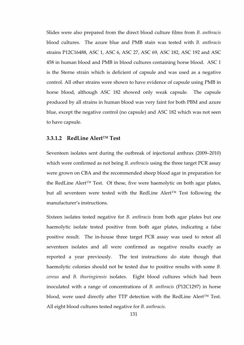
131
Slides were also prepared from the direct blood culture films from B. anthracis
blood cultures. The azure blue and PMB stain was tested with B. anthracis
strains P12C16488, ASC 1, ASC 6, ASC 27, ASC 69, ASC 182, ASC 192 and ASC
458 in human blood and PMB in blood cultures containing horse blood. ASC 1
is the Sterne strain which is deficient of capsule and was used as a negative
control. All other strains were shown to have evidence of capsule using PMB in
horse blood, although ASC 182 showed only weak capsule. The capsule
produced by all strains in human blood was very faint for both PBM and azure
blue, except the negative control (no capsule) and ASC 182 which was not seen
to have capsule.
3.3.1.2 RedLine Alert™ Test
Seventeen isolates sent during the outbreak of injectional anthrax (2009–2010)
which were confirmed as not being B. anthracis using the three target PCR assay
were grown on CBA and the recommended sheep blood agar in preparation for
the RedLine Alert™ Test. Of these, five were haemolytic on both agar plates,
but all seventeen were tested with the RedLine Alert™ Test following the
manufacturer’s instructions.
Sixteen isolates tested negative for B. anthracis from both agar plates but one
haemolytic isolate tested positive from both agar plates, indicating a false
positive result. The in‐house three target PCR assay was used to retest all
seventeen isolates and all were confirmed as negative results exactly as
reported a year previously. The test instructions do state though that
haemolytic colonies should not be tested due to positive results with some B.
cereus and B. thuringiensis isolates. Eight blood cultures which had been
inoculated with a range of concentrations of B. anthracis (P12C1297) in horse
blood, were used directly after TTP detection with the RedLine Alert™ Test.
All eight blood cultures tested negative for B. anthracis.
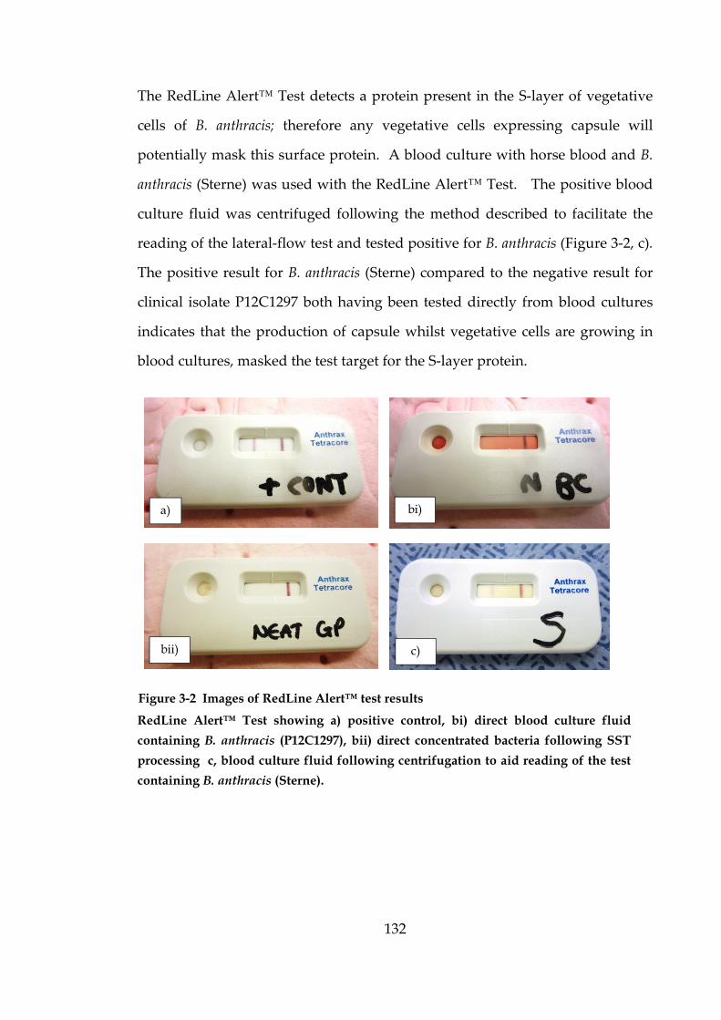
132
The RedLine Alert™ Test detects a protein present in the S‐layer of vegetative
cells of B. anthracis; therefore any vegetative cells expressing capsule will
potentially mask this surface protein. A blood culture with horse blood and B.
anthracis (Sterne) was used with the RedLine Alert™ Test. The positive blood
culture fluid was centrifuged following the method described to facilitate the
reading of the lateral‐flow test and tested positive for B. anthracis (Figure 3‐2, c).
The positive result for B. anthracis (Sterne) compared to the negative result for
clinical isolate P12C1297 both having been tested directly from blood cultures
indicates that the production of capsule whilst vegetative cells are growing in
blood cultures, masked the test target for the S‐layer protein.
RedLine Alert™ Test showing a) positive control, bi) direct blood culture fluid
containing B. anthracis (P12C1297), bii) direct concentrated bacteria following SST
processing c, blood culture fluid following centrifugation to aid reading of the test
containing B. anthracis (Sterne).
Figure 3-2 Images of RedLine Alert™ test results
a) bi)
bii) c)
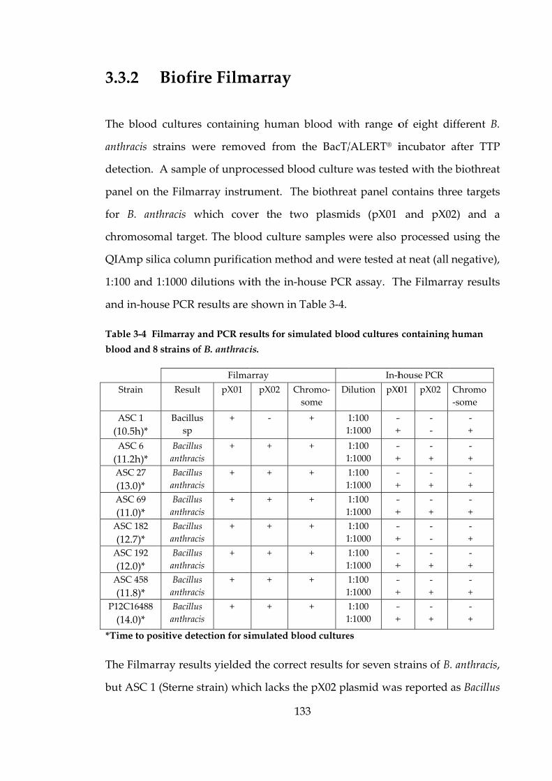
3.3.2
The bloo
anthracis
detection
panel on
for B. an
chromoso
QIAmp s
1:100 and
and in‐ho
Table 3-4
blood and
Strain
ASC 1
(10.5h)*
ASC 6
(11.2h)*
ASC 27
(13.0)*
ASC 69
(11.0)*
ASC 182
(12.7)*
ASC 192
(12.0)*
ASC 458
(11.8)*
P12C16488
(14.0)*
*Time to p
The Film
but ASC
Biofir
od cultures
strains w
n. A sampl
the Filma
nthracis w
omal targe
silica colum
d 1:1000 di
ouse PCR r
Filmarray a
d 8 strains of
Result
Bacillu
sp
Bacillus
anthraci
Bacillus
anthraci
Bacillus
anthraci
Bacillus
anthraci
Bacillus
anthraci
Bacillus
anthraci
8 Bacillus
anthraci
positive dete
array resu
1 (Sterne s
re Film
s containin
were remov
le of unpro
array instru
which cove
et. The blo
mn purifica
ilutions wi
results are
and PCR res
f B. anthraci
Filma
t pX01
s +
s
is
+
s
is
+
s
is
+
s
is
+
s
is
+
s
is
+
s
is
+
ection for si
lts yielded
strain) whi
1
marray
ng human
ved from
ocessed blo
ument. Th
er the tw
od culture
ation meth
ith the in‐h
shown in
sults for sim
is.
array
pX02 C
‐
+
+
+
+
+
+
+
imulated bl
d the correc
ich lacks th
33
blood wi
the BacT/
ood culture
he biothrea
wo plasmi
e samples w
hod and we
house PCR
Table 3‐4.
mulated blo
Chromo‐
some
Di
+ 1
1
+ 1
1
+ 1
1
+ 1
1
+ 1
1
+ 1
1
+ 1
1
+ 1
1
lood culture
ct results f
he pX02 pl
ith range o
/ALERT® i
e was teste
at panel co
ds (pX01
were also
ere tested a
R assay. Th
od cultures
In‐h
ilution pX0
1:100
1:1000
‐
+
1:100
1:1000
‐
+
1:100
1:1000
‐
+
1:100
1:1000
‐
+
1:100
1:1000
‐
+
1:100
1:1000
‐
+
1:100
1:1000
‐
+
1:100
1:1000
‐
+
es
or seven st
asmid was
of eight d
incubator
ed with the
ontains thr
and pX0
processed
at neat (all
The Filmarr
containing
house PCR
01 pX02
‐
+
‐
‐
‐
+
‐
+
‐
+
‐
+
‐
+
‐
+
‐
+
‐
‐
‐
+
‐
+
‐
+
‐
+
‐
+
‐
+
trains of B
s reported
different B.
after TTP
e biothreat
ree targets
02) and a
d using the
negative),
ray results
g human
Chromo
‐some
‐
+
‐
+
‐
+
‐
+
‐
+
‐
+
‐
+
‐
+
B. anthracis,
as Bacillus
P
t
s
a
e
,
s
,
s
134
spp. due to not possessing all three targets. ASC 182 is the Pasteur strain which
lacks pX01 and was falsely identified by both the Filmarray and the in‐house
pX01 targets, however the in‐house assay was negative for pX02 instead.
Unknown external quality assurance samples from the QUANDHIP project
(Table 3‐5) were used to inoculate blood cultures containing horse blood; the
positive blood cultures were then tested using the Filmarray. The blood
cultures were subjected to SST processing to provide washed, concentrated
bacteria for inactivation and in‐house PCR testing.
Table 3-5 QUANDHIP live unknown sample results.
Filmarray Biothreat panel testing of simulated blood cultures and intended
QUANHIP results. *Time to positive detection for simulated blood cultures.
RKI Code Filmarray blood culture
result Intended result
RKI –Q3‐01 Francisella tularensis
(27.8h)*
Francisella tularensis
ssp.novicida
RKI –Q3‐02 Negative
(2.9 days)* Burkholderia mallei
RKI –Q3‐03 Negative
(31.0h)* No B.T. bacteria
RKI –Q3‐04
Bacillus Sp
PXO1 negative
(16.3h)*
Bacillus anthracis
(pXO1 deficient)
RKI –Q3‐05 Brucella Spp.
(27.5h)* Brucella suis
RKI –Q3‐06
Yersinia pestis.
YPT1 negative
(8.7h)*
Yersinia pestis
(PLA neg)
RKI –Q3‐07
Burkholderia
mallei/pseudomallei
(33.0h)*
Burkholderia
pseudomallei
Only samples for isolates RKI –Q3‐01, RKI –Q3‐04, RKI –Q3‐05, RKI –Q3‐06 and
RKI –Q3‐07 were sent for in‐house PCR testing based on preliminary PCR
testing of the original samples. Unfortunately, the preliminary PCR results did
not detect anything for sample RKI –Q3‐02 which actually contained
Burkholde
bacteria f
Filmarray
comment
were that
and many
Prelimina
media) w
different
method r
density r
Centrifug
density o
g (0.62).
eria mallei
from the b
y and inte
ts followin
t the RKI –
y laborator
S3.3.3
ary work u
was used
centrifuga
removed m
reading fo
gation with
of the resul
and shoul
blood cultu
ended QUA
ng the QU
– QC ‐02 sa
ries did no
SST pro
using non‐
to measu
ation prot
much of th
or the dif
h 1300 or 2
lting super
1
ld have be
ure had be
ANDHIP
UANDHIP
ample cont
ot detect th
ocessin
‐infectious
ure the op
tocols for
he blood er
fferent SS
2000 x g f
rnatant (0.
35
een positiv
een tested.
results are
P meeting
tained 3 x 1
his by direc
ng
material (h
ptical dens
SST proc
rythrocytes
ST protoco
for 10 min
54) followe
ve if the w
. The repo
e shown i
in Swede
104 CFU m
ct PCR.
horse bloo
sity of sup
cessing. T
s and the r
ols are sh
resulted i
ed by 1000
washed, con
orted resu
in Table 3
en (3‐5 Ma
ml‐1 Burkhold
od and blo
pernatant
The SST p
results of t
hown in T
in the low
0 x g (0.55)
ncentrated
ults for the
‐5. Initial
arch 2014)
deria mallei
od culture
following
processing
the optical
Table 3‐6.
est optical
) or 1400 x
d
e
l
)
i
e
g
g
l
.
l
x

136
Table 3-6 Optical density for different centrifugation protocols.
Protocol reference
number
RCF (g) Time (min) Optical density of
supernatant
BD minimum* 1000 10 0.55
BD minimum* 1000 5 0.67
BD maximum* 1300 10 0.54
BD maximum* 1300 5 0.64
A 1400 10 0.62
A 1400 5 0.64
E 2000 10 0.54
E 2000 5 0.64
D 160 5 0.72
C 2500 1 0.84
The results from the preliminary, non‐infectious work show that the BD
minimum protocol (1000 x g for 10 min), protocol A (1400 x g for 10 min) used
by Steinberger and Levy (2007), and protocol E (2000 x g for 10 min) used by
Funke & Funke‐Kissling, 2004 and Barman, Sengupta, & Singh, 2010 produced
low optical density results and were chosen for further investigation.
An investigation was undertaken to examine the effect of the processing
protocol on concentration of B. anthracis following SST processing. Triplicate
dilutions were made of the concentrated, washed bacteria following each of the
three spin protocol. Nine determinants (neat, 10‐1 and 10‐2 in triplicate) were
recorded for the three centrifugation protocols; 1 (BD minimum), 2 (A) and 3 (E)
and the resulting counts were Log transformed (Table 3‐7) because bacterial
count data does not follow a Normal distribution.

137
Table 3-7 Transformed counts for three centriguation teatment groups
Centrifugation (Treatment ) group
Triplicate
undiluted and
diluted SST
processed sample
Spin 1 (1000 RCF for
10 min)
Log transformed
counts
Spin 2 (1400 RCF for
10 min)
Log transformed
counts
Spin 3 (2000 RCF for
10 min)
Log transformed
counts
Undiluted (1)
Undiluted (2)
Undiluted (3)
5.6
4.6
5.6
5.6
5.6
5.6
5.5
5.5
5.5
10‐1 (1)
10‐1 (2)
10‐1 (3)
4.8
4.8
4.9
4.8
4.8
4.8
4.7
4.6
4.7
10‐2 (1)
10‐2 (2)
10‐2 (3)
4.2
4.3
4.3
4.2
4.2
4.2
4.0
4.0
4.0
To examine if the centrifugation protocol influenced the concentration of B.
anthracis recovered following SST processing, statistical analysis ANOVA (SPSS,
IBM) was conducted. The test statistic (F= 0.132) and corresponding p‐value (p=
0.877) indicate that there was no significant difference between the mean values
at the 5% significance level (p<0.05) and hence the processing method.
Using the optical density results and the observation that there was no
significant difference in the concentration of bacteria following the three
different centrifugation protocols, 1300 x g was chosen for all subsequent SST
centrifugation steps (equating to 3000 rpm on a 16cm diameter swinging bucket
rotor), this instrument was chosen as it is commonly used by front‐line
laboratories for processing blood samples.
138
3.3.3.1 SST processing for different time points
The blood culture bottles which had been used to examine growth at different
time points with B. anthracis (Vollum) at T4, T8, TTP, TTP +4 hours and TTP + 8
hours also underwent SST processing (section 3.3.4). An estimation of the
concentration bacteria following SST processing was made using TVC and the
geometric mean and 95% CI for triplicate bottles is shown in Table 3‐8,
alongside results from direct blood culture concentrations (section 2.4.4).
Table 3-8 Concentration of B. anthracis with and without SST processing.
Time
point
Concentration of blood culture
(CFU ml‐1)
Concentration of blood culture
following SST processing
(CFU ml‐1)
4 h 43.19 (5.2, 698.5) 1.02 x 10² (3.7,2.84x103)
8 h 1.39 x 10⁴ (6.08x103,3.19x104) 2.65 x 10⁴ (2.05 x10⁴ , 3.43 x10⁴)
TTP 1.28 x 10⁸ (5.09 x107,3.23 x108) 1.27 x 10⁸ (1.92 x10⁷ , 8.41 x10⁸)
TTP + 4 h 9.01 x10⁷ (4.74 x107,1.72 x108) 2.02 x 10⁸ (2.49 x10⁷ , 1.63 x10⁹)
TTP + 8 h 1.81x10⁸ (2.14 x10⁷, 1.54 x10⁹) 4.31 x 10⁸ (2.48 x10⁸ , 7.49 x10⁸)
The geometric mean (95% CI) for triplicate bottle concentrations for direct positive
blood cultures and following SST processing at different time points.
The difference between geometric mean concentrations with and without SST
processing was 58.81 CFU ml‐1 at 4 hours, increasing to 1.26 x10⁴ CFU ml‐1 for 8
hours, 1.00 x10⁶ CFU ml‐1 for TTP, 1.11 x10⁸ CFU ml‐1 for TTP+ 4 hours and 2.50
x108 CFU ml‐1 for TTP + 8 hours.
SST processing does appear to concentrate the bacteria but the degree of
concentration was small. The purpose of using the SST to process samples is to
aid in the use of the bacteria for other diagnostic methods.
Quantitat
bacterium
processed
using a
values w
calibrated
copy num
The therm
The geom
8.62x 101,
105, 1.79 x
TTP + 8 h
3.3.4
The limit
without S
methods
column p
were use
culture fl
107 CFU
8.86 x 10
samples w
pX02 PC
extraction
samples w
tive PCR
m) was co
d samples
1:2 dilutio
were doub
d control w
mbers (101
molysates f
metric mea
, 9.59 x102
x105), TTP
hours was 1
Limit
t of detect
SST proces
were eva
purification
ed to prep
luid. The u
ml‐1 using
01 CFU. T
were then
CR target.
ns were fo
were posit
(qPCR) fo
onducted
for each o
on of SST
led to giv
was tested
1 to 105) to
for time po
an by qPC
), time to
+ 4 hours
1.03 x10⁵ co
t of det
tion of B.
ssing was
aluated us
n method
pare six, te
undiluted p
TVC and
These samp
extracted
All Ins
ound to be
tive (Table
1
or the chr
on thermo
of the time
processed
ve copy n
d alongside
o generate
oint T4 had
CR for time
positive (T
s was 1.86
opies (95%
tection
anthracis (
explored u
sing therm
to extrac
en‐fold ser
positive bl
the six dil
ples under
by the thre
stagene an
negative f
3‐9). The
39
romosoma
olysates m
e points. T
d sample i
numbers fo
e the samp
a standar
d copy num
e point T8
TTP) was 1
x105 copie
% CI 1.19x 1
as dete
(Vollum) f
using PCR
molysates,
t nucleic a
rial dilutio
lood cultur
lutions pre
rwent SST
ee method
nd QIAm
for the pX
extraction
al PCR tar
made from
The therm
in BHI, th
or the ori
ples in trip
rd curve (
mbers too
was 2.88
1.41 x10⁵ c
s (95% CI
104, 8.95 x1
ermined
from blood
R and three
Instagene
acid. Pos
ons in hor
re was sho
esumed to
T processin
ds and teste
mp silica c
02 target a
ns made fro
rget (one
m the trip
molysates w
herefore qP
iginal sam
plicate for
(r2=0.95 an
low to be m
x102 copie
copies (95%
1.46x 10⁵,
105).
d by PC
d cultures
e different
e and QIA
sitive blood
rse blood a
own to con
contain 8.
ng and the
ed with th
column pu
and all the
om the blo
copy per
licate SST
were made
PCR copy
mple. The
a range of
nd r2=0.92).
measured.
es (95% CI
% CI 1.12x
2.36 x105),
CR
with and
extraction
Amp silica
d cultures
and blood
ntain 8.86 x
86 x 106 to
e resulting
e in‐house
urification
ermolysate
od culture
r
T
e
y
e
f
.
.
I
x
,
d
n
a
s
d
x
o
g
e
n
e
e
140
fluid without SST processing were tested alongside the neat extracted samples
for all three methods and only the thermolysate samples were found to give
positive signals. The thermolysates made from unprocessed blood cultures (10‐1
to 10‐6) were then tested and found to be positive but only to a dilution of 10‐4
(bottles containing 8.86 x 10³ CFU).
Table 3-9 PCR results following three different extraction methods.
Total
bacterial cunt
(CFU)
Instagene
pX02 PCR
with SST
processing
(Ct value)
QIAmp
pX02 PCR with
SST processing
(Ct value)
Thermolysate
pX02 PCR with
SST processing
(Ct value)
Thermolysate
pX02 PCR
without SST
processing
(Ct value)
8.86 x 10⁷ ND ND 19.68, 19.65, 19.74 19.64, 19.60, 19.47
8.86 x 10⁶ ND ND 22.11, 22.07, 22.11 23.14, 22.72, 22.79
8.86 x 10⁵ ND ND 22.45, 22.72, 22.51 23.63, 23.67, 23.86
8.86 x 10⁴ ND ND 24.30, 24.05, 23.92 25.67, 30.31, ND
8.86 x 10³ ND ND 25.20, 25.42, 25.53 29.72, 30.35, 30.18
8.86 x 10² ND ND 30.22, 30.04, 30.14 ND
8.86 x 101 ND ND 28.94, ND, 30.60 ND
PCR results (Ct value of three determinations) of reactions for three extraction methods
preparared in triplicate from SST processed samples and thermolysates without SST
processing. ND = Not detected.
Further testing with the pX01 and chromosomal PCR targets was performed on
undiluted extracted samples for all three methods and only the thermolysate
samples were found to be positive.
A blood culture with B. anthracis (P12C16488) was processed using the SST
procedure which was extracted by the thermolysate method. This sample was
later tested for the specific distinctive ‘heroin specific’ SNPs. The results
showed the same SNP signature as previous heroin associated B. anthracis

isolates i
analysis.
3.3.5
The diag
identifica
without
microsco
subseque
prepare p
thermoly
cultures
sample, u
The resu
without S
in Appen
the M’Fa
27 was a
both with
were neg
and γ ph
indicating
Diagn
gnostic str
ation and
SST proce
py and th
ent colonie
phenotypic
ysates for P
processed
used for pr
ults for ph
SST proces
ndix 3.4. B
dyean reac
lso found
h and with
gative for b
age and bo
that SST
nostic s
rategy emp
confirmat
essing. Th
hen subcult
es (follow
c confirma
PCR testin
with SST
reparing ph
henotypic
ssing are sh
B. anthracis
ction and t
to be neg
hout SST p
both the M
oth with or
1
processing
strategy
ployed in
tion of B.
he positiv
tured on t
wing overn
atory tests
ng. This t
T to provid
henotypic
confirmat
hown in Ta
(Sterne) is
the capsule
ative for t
processing.
M’Fadyean
r without S
41
g provide
y trial
this stud
anthracis
ve blood c
to CBA wi
night incu
(penicillin
testing stra
de a wash
confirmato
tory tests
able 3‐10 a
s capsule d
e gene targ
he M’Fady
. All ten n
reaction a
SST proces
d a suitab
dy was us
in blood
cultures w
ith overnig
ubation) w
n and γ ph
ategy was
hed, conce
ory tests an
for blood
and examp
deficient th
get on the
yean reacti
non B. anth
and were r
ssing.
ble sample
sed to com
cultures,
were used
ght incuba
were then
hage sensit
compared
entrated, B
nd thermo
d cultures
ple images
herefore ne
pX02 plasm
tion and pX
hracis bacte
resistant to
e for SNP
mpare the
with and
for direct
ation. The
n used to
tivity) and
d to blood
B. anthracis
olysates.
with and
are shown
egative for
mid. ASC
X02 target
eria tested
o penicillin
P
e
d
t
e
o
d
d
s
d
n
r
C
t
d
n
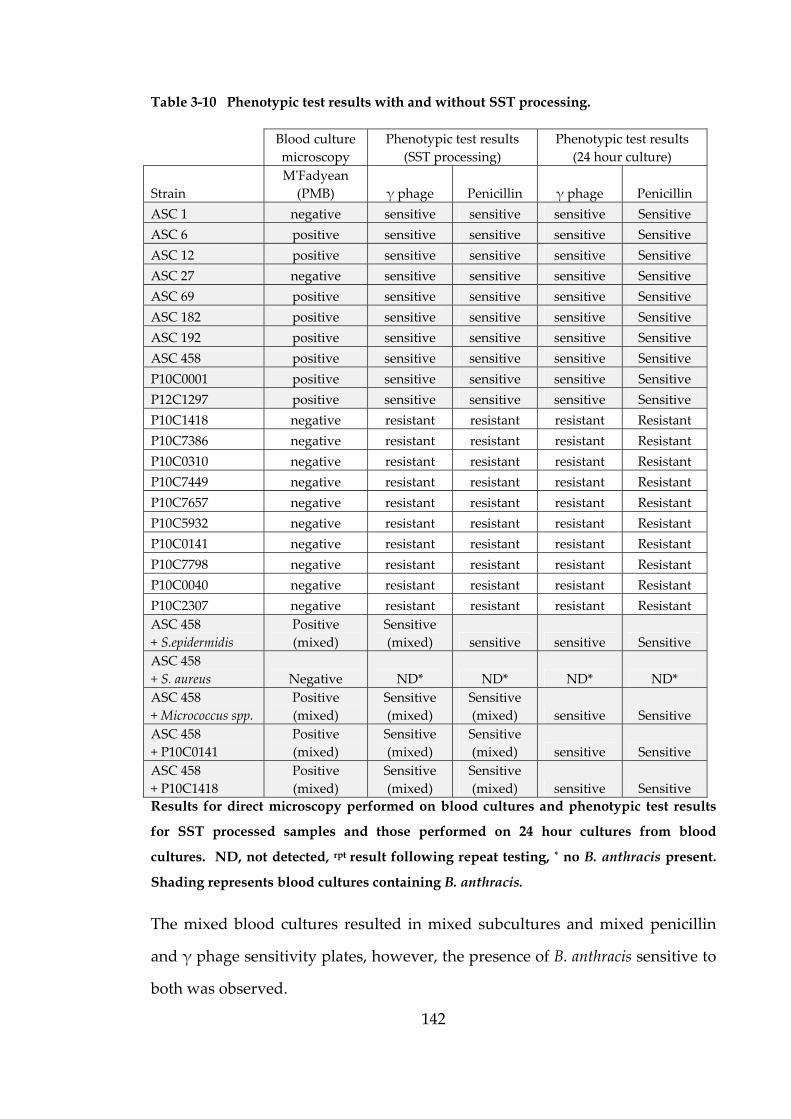
142
Table 3-10 Phenotypic test results with and without SST processing.
Blood culture
microscopy
Phenotypic test results
(SST processing)
Phenotypic test results
(24 hour culture)
Strain
MʹFadyean
(PMB) γ phage Penicillin γ phage Penicillin
ASC 1 negative sensitive sensitive sensitive Sensitive
ASC 6 positive sensitive sensitive sensitive Sensitive
ASC 12 positive sensitive sensitive sensitive Sensitive
ASC 27 negative sensitive sensitive sensitive Sensitive
ASC 69 positive sensitive sensitive sensitive Sensitive
ASC 182 positive sensitive sensitive sensitive Sensitive
ASC 192 positive sensitive sensitive sensitive Sensitive
ASC 458 positive sensitive sensitive sensitive Sensitive
P10C0001 positive sensitive sensitive sensitive Sensitive
P12C1297 positive sensitive sensitive sensitive Sensitive
P10C1418 negative resistant resistant resistant Resistant
P10C7386 negative resistant resistant resistant Resistant
P10C0310 negative resistant resistant resistant Resistant
P10C7449 negative resistant resistant resistant Resistant
P10C7657 negative resistant resistant resistant Resistant
P10C5932 negative resistant resistant resistant Resistant
P10C0141 negative resistant resistant resistant Resistant
P10C7798 negative resistant resistant resistant Resistant
P10C0040 negative resistant resistant resistant Resistant
P10C2307 negative resistant resistant resistant Resistant
ASC 458
+ S.epidermidis
Positive
(mixed)
Sensitive
(mixed) sensitive sensitive Sensitive
ASC 458
+ S. aureus Negative ND* ND* ND* ND*
ASC 458
+ Micrococcus spp.
Positive
(mixed)
Sensitive
(mixed)
Sensitive
(mixed) sensitive Sensitive
ASC 458
+ P10C0141
Positive
(mixed)
Sensitive
(mixed)
Sensitive
(mixed) sensitive Sensitive
ASC 458
+ P10C1418
Positive
(mixed)
Sensitive
(mixed)
Sensitive
(mixed) sensitive Sensitive
Results for direct microscopy performed on blood cultures and phenotypic test results
for SST processed samples and those performed on 24 hour cultures from blood
cultures. ND, not detected, rpt result following repeat testing, * no B. anthracis present.
Shading represents blood cultures containing B. anthracis.
The mixed blood cultures resulted in mixed subcultures and mixed penicillin
and γ phage sensitivity plates, however, the presence of B. anthracis sensitive to
both was observed.
143
Direct subculture of the mixed blood culture containing S. aureus resulted in
only a pure culture of S. aureus. There were, therefore no B. anthracis colonies
from which to perform molecular and phenotypic characterisation tests. In
addition this mixed blood culture provided evidence of B. anthracis in the Gram
microscopy however, very few B. anthracis cells were seen in the PMB stained
film and no capsule was observed. Despite these observations the PCR was
positive for all three PCR targets following SST processing (Table 3‐11),
however there was an apparently pure growth of S. aureus on the penicillin and
specific γ phage sensitivity plates. Subculture of the SST processed sample
provided a mixed culture of S. aureus and B. anthracis from which repeat testing
could be performed and compared to the direct subculture from the blood
culture without SST processing, which only contained a pure culture of S.
aureus.
The comparison of PCR results (Ct value) with and without SST processing is
shown in Table 3‐11. PCR was performed using the three target real‐time assay
which detects targets within the Lethal Factor genes (pX01), the capsule gene
(pX02) and a specific B. anthracis chromosomal target. As expected the pX02
PCR target was not detected for ASC 1, but was also unexpectedly missing from
ASC 27 even after repeat testing. All ten, non‐B. anthracis isolates were negative
for all three PCR targets either with or without SST processing.
ASC 27 was also negative for the pX02 target both with and without SST
processing and after repeat testing. The negative result for capsule by PCR is
consistent with the phenotypic negative result for M’Fadyean reaction. Mixed
blood cultures containing B. anthracis ASC 458 and B. endophyticus (P10C1418)
were initially negative for the pX01 target however this was detected on repeat
testing. The Ct values for this mixed sample were high (Ct >35) which would
indicate the sample had only a small amount of B. anthracis present
144
Table 3-11 Molecular test results with and without SST processing.
Results for PCR (ct value) of inactivated samples following SST processing and from 24
hour culture colony picks. ND, not detected, rpt result following repeat testing, * no B.
anthracis colonies from which to make an inactivated sample for PCR testing. Shading
represents blood cultures containing B. anthracis.
SST processed PCR samples 24 hour culture PCR samples
Strain
pX02
(ct value)
pX01
(ct value)
Chromo‐
some
(ct value)
pX02
(ct value)
pX01
(ctvalue)
Chromo‐
some
(ct value)
ASC 1 ND 26.14 26.95 ND 20.45 21.31
ASC 6 24.43 25.22 25.65 22.98 23.49 24.12
ASC 12 24.15 26.32 25.41 21.44 21.7 22.16
ASC 27 ND/ NDrpt 26.23 26.99 ND/ NDrpt 23.28 23.77
ASC 69 22.32 22.59 23.42 20.95 21.11 21.88
ASC 182 24.78 25.56 25.88 19.67 18.94 19.46
ASC 192 23.6 24.1 24.72 20.98 21.52 21.83
ASC 458 24.22 24.96 25.42 22.51 21.99 22.47
P10C0001 23.75 24.63 24.93 24.81 23.92 25.57
P12C1297 23.79 24.34 25.12 24.29 24.12 24.96
P10C1418 ND ND ND ND ND ND
P10C7386 ND ND ND ND ND ND
P10C0310 ND ND ND ND ND ND
P10C7449 ND ND ND ND ND ND
P10C7657 ND ND ND ND ND ND
P10C5932 ND ND ND ND ND ND
P10C0141 ND ND ND ND ND ND
P10C7798 ND ND ND ND ND ND
P10C0040 ND ND ND ND ND ND
P10C2307 ND ND ND ND ND ND
ASC 458
+S.epidermidis 24.77 24.97 25.88 22.79 23.25 24.22
ASC 458
+ S. aureus 20.77 25.09 22.85 ND* ND* ND*
ASC 458
+Micrococcus
spp. 23.96 23.92 24.4 22.77 22.94 23.54
ASC 458
+ P10C0141 22.72 23.6 24.37 21.89 22.34 23.11
ASC 458
+ P10C1418 29.96
ND/
35.41rpt 35.94 23.54 23.69 25.33
145
The time taken to process blood cultures for the preparation of microscopy
slides, SST processing, thermolysates and phenotypic confirmatory tests were
recorded during the diagnostic strategy testing and are shown in Table 3‐12.
The additional time requirements for surface decontamination performed
following steps one and two to remove centrifuge buckets from the MSC III for
SST centrifugation were included. After steps three and four were completed,
samples for PCR and culture were removed from the MSC III together within
the same decontamination period. Steps five and six were performed the
following day using overnight cultures prepared in step one (~18 hours).
The start time point of the analytical period was denoted t=0 when blood
cultures were removed from the BacT/ALERT® incubator. The mid analytical
time point when samples were ready for removal from the CL3 laboratory
following inactivation prior to PCR was t=1:38. The time to mid analytical time
point was 1 hour 38 minutes and represents the time to process 15 blood
cultures, with three 10 minute surface decontamination steps, two ten minute
centrifugation steps and the additional time requirements for thermolysate
preparation (step 4). If this was performed for a single blood culture the time
would be approximately 1 hour 2 minutes (step one taking ~1:30 min., step two
taking ~1:30 min, and steps three and four taking ~1:00 min each). The majority
of time is spent with surface decontamination (30 minutes) and centrifugation
steps (20 minutes).
The time taken to perform PCR would depend on whether a robot was used to
prepare the 96 well PCR plate. This can take ~45 minutes for a full plate and
~30 minutes for half a plate (15 samples), whereas a single sample can be set up
manually in ~10 minutes. The PCR assay took 30 minutes therefore if a real
clinical sample had been processed the inactivated samples would be removed
from the CL3 laboratory at t=1:38 and PCR results would have been available at
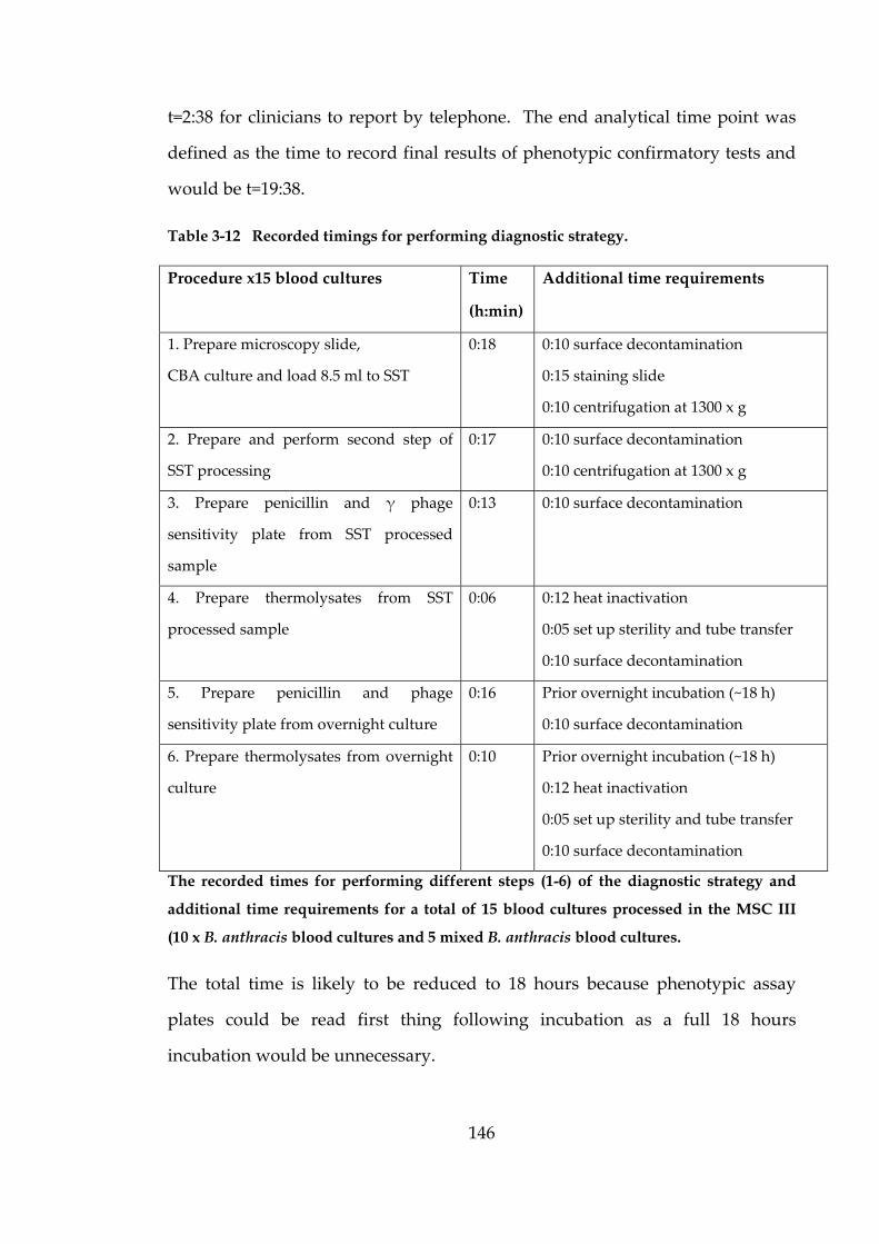
146
t=2:38 for clinicians to report by telephone. The end analytical time point was
defined as the time to record final results of phenotypic confirmatory tests and
would be t=19:38.
Table 3-12 Recorded timings for performing diagnostic strategy.
Procedure x15 blood cultures Time
(h:min)
Additional time requirements
1. Prepare microscopy slide,
CBA culture and load 8.5 ml to SST
0:18 0:10 surface decontamination
0:15 staining slide
0:10 centrifugation at 1300 x g
2. Prepare and perform second step of
SST processing
0:17 0:10 surface decontamination
0:10 centrifugation at 1300 x g
3. Prepare penicillin and γ phage
sensitivity plate from SST processed
sample
0:13 0:10 surface decontamination
4. Prepare thermolysates from SST
processed sample
0:06 0:12 heat inactivation
0:05 set up sterility and tube transfer
0:10 surface decontamination
5. Prepare penicillin and phage
sensitivity plate from overnight culture
0:16 Prior overnight incubation (~18 h)
0:10 surface decontamination
6. Prepare thermolysates from overnight
culture
0:10 Prior overnight incubation (~18 h)
0:12 heat inactivation
0:05 set up sterility and tube transfer
0:10 surface decontamination
The recorded times for performing different steps (1-6) of the diagnostic strategy and
additional time requirements for a total of 15 blood cultures processed in the MSC III
(10 x B. anthracis blood cultures and 5 mixed B. anthracis blood cultures.
The total time is likely to be reduced to 18 hours because phenotypic assay
plates could be read first thing following incubation as a full 18 hours
incubation would be unnecessary.

147
Without SST processing the blood culture were sub‐cultured and following
overnight incubation, used to prepare thermolysates and phenotypic
confirmatory tests which are then again incubated overnight. The
thermolysates were then used for PCR and in a real situation genotyping would
also be performed. In this situation full confirmation would not be made until a
total time of t=42:00. The envisaged diagnostic strategy using SST processing is
shown in Figure 3‐3, with the likely time line of events.
Figure 3-3 Diagnostric strategy

3.4
The stand
anthracis
CBA, CH
examinat
presump
anthracis
to the ref
isolate an
may take
laborator
the refe
purificati
house, th
would th
37˚C. Irr
cultured
to prepar
phenotyp
capsule p
The diagn
silica colu
as this is
shown to
The prese
appearan
Discu
dard diagn
in blood c
HOC, CLED
tion of pla
tive identi
is made, t
ference lab
nd potentia
e up to a
ry in the U
rence lab
ion for DN
hree target
hen be sub‐
respective
would hav
re thermoly
pic confirm
production
nostic stra
umn purif
labour inte
o be negativ
ence of lon
nce follow
ussion
nostic stra
culture, inv
D for aerob
ates follow
ification.
he laborat
boratory at
ally other c
day depen
UK. Curren
boratory,
NA extract
t PCR (pX0
‐cultured t
of the dire
ve their pla
ysates for
matory test
n with M’Fa
ategy empl
ication me
ensive and
ve from blo
ng chains o
wing micro
1
n
ategy used
volves dir
bic incubat
wing overn
If suspecte
tory would
t PHE Por
clinical sam
nding on
ntly, the st
would in
tion or the
01, pX02 a
to CBA and
ect PCR re
ates exami
PCR testin
ts for γ ph
adyean rea
loyed in th
ethod for D
d the extrac
ood cultur
of Gram po
oscopic ex
48
by front‐l
ect Gram
tion and FA
night incu
ed or a pr
d send a sl
ton for con
mples for d
the geogr
andard dia
nvolve di
ermolysate
and chrom
d examined
esults, clin
ined and su
ng. If pos
hage and p
action wou
his study d
DNA extra
ction, unle
res.
ositive baci
xamination
line labora
microscop
AA for ana
ubation wo
resumptive
lope agar c
nfirmation
direct dete
aphic loca
agnostic st
irect QIA
s for isola
mosomal ta
d after ove
ical sampl
uspect colo
sitive by P
penicillin s
uld be perfo
did not per
action from
ss diluted
illi with a c
n of bloo
atories to i
py and sub
aerobic cu
ould then
e identifica
culture of
n. The refe
ection of B
ation of th
trategy em
Amp silica
ates, follow
argets). Th
ernight inc
les which
onies wou
PCR, genoty
sensitivity
formed.
rform dire
m the blood
has been p
characteris
od culture
identify B.
bculture to
lture. The
lead to a
ation of B.
the isolate
erral of an
. anthracis,
he sending
mployed by
a column
wed by in‐
he sample
cubation at
have been
ld be used
yping and
as well as
ect QIAmp
d cultures,
previously
stic box car
es, should
o
e
a
e
n
,
g
y
n
‐
e
t
n
d
d
s
p
,
y
r
d
149
prompt the laboratory to consider the presence of B. anthracis. This may not
always be the case, following discussion with a laboratory worker who handled
an anthrax blood culture during the 2009–2010 injectional anthrax outbreak
they described a different Gram appearance. The blood culture had flagged
TTP detection overnight and was processed the following morning. The Gram
was made and reported as Gram positive cocci in chains presumed as
Streptococcus spp. after many laboratory and clinical staff had examined the
microscopy slide. The culture obviously did not agree with this microscopy
result as it was a non‐haemolytic bacillus. The bacteria in the blood culture
fluid must have presumably started to sporulate to give the chaining coccus
appearance. The examination of Gram slides prepared from the non‐B.
anthracis isolates sent during the outbreak, used to test the diagnostic strategy,
were Gram positive bacilli with various different morphologies. The
information gathered from real cases and this study will be used in the training
materials, the subject of Chapter Five.
The culture of B. anthracis in blood culture bottles provides an environment for
vegetative bacteria to express capsule, therefore making it an ideal sample to
visualise capsule as a presumptive direct identification method. This study
explored the use of an alternative to PMB (Azure Blue) for the M’Fadyean
reaction to see if this preparation would be suitable for front‐line laboratory use
as a presumptive identification method in addition to the Gram stain currently
used. The use of the M’Fadyean reaction dates back to the early nineteenth
century following M’Fadyean’s description of a ‘peculiar staining reaction of
the blood of animals dead of anthrax’ (1903). This simple, reliable, method was
widely used to aid in the presumptive identification of B. anthracis from direct
clinical material whilst anthrax was still prevalent in many countries. After the
decline in the disease, the quality of PMB was questioned and many front‐line
laboratories are unlikely now to possess this stain. The alternative azure blue
150
preparation recently proposed by Owen et al. (2013), was tested following
culture of B. anthracis in horse blood to elicit capsule expression. The films
prepared with azure blue were of very good quality showing clearly the
presence of capsule as a pink edge around the vegetative bacteria.
Unfortunately, the performance of the stain with simulated blood cultures
containing human blood was not as clear as those grown in horse blood. To
determine whether this stain would be beneficial to front‐line laboratories for
the presumptive identification of B. anthracis in conjunction with routine Gram
staining would require further work.
The RedLine Alert™ test correctly determined a negative B. anthracis result for
non‐B. anthracis isolates received during the outbreak from CBA cultures. The
one non‐B. anthracis isolate which did test positive was a haemolytic bacillus
which would not have been recommended for testing. The test is widely used
in the US in front‐line laboratories and in the situation of the outbreak of
injectional anthrax in the UK, could have provided laboratories with a simple
method. Suspected bacilli grown on routine media could be tested and negative
results could therefore reduce the cost of referral and testing at the reference
laboratory to rule out B. anthracis. The use of the RedLine Alert™ test directly
with blood culture supernatant was not appropriate due to the problem that
vegetative bacteria express capsule in blood containing media therefore
masking the specific S‐layer protein target of the test. This was demonstrated
with the negative results for B. anthracis (Vollum strain) compared to the
positive result obtained for a capsule deficient strain grown in blood cultures.
The Biofire Diagnotics Filmarray® biothreat panel performed well when tested
directly with blood cultures containing human blood and a range of B. anthracis
strains. This is believed to be the first time the biothreat panel has been tested
with blood culture samples and the extraction method was presumed to be the

151
same as that used in the blood culture panel for other more common bacterial
causes of bacteraemia, and therefore able to remove inhibitory substances in the
blood culture media. A recent study evaluated the sensitivity and specificity of
the Filmarray® biothreat panel with DNA samples of Bacillus anthracis,
Francisella tularensis and Yersinia pestis and live B. anthracis spores (Seiner et al.,
2013). The results found in this study support the conclusions of Seiner et al.
(2013), that the FilmArray® platform is a complete sample‐to‐answer system,
with simple sample preparation, PCR and data analysis. This study has also
found that the system is suited for biothreat testing for simulated blood culture
samples with results available in an hour following just a few minutes of
sample preparation. It is hoped the testing of blood cultures in this study is
useful to the evaluation of the system for military deployment, in particular for
use in the larger medical facilities deployed which do use automated blood
culture systems.
The testing of the Filmarray® biothreat panel with unknown EQA samples
from the European QUANDHIP project cultured in blood cultures provided the
same results as those reported by the reference laboratory. The blood cultures
were also processed with a SST protocol and provided suitable material for PCR
testing of thermolysates. The B. anthracis blood culture for sample RKI –Q3‐04
which was pX01 deficient was used to perform direct M’Fadyean reaction,
saving time for the reference laboratory, by not needing to culture the isolate in
horse blood for 5 hours to allow the vegetative bacteria to express capsule. In
addition, pX01 deficient B. anthracis strains have capsules which are often hard
to visualise due to some capsule regulation being on the pX01 plasmid. The
microscopy slides prepared from the blood culture containing the pX01
deficient B. anthracis clearly showed the presence of capsule.

152
The simple method to concentrate bacteria from blood cultures using SST has
been widely investigated to try and save time to perform automated
identification methods in front‐line laboratories. The method was explored to
determine the most appropriate protocol for this study. There is no
documented study that has compared the different protocols described in the
literature and it was discussed by Funke and Funke‐Kissling, (2004) that it is
unclear whether the different procedures used would influence the
performance of the systems evaluated. Information about the centrifugation
forces and time for SST to separate serum in conjunction with a simple
investigation of optical density with non‐infectious work, informed the choice
of three protocols. The three protocols were performed and comparisons of
total count data indicated that there was no significant difference in the
concentration of bacteria following either protocol. It was found that there was
little difference in the concentration of bacteria following SST processing of
blood cultures at different time points, however, the SST was used to produce a
suitable sample for further testing and was not concerned with concentrated
bacteria. The use of quantitative PCR, compared to labour intensive TVC, did
not provide comparable data to estimate the concentration of bacteria in the
sample. PCR was found to yield copy numbers (genome equivalents) that were
103 lower than the results obtained by TVC. The expectation would be to have
more genome equivalents detected by PCR than the TVC rather than less,
because even dead bacterial cells present would be detected by PCR. Blood
cultures for time point 4 h, were observed to contain more blood in the SST
after the initial centrifugation step. It is possible the blood was carried over
with the concentrated bacteria removed from the gel plug and not removed
during the wash step. This carry over may have caused inhibition during PCR
amplification of the chromosomal PCR target for time point T4.
153
For the purposes of this study, quantification using TVC was acceptable, even
considering the inherent limitations of the technique and the fact that results
were not available until 24 hours after samples were collected. However,
samples for PCR required 48 hour sterility testing.
The chosen protocol used the same centrifugation speed and time for both the
initial SST centrifugation step and the wash step. This protocol was practical to
perform in the CL3 laboratory. The protocol allowed for multiple blood
cultures to be processed in the MSC III, for example as more bottles were
removed from the BacT/ALERT® incubator, some bottles were ready for the
initial SST centrifugation whereas others were ready for the wash step
centrifugation. The protocol allowed for samples at both of these stages to be
loaded into centrifuge buckets within the MSC III and following surface
decontamination to be removed for centrifugation.
The polyanionic detergent SPS present in the blood culture media is known to
be a potent inhibitor for PCR (Fredricks and Relman, 1998). SPS has similar
properties to DNA in that both are high‐molecular‐weight polyanions that are
soluble in water but insoluble in alcohols (Fredricks and Relman, 1998). The
use of SST to concentrate and wash bacteria was aiming to remove SPS from the
sample to permit PCR amplification of the target. The physical separation of
blood cells from the blood culture fluid by the flow of gel in the SST was also
designed to remove the known inhibitory effects of haemoglobin on PCR.
The limit of PCR detection for blood cultures following SST processing was
examined and a concentration range of blood culture fluid was processed using
SST, followed by extraction using three different methods. All Instagene and
QIAmp silica column purification extractions were found to be negative for the
pX02 target.
154
Unfortunately these extractions were not further diluted post extraction to see if
1:100 or 1:000 dilution would dilute out inhibitors and permit PCR
amplification. Dilution (1:100 or 1:000 ) was performed on QIAmp silica
column purification extractions for the Filmarray testing and amplification was
observed which is consistent with finding reported by Fredricks and Relman,
(1998). The thermolysis method was successfully used with PCR amplification
to detect bacteria following SST processing from the 10‐6 diluted blood culture
(calculated to contain 8.86 x 101 CFU ml‐1). It was surprising to find that
thermolysates made from direct blood cultures without SST processing were
also amplified by PCR for the pX02 target. Blood cultures contained between
8.86 x10⁷ and 8.86 x 10³ CFU ml‐1. This was unexpected, as it was not thought
likely that simple heat inactivation would remove the inhibitory substances in
the blood culture fluid. It is possible that binding of SPS with haemoglobin as
was described by Edberg & Edberg, (1983) in the sample, prevented the
inhibitory effects of both the SPS and haemoglobin.
The testing of ten B. anthracis strains with and without the SST processing
method produced the same diagnostic test results and the SST processing
method provided results 24 hours earlier than current methods. The results for
ten non‐B. anthracis isolates also provided the same diagnostic results with and
without SST processing and were performed to represent matched negative
controls for the B. anthracis strains. The four mixed blood cultures containing
common contaminants (S. epidermidis and Micrococcus spp. and two other
Bacillus spp.) were shown to provide positive results for B. anthracis with PCR
and phenotypic confirmatory tests even though the penicillin and γ phage
plates were mixed, a result could still be read. The mixed blood culture with S.
aureus and B. anthracis highlighted the problem of this particular mix during
infection. Following SST processing the washed, concentrated bacteria were
cultured and used to prepare thermolysates and phenotypic confirmatory tests.
155
The thermolysates were positive for the three PCR targets, however the
penicillin and specific γ phage sensitivity plate apparently only contained S.
aureus. The direct blood culture on CBA apparently only contained S. aureus,
therefore following the standard diagnostic strategy there would be no colonies
of B. anthracis to pick for thermolysates and phenotypic testing in contrast to the
culture of the SST processed sample which provided a culture with a majority
of S. aureus, but colonies of B. anthracis were present.
The recording of start, mid‐analytical and end analytical time points provided
information to compare the diagnostic strategy using SST processing to the
current diagnostic strategy. The overall time to positive confirmation with SST
processing was approximately 24 hours less than the current strategy and the
incorporation of micro‐broth dilution susceptibility testing using PCR to read
end point minimum inhibitory concentrations could enable susceptibility
results to be available within the same time scale. The proposed strategy could
be performed on blood culture bottles sent by front‐line referring laboratories to
the reference laboratory (as was the case during the outbreak). Alternatively,
the method could be performed on blood culture fluid transferred to an SST by
the referring laboratory. An additional small volume of blood culture fluid to
accompany the SST would reduce the turnaround time in the reference
laboratory further by allowing the SST to be loaded directly into the centrifuge
bucket, whilst the additional sample could be used to prepare microscopy
slides and culture within the MSC III. The use of blood cultures and rapid
technologies for the identification of bacterial causes of bacteraemia has
potential to reduce turnaround times for blood culture identification and
therefore impact on patient treatment and financial costs for length of
hospitalisation and laboratory costs.
156
This chapter explored the use of simple presumptive identification methods
and a new technology for direct blood culture testing. The main emphasis was
the use of SST processing combined with specific PCR to potentially reduce the
turnaround times for the identification and confirmation of B. anthracis in blood
cultures. The main points may be summaries as follows:
M’Fadyean reaction could potentially support Gram microscopy of
blood cultures but further work would be required to investigate the
azure blue preparation for use in front‐line laboratories.
The RedLine Alert™ test was found to identify known positive and
negative isolates grown on agar containing horse blood and capsule
deficient strains in blood cultures.
The Biofire Filmarray biothreat panel correctly identified blood cultures
containing B. anthracis strains and other biothreat pathogens.
The SST protocol was successful in providing concentrated washed
bacteria for subsequent PCR amplification and genotyping and for
setting up phenotypic confirmatory tests 24 hours earlier than current
procedures.
The SST protocol allowed for the molecular identification of B. anthracis
in polymicrobial blood cultures and enabled the recovery of B. anthracis
colonies from S. aureus polymicrobial blood cultures.
The proposed method to reduce the turnaround times for confirmation of
B.anthracis in blood cultures and other aspects of processing blood cultures will
be examined in Chapter four, which discusses the study aspect of ‘safety’.
Chap
I4.1
The defin
“The app
laboratory
Biosafety
safely man
the potent
the enviro
To preve
infection,
along the
infection
risk asses
controllin
measures
exposure
Microbio
biosafety
practices
microbiol
with ap
pathogen
pter 4
Introd
nition of bi
plication of
y and envir
defines the
nipulated. T
tial exposur
onment to po
ent laborato
, this can b
e chain. W
and who
ssment. Ef
ng exposu
s are the p
e to biologi
logists ro
y is essent
and safe
logical pra
propriate
nic micro‐o
Bio
ductio
osafety wh
f knowledg
onmental e
e containm
The objectiv
re of the lab
otentially in
ory acquir
be achieved
We use ou
may be at
fforts are m
ure at sou
preferred m
cal agents
utinely w
tial to eve
e environ
actice and
risk asse
organisms i
1
osafet
on
hich will be
ge, techniq
exposure to
ent conditi
ve of contain
boratory wo
nfectious ag
red infectio
d by severa
ur knowled
t risk, to m
made to br
urce, and
methods o
in the UK
work with
ery aspect
nments to
d biosafety
essment
in the wor
57
ty inve
e used in t
ues and e
potentially
ions under
nment is to
orker, perso
gents.” (Me
on (LAI), w
al means o
dge of the
mitigate th
reak the ch
d procedu
of preventi
(HSE, 200
infectiou
of their
work in
y should b
protect th
k place.
estiga
he context
equipment
y infectious
which infe
confine bio
ons outside
edterms, 20
we need to
of protectio
hazard (p
he hazard u
hain at the
ral and e
ing, or ade
9).
s micro‐o
role, to en
n. The p
be integral
he labora
ations
t of this stu
to prevent
agents or
fectious age
ohazards an
of the labo
012).
o break th
on at differ
pathogen),
using the
first oppo
engineerin
equately c
organisms,
nsure safe
principles
l to proced
atory wor
udy is:
t personal,
biohazards.
ents can be
nd to reduce
ratory, and
he chain of
rent points
, routes of
process of
ortunity by
ng control
controlling
therefore
e working
of good
dures and
rker from
,
.
e
e
d
f
s
f
f
y
l
g
e
g
d
d
m
158
People who work with infectious material could be at risk of exposure,
therefore there are health and safety regulations in place to ensure a safe
working environment. Suitable and sufficient risk assessment is a requirement
under The Control of Substances Hazardous to Health regulations 2002 (COSHH).
Employers must also provide adequate instruction, training and information
when employees work with hazards in the workplace as directed by the Health
and Safety at Work Act 1974 and Management of Health and Safety at Work
regulations 1999. There is also a duty on employees to use equipment according
to the training provided by the employer and to inform employers of any gaps
in training and other health and safety concerns under the regulations. There is
also guidance from the HSE for occupations and operations that are in direct
contact with infected animals, or with contaminated materials or associated
industrial processes (Great Britain and Health and Safety Executive, 1997).
Appropriate risk assessments can be made within the laboratory setting for
known infectious agents, however in a diagnostic laboratory it is often the
clinical information which informs the risk assessment for the safe handling and
processing of clinical samples. In situations where insufficient or inaccurate
clinical information is provided, specimens could be handled and processed
under insufficient or inappropriate laboratory containment conditions.
Exposures to HG3 pathogens are often caused by a lack of clinical information.
The HSE has identified that the most common occurrences occur with Brucella
spp. or Salmonella spp. from clinical samples that were initially processed at CL
2. The HSE have issued a safety note (see Appendix 4.1) on this issue following
investigation of such incidents, to provide laboratories with recommendations
for the collection and communication of relevant clinical information.
159
A report discussing four cases of brucellosis diagnosed in five London
hospitals, (where blood cultures and cultures were manipulated on the open
bench) describes recommendations for managing staff with possible exposure
to Brucella and on the prevention of laboratory acquired brucellosis (Reddy et
al., 2010). Of the take‐home messages from the report, training is highlighted as
laboratory staff are typically unfamiliar with the phenotypic characteristics of
Brucella spp. and need periodic re‐education about uncommon hazardous
organisms. Improved communication between clinicians and laboratory staff to
alert staff handling potentially high risk samples was also highlighted which
supports the HSE recommendations on communication.
A report by Henkel, Miller and Weyant (2012), identified eleven laboratory
exposures in US research facilities associated with Brucella melitensis (4 cases),
Brucella suis (2 cases), Francisella tularensis (4 cases) and one case of Coccidioides
immitis/posadasii. Laboratory exposures to Burkholderia pseudomallei have been
reported and recommendations made for incidences and prevention (Peacock et
al., 2008). The problem with B. pseudomallei is that the clinical symptoms of
melioidosis mimic those of many other diseases and differentiation between
melioidosis and many other acute and chronic bacterial infections is often
impossible (Peacock et al., 2008). Cases of laboratory acquired infection with B.
anthracis are rare, a case did occur in a laboratory worker in Texas (2002),
resulting in cutaneous anthrax (Public Health Dispatch, 2002). The laboratory
worker was processing environmental samples, as part of the investigation of
the 2001 anthrax letter attacks, the likely exposure occurred whilst handling a
vial of B. anthracis when not wearing gloves and the vial had only been surface
decontaminated with 70% isopropyl alcohol which does not inactivate spores.
More recently, laboratory workers at the CDC, Atlanta were potentially
exposed to samples containing viable B. anthracis (CDC, 2014).
160
A new method of inactivation was performed on samples containing B.
anthracis which were later shown to be viable. Potential exposure occurred
when the samples were tested with MALDI‐TOF MS in a lower‐security
laboratory, not equipped to handle live B. anthracis.
The cutaneous case and the CDC cases highlight the importance of safe
laboratory procedures and vaccination of staff who routinely work with B.
anthracis. The guidance provided by the HPA during the outbreak of injectional
anthrax suggests vaccination for staff routinely handling B. anthracis, this is
standard for workers in the reference laboratory and other departments at PHE
Porton, but was viewed unnecessary for front‐line laboratory staff.
One individual’s perception of risk is different from another’s and the use of
data in evidence based risk assessments can be useful to communicate the
actual risks compared to the potentially fearful perception of the risks.
Investigations of microbial aerosol generation during laboratory accidents
(Bennett & Parks, 2006) provides data to support the recommended emergency
procedures for prompt evacuation from the laboratory. The training of basic
biosafety principles in Azerbaijan delivered by NADP Training, used this data
to convince laboratory directors to change their emergency procedures as they
believed staff should be locked in the lab for them to clear up the accident.
During the early period of the outbreak of injectional anthrax in the UK (2009–
2010), several front‐line laboratories were reluctant to process samples for
routine bacteriology, even after discussing the risks with clinicians from the
reference laboratory.
This chapter describes the investigations undertaken to explore the biosafety
concerns the front‐line laboratories may have, in the event of processing a blood
culture suspected to contain B. anthracis on the open bench. The use of SST’s
has been incorporated into the investigations, where appropriate.
This was
culture p
workers.
4.2
4.2.1.1
To determ
an ACDP
for B. an
performe
tracers fo
spore of
spore siz
atropheus
commonl
are simil
2007). A
generatio
suspensio
inform t
cultures.
vegetativ
more aer
scenario.
to assess w
processing,
Study
Aerosol
mine if aer
P HG1 bact
nthracis to
ed in an A
or aerosol
B. anthrac
ze of B. a
spores a
ly used sim
lar to B. a
A study
on of aero
ons (Pottag
the invest
The choi
ve cells, wa
rostable, (
whether th
would pre
y desi
l samplin
rosols are g
teria, Bacil
reduce th
ACDP CL2
sampling
cis as the i
anthracis c
are conside
mulants B.
anthracis (C
conducted
osols durin
ge et al., 20
igations in
ice of the
as based on
giving a m
1
he propose
esent an a
ign an
ng
generated
llus atrophe
he biologic
laboratory
and were
infectious
ompared
erably sm
cereus and
Carrera, Z
d by Biosa
ng a pipe
014). The
into aeros
surrogate
n the know
more strin
61
ed addition
dditional h
nd me
during the
eus (spore s
cal hazard
y. These
e chosen fo
form of a
to other
maller than
d B. thuring
Zandomeni
afety at P
tting exer
methodol
ol produc
Bacillus sp
wledge tha
ngent test)
nal use of t
hazard to f
ethodo
e processin
stock) was
d and enab
spores are
or this stu
anthrax. In
Bacillus sp
n B. anthr
giensis hav
i, Fitzgibb
PHE Porto
cise using
ogy of this
ction from
pp. and sp
t spores ar
) and repr
these tubes
front‐line
ology
ng of blood
s used as a
able the w
e commonl
udy to rep
Investigatio
pp. sugges
racis but o
ve dimensi
bon, and S
on investi
g B. atroph
s study w
m processi
pores as o
re easy to
resent a w
s, in blood
laboratory
d cultures,
a surrogate
work to be
ly used as
present the
ons of the
sts that B.
other, less
ons which
Sagripanti,
igated the
heus spore
as used to
ing blood
pposed to
aerosolise,
worse‐case
d
y
,
e
e
s
e
e
.
s
h
,
e
e
o
d
o
,
e
162
4.2.1.2 Microscopy slides
The inactivation of HG3 bacterial microscopy slides is limited to several studies
relating to Mycobacterium tuberculosis (MTB). Blackwood et al. (2005) examine
inactivation methods for MTB slides and other material prior to removal from
the containment laboratory. The results of the investigation showed viability of
all the slides and the protocol was altered to include a chemical inactivation
step, which was then shown to inactivate all slides tested (n=10). The chemical
inactivation was referred to in another investigation by Chedore et al. (2002),
which examined different chemical treatments for MTB slide inactivation.
A method applicable to this study was informed by the investigations described
by Chedore et al. (2002) and Blackwood et al. (2005) to simulate the realistic
treatment of slides in front‐line laboratories. The numbers of slides prepared
was chosen, based on the practicalities of performing the task in an MSC III and
the method for viability testing.
4.2.1.3 Sporulation
An investigation to determine the proportion of spores present in broth cultures
has previously been conducted at PHE Porton. The temperature of the heat
treatment is identical to that described by Giebel et al. (2010), (65˚C for 20
minutes) however, the Porton method has a duration of 30 minutes. To
visualise bacterial endospores, several techniques are available, the most simple
being direct microscopy using a spore stain. However, heating of malachite
green is not easy to perform within the MSC III, due to a lack of suitable
methods to sufficiently heat slides without a flame and cold staining has not
previously been successful. An alternative method used to determine the size
of spores is scanning electron microscopy (Fazzini, Schuch & Fischetti, 2010).
This met
topograp
inactivati
4.2.2
Personal
with fron
study to
risk asse
‘safety’
laborator
To
blo
To
mi
To
To
oth
thod coul
phy of spor
ion prior to
Aims
experienc
nt‐line labo
reflect gen
ssments.
by invest
ries when p
o investigat
ood cultur
o compare
icroscopy s
o investigat
o explore th
her blood i
d be used
res and ve
o processin
s
es, during
oratory sta
nuine conc
This secti
tigating th
processing
te potentia
es
the effect o
slides on th
te the pres
he potentia
investigati
1
d with bl
egetative b
ng for scan
g the outbr
aff, have i
cerns and
ion describ
he potent
g suspected
al for aeros
of common
he viability
sence of B.
al presence
ions.
63
lood cultu
bacterial ce
nning electr
reak inject
informed t
provide in
bes and d
tial biosa
d B. anthrac
sol generat
n fixation m
y of B. anth
anthracis s
e of viable
ures to vi
ells after a
ron micros
ional anth
the aims o
nformation
iscusses th
fety conc
cis blood cu
ion during
methods an
hracis.
pores in bl
B. anthraci
isualise th
appropriate
scopy.
hrax and d
of this asp
n for evide
he study r
cerns for
ultures, the
g the proce
nd staining
lood cultur
is in SSTs u
he surface
e chemical
discussions
pect of the
ence based
relating to
front‐line
e aims are:
essing of
g of
res.
used for
e
l
s
e
d
o
e
:
4.2.3
4.2.3.1
A spore
simulated
was remo
added to
4.2.3.2
All proce
contain c
during sa
sampling
after one
were turn
before the
To detect
Sartorius
vacuum p
for exper
experime
monitor).
efficiency
sampling
close to th
and micr
Materi
Spore s
stock of
d blood cu
oved and
achieve a
Air sam
edures we
contaminat
ampling, t
g and then
minute o
ned on fo
e next sam
t aerosols
MD8 sam
pump. Th
riments on
ents three
. This sa
y of 99.99
g head wa
he neck of
oscopy slid
ials and
tocks
B. atrophe
ultures. Blo
10 ml of h
concentrat
mpling
re carried
tion of B. a
the vacuum
n turned o
f air samp
r five min
mple was an
generated
mpling head
he vacuum
ne and two
and four
ampler wa
99% (Park
s positione
the blood
de for their
1
d Meth
eus (1.19 x
ood culture
horse bloo
tion of 109
out in a M
atropheus sp
m pump w
off to trans
pling. Afte
nutes to ve
nalysed.
d during th
d with gela
m pump wa
o and 118.
(measured
as shown
ks, Bennet
ed using a
culture bo
r inoculati
64
hods
x 1010 spo
e bottles co
od added,
spores ml‐
MSC III in
pores. The
was turned
sfer the ge
er transfer
ent the cab
he process
atine mem
as calibrate
.05 ± 7.55
d using a
to pick
tt, Speight
a clamp st
ottle and on
on (Figure
ores ml‐1) w
ontained 4
5 ml of sp
‐1.
n an ACDP
e MSC III f
d on for tw
elatine filte
r of the filt
binet of an
sing of blo
mbrane filte
ed to samp
L min‐1 (m
VR34179 4
up bacter
t, & Benb
tand set at
ne lower cl
e 4‐1).
was used
40 ml of m
pore stock
P CL2 lab
fans were
wo minute
er onto a
ter the MS
ny residua
ood culture
ers was att
ple air at 1
mean ± s.e
400 series
rial spores
bough, 19
t two posi
lose to the
to create
media, 5 ml
k was then
oratory to
turned off
es prior to
TSA plate
SC III fans
al aerosols
e bottles a
ached to a
103 L min‐1
e.; n=5) for
flow rate
s with an
96). The
itions, one
agar plate
e
l
n
o
f
o
e
s
s
a
a
1
r
e
n
e
e
e
165
Experiment 1: Simulated blood culture with and without added air
A preliminary trial of the method was carried out to practise using the
equipment in the MSC III. During the processing of B. anthracis blood cultures
in this study, some bottles emitted approximately 2 ml of air when the safety
adaptor attached to a Luer® lock syringe was placed on the bottle. To simulate
this situation, 2 ml of additional air was put into a blood culture bottle and
samples taken and compared to a normal bottle. Air samples were taken when
bottles were vented using a blood culture venting needle without the plastic
cover attached. Air samples were also taken during bottle processing, whilst
putting three drops of blood from the bottle onto a plate, three drops onto a
microscopy slide and the plate being streaked with a loop. For each of the
samples, a one minute duration air sample was taken and the gelatine filters
transferred to TSA plates and incubated at 37˚C overnight.
Experiment 2: Simulated blood culture processing using venting needles
To investigate if aerosols were generated during processing of blood cultures,
five blood culture bottles with the addition of 2 ml air were used.
Air was sampled for three determinations for five blood cultures during
venting, processing and the transfer of positive blood culture fluid into a SST
using a venting needle. This was achieved by pushing the blunt venting needle
into the SST septum and allowing the blood to be drawn out under vacuum.
Gelatine filters were transferred onto the surface of TSA plates and incubated at
37˚C overnight. Blood culture bottles were used to perform ten‐fold serial
dilutions to determine the TVC, 100 μl of fluid was diluted in 900 μl PBS and
100 μl of the 10‐6 and 10‐7 dilutions plated onto duplicate TSA plates. All plates
were inoculated at 37˚C overnight and examined to record the number of
colonies grown.
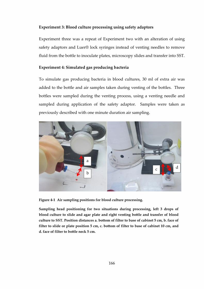
166
Experiment 3: Blood culture processing using safety adaptors
Experiment three was a repeat of Experiment two with an alteration of using
safety adaptors and Luer® lock syringes instead of venting needles to remove
fluid from the bottle to inoculate plates, microscopy slides and transfer into SST.
Experiment 4: Simulated gas producing bacteria
To simulate gas producing bacteria in blood cultures, 30 ml of extra air was
added to the bottle and air samples taken during venting of the bottles. Three
bottles were sampled during the venting process, using a venting needle and
sampled during application of the safety adaptor. Samples were taken as
previously described with one minute duration air sampling.
Figure 4-1 Air sampling positions for blood culture processing.
Sampling head positioning for two situations during processing, left 3 drops of
blood culture to slide and agar plate and right venting bottle and transfer of blood
culture to SST. Position distances a. bottom of filter to base of cabinet 5 cm, b. face of
filter to slide or plate position 5 cm, c. bottom of filter to base of cabinet 10 cm, and
d. face of filter to bottle neck 5 cm.
a
b c d
167
4.2.3.3 Fixation method comparison
A fresh culture of B. anthracis clinical isolate P12C008461 was used to inoculate
a blood culture bottle and following TTP detection used to make blood films on
microscopy slides. The blood culture bottle was gently inverted and then 10 μl
used to make each of 19 blood films. Eighteen slides were heat fixed on a hot
plate set to 70˚C for a minimum of one minutes and one slide was immersed in
95% methanol for one minute. One alcohol fixed slide was stained with PMB
for visualisation of capsule, the remaining 18 slides were added to 50 ml falcon
tubes each containing 20 ml BHI broth. The slides in broth were incubated for
72 hours and then cultured by spreading 100 μl onto a CBA plate and
incubation at 37˚C overnight.
Further 10 μl aliquots were used to make 19 blood films on microscopy slides, 9
slides were heat fixed at 85˚C for 2 minutes and 10 slides were immersed in 95%
methanol for 1 minute. One methanol fixed slide was stained with M’Fadyean
for visualisation of capsule, the remaining 18 slides were added to a 50 ml
falcon tube each containing 20 ml BHI broth and 10 ml BHI plus polymixin B
sulphate (7 mg L‐1). The slides in broth were incubated at 37˚C overnight. Each
broth was cultured by spreading 100 μl onto a CBA plate followed by
incubation at 37˚C overnight. The positive blood culture fluid was enumerated
as previously described in section 3.1.1.4.
4.2.3.4 Viability of stained slides
Slides were also prepared for isolate P10C1418 (isolate referred to the reference
laboratory for B. anthracis identification but shown to be not B. anthracis and
later identified as B. endophyticus) and B. anthracis isolate P12C016488. Aerobic
blood cultures were prepared using a fresh culture of each isolate and the BHI
cultures enumerated.
168
The positive blood cultures were used to prepare 20 microscopy slides and
enumeration of the positive fluid was also performed to determine the bacterial
load present on the slides. Eighteen slides were air dried and heated on a hot
plate at 70˚C for 3 minutes, two slides were fixed in 95% methanol for 1 minute.
Slides were then stained with Gram stains and PMB stain and the 2 alcohol
fixed slides were air dried and brought out of the MSC III after appropriate
surface decontamination of the slide box. These two slides (one Gram and one
PMB) were examined under x100 oil immersion magnification on the laboratory
light microscope. The other 18 slides were placed in 50 ml falcon tubes
containing 20 ml BHI and incubated overnight at 37˚C. The slides in broth were
examined the next day and all broths were cultured (irrespective of broth
turbidity) and plates examined for growth after overnight incubation at 37˚C.
4.2.3.5 Sporulation in blood cultures
A preliminary experiment to prepare samples for Scanning Electron
Microscopy (SEM) using blood cultures containing horse blood and B. anthracis
(P10C0001) was conducted. Samples were prepared from a blood culture after
TTP detection, following SST processing and after storage for seven days room
temperature. The method was then repeated as described below for blood
cultures containing human blood on day one (t=0) and day 5 (t=5).
Positive blood culture fluid from a blood culture containing human blood (3
bottles inoculated with the 10‐6 dilutions) was examined in the following way:
t=0: Blood culture fluid was enumerated using the method described in section
2.3.1.4. A ten‐fold dilution series was made and 0.7 ml aliquots were heat
treated at 65 ˚C for 30 minutes to kill any vegetative bacteria. After heat
treatment 100 μl of each sample was cultured on to duplicate CBA plates and
incubated overnight at 37˚C to determine if spores were present and if so how
169
many. The positive blood culture bottles were then stored securely at room
temperature for five days.
t=5: A repeat of the previous method to enumerate blood culture fluid followed
by heat treatment and enumeration of heat treated samples was performed.
Samples from days 1 and 5 were also taken for examination by SEM, all
pipetting was performed using a wide aperture 5 ml plastic pastette to reduce
shearing forces on the samples. 500 μl of positive blood culture fluid was
transferred into 1.5 ml microtubes with o‐rings. The samples were spun at 800
rpm for 1 minute in a micro centrifuge and following the removal of
supernatant were re‐suspended in 500 μl sterile PBS. Pellets were washed by
centrifugation at 800 rpm. The supernatant was removed and pellets were re‐
suspended in 4% formalin in PBS. Samples were left in the fridge until they
had been in 4% formalin in PBS for 24 hours. The samples were then gently
mixed in the MSC III and 50 μl of each sample streaked on to CBA media and
incubated at 37˚C for 48 hours. These plates were checked for sterility after
incubation. The samples were transferred to clean tubes after setting up the
sterility plates and removed from the cabinet after appropriate decontamination
and stored in the fridge. The samples were shown to be sterile when no growth
was observed on the sterility plates. Sterile samples were transferred out of the
CL3 laboratory to the Electron Microscopy Laboratory where SEM was
performed. Sample processing for SEM involved fixation in gluteraldehyde,
settling onto a poly‐l‐lysine coated coverslip, followed by solvent dehydration
with graded ethanol and replacement with hexamethyldisilazane (HMDS).
The sample was then coated in gold, using an ion beam sputter coater (Ultra‐
fine grain coating module, 700 series) and the scanning electron microscopy
was performed on a FEI XL30 FEG SEM, and examined at 4 kV.

170
4.2.3.6 Serum separator tube sample viability
To investigate the potential presence of viable B. anthracis in a bacteraemic
patients’ serum sample, the following method was used.
A BHI broth culture of B. anthracis (Vollum) prepared previously (section
2.2.3.8) was used to inoculate horse blood. A 100 μl aliquot of the undiluted
BHI culture and ten‐fold serial dilutions (100 to 10‐7 CFU ml‐1) were inoculated
into 8 x 5 ml aliquots of horse blood. These were transferred to 8 serum
separator tubes (SSTs) and mixed by gentle inversion 5 times and left for 30
minutes. The SSTs were centrifuged at 1000 x g for 10 minutes, gently inverted
5 times and then 100 μl of serum cultured on COH media and incubated at 37˚C
overnight. Following incubation the plates were examined and the numbers of
colonies counted.
This method was repeated for a BHI broth culture of B. anthracis P12C008461
with the following alterations, 1 ml of each broth suspension was added to 9 ml
horse blood and 5 ml of each spiked sample was then transferred to duplicate
SSTs. SSTs were mixed by gentle inversion five times and left for 30 minutes.
The SSTs were centrifuged at 1300 x g for 10 minutes, gently inverted five times
and then 100 μl of serum cultured on triplicate COH media and incubated at
37˚C overnight. SSTs were then stored in a fridge (2‐8˚C) for 5 days, mixed by
gentle inversion 5 times and 100 μl of serum cultured on triplicate COH media
and incubated at 37˚C overnight.
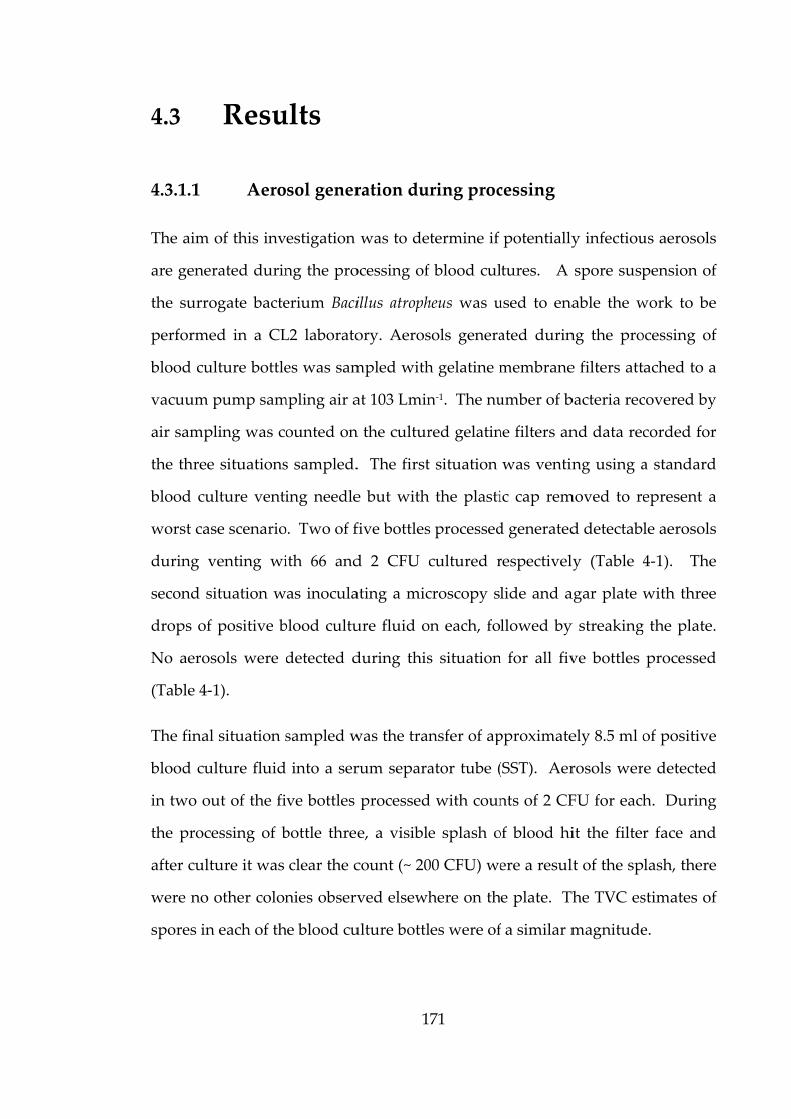
4.3
4.3.1.1
The aim
are gener
the surro
performe
blood cul
vacuum p
air sampl
the three
blood cu
worst cas
during v
second si
drops of
No aeros
(Table 4‐1
The final
blood cul
in two ou
the proce
after cultu
were no o
spores in
Resu
Aero
of this inv
rated durin
ogate bacte
ed in a CL
lture bottle
pump sam
ling was co
situations
lture venti
se scenario
venting wi
ituation w
positive b
sols were d
1).
situation s
lture fluid
ut of the fi
essing of b
ure it was
other colon
each of th
ults
sol gener
estigation
ng the pro
erium Baci
L2 laborato
es was sam
mpling air a
ounted on
s sampled.
ing needle
o. Two of f
ith 66 and
as inocula
blood cultu
detected d
sampled w
into a ser
ve bottles
bottle three
clear the c
nies observ
he blood cu
1
ration du
was to de
ocessing of
illus atroph
ory. Aeros
mpled with
at 103 Lmin
n the cultur
. The first
e but with
five bottles
d 2 CFU c
ating a mic
ure fluid o
during this
was the tran
rum separa
processed
e, a visible
count (~ 20
ved elsewh
ulture bottl
71
ring proc
termine if
f blood cul
heus was u
sols genera
h gelatine m
n‐1. The nu
red gelatin
t situation
h the plasti
s processed
cultured r
croscopy s
on each, fo
s situation
nsfer of ap
ator tube (
d with coun
e splash o
00 CFU) we
here on th
les were of
cessing
potentially
ltures. A
used to ena
ated durin
membrane
umber of b
ne filters an
was ventin
ic cap rem
d generated
respectivel
lide and a
llowed by
n for all fiv
pproximate
(SST). Aer
nts of 2 CF
of blood hi
ere a resul
e plate. Th
f a similar m
y infectiou
spore susp
able the w
ng the pro
e filters att
bacteria rec
nd data rec
ng using a
moved to re
d detectabl
ly (Table 4
agar plate w
y streaking
ve bottles
ely 8.5 ml o
rosols wer
FU for each
it the filter
lt of the sp
The TVC es
magnitude
us aerosols
pension of
work to be
ocessing of
ached to a
covered by
corded for
a standard
epresent a
le aerosols
4‐1). The
with three
g the plate.
processed
of positive
e detected
h. During
r face and
lash, there
stimates of
e.
s
f
e
f
a
y
r
d
a
s
e
e
.
d
e
d
g
d
e
f
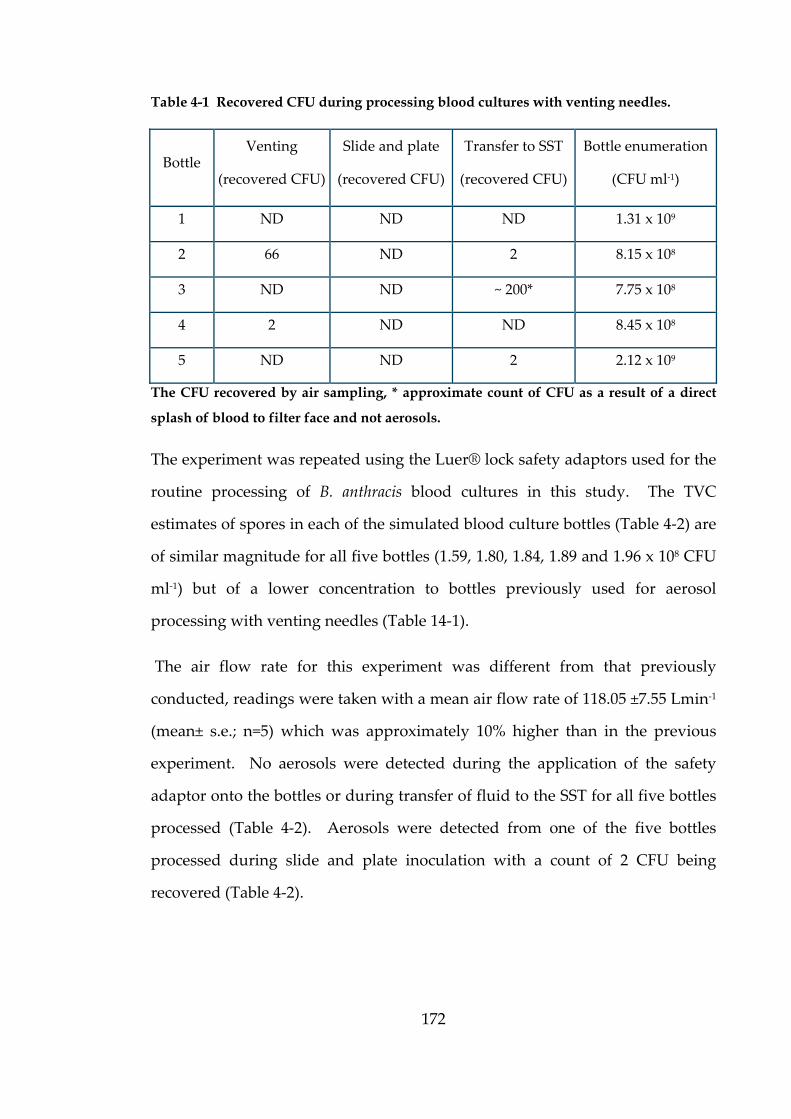
172
Table 4-1 Recovered CFU during processing blood cultures with venting needles.
Bottle Venting
(recovered CFU)
Slide and plate
(recovered CFU)
Transfer to SST
(recovered CFU)
Bottle enumeration
(CFU ml‐1)
1 ND ND ND 1.31 x 10⁹
2 66 ND 2 8.15 x 10⁸
3 ND ND ~ 200* 7.75 x 108
4 2 ND ND 8.45 x 10⁸
5 ND ND 2 2.12 x 10⁹
The CFU recovered by air sampling, * approximate count of CFU as a result of a direct
splash of blood to filter face and not aerosols.
The experiment was repeated using the Luer® lock safety adaptors used for the
routine processing of B. anthracis blood cultures in this study. The TVC
estimates of spores in each of the simulated blood culture bottles (Table 4‐2) are
of similar magnitude for all five bottles (1.59, 1.80, 1.84, 1.89 and 1.96 x 10⁸ CFU
ml‐1) but of a lower concentration to bottles previously used for aerosol
processing with venting needles (Table 14‐1).
The air flow rate for this experiment was different from that previously
conducted, readings were taken with a mean air flow rate of 118.05 ±7.55 Lmin‐1
(mean± s.e.; n=5) which was approximately 10% higher than in the previous
experiment. No aerosols were detected during the application of the safety
adaptor onto the bottles or during transfer of fluid to the SST for all five bottles
processed (Table 4‐2). Aerosols were detected from one of the five bottles
processed during slide and plate inoculation with a count of 2 CFU being
recovered (Table 4‐2).

173
Table 4-2 Recovered CFU during processing blood cultures with safety adaptors.
Bottle Venting
(recovered CFU)
Slide and plate
(recovered CFU)
Transfer to SST
(recovered CFU)
Bottle enumeration
(CFU ml‐1)
1 ND 2 ND 1.59 x 10⁸
2 ND ND ND 1.80 x 10⁸
3 ND ND ND 1.84 x 10⁸
4 ND ND ND 1.89 x 10⁸
5 ND ND ND 1.96 x 10⁸
The CFU recovered by air sampling during processing with application of safety
adaptors, 3 drops of blood to both slide and plate with plate streaking and transfer to
SST. (ND, not detected).
A calculation of the spray factor (SF) for both the use of venting needle and
safety adaptor to process bottles was performed using the aerosol counts
obtained (Table 4‐3). The SF was calculated by dividing the aerosol
concentration by the suspension concentration (Bennett & Parks, 2006).
Table 4-3 Summary of aerosol data for calculation of spray factor.
Aerosol
CFU
Air sampling
m‐3
[aerosol]
CFU m‐3
[suspension]
CFU ml‐1
Spray factor
66 (venting) 0.103 640.7 8.15 x 10⁸ 7.86 x 10‐7
2 (venting) 0.103 19.4 8.45 x 10⁸ 2.30 x 10‐8
2 (transfer) 0.103 19.4 8.15 x 10⁸ 2.38 x 10‐8
2 (transfer) 0.103 19.4 2.12 x 10⁹ 9.16 x 10‐9
2 (inoculation) 0.118 16.9 1.59 x 10⁸ 1.07 x 10‐7
The SF can be used as an indication of the severity of an accident and as a
component in dynamic risk assessment (Bennett & Parks, 2006).

174
More samples would be needed over a range of concentrations to determine a
stable SF for the individual steps sampled here so that a concentration not
tested could be used with the spray factor to calculate the theoretical aerosol
concentration and therefore the aerosol hazard to the laboratory worker.
If the spray factor (for 66 CFU recovered during venting) is used to calculate the
aerosol concentration from venting a blood culture bottle containing ~ 1 x 10⁸
CFU ml‐1 then the following example could be used as part of an evidence‐
based risk assessment following potential exposure from processing a blood
culture containing B. anthracis on the open bench in a CL2 laboratory.
The spray factor can be used for evidence based risk assessments, as an
example. The assumptions made were as follows:
A laboratory worker vents a blood culture bottle on the open bench containing
~ 1 x 10⁸ CFU ml‐1 which takes approximately 30 seconds, whilst they stand in
front of the bottle. The exposed worker has a breathing rate of 15 L min‐1, the
aerosol does not significantly deposit during the time and all aerosolized
bacteria are <5 μm and deposited in the lungs.
The calculated spray factor for venting bottles can now be used:
Aerosol concentration = (7.86 x 10‐7) x (1 x 108)
= 78.6 CFU m‐3
= 0.0786 CFU l‐1
Dose (30 seconds) = 7.5 x 0.0786 = 0.5895 CFU
Dose (1 minute) = 15 x 0.0786 = 1.179 CFU
175
The dose of <1 CFU would be potentially inhaled in 30 seconds if all the
assumptions made are correct, when using the spray factor calculated for
venting with a venting needle.
The air sampling results for bottles prepared with an addition 30 ml added air
and calculated spray factors are shown in Table 4‐4, with the concentration B.
atropheus in blood culture bottles. The application of the safety adaptor
generated higher recovered counts than with the venting needle.
Table 4-4 Recovered CFU during processing blood cultures with 30 ml added air.
Bottle Bottle
enumeration
(CFU ml‐1)
Venting with
venting needle
(recovered CFU)
Application of
safety adaptor
(recovered CFU)
Spray factor
1 4.2 x 10⁸ 30 6.05 x 10‐7
2 4.3 x 10⁸ 27 5.32 x 10‐7
3 5.0 x 10⁸ 114 1.93 x 10‐6
4 6.3 x 10⁸ 4 5.30 x 10‐8
5 5.6 x 10⁸ 11 1.66 x 10‐7
6 4.2 x 10⁸ 2 4.0 x 10‐8
4.3.1.2 Fixation method and viability of microscopy slides
The aim of this investigation was to determine the viability of B. anthracis on
fixed microscopy slides. The use of heat inactivation (at two temperatures)
versus alcohol fixation was initially compared to determine the potential
biological risk to front line microbiology staff. Heat fixation is commonly used
in front‐line laboratories and alcohol fixation is used by the national reference
laboratory.
176
Alcohol fixation is recommended in guidance for the fixation of blood cultures
suspected to contain B. anthracis, however the causative organism may not be
known until after the microscopy slide has been examined.
The viability results for slides treated by the three methods are shown in Table
4‐5. All slides treated at 70˚C were found to possess viable B. anthracis. One out
of nine slides had viable B. anthracis cells after treatment at 85 ˚C. In contrast
there were no detectable viable organisms isolated from microscopy slides
following alcohol fixation in 95% methanol for one minute and all nine turbid
broth cultures, contained a common contaminating bacterium.
Table 4-5 Viability of B. anthracis on microscopy slides following three treatments.
Heat fixation
(70˚ C) minimum 2
minutes
Heat fixation
(85˚ C) minimum 2
minutes
Alcohol fixation
(1 minute 95%
methanol)
Number of
slides 18 9 9
Broth
turbidity 18/18 turbid 0/9 turbid 9/9 turbid
Slide
viability 18/18 viable 1/9 viablea 0 viableb
a 2 broth grew a contaminant common to both and b 9 broth cultures grew a contaminant
common to all and no growth of B. anthracis.
4.3.1.3 Viability of stained slides
The viability of B. anthracis on microscopy slides treated with heat fixation at
70˚C for 2 minutes was further investigated to determine if the chemicals used
for Gram staining and M’Fadyeans reaction would inactivate viable bacilli after
the initial heat treatment.
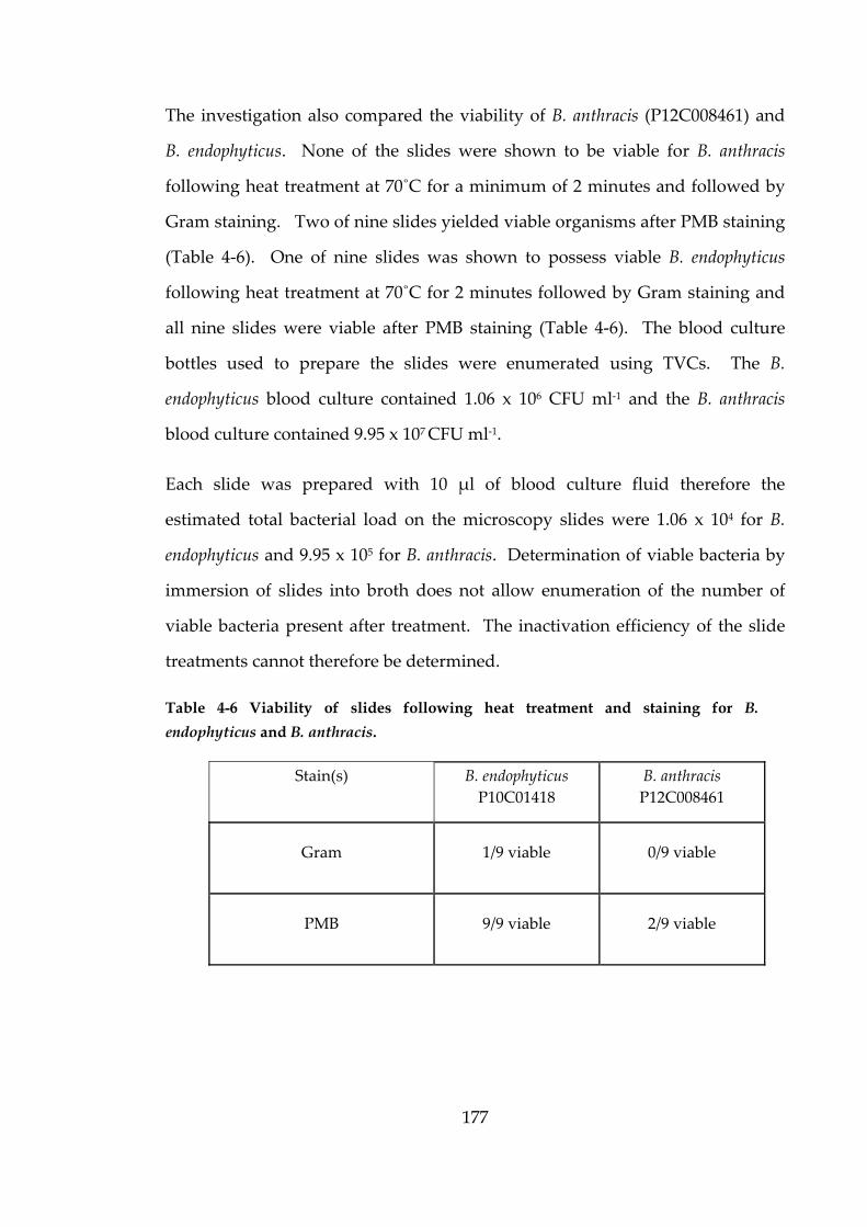
177
The investigation also compared the viability of B. anthracis (P12C008461) and
B. endophyticus. None of the slides were shown to be viable for B. anthracis
following heat treatment at 70˚C for a minimum of 2 minutes and followed by
Gram staining. Two of nine slides yielded viable organisms after PMB staining
(Table 4‐6). One of nine slides was shown to possess viable B. endophyticus
following heat treatment at 70˚C for 2 minutes followed by Gram staining and
all nine slides were viable after PMB staining (Table 4‐6). The blood culture
bottles used to prepare the slides were enumerated using TVCs. The B.
endophyticus blood culture contained 1.06 x 10⁶ CFU ml‐1 and the B. anthracis
blood culture contained 9.95 x 10⁷ CFU ml‐1.
Each slide was prepared with 10 μl of blood culture fluid therefore the
estimated total bacterial load on the microscopy slides were 1.06 x 10⁴ for B.
endophyticus and 9.95 x 10⁵ for B. anthracis. Determination of viable bacteria by
immersion of slides into broth does not allow enumeration of the number of
viable bacteria present after treatment. The inactivation efficiency of the slide
treatments cannot therefore be determined.
Table 4-6 Viability of slides following heat treatment and staining for B.
endophyticus and B. anthracis.
Stain(s) B. endophyticus
P10C01418
B. anthracis
P12C008461
Gram 1/9 viable 0/9 viable
PMB 9/9 viable 2/9 viable
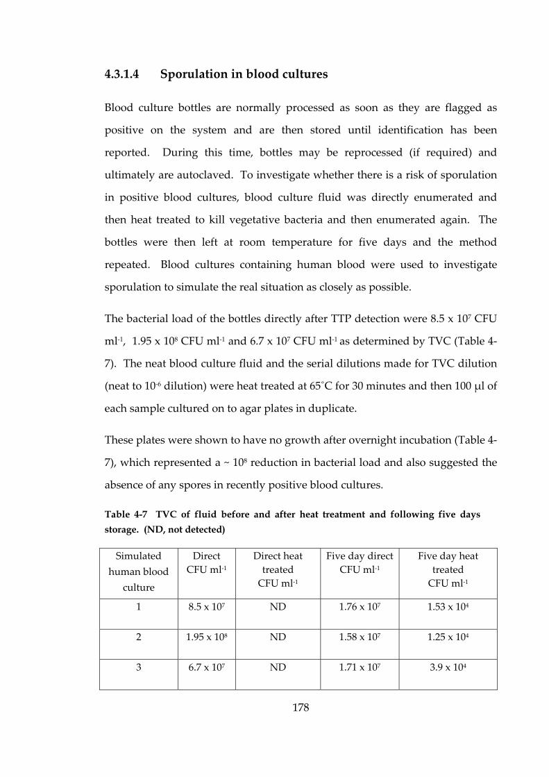
178
4.3.1.4 Sporulation in blood cultures
Blood culture bottles are normally processed as soon as they are flagged as
positive on the system and are then stored until identification has been
reported. During this time, bottles may be reprocessed (if required) and
ultimately are autoclaved. To investigate whether there is a risk of sporulation
in positive blood cultures, blood culture fluid was directly enumerated and
then heat treated to kill vegetative bacteria and then enumerated again. The
bottles were then left at room temperature for five days and the method
repeated. Blood cultures containing human blood were used to investigate
sporulation to simulate the real situation as closely as possible.
The bacterial load of the bottles directly after TTP detection were 8.5 x 10⁷ CFU
ml‐1, 1.95 x 10⁸ CFU ml‐1 and 6.7 x 10⁷ CFU ml‐1 as determined by TVC (Table 4‐
7). The neat blood culture fluid and the serial dilutions made for TVC dilution
(neat to 10‐6 dilution) were heat treated at 65˚C for 30 minutes and then 100 μl of
each sample cultured on to agar plates in duplicate.
These plates were shown to have no growth after overnight incubation (Table 4‐
7), which represented a ~ 108 reduction in bacterial load and also suggested the
absence of any spores in recently positive blood cultures.
Table 4-7 TVC of fluid before and after heat treatment and following five days
storage. (ND, not detected)
Simulated
human blood
culture
Direct
CFU ml‐1
Direct heat
treated
CFU ml‐1
Five day direct
CFU ml‐1
Five day heat
treated
CFU ml‐1
1 8.5 x 10⁷ ND 1.76 x 10⁷ 1.53 x 10⁴
2 1.95 x 10⁸ ND 1.58 x 10⁷ 1.25 x 10⁴
3 6.7 x 10⁷ ND 1.71 x 10⁷ 3.9 x 10⁴
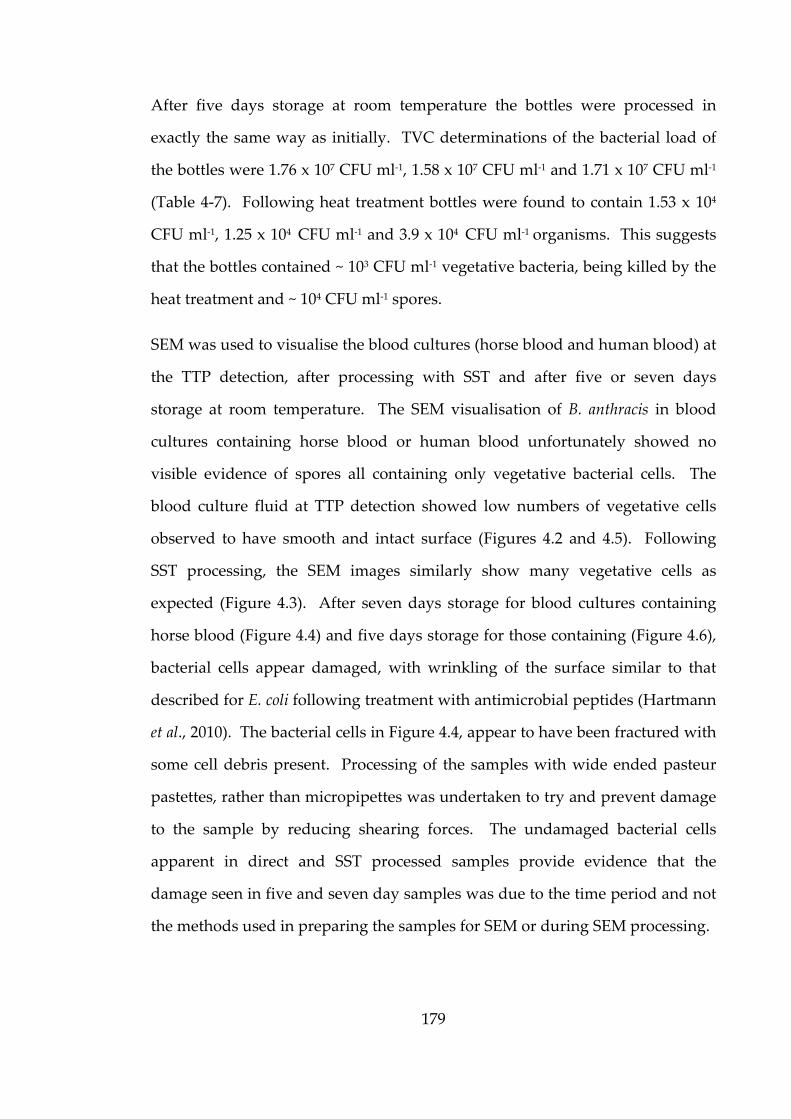
179
After five days storage at room temperature the bottles were processed in
exactly the same way as initially. TVC determinations of the bacterial load of
the bottles were 1.76 x 10⁷ CFU ml‐1, 1.58 x 10⁷ CFU ml‐1 and 1.71 x 10⁷ CFU ml‐1
(Table 4‐7). Following heat treatment bottles were found to contain 1.53 x 10⁴
CFU ml‐1, 1.25 x 10⁴ CFU ml‐1 and 3.9 x 10⁴ CFU ml‐1 organisms. This suggests
that the bottles contained ~ 103 CFU ml‐1 vegetative bacteria, being killed by the
heat treatment and ~ 104 CFU ml‐1 spores.
SEM was used to visualise the blood cultures (horse blood and human blood) at
the TTP detection, after processing with SST and after five or seven days
storage at room temperature. The SEM visualisation of B. anthracis in blood
cultures containing horse blood or human blood unfortunately showed no
visible evidence of spores all containing only vegetative bacterial cells. The
blood culture fluid at TTP detection showed low numbers of vegetative cells
observed to have smooth and intact surface (Figures 4.2 and 4.5). Following
SST processing, the SEM images similarly show many vegetative cells as
expected (Figure 4.3). After seven days storage for blood cultures containing
horse blood (Figure 4.4) and five days storage for those containing (Figure 4.6),
bacterial cells appear damaged, with wrinkling of the surface similar to that
described for E. coli following treatment with antimicrobial peptides (Hartmann
et al., 2010). The bacterial cells in Figure 4.4, appear to have been fractured with
some cell debris present. Processing of the samples with wide ended pasteur
pastettes, rather than micropipettes was undertaken to try and prevent damage
to the sample by reducing shearing forces. The undamaged bacterial cells
apparent in direct and SST processed samples provide evidence that the
damage seen in five and seven day samples was due to the time period and not
the methods used in preparing the samples for SEM or during SEM processing.

180
Figure 4-2 SEM of blood culture at TTP simulated with horse blood.
Figure 4-3 SEM of blood culture at TTP following SST processing simulated with
horse blood.
181
Figure 4-4 SEM of blood culture after 7 days storage simulated with horse blood.
Figure 4-5 SEM of blood culture at TTP simulated with human blood.

182
Figure 4-6 SEM of blood culture at TTP following SST processing simulated with
human blood.
Figure 4-7 SEM of blood culture after 5 days simulated with human blood.
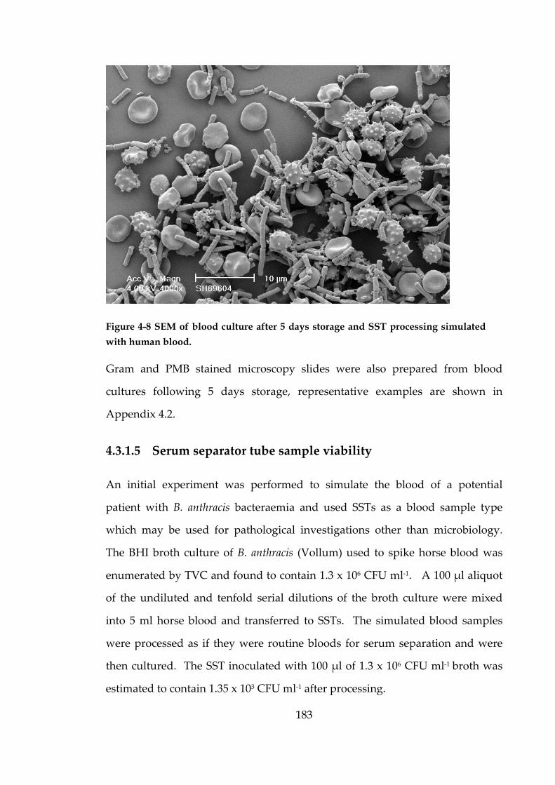
183
Figure 4-8 SEM of blood culture after 5 days storage and SST processing simulated
with human blood.
Gram and PMB stained microscopy slides were also prepared from blood
cultures following 5 days storage, representative examples are shown in
Appendix 4.2.
4.3.1.5 Serum separator tube sample viability
An initial experiment was performed to simulate the blood of a potential
patient with B. anthracis bacteraemia and used SSTs as a blood sample type
which may be used for pathological investigations other than microbiology.
The BHI broth culture of B. anthracis (Vollum) used to spike horse blood was
enumerated by TVC and found to contain 1.3 x 10⁶ CFU ml‐1. A 100 μl aliquot
of the undiluted and tenfold serial dilutions of the broth culture were mixed
into 5 ml horse blood and transferred to SSTs. The simulated blood samples
were processed as if they were routine bloods for serum separation and were
then cultured. The SST inoculated with 100 μl of 1.3 x 10⁶ CFU ml‐1 broth was
estimated to contain 1.35 x 10³ CFU ml‐1 after processing.
184
This initial experiment was used to explore the potential presence of bacteria in
a simulated blood sample because it was known that when a SST is centrifuged,
bacteria are concentrated from the sample onto the gel plug and anthrax
patients with bacteraemia can reach very high concentrations of bacteria in the
blood. The experiment was altered by preparing duplicate SST(s) for each
inoculum and estimating the concentration of bacteria initially and after storage
in the fridge for five days; the viable count data are shown in Table 4‐8.
Table 4-8 Triplicate counts for duplicate serum samples at t=0 and t=5 days storage
in the fridge (TNTC – Too Numerous To Count, ND – Not detected).
Concentration of B. anthracis in blood prior to SST processing
1.42 x10⁵
CFU ml‐1
1.42 x10⁴
CFU ml‐1
1.42 x10³
CFU ml‐1
1.42 x10²
CFU ml‐1
1.42 x101
CFU ml‐1
1.42 x100
CFU ml‐1
t=0
(1)
TNTC
TNTC
TNTC
TNTC
TNTC
TNTC
44
28
36
7
4
3
ND
ND
ND
ND
ND
3
t=0
(2)
TNTC
TNTC
TNTC
TNTC
TNTC
TNTC
110
95
107
23
11
29
2
3
11
ND
2
3
t=5
(1)
185
172
195
14
12
17
1
1
ND
ND
ND
ND
ND
ND
ND
ND
ND
ND
t=5
(2)
189
173
192
18
27
19
ND
ND
2
ND
ND
ND
ND
ND
ND
ND
ND
ND
The seru
contain 1
with 1.42
1.42 x 10⁴
with 1.42
indicate t
blood sam
the serum
five days
103 CFU m
4.4
For the d
the chain
these con
transmiss
suitable w
hazard a
physical
upon to p
help prev
In contra
and radia
fatal outc
after expo
um culture
1840 and 1
2 x 10⁵ CFU
4 or 103 CF
2 x 10², 101
that a patie
mples take
m on day o
s storage in
ml‐1.
Discu
discussion
n of infectio
ncerns fall
sion via th
work pract
any further
barrier bu
provide pr
vent the sev
ast to expo
ation, infec
comes due
osure.
es followin
846 CFU m
U ml‐1 B. an
U ml‐1 had
and 100 CF
ent with a
en for seru
one. The c
n the fridg
ussion
of the bio
on will be
l. The fir
e aerosol r
tices and e
r we have
ut only pro
rotection a
verity of in
osures wit
ctious agen
e to the po
1
ng five day
ml‐1 in dup
nthracis. T
d counts le
FU ml‐1 had
bacteraem
um prepar
concentrati
ge was det
n
osafety con
used to id
rst opportu
route by is
engineering
e personal
otects the in
and is not a
nfection, as
th other ha
nts at very
otential for
85
ys storage
plicate tub
The tubes i
ess than 30
d no detect
mia of 1.42
ration, ther
ion of viab
ermined to
ncerns in c
dentify whe
unity to b
olating the
g controls.
protective
ndividual.
available f
s can prom
azardous s
low conce
r the agen
e in the fri
es original
initially ino
colonies a
table organ
x 105 CFU
re would b
ble bacteria
o contain 1
context exp
ere in the h
break the
e infectious
Then if w
e equipme
Vaccinat
or all path
mpt prophy
substances
ntrations c
t to multip
ridge were
lly contain
noculated w
and those i
nisms. Th
U ml‐1 B. ant
be viable b
a in serum
1.84 x 10³
plored in t
hierarchy o
chain is p
s material
we cannot r
ent which
tion canno
hogens, ho
ylactic trea
s, such as
can cause s
ply within
e found to
ning blood
with either
inoculated
ese results
thracis had
bacteria in
following
and 1.85 x
this study,
of controls
preventing
and using
reduce the
is the last
t be relied
wever can
tment.
chemicals
severe and
n the body
o
d
r
d
s
d
n
g
x
,
s
g
g
e
t
d
n
s
d
y

186
It could be argued that a needle stick route of transmission presents the greatest
risk to laboratory workers however, the use of sharps can be controlled and
accidents are unlikely to infect more than one laboratory worker. It is generally
accepted that the greatest risk to laboratory workers is inhalation of aerosolized
infectious particles, because the respiratory route of transmission is the hardest
to control. For a laboratory worker to become infected by aerosolized infectious
particles there are several factors to consider, firstly the agent must retain
characteristics necessary to cause disease, secondly the agent must be in the air
in sufficient quantity and time for the infectious dose to be inhaled and finally
there has to be a susceptible host in direct contact with it, in this case inhaling
the airborne infectious particles (Burge, 1995). To prevent exposure from any
infectious aerosols there are several options, the susceptible host(s) can be
isolated; filter ambient air or breathing zone air to physically remove the agent,
dilute the aerosol to levels below the infectious dose or kill the agent at source
either by disinfection of the air, surface disinfection or with antibiotics if a
person is the contagious source.
Patients with anthrax are not contagious but other pathogens can be
transmitted person to person. Some of these general measures are practically
achievable in the microbiology laboratory and form the basis of engineering
controls. Engineering controls required for CL3 laboratories are used to dilute
the air by allowing laboratory air changes to remove aerosols and in
conjunction with the negative pressure, prevent air flowing out of the
laboratory into other work areas. Another engineering control is the
microbiological safety cabinet which can be used in both the CL3 and CL2
laboratory. MSCs isolate, dilute and filter the air therefore reduce or remove
the aerosol hazard however, operators need to understand that where there are
aerosols, there is always potential for contamination of surfaces. If there was an
aerosol exposure on the open bench in a CL2 laboratory, there may be no air
187
changes to dilute the aerosol and it is not feasible to disinfect the air, so
emergency procedures are used to swiftly evacuate the area as a means to
isolate susceptible host(s) from prolonged exposure.
In a front‐line diagnostic laboratory it is possible for unexpected HG3 bacteria
to be isolated from blood cultures, with little clinical information to highlight
the potential risks (Reddy et al., 2010). In this situation the blood culture would
be processed on the open bench (unless the laboratory has access to a MSC I in
the CL2 laboratory and locally decided to process all blood cultures within a
MSC). The HSE have identified that a lack of clinical information available to
laboratory staff, does not allow staff to make the appropriate risk assessment
for handling clinical material and samples at CL3. The aerosol investigations
undertaken aimed to provide laboratory staff with some evidence of the aerosol
risk for processing bottles on the open bench, in the event that insufficient
clinical information is received to indicate the requirement to process the blood
cultures in a MSC. A recent study explored a range of air and surface
contamination produced during serial dilution (pipetting) of high titre
suspensions of B. atropheus (Pottage et al. 2014). The average bacterial counts
reported by Pottage et al. (2014) were 6.9 CFU and 13.6 CFU. The bacterial
counts determined in this study for processing blood cultures were also low,
the highest being 66 CFU (for one determination of venting with a venting
needle).
From the blood culture work with B. anthracis, bottles at TTP detection were
found to contain approximately 108 CFU ml‐1 vegetative, encapsulated bacteria,
with the potential to also contain spores after five days storage. The aerosol
sampling data were used to calculate spray factors for the various
manipulations examined such as venting bottles with venting needles or the
addition of a safety adaptor and the processing of the bottle to inoculate
188
microscopy slides and agar plates. The example used to illustrate the use of
spray factors (calculated using highest bacterial count 66 CFU) gave a
theoretical worse‐case scenario of < 1CFU (for a 30 second exposure) and 1.179
CFU (for a one minute exposure) being inhaled. These numbers are low and
assumed that the entire aerosol was inhaled and that particles were able to
penetrate into the lower respiratory tract. The spray factors determined in this
study for venting bottles were 2.30 x 10‐8 and 7.86 x 10‐7. A calculated spray
factor of 5.3 x 10‐7 was determined for a simulated accident (for dropping a flask
containing 50 ml culture at 5x10⁹ organism) by Bennett & Parks (2006). Using
this calculated spray factor a worker could potentially inhale 19.9 CFU during a
30 second period which is greater than that determined in this study for venting
blood cultures.
Spores were chosen for this scenario and the data generated in this study
showed that blood cultures processed directly after TTP contained vegetative
bacteria and a proportion of spores after five days storage. To be useful for
different scenarios the spray factor should be constant or have a determined
range to back calculate the likely concentration of aerosol present with any
given sample concentration. More work would need to be carried out to
determine if the spray factor is constant for venting or varies. To have
confidence in the use of spray factor calculations in evidence based risk
assessments further work would be required as described with additional
determinations to assess the assumptions made. The inhalation of particles
<5μm can penetrate into the lower respiratory tract, therefore investigation of
aerosol particle size from blood cultures containing spores compared to
vegetative bacteria would allow an assessment of whether aerosols are likely to
be inhaled. The air sampler used for this study allowed a determination of total
bacterial count, however, it did not allow analysis of the different particle sizes
produced therefore the calculations of spray factor and potential inhaled
189
aerosols during blood culture processing make the assumption that they are <5
μm.
Air samplers could also be positioned above the bottle at a height simulating
the laboratory worker processing the bottle. Discussion with front‐line staff
who have processed bottles containing gas producing bacteria, have
commented that when inserting the venting needle (with plastic sheath in
place) into the bulging septum the spray was sideways, justifying the
positioning for this study. An understanding of the particle size generated
from blood culture aerosols could also be related to contamination, as large
particles and droplets fall out of the air quicker and contaminate surfaces. The
spore is considered the primary infectious particle in the transmission of
anthrax and the infectious dose for humans via the respiratory route could
theoretically be as low as one spore, dependant on reaching the correct site and
upon other host factors (WHO, OIE, FAO, 2008). Data discussed in Chapter
One described low rates of infection with industrial exposure to 600 ‐1300
spores over 8 hour shift periods.
The structure of B. anthracis spores allows survival in harsh environments, and
therefore requiring appropriate decontamination and disposal methods by
ensuring destruction cycles are used when material is autoclaved. The HPA
guidance produced during the outbreak of injectional anthrax, detailed the use
of 10,000 ppm available chlorine disinfectant for dealing with spillages of
suspected B. anthracis clinical samples. This high concentration of disinfectant
will inactivate B. anthracis spores and this study would recommend this for any
blood culture spillage, even if being processed directly after TTP.
Contamination may occur as a consequence of other aspects for processing
blood cultures; the practices surrounding them would be important to prevent
laboratory acquired infections.
190
A large study investigating different methods of Mycobacterium tuberculosis
inactivation concluded that conventional practices cannot be assumed to render
the organism non‐viable and methods should be validated locally to safely
move material outside of a CL3 laboratory (Blackwood et al., 2005). In the
study, microscope slide viability was assessed and all of the slides tested
contained viable MTB after heat treatment, resulting in their local protocol
being altered to include an additional chemical treatment step. If a blood
culture suspected to contain B. anthracis was processed on the open bench, it
would be hoped laboratory workers would use common sense to appropriately
dispose of microscopy slides into sharp safe containers and handle them
carefully to prevent sharps injury. The evidence from the viability
investigations suggests that B. anthracis may be inactivated by heat treatment at
70˚C followed by standard Gram staining but not by PMB staining for the
visualisation of the presence of capsule. Blood culture microscopy slides
provide first indications of the causative agents of bacteraemia and anthrax may
not have been initially identified by clinical presentation or details and
therefore the initial slide may not be known to be B. anthracis. The UK
guidelines for processing cultures suspected to contain B. anthracis recommend
the use of alcohol fixation, routinely used at PHE Porton as a validated method
of fixing and inactivating B. anthracis slides. In front‐line laboratories this
method may not be used on initial slides, therefore care should be taken and
staff made aware of the potential for viable material on slides. It is not known
whether some laboratories leave slides to naturally dry without the use of a hot
plate and further studies would be needed to investigate this scenario.
Serum is used for many pathological investigations other than microbiology, for
example within haematology and clinical chemistry departments. The presence
of spores in clinical material was explored by simulating serum samples from a
bacteraemic anthrax patient.
191
The data suggested that viable B. anthracis may be present in serum samples
because the bacteria in the blood can be at high concentrations and the bacteria
are captured on the gel plug of serum separator tubes. The simulated blood
samples contained a final concentration of 1.42 x 10⁵ CFU ml‐1 B. anthracis which
does not represent the very high ~108 CFU ml‐1 which can be reached prior to
death but was suitable for the purposes of the study. After processing to
separate serum, followed by storage in the fridge for 5 days, the samples were
found to contain 1.8 x 10³ CFU ml‐1, a reduction of 2 logs, however it was not
determined whether the storage resulted in the induction of sporulation. It is
possible to have breaks in communication between clinical staff and between
pathology departments and spillage of such a sample would present a
contamination issue, especially if the bacteria had sporulated. The HPA
guidance, mentioned previously, covers information for blood science
laboratories for spillage and alerts workers to the care needed to prevent
inoculation injuries. In addition the guidance highlights the need for staff to
strictly follow standard procedures which should already be in place to prevent
the acquisition of blood borne viruses. The data from this study also supports
the guidance and presents data to support the assumed situation.
Heat treatment of the positive bottles directly on day one was sufficient to
reduce counts by ~108 therefore, there appears no reason for this reduction to be
different for the bottles after five days storage and that the growth observed
was a result of the presence of spores. Simulated bottles at TTP were estimated
to contain between 107 and 108 CFU ml‐1 and following storage for five days and
heat treatment contained approximately 104 CFU ml‐1 spores. Microscopic
examination of the samples would have been useful to see the presence of
spores. The use of conventional spore stains, however require heating which is
difficult in an MSC and the cold version of the staining procedure was reported
to provide poor results when used by other research groups at PHE Porton.
192
Gram and PMB stained films (Appendix 4.2) suggest sporulation however, not
by specifically staining spores. To take advantage of the facilities at PHE
Porton, the visualisation of simulated blood cultures was performed using
SEM. The simulated human blood cultures did not show visual presence of
spores following storage at room temperature for five days however, vegetative
cells were fractured or showed wrinkled surfaces. Spores in high titre in a
blood culture bottle may become a safety issue in the routine laboratory if
laboratory workers needed to reprocess or culture the bottle prior to disposal.
In this situation there is unlikely to be an aerosol risk due to the bottle
previously being vented but a spillage would require appropriate disinfection
and may present an aerosol risk dependent on the accident. Risk assessments
can be based on the evidence of knowing if and when, under normal conditions
(room temperature), the bacteria sporulate and therefore impose procedural
controls to reduce the risk of infection to the laboratory worker. It would be
hoped that the blood culture would be identified as presumptive B. anthracis
within 24 hours and therefore moved to the CL3 laboratory were all
manipulations would be conducted in an MSC I and all samples and waste
disposed of in the CL3 waste stream. The SAPO regulations enforced by
DEFRA, has strict licensing of laboratories able to handle and store SAPO
pathogens such as B. anthracis and Brucella spp. Once a front‐line laboratory has
had a patient confirmed as having anthrax, all cultures, samples and patient
material should be appropriately disposed of to prevent release into the
environment.
A serious accident scenario to consider, though very unlikely to occur, would
be accidental self‐inoculation of positive blood culture fluid. The use of routine
personal protective equipment such as gloves would not necessarily prevent
this exposure unless sharps resistant gloves were used.
193
In addition, the venting needles used to vent blood cultures are blunt and the
sharps hazard is present when first inserting a clean needle into the bottle
septum.
Injury could occur when removing this needle which would be contaminated
with the positive blood culture fluid containing a high concentration (~108 CFU
ml‐1) of bacteria. The in‐vivo experiments described by Levy et al. (2014) to
artificially create bacteraemia by intravenous injection of B. anthracis
encapsulated vegetative cells into rabbits, could be likened to such an
inoculation accident though the volume would be small. In the rabbit model,
this method of inoculation allowed haematogenous spread of the bacilli
resulting in an artificial bacteraemia that resembled the systemic stage of
disease (Kobiler, 2006). Inoculation with 107 CFU ml‐1 resulted in a bacteraemia
of 105 CFU ml‐1 5 hours post inoculation and reached a maximum bacteraemia
of 108 CFU ml‐1 after 24 hours. An inoculation injury would be extremely
serious irrespective of the bacteria, due to the high concentration of bacteria in a
blood culture being directly administered into the body. This type of accident
would require medical attention and prompt prophylactic treatment. This type
of accident was discussed with Dr Tim Brooks, whose personal opinion was
‘entry of low concentrations of vegetative B. anthracis, into breaches of the skin are
likely to be overwhelmed by the immune response especially in vaccinated individuals,
as the bacteria would not be aided by the protective action of the exotoxins’.
In all accidents or potential exposures, appropriate medical attention is
paramount and risk assessment of exposure would be undertaken by medical
staff as to whether prophylactic treatment or monitoring of symptoms is
undertaken.
A review of illness surveillance data from an archive of the U.S. offensive
biological warfare program (from 1943 to 1969) was examined to assess the
194
impact of safety measures such as MSCs before and after vaccine availability on
disease prevention (Rusnak et al., 2004). The records suggest that personal
protective measures and safety training alone were able to prevent laboratory‐
acquired infections for anthrax, glanders and plague.
In contrast, safety measures to include the use of MSC without vaccination
failed to prevent infections with a very low infectious dose such as tularemia
and Q fever. The numbers of these infections dramatically dropped when
vaccination was available and the publication suggests that only laboratories
experienced to handle these highly infectious organisms, who have suitable
training and precautions should conduct research. In the UK, vaccination is
available to staff who routinely work with B. anthracis and very few institutions
such as PHE Porton and the Defence Science Technology Laboratories (DSTL,
Porton) conduct research with these organisms. Diagnostic laboratories may
infrequently culture HG3 bacteria, however, as previously discussed they may
not always know to handle it at CL3, due to a lack of clinical information. The
training provided by NADP training at PHE Porton is therefore an important
means to increase awareness of these pathogens by front‐line staff and an
opportunity to share the best practices for handling pathogens.
4.5
This cha
biosafety
anthracis
blood cul
may be su
Th
po
ve
Mi
ind
ba
Po
no
aft
Ot
as
an
The conc
laborator
assessme
explored
of the ‘
laborator
Summ
pter descr
y concerns
in front‐li
ltures and
ummarised
he data co
ositive bloo
enting need
icroscopy
dicated th
acteria, how
ositive bloo
ot likely to
ter five day
ther blood
serum, co
thracis eve
clusions of
ry worker
ents. The
in Chapte
process’ a
ries.
mary
ribed the
for proce
ine labora
blood sam
d as follow
ollected in
od culture
dle or safet
slides ma
at chemica
wever the P
od culture
o contain s
ys storage.
containing
ollected us
n after stor
f the stud
rs with
developm
er Five. Th
and ‘safet
1
experimen
essing bloo
atories. Th
mples and
ws:
ndicated th
e containin
ty adaptor.
ay be viab
als in the
PMB stain
es processe
spores at t
.
g samples
sing serum
rage in a fr
dy relating
informatio
ent of trai
he study as
ty’ to pro
95
nts perform
od culture
he data w
the main
hat the ae
ng B. anth
.
ble after h
Gram stai
does not b
ed directly
this time h
from bact
m separato
ridge for fi
g to the a
on to pe
ining mate
spect of ‘tra
oduce trai
med to ex
es suspecte
was genera
conclusion
erosol risk
hracis was
heat fixing
in inactiva
behave sim
y following
however th
teraemic an
r tubes ma
ive days.
spect of ‘s
erform ev
erial for la
aining’ bri
ining mat
xplore the
ted of con
ated from
ns from th
k from pro
low, usin
g at 70˚C,
ate remain
milarly.
g TTP det
hey may b
nthrax pat
ay contain
safety’ wi
vidence b
aboratory w
ings togeth
terial for
e potential
ntaining B.
simulated
his chapter
ocessing a
ng either a
, the data
ning viable
tection are
be present
tients such
n viable B.
ll provide
ased risk
workers is
her aspects
front‐line
l
.
d
r
a
a
a
e
e
t
h
e
k
s
s
e

Chap
m
I5.1
This chap
the study
on the la
training l
purpose
scientists
professio
The Hea
responsib
performa
Scientists
scientist p
to regist
continual
demonstr
The Inst
biomedic
and diplo
along a c
follow th
pter 5
materi
Introd
pter discus
y to produ
boratory d
laboratory
requires a
as part
onal develo
alth and C
ble for set
ance and
s. There
profession
er with th
l professi
rate they a
titute of B
cal science
omas for m
career path
he codes of
Dev
ial
ductio
sses the co
ce speciali
detection o
. To deve
an underst
of their
opment (CP
Care Profe
tting and
conduct o
are severa
nal and tho
he HCPC
onal deve
are keeping
Biomedica
in the UK
members to
hway. Me
conduct fo
1
velopm
on
ombination
ised trainin
of Anthrax
elop trainin
tanding of
on the jo
PD).
essions Co
maintaini
of healthca
al paths fo
ose with ap
C. The H
elopment
g up to dat
al Science
K and has a
o demonst
embership
or biomedi
96
ment o
n of ‘the p
ng materia
x and pre e
ng materia
the traini
ob trainin
ouncil (HC
ing standa
are profes
or individ
ppropriate
HCPC requ
(CPD) to
te with dev
(IBMS) i
a role to d
trate levels
p to the IB
ical science
of trai
process’ an
al for front‐
entry infor
al which is
ing undert
ng and as
CPC) is th
ards of pr
ssionals in
uals to be
e qualificat
uire regist
o maintai
velopment
s the pro
develop qu
s of expert
BMS brings
e professio
ining
nd ‘safety’
‐line labor
rmation fo
s suitable
taken by b
s part of
he regulat
rofessional
ncluding B
ecome a b
tions and e
trants to
in registra
ts in their p
ofessional
ualification
tise and co
s a respon
onals.
aspects of
ratory staff
or the PHE
and fit for
biomedical
continual
tory body
l training,
Biomedical
biomedical
experience
undertake
ation and
profession.
body for
ns, training
ompetency
nsibility to
f
f
E
r
l
l
y
,
l
l
e
e
d
.
r
g
y
o
197
The aspects of the codes of conduct relevant to this study are competence and
behaviour. Biomedical scientists must continuously develop and demonstrate
their knowledge and skills to reflect professional and scientific advances and
guide best professional practice. Biomedical scientists are expected to do this
by undertaking courses, on the job training, reflection and self‐management
processes such as CPD (IBMS, 2014). They must also co‐operate with employer
and professional colleagues in the interests of providing a safe and high‐quality
service, as a behaviour requirement in the code of conduct.
As discussed in Chapter Four, the employer has a legal responsibility to
provide suitable and sufficient training for staff undertaking work with
potential hazards. Employers will have incorporated this requirement into
local training programmes upon employment and stipulate in contracts the
employees duties to comply with safety policies and use safety equipment
following appropriate training. The HSE places a large emphasis on the
training of staff who work with potentially hazardous material or a hazardous
working environments.
Clinical Pathology Accreditation UK Ltd. (CPA) is an accreditation service
which assesses medical laboratories and external control schemes within the
UK by external audit. The standards for medical laboratories include the
requirements for Health and Safety (Standard C5) and for laboratories to have
personnel with the authority, training and resources to carry out their duties
(Standard A 1.3). All staff should be given the opportunity for further
education and training in relation to the needs of the service and their
professional development (Standard B 9.1) and training programmes to include
health and safety, including the prevention or containment of the effects of
adverse incidents (Standard B9.2). The United Kingdom Accreditation Service
(UKAS) is now managing the transition to the use of the internationally
198
recognised standard ISO 15189:2012 for Medical Laboratories; the aspects of
training, competency and safety are also within the standard.
Staff working in front‐line laboratories, therefore have responsibilities to
undertake training and maintain competence to fulfil legal requirements, as
well as requirements from professional bodies and quality systems. The HSE
describe competence as the combination of training, skills, experience and
knowledge that a person has and their ability to apply them to perform a task
safely. Competence is therefore an ability which can be assessed, for example,
diagnostic laboratory staff are locally assessed for their competence to perform
a given diagnostic test accurately and safely after undertaking appropriate
training. Staff can then be reassessed to show they have maintained
competence by being assessed at a later date or by correctly performing the test
with external quality assurance samples. The competence of staff to correctly
identify HG3 bacteria such as Mycobacterium tuberculosis, Salmonella typhi and
paratyphi, Shigella dysenteriae (type 1) and vero‐toxic Escherichia coli which are
commonly encountered in a routine diagnostic laboratory can be easily
assessed.
The assessment of competency for specialised areas, such as the identification of
HG3 bacteria, which are not commonly encountered, is far more problematic
because too few cases occur and they are not covered by standard external
quality assurance schemes such as the National External Quality Assurance
Scheme (NEQAS). Laboratories, such as RIPL, do take part in external quality
assurance schemes for rare pathogens such as the EQADeBa and QUANDHIP
EU projects, but these are international schemes for specialist laboratories and
are too limited (e.g. staff in three laboratories in the UK).
Until 2013, PHE Porton (formerly HPA) provided a week long course for
biomedical scientists from front‐line laboratories to cover the identification of

199
potential deliberate release bacterial agents. Delegates would have the
opportunity to study the HG3 bacteria B. anthracis, F. tularensis, B. pseudomallei
and Y. pestis at first hand, within the safe environment of the training CL3
laboratory, with the bacteria contained within MSC III. This course was
condensed to form a one day course as part of Olympic preparedness in 2012 to
allow more staff to attend and have the same opportunity to study HG3
bacterial cultures as well as having an overview of the laboratory identification
of the bacteria, delivered in a single day.
This one day format has been adapted and now replaces the five day deliberate
release course to provide the opportunity for front‐line staff to increase their
awareness of the HG3 bacteria; B. anthracis, Brucella spp., F. tularensis, B.
pseudomallei, Y. pestis and the more commonly encountered Salmonella typhi and
paratyphi, Shigella dysenteriae (type 1) and vero‐toxic Escherichia coli. This course
was developed and run at the request of the Biological Agents Steering Group
(BASG) as in the past Brucella spp. and Burkholdheria pseudomallei have been
isolated in regional PHE CL2 laboratories. The laboratory staff concerned, were
unfamiliar with the identification of these agents and culture plates were shown
to several laboratory staff, increasing the number who were potentially exposed
and therefore offered extended courses of antibiotic prophylaxis.
The HG3 bacteria awareness course has been delivered in November 2013 and
June 2014 for front‐line staff working in PHE and NHS laboratories. The
intention is not to assess staff competency for the identification of these
bacteria, but to increase their awareness. The experience of studying real
cultures of HG3 bacteria in a safe environment is designed to equip staff with
the necessary knowledge and experience to identify suspect cultures. If these
cultures occur on the open bench, staff should be able to recognise it rapidly
and transfer it to the CL3 laboratory. Here, manipulations can be safely carried
200
out within a MSC I and then samples/cultures cab be referred to the appropriate
reference laboratory.
Another situation which prompted the development of the training material as
part of this study, occurred during the early period of the outbreak of
injectional anthrax in the UK (2009–2010). Front‐line laboratories were reluctant
to process samples with the potential to contain B. anthracis (as described by Dr
Tim Brooks in personal communications with the referring laboratories). Even
after discussion about the appropriate handling of specimens with Dr Brooks,
several laboratories would not undertake culture for routine pathogens from
specimens from PWID.
In these situations, all the clinical samples were sent to RIPL to rule out B.
anthracis however, being a specialist laboratory, RIPL was not in a position to
appropriately identify common pathogens which, may also have requirements
for prompt treatment. Risk, refers to the possibility of harm in a particular
situation and one individual’s perception of risk can be very different from
another’s. An individual’s perception of risk is determined by several factors,
such as their personality, previous experiences of similar situations,
behavioural, attitudinal and situational biases (Cooper, 2003). A situational
factor that can influence risk perception is the manner in which information is
communicated. The data generated in this study relating to the biosafety
investigations was designed to provide individuals with information for them
to assess the actual risks in contrast to potentially fearful perceptions of the
risks and therefore alleviate the problems encountered during the outbreak.
The mode of delivery of the training material developed during this study,
must be relevant to professional education. There is a gap between what
professionals are taught (technical rationality) and the actual competencies
required in everyday practice, termed professional artistry, or ‘knowing how’
201
(Schon, 1987). Professional practice encounters uncertainty, ambiguity and is
not as clear cut as academia. Artistry, can be described as ‘the competences by
which practitioners actually handle indeterminate zones of practice, however
that competence may relate to technical rationality’ Schon (1987). Therefore,
training material produced as part of this study has been designed to be used in
conjunction with the delivery of practical courses at PHE Porton to provide a
blended learning approach. The blended approach has been advocated by
several reports evaluating the use of e‐learning for healthcare professionals
(Webb & Choi, 2013; Yeh et al., 2014) and this approach can improve learners’
professional knowledge, facilitate correct attitudes, and influence good practice
(Yeh et al., 2014).
The combination of technical rationality delivered in an e‐learning format and
the opportunity for discussions with others about experiences of real incidents
and the study of HG3 cultures on the practical course, is hoped to provide a
rounded approach to the delivery of professional training for this subject.
Information gathered from front‐line laboratory staff who detected B. anthracis
in blood cultures during the outbreak of injectional anthrax will be incorporated
into the training material, examples include digital images of Gram stained
slides showing B. anthracis appearing more similar to Gram positive cocci in
chains.
Finally, the study as a whole, considered the three aspects of ‘the process’,
‘safety’ and ‘training’, which can be used as a model of best practice, in respect
to biosafety because it incorporates everything which is needed for risk
assessment. Risk assessment is the backbone to working safely in the
laboratory and firstly involves the identification of hazards which may be made
from previous experience and observation. In addition, information is gathered
from sources such as scientific publications, epidemiological studies, clinical
202
studies, organism characteristics, hazard groups and information on analogous
organisms. It is important to gather information about infectious dose, mode of
transmission (natural and laboratory), persistence, mortality and morbidity.
Once hazards are identified and consideration to who may be harmed, we look
to reduce the hazard by:
- eliminating the hazard for a safer surrogate,
- reduce the hazard in respect to volume and titre,
- isolating the hazard by using engineering controls and practices,
- control the exposure by determining standard procedures and
maintenance of equipment
- use of personal protective equipment
- discipline for standard and special laboratory practices
After identifying and reducing the hazard, knowledge is used to evaluate the
level of risk and decide on precautions or control measures which can be used
to mitigate the risk. The knowledge of the proper use of containment
equipment and the assurance of appropriate disinfection and sterilisation
methods validated for the purpose, not only ensures the safety of the laboratory
worker but also anyone downstream of the process and the wider environment.
The risk assessment process also involves communication and training, by
recording the findings and implementing them into practice in the laboratory.
The risk assessment is then managed to ensure a process of review is
undertaken and updated if and when changes to information, equipment,
material or procedures occur.
The study incorporates all of the aspects of good risk assessment by; gathering
the appropriate information in the introductory chapter, exploring the use of a
method to safely prepare and process blood cultures simulated with B. anthracis
within the MSC III (Chapter Two), identifying the potential hazards to front‐
line work
and final
developm
5.2
5.2.1
The aud
diagnosti
many asp
As a per
directed,
from pos
their mo
careers th
method i
reflecting
need to k
often und
Finally,
and form
organised
There are
character
kers by co
lly, in this
ment of trai
Train
E‐learn
dience for
ic, microb
pects of ad
rson matu
experienc
tponed ap
otivation to
here are v
is experien
g. Another
know what
dertaken by
are non‐ f
mal learni
d event (Sh
e two main
ristics relat
onsidering
chapter t
ining mate
ning m
ning
this train
iology lab
ult learnin
ures, their
ce become
plication o
o learn co
various co
ntial learnin
r context is
t or how to
y self‐direc
formal lea
ing also r
hepherd, 2
n character
ting to a p
2
the proce
the finding
erial and h
materia
ning mate
boratories
ng in this en
concept o
es an impo
of knowled
omes from
ontexts in
ng on the
s on‐dema
o do some
ction to sou
arning requ
required
2011).
ristics of a
persons’ ag
203
sses under
gs are now
how they sh
al
erial are
and thus
nvironmen
of self, mo
ortant reso
dge to imm
m within (
which adu
job, where
and learnin
ething (just
urce inform
uired for f
for future
adult learn
ge, life stag
rtaken by
w being co
hould be im
adults wo
e‐learning
nt.
oves from
ource, tim
mediacy of
Knowles,
ults learn,
e adults le
ng when th
t‐in‐time an
mation.
future situ
e situation
ners to con
ge and dev
staff (Cha
ommunicat
mplemente
orking in
g is appro
m dependen
me perspec
the task at
2011). D
, the most
earn from
here is an i
nd just‐en
uations (ju
ns but th
nsider, thei
velopment
apter four)
ted by the
ed.
front‐line
opriate for
nt to self‐
ctives shift
t hand and
During our
t common
doing and
immediate
ough) and
ust‐in‐case)
hrough an
ir personal
stage and
)
e
e
r
‐
t
d
r
n
d
e
d
)
n
l
d
situationa
or wheth
situationa
compulso
relevance
whether
impetus i
the traini
interest to
and ther
mysteriou
cases in t
and the c
the most
course an
opportun
E‐learnin
learning
Finally, e
in the eve
5.2.2
The HSE
the steps
how it wi
al characte
her the stu
al characte
orily fulfil
e of the t
they feel
is from a g
ing mater
o many mi
e are safe
us appeal
the UK. A
choice to a
appropria
nd therefor
nity as evid
ng can also
at a pace s
e‐learning
ent of a sus
Focusi
has produ
s in this gu
ill be used
eristics rela
udy is vol
eristics for
CPD as p
training m
there is a
general int
ial produc
icrobiologi
ety implica
due to bein
Attending
attend it w
ate staff. I
re more sta
dence of CP
o provide le
suitable fo
could be u
spected B.
ing the
uced a leaf
uidance ha
as part of
2
ating to the
luntary or
r laborato
part of em
material to
personal
terest in th
ced as an
ists becaus
ations for
ng used co
the HG3
would large
In contrast
aff can acce
PD.
earners wi
or their per
used as a r
anthracis b
e trainin
flet for emp
ave been u
wider trai
204
eir ability
r compulso
ory worker
mployment
the indiv
need to u
he subject
outcome
se the bacte
handling
overtly but
awareness
ely be mad
t staff can
ess and un
ith the cha
rsonal nee
resource to
blood cultu
ng mat
ployers ab
used to fo
ning progr
to study p
ory (Cross
rs would
and HCP
vidual is
undertake a
matter. T
from the
erium B. an
it. It als
t also relev
s practical
de by man
self‐elect t
ndertake it
ance to und
ds and wo
o provide o
ure arriving
erial
out health
ocus the tr
rammes.
part time v
s, 1981). T
be requir
PC registra
likely to
an activity
The subject
study is o
nthracis is d
so has a
vance with
course is
nagement,
to take an
as a forma
dertake se
orking env
on‐deman
g in their l
h and safety
raining ma
vs full time
The likely
ements to
ation. The
determine
y or if the
t matter of
of general
dangerous
somewhat
the recent
voluntary
to choose
e‐learning
al learning
lf‐directed
vironment.
d learning
aboratory.
y training,
aterial and
e
y
o
e
e
e
f
l
s
t
t
y
e
g
g
d
.
g
,
d
205
STEP 1 ‘decide what training your organisation needs’
The Health and Safety department at PHE have reviewed incidence of HG3
cultures being handled at CL2 and there have been cases of B. pseudomallei and
Brucella spp. being cultured from patient material at CL2. There have also been
cases where enteric pathogens have been sent to the reference laboratories at
PHE, Colindale for identification and have turned out to be HG3 pathogens
which were handled at CL2. The Health and Safety department and BASG
have identified that HG3 awareness is vital to reduce potential future incidents
where cultures are handled at the wrong containment level.
STEP 2, ‘Decide your training priorities’
To comply with Health and Safety requirements, PHE carries out specific
training for staff at PHE Porton, who are required to routinely handle HG3
bacteria for diagnostic or research purposes. The training routinely offered is a
high priority for PHE. There is also a duty of care to provide appropriate
training for laboratory workers elsewhere in the organisation, who may even
infrequently handle HG3 bacteria as part of routine diagnostic work.
This is a priority, in this situation a lack of information and/or training might
result in serious harm (LAI with HG3 pathogen).
STEP 3 ‘Choose your training methods and resources’
The choices of training method and resources have been considered and the
provision of hands on practical training in small groups, with instruction is the
method thought to be most beneficial to scientific staff due to the practical
nature of their jobs. This practical training will be used in conjunction with
information delivery and discussion within a ‘classroom/meeting room/lecture
theatre’ setting prior to entering the training laboratory.

206
Finally, a computer‐based, interactive learning format will be developed and
used to access pre‐course information and as a refresher for those who have
attended the practical training.
There were several resources already available which were incorporated into
the overall training programme, these include:
- Guidelines for Action in the Event of a Deliberate Release: Anthrax
- Guidelines for Cutaneous and Inhalation anthrax (algorithms)
- Guidance for Anthrax in heroin users (clinical presentation and case
definition, laboratory guidance and infection control precautions)
- Information by Health Protection Scotland for anthrax in heroin users
- E‐health e‐learning module on Anthrax covering the management of
biological incidents.
The guidelines for action in the event of a deliberate release (anthrax, plague,
melioidosis and glanders, tularemia and brucellosis) are currently available via
the PHE website.
The E‐health module covers the management of biological incidents for anthrax
(plague is also available) and has been developed for a wider healthcare and
emergency response audience, these would be useful to provide an interactive
overview of anthrax as a disease and its management, prior to attending the
practical course.
STEP 5 ‘Check that the training has worked’
To evaluate the developed training material, delegates on the HG3 awareness
course have provided feedback on the pre‐course information module which
will be used to decide whether improvements need to be made and if the
material is fit for purpose. Feedback for the Anthrax refresher module will be
207
gathered from laboratory staff with experience of anthrax cases during the
outbreak, once this module has been released.
Front‐line PHE staff from across the specialist microbiology services (SMS)
network (consisting of eight public health (PHL) and five food water and
environmental (FWE) laboratories operating across England) were invited to
attend the training and NHS laboratories from across England. Twenty eight
laboratory staff from PHE reference and front‐line laboratories and NHS
laboratories from across England and Wales attended the training courses in
June 2014 (Appendix 5.3). The associated e‐learning material developed in this
study for pre‐course information was available to all PHE and NHS staff
attending the course via the internet. The usage of the modules was recorded
by a learning management system and this will continue gathering information
in the future. PHE Health and Safety department collects information on
incidents and accidents from across the organisation which would be used to
examine the incidents versus number of staff or laboratories attending the
course. The numbers of cases of HG3 bacteria being handled at CL2 is low each
year. Every Health and Safety and quality incident or near miss is reported
within PHE using an information governance system (IGI). These reports state
how many people were potentially exposed and how effective the response
mounted was. Therefore the impact of the course and refresher module may be
seen through studies of these reports.
An evaluation study would be needed to fully explore the long term benefits
from the HG3 awareness course and the associated e‐learning material
produced as part of this study. Evaluations of e‐learning as a training tool for
healthcare staff has been reported to be an effective and economically
convenient means to support educational interventions but not appropriate for
all educational needs (Mazzoleni, Maugeri, Rognoni, Cantoni, and Imbriani,
2012). A
students
module u
assessme
integrated
integratio
learning e
would be
and Safet
5.2.3
To suppl
reference
course in
sheet con
identifica
delegates
with the u
Anthrax
provide o
needed.
Summary
A
de
Sa
recent stu
in the Fac
using a qu
ent, and tra
d solution
on, and con
environme
e used in c
ty departm
Other
lement HG
e ‘bench gu
ncluding B.
ntaining pi
ation for B
s attending
updated ‐
and with
on the job
y of materi
short e‐lea
elegates at
afety requir
dy comple
ulty of Me
uestionnair
acking dat
n can be an
ntextualiza
ent (Webb
conjunctio
ment.
supple
G3 awarene
uidance’ sh
anthracis.
ictures and
. anthracis
g the HG3
Guideline
the Anthr
b (just‐in‐ti
ial produce
arning mod
ttending th
rements fo
2
eted at Sou
edicine, ex
re, pre‐ an
ta. The s
n efficient
ation of an
& Choi, 2
on with the
ementa
ess course
heet was p
This supp
d informat
(Appendi
awareness
es for Acti
rax e‐learn
ime) guida
ed as an ou
dule ‘HG3
he HG3 a
or the cours
208
uthampton
xplored the
nd post‐te
tudy repo
method f
natomy and
013). The
e data coll
ary mat
material p
produced f
plementary
ion about
ix 5.4). Cu
s course bu
ion in the
ning modu
ance mate
utcome of
3 awarenes
awareness
se.
n Universit
e effectiven
sts, focus
rted that a
for facilitat
d radiology
methods u
lected by t
terial
provided t
for all the H
y material i
microscop
urrently th
ut it is hop
Event of a
ules. The s
rial for qu
this study:
ss, pre‐cou
course to
ty for unde
ness of an
groups, s
a well‐des
ting the ap
y to create
used in the
the LMS a
to delegate
HG3 bacte
is a double
py, culture
hese are av
ped to pro
a Deliberat
sheet is de
uick refere
:
urse inform
o cover H
ergraduate
e‐learning
summative
igned and
pplication,
a blended
ese studies
nd Health
es, a quick
eria on the
e sided A4
and basic
vailable to
vide these
te Release:
esigned to
ence when
mation’, for
Health and
e
g
e
d
,
d
s
h
k
e
4
c
o
e
:
o
n
r
d
A
lab
wo
A
att
A
the
5.2.4
Poorly‐de
understan
neither r
guidance
’60 minu
the field
described
The first
the audie
clear lear
learner.
measured
associated
recall and
analyse, a
expected
completin
short e‐l
boratory u
orking with
refresher
tended the
quick refe
e resources
Design
esigned m
nding, ov
remembere
e on the de
ute masters
. The sim
d and were
considerat
ence which
rning objec
The use
d and the f
d verbs; n
d recogniz
apply, dem
to appra
ng a learn
learning m
users abou
h blood cu
‘Anthrax’
e HG3 awa
erence ‘ben
s section o
n
materials c
verload the
ed nor ap
esign of e‐l
s’ project (
mple rule
e used to d
tion to des
h are impo
ctives whic
of verbs i
following
novices sho
e, whereas
monstrate,
aise, critiqu
ning exerci
2
module ‘A
ut the ris
ultures that
e‐learning
areness cou
nch guide’
of both anth
can fail t
e learner w
pplied back
learning m
(2007), to e
es based o
design the o
sign is the
ortant for t
ch can be
in learnin
are examp
ould be ab
s more exp
report and
ue, design
ise. The
209
Anthrax ‐
sks and su
t may cont
g module f
urse.
for B. anth
hrax e‐lear
to engage
with unne
k on‐the‐jo
material has
enable the
on the ex
online mat
establishm
he deliver
tailored to
ng objectiv
ples of the
ble to descr
perienced p
d review an
n, develop
decision f
blood cu
uitable co
tain B. anth
for staff w
hracis, doc
ning modu
e the lear
cessary co
ob (Sheph
s been com
e sharing o
tensive re
terials as pa
ment of dir
y of any tr
o the level
es enable
different l
ribe, define
practitione
nd finally a
p, predict
or clear, m
cultures’, t
ontrol mea
hracis.
who have p
cument ass
ules.
rner, lead
ontent and
herd, 2011
mpiled as p
of experien
esearch ar
art of this
rection and
raining ma
of experie
the outco
levels of le
ne, compar
ers should
and expert
and prop
measurable
to inform
asures for
previously
sociated in
d to poor
d are often
). Expert
part of the
nce within
re broadly
study.
d knowing
aterial and
ence of the
ome to be
earner and
e, explain,
be able to
t would be
pose, after
e learning
m
r
y
n
r
n
t
e
n
y
g
d
e
e
d
,
o
e
r
g

210
objectives should then enable the choice of the most appropriate content,
however, consideration must be made to minimise the risk of overload. This
can be achieved by breaking up the content into shorter modules, making the
most of appropriate media for delivery, supplementing the e‐learning with
activities away from the computer relating to on the job practices, coaching and
discussion forums. The simple rules for design, outlined in the ’60 minute
masters’ project includes the need to engage with the learner, to put ideas into
context and encourage the learner to work with the ideas. If a learner is not
engaged, then little learning will take place, by emotionally engaging the
learner with the topic the audience is more likely to be attentive, excited and
curious.
The ideas should be put into context so the learner can see the relevance to their
work situation and the use of demonstrations, examples, cases and stories are
all good ways to achieve this. By delivering the ideas in relevant ways it is then
possible to encourage the learner to work with the ideas by using meaningful,
challenging interactions. Interactions can not only provide an opportunity for
practice, build on prior knowledge and judge progress, but maintain attention
and aid retention.
Finally it should be considered what the learner does after completing the e‐
learning material, by pointing the learner towards the next step or action will
reinforce the relevance to the learner of why they have just undertaken the
activity.
5.2.4.1 Methods
To supplement the practical training provided by NADP training for applied
microbiology and biosafety courses, e‐learning was an avenue being explored
to cover the use of MSC I. The initial draft modules were developed in‐house

with con
software
replaced
and auth
content.
provided
learning m
Three mo
the conte
course m
for the an
interactiv
provided
5.2.5
The Artic
Albans, H
course an
import co
and the
published
5.1 or Mo
devices u
ntent from
Moodle (l
with Artic
horing tool,
Attendan
d the basic
material u
odules wer
ent, design
module, sec
nthrax‐ blo
vity a brief
d.
Artic
culate Stor
Hertfordsh
nd one da
ontent from
layout an
d e‐learnin
obile Safari
using Appl
m subject m
licence free
culate Stor
, which pr
nce of a tra
skills and
sing Storyl
re develop
n and inte
ction 5.2.7
ood culture
f descriptio
ulate S
yline softw
hire) and p
ay Instruct
m Microso
nd format
ng material
i on Apple
le iOS.
2
matter exp
e e‐learnin
ryline (Art
rovided m
aining cou
knowledg
line.
ped using
eractivity i
for the ant
es module
on of the
Storylin
ware was p
provided a
tional desi
oft PowerP
of the so
l can be ea
e iOS 5.0 or
211
perts and
ng platform
ticulate Gl
ore functio
urse for au
ge for NAD
Storyline f
s provided
thrax refre
e. To under
terms use
ne softw
provided b
a certified
ign course
Point for c
oftware is
asily viewe
r later on iP
then put
m). This pr
obal, Inc.)
onality and
uthoring a
DP training
for this stu
d in sectio
esher modu
rstand the
d to devel
ware an
by Omniple
Articulate
. The Sto
content alr
very intu
ed with Go
Pad/iPhone
into the
rogramme
e‐learning
d interactiv
and conten
g staff to p
udy, an ov
on 5.2.6 fo
ule and se
content, d
lop the e‐l
nd term
ex Ltd (UK
e three da
oryline sof
ready in th
uitive to
oogle Chro
e and also
e‐learning
e was later
g software
vity of the
nt delivery
produce e‐
verview of
or the pre‐
ection 5.2.8
design and
learning is
ms
K office, St
ay training
ftware can
his format
use. The
ome, Safari
on mobile
g
r
e
e
y
‐
f
‐
8
d
s
t
g
n
t
e
i
e
212
The interactivity is built by the developer using a combination of the following
features:
Slide layers ‐ allows overlaying of objects, and learners can make an action to
trigger content to appear.
Triggers – are assigned to objects, base layers and slide layers to allow the
learner to make things happen. Triggers can be combined to create increasingly
complex interactions.
States – are assigned to objects and allow them to react to a learners action, such
as changing the colour of a button to show it has already been clicked, or
expression on a characters face when something is wrong.
Convert to free form – Turn an object such a picture into interactive questions
or activities such as drag and drop.
Variables – Variables can capture information about the choices learners make
and can be used to present dynamic content depending on the learner’s activity.
Markers – Markers can be placed anywhere and can be used as pop‐up labels or
contain information which is revealed when the learner hovers over them.
Lightboxes – Can be used to present content by appearing over the slide when
the user clicks a button or object and then closed to remain on the same slide.
Data entry – Allows you to capture information from the learner which can be
used later such as a name for the module certificate.
Question banks and quizzes – A question bank can be created using different
types of questions.
Hotspots – Allows you to make an object such as a picture clickable so the
learner can choose an option to then trigger and interaction or response.
Multimed
voice ov
movemen
The comb
of the m
which are
the same
module a
5.2.6
The audie
CL3 labo
regulator
determin
relate to
to recall a
CL3 train
read and
assessme
resources
understan
associated
Aim: To
safety asp
HG3 awa
dia – Vide
vers or sh
nt of the m
bination of
module and
e led by th
e module.
and see the
Pre‐co
ence for th
oratories a
ry require
ned to have
a novice.
and recogn
ning labor
d understo
ents which
s section. T
nding of
d documen
o provide
pects of w
areness cou
o, audio an
how how
mouse.
f content w
d allows t
he learner a
The story
e structure
urse in
he module
t PHE Po
ements are
e a level o
The chose
nise specifi
atory. Atte
ood the l
are sent v
The modu
these do
ntation for
delegates
orking in o
urse.
2
nd screen
to use a
with trigge
the autho
and even d
y view als
of slides a
nformat
are experi
rton are s
e implem
of prior kn
en learning
ic safety or
ending the
laboratory
via email bu
ule is theref
ocuments
r attending
with the
our trainin
213
capturing
computer
ers and var
r to deve
different fo
so allows
and scenes.
tion mo
ienced CL3
specialised
mented. T
nowledge b
g objective
r regulator
e course r
code of
ut are also
fore design
and thei
g the course
informatio
ng CL3 lab
can be use
r program
riables crea
lop sophis
r a variety
the author
.
odule
3 laborator
and parti
The audie
but learnin
es reflect th
ry requirem
requires th
practice a
o attached t
ned to aid
r requirem
e.
on relating
oratory, pr
sed to add
m by capt
ates the in
sticated in
y of audien
r to view
ry staff, ho
ticular pra
ence was
ng objectiv
he ability o
ments rela
he delegate
and assoc
to the mod
the delega
ments to
g to the h
rior to atte
film clips,
turing the
nteractivity
nteractions
nces within
the entire
owever the
ctices and
therefore
ves would
of learners
ting to the
es to have
ciated risk
dule in the
ate in their
complete
health and
ending the
,
e
y
s
n
e
e
d
e
d
s
e
e
k
e
r
e
d
e
214
Objectives: To aid delegates understanding of:
‐ the CL3 training laboratory and the associated code of practice and risk
assessments
‐ the practices and procedures we use in the CL3 training laboratory, which
may be different to those in their own laboratory
‐ why we have asked delegates to complete the documentation associated with
the course
Specific learning objectives:
On completion of the module the learner should be able to:
- Recall the order of importance for preventative measures used to prevent
laboratory acquired infection
- Recall the most likely incident which may occur whilst working in a
MSC III
- Recall the pathogens which fall under the specified animal pathogens
order
- Identify the emergency exit in the training laboratory
- Recognise the emergency situations and appropriate responses
The module is hoped to replace the necessity to cover this information when
delegates arrive on the course, using the time saved to dedicate more time to
discussing the bacteria. A session is provided to concentrate on the practical
skills required for working in a MSC III which they can experience in a CL2
laboratory before entering the CL3 laboratory.
The structure of the module was developed using the content of the PowerPoint
presentation previously delivered to delegates attending the course.
215
The subject areas represent the six module sections on the main menu screen
(Figure 5‐2, a). The sections cover Biosafety principles, laboratory practices, the
training CL3 laboratory, microbiological safety cabinets, emergency procedures
and documentation. Each section highlights the main points delegates need to
know before attending the course, from the safety measures we use to prevent
laboratory exposure to the laboratory discipline we expect from them.
Information is sourced from the laboratory code of practice and the lecture
based session which was used before the module had been developed.
Following the last section, there is a summary slide before the end of module
quiz, used to test whether the delegates have understood the main important
safety information.
The module was designed to gather information on the slides visited in a
section using variables, these are used to restrict the delivery of content by only
allowing the delegate to have access to the next section, when all the content
has been viewed in the current section.
In the first section, the subject matter contains a lot of text for the delegate to
read so subsequent sections were designed to have as little text as possible.
Some of the interactions incorporated into the module include picture triggers
to reveal information, a hotspot for learners to identify the emergency exit on
the laboratory picture and the PPE used in the laboratory, a video to show the
airflow in a MSC I and a quiz bank to conclude the module. By providing the e‐
learning module prior to the course, results of the e‐learning quiz can be used to
discuss the responses to the quiz, check understanding of the main safety points
and answer any questions about the practices and procedures used in the
training CL3 laboratory.
216
A story view of the module can be seen in Figure 5‐1 and example slides in
Figure 5‐2. Documents have been incorporated into the resources section, these
include pdf copies of the laboratory code of practice and the risk assessments
associated with the course.
The pre‐course information module was designed to take delegates
approximately 30 minutes to complete however, the module is flexible to allow
delegates the option to continue the module from where they had reached, if
they cannot view the entire module in any one session.
The pre‐course information module was manually uploaded to a learning
management system (LMS) provided by Omniplex and controlled locally by
NADP training. The LMS allows control of access and can be used to generate
reports for the e‐learning modules such as how long individuals spent viewing
a module.
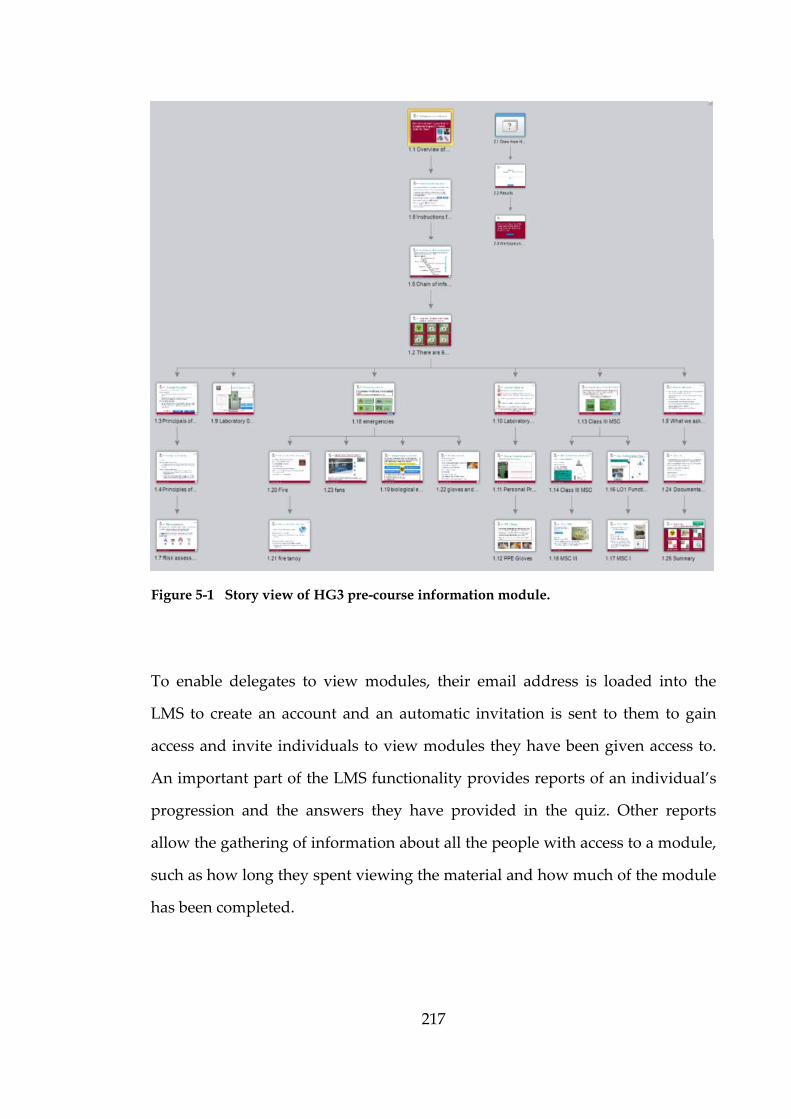
217
To enable delegates to view modules, their email address is loaded into the
LMS to create an account and an automatic invitation is sent to them to gain
access and invite individuals to view modules they have been given access to.
An important part of the LMS functionality provides reports of an individual’s
progression and the answers they have provided in the quiz. Other reports
allow the gathering of information about all the people with access to a module,
such as how long they spent viewing the material and how much of the module
has been completed.
Figure 5-1 Story view of HG3 pre-course information module.
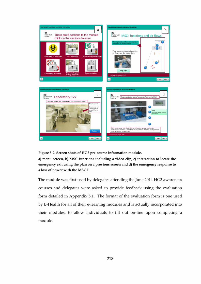
218
a) menu screen, b) MSC functions including a video clip, c) interaction to locate the
emergency exit using the plan on a previous screen and d) the emergency response to
a loss of power with the MSC I.
The module was first used by delegates attending the June 2014 HG3 awareness
courses and delegates were asked to provide feedback using the evaluation
form detailed in Appendix 5.1. The format of the evaluation form is one used
by E‐Health for all of their e‐learning modules and is actually incorporated into
their modules, to allow individuals to fill out on‐line upon completing a
module.
Figure 5-2 Screen shots of HG3 pre-course information module.
a b
c d

5.2.7
The audie
serve two
time) reso
to enable
CPD. Th
about blo
reflect the
have a di
audience
of data t
actions w
Aim:
To ensur
arriving o
potential
Specific le
On comp
- Ide
su
- Ide
- Ide
- M
ev
Ant
ence for th
o purpose
ource in th
staff to un
he audienc
ood cultur
e ability of
irect respo
are actual
to make r
will vary de
re staff kno
on the ope
B. anthraci
earning ob
pletion of th
entify the
uspected to
entify who
entify the c
ake a reas
vent of a blo
thrax –
his module
s; one wh
he event of
ndertake le
ce was det
res and ri
f learners t
onsibility f
lly practiti
reasoned ju
epending o
ow exactly
en bench a
is cultures.
bjectives:
he module
e hazards
contain B.
o may be a
correct con
oned judg
ood cultur
2
Blood
e are front‐
here there
f a suspect
earning for
termined t
isk assessm
to recall an
for perform
ioners is re
udgement
on their lev
y what to
and to ens
.
e the learne
s associate
. anthracis.
t risk of po
ntrol measu
gment for t
re being pr
219
culture
‐line labora
is an imm
ted anthra
r future situ
o have a b
ment but
nd recognis
ming risk a
eflected in
ts and the
vel of expe
do in the
sure staff f
er should b
ed with
otential exp
ures for sa
the risk of
rocessed on
es mod
atory staff
mediate nee
x case occu
uations (ju
basic level
learning o
se hazards
assessment
n learning o
e outcome
rience.
event of a
feel confid
be able to:
processing
posure
fely handl
exposure
n the open
dule
and it is d
ed to know
urring and
ust‐in‐case)
of prior k
objectives
s, in case th
ts. The fa
objective f
of the po
a B. anthra
dent to safe
g a bloo
ling B.anthr
to B.anthr
bench.
esigned to
w (just‐in‐
d secondly
) as part of
knowledge
chosen to
hey do not
ct that the
for the use
ost course
cis culture
ely handle
d culture
racis
racis in the
o
‐
y
f
e
o
t
e
e
e
e
e
e
e
- Re
rem
- Re
- Ide
- Ide
sam
The mod
generated
learners
injectiona
situations
Some of
to reveal
to show
incorpora
Documen
pdf copie
anthrax.
personal
how conf
up on the
5.2.8
The exis
Response
content w
ecall the
move B. an
ecall the co
entify the c
entify the
mples and
dule conte
d during t
are emotio
al anthrax
s are given
the interac
informatio
w the aer
ated throu
nts have be
es of the gu
The modu
responsibi
fident they
e open ben
Anthra
ting E‐He
e Departm
was develo
appropria
nthracis spo
orrect waste
correct refe
correct tra
d isolates.
ent has b
this study
onally eng
x cases in
n where po
ctions inco
on, hotspo
rosol sam
ughout th
een incorp
uidance do
ule ends w
ilities for r
y would be
nch, on a Fr
ax refre
ealth Anth
ment (ERD)
oped using
2
te disinfe
ores
e stream fo
erral labor
ansportatio
been gath
y. The de
gaged by t
n the UK.
ossible to m
orporated i
ots for lear
mpling exp
he module
porated int
ocuments c
with the lea
risk assessm
e in respon
riday aftern
esher m
hrax modu
) and is av
g subject m
220
ection for
or all mate
ratory for s
on require
hered from
sign of th
he openin
During
make the c
into the m
rners to ide
periments
e rather th
to the reso
created dur
arner havin
ment, supe
nding to a
noon.
module
ule was d
vailable to
matter exp
surface d
rial contain
suspected B
ements for
m various
he module
ng slide set
the story
content rele
module incl
entify colo
and que
han an en
ources sect
ring the ou
ng the actio
ervision of
culture of
e
developed
all PHE a
perts and c
decontami
ning B. ant
B. anthracis
referring
sources
is story b
tting the s
y, example
evant and
lude pictur
ony picture
estions h
nd of mo
tion, which
utbreak of
on to refle
f untrained
f B. anthrac
by the E
and NHS
covers the
ination to
thracis
s
suspected
and data
based and
scene with
es of real
engaging.
re triggers
es, a video
have been
odule test.
h included
injectional
ct on their
d staff and
cis turning
Emergency
staff. The
following
o
d
a
d
h
l
.
s
o
n
.
d
l
r
d
g
y
e
g

221
areas; introduction to the organism, disease characteristics, testing and
diagnosis, management after diagnosis, public health measures and an anthrax
scenario.
The HG3 awareness ‘Anthrax’ refresher e‐learning module produced as part of
this study, was therefore developed with a more narrow audience in mind and
specifically those who have attended the HG3 awareness practical course. The
audience is therefore identified as experienced laboratory workers who have
previously handled cultures of B. anthracis and have a basic level of knowledge
of the bacteria and disease. The learning objectives therefore reflect a
progression from novice to practitioner with a few objectives to recall
information from the HG3 awareness course and the majority requiring the
learner to apply, demonstrate and review. The content has been developed to
be more relevant to the identification of B. anthracis in the laboratory, the range
of clinical presentations and likely clinical sample types. The module has
elements of demonstration in the form of videos and examples but not
structured with a story compared to the Anthrax – blood culture module. On
completion of the module learners are similarly asked to undertake an action to
reflect, in this case on their confidence to identify cultures, clinical details and
respond to a suspected culture.
Aim:
To ensure staff are able to identify suspected cultures of B. anthracis and aware
of the clinical symptoms for different presentations of anthrax, likely sample
types and the different circumstances in which patients may have been
exposed.
Specific le
On comp
- Re
- Re
- Ap
pa
- Ap
- Ap
- De
- Ap
su
- Re
- Pr
/sa
T5.3
The blen
practical
with self‐
envisaged
learning a
The exist
rationalit
have a ba
earning ob
pletion of th
ecall the co
ecall the Gr
pply know
ast cases to
pply know
pply know
emonstrate
pply know
urface deco
ecall regula
opose corr
ample
Train
nded learn
demonstr
‐directed c
d training
approach.
ting E‐heal
ty of the su
asic unders
bjectives:
he module
olony appe
ram stain a
wledge of g
how patie
wledge of v
wledge of cl
e the correc
wledge of
ntaminatio
atory requi
rect action
ing st
ning appro
ations, han
computer b
strategy fo
lth Anthra
ubject whi
standing of
2
e the learne
arance of B
appearance
geographi
ents may h
virulence fa
linical pres
ct procedu
f spores t
on
irements su
ns in the e
trategy
oach incorp
nds on ex
based e‐le
or raising a
ax module
ich is appr
f and incre
222
er should b
B. anthracis
e of B. anth
cal distrib
have been e
actors to id
sentation t
ures for tran
to choose
urroundin
event of a
y
porates fa
xperience a
earning mo
awareness
provides
ropriate fo
eased awar
be able to:
s on blood
hracis
ution, rou
exposed
dentify sym
o identify l
nsportation
appropria
ng B. anthra
confirmed
ce to face
and discus
odules. Fi
of B. anthr
backgroun
or front‐lin
reness of th
agar
utes of infe
mptoms
likely sam
n and refe
ate disinf
acis
d B. anthra
e training,
ssion, supp
igure 5‐3.
racis using
nd and the
ne laborato
he disease.
ection and
mple types
rral
ection for
acis isolate
involving
plemented
shows the
a blended
e technical
ory staff to
.
d
r
e
g
d
e
d
l
o

223
The hands on practical HG3 awareness course provided experiential learning
opportunities for staff to study bacterial cultures which are not often seen in
front‐line laboratories.
There was also an opportunity for those attending to discuss situations where
cultures have been handled on the open bench and what can be learnt from
incidents that have occurred across the country. Attendance of the HG3
awareness course should promote opportunities to open discussions between
front‐line laboratories and between front‐line laboratories and experts at PHE.
The following Anthrax refresher e‐learning module can be completed at a later
date as part of maintaining knowledge, CPD and self‐directed learning.
Figure 5-3 Training strategy, describes the blending of existing courses e-learning
(blue) and practical course (red) with the e-learning modules developed as part of
the study (green).
Front‐line staff
Anthrax ‐ Blood cultures e‐learning module
Introductory information
Access E‐Health Anthrax module
Register for HG3 Awareness day
Sent access to pre‐course information e‐learning
Attend HG3 awareness day
Access to Anthrax e‐learning module as a refresher
The Anth
anyone, w
At all sta
to ensure
back into
front‐line
the Heal
learners
Anthrax
laborator
O5.4
The simp
material
mainly fr
produced
resources
The pre‐
learning
on pages
interactio
simplest
developm
e‐learning
minimum
hrax‐blood
whether th
ages, feedb
e any revi
o the HG3
e laborator
lth and Sa
following
refresher
ries and pro
Outco
plest traini
(Appendix
rom the HG
d in Micros
s section of
course inf
using the
s with text
ons and the
to deliver
ment profe
g module
m being 49
d culture
here is an im
ack can be
ew incorp
awareness
ies, experi
afety depa
completio
module ar
ovide an o
mes
ing materia
x 5.4), wh
G3 course
soft Power
f both Anth
formation
categorisa
and graph
ere are test
and a surv
essional de
lasting on
and maxim
2
e‐learning
mmediate
e used to in
porates new
s course to
iences follo
artment if
on of the A
re designe
opportunity
al to creat
hich involv
material a
rPoint and
hrax e‐lear
module c
ation of e‐l
hics. Ther
t questions
vey of 249
etermined
ne hour, t
mum of 12
224
g module
or future n
nform and
w informa
o promote
owing incid
concerns
Anthrax‐b
ed to prom
y for learn
te was the
ved gather
already ava
saved as a
rning modu
can be des
learning m
re is simp
s. This typ
organisatio
the averag
takes appr
25 hours (C
can be us
need as par
d develop t
ation. Info
the sharin
dents, near
are raised
lood cultu
mote furthe
ers to cons
‘bench gu
ing inform
ailable. Th
a pdf for in
ules.
scribed as
material be
le incorpo
pe of e‐lear
ons repres
ge time to
roximately
Chapman, 2
sed at any
rt of CPD.
the trainin
ormation c
ng of best p
r misses an
d. The a
ure modul
er discussi
solidate lea
uide’ supp
mation and
he ‘bench g
ncorporatio
a basic, l
ecause it h
oration of v
rning mate
senting 394
develop a
y 79 hours
2010).
y time by
ng material
can be fed
practice in
nd involve
actions for
le and the
ion within
arning.
plementary
d pictures,
guide’ was
on into the
level 1, e‐
as content
video, few
erial is the
47 learning
a level one
s with the
y
l
d
n
e
r
e
n
y
,
s
e
‐
t
w
e
g
e
e
225
The actual time to create this module was not recorded but it was
approximately 25 hours for a 30 minute module because the structure was
simple and the content readily available from two sources one being a
PowerPoint containing most of the information which was easily imported into
the authoring software and the video was also from existing material.
The feedback received from delegates attending the June 2014 HG3 awareness
course are very positive with 81.48 % of delegates rating the module excellent
for meeting the objectives and 88.89 % rating it excellent for how interesting the
material was presented. The general consensus when asked on the course was
that this method of delivering information was much more interesting than
reading the code of practice and risk assessments alone. A full summary of the
evaluation is presented in Appendix 5.1. Some delegates had problems with
gaining access to the quiz and the navigation of the drag and drop quiz
questions.
Access to the quiz was only permitted by the use of variables to determine
whether the learner had viewed every slide. Delegates reporting this problem
prior to attending the course were directed to make sure every slide had been
viewed and most were then able to access the quiz. The variables controlling
access have been altered, however this does mean not every slide may be
viewed before accessing the quiz. A slight alteration to the quiz questions
which involve drag and drop interactions is hoped to make these slides clearer
regarding what the learner needs to do, to answer the questions.
The LMS reports of module usage and quiz scores is given in Appendix 5.2 and
overall most learners scored over 70%. The questions which were answered
incorrectly were ones which were more related to recalling information rather
than ones which would be of concern for attending the course.

226
A question which was answered incorrectly by several delegates was that on
ordering the importance of different control measure used to prevent laboratory
acquired infection. On the day of the course this question was discussed to
check understanding of the principle that we put emphasis onto control at
source with practices and engineering controls rather than PPE which several
people offered as an answer. The discussion of the quiz responses on the
course were not directed at any one individual but discussed as a whole to
prevent anyone feeling uncomfortable.
The Anthrax refresher material would also be categorised as a basic, level 1 e‐
learning module, the structure was simple and the content was gathered from a
variety of sources. The videos were filmed using a digital camera in the CL3
training laboratory from within the MSC III. A colleague assisted in filming of
the video to show colony texture but the other video was filmed with one hand
which was awkward, a tripod will be used for future filming! There is no
estimation for how long it took to create but it was designed in stages over
several months and was originally structured for different levels of users
depending on role within the laboratory. This original structure involved the
complex use of variables which had the potential to not work correctly if a
single mistake occurred in one variable so this was later removed and
simplified to a single level of learner.
The Anthrax‐ blood culture material was categorised as an interactive, level 2 e‐
learning module because it contained the level 1 content and 25% or more
interactive exercises with the liberal use of multimedia. This type of material
takes a minimum of 127 hours for a one hour module, an average of 187 hours
and maximum of 267 hours (Chapman, 2010). The story structure was much
more complex than the pre‐course information module and the content was
drawn from multiple sources, there is no estimation for how long it took to
227
create but it was designed in stages over several months. The video was made
during the biosafety experiments in a CL2 laboratory using a digital camera
which didn’t take too long.
The Anthrax refresher module will be distributed to delegates who attended the
HG3 awareness course a year previously; November 2013 delegates will be the
first to provide feedback on the refresher module in November 2014. Their
comments will inform any alterations needing to be made before who attended
June 2014 undertake the refresher module in June 15. The LMS will be used to
collect data on the time spent and break down of question responses collected
for the pre‐course information module. The Anthrax‐blood cultures e‐learning
module will be distributed to laboratories that experienced cases of injectional
anthrax during the outbreak for first review, because they would be best placed
to provide feedback to the relevance of the module having experienced anthrax
first hand. The feedback and information gathered using the LMS will again
inform alterations to the module content and structure. The success of the e‐
learning developed as an outcome of this study will also inform the
development of modules for other HG3 bacteria, with a priority for Brucella spp.
and Burkholderia pseudomallei which have both been handled on the open bench
on more occasions than the other bacteria.
Blood culture data can also be gathered for these bacteria and further work to
examine aerosol and contamination will be of particular relevance to Brucella
spp. which has a very low infectious dose.
D5.5
Epistemo
what we
somethin
scientists
whether
scientific
media an
by the M
individua
the explo
study.
exposure
routine s
amounts
(Pottage e
handled
generated
evidence
preferenc
biases. R
as a less
unknown
2011).
Discu
ology is th
know, ho
ng to coun
are not di
the know
investigat
nd cause th
MMR vacc
als to form
oration of
Microbiolo
e and the l
ervicing o
of microb
et al., 2014)
at CL2 o
d in this st
. Psycholo
ces in decis
Risks arisin
ser degree
n or less f
ussion
e area of p
ow we kno
nt as know
irectly con
wledge the
tion. It is p
he public t
cine and
m their own
the safety
ogical safe
level of pr
f cabinets.
ial aerosol
) which is o
on the op
tudy is lim
ogy and ma
sion makin
ng from a
of risk av
familiar so
2
n
philosophy
ow it and
wledge (D
ncerned by
ey use for
possible fo
to change
autism sc
n beliefs a
y concerns
fety cabine
rotection af
There is
l generated
of importa
pen bench
mited in siz
athematica
ng and des
known or
version th
ource of u
228
y concerne
identifying
Dupre, 200
y the philos
r whatever
or ‘bad’ sci
their beha
cares in 1
and percep
s for proc
ets are de
fforded is
however,
d during co
ance when
and furt
ze but can
al probabil
scribe amb
more fam
han in a c
uncertainty
ed with kn
g the cond
07). In a
sophy of k
r purpose,
ence to be
aviour; this
998. It i
tions of ris
cessing blo
esigned to
known an
limited ev
ommon lab
an unknow
ther manip
n contribute
lity have b
iguity aver
miliar sourc
ase for ris
y (Chew, E
nowledge,
ditions to b
a laborator
knowledge
, is based
dissemina
s was clea
is also po
sk, which
ood cultur
o minimis
nd measur
vidence on
boratory p
wn HG3 p
pulated.
te somethin
oth explor
rsion and f
ce may be
sks arising
Ebstein an
exploring
be met for
ry setting,
e but more
d on good
ated in the
rly shown
ossible for
prompted
res in this
se aerosol
red during
n the likely
procedures
athogen is
The data
ng current
red human
familiarity
perceived
g from an
nd Zhong,
g
r
,
e
d
e
n
r
d
s
l
g
y
s
s
a
t
n
y
d
n
,
229
Competence is an ability to perform and a combination of knowledge, skills and
experience. Assessment of competence to identify B. anthracis cultures in a
front‐line laboratory setting is not appropriate. Training can be provided to
raise awareness and provide experiential learning by handling cultures in the
safe environment of the CL3 training laboratory at PHE Porton. The blended
learning approach proposed in the training strategy is designed to equip
laboratory staff with the knowledge and experience to safely handle and swiftly
move suspected B. anthracis into the CL3 laboratory. The HG3 pre‐course
information e‐learning module has been tested with course delegates and
determined to be fit for purpose following a few alterations made on the basis
of feedback. The other anthrax modules will be assessed once distributed to the
appropriate audiences.
Introductory, refresher and advanced training courses are regularly run with a
blend of theory and practical sessions delivered from the training facilities at
PHE, Porton. These courses allow delegates the benefit of gaining up to date
information from experts in the field, hands on training with the guidance of an
experienced trainer, as well as an opportunity to meet and network with other
delegates and share experiences. The development and delivery of computer
based, e‐learning material by subject matter experts, can be used to supplement
the existing practical based courses by providing an interesting method of
delivering information and interactivity to encourage and create learning
opportunities.
Information is not the only type of content which can be delivered by e‐
learning. Modules can be designed to engage with learners by testing softer
skills such as dealing with situations and scenarios where decision making
results in multiple possible outcomes from which to learn.

230
The flexibility and functionality of the Articulate Storyline software therefore
allows e‐learning modules to be developed for different audiences. By
engaging with learners of different levels (novice, practitioner and expert) the
content, structure and delivery can be tailored to factor in their differences.
Online materials with interactive content can therefore provide an effective and
efficient way to deliver self‐directed learning but the learners may feel isolated
if there is no means of feedback or follow on.
The objective of the study to produce training material was to use and
disseminate the outcomes from the other study aspects of the ‘process’ and
‘safety’. The ‘training’ aspect has successfully incorporated the data generated
from the biosafety investigations resulting from the safety concerns arising
from processing a blood culture suspected to contain B. anthracis on the open
bench, into the e‐learning module ‘Anthrax – Blood cultures’. The information
generated about the time to positive detection and concentration of inoculum
requires further work to produce calibration data which could inform
laboratories to the likely load of bacteria in the blood during anthrax
bacteraemia. The beauty of e‐learning material is that it can be quickly updated
to incorporate new information, therefore this information can be added when
available. The use of serum separator tubes to reduce turnaround times for the
confirmation of B. anthracis in blood cultures requires pilot testing within RIPL
before incorporation into routine use. The recommendation of this sample type
to front‐line laboratories was therefore not incorporated into the training
material.
The simple, method to concentrate and wash bacteria for use with molecular
assays and to perform antimicrobial susceptibility testing is at present still
attractive for many other more common bacteria.
231
There will always be a need to reduce the time to identify and provide
susceptibility results for cases of bacteraemia and until there is replacement of
the blood culture as a sample type by some other new technology then such
methods will be of use. Not everywhere uses automated blood culture systems
and or molecular detection and in many developing countries the use of
manual blood cultures with rapid lateral flow devices for common pathogens
would be more suitable and beneficial in the treatment of bacteraemia.
In conclusion, the study has explored a variety of areas surrounding B. anthracis
in blood cultures, as a whole it provided an example of best practice for
biosafety and risk assessment and the use of e‐learning has the potential to
disseminate the findings. The audience for the e‐learning material produced in
this study is potentially far wider than just front‐line laboratory staff in the UK.
With access via the internet, anyone with a connection and access could use the
material. The e‐learning modules have also been designed to work on mobile
device technology allowing access on mobile phones and tablets. Even in
remote locations where internet access is not available, material can be stored
on a memory stick or CD‐ROM.
Ultimately, knowledge can enable people to make informed judgements and
those made about safety can help prevent both laboratory acquired infections
and individuals from generating fearful perceptions of risk. Together, these
provide laboratory workers with a safe environment in which to work and the
confidence to undertake their work safely. Of the many laboratory
investigations that are undertaken, blood cultures provide a simple tool to
detect bacterial invasion of the blood, being one of the most important sequelae
of bacterial infection. The timely identification and antimicrobial susceptibility
testing of blood cultures can save lives and by reducing the time this takes will
not only save money but hopefully more lives.
S5.6
This chap
study and
B. anthra
conclusio
A
ap
E‐l
dif
Th
ma
to
Fe
fit
Th
wi
aw
lab
Summ
pter descri
d how it c
acis and
ons from th
training
pproach
learning m
fferent asp
he authorin
aterial for
a wide au
eedback fro
for purpo
he distribu
ill provide
wareness o
boratories.
mary
ibed the tr
ould be us
its safe h
his chapter
strategy
modules a
pects of the
ng softwar
computer
dience
om one ba
se
ution of th
e feedback
of B. anth
2
raining m
sed in a tra
handling i
r may be su
was prop
and supple
e training s
re enabled
based self
asic modul
he two anth
k to their
hracis and
232
aterial dev
aining stra
in front‐li
ummarised
posed to
ementary
strategy
the design
f‐directed
le was gat
hrax modu
r suitabilit
d its safe
veloped as
ategy to pr
ine labora
d as:
provide a
material w
n of interes
learning, w
hered and
ules to ap
ty and fu
e handling
s an outco
romote aw
atories. T
a blended
were deve
sting and i
which was
d was dete
ppropriate
uture use
g in the
ome of the
wareness of
The main
d learning
eloped for
interactive
s available
ermined as
audiences
in raising
front‐line
e
f
n
g
r
e
e
s
s
g
e
Chap
The stud
and confi
safety an
assessme
laborator
confirma
surround
in a CL2
of anthra
targeting
T6.1
The use
method t
working
available
was show
strains. T
linear re
bacterial
method w
blood and
not result
pter 6
dy explored
irmation o
nd (iii) train
ent of a m
ry and met
tion of B.
ding the pr
laboratory
ax, identifi
g laboratory
The Pr
of resealin
to work w
in a CL3
defibrinat
wn to gen
The concen
elationship
blood con
were ident
d alteration
t in dispara
Con
d three ma
f B. anthrac
ning. The
method to
thods to re
anthracis.
rocessing o
y. Trainin
ication of
y staff in fr
ocess
ng caps an
with B. anth
3 laborator
ted horse b
nerate con
ntration of
which h
ncentration
tified and i
n of the he
ate results
2
nclusio
ain areas i
cis in blood
e process w
o safely s
educe turn
The aspe
of blood cu
ng material
cultures a
ront‐line la
nd safety
hracis in b
ry. Bottle
blood and
nsistent TT
f inoculum
had the p
n of anthra
investigate
ead space a
of TTP de
233
ons an
involved i
d cultures
was broken
simulate b
naround ti
ct of safet
ultures su
l was then
and the sa
aboratories
adaptors p
blood cultu
es were in
suspension
TP detectio
m and TTP
potential t
ax patients
ed to deter
atmospher
etection.
nd fur
in the dete
which wer
n down int
blood cultu
mes for th
y explored
spected to
developed
fe handlin
s as the aud
provided a
ures, withi
noculated
ns of B. ant
on for a ra
detection
to estimat
. Two ma
rmine their
e of aerobi
rther w
ection, ide
re (i) the p
to develop
tures with
he identific
d biosafety
o contain B
d to raise
ng of bloo
dience.
a safe and
in the con
with com
thracis. Th
ange of B
revealed
te the ret
ain limitati
r effect. T
ic blood cu
work
entification
process, (ii)
pment and
hin a CL3
cation and
y concerns
B. anthracis
awareness
d cultures
d practical
nstraints of
mmercially
his method
B. anthracis
an inverse
trospective
ions of the
The type of
ultures did
n
)
d
3
d
s
s
s
s
l
f
y
d
s
e
e
e
f
d
234
Further work to eliminate these factors could provide data to generate a
calibration curve. This would provide a more reliable estimate of the bacterial
concentration in anthrax patients, although the clinical significance of this is
unknown.
Presumptive identification methods were successfully performed on direct
positive B. anthracis blood culture fluid using the Biofilm Biothreat panel and
microscopic examination for the presence of capsule. Of the two methods, the
Biofilm array could detect specific B. anthracis nucleic acid targets in
approximately one hour. In contrast, the phenotypic M’Fadyean reaction could
be performed within a few minutes, but is open to human interpretation and
quality of stain. The presence of capsule would support the presumptive
identification alongside Gram microscopy in front‐line laboratories. To choose
between the two methods laboratories would have to consider the balance
between cost and confidence in results. The rapid immuno‐chromatographic
test could not be used for directly testing blood cultures and its use would
require overnight incubation of cultures. Turnaround times would not be
reduced with this method however it may appeal to front‐line laboratories to
assist with the presumptive identification of this typically rare cause of infection
in the UK. The method has been used during the practical HG3 awareness
course to provide delegates with results for correctly identifying B. anthracis
from mixed bacteria cultures. In this setting, delegates commented that the
simple rapid test provided additional confidence for their ability to correctly
isolate B. anthracis colonies in the future.
The SST protocol used in this study was successful in providing concentrated
washed, bacteria. This sample could be used for both inactivation for molecular
identification methods and setting up phenotypic confirmatory tests, providing
correct results 24 hours earlier than current procedures.
235
The SST method also provided material suitable for molecular identification of
B. anthracis in polymicrobial blood cultures and suitable cultures to perform
phenotypic confirmatory tests. Molecular identification was also achieved
following inactivation of direct blood culture fluid using the method to prepare
thermolysates. This simple, quick method has huge potential for use in front‐
line laboratories to perform molecular identification of more common causes of
bacteraemia from blood cultures.
Discussion with the network of European laboratories involved in the
QUANDHIP project has indicated that most do not receive clinical samples
directly from front‐line laboratories. The European national reference
laboratories receive bacterial isolates as a typical sample type for the
identification of B. anthracis and other HG3 bacteria. This was also the case in
the UK until the outbreak of injectional anthrax, when a range of clinical
samples were received and in large numbers. The national reference laboratory
at PHE Porton is now prepared for such an event occurring again in the future.
The addition of positive blood culture fluid delivered in SST alongside other
clinical samples would provide the gold standard culture confirmation 24 hours
earlier if implemented. There is also the potential to use SST processed blood
cultures for rapid antimicrobial susceptibility testing using off‐the‐shelf micro‐
broth dilution plates and end point detection using PCR. Further work, would
aim to establish the use of these methods into the B. anthracis diagnostic service
at PHE Porton. Several guidance documents for anthrax already exist which
are available via the PHE website and were identified in section 5.2.2. The
documents provide guidelines for action in the event of a deliberate release,
algorithms for cutaneous and inhalational anthrax and clinical presentation and
case definition, laboratory guidance and infection control precautions for
injectional anthrax. Further work would look to update these documents which
are aimed specifically for front‐line laboratory use.
Sa6.2
The possi
at CL2 is
indicated
venting n
bacterial
culture fl
of SST tra
hazard to
consisten
processin
The use o
non‐viabl
In additio
unsuspec
fixing and
in contras
Positive b
contain s
disposal.
disinfecta
anthracis
during an
therefore
SST is ro
pathologi
afety
ibility of B
s a valid
d that the r
needle or
counts re
luid into S
ansfer to ro
o front‐line
ncy of spra
ng.
of alcohol t
le, howeve
on, the pre
cted until
d Gram sta
st to PMB
blood cult
spores, ho
The reco
ant follow
should b
nthrax can
blood sam
outinely u
ical investi
B. anthracis
concern fo
risk of infe
safety ad
ecovered b
ST was ea
outine proc
e workers.
ay factors
to fix slide
er this meth
esumptive
after prep
aining slid
stain whic
tures proce
wever, the
ommended
wing any s
be followe
n reach ext
mples take
used to pr
igations.
2
blood cult
or front‐lin
ection by th
daptor, w
by air sam
asily achiev
cessing wo
Further w
and explo
es as recom
thod is not
e identifica
paration of
des was de
ch does not
essed dire
ey may b
d guidanc
spill of cli
d to effec
tremely hig
en from pa
repare seru
236
tures being
ne laborat
he aerosol
ith safety
mpling. T
ved using
ould, there
work wou
ore contam
mmended b
routinely
ation of B.
f the Gram
monstrate
t behave si
ectly after
e present
e to use 1
inical mat
ctively ina
gh bacteria
atients may
um sampl
g processed
ory staff.
route was
adaptors
he transfe
safety ada
efore, not p
ld be need
mination d
by PHE sho
used in fro
anthracis m
m film. In
d to inactiv
milarly.
TTP detec
if bottles
10,000 ppm
erial susp
activate sp
al blood co
y represent
es which
d on the o
The data
s low, usin
resulting
er of posit
aptors. Th
present an
ded to dete
during bloo
ould rende
ont‐line lab
may be un
n this situ
ivate viable
ction are u
are stored
m availabl
pected to c
pores. Ba
oncentratio
t a greater
are used
pen bench
a collected
ng either a
in fewer
tive blood
he addition
additional
ermine the
od culture
er material
boratories.
nknown or
ation heat
e material,
unlikely to
d prior to
le chlorine
contain B.
acteraemia
ons (~ 108)
r risk. The
for many
h
d
a
r
d
n
l
e
e
l
.
r
t
,
o
o
e
a
)
e
y
Accident
lead to c
induced.
spores in
is needed
suspected
T6.3
A direct
material
aetiologic
laborator
e‐learning
specific l
practical
training s
learning.
around t
Consider
presentat
reduce f
ultimatel
appropria
Front‐line
diagnosis
automati
al exposur
contaminat
Routine
nactive ther
d to preve
d anthrax.
Trainin
outcome o
to raise a
cal agent a
ry setting.
g format to
laboratory
training a
strategy to
Compute
the world
rations for
tion of dat
fearful per
y aimed
ate treatme
e microbio
s of infecti
on and rap
re, spillage
tion of the
disinfectio
refore clea
ent such s
ng
of the stud
awareness
and the saf
The major
o provide
y aspects.
already pr
o provide a
er based m
to learn
safety hav
ta generat
rceptions
to aid in
ent to resu
ology labor
ious diseas
pid technol
2
e or poor
e environm
on method
ar commun
situations o
dy was the
of anthra
fe handlin
rity of the
front‐line
The inc
rovided in
a blended
methods of d
about the
ve also be
ted from t
of risk d
n the time
ult in positi
ratories aim
ses and, in
logies are h
237
handling
ment whe
ds are likel
nication be
occurring
e developm
ax, the lab
g of clinica
training m
staff with
corporation
n the UK
learning a
delivery ca
e disease
een incorpo
the biosafe
developing
ely confir
ive clinical
m to deliv
n many de
helping to
of these b
re sporula
ly to be in
etween pat
in the eve
ment and
boratory i
al material
material is i
the opport
n of e‐lea
is present
approach a
an enable l
and labor
orated into
ety investi
g. The t
mation of
outcomes
ver fast, rel
eveloped c
achieve th
blood samp
ation is lik
nsufficient
thology de
ent of pat
delivery o
identificati
l and cultu
in a compu
tunity to le
arning ma
ted in the
appropriate
laboratory
ratory iden
o the mate
igations is
training m
f anthrax,
s.
liable resu
countries,
his goal.
ples could
kely to be
to render
epartments
ients with
of training
ion of the
ures in the
uter based
earn about
terial and
proposed
e for adult
staff from
ntification.
erial. The
aimed to
material is
, assisting
ults for the
the use of
d
e
r
s
h
g
e
e
d
t
d
d
t
m
.
e
o
s
g
e
f
Financial
laborator
training.
(which h
need tra
encounte
importan
investiga
6.3.1
Of those
diagnosti
bacteria
cultures m
limited cl
It is there
line staff
next time
swift and
throughp
The study
anthrax a
therefore
implemen
of suppor
in some p
l constrain
ries are re
Reliance
as been sh
ained, com
ered. The c
nt for patie
ations but a
Futur
e who wo
ic staff wh
occurring
may arise
linical info
efore of pa
with the n
e anthrax r
d confiden
put, time p
y was cond
and a centr
perfectly
ntation of
rting train
part contrib
nts and
esulting in
on results
hown to in
mpetent st
correct iden
ent outcom
also for lab
e aim
ork within
ho are mos
in isolate
unexpecte
ormation to
aramount
necessary k
rears its fe
nt manner
ressured fr
ducted at P
re for exce
y positione
a simple m
ing materi
bute to the
2
the const
n longer
s from new
ncorrectly i
taff to ‘e
ntification
me, identific
boratory sta
n microbio
t likely to
d cases, o
edly on the
o provide n
importanc
knowledge
ear provok
r. All th
ront‐line d
PHE Porto
ellence in c
ed to lea
method wi
ial are the
e goal.
238
tant press
working h
w technolo
identify B.
engage br
of highly p
cation of b
aff safety.
ology labo
be the firs
outbreaks
e open ben
no clues to
ce that we
e, skills and
king head,
his within
diagnostic l
on being th
containmen
ad the ach
ith minima
outcomes
sures imp
hours and
ogies such
anthracis)
ain’ when
pathogenic
bioterrorist
oratories,
st to cultur
or bioterro
nch at CL2
as what sa
have the
d experien
it can be
the conte
laboratory.
he national
nt microbio
hievement
al practical
of the stud
posed on
d reduced
as MALD
is worryin
n such re
c bacteria i
t events an
it is the
re highly p
orist even
2, accompa
amples ma
goal to eq
nces to mak
dealt with
ext of a b
.
l reference
ology and
t of this
al effort an
dy and are
front‐line
d time for
DI‐TOF MS
ng and we
esults are
is not only
nd forensic
front‐line
pathogenic
nts. These
anied with
ay contain.
quip front‐
ke sure the
h in a safe,
busy, high
centre for
biosafety,
goal. The
d delivery
e aimed to,
e
r
S
e
e
y
c
e
c
e
h
.
‐
e
,
h
r
,
e
y
,
239
The bacterium B. anthracis was the focus of this study and future work should
be conducted with other HG3 bacteria which feature on the HG3 awareness
course. This study therefore provides a template to investigate the aspects of
the ‘process’ involved with the detection, identification and confirmation of
other HG3 bacteria. The aspect of ‘safety’ should be explored further to
include considerations for highly pathogenic bacteria with extremely low
infectious doses such as Brucella spp. and F. tularensis.
Finally, the outcomes for other HG3 bacteria should be incorporated into
existing guidance documents and used to develop supplementary training
material such as e‐learning. It is hoped this study and future work, results in
meaningful change to the safety and confidence of front‐line laboratory staff
and significant contributions to patient outcomes.
240
Chapter 7 Reflection
This final chapter looks reflectively on the learning undertaken during my
studies that have enabled me to plan, conduct and report my findings and
ultimately put the final full stop at the end of this thesis. Reflection was
introduced as part of my training as a biomedical scientist, as an activity to
learn from experiences. Unfortunately, it was not until I completed the first
part of the professional doctorate that I actually understood why reflection was
an important activity for professionals or even how to actually critically reflect.
I have used what I have learnt about the reflective process and the Gibb’s
reflective cycle (Gibbs, 1988) is a model that I have used for critical reflections. I
have incorporated my skills in reflective processes into my daily work as a
microbiology trainer at PHE Porton. For the past two years, we have held a
trainee BMS day at PHE Porton for students undertaking biomedical sciences at
university who have a placement year within PHE laboratories at our site in
Colindale. As part of their day at Porton, I deliver a session on critical reflection
based on a presentation I delivered in 2011 for the HPA annual conference
within the training session. The purpose of the session is to share my
experiences of reflection to enable them to understand why and how to
undertake critical reflections. Reflections form part of the evidence for
inclusion in their training portfolios and I hope they see that reflection is useful
for their personal development and not a worthless paper pushing activity,
which was my view of reflection when I was a trainee, due to my lack of
understanding of the process.
To further spread the message that biomedical scientists can do more than
reflection ‘in action’ I have also authored a poster which was presented at the
Wessex A
Scientists
This post
European
audience
training a
also bene
the huma
human fa
Before re
position o
in blood
be a mer
provide i
T7.1
When I
Universit
with a M
working
portfolio
Hospital
post at H
specialist
Applied M
s Guide to
ter was us
n Biosafety
of biosaf
and I iden
efit from cr
an factors u
actors.
eflecting on
on the Pro
cultures as
re iteration
insight to t
The st
started t
ty I initiall
MSc, who co
in a resear
directly a
where I co
HPA Porto
t portfolio
Microbiolo
Reflection’
sed as the
y Associati
ety officer
ntified that
ritical refle
underlying
n my lear
ofessional D
s the subje
n of Chapte
he non‐aca
tudy p
the Profes
ly saw it as
ouldn’t fea
rch setting.
after state
ompleted m
on (now P
without g
2
ogy Confe
’ (Append
basis of a
on Congre
rs. The N
t the scien
ection. Cri
g accidents
rning and
Doctorate p
ct of my st
er one’s p
ademic rea
propo
ssional Do
s the most
asibly comp
. I had sta
registering
my trainin
PHE Porto
going to a
241
rence in
ix 7.1).
nother pos
ess held in
NADP train
ntists with
itical reflec
s and incid
developm
programm
tudy. Initi
ositioning,
asons for m
osal
octorate p
t logical ro
plete the IB
rted gathe
g, whilst w
g. Howev
on) it was
a routine c
2012, enti
ster that w
Estoril, Po
ning grou
a biosafety
ctions may
dents as mo
ment I will
me and why
ally you m
, however
my explorat
programme
oute for my
BMS speci
ring evide
working as
ver, upon m
not possib
clinical lab
itled ‘A B
was presen
ortugal in 2
up provide
y officer r
y help to u
ost acciden
start by g
y I chose B
may think t
this discu
tion of the
e with Po
y situation
ialist portfo
ence for the
s a BMS a
moving to
ible to com
boratory be
Biomedical
nted at the
2011, to an
e biosafety
ole would
understand
nts involve
giving my
B. anthracis
this would
ussion will
e subject.
ortsmouth
n as a BMS
olio whilst
e specialist
at Treliske
a research
mplete the
ecause the
l
e
n
y
d
d
e
y
s
d
l
h
S
t
t
e
h
e
e
242
reference laboratory at Porton was so specialised, it would not cover all the
areas needed. Lengthy secondments to a local laboratory were not possible.
The professional doctorate was listed on the IBMS route of professional
progression and I found out someone else at Porton had previously attended
the one offered at the University of Portsmouth. That person was Allen
Roberts, at the time the general project manager for the research department I
was working in, so it all became possible and Allen would go on to be my
project supervisor, confidant and life mentor. At the time of starting the
programme I do not think I appreciated just how much work the Professional
Doctorate entailed and having spoken to other part time academic students at
Porton, the part time option was more demanding than a full time PhD in many
aspects.
During the taught aspect of the professional doctorate programme I had an
initial project idea related to my research position, conducting antimicrobial
susceptibility testing for HG3 bacteria. Within the group, molecular assays
were being explored to enable rapid susceptibility determination with the
micro‐broth dilution MIC technique. I thought this rapid method could be
useful in aiding prompt, appropriate treatment of HG3 bacterial infections and
therefore benefit patients and have a professional outcome. This initial project
idea was abandoned, when I changed position within HPA Porton and became
a senior BMS in the Special Pathogens Reference Unit (SPRU) which was later
renamed RIPL. After changing position I was worried about developing a new
study idea and more importantly whether I would actually be able to complete
a study whilst working in a busy, understaffed specialist diagnostic laboratory.
Another professional concern was my lack of virology knowledge, skills and
experience for working in the SPRU which predominately handled virology
diagnostics.
243
The question was how I was going to cope with learning all that was new in my
new role whilst completing my doctorate. Two major events occurred which
influenced what happened next, the first was being given responsibility for
leading SPRU’s involvement with a European project for the quality assurance
for highly pathogenic bacteria (EQADeBa) and the UK outbreak of injectional
anthrax. Both involved HG3 bacteria which I was familiar with, so building on
my previous knowledge, skills and experience therefore making me feel more
confident in my capabilities.
I may have felt more confident but these two experiences were extremely
challenging and not easy going with the pressures of the day‐to‐day virology
diagnostics and quality of life outside of work. Involvement in the EQADeBa
project introduced a heavy personal pressure, because I felt responsible for
SPRU doing well in the quality assurance ring trials, which I also felt was a
reflection of the UK’s capabilities. In addition, I had to attend meetings in
Europe following the ring trials and on one occasion present the UK results at a
meeting in Rome. To make things worse a meeting had been scheduled in the
UK and I had to try to organise a venue, evening meal, programme and make
sure we could deliver a memorable meeting in Salisbury. With the help of the
SPRU and administration staff the meeting was delivered and even though it
was not as spectacular as several of the meetings held in other European
countries everyone enjoyed their time in historic Salisbury. I have gained a
great deal of inter personal skills from being part of the EQADeBa project,
gained contacts with all the European laboratories working with HG3
diagnostic and reference work, travelled and attended training for
environmental field sampling for B. anthracis in Italy. Upon reflection of the
experience as a whole, I can say involvement in the EQADeBa project was
monumental in my personal and professional development and though it was
extremely demanding it was definitely worthwhile.
244
The second major event was dealing with one of the largest outbreaks of
anthrax in the western world occurring in PWID in 2009–2010. In December
2009, I was undertaking routine molecular testing for B. anthracis for two
bacterial isolates sent by a hospital laboratory querying anthrax. To my
surprise, they were positive, this marked the start of an outbreak which was to
take over my life for the next 6 months. These first two cases prompted
requests by the police to test drugs and related materials, in addition to a huge
range of clinical samples arriving on a daily basis and of course, results were
always wanted five minutes ago. Initially SPRU responded by providing a 24/7
service which was later scaled down to limited cover over the weekends.
Samples received in the morning would have PCR results released by the
afternoon, culture isolation and confirmation could be as long as five days for
tissues. The demand and sheer number of samples received for testing was not
foreseen and it took a while for the service to settle into a routine for increased
capacity. Staff trained to work at CL3, with experience of B. anthracis were
drafted in from other departments on site and the whole situation required
organisation of staff rotas, increased demand on administration, setting up high
throughput molecular testing and validating unusual sample types for
molecular detection. As with my involvement with the EQADeBa project, there
was a personal pressure and impact on a healthy work‐life balance. As the
senior BMS and bacteriology lead in SPRU, I helped to organise the laboratory
and make sure the anthrax service ran as smoothly as possible. Upon reflection,
the experience had a huge influence on my knowledge, skills and experience of
working with B. anthracis and developed my inter personal skills to deal with
staff moral as the outbreak continued.
An idea for a new project emerged to develop a ‘Laboratory emergency
response plan’ which could be used by specialist reference laboratories like
SPRU an
anthrax o
thought
working
in positio
to the No
on the ou
blood cu
several b
SPRU tha
to the pre
study pr
described
C7.2
This secti
the cours
programm
evaluate
we could
one of th
in order
providing
The stag
evaluatio
nd those in
outbreak a
this woul
in an und
on was aga
ovel and D
utbreak, an
ulture syste
blood cultu
at we woul
esence of in
esented in
d and two
Critic
ion aims t
se of the s
me at Port
how we d
d do differe
he main cha
to engage
g structure
ges of thi
on, analysi
n the EQAD
and to also
ld be use
derstaffed,
ain going t
Dangerous
nd the one
em in the
ures were s
ld not be a
nhibitory s
n this thes
changes in
cal refl
o critically
study unde
tsmouth U
do things, c
ently (Kolb
aracteristic
e in a proc
e, Gibbs’ R
s cycle b
s and then
2
DeBa proje
o include
eful and a
busy spec
to result in
Pathogens
thing whic
e training
sent for co
able to perf
substances
sis has bee
n job!
lectio
y reflect up
ertaken as
University.
considerin
b, 1984). T
cs of profe
cess of con
Reflective C
begin with
n a conclu
245
ect, to lear
the other
achievable
cialised dia
n abandoni
s Training
ch caught m
CL3 labor
onfirmation
form direc
s. The rest,
en influen
n
pon my le
s part two
Kolb’s t
ng what wo
his is subs
ssional pra
ntinuous l
Cycle will b
h a descri
usion which
rn from ou
HG3 bacte
given th
agnostic lab
ing a proje
team. My
my eye, w
ratory. D
n and I ex
t PCR on t
, as they sa
ced by th
arning and
of the pr
theory that
orks, what
stantiated b
actice, is th
earning. F
be used.
iption, fol
h incorpor
ur experien
teria. At t
he circums
aboratory.
ect idea an
y thoughts
was the BacT
During the
xplained to
this sample
ay, is histo
he two ma
d developm
rofessional
at reflection
t does not
by Schon, (
he capacity
For the pu
llowed by
rates an ac
nces of the
the time, I
stances of
A change
d I moved
s were still
T/ALERT®
outbreak,
o others in
e type due
ry and the
ajor events
ment over
doctorate
n helps us
and what
(1987) that
y to reflect
urposes of
y feelings,
ction plan.
e
I
f
e
d
l
®
,
n
e
e
s
r
e
s
t
t
t
f
,
.
246
All humans have a negativity bias and therefore reflect more on negative
experiences and although we can learn from them, we need to put emphasis on
learning from positive experiences also (Peeters & Czapinski, 1990). The
evaluation and analysis stages are extremely useful to consider a balanced view
of highs and lows and what did or did not go well. The scale of negative
experiences and their outcomes may have been overwhelming, however I now
have a balanced view of these situations and have drawn out positive outcomes
upon later reflection, which at the time seemed impossible.
Description:
Once I had decided on the final study idea, the project proposal was completed
and accepted by the University. The study was concerned with the potential
issue of blood cultures turning up unexpectedly in front‐line hospital
laboratories, being processed on the open bench and proposed the use of a
simple method to reduce the time for confirmation of B. anthracis in blood
cultures. Preliminary work was undertaken to explore the feasibility of the
study. The practical aspects of the study were undertaken in the CL3 training
laboratory over a period of three years. The work was scheduled throughout
the year with the majority being undertaken during the summer and Christmas
holiday periods when there were no major training courses scheduled. The
NADP training team were involved with assisting with some of the practical
work and provided feedback on the e‐learning module. In between the
sessions of practical work, I had the opportunity to meet many professionals
attending our training courses when delivering the live agent courses for PHE
and front‐line biomedical scientists. During the study, I was fortunate to make
contact with experts in the field, received support from RIPL and their medical
staff and was able to contact the hospitals who sent samples during the
outbreak of anthrax in PWID. In addition, I also had interactions with the
247
Biosafety department for the aerosol sampling experiments, Diagnostic support
group that develop assays for RIPL, those who routinely perform genotyping
for B. anthracis, colleagues I used to work with in research and the electron
microscopy department.
Feelings:
The feelings experienced at the initiation of the study were a combination of
excitement, fear, trepidation, anxiety and nervousness which would be
common emotions when starting any new journey. In terms of evolutionary
survival, I believe these feelings have been necessary as positive stresses for our
ancestors to avoid dangers in new environments (Hanson, 2009). Experiencing
these feelings were probably my reaction to actually starting the study, it
seemed such a long time since beginning the doctorate and now was finally
challenged to successfully pull it off. At the time, I doubted my own
capabilities and did not want to lose face amongst my colleagues, many of
whom had completed PhDs and it was obvious my doctoral study was
completely different to the norm at Porton. Excitement and pleasure emerged
once I started and the practical work was underway, generating some
meaningful results. This change in emotions could be explained by the
increasing confidence I experienced over the first summer of conducting the
practical investigations in the CL3 laboratory, bringing together the knowledge
and experience of working with B. anthracis over my career at PHE Porton. I
was able to use my knowledge and experience to critically appraise methods
described in the literature and make reasoned judgements to whether they were
practical and feasible to conduct in the CL3 laboratory.
Interest and motivation were felt in vast troughs and peaks over the following
three years of the study, due to the availability of the laboratory to conduct the
248
study and the opportunity to have time away from the daily responsibilities of
my position.
When talking to colleagues about the progress of the study I was enthused and
refuted my initial personal doubts as to the choice of study, by their interest and
comments of the relevance and importance this study may have. With
experience and awareness, a feeling of pride emerged that may be explained by
reaching the later stages of the study and being in a position to discuss and
present some of the study outcomes. The final stages of the study were again
filled with the initial emotions of fear, trepidation, anxiety and nervousness as
the end was in sight. The continued effort to maintain motivation was hard and
on many occasions I doubted my capability to write the thesis and was often
angry at myself regarding the speed of progress at times. I now feel relieved at
completing the study, though I still feel a cloud of doubt in the adequacy of my
efforts to communicate the study in this thesis but look forward to defending it
during my viva voce and transmitting my passion and enthusiasm for this work.
Evaluation:
The evaluation stage of the reflective cycle has been broken down into highs
and lows. A general low has been trying to maintain a healthy work‐life balance
at several points during the professional doctorate programme and study. I
care about everything I do and have high expectations of myself and others and
felt the only way to cope was working even more hours. For some situations,
time management felt out of my hands but other times, my time management
has been poor, due to my lack of focus. In the training group I would often
want to please everyone by responding immediately to emails and requests for
help.
In these situations I should have prioritised my time during the day so that I
could work on my study at home, rather than continuing with computer based
249
work tasks at home. A significant low was isolation felt in two respects, one
whilst performing some of the practical work in the holiday periods. I felt I
could not ask others to help when they had their own study or holidays
planned especially when Clare had helped so much early on in the practical
work, giving up her own time and working long hours with me in the CL3
laboratory. The second, whilst writing this thesis, I was able to discuss aspects
of writing the thesis with colleagues, but taking time off and isolating myself
from my family and friends to focus and being particularly unsociable was
especially hard.
Highs included presenting two posters and giving an oral presentation to
colleagues at PHE Porton about my study. The first poster was presented at the
International conference on anthrax held in Februay 2012 in RUSI, Whitehall,
London (Appendix 7.2). The same poster was presented at the post graduate
poster day at NIBSC where I won my category and was given a commendation
at the annual Porton poster day. The second poster detailing the biosafety
aspects of the study was accepted and presented at EBSA congress 2014 in
Ghent, Belgium (Appendix 7.3). This poster has also been accepted by Sheffield
Microbe where it will be presented in September 2014. Students conducting
academic study at PHE Porton, are expected to present to staff as part of the
research seminar program. In April 2014, I presented my study findings to
date, this was also my first opportunity to show staff the e‐learning module I
had been working on. I was pleasantly surprised to have a full audience,
representing many groups and departments across site. Another high, was
learning how to use the e‐learning authoring software and designing my
anthrax e‐learning module. It was possible to use many interactions to make
the module interesting, engaging and also developed my skills in selection of
suitable content and creativity.
250
The training for the software and time creating the module has advanced my
skills and knowledge of using computer based learning in conjunction with
more traditional teaching approaches.
Analysis:
The analysis stage of Gibb’s Reflective Cycle explores what did not go well
versus what did go well, across the time span of the study. There are several
issues that did not go well and these relate to my focus, motivation and initially
coping with the pressures of completing the study, whilst working in my new
role as a microbiology trainer. With the new job, came increased time away
from home to deliver training abroad and the personal need to increase my
knowledge and experience to feel confident in delivering training. At the time,
I may have perceived that the balance between work duties and carrying out
the investigations in the laboratory did not go well. There were many times I
wanted to continue with the study for a few more weeks, as I knew it would be
another three or four months before I could do more in the laboratory. Upon
reflection, this may actually have been a positive situation, as this broke up the
practical work and allowed review of the results, before planning the next part
to be undertaken. At times I was frustrated that I would not be able to generate
enough data due to the practical limitations of working at CL3. During the
taught aspect of the doctoral programme I had learnt more about quantitative
research design and really wanted to carry out one of the standard research
designs used by front‐line diagnostic studies but this would not be possible for
a low incident pathogen. The final aspect, which did not go as well as expected
was contacting the hospital laboratories who referred samples during the
outbreak. Many were very helpful which was positive however, the actual data
on blood cultures received ranged in detail.
251
The most positive experience, which went well, was changing job and moving
into the NADP training team, because this meant I could actually have some
time to conduct the practical aspects of the study. Other elements that I saw as
going well included; interest in new areas of work/learning, interaction with
course delegates, improving my inter‐personal skills and general confidence,
and seeing the positive aspects of moving into new subject areas and roles as a
trainer and CL3 laboratory supervisor.
The time to positive investigations with simulated blood cultures went well and
I was surprised how consistent and reproducible the simulated blood cultures
turned out to be. The practical investigations involved basic bacteriology
techniques but the addition of genotyping, electron microscopy and new
technology such as the Biofilm array increased my knowledge, skills and
experience of techniques which were unfamiliar. There were aspects of the e‐
learning development which went well, these included the incorporation of
videos I had filmed in the CL3 laboratory, ways of using interactions to reduce
the text content of slides and ways to stimulate rational thinking in the
audience.
Learning Style:
Before the conclusion element to Gibbs’ Cycle, it is necessary to discuss my
learning style which has been effectively demonstrated by reflecting upon and
analysing my professional doctorate study. My learning style has been
consistently categorised as a moderate pragmatist, moderate theorist and
moderate reflector as defined by Honey and Mumford, (1992), which is loosely
based on Kolb’s humanist experiential learning model (1984). Kolb suggests
that it is through experience that we learn and he advocates a cyclical model
that incorporates the stages of doing (concrete experience), observing (reflective
observation), thinking (abstract conceptualisation) and planning (active
252
experimentation). Under his styles, I come out as a converger; that is, someone
who learns better when provided with practical applications of concepts and
theories. Honey and Mumford’s pragmatist prefers practically based and
relevant learning opportunities that allow for the immediate integration of
theory into practice. The theorist is cited as responding best to logical and
rational structure with clear aims. I can relate to these attributes, although I act
quickly, get to the point and I believe that I think logically and rationally. Now
that I am in the NADP training group, I can relate more to the theorist element
where I use what I know of the principles and practices of working safely.
These principles and practices are then adapted to different laboratory
situations and institutes when we discuss issues with delegates from all over
the country and also internationally. During a recent trip to assist in the
practical delivery of a European course I was able to relate the concepts to an
unfamiliar way of working. The learning about theorists’ philosophies and
educational models have also helped me develop awareness of the need to
understand others’ learning styles which is essential in my role as trainer.
Conclusion:
During the conclusion part of Gibb’s Cycle I needed to make judgements and
consider how I could have done things differently. I could have stayed in my
initial research position at PHE Porton and would possibly have completed the
first study idea several years ago. The research environment could have been
the ideal place to complete the project in terms of work‐study balance, however,
I believe in fate and proactively ‘opening doors’ and I would have possibly
regretted not taking the opportunities presented to me. I have recognised how
changes in my position at PHE Porton have shaped my career, lead to my
current position and influenced the direction and progress of the study.
253
I accept that two major events were at the time viewed with negativity due to
work‐life balance but ultimately provided unique experiences and positive
learning opportunities. The culmination of a wide variety of experiences has
enabled my successful move into a training position. I have had many contacts
with a wide range of scientists and at times felt it was fate that I happened to
have a chance conversation with someone, which then had some influence on
the study. An example of this occurred at an EBSA conference when I had a
chance conversation with a scientist from the Israel Institute for Biological
Research about their work with B. anthracis in‐vivo investigations exploring
bacteraemia. I do not believe I could have changed the final scope of the study
because it is a subject area which is both interesting from a clinical aspect,
practical application and training perspective.
I could have changed the study design to involve more replicates rather than
the small data sets which were exploratory in nature however, it takes
considerably more time to carry out work at CL3 compared to CL2, impacting
cost of laboratory overheads and staff resources. I could have reduced the
study in terms of the aspects explored, this would have then enabled more
replicates to be conducted. I would argue that to give consideration to the
overall picture of the problem all of the study areas were important and can
now provide more directed further work. The most influential aspect of the
study which I would have changed, surrounds time management. With better
time management and planning I could have contacted the laboratories who
referred samples. I left it too late on in the study which meant some of the data
surrounding the automated blood culture systems was not available. Several of
the labs had moved, changed computer system and even blood culture system,
therefore, unless the data had been recorded in the patient notes it was either
lost or vague.
254
I could have contacted laboratories during the outbreak when I was in RIPL
however, the work pressures during the outbreak were unexpected and at the
time my study idea did not include blood cultures. Once moving to NADP
training, I could have contacted labs within a year of the outbreak but I was
more concerned with starting the practical investigations, rather than collecting
data for use later in the study. Better time management and the confidence to
ask for help could have enabled me to co‐ordinate with other members of the
training team to plan practical work and not end up feeling isolated or put
undue stress on their routine duties. The final aspect I could have changed, was
planning and writing the thesis, I procrastinated in initiating the planning and
preparation of the introduction, when I had opportunities to do this. In
addition, if I had been more effective in writing up my practical investigations
as they were completed this would have reduced the pressure for completing
the thesis. At the time of undertaking the work, results were recorded in my
lab book but not always fully analysed and initially hand written instead of
typed up. Better time management of all of the aspects described and being
more focused, would then have improved the work‐study‐life balance. This
would have ultimately lead me to being more motivated and positive towards
the study throughout the time period.
Action plan:
In this action plan I have decided upon a few ideas to address some of the
difficulties experienced whilst undertaking the study for my professional
doctorate. Some aspects I cannot intentionally change if they were to arise
again, such as changes in career because you cannot foresee how they may or
may not influence a situation either positively or negatively.

255
I should know from all the practical skills and experiences I have learnt both at
work and home, that preparation is the key to a good finish. Time management
is key to being able to prepare, deliver and complete tasks and I will always
keep this in mind for the future. Critical thinking and appraisal of literature are
skills relevant to many aspects of work and life, and I should further develop
these skills and apply them to many situations if I manage my time effectively.
The writing of a thesis, report or publication requires planning and focus, both
of which I need to practice to ensure written communications in the future are
delivered to a high standard and to reduce the pressure for meeting a deadline.
I have recently benefitted from practicing meditation and I believe this is one
way for me to maintain focus on and also deal with stressful situations
therefore also benefitting a good work‐life balance. I have recently discussed
with Biosafety professionals at the EBSA conference whether the practice of
being fully present in the moment and mindfulness may help to prevent
common accidents at work. This is a really interesting area, bringing together
the human factors associated with accidents and our greater understanding of
neurology of how we think and why.
If I was to complete a study in the future requiring the gathering of data from
laboratories around the country I would engage in communications as soon as
possible. I have been fortunate to have contact with many professionals across
the UK and abroad and hope to use the inter‐personal skills I have developed to
confidently engage with people if the need arises.
256
Personal remarks:
I admit I am a ‘jack of all trades, master of none’ but having completed this
study, I can see this not as a failing but rather an aid to pulling this project off
and a positive attribute.
Without the insight and experience I have gained over the years, I would not
have been able to appreciate the front end of the detection process, the back end
confirmation of B. anthracis, the research and techniques to conduct the
experiments, nor the importance of training and how to best raise awareness for
safety concerns. I have been a teacher, biomedical scientist, researcher, senior
biomedical scientist and microbiology trainer. In all my positions, I have felt a
lack of depth with my understanding which comes with experience and
moving positions has not helped in consolidating the learning in each area.
Maybe I should accept the fact I am not a specialist focused on a narrow field
but instead able to draw on many experiences from different careers to see the
bigger picture. Now, finally as a senior microbiology trainer, it makes me feel
fortunate to have come full circle by now being able to teach and train others in
containment microbiology. In this field, I believe oversight maybe more
beneficial than specialism because the principles of containment can be used in
many situations all over the world by using different approaches to achieve the
overall goal of safe working practices. By conducting this study I have been
able to explore a process, the safety surrounding the process and developed
training materials. These are all research in the fields of biosafety and
containment and therefore directly related to my current role in containment
training. Studying at doctoral level has opened my eyes to the awe and
wonder of the scientific process, philosophy and the important skills of critical
thinking, critical appraisal and critical reflection, all of which require practice
and I hope to further develop these skills throughout the rest of my career.

257
In summary, I have been able to identify the underlying desires for conducting
a professional doctorate, the knowledge, skills and experiences I have gained
and how to use them in the future. The reasons for and scope of the study were
borne through the empathy and compassion I have for fellow laboratory
workers, especially the front‐line laboratory staff.
I believe confident, competent professionals are essential for achieving a safe
working environment in the laboratory. The dissemination of the study
outcomes will go in some way to provide biomedical scientists with the
knowledge, skills and experience needed to make informed judgements as to
the hazards and risks when a bacterium such as B. anthracis is isolated in their
laboratory. I hope to develop what I have learnt over the course of the study to
plan, conduct and disseminate research by continuing with the further work I
have suggested and to further develop my skills and experience in training at
PHE.

258
References
60 minute masters project (2007). Retrieved from http://onlignment.com/the‐60‐
minute‐masters/.
Antwerpen, M. H., Zimmermann, P., Bewley, K., Frangoulidis, D., & Meyer, H.
(2008). Real‐time PCR system targeting a chromosomal marker specific for
Bacillus anthracis. Molecular and Cellular Probes, 22(5–6), 313–315.
Artenstein, A. W., & Opal, S. M. (2012). Novel approaches to the treatment of
systemic anthrax. Clinical Infectious Diseases: An Official Publication of the
Infectious Diseases Society of America, 54(8), 1148–1161.
Atkinson, B., Nash, D., Osborne, J., Hewson., & Silman, N. (2007). The analysis
of genetic divergence between numerous geographically distinct strains of
Bacillus anthracis. Health Protection Agency, Poster.
Barenfanger, J., Drake, C., & Kacich, G. (1999). Clinical and Financial Benefits of
Rapid Bacterial Identification and Antimicrobial Susceptibility Testing. Journal
of Clinical Microbiology, 37(5), 1415–1418.
Barakat, L. A., Quentzel, H.L., Jernigan. J.A., Kirschke, D. L., Griffith, K., Spear,
S, M.,…Hadler, J, L. (2002). Fatal inhalational anthrax in a 94‐year‐old
connecticut woman. Journal of the American Medical Association, 287(7), 863–
868.
Barman, P., Sengupta, S., & Singh, S. (2010). Study of a novel method to assist in
early reporting of sepsis from the microbiology laboratory. The Journal of
Infection in Developing Countries, 4(12), 822–827.
259
Barnes, J. M. (1947). Penicillin and B. anthracis. The Journal of Pathology and
Bacteriology, 59(1‐2), 113–125.
Bennett, E., Bennett, A., Pottage, T., Amlôt, R., Leach S., & Hall, I. (2014).
Assessing the infection risk from exposure to low level contamination with
Bacillus anthracis. Public Health England, Poster.
Bennett, A., & Parks, S. (2006). Microbial aerosol generation during laboratory
accidents and subsequent risk assessment. Journal of Applied Microbiology,
100(4), 658–663.
Beuving, J., van der Donk, C. F. M., Linssen, C. F. M., Wolffs, P. F. G., & Verbon,
A. (2011a). Evaluation of direct inoculation of the BD PHOENIX system from
positive BACTEC blood cultures for both Gram‐positive cocci and Gram‐
negative rods. Bio Med Central Microbiology, 11, 156.
Beuving, J., Verbon, A., Gronthoud, F. A., Stobberingh, E. E., & Wolffs, P. F. G.
(2011b). Antibiotic Susceptibility Testing of Grown Blood Cultures by
Combining Culture and Real‐Time Polymerase Chain Reaction Is Rapid and
Effective. PLoS ONE, 6(12).
Blackwood, K. S., Burdz, T. V., Turenne, C. Y., Sharma, M. K., Kabani, A. M., &
Wolfe, J. N. (2005). Viability testing of material derived from Mycobacterium
tuberculosis prior to removal from a containment level‐III laboratory as part of
a Laboratory Risk Assessment Program. Bio Med Central Infectious Diseases, 5,
(4).
Blaschke, A. J., Heyrend, C., Byington, C. L., Fisher, M. A., Barker, E., Garrone,
N. F., … Poritz, M. A. (2012). Rapid Identification of Pathogens from Positive
Blood Cultures by Multiplex PCR using the FilmArray System. Diagnostic
Microbiology and Infectious Disease, 74(4), 349–355.

260
Booth, M. G., Hood, J., Brooks, T. J., & Hart, A. (2010). Anthrax infection in drug
users. The Lancet, 375(9723), 1345–1346.
Brunsing, R. L., La Clair, C., Tang, S., Chiang, C., Hancock, L. E., Perego, M., &
Hoch, J. A. (2005). Characterization of sporulation histidine kinases of Bacillus
anthracis. Journal of Bacteriology, 187(20), 6972–6981.
Burge, H. A. (Ed.). (1995). Bioaerosols. Boca Raton: Lewis Publishers.
Carrera, M., Zandomeni, R. O., Fitzgibbon, J., & Sagripanti, J.‐L. (2007).
Difference between the spore sizes of Bacillus anthracis and other Bacillus
species. Journal of Applied Microbiology, 102(2), 303–312.
Carter, T. (2004). The dissemination of anthrax from imported wool:
Kidderminster 1900–14. Occupational and Environmental Medicine, 61(2), 103–
107.
Castonguay‐Vanier, J., Davong, V., Bouthasavong, L., Sengdetka, D.,
Simmalavong, M., Seupsavith, A., … Newton, P. N. (2013). Evaluation of a
Simple Blood Culture Amplification and Antigen Detection Method for
Diagnosis of Salmonella enterica Serovar Typhi Bacteremia. Journal of Clinical
Microbiology, 51(1), 142–148.
Centers for Disease Control and Prevention (CDC). (2010). Gastrointestinal
anthrax after an animal‐hide drumming event ‐ New Hampshire and
Massachusetts, 2009. MMWR. Morbidity and Mortality Weekly Report, 59(28),
872–877.
Centers for Disease Control and Prevention (CDC). (2014). Report on the
Potential Exposure to Anthrax. Retrieved from
http://www.cdc.gov/od/science/integrity/.
261
Chakraborty, A., Khan, S. U., Hasnat, M. A., Parveen, S., Islam, M. S., Mikolon,
A., … Hossain, M. J. (2012). Anthrax Outbreaks in Bangladesh, 2009‐2010. The
American Journal of Tropical Medicine and Hygiene, 86(4), 703–710.
Chapman Alliance: LMS Selection Services, Learning Technology Consulting,
Choosing an LMS ‐ Home. (n.d.). Retrieve from
http://www.chapmanalliance.com/
Chedore, P., Th’ng, C., Nolan, D. H., Churchwell, G. M., Sieffert, D. E., Hale, Y.
M., & Jamieson, F. (2002). Method for inactivating and fixing unstained smear
preparations of mycobacterium tuberculosis for improved laboratory safety.
Journal of Clinical Microbiology, 40(11), 4077–4080.
Chew, S. H., Ebstein, R. P., & Zhong, S. (2011). Ambiguity Aversion and
Familiarity Bias: Evidence from Behavioral and Gene Association Studies
(SSRN Scholarly Paper No. ID 1883999). Rochester, NY: Social Science Research
Network. Retrieved from http://papers.ssrn.com/abstract=1883999.
Christner, M., Rohde, H., Wolters, M., Sobottka, I., Wegscheider, K., &
Aepfelbacher, M. (2010). Rapid identification of bacteria from positive blood
culture bottles by use of matrix‐assisted laser desorption‐ionization time of
flight mass spectrometry fingerprinting. Journal of Clinical Microbiology, 48(5),
1584–1591.
Cinquetti, G., Banal, F., Dupuy, A.‐L., Girault, P.‐Y., Couderc, A., Guyot, P., …
Veran, Y. (2009). Three related cases of cutaneous anthrax in France: clinical and
laboratory aspects. Medicine, 88(6), 371–375.
Collins, C. H. (1983). Laboratory‐acquired infections: history, incidence, causes
and prevention. Canada: Butterworths.
Cooper. (2003). Psycology, risk and safety. Professional Safety, 48(11), 39–46.
262
Cross, K.P. (1981). Adults as learners. San Francisco: Jossey‐Bass.
Dahlgren, C. M., Buchanan, L. M., Decker, H. M., Freed, S. W., Phillips, C. R., &
Brachman, P. S. (1960). Bacillus anthracis aerosols in goat hair processing mills.
American Journal of Hygiene, 72, 24–31.
Department of Health. (2007). Saving Lives: reducing infection, delivering clean
and safe care Taking blood cultures A summary of best practice. DH 078118.
Doganay, M., Metan, G., & Alp, E. (2010). A review of cutaneous anthrax and its
outcome. Journal of Infection and Public Health, 3(3), 98–105.
Doolan, D. L., Freilich, D. A., Brice, G. T., Burgess, T. H., Berzins, M. P., Bull, R.
L., … Martin, G. J. (2007). The US capitol bioterrorism anthrax exposures:
clinical epidemiological and immunological characteristics. The Journal of
Infectious Diseases, 195(2), 174–184.
Driks, A. (2009). The Bacillus anthracis spore. Molecular Aspects of Medicine,
30(6), 368–373.
Dupré, B. (2007). 50 philosophy ideas you really need to know. London:
Quercus.
Edberg, S. C., & Edberg, M. K. (1983). Inactivation of the polyanionic detergent
sodium polyanethol sulfonate by hemoglobin. Journal of Clinical Microbiology,
18(5), 1047–1050.
Erkek, E., Ayaslioglu, E., Beygo, B., & Ozluk, U. (2005). An unusually extensive
case of cutaneous anthrax in a patient with type II diabetes mellitus. Clinical
and Experimental Dermatology, 30(6), 652–654.
263
Ezzell, J. W. & Abshire, T.G. (1995). Encapsulation of Bacillus anthracis spores
and spore identification. Proceedings of the International Workshop on
Anthrax, Winchester, UK, 19–21 September. Salisbury Medical Bulletin.
Ezzell, J. W., & Welkos, S. L. (1999). The capsule of Bacillus anthracis, a review.
Journal of Applied Microbiology, 87(2), 250–250.
Fazzini, M. M., Schuch, R., & Fischetti, V. A. (2010). A novel spore protein,
ExsM, regulates formation of the exosporium in Bacillus cereus and Bacillus
anthracis and affects spore size and shape. Journal of Bacteriology, 192(15),
4012–4021.
Ferreira, L., Sánchez‐Juanes, F., Muñoz‐Bellido, J. L., & González‐Buitrago, J. M.
(2011). Rapid method for direct identification of bacteria in urine and blood
culture samples by matrix‐assisted laser desorption ionization time‐of‐flight
mass spectrometry: intact cell vs. extraction method. Clinical Microbiology and
Infection: The Official Publication of the European Society of Clinical
Microbiology and Infectious Diseases, 17(7), 1007–1012.
Foster, A. G. W. (2013). Rapid Identification of Microbes in Positive Blood
Cultures by Use of the Vitek MS Matrix‐Assisted Laser Desorption Ionization–
Time of Flight Mass Spectrometry System. Journal of Clinical Microbiology,
51(11), 3717–3719.
Fouet, A. (2010). AtxA, a Bacillus anthracis global virulence regulator. Research
in Microbiology, 161(9), 735–742.
Fredricks, D. N., & Relman, D. A. (1998). Improved amplification of microbial
DNA from blood cultures by removal of the PCR inhibitor sodium polyanethol
sulfonate. Journal of Clinical Microbiology, 36(10), 2810–2816.
264
Frey, K. G., Herrera‐Galeano, J. E., Redden, C. L., Luu, T. V., Servetas, S. L.,
Mateczun, A. J., … Bishop‐Lilly, K. A. (2014). Comparison of three next‐
generation sequencing platforms for metagenomic sequencing and
identification of pathogens in blood. Bio Med Central Genomics, 15(1).
Funke, G., & Funke‐Kissling, P. (2004). Use of the BD PHOENIX Automated
Microbiology System for direct identification and susceptibility testing of gram‐
negative rods from positive blood cultures in a three‐phase trial. Journal of
Clinical Microbiology, 42(4), 1466–1470.
Giebel, J. D., Carr, K. A., Anderson, E. C., & Hanna, P. C. (2009). The
germination‐specific lytic enzymes SleB, CwlJ1, and CwlJ2 each contribute to
Bacillus anthracis spore germination and virulence. Journal of Bacteriology,
191(18), 5569–5576.
Gibbs, G., (1998). Learning by doing: a guide to teaching and learning methods.
Great Britain, & Further Education Unit. London: FEU.
Green, B. D., Battisti, L., Koehler, T. M., Thorne, C. B., & Ivins, B. E. (1985).
Demonstration of a capsule plasmid in Bacillus anthracis. Infection and
Immunity, 49(2), 291–297.
Grunow, R., Verbeek, L., Jacob, D., Holzmann, T., Birkenfeld, G., Wiens, D., …
Reischl, U. (2012). Injection Anthrax‐‐a New Outbreak in Heroin Users.
Deutsches Ärzteblatt International, 109(49), 843–848.
Grunow, R., Klee, S. R., Beyer, W., George, M., Grunow, D., Barduhn, A., …
Schaade, L. (2013). Anthrax among heroin users in Europe possibly caused by
same Bacillus anthracis strain since 2000. Euro Surveillance: European
Communicable Disease Bulletin, 18(13).
265
Hachisuka, Y. (1969). Germination of B. anthracis spores in the peritoneal cavity
of rats and establishment of anthrax. Japanese Journal of Microbiology, 13(2),
199–207.
Hansen, D. S., Jensen, A. G., Nørskov‐Lauritsen, N., Skov, R., & Bruun, B.
(2002). Direct identification and susceptibility testing of enteric bacilli from
positive blood cultures using VITEK (GNI+/GNS‐GA). Clinical Microbiology
and Infection: The Official Publication of the European Society of Clinical
Microbiology and Infectious Diseases, 8(1), 38–44.
Hanson, R. (2009). Buddha’s brain: the practical neuroscience of happiness, love
& wisdom. Oakland, CA: New Harbinger Publications.
Hansen, W. L. J., Beuving, J., Verbon, A., & Wolffs, P. F. G. (2012). One‐day
Workflow Scheme for Bacterial Pathogen Detection and Antimicrobial
Resistance Testing from Blood Cultures. Journal of Visualized Experiments :
JoVE, (65).
Hartmann, M., Berditsch, M., Hawecker, J., Ardakani, M. F., Gerthsen, D., &
Ulrich, A. S. (2010). Damage of the Bacterial Cell Envelope by Antimicrobial
Peptides Gramicidin S and PGLa as Revealed by Transmission and Scanning
Electron Microscopy. Antimicrobial Agents and Chemotherapy, 54(8), 3132–
3142.
Hawkey, P. M., & Lewis, D. A. (2004). Medical bacteriology: a practical
approach. Oxford; New York: Oxford University Press.
Hawkins, R. (2012). Managing the Pre‐ and Post‐analytical Phases of the Total
Testing Process. Annals of Laboratory Medicine, 32(1), 5–16.
Health and Safety Executive. (1997). Anthrax: safe working and the prevention
of infection. Sudbury: HSE.
266
Health and Safety Executive. (2011). Provision of key clinical information on
laboratory specimen request forms. HID SI4 (Biological Agents Unit). Retrieved
from http://www.hse.gov.uk/safetybulletins/clinicalinformation.htm.
Helgason, E., Økstad, O. A., Caugant, D. A., Johansen, H. A., Fouet, A., Mock,
M., … Kolstø, A.‐B. (2000). Bacillus anthracis, Bacillus cereus, and Bacillus
thuringiensis—One Species on the Basis of Genetic Evidence. Applied and
Environmental Microbiology, 66(6), 2627–2630.
Heptonstall, J. & Gent, N., (2008). CBRN incidents: clinical management &
health protection. (v3.0) (p. Microbiological testing). London: Health Protection
Agency.
Henderson, D. W., Peacock, S., & Belton, F. C. (1956). Observations on the
prophylaxis of experimental pulmonary anthrax in the monkey. The Journal of
Hygiene, 54(1), 28–36.
Hendricks, K. A., Wright, M. E., Shadomy, S. V., Bradley, J. S., Morrow, M. G.,
Pavia, A. T., … Bower, W. A. (2014). Centers for Disease Control and Prevention
Expert Panel Meetings on Prevention and Treatment of Anthrax in Adults.
Emerging Infectious Diseases, 20(2).
Henkel, R.D., Miller, T., Weyant, R.S., (2012). Monitoring select agent theft, loss
and release reports in the United States—2004‐2010. Applied Biosafety,
17(4):171‐80
Heritage, E. (1999). Anthrax and Historic Plaster. Publication Cover Data.
Retrieved from http://www.english‐heritage.org.uk/publications/anthrax‐and‐
historic‐plaster/.
Hill, K. K., Ticknor, L. O., Okinaka, R. T., Asay, M., Blair, H., Bliss, K. A., …
Jackson, P. J. (2004). Fluorescent Amplified Fragment Length Polymorphism

267
Analysis of Bacillus anthracis, Bacillus cereus, and Bacillus thuringiensis Isolates.
Applied and Environmental Microbiology, 70(2), 1068–1080.
Hill, A., & Spencer, R. C,. (2007). Ability of the BacT/ALERT® Classic System to
detect Bacillus anthracis in blood cultures. Health Protection Agency, poster
presented at the American Biological Safety Association conference.
Holty, J.‐E. C., Bravata, D. M., Liu, H., Olshen, R. A., McDonald, K. M., &
Owens, D. K. (2006). Systematic review: a century of inhalational anthrax cases
from 1900 to 2005. Annals of Internal Medicine, 144(4), 270–280.
Holty, J.‐E. C., Kim, R. Y., & Bravata, D. M. (2006). Anthrax: a systematic review
of atypical presentations. Annals of Emergency Medicine, 48(2), 200–211.
Holzmann, T., Frangoulidis, D., Simon, M., Noll, P., Schmoldt, S., Hanczaruk,
M., … Reischl, U. (2012). Fatal anthrax infection in a heroin user from southern
Germany, June 2012. Euro Surveillance: European Communicable Disease
Bulletin, 17(26).
Honey, P., & Mumford, A. (1992). The manual of learning styles. Maidenhead:
P. Honey.
Health and Safety Executive. (2009) The management, design and operation of
microbiological containment laboratories. Retrieved from
http://www.hse.gov.uk/pubns/books/microbio‐cont.htm.
Huang, L., Sun, L., & Yan, Y. (2014). Time to positivity of blood culture is
predictive for nosocomial infection and infectious endocarditis instead of other
clinical characteristics and prognosis in Acintobacter baumannii bloodstream
infection. The Journal of Infection, 68(2), 198–200.

268
Hugbo, P. G., & Akpan, U. E. (1989). Survival of beta‐lactamase‐producing and
non‐producing bacteria in intravenous solutions. DICP: The Annals of
Pharmacotherapy, 23(3), 210–213.
Hsu, M.‐S., Huang, Y.‐T., Hsu, H.‐S., & Liao, C.‐H. (2014). Sequential time to
positivity of blood cultures can be a predictor of prognosis of patients with
persistent Staphylococcus aureus bacteraemia. Clinical Microbiology and
Infection: The Official Publication of the European Society of Clinical
Microbiology and Infectious Diseases.
IBMS. (2014). IBMS Code of Conduct. Retrieved from
https://www.ibms.org/go/practice‐development/code‐conduct.
Inglesby, T. V., O’Toole, T., Henderson, D. A., Bartlett, J. G., Ascher, M. S.,
Eitzen, E., … Working Group on Civilian Biodefense. (2002). Anthrax as a
biological weapon, 2002: updated recommendations for management. JAMA:
The Journal of the American Medical Association, 287(17), 2236–2252.
Jackson, P. J., Hugh‐Jones, M. E., Adair, D. M., Green, G., Hill, K. K., Kuske, C.
R., … Keim, P. (1998). PCR analysis of tissue samples from the 1979 Sverdlovsk
anthrax victims: the presence of multiple Bacillus anthracis strains in different
victims. Proceedings of the National Academy of Sciences of the United States
of America, 95(3), 1224–1229.
Jallali, N., Hettiaratchy, S., Gordon, A. C., & Jain, A. (2011). The surgical
management of injectional anthrax. Journal of Plastic, Reconstructive &
Aesthetic Surgery, 64(2), 276–277.
Khatib, R., Riederer, K., Saeed, S., Johnson, L. B., Fakih, M. G., Sharma, M., …
Khosrovaneh, A. (2005). Time to Positivity in Staphylococcus aureus Bacteremia:
Possible Correlation with the Source and Outcome of Infection. Clinical
Infectious Diseases, 41(5), 594–598.
269
Keim, P., Gruendike, J. M., Klevytska, A. M., Schupp, J. M., Challacombe, J., &
Okinaka, R. (2009). The genome and variation of Bacillus anthracis. Molecular
Aspects of Medicine, 30(6), 397–405.
Keim, P., Price, L. B., Klevytska, A. M., Smith, K. L., Schupp, J. M., Okinaka, R.,
… Hugh‐Jones, M. E. (2000). Multiple‐Locus Variable‐Number Tandem Repeat
Analysis Reveals Genetic Relationships within Bacillus anthracis. Journal of
Bacteriology, 182(10), 2928–2936.
Keim, P., Van Ert, M. N., Pearson, T., Vogler, A. J., Huynh, L. Y., & Wagner, D.
M. (2004). Anthrax molecular epidemiology and forensics: using the
appropriate marker for different evolutionary scales. Infection, Genetics and
Evolution: Journal of Molecular Epidemiology and Evolutionary Genetics in
Infectious Diseases, 4(3), 205–213.
Kerremans, J. J., Verboom, P., Stijnen, T., Hakkaart‐van Roijen, L., Goessens, W.,
Verbrugh, H. A., & Vos, M. C. (2008). Rapid identification and antimicrobial
susceptibility testing reduce antibiotic use and accelerate pathogen‐directed
antibiotic use. The Journal of Antimicrobial Chemotherapy, 61(2), 428–435.
Knowles, M. S. (2011). The adult learner: the definitive classic in adult education
and human resource development (7th ed.). Amsterdam ; Boston: Elsevier.
Knox, D., Murray, G., Millar, M., Hamilton, D., Connor, M., Ferdinand, R. D., &
Jones, G. A. (2011). Subcutaneous anthrax in three intravenous drug users: a
new clinical diagnosis. The Journal of Bone and Joint Surgery. British Volume,
93(3), 414–417.
Kobiler, D., Weiss, S., Levy, H., Fisher, M., Mechaly, A., Pass, A., & Altboum, Z.
(2006). Protective Antigen as a Correlative Marker for Anthrax in Animal
Models. Infection and Immunity, 74(10), 5871–5876.
270
Koehler, T. M. (2002). Anthrax. Berlin; New York: Springer.
Koehler, T. M. (2009). Bacillus anthracis Physiology and Genetics. Molecular
Aspects of Medicine, 30(6), 386–396.
Kolb, D. A. (1984). Experiential learning: experience as the source of learning
and development. Englewood Cliffs, N.J: Prentice‐Hall.
Kroumova, V., Gobbato, E., Basso, E., Mucedola, L., Giani, T., & Fortina, G.
(2011). Direct identification of bacteria in blood culture by matrix‐assisted laser
desorption/ionization time‐of‐flight mass spectrometry: a new methodological
approach. Rapid Communications in Mass Spectrometry: RCM, 25(15), 2247–
2249.
La Duc, M. T., Satomi, M., Agata, N., & Venkateswaran, K. (2004). gyrB as a
phylogenetic discriminator for members of the Bacillus anthracis–cereus–
thuringiensis group. Journal of Microbiological Methods, 56(3), 383–394.
Lanska, D. J. (2002). Anthrax meningoencephalitis. Neurology, 59(3), 327–334.
Lehmann, L. E., Herpichboehm, B., Kost, G. J., Kollef, M. H., & Stüber, F. (2010).
Cost and mortality prediction using polymerase chain reaction pathogen
detection in sepsis: evidence from three observational trials. Critical Care
(London, England), 14(5), 186.
Levy, H., Glinert, I., Weiss, S., Sittner, A., Schlomovitz, J., Altboum, Z., &
Kobiler, D. (2014). Toxin‐independent virulence of Bacillus anthracis in rabbits.
PloS One, 9(1).
Lightfoot, N,F., Scott, R. J., Turnbull P. C. B., (1990). Antimicrobial susceptibility
of Bacillus anthracis. Salisbury Medical Bulletin 68: 955 – 985.
271
Lim, D. V., Simpson, J. M., Kearns, E. A., & Kramer, M. F. (2005). Current and
developing technologies for monitoring agents of bioterrorism and biowarfare.
Clinical Microbiology Reviews, 18(4), 583–607.
Lista, F., Faggioni, G., Valjevac, S., Ciammaruconi, A., Vaissaire, J., Doujet, C. le,
… Vergnaud, G. (2006). Genotyping of Bacillus anthracis strains based on
automated capillary 25‐loci Multiple Locus Variable‐Number Tandem Repeats
Analysis. Bio Med Central Microbiology, 6(1), 33.
Liu, S., Moayeri, M., & Leppla, S. H. (2014). Anthrax lethal and edema toxins in
anthrax pathogenesis. Trends in Microbiology. 22(6):317‐25
Logan, N. A., Carman, J. A., Melling, J., & Berkeley, R. C. W. (1985).
Identification of Bacillus Anthracis by Api Tests. Journal of Medical
Microbiology, 20(1), 75–85.
Loonen, A. J. M., Jansz, A. R., Stalpers, J., Wolffs, P. F. G., & van den Brule, A. J.
C. (2011). An evaluation of three processing methods and the effect of reduced
culture times for faster direct identification of pathogens from BacT/ALERT®
blood cultures by MALDI‐TOF MS. European Journal of Clinical Microbiology
& Infectious Diseases: Official Publication of the European Society of Clinical
Microbiology. 31(7), 1575‐83.
Lowe, D. E., & Glomski, I. J. (2012). Cellular and Physiological Effects of
Anthrax Exotoxin and Its Relevance to Disease. Frontiers in Cellular and
Infection Microbiology, 2, 76.
Martinez, R. M., Bauerle, E. R., Fang, F. C., & Butler‐Wu, S. M. (2014).
Evaluation of Three Rapid Diagnostic Methods to Directly Identify
Microorganisms from Positive Blood Cultures. Journal of Clinical Microbiology,
52(7), 2521‐2529.

272
Mazzoleni, M. C., Maugeri, C., Rognoni, C., Cantoni, A., & Imbriani, M. (2012).
Is it worth investing in online continuous education for healthcare staff? Studies
in Health Technology and Informatics, 180, 939–943.
Medterms. (2012). Definition of Biosafety. Retrieved from http://
http://www.medterms.com/script/main/art.asp?articlekey=33817
Mikesell, P., Ivins, B. E., Ristroph, J. D., & Dreier, T. M. (1983). Evidence for
plasmid‐mediated toxin production in Bacillus anthracis. Infection and
Immunity, 39(1), 371–376.
MMWR. (2000). Centres for Disease Control and Prevention. Human ingestion
of Bacillus anthracis‐contaminated meat Minnesota. MMWR. Morbidity and
Mortality Weekly Report, 49(13), 813–816.
Mylotte, J. M., & Tayara, A. (2000). Blood cultures: clinical aspects and
controversies. European Journal of Clinical Microbiology & Infectious Diseases:
Official Publication of the European Society of Clinical Microbiology, 19(3),
157–163.
Ndyabahinduka, D. G., Chu, I. H., Abdou, A. H., & Gaifuba, J. K. (1984). An
outbreak of human gastrointestinal anthrax. Annali dell’Istituto Superiore Di
Sanità, 20(2‐3), 205–208.
Owen, M. P., Schauwers, W., Hugh‐Jones, M. E., Kiernan, J. A., Turnbull, P. C.
B., & Beyer, W. (2013). A simple, reliable M’Fadyean stain for visualizing the
Bacillus anthracis capsule. Journal of Microbiological Methods, 92(3), 264–269.
Palmateer, N. E., Hope, V. D., Roy, K., Marongiu, A., White, J. M., Grant, K. A.,
… Ncube, F. (2013). Infections with spore‐forming bacteria in persons who
inject drugs, 2000‐2009. Emerging Infectious Diseases, 19(1), 29–34.
273
Parks, S. R., Bennett, A. M., Speight, S. E., & Benbough, J. E. (1996). An
assessment of the Sartorius MD8 microbiological air sampler. The Journal of
Applied Bacteriology, 80(5), 529–534.
Parry, J. M., Turnbull, P. C. B., & Gibson, J. R. (1983). A colour atlas of Bacillus
species. London: Wolfe Medical Publications.
Pati, H. P., & Singh, G. (2014). Turnaround Time (TAT): Difference in Concept
for Laboratory and Clinician. Indian Journal of Hematology & Blood
Transfusion: An Official Journal of Indian Society of Hematology and Blood
Transfusion, 30(2), 81–84.
Peacock, S. J., Schweizer, H. P., Dance, D. A. B., Smith, T. L., Gee, J. E.,
Wuthiekanun, V., … Currie, B. J. (2008). Management of Accidental Laboratory
Exposure to Burkholderia pseudomallei and B. mallei. Emerging Infectious
Diseases, 14(7), e2.
Pearson. (2009). Antimicrobial susceptibility testing of geographical isolates of
B. anthracis ‐ A comparison of traditional and molecular methods. (Unpublished
postgraduate dissertation). University of Surrey, Surrey.
Pearson, T., Busch, J. D., Ravel, J., Read, T. D., Rhoton, S. D., U’Ren, J. M., …
Keim, P. (2004). Phylogenetic discovery bias in Bacillus anthracis using single‐
nucleotide polymorphisms from whole‐genome sequencing. Proceedings of the
National Academy of Sciences of the United States of America, 101(37), 13536–
13541.
Peeters, G., & Czapinski, J. (1990). Positive‐Negative Asymmetry in
Evaluations: The Distinction Between Affective and Informational Negativity
Effects. European Review of Social Psychology, 1(1), 33–60.

274
Pezard, C., Berche, P., & Mock, M. (1991). Contribution of individual toxin
components to virulence of Bacillus anthracis. Infection and Immunity, 59(10),
3472–3477.
Pezard, C., Weber, M., Sirard, J. C., Berche, P., & Mock, M. (1995). Protective
immunity induced by Bacillus anthracis toxin‐deficient strains. Infection and
Immunity, 63(4), 1369–1372.
Powell, A. G. M. T., Crozier, J. E. M., Hodgson, H., & Galloway, D. J. (2011). A
case of septicaemic anthrax in an intravenous drug user. Bio Med Central
Infectious Diseases, 11, 21.
Price, E. P., Seymour, M. L., Sarovich, D. S., Latham, J., Wolken, S. R., Mason, J.,
… Keim, P. (2012). Molecular Epidemiologic Investigation of an Anthrax
Outbreak among Heroin Users, Europe. Emerging Infectious Diseases, 18(8),
1307–1313.
Public Health Dispatch: Update: cutaneous anthrax in a laboratory worker—
texas, 2002. (2002). Journal of the American Medical Association, 288(4), 444–
444.
Putnam, L. R., Howard, W. J., Pfaller, M. A., Koontz, F. P., & Jones, R. N. (1997).
Accuracy of the Vitek system for antimicrobial susceptibility testing
Enterobacteriaceae bloodstream infection isolates: use of “direct” inoculation
from Bactec 9240 blood culture bottles. Diagnostic Microbiology and Infectious
Disease, 28(2), 101–104.
Quinn, C. P., Dull, P. M., Semenova, V., Li, H., Crotty, S., Taylor, T. H., …
Stephens, D. S. (2004). Immune Responses to Bacillus anthracis Protective
Antigen in Patients with Bioterrorism‐Related Cutaneous or Inhalation
Anthrax. Journal of Infectious Diseases, 190(7), 1228–1236.
275
Ramsay, C. N., Stirling, A., Smith, J., Hawkins, G., Brooks, T., Hood, J., …
Scottish National Outbreak Control Teams. (2010). An outbreak of infection
with Bacillus anthracis in injecting drug users in Scotland. Euro Surveillance:
European Communicable Disease Bulletin, 15(2).
Rand, K. H., & Delano, J. P. (2014). Direct identification of bacteria in positive
blood cultures: comparison of two rapid methods, FilmArray and mass
spectrometry. Diagnostic Microbiology and Infectious Disease, 79(3), 293–297.
Reddy, S., Manuel, R., Sheridan, E., Sadler, G., Patel, S., & Riley, P. (2010).
Brucellosis in the UK: a risk to laboratory workers? Recommendations for
prevention and management of laboratory exposure. Journal of Clinical
Pathology, 63(1), 90–92.
Reimer, L. G., Wilson, M. L., & Weinstein, M. P. (1997). Update on detection of
bacteremia and fungemia. Clinical Microbiology Reviews, 10(3), 444–465.
Riley A. (2007). Report on the management of an anthrax incident in the
Scottish boarders. July 2006 to May 2007. NHS Boarders.
Ringertz, S. H., Høiby, E. A., Jensenius, M., Maehlen, J., Caugant, D. A.,
Myklebust, A., & Fossum, K. (2000). Injectional anthrax in a heroin skin‐popper.
Lancet, 356(9241), 1574–1575.
Rusnak, J. M., Kortepeter, M. G., Hawley, R. J., Anderson, A. O., Boudreau, E.,
& Eitzen, E. (2004). Risk of occupationally acquired illnesses from biological
threat agents in unvaccinated laboratory workers. Biosecurity and Bioterrorism:
Biodefense Strategy, Practice, and Science, 2(4), 281–293.
Russell, L., Pedersen, M., Jensen, A. V., SØes, L. M., & Hansen, A.‐B. E. (2013).
Two anthrax cases with soft tissue infection, severe oedema and sepsis in
Danish heroin users. Bio Med Central Infectious Diseases, 13, 408.
276
Saito, T. (2009). Measures to reduce turnaround time (TAT): attempts for on
blood cultures in Kyoto University Hospital. Journal of the Association for
Rapid Method and Automation in Microbiology, 20(1‐2), 33–39.
Salventi, J. F., Davies, T. A., Randall, E. L., Whitaker, S., & Waters, J. R. (1979).
Effect of blood dilution on recovery of organisms from clinical blood cultures in
medium containing sodium polyanethol sulfonate. Journal of Clinical
Microbiology, 9(2), 248–252.
Schmidt, V., Jarosch, A., März, P., Sander, C., Vacata, V., & Kalka‐Moll, W.
(2012). Rapid identification of bacteria in positive blood culture by matrix‐
assisted laser desorption ionization time‐of‐flight mass spectrometry. European
Journal of Clinical Microbiology & Infectious Diseases: Official Publication of
the European Society of Clinical Microbiology, 31(3), 311–317.
Schneiderhan, W., Grundt, A., Wörner, S., Findeisen, P., & Neumaier, M. (2013).
Work flow analysis of around‐the‐clock processing of blood culture samples
and integrated MALDI‐TOF mass spectrometry analysis for the diagnosis of
bloodstream infections. Clinical Chemistry, 59(11), 1649–1656.
Schon, D. A. (1987). Educating the relective practitioner: towards a new design
for teaching and learning. San Francisco: Jossey Bass.
Seiner, D. r., Colburn, H. a., Baird, C., Bartholomew, R. a., Straub, T., Victry, K.,
… Bruckner‐Lea, C. j. (2013). Evaluation of the FilmArray® system for detection
of Bacillus anthracis, Francisella tularensis and Yersinia pestis. Journal of Applied
Microbiology, 114(4), 992–1000.
Sela‐Abramovich, S., Chitlaru, T., Gat, O., Grosfeld, H., Cohen, O., &
Shafferman, A. (2009). Novel and Unique Diagnostic Biomarkers for Bacillus
anthracis Infection. Applied and Environmental Microbiology, 75(19), 6157–
6167.
277
Sharp, R. J., & Roberts, A. G. (2006). Anthrax: the challenges for
decontamination. Journal of Chemical Technology & Biotechnology, 81(10),
1612–1625.
Shepherd, C. (2011). The new learning architect. Chesterfield, U.K. Onlignment.
Retrieved from http://www.books24x7.com/marc.asp?bookid=46382
Silman, N. J. (2013). Rapid diagnosis of sepsis using biomarker signatures.
Critical Care, 17(6), 1020.
Sirisanthana, T., & Brown, A. E. (2002). Anthrax of the Gastrointestinal Tract.
Emerging Infectious Diseases, 8(7), 649–651.
Sirisanthana, T., Navachareon, N., Tharavichitkul, P., Sirisanthana, V., &
Brown, A. E. (1984). Outbreak of oral‐oropharyngeal anthrax: an unusual
manifestation of human infection with Bacillus anthracis. The American Journal
of Tropical Medicine and Hygiene, 33(1), 144–150.
Skvarc, M., Stubljar, D., Rogina, P., & Kaasch, A. J. (2013). Non-culture-based
methods to diagnose bloodstream infection: Does it work? European Journal of
Microbiology & Immunology, 3(2), 97–104.
Solomon, H. M., & Jackson, D. (1992). Rapid diagnosis of Brucella melitensis in
blood: some operational characteristics of the BacT/ALERT®. Journal of Clinical
Microbiology, 30(1), 222–224.
Spencer, R. C. (2003). Bacillus anthracis. Journal of Clinical Pathology, 56(3), 182–
187.
Steinberger‐Levy, I., Zahavy, E., Cohen, S., Flashner, Y., Mamroud, E., Aftalion,
M., Gur, D., Ber, R., (2007). Enrichment of Yersinia pestis from blood cultures
enables rapid antimicrobial susceptibility determination by flow cytometry.
Advances in Experimental Medicine and Biolology. 007,(603), 339‐50.
278
Steinbruckner et al., Jahrestagung Dtsch. Ges. Hyg. Mikrobiol., poster P16, 2001.
Stevenson, L. G., Drake, S. K., & Murray, P. R. (2010). Rapid identification of
bacteria in positive blood culture broths by matrix‐assisted laser desorption
ionization‐time of flight mass spectrometry. Journal of Clinical Microbiology,
48(2), 444–447.
Tse, E., Yeung, C. K., Trendell‐Smith, N., & Kwong, Y. L. (2002). A cutaneous
sore with black eschar in a cowhide worker. The Lancet, 360(9329), 306.
Tucker, J. B. (2000). Solving the Sverdlovsk Mystery. Science, 287(5454), 812–
812.
Turnbull, P. C., Lindeque, P. M., Le Roux, J., Bennett, A. M., & Parks, S. R.
(1998). Airborne movement of anthrax spores from carcass sites in the Etosha
National Park, Namibia. Journal of Applied Microbiology, 84(4), 667–676.
Turnbull, P. C. B., Sirianni, N. M., LeBron, C. I., Samaan, M. N., Sutton, F. N.,
Reyes, A. E., & Peruski, L. F. (2004). MICs of selected antibiotics for Bacillus
anthracis, Bacillus cereus, Bacillus thuringiensis, and Bacillus mycoides from a
range of clinical and environmental sources as determined by the Etest. Journal
of Clinical Microbiology, 42(8), 3626–3634.
Van Ert, M. N., Easterday, W. R., Huynh, L. Y., Okinaka, R. T., Hugh‐Jones, M.
E., Ravel, J., … Keim, P. (2007). Global genetic population structure of Bacillus
anthracis. PloS One, 2(5), e461.
Vietri, N. J., Purcell, B. K., Tobery, S. A., Rasmussen, S. L., Leffel, E. K.,
Twenhafel, N. A., … Friedlander, A. M. (2009). A short course of antibiotic
treatment is effective in preventing death from experimental inhalational
anthrax after discontinuing antibiotics. The Journal of Infectious Diseases,
199(3), 336–341.
279
Von Terzi, B., Turnbull, P. C. B., Bellan, S. E., & Beyer, W. (2014). Failure of
Sterne‐ and Pasteur‐Like Strains of Bacillus anthracis to Replicate and Survive in
the Urban Bluebottle Blow Fly Calliphora vicina under Laboratory Conditions.
PLoS ONE, 9(1). e83860.
Walsh, J. J., Pesik, N., Quinn, C. P., Urdaneta, V., Dykewicz, C. A., Boyer, A. E.,
… Stephens, D. S. (2007). A case of naturally acquired inhalation anthrax:
clinical care and analyses of anti‐protective antigen immunoglobulin G and
lethal factor. Clinical Infectious Diseases: An Official Publication of the
Infectious Diseases Society of America, 44(7), 968–971.
Webb, A. L., & Choi, S. (2013). Interactive radiological anatomy eLearning
solution for first year medical students: Development, integration, and impact
on learning. Anatomical Sciences Education.
Weinstein, M. P., & Doern, G. V. (2011). A Critical Appraisal of the Role of the
Clinical Microbiology Laboratory in the Diagnosis of Bloodstream Infections.
Journal of Clinical Microbiology, 49(9 Supplement), S26–S29.
Weiss, S., Kobiler, D., Levy, H., Pass, A., Ophir, Y., Rothschild, N., … Altboum,
Z. (2011). Antibiotics cure anthrax in animal models. Antimicrobial Agents and
Chemotherapy, 55(4), 1533–1542.
Welkos, S., Friedlander, A., Weeks, S., Little, S., & Mendelson, I. (2002). In‐vitro
characterisation of the phagocytosis and fate of anthrax spores in macrophages
and the effects of anti‐PA antibody. Journal of Medical Microbiology, 51(10),
821–831.
Welkos, S., Little, S., Friedlander, A., Fritz, D., & Fellows, P. (2001). The role of
antibodies to Bacillus anthracis and anthrax toxin components in inhibiting the
early stages of infection by anthrax spores. Microbiology (Reading, England),
147(6), 1677–1685.
280
Willmann, M., Kuebart, I., Vogel, W., Flesch, I., Markert, U., Marschal, M., …
Peter, S. (2013). Time to positivity as prognostic tool in patients with
Pseudomonas aeruginosa bloodstream infection. The Journal of Infection, 67(5), 416–
423.
Willis, L. M., & Whitfield, C. (2013). Structure, biosynthesis, and function of
bacterial capsular polysaccharides synthesized by ABC transporter‐dependent
pathways. Carbohydrate Research, 378, 35–44.
Wilson, A. C., Soyer, M., Hoch, J. A., & Perego, M. (2008). The bicarbonate
transporter is essential for Bacillus anthracis lethality. PLoS Pathogens, 4(11),
e1000210.
WHO. (2003). Manual for laboratory diagnosis of Anthrax. Geneva:World
Health Organisation, (SEA/HLM/371).
World Health Organization, International Office of Epizootics, & Food and
Agriculture Organization of the United Nations. (2008). Anthrax in humans and
animals (4th ed.). Geneva, Switzerland: World Health Organization.
Yeh, Y.‐T., Chen, H.‐Y., Cheng, K.‐J., Hou, S.‐A., Yen, Y.‐H., & Liu, C.‐T. (2014).
Evaluating an online pharmaceutical education system for pharmacy interns in
critical care settings. Computer Methods and Programs in Biomedicine, 113(2),
682–689.

281
Appendices
282
Appendix 1.1 Ethical review‐ BSREC and UPR16
283
284

285
Appendix 1.2 Blood donor consent form
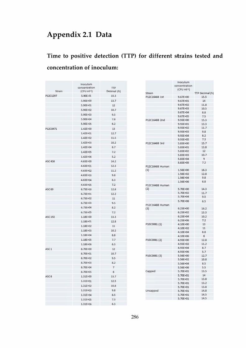
Appen
Time to
concent
ndix 2.1
o positiv
tration of
1 Data
e detecti
f inoculu
2
a
ion (TTP
um:
286
P) for diffferent sttrains tes
sted andd
Counts for diffeerent time
2
e point b
287
blood culttures:
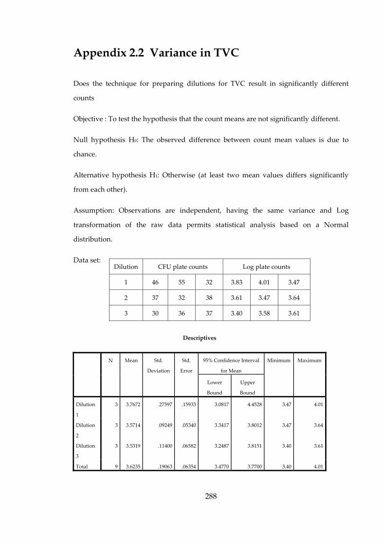
288
Appendix 2.2 Variance in TVC
Does the technique for preparing dilutions for TVC result in significantly different
counts
Objective : To test the hypothesis that the count means are not significantly different.
Null hypothesis H0: The observed difference between count mean values is due to
chance.
Alternative hypothesis H1: Otherwise (at least two mean values differs significantly
from each other).
Assumption: Observations are independent, having the same variance and Log
transformation of the raw data permits statistical analysis based on a Normal
distribution.
Data set:
Descriptives
N Mean Std.
Deviation
Std.
Error
95% Confidence Interval
for Mean
Minimum Maximum
Lower
Bound
Upper
Bound
Dilution
1
3 3.7672 .27597 .15933 3.0817 4.4528 3.47 4.01
Dilution
2
3 3.5714 .09249 .05340 3.3417 3.8012 3.47 3.64
Dilution
3
3 3.5319 .11400 .06582 3.2487 3.8151 3.40 3.61
Total 9 3.6235 .19063 .06354 3.4770 3.7700 3.40 4.01
Dilution CFU plate counts Log plate counts
1 46 55 32 3.83 4.01 3.47
2 37 32 38 3.61 3.47 3.64
3 30 36 37 3.40 3.58 3.61
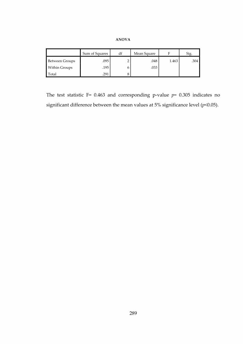
289
ANOVA
Sum of Squares df Mean Square F Sig.
Between Groups .095 2 .048 1.463 .304
Within Groups .195 6 .033
Total .291 8
The test statistic F= 0.463 and corresponding p‐value p= 0.305 indicates no
significant difference between the mean values at 5% significance level (p<0.05).
Appen
ndix 2.33 Injec
2
ctional
290
anthraax casess

Appen
TVC da
Spin pr
Does the
recovered
Objective
groups ar
Null hyp
due to ch
Alternativ
significan
Assumpt
transform
distributi
ndix 3.1
ata transf
otocol co
spin prot
d from the
e : To test
re not sign
pothesis H
hance.
ve hypoth
ntly from e
tion: Obser
mation of t
ion.
1 Spin
ormed fo
ompariso
tocol signif
gel plug o
the hypot
nificantly d
0: The obs
hesis H1:
each other)
rvations ar
the raw da
2
n protoc
or spin p
on – statis
ficantly in
of the serum
thesis that
different.
served diff
Otherwis
).
re indepen
ata permit
291
col com
rotocol c
stical ana
fluence th
m separato
t the samp
ference bet
se (at lea
ndent, havi
s statistica
mpariso
ompariso
alysis
e concentr
or tubes.
ple means
tween sam
st two m
ng the sam
al analysis
on
on:
ration of B
for these
mple mean
mean valu
me varianc
based on
B. anthracis
three spin
n values is
ues differs
ce and Log
a Normal
s
n
s
s
g
l
292
Descriptives
N Mean Std.
Deviation
Std.
Error
95% Confidence Interval
for Mean
Minimum Maximum
Lower
Bound
Upper
Bound
Spin 1 9 4.7774 .51708 .17236 4.3799 5.1749 4.25 5.58
Spin 2 9 4.8689 .59846 .19949 4.4089 5.3289 4.22 5.61
Spin 3 9 4.7291 .63720 .21240 4.2393 5.2189 4.02 5.51
Total 27 4.7918 .56647 .10902 4.5677 5.0159 4.02 5.61
ANOVA
Sum of Squares df Mean Square F Sig.
Between Groups .091 2 .045 .132 .877
Within Groups 8.252 24 .344
Total 8.343 26
The test statistic F= 0.132 and corresponding p‐value p= 0.877 indicate no
significant difference between the mean values at the 5% significance level
(p>0.05).

293
Multiple Comparisons
Dependent Variable: count
(I) SPIN (J) SPIN Mean
Difference (I‐
J)
Std. Error Sig. 95% Confidence Interval
Lower
Bound
Upper
Bound
Tukey
HSD
Spin 1 Spin 2 ‐.09152 .27643 .942 ‐.7818 .5988
Spin 3 .04831 .27643 .983 ‐.6420 .7386
Spin 2 Spin 1 .09152 .27643 .942 ‐.5988 .7818
Spin 3 .13983 .27643 .869 ‐.5505 .8301
Spin 3 Spin 1 ‐.04831 .27643 .983 ‐.7386 .6420
Spin 2 ‐.13983 .27643 .869 ‐.8301 .5505
LSD
Spin 1 Spin 2 ‐.09152 .27643 .743 ‐.6620 .4790
Spin 3 .04831 .27643 .863 ‐.5222 .6188
Spin 2 Spin 1 .09152 .27643 .743 ‐.4790 .6620
Spin 3 .13983 .27643 .618 ‐.4307 .7103
Spin 3 Spin 1 ‐.04831 .27643 .863 ‐.6188 .5222
Spin 2 ‐.13983 .27643 .618 ‐.7103 .4307
The table presents the difference between group means, there is no significant
difference between the spin groups.
count
SPIN N Subset for alpha
= 0.05
1
Tukey HSDa
Spin 3 9 4.7291
Spin 1 9 4.7774
Spin 2 9 4.8689
Sig. .869

Appen
S
ndix 3.
ST pro
.2 Tim
cessing
2
me poin
g
294
nt dataa with
and w
withoutt
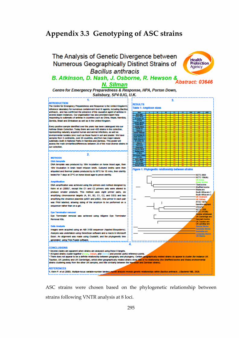
295
Appendix 3.3 Genotyping of ASC strains
ASC strains were chosen based on the phylogenetic relationship between
strains following VNTR analysis at 8 loci.

296
Appendix 3.4 Microscopy images
Simulated human blood culture (P12C16488) PMB stained slide at TTP.
Simulated human blood culture (P12C16488) Gram stained slide at TTP.

297
Simulated human blood culture (B. endophyticus P12C1418) Gram stained slide
at TTP.
Simulated human blood culture (B. anthracis ASC 458 and S. aureus ) Gram
stained slide at TTP.

298
Simulated human blood culture (B. anthracis ASC 458 and S. aepidermidis) Gram
stained slide at TTP.
Simulated human blood culture (B. anthracis ASC 458 and Bacillus spp.
P10C0141) Gram stained slide at TTP.

299
Simulated human blood culture (B. anthracis ASC 458 and B.endophyticus
P10C01418) Gram stained slide at TTP.
Simulated human blood culture (Aneurinibacillus aneurinilyticus P10C2307)
Gram stained slide at TTP.

300
Appendix 3.5 Culture plate images
Overnight culture on CBA a) P10C0141 B. megaterium, b) P10C1418 B.
endophyticus, c) P10C2307 Aneurinibacillus aneurinilyticus and d) ASC 6 (Vollum)
B. anthracis
a b
d c

301
Penicillin and specific γ phage sensitivity for non B. anthracis isolates a)
P10C7386 Non‐reactive Bacillus, b) P10C1418 B. endophyticus, c) P10C0301 B.
mycoides and d) P10C7449 B. cereus.
Penicillin and specific γ phage sensitivity for mixed B. anthracis a) ASC 458 and
Micrococcus spp., b) ASC 458 and P10C1418 B. endophyticus, c) ASC 458 and
P10C141 B. megaterium.
d
b
c
a
a b
c
302
Appendix 4.1 HSE safety note
Provision of key clinical information on laboratory specimen request forms
Introduction:
The purpose of this Safety Notice is to alert health and social care services to potential risks to
laboratory staff, if specimen request forms do not contain relevant details.
It will be of particular relevance to medical staff within organisations that provide specimens
from patients to laboratories for testing. This will include all persons who have direct patient
contact who complete the electronic or paper specimen request forms e.g. clinicians, nurses and
General Practitioners.
Following reports made under The Reporting of Injuries, Diseases and Dangerous Occurrences
Regulations 1995 (RIDDOR), HSEʹs Biological Agents Unit (BAU) have conducted a number of
investigations at Clinical Diagnostic Laboratories.
A common theme is the lack of sufficient relevant clinical details being provided on specimen
request forms. This has resulted in samples being handled at the wrong biological containment
level with resulting increased risk of infection to laboratory staff.
Medical staff should ensure that appropriate information, including relevant travel history is
provided in order to alert laboratory staff of potential dangers.
The most common occurrences have been in relation to the isolation and handling of Hazard
Group 3 agents, Brucella or Salmonella spp from clinical samples that were initially processed at
Containment Level 2. Similar problems could also occur when handling specimens that may
contain other Hazard Group 3 or 4 Biological Agents.
Where appropriate arrangements have not been implemented; HSE Inspectors will take
appropriate enforcement action in line with our enforcement policies and procedures.
Background
Where a laboratory sample or specimen is considered likely to contain a human pathogen, it is
important that the appropriate level of laboratory containment is provided in order to ensure
the effective control of the risk of exposure / infection.
Specimens processed for microbiological analysis and considered likely to contain Hazard
Group 3 or 4 pathogens must be processed within appropriate containment conditions.
303
Specimens liable to result in propagation or culture of Hazard Group 3 pathogens for example
must be processed in a Containment Level 3 laboratory with associated management
arrangements.
Specimens should be supplied with relevant clinical details from requesting clinicians. This can
be used to inform the assessment and further laboratory processing e.g. the types of organisms
that might be present in specimens from a returning traveller or those associated with an
outbreak scenario.
In clinical laboratories, specimens are sorted and processed on the basis of the information
provided. If clinical details are inaccurate or incomplete or there is delay in disclosing new
information to the laboratory then this can result in specimens being processed under
insufficient laboratory containment conditions.
In a number of recent investigations this has resulted in laboratory staff being placed at
increased risk of infection by hazard group 3 agents such as Brucellaspp. and Salmonella typhi.
Had the relevant clinical information been included on the initial form, then the specimen is
likely to have been processed using appropriate containment with all protective control
measures in place.
Action required:
Ensure that clear guidelines for the completion of specimen request forms are in place, along
with measures to provide assurance that guidelines are followed.
Ensure that procedures are reviewed to ensure that they adequately cover the completion of
specimen request documentation e.g. recent history of relevant foreign travel that may
increase the likelihood of exotic agents being present.
Ensure that clinical details supplied on specimen request forms contain clear information
regarding the nature of test being requested and sufficient detail to inform laboratory staff
upon the safety precautions they need to take in order to process the specimen without risk
of infection.
Ensure that guidelines include a system to link different specimens from a patient so that all
contain the same information in relation to safety.
Ensure that if, during patient intervention, further information becomes available that has
implications for the safety of laboratory staff then this is communicated immediately to the
laboratory so that appropriate steps regarding containment can be taken.
Ensure that key personnel involved in the collection of relevant clinical details and the
completion of specimen request documentation receive appropriate training, including
refresher training.
Ensure that a system is in place for monitoring and auditing the correct completion of
specimen request documentation and for taking appropriate action.
304
Appendix 4.2 Microscopy images
Microscopy films of simulated blood cultures following 5 day storage:
Simulated human blood culture (P12C16488) Gram stained slide
Simulated human blood culture (P12C16488) PMB stained slide
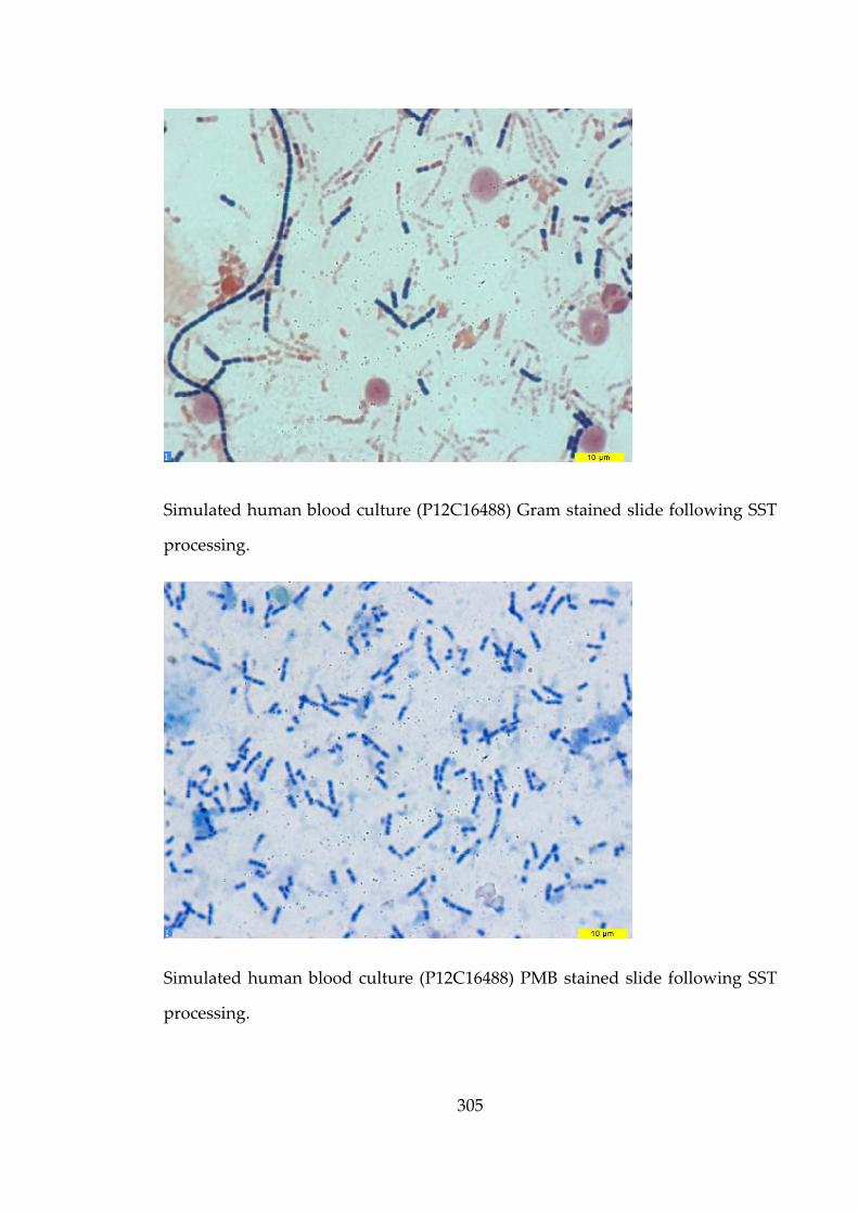
305
Simulated human blood culture (P12C16488) Gram stained slide following SST
processing.
Simulated human blood culture (P12C16488) PMB stained slide following SST
processing.
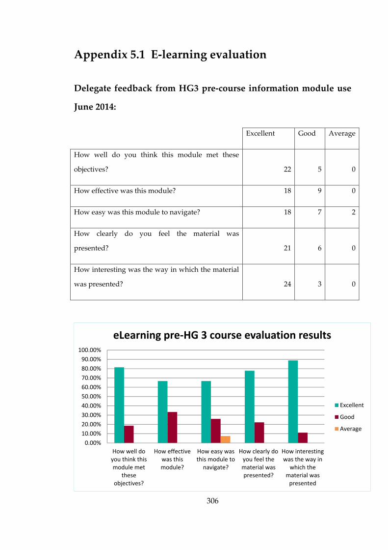
306
Appendix 5.1 E‐learning evaluation
Delegate feedback from HG3 pre‐course information module use
June 2014:
Excellent Good Average
How well do you think this module met these
objectives? 22 5 0
How effective was this module? 18 9 0
How easy was this module to navigate? 18 7 2
How clearly do you feel the material was
presented? 21 6 0
How interesting was the way in which the material
was presented? 24 3 0
0.00%
10.00%
20.00%
30.00%
40.00%
50.00%
60.00%
70.00%
80.00%
90.00%
100.00%
How well doyou think thismodule met
theseobjectives?
How effectivewas thismodule?
How easy wasthis module tonavigate?
How clearly doyou feel thematerial waspresented?
How interestingwas the way inwhich the
material waspresented
eLearning pre‐HG 3 course evaluation results
Excellent
Good
Average
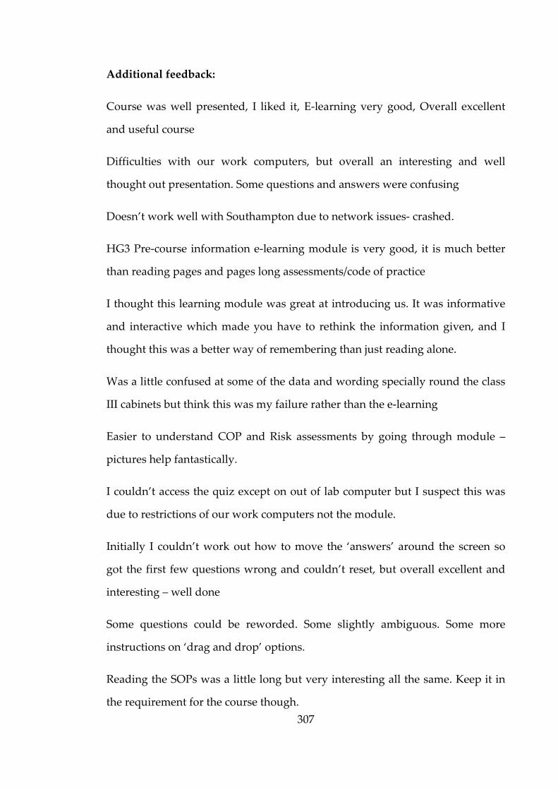
307
Additional feedback:
Course was well presented, I liked it, E‐learning very good, Overall excellent
and useful course
Difficulties with our work computers, but overall an interesting and well
thought out presentation. Some questions and answers were confusing
Doesn’t work well with Southampton due to network issues‐ crashed.
HG3 Pre‐course information e‐learning module is very good, it is much better
than reading pages and pages long assessments/code of practice
I thought this learning module was great at introducing us. It was informative
and interactive which made you have to rethink the information given, and I
thought this was a better way of remembering than just reading alone.
Was a little confused at some of the data and wording specially round the class
III cabinets but think this was my failure rather than the e‐learning
Easier to understand COP and Risk assessments by going through module –
pictures help fantastically.
I couldn’t access the quiz except on out of lab computer but I suspect this was
due to restrictions of our work computers not the module.
Initially I couldn’t work out how to move the ‘answers’ around the screen so
got the first few questions wrong and couldn’t reset, but overall excellent and
interesting – well done
Some questions could be reworded. Some slightly ambiguous. Some more
instructions on ‘drag and drop’ options.
Reading the SOPs was a little long but very interesting all the same. Keep it in
the requirement for the course though.

308
Appendix 5.2 LMS reports
LMS reports for HG3 pre‐course information module
Traffic for module since launch:
309
Progress report for 20 users:
Users are identified by
their email account.

310
Answer breakdown report for first two questions:
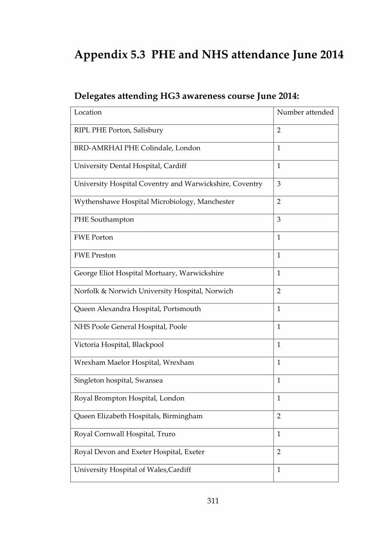
311
Appendix 5.3 PHE and NHS attendance June 2014
Location Number attended
RIPL PHE Porton, Salisbury 2
BRD‐AMRHAI PHE Colindale, London 1
University Dental Hospital, Cardiff 1
University Hospital Coventry and Warwickshire, Coventry 3
Wythenshawe Hospital Microbiology, Manchester 2
PHE Southampton 3
FWE Porton 1
FWE Preston 1
George Eliot Hospital Mortuary, Warwickshire 1
Norfolk & Norwich University Hospital, Norwich 2
Queen Alexandra Hospital, Portsmouth 1
NHS Poole General Hospital, Poole 1
Victoria Hospital, Blackpool 1
Wrexham Maelor Hospital, Wrexham 1
Singleton hospital, Swansea 1
Royal Brompton Hospital, London 1
Queen Elizabeth Hospitals, Birmingham 2
Royal Cornwall Hospital, Truro 1
Royal Devon and Exeter Hospital, Exeter 2
University Hospital of Wales,Cardiff 1
Delegates attending HG3 awareness course June 2014:

312
Appendix 5.4 B. anthracis ‘bench guide’
313
Appendix 6.1 Guidance documents available to
front‐line staff
The HPA website contains links to many guidance documents available to
front-line laboratories for anthrax.

314
Appendix 7.1 Poster – WAM 2012
315
Appendix 7.2 Poster – RUSI 2013
316
Appendix 7.3 Poster – EBSA 2014
Copyright © 2022 FDOKUMEN