Branding Strategy Berbasis Ekonomi Kreatif, Triple Helix vs. Quadruple Helix
Isvoran et al. BMC Pharmacology and Toxicology 2013, 14:31http://www.biomedcentral.com/2050-6511/14/31
RESEARCH ARTICLE Open Access
Computational analysis of protein-proteininterfaces involving an alpha helix: insights forterphenyl–like molecules bindingAdriana Isvoran1,2†, Dana Craciun3†, Virginie Martiny4,5, Olivier Sperandio4,5 and Maria A Miteva4,5*
Abstract
Background: Protein-Protein Interactions (PPIs) are key for many cellular processes. The characterization of PPIinterfaces and the prediction of putative ligand binding sites and hot spot residues are essential to design efficientsmall-molecule modulators of PPI. Terphenyl and its derivatives are small organic molecules known to mimic oneface of protein-binding alpha-helical peptides. In this work we focus on several PPIs mediated by alpha-helicalpeptides.
Method: We performed computational sequence- and structure-based analyses in order to evaluate several keyphysicochemical and surface properties of proteins known to interact with alpha-helical peptides and/or terphenyland its derivatives.
Results: Sequence-based analysis revealed low sequence identity between some of the analyzed proteins bindingalpha-helical peptides. Structure-based analysis was performed to calculate the volume, the fractal dimensionroughness and the hydrophobicity of the binding regions. Besides the overall hydrophobic character of the bindingpockets, some specificities were detected. We showed that the hydrophobicity is not uniformly distributedin different alpha-helix binding pockets that can help to identify key hydrophobic hot spots.
Conclusions: The presence of hydrophobic cavities at the protein surface with a more complex shape than theentire protein surface seems to be an important property related to the ability of proteins to bind alpha-helicalpeptides and low molecular weight mimetics. Characterization of similarities and specificities of PPI binding sitescan be helpful for further development of small molecules targeting alpha-helix binding proteins.
BackgroundProtein-Protein Interactions (PPIs) are key to many cellu-lar processes. Abnormal PPIs contribute to many diseasestates and as such, PPIs represent today a new class ofdrug targets essentially unexploited for drug discovery.Indeed, the size of the human interactome has been esti-mated to be between 300,000 [1] and 650,000 interactions[2]. In the last decade many studies have been performedin order to target PPIs [3]. Several small-molecule inhibi-tors of PPIs have been demonstrated therapeutic potential[4-8]. However, efficient targeting of PPIs is still being
* Correspondence: [email protected]†Equal contributors4Université Paris Diderot, Sorbonne Paris Cité, Molécules Thérapeutiques insilico, Inserm UMR-S 973, 35 rue Helene Brion, Paris 75013, France5INSERM, U973, Paris F-75205, FranceFull list of author information is available at the end of the article
© 2013 Isvoran et al.; licensee BioMed CentralCommons Attribution License (http://creativecreproduction in any medium, provided the or
considered as an important challenge [3,9,10]. In contrastto enzyme-substrate interactions, protein-protein recogni-tion often occurs through flat surfaces or wide shallowgrooves. Recent structural analyses of PPI interfaces andsmall molecules disrupting PPIs suggested that such li-gands might mimic the structural characteristics of theprotein partner [6,11]. To facilitate the discovery of newPPI small-molecule inhibitors, the characterization of PPIinterfaces [12,13] and the prediction of putative ligandbinding sites are essential. Physicochemical properties ofboth ligand and protein are key to mediate the binding[14], such as cavity sizes, shape complementarity, electro-static potential and hydrophobicity [12,15].The role of alpha-helical peptides in mediating many
PPIs is well demonstrated and development of small or-ganic molecules mimicking such peptides becomes im-portant [16]. Recent studies have been carried out on
Ltd. This is an Open Access article distributed under the terms of the Creativeommons.org/licenses/by/2.0), which permits unrestricted use, distribution, andiginal work is properly cited.

Isvoran et al. BMC Pharmacology and Toxicology 2013, 14:31 Page 2 of 11http://www.biomedcentral.com/2050-6511/14/31
the whole Protein Data Bank (PDB) in order to establisha druggability profile of alpha-helix mediated PPIs andto predict which of them could bind a small molecule[17]. More specifically, terphenyl and its derivates aresmall organic molecules [18-26] mimicking one face ofan alpha-helical peptide, i.e. the side chains of three keyresidues occupying positions i, i+3 and i+7 [25,26] or i,i+4 and i+7 [20] of the bound helix. It has been sug-gested that terphenyl compounds can serve as pharma-cological probes because they are membrane permeable[22]. Terphenyl 1 and 2, which mimic the calmodulinbinding face of smooth muscle myosin light chain kinase(smMLCK), have been shown to inhibit the interactionsof calmodulin (CaM) with the enzyme 3'-5'-cyclic nu-cleotide phosphodiesterase (PDE) and with the helicalpeptide C20W of the plasma membrane calcium pumps[18]. Following the similarity between the calmodulinand human centrin 2 (HsCen2) alpha-helix binding sites,we recently suggested that terphenyl 2 might also inhibitthe interaction between HsCen2 and a 17 residues pep-tide of Xeroderma Pigmentosum Group C (XPC) protein
Table 1 Protein – alpha-helical peptide complexes
Protein complex
Chicken calmodulin in complex with smooth muscle myosin light chain kina(smMLCK)
Human calmodulin in complex with a mutant peptide of human DRP-1 kinas
Human calmodulin in complex with CAV1.1 IQ peptide
Human calmodulin in complex with CAV2.2 IQ peptide
E Coli calmodulin in complex with RS20 peptide of smMLCK
Rat calmodulin in complex with NMDA receptor NR1C1peptide
Human centrin 2 in complex with the centrin binding region of XPC protein
C-terminal domain of human centrin 2 in complex with a repeat sequence ohuman Sfi 1
Scherffelia dubia centrin in complex with smMLCK peptide
Human BCL-XL in complex with BAK peptide
Human E3 ubiquitin-protein ligase MDM2 in complex with p53 tumortransactivation domain (fragment 17-125)
Rabbit cardiac troponin C in complex with a fragment (residues 1-47) of cardtroponin I
*known to be disrupted by terphenyl or its derivatives.
[27]. Terphenyl derivates mimicking the alpha-helicalstructure of p53 N-terminal peptide inhibit the p53-MDM2 [22] and the p53-HDM2 interactions [21]. Thesemolecules also mimic the alpha-helical region of BakBH3 domain, which binds BCL-X2, thus disrupting theBCL-X2/Bak interaction [19,20,24].In this work we performed a computational analysis in
order to evaluate several key physicochemical and surfaceproperties of proteins known to interact with alpha-helical peptides or to bind terphenyl and its derivatives.We calculated the binding pocket volumes and the fractaldimensions of the surface cavities for the entire proteinand for the binding pockets. We identified several simila-rities and specificities characterizing such protein bindingsites that can be helpful for future development of moreefficient small-molecule inhibitors targeting alpha-helixbinding proteins.
MethodsIn this study we compared the sequence and surface prop-erties of the investigated proteins. In order to analyze the
PDB codeResolution
SwissProtcode
Interacting residues of thebound alpha-helix
se 2O5G* P62149 TRP5, THR8, VAL12
1.08 Å
e 1ZUZ P62158 TRP305, PHE309, VAL312
1.91 Å
2VAY* P62158 THR526, ILE529, PHE533
1.94 Å
3DVE P62158 MET854, VAL857, MET161
2.35 Å
1QTX - TRP5, THR8, VAL12
1.65 Å
2HQW P62161 PHE880, THR884, LEU887
1.90 Å
2GGM P41208 TRP848, LEU851, LEU855
2.35 Å
f 2K2I P41208 LEU651, LEU655, TRP658
NMR
3KF9 Q06827 TRP4, PHE8, VAL11
2.60 Å
1BXL* Q07817 VAL574, LEU578, ILE581
NMR
1YCR* Q00987 PHE19, TRP23, LEU26
2.60 Å
iac 1A2X P02586 LEU17, MET21, ILE24
2.30 Å

Isvoran et al. BMC Pharmacology and Toxicology 2013, 14:31 Page 3 of 11http://www.biomedcentral.com/2050-6511/14/31
sequence similarities we performed sequence alignmentusing the CLUSTALW software [28]. Interacting residuesat the protein-protein interface in terms of contact dis-tances were found using the ContPro online freely avai-lable tool [29]. We identified the protein residuesinteracting with the three key residues of the alpha-helicalpeptide (occupying positions i, i+3 and i+7 or i, i+4 andi+7) those relative positions are mimicked by terphenyland its derivatives. The distance threshold was set to 5 Åfor the side chain atoms.In order to evaluate the protein surface properties, the
bound peptide was removed for each complex. The surfacecharacteristics of the entire protein and those of thepeptide-binding cavity were analyzed. Using the ap-proach of the fractal geometry we quantitatively de-scribed the surface roughness for the entire protein and
Figure 1 Sequence alignment of alpha-helix binding proteins. The amin red.
for the binding cavity, expressed by global surface frac-tal dimension (DS) and local surface fractal dimension(DL), respectively. In order to calculate the surface frac-tal dimension we used the method proposed by Lewisand Rees [30] based on the scaling law between the sur-face area (SA) and the radius of the rolling probe mol-ecule (R) on the surface, i.e. SA is proportional to theradius to the power 2-Ds:
SAeR2−DS ð1Þ
The surface fractal dimension was determined fromthe slope of the double logarithmical plot of SA versusR. The surface area of the protein was computed usingthe on-line available software GETAREA [31]. Proberadii of 1, 1.2, 1.4, 1.6, 1.8 and 2 Å were used. For the
ino acid residues interacting with alpha-helical peptides are presented

Table 2 Sequence identity (in %) between the considered proteins (the binding area/entire protein)
Protein/ sequence identity Humancalmodulin
Humancentrin 2
Scherffelia dubiacentrin
HumanBCL-X2
Human E3 ubiquitin-protein ligaseMDM2
Human centrin 2 54/50
Scherffelia dubia centrin 56/55 90/74
Human BCL-X2 5/7 5/5 5/8
Human E3 ubiquitin-protein ligaseMDM2
5/4 5/10 7/6 9/5
Rabbit cardiac troponin C 57/51 57/34 37/32 5/9 5/19
The binding area was defined here as all residues of the protein interacting with the helical peptide.
Figure 2 3D structures of the complexes formed by: (a) human centrin 2 and a 10 residue peptide of Xeroderma Pigmentosum group Cprotein, code entry 2GGM. (b) chicken calmodulin and smooth muscle myosin light chain kinase (smMLCK), code entry 2O5G. (c) scherffelia dubiacentrin and smMLCK peptide, code entry 3KF9. (d) rabbit cardiac troponin C and a fragment of cardiac troponin I, code entry 1A2X. (e) humanBCL-XL and BAK peptide, code entry 1BXL. (f) human E3 ubiquitin-protein ligase MDM2 and p53 tumor transactivation domain, code entry 1YCR.All proteins are shown as surface in atom color type (C and H-white, N – blue, O -red, S – yellow) and ligands are shown in magenta cartoonwith hydrophobic interacting residues given as sticks.
Isvoran et al. BMC Pharmacology and Toxicology 2013, 14:31 Page 4 of 11http://www.biomedcentral.com/2050-6511/14/31
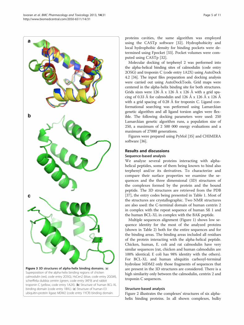
Figure 3 3D structures of alpha-helix binding domains. (a)Superposition of the alpha-helix binding regions of chickencalmodulin (red, code entry 2O5G), HsCen2 (blue, code entry 2GGM),scherffelia dubbia centrin (green, code entry 3KF9) and rabbittroponin C (yellow, code entry 1A2X). (b) Structure of human BCL-XLbinding domain (code entry 1BXL). (c) Structure of human E3ubiquitin-protein ligase MDM2 (code entry 1YCR) binding domain.
Isvoran et al. BMC Pharmacology and Toxicology 2013, 14:31 Page 5 of 11http://www.biomedcentral.com/2050-6511/14/31
proteins cavities, the same algorithm was employedusing the CASTp software [32]. Hydrophobicity andlocal hydrophobic density for binding pockets were de-termined using Fpocket [33]. Pocket volumes were com-puted using CASTp [32].Molecular docking of terphenyl 2 was performed into
the alpha-helical binding sites of calmodulin (code entry2O5G) and troponin C (code entry 1A2X) using AutoDock4.2 [34]. The input files preparation and docking analysiswere carried out using AutoDockTools. Grid maps werecentered in the alpha-helix binding site for both structures.Grids sizes were 126 Å x 126 Å x 126 Å with a grid spa-cing of 0.33 Å for calmodulin and 126 Å x 126 Å x 126 Åwith a grid spacing of 0.28 Å for troponin C. Ligand con-formational searching was performed using Lamarckiangenetic algorithm and all ligand torsion angles were flex-ible. The following docking parameters were used: 250Lamarckian genetic algorithm runs, a population size of250, a maximum of 2 500 000 energy evaluations and amaximum of 27000 generations.Figures were prepared using PyMol [35] and CHIMERA
software [36].
Results and discussionsSequence-based analysisWe analyze several proteins interacting with alpha-helical peptides, some of them being known to bind alsoterphenyl and/or its derivatives. To characterize andcompare their surface properties we examine the se-quences and the three dimensional (3D) structures ofthe complexes formed by the protein and the boundpeptide. The 3D structures are retrieved from the PDB[37], the entry codes being presented in Table 1. Most ofthe structures are crystallographic. Two NMR structuresare also used: the C-terminal domain of human centrin 2in complex with the repeat sequence of human Sfi 1 andthe human BCL-XL in complex with the BAK peptide.Multiple sequences alignment (Figure 1) shows low se-
quence identity for the most of the analyzed proteins(shown in Table 2) both for the entire sequences and forthe binding areas. The binding areas included all residuesof the protein interacting with the alpha-helical peptide.Chicken, human, E. coli and rat calmodulin have verysimilar sequences (rat, chicken and human calmodulin are100% identical; E coli has 98% identity with the others).For BCL-XL and human ubiquitin carboxyl-terminalhydrolase MDM2 only those fragments of sequences thatare present in the 3D structures are considered. There is ahigh similarity only between the calmodulin, centrin 2 andtroponin C sequences.
Structure-based analysisFigure 2 illustrates the complexes’ structures of six alpha-helix binding proteins. In all shown complexes, bulky

Figure 4 Illustration of the interacting residues (in sticks) of the protein (atom color type) and the bound peptide (red): (a) chickencalmodulin and smMLCK (code entry 2O5G), (b) human centrin 2 and the centrin binding region of XPC (code entry 2GGM), (c) human BCL-XLprotein and BAK (code entry 1BXL), (d) human E3 ubiquitin- protein ligase MDM2 and p53 tumor transactivation domain (code entry 1YCR), (e)rabbit cardiac troponin C and cardiac troponin I (code entry 1A2X).
Isvoran et al. BMC Pharmacology and Toxicology 2013, 14:31 Page 6 of 11http://www.biomedcentral.com/2050-6511/14/31
hydrophobic residues of the bound peptide anchor intothe protein binding pocket. Following the sequence simi-larities we superimposed the alpha-helix binding regionsstructures of calmodulin, human centrin 2, scherffeliadubia centrin and rabbit troponin C (Figure 3a). Strongstructural homology for binding regions is seen followingthe sequence similarity of these proteins. Figure 3b and 3cillustrate the binding pockets of BCL-XL and human E3ubiquitin-protein ligase MDM2, respectively.The interacting residues of the proteins and bound
peptides, identified with ContPro [29], are shown inFigures 1 and 4 and Table 1. The results reveal that usually
hydrophobic residues such as TRP, LEU, ILE, PHE, VAL,MET are involved in the interactions. The presence of hy-drophobic residues suggests a favorable interaction withterphenyl-like molecules anchoring in the hydrophobiccavities. Most of the residues involved in the interactionsbetween the proteins and alpha-helices are hydrophobicfor both partners, as also observed in other studies [38].We notice several key residues involved in the interactionof the same protein with different peptide partners. Forexample, in the case of calmodulin, PHE92, MET124,PHE141, MET144 and MET145 are involved in most ofthe peptides’ interactions. These residues can thus be

Figure 5 Surface lipophilicity (shown in green) of alpha-helixbinding proteins computed using MOE. (a) Human E3 ubiquitin-protein ligase MDM2 (PDB code 1YCR), (b) Scherffelia dubia centrin(PDB code 3KF9).
Isvoran et al. BMC Pharmacology and Toxicology 2013, 14:31 Page 7 of 11http://www.biomedcentral.com/2050-6511/14/31
considered as key for the interaction with terphenyl andits derivatives, or other alpha-helix mimetics. We noticedthe presence of MET residues in most of the alpha-helixbinding pockets analyzed here. In a recent study, METresidues have not been identified to be a part of hot spotamino acids, in particular in alpha-helix mediated proteininterfaces [39]. However, our analysis clearly indicatestheir presence in positions that are key for the inter-action with the alpha-helical partner. Furthermore, Maand Nussinov [40] have also concluded that the aminoacids TRP, MET, and PHE are important for protein-protein interactions. They showed that TRP/MET/PHE residues play roles in the dimerization of the
transcriptase (p51/p66) and in cell-fusion processes,including the gp120-CD4 interaction and the gp41six-helix bundle formation. They suggested that pola-rizability of MET allows it to assume roles of bothhydrophobic and hydrophilic residues [40]. Further, itslarger flexibility compared to other hydrophobic resi-dues may facilitate the plasticity of hydrophobic bind-ing pockets allowing to accommodate different ligands[27].We used Fpocket [33] and CASTp [32] to calculate
geometrical and physicochemical characteristics of thebinding pockets taking into account the protein residuesinteracting with the alpha-helical peptides. The overallhydrophobic character of the binding pockets is againclearly identified. Yet, some specificity is also observed,several pockets show high hydrophobicity score but lowlocal hydrophobic density, or vice versa, demonstratingthat the hydrophobic patches are not always regularlydistributed in the binding pockets. For example, 1YCRand 3KF9 have similar hydrophobicity scores but highand low calculated hydrophobic density, respectively.The differences of the hydrophobicity distribution areillustrated in Figure 5.The volumes of the detected pockets in the peptide-
binding regions computed with CASTp are given inTable 3. The average volume of the sub-cavities present atthe PPI interfaces found by Fuller et al [41] was ~60 Å3.Sonavane & Chakrabarti [42] found PPI pocket volumesto be up to ~330 Å3. We found similar volumes to thosereported in Bourgeas et al. [43]. Taking into account thevarious algorithms and different concepts for bindingpocket definition, such differences for the computed vol-umes can be expected. Several small cavities are present inthe binding region (seen in Figure 2 and Figure 5), as ithas been previously observed for other targeted PPI inter-faces [39]. For the proteins studied here, the presence ofseveral small hydrophobic cavities in the alpha-helix bind-ing region seems to be a typical surface feature guidingthe anchoring of hydrophobic residues from the peptideside. Such characteristics can also facilitate targeting PPImediated by alpha-helices by small molecules containinghydrophobic anchors (as terphenyl or other mimetics).Further, we decided to explore the roughness of the
alpha-helix binding sites. The methodology implementedto calculate the fractal surface dimensions, used for theroughness evaluation, is illustrated in Figure 6 for theglobal surface roughness of chicken calmodulin. Thefractal global surface dimension and the fractal local sur-face dimension for the binding site of chicken calmodu-lin are calculated to be DS=2.238; ± 0.006 and DL= 2.616 ±0.072, respectively. The global and local fractal dimensionsfor the other proteins are given in Table 4. Our resultsand other previously published data [44-47] suggestthat the global fractal dimension of protein surface is

Table 3 Geometrical and physicochemical characteristics of the identified pockets
Protein code PDB Volume (Å3) Hydrophobicity score Local hydrophobic density
Chicken calmodulin 312.0 68.86 43.00
2O5G
Human calmodulin 203.0 68.86 42.00
1ZUZ
Human calmodulin 219.8 59.62 40.00
2VAY
Human calmodulin 226.4 61.00 39.32
3DVE
E.coli calmodulin 317.9 56.63 40.15
1QTX
Rat calmodulin 310.6 56.62 43.78
2HQW
Human centrin 2 147.9 41.47 32.00
2GGM
Human centrin 2 210.9 39.93 35.08
2K2I
Scherffelia dubia centrin 221.5 58.19 31.00
3KF9
Human BCL-XL 321.5 36.91 42.04
1BXL
Human E3 ubiquitin-protein ligase MDM2 201.9 51.18 55.20
1YCR
Rabbit cardiac troponin C 213.1 63.07 39.15
1A2X
Table 4 Global (DS) and local (DL) surface fractal
Isvoran et al. BMC Pharmacology and Toxicology 2013, 14:31 Page 8 of 11http://www.biomedcentral.com/2050-6511/14/31
about 2. The local surface fractal dimensions for thebinding cavities are computed to be larger than theglobal surface fractal dimensions for all studied pro-teins. This reflects the higher roughness of the bindingsite and its more complex shape and that can be consid-ered as important for ligand binding. The most importantdifferences between DS and DL are obtained for human
Figure 6 Double logarithmical plot of the surface area versusprobe radii for chicken calmodulin (PDB code 2O5G).
calmodulin (2VAY), centrin (3KF9, 2K2I), BCL-XL(1BXL), MDM2 (1YCR) and troponin C (1A2X). It hasbeen experimentally demonstrated that human calmodu-lin [18], BCL-XL [19,20] and MDM2 [21,22] interactwith terphenyl or its derivatives. Recently, we suggested
dimensions of investigated proteins
Code PDB DS DL
2O5G 2.238 ± 0.006 2.616 ± 0.072
1ZUZ 2.181 ± 0.007 2.487 ± 0.058
2VAY 2.183 ± 0.006 2.757 ± 0.108
3DVE 2.217 ± 0.003 2.418 ± 0.040
1QTX 2.302 ± 0.002 2.494 ± 0.069
2HQW 2.172 ± 0.002 2.454 ± 0.082
2GGM 2.247 ± 0.004 2.373 ± 0.018
2K2I 2.167 ± 0.008 2.892 ± 0.124
3KF9 2.179 ± 0.006 2.892 ± 0.153
1BXL 2.230 ± 0.007 2.696 ± 0.225
1YCR 2.173 ± 0.014 2.708 ± 0.055
1A2X 2.177 ± 0.005 2.624 ± 0.032

Figure 7 Best scored docking poses of terphenyl. The posesafter docking-scoring with AutoDock are shown in cyan. (a) chickencalmodulin, code entry 2O5G, (b) rabbit cardiac troponin C, codeentry 1A2X.
Isvoran et al. BMC Pharmacology and Toxicology 2013, 14:31 Page 9 of 11http://www.biomedcentral.com/2050-6511/14/31
a possible binding of terphenyl 2, which mimics the rela-tive positions of the side chains of residues TRP848,LEU851, LEU855 of the XPC peptide, into human centrin2 following our energetic and conformational flexibilityanalysis performed for the alpha-helical peptide-bindingpocket of centrin 2 [27]. The DL value for the peptide-binding site of troponin C shows rougher surface than theentire protein, similarly to the above listed terphenyl-binding proteins.Taking into consideration the sequence and structural
homology of troponin C and calmodulin and otherphysicochemical similarities of the binding sites asdiscussed above, we decided to probe putative terphenylbinding into troponin C. We performed docking ofterphenyl 2 into the peptide-binding sites of calmodulinand troponin C using AutoDock. The best scoreddocking poses are shown in Figure 7. The terphenyl ori-entations in the best scored poses correspond to the
position of the bound alpha-helical peptides shown inFigure 2. The predicted interaction energies of -7.98and -8.18 kcal/mol for terphenyl binding in calmodulinand troponin C, respectively, suggest favorable interac-tions with the two proteins.In the light of the results obtained here, it is now
interesting to discuss the physicochemical properties ofknown PPI modulators, such as terphenyl. In a previouswork [10] we gathered a set of 66 PPI inhibitors amongwhich some terphenyl derivatives and other inhibitorsof alpha-helix mediated PPI were present. In that workwe demonstrated the more hydrophobic character ofthese compounds but also their bigger size. Interes-tingly, we also showed the importance of a critical num-ber of aromatic bonds and some specific molecularshapes (T-shaped, star-shaped, or L-shaped com-pounds), among which some correspond to terphenylderivatives. The present work therefore confirms thatsuch genuine properties on the ligand side seem to becavity-driven, and that these small molecules must pos-sess certain properties in order to efficiently modulatean alpha-helix mediated PPI and to mimic the nativepartner and its properties.
ConclusionsModulating protein-protein interactions using small mole-cules based on surface recognition has been a field of in-creasing interest during the last decade. PPI interfaces arevery complex and need to be analyzed in order to be effi-ciently targeted for drug discovery purposes. Designedcompounds must bind with high affinity and selectivity tothe target protein. The low sequence identity found be-tween some of the analyzed proteins suggests that thereare no sequence requirements for the ability of proteins tobind alpha-helical peptides and consequently small-molecule mimetics.From the structural point of view, all investigated
proteins show larger surface fractal dimensions for thepeptide-binding pockets than the entire protein sur-face reflecting the higher complexity of the shape ofthe binding sites. Also, the presence of several hydro-phobic patches at the protein surface seems to be animportant property related to the ability of the proteinto bind alpha-helical peptides and mimetics. Further-more, we showed that hydrophobicity is not uniformlydistributed across different alpha-helix binding pocketsand that its distribution can be used to identify hydro-phobic hot spots.Many similarities between the binding sites studied
here are observed and terphenyl or its derivatives bind-ing to various alpha-helix binding proteins can be sug-gested. However, targeting various PPI complexes bysimilar small molecules can rise selectivity problems inthe context of drug discovery or chemical biology

Isvoran et al. BMC Pharmacology and Toxicology 2013, 14:31 Page 10 of 11http://www.biomedcentral.com/2050-6511/14/31
projects. Thus, the specificities found here for differentbinding sites, e.g. key residues, roughness and localhydrophobic density, can be further exploited tooptimize terphenyl-like ligands in order to improvetheir selectivity.
AbbreviationsPPI: Protein-Protein interactions; smMLCK: smooth muscle myosin light chainkinase; CaM: Calmodulin; HsCen2: Human centrin 2; PDE: 3'-5'-cyclicnucleotide phosphodiesterase; XPC: Xeroderma pigmentosum group C.
Competing interestsThe authors declare that they have no competing interests.
Authors’ contributionsAI carried out the sequence alignment and binding pockets analysis. DCcarried out the fractal calculations. AI and DC drafted the manuscript. VMcarried out the volume calculations and docking analysis. OS participated inthe protein-protein interface analysis and discussion writing. MAM designedand coordinated the study. All authors participated in manuscript writingand approved the final manuscript.
AcknowledgmentsThe financial support from the West University of Timisoara, the Inserminstitute and the University Paris Diderot is greatly appreciated.
Author details1Department of Biology and Chemistry, West University of Timisoara, 16Pestalozzi, Timisoara 300115, Romania. 2Advanced Environmental ResearchesLaboratory, 4 Oituz, Timisoara 300086, Romania. 3Teacher TrainingDepartment, West University of Timisoara, 4 Blvd. V. ParvanTimisoara 300223,Romania. 4Université Paris Diderot, Sorbonne Paris Cité, MoléculesThérapeutiques in silico, Inserm UMR-S 973, 35 rue Helene Brion, Paris 75013,France. 5INSERM, U973, Paris F-75205, France.
Received: 23 January 2013 Accepted: 11 June 2013Published: 14 June 2013
References1. Hunter T, Maniatis T, Califano A, Honig B, Zhang QC, Petrey D, Deng L,
Qiang L, Shi Y, Thu CA, Bisikirska B, Lefebvre C, Accili D: Structure-basedprediction of protein-protein interactions on a genome-wide scale.Nature 2012, 490:556–660.
2. Stumpf MP, Thorne T, de Silva E, Stewart R, An HJ, Lappe M, Wiuf C:Estimating the size of the human interactome. Proc Natl Acad Sci USA2008, 105:6959–6964.
3. Stockwell BR: Exploring biology with small organic molecules. Nature2004, 432:846–854.
4. Wilson CG, Arkin MR: Small-molecule inhibitors of IL-2/IL-2R: Lessonslearned and applied. Curr Top Microbiol Immunol 2011, 348:25–29.
5. Villoutreix BO, Bastard K, Sperandio O, Fahraeus R, Poyet JL, Calvo F, DeprezB, Miteva MA: In silico-in vitro screening of protein-protein interactions:towards the next generation of therapeutics. Curr Pharm Biotechnol 2008,9:103–122.
6. Fry DC: Protein-protein interactions as targets for small molecule drugdiscovery. Biopolymers 2006, 84:535–552.
7. Gautier B, Miteva MA, Goncalves V, Huguenot F, Coric P, Bouaziz S, Seijo B,Gaucher JF, Broutin I, Garbay C, Lesnard A, Rault S, Inguimbert N, VilloutreixBO, Vidal M: Targeting the proangiogenic VEGF-VEGFR protein-proteininterface with drug-like compounds by in silico and in vitro screening.Chem Biol 2011, 18(12):1631–1639.
8. Villoutreix BO, Laconde G, Lagorce D, Martineau P, Miteva MA, DariavachP: Tyrosine kinase syk non-enzymatic inhibitors and potentialanti-allergic drug-like compounds discovered by virtual and in vitroscreening. PLoS One 2011, 6(6):e21117.
9. Wells JA, McClendon CL: Reaching for high-hanging fruit in drugdiscovery at protein-protein interfaces. Nature 2007, 450:1001–1009.
10. Sperandio O, Reynes CH, Camproux AC, Villoutreix BO: Rationalizing thechemical space of protein-protein interaction inhibitors. Drug DiscovToday 2010, 15:220–229.
11. Morelli X, Bourgeas R, Roche P: Chemical and structural lessons fromrecent successes in protein-protein interaction inhibition (2P2I). Curr OpinChem Biol 2011, 15:475–481.
12. Tripathi A, Kellogg GE: A Novel and Efficient Tool for Locating andCharacterizing Protein Cavities and Binding Sites. Proteins 2010,78(4):825–842.
13. Grosdidier S, Fernández-Recio J: Protein-protein Docking and Hot-spotPrediction for Drug Discovery. Curr Pharm Des 2012, 18(30):4607–4618.
14. Andersson CD, Chen BY, Linusson A: Mapping of ligand-binding cavities inproteins. Proteins 2010, 78(6):1408–1422.
15. Koes D, Khoury K, Huang Y, Wang W, Bista M, Popowicz GM, Wolf S, HolakTA, Domling A, Camacho CJ: Enabling Large-Scale Design, Synthesis andValidation of Small Molecule Protein-Protein Antagonists. PLoS One 2012,7(3):e32839.
16. Petsko GA, Ringe D: Protein structure and function. London: New SciencePress Ltd; 2004.
17. Bullock BN, Jochim AL, Arora PS: Assessing helical protein interfacesfor inhibitor Design. J Am Chem Soc 2011, 133(36):14220–14223.
18. Orner BP, Ernst JT, Hamilton AD: Towards Proteomimetics: TerphenylDerivatives as Structural and Functional Mimics of Extended Regions ofan a-Helix. J Am Chem Soc 2001, 123(22):5382–5383.
19. Kutzki O, Park HS, Ernst JT, Orner BP, Yin H, Hamilton AD: Development of aPotent Bcl-xL Antagonist Based on r-Helix Mimicry. J Am Chem Soc 2002,124(40):11838–11839.
20. Yin H, Lee GI, Sedey KA, Kutzki O, Park HS, Orner BP, Ernst JT, Wang HG,Sebti SM, Hamilton AD: Terphenyl-Based Bak BH3 alpha-helicalproteomimetics as low-molecular-weight antagonists of Bcl-xL. J AmChem Soc 2005, 127(29):10191–10196.
21. Yin H, Lee G, Park HS, Payne GA, Rodriguez JM, Sebti SM, Hamilton AD:Terphenyl-Based Helical Mimetics That Disrupt the p53/HDM2Interaction. Angew Chem 2005, 117(18):2764–2767.
22. Chen L, Yin H, Farooqi B, Sebti S, Hamilton AD, Chen J: p53 alpha-Helixmimetics antagonize p53/MDM2 interaction and activate p53. Mol CancTher 2005, 4(6):1019–1025.
23. Che Y, Brooks BR, Marshall GR: Protein recognition motifs: design ofpeptidomimetics of helix surfaces. Biopolymers 2007, 86(4):288–297.
24. Becerril J, Rodriguez JM, Wyrembak PN, Hamilton AD: Inhibition ofprotein-protein interaction by peptide mimics. In Protein SurfaceRecognition: Approaches for Drug Discovery. Edited by Giralt E, Peczuh M,Salvatella X. London: John Willey&Sons; 2011.
25. Fairlie DP, West ML, Wong AK: Towards protein surface mimetics. Curr MedChem 1998, 5(1):29–62.
26. Maity P, König B: Synthesis and structure of 1,4-dipiperazino benzenes:chiral terphenyl-type peptide helix mimetics. Org Lett 2008,10(7):1473–1476.
27. Isvoran A, Badel A, Craescu CT, Miron S, Miteva MA: Exploring NMRensembles of calcium binding proteins: Perspectives to design inhibitorsof protein-protein interactions. BMC Struct Biol 2011, 11:24.
28. Thompson JD, Higgins DG, Gibson TJ: CLUSTAL W: lmproving thesensitivity of progressive multiple sequence alignment throughsequence weighting, position specific gap penalties and weight matrixchoice. Nucleic Acids Res 1994, 22:4673–4680.
29. Firoz A, Malik A, Afzal O, Jha V: ContPro: A web tool for calculating aminoacid contact distances in protein from 3D –structure at differentdistance threshold. Bioinformation 2010, 5(2):55–57.
30. Lewis M, Rees DC: Fractal surfaces of proteins. Science 1985,230:1163–1165.
31. Fraczkiewicz R, Braun W: Exact and efficient analytical calculation ofaccesible surface areas and their gradients for macromolecules. J ComplChem 1998, 19:319–333.
32. Dundas J, Ouyang Z, Tseng J, Binkowski A, Turpaz Y, Liang J: CASTp:computed atlas of surface topography of proteins with structural andtopographical mapping of functionally annotated resiudes. Nucleic AcidRes 2006, 34:W116–W118.
33. Guilleoux VL, Schmidtke P, Tuffery P: Fpocket; An open source platform forligand binding pocket detection. BMC Bioinforma 2009, 10:168.
34. Morris GM, Huey R, Lindstrom W, Sanner MF, Belew RK, Goodsell DS, OlsonAJ: AutoDock4 and AutoDockTools4: Automated docking with selectivereceptor flexibility. J Comput Chem 2009, 30(16):2785–2791.
35. DeLano WL: The PyMol molecular graphics system. San Carlos: DeLanoScientific; 2002.

Isvoran et al. BMC Pharmacology and Toxicology 2013, 14:31 Page 11 of 11http://www.biomedcentral.com/2050-6511/14/31
36. Pettersen EF, Goddard TD, Huang CC, Couch GS, Greenblatt DM, Meng EC,Ferrin TE: UCSF Chimera--a visualization system for exploratory researchand analysis. J Comput Chem 2004, 25(13):1605–1612.
37. Berman HM, Westbrook J, Feng Z, Gilliland G, Bhat TN, Weissig H,Shindyalov IN, Bourne PE: The Protein Data Bank. Nucleic Acids Res 2000,28:235–242.
38. Moreira IS, Fernandes PA, Ramos MJ: Hot spots—A review of theprotein–protein interface determinant amino-acid residues. ProteinsStruct Funct Bioinform 2007, 68(4):803–812.
39. Brooke N, Bullock A, Paramjit SA: Assessing Helical Protein Interfaces forInhibitor Design. J Am Chem Soc 2011, 133(36):14220–14223.
40. Ma B, Nussinov R: Trp/Met/Phe Hot Spots in Protein-Protein Interactions:Potential Targets in Drug Design. Curr Top Med Chem 2007, 7:999–1005.
41. Fuller JC, Burgoyne NJ, Jackson RM: Predicting druggable binding sites atthe protein–protein interface. Drug Discov Today 2009, 14(3–4):155–161.
42. Sonavane S, Chakrabarti P: Cavities and Atomic Packing in ProteinStructures and Interfaces. PLoS Comput Biol 2008, 4:e100001188.
43. Bourgeas R, Basse MJ, Morelli X, Roche P: Atomic Analysis of Protein-Protein Interfaces with Known Inhibitors: The 2P2I Database. PLoSOne 2010, 3:e9598.
44. Goetze T, Brickmann J: Self similarity of protein surfaces. Biophys J 1992,61:109–118.
45. Pettit FK, Bowie JU: Protein surface roughness and small molecularbinding sites. J Mol Biol 1999, 285(4):1377–1382.
46. Stawiski EW, Baucom AE, Lohr SC, Gregoret LM: Predicting protein functionfrom structure: Unique structural features of proteases. Proc Natl Acad Sci2000, 97:3954–3958.
47. Stawiski EW, Mandel-Goutfreund Y, Lowenthal AC, Gregoret LM: Progress inpredicting protein structure from sequence: unique features of O-glycosidases. Pacific Symp Biocomput 2002, 7:637–648.
doi:10.1186/2050-6511-14-31Cite this article as: Isvoran et al.: Computational analysis of protein-protein interfaces involving an alpha helix: insights for terphenyl–likemolecules binding. BMC Pharmacology and Toxicology 2013 14:31.
Submit your next manuscript to BioMed Centraland take full advantage of:
• Convenient online submission
• Thorough peer review
• No space constraints or color figure charges
• Immediate publication on acceptance
• Inclusion in PubMed, CAS, Scopus and Google Scholar
• Research which is freely available for redistribution
Submit your manuscript at www.biomedcentral.com/submit
Copyright © 2022 FDOKUMEN