Odor Representations in Olfactory Cortex: Distributed Rate ...
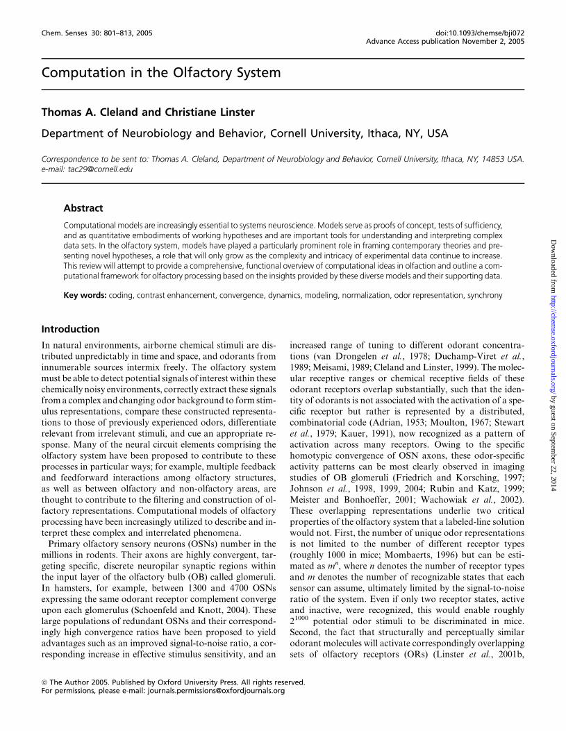
Computation in the Olfactory System
Thomas A. Cleland and Christiane Linster
Department of Neurobiology and Behavior, Cornell University, Ithaca, NY, USA
Correspondence to be sent to: Thomas A. Cleland, Department of Neurobiology and Behavior, Cornell University, Ithaca, NY, 14853 USA.e-mail: [email protected]
Abstract
Computational models are increasingly essential to systems neuroscience. Models serve as proofs of concept, tests of sufficiency,and as quantitative embodiments of working hypotheses and are important tools for understanding and interpreting complexdata sets. In the olfactory system, models have played a particularly prominent role in framing contemporary theories and pre-senting novel hypotheses, a role that will only grow as the complexity and intricacy of experimental data continue to increase.This review will attempt to provide a comprehensive, functional overview of computational ideas in olfaction and outline a com-putational framework for olfactory processing based on the insights provided by these diverse models and their supporting data.
Key words: coding, contrast enhancement, convergence, dynamics, modeling, normalization, odor representation, synchrony
Introduction
In natural environments, airborne chemical stimuli are dis-
tributed unpredictably in time and space, and odorants from
innumerable sources intermix freely. The olfactory system
must be able to detect potential signals of interest within these
chemically noisy environments, correctly extract these signalsfrom a complex and changing odor background to form stim-
ulus representations, compare these constructed representa-
tions to those of previously experienced odors, differentiate
relevant from irrelevant stimuli, and cue an appropriate re-
sponse. Many of the neural circuit elements comprising the
olfactory system have been proposed to contribute to these
processes in particular ways; for example, multiple feedback
and feedforward interactions among olfactory structures,as well as between olfactory and non-olfactory areas, are
thought to contribute to the filtering and construction of ol-
factory representations. Computational models of olfactory
processing have been increasingly utilized to describe and in-
terpret these complex and interrelated phenomena.
Primary olfactory sensory neurons (OSNs) number in the
millions in rodents. Their axons are highly convergent, tar-
geting specific, discrete neuropilar synaptic regions withinthe input layer of the olfactory bulb (OB) called glomeruli.
In hamsters, for example, between 1300 and 4700 OSNs
expressing the same odorant receptor complement converge
upon each glomerulus (Schoenfeld and Knott, 2004). These
large populations of redundant OSNs and their correspond-
ingly high convergence ratios have been proposed to yield
advantages such as an improved signal-to-noise ratio, a cor-
responding increase in effective stimulus sensitivity, and an
increased range of tuning to different odorant concentra-
tions (van Drongelen et al., 1978; Duchamp-Viret et al.,
1989;Meisami, 1989; Cleland and Linster, 1999). The molec-
ular receptive ranges or chemical receptive fields of these
odorant receptors overlap substantially, such that the iden-tity of odorants is not associated with the activation of a spe-
cific receptor but rather is represented by a distributed,
combinatorial code (Adrian, 1953; Moulton, 1967; Stewart
et al., 1979; Kauer, 1991), now recognized as a pattern of
activation across many receptors. Owing to the specific
homotypic convergence of OSN axons, these odor-specific
activity patterns can be most clearly observed in imaging
studies of OB glomeruli (Friedrich and Korsching, 1997;Johnson et al., 1998, 1999, 2004; Rubin and Katz, 1999;
Meister and Bonhoeffer, 2001; Wachowiak et al., 2002).
These overlapping representations underlie two critical
properties of the olfactory system that a labeled-line solution
would not. First, the number of unique odor representations
is not limited to the number of different receptor types
(roughly 1000 in mice; Mombaerts, 1996) but can be esti-
mated as mn, where n denotes the number of receptor typesand m denotes the number of recognizable states that each
sensor can assume, ultimately limited by the signal-to-noise
ratio of the system. Even if only two receptor states, active
and inactive, were recognized, this would enable roughly
21000 potential odor stimuli to be discriminated in mice.
Second, the fact that structurally and perceptually similar
odorant molecules will activate correspondingly overlapping
sets of olfactory receptors (ORs) (Linster et al., 2001b,
Chem. Senses 30: 801–813, 2005 doi:10.1093/chemse/bji072Advance Access publication November 2, 2005
ª The Author 2005. Published by Oxford University Press. All rights reserved.For permissions, please e-mail: [email protected]
by guest on September 22, 2014
http://chemse.oxfordjournals.org/
Dow
nloaded from
2002; Cleland et al., 2002) establishes a basis for the recog-
nition of stimulus similarity in the olfactory system. This is
a prerequisite for basic postsensory cognitive processes such
as generalization (Shepard and Chang, 1963; Shepard, 1987;
Cleland et al., 2002) and a tolerance for variance among re-peated stimulus samples that a labeled-line system would
have no clear means of generating.
Distributed patterns of activity in response to chemical
stimuli are transmitted to the OB via OSN axons that termi-
nate in the glomeruli of its input layer. The OB is believed to
filter and transform these incoming sensory data, performing
normalization, contrast enhancement, and similar opera-
tions before conveying the processed olfactory informationto several different secondary olfactory structures via mitral
cell axon collaterals (Cleland and Linster, 2003). Notably,
the bulb constitutes the last common stage at which olfactory
sensory representations can be processed before the signal
diverges dramatically into these multiple secondary struc-
tures. It is clear from recent investigations that the perceptual
qualities of odorants can be predicted, to a limited degree,
from the patterns of activation that they evoke at the OB in-put layer (Linster and Hasselmo, 1999; Linster et al., 2001b,
2002; Cleland et al., 2002). However, several aspects of odor
perception, for example, changes in perception and discrim-
ination capacity due to odor intensity or prior experience,
cannot be predicted solely by this first-order representation
as reflected in glomerular activation patterns. Nor is the con-
verse true; mitral cell responses in behaving animals cannot
be predicted solely by the odor(s) presented but depend sub-stantially on odor contingency (Kay and Laurent, 1999),
as previously suggested in field potential recordings from
the OB (Di Prisco and Freeman, 1985; Gray et al., 1986;
Freeman and Grajski, 1987; Grajski and Freeman, 1989).
Furthermore, the OB receives substantial centrifugal projec-
tions from both cortical and neuromodulatory centers, and
its responses to odor presentations are strongly regulated by
these centrifugal inputs as well as odor learning and experi-ence (Kay et al., 1996; Kay, 2003, 2005; Ravel et al., 2003;
Wilson and Stevenson, 2003; Martin et al., 2004; Wilson
et al., 2004). It is therefore safe to assume that the OB plays
an important role in processing incoming sensory informa-
tion. Accordingly, many models of OB signal processing
have been developed, which are grouped here into studies
of (1) filtering and contrast enhancement, (2) mechanisms
underlying oscillations and spike synchronization, and (3)odor segmentation and associative memory function. In ad-
dition, a number of detailed biophysical models of bulbar
neurons have been constructed, in many cases to address
how their intrinsic properties underlie and interact with net-
work properties.
Filtering and contrast enhancement
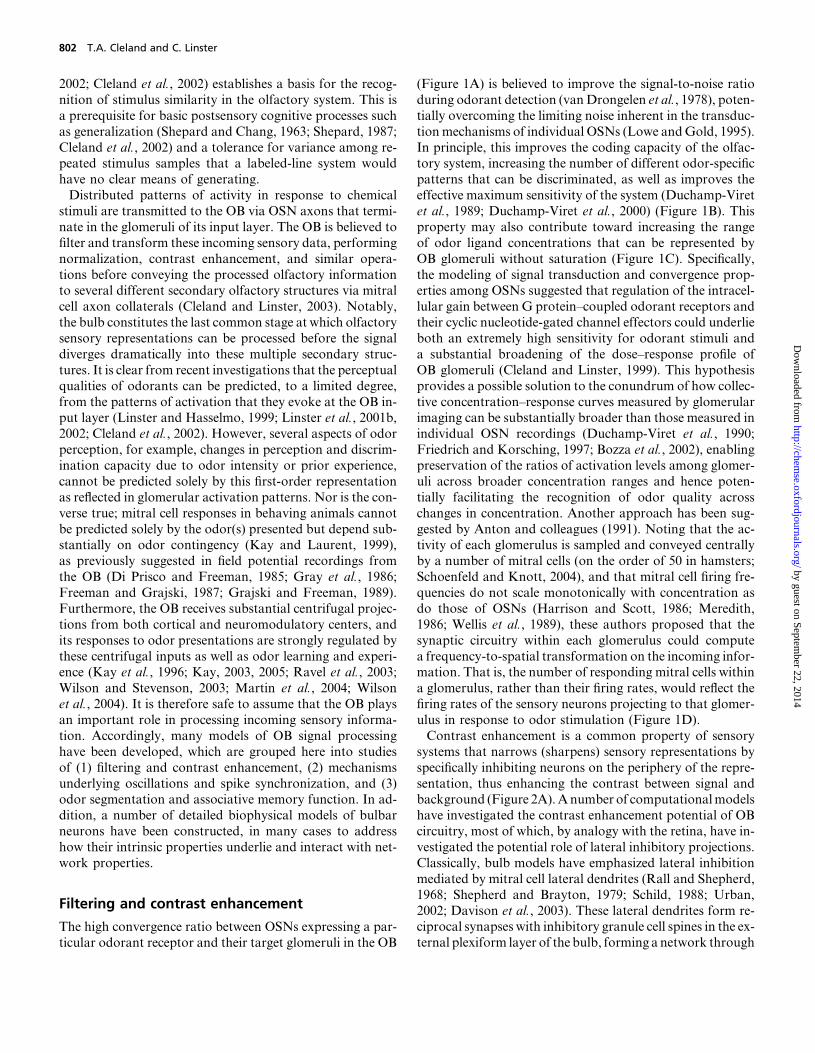
The high convergence ratio between OSNs expressing a par-
ticular odorant receptor and their target glomeruli in the OB
(Figure 1A) is believed to improve the signal-to-noise ratio
during odorant detection (vanDrongelen et al., 1978), poten-
tially overcoming the limiting noise inherent in the transduc-
tionmechanisms of individual OSNs (Lowe andGold, 1995).
In principle, this improves the coding capacity of the olfac-tory system, increasing the number of different odor-specific
patterns that can be discriminated, as well as improves the
effective maximum sensitivity of the system (Duchamp-Viret
et al., 1989; Duchamp-Viret et al., 2000) (Figure 1B). This
property may also contribute toward increasing the range
of odor ligand concentrations that can be represented by
OB glomeruli without saturation (Figure 1C). Specifically,
the modeling of signal transduction and convergence prop-erties among OSNs suggested that regulation of the intracel-
lular gain between G protein–coupled odorant receptors and
their cyclic nucleotide-gated channel effectors could underlie
both an extremely high sensitivity for odorant stimuli and
a substantial broadening of the dose–response profile of
OB glomeruli (Cleland and Linster, 1999). This hypothesis
provides a possible solution to the conundrum of how collec-
tive concentration–response curves measured by glomerularimaging can be substantially broader than those measured in
individual OSN recordings (Duchamp-Viret et al., 1990;
Friedrich and Korsching, 1997; Bozza et al., 2002), enabling
preservation of the ratios of activation levels among glomer-
uli across broader concentration ranges and hence poten-
tially facilitating the recognition of odor quality across
changes in concentration. Another approach has been sug-
gested by Anton and colleagues (1991). Noting that the ac-tivity of each glomerulus is sampled and conveyed centrally
by a number of mitral cells (on the order of 50 in hamsters;
Schoenfeld and Knott, 2004), and that mitral cell firing fre-
quencies do not scale monotonically with concentration as
do those of OSNs (Harrison and Scott, 1986; Meredith,
1986; Wellis et al., 1989), these authors proposed that the
synaptic circuitry within each glomerulus could compute
a frequency-to-spatial transformation on the incoming infor-mation. That is, the number of responding mitral cells within
a glomerulus, rather than their firing rates, would reflect the
firing rates of the sensory neurons projecting to that glomer-
ulus in response to odor stimulation (Figure 1D).
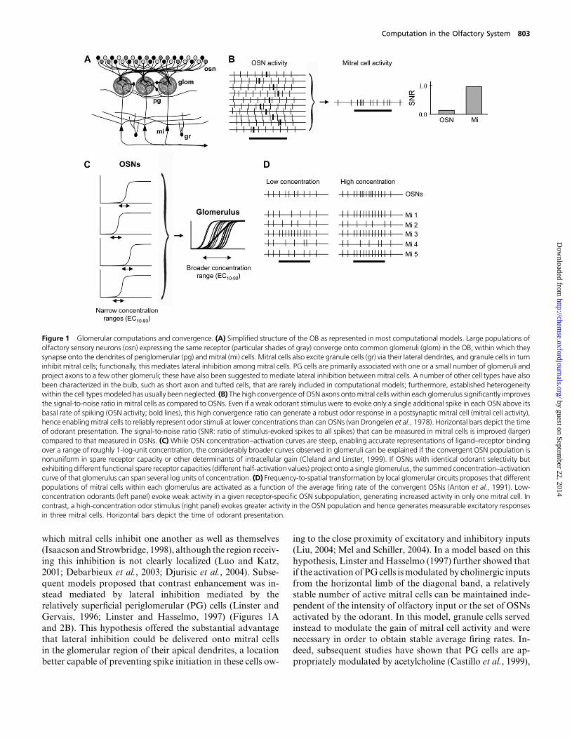
Contrast enhancement is a common property of sensory
systems that narrows (sharpens) sensory representations by
specifically inhibiting neurons on the periphery of the repre-
sentation, thus enhancing the contrast between signal andbackground (Figure 2A).Anumber of computationalmodels
have investigated the contrast enhancement potential of OB
circuitry, most of which, by analogy with the retina, have in-
vestigated the potential role of lateral inhibitory projections.
Classically, bulb models have emphasized lateral inhibition
mediated by mitral cell lateral dendrites (Rall and Shepherd,
1968; Shepherd and Brayton, 1979; Schild, 1988; Urban,
2002; Davison et al., 2003). These lateral dendrites form re-ciprocal synapses with inhibitory granule cell spines in the ex-
ternal plexiform layer of the bulb, forming a network through
802 T.A. Cleland and C. Linster
by guest on September 22, 2014
http://chemse.oxfordjournals.org/
Dow
nloaded from
which mitral cells inhibit one another as well as themselves
(Isaacson andStrowbridge, 1998), although the region receiv-
ing this inhibition is not clearly localized (Luo and Katz,2001; Debarbieux et al., 2003; Djurisic et al., 2004). Subse-
quent models proposed that contrast enhancement was in-
stead mediated by lateral inhibition mediated by the
relatively superficial periglomerular (PG) cells (Linster and
Gervais, 1996; Linster and Hasselmo, 1997) (Figures 1A
and 2B). This hypothesis offered the substantial advantage
that lateral inhibition could be delivered onto mitral cells
in the glomerular region of their apical dendrites, a locationbetter capable of preventing spike initiation in these cells ow-
ing to the close proximity of excitatory and inhibitory inputs
(Liu, 2004; Mel and Schiller, 2004). In a model based on this
hypothesis, Linster andHasselmo (1997) further showed thatif the activation of PGcells ismodulated by cholinergic inputs
from the horizontal limb of the diagonal band, a relatively
stable number of active mitral cells can be maintained inde-
pendent of the intensity of olfactory input or the set of OSNs
activated by the odorant. In this model, granule cells served
instead to modulate the gain of mitral cell activity and were
necessary in order to obtain stable average firing rates. In-
deed, subsequent studies have shown that PG cells are ap-propriately modulated by acetylcholine (Castillo et al., 1999),
Figure 1 Glomerular computations and convergence. (A) Simplified structure of the OB as represented in most computational models. Large populations ofolfactory sensory neurons (osn) expressing the same receptor (particular shades of gray) converge onto common glomeruli (glom) in the OB, within which theysynapse onto the dendrites of periglomerular (pg) andmitral (mi) cells. Mitral cells also excite granule cells (gr) via their lateral dendrites, and granule cells in turninhibit mitral cells; functionally, this mediates lateral inhibition among mitral cells. PG cells are primarily associated with one or a small number of glomeruli andproject axons to a few other glomeruli; these have also been suggested to mediate lateral inhibition betweenmitral cells. A number of other cell types have alsobeen characterized in the bulb, such as short axon and tufted cells, that are rarely included in computational models; furthermore, established heterogeneitywithin the cell typesmodeled has usually been neglected. (B) The high convergence of OSN axons ontomitral cells within each glomerulus significantly improvesthe signal-to-noise ratio in mitral cells as compared to OSNs. Even if a weak odorant stimulus were to evoke only a single additional spike in each OSN above itsbasal rate of spiking (OSN activity; bold lines), this high convergence ratio can generate a robust odor response in a postsynaptic mitral cell (mitral cell activity),hence enabling mitral cells to reliably represent odor stimuli at lower concentrations than can OSNs (van Drongelen et al., 1978). Horizontal bars depict the timeof odorant presentation. The signal-to-noise ratio (SNR: ratio of stimulus-evoked spikes to all spikes) that can be measured in mitral cells is improved (larger)compared to that measured in OSNs. (C) While OSN concentration–activation curves are steep, enabling accurate representations of ligand–receptor bindingover a range of roughly 1-log-unit concentration, the considerably broader curves observed in glomeruli can be explained if the convergent OSN population isnonuniform in spare receptor capacity or other determinants of intracellular gain (Cleland and Linster, 1999). If OSNs with identical odorant selectivity butexhibiting different functional spare receptor capacities (different half-activation values) project onto a single glomerulus, the summed concentration–activationcurve of that glomerulus can span several log units of concentration. (D) Frequency-to-spatial transformation by local glomerular circuits proposes that differentpopulations of mitral cells within each glomerulus are activated as a function of the average firing rate of the convergent OSNs (Anton et al., 1991). Low-concentration odorants (left panel) evoke weak activity in a given receptor-specific OSN subpopulation, generating increased activity in only one mitral cell. Incontrast, a high-concentration odor stimulus (right panel) evokes greater activity in the OSN population and hence generates measurable excitatory responsesin three mitral cells. Horizontal bars depict the time of odorant presentation.
Computation in the Olfactory System 803
by guest on September 22, 2014
http://chemse.oxfordjournals.org/
Dow
nloaded from
and some of the behavioral predictions from these models
have been confirmed in rats (Linster et al., 2001a; Linster
and Cleland, 2002).Contrast enhancement, the effects of which have been di-
rectly observed in the OB (Yokoi et al., 1995), can be func-
tionally defined as a process of competition between
neurons proportional to the similarity of the information that
they mediate. Simplified models of the olfactory system,
based on one-dimensional odor subspaces, have been able
to implement contrast enhancement using lateral inhibition
(Linster and Gervais, 1996; Linster and Hasselmo, 1997,1999; Linster and Smith, 1997; Linster and Cleland, 2001;
Cleland and Linster, 2002) as well as spike synchrony
(Cleland and Linster, 2002) and have been effective at inter-
preting behavioral and physiological data derived from single
monotonically varying odorant series (Yokoi et al., 1995;
Linster and Hasselmo, 1999; Cleland et al., 2002; Cleland
and Narla, 2003). However, to escape this limitation andmodelmore realistic, high-dimensional odor spaces (Hudson,
1999; Korsching, 2001; Alkasab et al., 2002), subsequent
models have relied upon networks constructed so that the
strength of PG-mediated inhibition is effectively propor-
tional to receptive field similarity rather than the physical
proximity of glomeruli. One network model based on this as-
sumption has been shown to best reproduce calcium imaging
data obtained from honeybee OSNs and projection neurons(analogous to mitral cells), while networks based on nearest-
neighbor lateral inhibition performed comparably to net-
works based on random inhibitory projections (Linster
et al., 2005). Another suchmodel has successfully reproduced
Figure 2 Contrast enhancement. (A) Contrast enhancement is a phenomenon observed in most sensory systems by which marginally activated neurons areexcluded from a stimulus-specific ensemble by inhibition, hence sharpening the sensory representation and differentiating it from other, similar representations.In the absence of contrast enhancement, the tuning curves of cells 1 and 2 substantially overlap (left panel), and the stimulus represented by the vertical lineevokes activity in both cells. If contrast enhancement is enabled, for example, by the addition of lateral inhibition such that each cell inhibits the other, the twocells both becomemore narrowly tuned, and their receptive fieldsmay no longer overlap. The stimulus represented by the vertical line now evokes activity only incell 2, while cell 1 is inhibited (right panel). (B) Computational models of the OB have proposed that inhibitory PG cells could mediate contrast enhancement inthe glomerular layer of the OB. In these models, PG cells receive direct sensory input within a given glomerulus and inhibit mitral cells in neighboring glomerulivia axonal projections. If an odor stimulus activates a range of neighboring glomeruli, increasing the level of PG cell–mediated inhibition would lead to con-comitantly sharpened representations among mitral cells. Left panel: in the absence of PG cell–mediated inhibition, mitral cell activity directly reflects OSNactivity. Right panel: the addition of inhibitory projections mediated by PG activity inhibits the more weakly activated mitral cell out of the active ensemble. Withstrong contrast enhancement, only the most strongly activatedmitral cells become activated by odor inputs, while weakly activated mitral cells are inhibited. (C)Nontopographical contrast enhancement based on local glomerular computations (Cleland and Sethupathy, 2004). In this model, contrast enhancement isgenerated within the stimulus-response profile of each individual glomerulus, resulting in sharpening of the odor-evoked representation across the glomerularlayer. Within each glomerulus, mitral (Miin) and periglomerular (PGin) cells have the same tuning curves but different response properties and both receiveparallel input from OSNs. Mitral cell output (Miout) is additionally shaped by PG-mediated dendrodendritic inhibition, such that only the most strongly excitedmitral cells are activated (lower right panel), whilemoreweakly excitedmitral cells exhibit net inhibitory responses (lower left panel). The shape of theMiout curvegenerates the familiar on-center/inhibitory-surround function of contrast enhancement (inset).
804 T.A. Cleland and C. Linster
by guest on September 22, 2014
http://chemse.oxfordjournals.org/
Dow
nloaded from
mixture processing properties measured in rats (Wiltrout
et al., 2003; Linster and Cleland, 2004). One set of models
has proposed a means for contrast enhancement to be
effected by spike synchronization, independent of the under-
lying firing rates (Linster and Cleland, 2001; Cleland andLinster, 2002). Finally, nontopographical models of contrast
enhancement are also capable of distributing inhibition in
proportion to receptive field similarity, but they approach
the problem differently, relying on intraglomerular computa-
tions and broad feedback inhibition to effect contrast en-
hancement via a ‘‘winner-take-most’’ algorithm in which
the most active neurons inhibit those that are less active
(Figure 2C; Cleland and Sethupathy, 2004). Furthermore,unlike mechanisms based upon lateral projections, nontopo-
graphical contrast enhancement does not require a built-in
foreknowledge of the similarities in molecular receptive
ranges expressed by different OB glomeruli in order to dis-
tribute inhibition correctly and is entirely independent of
the physical location of glomeruli within the OB.
Mechanisms underlying oscillations and spikesynchronization
While recent models have begun to favor glomerular-layer
mechanisms for contrast enhancement, granule cell activity
also clearly shapesmitral cell response patterns and hence the
presumptive odor representations that emerge from the OB.
Specifically, several computational models of the OB havesuggested that the temporal pattern of spiking among mitral
cells may play a role in odor representation (Schild, 1988;
Meredith, 1992; White et al., 1992, 1998; White and Kauer,
2001). While it is clear that temporal response patterns in mi-
tral cells do change as a function of odor identity, there is as
yet no broadly accepted theory of how these response pat-
terns may contribute to the representation of odorant stim-
uli. Figure 3A illustrates an example of how odor identitycould be represented by temporal patterning in mitral cells.
Each of four odors A–D evokes characteristic temporal spike
patterns in two mitral cells (left panel). If the instantaneous
spike rates of these mitral cells are plotted against each other
during the odor response, each odor evokes a different tra-
jectory representative of the odor (right panel).
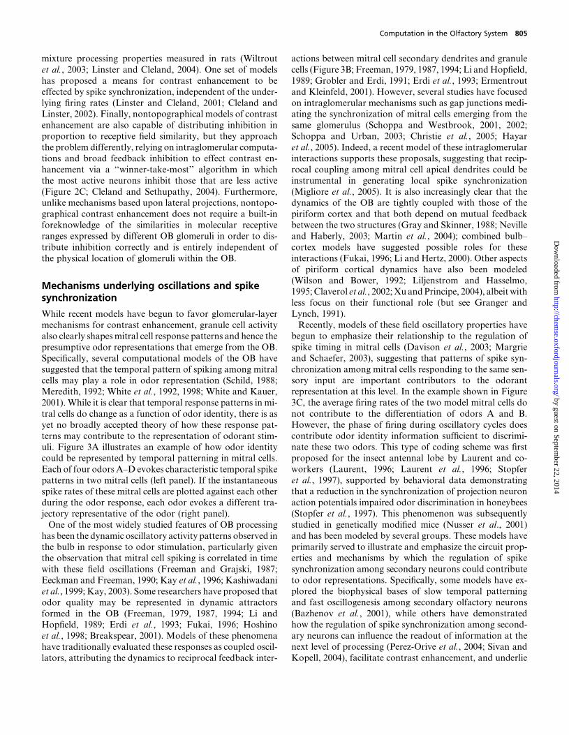
One of the most widely studied features of OB processing
has been the dynamic oscillatory activity patterns observed inthe bulb in response to odor stimulation, particularly given
the observation that mitral cell spiking is correlated in time
with these field oscillations (Freeman and Grajski, 1987;
Eeckman and Freeman, 1990; Kay et al., 1996; Kashiwadani
et al., 1999; Kay, 2003). Some researchers have proposed that
odor quality may be represented in dynamic attractors
formed in the OB (Freeman, 1979, 1987, 1994; Li and
Hopfield, 1989; Erdi et al., 1993; Fukai, 1996; Hoshinoet al., 1998; Breakspear, 2001). Models of these phenomena
have traditionally evaluated these responses as coupled oscil-
lators, attributing the dynamics to reciprocal feedback inter-
actions between mitral cell secondary dendrites and granule
cells (Figure 3B; Freeman, 1979, 1987, 1994; Li andHopfield,
1989; Grobler and Erdi, 1991; Erdi et al., 1993; Ermentrout
and Kleinfeld, 2001). However, several studies have focused
on intraglomerular mechanisms such as gap junctions medi-ating the synchronization of mitral cells emerging from the
same glomerulus (Schoppa and Westbrook, 2001, 2002;
Schoppa and Urban, 2003; Christie et al., 2005; Hayar
et al., 2005). Indeed, a recent model of these intraglomerular
interactions supports these proposals, suggesting that recip-
rocal coupling among mitral cell apical dendrites could be
instrumental in generating local spike synchronization
(Migliore et al., 2005). It is also increasingly clear that thedynamics of the OB are tightly coupled with those of the
piriform cortex and that both depend on mutual feedback
between the two structures (Gray and Skinner, 1988; Neville
and Haberly, 2003; Martin et al., 2004); combined bulb–
cortex models have suggested possible roles for these
interactions (Fukai, 1996; Li and Hertz, 2000). Other aspects
of piriform cortical dynamics have also been modeled
(Wilson and Bower, 1992; Liljenstrom and Hasselmo,1995;Claverol et al., 2002;Xu andPrincipe, 2004), albeit with
less focus on their functional role (but see Granger and
Lynch, 1991).
Recently, models of these field oscillatory properties have
begun to emphasize their relationship to the regulation of
spike timing in mitral cells (Davison et al., 2003; Margrie
and Schaefer, 2003), suggesting that patterns of spike syn-
chronization among mitral cells responding to the same sen-sory input are important contributors to the odorant
representation at this level. In the example shown in Figure
3C, the average firing rates of the two model mitral cells do
not contribute to the differentiation of odors A and B.
However, the phase of firing during oscillatory cycles does
contribute odor identity information sufficient to discrimi-
nate these two odors. This type of coding scheme was first
proposed for the insect antennal lobe by Laurent and co-workers (Laurent, 1996; Laurent et al., 1996; Stopfer
et al., 1997), supported by behavioral data demonstrating
that a reduction in the synchronization of projection neuron
action potentials impaired odor discrimination in honeybees
(Stopfer et al., 1997). This phenomenon was subsequently
studied in genetically modified mice (Nusser et al., 2001)
and has been modeled by several groups. These models have
primarily served to illustrate and emphasize the circuit prop-erties and mechanisms by which the regulation of spike
synchronization among secondary neurons could contribute
to odor representations. Specifically, some models have ex-
plored the biophysical bases of slow temporal patterning
and fast oscillogenesis among secondary olfactory neurons
(Bazhenov et al., 2001), while others have demonstrated
how the regulation of spike synchronization among second-
ary neurons can influence the readout of information at thenext level of processing (Perez-Orive et al., 2004; Sivan and
Kopell, 2004), facilitate contrast enhancement, and underlie
Computation in the Olfactory System 805
by guest on September 22, 2014
http://chemse.oxfordjournals.org/
Dow
nloaded from

the salience of olfactory stimuli (Linster and Cleland, 2001;
Cleland and Linster, 2002).
Odor segmentation and associative memoryfunction
Odor segmentation is the general term for the problem
of how the olfactory system is able to segregate and iden-
tify different odorants that are encountered simultaneously.
As most odors comprise multiple separate odorant mole-
cules, it is far from clear how the olfactory system can
parse the multitude of odorant stimuli present at any given
time and attribute each to appropriately separate sources.One approach has been to hypothesize that odors emitted
from different sources can be segregated by OB circuitry
based upon their differential fluctuations in time (Fort
and Rospars, 1992; Hendin et al., 1998; Hopfield, 1999).
Odor segmentation could thereafter be performed in the
OB using source-separation algorithms dependent upon
associative memory function. Generally, such models hy-
pothesize that associative memories for patterns of OB
activity evoked by known odorants become embedded in
bulbar circuitry and can then be used to recognize these
patterns when they recur, even in degraded form. Specifi-cally, a model by Hendin et al. (1998) illustrates how, if
the glomerular layer feeds into a mitral-granule cell layer
for which appropriate dynamics for an associative memory
function have been implemented, each odor can be sepa-
rately represented in successive inhalation cycles when
multiple (known) odors are presented at the same time.
Olfactory associative memory functions have been more
commonly attributed to the piriform cortex, one of the
Figure 3 Oscillations and synchrony. (A) Any given mitral cell in the OB may respond to different odorant stimuli (A, B, C, and D) with a variety of temporallycomplex spike patterns including interwoven excitatory and inhibitory phases. It has been proposed that these temporal patterns may contribute to odorrepresentations in the vertebrate OB and the analogous insect antennal lobe (Laurent, 1999; Laurent et al., 2001). If the instantaneous firing rates oftwo cells are depicted as a function of each other, a distinct trajectory (in time) can be plotted for each odorant stimulation. For clarity, three discrete epochsare depicted rather than a continuous function; the three time windows depicted during odorant presentation (left panels; horizontal bar) correspond to thethree vectors comprising each trajectory in the graphs (right panels). In contrast, if average firing rates over the application of the stimulus were plotted, theresponses to the three odorants could not be differentiated (not shown). (B) The reciprocal synaptic interactions between mitral (Mi) and granule (Gr) cells haveoften been simulated as a system of coupled oscillators driven by external inputs. In such models, the variance among the stimulus amplitudes across theseinputs generates a map of field oscillations with variable amplitudes and fixed phase lags across the OB. For clarity, bidirectional connections are depicted fromonly a single column. Oscillation amplitudes across the OB are representative of the odor stimulus (compare the patterns evoked by the two odors). (C) Fieldoscillatory dynamics are believed to reflect and/or influence spike timing in mitral cells, potentially resulting in odor-specific populations of mitral cells based onspike synchrony rather than overall activity. While the overall activity patterns evoked by odors A and B are very similar, selection for spikes relatively synchronizedwith one another andwith the oscillatory field potential reveals two clearly odor-specific subpopulations. Consequently, when the firing rates of the two cells areplotted against one another, the representations of the two odors are nearly indistinguishable (middle panel). In contrast, when the relative phases of the twocells are plotted, clearly different representations of the two odors can be observed (right panel).
806 T.A. Cleland and C. Linster
by guest on September 22, 2014
http://chemse.oxfordjournals.org/
Dow
nloaded from
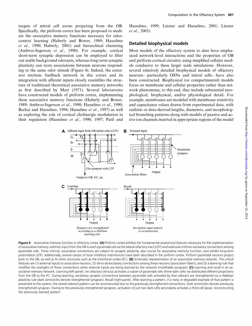
targets of mitral cell axons projecting from the OB.
Specifically, the piriform cortex has been proposed to medi-
ate the associative memory functions necessary for odor-
context learning (Haberly and Bower, 1989; Hasselmo
et al., 1990; Haberly, 2001) and hierarchical clustering(Ambros-Ingerson et al., 1990). For example, cortical
short-term synaptic depression can be employed to filter
out stable background odorants, whereas long-term synaptic
plasticity can store associations between neurons respond-
ing to the same odor stimuli (Figure 4). Indeed, the exten-
sive intrinsic feedback network in this cortex and its
integration with afferent inputs closely resembles the struc-
ture of traditional theoretical associative memory networksas first described by Marr (1971). Several laboratories
have constructed models of piriform cortex, implementing
these associative memory functions (Haberly and Bower,
1989; Ambros-Ingerson et al., 1990; Hasselmo et al., 1990;
Barkai and Hasselmo, 1994; Hasselmo et al., 1997) as well
as exploring the role of cortical cholinergic modulation in
their regulation (Hasselmo et al., 1990, 1997; Patil and
Hasselmo, 1999; Linster and Hasselmo, 2001; Linster
et al., 2003).
Detailed biophysical models
Most models of the olfactory system to date have empha-
sized network-level interactions and the properties of OB
and piriform cortical circuitry using simplified cellular mod-
els conducive to these larger scale simulations. However,
several relatively detailed biophysical models of olfactory
neurons—particularly OSNs and mitral cells—have also
been constructed. Biophysical (or compartmental) modelsfocus on membrane and cellular properties rather than net-
work phenomena; to this end, they include substantial mor-
phological, biophysical, and/or physiological detail. For
example, membranes are modeled with membrane resistivity
and capacitance values drawn from experimental data, with
realistic or data-derived lengths, diameters, and morpholog-
ical branching patterns along with models of passive and ac-
tive ion channels inserted in appropriate regions of the model
Figure 4 Associative memory function in olfactory cortex. (A) Piriform cortex exhibits the fundamental anatomical features necessary for the implementationof associativememory: extrinsic input from the OB to each pyramidal cell via the lateral olfactory tract (LOT) and extensive intrinsic excitatory connections amongpyramidal cells. These intrinsic associative connections are subject to synaptic plasticity, also crucial for associative memory function, and exhibit long-termpotentiation (LTP). Additionally, several classes of local inhibitory interneurons have been described in the piriform cortex. Piriform pyramidal neurons projectback to the OB, as well as to other structures such as the entorhinal cortex (EC). (B) Schematic representation of an associative memory network. The criticalfeatures are (1) external inputs to associative neurons, (2) all-to-all excitatory connections among these neurons (association fibers), and (3) a learning rule thatmodifies the strengths of these connections when external inputs are being learned by the network (modifiable synapses). (C) Learning and recall in an as-sociative memory network. Learning (left panel). An olfactory stimulus activates a subset of pyramidal cells (three dark cells) via distributed afferent projectionsfrom the OB to the PC. During learning, excitatory synaptic connections between pyramidal cells activated by that odorant are strengthened via a Hebbianplasticity rule (dark semicircles denote strengthened synapses). Recall (right panel). After learning a pattern, if a noisy or degraded example of that pattern ispresented to the system, the stored odorant pattern can be reconstructed due to the previously strengthened connections. Dark semicircles denote previouslystrengthened synapses. Owing to the previously strengthened synapses, activation of just two dark cells secondarily activates a third cell (gray), reconstructingthe previously learned pattern.
Computation in the Olfactory System 807
by guest on September 22, 2014
http://chemse.oxfordjournals.org/
Dow
nloaded from
neuron. Biophysical models usually can be directly inter-
related with electrophysiological data. However, they are
often poor choices for large-scale models, due to the large
numbers of weakly defined parameters as well as their sub-
stantial computational costs. Several detailed OSN modelshave illustrated how ligand–receptor binding and nonlinear
transduction processes can underlie the experimentally ob-
served response properties of these cells (Malaka et al.,
1995; Rospars et al., 1996; Vermeulen et al., 1996, 1997;
Lansky and Rospars, 1998; Vermeulen and Rospars, 1998;
Kaissling, 1998, 2001; Cleland and Linster, 1999; Rospars
et al., 2000; Kaissling and Rospars, 2004). Compartmental
models of mitral cells have been used to elucidate intrinsiccellular phenomena, such as the localization of spike initi-
ation in mitral cells (Shen et al., 1999; Chen et al., 2002)
and the effects of intraglomerular gap junctions (Migliore
et al., 2005), as well as synaptic phenomena such as long-
term potentiation at the OSN–mitral synapse (Ennis et al.,
1998). Other compartmental models have focused on bridg-
ing the gap between cellular and systems properties in both
vertebrate and insect systems (Bhalla and Bower, 1993;Davison et al., 2000, 2003; Bazhenov et al., 2001; Cleland
and Sethupathy, 2004). These detailed models make a clear
case that the morphological and biophysical properties of
OB neurons underlie and define their computational capa-
bilities. While many emergent network properties are best
studied with simple cellular models, biophysical models
have revealed computational mechanisms that are beyond
the capacity of these simpler models to elucidate. Ulti-mately, reconciliation of these detailed models with large-
scale functional models will be necessary for progress in the
understanding of olfactory processing.
Synthesis
Computational models of the olfactory system have contrib-uted immensely to the framing of experimental problems and
the construction of complex hypotheses regarding its func-
tion. Here, we briefly outline a working hypothesis of olfac-
tory system function, integrating the insights derived from
the models reviewed above and constrained by their support-
ing data sets.
Inhaled odorants bind to specific populations of ORs
expressed on the apical surface of primary OSNs, whichare distributed across the nasal epithelium. ORs are broadly
tuned for odorant ligands, such that even simple monomo-
lecular odorants activate a number of receptor types to dif-
fering degrees, producing combinatorial patterns of
activation among OR classes that reflect odor quality. While
the pattern of OSN activation depends primarily upon the
ligand–receptor affinities of multiple OR populations for
the various molecular moieties (odotopes) of odors’ compo-nent molecules, it also is likely influenced by other factors
such as the fluid dynamics of inhalation, the net molecular
sorptiveness of odorant molecules, and the behavioral
regulation of odor sampling (reviewed in Schoenfeld and
Cleland, 2005). Other physical factors that affect the pattern
of OSN activation include the concentration of odorants as
well as interference caused by overlaps among the represen-
tations of multiple odotopes that are simultaneously pre-sented to the olfactory epithelium. Different odotopes
may compete for receptors for which they have not only dif-
ferent affinities but also different efficacies (Duchamp-Viret
et al., 2003; Araneda et al., 2004; Oka et al., 2004; Sanz et al.,
2005), such that a reduction in the activation of an OR gene–
specific OSN population may as well connote an increased
concentration of a relatively antagonistic odotope as a re-
duced concentration of an agonist odotope. In short, the pat-tern of OSN activation in any natural scene context is likely
to be an unknown composite of multiple, overlapping, and
degraded primary odorant representations. It is from this un-
promising rawmaterial that the olfactory systemmust detect
and identify relevant stimuli.
OSNs expressing the same OR and hence sharing the same
molecular receptive range project their axons to specific glo-
meruli within the OB input layer (Mombaerts et al., 1996).Hence, the primary olfactory representation can be conve-
niently measured by imaging glomeruli: that is, the axonal
arborizations of convergent OSNs in the OB glomerular
layer. Some glomerular imaging techniques are explicitly
presynaptic (Friedrich and Korsching, 1997; Wachowiak
et al., 2002), even those that are not are likely to reflect pre-
dominantly presynaptic activity due to the disproportionate
number of OSNs arborizing within each glomerulus com-pared with the number of bulbar neurons arborizing therein
(Shepherd and Greer, 1998; Schoenfeld and Knott, 2004);
glomeruli, of course, do not contain cell bodies. Under sim-
ple experimental conditions, these glomerular response pro-
files are predictive of odor quality, as measured behaviorally
(Johnson and Leon, 2000; Linster et al., 2001b; Cleland et al.,
2002; Leon and Johnson, 2003). However, this concordance
is not robust even to changes in odorant concentrations andcertainly cannot be expected to persist in the context of
a complex olfactory natural scene.
Mitral cells, along withmiddle and deep tufted cells, are the
principal output neurons of the OB. They are directly post-
synaptic to OSNs, and as such the pattern of mitral/tufted
cell activation across the bulb constitutes the secondary ol-
factory representation. In mammals, mitral cells sample
from only a single glomerulus, hence a glomerulus along withits associated mitral cells and interneurons has been referred
to as an ‘‘odor column’’ (Shepherd and Greer, 1998) that
derives its receptive field primarily from a single population
of convergent OSNs. However, the mitral/tufted activation
pattern also depends on the activity of several classes of
bulbar interneurons and hence is substantially transformed
with respect to the primary olfactory representation. For ex-
ample, some form of normalization of stimulus concentra-tions is clearly evident in the concentration–response
profiles of mitral cells. Mitral cell activity patterns are
808 T.A. Cleland and C. Linster
by guest on September 22, 2014
http://chemse.oxfordjournals.org/
Dow
nloaded from
relatively stable across concentrations compared with the
changes in OSN responses (Chalansonnet and Chaput,
1998), and when they are affected by odorant concentration
changes, these changes are often complex and difficult to
predict. Some mitral cells progress from excitation to inhibi-tion with increasing concentrations, while others become
more excited, often exhibiting shorter latencies to first spike;
other cells display yet other profiles (Harrison and Scott,
1986; Meredith, 1986; Wellis et al., 1989; Chalansonnet
and Chaput, 1998). What is clear, however, is that mitral
cells do not monotonically increase their activity in response
to increased stimulus intensities, nor, consequently, is there
likely to be a substantial broadening of mitral cell activityacross the bulb owing to the recruitment of lower affinity
receptors. These are two essential ways in which the structure
of the secondary (mitral) olfactory representation differs
from that of the primary representation as measured among
OSNs, and this transformation provides essential constraints
to models of bulbar mechanisms.
Normalization, the process by which sets of values are
rescaled to a common or tractable range, requires a negativefeedback loop that is effectively global in scope; that is, the
strengthof feedback inhibition shouldbe scaled to theaverage
activity across the bulb rather than to local activation levels
if the profiles of relative activation among odor columns are
to be preserved. Contrast enhancement, in turn, requires
the delivery of inhibition onto odor columns proportional
to the activation of columns exhibiting similar receptive fields
(molecular receptive ranges). In the OB, the synaptic triadconnecting OSN arbors, PG cell dendritic spines, and mitral
cell dendrites in close proximity (reviewed by Shepherd and
Greer, 1998) coupled with the lateral excitatory network me-
diated by external tufted and short axon cells (Aungst et al.,
2003; Hayar et al., 2004) can effect both normalization and
contrast enhancement between the primary and secondary
olfactory representations (Cleland and Sethupathy, 2004).
Briefly, OSNs activate mitral cells, external tufted cells,and PG cell dendritic spines in parallel, and these PG spines
deliver inhibition onto mitral cell apical dendrites, closely
apposing the excitatory OSN inputs. Nontopographical con-
trast enhancement models (Cleland and Sethupathy, 2004)
predict that PG cell–mediated feedforward inhibition of
the mitral cell will predominate when the odor column is
weakly activated, while direct OSN activation of the mitral
cell will predominate when the odor column is strongly acti-vated (Figure 2C), consistent with recordings from rabbit
mitral cells (Yokoi et al., 1995). Normalization is effected
via the activation of external tufted cells, which proportion-
ately excite a lateral excitatory network of external tufted and
short axon cells, thereby broadly inhibitingmitral cell activity
across the bulb via sign-inverting PG cells.
The net activation of mitral cells is then translated into
trains of action potentials, the precise timing of whichappears to be regulated by coordinated oscillations measur-
able in field recordings across the OB. These bulbar oscilla-
tions are thought to depend on an extensive excitatory–
inhibitory network of mitral cell secondary dendrites and
granule cell interneurons, as well as reciprocal cortical con-
nections that modulate bulbar activity according to behav-
ioral state (Kay, 2003; Ravel et al., 2003; Lagier et al., 2004;Martin et al., 2004). Mitral cell spike synchronization pat-
terns can mediate a second level of feature extraction to
the odor representation, as follower neurons in diverse cen-
tral olfactory structures (Cleland and Linster, 2003) process
incoming mitral cell spikes via synaptic learning rules, the
best known of which rely upon precise spike timing (Song
et al., 2000; Cleland and Linster, 2002). Centrifugal modu-
latory inputs also influence olfactory processing and learningmechanisms within the OB (Sullivan et al., 2000; Linster and
Cleland, 2002; Yuan et al., 2003), emphasizing the active role
of the OB in shaping and transforming odor signals and the
importance of behavioral state.
Conclusions
Computational models have an established and growing role
within systems neuroscience. As our understanding of neural
processing and interactions becomes more sophisticated,
computer models of these systems are increasingly necessary
in order to understand and interpret experimental results. In
the olfactory system in particular, computational modeling
will no doubt be essential to understand the integration of
the many factors influencing the construction and transfor-mation of odor representations.
References
Adrian, E.D. (1953) Sensory messages and sensation; the response of theolfactory organ to different smells. Acta Physiol. Scand., 29, 5–14.
Alkasab, T.K., White, J. and Kauer, J.S. (2002) A computational systemfor simulating and analyzing arrays of biological and artificial chemicalsensors. Chem. Senses, 27, 261–275.
Ambros-Ingerson, J., Granger, R. and Lynch, G. (1990) Simulation of pale-ocortex performs hierarchical clustering. Science, 247, 1344–1348.
Anton, P.S., Lynch, G. and Granger, R. (1991) Computation of frequency-to-spatial transform by olfactory bulb glomeruli. Biol. Cybern., 65, 407–414.
Araneda, R.C., Peterlin, Z., Zhang, X., Chesler, A. and Firestein, S. (2004)A pharmacological profile of the aldehyde receptor repertoire in rat olfac-tory epithelium. J. Physiol., 555(Pt 3), 743–756.
Aungst, J.L., Heyward, P.M., Puche, A.C., Karnup, S.V., Hayar, A.,Szabo, G. and Shipley, M.T. (2003) Centre-surround inhibition amongolfactory bulb glomeruli. Nature, 426, 623–629.
Barkai, E. andHasselmo,M.E. (1994)Modulation of the input/output func-tion of rat piriform cortex pyramidal cells. J. Neurophysiol., 72, 644–658.
Bazhenov, M., Stopfer, M., Rabinovich, M., Abarbanel, H.D.,Sejnowski, T.J. and Laurent, G. (2001) Model of cellular and net-work mechanisms for odor-evoked temporal patterning in the locustantennal lobe. Neuron, 30, 569–581.
Bhalla, U.S. and Bower, J.M. (1993) Exploring parameter space in detailedsingle neuron models: simulations of the mitral and granule cells of theolfactory bulb. J. Neurophysiol., 69, 1948–1965.
Computation in the Olfactory System 809
by guest on September 22, 2014
http://chemse.oxfordjournals.org/
Dow
nloaded from
Bozza, T., Feinstein, P., Zheng, C. and Mombaerts, P. (2002) Odorant re-ceptor expression defines functional units in the mouse olfactory system.J. Neurosci., 22, 3033–3043.
Breakspear, M. (2001) Perception of odors by a nonlinear model of theolfactory bulb. Int. J. Neural. Syst., 11, 101–124.
Castillo, P.E., Carleton, A., Vincent, J.D. and Lledo, P.M. (1999) Multipleand opposing roles of cholinergic transmission in the main olfactory bulb.J. Neurosci., 19, 9180–9191.
Chalansonnet, M. and Chaput,M.A. (1998)Olfactory bulb output cell tem-poral response patterns to increasing odor concentrations in freely breath-ing rats. Chem. Senses, 23, 1–9.
Chen, W.R., Shen, G.Y., Shepherd, G.M., Hines, M.L. and Midtgaard, J.
(2002) Multiple modes of action potential initiation and propagation inmitral cell primary dendrite. J. Neurophysiol., 88, 2755–2764.
Christie, J.M., Bark, C., Hormuzdi, S.G., Helbig, I., Monyer, H. and
Westbrook, G.L. (2005) Connexin36 mediates spike synchrony inolfactory bulb glomeruli. Neuron, 46, 761–772.
Claverol, E.T., Brown, A.D. and Chad, J.E. (2002) A large-scale simulationof the piriform cortex by a cell automaton-based network model. IEEETrans. Biomed. Eng., 49, 921–935.
Cleland, T.A. and Linster, C. (1999) Concentration tuning mediated byspare receptor capacity in olfactory sensory neurons: a theoretical study.Neural. Comput., 11, 1673–1690.
Cleland, T.A. and Linster, C. (2002) How synchronization properties amongsecond-order sensory neurons can mediate stimulus salience. Behav.
Neurosci., 116, 212–221.
Cleland, T.A. and Linster, C. (2003) Central olfactory processing. In Doty,
R.L. (ed.), Handbook of Olfaction and Gustation, 2nd ed. Marcel Dekker,
New York, pp. 165–180.
Cleland, T.A., Morse, A., Yue, E.L. and Linster, C. (2002) Behavioralmodels of odor similarity. Behav. Neurosci., 116, 222–231.
Cleland, T.A. and Narla, V.A. (2003) Intensity modulation of olfactoryacuity. Behav. Neurosci., 117, 1434–1440.
Cleland, T.A. and Sethupathy, P. (2004) Non-topographical contrastenhancement disambiguates high-dimensional odor representations.Soc. Neurosci. Abstr., 531.2.
Davison, A.P., Feng, J. and Brown, D. (2000) A reduced compartmentalmodel of the mitral cell for use in network models of the olfactory bulb.Brain Res. Bull., 51, 393–399.
Davison, A.P., Feng, J. and Brown, D. (2003) Dendrodendritic inhibitionand simulated odor responses in a detailed olfactory bulb network model.J. Neurophysiol., 90, 1921–1935.
Debarbieux, F., Audinat, E. and Charpak, S. (2003) Action potential prop-agation in dendrites of rat mitral cells in vivo. J. Neurosci., 23, 5553–5560.
Di Prisco, G.V. and Freeman, W.J. (1985) Odor-related bulbar EEG spatialpattern analysis during appetitive conditioning in rabbits. Behav. Neuro-sci., 99, 964–978.
Djurisic, M., Antic, S., Chen, W.R. and Zecevic, D. (2004) Voltage imagingfrom dendrites of mitral cells: EPSP attenuation and spike trigger zones.J. Neurosci., 24, 6703–6714.
Duchamp-Viret, P., Duchamp, A. and Chaput, M.A. (2000) Peripheralodor coding in the rat and frog: quality and intensity specification. J. Neu-rosci., 20, 2383–2390.
Duchamp-Viret, P., Duchamp, A. and Chaput, M.A. (2003) Single olfac-tory sensory neurons simultaneously integrate the components of anodour mixture. Eur. J. Neurosci., 18, 2690–2696.
Duchamp-Viret,P.,Duchamp,A.andVigouroux,M. (1989)Amplifyingrole
of convergence inolfactory systemacomparative studyof receptor cell and
second-order neuron sensitivities. J. Neurophysiol., 61, 1085–1094.
Duchamp-Viret, P., Duchamp, A. and Vigouroux, M. (1990) Temporal
aspectsof informationprocessing inthefirst twostagesof thefrogolfactory
system: influence of stimulus intensity. Chem. Senses, 15, 349–365.
Eeckman, F.H. and Freeman, W.J. (1990) Correlations between unit firing
and EEG in the rat olfactory system. Brain. Res., 528, 238–244.
Ennis, M., Linster, C., Aroniadou-Anderjaska, V., Ciombor, K. and
Shipley, M.T. (1998) Glutamate and synaptic plasticity at mammalian
primary olfactory synapses. Ann. N. Y. Acad. Sci., 855, 457–466.
Erdi, P., Grobler, T., Barna, G. and Kaski, K. (1993) Dynamics of the olfac-
tory bulb: bifurcations, learning, and memory. Biol. Cybern., 69, 57–66.
Ermentrout, G.B. and Kleinfeld, D. (2001) Traveling electrical waves in cor-
tex: insights from phase dynamics and speculation on a computational
role. Neuron, 29, 33–44.
Fort, J.C. and Rospars, J.P. (1992)Modelling of the qualitative discrimination
of odours in the first two layers of olfactory system by Jutten and Herault
algorithm. C. R. Acad. Sci. III, 315, 331–336.
Freeman, W.J. (1979) Nonlinear dynamics of paleocortex manifested in the
olfactory EEG. Biol. Cybern., 35, 21–37.
Freeman, W.J. (1987) Simulation of chaotic EEG patterns with a dynamic
model of the olfactory system. Biol. Cybern., 56, 139–150.
Freeman,W.J. (1994) Characterization of state transitions in spatially distrib-
uted, chaotic, nonlinear, dynamical systems in cerebral cortex. Integr.
Physiol. Behav. Sci., 29, 294–306.
Freeman, W.J. and Grajski, K.A. (1987) Relation of olfactory EEG to behav-
ior: factor analysis. Behav. Neurosci., 101, 766–777.
Friedrich, R.W. and Korsching, S.I. (1997) Combinatorial and chemotopic
odorant coding in the zebrafish olfactory bulb visualized by optical imag-
ing. Neuron, 18, 737–752.
Fukai, T. (1996) Bulbocortical interplay in olfactory information processing
via synchronous oscillations. Biol. Cybern., 74, 309–317.
Granger, R. and Lynch, G. (1991) Higher olfactory processes: perceptual
learning and memory. Curr. Opin. Neurobiol., 1, 209–214.
Grajski, K.A. and Freeman, W.J. (1989) Spatial EEG correlates of nonasso-
ciative and associative olfactory learning in rabbits. Behav. Neurosci., 103,
790–804.
Gray, C.M., Freeman, W.J. and Skinner. J.E. (1986) Chemical dependen-
cies of learning in the rabbit olfactory bulb: acquisition of the transient
spatial pattern change depends on norepinephrine. Behav. Neurosci.,
100, 585–596.
Gray, C.M. and Skinner, J.E. (1988) Centrifugal regulation of neuronal ac-
tivity in the olfactory bulb of the waking rabbit as revealed by reversible
cryogenic blockade. Exp. Brain Res., 69, 378–386.
Grobler, T. and Erdi, P. (1991) Dynamic phenomena in the olfactory bulb.
Acta Biochim. Biophys. Hung., 26, 61–65.
Haberly, L.B. (2001) Parallel-distributed processing in olfactory cortex: new
insights from morphological and physiological analysis of neuronal
circuitry. Chem. Senses, 26, 551–576.
Haberly, L.B. and Bower, J.M. (1989) Olfactory cortex: model circuit for
study of associative memory? Trends Neurosci., 12, 258–264.
Harrison, T.A. and Scott, J.W. (1986)Olfactory bulb responses to odor stim-
ulation: analysis of response pattern and intensity relationships. J. Neuro-
physiol., 56, 1571–1589.
810 T.A. Cleland and C. Linster
by guest on September 22, 2014
http://chemse.oxfordjournals.org/
Dow
nloaded from
Hasselmo, M.E., Linster, C., Patil, M., Ma, D. and Cekic, M. (1997) Nor-adrenergic suppression of synaptic transmission may influence corticalsignal-to-noise ratio. J. Neurophysiol., 77, 3326–3339.
Hasselmo, M.E., Wilson, M.A., Anderson, B.P. and Bower, J.M. (1990)
Associative memory function in piriform (olfactory) cortex: computationalmodeling and neuropharmacology. Cold Spring Harb. Symp. Quant. Biol.,
55, 599–610.
Hayar, A., Karnup, S., Ennis, M. and Shipley, M.T. (2004) External tuftedcells: a major excitatory element that coordinates glomerular activity.J. Neurosci., 24, 6676–6685.
Hayar, A., Shipley, M.T. and Ennis, M. (2005) Olfactory bulb external
tufted cells are synchronized by multiple intraglomerular mechanisms.J. Neurosci., 25, 8197–8208.
Hendin, O., Horn, D. and Tsodyks, M.V. (1998) Associative memory andsegmentation in an oscillatory neural model of the olfactory bulb. J. Com-
put. Neurosci., 5, 157–169.
Hopfield, J.J. (1999) Odor space and olfactory processing: collective algo-rithms and neural implementation. Proc. Natl Acad. Sci. USA, 96,
12506–12511.
Hoshino, O., Kashimori, Y. and Kambara, T. (1998) An olfactory recogni-tion model based on spatio-temporal encoding of odor quality in theolfactory bulb. Biol. Cybern., 79, 109–120.
Hudson, R. (1999) From molecule to mind: the role of experience in shapingolfactory function. J. Comp. Physiol. A, 185, 297–304.
Isaacson, J.S. and Strowbridge, B.W. (1998) Olfactory reciprocal synapses:dendritic signaling in the CNS. Neuron, 20, 749–761.
Johnson, B.A., Farahbod, H., Xu, Z., Saber, S. and Leon, M. (2004) Localand global chemotopic organization: general features of the glomerularrepresentations of aliphatic odorants differing in carbon number.J. Comp. Neurol., 480, 234–249.
Johnson, B.A. and Leon, M. (2000)Modular representations of odorants inthe glomerular layer of the rat olfactory bulb and the effects of stimulusconcentration. J. Comp. Neurol., 422, 496–509.
Johnson, B.A., Woo, C.C., Hingco, E.E., Pham, K.L. and Leon, M. (1999)
Multidimensional chemotopic responses to n-aliphatic acid odorants inthe rat olfactory bulb. J. Comp. Neurol., 409, 529–548.
Johnson, B.A., Woo, C.C. and Leon, M. (1998) Spatial coding of odorantfeatures in the glomerular layer of the rat olfactory bulb. J. Comp. Neurol.,
393, 457–471.
Kaissling, K.E. (1998) A quantitative model of odor deactivation based onthe redox shift of the pheromone-binding protein in moth antennae.Ann.N. Y. Acad. Sci., 855, 320–322.
Kaissling, K.E. (2001) Olfactory perireceptor and receptor events in moths:a kinetic model. Chem. Senses, 26, 125–150.
Kaissling, K.E. and Rospars, J.P. (2004) Dose-response relationships in anolfactory flux detector model revisited. Chem. Senses, 29, 529–531.
Kashiwadani,H., Sasaki,Y.F.,Uchida,N.andMori,K. (1999)Synchronizedoscillatory discharges of mitral/tufted cells with different molecular recep-tive ranges in the rabbit olfactory bulb. J. Neurophysiol., 82, 1786–1792.
Kauer, J.S. (1991)Contributionsof topographyandparallelprocessingtoodorcoding in the vertebrate olfactory pathway. Trends Neurosci., 14, 79–85.
Kay, L.M. (2003) Two species of gamma oscillations in the olfactory bulb:dependence on behavioral state and synaptic interactions. J. Integr.
Neurosci., 2, 31–44.
Kay, L.M. (2005) Theta oscillations and sensorimotor performance. Proc. NatlAcad. Sci. USA, 102, 3863–3868.
Kay, L.M., Lancaster, L.R. and Freeman, W.J. (1996) Reafference and
attractors in the olfactory system during odor recognition. Int. J. Neural.
Syst., 7, 489–495.
Kay, L.M. and Laurent, G. (1999)Odor- and context-dependent modulation
of mitral cell activity in behaving rats. Nat. Neurosci., 2, 1003–1009.
Korsching, S.I. (2001) Odor maps in the brain: spatial aspects of odor rep-
resentation in sensory surface and olfactory bulb. Cell. Mol. Life Sci., 58,
520–530.
Lagier, S., Carleton, A. and Lledo, P.M. (2004) Interplay between local
GABAergic interneurons and relay neurons generates gamma oscillations
in the rat olfactory bulb. J. Neurosci., 24, 4382–4392.
Lansky, P. and Rospars, J.P. (1998) Odorant concentration and receptor
potential in olfactory sensory neurons. Biosystems, 48, 131–138.
Laurent, G. (1996) Dynamical representation of odors by oscillating and
evolving neural assemblies. Trends Neurosci., 19, 489–496.
Laurent, G. (1999) A systems perspective on early olfactory coding. Science,
286, 723–728.
Laurent, G., Stopfer, M., Friedrich, R.W., Rabinovich, M.I., Volkovskii,
A. and Abarbanel, H.D. (2001) Odor encoding as an active, dynamical
process: experiments, computation, and theory. Annu. Rev. Neurosci., 24,
263–297.
Laurent, G., Wehr, M. and Davidowitz, H. (1996) Temporal representa-
tions of odors in an olfactory network. J. Neurosci., 16, 3837–3847.
Leon, M. and Johnson, B.A. (2003) Olfactory coding in the mammalian
olfactory bulb. Brain Res. Brain Res. Rev., 42, 23–32.
Li, Z. and Hertz, J. (2000) Odour recognition and segmentation by a model
olfactory bulb and cortex. Network, 11, 83–102.
Li, Z. and Hopfield, J.J. (1989) Modeling the olfactory bulb and its neural
oscillatory processings. Biol. Cybern., 61, 379–392.
Liljenstrom, H. and Hasselmo, M.E. (1995) Cholinergic modulation of
cortical oscillatory dynamics. J. Neurophysiol., 74, 288–297.
Linster, C. and Cleland, T.A. (2001) How spike synchronization among
olfactory neurons can contribute to sensory discrimination. J. Comput.
Neurosci., 10, 187–193.
Linster, C. and Cleland, T.A. (2002) Cholinergic modulation of sensory rep-
resentations in the olfactory bulb. Neural Netw., 15, 709–717.
Linster,C.andCleland,T.A. (2004)Configurationalandelementalodormixture
perception can arise from local inhibition. J. Comput. Neurosci., 16, 39–47.
Linster, C., Garcia, P.A., Hasselmo, M.E. and Baxter, M.G. (2001a) Selec-
tive loss of cholinergic neurons projecting to the olfactory system
increases perceptual generalization between similar, but not dissimilar,
odorants. Behav. Neurosci., 115, 826–833.
Linster, C. and Gervais, R. (1996) Investigation of the role of interneurons
and their modulation by centrifugal fibers in a neural model of the olfac-
tory bulb. J. Comput. Neurosci., 3, 225–246.
Linster, C. and Hasselmo, M. (1997)Modulation of inhibition in a model of
olfactory bulb reduces overlap in the neural representation of olfactory
stimuli. Behav. Brain Res., 84, 117–127.
Linster, C. and Hasselmo, M.E. (1999) Behavioral responses to aliphatic
aldehydes can be predicted from known electrophysiological responses
of mitral cells in the olfactory bulb. Physiol. Behav., 66, 497–502.
Linster, C. andHasselmo,M.E. (2001) Neuromodulation and the functional
dynamics of piriform cortex. Chem. Senses, 26, 585–594.
Computation in the Olfactory System 811
by guest on September 22, 2014
http://chemse.oxfordjournals.org/
Dow
nloaded from
Linster, C., Johnson, B.A., Morse, A., Yue, E. and Leon, M. (2002) Spon-
taneous versus reinforced olfactory discriminations. J. Neurosci., 22,
6842–6845.
Linster, C., Johnson, B.A., Yue, E., Morse, A., Xu, Z., Hingco, E.E., Choi,
Y., Choi, M., Messiha, A. and Leon, M. (2001b) Perceptual correlates of
neural representations evoked by odorant enantiomers. J. Neurosci., 21,
9837–9843.
Linster, C., Maloney, M., Patil, M. and Hasselmo, M.E. (2003) Enhanced
cholinergic suppression of previously strengthened synapses enables the
formation of self-organized representations in olfactory cortex.Neurobiol.
Learn. Mem., 80, 302–314.
Linster, C., Sachse, S. and Galizia, G. (2005) Computational modeling sug-
gests that response properties rather than spatial position determine con-
nectivity between olfactory glomeruli. J. Neurophysiol., 93, 3410–3417.
Linster, C. and Smith, B.H. (1997) A computational model of the response of
honeybeeantennal lobecircuitry toodormixtures:overshadowing,blocking
andunblocking can arise from lateral inhibition.Behav. Brain Res., 87, 1–14.
Liu, G. (2004) Local structural balance and functional interaction of excitatory
and inhibitory synapses in hippocampal dendrites. Nat. Neurosci., 7, 373–
379.
Lowe, G. and Gold, G.H. (1995) Olfactory transduction is intrinsically noisy.
Proc. Natl Acad. Sci. USA, 92, 7864–7868.
Luo, M. and Katz, L.C. (2001) Response correlation maps of neurons in the
mammalian olfactory bulb. Neuron, 32, 1165–1179.
Malaka, R., Ragg, T. and Hammer, M. (1995) Kinetic models of odor trans-
duction implemented as artificial neural networks. Simulations of complex
response properties of honeybee olfactory neurons. Biol. Cybern., 73,
195–207.
Margrie, T.W. and Schaefer, A.T. (2003) Theta oscillation coupled spike
latencies yield computational vigour in a mammalian sensory system. J.
Physiol., 546(Pt 2), 363–374.
Marr, D. (1971) Simple memory: a theory for archicortex. Philos. Trans. R Soc.
Lond B Biol. Sci., 262, 23–81.
Martin, C., Gervais, R., Hugues, E., Messaoudi, B. and Ravel, N. (2004)
Learning modulation of odor-induced oscillatory responses in the rat ol-
factory bulb: a correlate of odor recognition? J. Neurosci., 24, 389–397.
Meisami, E. (1989) A proposed relationship between increases in the num-
ber of olfactory receptor neurons, convergence ratio and sensitivity in the
developing rat. Brain Res. Dev. Brain Res., 46, 9–19.
Meister, M. and Bonhoeffer, T. (2001) Tuning and topography in an odor
map on the rat olfactory bulb. J. Neurosci., 21, 1351–1360.
Mel, B.W. and Schiller, J. (2004) On the fight between excitation and in-
hibition: location is everything. Sci. STKE, 2004, PE44.
Meredith, M. (1986) Patterned response to odor in mammalian olfactory
bulb: the influence of intensity. J. Neurophysiol., 56, 572–597.
Meredith, M. (1992) Neural circuit computation: complex patterns in the
olfactory bulb. Brain Res. Bull., 29, 111–117.
Migliore, M., Hines, M.L. and Shepherd, G.M. (2005) The role of distal
dendritic gap junctions in synchronization of mitral cell axonal output.
J. Comput. Neurosci., 18, 151–161.
Mombaerts, P. (1996) Targeting olfaction.Curr. Opin. Neurobiol., 6, 481–486.
Mombaerts, P., Wang, F., Dulac, C., Chao, S.K., Nemes, A., Mendelsohn,
M., Edmondson, J. and Axel, R. (1996) Visualizing an olfactory sensory
map. Cell, 87, 675–686.
Moulton, D.G. (1967) Spatio-temporal patterning of response in the olfac-tory system. In Hayashi, T. (ed.), Olfaction and Taste II. Pergamon Press,
New York, pp. 109–116.
Neville, K.R. and Haberly, L.B. (2003) Beta and gamma oscillations in theolfactory system of the urethane-anesthetized rat. J. Neurophysiol., 90,3921–3930.
Nusser, Z., Kay, L.M., Laurent, G., Homanics, G.E. and Mody, I. (2001)Disruption of GABA(A) receptors on GABAergic interneurons leads to in-creased oscillatory power in the olfactory bulb network. J. Neurophysiol.,86, 2823–2833.
Oka, Y., Omura, M., Kataoka, H. and Touhara, K. (2004) Olfactory recep-tor antagonism between odorants. EMBO J., 23, 120–126.
Patil, M.M. and Hasselmo, M.E. (1999) Modulation of inhibitory synapticpotentials in the piriform cortex. J. Neurophysiol., 81, 2103–2118.
Perez-Orive, J., Bazhenov, M. and Laurent, G. (2004) Intrinsic and circuitproperties favor coincidence detection for decoding oscillatory input.J. Neurosci., 24, 6037–6047.
Rall, W. and Shepherd, G.M. (1968) Theoretical reconstruction of fieldpotentials and dendrodendritic synaptic interactions in olfactory bulb.J. Neurophysiol., 31, 884–915.
Ravel, N., Chabaud, P., Martin, C., Gaveau, V., Hugues, E., Tallon-
Baudry, C., Bertrand, O. and Gervais, R. (2003) Olfactory learningmodifies the expression of odour-induced oscillatory responses in thegamma (60–90 Hz) and beta (15–40 Hz) bands in the rat olfactory bulb.Eur. J. Neurosci., 17, 350–358.
Rospars, J.P., Lansky, P., Duchamp-Viret, P. and Duchamp, A. (2000)
Spiking frequency versus odorant concentration in olfactory receptor neu-rons. Biosystems, 58, 133–141.
Rospars, J.P., Lansky, P., Tuckwell, H.C. and Vermeulen, A. (1996) Cod-ing of odor intensity in a steady-state deterministic model of an olfactoryreceptor neuron. J. Comput. Neurosci., 3, 51–72.
Rubin, B.D. and Katz, L.C. (1999) Optical imaging of odorant representa-tions in the mammalian olfactory bulb. Neuron, 23, 499–511.
Sanz, G., Schlegel, C., Pernollet, J.C. and Briand, L. (2005) Comparison ofodorant specificity of two human olfactory receptors from different phy-logenetic classes and evidence for antagonism. Chem. Senses, 30, 69–80.
Schild, D. (1988) Principles of odor coding and a neural network for odordiscrimination. Biophys. J., 54, 1011–1011.
Schoenfeld, T.A. and Cleland, T.A. (2005) The anatomical logic of smell.Trends Neurosci., 28, 620–627.
Schoenfeld, T.A. and Knott, T.K. (2004) Evidence for the disproportionatemapping of olfactory airspace onto the main olfactory bulb of the ham-ster. J. Comp. Neurol., 476, 186–201.
Schoppa, N.E. andUrban, N.N. (2003) Dendritic processing within olfactorybulb circuits. Trends Neurosci., 26, 501–506.
Schoppa, N.E. and Westbrook, G.L. (2001) Glomerulus-specific synchroni-zation of mitral cells in the olfactory bulb. Neuron, 31, 639–651.
Schoppa, N.E. andWestbrook, G.L.. (2002) AMPA autoreceptors drive cor-related spiking in olfactory bulb glomeruli. Nat. Neurosci., 5, 1194–1202.
Shen, G.Y., Chen, W.R., Midtgaard, J., Shepherd, G.M. and Hines, M.L.
(1999) Computational analysis of action potential initiation in mitral cellsoma and dendrites based on dual patch recordings. J. Neurophysiol., 82,3006–3020.
Shepherd, G.M. and Brayton, R.K. (1979) Computer simulation of a den-drodendritic synaptic circuit for self- and lateral-inhibition in the olfactorybulb. Brain Res., 175, 377–382.
812 T.A. Cleland and C. Linster
by guest on September 22, 2014
http://chemse.oxfordjournals.org/
Dow
nloaded from
Shepherd, G.M. and Greer, C.A. (1998) Olfactory bulb. In Shepherd, G.M.(ed.), The Synaptic Organization of the Brain, 4th ed. Oxford UniversityPress, New York, pp. 159–204.
Shepard, R.N. (1987) Toward a universal law of generalization for psycho-logical science. Science, 237, 1317–1323.
Shepard, R.N. and Chang, J.J. (1963) Stimulus generalization in the learningof classifications. J. Exp. Psychol., 65, 94–102.
Sivan, E. and Kopell, N. (2004) Mechanism and circuitry for clustering andfine discrimination of odors in insects. Proc. Natl Acad. Sci. USA, 101,17861–17866.
Song, S.,Miller, K.D. andAbbott, L.F. (2000) Competitive Hebbian learningthrough spike-timing-dependent synaptic plasticity. Nat. Neurosci., 3,919–926.
Stewart, W.B., Kauer, J.S. and Shepherd, G.M. (1979) Functional organi-zation of rat olfactory bulb analysed by the 2-deoxyglucose method.J. Comp. Neurol., 185, 715–734.
Stopfer, M., Bhagavan, S., Smith, B.H. and Laurent, G. (1997) Impairedodour discrimination on desynchronization of odour-encoding neuralassemblies. Nature, 390, 70–74.
Sullivan, R.M., Stackenwalt, G., Nasr, F., Lemon, C. and Wilson, D.A.(2000) Association of an odor with activation of olfactory bulb noradren-ergic beta-receptors or locus coeruleus stimulation is sufficient to producelearned approach responses to that odor in neonatal rats. Behav. Neuro-sci., 114, 957–962.
Urban, N.N. (2002) Lateral inhibition in the olfactory bulb and in olfaction.Physiol. Behav., 77, 607–612.
van Drongelen, W., Holley, A. and Doving, K.B. (1978) Convergence inthe olfactory system: quantitative aspects of odour sensitivity. J. Theor.Biol., 71, 39–48.
Vermeulen, A., Lansky, P., Tuckwell, H. and Rospars, J.P. (1997) Codingof odour intensity in a sensory neuron. Biosystems, 40, 203–210.
Vermeulen, A. and Rospars, J.P. (1998) Dendritic integration in olfactorysensory neurons: a steady-state analysis of how the neuron structureand neuron environment influence the coding of odor intensity. J. Com-put. Neurosci., 5, 243–266.
Vermeulen, A., Rospars, J.P., Lansky, P. and Tuckwell, H.C. (1996) Cod-ing of stimulus intensity in an olfactory receptor neuron: role of neuronspatial extent and passive dendritic backpropagation of action potentials.Bull. Math. Biol., 58, 493–512.
Wachowiak, M., Cohen, L.B. and Zochowski, M.R. (2002) Distributed and
concentration-invariant spatial representations of odorants by receptor
neuron input to the turtle olfactory bulb. J. Neurophysiol., 87, 1035–
1045.
Wellis, D.P., Scott, J.W. and Harrison, T.A. (1989) Discrimination among
odorants by single neurons of the rat olfactory bulb. J. Neurophysiol., 61,
1161–1177.
White, J., Dickinson, T.A., Walt, D.R. and Kauer, J.S. (1998) An olfactory
neuronal network for vapor recognition in an artificial nose. Biol. Cybern.,
78, 245–251.
White, J., Hamilton, K.A., Neff, S.R. and Kauer, J.S. (1992) Emergent
properties of odor information coding in a representational model of
the salamander olfactory bulb. J. Neurosci., 12, 1772–1780.
White, J. and Kauer, J.S. (2001) Exploring olfactory population coding using
an artificial olfactory system. Prog. Brain Res., 130, 191–203.
Wilson, D.A., Best, A.R. and Sullivan, R.M. (2004) Plasticity in the olfactory
system: lessons for the neurobiology of memory. Neuroscientist, 10, 513–
524.
Wilson, D.A. and Stevenson, R.J. (2003) Olfactory perceptual learning: the
critical role of memory in odor discrimination. Neurosci. Biobehav. Rev.,
27, 307–328.
Wilson, M. and Bower, J.M. (1992) Cortical oscillations and temporal inter-
actions in a computer simulation of piriform cortex. J. Neurophysiol., 67,
981–995.
Wiltrout, C., Dogra, S. and Linster, C. (2003) Configurational and noncon-
figurational interactions between odorants in binary mixtures. Behav.
Neurosci., 117, 236–245.
Xu, D. and Principe, J.C. (2004) Dynamical analysis of neural oscillators in an
olfactory cortex model. IEEE Trans. Neural Netw., 15, 1053–1062.
Yokoi, M., Mori, K. and Nakanishi, S. (1995) Refinement of odor molecule
tuning by dendrodendritic synaptic inhibition in the olfactory bulb. Proc.
Natl Acad. Sci. USA, 92, 3371–3375.
Yuan, Q., Harley, C.W. and McLean, J.H. (2003) Mitral cell beta1 and
5-HT2A receptor colocalization and cAMP coregulation: a new model
of norepinephrine-induced learning in the olfactory bulb. Learn. Mem.,
10, 5–15.
Accepted October 10, 2005
Computation in the Olfactory System 813
by guest on September 22, 2014
http://chemse.oxfordjournals.org/
Dow
nloaded from
Copyright © 2022 FDOKUMEN