CXCL10 blockade protects mice from cyclophosphamide-induced cystitis
Immunity
Article
Compromised Intestinal Epithelial BarrierInduces Adaptive Immune Compensationthat Protects from ColitisManirath Khounlotham,1 Wooki Kim,1,4 Eric Peatman,1,3 Porfirio Nava,1 Oscar Medina-Contreras,2 Caroline Addis,1
Stefan Koch,1 Benedicte Fournier,1 Asma Nusrat,1 Timothy L. Denning,1,2,* and Charles A. Parkos1,*1Epithelial Pathobiology and Mucosal Inflammation Research Unit, Department of Pathology and Laboratory Medicine2Department of PediatricsEmory University School of Medicine, Atlanta, GA 30322, USA3Present address: Department of Fisheries and Allied Aquacultures, Auburn University, Auburn, AL 36849, USA4Present address: Department of Food Science and Biotechnology, College of Life Sciences, Kyung Hee University, 1732 Deogyeong-daero,
Giheung-gu, Yongin-si, Gyeonggi-do, 446-701, Republic of Korea*Correspondence: [email protected] (T.L.D.), [email protected] (C.A.P.)
http://dx.doi.org/10.1016/j.immuni.2012.06.017
SUMMARY
Mice lacking junctional adhesionmolecule A (JAM-A,encoded by F11r) exhibit enhanced intestinal epi-thelial permeability, bacterial translocation, andelevated colonic lymphocyte numbers, yet do notdevelop colitis. To investigate the contribution ofadaptive immune compensation in response toincreased intestinal epithelial permeability, we exam-ined the susceptibility of F11r�/�Rag1�/� mice toacute colitis. Although negligible contributions ofadaptive immunity in F11r+/+Rag1�/� mice wereobserved, F11r�/�Rag1�/� mice exhibited increasedmicroflora-dependent colitis. Elimination of T cellsubsets and cytokine analyses revealed a protectiverole for TGF-b-producing CD4+ T cells in F11r�/�
mice. Additionally, loss of JAM-A resulted in elevatedmucosal and serum IgA that was dependent uponCD4+ T cells and TGF-b. Absence of IgA in F11r+/+
Igha�/� mice did not affect disease, whereas F11r�/�
Igha�/� mice displayed markedly increased sus-ceptibility to acute injury-induced colitis. Thesedata establish a role for adaptive immune-mediatedprotection from acute colitis under conditions ofintestinal epithelial barrier compromise.
INTRODUCTION
The pathogenesis of many inflammatory conditions of mucosal
surfaces involves the combined dysfunction of the mucosal
barrier and local immune responses to gut lumenal contents.
For example, compromised intestinal barrier function reported
in people with inflammatory bowel disease (IBD) (Mankertz
and Schulzke, 2007; Welcker et al., 2004) has been linked to
alterations in expression of barrier-forming tight junction
molecules (Do�gan et al., 1995; Gassler et al., 2001; Jankowski
et al., 1998; Karayiannakis et al., 1998; Kucharzik et al., 2001).
Im
One transmembrane protein component of tight junctions that
has been linked to regulation of intestinal permeability and IBD
is junctional adhesion molecule A (JAM-A) (Laukoetter et al.,
2007; Vetrano et al., 2008). JAM-A (encoded by F11r) has been
implicated in a number of cellular functions, including regulation
of paracellular permeability, cell migration, and proliferation
(Corada et al., 2005; Liu et al., 2000; Mandell et al., 2005;
Martın-Padura et al., 1998; Nava et al., 2011; Severson et al.,
2008, 2009). JAM-A is expressed mainly at cell-cell junctions
between epithelia and endothelia and is also expressed on the
surface of certain immune cells including neutrophils and
dendritic cells (Cera et al., 2004; Liu et al., 2000). We have previ-
ously shown that JAM-A deficiency in mice leads to a 10-fold
increase in intestinal epithelial permeability (Laukoetter et al.,
2007). Another unique feature of JAM-A-deficient mice that we
previously observed is increased numbers of mucosal and
submucosal isolated lymphoid aggregates in the colon, which
predominantly contain B220+ cells.
Interestingly, despite a 10-fold increase in colonic permeability
and lymphoid follicular hyperplasia in the colon, JAM-A-deficient
mice do not develop spontaneous colitis. Althoughmany reports
have emphasized the pathogenic role of T and B lymphocytes
in the development of colitis (Davidson et al., 1996; Mombaerts
et al., 1993), subsets of T cells such as CD4+CD25+Foxp3+
T regulatory (Treg) cells are clearly involved in limiting or sup-
pressing intestinal inflammation (Barnes and Powrie, 2009). In
addition to their direct role in maintaining intestinal homeo-
stasis, T cells also play a crucial role in the induction of IgA-
mediated mucosal immunity. The main function of lumenal IgA
antibodies is to neutralize and prevent bacteria from penetrating
deeper into mucosal tissues and causing immune activation
(Boullier et al., 2009; Fernandez et al., 2003; Robinson et al.,
2001). Such adaptive immune protection may be critical in the
face of increased intestinal permeability where there is enhanced
exposure to lumenal microbial products, yet immune compen-
satory mechanisms in response to compromised intestinal
permeability remain poorly understood.
In this study, we used JAM-A-deficient (F11r�/�) mice to
investigate how adaptive immune pathways may compensate
for a major increase in intestinal permeability to prevent
munity 37, 563–573, September 21, 2012 ª2012 Elsevier Inc. 563

Immunity
Adaptive Immunity in JAM-A-Deficient Mice
spontaneous intestinal inflammation. Our results demonstrate
that enhanced mucosal T and B cell responses, TGF-b pro-
duction, and IgA secretion compensate for the leaky barrier
and increased bacterial translocation in JAM-A-deficient
mice. Elimination of individual adaptive immune components
revealed a critical protective role for TGF-b-producing CD4+
T cells promoting IgA secretion in JAM-A-deficient mice.
Altogether, our findings establish a role for adaptive immune
responses in limiting severe, acute mucosal injury in the context
of intestinal barrier compromise.
RESULTS
Loss of T and B Cells in JAM-A-Deficient Mice IncreasesSusceptibility to Acute ColitisGiven our previous observations that JAM-A-deficient mice
display a 10-fold increase in intestinal epithelial permeability
and greatly increased numbers of colonic lymphoid aggregates,
we investigated whether adaptive immune cells play a com-
pensatory role in preventing JAM-A-deficient mice from devel-
oping spontaneous colitis. We first evaluated the lymphocyte
composition in the large intestine of F11r+/+ and F11r�/� (JAM-
A-deficient) mice. As shown in Figure 1A, F11r�/�mice exhibited
a significant increase in T and B cells. The lamina propria of
F11r�/� mice displayed a 2.4- ± 0.8-fold increase in TCRb+ cells
and a 5.2- ± 1.0-fold increase in B220+ cells compared to F11r+/+
mice. Additionally, we observed a 4.5- ± 1.3-fold increase in
CD4+IL-17A+ T cells in F11r�/� mice compared to F11r+/+ con-
trols (Figure 1B). In contrast, no significant differences were
observed in the numbers of CD4+IL-4+ or CD4+IFN-g+ T cells
between F11r�/� and F11r+/+ mice. Notably, CD3+CD4+,
CD3+CD8+ T cells, and B220+ cells did not express cell surface
JAM-A (data not shown), excluding the possibility that absence
of JAM-A expression on lymphocytes in F11r�/� mice could
directly alter their accumulation and/or function. We then gener-
ated F11r�/� mice deficient in T and B cells by crossing F11r�/�
mice with Rag1�/� mice. F11r�/�Rag1�/� mice did not develop
spontaneous colitis; however, �15% of the animals developed
spontaneous severe mucocutaneous infections from com-
mensal Pasteurella microorganisms, requiring sacrifice. All
experiments presented here were performed using healthy
mice that had no sign of infection.
In order to evaluate whether adaptive immunity played a role
in the response to acute mucosal injury, we treated F11r�/�
Rag1�/� mice with DSS and monitored disease activity. F11r�/�
Rag1�/� mice were far more susceptible to DSS-induced colitis
when compared to F11r�/� mice, characterized by significant
body weight loss (4.8% ± 0.07% of initial body weight) and
presence of blood in their stools as early as day 3 of DSS
treatment (Figures 1C and 1D). By day 5, disease activity in the
F11r�/�Rag1�/� mice was so severe that sacrifice was neces-
sary, with animals consistently losingmore than 20%of the initial
body weight (23% ± 0.2%) and displaying severe diarrhea
and macroscopic signs of bleeding. Histological analyses at
day 5 revealed extensive colonic injury in F11r�/�Rag1�/� mice
compared to F11r�/�, Rag1�/�, or F11r+/+ control mice (Figures
1E and 1F). The extent of mucosal injury characterized by crypt
loss, epithelial damage, and ulceration was significantly less in
other groups at the same time point. Colonic inflammation was
564 Immunity 37, 563–573, September 21, 2012 ª2012 Elsevier Inc.
mainly restricted to the mucosa and submucosa; however, focal
areas of transmural inflammation were also observed in F11r�/�
Rag1�/� mice. Analysis of several proinflammatory cytokines
in colonic homogenates from F11r�/�Rag1�/� mice revealed
increased levels of IL-1b, IL-6, and TNF-a (Figure 1G).
Next, we investigated whether the enhanced susceptibility to
DSS-mediated acute colitis in F11r�/�Rag1�/� mice was driven
by the local increased exposure to the intestinal microflora.
F11r�/�Rag1�/� mice were administered a mixture of broad-
spectrum antibiotics for 7 days prior to DSS treatment to limit
the gut flora. Following DSS treatment, we found that antibi-
otic-treated F11r�/�Rag1�/� mice were significantly less sus-
ceptible to DSS compared with mice not treated with antibiotics.
The reduced susceptibility to DSS was evidenced by a general
decrease in body weight loss (Figure 1H) and disease activity
index (Figure 1I). The same trend was observed in F11r�/�
mice where there was a decrease in disease activity index in
antibiotic-treated mice compared to untreated controls (data
not shown). Altogether, these results demonstrate that adaptive
immunity is a critical compensatory component that limits
bacterial-driven acute colitis in JAM-A-deficient mice.
CD4+ T Cells Are a Key Component in ProtectingJAM-A-Deficient Mice from Acute Mucosal InjurySince we observed that lack of adaptive immune cells in JAM-A-
deficient mice promotes intestinal inflammation during acute
DSS-mediated colitis, we examined the specific role of T cells
in controlling intestinal inflammation in JAM-A-deficient mice
using an antibody-based depletion approach. To this end, we
used CD4 and CD8a antibodies to deplete the respective T cell
subsets in vivo and tested whether they were critical in prevent-
ing excessive intestinal inflammation in F11r�/� mice following
injury-induced acute colitis. F11r�/� mice treated with anti-
CD4 were significantly more susceptible to DSS than untreated
F11r�/� mice, losing 6.1% of their initial body weight starting
at day 3 and 21.0% by day 6 (Figure 2A). Anti-CD4 treated
F11r�/� mice exhibited disease symptoms as early as day 4,
similar to what was observed for F11r�/�Rag�/� mice, including
loose stool and presence of macroscopic, fecal blood. The
disease was more severe at days 5 and 6, with signs of diarrhea
and further body weight loss (Figure 2B). Histological analyses
of the large intestine at day 6 revealed severe mucosal ulcera-
tion reminiscent of the histopathology associated with F11r�/�
Rag�/� mice (Figures 2C and 2D). In contrast, treatment of
F11r�/� mice with anti-CD8 did not have any effect on the
susceptibility to DSS, with anti-CD8-treated F11r�/� mice dis-
playing disease activity indices and histopathology scores
similar to untreated F11r�/� mice. Importantly, T cell subset
depletion in the spleens and colons was confirmed by flow cy-
tometry (data not shown). Furthermore, depleting antibodies
did not affect DSS-induced colitis in F11r+/+ mice whether
administered in combination or individually (data not shown).
To further investigate if particular CD4+ T cell subsets
were responsible for limiting excessive intestinal inflammation
in JAM-A-deficient mice during acute mucosal injury, we inves-
tigated the role of CD4+CD25+Foxp3+ Treg cells in compen-
sating for defective barrier function. We first determined the
numbers of CD4+CD25+Foxp3+ Treg cells in F11r�/� lamina
propria cell preparations using flow cytometry. F11r�/� mice
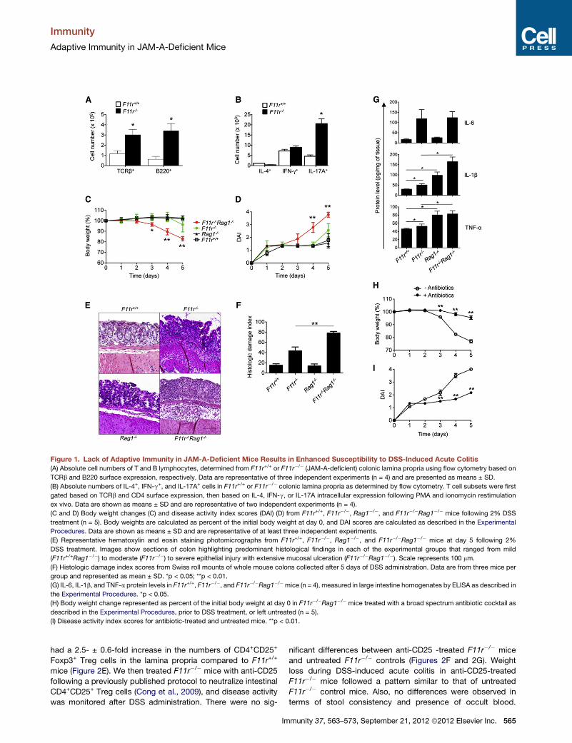
Figure 1. Lack of Adaptive Immunity in JAM-A-Deficient Mice Results in Enhanced Susceptibility to DSS-Induced Acute Colitis
(A) Absolute cell numbers of T and B lymphocytes, determined from F11r+/+ or F11r�/� (JAM-A-deficient) colonic lamina propria using flow cytometry based on
TCRb and B220 surface expression, respectively. Data are representative of three independent experiments (n = 4) and are presented as means ± SD.
(B) Absolute numbers of IL-4+, IFN-g+, and IL-17A+ cells in F11r+/+ or F11r�/� colonic lamina propria as determined by flow cytometry. T cell subsets were first
gated based on TCRb and CD4 surface expression, then based on IL-4, IFN-g, or IL-17A intracellular expression following PMA and ionomycin restimulation
ex vivo. Data are shown as means ± SD and are representative of two independent experiments (n = 4).
(C and D) Body weight changes (C) and disease activity index scores (DAI) (D) from F11r+/+, F11r�/�, Rag1�/�, and F11r�/�Rag1�/� mice following 2% DSS
treatment (n = 5). Body weights are calculated as percent of the initial body weight at day 0, and DAI scores are calculated as described in the Experimental
Procedures. Data are shown as means ± SD and are representative of at least three independent experiments.
(E) Representative hematoxylin and eosin staining photomicrographs from F11r+/+, F11r�/�, Rag1�/�, and F11r�/�Rag1�/� mice at day 5 following 2%
DSS treatment. Images show sections of colon highlighting predominant histological findings in each of the experimental groups that ranged from mild
(F11r+/+Rag1�/�) to moderate (F11r�/�) to severe epithelial injury with extensive mucosal ulceration (F11r�/�Rag1�/�). Scale represents 100 mm.
(F) Histologic damage index scores from Swiss roll mounts of whole mouse colons collected after 5 days of DSS administration. Data are from three mice per
group and represented as mean ± SD. *p < 0.05; **p < 0.01.
(G) IL-6, IL-1b, and TNF-a protein levels in F11r+/+, F11r�/�, and F11r�/�Rag1�/�mice (n = 4), measured in large intestine homogenates by ELISA as described in
the Experimental Procedures. *p < 0.05.
(H) Body weight change represented as percent of the initial body weight at day 0 in F11r�/�Rag1�/� mice treated with a broad spectrum antibiotic cocktail as
described in the Experimental Procedures, prior to DSS treatment, or left untreated (n = 5).
(I) Disease activity index scores for antibiotic-treated and untreated mice. **p < 0.01.
Immunity
Adaptive Immunity in JAM-A-Deficient Mice
had a 2.5- ± 0.6-fold increase in the numbers of CD4+CD25+
Foxp3+ Treg cells in the lamina propria compared to F11r+/+
mice (Figure 2E). We then treated F11r�/� mice with anti-CD25
following a previously published protocol to neutralize intestinal
CD4+CD25+ Treg cells (Cong et al., 2009), and disease activity
was monitored after DSS administration. There were no sig-
Im
nificant differences between anti-CD25 -treated F11r�/� mice
and untreated F11r�/� controls (Figures 2F and 2G). Weight
loss during DSS-induced acute colitis in anti-CD25-treated
F11r�/� mice followed a pattern similar to that of untreated
F11r�/� control mice. Also, no differences were observed in
terms of stool consistency and presence of occult blood.
munity 37, 563–573, September 21, 2012 ª2012 Elsevier Inc. 565

Figure 2. CD4+ T Cell Depletion in JAM-A-Deficient Mice Leads to Enhanced Susceptibility to DSS-Induced Acute Colitis
(A) Body weight changes represented as percent of the initial body weight at day 0 from F11r+/+ and F11r�/� (JAM-A-deficient) mice treated with 500 mg anti-CD4
(GK1.5) or anti-CD8 (YTS169.4) antibodies at day �2 and day �1 prior to DSS treatment, or left untreated.
(B) Disease activity index scores for anti-CD4 or anti-CD8-treated mice and untreated controls. Data are shown as means ± SEM and are pooled from three
independent experiments (n = 6).
(C) Photomicrographs of representative hematoxylin and eosin-stained colon sections from F11r+/+ and F11r�/� mice treated with anti-CD4 or anti-CD8 anti-
bodies or untreated control groups at day 5 following 2%DSS treatment. Images show sections of colon highlighting predominant histological findings in each of
the experimental groups. Scale represents 100 mm.
(D) Histologic damage index scores from Swiss roll mounts of whole mouse colons collected after 5 days of DSS treatment. Data are from three mice per group
and represented as mean ± SD. *p < 0.05; **p < 0.01.
(E) Absolute cell numbers of CD4+, CD8+, and Foxp3+ T cell subsets, measured by flow cytometry on lamina propria cell preparations from F11r+/+ and F11r�/�
mice (n = 4).
(F) Body weight changes in F11r+/+ and F11r�/� mice treated with 100 mg of anti-CD25 antibody at day �5 and day �2 prior to DSS treatment, or left untreated.
(G) Disease activity index scores for anti-CD25 antibody-treated and untreated animals. Data are pooled from three independent experiments (n = 9) and are
represented as means ± SEM.
Immunity
Adaptive Immunity in JAM-A-Deficient Mice
Depletion of CD4+Foxp3+ Treg cells in the intestinal lamina
propria was incomplete (�30%depletion), thus limiting definitive
assessment of the role of Foxp3+ Treg cells in this protective
response. Altogether, these results highlight an important role
of CD4+ T cells in adaptive immune compensation to acute injury
under conditions of chronic barrier compromise.
CD4-Dependent TGF-b Production Suppresses AcuteColitis in JAM-A-Deficient MiceIn addition to CD4+ Treg cells, TGF-b-producing CD4+ T cells are
also well appreciated to regulate intestinal immune responses
(Thorstenson and Khoruts, 2001; Zhang et al., 2001). Interest-
566 Immunity 37, 563–573, September 21, 2012 ª2012 Elsevier Inc.
ingly, we observed that baseline TGF-bmRNA and protein levels
in colonic homogenates were more than 2-fold higher in F11r�/�
mice when compared to F11r+/+ mice (Figures 3A and 3B). To
investigate the role for enhanced TGF-b expression in F11r�/�
mice, animals were treated with neutralizing TGF-b antibodies
at day �5 and day �2 prior to DSS treatment. Antibody-treated
F11r�/� mice were far more susceptible to DSS-induced acute
colitis than untreated F11r�/� mice, beginning to lose weight
from day 3 post-DSS treatment (6.6% ± 1.1% weight loss) and
losing close to 20% of their initial body weight by day 6
(20.6% ± 4.4% weight loss) (Figure 3C). In addition, anti-TGF-
b-treated F11r�/�mice also developed diarrhea at amuch earlier

Figure 3. TGF-b Signaling Protects from
Acute Colonic Inflammation JAM-A-Defi-
cient Mice
(A) TGF-b mRNA levels in F11r+/+ and F11r�/�
(JAM-A-deficient) mice were evaluated by real-
time PCR using total RNA from colon as described
in the Experimental Procedures. Data are pre-
sented as relative expression following normali-
zation with respect to gapdh.
(B) TGF-b protein levels in F11r+/+ and F11r�/�
mice were determined by ELISA using colonic
tissue homogenates.
(C and D) Body weight changes (C) and disease
activity index scores (DAI) (D) from F11r+/+ and
F11r�/� mice treated with 100 mg anti-TGF-b
neutralizing antibodies at day �5 and day �2 prior
to DSS treatment, or left untreated. Data are
represented as means ± SEM and are pooled
from three independent experiments (n = 10).
(E) Photomicrographs of representative hema-
toxylin and eosin-stained colon sections from
F11r+/+ and F11r�/� mice treated with anti-TGF-
b neutralizing antibodies or untreated control
groups at day 5 following 2% DSS treatment.
Images show sections of colon highlighting pre-
dominant histological findings in each of the
experimental groups. Scale represents 100 mm.
(F) Histologic damage index scores from Swiss
roll mounts of whole mouse colons collected
after 5 days of DSS treatment. Data are from three
mice per group and represented as mean ± SD.
*p < 0.05; **p < 0.01.
(G) TGF-b mRNA levels were measured in colonic
intestinal epithelial cells (IEC) and lamina propria
lymphocyte (LPL) samples by real-time PCR. Data
are normalized to the endogenous control gapdh.
(H) TGF-b mRNA levels in colons of F11r+/+ and
F11r�/� mice treated with anti-CD4-depleting
antibodies (GK1.5) at day �2 and day �1 with
500 mg of antibody per mouse per injection, or
left untreated, followed by 6 days of DSS treat-
ment. Data are shown as the mean ± SEM (n = 3)
percent reduction in TGF-b mRNA levels after
CD4 T cell depletion. Expression levels were
normalized to the endogenous control gapdh.
*p < 0.05; **p < 0.01.
Immunity
Adaptive Immunity in JAM-A-Deficient Mice
time point (day 4 post-DSS treatment) than their untreated coun-
terparts, which contributed to a significantly higher disease
activity index (3.2 ± 0.1 versus 2.1 ± 0.1) (Figure 3D). Histologic
analyses revealed extensive ulceration, mucosal injury, and
inflammation in the anti-TGF-b-treated F11r�/� mice that were
similar to that observed with F11r�/�Rag�/� mice treated with
DSS (Figures 3E and 3F). In contrast, the increased susceptibility
to DSS following anti-TGF-b administration was not observed
in F11r+/+ control mice. In addition, treatment of F11r�/� mice
with an IgG1 isotype control antibody did not have any effect
on the course of the disease (data not shown).
Because JAM-A-deficient mice were observed to have
increased mucosal TGF-b production, and anti-CD4 as well as
anti-TGF-b administration led to enhanced susceptibility to
DSS, we investigated whether CD4+ T cells were the source of
TGF-b. Real-time PCR data from lamina propria lymphocyte
Im
fractions, which are highly enriched in CD4+ T cells, indeed re-
vealed that lymphocytes are amajor source of TGF-b (Figure 3G).
Furthermore, to evaluate whether CD4+ T cells contribute to the
elevated TGF-b amounts observed in F11r�/� mice, we quanti-
fied cytokine mRNA in the colons of DSS-treated F11r+/+ and
F11r�/� mice after CD4+ T cell depletion. Depletion of CD4+
T cells in DSS-treated F11r�/� mice resulted in nearly an 80%
reduction of TGF-b compared to that of F11r+/+ controls (Fig-
ure 3H), consistent with CD4+ T cells being the major source
for TGF-b in F11r�/� mice.
Increased IgA and Bacterial Translocationin JAM-A-Deficient MiceWe previously demonstrated that JAM-A-deficient mice have
increased numbers of isolated lymphoid follicles in the large
intestine when compared to wild-type mice (Laukoetter et al.,
munity 37, 563–573, September 21, 2012 ª2012 Elsevier Inc. 567
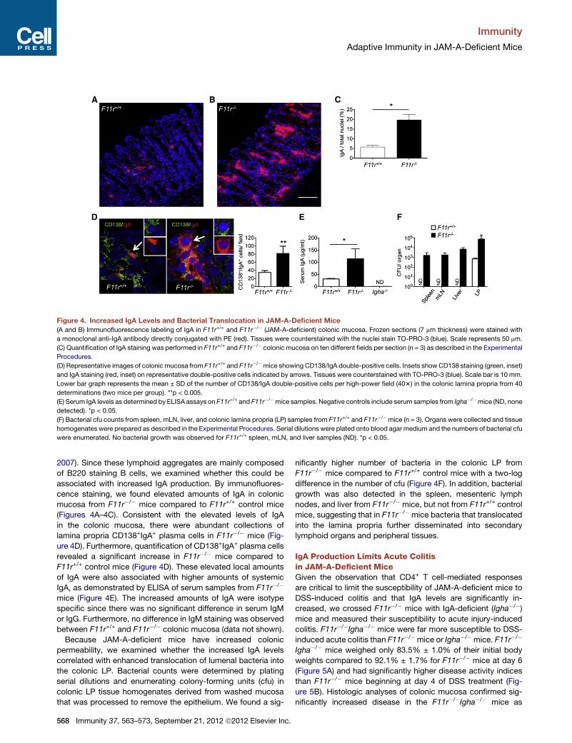
Figure 4. Increased IgA Levels and Bacterial Translocation in JAM-A-Deficient Mice
(A and B) Immunofluorescence labeling of IgA in F11r+/+ and F11r�/� (JAM-A-deficient) colonic mucosa. Frozen sections (7 mm thickness) were stained with
a monoclonal anti-IgA antibody directly conjugated with PE (red). Tissues were counterstained with the nuclei stain TO-PRO-3 (blue). Scale represents 50 mm.
(C) Quantification of IgA staining was performed in F11r+/+ and F11r�/� colonic mucosa on ten different fields per section (n = 3) as described in the Experimental
Procedures.
(D) Representative images of colonic mucosa from F11r+/+ and F11r�/�mice showing CD138/IgA double-positive cells. Insets showCD138 staining (green, inset)
and IgA staining (red, inset) on representative double-positive cells indicated by arrows. Tissues were counterstained with TO-PRO-3 (blue). Scale bar is 10 mm.
Lower bar graph represents the mean ± SD of the number of CD138/IgA double-positive cells per high-power field (403) in the colonic lamina propria from 40
determinations (two mice per group). **p < 0.005.
(E) Serum IgA levels as determined by ELISA assays on F11r+/+ and F11r�/�mice samples. Negative controls include serum samples from Igha�/�mice (ND, none
detected). *p < 0.05.
(F) Bacterial cfu counts from spleen, mLN, liver, and colonic lamina propria (LP) samples from F11r+/+ and F11r�/� mice (n = 3). Organs were collected and tissue
homogenates were prepared as described in the Experimental Procedures. Serial dilutions were plated onto blood agar medium and the numbers of bacterial cfu
were enumerated. No bacterial growth was observed for F11r+/+ spleen, mLN, and liver samples (ND). *p < 0.05.
Immunity
Adaptive Immunity in JAM-A-Deficient Mice
2007). Since these lymphoid aggregates are mainly composed
of B220 staining B cells, we examined whether this could be
associated with increased IgA production. By immunofluores-
cence staining, we found elevated amounts of IgA in colonic
mucosa from F11r�/� mice compared to F11r+/+ control mice
(Figures 4A–4C). Consistent with the elevated levels of IgA
in the colonic mucosa, there were abundant collections of
lamina propria CD138+IgA+ plasma cells in F11r�/� mice (Fig-
ure 4D). Furthermore, quantification of CD138+IgA+ plasma cells
revealed a significant increase in F11r�/� mice compared to
F11r+/+ control mice (Figure 4D). These elevated local amounts
of IgA were also associated with higher amounts of systemic
IgA, as demonstrated by ELISA of serum samples from F11r�/�
mice (Figure 4E). The increased amounts of IgA were isotype
specific since there was no significant difference in serum IgM
or IgG. Furthermore, no difference in IgM staining was observed
between F11r+/+ and F11r�/� colonic mucosa (data not shown).
Because JAM-A-deficient mice have increased colonic
permeability, we examined whether the increased IgA levels
correlated with enhanced translocation of lumenal bacteria into
the colonic LP. Bacterial counts were determined by plating
serial dilutions and enumerating colony-forming units (cfu) in
colonic LP tissue homogenates derived from washed mucosa
that was processed to remove the epithelium. We found a sig-
568 Immunity 37, 563–573, September 21, 2012 ª2012 Elsevier Inc.
nificantly higher number of bacteria in the colonic LP from
F11r�/� mice compared to F11r+/+ control mice with a two-log
difference in the number of cfu (Figure 4F). In addition, bacterial
growth was also detected in the spleen, mesenteric lymph
nodes, and liver from F11r�/� mice, but not from F11r+/+ control
mice, suggesting that in F11r�/� mice bacteria that translocated
into the lamina propria further disseminated into secondary
lymphoid organs and peripheral tissues.
IgA Production Limits Acute Colitisin JAM-A-Deficient MiceGiven the observation that CD4+ T cell-mediated responses
are critical to limit the susceptibility of JAM-A-deficient mice to
DSS-induced colitis and that IgA levels are significantly in-
creased, we crossed F11r�/� mice with IgA-deficient (Igha�/�)mice and measured their susceptibility to acute injury-induced
colitis. F11r�/�Igha�/� mice were far more susceptible to DSS-
induced acute colitis than F11r�/� mice or Igha�/� mice. F11r�/�
Igha�/� mice weighed only 83.5% ± 1.0% of their initial body
weights compared to 92.1% ± 1.7% for F11r�/� mice at day 6
(Figure 5A) and had significantly higher disease activity indices
than F11r�/� mice beginning at day 4 of DSS treatment (Fig-
ure 5B). Histologic analyses of colonic mucosa confirmed sig-
nificantly increased disease in the F11r�/�Igha�/� mice as
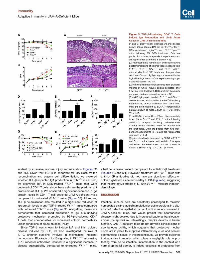
Figure 5. TGF-b-Producing CD4+ T Cells
Induce IgA Production and Limit Acute
Colitis in JAM-A-Deficient Mice
(A and B) Body weight changes (A) and disease
activity index scores (DAI) (B) in F11r+/+, F11r�/�
(JAM-A-deficient), Igha�/�, and F11r�/�Igha�/�
mice following 2% DSS treatment. Data are
pooled from three independent experiments and
are represented as means ± SEM (n = 9).
(C) Representative hematoxylin and eosin staining
photomicrographs of colonic tissue sections from
F11r+/+, F11r�/�, Igha�/�, and F11r�/�Igha�/�
mice at day 5 of DSS treatment. Images show
sections of colon highlighting predominant histo-
logical findings in each of the experimental groups.
Scale represents 100 mm.
(D) Histologic damage index scores from Swiss roll
mounts of whole mouse colons collected after
5 days of DSS treatment. Data are from three mice
per group and represented as mean ± SD.
(E and F) IgA protein levels in F11r+/+ and F11r�/�
colonic tissues, with or without anti-CD4 antibody
treatment (E), or with or without anti-TGF-b treat-
ment (F), as measured by ELISA. Representative
data are shown as mean ± SEM (n = 4). *p < 0.05;
**p < 0.01.
(G and H) Body weight loss (G) and disease activity
index (H) in F11r+/+ and F11r�/� mice following
anti-IL-10 receptor antibody administration.
Control groups included mice not treated with
the antibodies. Data are pooled from two inde-
pendent experiments (n = 9) and are represented
as means ± SEM.
(I) IgA protein levels measured by ELISA in F11r+/+
and F11r�/� mice treated with anti-IL-10 receptor
antibodies. Representative data are shown as
means ± SEM (n = 4). *p < 0.05; **p < 0.01.
Immunity
Adaptive Immunity in JAM-A-Deficient Mice
evident by extensive mucosal injury and ulceration (Figures 5C
and 5D). Given that TGF-b is important for IgA class switch
recombination and plasma cell differentiation, we explored
whether TGF-b impacted IgA production in F11r�/� mice. First,
we examined IgA in DSS-treated F11r�/� mice that were
depleted of CD4+ T cells, since these cells are the predominant
producers of TGF-b. We observed a significant decrease in IgA
protein levels in CD4+ T cell-depleted JAM-A-deficient mice
compared to untreated F11r�/� mice (Figure 5E). Moreover,
TGF-b neutralization also resulted in a significant reduction of
IgA protein levels in anti-TGF-b-treated F11r�/� mice compared
with untreated F11r�/� mice (Figure 5F). Altogether, these data
demonstrate that increased production of IgA is a unifying
protective mechanism promoted by TGF-b-producing CD4+
T cells that compensates for increased colonic permeability
under conditions of acute mucosal injury.
Since TGF-b was shown to induce IgA and limit colonic
disease induced by DSS, we also investigated the role of
IL-10, another cytokine involved in maintaining intestinal
homeostasis. Blockade of IL-10 signaling in F11r�/� mice using
IL-10 receptor antibodies resulted in a significant increase in
disease susceptibility compared to untreated F11r�/� mice,
Im
albeit to a lesser extent compared to anti-TGF-b treatment
(Figures 5G and 5H). However, treatment of F11r�/� mice with
anti-IL-10R antibodies did not have any significant effects on
colonic IgA levels as determined by ELISA (Figure 5I), suggesting
that the protective effects of IL-10 in F11r�/� mice are indepen-
dent of IgA.
DISCUSSION
Intestinal immune cells are constantly challenged to maintain
homeostasis in the face of stimulation by gutmicrobiota. In a situ-
ation of defective epithelial barrier function as encountered in
JAM-A-deficient mice, one would predict that spontaneous
disease might develop due to increased bacterial translocation
across the epithelium. Interestingly, despite defects in barrier
function, JAM-A-deficient mice do not develop clinical signs of
spontaneous colitis, which suggests that protective mecha-
nisms are in place to suppress inflammatory cues and prevent
spontaneous disease. In the present study, we provide evidence
that adaptive immunity, which plays a negligible role in pro-
tecting from acute intestinal inflammation in the context of a
normal epithelial barrier, is indeed essential in protecting from
munity 37, 563–573, September 21, 2012 ª2012 Elsevier Inc. 569
Immunity
Adaptive Immunity in JAM-A-Deficient Mice
acute, injury-induced colitis in the context of increased
intestinal permeability. In particular, this adaptive immunity-
based ‘‘protective’’ effect is dependent on TGF-b-producing
CD4+ T cells that serve to promote IgA secretion in JAM-A-
deficient mice. Altogether, these observations establish a role
for adaptive immune responses in limiting severe, acutemucosal
injury in the context of intestinal barrier compromise.
Our findings that acute intestinal inflammation is markedly
enhanced in JAM-A-deficient mice lacking CD4+ T cells are in
accordance with the well-established role of immunoregulatory
T cells in suppressing colitis (Groux et al., 1997; Kim et al.,
2007; Powrie et al., 1996; Rubtsov et al., 2008; Sakaguchi
et al., 1985; Zhang et al., 2001). Thus, it would be tempting to
speculate that the enhanced susceptibility of JAM-A-deficient
mice to DSS-induced acute colitis is due to the absence of
functional CD4+CD25+ Treg cells. A critical role for CD4+CD25+
Treg cells in mediating IgA production in response to commensal
flora has been reported (Cong et al., 2009). Here we used anti-
CD25 to attempt to eliminate CD4+Foxp3+ Treg cells but only
achieved partial depletion and observed no increase in suscep-
tibility to colitis. While our findings preclude definitive assess-
ment of the role of Foxp3+ Treg cells in regulating the adaptive
immune compensation observed here, they are consistent with
those reported by Cong et al. (Cong et al., 2009) and highlight
a critical role for intestinal permeability in regulating TGF-
b-producing Treg cell-mediated IgA production.
We also evaluated the relative contribution of TGF-b, which is
well appreciated for its role in mediating Treg cell immunosup-
pressive functions (Li et al., 2007; Marie et al., 2005). Neutraliza-
tion of TGF-b in JAM-A-deficient mice led to the striking finding
that compensation for intestinal barrier defects also depends on
TGF-b signaling. It is possible that the increased TGF-b pro-
duction in JAM-A-deficient mice compared with wild-type
controls represents a secondary response to increased bacterial
translocation and subsequent immune activation. Given the fact
that TGF-b amounts in JAM-A/Rag1-deficient mice are greatly
reduced compared to JAM-A-deficient mice (data not shown),
we demonstrated that TGF-b-producing T cells are a major
source of TGF-b in JAM-A-deficient mice. We further showed
that increased colonic TGF-b production in JAM-A-deficient
mice during acute mucosal injury-induced colitis is CD4+ T cell
dependent. These findings suggest that TGF-b-secreting cells
(Weiner, 2001) may be a critical component of immune-mediated
protection during increased intestinal permeability.
The role of T cells in promoting TGF-b-mediated immune regu-
lation of intestinal inflammation has been previously described
in a model of CD4+CD45RBhi-induced colitis. T cells that ex-
press dnTGF-bRII escape immunoregulation by Treg cells,
indicating that TGF-b-mediated control of inflammation is
T cell dependent (Fahlen et al., 2005). Thus, in JAM-A-deficient
mice, TGF-b-producing CD4+ T cells may regulate pathogenic
CD4+ T cells to limit intestinal inflammation. However, it is
also conceivable that TGF-b exerts its effect via T cell-indepen-
dent mechanisms. Our data are also consistent with
TGF-b-producing CD4+ T cells playing a critical role in IgA iso-
type class-switch recombination in B cells, thereby promoting
IgA-mediated immune responses and control of intestinal
inflammation in the gut induced by antigen exposure (Borsutzky
et al., 2004; Cazac and Roes, 2000; Cerutti, 2008).
570 Immunity 37, 563–573, September 21, 2012 ª2012 Elsevier Inc.
Importantly, loss of IgA appears to be a unifying mechanism
by which depletion of CD4+ T cells and neutralization of TGF-b
increases susceptibility to acute mucosal damage in the context
of a leaky epithelial barrier. Based on our data, it is clear that
IgA-associated responses are critical for immune mediated-
compensation during enhanced intestinal permeability. Murine
studies have demonstrated an important role for IgA antibodies
in immune exclusion. For example, mice deficient in the poly-
meric immunoglobulin receptor, which are unable to secrete
IgA into the gut lumen, display increased mucosal leakiness
and antigen uptake (Johansen et al., 1999; Sait et al., 2007).
The current study highlights the observations that: (1) IgA
production is augmented in mice with a leaky gut barrier, most
likely representing a humoral compensatory mechanism in
response to the increased antigen uptake across the epithelium,
and (2) that regulation of intestinal inflammation in the specific
context of increased epithelial permeability and acute mucosal
injury relies on IgA-mediated immunity. The exact mechanisms
by which IgA antibodies participate in dampening tissue inflam-
mation in JAM-A-deficient mice remain unclear, but may involve
increased immune exclusion at the interface between lumen
and epithelial cells and/or increased transport of antigens across
the epithelium and subsequent recognition and phagocytosis
by antigen-presenting cells.
It is important to note that the development of intestinal
inflammation during DSS-induced acute mucosal injury is a
lymphocyte-independent process in wild-type mice (Dieleman
et al., 1994). However, in JAM-A-deficient mice, which have
enhanced colonic permeability, severity of colitis depends on
adaptive immune cells, as demonstrated by the increased
susceptibility to DSS in JAM-A/Rag1-deficient mice compared
to JAM-A-deficient mice. This finding implicates a crucial role
of adaptive immunity under conditions of existing defects in
barrier function.
In summary, our findings provide new insights into the regu-
lation of mucosal homeostasis in the context of impaired intes-
tinal epithelial barrier function. JAM-A-deficient mice represent
an important tool to study immune mechanisms implicated
in the containment of acute inflammatory responses triggered
by enhanced exposure to lumenal antigens and immune activa-
tion. We have identified an essential role for TGF-b-producing
CD4+ T cells promoting IgA secretion in controlling intestinal
inflammation in the presence of a compromised intestinal epithe-
lial barrier.
EXPERIMENTAL PROCEDURES
Mice
JAM-A-deficient mice (F11r�/�) and littermate controls were bred onsite.
Rag1tm1Mom (Rag1�/�) mice on a C57BL/6 background were purchased from
The Jackson Laboratories (Bar Harbor, ME). IgA-deficient mice (Igha�/�)(Harriman et al., 1999) were a kind gift from Dr. L. Eckmann. Rag1�/� and
Igha�/� mice were crossed with JAM-A-deficient mice to generate F11r�/�
Rag1�/� and F11r�/�Igha�/� mice. Mice were maintained under specific
pathogen-free conditions at Emory University. All animal procedures were
reviewed and approved by the Emory University Institutional Animal Care
and Use Committee.
Genotyping
PCRs were performed on tail genomic DNA using the following primers:
F11r forward 50-TCTTTTCACCAATCGGAACG-30 and reverse 50-CGGCAT
Immunity
Adaptive Immunity in JAM-A-Deficient Mice
TAATCCCAGAAGGT-30; Rag1 forward common 50-CCGGACAAGTTTTTCA
TCGT-30, reverse wild-type allele 50-GAGGTTCCGCTACGACTCTG-30, and
reverse mutant allele 50-TGGATGTGGAATGTGTGCGAG-30.
DSS-Induced Colitis
Mice were provided 2% (wt/vol) DSS (molecular mass = 36–50 kDa) (MP
Biomedicals) for 5–7 days, and then colons were assessed for weight, length,
and histology. Daily clinical assessment of DSS-treated animals included eval-
uation of stool consistency, detection of blood in stool, and body weight loss
measurement. An individual score (ranging from 0 to 4) was attributed for each
one of these parameters, and a disease activity index (DAI) ranging from 0 to 4
was calculated by combining all three scores (Laukoetter et al., 2007).
Histology
Histological examination was performed on whole colons of experimental and
control mice. In addition, colonic tissue samples were embedded in OCT for
immunofluorescence staining and stored at �80�C. Histological parameters
were quantified in a blinded fashion by two experienced gastrointestinal
pathologists using parameters as previously described (Nava et al., 2010)
with some modification. Briefly, hematoxylin and eosin sections of Swiss roll
mounts of entire mouse colons were assessed for the percentage of the
mucosa containing ulceration or crypt epithelial injury/damage/active inflam-
mation comprising at least 1/3 of the thickness of the mucosa. The ulceration
and injury/damage values were added and reported as a histologic damage
index.
Flow Cytometry
Lamina propria lymphocyte isolation was performed as previously described
(Denning et al., 2011; Medina-Contreras et al., 2011). Intestinal cell prepara-
tions included lymphoid follicles. Cells were stained with PerCP-conjugated
anti-CD45 (30-F11), FITC-conjugated anti-CD19 (1D3), allophycocyanin-
conjugated TCRb (H57-597), eFluor 450-conjugated anti-CD4 (L3T4)
(eBioscience), and PE/Texas Red-conjugated anti-CD8a (5H10) (Invitrogen)
monoclonal antibodies. Data were acquired using a LSR II flow cytometer
(Becton Dickinson) and analyzed using Flowjo software (Treestar). For intracel-
lular cytokine detection, cells were restimulated with PMA and ionomycin
ex vivo in the presence of GolgiPlug (BD Biosciences).
Cytokine and IgA Assays
For TGF-b1 protein level analyses, proximal colonic tissues were homoge-
nized in sterile 13 PBS supplemented with 1% Triton X-100 and protease
and phosphatase inhibitor cocktails (Sigma) and centrifuged for 20 min at
maximum speed at 4�C. Supernatants were used to measure TGF-b1, IL-6,
IL-1b, and TNF-a protein levels using ELISA kits (eBioscience). IgA protein
levels were evaluated on colonic homogenate supernatants or serum samples
using an ELISA kit (Immunology Consultants Laboratory, Inc.).
Broad-Spectrum Antibiotic Treatment
Mice were treated using a modified protocol described elsewhere (Rakoff-
Nahoum et al., 2004).
Bacterial Translocation
Organs were weighed and colon length was measured. For the spleen and
mLN, the whole organ was homogenized in 500 ml sterile PBS. For the liver,
a piece (200 mg) of the right lobe was collected and homogenized. For colonic
bacterial counts, fecal contents were removed by rinsing longitudinally opened
colons with sterile PBS. Colons were then cut into 1.5 cm segments, and
epithelial cells were removed by vigorous horizontal shaking at 37�C for
20 min in HBSS containing 5% FBS, 10 mM HEPES, and 2 mM EDTA (pH
8.0). This procedure was repeated two times. Samples were subsequently
homogenized in 500 ml sterile PBS using an Omni-Prep homogenizer. Serial
dilutions were then plated onto blood agar plates and incubated at 37�C for
36 hr, and cfu were enumerated.
Antibody-Mediated Depletion
Anti-CD4 (clone GK1.5) and anti-CD8a (YTS169.4) neutralizing antibodies
(Bio X Cell) were used. For each antibody, two 500 mg injections were given
i.p. at day �2 and day �1 prior to DSS treatment. CD25 depletion was
Im
performed as previously described (Cong et al., 2009). To assess depletion
efficiency, leukocytes were isolated from large intestine, small intestine,
spleen, and mLN and subjected to flow cytometric analyses. TGF-b depletion
prior to DSS treatment was carried out by performing two 100 mg injections i.p.
at day �5 and day �2 prior to DSS treatment using anti-TGF-b (1D11.16.8)
monoclonal antibody (Bio X Cell). For IgA protein level measurement, chronic
TGF-b depletion was conducted by injecting 1-week-old mice with 100 mg
anti-TGF-b neutralizing antibodies twice weekly for the first 2 weeks followed
by 100 mg once every week for a total of 7 weeks.
Real-Time PCR Analysis
Total RNA was extracted from proximal colons of F11r�/� and F11r+/+ mice
using RNeasy Mini kit (Invitrogen), and genomic DNA contamination was
removed following DNaseI treatment (QIAGEN). Real-time PCR reactions
were set up using the iScript One Step RT-PCR kit with SYBR Green (Bio-
Rad Laboratories) and the following primers: forward tgfb1 50-ACCATGCC
AACTTCTGTCTG-30 and reverse tgfb1 50-CGGGTTGTGTTGGTTGTAGA-30;forward gapdh 50-TCATTGACCTCAACTACATGGTCTA-30 and reverse gapdh
50-ACACCAGTAGACTCCACGACATACT-30. For the preparations of IECs
and lamina propria-derived lymphocytes, cell suspensions were processed
through a Percoll gradient and individual fractions were collected and used
for RNA extraction. Reactions were run on a MyiQ Single Color Real-Time
PCR Detection system (Bio-Rad Laboratories), and Ct values were used to
calculate relative expression.
Confocal Microscopy
Frozen tissue sections (7 mm thickness) from large intestines were fixed in
cold ethanol for 20 min at �20�C. Sections were then blocked with 3% BSA
prior to incubation with PE-conjugated rat anti-mouse IgA (eBioscience) for
20 min. Plasma cells were stained using anti-mouse CD138 (eBioscience).
Tissues were counterstained with TO-PRO-3 for 3 min and mounted on
coverslips. Images were acquired with a LSM 510 confocal microscope (Zeiss)
and analyzed using ImageJ software (National Institutes of Health).
Data Analysis
Results are expressed as means ± SEM unless otherwise indicated. p values
were calculated using one-way ANOVA and unpaired two-tailed Student’s
t test. p < 0.05 was considered statistically significant.
ACKNOWLEDGMENTS
The authors thank Dr. L. Eckmann for providing Igha�/� mice, Dr. R. Ahmed
andDr. R.S. Mittler for providing GK1.5 and YTS169.4 antibodies, andWinston
Lee and Christopher Capaldo for discussions. This work was supported by
grants from the NIH (DK061379 and DK72564 to C.A.P., DK055679 and
DK59888 to A.N., and AA017870 and AI083554 to T.L.D.), core support from
a Digestive Diseases Minicenter grant (DK 064399), a Emory Egleston
Children’s Research Center seed grant to T.L.D., a Crohn’s and Colitis
Foundation of America Career Development Award and an AGA Foundation
for Digestive Health and Nutrition Research Scholar Award to P.N., and
Crohn’s and Colitis Foundation of America Research Fellowship and senior
research awards to M.K. and B.F., respectively.
Received: June 29, 2011
Revised: April 13, 2012
Accepted: June 7, 2012
Published online: September 13, 2012
REFERENCES
Barnes, M.J., and Powrie, F. (2009). Regulatory T cells reinforce intestinal
homeostasis. Immunity 31, 401–411.
Borsutzky, S., Cazac, B.B., Roes, J., and Guzman, C.A. (2004). TGF-beta
receptor signaling is critical for mucosal IgA responses. J. Immunol. 173,
3305–3309.
Boullier, S., Tanguy, M., Kadaoui, K.A., Caubet, C., Sansonetti, P., Corthesy,
B., and Phalipon, A. (2009). Secretory IgA-mediated neutralization of
munity 37, 563–573, September 21, 2012 ª2012 Elsevier Inc. 571

Immunity
Adaptive Immunity in JAM-A-Deficient Mice
Shigella flexneri prevents intestinal tissue destruction by down-regulating
inflammatory circuits. J. Immunol. 183, 5879–5885.
Cazac, B.B., and Roes, J. (2000). TGF-beta receptor controls B cell respon-
siveness and induction of IgA in vivo. Immunity 13, 443–451.
Cera, M.R., Del Prete, A., Vecchi, A., Corada, M., Martin-Padura, I., Motoike,
T., Tonetti, P., Bazzoni, G., Vermi, W., Gentili, F., et al. (2004). Increased DC
trafficking to lymph nodes and contact hypersensitivity in junctional adhesion
molecule-A-deficient mice. J. Clin. Invest. 114, 729–738.
Cerutti, A. (2008). The regulation of IgA class switching. Nat. Rev. Immunol. 8,
421–434.
Cong, Y., Feng, T., Fujihashi, K., Schoeb, T.R., and Elson, C.O. (2009). A domi-
nant, coordinated T regulatory cell-IgA response to the intestinal microbiota.
Proc. Natl. Acad. Sci. USA 106, 19256–19261.
Corada, M., Chimenti, S., Cera, M.R., Vinci, M., Salio, M., Fiordaliso, F., De
Angelis, N., Villa, A., Bossi, M., Staszewsky, L.I., et al. (2005). Junctional
adhesion molecule-A-deficient polymorphonuclear cells show reduced
diapedesis in peritonitis and heart ischemia-reperfusion injury. Proc. Natl.
Acad. Sci. USA 102, 10634–10639.
Davidson, N.J., Leach, M.W., Fort, M.M., Thompson-Snipes, L., Kuhn, R.,
Muller, W., Berg, D.J., and Rennick, D.M. (1996). T helper cell 1-type CD4+
T cells, but not B cells, mediate colitis in interleukin 10-deficient mice.
J. Exp. Med. 184, 241–251.
Denning, T.L., Norris, B.A., Medina-Contreras, O., Manicassamy, S., Geem,
D., Madan, R., Karp, C.L., and Pulendran, B. (2011). Functional specializations
of intestinal dendritic cell and macrophage subsets that control Th17 and
regulatory T cell responses are dependent on the T cell/APC ratio, source of
mouse strain, and regional localization. J. Immunol. 187, 733–747.
Dieleman, L.A., Ridwan, B.U., Tennyson, G.S., Beagley, K.W., Bucy, R.P., and
Elson, C.O. (1994). Dextran sulfate sodium-induced colitis occurs in severe
combined immunodeficient mice. Gastroenterology 107, 1643–1652.
Do�gan, A., Wang, Z.D., and Spencer, J. (1995). E-cadherin expression in
intestinal epithelium. J. Clin. Pathol. 48, 143–146.
Fahlen, L., Read, S., Gorelik, L., Hurst, S.D., Coffman, R.L., Flavell, R.A., and
Powrie, F. (2005). T cells that cannot respond to TGF-beta escape control
by CD4(+)CD25(+) regulatory T cells. J. Exp. Med. 201, 737–746.
Fernandez, M.I., Pedron, T., Tournebize, R., Olivo-Marin, J.C., Sansonetti,
P.J., and Phalipon, A. (2003). Anti-inflammatory role for intracellular dimeric
immunoglobulin a by neutralization of lipopolysaccharide in epithelial cells.
Immunity 18, 739–749.
Gassler, N., Rohr, C., Schneider, A., Kartenbeck, J., Bach, A., Obermuller, N.,
Otto, H.F., and Autschbach, F. (2001). Inflammatory bowel disease is associ-
ated with changes of enterocytic junctions. Am. J. Physiol. Gastrointest. Liver
Physiol. 281, G216–G228.
Groux, H., O’Garra, A., Bigler, M., Rouleau, M., Antonenko, S., de Vries, J.E.,
and Roncarolo, M.G. (1997). A CD4+ T-cell subset inhibits antigen-specific
T-cell responses and prevents colitis. Nature 389, 737–742.
Harriman, G.R., Bogue, M., Rogers, P., Finegold, M., Pacheco, S., Bradley, A.,
Zhang, Y., and Mbawuike, I.N. (1999). Targeted deletion of the IgA constant
region in mice leads to IgA deficiency with alterations in expression of other
Ig isotypes. J. Immunol. 162, 2521–2529.
Jankowski, J.A., Bedford, F.K., Boulton, R.A., Cruickshank, N., Hall, C., Elder,
J., Allan, R., Forbes, A., Kim, Y.S., Wright, N.A., and Sanders, D.S. (1998).
Alterations in classical cadherins associated with progression in ulcerative
and Crohn’s colitis. Lab. Invest. 78, 1155–1167.
Johansen, F.E., Pekna, M., Norderhaug, I.N., Haneberg, B., Hietala, M.A.,
Krajci, P., Betsholtz, C., and Brandtzaeg, P. (1999). Absence of epithelial
immunoglobulin A transport, with increased mucosal leakiness, in polymeric
immunoglobulin receptor/secretory component-deficient mice. J. Exp. Med.
190, 915–922.
Karayiannakis, A.J., Syrigos, K.N., Efstathiou, J., Valizadeh, A., Noda, M.,
Playford, R.J., Kmiot, W., and Pignatelli, M. (1998). Expression of catenins
and E-cadherin during epithelial restitution in inflammatory bowel disease.
J. Pathol. 185, 413–418.
572 Immunity 37, 563–573, September 21, 2012 ª2012 Elsevier Inc.
Kim, J.M., Rasmussen, J.P., and Rudensky, A.Y. (2007). Regulatory T cells
prevent catastrophic autoimmunity throughout the lifespan of mice. Nat.
Immunol. 8, 191–197.
Kucharzik, T., Walsh, S.V., Chen, J., Parkos, C.A., and Nusrat, A. (2001).
Neutrophil transmigration in inflammatory bowel disease is associated with
differential expression of epithelial intercellular junction proteins. Am. J.
Pathol. 159, 2001–2009.
Laukoetter, M.G., Nava, P., Lee, W.Y., Severson, E.A., Capaldo, C.T., Babbin,
B.A., Williams, I.R., Koval, M., Peatman, E., Campbell, J.A., et al. (2007).
JAM-A regulates permeability and inflammation in the intestine in vivo.
J. Exp. Med. 204, 3067–3076.
Li, M.O., Wan, Y.Y., and Flavell, R.A. (2007). T cell-produced transforming
growth factor-beta1 controls T cell tolerance and regulates Th1- and Th17-
cell differentiation. Immunity 26, 579–591.
Liu, Y., Nusrat, A., Schnell, F.J., Reaves, T.A., Walsh, S., Pochet, M., and
Parkos, C.A. (2000). Human junction adhesion molecule regulates tight
junction resealing in epithelia. J. Cell Sci. 113, 2363–2374.
Mandell, K.J., Babbin, B.A., Nusrat, A., and Parkos, C.A. (2005). Junctional
adhesion molecule 1 regulates epithelial cell morphology through effects on
beta1 integrins and Rap1 activity. J. Biol. Chem. 280, 11665–11674.
Mankertz, J., and Schulzke, J.D. (2007). Altered permeability in inflammatory
bowel disease: pathophysiology and clinical implications. Curr. Opin.
Gastroenterol. 23, 379–383.
Marie, J.C., Letterio, J.J., Gavin, M., and Rudensky, A.Y. (2005). TGF-beta1
maintains suppressor function and Foxp3 expression in CD4+CD25+ regula-
tory T cells. J. Exp. Med. 201, 1061–1067.
Martın-Padura, I., Lostaglio, S., Schneemann, M., Williams, L., Romano, M.,
Fruscella, P., Panzeri, C., Stoppacciaro, A., Ruco, L., Villa, A., et al. (1998).
Junctional adhesion molecule, a novel member of the immunoglobulin
superfamily that distributes at intercellular junctions and modulates monocyte
transmigration. J. Cell Biol. 142, 117–127.
Medina-Contreras, O., Geem, D., Laur, O., Williams, I.R., Lira, S.A., Nusrat, A.,
Parkos, C.A., and Denning, T.L. (2011). CX3CR1 regulates intestinal macro-
phage homeostasis, bacterial translocation, and colitogenic Th17 responses
in mice. J. Clin. Invest. 121, 4787–4795.
Mombaerts, P., Mizoguchi, E., Grusby, M.J., Glimcher, L.H., Bhan, A.K., and
Tonegawa, S. (1993). Spontaneous development of inflammatory bowel
disease in T cell receptor mutant mice. Cell 75, 274–282.
Nava, P., Koch, S., Laukoetter, M.G., Lee, W.Y., Kolegraff, K., Capaldo, C.T.,
Beeman, N., Addis, C., Gerner-Smidt, K., Neumaier, I., et al. (2010). Interferon-
gamma regulates intestinal epithelial homeostasis through converging beta-
catenin signaling pathways. Immunity 32, 392–402.
Nava, P., Capaldo, C.T., Koch, S., Kolegraff, K., Rankin, C.R., Farkas, A.E.,
Feasel, M.E., Li, L., Addis, C., Parkos, C.A., and Nusrat, A. (2011). JAM-A
regulates epithelial proliferation through Akt/b-catenin signalling. EMBO
Rep. 12, 314–320.
Powrie, F., Carlino, J., Leach, M.W., Mauze, S., and Coffman, R.L. (1996). A
critical role for transforming growth factor-beta but not interleukin 4 in the
suppression of T helper type 1-mediated colitis by CD45RB(low) CD4+
T cells. J. Exp. Med. 183, 2669–2674.
Rakoff-Nahoum, S., Paglino, J., Eslami-Varzaneh, F., Edberg, S., and
Medzhitov, R. (2004). Recognition of commensal microflora by toll-like
receptors is required for intestinal homeostasis. Cell 118, 229–241.
Robinson, J.K., Blanchard, T.G., Levine, A.D., Emancipator, S.N., and Lamm,
M.E. (2001). A mucosal IgA-mediated excretory immune system in vivo.
J. Immunol. 166, 3688–3692.
Rubtsov, Y.P., Rasmussen, J.P., Chi, E.Y., Fontenot, J., Castelli, L., Ye, X.,
Treuting, P., Siewe, L., Roers, A., Henderson, W.R., Jr., et al. (2008).
Regulatory T cell-derived interleukin-10 limits inflammation at environmental
interfaces. Immunity 28, 546–558.
Sait, L.C., Galic, M., Price, J.D., Simpfendorfer, K.R., Diavatopoulos, D.A.,
Uren, T.K., Janssen, P.H., Wijburg, O.L., and Strugnell, R.A. (2007).
Secretory antibodies reduce systemic antibody responses against the
gastrointestinal commensal flora. Int. Immunol. 19, 257–265.

Immunity
Adaptive Immunity in JAM-A-Deficient Mice
Sakaguchi, S., Fukuma, K., Kuribayashi, K., and Masuda, T. (1985). Organ-
specific autoimmune diseases induced in mice by elimination of T cell subset.
I. Evidence for the active participation of T cells in natural self-tolerance; deficit
of a T cell subset as a possible cause of autoimmune disease. J. Exp. Med.
161, 72–87.
Severson, E.A., Jiang, L., Ivanov, A.I., Mandell, K.J., Nusrat, A., and Parkos,
C.A. (2008). Cis-dimerization mediates function of junctional adhesion mole-
cule A. Mol. Biol. Cell 19, 1862–1872.
Severson, E.A., Lee, W.Y., Capaldo, C.T., Nusrat, A., and Parkos, C.A. (2009).
Junctional adhesion molecule A interacts with Afadin and PDZ-GEF2 to
activate Rap1A, regulate beta1 integrin levels, and enhance cell migration.
Mol. Biol. Cell 20, 1916–1925.
Thorstenson, K.M., and Khoruts, A. (2001). Generation of anergic and poten-
tially immunoregulatory CD25+CD4 T cells in vivo after induction of peripheral
tolerance with intravenous or oral antigen. J. Immunol. 167, 188–195.
Im
Vetrano, S., Rescigno, M., Cera, M.R., Correale, C., Rumio, C., Doni, A.,
Fantini, M., Sturm, A., Borroni, E., Repici, A., et al. (2008). Unique role of junc-
tional adhesion molecule-a in maintaining mucosal homeostasis in inflamma-
tory bowel disease. Gastroenterology 135, 173–184.
Weiner, H.L. (2001). Induction and mechanism of action of transforming
growth factor-beta-secreting Th3 regulatory cells. Immunol. Rev. 182,
207–214.
Welcker, K., Martin, A., Kolle, P., Siebeck, M., and Gross, M. (2004). Increased
intestinal permeability in patients with inflammatory bowel disease. Eur. J.
Med. Res. 9, 456–460.
Zhang, X., Izikson, L., Liu, L., and Weiner, H.L. (2001). Activation of CD25(+)
CD4(+) regulatory T cells by oral antigen administration. J. Immunol. 167,
4245–4253.
munity 37, 563–573, September 21, 2012 ª2012 Elsevier Inc. 573
Copyright © 2022 FDOKUMEN