Ca2+ Extrusion by NCX Is Compromised in Olfactory Sensory Neurons of OMP−/− Mice
11
Ca 2+ Extrusion by NCX Is Compromised in Olfactory Sensory Neurons of OMP 2/2 Mice Hyun J. Kwon 1,2. , Jae Hyung Koo 1. , Frank Zufall 3 , Trese Leinders-Zufall 3 *, Frank L. Margolis 1 * 1 Department of Anatomy and Neurobiology, School of Medicine, University of Maryland, Baltimore, Maryland, United States of America, 2 Department of Engineering and Computer Science, Andrews University, Berrien Springs, Michigan, United States of America, 3 Department of Physiology, University of Saarland, Homburg, Germany Abstract Background: The role of olfactory marker protein (OMP), a hallmark of mature olfactory sensory neurons (OSNs), has been poorly understood since its discovery. The electrophysiological and behavioral phenotypes of OMP knockout mice indicated that OMP influences olfactory signal transduction. However, the mechanism by which this occurs remained unknown. Principal Findings: We used intact olfactory epithelium obtained from WT and OMP 2/2 mice to monitor the Ca 2+ dynamics induced by the activation of cyclic nucleotide-gated channels, voltage-operated Ca 2+ channels, or Ca 2+ stores in single dendritic knobs of OSNs. Our data suggested that OMP could act to modulate the Ca 2+ -homeostasis in these neurons by influencing the activity of the plasma membrane Na + /Ca 2+ -exchanger (NCX). Immunohistochemistry verifies colocalization of NCX1 and OMP in the cilia and knobs of OSNs. To test the role of NCX activity, we compared the kinetics of Ca 2+ elevation by stimulating the reverse mode of NCX in both WT and OMP 2/2 mice. The resulting Ca 2+ responses indicate that OMP facilitates NCX activity and allows rapid Ca 2+ extrusion from OSN knobs. To address the mechanism by which OMP influences NCX activity in OSNs we studied protein-peptide interactions in real-time using surface plasmon resonance technology. We demonstrate the direct interaction of the XIP regulatory-peptide of NCX with calmodulin (CaM). Conclusions: Since CaM also binds to the Bex protein, an interacting protein partner of OMP, these observations strongly suggest that OMP can influence CaM efficacy and thus alters NCX activity by a series of protein-protein interactions. Citation: Kwon HJ, Koo JH, Zufall F, Leinders-Zufall T, Margolis FL (2009) Ca 2+ Extrusion by NCX Is Compromised in Olfactory Sensory Neurons of OMP 2/2 Mice. PLoS ONE 4(1): e4260. doi:10.1371/journal.pone.0004260 Editor: Peter Wenner, Emory University, United States of America Received September 19, 2008; Accepted December 9, 2008; Published January 23, 2009 Copyright: ß 2009 Kwon et al. This is an open-access article distributed under the terms of the Creative Commons Attribution License, which permits unrestricted use, distribution, and reproduction in any medium, provided the original author and source are credited. Funding: This work was supported by research grants from NIH/NIDCD R01-DC-003112 (FLM), R01-DC-02227 (TLZ), R01-DC-005249 (FZ) and T32 DC-00054 (FLM) and Andrews University (HJK). TLZ is a Lichtenberg Professor of the Volkswagen Foundation. The funders had no role in study design, data collection and analysis, decision to publish, or preparation of the manuscript. Competing Interests: The authors have declared that no competing interests exist. * E-mail: [email protected] (TLZ); [email protected] (FLM) . These authors contributed equally to this work. Introduction Olfactory marker protein (OMP) is an abundant cytosolic protein whose expression is highly restricted to mature OSNs in all vertebrates [1–8]. Although of unknown function, it has been implicated to play a role in olfactory transduction [9–15]. Homozygous OMP knockout (OMP 2/2 ) mice exhibit strongly reduced onset and decay kinetics of odor responses, resulting in delayed recovery following odor stimulation [10,15]. Changes in the response kinetics of olfactory sensory neurons (OSNs) have previously been shown to depend on changes in intracellular Ca 2+ concentration [16–20], leading to the hypothesis that OMP could be involved in modulating the Ca 2+ homeostasis in these neurons. The initial steps leading to Ca 2+ entry in vertebrate OSNs begin in the cilia when odor molecules bind to specific receptors and initiate a cascade of events leading to rapid cAMP formation, followed by the opening of cyclic nucleotide-gated (CNG) cation channels [21–26]. The considerable Ca 2+ permeability of CNG channels [27,28] causes a rapid intraciliary Ca 2+ increase [19,29,30] which, in turn, causes further excitation through subsequent activation of Ca 2+ -dependent Cl 2 channels[31–35]. The Ca 2+ rise also mediates adaptation by modulating CNG channel activity [17,36] and Ca 2+ /calmodulin kinase II-dependent attenuation of adenylyl cyclase activity [18,20]. Despite a recent report suggesting that OMP plays a modulatory role in cAMP formation [9], OMP might play additional roles to influence a common regulator of several steps in the transduction cascade. One possible target could be calmodulin (CaM) that is Ca 2+ -dependent and a critical effector at several steps of olfactory transduction. Interestingly, Bex protein, an interacting partner of OMP [37–40], binds CaM in a Ca 2+ - dependent manner [41], providing the basis for a potential mechanistic link among OMP, Bex and CaM with various players of the olfactory signal transduction cascade. To investigate the possibility that OMP participates in modulating Ca 2+ homeostasis, we imaged Ca 2+ dynamics of single olfactory knobs in wild-type and OMP 2/2 mice. Ca 2+ responses were induced by activating and blocking various pathways involved in Ca 2+ influx, sequestration, and extrusion. We present here evidence that OMP facilitates NCX activity and allows rapid Ca 2+ extrusion from OSN knobs. Furthermore, our protein interaction data suggest that the influence of OMP on NCX activity may be mediated through its interactions with Bex protein and calmodulin. PLoS ONE | www.plosone.org 1 January 2009 | Volume 4 | Issue 1 | e4260
-
Upload
independent -
Category
Documents
-
view
0 -
download
0
Transcript of Ca2+ Extrusion by NCX Is Compromised in Olfactory Sensory Neurons of OMP−/− Mice
Ca2+ Extrusion by NCX Is Compromised in OlfactorySensory Neurons of OMP2/2 MiceHyun J. Kwon1,2., Jae Hyung Koo1., Frank Zufall3, Trese Leinders-Zufall3*, Frank L. Margolis1*
1 Department of Anatomy and Neurobiology, School of Medicine, University of Maryland, Baltimore, Maryland, United States of America, 2 Department of Engineering
and Computer Science, Andrews University, Berrien Springs, Michigan, United States of America, 3 Department of Physiology, University of Saarland, Homburg, Germany
Abstract
Background: The role of olfactory marker protein (OMP), a hallmark of mature olfactory sensory neurons (OSNs), has beenpoorly understood since its discovery. The electrophysiological and behavioral phenotypes of OMP knockout mice indicatedthat OMP influences olfactory signal transduction. However, the mechanism by which this occurs remained unknown.
Principal Findings: We used intact olfactory epithelium obtained from WT and OMP2/2 mice to monitor the Ca2+ dynamicsinduced by the activation of cyclic nucleotide-gated channels, voltage-operated Ca2+ channels, or Ca2+ stores in singledendritic knobs of OSNs. Our data suggested that OMP could act to modulate the Ca2+-homeostasis in these neurons byinfluencing the activity of the plasma membrane Na+/Ca2+-exchanger (NCX). Immunohistochemistry verifies colocalizationof NCX1 and OMP in the cilia and knobs of OSNs. To test the role of NCX activity, we compared the kinetics of Ca2+ elevationby stimulating the reverse mode of NCX in both WT and OMP2/2 mice. The resulting Ca2+ responses indicate that OMPfacilitates NCX activity and allows rapid Ca2+ extrusion from OSN knobs. To address the mechanism by which OMPinfluences NCX activity in OSNs we studied protein-peptide interactions in real-time using surface plasmon resonancetechnology. We demonstrate the direct interaction of the XIP regulatory-peptide of NCX with calmodulin (CaM).
Conclusions: Since CaM also binds to the Bex protein, an interacting protein partner of OMP, these observations stronglysuggest that OMP can influence CaM efficacy and thus alters NCX activity by a series of protein-protein interactions.
Citation: Kwon HJ, Koo JH, Zufall F, Leinders-Zufall T, Margolis FL (2009) Ca2+ Extrusion by NCX Is Compromised in Olfactory Sensory Neurons of OMP2/2
Mice. PLoS ONE 4(1): e4260. doi:10.1371/journal.pone.0004260
Editor: Peter Wenner, Emory University, United States of America
Received September 19, 2008; Accepted December 9, 2008; Published January 23, 2009
Copyright: � 2009 Kwon et al. This is an open-access article distributed under the terms of the Creative Commons Attribution License, which permitsunrestricted use, distribution, and reproduction in any medium, provided the original author and source are credited.
Funding: This work was supported by research grants from NIH/NIDCD R01-DC-003112 (FLM), R01-DC-02227 (TLZ), R01-DC-005249 (FZ) and T32 DC-00054 (FLM)and Andrews University (HJK). TLZ is a Lichtenberg Professor of the Volkswagen Foundation. The funders had no role in study design, data collection and analysis,decision to publish, or preparation of the manuscript.
Competing Interests: The authors have declared that no competing interests exist.
* E-mail: [email protected] (TLZ); [email protected] (FLM)
. These authors contributed equally to this work.
Introduction
Olfactory marker protein (OMP) is an abundant cytosolic
protein whose expression is highly restricted to mature OSNs in all
vertebrates [1–8]. Although of unknown function, it has been
implicated to play a role in olfactory transduction [9–15].
Homozygous OMP knockout (OMP2/2) mice exhibit strongly
reduced onset and decay kinetics of odor responses, resulting in
delayed recovery following odor stimulation [10,15]. Changes in
the response kinetics of olfactory sensory neurons (OSNs) have
previously been shown to depend on changes in intracellular Ca2+
concentration [16–20], leading to the hypothesis that OMP could
be involved in modulating the Ca2+ homeostasis in these neurons.
The initial steps leading to Ca2+ entry in vertebrate OSNs begin
in the cilia when odor molecules bind to specific receptors and
initiate a cascade of events leading to rapid cAMP formation,
followed by the opening of cyclic nucleotide-gated (CNG) cation
channels [21–26]. The considerable Ca2+ permeability of CNG
channels [27,28] causes a rapid intraciliary Ca2+ increase
[19,29,30] which, in turn, causes further excitation through
subsequent activation of Ca2+-dependent Cl2 channels[31–35].
The Ca2+ rise also mediates adaptation by modulating CNG
channel activity [17,36] and Ca2+/calmodulin kinase II-dependent
attenuation of adenylyl cyclase activity [18,20].
Despite a recent report suggesting that OMP plays a
modulatory role in cAMP formation [9], OMP might play
additional roles to influence a common regulator of several steps in
the transduction cascade. One possible target could be calmodulin
(CaM) that is Ca2+-dependent and a critical effector at several
steps of olfactory transduction. Interestingly, Bex protein, an
interacting partner of OMP [37–40], binds CaM in a Ca2+-
dependent manner [41], providing the basis for a potential
mechanistic link among OMP, Bex and CaM with various players
of the olfactory signal transduction cascade.
To investigate the possibility that OMP participates in
modulating Ca2+ homeostasis, we imaged Ca2+ dynamics of single
olfactory knobs in wild-type and OMP2/2 mice. Ca2+responses
were induced by activating and blocking various pathways
involved in Ca2+ influx, sequestration, and extrusion. We present
here evidence that OMP facilitates NCX activity and allows rapid
Ca2+ extrusion from OSN knobs. Furthermore, our protein
interaction data suggest that the influence of OMP on NCX
activity may be mediated through its interactions with Bex protein
and calmodulin.
PLoS ONE | www.plosone.org 1 January 2009 | Volume 4 | Issue 1 | e4260

Results
To test whether OMP is involved in Ca2+ signaling and to
determine where in the signal transduction cascade OMP might
be acting, we compared the response kinetics of the Ca2+
transients elicited in OSN knobs of WT and OMP2/2 mice by
confocal Ca2+ imaging. All elements required during olfactory
signal transduction have been reported to be present in both the
cilia and knob [42,43], except for the olfactory receptors that are
only present on OSN knobs during development [44,45]. The
primary source of Ca2+ entry during the olfactory signal
transduction cascade are the cyclic-nucleotide gated (CNG)
channels in OSN cilia [29]. These CNG channels are not only
highly enriched in the ciliary compartment, but also at the
olfactory knob [46,47] and have been shown to be to a high degree
the source of Ca2+ increases seen in this OSN compartment too
[19,29,48]. Visualization of Ca2+ dynamics in the mouse olfactory
knob could be achieved at excellent spatial and temporal
resolution using confocal imaging (Figure 1), but unlike previous
experiments using salamander OSNs [19,29,48] we were unable to
visualize stimulus-induced Ca2+ signals in individual cilia under
these conditions. The knobs were identified by their characteristic
circular shape and size (,1 mm) observed in both the transmission
(Figure 1A, D) and the confocal fluorescence image (Figure 1B, C,
E). The dynamics of the Ca2+ signal occurring in the knob of WT
OSNs in response to receptor-independent elevation of the
intracellular cAMP concentration, induced by a brief focal pulse
of the phosphodiesterase inhibitor IBMX (100 mM), were captured
(Figure 1F–I) and time courses analyzed (Figure 1J). The IBMX-
induced Ca2+ transient was abolished reversibly by lowering the
Figure 1. Imaging Ca2+-transients in single dendritic knobs of mouse OSNs in situ. A–B, Low resolution transmitted light (A) and confocalfluoresence image in pseudocolor (B) of the olfactory epithelium preparation at rest. Scale bar, 5 mm. Multiple olfactory knobs are clearly visible(white arrows). A single Bowman gland (arrowhead) is also identifiable based on its size and shape. C, Resting fluorescence Ca2+ signal superimposedonto the anatomical map. D, E, High resolution transmitted light (D) and confocal fluorescence image (E) of the olfactory knob delimited by theblack box in A (arrow). F–I, Time series images of the same knob shown at rest (F), after focal application of a 1-s IBMX pulse (100 mM) (G–H), andfollowing recovery of the fluorescence signal (I). The time points at which these images were acquired are indicated in (J). J, Analysis of the timecourse of the IBMX-evoked Ca2+ signal. The response was reversibly abolished by lowering the external Ca2+ concentration from 1 mM (normal Ca2+)to 0.6 mM (low Ca2+).doi:10.1371/journal.pone.0004260.g001
OMP Facilitates NCX Activity
PLoS ONE | www.plosone.org 2 January 2009 | Volume 4 | Issue 1 | e4260

external Ca2+ concentration in the bath solution from 1 mM
(normal Ca2+) to 0.6 mM (low Ca2+) (Figure 1J), demonstrating
that this Ca2+ rise was caused primarily by Ca2+ entry. This
experiment also shows that multiple evoked Ca2+ transients could
be recorded from a single knob upon repetitive stimulation under
various ionic manipulations. Unless otherwise stated, subsequent
experiments were performed using the same basic protocol.
Slower recovery kinetics of Ca2+ transients in OSNs ofOMP2/2 mice
To determine whether Ca2+ signaling differs in knobs of
OMP2/2 vs. WT OSNs, we first compared the Ca2+ transients
evoked by a 1-s pulse of IBMX (100 mM) (Figure 2A, B). Analysis
of the kinetics of the resulting Ca2+ transients indicated that there
was no significant difference in the onset of the signal (the time-to-
peak: WT: 5.960.7 s, number of knobs, n = 25, number of
animals, N = 8; OMP2/2: 6.761.4 s, n = 13, N = 4). By contrast,
the recovery kinetics of the Ca2+ signal back to baseline were
significantly altered between WT and OMP2/2 (p,0.0001). Fits
of the single-exponential decay yielded a time constant (t) that
gave a measure of the Ca2+ recovery rate. On average, the decay
time constant of an IBMX-evoked Ca2+ transient was approxi-
mately 2.5-fold slower in OMP2/2 vs. WT knobs (Figure 2A, B,
F). Hence, OMP2/2 OSNs show a pronounced deficit in
eliminating Ca2+ that has entered the cell following cAMP
formation and CNG channel gating. This finding supports our
general hypothesis that OMP participates in modulation of Ca2+
Figure 2. Recovery of elevated intracellular Ca2+ levels is compromised in OMP2/2 mice. A–C, Comparison of fluorescence intensitychanges (DF/F) of Ca2+ responses in WT and OMP2/2 knobs to a 1-s pulse of 100 mM IBMX (A, B) or 80 mM KCl (C) using 1 mM external Ca2+.Recovery time course of the signals was fitted with single exponential functions (dashed lines). The decay time constants (t) of the fitted curves areindicated. These results demonstrate an apparent defect in the kinetics of removal of elevated Ca2+
i from the dendritic knobs of the OSNs of OMP2/2
mice. D, Ca2+ response of a single WT knob stimulated with a 1-s pulse of IBMX (100 mM) followed by a 5-s pulse of caffeine (10 mM), both in lowexternal Ca2+ solution (0.6 mM), confirming that the caffeine-induced Ca2+ transient depends on an intracellular source. E, Comparison of Ca2+
transients in WT and OMP2/2 knobs evoked by a 5-s pulse of caffeine (10 mM) in low extracellular Ca2+ solution (0.6 mM). Recovery time course of thesignals was fitted with single exponential functions (dashed lines). The decay time constants (t) of the fitted curves are indicated. F, Bar graphsshowing collected results from OSN knobs of WT and OMP2/2 mice. For stimulation with IBMX, WT: t= 8.561.3 s (n = 25, N = 8), OMP2/2:t= 21.362.2 s (n = 13, N = 4). For stimulation with KCl, WT: t= 20.161.7 s (n = 13, N = 4), OMP2/2: t= 28.763.2 s (n = 13, N = 4). For stimulation withcaffeine in 1 mM Ca2+
o, WT: t= 13.961.6 s (n = 15, N = 5), OMP2/2: t= 26.861.9 s (n = 15, N = 4), **p,0.0001 and in low external Ca2+ (0.6 mM), WT:t= 13.161.8 s (n = 19, N = 6), OMP2/2: t= 27.361.9 s (n = 15, N = 5), *p,0.001; **p,0.0001.doi:10.1371/journal.pone.0004260.g002
OMP Facilitates NCX Activity
PLoS ONE | www.plosone.org 3 January 2009 | Volume 4 | Issue 1 | e4260
signaling in OSN knobs. IBMX application bypasses the initial
steps of the olfactory signal transduction cascade (ligand-receptor
binding, and G-protein activation of adenylyl cyclase), therefore,
the deficit in Ca2+ recovery kinetics is attributed to a compromised
ability of events subsequent to cAMP generation, e.g. inefficient
adaptation of CNG channels allowing Ca2+ entry for a longer
period of time or a defect in the Ca2+ clearance processes.
Since Ca2+ influx through CNG channels is an important factor
in determining the overall Ca2+ flux, we next asked whether there
could be altered feedback inhibition of CNG channels due to
elevated resting Ca2+i in the OMP2/2 mouse. Resting endogenous
Ca2+i levels in OSNs were measured using calibration procedures
with either fluo-4 or fura-2. From single wavelength calibration
with fluo-4, the free Ca2+i at rest was estimated to be
76.363.3 nM (n = 16, N = 6) in WT knobs and 79.464.3 nM
(n = 10, N = 4) in OMP2/2 knobs. Based on ratiometric calibration
with fura-2, resting levels of Ca2+i were calculated to be
83.0613.7 nM in WT (n = 13, N = 4) and 87.1615.6 nM (n = 9,
N = 3) in OMP2/2 knobs. In neither measurement was there any
significant difference in Ca2+i resting levels between WT and
OMP2/2 mice, arguing against the possibility of altered inhibition
of CNG channels due to elevated Ca2+i in OMP2/2 mice. To
determine whether the reduced Ca2+ recovery rate in OMP2/2
mice was dependent on the source of Ca2+-entry, we tested the
effects of raising Ca2+i by membrane depolarisation which leads to
the opening of voltage-operated Ca2+ channels, or by releasing
Ca2+ from intracellular stores [48,49]. Therefore, Ca2+ transients
evoked by application of KCl or caffeine were monitored in
subsequent experiments. We found that Ca2+ transients elicited by
a 1-s pulse of KCl (80 mM) showed an approximately 1.5-fold
slower recovery time constant in OMP2/2 knobs compared to
WT controls (p,0.001) (Figure 2C, F). Thus, a reduced Ca2+
recovery rate in OMP2/2 knobs was observed irrespective of
whether the Ca2+ transient was elicited by the opening of CNG- or
voltage-gated Ca2+ channels, consistent with the hypothesis that
OMP2/2 OSN knobs exhibit a deficit in Ca2+ removal
mechanisms. This was further supported in experiments using
caffeine, a ryanodine receptor agonist that depletes Ca2+ from a
releasable pool of intracellular Ca2+ stores [48]. Pulsed application
of caffeine (5 s, 10 mM) evoked a Ca2+ transient in olfactory knobs
of both genotypes, but the recovery rate was significantly slower in
the OMP2/2 knobs, by approximately 2-fold (p,0.0001;
Figure 2E, F). Importantly, these latter experiments were
performed under reduced external Ca2+ (0.6 mM), a condition
under which IBMX failed to induce a measurable Ca2+ rise
(Figure 2D). This shows that the caffeine-evoked Ca2+ signal was
derived solely from intracellular sources, demonstrating that
mammalian olfactory knobs contain substantial amounts of
releasable intracellular Ca2+ stores. The same phenotype was also
observed at normal (1 mM) external Ca2+ (Figure 2F). Together,
these results demonstrate that OMP2/2 OSN knobs are impaired
in Ca2+ recovery kinetics even when elevated Ca2+i resulted from
emptying intracellular stores. Thus, under conditions where Ca2+i
was elevated by activating any one of three different mechanisms,
slower Ca2+ recovery kinetics were reproducibly observed,
indicating that the phenomenon was independent of the source
of Ca2+. Together, these results demonstrate an apparent defect in
the kinetics of removal of elevated Ca2+i from the dendritic knobs
of the OSNs of OMP2/2 mice.
NCX provides a major Ca2+ extrusion mechanism in OSNknobs
The observed delay in restitution kinetics of the Ca2+i transients
of OMP2/2 mice indicates that there is a defect in the Ca2+
clearance mechanism in the OSN knobs. In mammalian cells,
elevated Ca2+i can be reduced by sequestering it into Ca2+ stores
through sarco-endoplasmic-reticulum Ca2+-ATPase (SERCA)
pumps or by expelling Ca2+ through the plasma membrane by a
Na+/Ca2+ exchanger (NCX) and/or plasma membrane Ca2+-
ATPases (PMCA). Using electrophysiological criteria it has been
reported that NCX is the major means of Ca2+ extrusion following
odor-evoked stimulation of the apical region of amphibian and
mouse OSNs [50,51]. The demonstration of NCX1 in isolated
cilia by immunoblotting [52] is consistent with these reports. This
has not been previously analyzed using Ca2+ imaging of mouse
OSN knobs. Recent reports suggests that PMCA may also play a
significant role in Ca2+ removal in the rat [52] and mouse [53]
OSNs. Therefore, we evaluated the contribution of each Ca2+
clearance mechanism to the removal of elevated Ca2+i.
Since mouse OSN knobs contain accessible Ca2+ stores, we next
evaluated whether the SERCA pumps could play a significant role
in Ca2+ removal in the WT OSNs. To test this, the decay rate
constants of the depolarization-induced Ca2+ transients were
characterized both before and after inhibition of the SERCA
pumps with 200 nM thapsigargin (Tg) (Figure 3A). After treatment
with Tg, the decay rate of the depolarization-induced Ca2+
transients remained unchanged in the WT knobs when the
contribution of the SERCA pump was eliminated. We were not
able to observe a caffeine-induced Ca2+ response (10 mM; n = 10;
N = 3) following treatment with Tg (Figure 3B), confirming that
the Tg treatment was sufficient to discharge the Tg-sensitive Ca2+
pool and impair the SERCA pumps irreversibly. These results
demonstrate that the contribution of the SERCA pumps to
eliminate the elevated Ca2+i is negligible. When we tested OMP2/
2 knobs in the Tg paradigm the Ca2+ recovery kinetics were
unchanged subsequent to the Tg treatment (data not shown,
n = 13; N = 4) confirming that SERCA pumps do not affect the
recovery kinetics of Ca2+ responses of the OMP2/2 mice. These
results suggest that the elimination of Ca2+i from OSN knobs
should be mediated mostly by plasma membrane-dependent Ca2+
extrusion mechanisms.
Therefore, we next determined the relative contributions of the
plasma membrane Ca2+-ATPase and the Na+/Ca2+ exchanger to
Ca2+i removal. To achieve this, decay kinetics of Ca2+ transients
were evaluated under conditions where each was selectively
inhibited. In the presence of carboxyeosin (CE, 10 mM), a potent
PMCA inhibitor [54,55], the IBMX-induced Ca2+ transients
showed, on average, less than 1.5-fold reduction in the recovery
rate to basal level (Figure 3C, E). This result indicates that
although PMCA does participate in Ca2+ removal from OSN
knobs [52,53] another mechanism is still required to account for
removal of the bulk of the elevated Ca2+i.
To evaluate the contribution of plasma membrane NCX in
reducing elevated Ca2+i we tested the effect of the NCX inhibitor
3,4-dichlorobenzamil hydrochloride (DCB, 10 mM) on WT knobs.
In the presence of DCB, the recovery rate of an IBMX-induced
Ca2+ transient was considerably altered, increasing by approxi-
mately 3.8-fold (Figure 3D, E). When DCB was washed out, the
kinetics of Ca2+i recovery were restored close to those observed
before inhibition (data not shown), demonstrating both the efficacy
and reversibility of the DCB effect. Conversely, DCB has been
reported to block CNG channels [56–58] and could affect the
Ca2+ influx. We observed a slight decrease in maximal IBMX-
induced fluorescence intensity during DCB treatment (control:
F = 16.761.4%, n = 12; DCB: F = 13.861.5%, n = 12), however
this decrease was not significant (t-test: p = 0.16) excluding a
potential involvement of CNG channels. These results suggest,
therefore, that OSN knobs use NCX as their primary Ca2+i
OMP Facilitates NCX Activity
PLoS ONE | www.plosone.org 4 January 2009 | Volume 4 | Issue 1 | e4260
extrusion mechanism in response to stimulus-induced Ca2+i
elevation (Figure 3E). Furthermore it is consistent with reports
from other laboratories [51,59,60] suggesting that NCX serves as a
major Ca2+ extrusion pathway for Ca2+i transients raised by
odorant stimulation. Hence, the slower restitution kinetics in the
OMP2/2 mice might be indicative of a deficit in NCX activity.
NCX activity is compromised in OMP2/2 miceNCX activity was further characterized by monitoring the rise
of Ca2+i in response to a stepwise reduction of external Na+.
Reduction of extracellular Na+ activates the Ca2+ entry mode of
NCX with concomitant extrusion of Na+ out of the cell
accompanied by an elevation of Ca2+i. When 24 mM external
Na+ was puffed onto the knobs for 20 s, a robust rise in Ca2+i was
observed, indicating that NCX was acting in the reverse mode
(Figure 4A). To substantiate that the rise in Ca2+i was a result of
NCX activity, we tested the effects of pharmacological inhibitors.
An effective blocker of NCX in its Ca2+ entry mode, the
isothiourea derivative KB-R7943 (10 mM) [61], effectively and
reversibly abolished the Ca2+ rise (Figure 4A, B), whereas 5 mM
KB-R7943 was ineffective (Figure 4B), confirming the known
concentration dependence of this drug. Another potent NCX
inhibitor, the amiloride analogue 3,4-dichlorobenzamil (DCB;
10 mM), also diminished the Ca2+ influx in a reversible manner
(Figure 4A, B). NCX-mediated Ca2+ influx is augmented as the
cytosolic Na+ increases [62]. Therefore, to further characterize the
function of NCX we tested the effect of elevating Na+i by oubain
exposure. Oubain, an inhibitor of the Na+/K+-ATPase, results in
elevated Na+i. After ouabain (1 mM) treatment, the magnitude of
the Ca2+ signal was significantly enhanced, by as much as 50–
100%, confirming the Na+i-dependent nature of the Ca2+ influx
through NCX (Figure 4B). These analyses were repeated in the
OMP2/2 mice (Figure 4B) and confirmed the functional presence
of NCX and its equivalent response to the pharmacological agents
in OMP2/2 knobs. Thus, the pharmacological analyses illustrate
that functional activity of NCX can be probed by manipulating
Na+o, Na+
i, and by specific pharmacological agents.
To test if a deficit in NCX function is responsible for the delayed
recovery of elevated Ca2+i in the OMP2/2 mouse, the time course of
Ca2+i responses induced by a 20-s pulse of 24 mM external Na+, a
measure of NCX efficiency, was compared for WT and OMP2/2
mice. The Ca2+i rise of WT mice reached its peak rapidly, with a
slow decay during the pulse (Figure 4C). In contrast, in the OMP2/2
knobs, only a slow and steady Ca2+i increase was observed that
reached its peak almost at the end of the pulse (Figure 4C). The time-
to-peak was 2.5-fold slower in the OMP2/2 mice (n = 26, N = 5;
p,0.0001) compared to WT mice (n = 27, N = 5), indicating a
reduced activity of the Ca2+ entry mode of NCX in the OMP2/2
knobs. When the low Na+ pulse was turned off, the modality of NCX
reversed to once again extrude the elevated Ca2+i. The decay rate
measured in the Ca2+ efflux mode (Figure 4D) was approximately 2-
fold slower in the OMP2/2 mouse (20 s Na+ stimulation: WT:
t= 3.8 s, n = 26, N = 5; OMP2/2: t= 8.5 s, n = 27, N = 5). These
results suggest that NCX activity in the OMP2/2 mice is reduced in
the Ca2+ efflux mode as well as in the reverse Ca2+ entry mode. To
partially decrease the adaptive behavior of the NCX activity in the
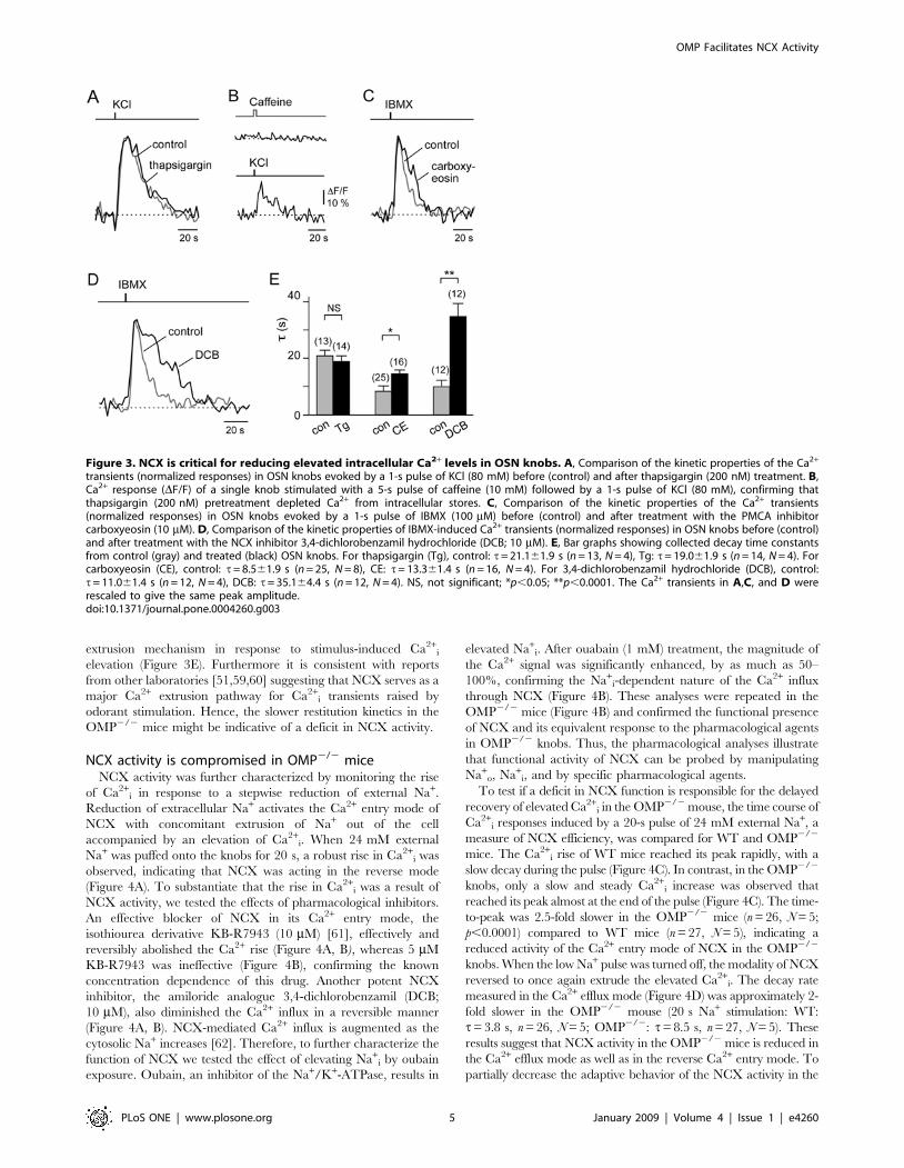
Figure 3. NCX is critical for reducing elevated intracellular Ca2+ levels in OSN knobs. A, Comparison of the kinetic properties of the Ca2+
transients (normalized responses) in OSN knobs evoked by a 1-s pulse of KCl (80 mM) before (control) and after thapsigargin (200 nM) treatment. B,Ca2+ response (DF/F) of a single knob stimulated with a 5-s pulse of caffeine (10 mM) followed by a 1-s pulse of KCl (80 mM), confirming thatthapsigargin (200 nM) pretreatment depleted Ca2+ from intracellular stores. C, Comparison of the kinetic properties of the Ca2+ transients(normalized responses) in OSN knobs evoked by a 1-s pulse of IBMX (100 mM) before (control) and after treatment with the PMCA inhibitorcarboxyeosin (10 mM). D, Comparison of the kinetic properties of IBMX-induced Ca2+ transients (normalized responses) in OSN knobs before (control)and after treatment with the NCX inhibitor 3,4-dichlorobenzamil hydrochloride (DCB; 10 mM). E, Bar graphs showing collected decay time constantsfrom control (gray) and treated (black) OSN knobs. For thapsigargin (Tg), control: t= 21.161.9 s (n = 13, N = 4), Tg: t= 19.061.9 s (n = 14, N = 4). Forcarboxyeosin (CE), control: t= 8.561.9 s (n = 25, N = 8), CE: t= 13.361.4 s (n = 16, N = 4). For 3,4-dichlorobenzamil hydrochloride (DCB), control:t= 11.061.4 s (n = 12, N = 4), DCB: t= 35.164.4 s (n = 12, N = 4). NS, not significant; *p,0.05; **p,0.0001. The Ca2+ transients in A,C, and D wererescaled to give the same peak amplitude.doi:10.1371/journal.pone.0004260.g003
OMP Facilitates NCX Activity
PLoS ONE | www.plosone.org 5 January 2009 | Volume 4 | Issue 1 | e4260
WT response, 24 mM Na+o was also applied for 10 s. The decay
rate was again almost 2-fold slower in the OMP2/2 mice (n = 12,
N = 4), suggesting that the decay kinetics were not affected by
adaptation. Taken together, our data demonstrate that in the
absence of OMP a kinetic deficit exists in transporting Ca2+ in both
directions through NCX, suggesting that OMP serves as a
modulator of NCX function in OSN knobs.
OMP influences NCX activity via CaM and Bex1NCX protein has been reported on the apical regions of OSNs
[59,63,64]. In situ hybridization indicates that mRNAs for multiple
NCX genes are present in OSNs [63,65], and the NCX1 protein is
expressed in nearly all neurons of the olfactory epithelium [63]. To
determine whether the influence of OMP on NCX activity is
reflected in cellular localization of the two proteins in OSNs we
performed double-immunofluorescent staining for NCX1 and
OMP on olfactory epithelium of the WT mouse. At high
magnification (Figure 5A), it is evident that OMP and NCX1
are co-localized in the OSN cell bodies, dendrites, dendritic knobs
and sensory cilia. A similar expression pattern of NCX1 was
observed in the OMP2/2 mouse (not shown). These observations
provide supporting evidence that the compromised ability in
reducing the elevated [Ca2+]i in OMP2/2 mice could rather be
attributed to a functional deficit of NCX activity than to a deficit
in NCX protein expression.
A possible mechanism by which OMP could influence NCX
activity is through a dynamic interaction of OMP and Bex1. Bex1
protein was recently identified as an interacting protein partner of
OMP [37–40] that binds calmodulin (CaM) in a Ca2+-dependent
manner [41]. These observations provide the basis for a potential
mechanistic link between OMP, Bex protein, CaM and NCX. An
interaction between CaM and NCX1 was suggested to be
mediated through the exchanger inhibitory peptide (XIP) site on
the large intracellular loop of NCX1 [66]. This prompted us to
evaluate the interactions of the XIP peptide of NCX1 and the
CaM binding region of Bex1 with recombinant calmodulin (CaM)
and OMP in real-time using surface plasmon resonance (SPR). To
address whether CaM is able to bind to NCX, synthetic XIP
peptide (NCX1 amino acids 251–271) was immobilized in flow
cells of a CM5 sensor chip, and purified CaM at various
concentrations was passed over the surface of the flow cell. CaM
bound to the immobilized XIP (Figure 5B) in a Ca2+-dependent
manner. Using the 1:1 Langmuir binding model, the Kd value for
XIP peptide binding to CaM was calculated to be 2066 nM.
We also confirmed Ca2+-dependent interaction of Bex1 peptide
with CaM by SPR analysis. CaM interacted with immobilized Bex1
(residues 50–75) rapidly and reversibly (Figure 5C), consistent with
our previous report [41]. Due to the fast on/off rate, the affinity
value was evaluated using steady-state analysis [67]. The dissociation
constant was determined to be 280 nM683 nM for the Bex1
peptide. The interactions of CaM with each of these peptides were
dependent on the presence of Ca2+. By contrast, the interaction of
both OMP and XIP (Kd = 700 nM698 nM) and OMP and Bex1
peptide (Kd = 90612 mM) were Ca2+-independent (not shown).
To further assess the interactions of Bex and XIP with CaM, we
evaluated the ability of these peptides to influence the CaM-
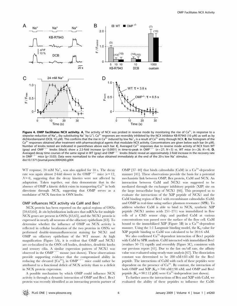
Figure 4. OMP facilitates NCX activity. A, The activity of NCX was probed in reverse mode by monitoring the rise of Ca2+i in response to a
stepwise reduction of Na+o (by substituting Na+ by Li+). Ca2+ responses are reversibly inhibited by the NCX inhibitor KB-R7943 (10 mM) as well as by
dichlorobenzamil (DCB, 10 mM). This confirms that the rise in Ca2+ induced by low Na+o is a result of Ca2+ entry through NCX. B, Bar histogram of the
Ca2+ responses obtained after treatment with pharmacological agents that modulate NCX activity. Concentrations are given below each bar (in mM).Number of knobs tested are indicated in parentheses above each bar. C, Averaged Ca2+ responses due to reverse mode activity of NCX from WT(gray) and OMP2/2 knobs (black) show a 2.5-fold increase (p,0.0001) in time-to-peak in OMP2/2 (n = 27; N = 5) vs. WT mice (n = 26; N = 4). D,Averaged decay time courses of the same signal in WT (gray) and OMP2/2 knobs (black) reveal an approximately 2-fold increase in the recovery ratein OMP2/2 mice (p,0.03). Data were normalized to the value obtained immediately at the end of the 20-s low Na+ stimulus.doi:10.1371/journal.pone.0004260.g004
OMP Facilitates NCX Activity
PLoS ONE | www.plosone.org 6 January 2009 | Volume 4 | Issue 1 | e4260

dependent activation of cAMP-PDE activity [68]. Each of these
peptides strongly interferes with the CaM-dependent activation of
cAMP-PDE activity in a concentration dependent manner (J.
Margolis personal comm.). The XIP peptide is 25–50 fold more
effective than is the Bex1 peptide consistent with our SPR data
(data not shown). In addition, full-length recombinant Bex1
protein also effectively inhibits the CaM-dependent activation of
the cAMP-PDE activity (J. Margolis personal comm.). These
observations are consistent with a model in which OMP influences
NCX activity in OSN knobs by a dynamic series of multi-protein
interactions between OMP, Bex1 and CaM that result in
alterations of the efficacy of CaM. Our observations thus also
extend prior reports that NCX1 activity is regulated as a multi-
protein macromolecular complex [69,70].
Discussion
Several major results emerge from this study. (1) Ca2+ imaging
of the dendritic knobs of OSNs from OMP2/2 mice shows that
the lack of OMP compromises rapid extrusion of elevated Ca2+i
generated in response to activation of the prototypic cAMP
signalling pathway that is used in canonical olfactory sensory
neurons for signal transduction. (2) The findings indicate that this
effect depends on altered regulation of NCX activity in the OSN
dendritic knobs of OMP2/2 mice. (3) Molecular analysis indicates
that proper Ca2+ extrusion might depend on a series of protein-
protein interactions among OMP, Bex1, Ca2+/CaM and NCX, and
that these interactions could be altered in OSNs from OMP2/2
mice. (4) Together, these findings provide new insight into the
mechanisms underlying Ca2+ regulation in dendritic knobs of mouse
OSNs.
OMP is known to act as a modulator of olfactory signal
transduction as demonstrated by the electrophysiological and
behavioral deficits of OMP2/2 mice [12–15] and by the rescue of
these deficits with an OMP-expressing adenovirus [10,11]. In the
present report we have identified a potential site in the signal
transduction cascade where OMP acts and provide evidence for
a mechanism by which this occurs. Our demonstration that
the recovery of Ca2+ transients was significantly slower in the
OMP2/2 mouse illustrates that OMP plays an important role in
the processes of Ca2+ removal in the dendritic knob as distinct
from the various mechanisms of Ca2+ entry through CNG
channels, voltage-gated Ca2+ channels, or release from Ca2+
stores (Figure 1). Furthermore, our data demonstrate that the
mechanism of Ca2+ removal is by way of extrusion primarily via
NCX (Figure 2), which is abundantly present in the dendritic
knobs (Figure 4), and not by intracellular Ca2+ re-uptake. We
found no significant difference in resting level of Ca2+i between
WT and OMP2/2 mice that might directly influence other steps
in the transduction cascade.
To demonstrate functional deficiency of NCX activity in the
absence of OMP, we took advantage of the bi-directional nature of
NCX that can transport Ca2+ into, or out of, cells depending on
the relative concentrations of Ca2+ and Na+ (Figure 3). The highly
significant kinetic deficit in transporting Ca2+ through NCX in
OSN dendritic knobs of OMP2/2 mice indicates that OMP
facilitates NCX activity. We propose a mechanism by which this
occurs: OMP influences NCX activity in OSNs by a series of
multi-protein interactions involving OMP, Bex1 and CaM. We
have previously reported that OMP interacts with Bex1 [37,38];
that this interaction preferentially takes place with a short-lived
OMP-dimer [39]; and that the Bex1 protein interacts with CaM in
Figure 5. Evidence that OMP regulates NCX1 through interaction with CaM and Bex. A, NCX1 and OMP are highly expressed and co-localized in cell bodies, dendrites, dendritic knobs (long arrows) and cilia (short arrows) of mature OSNs of WT mice. Scale bar, 5 mm. B, Sensorgramsfor interaction of XIP peptide immobilized on a CM5 sensorchip and CaM (10, 25, 50,100, 250 nM) using a BIAcore 3000 biosensor. Running bufferused during recording contained 0.1 mM Ca2+. Data were fitted with a 1:1 Langmuir binding model using BIAevaluation software, giving Kd = 20 nM.C, Sensorgram for interaction of immobilized Bex1 (50–75) peptide and CaM (31.25, 62.5, 125, 250, 500 nM) on a CM5 chip in presence of 0.1 mMCa2+. D, Because of the rapid on/off kinetics of the interaction between CaM and Bex1 peptide, saturation curves of equilibrium response versus CaMconcentration were analyzed using a nonlinear 1:1 binding isotherm model, giving Kd = 280 nM.doi:10.1371/journal.pone.0004260.g005
OMP Facilitates NCX Activity
PLoS ONE | www.plosone.org 7 January 2009 | Volume 4 | Issue 1 | e4260

a Ca2+-dependent manner [41]. The eXchanger Inhibitory
Peptide (XIP) region (residues 251–271) of NCX1 specifically
inhibits NCX exchange activity, interacts with CaM [66] and is a
high affinity CaM binding site (Kd = 20 nM) as confirmed by our
SPR experiments (Figure 5). Furthermore, OMP can interact with
the XIP peptide as well as with Bex1 implicating OMP as a
modulator of CaM dependent processes in the transduction
cascade.
Thus the interaction of the XIP site of NCX with CaM and with
OMP provides a linkage between OMP and NCX through the
interactions of OMP, XIP, Bex1 and CaM. Consistent with this
postulate is our recent observation that odor-evoked field potential
responses (EOG) of the Bex12/2 mouse show a delayed onset and
recovery time course (Leinders-Zufall and Margolis, unpub.)
similar to that reported for OMP2/2 mice [10,15]. The
involvement of CaM in almost every step of the olfactory signal
transduction pathway [35,71] suggests that other steps of this
pathway should also be influenced in the OMP-KO. This is
apparent from the delayed onset and prolonged recovery in
response to stimulus application observed in the EOG recordings
of the OMP-KO [10,15] as well as the altered responses seen in
voltage-gated dye recordings in response to stimulus application to
the olfactory epithelium of OMP-KO mice [12]. Curiously, our
data indicating that OMP influences a very late step in the
transduction cascade might appear to be in disagreement with the
conclusion by Reisert et al. [9] that OMP acts at a very early step
in the cascade to regulate cAMP kinetics. In that study single cell
recordings of dissociated OSNs of OMP-KO mice were monitored
using suction electrodes, a method thought to report primarily
from events occurring in the cilia. However, rather than being
discordant with our interpretations, their data are consistent with
our hypothesis that the interaction of OMP and Bex can modulate
multiple CaM-mediated events throughout the transduction
cascade, and with the previous reports [10,15] that both early
and late events in the EOG are altered in the OMP-KO mouse.
These observations are consistent with the complex role of
calmodulin that influences multiple steps in the olfactory
transduction pathway. It is also consistent with the recent reports
[52,53] of a significant role for PMCA, another CaM regulated
participant in the transduction process.
In conclusion, we have demonstrated that OMP plays an
important role in olfactory signal transduction by influencing the
rate of removal of elevated Ca2+i in OSN dendritic knobs. The
persistently delayed restitution of Ca2+ transients in the OSN
knobs of OMP2/2 mice, demonstrates that OMP participates in
the regulation of NCX activity. In summary, we propose a model,
and provide evidence, to explain the involvement of OMP in the
regulation of NCX activity that involves the interaction of OMP,
Bex and CaM to regulate NCX activity. Further, we suggest that
the dynamic interactions among these proteins provides a
mechanism to help explain the complex phenotype of the OMP-
KO mouse based on OMP modulation of the efficacy of CaM at
multiple steps of the olfactory chemo-transduction pathway. This
provides the first mechanistic insight to the elusive role of OMP
and provides a framework for further investigation of its function.
Materials and Methods
Confocal Ca2+ imaging using intact mouse MOEpreparation
Mice (3–6 month old) of homozygous OMP2/2 mice or the
parental strain (129X1/Sv; Jackson Laboratories, Bar Harbor,
ME) were used. The generation and genotyping of the OMP-null
mice has been described previously [15]. All procedures were
approved by the Institutional Animal Care and Use Committee of
the University of Maryland School of Medicine. The main
olfactory epithelium (MOE) was removed from the nasal septum
and mounted in a recording chamber with the mucous layer facing
up [72,73]. The preparation was superfused continuously at room
temperature with normal oxygenated external solution (95% O2/
5% CO2) containing (in mM) 120 NaCl, 25 NaHCO3, 5 KCl, 5
BES, 1 MgSO4, 1 CaCl2, and 10 mM glucose. The epithelial
preparation was loaded with the Ca2+ indicator fluo-4/AM
[74,75]. Changes in intracellular Ca2+ concentration were imaged
in single knobs by using a confocal laser system [74,75]. Optical
sections were ,9 mm thick. Images were acquired at rates between
0.63 and 0.45 Hz using BioRad’s LaserSharp software and
analyzed using NIH Image 1.63 and Igor Pro software (Wave-
metrics, Lake Oswego, OR). Data are expressed as mean6SEM
with the number of knobs (n) and mice (N) indicated. Unless
otherwise stated, Ca2+ transients shown are averaged traces from 5
representative knobs. Statistical significance between two groups
was evaluated using the Student’s unpaired t-test. Probability
values (p) of ,0.05 were considered statistically significant.
Stimuli were diluted in extracellular solution [in mM: 145 NaCl,
5 KCl, 1 CaCl2, 1 MgCl2, 10 Hepes, pH = 7.3 (NaOH),
300 mOsm (glucose)] immediately before use and focally ejected
onto the olfactory knobs by air pressure and using multi-barrelled
stimulation pipettes. Low external Ca2+ solution contained (in
mM): 120 NaCl, 25 NaHCO3, 5 KCl, 4.25 CaCl2, 5 EGTA, 1
MgCl2, 5 BES, and 6 glucose at pH = 7.3 and 300 mOsm, giving
a free Ca2+ concentration of approximately 0.6 mM. Low external
Na+ solution contained (mM): 125 LiCl, 20 NaCl, 5 KCl, 1 CaCl2,
1 Mg Cl2, 10 Hepes, 4 NaOH, pH = 7.3, 300 mOsm (glucose). 3-
isobutyl-1-methylxanthine (IBMX), KB-R7943 (kindly provided
by Nippon Organon, Inc., Osaka, Japan), 3,4-dichlorobenzamil
hydrochloride, and 5,6-carboxyeosin diacetate, succinimidyl ester
(CE; Invitrogen, Eugene, OR) were prepared in DMSO. Dilutions
were made freshly with final DMSO concentrations of ,0.1% (v/
v). Unless otherwise stated, chemicals were obtained from Sigma
(St. Louis, MO).
Carboxyeosin (CE) stock solution, initially made in DMSO, was
added to the extracellular solution (for composition see above) to
give a concentration of 10 mM. The tissue was preincubated for
15 minutes and then superfused with CE-free solution for an
additional 10 minutes to allow de-esterification of CE and to wash
off the residual compound in the bath. The concentration of
10 mM CE is 500-fold higher than the reported IC50 value [76].
KB-R7943 and DCB (3,4-dichlorobenzamil hydrochloride)
were both first dissolved in DMSO and then further diluted in
extracellular solution immediately before use to yield a concen-
tration of 5–10 mM and 10 mM, respectively. Both KB-R7943 and
DCB were directly applied to the knobs 5 minutes prior and
during additional measurements. DCB was also tested at a
concentration of 20 mM. The effect of 10 or 20 mM DCB was
indistinguishable, except at the higher concentration where the
inhibitory effect of DCB was irreversible. The general inhibitory
working concentration of DCB on NCX used by several authors is
10–40 mM in various systems [e.g. [77–79]].
Thapsigargin (Tg) was initially dissolved in DMSO to give a
2 mM stock solution. The agent was diluted to the final
concentration of 200 nM immediately before use, sonicated, and
applied to the extracellular solution for 15 minutes. In some
experiments recordings were made from the same knob before and
after loading with Tg.
For a few selected experiments, we used freshly dissociated
mouse OSNs by adapting previously described procedures [80]. In
this case the septal MOE was minced into small pieces, incubated
OMP Facilitates NCX Activity
PLoS ONE | www.plosone.org 8 January 2009 | Volume 4 | Issue 1 | e4260
for 15 min at 37uC in low Ca2+ solution containing papain
(1.4 mg/ml) and DNase (1 U/ml; Promega). The tissue was then
gently extruded in normal oxygenated external solution and
centrifuged for 10 min at 600 rpm. The pellet was resuspended in
external solution and the released single cells were plated onto a
glass cover slip previously coated with 0.01% poly-L-lysine and
0.01% laminin. Resting Ca2+concentrations were determined by
imaging olfactory knobs using the in situ preparation or freshly
dissociated OSNs. Single-wavelength calibration (fluo-4/AM) was
performed as described by Kao et al. [81] and Leinders-Zufall et
al. [29]. Dual-wavelength calibration (fura-2/AM) was performed
in dissociated OSNs by using a microscope system equipped with
UV laser and CCD camera (kindly provided by Dr. J.P.Y. Kao,
University of Maryland, Baltimore). The following equation was
used to determine resting Ca2+i in fura-2/AM loaded OSNs:
Ca2z� �
i~Kd R{Rminð Þ= Rmax{Rð Þ Sf2=Sb2
where Kd is the Ca2+-fura-2 dissociation constant, 224 nM. R is
(w12w)/(w22w) where w1 and w2 are the fluorescence intensity at
excitation wavelength of 340 and 380 nm, respectively, and w
equals autofluorescence after cell lysis with digitonin (50 mM).
Some of the values were predetermined by Dr. Kao for this
specific imaging system: Rmin = 0.35860.0585; Rmax = to4.696
0.678; Sf2/Sb2 = 4.07260.533.
Colocalization of OMP and NCX1 by immunofluorescentstaining
Olfactory neuroepithelial tissue was obtained from postnatal
day 7 (P7) WT mice. Mice were anesthetized with 60 mg/kg
nembutal and perfused transcardially with 20 ml of ice cold PBS
followed by 30 ml of 4% freshly prepared phosphate buffered
paraformaldehyde. Tissue was dissected and postfixed for two
hours in cold fixative and cryoprotected overnight in 30% sucrose
at 4uC. Tissues were embedded in OCT (Tissue Tek, Sakura,
Torrance, CA) and snap-frozen in a dry ice/isopentane bath.
Coronal cryostat sections of olfactory tissue (12 mm) were attached
to Superfrost-plus microscope slides (Fisher, Pittsburgh, PA), dried
at 37uC for 15 min and stored at 280uC until needed. For single
and double-labeled immunofluorescence staining goat anti-OMP
antibody (1:20,000, [3] and rabbit anti-NCX1 antibody (1:2,000,
Swant, Bellinzona, Switzerland) were applied to the olfactory
turbinate sections on slides and processed essentially as described
previously [40].
Anti-OMP staining used a polyclonal goat primary antiserum
generated against OMP purified from rat olfactory tissue [3]. This
antiserum has been characterized extensively for immunocyto-
chemistry [e.g. [2,7], and its specificity has been verified by the
absence of immunoreactivity in OMP2/2 mice [1].
Anti-NCX1 staining used a rabbit polyclonal anti-NCX1
antiserum directed against canine cardiac sarcolemmal NCX1
originally reported by Philipson et al. [82]. This antiserum gives
the same staining pattern in mouse olfactory epithelium [83] as
does the anti-NCX1-peptide antiserum AbO-8 [84]. Additional
characterization of this antiserum has been published elsewhere
[85,86].
Surface plasmon resonance (SPR) experimentsThe Biacore 3000 (Biacore AB, Uppsala, Sweden) was used to
study peptide-protein interactions by SPR. Bovine calmodulin was
purchased from Sigma, recombinant rat OMP was expressed and
purified as previously described [87]. The XIP peptide (NCX1
251–271, RRLLFYKYVYKRYRAGKQRGM) and the CaM
binding peptide of Bex1 (Bex1 50–75, RGGRRRFRVRQPIA-
HYRWDLMQRVGE) were synthesized and HPLC purified in
the biopolymer/genomics core facility of the University of
Maryland. Each peptide was separately immobilized on a
carboxymethylated-dextran (CM5) chip surface using standard
carbodiimide chemistry according to the manufacturer’s instruc-
tions. The average amount of each immobilized peptide was about
700 RU (resonance unit). Binding was evaluated over a
concentration range of 0–500 nM CaM or OMP in running
buffer in the absence (NaCl 150 mM; HEPES 10 mM; pH 7.4) or
presence of Ca2+ (NaCl 150 mM; HEPES 10 mM; CaCl20.1 mM; pH 7.4) under a continuous flow of 5 ml/min for
2 minutes, unless otherwise stated. Flow cells were regenerated
by flushing with running buffer without CaCl2. Binding data were
fitted and analyzed using Biacor evaluation version 4.1 software
(Biacore). Data are representative of three separate experiments.
Acknowledgments
We thank Dr. Joseph P.Y. Kao for valuable discussions and equipment
support, Drs. K.W.Yau and J. Reisert for useful discussions, Dr. Y. Zhang
for valuable assistance in set-up and data analysis with the Biacore 300, Ms.
J. Margolis for access to unpublished data, and Ms. F. Scipio for technical
assistance. Nippon-Organon K,K, generously provided a sample of KB-
R7943. TLZ is a Lichtenberg Professor of the Volkswagen Foundation.
Author Contributions
Conceived and designed the experiments: FZ TLZ FLM. Performed the
experiments: HJK JHK. Analyzed the data: HJK JHK FZ TLZ FLM.
Contributed reagents/materials/analysis tools: FZ TLZ FLM. Wrote the
paper: HJK JHK FZ TLZ FLM.
References
1. Buiakova OI, Krishna NS, Getchell TV, Margolis FL (1994) Human and rodent
OMP genes: conservation of structural and regulatory motifs and cellular
localization. Genomics 20: 452–462.
2. Monti-Graziadei GA, Margolis FL, Harding JW, Graziadei PP (1977)
Immunocytochemistry of the olfactory marker protein. J Histochem Cytochem
25: 1311–1316.
3. Keller A, Margolis FL (1975) Immunological studies of the rat olfactory marker
protein. J Neurochem 24: 1101–1106.
4. Schwob JE, Szumowski KE, Stasky AA (1992) Olfactory sensory neurons are
trophically dependent on the olfactory bulb for their prolonged survival.
J Neurosci 12: 3896–3919.
5. Margolis FL, Verhaagen J, Biffo S, Huang FL, Grillo M (1991) Regulation of
gene expression in the olfactory neuroepithelium: a neurogenetic matrix. Prog
Brain Res 89: 97–122.
6. Margolis FL (1982) Olfactory marker protein (OMP). Scand J Immunol Suppl 9:
181–199.
7. Farbman AI, Margolis FL (1980) Olfactory marker protein during ontogeny:
immunohistochemical localization. Dev Biol 74: 205–215.
8. Margolis FL (1972) A brain protein unique to the olfactory bulb. Proc Natl Acad
Sci U S A 69: 1221–1224.
9. Reisert J, Yau KW, Margolis FL (2007) Olfactory marker protein modulates the
cAMP kinetics of the odour-induced response in cilia of mouse olfactory receptor
neurons. J Physiol 585: 731–740.
10. Ivic L, Pyrski MM, Margolis JW, Richards LJ, Firestein S, et al. (2000)
Adenoviral vector-mediated rescue of the OMP-null phenotype in vivo. Nat
Neurosci 3: 1113–1120.
11. Youngentob SL, Pyrski MM, Margolis FL (2004) Adenoviral vector-mediated
rescue of the OMP-null behavioral phenotype: enhancement of odorant
threshold sensitivity. Behav Neurosci 118: 636–642.
12. Youngentob SL, Kent PF, Margolis FL (2003) OMP gene deletion results in an
alteration in odorant-induced mucosal activity patterns. J Neurophysiol 90:
3864–3873.
13. Youngentob SL, Margolis FL, Youngentob LM (2001) OMP gene deletion results
in an alteration in odorant quality perception. Behav Neurosci 115: 626–631.
14. Youngentob SL, Margolis FL (1999) OMP gene deletion causes an elevation in
behavioral threshold sensitivity. Neuroreport 10: 15–19.
OMP Facilitates NCX Activity
PLoS ONE | www.plosone.org 9 January 2009 | Volume 4 | Issue 1 | e4260
15. Buiakova OI, Baker H, Scott JW, Farbman A, Kream R, et al. (1996) Olfactory
marker protein (OMP) gene deletion causes altered physiological activity of
olfactory sensory neurons. Proc Natl Acad Sci U S A 93: 9858–9863.
16. Matthews HR, Reisert J (2003) Calcium, the two-faced messenger of olfactory
transduction and adaptation. Curr Opin Neurobiol 13: 469–475.
17. Liu M, Chen TY, Ahamed B, Li J, Yau KW (1994) Calcium-calmodulin
modulation of the olfactory cyclic nucleotide-gated cation channel. Science 266:
1348–1354.
18. Leinders-Zufall T, Ma M, Zufall F (1999) Impaired odor adaptation in olfactory
receptor neurons after inhibition of Ca2+/calmodulin kinase II. J Neurosci 19:
RC19.
19. Leinders-Zufall T, Greer CA, Shepherd GM, Zufall F (1998) Imaging odor-
induced calcium transients in single olfactory cilia: specificity of activation and
role in transduction. J Neurosci 18: 5630–5639.
20. Wei J, Zhao AZ, Chan GC, Baker LP, Impey S, et al. (1998) Phosphorylation
and inhibition of olfactory adenylyl cyclase by CaM kinase II in Neurons: a
mechanism for attenuation of olfactory signals. Neuron 21: 495–504.
21. Frings S (2001) Chemoelectrical signal transduction in olfactory sensory neurons
of air-breathing vertebrates. Cell Mol Life Sci 58: 510–519.
22. Breer H (1993) Second messenger signalling in olfaction. Ciba Found Symp 179:
97–109; discussion 109–114, 147–109.
23. Lancet D (1986) Vertebrate olfactory reception. Annu Rev Neurosci 9: 329–355.
24. Zufall F, Firestein S, Shepherd GM (1994) Cyclic nucleotide-gated ion channels
and sensory transduction in olfactory receptor neurons. Annu Rev Biophys
Biomol Struct 23: 577–607.
25. Reed RR (1992) Signaling pathways in odorant detection. Neuron 8: 205–209.
26. Kleene SJ (2008) The Electrochemical Basis of Odor Transduction in Vertebrate
Olfactory Cilia. Chem Senses.
27. Dzeja C, Hagen V, Kaupp UB, Frings S (1999) Ca2+ permeation in cyclic
nucleotide-gated channels. Embo J 18: 131–144.
28. Frings S, Seifert R, Godde M, Kaupp UB (1995) Profoundly different calcium
permeation and blockage determine the specific function of distinct cyclic
nucleotide-gated channels. Neuron 15: 169–179.
29. Leinders-Zufall T, Rand MN, Shepherd GM, Greer CA, Zufall F (1997)
Calcium entry through cyclic nucleotide-gated channels in individual cilia of
olfactory receptor cells: spatiotemporal dynamics. J Neurosci 17: 4136–4148.
30. Zufall F, Leinders-Zufall T (2000) The cellular and molecular basis of odor
adaptation. Chem Senses 25: 473–481.
31. Hallani M, Lynch JW, Barry PH (1998) Characterization of calcium-activated
chloride channels in patches excised from the dendritic knob of mammalian
olfactory receptor neurons. J Membr Biol 161: 163–171.
32. Kurahashi T, Yau KW (1993) Co-existence of cationic and chloride components
in odorant-induced current of vertebrate olfactory receptor cells. Nature 363:
71–74.
33. Kleene SJ, Gesteland RC (1991) Calcium-activated chloride conductance in frog
olfactory cilia. J Neurosci 11: 3624–3629.
34. Kleene SJ (1993) Origin of the chloride current in olfactory transduction.
Neuron 11: 123–132.
35. Kaneko H, Mohrlen F, Frings S (2006) Calmodulin contributes to gating control
in olfactory calcium-activated chloride channels. J Gen Physiol 127: 737–748.
36. Kurahashi T, Menini A (1997) Mechanism of odorant adaptation in the
olfactory receptor cell. Nature 385: 725–729.
37. Baldisseri DM, Margolis JW, Weber DJ, Koo JH, Margolis FL (2002) Olfactory
marker protein (OMP) exhibits a beta-clam fold in solution: implications for
target peptide interaction and olfactory signal transduction. J Mol Biol 319:
823–837.
38. Behrens M, Margolis JW, Margolis FL (2003) Identification of members of the
Bex gene family as olfactory marker protein (OMP) binding partners.
J Neurochem 86: 1289–1296.
39. Koo JH, Gill S, Pannell LK, Menco BP, Margolis JW, et al. (2004) The
interaction of Bex and OMP reveals a dimer of OMP with a short half-life.
J Neurochem 90: 102–116.
40. Koo JH, Saraswati M, Margolis FL (2005) Immunolocalization of Bex protein in
the mouse brain and olfactory system. J Comp Neurol 487: 1–14.
41. Koo JH, Smiley MA, Lovering RM, Margolis FL (2007) Bex1 knock out mice
show altered skeletal muscle regeneration. Biochem Biophys Res Commun 363:
405–410.
42. Asanuma N, Nomura H (1993) Cytochemical localization of cyclic 39,59-
nucleotide phosphodiesterase activity in the rat olfactory mucosa. Histochem J
25: 348–356.
43. Asanuma N, Nomura H (1991) Cytochemical localization of adenylate cyclase
activity in rat olfactory cells. Histochem J 23: 83–90.
44. Menco BP, Cunningham AM, Qasba P, Levy N, Reed RR (1997) Putative
odour receptors localize in cilia of olfactory receptor cells in rat and mouse: a
freeze-substitution ultrastructural study. J Neurocytol 26: 691–706.
45. Schwarzenbacher K, Fleischer J, Breer H (2005) Formation and maturation of
olfactory cilia monitored by odorant receptor-specific antibodies. Histochem
Cell Biol 123: 419–428.
46. Kurahashi T, Kaneko A (1991) High density cAMP-gated channels at the ciliary
membrane in the olfactory receptor cell. Neuroreport 2: 5–8.
47. Nakamura T, Gold GH (1987) A cyclic nucleotide-gated conductance in
olfactory receptor cilia. Nature 325: 442–444.
48. Zufall F, Leinders-Zufall T, Greer CA (2000) Amplification of odor-induced
Ca(2+) transients by store-operated Ca(2+) release and its role in olfactory signal
transduction. J Neurophysiol 83: 501–512.
49. Schild D, Jung A, Schultens HA (1994) Localization of calcium entry through
calcium channels in olfactory receptor neurones using a laser scanning
microscope and the calcium indicator dyes Fluo-3 and Fura-Red. Cell Calcium
15: 341–348.
50. Reisert J, Matthews HR (2001) Response properties of isolated mouse olfactory
receptor cells. J Physiol 530: 113–122.
51. Reisert J, Matthews HR (1998) Na+-dependent Ca2+ extrusion governs
response recovery in frog olfactory receptor cells. J Gen Physiol 112: 529–535.
52. Castillo K, Delgado R, Bacigalupo J (2007) Plasma membrane Ca(2+)-ATPase
in the cilia of olfactory receptor neurons: possible role in Ca(2+) clearance.
Eur J Neurosci 26: 2524–2531.
53. Weeraratne SD, Valentine M, Cusick M, Delay R, Van Houten JL (2006)
Plasma membrane calcium pumps in mouse olfactory sensory neurons. Chem
Senses 31: 725–730.
54. Shmigol A, Eisner DA, Wray S (1998) Carboxyeosin decreases the rate of decay
of the [Ca2+]i transient in uterine smooth muscle cells isolated from pregnant
rats. Pflugers Arch 437: 158–160.
55. Fierro L, DiPolo R, Llano I (1998) Intracellular calcium clearance in Purkinje
cell somata from rat cerebellar slices. J Physiol 510(Pt 2): 499–512.
56. Kuzmiski JB, MacVicar BA (2001) Cyclic nucleotide-gated channels contribute
to the cholinergic plateau potential in hippocampal CA1 pyramidal neurons.
J Neurosci 21: 8707–8714.
57. Kolesnikov SS, Kosolapov AV (1993) Cyclic nucleotide-activated channels in
carp olfactory receptor cells. Biochim Biophys Acta 1150: 63–72.
58. Kolesnikov SS, Zhainazarov AB, Kosolapov AV (1990) Cyclic nucleotide-
activated channels in the frog olfactory receptor plasma membrane. FEBS Lett
266: 96–98.
59. Noe J, Tareilus E, Boekhoff I, Breer H (1997) Sodium/calcium exchanger in rat
olfactory neurons. Neurochem Int 30: 523–531.
60. Jung A, Lischka FW, Engel J, Schild D (1994) Sodium/calcium exchanger in
olfactory receptor neurones of Xenopus laevis. Neuroreport 5: 1741–1744.
61. Iwamoto T, Watano T, Shigekawa M (1996) A novel isothiourea derivative
selectively inhibits the reverse mode of Na+/Ca2+ exchange in cells expressing
NCX1. J Biol Chem 271: 22391–22397.
62. Rasgado-Flores H, Santiago EM, Blaustein MP (1989) Kinetics and stoichiom-
etry of coupled Na efflux and Ca influx (Na/Ca exchange) in barnacle muscle
cells. J Gen Physiol 93: 1219–1241.
63. Pyrski M, Koo JH, Polumuri SK, Ruknudin AM, Margolis JW, et al. (2007)
Sodium/calcium exchanger expression in the mouse and rat olfactory systems.
J Comp Neurol 501: 944–958.
64. Lucero MT, Huang W, Dang T (2000) Immunohistochemical evidence for the
Na+/Ca2+ exchanger in squid olfactory neurons. Philos Trans R Soc
Lond B Biol Sci 355: 1215–1218.
65. Schulze DH, Pyrski M, Ruknudin A, Margolis JW, Polumuri SK, et al. (2002)
Sodium-calcium exchangers in olfactory tissue. Ann N Y Acad Sci 976: 67–72.
66. Li Z, Nicoll DA, Collins A, Hilgemann DW, Filoteo AG, et al. (1991)
Identification of a peptide inhibitor of the cardiac sarcolemmal Na(+)-Ca2+exchanger. J Biol Chem 266: 1014–1020.
67. Turner JH, Gelasco AK, Raymond JR (2004) Calmodulin interacts with the
third intracellular loop of the serotonin 5-hydroxytryptamine1A receptor at two
distinct sites: putative role in receptor phosphorylation by protein kinase C. J Biol
Chem 279: 17027–17037.
68. Thompson MP (1988) Calcium-binding proteins: Characterization and
properties. Boca Raton, FL, USA: CRC Press. pp 89–93.
69. Ruknudin AM, Wei SK, Haigney MC, Lederer WJ, Schulze DH (2007)
Phosphorylation and other conundrums of Na/Ca exchanger, NCX1.
Ann N Y Acad Sci 1099: 103–118.
70. Schulze DH, Muqhal M, Lederer WJ, Ruknudin AM (2003) Sodium/calcium
exchanger (NCX1) macromolecular complex. J Biol Chem 278: 28849–28855.
71. Menini A (1999) Calcium signalling and regulation in olfactory neurons. Curr
Opin Neurobiol 9: 419–426.
72. Ma M, Shepherd GM (2000) Functional mosaic organization of mouse olfactory
receptor neurons. Proc Natl Acad Sci U S A 97: 12869–12874.
73. Leinders-Zufall T, Cockerham RE, Michalakis S, Biel M, Garbers DL, et al.
(2007) Contribution of the receptor guanylyl cyclase GC-D to chemosensory
function in the olfactory epithelium. Proc Natl Acad Sci U S A 104:
14507–14512.
74. Leinders-Zufall T, Brennan P, Widmayer P, Chandramani SP, Maul-Pavicic A,
et al. (2004) MHC class I peptides as chemosensory signals in the vomeronasal
organ. Science 306: 1033–1037.
75. Leinders-Zufall T, Lane AP, Puche AC, Ma W, Novotny MV, et al. (2000)
Ultrasensitive pheromone detection by mammalian vomeronasal neurons.
Nature 405: 792–796.
76. Gatto C, Milanick MA (1993) Inhibition of the red blood cell calcium pump by
eosin and other fluorescein analogues. American Journal of Physiology 264:
C1577–1586.
77. Krasznai Z, Krasznai ZT, Morisawa M, Bazsane ZK, Hernadi Z, et al. (2006)
Role of the Na+/Ca2+ exchanger in calcium homeostasis and human sperm
motility regulation. Cell Motil Cytoskeleton 63: 66–76.
OMP Facilitates NCX Activity
PLoS ONE | www.plosone.org 10 January 2009 | Volume 4 | Issue 1 | e4260

78. Rogister F, Laeckmann D, Plasman PO, Van Eylen F, Ghyoot M, et al. (2001)
Novel inhibitors of the sodium-calcium exchanger: benzene ring analogues of N-
guanidino substituted amiloride derivatives. Eur J Med Chem 36: 597–614.
79. Siegl PK, Cragoe EJ, Trumble MJ, Kaczorowski GJ (1984) Inhibition of Na+/
Ca2+ exchange in membrane vesicle and papillary muscle preparations from
guinea pig heart by analogs of amiloride. Proc Natl Acad Sci U S A 81:
3238–3242.
80. Lucas P, Ukhanov K, Leinders-Zufall T, Zufall F (2003) A diacylglycerol-gated
cation channel in vomeronasal neuron dendrites is impaired in TRPC2 mutant
mice: mechanism of pheromone transduction. Neuron 40: 551–561.
81. Kao JP, Harootunian AT, Tsien RY (1989) Photochemically generated cytosolic
calcium pulses and their detection by fluo-3. J Biol Chem 264: 8179–8184.
82. Philipson KD, Longoni S, Ward R (1988) Purification of the cardiac Na+-Ca2+exchange protein. Biochem Biophys Acta 945: 298–306.
83. Pyrski M, Koo JH, Polumuri SK, Ruknudin A, Margolis JW, et al. (2007)
Sodium/calcium exchanger expression in the mouse and rat olfactory systems.J Comp Neurol 501: 944–958.
84. Cook O, Low W, Rahamimoff H (1998) Membrane topology of the rat brain
Na+-Ca2+ exchanger. Biochim Biophys Acta 1371: 40–52.85. Saba RI, Bollen A, Herchuelz A (1999) Characterization of the 70 kDa
polypeptide of the Na/Ca exchanger. Biochem J 338: 139–145.86. Frank JS, Mottino G, Reid D, Molday RS, Philipson KD (1992) Distribution of
the Na(+)-Ca2+ exchange protein in mammalian cardiac myocytes: an
immunofluorescence and immunocolloidal gold-labeling study. J Cell Biol 117:337–345.
87. Carr VM, Walters E, Margolis FL, Farbman AI (1998) An enhanced olfactorymarker protein immunoreactivity in individual olfactory receptor neurons
following olfactory bulbectomy may be related to increased neurogenesis.J Neurobiol 34: 377–390.
OMP Facilitates NCX Activity
PLoS ONE | www.plosone.org 11 January 2009 | Volume 4 | Issue 1 | e4260