Parametric identification of robotic systems with stable time-varying Hopfield networks
Upload
independentCategory
view
0download
0
Computational Perspectives on Forebrain MicrocircuitsImplicated in Reinforcement Learning, Action Selection, andCognitive Control
Daniel Bullock*,Boston University, Department of Cognitive and Neural Systems, 677 Beacon Street, Boston, MA02215
Can Ozan Tan, andHarvard Medical School, Boston, MA Spaulding Rehabilitation Hospital, 125 Nashua Street, Boston,MA 02114
Yohan J. JohnBoston University, Department of Cognitive and Neural Systems, 677 Beacon Street, Boston, MA02215
AbstractAbundant new information about signaling pathways in forebrain microcircuits presents manychallenges, and opportunities for discovery, to computational neuroscientists who strive to bridgefrom microcircuits to flexible cognition and action. Accurate treatment of microcircuit pathways isespecially critical for creating models that correctly predict the outcomes of candidate neurologicaltherapies. Recent models are trying to specify how cortical circuits that enable planning and voluntaryactions interact with adaptive sub-cortical microcircuits in the basal ganglia. The basal ganglia arestrongly implicated in reinforcement learning, and in all behavior and cognition over which the frontallobes exert flexible control. The persisting role of the basal ganglia shows that ancient vertebratedesigns for motivated action-selection proved adaptable enough to support many “modern”behavioral innovations, including fluent generation of language and speech. This paper summarizeshow recent models have incorporated realistic representations of microcircuit features, and havebegun to trace their computational implications. Also summarized are recent empirical discoveriesthat provide guidance regarding how to formulate the rules for synaptic modification that governlearning in cortico-striatal pathways. Such efforts are contributing to an emerging synthesis basedon an interlocking set of computational hypotheses regarding cortical interactions with basal gangliaand thalamic nuclei. These hypotheses specify how specialized microcircuits solve learning andcontrol problems inherent to the brain's parallel design.
KeywordsBasal ganglia; acetylcholine; dopamine; striatum; decision making
© 2009 Elsevier Ltd. All rights reserved.*Corresponding author Email addresses: [email protected] (Daniel Bullock), [email protected] (Can Ozan Tan), [email protected](Yohan J. John).Publisher's Disclaimer: This is a PDF file of an unedited manuscript that has been accepted for publication. As a service to our customerswe are providing this early version of the manuscript. The manuscript will undergo copyediting, typesetting, and review of the resultingproof before it is published in its final citable form. Please note that during the production process errors may be discovered which couldaffect the content, and all legal disclaimers that apply to the journal pertain.
NIH Public AccessAuthor ManuscriptNeural Netw. Author manuscript; available in PMC 2010 July 1.
Published in final edited form as:Neural Netw. 2009 ; 22(5-6): 757–765. doi:10.1016/j.neunet.2009.06.008.
NIH
-PA Author Manuscript
NIH
-PA Author Manuscript
NIH
-PA Author Manuscript

1. Introduction: Forebrain Circuits for Motivated Action SelectionA recurring task for an animal is to select, among probably-achievable action plans, those actionplans that are more likely to promote its well-being. Thus, the planner needs access to frequentlyupdated estimates not only of act-outcome probabilities and outcome values, but also of theachievability of the actions, given both the actor's current state and the context of action.Because plan evaluation and selection may occur well before the best time to execute a plan,and because plans take time to execute, new options may be noticed after plan selection, butbefore plan completion. Thus, it will often pay to interrupt execution of a plan, perform another,and then resume or abandon the original plan. Coping well with such complications requiressophistication in the forebrain circuitry that enables intelligent planning in mammals.
Although the forebrain encompasses the cerebral cortex and BG (basal ganglia), mosttreatments of intelligent planning and motivated action–selection validly focus on the frontalcortex and BG, which are so linked that the BG exert a much stronger and more direct influenceon frontal than on posterior cortex. Indeed, a general rule is that for every planning and actiondeficit that results from lesioning a discrete region within frontal cortex, a highly similar deficitcan be produced by lesioning whatever discrete part of the BG circuit projects its output towardthe given region within frontal cortex. This makes sense on the hypothesis, now broadlysupported (Bullock, 2004b), that frontal cortex can represent many potential types of cognitiveand other actions, whereas the BG are responsible for selecting which such actions to execute(Redgrave et al., 1999a).
Given that selection among action plans is strongly influenced by reinforcement-guidedlearning, it is not surprising that the BG play multiple roles in action choice and learning. Atthe coarse grain of a tri-partite division of the conventional BG's major input nucleus, thestriatum, it is possible to distinguish: the ventral striatum as important for processing context-conditioned reward expectations, medial-dorsal striatum for act-outcome expectations, andlateral-dorsal striatum for habitual condition action relations. Two of these divisionscorrespond to an actor-critic architecture (Houk et al., 1995), with learning in both actor andcritic compartments governed in part by reward-prediction errors (RPEs), as expected bytemporal difference (TD) models of reinforcement learning (e.g., Sutton and Barto, 1981).However, many microcircuit specializations in the BG go beyond or deviate from what waspredicted by actor-critic architectures, or by associated TD models, and their existence raisesthe problem of identifying their computational implications. Moreover, Swanson (2000) hasargued for extending the concept of the BG to encompass further regions, such as some nucleiof the extended amygdala. Any such extension would entail further di erentiation ofarchitectures and models. In this review, we detail BG microcircuit features, particularly instriatum, which suggest revisions to actor-critic systems and TD-based learning rules;summarize some of their computational implications; and note in-depth treatments publishedelsewhere. A briefer treatment of these issues appeared in Bullock and Tan (2009).
2. Microcircuit Specializations of the Basal Ganglia, with ComputationalImplications2.1. Dopamine (DA) cell firing provides an asymmetrical representation of positive andnegative reward prediction errors (RPEs)
Dopamine cells are well established as carriers of conditioned (i.e., stimulus- and learning-history-dependent) RPE signals. That they discharge spontaneously at a constant rate meansthat positive rate deviations can signal positive RPEs and negative deviations (dips belowbaseline rate) negative RPEs. However, the low value of the baseline firing rate implies atruncation of negative RPE signals, whereas some DA burst signals to positive RPEs remain
Bullock et al. Page 2
Neural Netw. Author manuscript; available in PMC 2010 July 1.
NIH
-PA Author Manuscript
NIH
-PA Author Manuscript
NIH
-PA Author Manuscript
proportionate. For a general class of dual-path models capable of learning to compute RPEsin a network including DA cells, Tan et al. (2008) derived a key computational implication ofthis asymmetry, which accords with DA signal measurements reported by Tobler et al.(2005). Tan et al. (2008) showed that this asymmetry so a ects incremental learning that, afterlearning, the magnitude of residual phasic DA burst signals generated in response to deliveryof a CS-predicted primary reward will scale with the learned conditional probability of rewardomission (i.e., 1 − p(R*|CS)) given the predictive cue (CS). Note that in their derivation, as inthe data (Tobler et al., 2005), the residual DA burst signal is independent of the absolute rewardmagnitude, R*. Yet the dopamine burst signal generated earlier, at CS-onset, reflects both R*and p(R*|CS). Although the concept of RPEs is helpful for interpreting DA burst signals, anyinterpretation that equates the two is incorrect.
2.2. Conditional components of DA signaling go well beyond RPEs, to include most (all?) ofwhat is expected from behavioral reinforcement research
Behavioral research on what constitutes a reinforcer has shown that, in addition to prototypicalevents, such as reward delivery, several other types of events behave as reinforcers, i.e., suchevents’ contingent delivery raises the future probability of cue-conditioned instrumentalbehavior. Notable cases are: contingent cessation of an aversive input, contingent delivery ofnon-aversive stimuli that are novel but not linked to tangible rewards, and contingent accessto the opportunity to engage in a more preferred behavior. Of these, the first two have beenshown to have the effect on DA release that would be expected if such release also mediatesthese components of internal reinforcement signaling (see also Ungless et al., 2004). Someinitial computational implications were disclosed using the new striatal microcircuit model ofTan and Bullock (2008b). The novelty-related DA cell responses present a further challengeto the classical view of DA signals as pure RPE signals. Such novel events are neitherconventional rewards nor conventional learned predictors of such. Recently, two studiesattempted to address the novelty-related increase in DA release within a formal TD framework.Kakade and Dayan (2002) assumed that novelty, in itself, provides “reward bonuses” that alterthe sum of experienced reward, thereby interfering with RPE estimations with respect to normalexternal rewards. In an attempt to avoid treating novelty as inherently rewarding, Laurent(2008) presented a TD–based simulation that showed how positive RPEs to novel events (stateentries) might arise from prior reinforcement learning. Although it seems likely that some ofthe ability of novel stimuli to generate DA responses results from their similarity to otherreward-predicting cues, the treatment in Laurent (2008) does not appear to be compatible withthe key observation that the novelty-related DA responses habituate as a cue's novelty wanes(e.g., Red-grave et al., 1999b). Note that novelty wanes as the cognitive system learns correctpredictions in general - not just correct predictions about rewards. Again, DA burst signalscannot be accurately modeled from a perspective that focuses exclusively on RPEs.
2.3. Sustained dopamine cell signal components reflect uncertainty in reward predictionContrary to expectations of TD models, there is evidence (Fiorillo et al., 2003, 2005) that DAcells exhibit an uncertainty response that is a non-monotonic (inverted-U) function of rewardprobability conditional on the cue: when there is a probabilistic relationship between apredictive cue and a rewarding outcome that may (or may not) occur at a fixed time after cueonset, then there is a gradual buildup of DA cell firing rate between the cue onset and theexpected time of the uncertain reward. In an attempt to derive an uncertainty response withinthe TD framework Niv et al. (2005) proposed that this response emerges as an artifact ofaveraging gradually back-propagating RPEs across successive learning trials. To the contrary,there is no evidence that RPE signals “propagate gradually backwards” in the time betweencue and reward, and the data (Fiorillo et al., 2003, 2005) show that uncertainty responses arerobust on single trials. Uncertainty response appear to be inexplicable using a standard TDmodel.
Bullock et al. Page 3
Neural Netw. Author manuscript; available in PMC 2010 July 1.
NIH
-PA Author Manuscript
NIH
-PA Author Manuscript
NIH
-PA Author Manuscript

More recently, Tan and Bullock (2008b,c) proposed that this signal component may becomputed by a surprisingly common but rarely simulated property of neurons: co-release ofmore than one chemical signaler from the same axon terminal. They showed that the well-established, but computationally mysterious, co-release of GABA and the neuropeptide SP(substance P) from striato-nigral terminals can explain robust single trial computation ofuncertainty responses, which are a non-monotonic function of the conditional probability of areward (R*) given a cue (CS), i.e., p(R*|CS). Under broad conditions, such co-release willproduce a signal proportional to p(1 − p), which shows a peak when uncertainty is maximal,i.e., when p(R*|CS) = .5. Although the discoverers of this DA signal component aptly proposedthat it may be important to explain habitual gambling, Tan and Bullock (2008b) noted that thebroadcast of the DA signal to many brain sites beyond the dorsolateral striatum implies that itcan function much more broadly, and adaptively, to optimize computations in both learningand performance. Notably, it can promote search for more-predictive representations, and rapidswitching away from no-longer-rewarding alternatives. This functional interpretation of therole of sustained elevation of DA level and its origins (co-release of SP from striato-nigralterminals) deviates significantly from simple RPE and TD frameworks, but provides anexplanation for the behavioral effects of SP: injection of SP into the VTA enhances respondingfor conditioned rewards in general, but also disrupts reward discrimination processes andthereby results in (some degree of) response generalization (Placenza et al., 2004; Kelley etal., 1989).
Such specialized task-dependent firing patterns of the DA cells highlight shortcomings of thecomputational models of DA responses that are based on the formal RPE-TD framework. Thegenesis and effects of at least three distinct DA cell firing patterns (reward-related bursts,novelty responses, and sustained uncertainty responses), which exhibit distinct task-dependencies and operate on di erent time-scales, suggest that DAergic projections to striataltarget structures (in particular, to dorsal striatum) engender computations that go beyond thoseenvisioned in the RPE hypothesis of DA and the “actor-critic” concept of BG architecture. Inaddition, oft-neglected interactions between neurotransmitters, coupled with specializedmicrocircuits of the BG, indicate a need to revise common reinforcement rules, and imply afar-reaching role for the BG, not only in reinforcement learning but also in evaluation, selection,and execution of actions whose outcomes are contingent on diverse types of factors. We discussneurotransmitter interactions next, and BG micro- and macrocircuits in Section 2.7
2.4. Striatal learning, adaptive timing and action-gating are governed by a dopamine-acetylcholine (DA-ACh) cascade
Early studies of Parkinson's disease emphasized the striatal balance between DA and ACh inthe performance-control functions of the striatum. Because the only sources for striatal AChare the giant ACh neurons of the striatum itself, the striatal ACh signal source is anatomicallydistinct from the ACh cells whose projections strongly affect attention/arousal as well asneocortical (Kilgard and Merzenich, 1998) and hippocampal (Hasselmo, 2006) learning.Striatal ACh neurons are often called TANs (for “tonically active neurons”), and theirfunctional signaling obeys di erent principles than non-striatal ACh neurons. Like DA cells,they show learning-dependent changes. Recently Tan and Bullock (2008a) showed, in abiophysically realistic simulation of TANs, that many of these learning-dependent changes areattributable to learning-dependent changes in the behavior of DA cells whose axons synapseon the TANs. Because both DA and ACh modulate learning of cortico-striatal synapses(Centonze et al., 1999; Pawlak and Kerr, 2008; Wang et al., 2006), it is now clear that bothstriatal learning and striatal performance functions are strongly dependent on a DA-AChcascade. One immediate implication is that common reinforcement learning rules needupdating to reflect an additional ACh dependency.
Bullock et al. Page 4
Neural Netw. Author manuscript; available in PMC 2010 July 1.
NIH
-PA Author Manuscript
NIH
-PA Author Manuscript
NIH
-PA Author Manuscript

Many common reinforcement learning rules have been solely based on DAergic rewardprediction errors (RPEs). These rules are often used in combination with the concept of directand indirect pathways in the BG, (e.g., Albin et al., 1989). According to this scheme, corticalsignals are distributed to two classes of striatal output neurons (medium spiny projectionneurons; MSPN). MSPNs that contain neuropeptide substance-P (SP) and express mainly D1-type DA receptors (D1-SP-MSPNs hereafter) make direct contact with the BG output nuclei,forming the direct pathway. MSPNs that contain enkephalin and express mainly D2-type DAreceptors (D2-ENK-MSPNs hereafter) contact BG output nuclei indirectly via relays in theglobus pallidus and STN (subthalamic nucleus), forming the indirect pathway. The directpathway is assumed to promote or permit behaviors (the “GO” pathway), whereas the indirectpathway is assumed to suppress or inhibit behaviors (the “NO-GO” or “STOP” pathway).Reinforcement learning rules for acquiring behaviors in this simplified system posit a D1receptor-mediated long-term potentiation (LTP) of cortico-striatal synapses onto the directpathway MSPNs, and D2 receptor-mediated long-term depression (LTD) of cortico-striatalsynapses onto the indirect pathway MSPNs. Therefore, phasic DA signals (presumed to reflectRPEs, but see above) are assumed to drive learning in opposite directions in these two pathways(e.g. Frank, 2005; Brown et al., 2004). This presumption is based on the earlier observationthat LTD and LTP occur at the synapses between cortical pyramidal cells and striatal MSPNs(Calabresi et al., 1992a,b), and that dopaminergic D2 (and to some extent, D1) receptors arecrucial for LTD induction (Calabresi et al., 1992a; Kerr and Wickens, 2001), whereas inductionof LTP depends critically on the D1 dopamine receptors (Kerr and Wickens, 2001; Schotanusand Chergui, 2008). However, as briefly mentioned above, a growing body of evidencecontradicts this simple (yet convenient) learning rule: (1) ACh strongly modulates striatalplasticity via muscarinic receptors, and (2) both LTD and LTP can occur at synapses onto bothD1-receptor bearing and D2-receptor bearing MSPN classes.
Both in vitro pharmacological and in vivo gene-knockout studies have shown that a pause inthe striatal ACh signal is required for LTD at corticostriatal synapses onto both classes ofMSPNs, whereas baseline or elevated cholinergic transmission is necessary for LTP (Centonzeet al., 1999; Bonsi et al., 2008; Wang et al., 2006). More specifically, Bonsi et al. (2008) showedthat either pharmacologic blockade or genetic deletion of muscarinic M2/M4 receptors, whichserve as autoreceptors on TANs that limit striatal ACh level, impairs LTD but not LTP, andthis impairment is alleviated by either depleting striatal ACh or blocking postsynaptic M1receptors, which are located on MSPNs. Although LTP induction was una ected by blockade/deletion of presynaptic M2/M4 autoreceptors, activation of postsynaptic M1 receptors isnecessary for LTP induction. In fact, in the presence of an M1 receptor antagonist, corticalhigh-frequency stimulation failed to induce LTP on the recipient MSPNs, even when thedopamine D2 receptors were blocked concomitantly. This observation suggests that the lackof LTP induction was not due to interference by the dopamine D2 receptors, and confirms arole for postsynaptic muscarinic M1 receptors in LTP induction. In summary, it appears thatLTD requires intact presynaptic M2/M4 autoreceptors, or a reduction of the striatal ACh signalto below-baseline levels, whereas LTP induction requires stimulation of postsynaptic M1 bybaseline or elevated ACh transmission.
Spike timing dependent plasticity (STDP) adds another piece to the puzzle of cortico-striatallearning. Rules of cortico-striatal STDP for synapses onto MSPNs are of reversed direction(Fino et al., 2005) compared to those abstracted from observations on other brain structures(e.g., Sjöström and Nelson, 2002). That is, LTP occurs when a postsynaptic MSPN is activatedbefore cortical high frequency stimulation (“post-pre” LTP), whereas LTD is observed whena postsynaptic MSPN is activated after cortical stimulation (“pre-post” LTD). In addition,while this study by Fino et al. (2005) did not identify MSPN classes, they reported thatbidirectional plasticity (i.e., both LTP and LTD) occurs at the same cortico-striatal synapses,contradicting the prior views that LTP and LTD occur at synapses onto distinct MSPN classes.
Bullock et al. Page 5
Neural Netw. Author manuscript; available in PMC 2010 July 1.
NIH
-PA Author Manuscript
NIH
-PA Author Manuscript
NIH
-PA Author Manuscript

This latter observation appears to be tightly linked to the role striatal ACh transmission playsin striatal plasticity. In an attempt to solve the paradox that D2 receptor-dependent LTD ispossible in striatal MSPNs even though all do not express postsynaptic D2 receptors, Wang etal. (2006) showed that D2 receptor-dependent LTD requires the activation of D2 receptors onstriatal TANs. Indeed, this result nicely complements those in Bonsi et al. (2008): activationof D2 receptors on striatal TANs slows the autonomous spiking of TANs, reducing AChrelease. Indeed, LTD induction is reinstated by D2 receptor antagonists and by loweringpostsynaptic M1 receptor activation. Thus, D2 receptor-dependence of LTD appears to beanother manifestation of ACh-dependence. These reports cohere with those of Centonze et al.(1999), and together they suggest that a pause in ACh transmission is permissive of striatalLTD induction, whereas baseline or elevated ACh level is required for striatal LTP induction.Reinforcement learning rules based on the presumption of distinct processes operating on dierent classes of MSPNs will have to be replaced with more realistic learning rules that reflectthese factors.
Studies of interval timing shed more light on the roles of neurotransmitters in reinforcementlearning in the BG. Cortico-striatal circuits have been implicated in the timing of intervals inthe seconds-to-minutes range, and dopaminergic and cholinergic drugs have been reported(Meck, 1996; Buhusi and Meck, 2005) to advance or delay the transition from low- to high-rate responding that occurs when an animal expects that action-contingent reward is imminent.For example, systemic administration of DA agonists (e.g., methamphetamine) causes animmediate, proportional leftward shift in the distribution of peak response times (i.e., promotesearly responses), whereas DA antagonists cause similar rightward shifts (Meck, 1983, 1986).In contrast, systemic administration of ACh agonists (such as physostigmine) produces noimmediate effect, but if continued for multiple learning sessions, causes a gradual, proportionalleftward shift in the distribution of peak response times (Meck, 1983; Meck and Church,1987), whereas ACh antagonists cause rightward shifts. However, a conspicuous di erencebetween DAergic and cholinergic involvement in adaptive interval timing is that DAergic drugeffects are compensable by further learning while on the drug. When drug administration isdiscontinued, a temporary rebound effect with opposite latency occurs, after which the animalonce again returns to the appropriate response time by further learning (Meck, 1996). Thecholinergic drug effects, however, are not compensable by learning and do not show reboundeffects. Based on these observations, the DA effect has been called a performance or “clockspeed” effect, whereas the ACh effect is interpreted as altering the learned response time, a“memory effect”. Nevertheless, the normative role of DA and ACh in adaptive interval timingremains to be explicated. As local interactions within the cortex-BG circuits are disclosed inall their complexity, realistic models will be vital to compute their mutual implications.
2.5. Striatal acetylcholine release depends on expected value, stimulus salience, and task-relevance
Beyond its role in striatal learning and interval timing, a further implication of the task-dependent DA-ACh cascade in the striatum (Tan and Bullock, 2008a, Section 2.4) is its bearingon striatal performance functions. The dominant response of TANs to a cue-induced burst DAsignal, indicating a positive RPE, is a pause followed by a rebound reactivation. Thus TANresponses reflect expected value of cues. However, TANs also receive inputs from the thalamiccentromedian and parafascicular (CM-Pf) nuclei, whose responses reflect the novelty, salience,and task-relevance of cues. One computational implication, partly explicated as a bi-conditional response surface computed by Tan and Bullock (2008a), is that it is combinationsof a cue's expected value, perceptual salience, and task-relevance that control striatal decisionmaking, not expected value alone. One immediate consequence of interactions among thesethree decision variables for striatal cholinergic signaling is that, not only phasic DA elevations,but also the gradual build-up of DA during the delay period can influence the striatal decision-
Bullock et al. Page 6
Neural Netw. Author manuscript; available in PMC 2010 July 1.
NIH
-PA Author Manuscript
NIH
-PA Author Manuscript
NIH
-PA Author Manuscript
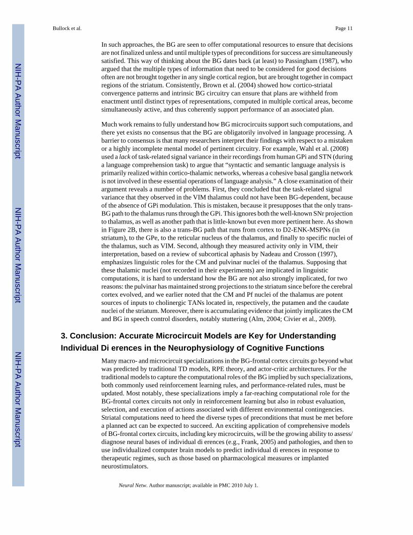
making process via cholinergic transmission, especially when the stimuli have significantsalience/relevance (Figure 1). Striatal ACh transmission, in turn, exerts direct control on striatalperformance functions via its effects on the MSPNs that target the output nuclei of the basalganglia (Wang and McGinty, 1997; Alcantara et al., 2001).
It has been assumed in some computational models that DA facilitates D1-SP-MSPNs whilesuppressing D2-ENK-MSPNs (e.g. Gurney et al., 2001a,b; Humpries et al., 2006), and thatACh has an effect opposite to DA. However, data show a more complicated picture. AChstabilizes the prevailing MSPN state by modulating several intrinsic currents (Howe andSurmeier, 1995; Gabel and Nisenbaum, 1999; Surmeier et al., 2005). DA, in contrast, has astate-dependent effect on MSPNs (Flores-Hernandez et al., 2002; Gruber et al., 2003): itfacilitates MSPN responses when MSPNs are in a depolarized (up) state while depressingMSPNs in a hyperpolarized (down) state. Therefore, it is probable that the response of striatalMSPNs to a given corticostriatal glutamatergic input in vivo strongly depends on the patterningwithin the cascading DA-ACh signal (cf. Tan and Bullock, 2008a, see also Section 2.4) in thestriatum. This contrasts to the common presumptions of the actor-critic architecture that (1)phasic DA level in the striatum has only a direct effect on striatal processing by exciting orinhibiting striatal output neurons (e.g. Frank, 2005; Frank and O'Reilly, 2006), and (2) striatalaction-gating is a simple linear or sigmoidal function of expected reward value. Contrary tothese ideas, emergent interactions among various neurotransmitters (glutamate, ACh, DA,GABA) in the striatum provide a much more flexible schema governing striatal performancefunctions, involving an interplay among at least the three aforementioned decision variables.Therefore, performance rules governing the actor component of the actor-critic system alsoneed modifications to better reflect biological reality.
Whether any decision centers other than striatum can offer similar, to say nothing of better,sensitivity to multiple desiderata, remains to be seen. One key region to consider is theorbitofrontal cortex, which has been strongly implicated in the ability to resist framing effectsin decision making (DeMartino et al., 2006), and which has been modeled recently (Draniaset al., 2008) as a key nexus in an evaluative neuraxis that includes the hypothalamus andamygdala (cf. also Frank and Claus, 2006).
2.6. Should any nuclei of the extended amygdala be regarded as parts of the striatum?Swanson (2000) has argued for extending the concept of the BG to encompass further forebrainnuclei, notably parts of the “extended amygdala” (e.g., de Olmos and Heimer, 1999). Althoughsome nuclei of the amygdala are far more “cortical” (because the principle neurons areglutamatergic) than “striatal”, McDonald (2003) suggested a “consensus ... that the lateralportions of the central nucleus [of the amygdala] are striatal-like (p. 13).” Notably, this regiondoes not reciprocate its projections from cortex, its principle neurons are GABAergic MSPNs,and it connects appropriately with midbrain DA neurons. However, from a computationalperspective, dis-analogies are equally important, if they imply that a generic striatal circuitmodel cannot be used to simulate processing in CeA (central nucleus of amygdala). Two dis-analogies may prove to be decisive. First, it is generally believed that the CeA's ACh is suppliedby a erents arriving from basal forebrain (e.g. Schäafer et al., 1988), and not by intrinsic giantcholinergic neurons (TANs) like those found in “traditional” striatum. Second, although outputfrom CeA MSPNs is potently regulated by feedforward inhibition (Paré et al., 2003), Zahm etal. (2003) found that the GABAergic parvalbumin immunoreactive (PV+) fast-spikinginterneurons (FS-INs) characteristic of the striatum are absent from the extended amygdala.Both di erences would preclude readily adapting any computational model of normal striatumto simulate information processing in lateral CeA, the “most striatal” part of the amygdala.That said, it must be admitted that even within the traditional striatum, any model must beadapted to capture important regional variations.
Bullock et al. Page 7
Neural Netw. Author manuscript; available in PMC 2010 July 1.
NIH
-PA Author Manuscript
NIH
-PA Author Manuscript
NIH
-PA Author Manuscript

2.7. Basal ganglia architecture cannot be captured in feed-forward models: Toward a betterunderstanding of perseverance, lockout protection, interrupts, switching, and resumption
Most published representations of the BG are so incomplete as to promote severeunderestimations of the computational competence of the BG. That situation is being rectifiedby some of the microcircuit models noted above. However, the BG macrocircuit is also muchdi erent than typically depicted. First, the typical depiction aptly emphasizes that the cortico-striatal projection is not reciprocated by a striato-cortical projection. This promotes thinkingof the BG as a structure dominated by a feed-forward flow along the path: cortex-striatum-pallidumthalamus (and back to cortex). However, as briefly explored in Brown et al. (2004)(see also Srihasam et al., 2009), and as the more complete circuit in Figure 2B suggests evenmore emphatically, the “feedforward BG” conception has little basis. In fact, Brown et al.(2004) showed the importance of recognizing that many of the cells of origin of the cortico-striatal projection are not identical to the cells of origin of the cortico-STN projection (see alsoTurner and DeLong, 2000). Because of their non-identity, the former class can serve as planrepresentations, whereas the latter can be activated only at time of plan execution. Theprojection of these cells’ output back into the BG via the STN can then be understood as helpingto lockout competing plans and thereby provide the selected plan enough time to execute - atleast in the general case.
Another conspicuous “feedback flow” is mediated by the projection from GPe back to thestriatum. This projection originates from a subset of GABAergic parvalbumin immunoreactive(PV+) neurons that are recipients of STN projections, and targets exclusively the PV+ fast-spiking interneurons (FS-INs) in the striatum (Bevan et al., 1998). Though this feedbackprojection has been neglected by most modelers, anatomical considerations shed some lighton its functional implications. Striatal MSPNs and FSINs receive similar plan-related inputs.The prominence of collaterals between MSPNs has been taken as evidence for a winner-take-all competition between these output neurons. However, recent data challenged thisassumption, showing that inhibitory communication occurs almost exclusively between dierent classes of MSPNs, and is not reciprocated (Venance et al., 2004; Taverna et al., 2008,see also below). Such data and other considerations inspired the proposal that feed-forwardinhibition via striatal FS-INs mediates striatal competition and selection (e.g., Brown et al.,2004; Bullock and Tan, 2007). Furthermore, striatal FS-INs are coupled via gap-junctions(Koos and Tepper, 1999; Tepper et al., 2004). Such coupling can promote synchronous activityof FS-INs, yet preserves topographic organization by allowing cortico-striatal terminals withrestricted distributions to nevertheless recruit FS-INs broadly, allowing robust feedforwardinhibition of striatal MSPNs. Thus, it is conceivable that selection of a plan among severalothers is highly sensitive to the relative cortical activation levels and/or corticostriatal synapticweights at the moment when one plan wins the competition and activates a correspondingMSPN despite feedfor-ward inhibition via FS-INs. However, if FS-IN inhibition of the winningMSPN were to persist during the entire movement interval, the winning MSPN would remainat risk of falling below its activation threshold, especially if competing cortical planrepresentations remain active and are of nearly equal strength. Nevertheless, this “risk” can bereduced by channel-specific inhibitory feedback from GPe to FS-INs that are in theneighborhood of the winning MSPN: cortico-subthalamo-pallidal projections can activate cellsof origin of the pallido-striatal feedback pathway, thereby inhibiting the subset of FS-INs thatare recipients of selected cortical plan representations, disinhibiting corresponding MSPNs.Therefore, one possible computational role of the feedback projection from GPe to striatal FS-INs is to enable a “real-time contrast-enhancement” at the striatum while the outcome of theongoing selection process is still unfolding.
Added to this emerging picture are the “horizontal” interactions of two classes of MSPNs. Asmentioned above, the assumption of a winner-take-all (WTA) competition between D1-SP-
Bullock et al. Page 8
Neural Netw. Author manuscript; available in PMC 2010 July 1.
NIH
-PA Author Manuscript
NIH
-PA Author Manuscript
NIH
-PA Author Manuscript

and D2-ENK-MSPNs has been challenged on the basis of electrophysiology (Jaeger et al.,1994) and logic (Brown et al., 2004, p. 476). Here we add that the breadth of collateralarborization falls far short of what would be needed to achieve WTA selection across broadregions of the striatum. Furthermore, recent data show that: (1) a subset of MSPNs in thestriatum are coupled via gap junctions (Onn and Grace, 1994; Venance et al., 2004); (2) thiselectrotonic coupling is mostly confined to D2-ENK-MSPNs (Onn and Grace, 1994; Venanceet al., 2004); (3) chemical (GABAergic) transmission between MSPNs is potent butunidirectional (Tunstall et al., 2002; Venance et al., 2004); and, equally important, (4)electrotonic and unidirectional chemical communication among MSPNs are mutuallyexclusive (Venance et al., 2004). The first implication is that D2-ENK-MSPNs do not inhibiteach other. That D2-ENK-MSPNs are not mutually inhibitory complements their gap-junctioncoupling because it allows even a focused glutamatergic input of su cient strength and durationto recruit them synchronously and in significant numbers. This is consistent with the “STOP”or “NO-GO” function attributed to D2-ENK-MSPNs in some models (e.g., Brown et al.,2004; Frank, 2005), whereas, had the contrary been found, i.e., had it been found that D1-SP-MSPNs were both electro-tonically coupled and not mutually inhibitory, it would havedisconfirmed all recent BG models. Beyond this key conclusion, the data force a choicebetween two possibilities in any model. Either active D1-SP-MSPNs inhibit D2-ENK-MSPNs,or vice versa, but not both (because chemical transmission is non-reciprocated). Here it isimportant to recall that because of the limited arborization of MSPN feedback collaterals,feedback inhibition by D2-ENK-MSPNs would be much more powerful (than by D1-SP-MSPNs), because their electrotonic coupling would enable feedback inhibition to be muchbroader than that overtly implied by the limited arborization. This would nicely enhance theproposed “STOP” signal function of the indirect pathway. By contrast, D1-SP-MSPN feedbackinhibition would remain highly focused, would not support a WTA property, would not sharpencontrast among competing direct pathway MSPNs, but would interfere with the STOP-signalfunction by inhibiting nearby D2-ENK-MSPNs. From a global functional perspective, ittherefore seems much more likely that the observed unidirectional GABAergic transmissionbetween MSPNs (Venance et al., 2004) runs from D2-ENK-MSPNs to D1-SPMSPNs. In fact,it has recently been shown that feedback inhibitory communication directed from D2-ENK-MSPNs to D1-SP-MSPNs is much more prominent than the alternatives (Taverna et al.,2008).
The difference in neuropeptide co-release by di erent classes of MSPNs (substance-P vs.enkephalin) begets a further asymmetry. Cortico-striatal terminals in the striatum expresspresynaptic neurokinin-1 receptors (primary target of substance-P in primates and humans;Regoli et al., 1994), and it has recently been shown that endogenous SP released by D1-SP-MSPNs may enhance presynaptic glutamate release, thereby facilitating postsynapticresponses in neighboring MSPNs (Blomeley et al., 2009). It should be noted that only D1-SP-MSPNs can partake in such a feedback excitation by virtue of somatically co-releasing SP withGABA. Through this feedback excitation, a subset of MSPNs, once selected, can recruitneighboring MSPNs (presumably belonging to the same functional channel), perhaps to furtherenhance the contrast between selected and competing plan representations.
The data summarized above clearly suggest that local interactions within the BG, particularlyin the striatum, are not only more complicated than assumed in most models of the BG, butalso can offer substantial computational abilities that most current models of the BG lack.Nevertheless, human intuition is generally insu cient to predict ramifications of such complexinteractions, and computational models that reflect these microcircuit specializations can revealtheir implications for BG functions. Recently, Bullock and Tan (2007) used the more completemacro-circuit in Figures 2B and 2C as a basis for exploring how the BG circuit enables a morecomplete set of fundamental abilities that serve as a basis for intelligent cognitive control ofbehavioral scheduling, sequencing, and interleaving, including the ability to interrupt, switch,
Bullock et al. Page 9
Neural Netw. Author manuscript; available in PMC 2010 July 1.
NIH
-PA Author Manuscript
NIH
-PA Author Manuscript
NIH
-PA Author Manuscript

and resume planned behavior. Demonstration of such abilities of the BG, with models that takea more complete macro- and microcircuit of the BG into account than traditional TD-and RPE-based actor-critic architecture, in turn, opens up exciting avenues for integrating the BG intocomputational models of higher cognitive and behavioral functions.
2.8. Basal ganglia architecture is pivotal for advanced human abilities, including speechTo some who considered language as the most “neo” of neo-cortical functions, it came as ashock when early fMRI studies (e.g., Ullman et al., 1997) strongly implicated the BG in suchprototypical linguistic functions as control of regular past-tense production (e.g., postfixing –ed in English). The BG have since also been implicated in arithmetic rule application(Teichmann et al., 2008). Despite such data clues, few computational models of speech,linguistic or arithmetic rule application make integral use of either the cortex-thalamus-BGmacrocircuit or BG microcircuits. Steps to rectify this shortfall have recently been taken byseveral research groups. For example, Bohland et al. (2009) are exploring the hypothesis thatthe BG are critical for ensuring that speech acts satisfy the multiple types of constraints thatmust be met by linguistic productions if they are to succeed as conventional communications.
Consider the difference between computational models of de-contextualized sequenceproduction and meaning-communicative sequence production, which obeys learned rules. Forexample, chronometric (latency) patterns of anticipatory or deferral errors in non-linguisticsequence production (Farrell and Lewandowsky, 2004) as well as electrophysiologicalrecordings (Averbeck et al., 2002; Rhodes et al., 2004) have strongly supported a class ofsequence control models, called competitive queuing (CQ) models (Averbeck et al., 2002;Bullock, 2004a,b; Rhodes et al., 2004; Ivey et al., 2008), that are defined by two assumptions(cf. Grossberg, 1978): (1) the sequential order relation among plan-representations offorthcoming acts is represented by an analog gradient (“primacy gradient”) of activation levelsestablished over the plan representations in a WM (working memory), and (2) once a planrepresentation is chosen for enactment, its representation is deleted from the planning WM,and thus eliminated from the competition (among the surviving representations) thatdetermines which plan to perform next. However, it has not been clear whether CQ modelsshould, or how they could, be extended to explain linguistic sequences controls. For example,although many linguistic sequencing errors are exchange errors, as predicted by CQ theory,elementary CQ theory does not explain why linguistic exchange errors respect linguistic class.The connectionist language production model of Ward (1994), which utilized concepts fromconstruction grammar (e.g., Goldberg, 2006), explored one way that a CQ process could beextended to ensure that next-word choices simultaneously obeyed semantic and syntacticconstraints. However, Ward (1994) offered no interpretation of model components in terms ofidentified brain circuits. Although it did not include BG microcircuits, the computational modelof language processing in Dominey et al. (2006) also adopted ideas from construction grammar,and proposed that the cortical-striatal projection mediates retrieval of form-to-meaningmappings. The computational model of Bohland et al. (2009) incorporated macro- and somemicro-circuit details from the Brown et al. (2004) model of fronto-BG function to illustrateone way to use BG circuitry to offer a CQ-consistent explanation for multi-syllabic speechproduction. In this model, exchange errors are appropriately class-constrained, a consequenceof the model's ability to ensure that next-sound choices obey both phonemic and syllabicconstraints. The model's mapping of computations to neurobiological circuits enabled it topinpoint candidate neural bases of speech stuttering errors (Civier et al., 2009), and similarfuture models should be able to use BG computations to ensure the simultaneous satisfactionof multiple types of constraints in language processing, e.g., to achieve the integrative-well-formedness checks, based on semantic, syntactic, and pragmatic constraints, that were recentlyattributed to the BG by Bornkessel and Schlesewsky (2006).
Bullock et al. Page 10
Neural Netw. Author manuscript; available in PMC 2010 July 1.
NIH
-PA Author Manuscript
NIH
-PA Author Manuscript
NIH
-PA Author Manuscript
In such approaches, the BG are seen to offer computational resources to ensure that decisionsare not finalized unless and until multiple types of preconditions for success are simultaneouslysatisfied. This way of thinking about the BG dates back (at least) to Passingham (1987), whoargued that the multiple types of information that need to be considered for good decisionsoften are not brought together in any single cortical region, but are brought together in compactregions of the striatum. Consistently, Brown et al. (2004) showed how cortico-striatalconvergence patterns and intrinsic BG circuitry can ensure that plans are withheld fromenactment until distinct types of representations, computed in multiple cortical areas, becomesimultaneously active, and thus coherently support performance of an associated plan.
Much work remains to fully understand how BG microcircuits support such computations, andthere yet exists no consensus that the BG are obligatorily involved in language processing. Abarrier to consensus is that many researchers interpret their findings with respect to a mistakenor a highly incomplete mental model of pertinent circuitry. For example, Wahl et al. (2008)used a lack of task-related signal variance in their recordings from human GPi and STN (duringa language comprehension task) to argue that “syntactic and semantic language analysis isprimarily realized within cortico-thalamic networks, whereas a cohesive basal ganglia networkis not involved in these essential operations of language analysis.” A close examination of theirargument reveals a number of problems. First, they concluded that the task-related signalvariance that they observed in the VIM thalamus could not have been BG-dependent, becauseof the absence of GPi modulation. This is mistaken, because it presupposes that the only trans-BG path to the thalamus runs through the GPi. This ignores both the well-known SNr projectionto thalamus, as well as another path that is little-known but even more pertinent here. As shownin Figure 2B, there is also a trans-BG path that runs from cortex to D2-ENK-MSPNs (instriatum), to the GPe, to the reticular nucleus of the thalamus, and finally to specific nuclei ofthe thalamus, such as VIM. Second, although they measured activity only in VIM, theirinterpretation, based on a review of subcortical aphasis by Nadeau and Crosson (1997),emphasizes linguistic roles for the CM and pulvinar nuclei of the thalamus. Supposing thatthese thalamic nuclei (not recorded in their experiments) are implicated in linguisticcomputations, it is hard to understand how the BG are not also strongly implicated, for tworeasons: the pulvinar has maintained strong projections to the striatum since before the cerebralcortex evolved, and we earlier noted that the CM and Pf nuclei of the thalamus are potentsources of inputs to cholinergic TANs located in, respectively, the putamen and the caudatenuclei of the striatum. Moreover, there is accumulating evidence that jointly implicates the CMand BG in speech control disorders, notably stuttering (Alm, 2004; Civier et al., 2009).
3. Conclusion: Accurate Microcircuit Models are Key for UnderstandingIndividual Di erences in the Neurophysiology of Cognitive Functions
Many macro- and microcircuit specializations in the BG-frontal cortex circuits go beyond whatwas predicted by traditional TD models, RPE theory, and actor-critic architectures. For thetraditional models to capture the computational roles of the BG implied by such specializations,both commonly used reinforcement learning rules, and performance-related rules, must beupdated. Most notably, these specializations imply a far-reaching computational role for theBG-frontal cortex circuits not only in reinforcement learning but also in robust evaluation,selection, and execution of actions associated with different environmental contingencies.Striatal computations need to heed the diverse types of preconditions that must be met beforea planned act can be expected to succeed. An exciting application of comprehensive modelsof BG-frontal cortex circuits, including key microcircuits, will be the growing ability to assess/diagnose neural bases of individual di erences (e.g., Frank, 2005) and pathologies, and then touse individualized computer brain models to predict individual di erences in response totherapeutic regimes, such as those based on pharmacological measures or implantedneurostimulators.
Bullock et al. Page 11
Neural Netw. Author manuscript; available in PMC 2010 July 1.
NIH
-PA Author Manuscript
NIH
-PA Author Manuscript
NIH
-PA Author Manuscript

AcknowledgmentsThis work was supported in part by the U.S. National Science Foundation under Science of Learning Center GrantSBE-354378 and in part by NIH Grant R01DC007683.
ReferencesAlbin RL, Young AB, Penney JB. The functional anatomy of basal ganglia disorders. Trends Neurosci
1989;12:366. [PubMed: 2479133]Alcantara AA, Mrzljak L, Jakab RL, Levey AI, Hersch SM, Goldman-Rakic PS. Muscarinic m1 and m2
receptor proteins in local circuit and projection neurons of the primate striatum: Anatomical evidencefor cholinergic modulation of glutamatergic prefronto-striatal pathways. J Comp Neurol2001;434:445–460. [PubMed: 11343292]
Alm PA. Stuttering and the basal ganglia: a critical review of possible relations. J Commun Disord2004;37:325–369. [PubMed: 15159193]
Averbeck BB, Chafee MV, Crowe DA, Georgopoulos AP. Parallel processing of serial movements inprefrontal cortex. Proc Natl Acad Sci USA 2002;99:13172–13177. [PubMed: 12242330]
Bevan MD, Booth PA, Eaton SA, Bolam JP. Selective innervation of neostriatal interneurons by asubclass of neuron in the globus pallidus of the rat. J Neurosci 1998;18:9438–9452. [PubMed:9801382]
Blomeley CP, Kehoe LA, Bracci E. Substance P mediates excitatory interactions between striatalprojection neurons. J Neurosci 2009;29:4953–4963. [PubMed: 19369564]
Bohland JW, Bullock D, Guenther FH. Neural representations and mechanisms for the performance ofsimple speech sequences. J Cogn Neurosci. 2009in press
Bonsi P, Martella G, Cuomo D, Platania P, Sciamanna G, Bernardi G, Wess J, Pisani A. Loss of muscarinicautoreceptor function impairs long-term depression but not long-term potentiation in the striatum. JNeurosci 2008;28:6258. [PubMed: 18550768]
Bornkessel I, Schlesewsky M. The extended argument dependency model: a neurocognitive approach tosentence comprehension across languages. Psychol Rev 2006;113:787–821. [PubMed: 17014303]
Brown JW, Bullock D, Grossberg S. How laminar frontal cortex and basal ganglia circuits interact tocontrol planned and reactive saccades. Neural Netw 2004;17:471–510. [PubMed: 15109680]
Buhusi CV, Meck WH. What makes us tick? functional and neural mechanisms of interval timing. NatRev Neurosci 2005;6:755–765. [PubMed: 16163383]
Bullock D. Adaptive neural models of queuing and timing in fluent action. Trends Cogn Sci 2004a;8:426–433. [PubMed: 15350244]
Bullock D. From parallel sequence representations to calligraphic control: a conspiracy of neural circuits.Motor Control 2004b;8:371–391. [PubMed: 15585895]
Bullock D, Tan CO. Role of basal ganglia in decision sequences: A computational model of dopamineand acetylcholine regulation of action selection, interruption, resumptions, and switching. SocNeurosci Abs. 2007page S514
Bullock D, Tan CO. Computational implications of microcircuit specializations in forebrain circuits formotivated action selection. IEEE Proc Int Joint Conf Neur Netw. 2009
Calabresi P, Maj R, Pisani A, Mercuri NB, Bernardi G. Long-term synaptic depression in the striatum:physiological and pharmacological characterization. J Neurosci 1992a;12:4224–4233. [PubMed:1359031]
Calabresi P, Pisani A, Mercuri NB, Bernardi G. Long-term potentiation in the striatum is unmasked byremoving the voltage-dependent magnesium block of NMDA receptor channels. Eur J Neurosci1992b;4:929–935. [PubMed: 12106428]
Centonze D, Gubellini P, Bernardi G, Calabresi P. Permissive role of interneurons in cortical synapticplasticity. Brain Res Rev 1999;31:1–5. [PubMed: 10611492]
Civier, A.; Bullock, D.; Max, L.; Guenther, F. Simulating neural impairments to syllable-level commandgeneration in stuttering.. Sixth World Congress on Fluency Disorders; Rio de Janerio, Brazil. 5 − 8August; 2009.
Bullock et al. Page 12
Neural Netw. Author manuscript; available in PMC 2010 July 1.
NIH
-PA Author Manuscript
NIH
-PA Author Manuscript
NIH
-PA Author Manuscript

de Olmos JS, Heimer L. The concepts of the ventral striatopallidal system and extended amygdala. AnnN Y Acad Sci 1999;877:1–32. [PubMed: 10415640]
DeMartino B, Kumaran D, Seymour B, Dolan RJ. Frames, biases, and rational decision-making in thehuman brain. Science 2006;313:684–687. [PubMed: 16888142]
Dominey PF, Hoen M, Inui T. A neurolinguistic model of grammatical construction processing. J CognNeurosci 2006;18:2088–2107. [PubMed: 17129193]
Dranias MR, Grossberg S, Bullock D. Dopaminergic and non-dopaminergic value systems inconditioning and outcome-specific revaluation. Brain Res 2008;1238:239–287. [PubMed:18674518]
Farrell S, Lewandowsky S. Modeling transposition latencies: Constraints for theories of serial ordermemory. J Mem Lang 2004;51:115–135.
Fino E, Glowinski J, Venance L. Bidirectional activity-dependent plasticity at corticostriatal synapses. JNeurosci 2005;25:11279–11287. [PubMed: 16339023]
Fiorillo CD, Tobler PN, Schultz W. Discrete coding of reward probability and uncertainty by dopamineneurons. Science 2003;299:1898–1902. [PubMed: 12649484]
Fiorillo CD, Tobler PN, Schultz W. Evidence that the delay-period activity of dopamine neuronscorresponds to reward uncertainty rather than backpropagating TD errors. Behav Brain Funct2005;1:7. [PubMed: 15958162]
Flores-Hernandez J, Cepeda C, Hernandez-Echeagaray E, Calvert CR, Jokel ES, Fienberg AA, GreengardP, Levine MS. Dopamine enhancement of NMDA currents in dissociated medium-sized striatalneurons: Role of D1 receptors and DARPP-32. J Neurphysiol 2002;88:3010–3020.
Frank MJ. Dynamic dopamine modulation in the basal ganglia: a neurocomputational account ofcognitive deficits in medicated and nonmedicated parkinsonism. J Cogn Neurosci 2005;17:51–72.[PubMed: 15701239]
Frank MJ, Claus ED. Anatomy of a decision: Striato-orbitofrontal interactions in reinforcement learning,decision making, and reversal. Psychol Rev 2006;113:300–326. [PubMed: 16637763]
Frank MJ, O'Reilly RC. A mechanistic account of striatal dopamine function in human cognition:Psychopharmacological studies with cabergoline and haloperidol. Behav Neurosci 2006;120:497–517. [PubMed: 16768602]
Gabel LA, Nisenbaum ES. Muscarinic receptors differentially modulate the persistent potassium currentsin striatal spiny projection neurons. J Neurophysiol 1999;81:1418–1423. [PubMed: 10085367]
Goldberg, AE. Constructions at work: The nature of generalization in language. Oxford; New York: 2006.Grossberg, S. A theory of human memory: Self-organization and performance of sensory-motor codes,
maps, and plans.. In: Rosen, R.; Snell, F., editors. Progress in Theoretical Biology. Vol. 5. AcademicPress; New York: 1978. p. 233-374.
Gruber AJ, Solla SA, Surmeier DJ, Houk JC. Modulation of striatal single units by expected reward: Aspiny neurons model displaying dopamine-induced bistability. J Neurophysiol 2003;90:1095–1114.[PubMed: 12649314]
Gurney K, Prescott TJ, Redgrave P. A computational model of action selection in the basal ganglia. i. anew functional anatomy. Biol Cybern 2001a;84:401–410. [PubMed: 11417052]
Gurney K, Prescott TJ, Redgrave P. A computational model of action selection in the basal ganglia. ii.analysis and simulation of behavior. Biol Cybern 2001b;84:411–423. [PubMed: 11417053]
Hasselmo ME. The role of acetylcholine in learning and memory. Curr Opin. Neurobiol 2006;16:710–715. [PubMed: 17011181]
Houk, J.; Adams, JL.; Barto, AG. A Model of How the Basal Ganglia Generate and Use Signals ThatPredic Reinforcement. MIT Press; Cambridge, MA: 1995. p. 249-270.
Howe AR, Surmeier DJ. Muscarinic receptors modulate N-, P-, and L-type Ca+2 currents in rat striatalneurons through parallel pathways. J Neurosci 1995;15:458–469. [PubMed: 7823150]
Humpries MD, Stewart RD, Gurney KN. A physiologically plausible model of action selection andoscillatory activity in the basal ganglia. J Neurosci 2006;26:12921–12942. [PubMed: 17167083]
Ivey, R.; Bullock, D.; Grossberg, S. A neuromorphic model of spatial lookahead planning.. Proceedingsof the 12th International Conference on Cognitive and Neural Systems; Boston, MA. 2008.
Bullock et al. Page 13
Neural Netw. Author manuscript; available in PMC 2010 July 1.
NIH
-PA Author Manuscript
NIH
-PA Author Manuscript
NIH
-PA Author Manuscript

Jaeger D, Kita H, Wilson CJ. Surround inhibition among projection neurons is weak or nonexistent inthe rat neostriatum. J Neurophysiol 1994;72:2555–2558. [PubMed: 7884483]
Kakade S, Dayan P. Dopamine: Generalization and bonuses. Neur Netw 2002;15:549–559.Kelley AE, Cador M, Stinus L, Le MM. Neurotensin, substance P, neurokinin-alpha, and enkephalin:
Injection into ventral tegmental area in the rat produces di erential effects on operant responding.Psychopharmacology (Berl) 1989;97:243–252. [PubMed: 2471221]
Kerr JND, Wickens JR. Dopamine D-1/D-5 receptor activation is required for long-term potentiation inthe rat neostriatum in vitro. J Neurophysiol 2001;85:117–124. [PubMed: 11152712]
Kilgard MP, Merzenich MM. Cortical map reorganization enabled by nucleus basalis activity. Science1998;279:1714–1718. [PubMed: 9497289]
Koos T, Tepper JM. Inhibitory control of neostriatal projection neurons by GABAergic interneurons. NatNeurosci 1999;2:467–472. [PubMed: 10321252]
Laurent PA. The emergence of saliency and novelty responses from reinforcement learning principles.Neur Netw 2008;21:1493–1499.
McDonald AJ. Is there an amygdala and how far does it extend? An anatomical perspective. Ann N YAcad Sci 2003;985:1–21. [PubMed: 12724144]
Meck WH. Selective adjustment of the speed of internal clock and memory processes. J Exp PsycholAnim Behav Proc 1983;9:171.
Meck WH. A nity for the dopamine D2 receptor predicts neuroleptic potency in decreasing the speed ofan internal clock. Pharm Biochem Behav 1986;25:1185.
Meck WH. Neuropharmacology of timing and time perception. Cogn Brain Res 1996;3:227–242.Meck WH, Church RM. Cholinergic modulation of the content of temporal memory. Behav Neurosci
1987;101:457–464. [PubMed: 2820435]Nadeau SE, Crosson B. Subcortical aphasia. Brain Lang 1997;58:355–402. [PubMed: 9222518]Niv Y, Du MO, Dayan P. Dopamine, uncertainty and td learning. Behav Brain Funct 2005;1:6. [PubMed:
15953384]Onn S-P, Grace AA. Dye coupling between rat striatal neurons recorded in vivo: compartmental
organization and modulation by dopamine. J Neurophysiol 1994;71:1917–1934. [PubMed: 8064357]Paré D, Royer S, Smith Y, Lang EJ. Contextual inhibitory gating of impulse tra c in the intra-amygdaloid
network. Ann N Y Acad Sci 2003;985:78–91. [PubMed: 12724150]Passingham, RE. From where does the motor cortex get its instructions?. In: Wise, SP., editor. Higher
brain functions. Wiley; New York: 1987. p. 67-97.Pawlak V, Kerr JN. Dopamine receptor activation is required for corticostriatal spike-timing-dependent
plasticity. J Neurosci 2008;28:2435–2446. [PubMed: 18322089]Placenza FM, Fletcher PJ, Rotzinger S, Vaccarino FJ. Infusion of the substance P analogue, DiMe-C7,
into the ventral tegmental area induces reinstatement of cocaine-seeking behaviour in rats.Psychopharmacology (Berl) 2004;177:111–120. [PubMed: 15167979]
Redgrave P, Prescott TJ, Gurney K. The basal ganglia: a vertebrate solution to the selection problem?Neuroscience 1999a;89:1009–1023. [PubMed: 10362291]
Redgrave P, Prescott TJ, Gurney K. Is the short-latency dopamine response too short to signal rewarderror? Trends Neurosci 1999b;22:146–151. [PubMed: 10203849]
Regoli D, Boudon A, Fauchere J-L. Receptors and antagonists for substance P and related peptides.Pharmacol Rev 1994;45:551–599. [PubMed: 7534932]
Rhodes BJ, Bullock D, Verwey WB, Averbeck BB, Page MP. Learning and production of movementsequences: Behavioral, neurophysiological, and modeling perspectives. Hum Mov Sci 2004;23:699–746. [PubMed: 15589629]
Schäfer MK, Eiden LE, Weihe E. Cholinergic neurons and terminal fields revealed byimmunohistochemistry for the vesicular acetylcholine transporter. I. Central nervous system.Neuroscience 1988;1998:331–359.
Schotanus SM, Chergui K. Dopamine D1 receptors and group I metabotropic glutamate receptorscontribute to the induction of long-term potentiation in the nucleus accumbens. Neuropharmacology2008;54:837–844. [PubMed: 18272187]
Bullock et al. Page 14
Neural Netw. Author manuscript; available in PMC 2010 July 1.
NIH
-PA Author Manuscript
NIH
-PA Author Manuscript
NIH
-PA Author Manuscript

Sjöström PJ, Nelson SB. Spike timing, calcium signals, and synaptic plasticity. Curr Opin Neurobiol2002;12:305–314. [PubMed: 12049938]
Srihasam K, Bullock D, Grossberg S. Target selection by frontal cortex during coordinated saccadic andsmooth pursuit eye movements. J Cogn Neurosci 2009;21in press
Surmeier DJ, Mercer JN, Chan CS. Autonomous pacemakers in the basal ganglia: who needs excitatorysynapses anyway? Curr Opin Neurobiol 2005;15:312–318. [PubMed: 15916893]
Sutton RS, Barto AG. Toward a modern theory of adaptive networks: Expectation and prediction. PsycholRev 1981;88:135–170. [PubMed: 7291377]
Swanson LW. Cerebral hemisphere regulation of motivated behavior. Brain Res 2000;886:113–164.[PubMed: 11119693]
Tan CO, Anderson E, Dranias M, Bullock D. Can the apparent adaptation of dopamine neurons’ mismatchsensitivities be reconciled with their computation of reward prediction errors? Neurosci Lett2008;438:14–16. [PubMed: 18482798]
Tan CO, Bullock D. A dopamine-acetylcholine cascade: Simulating learned and lesion-induced behaviorof striatal cholinergic interneurons. J Neurophysiol 2008a;100:2409–2421. [PubMed: 18715897]
Tan CO, Bullock D. A local circuit model of learned striatal and dopamine cell responses underprobabilistic schedules of reward. J Neurosci 2008b;28:10062–10074. [PubMed: 18829964]
Tan CO, Bullock D. Neuropeptide co-release with GABA may explain functional non-monotonicuncertainty responses in dopamine neurons. Neurosci Lett 2008c;430:218–223. [PubMed:18053647]
Taverna S, Ilijic E, Surmeier DJ. Recurrent collateral connections of striatal medium spiny neurons aredisrupted in models of parkinson's disease. J Neurosci 2008;28:5504–5512. [PubMed: 18495884]
Teichmann M, Guara V, Démonet JF, Supiot F, Delliaux M, Verny C, Renou P, Remy P, Bachoud-LévyAC. Language processing within the striatum: evidence from a PET correlation study in Huntington'sdisease. Brain 2008;131:1046–1056. [PubMed: 18334537]
Tepper JM, Koos T, Wilson CJ. GABAergic microcircuits in the neostriatum. Trends Neurosci2004;27:662–669. [PubMed: 15474166]
Tobler PN, Fiorillo CD, Schultz W. Adaptive coding of reward value by dopamine neurons. Science2005;307:1642–1645. [PubMed: 15761155]
Tunstall MJ, Oorschot DE, Kean A, Wickens JR. Inhibitory interactions between spiny projection neuronsin the rat striatum. J Neurophysiol 2002;88:1263–1269. [PubMed: 12205147]
Turner RS, DeLong MR. Corticostriatal activity in primary motor cortex of the macaque. J Neurosci2000;20:7096–7108. [PubMed: 10995857]
Ullman MT, Corkin S, Coppola M, Hickok G, Growdon JH, Koroshetz WJ, Pinker S. A neuraldissociation within language: Evidence that the mental dictionary is part of declarative memory, andthat grammatical rules are processed by the procedural system. J Cogn Neurosci 1997;9:266–276.
Ungless MA, Magill PJ, Bolam JP. Uniform inhibition of dopamine neurons in the ventral tegmental areaby aversive stimuli. Science 2004;303:2040–2042. [PubMed: 15044807]
Venance L, Glowinski J, Giaume C. Electrical and chemical transmission between striatal GABAergicoutput neurones in rat brain slices. J Physiol 2004;559:215–230. [PubMed: 15235091]
Wahl M, Marzinzik F, Friederici AD, Hahne A, Kupsch A, Schneider GH, Saddy D, Curio G,Klostermann F. The human thalamus processes syntactic and semantic language violations. Neuron2008;59:695–707. [PubMed: 18786354]
Wang JQ, McGinty JF. The full D1 dopamine receptor agonist SKF-82958 induces neuropeptide mRNAin the normosensitive striatum of rats: Regulation of D1/D2 interactions by muscarinic receptors. JPharmacol Exp Ther 1997;281:972–982. [PubMed: 9152408]
Wang Z, Kai L, Day M, Ronesi J, Yin HH, Ding J, Tkatch T, Lovinger DM, Surmeier DJ. Dopaminergiccontrol of corticostriatal long-term synaptic depression in medium spiny neurons is mediated bycholinergic interneurons. Neuron 2006;50:443–452. [PubMed: 16675398]
Ward, N. A connectionist language generator. Ablex Publishing; Norwood, NJ: 1994.Zahm DS, Grosu S, Irving JC, Williams EA. Discrimination of striatopallidum and extended amygdala
in the rat: a role for parvalbumin immunoreactive neurons? Brain Res 2003;978:141–154. [PubMed:12834908]
Bullock et al. Page 15
Neural Netw. Author manuscript; available in PMC 2010 July 1.
NIH
-PA Author Manuscript
NIH
-PA Author Manuscript
NIH
-PA Author Manuscript
Figure 1.The tight coupling between striatal DAergic, cholinergic, and thalamo-striatal glutamatergictransmission. The upper panel's color values show the normalized cholinergic interneuron(TAN) responses (baseline firing rate 0.5), if allowed to reach equilibrium (Tan and Bullock,2008a), as a function both of phasic and sustained dopamine signal inputs (shown in the lowerpanel) that reflect reward expectations, and of thalamo-striatal glutamatergic inputs (from CM-Pf nuclei, shown on the ordinate) that reflect stimulus salience. The lower panel shows the(normalized) learned DA response to a CS-then-reward sequence, after training with a p = 0.5conditional probability schedule during delay conditioning with a 2 second-long CS (Tan andBullock, 2008b). The phasic DA burst in response to CS onset (t = 1.2 sec) reflects the expectedvalue of reward; that in response to primary reward (t = 3.2 sec) reflects the probabilisticschedule; and the gradual increase during the delay period reflects the uncertainty in theexpected outcome (Tobler et al., 2005; Tan and Bullock, 2008b; Fiorillo et al., 2003, 2005).Although the TAN responses (upper panel) also reflect such task-dependent variables, TANresponses also depend upon stimulus salience.
Bullock et al. Page 16
Neural Netw. Author manuscript; available in PMC 2010 July 1.
NIH
-PA Author Manuscript
NIH
-PA Author Manuscript
NIH
-PA Author Manuscript

Figure 2.The basal ganglia (BG) system is not correctly represented as a feedforward structure. PanelA depicts a common view of BG information processing. Panel B adds further details to clarifysome of the multiple kinds of feedback that need to be considered in a realistic model. PanelC shows the microcircuit-level interactions between medium spiny projection neurons and fast-spiking interneurons in the striatum. Black arrows denote excitatory connections, black semi-circles denote adaptive synaptic connections, black circles (and gray lines/circles in Panel C)denote inhibitory connections. Key: Vb, layer Vb of frontal cortex; III/Va, layers III and Vaof frontal cortex; STN, subthalamic nucleus; FS-IN, fast-spiking GABAergic interneuron ofthe striatum; D2-ENK-MSPN, a dopamine D2-type receptor-expressing, enkephalin co-
Bullock et al. Page 17
Neural Netw. Author manuscript; available in PMC 2010 July 1.
NIH
-PA Author Manuscript
NIH
-PA Author Manuscript
NIH
-PA Author Manuscript

releasing, GABAergic medium spiny projection neuron of the striatum; D1-SP-MSPN, adopamine D1-type receptor-expressing, substance P co-releasing, GABAergic medium spinyprojection neuron of the striatum; GPe, external (lateral) globus pallidus; GPi, internal (medial)globus pallidus; Ret. Nuc.: reticular nucleus of thalamus.
Bullock et al. Page 18
Neural Netw. Author manuscript; available in PMC 2010 July 1.
NIH
-PA Author Manuscript
NIH
-PA Author Manuscript
NIH
-PA Author Manuscript
Copyright © 2022 FDOKUMEN