Complete Polarization of Single Intestinal Epithelial Cells upon Activation of LKB1 by STRAD
10
Cell, Vol. 116, 457–466, February 6, 2004, Copyright 2004 by Cell Press Complete Polarization of Single Intestinal Epithelial Cells upon Activation of LKB1 by STRAD 2002). LKB1 has been proposed to be involved in the control of cell cycle arrest (Tiainen et al., 1999), p53 mediated apoptosis (Karuman et al., 2001), Wnt signal- ing (Spicer et al., 2003; Ossipova et al., 2003), TGF- signaling (Smith et al., 2001), ras-induced cell transfor- Annette F. Baas, 1 Jeroen Kuipers, 1 Nicole N. van der Wel, 2 Eduard Batlle, 1 Henk K. Koerten, 3 Peter J. Peters, 2 and Hans C. Clevers 1, * 1 Hubrecht Laboratory Center for Biomedical Genetics mation (Bardeesy et al., 2002), and energy metabolism (Hawley at al., 2003; Woods et al., 2003). Uppsalalaan 8 3584 CT Utrecht Genetic studies in C. elegans and Drosophila have indicated an essential function of putative LKB1 homo- The Netherlands 2 Netherlands Cancer Institute logs (Par-4 and dLKB1, respectively) in the establish- ment of cell polarity (Watts et al., 2000; Martin and St 1066 CX Amsterdam The Netherlands Johnston, 2003). This may suggest a model in which hu- man LKB1 loss causes cellular transformation through 3 Department of Molecular Cell Biology Leiden University Medical Center disruption of epithelial polarity. Studies of LKB1 have been hampered by the fact that LKB1 exhibits weak The Netherlands catalytic activity in vivo and in vitro. We recently reported the identification of endogenous complexes of human LKB1 with two proteins, STRAD and MO25. The STE20- Summary related pseudokinase STRAD binds to the kinase do- main of LKB1 (Baas et al., 2003). In doing so, STRAD The LKB1 gene encodes a serine/threonine kinase that is mutated in the Peutz-Jeghers cancer syndrome. LKB1 transports LKB1 from nucleus to cytoplasm, activates the kinase activity of LKB1, and is phosphorylated by is homologous to the Par-4 polarity genes in C. elegans and D. melanogaster. We have previously reported the LKB1 (Baas et al., 2003). The small, ubiquitous protein MO25 binds to the C terminus of STRAD and further identification and characterization of an LKB1-specific adaptor protein, STRAD, which activates LKB1 and enhances the effects of STRAD on LKB1 (Boudeau et al., 2003). translocates it from nucleus to cytoplasm. We have now constructed intestinal epithelial cell lines in which We have now constructed human intestinal epithelial cell lines in which the induced expression of STRAD inducible STRAD activates LKB1. Upon LKB1 activa- tion, single cells rapidly remodel their actin cytoske- activates LKB1. Upon LKB1 activation, single cells rap- idly remodel the actin cytoskeleton to form an apical leton to form an apical brush border. The junctional proteins ZO-1 and p120 redistribute in a dotted circle brush border. Several junctional proteins redistribute in a dotted circle surrounding the brush border. Apical and peripheral to the brush border, in the absence of cell- cell contacts. Apical and basolateral markers sort to basolateral markers sort to their respective membrane domains. Caco-2 cells in which LKB1 is depleted by their respective membrane domains. We conclude that LKB1 can induce complete polarity in intestinal epithe- means of RNA interference are affected in their sponta- neous polarization. These observations imply that the lial cells. In contrast to current thinking on polarization of simple epithelia, these cells can fully polarize in the activation of LKB1 leads to the execution of a complete epithelial polarity program in a cell-autonomous fashion absence of junctional cell-cell contacts. in single cells. Introduction Results Peutz-Jeghers Syndrome (PJS) patients are clinically characterized by mucocutaneous pigmentation, the de- Construction of Intestinal Epithelial Cell Lines velopment of hamartomas in the gastrointestinal tract, with Inducible LKB1 Activity and a predisposition to rare types of cancer (Peutz, We observed that the expression of LKB1 mRNA and 1921; Jeghers et al., 1949; Giardiello et al., 2000). The especially of STRAD mRNA is very low to absent in tumor suppressor protein LKB1 is a serine/threonine intestinal epithelial cancer cell lines (A.B., unpublished kinase, which is mutated in the germline of the large data). In order to study molecular details of activated majority of PJS patients (Hemminki et al., 1998; Jenne et LKB1, we attempted to reconstruct the in vivo situation al., 1998). Lkb1 +/- mice develop gastrointestinal polyps by stably expressing recombinant LKB1 to levels ob- and hepatocellular carcinomas, confirming its role as a served in other cell sources. As recipient cell lines, we tumor suppressor (Bardeesy et al., 2002; Jishage et al., chose LS174T, a P53 wild-type, E-cadherin mutant co- 2002; Miyoshi et al., 2002; Nakau et al., 2002; Rossi et lon epithelial cell line, and DLD-1, an E-cadherin wt, al., 2002). LKB1 is essential during mouse development: P53 mutant colon epithelial cell line. While DLD-1 cells Lkb1 -/- mice die at midgestation, displaying a variety display some degree of polarity when grown to conflu- of developmental abnormalities, including in the yolk ency, LS174T cells essentially stay unpolarized under sac and placenta (Ylikorkala et al., 2001; Jishage et al., all cell culture conditions, presumably due to their inabil- ity to form junctional complexes. We transfected these cells with a tetracycline (tet)- *Correspondence: [email protected]
-
Upload
independent -
Category
Documents
-
view
3 -
download
0
Transcript of Complete Polarization of Single Intestinal Epithelial Cells upon Activation of LKB1 by STRAD
Cell, Vol. 116, 457–466, February 6, 2004, Copyright 2004 by Cell Press
Complete Polarization of Single IntestinalEpithelial Cells upon Activationof LKB1 by STRAD
2002). LKB1 has been proposed to be involved in the
control of cell cycle arrest (Tiainen et al., 1999), p53
mediated apoptosis (Karuman et al., 2001), Wnt signal-
ing (Spicer et al., 2003; Ossipova et al., 2003), TGF-�
signaling (Smith et al., 2001), ras-induced cell transfor-
Annette F. Baas,1 Jeroen Kuipers,1
Nicole N. van der Wel,2 Eduard Batlle,1
Henk K. Koerten,3 Peter J. Peters,2
and Hans C. Clevers1,*1Hubrecht Laboratory
Center for Biomedical Genetics mation (Bardeesy et al., 2002), and energy metabolism
(Hawley at al., 2003; Woods et al., 2003).Uppsalalaan 8
3584 CT Utrecht Genetic studies in C. elegans and Drosophila have
indicated an essential function of putative LKB1 homo-The Netherlands2 Netherlands Cancer Institute logs (Par-4 and dLKB1, respectively) in the establish-
ment of cell polarity (Watts et al., 2000; Martin and St1066 CX Amsterdam
The Netherlands Johnston, 2003). This may suggest a model in which hu-
man LKB1 loss causes cellular transformation through3 Department of Molecular Cell Biology
Leiden University Medical Center disruption of epithelial polarity. Studies of LKB1 have
been hampered by the fact that LKB1 exhibits weakThe Netherlands
catalytic activity in vivo and in vitro. We recently reported
the identification of endogenous complexes of human
LKB1 with two proteins, STRAD and MO25. The STE20-Summary
related pseudokinase STRAD binds to the kinase do-
main of LKB1 (Baas et al., 2003). In doing so, STRADThe LKB1 gene encodes a serine/threonine kinase that
is mutated in the Peutz-Jeghers cancer syndrome. LKB1 transports LKB1 from nucleus to cytoplasm, activates
the kinase activity of LKB1, and is phosphorylated byis homologous to the Par-4 polarity genes in C. elegans
and D. melanogaster. We have previously reported the LKB1 (Baas et al., 2003). The small, ubiquitous protein
MO25 binds to the C terminus of STRAD and furtheridentification and characterization of an LKB1-specific
adaptor protein, STRAD, which activates LKB1 and enhances the effects of STRAD on LKB1 (Boudeau et
al., 2003).translocates it from nucleus to cytoplasm. We have
now constructed intestinal epithelial cell lines in which We have now constructed human intestinal epithelial
cell lines in which the induced expression of STRADinducible STRAD activates LKB1. Upon LKB1 activa-
tion, single cells rapidly remodel their actin cytoske- activates LKB1. Upon LKB1 activation, single cells rap-
idly remodel the actin cytoskeleton to form an apicalleton to form an apical brush border. The junctional
proteins ZO-1 and p120 redistribute in a dotted circle brush border. Several junctional proteins redistribute in
a dotted circle surrounding the brush border. Apical andperipheral to the brush border, in the absence of cell-
cell contacts. Apical and basolateral markers sort to basolateral markers sort to their respective membrane
domains. Caco-2 cells in which LKB1 is depleted bytheir respective membrane domains. We conclude that
LKB1 can induce complete polarity in intestinal epithe- means of RNA interference are affected in their sponta-
neous polarization. These observations imply that thelial cells. In contrast to current thinking on polarization
of simple epithelia, these cells can fully polarize in the activation of LKB1 leads to the execution of a complete
epithelial polarity program in a cell-autonomous fashionabsence of junctional cell-cell contacts.
in single cells.
Introduction
ResultsPeutz-Jeghers Syndrome (PJS) patients are clinically
characterized by mucocutaneous pigmentation, the de- Construction of Intestinal Epithelial Cell Linesvelopment of hamartomas in the gastrointestinal tract, with Inducible LKB1 Activityand a predisposition to rare types of cancer (Peutz, We observed that the expression of LKB1 mRNA and1921; Jeghers et al., 1949; Giardiello et al., 2000). The especially of STRAD mRNA is very low to absent intumor suppressor protein LKB1 is a serine/threonine intestinal epithelial cancer cell lines (A.B., unpublishedkinase, which is mutated in the germline of the large data). In order to study molecular details of activatedmajority of PJS patients (Hemminki et al., 1998; Jenne et LKB1, we attempted to reconstruct the in vivo situational., 1998). Lkb1�/� mice develop gastrointestinal polyps by stably expressing recombinant LKB1 to levels ob-and hepatocellular carcinomas, confirming its role as a served in other cell sources. As recipient cell lines, wetumor suppressor (Bardeesy et al., 2002; Jishage et al., chose LS174T, a P53 wild-type, E-cadherin mutant co-2002; Miyoshi et al., 2002; Nakau et al., 2002; Rossi et lon epithelial cell line, and DLD-1, an E-cadherin wt,al., 2002). LKB1 is essential during mouse development: P53 mutant colon epithelial cell line. While DLD-1 cellsLkb1�/� mice die at midgestation, displaying a variety display some degree of polarity when grown to conflu-of developmental abnormalities, including in the yolk ency, LS174T cells essentially stay unpolarized undersac and placenta (Ylikorkala et al., 2001; Jishage et al., all cell culture conditions, presumably due to their inabil-
ity to form junctional complexes.
We transfected these cells with a tetracycline (tet)-*Correspondence: [email protected]
Cell458
(Baas et al., 2003). As expected, cells expressing any
of the three versions of LKB1 remained unchanged in
terms of morphology, cell cycle progression, or apopto-
sis when STRAD expression was not induced (data not
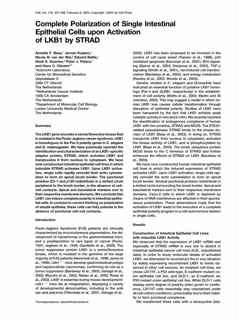
shown). As demonstrated in Figure 1A, several clones
were identified for each of the combinations. Figure 1B
represents a Western blot analysis of the induction of
STRAD in one of the LKB1-expressing clones. Expres-
sion of STRAD was visible within 3 hr after induction.
After 12 to 24 hr, LKB1 protein started to accumulate.
This was a consequence of protein stabilization, medi-
ated by STRAD, since mRNA levels were unaffected
upon induction (Figure 1A).
Confocal analysis confirmed that LKB1-WT, as well
as LKB1-SL26, were predominantly nuclear before in-
duction. The induction of STRAD, itself expressed
throughout the cell in the absence of LKB1, led to cyto-
plasmic translocation of LKB1-WT, but not of the LKB1-
SL26 mutant. Movies of GFP-tagged LKB1 which visual-
ize the STRAD-induced translocation and stabilization
in both LS174T and DLD-1 cells are provided as Supple-
mental Movie S1 available at http://www.cell.com/cgi/
content/full/116/3/457/DC1.
Activation of LKB1 Reorganizes the Actin
Cytoskeleton and Induces the Formation
of a Brush Border
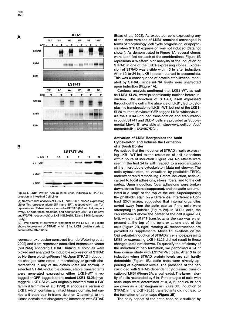
We noticed that the induction of STRAD in cells express-
ing LKB1-WT led to the retraction of cell extensions
within hours of induction (Figure 2A). No effects were
seen in the first 24 hr with respect to a reorganization
of the microtubule cytoskeleton (data not shown). The
actin cytoskeleton, as visualized by phalloidin-TRITC,
underwent rapid remodeling. Before induction, actin lo-
calized to focal adhesions, stress fibers, and to the cell
cortex. Upon induction, focal adhesions were broken
down, stress fibers disappeared, and the actin accumu-Figure 1. LKB1 Protein Accumulates upon Inducible STRAD Ex- lated in a “cap” at the top of the cell. Superimposingpression in Intestinal Cell Lines the phalloidin stain on a Differential Interference Con-(A) Northern blot analysis of LS174T and DLD-1 clones expressing trast (DIC) image, suggested that internal organelleseither Tet-repressor alone (TR1 and TR7, respectively), the Tet- sorted away from the actin cap as if the cells wererepressor and Tet-repressor-controlled STRAD (1-6 and 2-1, respec-
attempting to polarize (Figure 2A). In DLD-1 cells, thetively), or both these plasmids, and additionally LKB1-WT (W4/W5
cap remained above the center of the cell (Figure 2B,and W5/W8, respectively) or LKB1-SL26 (S1/S2 and S8/S10, respec-left), while in LS174T transfectants the cap was eithertively).
(B) Time course of doxycyclin treatment of the LS174T-W4 clone present at the top of the cells or at one side of theshows expression of STRAD within 3 hr. LKB1 protein starts to cells (Figure 2B, right; rotating 3D reconstructions areaccumulate after 12 hr. provided as Supplemental Movie S2 available on the
Cell website). Induction of STRAD in cells not expressing
LKB1 or expressing LKB1-SL26 did not result in theserepressor expression construct (van de Wetering et al.,
2002) and a tet-repressor-controlled expression vector changes (data not shown). To quantify the efficiency of
the induction of cap formation, we performed a 24 hr(pCDNA4) encoding STRAD. Individual colonies were
picked and analyzed for inducible expression of STRAD time course study with LS174T-W5 cells. After 3 hr of
induction when STRAD protein levels are still hardlyby Northern blotting (Figure 1A). Upon STRAD induction,
no changes were noted in morphology or growth cha- detectable (Figure 1B), actin caps were already ap-
pearing at significant levels. The presence of the capracteristics in any of the clones (data not shown). In
selected STRAD-inducible clones, stable transfectants coincided with STRAD-dependent cytoplasmic translo-
cation of LKB1 (Figure 3A, arrowheads). The large major-were generated expressing either LKB1-WT (myc-
tagged or GFP-tagged), or the mutant LKB1-SL26 (myc- ity of cells responded by 6 hr. Percentages of cells with
actin caps were determined at 0, 3, 6, and 24 hr andtagged). LKB1-SL26 was originally isolated from a PJS
family (Hemminki et al., 1998). It encodes a version of are given as a bar diagram in Figure 3C. Induction of
STRAD in the LKB1-SL26 transfectants did not lead toLKB1, which contains an intact kinase domain, but car-
ries a 9 base-pair in-frame deletion C-terminal to the the formation of actin caps (Figure 3B).
The hairy aspect of the actin caps as visualized bykinase domain that abrogates the interaction with STRAD
Complete Polarization by LKB1/STRAD459
Figure 2. Remodeling of the Actin Cytoskeleton upon Activation of
the LKB1/STRAD Complex
(A) DIC image of the LS174T-W4 clone superimposed on a phalloi-
din-TRITC staining shows retraction of cell extensions and actin
cytoskeleton reorganization upon 24 hr doxycyclin treatment. Cell
organelles redistribute in a polarized fashion.
(B) Compilations of individual confocal planes show an actin cap
on the cell surface of LS174T-W4 and DLD-1-W5 cells after a 24 hr Figure 3. Rapid Actin-Cytoskeleton Remodeling Correlates withdoxycyclin treatment by phalloidin-TRITC staining. LKB1 Translocation
(A) LS174T-W5 cells were subjected to a doxycyclin treatment for the
indicated time points. The actin-cytoskeleton is rapidly remodeled inDIC optics suggested that some of the actin was present induced LS174T-W5 cells, as analyzed by double staining for actin
and myc-LKB1. At the 3 hr time point, the presence of actin capsin microvilli. Brush borders consist of highly ordered,correlates with cytoplasmic translocation of LKB1 (arrowheads).dense arrays of microvilli covering the apical surface of(B) The induced LS174T-S2 clone is unable to form actin caps, andvillus epithelial cells in the intestine. Intestinal cell linesLKB1-SL26 resides in the nucleus upon STRAD expression.
such as Caco-2, or to a lesser extent DLD-1, can form(C) Bar diagram of the percentage of LS174T-W5 cells with actin
such structures in vitro. However, the only known way to caps after doxycyclin treatment for the indicated intervals.coerce these cell lines into forming apical brush borders
involves long-term culture at confluency (Pinto et al.,
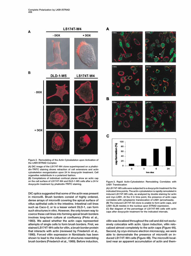
1983). We asked whether the actin caps represented villin was localized throughout the cell and did not exclu-
sively colocalize with actin. Upon induction, villin relo-attempts of single cells to form brush borders. First, we
stained LS174T-W4 cells for villin, a brush border protein calized almost completely to the actin caps (Figure 4A).
Second, by cryo-immuno electron microscopy, we werethat interacts with actin (reviewed by Friederich et al.,
1990). Forced villin expression in fibroblasts has been able to demonstrate the presence of microvilli on in-
duced LS174T-W4 cells (Figure 4B). The microvilli local-shown to lead to the induction of structures resembling
brush borders (Friederich et al., 1989). Before induction, ized near an apparent accumulation of actin and them-
Cell460
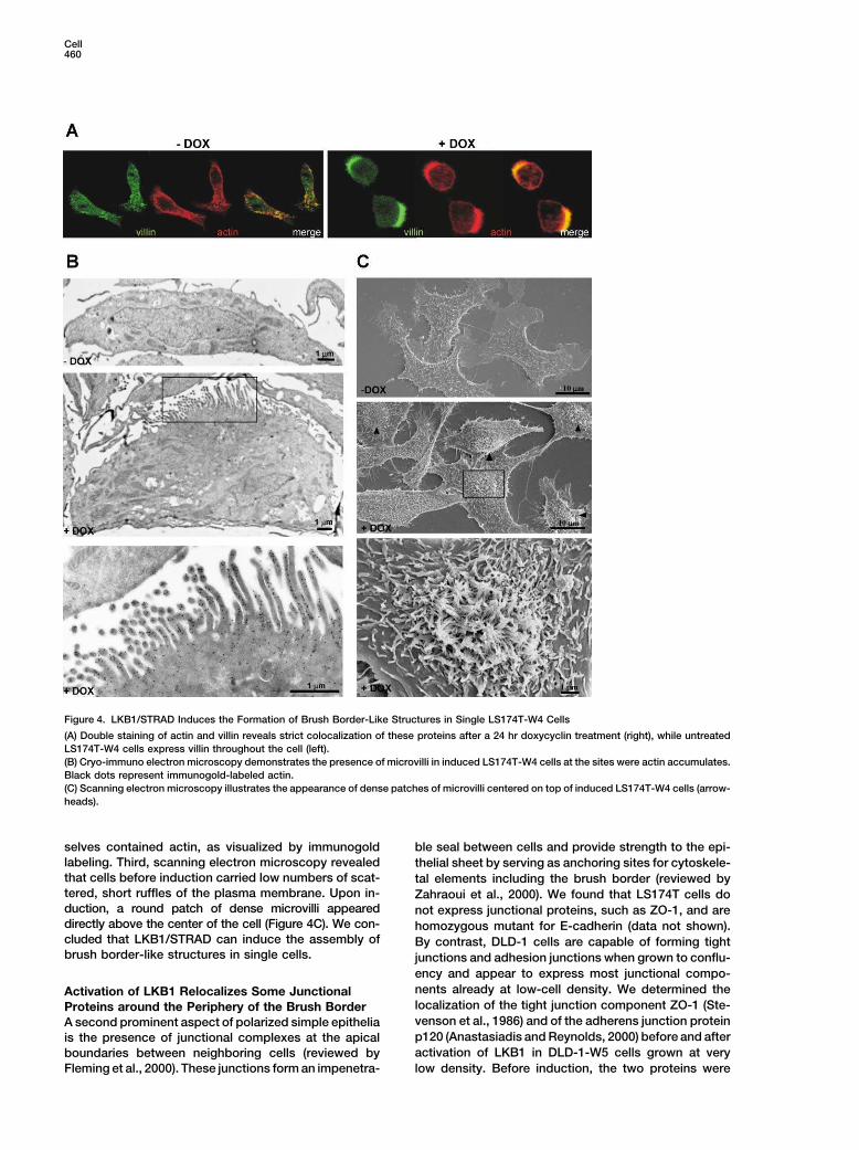
Figure 4. LKB1/STRAD Induces the Formation of Brush Border-Like Structures in Single LS174T-W4 Cells
(A) Double staining of actin and villin reveals strict colocalization of these proteins after a 24 hr doxycyclin treatment (right), while untreated
LS174T-W4 cells express villin throughout the cell (left).
(B) Cryo-immuno electron microscopy demonstrates the presence of microvilli in induced LS174T-W4 cells at the sites were actin accumulates.
Black dots represent immunogold-labeled actin.
(C) Scanning electron microscopy illustrates the appearance of dense patches of microvilli centered on top of induced LS174T-W4 cells (arrow-
heads).
selves contained actin, as visualized by immunogold ble seal between cells and provide strength to the epi-
labeling. Third, scanning electron microscopy revealed thelial sheet by serving as anchoring sites for cytoskele-
that cells before induction carried low numbers of scat- tal elements including the brush border (reviewed bytered, short ruffles of the plasma membrane. Upon in- Zahraoui et al., 2000). We found that LS174T cells doduction, a round patch of dense microvilli appeared not express junctional proteins, such as ZO-1, and aredirectly above the center of the cell (Figure 4C). We con- homozygous mutant for E-cadherin (data not shown).cluded that LKB1/STRAD can induce the assembly of By contrast, DLD-1 cells are capable of forming tightbrush border-like structures in single cells. junctions and adhesion junctions when grown to conflu-
ency and appear to express most junctional compo-
nents already at low-cell density. We determined theActivation of LKB1 Relocalizes Some Junctionallocalization of the tight junction component ZO-1 (Ste-Proteins around the Periphery of the Brush Bordervenson et al., 1986) and of the adherens junction proteinA second prominent aspect of polarized simple epitheliap120 (Anastasiadis and Reynolds, 2000) before and afteris the presence of junctional complexes at the apical
activation of LKB1 in DLD-1-W5 cells grown at veryboundaries between neighboring cells (reviewed by
Fleming et al., 2000). These junctions form an impenetra- low density. Before induction, the two proteins were
Complete Polarization by LKB1/STRAD461
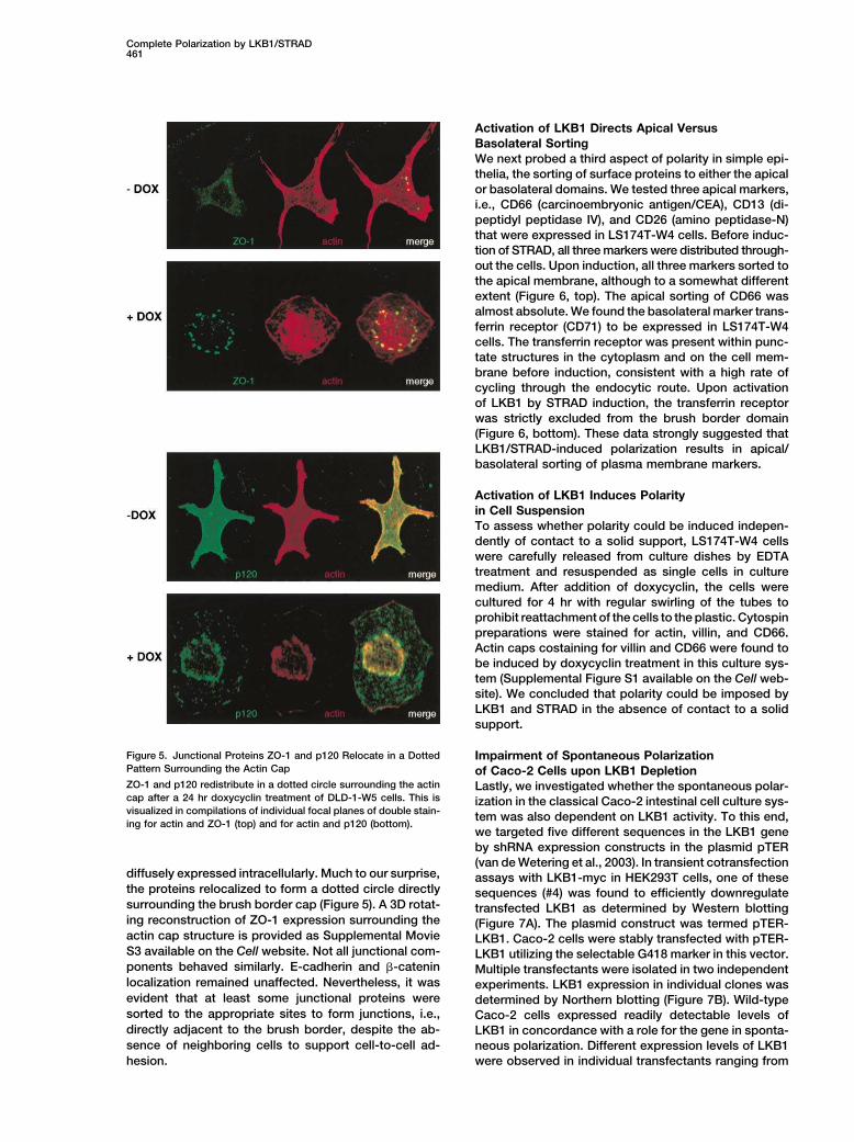
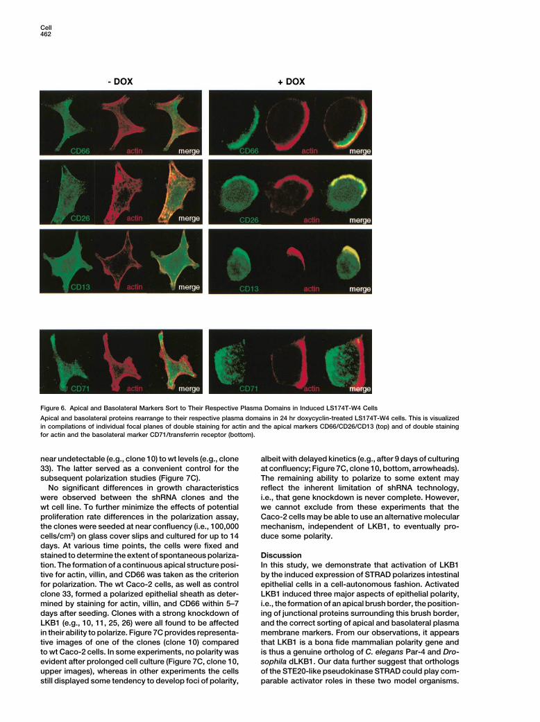
Activation of LKB1 Directs Apical Versus
Basolateral Sorting
We next probed a third aspect of polarity in simple epi-
thelia, the sorting of surface proteins to either the apical
or basolateral domains. We tested three apical markers,
i.e., CD66 (carcinoembryonic antigen/CEA), CD13 (di-
peptidyl peptidase IV), and CD26 (amino peptidase-N)
that were expressed in LS174T-W4 cells. Before induc-
tion of STRAD, all three markers were distributed through-
out the cells. Upon induction, all three markers sorted to
the apical membrane, although to a somewhat different
extent (Figure 6, top). The apical sorting of CD66 was
almost absolute. We found the basolateral marker trans-
ferrin receptor (CD71) to be expressed in LS174T-W4
cells. The transferrin receptor was present within punc-
tate structures in the cytoplasm and on the cell mem-
brane before induction, consistent with a high rate of
cycling through the endocytic route. Upon activation
of LKB1 by STRAD induction, the transferrin receptor
was strictly excluded from the brush border domain
(Figure 6, bottom). These data strongly suggested that
LKB1/STRAD-induced polarization results in apical/
basolateral sorting of plasma membrane markers.
Activation of LKB1 Induces Polarity
in Cell Suspension
To assess whether polarity could be induced indepen-
dently of contact to a solid support, LS174T-W4 cells
were carefully released from culture dishes by EDTA
treatment and resuspended as single cells in culture
medium. After addition of doxycyclin, the cells were
cultured for 4 hr with regular swirling of the tubes to
prohibit reattachment of the cells to the plastic. Cytospin
preparations were stained for actin, villin, and CD66.
Actin caps costaining for villin and CD66 were found to
be induced by doxycyclin treatment in this culture sys-
tem (Supplemental Figure S1 available on the Cell web-
site). We concluded that polarity could be imposed by
LKB1 and STRAD in the absence of contact to a solid
support.
Figure 5. Junctional Proteins ZO-1 and p120 Relocate in a Dotted Impairment of Spontaneous PolarizationPattern Surrounding the Actin Cap of Caco-2 Cells upon LKB1 DepletionZO-1 and p120 redistribute in a dotted circle surrounding the actin Lastly, we investigated whether the spontaneous polar-cap after a 24 hr doxycyclin treatment of DLD-1-W5 cells. This is ization in the classical Caco-2 intestinal cell culture sys-visualized in compilations of individual focal planes of double stain-
tem was also dependent on LKB1 activity. To this end,ing for actin and ZO-1 (top) and for actin and p120 (bottom).
we targeted five different sequences in the LKB1 gene
by shRNA expression constructs in the plasmid pTER
(van de Wetering et al., 2003). In transient cotransfectiondiffusely expressed intracellularly. Much to our surprise, assays with LKB1-myc in HEK293T cells, one of thesethe proteins relocalized to form a dotted circle directly sequences (#4) was found to efficiently downregulatesurrounding the brush border cap (Figure 5). A 3D rotat- transfected LKB1 as determined by Western blottinging reconstruction of ZO-1 expression surrounding the (Figure 7A). The plasmid construct was termed pTER-actin cap structure is provided as Supplemental Movie LKB1. Caco-2 cells were stably transfected with pTER-S3 available on the Cell website. Not all junctional com- LKB1 utilizing the selectable G418 marker in this vector.ponents behaved similarly. E-cadherin and �-catenin Multiple transfectants were isolated in two independentlocalization remained unaffected. Nevertheless, it was experiments. LKB1 expression in individual clones wasevident that at least some junctional proteins were determined by Northern blotting (Figure 7B). Wild-typesorted to the appropriate sites to form junctions, i.e., Caco-2 cells expressed readily detectable levels ofdirectly adjacent to the brush border, despite the ab- LKB1 in concordance with a role for the gene in sponta-
sence of neighboring cells to support cell-to-cell ad- neous polarization. Different expression levels of LKB1
were observed in individual transfectants ranging fromhesion.
Cell462
Figure 6. Apical and Basolateral Markers Sort to Their Respective Plasma Domains in Induced LS174T-W4 Cells
Apical and basolateral proteins rearrange to their respective plasma domains in 24 hr doxycyclin-treated LS174T-W4 cells. This is visualized
in compilations of individual focal planes of double staining for actin and the apical markers CD66/CD26/CD13 (top) and of double staining
for actin and the basolateral marker CD71/transferrin receptor (bottom).
near undetectable (e.g., clone 10) to wt levels (e.g., clone albeit with delayed kinetics (e.g., after 9 days of culturing
at confluency; Figure 7C, clone 10, bottom, arrowheads).33). The latter served as a convenient control for the
subsequent polarization studies (Figure 7C). The remaining ability to polarize to some extent may
reflect the inherent limitation of shRNA technology,No significant differences in growth characteristics
were observed between the shRNA clones and the i.e., that gene knockdown is never complete. However,
we cannot exclude from these experiments that thewt cell line. To further minimize the effects of potential
proliferation rate differences in the polarization assay, Caco-2 cells may be able to use an alternative molecular
mechanism, independent of LKB1, to eventually pro-the clones were seeded at near confluency (i.e., 100,000
cells/cm2) on glass cover slips and cultured for up to 14 duce some polarity.
days. At various time points, the cells were fixed and
stained to determine the extent of spontaneous polariza- Discussion
In this study, we demonstrate that activation of LKB1tion. The formation of a continuous apical structure posi-
tive for actin, villin, and CD66 was taken as the criterion by the induced expression of STRAD polarizes intestinal
epithelial cells in a cell-autonomous fashion. Activatedfor polarization. The wt Caco-2 cells, as well as control
clone 33, formed a polarized epithelial sheath as deter- LKB1 induced three major aspects of epithelial polarity,
i.e., the formation of an apical brush border, the position-mined by staining for actin, villin, and CD66 within 5–7
days after seeding. Clones with a strong knockdown of ing of junctional proteins surrounding this brush border,
and the correct sorting of apical and basolateral plasmaLKB1 (e.g., 10, 11, 25, 26) were all found to be affected
in their ability to polarize. Figure 7C provides representa- membrane markers. From our observations, it appears
that LKB1 is a bona fide mammalian polarity gene andtive images of one of the clones (clone 10) compared
to wt Caco-2 cells. In some experiments, no polarity was is thus a genuine ortholog of C. elegans Par-4 and Dro-
sophila dLKB1. Our data further suggest that orthologsevident after prolonged cell culture (Figure 7C, clone 10,
upper images), whereas in other experiments the cells of the STE20-like pseudokinase STRAD could play com-
parable activator roles in these two model organisms.still displayed some tendency to develop foci of polarity,
Complete Polarization by LKB1/STRAD463
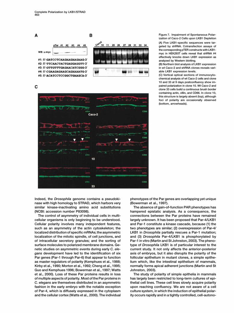
Figure 7. Impairment of Spontaneous Polar-
ization of Caco-2 Cells upon LKB1 Depletion
(A) Five LKB1-specific sequences were tar-
geted by shRNA. Cotransfection assays of
the corresponding pTER constructs with LKB1-
myc in HEK293T cells reveal that shRNA #4
effectively knocks down LKB1 expression as
analyzed by Western blotting.
(B) Northern blot analysis of LKB1 expression
in wt Caco-2 and shRNA clones reveals vari-
able LKB1 expression levels.
(C) Vertical optical sections of immunocyto-
chemical analysis of wt Caco-2 cells and clone
10 and 33 at 9 days postconfluency show im-
paired polarization in clone 10. Wt Caco-2 and
clone 33 cells build a continuous brush border
containing actin, villin, and CD66. In clone 10,
this structure is largely absent (top), although
foci of polarity are occasionally observed
(bottom, arrowheads).
Indeed, the Drosophila genome contains a pseudoki- phenotypes of the Par genes are overlapping yet unique
(Bowerman et al., 1997).nase with high homology to STRAD, which harbors very
similar kinase-inactivating amino acid substitutions The absence of gain-of-function PAR phenotypes has
hampered epistatic analysis. As a consequence, the(NCBI: accession number P83098).
The control of asymmetry of individual cells in multi- connections between the Par proteins have remained
largely unknown. It has been proposed that Par-4/LKB1cellular organisms is only beginning to be understood.
Cellular polarity involves many independent features, and Par-1 constitute a kinase cascade, because (1) the
two phenotypes are similar; (2) overexpression of Par-4/such as an asymmetry of the actin cytoskeleton; the
localized distribution of specific mRNAs; the asymmetric LKB1 in Drosophila partially rescues a Par-1 mutation;
and (3) Drosophila Par-4/LKB1 is phosphorylated bylocalization of the mitotic spindle, of cell junctions, and
of intracellular secretory granules; and the sorting of Par-1 in vitro (Martin and St Johnston, 2003). The pheno-
type of Drosophila LKB1 is of particular interest to thesurface molecules to polarized membrane domains. Ge-
netic studies on asymmetric events during early C. ele- current study. It not only affects the anterior-posterior
axis of embryos, but it also disrupts the polarity of thegans development have led to the identification of six
Par genes (Par-1 through Par-6) that appear to function follicular epithelium in mutant clones, a simple epithe-
lium which, like the intestinal epithelium of mammals,as master regulators of polarity (Kemphues et al., 1988;
Kirby et al., 1990; Morton et al., 1992; Cheng et al., 1995; normally forms apical adherent junctions (Martin and St
Johnston, 2003).Guo and Kemphues 1996; Bowerman et al., 1997; Watts
et al., 2000). Loss of these Par proteins results in loss The study of polarity of simple epithelia in mammals
has largely been restricted to long-term cultures of epi-of multiple aspects of polarity. Most of the Par proteins in
C. elegans are themselves distributed in an asymmetric thelial cell lines. These cell lines slowly acquire polarity
upon reaching confluency. We are not aware of a cellfashion in the early embryo with the notable exception
of Par-4, which is diffusely expressed in the cytoplasm culture system, in which the induction of epithelial polar-
ity occurs rapidly and in a tightly controlled, cell-autono-and the cellular cortex (Watts et al., 2000). The individual
Cell464
mous fashion. Therefore, the cellular model described regulated LKB1-STRAD interaction may be a key control
here may be a valuable complement to the genetic loss- point of an LKB1/Par-4 polarity-signaling pathway.
of-function studies in model organisms. The gain-of- LKB1 has been identified originally as a tumor sup-
function effects induced by STRAD in our cell lines can pressor. It has since been shown to affect a wide variety
be combined with siRNA technology and other ap- of cell growth characteristics (Tiainen et al., 1999, 2002;
proaches to resolve molecular details of the various Marignani et al., 2001; Karuman et al., 2001). Of note,aspects of polarization of simple epithelia. our cell lines do undergo growth arrest upon induction
One unexpected finding of the current study is the of STRAD (data not shown). This is first observed afterobservation that individual epithelial cells can be in- 24 hr, long after the cells express the polarized pheno-duced to polarize in the absence of cell-cell contacts, type. When the same cells are induced to undergo awhich contradicts current thinking (reviewed by Wodarz, G1 arrest by blocking the Wnt cascade or by inducible2000). Previous studies on model cell lines such as expression of cell-cycle inhibitor p21 (van de WeteringMDCK (Cereijido et al., 1980) and Caco-2 (Pinto et al., et al., 2002), they do not polarize (data not shown). We1983) have always been restricted to the spontaneous believe that many of the phenomena that have beenappearance of polarity after the cells are allowed to form attributed to LKB1 may be the indirect consequence ofcell junctions. changes in cellular polarity. As suggested previously
The dynamics of the actin reorganization in our cells (Martin and St Johnston, 2003), loss of the tumor sup-suggests that the brush border formation is a direct pressor LKB1 may result in cellular transformation as aconsequence of LKB1 activation and may be required consequence of disrupted epithelial polarity.for the additional polarity phenomena to occur. It ap-
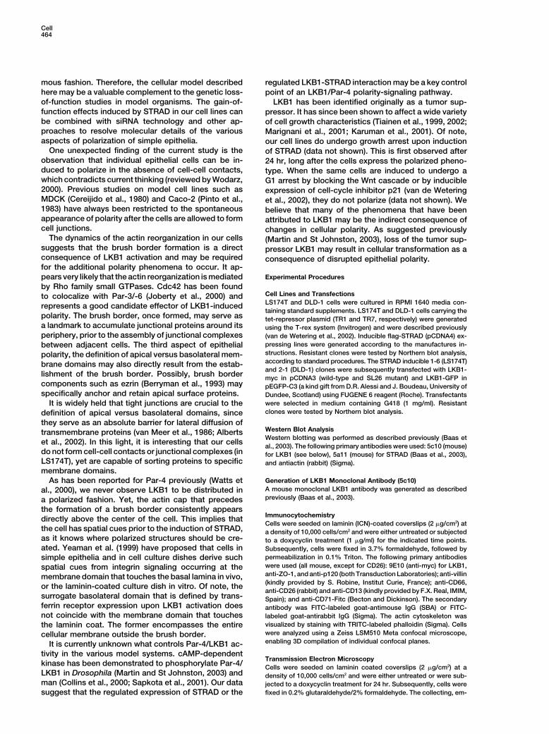
Experimental Procedurespears very likely that the actin reorganization is mediated
by Rho family small GTPases. Cdc42 has been foundCell Lines and Transfectionsto colocalize with Par-3/-6 (Joberty et al., 2000) andLS174T and DLD-1 cells were cultured in RPMI 1640 media con-
represents a good candidate effector of LKB1-inducedtaining standard supplements. LS174T and DLD-1 cells carrying the
polarity. The brush border, once formed, may serve astet-repressor plasmid (TR1 and TR7, respectively) were generated
a landmark to accumulate junctional proteins around its using the T-rex system (Invitrogen) and were described previously
periphery, prior to the assembly of junctional complexes (van de Wetering et al., 2002). Inducible flag-STRAD (pCDNA4) ex-
pressing lines were generated according to the manufactures in-between adjacent cells. The third aspect of epithelialstructions. Resistant clones were tested by Northern blot analysis,polarity, the definition of apical versus basolateral mem-according to standard procedures. The STRAD inducible 1-6 (LS174T)brane domains may also directly result from the estab-and 2-1 (DLD-1) clones were subsequently transfected with LKB1-
lishment of the brush border. Possibly, brush bordermyc in pCDNA3 (wild-type and SL26 mutant) and LKB1-GFP in
components such as ezrin (Berryman et al., 1993) may pEGFP-C3 (a kind gift from D.R. Alessi and J. Boudeau, University ofspecifically anchor and retain apical surface proteins. Dundee, Scotland) using FUGENE 6 reagent (Roche). Transfectants
were selected in medium containing G418 (1 mg/ml). ResistantIt is widely held that tight junctions are crucial to theclones were tested by Northern blot analysis.definition of apical versus basolateral domains, since
they serve as an absolute barrier for lateral diffusion ofWestern Blot Analysistransmembrane proteins (van Meer et al., 1986; AlbertsWestern blotting was performed as described previously (Baas et
et al., 2002). In this light, it is interesting that our cellsal., 2003). The following primary antibodies were used: 5c10 (mouse)
do not form cell-cell contacts or junctional complexes (in for LKB1 (see below), 5a11 (mouse) for STRAD (Baas et al., 2003),LS174T), yet are capable of sorting proteins to specific and antiactin (rabbit) (Sigma).
membrane domains.
Generation of LKB1 Monoclonal Antibody (5c10)As has been reported for Par-4 previously (Watts etA mouse monoclonal LKB1 antibody was generated as describedal., 2000), we never observe LKB1 to be distributed inpreviously (Baas et al., 2003).a polarized fashion. Yet, the actin cap that precedes
the formation of a brush border consistently appearsImmunocytochemistry
directly above the center of the cell. This implies thatCells were seeded on laminin (ICN)-coated coverslips (2 �g/cm2) at
the cell has spatial cues prior to the induction of STRAD, a density of 10,000 cells/cm2 and were either untreated or subjectedas it knows where polarized structures should be cre- to a doxycyclin treatment (1 �g/ml) for the indicated time points.
ated. Yeaman et al. (1999) have proposed that cells in Subsequently, cells were fixed in 3.7% formaldehyde, followed by
permeabilization in 0.1% Triton. The following primary antibodiessimple epithelia and in cell culture dishes derive suchwere used (all mouse, except for CD26): 9E10 (anti-myc) for LKB1,spatial cues from integrin signaling occurring at theanti-ZO-1, and anti-p120 (both Transduction Laboratories); anti-villinmembrane domain that touches the basal lamina in vivo,(kindly provided by S. Robine, Institut Curie, France); anti-CD66,
or the laminin-coated culture dish in vitro. Of note, theanti-CD26 (rabbit) and anti-CD13 (kindly provided by F.X. Real, IMIM,
surrogate basolateral domain that is defined by trans- Spain); and anti-CD71-Fitc (Becton and Dickinson). The secondaryferrin receptor expression upon LKB1 activation does antibody was FITC-labeled goat-antimouse IgG (SBA) or FITC-
not coincide with the membrane domain that touches labeled goat-antirabbit IgG (Sigma). The actin cytoskeleton was
visualized by staining with TRITC-labeled phalloidin (Sigma). Cellsthe laminin coat. The former encompasses the entirewere analyzed using a Zeiss LSM510 Meta confocal microscope,cellular membrane outside the brush border.enabling 3D compilation of individual confocal planes.It is currently unknown what controls Par-4/LKB1 ac-
tivity in the various model systems. cAMP-dependentTransmission Electron Microscopy
kinase has been demonstrated to phosphorylate Par-4/Cells were seeded on laminin coated coverslips (2 �g/cm2) at a
LKB1 in Drosophila (Martin and St Johnston, 2003) and density of 10,000 cells/cm2 and were either untreated or were sub-man (Collins et al., 2000; Sapkota et al., 2001). Our data jected to a doxycyclin treatment for 24 hr. Subsequently, cells were
fixed in 0.2% glutaraldehyde/2% formaldehyde. The collecting, em-suggest that the regulated expression of STRAD or the
Complete Polarization by LKB1/STRAD465
bedding and processing of the fixed cells for cryosectioning with MO25�/� interact with STRAD�/� enhancing their ability to bind,
activate and localise LKB1 in the cytoplasm. EMBO J. 22, 5102–a Leica FCS was performed as described previously (Peters and
Hunziker, 2001). Samples were immunogold-labeled using a poly- 5114.
clonal antibody against actin (a kind gift from C. Chaponnier, Univer- Bowerman, B., Ingram, M.K., and Hunter, C.P. (1997). The maternalsity of Geneva, Switzerland) and 15 nm gold conjugated to protein-A par genes and the segregation of cell fate specification activities in(EM laboratory, Utrecht University). Sections were analyzed on a early Caenorhabditis elegans embryos. Development 124, 3815–Fei CM10 and Tecnai 12 transmission electron microscope. 3826.
Cereijido, M., Ehrenfeld, J., Meza, I., and Martinez-Palomo, A. (1980).Scanning Electron Microscopy
Structural and functional membrane polarity in cultured monolayersCells were seeded on laminin coated coverslips (2 �g/cm2) at a
of MDCK cells. J. Membr. Biol. 52, 147–159.density of 10,000 cells/cm2 and were either untreated or subjected
Cheng, N.N., Kirby, C.M., and Kemphues, K.J. (1995). Control ofto a doxycyclin treatment for 24 hr. Subsequently, cells were fixedcleavage spindle orientation in Caenorhabditis elegans: the role ofin 0.2% glutaraldehyde/2% formaldehyde. After fixation, the cellsthe genes par-2 and par-3. Genetics 139, 549–559.were dehydrated in a graded ethanol series up to 100% and criticalCollins, S.P., Reoma, J.L., Gamm, D.M., and Uhler, M.D. (2000).point dried under carbon dioxide in a Baltec model CPD 030 criticalLKB1, a novel serine/threonine protein kinase and potential tumourpoint drier. The dried specimens were covered with a layer of goldsuppressor is phosphorylated by cAMP-dependent protein kinaseabout 3 nm thick in an EMITECH K650X sputter coater and examined(PKA) and prenylated in vivo. Biochem. J. 345, 673–680.at room temperature in a JEOL JSM6700F field emission scanning
electron microscope at an accelerating voltage of 2.5 kV. Fleming, T.P., Papenbrock, T., Fesenko, I., Hausen, P., and Sheth,
B. (2000). Assembly of tight junctions during early vertebrate devel-
LKB1 RNA-Interference in Caco-2 Cells opment. Semin. Cell Dev. Biol. 11, 291–299.
Five LKB1-specific sequences were targeted for RNA-interference Friederich, E., Huet, C., Arpin, M., and Louvard, D. (1989). Villinby cloning compatible oligonucleotides (Figure 7A, additional se- induces microvilli growth and actin redistribution in transfected fi-quence information available upon request) into pTER (van de Wet- broblasts. Cell 59, 461–475.ering et al., 2003). For stable transfection, the G418 resistance
Friederich, E., Pringault, E., Arpin, M., and Louvard, D. (1990). Frommarker was cloned from pCDNA3 into the pertinent pTER construct.
the structure to the function of villin, an actin-binding protein of theThe resulting plasmid was termed LKB1-pTER. Caco-2 cells were
brush border. Bioessays 12, 403–408.transfected with LKB1-pTER and G418-resistant clones were ana-
Giardiello, F.M., Brensinger, J.D., Tersmette, A.C., Goodman, S.N.,lyzed for LKB1 expression by Northern blotting. To examine thePetersen, G.M., Booker, S.V., Cruz-Correa, M., and Offerhaus, J.A.extent of spontaneous polarization, wt Caco-2 cells and LKB1-pTER(2000). Very high risk of cancer in familial Peutz-Jeghers syndrome.clones were seeded on laminin-coated coverslips (2 �g/cm2) at aGastroenterology 119, 1447–1453.density of 100,000 cells/cm2. Media of the confluent cells was re-
freshed daily. At various time points postconfluency, morphology Guo, S., and Kemphues, K.J. (1996). Molecular genetics of asymmet-
was examined and immunocytochemistry experiments for actin, vil- ric cleavage in the early Caenorhabditis elegans embryo. Curr. Opin.
lin, and CD66 were performed as described above. Genet. Dev. 6, 408–415.
Hawley, S.A., Boudeau, J., Reid, J.L., Mustard, K.J., Udd, L., Makela,Acknowledgments T.P., Alessi, D.R., and Hardie, D.G. (2003). Complexes between the
LKB1 tumor suppressor, STRADalpha/beta and MO25alpha/betaThe authors thank D.R. Alessi and J. Boudeau (University of Dundee, are upstream kinases in the AMP-activated protein kinase cascade.Scotland); S. Robine (Institute Curie, France); F.X. Real (IMIM, Spain); J. Biol. 2, 28.and C. Chaponnier (University of Geneva, Switzerland) for supplied
Hemminki, A., Markie, D., Tomlinson, I., Avizienyte, E., Roth, S.,materials. We thank the Division of Cell Biology, UMCU, the Nether-
Loukola, A., Bignell, G., Warren, W., Aminoff, M., Hoglund, P., et al.lands for use of the life cell imaging station, and all members of the
(1998). A serine/threonine kinase gene defective in Peutz-JeghersClevers laboratory, as well as F.X. Real, for valuable discussions.
syndrome. Nature 391, 184–187.
Jeghers, H., McKusick, V.A., and Karz, K.H. (1949). Generalised in-Received: September 22, 2003
testinal polyposis and melanin spots of oral mucosa, lips and digits:Revised: December 17, 2003
a syndrome of diagnostic significance. N. Engl. J. Med. 241, 992–Accepted: January 12, 2004
1005.Published: February 5, 2004
Jenne, D.E., Reimann, H., Nezu, J., Friedel, W., Loff, S., Jeschke,
R., Muller, O., Back, W., and Zimmer, M. (1998). Peutz-Jeghers syn-Referencesdrome is caused by mutations in a novel serine threonine kinase.
Nat. Genet. 18, 38–43.Alberts, B., Johnson, A., Lewis, J., Raff, M., Roberts, K., and Walter,
Jishage, K., Nezu, J., Kawase, Y., Iwata, T., Watanabe, M., Miyoshi,P. (2002). Molecular biology of the cell. 4th ed. (New York, New York:
A., Ose, A., Habu, K., Kake, T., Kamada, N., et al. (2002). Role ofGarland Science), 609–612.
Lkb1, the causative gene of Peutz-Jegher’s syndrome, in em-Anastasiadis, P.Z., and Reynolds, A.B. (2000). The p120 cateninbryogenesis and polyposis. Proc. Natl. Acad. Sci. USA 99, 8903–family: complex roles in adhesion, signaling and cancer. J. Cell Sci.8908.113, 1319–1334.
Joberty, G., Petersen, C., Gao, L., and Macara, I.G. (2000). The cellBaas, A.F., Boudeau, J., Sapkota, G.P., Smit, L., Medema, R., Mor-polarity protein par6 links par3 and atypical protein kinase C torice, N.A., Alessi, D.R., and Clevers, H.C. (2003). Activation of theCdc42. Nat. Cell Biol. 2, 531–539.tumour suppressor kinase LKB1 by the STE20-like pseudokinase
Karuman, P., Gozani, O., Odze, R.D., Zhou, X.C., Zhu, H., Shaw, R.,STRAD. EMBO J. 22, 3062–3072.
Brien, T.P., Bozzuto, C.D., Ooi, D., Cantley, L.C., and Yuan, J. (2001).Bardeesy, N., Sinha, M., Hezel, A.F., Signoretti, S., Hathaway, N.A.,The Peutz-Jegher gene product LKB1 is a mediator of p53-depen-Sharpless, N.E., Loda, M., Carrasco, D.R., and DePinho, R.A. (2002).dent cell death. Mol. Cell 7, 1307–1319.Loss of the Lkb1 tumour suppressor provokes intestinal polyposis
Kemphues, K.J., Priess, J.R., Morton, D.G., and Cheng, N.S. (1988).but resistance to transformation. Nature 419, 162–167.
Identification of genes required for cytoplasmic localization in earlyBerryman, M., Franck, Z., and Bretscher, A. (1993). Ezrin is concen-C. elegans embryos. Cell 52, 311–320.trated in the apical microvilli of a variety of epithelial cells whereas
Kirby, C., Kusch, M., and Kemphues, K. (1990). Mutations in the parmoesin is found primarily in endothelial cells. J. Cell Sci. 105, 1025–
genes of Caenorhabditis elegans affect cytoplasmic reorganization1043.
during the first cell cycle. Dev. Biol. 142, 203–215.Boudeau, J., Baas, A.F., Deak, M., Morrice, N., Kieloch, A., Schut-
kowski, M., Prescott, A.R., Clevers, H.C., and Alessi, D.R. (2003). Marignani, P.A., Kanai, F., and Carpenter, C.L. (2001). LKB1 associ-
Cell466
ates with Brg1 and is necessary for Brg1-induced growth arrest. J. Van Meer, G., Gumbiner, B., and Simons, K. (1986). The tight junction
does not allow lipid molecules to diffuse from one epithelial cell toBiol. Chem. 276, 32415–32418.
the next. Nature 322, 639–641.Martin, S.G., and St Johnston, D. (2003). A role for Drosophila LKB1
Watts, J.L., Morton, D.G., Bestman, J., and Kemphues, J. (2000). Thein anterior-posterior axis formation and epithelial polarity. Nature
C. elegans par-4 gene encodes a putative serine-threonine kinase421, 379–384.
required for establishing embryonic asymmetry. DevelopmentMiyoshi, H., Nakau, M., Ishikawa, T.O., Seldin, M.F., Oshima, M.,127, 1467–1475.and Taketo, M.M. (2002). Gastrointestinal hamartomatous polyposis
Wodarz, A. (2002). Establishing cell polarity in development. Nat.in Lkb1 heterozygous knockout mice. Cancer Res. 62, 2261–2266.
Cell Biol. 4, E39–E44.Morton, D.G., Roos, J.M., and Kemphues, K.J. (1992). par-4, a geneWoods, A., Johnstone, S.R., Dickerson, K., Leiper, F.C., Fryer, L.G.,required for cytoplasmic localization and determination of specificNeumann, D., Schlattner, U., Wallimann, T., Carlson, M., and Carling,cell types in Caenorhabditis elegans embryogenesis. GeneticsD. (2003). LKB1 is the upstream kinase in the AMP-activated protein130, 771–790.kinase cascade. Curr. Biol. 13, 2004–2008.
Nakau, M., Miyoshi, H., Seldin, M.F., Imamura, M., Oshima, M., andYeaman, C., Grindstaff, K.K., and Nelson, W.J. (1999). New perspec-Taketo, M.M. (2002). Hepatocellular carcinoma caused by loss oftives on mechanisms involved in generating epithelial cell polarity.heterozygosity in lkb1 gene knockout mice. Cancer Res. 62, 4549–Phys. Rev. 79, 73–98.4553.
Ylikorkala, A., Rossi, D.J., Korsisaari, N., Luukko, K., Alitalo, K.,Ossipova, O., Bardeesy, N., DePinho, R.A., and Green, J.B. (2003).Henkemeyer, M., and Makela, T.P. (2001). Vascular abnormalitiesLKB1 (XEEK1) regulates Wnt signalling in vertebrate development.and deregulation of VEGF in Lkb1-deficient mice. Science 293,Nat. Cell Biol. 10, 889–894.1323–1326.
Peters, P.J., and Hunziker, W. (2001). Subcellular localization ofZahraoui, A., Louvard, D., and Galli, T. (2000). Tight junction, a plat-Rab17 by cryo-immunogold electron microscopy in epithelial cellsform for trafficking and signaling protein complexes. J. Cell Biol.grown on polycarbonate filters. Methods Enzymol. 329, 210–225.151, F31–F36.
Peutz, J.L. (1921). On a very remarkable case of familial polyposis
of the mucous membrane of the intestinal tract and nasopharynx
accompanied by peculiar pigmentation of the skin and mucous
membranes. Ned. Tijdschr. Geneeskd. 10, 134–146.
Pinto, M., Robine-Leon, S., Appay, M.D., Kedinger, M., Triadou, N.,
Dussanly, E., Lacroix, B., Simon-Assmann, P., Haffen, K., Fogh, J.,
and Zweibaum, A. (1983). Enterocyte-like differentiation and polar-
ization of the human colon carcinoma cell line Caco-2 in culture.
Biol. Cell. 47, 323–330.
Rossi, D.J., Ylikorkala, A., Korsisaari, N., Salovara, R., Luukko, K.,
Launonen, V., Henkemeyer, M., Ristmaki, A., Aaltonen, L.A., and
Makela, T.P. (2002). Induction of cyclooxygenase-2 in a mouse
model of Peutz-Jeghers polyposis. Proc. Natl. Acad. Sci. U.S.A.
99, 505–509.
Sapkota, G.P., Kieloch, A., Lizcano, J.M., Lain, S., Arthur, J.S., Wil-
liams, M.R., Morrice, N., Deak, M., and Alessi, D.R. (2001). Phosphor-
ylation of the protein kinase mutated in Peutz-Jeghers cancer syn-
drome, LKB1/STK11, at Ser431 by p90(RSK) and cAMP-dependent
protein kinase, but not its farnesylation at Cys(433), is essential for
LKB1 to suppress cell growth. J. Biol. Chem. 276, 19469–19482.
Spicer, J., Sydonia, R., Neville, Y., Richard, E., Ashworth, A., and
Smith, D. (2003). Regulation of the Wnt signalling component PAR1A
by the Peutz-Jeghers syndrome kinase LKB1. Oncogene 22, 4752–
4756.
Smith, D.P., Rayter, S.I., Niederlander, C., Spicer, J., Jones, C.M.,
and Ashworth, A. (2001). LIP1, a cytoplasmic protein functionally
linked to the Peutz-Jeghers syndrome kinase LKB1. Hum. Mol.
Genet. 10, 2869–2877.
Stevenson, B.R., Siliciano, J.D., Mooseker, M.S., and Goodenough,
D.A. (1986). Identification of ZO-1: a high molecular weight polypep-
tide associated with the tight junction (zonula occludens) in a variety
of epithelia. J. Cell Biol. 103, 755–766.
Tiainen, M., Ylikorkala, A., and Makela, T.P. (1999). Growth suppres-
sion by Lkb1 is mediated by a G(1) cell cycle arrest. Proc. Natl.
Acad. Sci. USA 96, 9248–9251.
Tiainen, M., Vaahtomeri, K., Ylikorkala, A., and Makela, T.P. (2002).
Growth arrest by the LKB1 tumor suppressor: induction of
p21(WAF1/CIP1). Hum. Mol. Genet. 11, 1497–1504.
van de Wetering, M., Sancho, E., Verweij, C., de Lau, W., Oving, I.,
Hurlstone, A., van der Horn, K., Batlle, E., Coudreuse, D., Haramis,
A.P., et al. (2002). The �-catenin/TCF-4 complex imposes a crypt
progenitor phenotype on colorectal cancer cells. Cell 111, 241–250.
van de Wetering, M., Oving, I., Muncan, V., Pon Fong, M.T., Brantjes,
H., van Leenen, D., Holstege, F.C., Brummelkamp, T.R., Agami, R.,
and Clevers, H. (2003). Specific inhibition of gene expression using
a stably integrated, inducible small-interfering-RNA vector. EMBO
Rep. 4, 609–615.