Complete genome sequence of Streptosporangium roseum type strain (NI 9100)
Standards in Genomic Sciences (2011) 4:154-162 DOI:10.4056/sigs.1623941
The Genomic Standards Consortium
Complete genome sequence of Marivirga tractuosa type strain (H-43T)
Ioanna Pagani1, Olga Chertkov1,2, Alla Lapidus1, Susan Lucas1, Tijana Glavina Del Rio1, Hope Tice1, Alex Copeland1, Jan-Fang Cheng1, Matt Nolan1, Elizabeth Saunders1,2, Sam Pitluck1, Brittany Held1.2, Lynne Goodwin1,2, Konstantinos Liolios1, Galina Ovchinikova1, Natalia Ivanova1, Konstantinos Mavromatis1, Amrita Pati1, Amy Chen3, Krishna Palaniappan3, Miriam Land1,4, Loren Hauser1,4, Cynthia D. Jeffries1,4, John C. Detter1,4, Cliff Han1,2, Roxanne Tapia1,2, Olivier D. Ngatchou-Djao5, Manfred Rohde5, Markus Göker6, Stefan Spring6, Johannes Sikorski6, Tanja Woyke1, Jim Bristow1, Jonathan A. Eisen1,7, Victor Markowitz3, Philip Hugenholtz1,8, Hans-Peter Klenk6 and Nikos C. Kyrpides1*
1 DOE Joint Genome Institute, Walnut Creek, California, USA 2 Los Alamos National Laboratory, Bioscience Division, Los Alamos, New Mexico, USA 3 Biological Data Management and Technology Center, Lawrence Berkeley National
Laboratory, Berkeley, California, USA 4 Oak Ridge National Laboratory, Oak Ridge, Tennessee, USA 5 HZI – Helmholtz Centre for Infection Research, Braunschweig, Germany 6 DSMZ – German Collection of Microorganisms and Cell Cultures GmbH, Braunschweig,
Germany 7 University of California Davis Genome Center, Davis, California, USA 8 Australian Centre for Ecogenomics, School of Chemistry and Molecular Biosciences, The
University of Queensland, Brisbane, Australia
*Corresponding authors: Nikos C Kyrpides
Keywords: mesophilic, chemoorganotrophic, strictly aerobic, Gram-negative, slender and flexible rod-shaped, non-sporeforming, motile by gliding, Flammeovirgaceae, GEBA
Marivirga tractuosa (Lewin 1969) Nedashkovskaya et al. 2010 is the type species of the genus Marivirga, which belongs to the family Flammeovirgaceae. Members of this genus are of in-terest because of their gliding motility. The species is of interest because representative strains show resistance to several antibiotics, including gentamicin, kanamycin, neomycin, polymix-in and streptomycin. This is the first complete genome sequence of a member of the family Flammeovirgaceae. Here we describe the features of this organism, together with the com-plete genome sequence and annotation. The 4,511,574 bp long chromosome and the 4,916 bp plasmid with their 3,808 protein-coding and 49 RNA genes are a part of the Genomic En-cyclopedia of Bacteria and Archaea project.
Introduction Strain H-43T (= DSM 4126 = ATCC 23168 = NBRC 15989) is the type strain of the species Marivirga tractuosa. The genus Marivirga, whose type spe-cies is M. tractuosa, contains only one additional species: M. sericea. The generic name ‘Marivirga’ derives from Latin words ‘mare’, the sea and ‘vir-ga’, rod, meaning ‘a rod that inhabits marine envi-ronments’ [1]. The species epithet ‘tractuosa’ is a Latin adjective meaning ‘that draws to itself, gluey, viscous’, probably referring to the phenotype of gliding motility [1]. Strain H-43T was isolated in
1969 from a beach sand sample collected from Nhatrang (South China Sea), Vietnam [2] and was initially named ‘Microscilla tractuosa’ by Lewin [3], but was never validly published under this name. The strain was then in 1974 joined to the genus Flexibacter by Leadbetter [4]. In 2010, strain H-43T was reclassified to the novel genus Marivirga, based on a polyphasic approach [1]. Other strains have been isolated worldwide from mud in the Orne Estuary, France and silty sand in Penang, Malaysia [5], as well as from brown mud

Pagani et al.
http://standardsingenomics.org 155
from Muigh Inis, Ireland, underneath frozen sand in the upper littoral zone at Auke Bay, Alaska, red-brown mud from Helgoland Island, Germany, and from brown sand at Moreton Bay, Australia [6]. These sampling sites suggest an ecological prefe-rence of M. tractuosa for wet terrestrial habitats [1,2]. Here we present a summary classification and a set of features for M. tractuosa strain H-43T, together with the description of the complete ge-nomic sequencing and annotation.
Classification and features The 16S rRNA gene sequence of the strain H-43T shares the highest degree of similarity (99.1%) with M. sericea, the only other member of the genus Marivirga (Figure 1) [12], and with an uncultured Bacteroidetes clone SHBC423 (99%, GQ350249) from oceanic dead zones [13]. A representative ge-nomic 16S rRNA gene sequence of M. tractuosa was compared using NCBI BLAST under default values with the most recent release of the Greengenes da-tabase [14] and the relative frequencies, weighted by BLAST scores, of taxa and keywords (reduced to their stem [15]) were determined. The five most frequent genera were Flexibacter (= not yet re-named Marivirga hits) (26.8%), Pontibacter
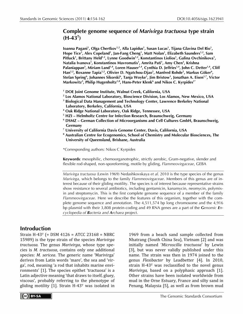
(21.6%), Hymenobacter (21.4%), Adhaeribacter (8.3%) and Microscilla (8.0%) (57 hits in total). The highest-scoring environmental sequence was EU447282 ('Flexibacteraceae bacterium KMM 6276'), which showed an identity of 100.0% and an HSP coverage of 97.6%, but most probably represents a Marivirga strain. The five most fre-quent keywords within the labels of environmental samples which yielded hits were 'microbi' (4.0%), 'sediment' (3.1%), 'site' (1.9%), 'group' (1.7%) and 'coral' (1.6%) (192 hits in total). These keywords support the ecological preference of M. tractuosa for wet habitats, as deduced from the sampling sites of the cultivated strains. Environmental sam-ples which yielded hits of a higher score than the highest scoring species were not found. Figure 1 shows the phylogenetic neighborhood of M. tractuosa H-43T in a 16S rRNA based tree. The sequences of the two identical 16S rRNA gene cop-ies in the genome do not differ from the previous-ly published 16S rRNA sequence (AB078072). The cells of strain H-43T are long, slender and flexi-ble rods 0.4-0.5 µm in diameter and 10-50 µm in length or longer (Figure 2). Strain H-43T is a Gram-negative non-spore-forming bacterium (Table 1) that exhibits gliding motility [1].
Figure 1. Phylogenetic tree highlighting the position of M. tractuosa relative to the other type strains within the family Flammeovirgaceae. The trees were inferred from 1,408 aligned characters [7,8] of the 16S rRNA gene se-quence under the maximum likelihood criterion [9] and rooted in accordance with the family Sphingobacteria-ceae. The branches are scaled in terms of the expected number of substitutions per site. Numbers above branches are support values from 1,000 bootstrap replicates [10] if larger than 60%. Lineages with type strain genome se-quencing projects registered in GOLD [11] are shown in blue, published genomes in bold.
Marivirga tractuosa type strain (H-43T)
156 Standards in Genomic Sciences
Strain H-43T is strictly aerobic and chemoorgano-trophic [1]. Growth is observed at 10-40ºC and with 0.5–10% NaCl, with optimal growth at 28-32ºC and 4-7% NaCl [1]. Colonies are circular, shiny and 2-4 mm in diameter after 72 h of incubation on marine agar [1]. They are usually dark-orange in color but whitish or yellow-pigmented variants may occur [1]. Pigment type three was found in the strain H-43T, the main pigment being saproxanthin [2]. In n-hexane, the absorption maxima of the pigments from crude extract were 425 nm, 447 nm, 471 nm and 505 nm [2]. Flexirubin-type pigments are not produced. Arginine dihydrolase, ornithine decar-boxylase, lysine decarboxylase and tryptophan dea-minase activities were described to be absent [1], however, Srinivas et al. [22] found that strain H-43T could utilize arginine, and also that growth on ala-nine and cysteine was weak. Nitrate is not reduced. Indole and acetoin (Voges–Proskauer reaction) are not produced [1]. Gelatin, Tween 20, Tween 40, Tween 80 and DNA are hydrolyzed, as well as agar, starch, urea, cellulose (CM-cellulose and filter paper) and chitin [1,2], however, again in contrast to the original description [1], Srinivas et al. reported that the strain does not hydrolyze Tween 20, Tween 40 or Tween 80 [22]. Acid is not produced from L-arabinose, cellobiose, L-fucose, D-galactose, glycerol, lactose, melibiose, raffinose, L-rhamnose, L-sorbose, sucrose, trehalose, DL-xylose, N-acetylglucosamine, citrate, acetate, fumarate, malate, adonitol, dulcitol, inositol or mannitol. In the API 50 CH gallery, acid is produced only from esculin and arbutin. Production of hydrogen sulfide and hydrolysis of casein are va-riable [1]. Citrate is utilized but lactose, inositol, glu-
conate, caprate, phenylalanine and malonate are not. Utilization of arabinose, D-glucose, D-mannose, su-crose, mannitol, N-acetylglucosamine, maltose, adi-pate, malate and sorbitol is variable [1]. Glucose, gly-cerol, galactose and sucrose (5.1 g/l, each) are used as carbon sources and stimulate the growth of strain H-43T, while sodium acetate and sodium lactate do not [2]. Nitrogen sources supporting growth include tryptone (1 g/l) and casamino acids (1 g/l), but not sodium glutamate or NO3- [2]. Alkaline phosphatase, esterase (C4), esterase lipase (C8), leucine arylami-dase, valine arylamidase, cystine arylamidase, α-chymotrypsin, acid phosphatase, naphthol-AS-BI-phosphohydrolase, β-galactosidase and α- and β-glucosidase activities are present, but lipase (C14), trypsin, α-galactosidase, β-glucuronidase, N-acetyl β-glucosaminidase, α-mannosidase and α-fucosidase activities are negative in the API ZYM gallery [1]. In litmus-milk, the dye was reduced and the clotting occurred. Moreover, litmus turned pink due to acidi-fication and the curd was re-digested because of proteolysis [2]. Strain H-43T is sensitive to ampicillin (10 µg), benzylpenicillin (10 U), carbenicillin (100 µg), chloramphenicol (30 µg), doxycycline (10 µg), erythromycin (15 µg), lincomycin (15 µg), oleando-mycin (15 µg) and tetracycline (30 µg), but resistant to gentamicin (10 µg), kanamycin (30 µg), neomycin (30 µg), polymixin (300 U) and streptomycin (30 µg) [1]. Cytochrome oxidase, catalase and alkaline phos-phatase tests were positive [1], although Srinivas et al. [22] found only a weak reaction in the catalase test. When growing, the strain was able to degrade dihydroxyphenyl alanine and tyrosine (5 g/l) [2].
Figure 2. Scanning electron micrograph of M. tractuosa H-43T

Pagani et al.
http://standardsingenomics.org 157
Table 1. Classification and general features of M. tractuosa H-43T according to the MIGS recommendations [16] MIGS ID Property Term Evidence code
Current classification
Domain Bacteria TAS [17] Phylum Bacteroidetes TAS [19] Class Sphingobacteria TAS [18] Order Sphingobacteriales TAS [18] Family Flammeovirgaceae TAS [18] Genus Marivirga TAS [1] Species Marivirga tractuosa TAS [1] Type strain H-43 TAS [1]
Gram stain negative TAS [1,2] Cell shape long, slender and flexible rods TAS [1] Motility motile by gliding TAS [1,2] Sporulation no TAS [1,2] Temperature range 10°C-40°C TAS [1] Optimum temperature 28°C-32°C TAS [1,2] Salinity 0.5%-10% NaCl TAS [1] MIGS-22 Oxygen requirement strictly aerobic TAS [1,2] Carbon source glycerol, glucose, galactose, sucrose TAS [2] Energy metabolism chemoorganotroph TAS [1] MIGS-6 Habitat wet terrestrial habitats, occasionally fresh water TAS [2] MIGS-15 Biotic relationship free-living NAS MIGS-14 Pathogenicity not reported NAS Biosafety level 1 TAS [20] Isolation beach sand sample TAS [1] MIGS-4 Geographic location Nhatrang (South China Sea), Vietnam TAS [1] MIGS-5 Sample collection time 1969 or before TAS [2] MIGS-4.1 MIGS-4.2
Latitude Longitude
12.25 109.20
NAS
MIGS-4.3 Depth not reported NAS MIGS-4.4 Altitude not reported NAS
Evidence codes - IDA: Inferred from Direct Assay (first time in publication); TAS: Traceable Author Statement (i.e., a direct report exists in the literature); NAS: Non-traceable Author Statement (i.e., not directly observed for the liv-ing, isolated sample, but based on a generally accepted property for the species, or anecdotal evidence). These evidence codes are from of the Gene Ontology project [21]. If the evidence code is IDA, then the property was directly observed by one of the authors or an expert mentioned in the acknowledgements
Chemotaxonomy The predominant cellular fatty acid of the strain H-43T were iso-C15:0 (36.8%), iso-C15:1 (23.0%) and iso-C17:03-OH (12.2%), with a detailed listing given in Nedashkovskaya et al. [1]. Srinivas et al. re-ported fundamentally different observations for strain H-43T, with the C16:0 (69% of the total fatty acids) to be the most important fatty acids in the strain H-43T, whereas iso-C15:0 was not detectable [22]. The main respiratory quinone is MK-7 [1].
Genome sequencing and annotation Genome project history This organism was selected for sequencing on the basis of its phylogenetic position [23], and is part of the Genomic Encyclopedia of Bacteria and Arc-haea project [24]. The genome project is depo-sited in the Genomes On Line Database [11] and the complete genome sequence is deposited in GenBank. Sequencing, finishing and annotation were performed by the DOE Joint Genome Insti-tute (JGI). A summary of the project information is shown in Table 2.

Marivirga tractuosa type strain (H-43T)
158 Standards in Genomic Sciences
Table 2. Genome sequencing project information MIGS ID Property Term MIGS-31 Finishing quality Finished
MIGS-28 Libraries used Three genomic libraries: one 454 pyrosequence standard library, one 454 PE library (10 kb insert size), one Illumina library
MIGS-29 Sequencing platforms Illumina GAii, 454 GS FLX Titanium MIGS-31.2 Sequencing coverage 60.1 × Illumina; 44.4 × pyrosequence
MIGS-30 Assemblers Newbler version 2.1-PreRelease-4-28-2009-gcc-3.4.6-threads, Velvet, phrap
MIGS-32 Gene calling method Prodigal 1.4, GenePRIMP
INSDC ID CP002349 (chromosome) CP002350 (plasmid FTRAC01)
Genbank Date of Release December 7, 2010 GOLD ID Gc01555 NCBI project ID 37901 Database: IMG-GEBA 2503538019 MIGS-13 Source material identifier DSM 4126 Project relevance Tree of Life, GEBA
Growth conditions and DNA isolation M. tractuosa H-43T, DSM 4126, was grown in DSMZ medium 172 (Cytophaga (marine) medium) [25] at 25°C. DNA was isolated from 0.5-1 g of cell paste using MasterPure Gram-positive DNA purifi-cation kit (Epicentre MGP04100) following the standard protocol as recommended by the manu-facturer with modification st/DL for cell lysis as described in Wu et al. [24]. DNA is available through the DNA Bank Network [26,27].
Genome sequencing and assembly The genome was sequenced using a combination of Illumina and 454 sequencing platforms. All general aspects of library construction and se-quencing can be found at the JGI website [28]. Py-rosequencing reads were assembled using the Newbler assembler version 2.1-Pre-release-4-28-2009-gcc-3.4.6-threads (Roche). The initial Newb-ler assembly consisted of 115 contigs in one scaf-fold and was converted into a phrap [29] assembly by making fake reads from the consensus, collect-ing the read pairs in the 454 paired end library. Illumina GAii sequencing data (496 Mb) was as-sembled with Velvet [30] and the consensus se-quences were shredded into 1.5 kb overlapped fake reads and assembled together with the 454 data. The 454 draft assembly was based on 201.9 Mb 454 draft data and all of the 454 paired end data. Newbler parameters are -consed -a 50 -l 350 -g -m -ml 20. The Phred/Phrap/Consed software package [29] was used for sequence assembly and
quality assessment in the following finishing process. After the shotgun stage, reads were as-sembled with parallel phrap (High Performance Software, LLC). Possible mis-assemblies were cor-rected with gapResolution [28], Dupfinisher, or sequencing cloned bridging PCR fragments with subcloning or transposon bombing (Epicentre Biotechnologies, Madison, WI) [31]. Gaps between contigs were closed by editing in Consed, by PCR and by Bubble PCR primer walks (J.-F.Chang, un-published). A total of 336 additional reactions were necessary to close gaps and to raise the qual-ity of the finished sequence. Illumina reads were also used to correct potential base errors and in-crease consensus quality using a software Polisher developed at JGI [32]. The error rate of the com-pleted genome sequence is less than 1 in 100,000. Together, the combination of the Illumina and 454 sequencing platforms provided 104.5 × coverage of the genome. Final assembly contains 589,653 pyrosequence and 7,543,442 Illumina reads.
Genome annotation Genes were identified using Prodigal [33] as part of the Oak Ridge National Laboratory genome an-notation pipeline, followed by a round of manual curation using the JGI GenePRIMP pipeline [34]. The predicted CDSs were translated and used to search the National Center for Biotechnology In-formation (NCBI) nonredundant database, Uni-Prot, TIGRFam, Pfam, PRIAM, KEGG, COG, and In-
Pagani et al.
http://standardsingenomics.org 159
terPro databases. Additional gene prediction anal-ysis and functional annotation was performed within the Integrated Microbial Genomes - Expert Review (IMG-ER) platform [35].
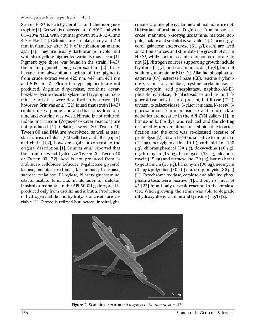
Genome properties The genome consists of a 4,511,574 bp long chro-mosome with a 35.5% G+C content and a 4,916 bp plasmid with 40% G+C content (Figure 3 and
Table 3). Of the 3,857 genes predicted, 3,808 were protein-coding genes, and 49 RNAs; Fifty-one pseudogenes were identified. The majority of the protein-coding genes (62.2%) were assigned with a putative function while the remaining ones were annotated as hypothetical proteins. The distribu-tion of genes into COGs functional categories is presented in Table 4.
Figure 3. Graphical circular map of the chromosome (plasmid map not shown). From outside to the center: Genes on forward strand (color by COG categories), Genes on reverse strand (color by COG categories), RNA genes (tRNAs green, rRNAs red, other RNAs black), GC content, GC skew.

Marivirga tractuosa type strain (H-43T)
160 Standards in Genomic Sciences
Table 3. Genome Statistics Attribute Value % of Total Genome size (bp) 4,516,490 100.00% DNA coding region (bp) 4,029,412 89.22% DNA G+C content (bp) 1,604,111 35.52% Number of replicons 2 Extrachromosomal elements 1 Total genes 3,857 100.00% RNA genes 49 1.27% rRNA operons 2 Protein-coding genes 3,808 98.73% Pseudo genes 51 1.32% Genes with function prediction 2,398 62.17% Genes in paralog clusters 396 10.27% Genes assigned to COGs 2,375 61.58% Genes assigned Pfam domains 2,609 67.64% Genes with signal peptides 1,113 28.86% Genes with transmembrane helices 997 25.85% CRISPR repeats 0
Table 4. Number of genes associated with the general COG functional categories
Code value % age Description J 157 6.1 Translation, ribosomal structure and biogenesis A 0 0.0 RNA processing and modification K 163 6.3 Transcription L 131 5.1 Replication, recombination and repair B 1 0.1 Chromatin structure and dynamics D 30 1.2 Cell cycle control, cell division, chromosome partitioning Y 0 0.0 Nuclear structure V 63 2.4 Defense mechanisms T 184 7.1 Signal transduction mechanisms M 236 9.1 Cell wall/membrane/envelope biogenesis N 10 0.4 Cell motility Z 1 0.0 Cytoskeleton W 0 0.0 Extracellular structures U 37 1.4 Intracellular trafficking and secretion, and vesicular transport O 112 4.3 Posttranslational modification, protein turnover, chaperones C 126 4.9 Energy production and conversion G 102 3.9 Carbohydrate transport and metabolism E 217 8.4 Amino acid transport and metabolism F 67 2.6 Nucleotide transport and metabolism H 118 4.6 Coenzyme transport and metabolism I 99 3.8 Lipid transport and metabolism P 136 5.3 Inorganic ion transport and metabolism Q 51 2.0 Secondary metabolites biosynthesis, transport and catabolism R 340 13.1 General function prediction only S 208 8.0 Function unknown - 1,482 38.4 Not in COGs

Pagani et al.
http://standardsingenomics.org 161
Acknowledgements We would like to gratefully acknowledge the help of Maren Schröder for growing M. tractuosa cultures and Susanne Schneider for DNA extraction and quality analysis (both at DSMZ). This work was performed un-der the auspices of the US Department of Energy Office of Science, Biological and Environmental Research Pro-gram, and by the University of California, Lawrence Berkeley National Laboratory under contract No. DE-
AC02-05CH11231, Lawrence Livermore National La-boratory under Contract No. DE-AC52-07NA27344, and Los Alamos National Laboratory under contract No. DE-AC02-06NA25396, UT-Battelle and Oak Ridge National Laboratory under contract DE-AC05-00OR22725, as well as German Research Foundation (DFG) INST 599/1-2.
References 1. Nedashkovskaya OI, Vancanneyt M, Kim SB, Bae
KS. Reclassification of Flexibacter tractuosus (Le-win 1969) Leadbetter 1974 and 'Microscilla seri-cea' Lewin 1969 in the genus Marivirga gen. nov. as Marivirga tractuosa comb. nov. and Marivirga sericea nom. rev., comb. nov. Int J Syst Evol Mi-crobiol 2010; 60:1858-1863. PubMed doi:10.1099/ijs.0.016121-0
2. Lewin RA, Lounsbery DM. Isolation, cultivation and characterization of flexibacteria. J Gen Mi-crobiol 1969; 58:145-170. PubMed
3. Lewin RA. A classification of flexibacteria. J Gen Microbiol 1969; 58:189-206. PubMed
4. Leadbetter ER. 1974.Genus II. Flexibacter Soriano 1945, 92, Lewin 1969, 192 emend. mut. char. In: Buchanan RE, Gibbons NE (eds), Bergey's Manual of Determinative Bacteriology, Eighth Edition, The Williams and Wilkins Co., Baltimore, p. 105-107.
5. American Type Culture Collection. http://www.atcc.org
6. National Collection of Industrial Bacteria http://www.ncimb.com
7. Castresana J. Selection of conserved blocks from multiple alignments for their use in phylogenetic analysis. Mol Biol Evol 2000; 17:540-552. PubMed
8. Lee C, Grasso C, Sharlow MF. Multiple sequence alignment using partial order graphs. Bioinformat-ics 2002; 18:452-464. PubMed doi:10.1093/bioinformatics/18.3.452
9. Stamatakis A, Hoover P, Rougemont J. A rapid bootstrap algorithm for the RAxML web servers. Syst Biol 2008; 57:758-771. PubMed doi:10.1080/10635150802429642
10. Pattengale ND, Alipour M, Bininda-Emonds ORP, Moret BME, Stamatakis A. How many bootstrap replicates are necessary? Lect Notes Comput Sci 2009; 5541:184-200. doi:10.1007/978-3-642-02008-7_13
11. Liolios K, Mavromatis K, Tavernarakis N, Kyrpides NC. The Genomes On Line Database (GOLD) in 2007: status of genomic and metagenomic projects and their associated metadata. Nucleic Acids Res 2008; 36:D475-D479. PubMed doi:10.1093/nar/gkm884
12. Nakagawa Y, Sakane T, Suzuki M, Hatano K. Phylogenetic structure of the genera Flexibacter, Flexithrix, and Microscilla deduced from 16S rRNA sequence analysis. J Gen Appl Microbiol 2002; 48:155-165. PubMed doi:10.2323/jgam.48.155
13. Walsh DA, Zaikova E, Howes CG, Song YC, Wright JJ, Tringe SG, Tortell PD, Hallam SJ. Meta-genome of a versatile chemolithoautotroph from expanding oceanic dead zones. Science 2009; 326:578-582. PubMed doi:10.1126/science.1175309
14. DeSantis TZ, Hugenholtz P, Larsen N, Rojas M, Brodie E, Keller K, Huber T, Dalevi D, Hu P, An-dersen G. Greengenes, a chimera-checked 16S rRNA gene database and workbench compatible with ARB. Appl Environ Microbiol 2006; 72:5069-5072. PubMed doi:10.1128/AEM.03006-05
15. Porter MF. An algorithm for suffix stripping. Pro-gram: electronic library and information systems 1980; 14:130-137. doi:10.1108/eb046814
16. Field D, Garrity G, Gray T, Morrison N, Selengut J, Sterk P, Tatusova T, Thomson N, Allen MJ, An-giuoli SV, et al. The minimum information about a genome sequence (MIGS) specification. Nat Biotechnol 2008; 26:541-547. PubMed doi:10.1038/nbt1360
17. Woese CR, Kandler O, Wheelis ML. Towards a natural system of organisms: proposal for the do-mains Archaea, Bacteria, and Eucarya. Proc Natl Acad Sci USA 1990; 87:4576-4579. PubMed doi:10.1073/pnas.87.12.4576
18. Garrity GM, Holt JG. 2001. Taxonomic Outline of the Archaea and Bacteria. In: Garrity GM, Boone

Marivirga tractuosa type strain (H-43T)
162 Standards in Genomic Sciences
DR, Castenholz RW (eds), Bergey's Manual of Systematic Bacteriology, Second Edition, Volume 1, Springer, New York, p. 155-166.
19. Garrity GM, Holt JG. The Road Map to the Ma-nual. In: Garrity GM, Boone DR, Castenholz RW (eds), Bergey's Manual of Systematic Bacteriolo-gy, Second Edition, Volume 1, Springer, New York, 2001, p. 119-169
20. Classification of bacteria and archaea in risk groups. http://www.baua.de TRBA 466.
21. Ashburner M, Ball CA, Blake JA, Botstein D, But-ler H, Cherry JM, Davis AP, Dolinski K, Dwight SS, Eppig JT, et al. Gene Ontology: tool for the unification of biology. Nat Genet 2000; 25:25-29. PubMed doi:10.1038/75556
22. Srinivas TNR, Anil Kumar P, Madhu S, Sunil B, Sharma TVRS, Shivaji S. Cesiribacter andamanen-sis gen. nov., sp. nov., a novel bacterium isolated from a soil sample of a mud volcano, Andaman Islands, India. Int J Syst Evol Microbiol 2010; (In press). PubMed doi:10.1099/ijs.0.025429-0
23. Klenk HP, Göker M. En route to a genome-based classification of Archaea and Bacteria? Syst Appl Microbiol 2010; 33:175-182. PubMed doi:10.1016/j.syapm.2010.03.003
24. Wu D, Hugenholtz P, Mavromatis K, Pukall R, Dalin E, Ivanova NN, Kunin V, Goodwin L, Wu M, Tindall BJ, et al. A phylogeny-driven genomic encyclopaedia of Bacteria and Archaea. Nature 2009; 462:1056-1060. PubMed doi:10.1038/nature08656
25. List of growth media used at DSMZ: http://www.dsmz.de/microorganisms/media_list.php.
26. Gemeinholzer B, Dröge G, Zetzsche H, Haszpru-nar G, Klenk HP, Güntsch A, Berendsohn WG, Wägele JW. The DNA Bank Network: the start from a German initiative. Biopreservation and
Biobanking 2011; 9:51-55. doi:10.1089/bio.2010.0029
27. DNA bank Network. http://www.dnabank-network.org
28. DOE Joint Genome Institute. http://www.jgi.doe.gov
29. Phrap and Phred for Windows. MacOS, Linux, and Unix. http://www.phrap.com
30. Zerbino DR, Birney E. Velvet: algorithms for de novo short read assembly using de Bruijn graphs. Genome Res 2008; 18:821-829. PubMed doi:10.1101/gr.074492.107
31. Sims D, Brettin T, Detter J, Han C, Lapidus A, Co-peland A, Glavina Del Rio T, Nolan M, Chen F, Lucas S, et al. Complete genome sequence of Ky-tococcus sedentarius type strain (541T). Stand Genomic Sci 2009; 1:12-20. PubMed doi:10.4056/sigs.761
32. Lapidus A, LaButti K, Foster B, Lowry S, Trong S, Goltsman E. POLISHER: An effective tool for us-ing ultra short reads in microbial genome assem-bly and finishing. AGBT, Marco Island, FL, 2008.
33. Hyatt D, Chen GL, LoCascio PF, Land ML, Lari-mer FW, Hauser LJ. Prodigal: prokaryotic gene recognition and translation initiation site identifi-cation. BMC Bioinformatics 2010; 11:119. PubMed doi:10.1186/1471-2105-11-119
34. Pati A, Ivanova N, Mikhailova N, Ovchinikova G, Hooper SD, Lykidis A, Kyrpides NC. GenePRIMP: A gene prediction improvement pipeline for mi-crobial genomes. Nat Methods 2010; 7:455-457. PubMed doi:10.1038/nmeth.1457
35. Markowitz VM, Ivanova NN, Chen IMA, Chu K, Kyrpides NC. IMG ER: a system for microbial ge-nome annotation expert review and curation. Bio-informatics 2009; 25:2271-2278. PubMed doi:10.1093/bioinformatics/btp393
Copyright © 2022 FDOKUMEN