Glycophorin C (CD236R) mediates vivax malaria parasite rosetting to normocytes

Comparative genomics of the neglected human malaria parasitePlasmodium vivax
Jane M. Carlton1,2, John H. Adams3, Joana C. Silva4,5, Shelby L. Bidwell1, HernanLorenzi1, Elisabet Caler1, Jonathan Crabtree1, Samuel V. Angiuoli5,6, Emilio F. Merino2,Paolo Amedeo1, Qin Cheng7, Richard M. R. Coulson8, Brendan S. Crabb9,10, Hernando A.del Portillo11, Kobby Essien12,13, Tamara V. Feldblyum5, Carmen Fernandez-Becerra11,Paul R. Gilson9, Amy H. Gueye14, Xiang Guo1, Simon Kang’a2, Taco W. A. Kooij15, MichaelKorsinczky7,16, Esmeralda V.-S. Meyer17, Vish Nene4,5, Ian Paulsen1,18, Owen White5,19,Stuart A. Ralph20, Qinghu Ren1, Tobias J. Sargeant9,21, Steven L. Salzberg6, Christian J.Stoeckert12, Steven A. Sullivan2, Marcio Massao Yamamoto22, Stephen L. Hoffman23,Jennifer R. Wortman5,24, Malcolm J. Gardner1, Mary R. Galinski17, John W. Barnwell25, andClaire M. Fraser-Liggett5,24
1The Institute for Genomic Research/J. Craig Venter Institute, 9712 Medical Research Drive, Rockville, MD20850, USA
2Department of Medical Parasitology, New York University Langone Medical Center, 341 East 25th Street,New York, NY 10010, USA
3Department of Global Health, College of Public Health, University of South Florida, 3720 Spectrum Blvd,Suite 304, Tampa, FL 33612, USA
4Department of Microbiology and Immunology, University of Maryland School of Medicine, 20 Penn Street,Baltimore, MD 21201, USA
5Institute for Genome Sciences, University of Maryland School of Medicine, 20 Penn Street, Baltimore, MD21201, USA
6Center for Bioinformatics and Computational Biology, University of Maryland, College Park, MD, USA
7Drug Resistance and Diagnostics, Australian Army Malaria Institute, Weary Dunlop Drive, GallipoliBarracks, Enoggera, Qld 4051, Australia
8Microarray Group, European Bioinfomatics Institute, Wellcome Trust Genome Campus, Hinxton,Cambridge CB10 1SD, UK
9The Walter & Eliza Hall Institute of Medical Research, 1G Royal Parade, Parkville, VIC 3050, Australia
10Burnet Institute, 85 Commercial Road, Melbourne, Victoria 3004, Australia
11ICREA and Barcelona Centre for International Health Research, Hospital Clinic/IDIBAPS, Universitatde Barcelona Roselló 132, 4a planta, 08036, Barcelona, Spain
Correspondence and requests for material should be sent to J.M.C. ([email protected]).Present addresses: Seattle Biomedical Research Center, 307 Westlake Avenue N., Suite 500, Seattle, WA 98109-5219, USA (M.J.G.)Competing interests statement The authors declare that they have no competing financial interests.Sequence and annotation data for the genome were deposited in GenBank under the project accession number AAKM00000000 and arealso available at the Plasmodium genome sequence database PlasmoDB at http://plasmodb.org. A minimal tiling path of clones coveringeach chromosome is available through the malaria repository MR4 www.mr4.org, and a long-oligo array through the Pathogen FunctionalGenomics Resource Center http://pfgrc.jcvi.org
NIH Public AccessAuthor ManuscriptNature. Author manuscript; available in PMC 2009 October 9.
Published in final edited form as:Nature. 2008 October 9; 455(7214): 757–763. doi:10.1038/nature07327.
NIH
-PA Author Manuscript
NIH
-PA Author Manuscript
NIH
-PA Author Manuscript

12Center for Bioinformatics and Department of Genetics, University of Pennsylvania School of Medicine,Philadelphia, PA 19104, USA
13Department of Bioengineering, University of Pennsylvania, Philadelphia, PA 19104, USA
14Hood College, Frederick, Maryland 21701, USA
15Department of Parasitology, Heidelberg University School of Medicine, Im Neuenheimer Feld 324, 69120Heidelberg, Germany
16Institute for Molecular Bioscience, University of Queensland, Brisbane, Queensland 4072, Australia
17Emory Vaccine Center, Yerkes National Primate Research Center and Department of Medicine, Divisionof Infectious Diseases, Emory University, Atlanta, GA 30329, USA
18Department of Chemistry and Biomolecular Sciences, Macquarie University, Sydney, New South Wales,Australia, 2109
19Department of Epidemiology and Preventive Medicine, University of Maryland School of Medicine, 20Penn Street, Baltimore, MD 21201, USA
20Department of Biochemistry & Molecular Biology, Bio21 Molecular Science and Biotechnology Institute,University of Melbourne, Victoria, 3010, Australia
21Department of Medical Biology, University of Melbourne, Parkville, Victoria 3010, Australia
22Departamento de Parasitologia, Instituto de Ciências Biomédicas, Universidade de São Paulo. Av. LineuPrestes 1374, São Paulo, SP 05508-900, Brazil
23Sanaria Inc., 9800 Medical Center Drive, Rockville, MD 20850, USA
24Department of Medicine, University of Maryland School of Medicine, 20 Penn Street, Baltimore, MD21201, USA
25Malaria Branch, Division of Parasitic Diseases, National Center for Zoonotic, Vector-borne and EntericDiseases, Centers for Disease Control and Prevention, Atlanta, GA 30341, USA
AbstractThe human malaria parasite Plasmodium vivax is responsible for 25-40% of the ~515 million annualcases of malaria worldwide. Although seldom fatal, the parasite elicits severe and incapacitatingclinical symptoms and often relapses months after a primary infection has cleared. Despite itsimportance as a major human pathogen, P. vivax is little studied because it cannot be propagated inthe laboratory except in non-human primates. We determined the genome sequence of P. vivax inorder to shed light on its distinctive biologic features, and as a means to drive development of newdrugs and vaccines. Here we describe the synteny and isochore structure of P. vivax chromosomes,and show that the parasite resembles other malaria parasites in gene content and metabolic potential,but possesses novel gene families and potential alternate invasion pathways not recognizedpreviously. Completion of the P. vivax genome provides the scientific community with a valuableresource that can be used to advance scientific investigation into this neglected species.
Plasmodium vivax is the major cause of malaria outside Africa, mainly afflicting Asia and theAmericas 1. A disease of poor people living on the margins of developing economies, vivaxmalaria traps many societies in a relentless cycle of poverty. Intermittent transmission makesprotective immunity rare, and the disease strikes all ages. Repeated acute febrile episodes ofdebilitating intensity can occur for months. In children this can lead to life-long learningimpairment, while incapacitation of adults has tremendous direct economic consequencesthrough lost productivity and depletion of meagre financial reserves. Drug resistance in P.vivax is spreading, hindering management of clinical cases, and reports of severe pathology,
Carlton et al. Page 2
Nature. Author manuscript; available in PMC 2009 October 9.
NIH
-PA Author Manuscript
NIH
-PA Author Manuscript
NIH
-PA Author Manuscript

including respiratory distress and coma, are challenging the description of vivax malaria as‘benign’1.
Several biological characteristics underlie the distinct pathogenic and epidemiologic nature ofvivax malaria. In contrast to P. falciparum, P. vivax is only capable of infecting reticulocytes,causing severe anaemia by dyserythropoiesis and destruction of infected and uninfectederythrocytes despite much lower parasitemias. P. vivax cannot infect Duffy blood group-negative reticulocytes (a trait shared with the closely-related monkey malaria parasite P.knowlesi), and is thus absent from West Africa where Duffy negativity predominates 2.Differences in Anopheles mosquito dynamics allow P. vivax transmission in temperate climatesnot tolerated by P. falciparum. In such regions P. vivax infects hepatocytes but may persist asdormant hypnozoites for months or years before initiating blood-stage infections (relapses)during another transmission season.
Since P. vivax kills infrequently and is not amenable to continuous in vitro culture, it has beenrelatively little studied in comparison to P. falciparum. The P. vivax genome sequence wereport here, and comparative analyses with sequenced malaria parasites P. falciparum 3, therodent parasite P. yoelii yoelii 4,5, and the primate parasite P. knowlesi 6 (an excellent modelfor in vivo studies of human malaria), provide significant insights into the biology of thisneglected parasite.
Genome sequencing and characteristicsThe ~26.8 Mb nuclear genome sequence of P. vivax (Salvador I) was sequenced by wholegenome shotgun methods to 10-fold coverage, targeted gap closure and finishing, and manualcuration of automated annotation. Details of these and other methods are given inSupplementary Information linked to this paper’s online version (www.nature.com/nature).Large contigs totalling ~22.6 Mb were assigned to the 14 P. vivax chromosomes; ~4.3 Mb ofsmall subtelomeric contigs remain unassigned due to their repetitive nature (SupplementaryTable 1). P. vivax chromosomes are unique among human Plasmodium species in exhibitinga form of isochore structure 7, with subtelomeric regions of low G+C content and chromosomeinternal regions of significantly higher G+C content. We finished the subtelomeric ends ofseveral P. vivax chromosomes, allowing us to define their isochore boundaries (Plate 1).
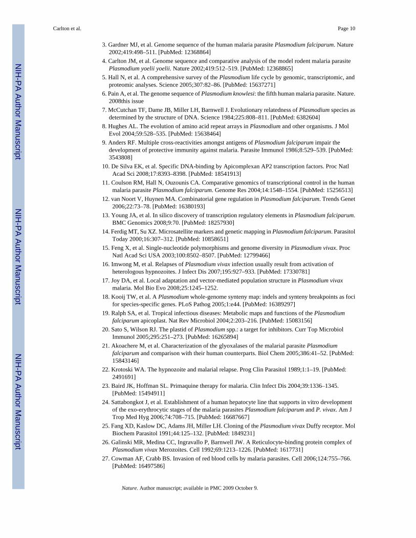
In many aspects, the genomes of mammalian Plasmodium species (P. falciparum, P. knowlesi,P. vivax, P. yoelii) are remarkably uniform, ranging from 23-27 Mb across 14 chromosomes,and comprising ~5,500 genes, most of which (~51%) contain at least one intron (Table 1).However, differences in nucleotide bias can be extreme (e.g. P. vivax and P. falciparum averagepercent G+C ~42.3 and ~19.4, respectively), and a large gene family found in P. y. yoelii raisedits gene count to ~5,880 4. A remarkable 77% of genes are orthologous between the four species(Supplementary Fig. 1); almost half of these encode conserved hypothetical proteins ofunknown function. In P. falciparum, the high incidence of tandem repeats and low complexityregions (LCRs) in proteins, especially antigens, has led researchers to propose that LCRs areinvolved in immune evasion mechanisms, such as antigen diversification 8 and reducing thehost’s antibody response to critical epitopes by acting as a ‘smokescreen’ 9. We found thatLCRs tend to constitute a smaller proportion of P. vivax proteins on average (39%) than P.falciparum proteins (60%; Supplementary Fig. 2), and that LCR expansion partly accounts forthe slightly larger size of P. falciparum proteins (Supplementary Table 2), but how this relatesto differences in immune evasion mechanisms between P. vivax and P. falciparum is unclear.
Notwithstanding the recent functional characterization of the Apicomplexan AP2 family oftranscriptional regulators in Plasmodium 10, the parasite appears to lack most of the standardeukaryotic transcriptional machinery, such as transcription-associated proteins (TAPs)11, but
Carlton et al. Page 3
Nature. Author manuscript; available in PMC 2009 October 9.
NIH
-PA Author Manuscript
NIH
-PA Author Manuscript
NIH
-PA Author Manuscript

is rich in regulatory sequences 12, fostering the idea that gene expression regulation inPlasmodium is complex and unusual. Our initial studies found no significant differences in theTAP repertoire between P. falciparum, P. vivax and P. knowlesi, indicating that transcriptionalmechanisms are similar in all three species (Supplementary Table 3). Genes encoding mRNAstability proteins containing a CCCH-zinc finger were abundant in all three species, affirmingthe importance of post-transcriptional regulation in the control of gene expression acrossPlasmodium. A genome scan of P. vivax for known core promoter elements such as TATAand CAAT boxes identified some candidates, but many of them lacked positional specificity.Similarly, a search for novel promoter elements in regions upstream of ~1,800 mappedtranscription start sites (5’ UTRs), and for RNA binding proteins in ~1,300 3’ UTRs, also failedto produce convincing candidates (data not shown). To determine whether binding sites areconserved between P. falciparum and other primate Plasmodium species, we searched for over-represented nucleotide ‘words’ in regions upstream of clusters of potentially co-regulated genesconserved in P.vivax, P. falciparum, P. knowlesi and P. y. yoelii (Supplementary Information).Seven putative novel regulatory binding sites conserved across at least two species wereidentified (Supplementary Table 4). These binding sites were associated with core eukaryoticprocesses such as dephosphorylation and with parasite-specific functions such as cell invasion.Independent support for two of our predicted sites comes from a recent report of the sporozoite-associated motif 5’-TGCATGCA-3’ and the merozoite invasion-related 5’-GTGTGCACAC-3’ 13 motif. In our analysis these two sites, together with thedephosphorylation-associated motif 5’-GCACGCGTGC-3’, were conserved across the fourPlasmodium species.
Examination of parasite population structure in the field is key to understanding transmissiondynamics, the spread of drug resistance, and to design and test malaria control efforts. Manypopulation studies have exploited the abundant polymorphic microsatellites in the P.falciparum genome, primarily simple sequence repeats such as [TA] dinucleotide and polyA/polyT 14. We screened the P. vivax genome for microsatellites, identifying ~160 that arepolymorphic between eight P. vivax laboratory lines (Supplementary Table 5; Plate 1). P.vivax microsatellites average 27.5% G+C, with an average repeat unit length of 3.1 nucleotidesand an average copy number of 19.1. We found fewer microsatellites in P. vivax than in P.falciparum (as noted previously 15), likely due to the more conventional nucleotidecomposition of the former. Even so, these genome-wide polymorphic markers are alreadyfacilitating studies of P. vivax population structure and genetic diversity 16,17.
Chromosome synteny and genome evolutionPrevious studies have indicated significant conservation of gene synteny betweenPlasmodium parasites 4 in direct proportion to their genetic distance. We generated a syntenymap of P. vivax, P. knowlesi, P. falciparum, and the rodent malaria parasites P. yoelii, P.berghei and P. chabaudi (considered as a single lineage 18 due to their virtually completesynteny; Plate 1). The P. vivax and P. knowlesi chromosomes are highly syntenic except formicrosyntenic breaks at species-specific genes (in particular the P. knowlesi kir and SICAvargenes; see ref. 6); a previous study identified such breaks as foci for the evolution of host-parasite interaction genes18. The karyotypes of P. vivax and P. knowlesi correspond to themost parsimonious reconstruction of the ancestral form of the six species; the karyotypes ofP. falciparum and the rodent malaria parasites can be reconstructed from this form throughnine and six chromosomal rearrangements, respectively (Supplementary Fig. 3). No ‘hot-spots’of synteny breakage were identified, indicating that intersyntenic breakpoints were not ‘reused’during the divergence of the species, and no obvious motifs except for AT-rich regions andLCRs were identified in regions of the P. vivax genome predicted to have recombined to givesingle P. falciparum chromosomes. Of the 3,336 orthologs between all six species, 3,305 (99%)were found to be positionally conserved (Supplementary Table 6).
Carlton et al. Page 4
Nature. Author manuscript; available in PMC 2009 October 9.
NIH
-PA Author Manuscript
NIH
-PA Author Manuscript
NIH
-PA Author Manuscript

We used 3,322 high-quality P. vivax / P. knowlesi orthologs to obtain maximum likelihoodestimates of the rate of substitution at synonymous (dS) and non-synonymous (dN) sites, aswell as ω (dN/dS; Supplementary Table 7 and Plate 1). P. vivax chromosomes differsignificantly in their average values for both dS and dN, but the two variables are stronglycorrelated within and between chromosomes (Supplementary Fig. 4). The chromosomes alsodiffer significantly in average %GC4, the G+C content in third codon positions of four-folddegenerate amino acids. This variable is positively correlated with average dS and inverselycorrelated with chromosome length, such that synonymous sites in genes on the smallestchromosomes (~1 Mb) evolve ~1.5 times faster than genes on the two largest (~3 Mb)chromosomes (Supplementary Fig. 5). These observations strongly suggest the existence ofheterogeneous mutation rates across the genome. It is unclear if this is due to cytosine-to-thymine deamination, which is more likely in G+C-rich regions, since it is not known whetherDNA methylation occurs in P. vivax. The degree of selective constraint (ω) also varies acrossclasses of genes. Genes encoding glycosylphosphatidylinositol (GPI) anchored proteins, celladhesion proteins, exportome proteins, and proteins with transmembrane or signal peptidemotifs, all of which are at least partly extra-cellular, were found to evolve significantly fasterthan genes involved in, for example, carbohydrate metabolism, enzyme regulation and cellstructure (Supplementary Table 8, Plate 1 inset). The host immune system, by targetingextracellular peptides, appears to have strongly influenced evolutionary rate variation betweengene classes in Plasmodium.
A highly-conserved Plasmodium metabolomeWe found that key metabolic pathways, housekeeping functions, and the repertoire of predictedmembrane transporters are highly conserved between the P. vivax and P. falciparum 3proteomes (Supplementary Table 9), suggesting that the two species have much the samemetabolic potential. Conservation of metabolic processes also extends to the apicoplast, anApicomplexan plastid secondarily acquired from an ancient cyanobacterium. The apicoplasthas lost photosynthetic function, but is essential to the parasite’s metabolism, hosting nuclear-encoded proteins that are targeted to the apicoplast lumen by a conserved bipartite N-terminalpresequence. The complete genome sequence of P. vivax offers an opportunity to update andimprove the apicoplast proteome that was predicted in silico 3. Apicoplast-targeted proteinsconserved in P. vivax participate in major metabolic processes previously recognized in P.falciparum 19, such as complete Type II fatty acid synthesis (FASII), isopentenyl diphosphate(IPP) and iron sulfur cluster assembly pathways, and a fragmented haem synthesis pathwaydistributed between the apicoplast and mitochondria. Conservation of these pathways in P.vivax is important because synthetic pathways for FASII and IPPs are targets for antimalarialchemotherapeutics 20. The revised Plasmodium apicoplast proteome (Supplementary Table10) also clarifies the localisation of two important processes. We show thiamine pyrophosphatebiosynthesis, previously hypothesised to take place in the apicoplast 19, to be cytosolic.Conversely, we confirm a glyoxalate pathway in the apicoplast, with glyoxalase I andglyoxalase II enzymes being targeted there 21; both enzymes are potential drug targets. Thus,comparison of overall apicoplast metabolic capabilities shows very few differences betweenP. vivax and P. falciparum.
P. vivax can form hypnozoites, a latent hepatic stage responsible for patent parasitemia relapsesmonths or even years after an initial mosquito-induced infection 22. Hypnozoites survive mostdrugs that kill blood stage parasites; complete elimination of P. vivax infections (radical cure)requires primaquine, the only licensed drug that can kill hypnozoite stages. However, resistanceto the drug is spreading 23, and its use is contraindicated in pregnant women or patients withglucose-6-phosphate dehydrogenase deficiency, which is common in malaria-endemic regions.After an initial examination of P. vivax-specific proteins failed to identify leads(Supplementary Table 11), we hypothesized that the genetic switch for hypnozoite formation
Carlton et al. Page 5
Nature. Author manuscript; available in PMC 2009 October 9.
NIH
-PA Author Manuscript
NIH
-PA Author Manuscript
NIH
-PA Author Manuscript

may involve P. vivax homologs of dormancy genes. Analysis of the predicted P. vivax proteomerevealed some candidates (Supplementary Table 12). However such an association remainsspeculative, and investigation of hypnozoite formation and activation will require continueddevelopment of in vitro systems for culturing P. vivax liver stages 24.
Gene families shape Plasmodium biologyPlasmodium lineages display differential gene family expansion (Table 2) that has shaped thespecific biology of each species. Phenotypes illustrating this include parasite invasion of redblood cells, and antigenic variation. Invasion of erythrocytes by extracellular Plasmodiummerozoites, crucial to the development of malaria in an infected individual, depends uponspecific interactions between merozoite ligands and erythrocyte surface receptors (Fig. 1).Plasmodium species-specific mechanisms act mostly during the preliminary phases of invasion(e.g., merozoite attachment and orientation). In P. vivax, but not P. falciparum, invasion isrestricted to Duffy-positive reticulocytes 2. P. vivax Duffy-binding protein (DBP 25) andreticulocyte-binding proteins (RBPs 26) are the archetypes of two distinct Plasmodium familiesof cell-binding proteins involved in erythrocyte selection (referred to as the Duffy-binding-like ‘DBL’, and reticulocyte-binding-like ‘RBL’ families, respectively). Homologs of rbp1and rbp2, two genes originally identified in P. vivax, include the P. falciparum rh/nbp genes(reviewed in ref. 27) and the Py235 family in P. yoelii (reviewed in ref. 28). Unexpectedly, weidentified additional rbp genes in the P. vivax genome (Supplementary Table 13), includingmultiple rbp2 genes, which could provide P. vivax with a diversity of invasion mechanismscomparable to that of P. falciparum. This finding dispels a view that P. vivax has a relativelyuncomplicated erythrocyte invasion mechanism. Instead, P. vivax likely has alternate invasionpathways, since differential expression of rbp homologs in P. falciparum 29 and P. yoelii 30 isclosely linked to switching of invasion pathways (Fig. 1). All rbp2 loci occur in thesubtelomeric regions of P. vivax chromosomes - non-syntenic, dynamic regions of the genomein which species-specific genes are generated (Supplementary Fig. 6).
The final phase of invasion, merozoite entry into an intraerythrocytic vacuole, uses anintracytoplasmic molecular motor (components of which are highly conserved betweenPlasmodium species) coupled to simultaneous shedding of crucial merozoite surface proteins(MSPs). There are at least ten distinct MSPs (Supplementary Table 14), and P. vivax genomeanalysis reveals two particularly interesting MSP families, MSP3 and MSP7. Eleven membersof the msp3 gene family occur in tandem on a ~60 kb region of P. vivax chromosome 10(Supplementary Fig. 7), and show weak similarity to four msp3 gene family members on P.falciparum chromosome 10 and to two P. knowlesi msp3 genes located on differentchromosomes. Thus, there has been a significant expansion of the msp3 gene family in P.vivax, perhaps as a means to enhance immune evasion, as P. falciparum and P. vivax msp3gene family members have been shown to be antigenic and to partially immunize non-humanprimates against blood stage parasites 31. In P. falciparum, MSP6 (a member of the MSP3family that lacks heptad repeats) non-covalently binds with MSP1, but there is no counterpartto MSP6 in P. vivax. MSP7, another P. falciparum antigen that binds to MSP1 on the surfaceof merozoites, has also been expanded in P. vivax, with eleven copies on chromosome 12,compared to six and three members in P. falciparum and P. y. yoelii respectively; it is notknown if any P. vivax MSP7 proteins bind to MSP1. The surface coats of merozoites andextracellular forms of Plasmodium parasites are composed largely of GPI-anchored proteins,many of which are important targets of protective immune responses and thus constitutepromising vaccine candidates. When we predicted the GPI-anchored proteome of P. vivax andcompared it to validated P. falciparum GPI-anchored proteins 32, 29 of the 30 GPI-anchoredproteins identified in P. falciparum had counterparts in P. vivax (Supplementary Fig. 8), anextraordinary level of conservation. MSP2 (the second most abundant merozoite surfaceprotein in P. falciparum) is absent in the P. vivax genome, and P. vivax contains one additional
Carlton et al. Page 6
Nature. Author manuscript; available in PMC 2009 October 9.
NIH
-PA Author Manuscript
NIH
-PA Author Manuscript
NIH
-PA Author Manuscript

GPI-anchored protein that appears to be a member of the ‘six cysteine’ apicomplexan-specificgene family 33. Both the P. vivax and P. knowlesi genomes encode an apparently paralogousgene next to msp1, which is the largest and most abundant protein on the P. falciparummerozoite surface. P. vivax MAP1 is not closely related to MSP1 (11% identity, 22% similarity)although their sizes, a predicted GPI-attachment site, and structural features such as a C-terminal double EGF module, are similar.
A second notable parasite phenotype is antigenic variation: the ability to vary surface proteinsduring the course of an infection in order to evade the host’s immune response. In P.falciparum, antigenic variation is mediated by species-specific gene families such as var,members of which are expressed clonally and regulated epigenetically 34. In P. vivax, thelargest multigene family vir, part of the pir (Plasmodium interspersed repeats) superfamilyfound in several Plasmodium species 5, has been implicated in antigenic variation; 35 genecopies were previously identified 35. We identified 346 vir genes in the P. vivax genome locatedwithin AT-rich subtelomeric regions of P. vivax chromosomes (Plate 1). Structurally, vir genesvary greatly, ranging from 156-2,316 bp in length and containing 1-5 exons. VIR proteins werepreviously classified into six subfamilies (A-F) based upon sequence similarity 35, andrepresentatives of these subfamilies were identified in patient isolates 36. Clustering the VIRsin the Salvador I genome yielded six new subfamilies (G-L) and confirmed gene expressionfor several of these in natural infections (Supplementary Table 15). Motif analysis of the totalVIR repertoire (Fig. 2) showed that approximately half (171) contain a transmembrane domain,and half (160) contain a motif similar to the PEXEL/VSP sequence linked to export of parasiteproteins 37,38. Introns from 25 vir genes contain a conserved motif proximal to the donorsplice site, suggesting possible functionality of the sequence in the control of vir geneexpression, as has been shown for P. falciparum var introns 39. Motif-shuffling among thesequences is apparent, particularly among large VIR proteins that have undergone an expansionof some motifs at the amino terminus. Similarly to P. falciparum var genes, in situ hybridizationanalysis has shown that P. vivax chromosome ends localize to the nuclear periphery 40, whereectopic recombination favors the generation of variants and gene expansion. Although therepeat structure of P. vivax subtelomeric regions is not as extensive as that seen in P.falciparum 6, P. vivax likely uses chromosomal exchange as a mechanism for generatingantigenic diversity. VIR proteins represent an extremely diverse family, members of whichcurrently appear more divergent than members of other partially characterized PIR familiessuch as the P. chabaudi CIR (135 members) and the P. berghei BIR (245 members) families(Supplementary Fig. 9). Shared structural characteristics have been shown between VIRsubfamily D proteins and the P. falciparum Pfmc-2tm family located at Maurer’s clefts, andVIR subfamily A proteins and the P. falciparum SURFIN family found on the surface ofinfected erythrocytes 41. We speculate that the extreme diversity and sub-structuring of VIRproteins indicate members different subcellular localizations and functions, including immuneevasion.
We identified eight novel gene families (Pv-fam-a to -e and -g to -i; Supplementary Table 16)in the P. vivax genome, most of which are located in subtelomeric regions (Plate 1). Ofparticular interest are (1) the PvTRAG (Pv-fam-a) gene family (36 genes), one member ofwhich was previously identified; it encodes a protein localised to the caveola-vesicle complexof infected erythrocytes, and has been shown to elicit a humoral immune response during thecourse of natural infections 42; and (2) the Pv-fam-e family (Supplementary Fig. 10), 36 copiesof which are found in two loci on either side of the predicted centromere on chromosome 5,with one 10-gene locus present in a 47% GC region, and a second 26-gene locus present in a36% GC region. While P. vivax proteins have a fairly balanced codon composition, using all61 sense codons almost equally (effective number of codons, Nc = 54.2), their orthologs in P.falciparum are more biased (Nc = 37.5), with G- and C-ending codons nearly absent from four-fold degenerate amino acids (Supplementary Table 17). However, P. vivax gene families,
Carlton et al. Page 7
Nature. Author manuscript; available in PMC 2009 October 9.
NIH
-PA Author Manuscript
NIH
-PA Author Manuscript
NIH
-PA Author Manuscript

which are predominantly located in AT-rich regions, have a codon composition of Nc = 47.This pattern suggests a strong influence of local mutation pattern on the nucleotide compositionof genes and indicates a potential for differential gene expression.
Plasmodium drug interaction genesThe sexual stages of P. vivax are produced before the onset of clinical symptoms, permittingmosquito transmission early in an infection. Such early parasite transmission may delaydevelopment of resistance to many of the antimalarial drugs used to treat vivax malaria, despitethe extensive long-term use of these drugs in regions endemic for both P. vivax and P.falciparum 43. Nevertheless, P. vivax can develop resistance to most of the current antimalarialdrugs. To understand the interactions between antimalarial drugs and the parasite proteinsimplicated in drug binding and resistance, we examined crystal structures and developedhomology models for several P. vivax proteins in the predicted proteome, and compared thepredicted binding sites and reported mutations with those of their P. falciparum orthologs(Table 2).
Currently, the most efficacious novel antimalarial drugs are derivatives of artemisinin(qinghaosu) and atovaquone, used predominantly in combination therapies. Arteminsininderivatives, the most potent drugs recommended for treatment, may target a sarcoplasmic/endoplasmic reticulum Ca2+ ATPase (SERCA)-type protein, ATPase6 44. We constructedhomology models of P. vivax and P. falciparum ATPase6 and identified two residues in theputative active sites for artemisinin that differ between the two species (P. vivax A263 andS1008, equivalent to L263 and N1039 in P. falciparum). A change in residue 263 from leucineto alanine results in a three-fold decrease in susceptibility of P. falciparum to artemisinin 44,and indeed the IC50 (inhibitory concentration at which fifty percent of parasites die) for someP. vivax field isolates appears higher than the IC50 for P. falciparum 45, although it should benoted that clinical resistance of any human Plasmodium species to artemisinin derivatives hasyet to be documented. Atovaquone, used in combination with the antifolate proguanil,selectively inhibits mitochondrial electron transport at the cytochrome bc(1) complex;mutations in the cytochrome b (cytb) gene can interfere with this inhibition, causing resistance.We constructed a homology model of P. vivax CYTB and compared it to the P. falciparumCYTB homology model 46, revealing almost identical structures, including the predictedatovaquone active sites. Although there are no reports of P. vivax treatment failures, our studiesindicate that should resistance arise, the same sites in P. vivax CYTB may be implicated.
Towards a policy shift for vivax malariaDespite the insights into parasite biology provided by the P. vivax genome, many importantquestions remain that can only be addressed by functional studies. For example, we were unableto find differences in the predicted P. vivax proteome that might explain the rheologicalbehaviour of P. vivax-infected erythrocytes, which remain flexible and can repeatedly passthrough the spleen, unlike P. falciparum-infected erythrocytes, whose rigidity facilitatescytoadherence and avoidance of splenic clearance 47. Studies of the hypnozoite transcriptome,although technically challenging, would radically increase our inadequate knowledge of thebiology of this dormant form. Studies are currently underway to develop new in vitro culturesystems 48, which could provide badly-needed biological material for such functional studies.
The malaria research and control communities were challenged recently to once again establishthe eradication of malaria as a policy goal 49. Given the significant contribution of P. vivax tothe global malaria situation 1,43, it is imperative that these efforts include elimination of P.vivax as well as P. falciparum. Elimination of P. vivax presents special challenges, in particularthe parasite’s production of dormant hypnozoites that enables relapses long after the initial
Carlton et al. Page 8
Nature. Author manuscript; available in PMC 2009 October 9.
NIH
-PA Author Manuscript
NIH
-PA Author Manuscript
NIH
-PA Author Manuscript

parasitemia has cleared. Indeed, an important aspect of P. vivax eradication will be thedevelopment of new drugs to replace primaquine for radical cure. Although the developmentof new drugs targeting P. vivax liver stages is a formidable task, recent developments offerhope that this goal can be accomplished 50.
Methods summaryGenome sequencing, assembly, mapping and annotation
Saimiri boliviensis boliviensis monkeys were infected with the Salvador I strain of P. vivaxisolated from a patient from El Salvador. Extracted parasite DNA was used to make genomicDNA libraries for shotgun sequencing. Reads were assembled into scaffolds, inter-scaffoldgaps closed, and scaffolds assigned to P. vivax chromosomes through hybridization of scaffold-specific probes to pulsed-field gel separated chromosomes. Gene prediction algorithms wereused to predict gene models, and each model was manually checked for structuralinconsistencies. Gene function was assigned using an automated annotation pipeline withsubsequent manual curation.
Genome analysisMethods for the in silco analysis of the genome sequence are described in the SupplementaryInformation.
Studies requiring laboratory experimentationPolymorphic microsatellite identification—Primers flanking 333 microsatellitesidentified from the genome sequence and designed for field studies where access to capillaryelectrophoresis equipment may not be possible, were used to amplify the loci from eight world-wide P. vivax laboratory strains adapted to growth in monkeys (Brazil I, Miami II, Pakchong,Panama I, Nica, Thai II, Vietnam IV and Indonesia XIX). Amplicons were separated byelectrophoresis on agarose gels and scored for size differences.
Vir gene expression—cDNA was generated from total RNA extracted from the SalvadorI isolate and from three patient isolates from Brazil. Primers were designed to eight vir genesubfamilies and used to amplify the loci.
Supplementary MaterialRefer to Web version on PubMed Central for supplementary material.
AcknowledgementsWe are indebted to the P. vivax research community for their support, and in particular to M. Gottlieb and V. McGovernfor facilitating financial support. Funding came from the following sources: P. vivax sequencing, assembly and closure,US Department of Defense and National Institute of Allergy and Infectious Diseases; genome mapping, BurroughsWellcome Fund; and selective constraint analysis, National Institute of General Medical Sciences. We wish to thankTIGR’s SeqCore, Closure and IFX core facilities, E. Lee, J. Sundaram, J. Orvis, B. Haas and T. Creasy for engineeringsupport, R. K. Smith Jr. for annotation support, E. Lyons and H. Zhang for technical assistance, H. Potts for statisticalanalysis, T. McCutchan for rDNA sequence annotation and S. Perkins for the Plasmodium phylogeny.
References1. Price RN, et al. Vivax Malaria: Neglected and Not Benign. Am J Trop Med Hyg 2007;77(Suppl 6):
79–87. [PubMed: 18165478]2. Miller LH, Mason SJ, Clyde DF, McGinniss MH. The resistance factor to Plasmodium vivax in blacks.
The Duffy-blood-group genotype, FyFy. N Engl J Med 1976;295:302–304. [PubMed: 778616]
Carlton et al. Page 9
Nature. Author manuscript; available in PMC 2009 October 9.
NIH
-PA Author Manuscript
NIH
-PA Author Manuscript
NIH
-PA Author Manuscript
3. Gardner MJ, et al. Genome sequence of the human malaria parasite Plasmodium falciparum. Nature2002;419:498–511. [PubMed: 12368864]
4. Carlton JM, et al. Genome sequence and comparative analysis of the model rodent malaria parasitePlasmodium yoelii yoelii. Nature 2002;419:512–519. [PubMed: 12368865]
5. Hall N, et al. A comprehensive survey of the Plasmodium life cycle by genomic, transcriptomic, andproteomic analyses. Science 2005;307:82–86. [PubMed: 15637271]
6. Pain A, et al. The genome sequence of Plasmodium knowlesi: the fifth human malaria parasite. Nature.2008this issue
7. McCutchan TF, Dame JB, Miller LH, Barnwell J. Evolutionary relatedness of Plasmodium species asdetermined by the structure of DNA. Science 1984;225:808–811. [PubMed: 6382604]
8. Hughes AL. The evolution of amino acid repeat arrays in Plasmodium and other organisms. J MolEvol 2004;59:528–535. [PubMed: 15638464]
9. Anders RF. Multiple cross-reactivities amongst antigens of Plasmodium falciparum impair thedevelopment of protective immunity against malaria. Parasite Immunol 1986;8:529–539. [PubMed:3543808]
10. De Silva EK, et al. Specific DNA-binding by Apicomplexan AP2 transcription factors. Proc NatlAcad Sci 2008;17:8393–8398. [PubMed: 18541913]
11. Coulson RM, Hall N, Ouzounis CA. Comparative genomics of transcriptional control in the humanmalaria parasite Plasmodium falciparum. Genome Res 2004;14:1548–1554. [PubMed: 15256513]
12. van Noort V, Huynen MA. Combinatorial gene regulation in Plasmodium falciparum. Trends Genet2006;22:73–78. [PubMed: 16380193]
13. Young JA, et al. In silico discovery of transcription regulatory elements in Plasmodium falciparum.BMC Genomics 2008;9:70. [PubMed: 18257930]
14. Ferdig MT, Su XZ. Microsatellite markers and genetic mapping in Plasmodium falciparum. ParasitolToday 2000;16:307–312. [PubMed: 10858651]
15. Feng X, et al. Single-nucleotide polymorphisms and genome diversity in Plasmodium vivax. ProcNatl Acad Sci USA 2003;100:8502–8507. [PubMed: 12799466]
16. Imwong M, et al. Relapses of Plasmodium vivax infection usually result from activation ofheterologous hypnozoites. J Infect Dis 2007;195:927–933. [PubMed: 17330781]
17. Joy DA, et al. Local adaptation and vector-mediated population structure in Plasmodium vivaxmalaria. Mol Bio Evo 2008;25:1245–1252.
18. Kooij TW, et al. A Plasmodium whole-genome synteny map: indels and synteny breakpoints as focifor species-specific genes. PLoS Pathog 2005;1:e44. [PubMed: 16389297]
19. Ralph SA, et al. Tropical infectious diseases: Metabolic maps and functions of the Plasmodiumfalciparum apicoplast. Nat Rev Microbiol 2004;2:203–216. [PubMed: 15083156]
20. Sato S, Wilson RJ. The plastid of Plasmodium spp.: a target for inhibitors. Curr Top MicrobiolImmunol 2005;295:251–273. [PubMed: 16265894]
21. Akoachere M, et al. Characterization of the glyoxalases of the malarial parasite Plasmodiumfalciparum and comparison with their human counterparts. Biol Chem 2005;386:41–52. [PubMed:15843146]
22. Krotoski WA. The hypnozoite and malarial relapse. Prog Clin Parasitol 1989;1:1–19. [PubMed:2491691]
23. Baird JK, Hoffman SL. Primaquine therapy for malaria. Clin Infect Dis 2004;39:1336–1345.[PubMed: 15494911]
24. Sattabongkot J, et al. Establishment of a human hepatocyte line that supports in vitro developmentof the exo-erythrocytic stages of the malaria parasites Plasmodium falciparum and P. vivax. Am JTrop Med Hyg 2006;74:708–715. [PubMed: 16687667]
25. Fang XD, Kaslow DC, Adams JH, Miller LH. Cloning of the Plasmodium vivax Duffy receptor. MolBiochem Parasitol 1991;44:125–132. [PubMed: 1849231]
26. Galinski MR, Medina CC, Ingravallo P, Barnwell JW. A Reticulocyte-binding protein complex ofPlasmodium vivax Merozoites. Cell 1992;69:1213–1226. [PubMed: 1617731]
27. Cowman AF, Crabb BS. Invasion of red blood cells by malaria parasites. Cell 2006;124:755–766.[PubMed: 16497586]
Carlton et al. Page 10
Nature. Author manuscript; available in PMC 2009 October 9.
NIH
-PA Author Manuscript
NIH
-PA Author Manuscript
NIH
-PA Author Manuscript

28. Gruner AC, et al. The Py235 proteins: glimpses into the versatility of a malaria multigene family.Microbes and Infection 2004;6:864–873. [PubMed: 15374009]
29. Duraisingh MT, et al. Phenotypic variation of Plasmodium falciparum merozoite proteins directsreceptor targeting for invasion of human erythrocytes. Embo J 2003;22:1047–1057. [PubMed:12606570]
30. Preiser PR, Jarra W, Capiod T, Snounou G. A rhoptry-protein-associated mechanism of clonalphenotypic variation in rodent malaria. Nature 1999;398:618–622. [PubMed: 10217144]
31. Roussilhon C, et al. Long-term clinical protection from falciparum malaria is strongly associated withIgG3 antibodies to merozoite surface protein 3. PLoS Medicine 2007;4:e320. [PubMed: 18001147]
32. Gilson PR, et al. Identification and stoichiometry of glycosylphosphatidylinositol-anchoredmembrane proteins of the human malaria parasite Plasmodium falciparum. Mol Cell Proteomics2006;5:1286–1299. [PubMed: 16603573]
33. Sanders PR, et al. Distinct protein classes including novel merozoite surface antigens in Raft-likemembranes of Plasmodium falciparum. J Biol Chem 2005;280:40169–40176. [PubMed: 16203726]
34. Dzikowski R, Templeton TJ, Deitsch K. Variant antigen gene expression in malaria. CellularMicrobiol 2006;8:1371–1381.
35. del Portillo HA, et al. A superfamily of variant genes encoded in the subtelomeric region ofPlasmodium vivax. Nature 2001;410:839–842. [PubMed: 11298455]
36. Fernandez-Becerra C, et al. Variant proteins of Plasmodium vivax are not clonally expressed in naturalinfections. Mol Micro 2005;58:648–658.
37. Marti M, Good RT, Rug M, Knuepfer E, Cowman AF. Targeting malaria virulence and remodelingproteins to the host erythrocyte. Science 2004;306:1930–1933. [PubMed: 15591202]
38. Hiller NL, et al. A host-targeting signal in virulence proteins reveals a secretome in malarial infection.Science 2004;306:1934–1937. [PubMed: 15591203]
39. Frank M, et al. Strict pairing of var promoters and introns is required for var gene silencing in themalaria parasite Plasmodium falciparum. J Biol Chem 2006;281:9942–9952. [PubMed: 16455655]
40. Scherf, A.; Figueiredo, L.; Freitas-Junior, LH. Genomes and Molecular Cell Biology of MalariaParasites. Horizon Press; Wymondham: 2004.
41. Merino EF, et al. Multi-character population study of the vir subtelomeric multigene superfamily ofPlasmodium vivax, a major human malaria parasite. Mol Biochem Parasitol 2006;149:10–16.[PubMed: 16730808]
42. Jalah R, et al. Identification, expression, localization and serological characterization of a tryptophan-rich antigen from the human malaria parasite Plasmodium vivax. Mol Biochem Parasitol2005;142:158–169. [PubMed: 15869815]
43. Mendis K, Sina BJ, Marchesini P, Carter R. The neglected burden of Plasmodium vivax malaria. AmJ Trop Med Hyg 2001;64:97–106. [PubMed: 11425182]
44. Uhlemann AC, et al. A single amino acid residue can determine the sensitivity of SERCAs toartemisinins. Nat Struct Mol Biol 2005;12:628–629. [PubMed: 15937493]
45. Russell B, et al. Determinants of in vitro drug susceptibility testing of Plasmodium vivax. AntimicrobAgents Chemother 2008;52:1040–1045. [PubMed: 18180357]
46. Korsinczky M, et al. Mutations in Plasmodium falciparum cytochrome b that are associated withatovaquone resistance are located at a putative drug-binding site. Antimicrob Agents Chemother2000;44:2100–2108. [PubMed: 10898682]
47. Suwanarusk R, et al. The deformability of red blood cells parasitized by Plasmodium falciparum andP. vivax. J Infect Dis 2004;189:190–194. [PubMed: 14722882]
48. Udomsangpetch R, Kaneko O, Chotivanich K, Sattabongkot J. Cultivation of Plasmodium vivax.Trends Parasitol 2008;24:85–88. [PubMed: 18180202]
49. Roberts L, Enserink M. Malaria. Did they really say … eradication? Science 2007;318:1544–1545.[PubMed: 18063766]
50. Carraz M, et al. A plant-derived morphinan as a novel lead compound active against malaria liverstages. PLoS Medicine 2006;3:e513. [PubMed: 17194195]
Carlton et al. Page 11
Nature. Author manuscript; available in PMC 2009 October 9.
NIH
-PA Author Manuscript
NIH
-PA Author Manuscript
NIH
-PA Author Manuscript

Figure 1. Predicted erythrocyte invasion pathways and dominant ligands of Plasmodium speciesRBL and DBL invasion families predicted from several Plasmodium proteomes are shownabove a Plasmodium merozoite colliding and reorientating on the red blood cell surface.Species-specific RBL families interact with an array of species-specific DBL proteins thatutilize both alternative (crossed arrows) and fixed (straight arrows) pathways with known orpredicted receptors on the surface of erythrocytes. Blocking these receptor-ligand interactionsoffers a potential mechanism to prevent clinical malaria. GPA/B/C: P. falciparum glycophorinA/B/C receptors; “X”, “Y”: predicted receptors; RH SA+/-: Rhesus sialic acid dependent (+)and independent (-) pathways; DARC: Duffy antigen receptor for chemokines dependent (+)and independent (-) pathways; * presence of this pathway is controversial.
Carlton et al. Page 12
Nature. Author manuscript; available in PMC 2009 October 9.
NIH
-PA Author Manuscript
NIH
-PA Author Manuscript
NIH
-PA Author Manuscript

Figure 2. VIR protein motifs and organizationThe structure of an archetypal vir gene is shown at top, followed by VIR subfamily motifsarranged from the N-terminus (left) to the C-terminus (right). Consensus motif sequencesnumbered in decreasing order of statistical significance are shown color coded below the figure.Motif 2: transmembrane (TM) domain; motif 3: PEXEL/VSP-like motif; all remaining motifsare predicted exposed globular domains. The overall organization and order of the motifs ismaintained, with the central core motifs 9, 1, 3, 6 and 10 followed by C-terminus motifs 7, 2,4, 8 and 5 embedded in a variant-sized portion of the molecule. Motifs are listed in theSupplementary Information.
Carlton et al. Page 13
Nature. Author manuscript; available in PMC 2009 October 9.
NIH
-PA Author Manuscript
NIH
-PA Author Manuscript
NIH
-PA Author Manuscript

Plate 1. Synteny maps showing the comparative organization of Plasmodium chromosomesPutative orthologs were computed between P. falciparum (Pf), P. vivax (Pv), P. knowlesi(Pk) and P. y. yoelii (Py) proteomes and used to define blocks of synteny (shaded regions)between Py - Pk, Pk - Pv, and Pv – Pf chromosomes. Genes on contigs that could not be assignedto chromosomes are not shown (see Supplementary Information). The composite rodentmalaria parasite (cRMP) chromosomes generated in Ref. 18 are shown. Plots below the Pvchromosomes display the following: MS, the position of polymorphic microsatellites; G+C-skew, the base composition within each strand; %G+C, the percent G+C; and for Pv-Pk, twoevolutionary parameters dS and ω. Inset: Distribution of selective constraints (ω) for BiologicalProcess (A), Molecular Function (B) and Cellular Component (C) Gene Ontologyclassifications. Selective constraint is also shown for several motifs (D): proteins containingpredicted transmembrane domains (TMM) and/or signal peptides (SP); GPI-anchored proteins;proteins predicted to be exported from the cell (exportome). Each grey box represents theinterquartile range (IQR), which contains the sample’s 25% to 75% range (quartiles Q1 to Q3,respectively), and the median is indicated (black tick within IQR). Horizontal tick marksoutside IQR show the range of all elements within Q1-1.5*IQR and Q3+1.5*IQR (~99.3%interval of a normal distribution).
Carlton et al. Page 14
Nature. Author manuscript; available in PMC 2009 October 9.
NIH
-PA Author Manuscript
NIH
-PA Author Manuscript
NIH
-PA Author Manuscript
NIH
-PA Author Manuscript
NIH
-PA Author Manuscript
NIH
-PA Author Manuscript
Carlton et al. Page 15
Table 1Comparison of nuclear genome features between four Plasmodium species.
Feature P. vivax P. knowlesi P. falciparum P. y. yoelii
Genome
Size (Mb) 26.8 23.5 23.3 23.1
No. of chromosomes 14 14 14 14
Coverage (fold) 10 8 14.5 5
(G+C) content (%) 42.3 37.5 19.4 22.6
Genes
No. genes 5433* 5188* 5403* 5878§
Mean gene length‡ 2164† 2180†† 2283 1298§
Gene density (bp/gene) 4462.9† 4593†† 4312 2566§
Percent coding‡ 48.5† 47.4†† 52.6 50.6§
Genes with introns (%) 52.1† 51.6†† 53.9 54.2§
Exons
Mean no. per gene 2.5† 2.6†† 2.4 2.0§
(G+C) content (%) 46.5† 40.2†† 23.7 24.8§
Mean length (bp) 957† 836.8†† 935 641§
Introns
(G+C) content (%) 49.8† 38.6†† 13.6 21.1§
Mean length (bp) 192† 224.4†† 179 209§
Intergenic regions
(G+C) content (%) 42.5† 34.56†† 13.7 20.7§
Mean length (bp) 1994† 2049.4†† 1745 859§
RNAs
No. of tRNA genes 44 41 43 39
No. 5S rRNA genes 3 0** 3 3
No. 5.8S/18S/28S rRNA units 7 5 7 4*Including pseudogenes and partial genes, excluding non-coding RNA genes
†Excluding genes from 2,745 small A+T-rich contigs
‡Excluding introns
§Excluding partial genes
**not present in P. knowlesi assembly version 4.0
††Excluding genes from 511 small contigs
Nature. Author manuscript; available in PMC 2009 October 9.

NIH
-PA Author Manuscript
NIH
-PA Author Manuscript
NIH
-PA Author Manuscript
Carlton et al. Page 16Ta
ble
2A
com
paris
on o
f th
e se
quen
ces
and
antim
alar
ial b
indi
ng s
ites
of P
. viv
ax a
nd P
. fal
cipa
rum
orth
olog
s pr
edic
ted
to b
e in
volv
ed in
arte
mis
inin
and
ato
vaqu
one
drug
inte
ract
ions
. Ref
eren
ces a
nd fu
rther
det
ails
are
giv
en in
the
Supp
lem
enta
ry In
form
atio
n.
Ant
imal
aria
l dru
gG
ene
Gen
e PI
D*
Prot
ein
PID
*X
-ray
(X)
stru
ctur
eor
Hom
olog
y(H
) mod
el
Dru
g bi
ndin
gsi
te r
esid
ues
(red
:po
lym
orph
ism
betw
een
P.vi
vax
and
P.fa
lcip
arum
)
Poly
mor
phis
ms
asso
ciat
ed o
r su
spec
ted
with
res
ista
nce
(blu
e:un
ique
to P
. fal
cipa
rum
)
Con
clus
ions
Arte
mis
inin
der
ivat
ives
P. v
ivax
atp
ase6
60.7
70.0
H*
(I26
1 A
2631†
F264
Q26
7L2
68 I2
71 I2
75A
313
I942
I946
N94
9 I9
50V
953
A95
4F9
57 S
1008
L100
9 I1
010
L101
5 Y
1018
I101
9)*
Non
e re
porte
d
P. v
ivax
pred
icte
d to
be
mor
e re
sist
ant t
oar
tem
isin
in th
anP.
falc
ipar
umP.
falc
ipar
um a
tpas
e6H
*(I
261
L263
1
F264
Q26
7L2
68 I2
71 I2
75A
313
I973
I977
N98
0 I9
81V
984
A98
5F9
88 N
1039
L104
4 I1
045
L105
0 Y
1053
I105
4)*
(E43
2K A
623E
S76
9N**
)2
Ato
vaqu
one
P. v
ivax
cyt
b
86.0
89.5
H*
(I11
9 F1
23Y
126
M13
3V
140
I141
L144
I258
P260
F26
4F2
67 Y
268
L271
V28
4L2
85 L
288)
*
Non
e re
porte
d
Res
ista
nce
mut
atio
ns in
P.
falc
ipar
um li
kely
to af
fect
the s
ame
resi
dues
in P
.vi
vax
P. fa
lcip
arum
cyt
bH
3(I
119
F123
Y12
6 M
133
V14
0 I1
41L1
44 I2
58P2
60 F
264
F267
Y26
8††L2
71 V
284
L285
L28
8)3
M13
3I3‡
T14
2I3‡
L14
4F3‡
I258
M3‡
F26
7I3‡
Y26
8S3 /
N4 L
271V
3‡ K
272R
3‡
P275
T3‡ G
280D
3‡
V28
4K3‡
* Det
erm
ined
dur
ing
this
stud
y
‡ Iden
tifie
d fr
om in
vitr
o dr
ug se
lect
ion
stud
ies
† Pred
icte
d to
be
loca
ted
in a
dru
g ac
tive
site
and
pre
dict
ed to
cau
se re
sist
ance
Nature. Author manuscript; available in PMC 2009 October 9.

NIH
-PA Author Manuscript
NIH
-PA Author Manuscript
NIH
-PA Author Manuscript
Carlton et al. Page 17††
Pred
icte
d to
be
loca
ted
in a
dru
g ac
tive
site
and
mut
atio
ns k
now
n to
cau
se re
sist
ance
**A
ssoc
iatio
n w
ith re
sist
ance
not
con
clus
ive
Abb
revi
atio
ns: P
ID, p
erce
nt id
entit
y; a
tpas
e6, a
deno
sine
trip
hosp
hata
se-6
; cyt
b, c
ytoc
hrom
e b.
Nature. Author manuscript; available in PMC 2009 October 9.
Copyright © 2022 FDOKUMEN