Severe hypocholesterolaemia is often neglected in haematological malignancies
9
Severe hypocholesterolaemia is often neglected in haematological malignancies Luca Pugliese a , Ilaria Bernardini b , Nazareno Pacifico c , Manola Peverini b , Eleni Damaskopoulou b , Samuela Cataldi b , Elisabetta Albi b, * a Polispecialistica Bios, Crotone, Italy b Department of Clinical and Experimental Medicine, Physiopathology Section, University of Perugia, Italy c Chemo-Clinical Analysis Laboratory, Silvestrini Hospital, 06100 Perugia, Italy ARTICLE INFO Article history: Received 18 December 2009 Received in revised form 25 March 2010 Accepted 31 March 2010 Available online 29 April 2010 Keywords: Cancer Hypocholesterolaemia Leukaemia Lymphoma Mevastatin ABSTRACT Aim of the study: It is generally believed that high levels of cholesterol (hypercholesterola- emia) are life-threatening, while low levels seem to be positive. Unfortunately this assump- tion is far from true, and can be indicative of an underlying serious medical condition in most of the cases (i.e. cancer). However, the biological role of severe hypocholesterolaemia is poorly understood. Here, the possible biological process is being investigated. Cholesterol plays a key role in cell proliferation, hence it has been suggested that low cholesterol levels are probably linked to the high cellular cholesterol demands from neoplastic cells. Summary of the methods: We used serum and isolated T-lymphocytes from patients with acute lymphoblast leukaemia and human lymphoblast cell line to test this hypothesis. Results: We found that patients with low serum cholesterol levels have instead high levels of cholesterol in lymphocytes. These data were supported with in vitro studies. In fact we have demonstrated that low cholesterol level in the culture medium was related to the neo- plastic cellular growth, suggesting a greater use by lymphoma cells for their proliferation. Therefore by inhibiting cholesterol synthesis by mevastatin, in vitro, we showed that cho- lesterol levels did not change significantly in culture medium and the cellular growth was inhibited. Concluding statement: Following these preliminary results, blood cholesterol levels could be potentially considered a good biological marker to follow up the neoplastic process. Ó 2010 Elsevier Ltd. All rights reserved. 1. Introduction High levels of blood cholesterol (CHO) have always been an interesting and highly speculated issue for both public and scientific community, 1 given their association with increased risk of cardiovascular disease. Conversely low levels of CHO (hypoCHO) are often neglected. However, there is evidence of hypoCHO in patients with solid tumours and haematolog- ical malignancies, 2–4 although the reason and the biological role of hypoCHO in patients with cancer are not known. To understand the possible relation between CHO and tu- mour, it is important to look inside the CHO metabolism and how this could potentially drive the tumour growth. It is generally known that CHO and its precursor mevalonate 0959-8049/$ - see front matter Ó 2010 Elsevier Ltd. All rights reserved. doi:10.1016/j.ejca.2010.03.041 * Corresponding author: Address: Department of Clinical and Experimental Medicine, Physiopathology Section, University of Perugia, Via Enrico dal Pozzo, 06100 Perugia, Italy. Tel./fax: +39 075 575855835. E-mail address: [email protected] (E. Albi). EUROPEAN JOURNAL OF CANCER 46 (2010) 1735 – 1743 available at www.sciencedirect.com journal homepage: www.ejconline.com
-
Upload
independent -
Category
Documents
-
view
0 -
download
0
Transcript of Severe hypocholesterolaemia is often neglected in haematological malignancies
E U R O P E A N J O U R N A L O F C A N C E R 4 6 ( 2 0 1 0 ) 1 7 3 5 – 1 7 4 3
. sc iencedi rec t . com
ava i lab le a t wwwjournal homepage: www.ejconl ine.com
Severe hypocholesterolaemia is often neglectedin haematological malignancies
Luca Pugliese a, Ilaria Bernardini b, Nazareno Pacifico c, Manola Peverini b,Eleni Damaskopoulou b, Samuela Cataldi b, Elisabetta Albi b,*
a Polispecialistica Bios, Crotone, Italyb Department of Clinical and Experimental Medicine, Physiopathology Section, University of Perugia, Italyc Chemo-Clinical Analysis Laboratory, Silvestrini Hospital, 06100 Perugia, Italy
A R T I C L E I N F O
Article history:
Received 18 December 2009
Received in revised form 25 March
2010
Accepted 31 March 2010
Available online 29 April 2010
Keywords:
Cancer
Hypocholesterolaemia
Leukaemia
Lymphoma
Mevastatin
0959-8049/$ - see front matter � 2010 Elsevidoi:10.1016/j.ejca.2010.03.041
* Corresponding author: Address: DepartmenEnrico dal Pozzo, 06100 Perugia, Italy. Tel./fa
E-mail address: [email protected] (E. Albi).
A B S T R A C T
Aim of the study: It is generally believed that high levels of cholesterol (hypercholesterola-
emia) are life-threatening, while low levels seem to be positive. Unfortunately this assump-
tion is far from true, and can be indicative of an underlying serious medical condition in
most of the cases (i.e. cancer). However, the biological role of severe hypocholesterolaemia
is poorly understood. Here, the possible biological process is being investigated. Cholesterol
plays a key role in cell proliferation, hence it has been suggested that low cholesterol levels
are probably linked to the high cellular cholesterol demands from neoplastic cells.
Summary of the methods: We used serum and isolated T-lymphocytes from patients with
acute lymphoblast leukaemia and human lymphoblast cell line to test this hypothesis.
Results: We found that patients with low serum cholesterol levels have instead high levels
of cholesterol in lymphocytes. These data were supported with in vitro studies. In fact we
have demonstrated that low cholesterol level in the culture medium was related to the neo-
plastic cellular growth, suggesting a greater use by lymphoma cells for their proliferation.
Therefore by inhibiting cholesterol synthesis by mevastatin, in vitro, we showed that cho-
lesterol levels did not change significantly in culture medium and the cellular growth
was inhibited.
Concluding statement: Following these preliminary results, blood cholesterol levels could be
potentially considered a good biological marker to follow up the neoplastic process.
� 2010 Elsevier Ltd. All rights reserved.
1. Introduction
High levels of blood cholesterol (CHO) have always been an
interesting and highly speculated issue for both public and
scientific community,1 given their association with increased
risk of cardiovascular disease. Conversely low levels of CHO
(hypoCHO) are often neglected. However, there is evidence
er Ltd. All rights reserved
t of Clinical and Experimex: +39 075 575855835.
of hypoCHO in patients with solid tumours and haematolog-
ical malignancies,2–4 although the reason and the biological
role of hypoCHO in patients with cancer are not known.
To understand the possible relation between CHO and tu-
mour, it is important to look inside the CHO metabolism
and how this could potentially drive the tumour growth. It
is generally known that CHO and its precursor mevalonate
.
ntal Medicine, Physiopathology Section, University of Perugia, Via
1736 E U R O P E A N J O U R N A L O F C A N C E R 4 6 ( 2 0 1 0 ) 1 7 3 5 – 1 7 4 3
play an essential role for cellular growth and division.5,6
While high CHO levels have been observed during cell
growth,7,8 CHO synthesis inhibitors arrest the cell cycle pro-
gression,9–11 modify molecular mechanisms for cell differen-
tiation12 and/or apoptosis13,14 and inhibit angiogenesis.15
Moreover CHO has also an important role in regulating
the synthesis of invadopodia: protrusion in the cell mem-
brane of some cells, displaying the properties of sphingomy-
elin (SM) and CHO-enriched membranes or lipid rafts
frequently seen in invasive and metastatic cancer cells that
invade the surrounding tissues.16 CHO depletion impairs the
formation and persistence of invadopodia and this process
is efficiently reverted by simply restoring the CHO de-
mand.17 It has been in fact demonstrated that lipid rafts
are implicated in tumour growth and aggressiveness.18 It
has been reported that in human colon cancer cells cisplatin
induces apoptosis with a redistribution of Fas, FADD and
procaspase-8 into membrane lipid rafts and consequent
early small GTPase RhoA activation which stimulates actin
microfilaments’ remodelling.19
Evidences regarding the important role that CHO plays in
the tumour process come also from epidemiological studies,
suggesting that the modern Western diet, which contains
substantial levels of CHO and other fatty substances, facili-
tates prostate cancer progression. On the other hand pro-
longed inhibition of CHO synthesis, by pharmacologic
intervention, has been associated with the reduction of risk
for advanced prostate cancer.18
These are evidences on how CHO and its metabolism
have an impact on the tumour process. Studies looking at
the link between CHO and tumours are endless shifting
from breast cancer resistance protein20 to chemotherapy
resistance.21 Therefore the biological mechanism behind
the role of CHO levels is probably linked to the high demand
for cell growth. It has been reported that cancer patients
had low density lipoprotein (LDL) and high density lipopro-
tein (HDL), which are essential for cell proliferation.22 A
study of 203 patients with altered lipid profile showed that
79 patients had serum LDL levels < or = 70 mg/dl while 124
patients had levels >70 mg/dl. The first group demonstrated
increased odds of haematological cancer by more than 15-
fold.23 In addition in 102 patients with multiple myeloma
(MM) the levels of total CHO and LDL were significantly low-
er than those of the controls and their value was higher in
the patients with stage I than those of stages II and III, sug-
gesting an increased LDL clearance and utilisation of CHO
by myeloma cells.24
In support of this hypothesis we would probably expect a
further reduction in serum CHO with a corresponding in-
crease in neoplastic cells.
To the best of our knowledge, no indication was reported
about the possible relation between low CHO blood level
and high CHO cell amount in cancer so far. The aim of this
study was to understand how neoplastic cells could benefit
from CHO by studying (1) the CHO level in serum and isolate
lymphocytes from patients with acute lymphoid leukaemia
(ALL); (2) the CHO role on lymphoblastic lymphoma cell
growth in vitro and (3) the ensuing modification of cancer cells
with mevastatine, daunorubicine or 25-hydroxy-cholesterol
(25-OH-CHO).
2. Materials and methods
2.1. Chemicals
Bovine serum albumin, CHO, 25-OH-CHO, phosphatidylcho-
line (PC), phosphatidylethanolamine(PE),SM,phosphatidylinosi-
tol (PI), phosphatidylserine (PS), phenylmethylsulphonylfluoride,
IgG peroxidase conjugate and polyclonal anti-PKCzeta anti-
body were obtained from Sigma Chemical Co. (St. Louis, MI,
USA). Thin layer chromatography (TLC) plates (silica Gel G60)
were from Merck, Darmstadt, Germany. Radioactive [Me-14C]
SM (54.5 Ci/mol, 2.04 GBq/mmol), [Me-3H] (L-3-phosphatidyl
[N-Me-3H] choline 1,2 dipalmitoyl, 81.0 Ci/mmol, 3.03 TBq/
mmol) and [3H]-thymidine were from Amersham Pharmacia
Biotech (Rainham, Essex, UK), Ecoscint A from National Diag-
nostic (Atlanta, GA, USA). Polyclonal anti-Bax, anti-cyclinD1,
anti-Signal Transducer and Activator of Transcription-3
(anti-STAT3), anti-Peroxisome Proliferator-Activated Receptor
Gamma (anti-PPARgamma) and monoclonal anti-RNA
polymerase II antibodies were obtained from Santa Cruz
Biotechnology Inc. (CA, USA).
2.2. Patients
Blood samples from patients affected by ALL, diagnosed from
the ‘Laboratorio Centralizzato di analisi chimico-cliniche
Ospedale Silvestrini, Perugia’, were collected over a 24-month
period. Young and without poor nutrition state or extremely
dietary control patients were chosen as experimental model.
From all patients (75), only those with a low level of CHO (12
patients) were analysed. The population was composed of 7
males and 5 females and average age was 33 years (range
20–55 years). Eight of 12 patients presented low level of CHO
(range = 100–110 mg/dl) and 4 very low level of CHO
(range = 65–100 mg/dl) referred to the laboratory analysis
study. The control group was composed of 15 healthy blood
donors: 10 males and 5 females and average age was 38 years
(range 18–58 years) with normal serum CHO levels. CHO and
CHO esters were measured in serum samples and in lympho-
cyte T isolated as reported by Boyum.25
2.3. Cell culture and treatments
Human T-lymphoblast cells (line SUP-T1) were grown in
DMEM supplemented with 10% foetal bovine serum, 2 mM L-
glutamine, 100 IU/ml penicillin, 100 g/ml streptomycin and
2.5 g/ml amphotericin B (fungizone). The cells were main-
tained at 37 �C in a saturating humidity atmosphere contain-
ing 95% air 5% CO2. To analyse cell growth, the cells were
counted until day six; the vitality of SUP-T1 cells were deter-
mined by trypan blue staining.
Increasing concentrations of CHO (200–1000 nM), mevasta-
tin and/or daunorubicin (1–20 lM) were added to the cultures.
2.4. Homogenate preparation of cell culture
The cells were washed two times with PBS and centrifuged at
800g for 10 min. The pellets were suspended in buffer con-
taining 1.5 M sucrose, 3 mM CaCl2, 2 mM Mg acetate, 0.5 mM
dithiothreitol + 1 mM PMSF, 3 mM Tris–HCl, pH 8.0 (1 ml/
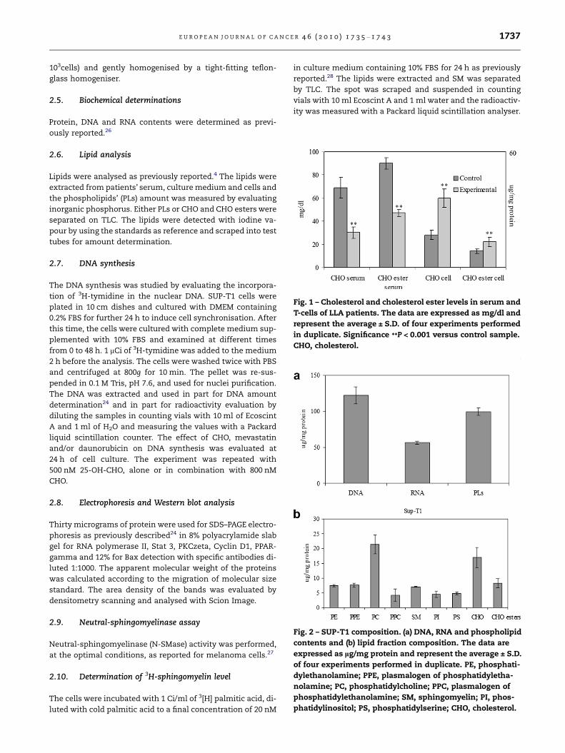
Fig. 1 – Cholesterol and cholesterol ester levels in serum and
T-cells of LLA patients. The data are expressed as mg/dl and
represent the average ± S.D. of four experiments performed
in duplicate. Significance **P < 0.001 versus control sample.
CHO, cholesterol.
Fig. 2 – SUP-T1 composition. (a) DNA, RNA and phospholipid
contents and (b) lipid fraction composition. The data are
expressed as lg/mg protein and represent the average ± S.D.
of four experiments performed in duplicate. PE, phosphati-
dylethanolamine; PPE, plasmalogen of phosphatidyletha-
nolamine; PC, phosphatidylcholine; PPC, plasmalogen of
phosphatidylethanolamine; SM, sphingomyelin; PI, phos-
phatidylinositol; PS, phosphatidylserine; CHO, cholesterol.
E U R O P E A N J O U R N A L O F C A N C E R 4 6 ( 2 0 1 0 ) 1 7 3 5 – 1 7 4 3 1737
103cells) and gently homogenised by a tight-fitting teflon-
glass homogeniser.
2.5. Biochemical determinations
Protein, DNA and RNA contents were determined as previ-
ously reported.26
2.6. Lipid analysis
Lipids were analysed as previously reported.4 The lipids were
extracted from patients’ serum, culture medium and cells and
the phospholipids’ (PLs) amount was measured by evaluating
inorganic phosphorus. Either PLs or CHO and CHO esters were
separated on TLC. The lipids were detected with iodine va-
pour by using the standards as reference and scraped into test
tubes for amount determination.
2.7. DNA synthesis
The DNA synthesis was studied by evaluating the incorpora-
tion of 3H-tymidine in the nuclear DNA. SUP-T1 cells were
plated in 10 cm dishes and cultured with DMEM containing
0.2% FBS for further 24 h to induce cell synchronisation. After
this time, the cells were cultured with complete medium sup-
plemented with 10% FBS and examined at different times
from 0 to 48 h. 1 lCi of 3H-tymidine was added to the medium
2 h before the analysis. The cells were washed twice with PBS
and centrifuged at 800g for 10 min. The pellet was re-sus-
pended in 0.1 M Tris, pH 7.6, and used for nuclei purification.
The DNA was extracted and used in part for DNA amount
determination24 and in part for radioactivity evaluation by
diluting the samples in counting vials with 10 ml of Ecoscint
A and 1 ml of H2O and measuring the values with a Packard
liquid scintillation counter. The effect of CHO, mevastatin
and/or daunorubicin on DNA synthesis was evaluated at
24 h of cell culture. The experiment was repeated with
500 nM 25-OH-CHO, alone or in combination with 800 nM
CHO.
2.8. Electrophoresis and Western blot analysis
Thirty micrograms of protein were used for SDS–PAGE electro-
phoresis as previously described24 in 8% polyacrylamide slab
gel for RNA polymerase II, Stat 3, PKCzeta, Cyclin D1, PPAR-
gamma and 12% for Bax detection with specific antibodies di-
luted 1:1000. The apparent molecular weight of the proteins
was calculated according to the migration of molecular size
standard. The area density of the bands was evaluated by
densitometry scanning and analysed with Scion Image.
2.9. Neutral-sphingomyelinase assay
Neutral-sphingomyelinase (N-SMase) activity was performed,
at the optimal conditions, as reported for melanoma cells.27
2.10. Determination of 3H-sphingomyelin level
The cells were incubated with 1 Ci/ml of 3[H] palmitic acid, di-
luted with cold palmitic acid to a final concentration of 20 nM
in culture medium containing 10% FBS for 24 h as previously
reported.28 The lipids were extracted and SM was separated
by TLC. The spot was scraped and suspended in counting
vials with 10 ml Ecoscint A and 1 ml water and the radioactiv-
ity was measured with a Packard liquid scintillation analyser.
1738 E U R O P E A N J O U R N A L O F C A N C E R 4 6 ( 2 0 1 0 ) 1 7 3 5 – 1 7 4 3
2.11. Statistical analysis
Data are expressed as mean ± S.D. ANOVA or t-test was used
for statistical analysis when appropriate.
3. Results
3.1. Cholesterol level in cancer patients
ALL patients with very low level of CHO (65–80 mg/dl) in ser-
um were chosen as experimental model. The analysis of the
serum lipid fraction showed that in the healthy blood donors
(control sample) CHO and CHO esters were 31 ± 5 and
68 ± 6 mg/dl, respectively, whereas in all patients (experimen-
tal sample) the values were 12 ± 5 and 23 ± 8 mg/dl. Since the
total value of CHO plus CHO esters was lower than that re-
ported in the laboratory analysis, the lipids were extracted
three times as reported in Section 2. After this treatment
the CHO level was 69 ± 9 and 30 ± 6 mg/dl whereas CHO esters
level was 99 ± 5 and 47 ± 7 mg/dl in the control and experi-
mental samples, respectively (Fig. 1). This treatment has been
used for all successive experiments. The data showed that in
the experimental sample the CHO was reduced 56.5% and
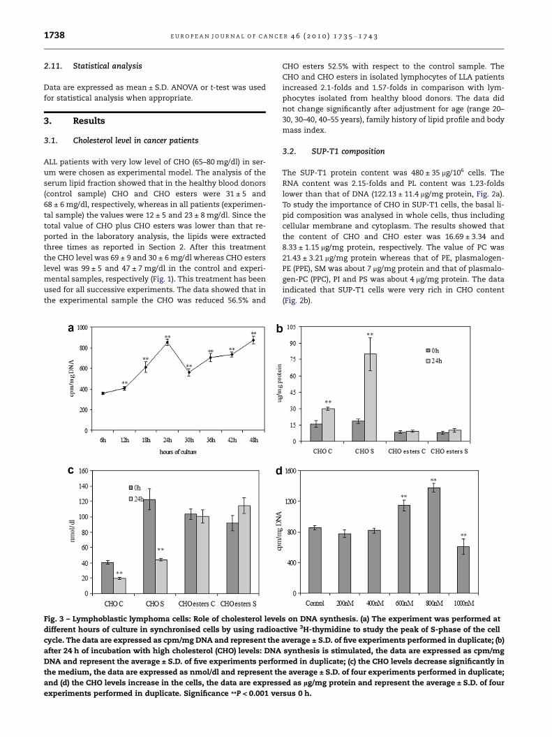
Fig. 3 – Lymphoblastic lymphoma cells: Role of cholesterol leve
different hours of culture in synchronised cells by using radioa
cycle. The data are expressed as cpm/mg DNA and represent the
after 24 h of incubation with high cholesterol (CHO) levels: DNA
DNA and represent the average ± S.D. of five experiments perfor
the medium, the data are expressed as nmol/dl and represent th
and (d) the CHO levels increase in the cells, the data are express
experiments performed in duplicate. Significance **P < 0.001 ve
CHO esters 52.5% with respect to the control sample. The
CHO and CHO esters in isolated lymphocytes of LLA patients
increased 2.1-folds and 1.57-folds in comparison with lym-
phocytes isolated from healthy blood donors. The data did
not change significantly after adjustment for age (range 20–
30, 30–40, 40–55 years), family history of lipid profile and body
mass index.
3.2. SUP-T1 composition
The SUP-T1 protein content was 480 ± 35 lg/106 cells. The
RNA content was 2.15-folds and PL content was 1.23-folds
lower than that of DNA (122.13 ± 11.4 lg/mg protein, Fig. 2a).
To study the importance of CHO in SUP-T1 cells, the basal li-
pid composition was analysed in whole cells, thus including
cellular membrane and cytoplasm. The results showed that
the content of CHO and CHO ester was 16.69 ± 3.34 and
8.33 ± 1.15 lg/mg protein, respectively. The value of PC was
21.43 ± 3.21 lg/mg protein whereas that of PE, plasmalogen-
PE (PPE), SM was about 7 lg/mg protein and that of plasmalo-
gen-PC (PPC), PI and PS was about 4 lg/mg protein. The data
indicated that SUP-T1 cells were very rich in CHO content
(Fig. 2b).
ls on DNA synthesis. (a) The experiment was performed at
ctive 3H-thymidine to study the peak of S-phase of the cell
average ± S.D. of five experiments performed in duplicate; (b)
synthesis is stimulated, the data are expressed as cpm/mg
med in duplicate; (c) the CHO levels decrease significantly in
e average ± S.D. of four experiments performed in duplicate;
ed as lg/mg protein and represent the average ± S.D. of four
rsus 0 h.
E U R O P E A N J O U R N A L O F C A N C E R 4 6 ( 2 0 1 0 ) 1 7 3 5 – 1 7 4 3 1739
3.3. Effect of cholesterol in SUP-T1 cell growth andsurvival
Cell growth was studied by the analysis of DNA synthesis per-
formed in synchronised cells. The results showed that in the
SUP-T1 cells the specific activity of the DNA, calculated as
cpm/g DNA, was very low at 6 and 12 h, increased at 18 h
and reached a peak at 24 h, which correspond to the S-phase
of the cell cycle (Fig. 3a). Increasing concentrations of CHO
(from 200 nM to 1000 nM) were added to the culture medium
at 0 h and its effect was evaluated at 24 h. At 0 h the culture
medium contained about 40 nmol/dl of CHO and 123 nmol/
dl of CHO ester and therefore the CHO concentration in-
creased to 60, 80, 100, 120 and 140 nmol/dl. Only 600 and
800 nM CHO stimulated the DNA synthesis 1.34· and 1.60·,
respectively, whereas higher concentration had inhibitor ef-
fect and lower concentration did not have effect (Fig. 3b). This
biphasic phenomenon of DNA synthesis which correlated
with CHO concentration supports the previous observations
reported in statin-treated cell experiments29 and in vivo ani-
mal models.30 It was evident that the optimal concentration
was 800 nM and therefore it was used to evaluate the incorpo-
ration of CHO in the cells. After 24 h the CHO present in the
culture medium was reduced 52% in the control sample
(without CHO addition) and 64% in experimental sample
(with 800 nM CHO, Fig. 3c). On the other hand the CHO pres-
ent in the cells increased 1.87-fold in control sample and
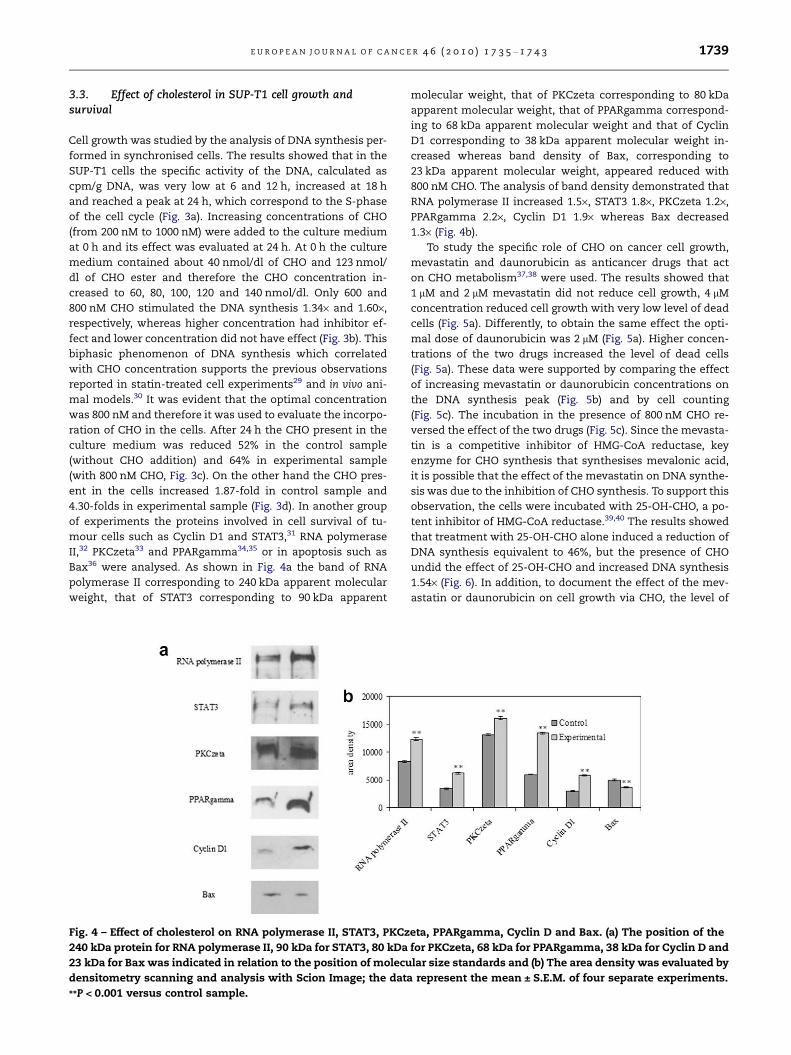
4.30-folds in experimental sample (Fig. 3d). In another group
of experiments the proteins involved in cell survival of tu-
mour cells such as Cyclin D1 and STAT3,31 RNA polymerase
II,32 PKCzeta33 and PPARgamma34,35 or in apoptosis such as
Bax36 were analysed. As shown in Fig. 4a the band of RNA
polymerase II corresponding to 240 kDa apparent molecular
weight, that of STAT3 corresponding to 90 kDa apparent
Fig. 4 – Effect of cholesterol on RNA polymerase II, STAT3, PKCz
240 kDa protein for RNA polymerase II, 90 kDa for STAT3, 80 kDa
23 kDa for Bax was indicated in relation to the position of molecu
densitometry scanning and analysis with Scion Image; the data
**P < 0.001 versus control sample.
molecular weight, that of PKCzeta corresponding to 80 kDa
apparent molecular weight, that of PPARgamma correspond-
ing to 68 kDa apparent molecular weight and that of Cyclin
D1 corresponding to 38 kDa apparent molecular weight in-
creased whereas band density of Bax, corresponding to
23 kDa apparent molecular weight, appeared reduced with
800 nM CHO. The analysis of band density demonstrated that
RNA polymerase II increased 1.5·, STAT3 1.8·, PKCzeta 1.2·,
PPARgamma 2.2·, Cyclin D1 1.9· whereas Bax decreased
1.3· (Fig. 4b).
To study the specific role of CHO on cancer cell growth,
mevastatin and daunorubicin as anticancer drugs that act
on CHO metabolism37,38 were used. The results showed that
1 lM and 2 lM mevastatin did not reduce cell growth, 4 lM
concentration reduced cell growth with very low level of dead
cells (Fig. 5a). Differently, to obtain the same effect the opti-
mal dose of daunorubicin was 2 lM (Fig. 5a). Higher concen-
trations of the two drugs increased the level of dead cells
(Fig. 5a). These data were supported by comparing the effect
of increasing mevastatin or daunorubicin concentrations on
the DNA synthesis peak (Fig. 5b) and by cell counting
(Fig. 5c). The incubation in the presence of 800 nM CHO re-
versed the effect of the two drugs (Fig. 5c). Since the mevasta-
tin is a competitive inhibitor of HMG-CoA reductase, key
enzyme for CHO synthesis that synthesises mevalonic acid,
it is possible that the effect of the mevastatin on DNA synthe-
sis was due to the inhibition of CHO synthesis. To support this
observation, the cells were incubated with 25-OH-CHO, a po-
tent inhibitor of HMG-CoA reductase.39,40 The results showed
that treatment with 25-OH-CHO alone induced a reduction of
DNA synthesis equivalent to 46%, but the presence of CHO
undid the effect of 25-OH-CHO and increased DNA synthesis
1.54· (Fig. 6). In addition, to document the effect of the mev-
astatin or daunorubicin on cell growth via CHO, the level of
eta, PPARgamma, Cyclin D and Bax. (a) The position of the
for PKCzeta, 68 kDa for PPARgamma, 38 kDa for Cyclin D and
lar size standards and (b) The area density was evaluated by
represent the mean ± S.E.M. of four separate experiments.
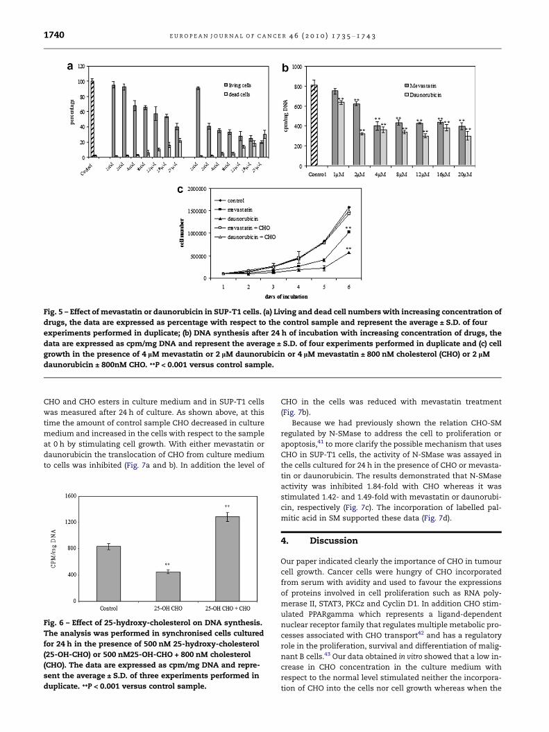
Fig. 5 – Effect of mevastatin or daunorubicin in SUP-T1 cells. (a) Living and dead cell numbers with increasing concentration of
drugs, the data are expressed as percentage with respect to the control sample and represent the average ± S.D. of four
experiments performed in duplicate; (b) DNA synthesis after 24 h of incubation with increasing concentration of drugs, the
data are expressed as cpm/mg DNA and represent the average ± S.D. of four experiments performed in duplicate and (c) cell
growth in the presence of 4 lM mevastatin or 2 lM daunorubicin or 4 lM mevastatin ± 800 nM cholesterol (CHO) or 2 lM
daunorubicin ± 800nM CHO. **P < 0.001 versus control sample.
1740 E U R O P E A N J O U R N A L O F C A N C E R 4 6 ( 2 0 1 0 ) 1 7 3 5 – 1 7 4 3
CHO and CHO esters in culture medium and in SUP-T1 cells
was measured after 24 h of culture. As shown above, at this
time the amount of control sample CHO decreased in culture
medium and increased in the cells with respect to the sample
at 0 h by stimulating cell growth. With either mevastatin or
daunorubicin the translocation of CHO from culture medium
to cells was inhibited (Fig. 7a and b). In addition the level of
Fig. 6 – Effect of 25-hydroxy-cholesterol on DNA synthesis.
The analysis was performed in synchronised cells cultured
for 24 h in the presence of 500 nM 25-hydroxy-cholesterol
(25-OH-CHO) or 500 nM25-OH-CHO + 800 nM cholesterol
(CHO). The data are expressed as cpm/mg DNA and repre-
sent the average ± S.D. of three experiments performed in
duplicate. **P < 0.001 versus control sample.
CHO in the cells was reduced with mevastatin treatment
(Fig. 7b).
Because we had previously shown the relation CHO-SM
regulated by N-SMase to address the cell to proliferation or
apoptosis,41 to more clarify the possible mechanism that uses
CHO in SUP-T1 cells, the activity of N-SMase was assayed in
the cells cultured for 24 h in the presence of CHO or mevasta-
tin or daunorubicin. The results demonstrated that N-SMase
activity was inhibited 1.84-fold with CHO whereas it was
stimulated 1.42- and 1.49-fold with mevastatin or daunorubi-
cin, respectively (Fig. 7c). The incorporation of labelled pal-
mitic acid in SM supported these data (Fig. 7d).
4. Discussion
Our paper indicated clearly the importance of CHO in tumour
cell growth. Cancer cells were hungry of CHO incorporated
from serum with avidity and used to favour the expressions
of proteins involved in cell proliferation such as RNA poly-
merase II, STAT3, PKCz and Cyclin D1. In addition CHO stim-
ulated PPARgamma which represents a ligand-dependent
nuclear receptor family that regulates multiple metabolic pro-
cesses associated with CHO transport42 and has a regulatory
role in the proliferation, survival and differentiation of malig-
nant B cells.43 Our data obtained in vitro showed that a low in-
crease in CHO concentration in the culture medium with
respect to the normal level stimulated neither the incorpora-
tion of CHO into the cells nor cell growth whereas when the

Fig. 7 – Cholesterol and sphingomyelin in SUP-T1 cell. (a) Cholesterol (CHO) and CHO esters in culture medium after 24 h of
incubation without or with mevastatin or daunorubicin, the data are expressed as nmol/dl and represent the average ± S.D. of
four experiments performed in duplicate; (b) CHO and CHO esters in the cells after 24 h of incubation without or with
mevastatin or daunorubicin, the data are expressed as lg/mg protein and represent the average ± S.D. of four experiments
performed in duplicate; (c) neutral sphingomyelinase activity, the data are expressed as pmol/mg protein/min and represent
the average ± S.D. of five experiments performed in duplicate; and (d) 3H-palmitic acid incorporation in sphingomyelin (SM),
the data are expressed as cpm/mg SM and represent the average ± S.D. of five experiments performed in duplicate. **P < 0.001
versus control sample.
E U R O P E A N J O U R N A L O F C A N C E R 4 6 ( 2 0 1 0 ) 1 7 3 5 – 1 7 4 3 1741
CHO concentration increased to 280 mg/dl, a strong CHO in-
take was detected and cell growth was significantly stimu-
lated. These data could suggest that if the blood CHO
concentration in patient with initial tumour is normal or
slightly increased, the effect of CHO in tumour growth is
low whereas the hyperCHO facilitates the entry of CHO in
cells stimulating cell growth and inducing severe hypoCHO.
Therefore the hyperCHO influences the development of tu-
mour and the severe hypoCHO is a sign of tumour progres-
sion. Di Vizio et al. have reported epidemiological evidences
suggesting that the modern Western diet, which contains
substantial levels of CHO and other fatty substances, influ-
ences prostate cancer.18
We showed here that the anticancer drugs act on tumour
cell CHO. In particular, the CHO synthesis is inhibited by mev-
astatin and 25-OH-CHO, both inhibitors of HMG-CoA reduc-
tase, whereas no effect is obtained with daunorubicin.
Differently, either mevastatin or daunorubicin prevents the
incorporation of CHO in tumour cells and induces them to
apoptosis via N-SMase. These data suggest that hypoCHO
originated from statin-related status is not correlated with
the increase in CHO content in cancer cells as that occurs in
hypoCHO of cancer patients. In addition mevastatin treat-
ment regulates the CHO level in the blood to normal values
and does not induce severe hypoCHO as in cancer. Therefore
only severe hypoCHO status with an increase in CHO content
in cancer cells was responsible for neoplastic proliferation.
On the other hand in lymphocytes isolated from patients with
AML there was an elevated receptor-mediated uptake of LDL
with a decrease in the plasma CHO level.3 After chemothera-
pic treatment, the leukaemic cells disappeared and the plas-
ma CHO levels returned to normal values.3 Considering our
data that indicate the importance of blood CHO level in the
patients with the initial tumour, we suggest that the therapy
acting via CHO may need to be tailored for individual patients
with tumour. Since our results indicated that during tumour
process it is important the entry of CHO in the cell, it is pos-
sible to suppose that it could modify the structure/function of
cell membrane modifying the specialised domains such as li-
pid rafts rich in CHO and SM that act as platform for different
proteins involved in cell signal.44 It has been proposed that
cancer progression involves dysregulation of lipid raft-resi-
dent signalling complexes.18 On the other hand death-induc-
ing signalling complex clusters in lipid raft aggregates were
reported as a supramolecular and physical entity responsible
for the induction of apoptosis in leukaemic cells by the anti-
tumour drug such as edelfosine.45
In conclusion we propose to use the level of CHO as a good
biological marker for following up (low level) the neoplastic
process.
Conflict of interest statement
None declared.

1742 E U R O P E A N J O U R N A L O F C A N C E R 4 6 ( 2 0 1 0 ) 1 7 3 5 – 1 7 4 3
Acknowledgements
We thank Remo Lazzarini for the technical assistance. We
also acknowledge the financial support through the contract
grants Ministero dell’ Universita e Ricerca (PRIN project) and
the Fondazione Cassa di Risparmio di Perugia.
R E F E R E N C E S
1. Jenkins DJ, Josse AR, Wong JM, Nguyen TH, Kendall CW. Theportfolio diet for cardiovascular risk reduction. CurrAtheroscler Rep 2007;9:501–7.
2. Tatidis L, Vitols S, Gruber A, Paul C, Axelson M. Cholesterolcatabolism in patients with acute myelogenous leukemiaand hypocholesterolemia: suppressed levels of a circulatingmarker for bile acid synthesis. Cancer Lett 2001;170:169–75.
3. Vitols S, Gahrton G, Bjorkholm M, Peterson C.Hypocholesterolaemia in malignancy due to elevated low-density-lipoprotein-receptor activity in tumour cells:evidence from studies in patients with leukaemia. Lancet1985;2:1150–4.
4. Pugliese L, Bernardini I, Viola-Magni MP, Albi E. Low levels ofserum cholesterol/phospholipids are associated with theantiphospholipid antibodies in monoclonal gammopathy. Int JImmunopathol Pharmacol 2006;19:331–7.
5. Quesney-Huneeus V, Galick HA, Siperstein MD, et al. The dualrole of mevalonate in the cell cycle. J Biol Chem1983;258:378–85.
6. Fairbanks KP, Witte LD, Goodman DS. Relationship betweenmevalonate and mitogenesis in human fibroblasts stimulatedwith platelet-derived growth factor. J Biol Chem1984;259:1546–51.
7. Myant NB. The biology of cholesterol and relatedsteroids. London: Williams Heinemann Medical Book; 1981.
8. Columbano A, Dessi S, Ledda-Columbano GM, et al. HMP-shunt and cholesterol metabolism in experimental modelsinvolving normal and preneoplastic liver growth. Toxicol Pathol1987;15:43–50.
9. Wachtershauser A, Akoglu B, Stein J. HMG-CoA reductaseinhibitor mevastatin enhances the growth inhibitory effect ofbutyrate in the colorectal carcinoma cell line Caco-2.Carcinogenesis 2001;22:1061–7.
10. Danesh FR, Sadeghi MM, Amro N, et al. 3-Hydroxy-3-methylglutaryl CoA reductase inhibitors prevent highglucose-induced proliferation of mesangial cells viamodulation of Rho GTPase/ p21 signaling pathway:Implications for diabetic nephropathy. Proc Natl Acad Sci USA2002;99:8301–5.
11. Alegret M, Silvestre JS. Pleiotropic effects of statins andrelated pharmacological experimental approaches. Meth FindExp Clin Pharmacol 2006;28:627–56.
12. Baba TT, Nemoto TK, Miyazaki T, Oida S. Simvastatinsuppresses the differentiation of C2C12 myoblast cells via aRac pathway. J Muscle Res Cell Motil 2008;29:127–34.
13. Perez-Sala D, Gilbert BA, Rando RR, Canada FJ. Analogs offarnesylcysteine induce apoptosis in HL-60 cells. FEBS Lett1998;426:319–24.
14. Garcıa-Roman N, Alvarez AM, Toro MJ, Montes A, Lorenzo MJ.Lovastatin induces apoptosis of spontaneously immortalizedrat brain neuroblasts: involvement of nonsterol isoprenoidbiosynthesis inhibition. Mol Cell Neurosci 2001;17:329–41.
15. Gauthaman K, Fong CY, Bongso A. Statins, stem cells, andcancer. J Cell Biochem 2009;106:975–83.
16. Weaver AM. Invadopodia: specialized cell structures forcancer invasion. Clin Exp Metastasis 2006;23:97–105.
17. Caldieri G, Giacchetti G, Beznoussenko G, et al. Invadopodiabiogenesis is regulated by caveolin-mediated modulationof membrane cholesterol levels. J Cell Mol Med 2009;13:1728–40.
18. Di Vizio D, Solomon KR, Freeman MR. Cholesterol andcholesterol-rich membranes in prostate cancer: an update.Tumori 2008;94:633–9.
19. Rebillard A, Jouan-Lanhouet S, Jouan E, et al. Cisplatin-induced apoptosis involves a Fas-ROCK-ezrin-dependentactin remodelling in human colon cancer cells. Eur J Cancer2010. [Epub ahead of print].
20. Storch CH, Ehehalt R, Haefeli WE, Weiss J. Localization of thehuman breast cancer resistance protein (BCRP/ABCG2) in lipidrafts/caveolae and modulation of its activity by cholesterolin vitro. J Pharmacol Exp Ther 2007;323:257–64.
21. Montero J, Morales A, Llacuna L, et al. Mitochondrialcholesterol contributes to chemotherapy resistance inhepatocellular carcinoma. Cancer Res 2008;68:5246–56.
22. Bensinger SJ, Bradley MN, Joseph SB, et al. LXR signalingcouples sterol metabolism to proliferation in the acquiredimmune response. Cell 2008;134:97–111.
23. Shor R, Wainstein J, Oz D, et al. Low serum LDL cholesterollevels and the risk of fever, sepsis, and malignancy. Ann ClinLab Sci 2007;37:343–8.
24. Yavasoglu I, Tombuloglu M, Kadikoylu G, Donmez A, CagirganS. Bolaman Z Cholesterol levels in patients with multiplemyeloma. Ann Hematol 2008;87:223–8.
25. Boyum A. Separation of lymphocytes, lymphocyte subgroupsand monocytes: a review. Lymphology 1977;10:71–6.
26. Rossi G, Magni MV, Albi E. Sphingomyelin–cholesterol anddouble stranded RNA relationship in the intranuclearcomplex. Arch Biochem Biophys 2007;459:27–32.
27. Albi E, La Porta CAM, Castaldi S, Viola Magni M. Nuclearsphingomyelin-synthase and protein kinase C delta inmelanoma cells. Arch Biochem Biophys 2005;438:156–61.
28. Albi E, Cataldi S, Bartoccini E, et al. Nuclear sphingomyelinpathway in serum deprivation-induced apoptosis ofembryonic hippocampal cells. J Cell Physiol 2006;206:189–95.
29. Weis M, Heeschen C, Glassford AJ, Cooke JP. Statins havebiphasic effects on angiogenesis. Circulation 2002;105:739–45.
30. Wang CY, Shui HA, Chang TC. In vivo evidence of dualityeffects for lovastatin in a nude mouse cancer model. Int JCancer 2010;126:578–82.
31. Alvaro T, Lejeune M, Garcıa JF, et al. Tumor-infiltratedimmune response correlates with alterations in the apoptoticand cell cycle pathways in Hodgkin and Reed–Sternberg cells.Clin Cancer Res 2008;14:6856–91.
32. Chen R, Keating MJ, Gandhi V, Plunkett W. Transcriptioninhibition by flavopiridol: mechanism of chronic lymphocyticleukemia cell death. Blood 2005;106:2513–9.
33. Cecconi D, Zamo A, Bianchi E, et al. Signal transductionpathways of mantle cell lymphoma: a phosphoproteome-based study. Proteomic 2008;8:4495–506.
34. Jo SH, Yang C, Miao Q, et al. Peroxisome proliferator-activatedreceptor gamma promotes lymphocyte survival through itsactions on cellular metabolic activities. J Immunol2006;177:3737–45.
35. Garcia-Bates TM, Peslak SA, Baglole CJ, et al. Peroxisomeproliferator-activated receptor gamma overexpression andknockdown: impact on human B cell lymphoma proliferationand serviva. Cancer Immunol Immunother 2009;58:1071–83.
36. Nakashima M, Cui ZG, Tabuchi Y, et al. Apoptosis induced byan alkylated purine, 6-dimethylaminopurine, and changes ingene expression in human lymphoma U937 cells. AnticancerRes 2008;28:609–20.
E U R O P E A N J O U R N A L O F C A N C E R 4 6 ( 2 0 1 0 ) 1 7 3 5 – 1 7 4 3 1743
37. Theivagt AE, Amanti EN, Beresford NJ, Tabernero L, Friesen JA.Characterization of an HMG-CoA reductase from Listeriamonocytogenes that exhibits dual coenzyme specificita.Biochemistry 2006;45:14397–406.
38. Banker DE, Mayer SJ, Li HY, et al. Cholesterol synthesis andimport contribute to protective cholesterol increments inacute myeloid leukemia cells. Blood 2004;104:1816–24.
39. Cerda SR, Wilkinson 4th J, Broitman SA. Regulation ofcholesterol synthesis in four colonic adenocarcinoma celllines. Lipid 1995;30:1083–92.
40. Tam SP, Ramharack R. The effect of 25-hydroxycholesterol onthe regulation of apolipoprotein E mRNA levels and secretionin the human hepatoma HepG2. Atherosclerosis1992;95:137–46.
41. Albi E, Viola Magni MP. Sphingomyelin: a small-big moleculein the nucleus. Recent Res Develop Biophys Biochem2006;37:211–27.
42. Yin Y, Russell RG, Dettin LE, et al. Peroxisome proliferator-activated receptor delta and gamma agonists differentiallyalter tumor differentiation and progression during mammarycarcinogenesis. Cancer Res 2005;65:3950–7.
43. Garcia-Bates TM, Peslak SA, Baglole CJ, et al. Peroxisomeproliferator-activated receptor gamma overexpression andknockdown: impact on human B cell lymphomaproliferation and serviva. Cancer Immunol Immunother2009;58:1071–83.
44. Edidin M. The state of lipid rafts: from model membrane tocells. Ann Rev Biophys Biomol Struct 2003;32:257–83.
45. Gajate C, Gonzalez-Camacho F, Mollinedo F. Involvement ofraft aggregates enriched in Fas/CD95 death-inducingsignaling complex in the antileukemic action of edelfosine inJurkat cells. PLoS One 2009;4:e5044.