Comparative genome analysis of entomopathogenic fungi reveals a complex set of secreted proteins
18
RESEARCH ARTICLE Open Access Comparative genome analysis of entomopathogenic fungi reveals a complex set of secreted proteins Charley Christian Staats 1 , Ângela Junges 1 , Rafael Lucas Muniz Guedes 2 , Claudia Elizabeth Thompson 1 , Guilherme Loss de Morais 2 , Juliano Tomazzoni Boldo 1 , Luiz Gonzaga Paula de Almeida 2 , Fábio Carrer Andreis 1 , Alexandra Lehmkuhl Gerber 2 , Nicolau Sbaraini 1 , Rana Louise de Andrade da Paixão 1 , Leonardo Broetto 1 , Melissa Landell 1 , Lucélia Santi 1 , Walter Orlando Beys-da-Silva 1 , Carolina Pereira Silveira 1 , Thaiane Rispoli Serrano 1 , Eder Silva de Oliveira 1 , Lívia Kmetzsch 1 , Marilene Henning Vainstein 1 , Ana Tereza Ribeiro de Vasconcelos 2 and Augusto Schrank 1* Abstract Background: Metarhizium anisopliae is an entomopathogenic fungus used in the biological control of some agricultural insect pests, and efforts are underway to use this fungus in the control of insect-borne human diseases. A large repertoire of proteins must be secreted by M. anisopliae to cope with the various available nutrients as this fungus switches through different lifestyles, i.e., from a saprophytic, to an infectious, to a plant endophytic stage. To further evaluate the predicted secretome of M. anisopliae, we employed genomic and transcriptomic analyses, coupled with phylogenomic analysis, focusing on the identification and characterization of secreted proteins. Results: We determined the M. anisopliae E6 genome sequence and compared this sequence to other entomopathogenic fungi genomes. A robust pipeline was generated to evaluate the predicted secretomes of M. anisopliae and 15 other filamentous fungi, leading to the identification of a core of secreted proteins. Transcriptomic analysis using the tick Rhipicephalus microplus cuticle as an infection model during two periods of infection (48 and 144 h) allowed the identification of several differentially expressed genes. This analysis concluded that a large proportion of the predicted secretome coding genes contained altered transcript levels in the conditions analyzed in this study. In addition, some specific secreted proteins from Metarhizium have an evolutionary history similar to orthologs found in Beauveria/Cordyceps. This similarity suggests that a set of secreted proteins has evolved to participate in entomopathogenicity. Conclusions: The data presented represents an important step to the characterization of the role of secreted proteins in the virulence and pathogenicity of M. anisopliae. Keywords: Genome sequence, Entomopathogenic fungi, Secretome, Phylogenomics * Correspondence: [email protected] 1 Centro de Biotecnologia, Universidade Federal do Rio Grande do Sul (UFRGS), P. O. Box 15005, Porto Alegre, RS CEP 91501-970, Brazil Full list of author information is available at the end of the article © 2014 Staats et al.; licensee BioMed Central Ltd. This is an Open Access article distributed under the terms of the Creative Commons Attribution License (http://creativecommons.org/licenses/by/4.0), which permits unrestricted use, distribution, and reproduction in any medium, provided the original work is properly credited. The Creative Commons Public Domain Dedication waiver (http://creativecommons.org/publicdomain/zero/1.0/) applies to the data made available in this article, unless otherwise stated. Staats et al. BMC Genomics 2014, 15:822 http://www.biomedcentral.com/1471-2164/15/822
Transcript of Comparative genome analysis of entomopathogenic fungi reveals a complex set of secreted proteins

Staats et al. BMC Genomics 2014, 15:822http://www.biomedcentral.com/1471-2164/15/822
RESEARCH ARTICLE Open Access
Comparative genome analysis ofentomopathogenic fungi reveals a complex setof secreted proteinsCharley Christian Staats1, Ângela Junges1, Rafael Lucas Muniz Guedes2, Claudia Elizabeth Thompson1,Guilherme Loss de Morais2, Juliano Tomazzoni Boldo1, Luiz Gonzaga Paula de Almeida2, Fábio Carrer Andreis1,Alexandra Lehmkuhl Gerber2, Nicolau Sbaraini1, Rana Louise de Andrade da Paixão1, Leonardo Broetto1,Melissa Landell1, Lucélia Santi1, Walter Orlando Beys-da-Silva1, Carolina Pereira Silveira1, Thaiane Rispoli Serrano1,Eder Silva de Oliveira1, Lívia Kmetzsch1, Marilene Henning Vainstein1, Ana Tereza Ribeiro de Vasconcelos2
and Augusto Schrank1*
Abstract
Background: Metarhizium anisopliae is an entomopathogenic fungus used in the biological control of someagricultural insect pests, and efforts are underway to use this fungus in the control of insect-borne human diseases.A large repertoire of proteins must be secreted by M. anisopliae to cope with the various available nutrients as thisfungus switches through different lifestyles, i.e., from a saprophytic, to an infectious, to a plant endophytic stage. Tofurther evaluate the predicted secretome of M. anisopliae, we employed genomic and transcriptomic analyses,coupled with phylogenomic analysis, focusing on the identification and characterization of secreted proteins.
Results: We determined the M. anisopliae E6 genome sequence and compared this sequence to otherentomopathogenic fungi genomes. A robust pipeline was generated to evaluate the predicted secretomes of M.anisopliae and 15 other filamentous fungi, leading to the identification of a core of secreted proteins.Transcriptomic analysis using the tick Rhipicephalus microplus cuticle as an infection model during two periods ofinfection (48 and 144 h) allowed the identification of several differentially expressed genes. This analysis concludedthat a large proportion of the predicted secretome coding genes contained altered transcript levels in theconditions analyzed in this study. In addition, some specific secreted proteins from Metarhizium have anevolutionary history similar to orthologs found in Beauveria/Cordyceps. This similarity suggests that a set of secretedproteins has evolved to participate in entomopathogenicity.
Conclusions: The data presented represents an important step to the characterization of the role of secretedproteins in the virulence and pathogenicity of M. anisopliae.
Keywords: Genome sequence, Entomopathogenic fungi, Secretome, Phylogenomics
* Correspondence: [email protected] de Biotecnologia, Universidade Federal do Rio Grande do Sul(UFRGS), P. O. Box 15005, Porto Alegre, RS CEP 91501-970, BrazilFull list of author information is available at the end of the article
© 2014 Staats et al.; licensee BioMed Central Ltd. This is an Open Access article distributed under the terms of the CreativeCommons Attribution License (http://creativecommons.org/licenses/by/4.0), which permits unrestricted use, distribution, andreproduction in any medium, provided the original work is properly credited. The Creative Commons Public DomainDedication waiver (http://creativecommons.org/publicdomain/zero/1.0/) applies to the data made available in this article,unless otherwise stated.

Staats et al. BMC Genomics 2014, 15:822 Page 2 of 18http://www.biomedcentral.com/1471-2164/15/822
BackgroundIt is estimated that over 600,000 species of fungi exist,and it is assumed that these species can be found in al-most all habitats on Earth. However, only a few of thesespecies have been described [1]. Most fungal specieshave developed saprophytic interactions in soil and inwater or in association with mycorrhizal plants, as eitherarbuscular mycorrhizae or ectomycorrhizae. Moreover,fungal species are known to cause disease in severalhosts, including mammals, arthropods, and plants [2].To adapt to such a large variety of habitats, fungi havedeveloped a prolific capability to export proteins to theextracellular space as an important mechanism to ac-quire nutrients [3]. Therefore, secretomes, which are de-fined as the global set of proteins produced by a cell andexported to the extracellular space in a determined timeand condition, represent an important target for under-standing the mechanisms of fungal adaptation. For in-stance, both saprophytic and pathogenic fungi mustquickly adapt to variations in carbon and nitrogen avail-ability. Because fungi generally obtain nutrients from thedigestion of extracellular polymers, such as cellulose andchitin, fungi must produce copious amounts of extracel-lular enzymes to allow for the efficient hydrolysis of bio-polymers during the infection process or from theirnatural environment [3].A diverse group of fungi is associated with arthropods,
the largest class of eukaryotic species on Earth, and playsa role in controlling their populations, in particular ofinsects [4]. The most well known insect-associated fungiare entomopathogens, which are necrotrophic fungi thatactively penetrate the host exoskeleton and proliferate inthe hemocoel until all internal tissues have been degraded.The infection process of entomopathogenic fungi dependson the secretion of a plethora of enzymes and toxins,which serve to penetrate and kill the host, as well as toprovide nutrients through the action of biopolymer-degrading enzymes [5-10]. The best-characterized ex-ample of a relation between an entomopathogenic fungusand its hosts is the genus Metarhizium. Several lines ofevidence suggest that the infection cycle of Metarhiziumcan be schematically divided into the following steps:(i) conidia adherence to the host cuticle through hydro-phobic interactions and thin mucilaginous material;(ii) conidia germination and development; (iii) germ-tubedifferentiation into appressoria; (iv) cuticle penetration;(v) hyphae differentiation into blastospores/hyphal bod-ies in the hemolymph; (vi) host colonization; (vii) extru-sion to the host cadaver surface; and (viii) conidiophoreformation and conidia production [11]. The participa-tion of many proteins, including secreted proteins, hasbeen described for the infection process (reviewed in[12]). More recently, the existence of alternative mecha-nisms has been suggested during the control of Aedes
aegypti, the mosquito vector of dengue and yellow fever[13]. Because different Metarhizium species can infectand kill more than 200 species from 50 insect andarthropod families [14], some isolates have been widelyused as bioagents to control a wide variety of pests [15].Indeed, almost 50 different formulations employing Metar-hizium are commercially available [16].In fact, the Metarhizium species generally regarded as
M. anisopliae is composed of nine different species,which can be most frequently isolated from either soil orinsects [17]. The genomes of the M. anisopliae ARSEF23, which are currently classified as M. robertsii [17], abroad-spectrum insect pathogen, and of the acridid-specific M. acridum CQMa 102, were characterized [18].The sequence analysis of these genomes revealed that theyare highly syntenic and possess many genes that allow forthe different lifestyles of Metarhizium spp. In addition, aphylogenomic analysis showed that M. robertsii and M.acridum are more related to plant endophytes and patho-gens than to animal pathogens. Moreover, this analysisshowed that the sequenced genome was from M. robertsii,which had been misclassified as M. anisopliae [18]. Fur-ther information concerning the evolution of entomo-pathogenic fungi originates from the characterization ofthe entomopathogen Beauveria bassiana genome, whichcontributed to the identification of a common set of genefamilies potentially associated with fungal entomopatho-genicity [19].The large collection of fungal genomes sequenced,
including entomopathogens, plant pathogens, mycopatho-gens, and mammal pathogens, allows the shared andexclusive genes present in the predicted secretomes offungal species to be identified. To analyze the import-ance of secreted proteins in the virulence of fungalpathogens, we sequenced the genome of M. anisopliaestrain E6 and performed a comparative study of thisgenome, emphasizing the predicted secretome amongdistinct fungal species.
Results and discussionGeneral features and comparative analyses of theM. anisopliae E6 genomeThe genome sequence of Metarhizium anisopliae E6 wasobtained using a 454-based pyrosequencing approach,with 19-fold genome coverage. The assembly performedusing Newbler 2.8 and WGS-CA 7.0 software resulted in191 and 516 scaffolds with 38,326,054 and 38,454,426 bp,respectively (Table 1). The longest scaffold identified usingNewbler software was 1,756,362 bp, whereas the lon-gest scaffold identified using WGS-CA software was638,367 bp. Using Newbler and WGS-CA software, theN50 scaffold size was calculated as 622.80 kb and as167.52 kb and the N50 contig size was measured as157.96 kb and as 125.69 kb, respectively. Both assemblies
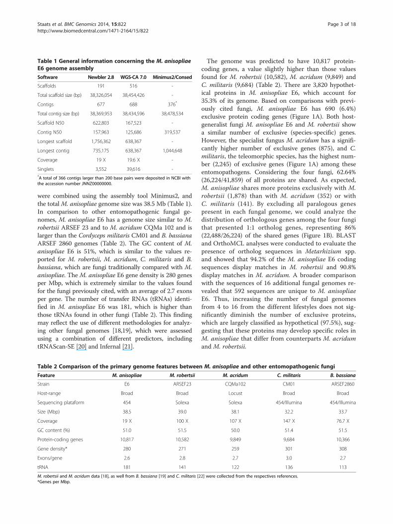
Table 1 General information concerning the M. anisopliaeE6 genome assembly
Software Newbler 2.8 WGS-CA 7.0 Minimus2/Consed
Scaffolds 191 516 -
Total scaffold size (bp) 38,326,054 38,454,426 -
Contigs 677 688 376*
Total contig size (bp) 38,369,953 38,434,596 38,478,534
Scaffold N50 622,803 167,523 -
Contig N50 157,963 125,686 319,537
Longest scaffold 1,756,362 638,367 -
Longest contig 735,175 638,367 1,044,648
Coverage 19 X 19.6 X -
Singlets 3,552 39,616 -*A total of 366 contigs larger than 200 base pairs were deposited in NCBI withthe accession number JNNZ00000000.
Staats et al. BMC Genomics 2014, 15:822 Page 3 of 18http://www.biomedcentral.com/1471-2164/15/822
were combined using the assembly tool Minimus2, andthe totalM. anisopliae genome size was 38.5 Mb (Table 1).In comparison to other entomopathogenic fungal ge-nomes, M. anisopliae E6 has a genome size similar to M.robertsii ARSEF 23 and to M. acridum CQMa 102 and islarger than the Cordyceps militaris CM01 and B. bassianaARSEF 2860 genomes (Table 2). The GC content of M.anisopliae E6 is 51%, which is similar to the values re-ported for M. robertsii, M. acridum, C. militaris and B.bassiana, which are fungi traditionally compared with M.anisopliae. The M. anisopliae E6 gene density is 280 genesper Mbp, which is extremely similar to the values foundfor the fungi previously cited, with an average of 2.7 exonsper gene. The number of transfer RNAs (tRNAs) identi-fied in M. anisopliae E6 was 181, which is higher thanthose tRNAs found in other fungi (Table 2). This findingmay reflect the use of different methodologies for analyz-ing other fungal genomes [18,19], which were assessedusing a combination of different predictors, includingtRNAScan-SE [20] and Infernal [21].
Table 2 Comparison of the primary genome features between
Feature M. anisopliae M. robertsii
Strain E6 ARSEF23
Host-range Broad Broad
Sequencing plataform 454 Solexa
Size (Mbp) 38.5 39.0
Coverage 19 X 100 X
GC content (%) 51.0 51.5
Protein-coding genes 10,817 10,582
Gene density* 280 271
Exons/gene 2.6 2.8
tRNA 181 141
M. robertsii and M. acridum data [18], as well from B. bassiana [19] and C. militaris [2*Genes per Mbp.
The genome was predicted to have 10,817 protein-coding genes, a value slightly higher than those valuesfound for M. robertsii (10,582), M. acridum (9,849) andC. militaris (9,684) (Table 2). There are 3,820 hypothet-ical proteins in M. anisopliae E6, which account for35.3% of its genome. Based on comparisons with previ-ously cited fungi, M. anisopliae E6 has 690 (6.4%)exclusive protein coding genes (Figure 1A). Both host-generalist fungi M. anisopliae E6 and M. robertsii showa similar number of exclusive (species-specific) genes.However, the specialist fungus M. acridum has a signifi-cantly higher number of exclusive genes (875), and C.militaris, the teleomorphic species, has the highest num-ber (2,245) of exclusive genes (Figure 1A) among theseentomopathogens. Considering the four fungi, 62.64%(26,224/41,859) of all proteins are shared. As expected,M. anisopliae shares more proteins exclusively with M.robertsii (1,878) than with M. acridum (352) or withC. militaris (141). By excluding all paralogous genespresent in each fungal genome, we could analyze thedistribution of orthologous genes among the four fungithat presented 1:1 ortholog genes, representing 86%(22,488/26,224) of the shared genes (Figure 1B). BLASTand OrthoMCL analyses were conducted to evaluate thepresence of ortholog sequences in Metarhizium spp.and showed that 94.2% of the M. anisopliae E6 codingsequences display matches in M. robertsii and 90.8%display matches in M. acridum. A broader comparisonwith the sequences of 16 additional fungal genomes re-vealed that 592 sequences are unique to M. anisopliaeE6. Thus, increasing the number of fungal genomesfrom 4 to 16 from the different lifestyles does not sig-nificantly diminish the number of exclusive proteins,which are largely classified as hypothetical (97.5%), sug-gesting that these proteins may develop specific roles inM. anisopliae that differ from counterparts M. acridumand M. robertsii.
M. anisopliae and other entomopathogenic fungi
M. acridum C. militaris B. bassiana
CQMa102 CM01 ARSEF2860
Locust Broad Broad
Solexa 454/Illumina 454/Illumina
38.1 32.2 33.7
107 X 147 X 76.7 X
50.0 51.4 51.5
9,849 9,684 10,366
259 301 308
2.7 3.0 2.7
122 136 113
2] were collected from the respectives references.

Figure 1 Comparative genomics analyses of entomopathogenic fungi. Venn diagram comparing the complete proteomes (A) or theortholog sequences alone (B). M. anisopliae (M. anisopliae E6), M. robertsii (M. anisopliae ARSEF 23), M. acridum (M. acridum CQMa 102) and C.militaris (C. militaris CM01) were used for comparisons. The diagram in B was constructed using best bidirectional BLAST hits, >60% positiveamino acid alignments, >60% subject coverage and a cut-off e-value of <1e-05, without paralogs.
Staats et al. BMC Genomics 2014, 15:822 Page 4 of 18http://www.biomedcentral.com/1471-2164/15/822
Comparative analysis of genes involved in pathogen-hostinteractionsTo evaluate the presence of genes known and suggestedto be involved in pathogenic and virulence pathways, thePathogen-Host Interaction (PHI) database was used tosearch for orthologous proteins in the M. anisopliae E6genome. Comparisons with the predicted proteome ofM. anisopliae E6 and the PHI-database were conductedemploying Blastp analysis and the results were fil-tered according a stringent criteria (coverage ≥ 50% ande-value ≤ 10-5). Of the 10,817 protein-coding genes,2,396 (22.1%) exhibited matches with proteins from thePHI database. Similar percentages of M. robertsii andM. acridum proteins also exhibited matches with thePHI databases (21.2% and 21.8%, respectively). However,B. bassiana had a lower percentage of PHI databasematches (16.9%). Considering only proteins that showover 70% identity with M. anisopliae orthologs, 50 of 94matches exhibited a “loss of pathogenicity or reducedvirulence” as phenotype characteristics in mutant strains.Chitinase Chi2 (MANI02801, ChimaB1 in M. anisopliaeE6) is among the classes of pathogenic proteins repre-sented in M. anisopliae for which mutants exhibitedreduced virulence against the cotton bug Dysdercusperuvianus [23]. The M. anisopliae protein MANI18860(ChimaD1 in M. anisopliae E6) was also found to be aputative virulence factor because this protein is hom-ologous to the chitinase BbCHIT1 from B. bassiana(AAN41259). The overexpression of this gene in B.bassiana led to enhanced virulence against the aphidMyzus persicae [24]. Additionally, three chitin synthasecoding genes could be identified (loci MANI15599,MANI17339, and MANI112231). M. anisopliae has anorthologous protein (MANI23390) to M. robertsii histidinekinase 1 (mhk1), whose null mutants showed reduced viru-lence to Tenebrio molitor larvae [25]. Superoxide dismutase
(SOD), mitogen-activated protein kinases (MAP kinases),urease, Cytochrome P450 monooxygenase, and otherswere also proteins from M. anisopliae with matches in thePHI database (Additional file 1). Therefore, such proteinsrepresent putative virulence determinants and should beexploited in future loss of function mutant experiments.
Comparative secretome analysesWe predicted the refined secretome of M. anisopliae E6,and from other 15 fungal genomes available, selected fromdifferent lifestyles, such as plant pathogens (Fusarium gra-minearum, Fusarium oxysporum, Magnaporthe oryzaeand Nectria haematococca), human pathogens (Aspergillusfumigatus and Aspergillus niger), entomopathogens (Beau-veria bassiana, C. militaris, M. robertsii and M. acridum),and mycopathogens (Trichoderma atroviride and Tricho-derma virens), as well as saprophytes (Aspergillus nidu-lans, Neurospora crassa and Trichoderma reesei). The goalwas to compare the secretome functionalities and tosearch for evolutionary traits. To perform this task, wecombined bioinformatic tools (Additional file 2) based onthe approach used for the plant pathogen Fusarium gra-minearum [26], which has shown high transcriptional andproteomic support. This procedure aimed to detect pro-tein sequences encompassing signal peptides (as detectedby SignalP and TargetP tools), a lack of or at most onetransmembrane domain (TM) if located within the first 60amino acids at the N-terminus (as detected by TMHMM),and sequences associated with the extracellular face of theplasma membrane via glycosylphosphatidylinositol (asdetected by GPI anchors) after a post-translational modifi-cation (PredGPI). Additional cellular localization tools(ProtComp and WoLF PSort) were applied to refinethe secretome predictions. Sequences lacking an initialmethionine or that were smaller than 20 amino acids wereexcluded. To ensure that sequences known to permanently
Staats et al. BMC Genomics 2014, 15:822 Page 5 of 18http://www.biomedcentral.com/1471-2164/15/822
reside inside the lumen of the endoplasmic reticulum werenot present, we scanned for the PROSITE pattern PS00014(Endoplasmic reticulum targeting sequence). Our analysisrelied on the association of different software modalities toimprove our prediction specificity because the utilization ofsingle programs would result in more annotation errors.For example, of the sixteen fungal species analyzed, 2.5% ofthe proteins predicted to have signal peptides by SignalPwere not considered secreted proteins by TargetP, whereas39.7% of the proteins predicted to have extracellularlocalization by WoLF PSort were rejected by ProtComp.Recent reports have revealed that the non-classical exportof proteins to the extracellular space through vesicles is aconserved mechanism in fungi [27-32]. Although we areaware that the secretory pathway analyzed in this studydoes not represent the entire repertoire of fungal secretedproteins, notably, the classical mechanism of protein se-cretion is an important and well-studied route. The M.anisopliae E6 refined secretome represented 3.8% of thecomplete proteome. Similar proportions were found forall species, ranging from 3.1% (M. acridum and C. mili-taris) to 4.8% (M. oryzae) (Additional file 3). This propor-tion was much higher in the previously predicted M.robertsii secretome (17.6%); however, this number wasbased solely on the presence of signal peptides [18], sug-gesting that its secretory repertoire may have been overes-timated. In contrast, our method predicted that the M.robertsii secretome accounts for 3.7% of the completeproteome (Additional file 3).To evaluate the functional diversity of the secretomes
studied, we employed classification based on the KEGGOrthology (KO) database and association of activitieswith different fungal lifestyles. The amount of predictedsecreted sequences associated with functional groupsvaried from 33.1% (M. oryzae) to 62.6% (A. niger), indi-cating a considerable number of proteins with unknownfunctions for all fungi genomes. For example, 72% of M.anisopliae E6 sequences without KO functions werehypothetical proteins. Among the entomopathogens, wefound that M. anisopliae E6 presented a higher numberof glycoside hydrolase (GH) sequences, which containboth canonical signatures to be secreted and distributedto cellular compartments. GHs (EC 3.2.1.-) are ubiquitousenzymes found in all domains of life. These proteins canbe both intra- and extracellular and play fundamentalroles in nutrition by degrading a variety of polymeric car-bohydrates [33]. Of note, when compared with fungi withdifferent lifestyles, entomopathogens have relatively fewerGHs, secreted or non-secreted. In fungi, one importantclass of GH enzymes is the chitinases (EC 3.2.1.14). Theseproteins are classified into the GH 18 family and areassumed to be involved in insect cuticle degradationand fungi cell wall digestion during morphologicalchanges [9,34]. As expected, our analysis revealed that
entomopathogens and mycoparasites displayed a largerset of secreted chitinases. This finding is consistentwith a previous report that characterized the presenceof distinct GH members in fungal genomes [35]. Althoughchitin is absent in mammals and in plants, human patho-gens and phytopathogens present a modest set of secretedchitinases that may have antifungal roles [36]. In addition,a consistent number of non-secreted chitinases was foundmost likely because these chitinases have other roles inthe fungal life cycle or are able to reach the extracellularspace through different secretion mechanisms or by vesicletransport [28].Comparative profiling of secretome by classification of
Gene Ontology Terms was also applied in order to obtainfunctionalities predominant in entomopathogen secre-tomes in relationship to other fungal lifestyles. The anno-tations related to proteolysis (GO 0006508) and related(peptidase activity – GO 0008233; serine-type peptidaseactivity – GO 0008236) could be found as overrepresentedin entomopathogens when compared to human andplant pathogens, mycopathogens as well to saprophytes(Additional file 4). To establish successful infection,entomopathogens secrete a variety of hydrolytic en-zymes, such as proteases (EC 3.4.-.-). A higher numberof secreted proteases in this lifestyle group was found;in M. anisopliae E6 and in M. robertsii, these secretedproteases were originated from gene family expansionof serine (EC 3.4.21.-) and aspartic endopeptidases (EC3.4.23.-). Although it has more genes coding for trypsin(27), the M. robertsii genome codes for slightly fewersecreted enzymes of this class (9) than the M. aniso-pliae E6 genome (10 of 17). Considering the two typesof hydrolyzing peptide bonds, the exopeptidase (EC3.4.11.-/EC 3.4.16.-/EC 3.4.17.-) and the endopeptidase(EC 3.4.21.-/EC 3.4.23.-/EC 3.4.24.-) families are widelydistributed in all analyzed secretomes.Comparing the predicted secretomes of the three
Metarhizium species herein analyzed, the acridid-specificM. acridum and the broad host-range M. anisopliae E6and M. roberstii, fewer genes in almost all enzyme cat-egories analyzed were found in M. acridum (Additionalfile 3). This finding is consistent with its narrow range ofsusceptible hosts. In agreement with this assumption,comparative genome hybridization assays [37] and acomparative genome analysis [18] revealed the absenceof several genes in M. acridum compared with M.robertsii. When the secretomes of entomopathogenicfungi were compared with those secretomes from hu-man fungal pathogens, A. niger was found to have thelargest repertoire of proteins with a predicted PFAMdomain.We reasoned that fungal secretomes could share a
common evolutionary trait. To evaluate this hypothesis,we performed a comparison of the predicted secretome

Staats et al. BMC Genomics 2014, 15:822 Page 6 of 18http://www.biomedcentral.com/1471-2164/15/822
employing the 16 genomes previously analyzed. This hy-pothesis was shown to be inconsistent. A reasonable ex-planation is obtained from comparisons of homologspresent in indifferent secretomes for all 405M. aniso-pliae E6 secreted sequences (Figure 2A and B), as wellthe analysis of gene duplication (≥2 copies) numbers(Figure 2C) for each predicted secretome. The numberof homologs is reduced from 384 for M. robertsii to 88for A. niger, and the duplication rates varied from 5.7%for T. reesei to 24.2% for A. niger. The closest secre-tomes to M. anisopliae E6 were the entomopathogensMetarhizium spp., B. bassiana and C. militaris, whereasthe human pathogens Aspergillus spp. was the least simi-lar. The plant pathogen Nectria haematococca presentedslightly more homologs (156) than the saprophyte T. ree-sei (149), whereas the other Trichoderma spp., T. virensand T. atroviride, presented 173 and 169 homologs, re-spectively. These latter numbers are consistent with thereduced repertoire of T. reesei secreted protein families[38], including cellulases, hemicellulases and polysac-charide degradation enzymes.The inspection of predicted secreted proteins among
homolog sequences reveals that any comparative analysisbased on simple homology inferences [26] should beconducted with caution. We observed that the speciationprocess led to differentiation in the orthologous sequencesbecause some direct homologs between two different
Figure 2 Conservation of secreted proteins in fungi. A. Comparison offungi genomes. B. Prediction of signal peptide in M. anisopliae E6 homologhomologs D. Signal peptide presence in putative alternative downstream t
species differ in the presence of canonical sequencesthat classify a protein as secreted (Figure 2B, dark grayand black bars). N. haematococca showed the smallestpercentage of secreted homologs (49.4%), whereas eventhe most taxonomically related M. robertsii had amuch larger percentage of secreted homologs (75.8%).Considering only secreted homologs (Figure 2A, lightgray bars), the mycoparasite T. atroviride exhibits morehomologs (112) than the entomopathogens B. bassiana(106) and C. militaris (100). A considerable number of ho-mologs, ranging from 8.1% to 29.5% for F. oxysporum andfor T. virens, respectively, had no detectable signal peptide(Figure 2B, black bars). This finding could represent differ-ences between the N-terminal sequences or alternativestart codon predictions [39,40]. Additionally, the ampli-tude between the total predicted secretome (Figure 2A),the number of secreted proteins with coding-genes pre-sented in copies (Figure 2C), and the total number ofhomologs reveals an important set of species-specificsecreted proteins for each proteome compared with M.anisopliae E6.The presence of alternative in-frame translation initi-
ation sites (TIS) is a common feature that has been experi-mentally verified in a wide range of organisms [41-44] andthat may directly influence the cellular localization of pro-teins [44]. Alternative TIS can occur at distances of hun-dreds of base pairs from the primary start codon site and
the M. anisopliae E6 secretome and its homologs in fifteen analyzeds. C. Comparison of copy numbers of the M. anisopliae E6 secretomeranslation initiation sites for methionine.

Staats et al. BMC Genomics 2014, 15:822 Page 7 of 18http://www.biomedcentral.com/1471-2164/15/822
has been recently predicted to be a phenomenon thatoccurs in approximately one-tenth of all Saccharomycescerevisiae proteins [42]. In an attempt to detect signalpeptides in a downstream methionine, which could be analternative TIS, the M. anisopliae E6 predicted proteomeand its homolog sequences were split, beginning with allmethionines present at the second to the last 30 residuesto the end of each protein. Subsequences were screenedfor the presence of signal peptides by both SignalP andTargetP tools and were subsequently divided into two cat-egories: (i) signal peptides already detected at the first me-thionine (M1) of the primary sequence and (ii) signalpeptides not detected (Figure 2D). From the 10,817M.anisopliae E6 coding sequences analyzed, 9,388 could besplit into subsequences. Of these subsequences, 987 hadsignal peptides detected at M1 (category (i)). For 112(11.3%) of these 987 proteins, an additional signal peptidecould be detected (Figure 2D, dark gray bars), accountingfor 136 subsequences. Approximately 57.3% of these me-thionines were located within 20 residues of M1, indicat-ing that M1 may be the same motif detected, although37.5% were located up to 100 amino acid residues fromM1. For 886 (10.5%) of the 8,401 proteins from categoryii, a signal peptide motif could be identified in a down-stream methionine (Figure 2B, black bars), accounting for1,254 subsequences. In 88.7% of these sequences, an alter-native TIS was present over 100 residues downstream ofM1. The same analysis was applied to any proteins classi-fied as secretome-related. Of the 2,524M. anisopliae E6secretome homologs, 2,056 could be split into subse-quences. Then, these sequences could be grouped intocategories (i), with 1,787 sequences, and (ii), with 269 se-quences. One hundred and twenty-eight sequences (7.2%)of the group with 1,787 proteins (category (i)) showed anadditional motif (alternative TIS), resulting in 134 subse-quences (Figure 2B, white bars), of which 70.1% mostlikely accounted for the same signal within 30 residues ofM1. Finally, for only 52 (19.3%) of the 269 proteins (cat-egory (ii)), a signal peptide was detected downstream ofM1 (Figure 2B, light gray bars), adding 57 subsequencesto the alternative TIS, with 35.1% within 50 residues fromM1. Together, these results indicate that even if alternativeTIS is actually occurring in a downstream methionine forthese proteins, the majority of homologs without a detect-able signal peptide at M1 may not be secreted, similar toM. anisopliae E6. Again, the possibility of alternative se-cretion mechanisms cannot be excluded. In fact, these re-sults strongly suggest that such mechanisms exist andmay account for the secretion of an important number ofproteins.Among the predicted extracellular proteins of M. aniso-
pliae E6, we could identify 80 glycosylphosphatidylinositol-anchored proteins (GPI-Ps). This post-translationalmodification has been implicated in protein sorting,
trafficking and dynamics in different cells [45]. In yeast,GPI lipids are synthesized in the endoplasmic reticulum,and their addition to target proteins is conducted by a path-way that is composed of 12 steps [46]. The number ofGPI-Ps in M. anisopliae E6 is higher than that in otherMetarhizium spp. (68 for M. robertsii and 63 for M.acridum) and lower than that in the two other entomo-pathogens (73 for C. militaris and 76 for B. bassiana). Themajority (60%) of the GPI-Ps identified represent conservedhypothetical proteins. However, the GPI-Ps of M. anisopliaeE6 showed considerable functional diversity, as revealed bythe analysis of conserved domains (Additional file 5). Of the80 predicted GPI-Ps, 11 proteins could be classified as glyco-side hydrolases, whose orthologs were characterized in N.crassa [47], in A nidulans [48], as well in M. robertsii [49].To identify possible GPI-P orthologs shared by the
genome sequences of Metarhizium spp., we conducted aBLAST analysis for M. anisopliae E6, M. robertsii andM. acridum. We found that most of the M. anisopliaeE6 GPI-Ps had orthologs in both M. robertsii and M.acridum that also had a GPI anchoring signal. However,in some cases, the GPI-Ps from M. anisopliae had ortho-logs only in either M. robertsii or M. acridum, or evenhad exclusive proteins (Additional file 6). In addition,despite the clear presence of orthologs among the threeMetarhizium spp., a few orthologous proteins to M. ani-sopliae E6 GPI-Ps differed only in the presence or ab-sence of the GPI anchoring signal in the Metarhiziumspp. counterparts, suggesting a possible difference intheir cellular location (Additional file 6). These data sug-gest that there are differences in their protein cell sur-face profile, despite their phylogenetic proximity. Indeed,the differences observed in their GPI-P profiles may rep-resent differences in fungal survival in the environment,virulence and host specificity.Because secretory proteins play a fundamental role in
fungi physiology and because their evolution is essentialfor fungal fitness, gene duplication rates could be signifi-cantly higher within secretome genes when comparedwith the remnant proteome. For the fungal pathogen F.graminearum, genes coding for secreted proteins havebeen preferentially found in chromosomal regions withhigher recombination frequencies [26]. Additionally,gene duplication is an important source of new bio-logical functions because mutations in one of the copiescan affect protein structure without being deleterious,whereas the other copy can retain functionality [50,51].To evaluate the prediction that secretome coding genesare more susceptible to duplication than the proteomecoding genes as a whole, a proportion test was con-ducted. This analysis supported this hypothesis for thetwo human pathogens A. fumigatus (p-value < 0.001)and A. niger (p-value < 0.001), for the plant pathogen M.oryzae (p-value < 0.001), for the three saprophytes A.

Staats et al. BMC Genomics 2014, 15:822 Page 8 of 18http://www.biomedcentral.com/1471-2164/15/822
nidulans (p-value < 0.001), N. crassa (p-value = 0.002)and T. reesei (p-value = 0.046) and for the two mycopara-sites T. atroviride (p-value < 0.001) and T. virens (p-value= 0.002) (Additional file 7). Conversely, for N. haemato-cocca (p-value = 0.024), this proportion was inverted, suchthat secreted protein coding genes had less duplication.This finding was consistent with the percentage found forpredicted secreted proteins and with the higher duplica-tion rate in supernumerary chromosomes [52]. M. aniso-pliae is adapted to a diverse range of niches [18,53,54],and our analysis revealed that the proportions of duplica-tions for the secretome coding genes compared with theproteome coding genes as a whole are statistically equal.This finding suggests that for entomopathogenic fungi, incontrast to human and plant pathogens, successful adap-tation to different habitats may be more qualitative. Thishypothesis argues that the presence or absence of specificgenes, in contrast to gene duplications, is required for theadaptation of Metarhizium spp. to different habitats.Longer (more complex) duplicated proteins are more
likely to be retained because these proteins have a higherprobability of generating new biological functions, aspreviously observed for several fungal species, includingsaprophytes and human pathogens [51]. Considering themean protein size for all sixteen analyzed fungi species,we found that duplicated genes are larger than singlecopy genes for both the secreted and non-secreted se-quences (Figure 3) and that longer genes have morecopies (with the exception of secreted proteins withfour or more copies). Additionally, independent of thecopy number, secreted proteins are generally smaller
Figure 3 Gene duplication and protein size box-plot for all sequencesrepresented as black dots, were compared with a one-tailed Student’s t-tescopy number, **Non-secreted mean greater than one copy less than the nthe secreted mean, ****Secreted mean smaller than one copy of the less sgrouped together.
than the average size of the rest of the proteome (Allp-values < 0.001 on one-tailed Student’s t-test). There-fore, evolutionary mechanisms for the selection of se-creted proteins based on their size must have occurredto allow for their translocation into the extracellularspace or to allow for those proteins to function prop-erly in that external environment.
Transcriptome analysisWe conducted the first RNA-Seq experiment to evaluatethe differential gene expression profiles of M. anisopliaeE6 acting on tick (Rhipicephalus microplus) cuticles tomimic the infection process (as described in the Methodssection). These experiments were conducted in threedifferent conditions: (i) a control condition, with sporesuspensions cultured in complete medium for 48 hours(C-48 h); (ii) an early cuticle infection condition (I-48 h),with tick cuticles inoculated with spore suspensions andincubated for 48 h in water-agar plates; and (iii) a late cu-ticle infection condition (I-144 h), with tick cuticles inocu-lated with spore suspensions and incubated for 144 h inwater-agar plates. After mapping reads from all three ex-perimental setups to the genome, approximately 89% ofthe 10,817 predicted protein coding sequences had at leasttwo uniquely mapped reads (Additional file 8). Notably,the transcriptome results validated our genome annota-tion since reads that mapped to 415 of the 690M. aniso-pliae exclusive genes (60%) could be detected (Additionalfile 8). The statistical package edgeR [55], which has re-cently shown better performance considering speed andaccuracy than other frequently used tools [56], was used
predicted to be secreted or non-secreted. Mean protein sizes,t. *Secreted mean smaller than the non-secreted mean with sameon-secreted mean, ***Secreted mean greater than one copy less thanecreted mean. All p-values < 0.001. Four or more copies were

Staats et al. BMC Genomics 2014, 15:822 Page 9 of 18http://www.biomedcentral.com/1471-2164/15/822
in two pairwise comparisons between the RNA samples,revealing distinct patterns of gene regulation in the treeexperimental setups. A principal component analysis(PCA) showed a satisfactory degree of variability amongthe biological replicates (Additional file 9). The conditionmimicking early infection (C-48 h × I-48 h, Figure 4A)triggered a higher amount of up-regulated genes (1,237genes (FDR ≤ 0.05, log2FC ≥ 1)) compared with down-regulated genes (1,062 genes (FDR ≤ 0.05, log2FC ≥ 1)),with similar proportions of predicted secreted sequences(4.9% and 3.0%, respectively). After an additional 4 daysof fungal contact with the host cuticle (I-48 h × I-144 h,Figure 4B), the scenario was inverted, with more genesdown-regulated (644) than up-regulated (564), whereasthe proportion of the predicted secretome was 8.5% and1.8%, respectively. Considering secretome genes withmore than two mapped reads in any replicate, 55.6%,59.5% and 47.4% were expressed for setups C-48 h, I-48 hand I-144 h, suggesting that most of these proteins maybe constitutively expressed in the condition analyzed inthis study.To observe the possible biological roles of differentially
expressed genes, superfamily functional categorization[57] was applied (Figure 5). At least 5 different expres-sion profiles could be detected, which encompassed the
Figure 4 RNA-Seq analysis of differentially expressed genes from M. a(Control-48 h) and I-48 h (Infection-48 h) (A) or I-48 h (Infection-48 h) andpackage. The overall expression of genes (left panel), the number of differegenes (right panel) are shown.
expression variation of 2,702 genes. This classification isbased on the differential expression of functional cat-egories when comparisons between C-48 h and I-48 h(Panels I) and I-48 h and I-144 h (Panels II) were con-ducted (Figure 5, A to E). The majority of the differen-tially expressed genes fell into category A (up-regulatedin the infection condition compared with the controlcondition) or E (down-regulated in the infection conditioncompared with the control condition). Neither the Aor E profiles showed differences in their gene expres-sion between the I-48 h and I-144 h conditions; how-ever, these profiles contain differences when C-48 hand I-48 h are compared. In all profiles, genes underconstant regulation from the categories “HA: Smallmolecule binding” (e.g., EC 1.1.1.-/EC 1.3.1.-, bindingto NAD or NADP; EC 1.14.13.-, binding to NADH orNADPH; and EC 1.5.3.-, binding to oxygen), “RB:Transferases” (e.g., EC 2.1.1.-, methyltransferases; EC2.6.1.-, transaminases) and “RC: Other enzymes” wereidentified. The “RA: Redox” category was also observedin almost all profiles (e.g., EC 1.14.-.-, incorporation or re-duction of molecular oxygen). As described for otherMetarhizium species [18], the cAMP response element-binding (CREB) protein, which is a major downstreamtranscription factor in mammals that has not yet been
nisopliae E6. Comparison of the expression profile from C-48 hI-144 h (Infection-144 h) (B) was conducted with the edgeR softwarentially expressed genes (middle panel), and the secretome-associated

Figure 5 Superfamily functional categories of identified expressed genes. The total gene numbers and the percentages of the predictedsecretome are for each of the five distinct regulation profiles (A-E). I: edgeR pairwise comparison C-48 h:I-48 h; II: I-48 h:I-144 h. F: Nucleotidem-tr*; G: Carbohydrate m-tr*; HA: Small molecule binding; J: Translation; LA: DNA-binding; O: Protein modification; OA: Proteases; OB: Kinases-phosphatases;P: Ion m-tr*; Q: Secondary metabolism; R: General; RA: Redox; RB: Transferases; RC: Other enzymes. *m-tr: Metabolism and transport. Categorieswith representativeness < 3% were grouped as Others. The complete classification can be found at: http://supfam.cs.bris.ac.uk/SUPERFAMILY/function.html.
Staats et al. BMC Genomics 2014, 15:822 Page 10 of 18http://www.biomedcentral.com/1471-2164/15/822
characterized in fungi, was up-regulated (Figure 5A andAdditional file 10). A considerable proportion of “G:carbohydrate metabolism and transport” and “OA: prote-ase” categories in profile B is of note because these cat-egories represent gene products involved in the earlystages of infection (profile B, up-regulated in I-48 h com-pared with C-48 h; Figure 5B). Moreover, profile B har-bored the highest set of genes (10.5%/37 genes) coding forproteins predicted to be secreted, which is consistent withthe expected function of the secretome (Additional file 10).Four subtilisins, which are important enzymes for host cu-ticle degradation and nutrition [58], were detected in thisexpression profile (Additional file 10), along with proteinsCAS1 and MAS1, whose orthologs in plant pathogens areknown to be involved in appressorium formation [59].Continued contact with the cuticle after 144 hours en-hanced the expression of categories “A: RNA binding, me-tabolism and transport”, “F: Nucleotide metabolism andtransport”, “J: Translation” and “RD: Protein interaction”(Figure 5C, profile C, Figure 5C). tRNA synthetases, whichare the enzymes responsible for charging the correctamino acid to its cognate tRNA, as well as enzymes actingon ribosome biogenesis and cell cycle progression, wereup-regulated [60], indicating that the fungal cells weremetabolically active (Additional file 10). As the host cu-ticle is exhausted, proteases are down-regulated (Figure 5Band D, profiles B and D), and primary metabolism isreduced. Thus, the category “C: energy”, which includes
glycolysis and tricarboxylic acid cycle components (e.g., EC1.2.1.12, Glyceraldehyde 3-phosphate dehydrogenase; EC5.3.1.1, Triosephosphate isomerase and EC 2.3.3.8, ATPcitrate synthase (Additional file 10), was down-regulated,as shown in profile D (Figure 5D).In this study, two trypsin (EC 3.4.21.4) isoforms, which
are a class of serine proteases, were found to be up-regulated during early infection, consistent with previousfindings [61]. However, one isoform was highly down-regulated (Additional file 10), which could representdifferential ambient pH responsiveness [62]. All threeisoforms were predicted to be secretome components.Although trypsins are usually more active at an alkalinepH, aspartic endopeptidases (EC 3.4.23.-) are more ac-tive at an acidic pH. Five predicted secreted asparticendopeptidases presented differential expression levelswith distinct responses to the environmental conditions.Two were highly down-regulated, whereas the otherthree were up-regulated during early or late infection(Additional file 10). Additionally, chymotrypsins, whichare another class of proteases that were likely originatedfrom horizontal gene transfer from bacteria [63], were alsopresent in two copies in M. anisopliae E6 (MANI06361and MANI115263, orthologs to M. robertsii MAA_07484).The RNA-Seq analysis revealed that these genes did notpresent differential transcript levels in the conditions eval-uated in this study. Although these enzymes are importantfor tick cuticle chitin degradation and for fungi cell wall

Staats et al. BMC Genomics 2014, 15:822 Page 11 of 18http://www.biomedcentral.com/1471-2164/15/822
remodeling, no chitinase was up-regulated in the condi-tions analyzed in this study. One chitin synthase (EC2.4.1.16) was up-regulated.The gene that was most down-regulated during the
contact period with the host cuticle was a nitrate reduc-tase (EC 1.7.1.3), which is an enzyme that is essential forreducing nitrate to ammonia. This down-regulation canbe explained by the physiological condition of nitrogenstarvation faced by the fungal cells, which have beenshown to be essential for activating distinct virulencefunctions in plant pathogenic fungi [64]. In accordance,starvation-stress gene A (ssgA), which is a hydrophobin-like protein that leads to decreased fungal sporulation andvirulence when deleted [65], was highly up-regulated(Additional file 10).Infection metabolism induction may produce deriva-
tives of reactive oxygen species (ROS), which are capableof causing damage to diverse cell components, as bypro-ducts. Examples of ROS molecules include hydrogen per-oxide (H2O2), superoxide anions (O2
-) and nitric oxide(NO). As a defense mechanism against this oxidativestress, superoxide dismutase (EC 1.15.1.1) expression wasup-regulated for the conversion of superoxide into O2 andH2O2. Similarly, catalase (EC 1.11.1.6) and catalase-peroxidase (EC 1.11.1.21) expression were up-regulatedfor H2O2 inactivation, and glutathione S-transferase (EC2.5.1.18) expression was up-regulated for neutralizingelectrophilic substrates. Additionally, a thioredoxin reduc-tase (EC 1.8.1.9) isoform responsible for reducing thiore-doxins, which can act as antioxidants by reducing otherproteins, was down-regulated. The accumulation of thior-edoxin in its oxidized form may also have a protective rolebecause thioredoxin is an effective cysteine oxidant [66]that regulates protein disulfide bond formation in the re-ducing environment of the cytoplasm, resulting in thedownstream regulation of oxidative stress transcriptionalfactors and chaperones [66]. Peroxiredoxin, which is alsoan antioxidant enzyme capable of degrading H2O2 (EC1.11.1.15), was also down-regulated because this enzyme re-quires the scarce reduced thioredoxin form for its properfunction (Additional file 10). For completeness, edgeR stat-istical analyses are provided (Additional file 11).
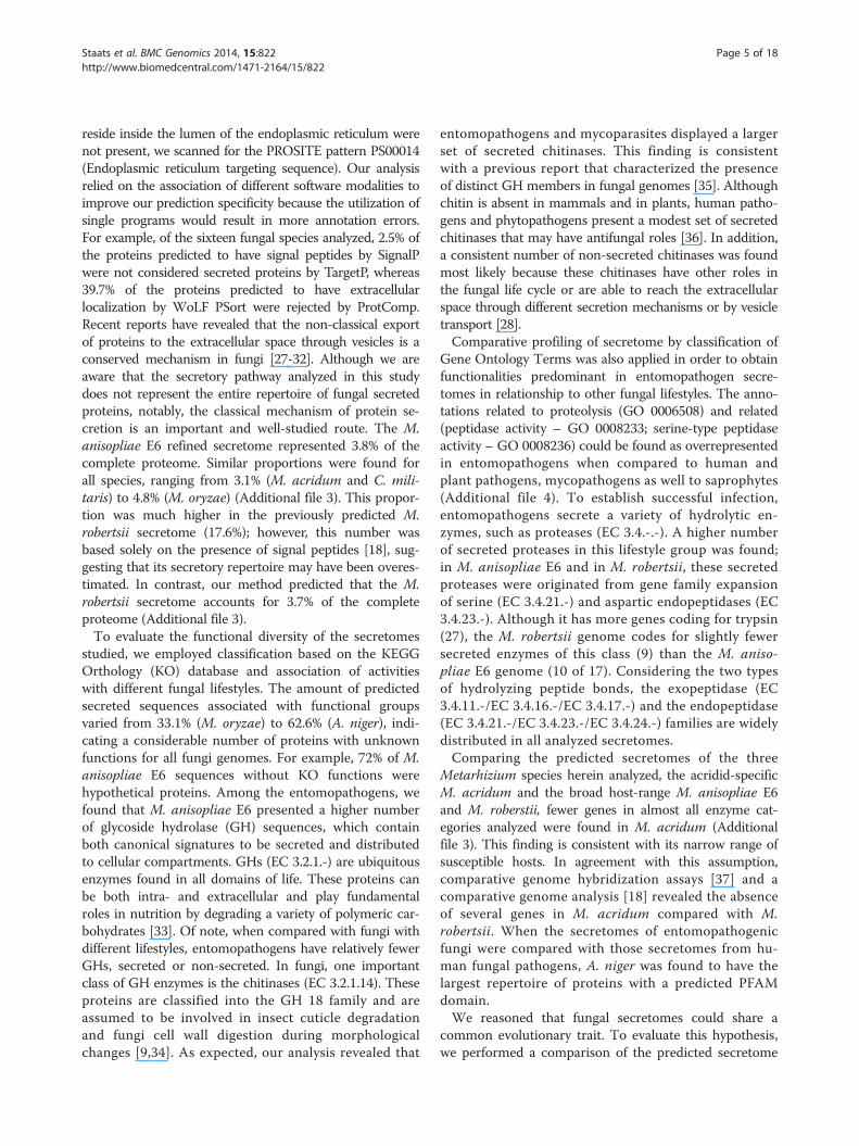
Phylogenomic analysesAs the content of the predicted secretomes change ac-cording to the fungal species analyzed, homology rela-tions were established among M. anisopliae E6 and a setof fifteen previously cited fungi to evaluate and to com-pare the evolutionary patterns of the M. anisopliae gen-ome and secretome. In addition a correlation with thetranscriptomic data was conducted. All 1:1 ortholog se-quences representing different CDSs were concatenated,leading to an aligned file containing 1,947,162 aminoacid residues. As expected, the Metarhizium, Trichoderma,
Fusarium, and Aspergillus genera formed monophyleticclusters [38,67-70]. The genera Neurospora and Aspergillusclustered together, with high bootstrap support. Addition-ally, Metarhizium and Trichoderma formed a statisticallywell-supported clade in the supermatrix and supertreeapproaches applied to the shared proteome, which wasformed by orthologous proteins only (Figure 6A). Whenconsidering the major clades obtained using the max-imum likelihood (Figure 6A) and the distance phylogen-etic methods (Additional file 12), the trees architecturesdid not changed significantly.Some fungal genera, including Trichoderma and Metar-
hizium, evolved a variety of nutrition strategies. For in-stance, some Trichoderma species are mycotrophic and,therefore, can grow on living fungi in a process known asmycoparasitism, whereas other species are saprotrophic[71]. Metarhizium species have already been isolated fromdiverse habitats, primarily from dead arthropod carcassesbut also from mycorrhiza and as endophytic [72]. Theshared proteome also indicates that the plant pathogensF. graminearum, F. oxysporum and N. haematococca[52,68,73] are related to the Metarhizium clade. Wyrebekand coworkers [54] have proposed that Metarhizium spe-cies show plant-specific rhizosphere associations withina habitat. Moreover, M. robertsii was also shown to bean endophyte that stimulates root development [53],thus making the direct transfer of nutrients from fungalto plant cells possible [74-76]. Thus, the phylogenomicanalysis presented in this study reveals that determiningthe life-style of M. anisopliae is far more complex thanthe global set of genes can predict.The phylogenomic analysis also indicated that M. aniso-
pliae E6 and M. anisopliae ARSEF 23, now identified asM. robertsii [18], share a high number of orthologs. Thesespecies formed a statistically well-supported clade, withM. acridum basal to these species, corroborating the re-sults of Bischoff and coworkers [17]. Notably, M. robertsiiis morphologically indistinguishable from M. anisopliae.Therefore, phylogenetic analysis is critical for establishingthe identity of these fungi.A phylogenetic analysis was conducted for 212 ortholog-
groups representing all proteins that had been identifiedas putative constituents of the secretome. The evolutionof the secretome indicated an important difference whencompared with the evolutionary history of these fungibased on their shared proteome. There is a closer evolu-tionary relation between the Metarhizium clade andthat formed by the genera Beauveria and Cordyceps(Figure 6B). Consequently, most proteins secreted byMetarhizium species are similar to those proteins fromthe Beauveria and Cordyceps genera. These proteins,some of which are important to the infection process,have a pattern of evolution that is extremely similar infungi with similar hosts.
Figure 6 Fungal evolutionary history obtained by using a phylogenomic approach. NJ methods were employed to all orthologs identifiedin the fungal genomes (A). Nearest neighbor interchange method with a neighbor-joining (NJ) tree calculated from average consensus distanceswere employed to all orthologs identified in the fungal secretomes (B). The percentage of replicate trees in which the associated taxa clusteredtogether in the bootstrap test (1,000 replicates for genomic and 100 for secretomic analysis) is shown next to the branches. The scale barrepresents substitutions of amino acids per site.
Staats et al. BMC Genomics 2014, 15:822 Page 12 of 18http://www.biomedcentral.com/1471-2164/15/822

Staats et al. BMC Genomics 2014, 15:822 Page 13 of 18http://www.biomedcentral.com/1471-2164/15/822
To identify the proteins responsible for the distinctiveevolutionary pattern of the secretome, all individual phy-logenies had their topologies compared with the topologyof the phylogenomic tree. At least one Metarhizium andone Trichoderma sequence, as well as one Cordyceps orBeauveria sequence, had to be present in a specific secre-tome ortholog file to be included in the analysis. Finally,37 secreted proteins were identified as being potentiallyresponsible for the different pattern of evolution of thesecretome when compared with the evolution of the or-ganisms represented by the phylogenomic tree. These pro-teins may explain why fungi with similar hosts clustertogether. Thirty four (92%) of those genes are expressed,and 16 (43%) of those genes are differentially expressedduring early cuticle infection (I-48 h) and/or during latecuticle infection (I-144 h) (Additional file 13). The bio-logical significance of these findings requires further in-vestigation. Nevertheless, our results show that specificsecreted proteins from Metarhizium, Beauveria and Cor-dyceps species, have an evolutionary history that pointsfor their adaptability to host infection.Using either proteome or secretome data, M. oryzae
and N. crassa form a basal cluster with high bootstrapsupport. These species belong to the distinct ordersMagnaporthales and Sordariales, respectively, and ourresults corroborate a previous study that noted theirevolutionary relation [77]. The Aspergillus genus, whichbelongs to the Eurotiomycetes order, clusters with theM. oryzae and N. crassa clades and, together, representsthe most basal clade in our study. The operational taxo-nomic units belonging to Hypocreales also form a clus-ter. These results indicate that the orthologs identifiedfor the proteome and for the secretome have an evolu-tionary pattern that is consistent with the actual taxo-nomic classification of fungi.
ConclusionsThe M. anisopliae E6 genome and expression profilinganalyses provide insights into the molecular mechanismsfor adapting to the distinct lifestyles of this entomo-pathogenic fungus. The comparative analyses presentedin this study reveal that Metarhizium spp. genomes har-bor a complex set of genes coding for secreted proteins.Such secreted proteins appear to have been selected andmaintained in the genome to cope with the distinct life-styles presented by Metarhizium spp., which can be eitherentomopathogen-, endophytic- or rhizosphere-associated.The transcriptome profiling of M. anisopliae exposed toinfection-mimicking conditions compared with laboratorygrowth conditions showed many genes that were differen-tially expressed. Among these genes, many genes codingfor secreted proteins could be found, which could repre-sent M. anisopliae virulence determinants. Our resultsoffer selected sequences for further characterization of
secreted proteins with potential roles in the M. anisopliaeinfectious process.
MethodsSample collection and DNA extractionM. anisopliae var. anisopliae strain E6 was isolated fromDeois flavopicta [11] collected in Espírito Santo State,Brazil. This strain was incubated at 28°C for 48 h in CM(Cove’s Medium) liquid medium [78] for the subsequentDNA isolation, as previously described [23]. The gen-omic DNA was extracted from the mycelium cultivatedin CM and was further purified using a DNeasy PlantMini Kit (QIAGEN, Hilden, Germany). The quality of theisolated genomic DNA was assessed spectrophotometrically.
Genome sequencing, assembly and annotationTwo shotgun (SG) and one long paired-end (LPE) librarieswere constructed using approximately 5 μg of DNA each.The library construction, titration, emulsion PCR and se-quencing steps were performed according to the manufac-turer’s protocol without modifications. SG libraries weresequenced using GS FLX Titanium chemistry (454-Roche,Brandford, CT, USA). One of the libraries was sequencedin one region of a two-region PicoTiterPlate (PTP) andthe other in both regions of a two-region PTP. The LPE li-brary was sequenced using GS FLX standard chemistry(454-Roche, Brandford, CT, USA) in both regions of atwo-region PTP.Replicates [79] software was used to identify and elimin-
ate the artificially replicated sequences produced duringthe 454-based pyrosequencing. Newbler Assembler ver-sion 2.8 and WGS-CA 7.0 software were used to performthe assembly procedures. Minimus2 software [80] was ap-plied to obtain a consensus assembly. The remainder gapswere filled using Consed software [81].All contig sequences were analyzed and functionally
annotated using the System for Automated Bacterial In-tegrated Annotation (SABIA) [82] altered to annotateeukaryotic genomes by the use of AUGUSTUS [83]. Theautomatic annotation criteria for assigning an ORF as“valid” included ORFs with BLASTp hits on KEGG,NCBI-nr or UniProtKB/Swiss-Prot databases, respect-ively; subject and query coverage ≥60%; and positives≥60%. ORFs with no BLASTp hits found on NCBI-nr,KEGG, UniProtKB/Swiss-Prot, TCDB and Interpro data-bases or not included in the criteria above were definedas “hypothetical” ORFs.
Selection of refined secretomes and functional analysesThe M. anisopliae strain E6 predicted proteome, as wellas those proteomes from fifteen other filamentous fungi(Aspergillus fumigatus Af293, Aspergillus nidulans FGSCA4, Aspergillus niger CBS 513.88, Beauveria bassianaARSEF 2860, Cordyceps militaris CM01, Fusarium
Staats et al. BMC Genomics 2014, 15:822 Page 14 of 18http://www.biomedcentral.com/1471-2164/15/822
graminearum PH-1, Fusarium oxysporum f. sp. cubenserace 1, Metarhizium anisopliae ARSEF 23, Metarhi-zium acridum CQMa 102, Magnaporthe oryzae 70-15,Neurospora crassa OR74A, Nectria haematococcampVI 77-13-4, Trichoderma atroviride IMI 206040, Tri-choderma reesei QM6a, and Trichoderma virens Gv29-8)were downloaded from the NCBI genome database(http://www.ncbi.nlm.nih.gov/genome/) and consideredfor in silico secretome analysis.The prediction of all refined secretomes was based on
the procedure described by Brown and coworkers [26]for the plant pathogen Fusarium graminearum. An auto-matic pipeline was developed using PERL scripts andthe MySQL database. Initially, all proteins were screenedto remove sequences without an initial methionine andwith a mature peptide size of less than 20 amino acids.To detect a signal peptide, proteins with predictions byboth SignalP v4.1 [84] (D-score = Y; http://www.cbs.dtu.dk/services/SignalP/) and TargetP v1.1 [85] (LOC = S;http://www.cbs.dtu.dk/services/TargetP/) tools were se-lected. These proteins were subsequently scanned forthe presence of transmembrane regions using thehidden Markov model topology predictor TMHMM[86] (TMHMM v2.0; http://www.cbs.dtu.dk/services/TMHMM/), and we kept those proteins with 0 or 1TM when a single TM was in the first 60 amino acidsin the N-terminal portion. This filtering was necessaryas the large majority of secreted proteins spam at theamino terminus of the protein at most 1 TM region,which resembles the signal peptide. PredGPI [87](FRate ≤ 0.005) was used to predict GPI-anchors (http://gpcr2.biocomp.unibo.it/predgpi/pred.htm). ProtCompv9.1 (with LocDB and PotLocDB, proteins predicted as se-creted by both NNets and Integral predictions; http://www.softberry.com) and WoLF PSort v0.2 [88] softwarewere combined to infer the protein localization for thefungi studied (Extr ≥ 17). Finally, a PROSITE scan [89]was used to remove sequences associated with the patternPS00014 (Endoplasmic reticulum targeting sequence),yielding the refined secretome with GPI-anchored proteins(GPI-Ps).To assign a predicted function, a BLASTp search (e-value
1e-5) was conducted with selected proteins against theKEGG Orthology [90] database (KO). To avoid spuriousdomain alignments, we discarded BLAST results forwhich the alignment size divided by subject size (coverage)was below 50%. Those proteins without significant hitswere analyzed with a PFAM-A database using pfam_scan.plscript [91]. The Pathogen-Host Interactions (PHI) database[92] (http://www.phi-base.org/) was used to search fororthologous proteins in M. anisopliae using an e-value of10-5 and ≥ 50% coverage as criteria. Then, the matcheswere filtered, and only proteins that shared over 70% iden-tity with M. anisopliae predicted proteins were included.
Of these proteins, proteins exhibiting a “loss of pathogen-icity or reduced virulence” as phenotype characteristics inthe mutant strains were analyzed.Statistical analyses were conducted using the R statis-
tical package. One-tailed proportion test (prop.test) wasused for evaluate secretome gene duplications. Statisticalanalysis for the comparison of GO enriched terms wasconducted with Blast2GO [93].
Transcriptome analysisRhipicephalus microplus cuticles were sterilized and usedas the sole nutrient source for M. anisopliae E6 growthand development. A spore suspension (5 × 10⁶ spores perml) was used to inoculate the cuticles by immersion for30 sec. The inoculated cuticles were disposed over 1%water agar plates and maintained for 48 h and 144 h at28°C. Each of the two biological replicates consisted of apool of five plates containing mycelium growth over thecuticles. The comparative control condition was con-ducted on 100 ml liquid complete medium for 48 h at28°C. The resulting fungal growth over the host cuticleand on liquid medium was ground to a powder in liquidnitrogen, and the total RNA was extracted using TRIzol®Reagent (Life Technologies, CA, USA) following themanufacturer’s instructions. The total isolated RNA wassubjected to DNase treatment using RNase-free DNaseI (Thermo Scientific, MA, USA). Large ribosomal RNAmolecules were selectively depleted from the total RNAusing a RiboMinus™ Eukaryote Kit for RNA-Seq (LifeTechnologies, CA, USA), and mRNA was concentratedand purified on a RiboMinus™ Concentration Module(Life Technologies, CA, USA).RNA-Seq was conducted with Ion Torrent technology
in an Ion Proton System. FastQC v0.10.0 [http://www.bio-informatics.babraham.ac.uk/projects/fastqc/] software wasused for a reads quality check, and the FASTX-Toolkitv0.0.13 (http://hannonlab.cshl.edu/fastx_toolkit/) was usedfor trimming. Reads smaller than 30 nucleotides were dis-carded. The remaining reads were mapped to the M. ani-sopliae genome using the spliced read mapper Tophatv2.0.10 [94] with default parameters. HTSeq v0.5.4p5(http://www-huber.embl.de/users/anders/HTSeq/doc/overview.html) was used to count reads aligned to only oneposition in protein coding regions, and the edgeR packagev3.4.0 [55] was used to assess for differential gene expres-sion, with a 5% false discovery rate (FDR ≤ 0.05) and withstringent log fold variation (logFC) ≥ or ≤ -1. Two pairwisecomparisons were performed for periods C-48 h × I-48 hand I-48 h × I-144 h. InterProScan was used to assign moregeneral superfamily functional categories v1.73 [57].
Phylogenetic and phylogenomic analysesOrthoMCL v2.0.8 software [95] was used with defaultparameters to identify orthologs and paralogs among the
Staats et al. BMC Genomics 2014, 15:822 Page 15 of 18http://www.biomedcentral.com/1471-2164/15/822
complete proteomes of all sixteen studied organisms. APERL script was developed to select only 1:1 ortholo-gous sequences from the OrthoMCL output such thatonly a single gene copy was selected from each predictedproteome. The multi-FASTA ortholog files of each proteinsequence were used as input for the multiple alignmentsusing CLUSTAL Omega algorithm [96] with default pa-rameters. Subsequently, SCaFos software [97] was usedto for the gene concatenation of the 2,684 alignmentfiles. Phylogenies involving the concatenated deducedamino acid sequences from all species were evaluatedthrough distance and probabilistic methods using thePHYLIP package [98], MEGA 5.2 Computing Core [99]and TREE-PUZZLE [100] software.Initially, multiple 100 bootstrapped data sets were gen-
erated by the Seqboot program of the PHYLIP package.Then, these data sets were submitted to ProtDist soft-ware analysis to compute a distance matrix under theJTT (Jones-Taylor-Thornton) model of amino acid re-placement. The Neighbor software applied the neighbor-joining (NJ) method [101] to the resulting multiple datasets, building trees through the successive clustering oflineages. A consensus tree was obtained using the Con-sense program of the PHYLIP package. Three differentdistance matrices (p-distance, Poisson, and JTT) wereevaluated using the MEGA 5.2 Computing Core, withthe complete deletion and pairwise deletion options forthe treatment of gaps. The bootstrap test for phylogenywas performed using 1,000 repetitions.The quartet-puzzling [102] search algorithm imple-
mented by TREE-PUZZLE was used to reconstruct phylo-genetic trees according to the maximum likelihood (ML)approach. The JTT model of amino acid substitution wasapplied. The quartet-puzzling tree topology was based on1,000 puzzling steps. The consensus tree was constructedbased on a 50% majority rule consensus. The TreeViewprogram [103] and MEGA 5 software were used tovisualize and to edit the resulting phylogenies.In total, 212 ortholog files of the secretome were sub-
mitted to phylogenetic analysis using distance methodsimplemented by MEGA software. The neighbor-joiningalgorithm, with pairwise deletion of gaps, was applied tothe set of data. The p-distance, poisson and JTT matri-ces were evaluated. The bootstrap test of phylogeny wasperformed using 1,000 repetitions.The resulting individual gene phylogenies were submit-
ted to the CLANN software [104] to generate the supertreeusing a heuristic search of the supertree space for identify-ing the best tree. The nearest neighbor-interchange andsubtree pruning and regrafting methods were testedstarting with a neighbor-joining tree calculated from theaverage consensus distances. A bootstrap analysis wasperformed using 100 replicates. Then, these individualphylogenies were compared with the phylogenomic tree
obtained from the 2,684 alignment files, which was con-sidered to represent the evolutionary history of the ana-lyzed fungi. The aim was to evaluate which secretomeproteins have an evolutionary pattern that is not com-patible with the organism’s evolution.During the submission process of this work, the genome
sequence from another strain of M. anisopliae (Ma69)was accepted for publication [105]. Altogether, thegenomic sequences of Metarhizium spp. will allow adeeper analysis of ancient mechanisms of virulence byentomopathogens.
Sequence submissionThe M. anisopliae E6 genome sequence is deposited inNCBI under Accession JNNZ00000000. RNAseq readswere deposited in NCBI SRA under Bioproject accessionPRJNA257269.
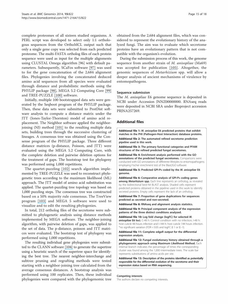
Additional files
Additional file 1: M. anisopliae E6 predicted proteins that exhibitmatches in the PHI (Pathogen-Host Interaction) database proteins.
Additional file 2: The automated refined secretome predictionpipeline used in this work.
Additional file 3: The primary functional categories and PFAMstructures of the refined predicted fungal secretomes.
Additional file 4: Enrichment analysis performed with the GOannotations of the predicted fungal secretomes. Comparisons wereconducted with GO annotations of differente lifestyles to entomopathogensemploying Fischer enrichment test of the Blast2GO software.
Additional file 5: Predicted GPI-Ps coded by the M. anisopliae E6genome.
Additional file 6: Comparative analysis of GPI-Ps coding genesamong Metarhizium spp. Each line represents an ortholog, as revealedby the bidirectional best-hit BLAST analysis. Shaded cells representpredicted proteins obtained in the pipeline used in this work to identifysecreted proteins. Empty cells represent the lack of an ortholog.
Additional file 7: Proportion of gene duplications for sequencespredicted as secreted and non-secreted.
Additional file 8: RNAseq and alignment analysis statistics.
Additional file 9: Principal component analysis of the expressionpatterns of the three distinct conditions analyzed.
Additional file 10: Log fold change (logFC) for selected M.anisopliae E6 loci. C-48 h: Control condition with no infection; I-48 h:host cuticle 48 hours infection and I-144 h: host cuticle 144 hours infection.*no significant variation (FDR < 0.05 and logFC≥ 1 or≤ -1).
Additional file 11: Complete edgeR output for the differentialexpression analysis.
Additional file 12: Fungal evolutionary history obtained through aphylogenomic approach using Maximum Likelihood Method. Eachinternal branch indicates the percentage of times the correspondingcluster was found among the 1,000 intermediate trees. The scale barrepresents substitutions of amino acids per site.
Additional file 13: Description of the proteins identified as potentiallyresponsible for the differential evolution of the secretome and theirexpression status based on RNA sequencing.
Competing interestsThe authors declare no competing interests.

Staats et al. BMC Genomics 2014, 15:822 Page 16 of 18http://www.biomedcentral.com/1471-2164/15/822
Authors’ contributionsConceived and designed the experiments: CCS, MHV, ATRV, and AS.Annotation curation: CCS, AJ, RLMG, CET, GLM, JTB, LGPA, FCA, ALG, NS,RLAP, LB, ML, LS, WOBS, CPS, TRS, EOS, and LK. Comparative analyses: CCS,AJ, RLMG, and CET. Secretome analysis: CCS, AJ, RLMG, and ESO.Transcriptome analysis: CCS, AJ, RLMG, and GLM. Phylogenomic analyses: CETand FCA. Contributed reagents/materials/analysis tools: MHV, ATRV, and AS.Wrote the article: CCS, AJ, RLMG, CET, ESO, and AS. All authors read andapproved the final manuscript.
AcknowledgmentsWe would like to thank the staff of LNCC for support. This work wassupported by the Conselho Nacional de Desenvolvimento Científico eTecnológico (CNPq), Programa de Aperfeiçoamento Pessoal de NívelSuperior (CAPES), Fundação de Amparo a Pesquisa do estado do Rio Grandedo Sul (FAPERGS) and Fundação Carlos Chagas Filho de Amparo à Pesquisado Estado do Rio de Janeiro (FAPERJ).
Author details1Centro de Biotecnologia, Universidade Federal do Rio Grande do Sul(UFRGS), P. O. Box 15005, Porto Alegre, RS CEP 91501-970, Brazil. 2LaboratórioNacional de Computação Científica (LNCC), Av. Getúlio Vargas, 333,Petrópolis, RJ, Brazil.
Received: 4 June 2014 Accepted: 29 August 2014Published: 29 September 2014
References1. Mora C, Tittensor DP, Adl S, Simpson AG, Worm B: How many species are
there on Earth and in the ocean? PLoS Biol 2011, 9:e1001127.2. Blackwell M: The Fungi: 1, 2, 3 … 5.1 million species? Am J Bot 2011,
98:426–438.3. Girard V, Dieryckx C, Job C, Job D: Secretomes: the fungal strike force.
Proteomics 2013, 13:597–608.4. Basset Y, Cizek L, Cuénoud P, Didham RK, Guilhaumon F, Missa O, Novotny V,
Ødegaard F, Roslin T, Schmidl J, Tishechkin AK, Winchester NN, Roubik DW,Aberlenc H-P, Bail J, Barrios H, Bridle JR, Castaño-Meneses G, Corbara B,Curletti G, da Rocha WD, De Bakker D, Delabie JHC, Dejean A, Fagan LL,Floren A, Kitching RL, Medianero E, Miller SE, de Oliveira EG, et al: Arthropoddiversity in a tropical forest. Science 2012, 338:1481–1484.
5. Hasan S, Ahmad A, Purwar A, Khan N, Kundan R, Gupta G: Production ofextracellular enzymes in the entomopathogenic fungus Verticilliumlecanii. Bioinformation 2013, 9:238–242.
6. da Silva WO B, Santi L, Correa AP, Silva LA, Bresciani FR, Schrank A, Vainstein MH:The entomopathogen Metarhizium anisopliae can modulate the secretion oflipolytic enzymes in response to different substrates including componentsof arthropod cuticle. Fungal Biol 2010, 114:911–916.
7. Santi L, Silva WO, Pinto AF, Schrank A, Vainstein MH: Metarhizium anisopliaehost-pathogen interaction: differential immunoproteomics reveals proteinsinvolved in the infection process of arthropods. Fungal Biol 2010,114:312–319.
8. Murad AM, Noronha EF, Miller RN, Costa FT, Pereira CD, Mehta A, Caldas RA,Franco OL: Proteomic analysis of Metarhizium anisopliae secretion in thepresence of the insect pest Callosobruchus maculatus. Microbiology 2008,154:3766–3774.
9. da Silva MV, Santi L, Staats CC, da Costa AM, Colodel EM, Driemeier D,Vainstein MH, Schrank A: Cuticle-induced endo/exoacting chitinaseCHIT30 from Metarhizium anisopliae is encoded by an ortholog of thechi3 gene. Res Microbiol 2005, 156:382–392.
10. Barreto CC, Staats CC, Schrank A, Vainstein MH: Distribution ofchitinases in the entomopathogen Metarhizium anisopliae and effectof N-acetylglucosamine in protein secretion. Curr Microbiol 2004,48:102–107.
11. Arruda W, Lubeck I, Schrank A, Vainstein MH: Morphological alterations ofMetarhizium anisopliae during penetration of Boophilus microplus ticks.Exp Appl Acarol 2005, 37:231–244.
12. Schrank A, Vainstein MH: Metarhizium anisopliae enzymes and toxins.Toxicon 2010, 56:1267–1274.
13. Butt TM, Greenfield BP, Greig C, Maffeis TG, Taylor JW, Piasecka J, Dudley E,Abdulla A, Dubovskiy IM, Garrido-Jurado I, Quesada-Moraga E, Penny MW,
Eastwood DC: Metarhizium anisopliae pathogenesis of mosquito larvae: averdict of accidental death. PLoS One 2013, 8:e81686.
14. Freimoser FM, Hu G, St Leger RJ: Variation in gene expression patterns asthe insect pathogen Metarhizium anisopliae adapts to different hostcuticles or nutrient deprivation in vitro. Microbiology 2005, 151:361–371.
15. Fernandes EK, Bittencourt VR, Roberts DW: Perspectives on the potential ofentomopathogenic fungi in biological control of ticks. Exp Parasitol 2012,130:300–305.
16. Faria MR, Wraight SP: Mycoinsecticides and Mycoacaricides: acomprehensive list with worldwide coverage and internationalclassification of formulation types. Biological Control 2007, 43:237–256.
17. Bischoff JF, Rehner SA, Humber RA: A multilocus phylogeny of theMetarhizium anisopliae lineage. Mycologia 2009, 101:512–530.
18. Gao Q, Jin K, Ying SH, Zhang Y, Xiao G, Shang Y, Duan Z, Hu X, Xie XQ,Zhou G, Peng G, Luo Z, Huang W, Wang B, Fang W, Wang S, Zhong Y, MaLJ, St Leger RJ, Zhao GP, Pei Y, Feng MG, Xia Y, Wang C: Genomesequencing and comparative transcriptomics of the modelentomopathogenic fungi Metarhizium anisopliae and M. acridum. PLoSGenet 2011, 7:e1001264.
19. Xiao G, Ying SH, Zheng P, Wang ZL, Zhang S, Xie XQ, Shang Y, St Leger RJ,Zhao GP, Wang C, Feng MG: Genomic perspectives on the evolution offungal entomopathogenicity in Beauveria bassiana. Sci Rep 2012, 2:483.
20. Schattner P, Brooks AN, Lowe TM: The tRNAscan-SE, snoscan and snoGPSweb servers for the detection of tRNAs and snoRNAs. Nucleic Acids Res2005, 33:W686–W689.
21. Laslett D, Canback B: ARAGORN, a program to detect tRNA genes andtmRNA genes in nucleotide sequences. Nucleic Acids Res 2004, 32:11–16.
22. Zheng P, Xia Y, Xiao G, Xiong C, Hu X, Zhang S, Zheng H, Huang Y, Zhou Y,Wang S, Zhao GP, Liu X, St Leger RJ, Wang C: Genome sequence of theinsect pathogenic fungus Cordyceps militaris, a valued traditionalChinese medicine. Genome Biol 2011, 12:R116.
23. Boldo JT, Junges A, do Amaral KB, Staats CC, Vainstein MH, Schrank A:Endochitinase CHI2 of the biocontrol fungus Metarhizium anisopliaeaffects its virulence toward the cotton stainer bug Dysdercus peruvianus.Curr Genet 2009, 55:551–560.
24. Fang W, Leng B, Xiao Y, Jin K, Ma J, Fan Y, Feng J, Yang X, Zhang Y, Pei Y:Cloning of Beauveria bassiana chitinase gene Bbchit1 and its applicationto improve fungal strain virulence. Appl Environ Microbiol 2005,71:363–370.
25. Zhou G, Wang J, Qiu L, Feng MG: A Group III histidine kinase (mhk1)upstream of high-osmolarity glycerol pathway regulates sporulation,multi-stress tolerance and virulence of Metarhizium robertsii, a fungalentomopathogen. Environ Microbiol 2012, 14:817–829.
26. Brown NA, Antoniw J, Hammond-Kosack KE: The predicted secretome of theplant pathogenic fungus Fusarium graminearum: a refined comparativeanalysis. PLoS One 2012, 7:e33731.
27. Oliveira DL, Rizzo J, Joffe LS, Godinho RM, Rodrigues ML: Where do theycome from and where do they go: candidates for regulatingextracellular vesicle formation in fungi. Int J Mol Sci 2013, 14:9581–9603.
28. Rodrigues ML, Nakayasu ES, Almeida IC, Nimrichter L: The impact ofproteomics on the understanding of functions and biogenesis of fungalextracellular vesicles. J Proteomics 2014, 97:177–186.
29. Harding CV, Heuser JE, Stahl PD: Exosomes: looking back three decadesand into the future. J Cell Biol 2013, 200:367–371.
30. Vallejo MC, Nakayasu ES, Matsuo AL, Sobreira TJ, Longo LV, Ganiko L,Almeida IC, Puccia R: Vesicle and vesicle-free extracellular proteome ofParacoccidioides brasiliensis: comparative analysis with other pathogenicfungi. J Proteome Res 2012, 11:1676–1685.
31. Rodrigues ML, Nosanchuk JD, Schrank A, Vainstein MH, Casadevall A,Nimrichter L: Vesicular transport systems in fungi. Future Microbiol 2011,6:1371–1381.
32. Rodrigues ML, Djordjevic JT: Unravelling secretion in Cryptococcusneoformans: more than one way to skin a cat. Mycopathologia 2012,173:407–418.
33. Hansen SF, Bettler E, Rinnan A, Engelsen SB, Breton C: Exploring genomesfor glycosyltransferases. Mol Biosyst 2010, 6:1773–1781.
34. Boldo JT, do Amaral KB, Junges A, Pinto PM, Staats CC, Vainstein MH,Schrank A: Evidence of alternative splicing of the chi2 chitinase genefrom Metarhizium anisopliae. Gene 2010, 462:1–7.
35. Gruber S, Seidl-Seiboth V: Self versus non-self: fungal cell wall degradationin Trichoderma. Microbiology 2012, 158:26–34.

Staats et al. BMC Genomics 2014, 15:822 Page 17 of 18http://www.biomedcentral.com/1471-2164/15/822
36. Brzezinska MS, Jankiewicz U: Production of antifungal chitinase byAspergillus niger LOCK 62 and its potential role in the biological control.Curr Microbiol 2012, 65:666–672.
37. Wang S, Leclerque A, Pava-Ripoll M, Fang W, St Leger RJ: Comparative genomicsusing microarrays reveals divergence and loss of virulence-associatedgenes in host-specific strains of the insect pathogen Metarhizium anisopliae.Eukaryot Cell 2009, 8:888–898.
38. Martinez D, Berka RM, Henrissat B, Saloheimo M, Arvas M, Baker SE,Chapman J, Chertkov O, Coutinho PM, Cullen D, Danchin EG, Grigoriev IV,Harris P, Jackson M, Kubicek CP, Han CS, Ho I, Larrondo LF, de Leon AL,Magnuson JK, Merino S, Misra M, Nelson B, Putnam N, Robbertse B, Salamov AA,Schmoll M, Terry A, Thayer N, Westerholm-Parvinen A, et al: Genomesequencing and analysis of the biomass-degrading fungus Trichodermareesei (syn. Hypocrea jecorina). Nat Biotechnol 2008, 26:553–560.
39. Lai JS, Cheng CW, Sung TY, Hsu WL: Computational comparative study oftuberculosis proteomes using a model learned from signal peptidestructures. PLoS One 2012, 7:e35018.
40. Braaksma M, Martens-Uzunova ES, Punt PJ, Schaap PJ: An inventory of theAspergillus niger secretome by combining in silico predictions withshotgun proteomics data. BMC Genomics 2010, 11:584.
41. Abaev I, Foster-Frey J, Korobova O, Shishkova N, Kiseleva N, Kopylov P,Pryamchuk S, Schmelcher M, Becker SC, Donovan DM: Staphylococcalphage 2638A endolysin is lytic for Staphylococcus aureus and harborsan inter-lytic-domain secondary translational start site. Appl MicrobiolBiotechnol 2013, 97:3449–3456.
42. Helsens K, Van Damme P, Degroeve S, Martens L, Arnesen T, Vandekerckhove J,Gevaert K: Bioinformatics analysis of a Saccharomyces cerevisiae N-terminalproteome provides evidence of alternative translation initiation andpost-translational N-terminal acetylation. J Proteome Res 2011,10:3578–3589.
43. Smollett KL, Fivian-Hughes AS, Smith JE, Chang A, Rao T, Davis EO: Experimentaldetermination of translational start sites resolves uncertainties in genomicopen reading frame predictions - application to Mycobacterium tuberculosis.Microbiology 2009, 155:186–197.
44. Chabregas SM, Luche DD, Van Sluys MA, Menck CF, Silva-Filho MC: Differentialusage of two in-frame translational start codons regulates subcellularlocalization of Arabidopsis thaliana THI1. J Cell Sci 2003, 116:285–291.
45. Fujita M, Kinoshita T: GPI-anchor remodeling: potential functions ofGPI-anchors in intracellular trafficking and membrane dynamics.Biochim Biophys Acta 1821, 2012:1050–1058.
46. Pittet M, Conzelmann A: Biosynthesis and function of GPI proteins in theyeast Saccharomyces cerevisiae. Biochim Biophys Acta 2007, 1771:405–420.
47. Maddi A, Fu C, Free SJ: The Neurospora crassa dfg5 and dcw1 genes encodealpha-1,6-mannanases that function in the incorporation of glycoproteinsinto the cell wall. PLoS One 2012, 7:e38872.
48. Choi CJ, Ju HJ, Park BH, Qin R, Jahng KY, Han DM, Chae KS: Isolationand characterization of the Aspergillus nidulans eglC gene encodinga putative beta-1,3-endoglucanase. Fungal Genet Biol 2005,42:590–600.
49. Chen Y, Zhu J, Ying SH, Feng MG: The GPI-anchored protein Ecm33 is vitalfor conidiation, cell wall integrity, and multi-stress tolerance of twofilamentous entomopathogens but not for virulence. Appl MicrobiolBiotechnol 2014, 98:5517–5529.
50. Zhang J: Evolution by gene duplication: an update. Trends Ecol Evol 2003,18:292–298.
51. Cohen-Gihon I, Sharan R, Nussinov R: Processes of fungal proteomeevolution and gain of function: gene duplication and domainrearrangement. Phys Biol 2011, 8:035009.
52. Coleman JJ, Rounsley SD, Rodriguez-Carres M, Kuo A, Wasmann CC,Grimwood J, Schmutz J, Taga M, White GJ, Zhou S, Schwartz DC, Freitag M,Ma LJ, Danchin EG, Henrissat B, Coutinho PM, Nelson DR, Straney D, NapoliCA, Barker BM, Gribskov M, Rep M, Kroken S, Molnar I, Rensing C, Kennell JC,Zamora J, Farman ML, Selker EU, Salamov A, et al: The genome of Nectriahaematococca: contribution of supernumerary chromosomes to geneexpansion. PLoS Genet 2009, 5:e1000618.
53. Sasan RK, Bidochka MJ: The insect-pathogenic fungus Metarhiziumrobertsii (Clavicipitaceae) is also an endophyte that stimulates plant rootdevelopment. Am J Bot 2012, 99:101–107.
54. Wyrebek M, Huber C, Sasan RK, Bidochka MJ: Three sympatrically occurringspecies of Metarhizium show plant rhizosphere specificity. Microbiology2011, 157:2904–2911.
55. Robinson MD, McCarthy DJ, Smyth GK: edgeR: a Bioconductor package fordifferential expression analysis of digital gene expression data.Bioinformatics 2010, 26:139–140.
56. Guo Y, Li CI, Ye F, Shyr Y: Evaluation of read count based RNAseq analysismethods. BMC Genomics 2013, 14(8):S2.
57. de Lima Morais DA, Fang H, Rackham OJ, Wilson D, Pethica R, Chothia C,Gough J: SUPERFAMILY 1.75 including a domain-centric gene ontologymethod. Nucleic Acids Res 2011, 39:D427–D434.
58. Bagga S, Hu G, Screen SE, St Leger RJ: Reconstructing the diversification ofsubtilisins in the pathogenic fungus Metarhizium anisopliae. Gene 2004,324:159–169.
59. Irie T, Matsumura H, Terauchi R, Saitoh H: Serial Analysis of GeneExpression (SAGE) of Magnaporthe grisea: genes involved inappressorium formation. Mol Genet Genomics 2003, 270:181–189.
60. Laronde-Leblanc N, Guszczynski T, Copeland T, Wlodawer A: Structure andactivity of the atypical serine kinase Rio1. FEBS J 2005, 272:3698–3713.
61. St Leger RJ, Joshi L, Bidochka MJ, Rizzo NW, Roberts DW: Biochemicalcharacterization and ultrastructural localization of two extracellulartrypsins produced by Metarhizium anisopliae in infected insect cuticles.Appl Environ Microbiol 1996, 62:1257–1264.
62. St Leger RJ, Joshi L, Roberts D: Ambient pH is a major determinant in theexpression of cuticle-degrading enzymes and hydrophobin byMetarhizium anisopliae. Appl Environ Microbiol 1998, 64:709–713.
63. Screen SE, St Leger RJ: Cloning, expression, and substrate specificity of afungal chymotrypsin. Evidence for lateral gene transfer from anactinomycete bacterium. J Biol Chem 2000, 275:6689–6694.
64. Lopez-Berges MS, Rispail N, Prados-Rosales RC, Di Pietro A: A nitrogenresponse pathway regulates virulence in plant pathogenic fungi: role ofTOR and the bZIP protein MeaB. Plant Signal Behav 2010, 5:1623–1625.
65. Sevim A, Donzelli BG, Wu D, Demirbag Z, Gibson DM, Turgeon BG:Hydrophobin genes of the entomopathogenic fungus, Metarhiziumbrunneum, are differentially expressed and corresponding mutants aredecreased in virulence. Curr Genet 2012, 58:79–92.
66. Garcia-Santamarina S, Boronat S, Calvo IA, Rodriguez-Gabriel M, Ayte J,Molina H, Hidalgo E: Is oxidized thioredoxin a major trigger for cysteineoxidation? Clues from a redox proteomics approach. Antioxid Redox Signal2013, 18:1549–1556.
67. Kubicek CP, Herrera-Estrella A, Seidl-Seiboth V, Martinez DA, Druzhinina IS,Thon M, Zeilinger S, Casas-Flores S, Horwitz BA, Mukherjee PK, Kredics L,Alcaraz LD, Aerts A, Antal Z, Atanasova L, Cervantes-Badillo MG, Challacombe J,Chertkov O, McCluskey K, Coulpier F, Deshpande N, von Dohren H, Ebbole DJ,Esquivel-Naranjo EU, Fekete E, Flipphi M, Glaser F, Gomez-Rodriguez EY,Gruber S, et al: Comparative genome sequence analysis underscoresmycoparasitism as the ancestral life style of Trichoderma. Genome Biol2011, 12:R40.
68. Srivastava SK, Huang X, Brar HK, Fakhoury AM, Bluhm BH, Bhattacharyya MK:The genome sequence of the fungal pathogen Fusarium virguliforme thatcauses sudden death syndrome in soybean. PLoS One 2014, 9:e81832.
69. Galagan JE, Calvo SE, Cuomo C, Ma LJ, Wortman JR, Batzoglou S, Lee SI,Basturkmen M, Spevak CC, Clutterbuck J, Kapitonov V, Jurka J, Scazzocchio C,Farman M, Butler J, Purcell S, Harris S, Braus GH, Draht O, Busch S, D'Enfert C,Bouchier C, Goldman GH, Bell-Pedersen D, Griffiths-Jones S, Doonan JH, Yu J,Vienken K, Pain A, Freitag M, et al: Sequencing of Aspergillus nidulans andcomparative analysis with A. fumigatus and A. oryzae. Nature 2005,438:1105–1115.
70. Cerqueira GC, Arnaud MB, Inglis DO, Skrzypek MS, Binkley G, Simison M,Miyasato SR, Binkley J, Orvis J, Shah P, Wymore F, Sherlock G, Wortman JR:The Aspergillus Genome Database: multispecies curation andincorporation of RNA-Seq data to improve structural gene annotations.Nucleic Acids Res 2014, 42:D705–D710.
71. Druzhinina IS, Seidl-Seiboth V, Herrera-Estrella A, Horwitz BA, Kenerley CM,Monte E, Mukherjee PK, Zeilinger S, Grigoriev IV, Kubicek CP: Trichoderma:the genomics of opportunistic success. Nat Rev Microbiol 2011, 9:749–759.
72. St Leger RJ, Joshi L, Roberts DW: Adaptation of proteases andcarbohydrates of saprophytic, phytopathogenic and entomopathogenicfungi to the requirements of their ecological niches. Microbiology 1997,143(Pt 6):1983–1992.
73. Cuomo CA, Guldener U, Xu JR, Trail F, Turgeon BG, Di Pietro A, Walton JD,Ma LJ, Baker SE, Rep M, Adam G, Antoniw J, Baldwin T, Calvo S, Chang YL,Decaprio D, Gale LR, Gnerre S, Goswami RS, Hammond-Kosack K, Harris LJ,Hilburn K, Kennell JC, Kroken S, Magnuson JK, Mannhaupt G, Mauceli E,

Staats et al. BMC Genomics 2014, 15:822 Page 18 of 18http://www.biomedcentral.com/1471-2164/15/822
Mewes HW, Mitterbauer R, Muehlbauer G, et al: The Fusarium graminearumgenome reveals a link between localized polymorphism and pathogenspecialization. Science 2007, 317:1400–1402.
74. Behie SW, Bidochka MJ: Ubiquity of insect-derived nitrogen transfer toplants by endophytic insect-pathogenic fungi: an additional branch ofthe soil nitrogen cycle. Appl Environ Microbiol 2014, 80:1553–1560.
75. Behie SW, Padilla-Guerrero IE, Bidochka MJ: Nutrient transfer to plants byphylogenetically diverse fungi suggests convergent evolutionary strat-egies in rhizospheric symbionts. Commun Integr Biol 2013, 6:e22321.
76. Behie SW, Zelisko PM, Bidochka MJ: Endophytic insect-parasitic fungitranslocate nitrogen directly from insects to plants. Science 2012,336:1576–1577.
77. Wang H, Xu Z, Gao L, Hao B: A fungal phylogeny based on 82 completegenomes using the composition vector method. BMC Evol Biol 2009,9:195.
78. Cove DJ: The induction and repression of nitrate reductase in the fungusAspergillus nidulans. Biochim Biophys Acta 1966, 113:51–56.
79. Gomez-Alvarez V, Teal TK, Schmidt TM: Systematic artifacts inmetagenomes from complex microbial communities. ISME J 2009,3:1314–1317.
80. Sommer DD, Delcher AL, Salzberg SL, Pop M: Minimus: a fast, lightweightgenome assembler. BMC Bioinformatics 2007, 8:64.
81. Gordon D, Green P: Consed: a graphical editor for next-generationsequencing. Bioinformatics 2013, 29:2936–2937.
82. Almeida LG, Paixao R, Souza RC, Costa GC, Barrientos FJ, Santos MT, Almeida DF,Vasconcelos AT: A System for Automated Bacterial (genome) IntegratedAnnotation–SABIA. Bioinformatics 2004, 20:2832–2833.
83. Stanke M, Steinkamp R, Waack S, Morgenstern B: AUGUSTUS: a web serverfor gene finding in eukaryotes. Nucleic Acids Res 2004, 32:W309–W312.
84. Petersen TN, Brunak S, von Heijne G, Nielsen H: SignalP 4.0: discriminatingsignal peptides from transmembrane regions. Nat Methods 2011, 8:785–786.
85. Emanuelsson O, Nielsen H, Brunak S, von Heijne G: Predicting subcellularlocalization of proteins based on their N-terminal amino acid sequence.J Mol Biol 2000, 300:1005–1016.
86. Krogh A, Larsson B, von Heijne G, Sonnhammer EL: Predicting transmembraneprotein topology with a hidden Markov model: application to completegenomes. J Mol Biol 2001, 305:567–580.
87. Pierleoni A, Martelli PL, Casadio R: PredGPI: a GPI-anchor predictor.BMC Bioinformatics 2008, 9:392.
88. Horton P, Park KJ, Obayashi T, Fujita N, Harada H, Adams-Collier CJ, Nakai K:WoLF PSORT: protein localization predictor. Nucleic Acids Res 2007,35:W585–W587.
89. Gattiker A, Gasteiger E, Bairoch A: ScanProsite: a reference implementationof a PROSITE scanning tool. Appl Bioinformatics 2002, 1:107–108.
90. Kanehisa M, Goto S, Kawashima S, Okuno Y, Hattori M: The KEGG resourcefor deciphering the genome. Nucleic Acids Res 2004, 32:D277–D280.
91. Finn RD, Bateman A, Clements J, Coggill P, Eberhardt RY, Eddy SR, Heger A,Hetherington K, Holm L, Mistry J, Sonnhammer EL, Tate J, Punta M: Pfam:the protein families database. Nucleic Acids Res 2014, 42:D222–D230.
92. Winnenburg R, Urban M, Beacham A, Baldwin TK, Holland S, Lindeberg M,Hansen H, Rawlings C, Hammond-Kosack KE, Kohler J: PHI-base update:additions to the pathogen host interaction database. Nucleic Acids Res2008, 36:D572–D576.
93. Gotz S, Garcia-Gomez JM, Terol J, Williams TD, Nagaraj SH, Nueda MJ,Robles M, Talon M, Dopazo J, Conesa A: High-throughput functionalannotation and data mining with the Blast2GO suite. Nucleic Acids Res2008, 36:3420–3435.
94. Kim D, Pertea G, Trapnell C, Pimentel H, Kelley R, Salzberg SL: TopHat2:accurate alignment of transcriptomes in the presence of insertions,deletions and gene fusions. Genome Biol 2013, 14:R36.
95. Li L, Stoeckert CJ Jr, Roos DS: OrthoMCL: identification of ortholog groupsfor eukaryotic genomes. Genome Res 2003, 13:2178–2189.
96. Sievers F, Wilm A, Dineen D, Gibson TJ, Karplus K, Li W, Lopez R, McWilliam H,Remmert M, Soding J, Thompson JD, Higgins DG: Fast, scalable generation ofhigh-quality protein multiple sequence alignments using Clustal Omega.Mol Syst Biol 2011, 7:539.
97. Roure B, Rodriguez-Ezpeleta N, Philippe H: SCaFoS: a tool for selection,concatenation and fusion of sequences for phylogenomics. BMC Evol Biol2007, 7(1):S2.
98. Willis LG, Winston ML, Honda BM: Phylogenetic relationships in thehoneybee (genus Apis) as determined by the sequence of the
cytochrome oxidase II region of mitochondrial DNA. Mol Phylogenet Evol1992, 1:169–178.
99. Kumar S, Stecher G, Peterson D, Tamura K: MEGA-CC: computing core ofmolecular evolutionary genetics analysis program for automated anditerative data analysis. Bioinformatics 2012, 28:2685–2686.
100. Schmidt HA, Strimmer K, Vingron M, von Haeseler A: TREE-PUZZLE:maximum likelihood phylogenetic analysis using quartets and parallelcomputing. Bioinformatics 2002, 18:502–504.
101. Saitou N, Nei M: The neighbor-joining method: a new method forreconstructing phylogenetic trees. Mol Biol Evol 1987, 4:406–425.
102. le Vinh S, Fuehrer A, von Haeseler A: Random Tree-Puzzle leads to theYule-Harding distribution. Mol Biol Evol 2011, 28:873–877.
103. Page RD: TreeView: an application to display phylogenetic trees onpersonal computers. Comput Appl Biosci 1996, 12:357–358.
104. Creevey CJ, McInerney JO: Clann: investigating phylogenetic informationthrough supertree analyses. Bioinformatics 2005, 21:390–392.
105. Pattemore JA, Hane JK, Williams AH, Wilson BAL, Stodart BJ, Ash GJ: Thegenome sequence of the biocontrol fungus Metarhizium anisopliae andcomparative genomics of Metarhizium species. BMC Genomics 2014,15:660.
doi:10.1186/1471-2164-15-822Cite this article as: Staats et al.: Comparative genome analysis ofentomopathogenic fungi reveals a complex set of secretedproteins. BMC Genomics 2014 15:822.
Submit your next manuscript to BioMed Centraland take full advantage of:
• Convenient online submission
• Thorough peer review
• No space constraints or color figure charges
• Immediate publication on acceptance
• Inclusion in PubMed, CAS, Scopus and Google Scholar
• Research which is freely available for redistribution
Submit your manuscript at www.biomedcentral.com/submit