Common uses and cited complications of energy in surgery
17
REVIEW Common uses and cited complications of energy in surgery Ganesh Sankaranarayanan • Rajeswara R. Resapu • Daniel B. Jones • Steven Schwaitzberg • Suvranu De Received: 23 October 2012 / Accepted: 5 November 2012 Ó Springer Science+Business Media New York 2013 Abstract Background Instruments that apply energy to cut, coag- ulate, and dissect tissue with minimal bleeding facilitate surgery. The improper use of energy devices may increase patient morbidity and mortality. The current article reviews various energy sources in terms of their common uses and safe practices. Methods For the purpose of this review, a general search was conducted through NCBI, SpringerLink, and Google. Articles describing laparoscopic or minimally invasive surgeries using single or multiple energy sources are con- sidered, as are articles comparing various commercial energy devices in laboratory settings. Keywords, such as laparoscopy, energy, laser, electrosurgery, monopolar, bipolar, harmonic, ultrasonic, cryosurgery, argon beam, laser, complications, and death were used in the search. Results A review of the literature shows that the perfor- mance of the energy devices depends upon the type of procedure. There is no consensus as to which device is optimal for a given procedure. The technical skill level of the surgeon and the knowledge about the devices are both important factors in deciding safe outcomes. Conclusions As new energy devices enter the market increases, surgeons should be aware of their indicated use in laparoscopic, endoscopic, and open surgery. Keywords Minimally invasive surgery Á Electrosurgery Á Ultrasonic Á Harmonic scalpel Á Argon beam coagulation Á Laser Á Radio frequency ablation Á Thermal damage Á Embolism Á Fire The vast majority of surgical procedures performed throughout the world involve the use of some device that applies energy to the tissue for cutting, coagulation, des- iccation, or fulguration for the destruction or manipulation of the tissue. Whereas various energy sources, including electricity, ultrasound, laser, argon gas, microwaves, or radiofrequency (RF) waves, may be used, the fundamental principle involves tissue necrosis and hemostasis by heat- ing. The process of denaturation of tissue begins with the irreversible aggregation of macromolecules and the unraveling of collagen helices around 60 °C. Protein denaturation occurs between 70 and 80 °C resulting in coagulation. Further heating to 90 °C results in dehydration or desiccation. Beyond 100 °C, the intercellular water boils, eventually vaporizing the cell allowing tissue cutting. Finally, tissue fulguration or carbonization occurs beyond 200 °C[1]. At present, no standardized curriculum for surgeons address the physics, safe use, and complications associated with these devices that promote the best out- comes for patients. In a recent study [2], it was found that many surgeons have knowledge gaps in the safe use of widely used energy-based devices. To address this issue, the Society of American Gastrointestinal and Endoscopic G. Sankaranarayanan Á R. R. Resapu Á S. De (&) JEC 2049, Department of Mechanical, Aerospace and Nuclear Engineering, Center for Modeling, Simulation and Imaging in Medicine, Rensselaer Polytechnic Institute, 110, 8th Street, Troy, NY 12180, USA e-mail: [email protected] D. B. Jones Beth Israel Deaconess Medical Center, Harvard Medical School, MA, Boston, USA S. Schwaitzberg Cambridge Health Alliance, Cambridge, MA, USA S. Schwaitzberg Harvard Medical School, Boston, MA 123 Surg Endosc DOI 10.1007/s00464-013-2823-9 and Other Interventional Techniques
Transcript of Common uses and cited complications of energy in surgery

REVIEW
Common uses and cited complications of energy in surgery
Ganesh Sankaranarayanan • Rajeswara R. Resapu •
Daniel B. Jones • Steven Schwaitzberg •
Suvranu De
Received: 23 October 2012 / Accepted: 5 November 2012
� Springer Science+Business Media New York 2013
Abstract
Background Instruments that apply energy to cut, coag-
ulate, and dissect tissue with minimal bleeding facilitate
surgery. The improper use of energy devices may increase
patient morbidity and mortality. The current article reviews
various energy sources in terms of their common uses and
safe practices.
Methods For the purpose of this review, a general search
was conducted through NCBI, SpringerLink, and Google.
Articles describing laparoscopic or minimally invasive
surgeries using single or multiple energy sources are con-
sidered, as are articles comparing various commercial
energy devices in laboratory settings. Keywords, such as
laparoscopy, energy, laser, electrosurgery, monopolar,
bipolar, harmonic, ultrasonic, cryosurgery, argon beam,
laser, complications, and death were used in the search.
Results A review of the literature shows that the perfor-
mance of the energy devices depends upon the type of
procedure. There is no consensus as to which device is
optimal for a given procedure. The technical skill level of
the surgeon and the knowledge about the devices are both
important factors in deciding safe outcomes.
Conclusions As new energy devices enter the market
increases, surgeons should be aware of their indicated use
in laparoscopic, endoscopic, and open surgery.
Keywords Minimally invasive surgery � Electrosurgery �Ultrasonic � Harmonic scalpel � Argon beam coagulation �Laser � Radio frequency ablation � Thermal damage �Embolism � Fire
The vast majority of surgical procedures performed
throughout the world involve the use of some device that
applies energy to the tissue for cutting, coagulation, des-
iccation, or fulguration for the destruction or manipulation
of the tissue. Whereas various energy sources, including
electricity, ultrasound, laser, argon gas, microwaves, or
radiofrequency (RF) waves, may be used, the fundamental
principle involves tissue necrosis and hemostasis by heat-
ing. The process of denaturation of tissue begins with the
irreversible aggregation of macromolecules and the
unraveling of collagen helices around 60 �C. Protein
denaturation occurs between 70 and 80 �C resulting in
coagulation. Further heating to 90 �C results in dehydration
or desiccation. Beyond 100 �C, the intercellular water
boils, eventually vaporizing the cell allowing tissue cutting.
Finally, tissue fulguration or carbonization occurs beyond
200 �C [1]. At present, no standardized curriculum for
surgeons address the physics, safe use, and complications
associated with these devices that promote the best out-
comes for patients. In a recent study [2], it was found that
many surgeons have knowledge gaps in the safe use of
widely used energy-based devices. To address this issue,
the Society of American Gastrointestinal and Endoscopic
G. Sankaranarayanan � R. R. Resapu � S. De (&)
JEC 2049, Department of Mechanical, Aerospace and Nuclear
Engineering, Center for Modeling, Simulation and Imaging in
Medicine, Rensselaer Polytechnic Institute, 110, 8th Street,
Troy, NY 12180, USA
e-mail: [email protected]
D. B. Jones
Beth Israel Deaconess Medical Center, Harvard Medical School,
MA, Boston, USA
S. Schwaitzberg
Cambridge Health Alliance, Cambridge, MA, USA
S. Schwaitzberg
Harvard Medical School, Boston, MA
123
Surg Endosc
DOI 10.1007/s00464-013-2823-9
and Other Interventional Techniques
Surgeons has recently initiated the Fundamental Use of
Surgical Energy (FUSE) program to develop an educational
curriculum that will cover both didactic and hands-on
training approaches to the use of energy in interventional
procedures in the operating room and endoscopic procedure
areas. The goals are to prevent untoward events, such as
operating room fire, patient injury, and surgeon/staff injury,
as well as promote optimal use. The purpose of this paper
was to summarize some of the major energy sources used in
laparoscopic surgery and to discuss their relative advanta-
ges and disadvantages.
Methods
The review includes a thorough search of the literature
concerning the physics, applications, success, complica-
tions, and comparison of various energy sources in surgery.
Priority was given to human studies and laparoscopic
procedures. However, studies involving animals (both
in vivo and in vitro) and open procedures that were unique
and relevant to the assessment of the energy sources were
considered. Emphasis was placed on more recent studies
covering the latest techniques and studies conducted on
live patients in the United States. The keywords used in the
search were numerous, and websites, such as Medline,
PubMed, SpringerLink, and Google, were extensively
covered along with various books published in the field.
Electrosurgery
Electrosurgery was developed by Dr. William T. Bovie in
the 1920s, where a spark gap generator was used to build
the first electrosurgery tool, commonly known as RF knife
or ‘‘Bovie’’ [3]. In the 1950s, the first bipolar unit was built
by Dr. Leonard Malis, wherein two electrodes were used
for gripping and manipulating the tissue.
Mechanism of electrosurgery
In electrosurgery, heat is generated in the tissue by the flow
of radio frequency (RF) electric current unlike electro-
cautery where the heat is transferred directly from the tool
to the tissue. The use of RF current (voltage in the range of
300–500 kHz) eliminates neuromuscular stimulation,
which ceases above 100 kHz. When the RF electrical
energy is made to concentrate in a very small area in the
tissue, typically by applying the energy through pointed or
hooked tool tips, the resulting high concentration of current
flow in a narrow area increases the cellular temperature,
which leads to various effects on the tissue, including,
coagulation, desiccation or dehydration, and carbonization.
The RF energy can be applied to tissue by using either
monopolar or bipolar tools. In monopolar electrosurgery,
the electrical circuit is completed by the passage of current
from the active electrode at the surgical site to the dis-
persive electrode (or the return electrode) attached to the
body of the patient. The active electrode can be of any form
(usually a point, hook, or blade) with sharp edges and/or
blunt edges. The sharp edges increase the current density
(the amount of current per unit area) and used for cutting
whereas the blunt edges are used for coagulation. The
return electrode is usually a wide pad, attached to the skin
of the patient, which disperses the heat and safely leads the
current out of the body. In bipolar electrosurgery, both
active and return electrodes are located in the same tool
and the electrical circuit is closed by the small area of
tissues that are grasped or manipulated by the tool. Thus,
bipolar tools usually are designed as grippers or forceps.
Because the current only has to travel short distances in
bipolar surgery, the voltage required for the surgery is low.
Lower voltage is better for uniform drying of the tissue,
which minimizes the chance of rebleeding. Thus, the
bipolar devices are more suitable for coagulation rather
than cutting.
The RF energy is applied through specialized electro-
surgical generators, which are RF wave form generators
with different duty cycles (percentage of time the energy is
applied). Different tissue effects, such as cutting and
coagulation, can be achieved by selecting different duty
cycles with 100 % duty cycle enables cutting, whereas
lower duty cycles can be effectively used for coagulation.
Although electrosurgical generators produced by each
manufacturer are different, they usually provide selections
such as pure cutting, coagulation, and blended waveforms.
The waveforms with different duty cycles can be used to
produce four main effects in electrosurgery: cutting,
coagulation, desiccation, and fulguration. The cutting is
achieved by using a continuous waveform (100 % duty
cycle) applied through the active electrode of a monopolar
electrosurgical tool with a pointed tip. The narrow tip
allows for large current concentration, and when placed
near the tissue but not in contact, generates an arc through
which the current rushes to the tissue generating large
amount of heat (greater than 100 �C), which leads to rapid
tissue vaporization and induces cutting. When a blunt
instrument tip is used with contact on the tissue, the
decreased current concentration due to increased surface
area leads to increase in the tissue temperature but not to
point of vaporization and creates a coagulum at tempera-
ture between 70 and 80 �C and desiccation at temperature
of 90 �C. To perform coagulation or desiccation, a lower
duty cycle high voltage waveform is used but also can be
performed with 100 % duty cycle lower voltage cutting
waveform as well. Finally, in fulguration, a lower duty
Surg Endosc
123

cycle high voltage waveform is applied through the active
electrode of a pointed monopolar electrosurgical tool tip in
noncontact mode close to the tissue. With high voltage and
low duty cycle (usually 6 %), the heat generated by the
current flowing through the arc from the tool tip heats the
tissues to form coagulum and with repeated application,
increases the temperate to 200 �C or more forming car-
bonization or fulguration.
The most commonly used electrosurgery devices are
listed in Table 1. The bipolar instruments provide com-
pression force in addition to thermal energy, which helps to
seal blood vessels better and attain better burst pressures.
Thus, these instruments seal larger blood vessels as
opposed to monopolar devices, which are limited to smaller
vessels, usually less than 2 mm [4]. The earlier generation
of electrosurgical tools did not have temperature control
and, hence, resulted in thermal injuries. The latest instru-
ments (e.g., Ethicon EndoSurgery’s ENSEAL) claim to
measure temperature or impedance to provide consistent
heating to prevent injuries.
Complications and recurrences in electrosurgery
Electrosurgery accounts for 80 % of all cutting and coag-
ulation in surgeries performed today. According to the
Association of periOpertaive Registered Nurses, there are
approximately 40,000 patient burn cases annually due to
faulty electrosurgical devices and in 1999 alone, nearly
$600 million was paid in claims for those injuries [5]. Care
should be taken when operating the electrosurgical devices,
particularly monopolar devices. The sparking effect at the
tool tip may cause an explosion when it comes into contact
with inflammable gases that often are used for anesthesia
during the operation. Furthermore, the current travelling
through the body can interfere with any implanted medical
devices, such as pacemakers [6] and defibrillators. A metal
instrument or implant that comes in the way of the current
passing through the body may create a different loop
causing tissue damage in unwanted regions. Other mech-
anisms through which injuries can occur during electro-
surgery include insulation failure [7], direct and capacitive
coupling. Insulation of the electrosurgical tool may break
due to repetitive use of the equipment, high intensity of
current flowing through the wire, and repeated sterilization
process. Burns occur at places of insulation defect and can
be fatal especially when the defect is small which leads to
high current concentration. Moreover, the insulation defect
often is very small and will be difficult to detect using the
naked eye and an active electrode monitoring system may
abate injury [8]. Direct coupling occurs when the active
electrode is either intentionally or inadvertently touched by
another tool or scope, in which case, energy can be trans-
mitted through the other tool to the tissues. Although in
many instances, it is used intentionally, in laparoscopic
procedures, it often is dangerous, because the view of
surrounding organs is limited. Capacitive coupling may
occur in a laparoscopic surgery when the tools, tissues, and
trocars are in close proximity creating capacitance effect
(build up of charge between two conductors separated by
an insulator). This stored charge may discharge causing
unintended tissue damage in the immediate vicinity. If
possible, the use of metal and hybrid (with plastic) cannula
should be avoided to eliminate injuries. This may be a
particular hazard in the emerging technique of single-port
laparoscopic surgery where all of the instruments are in
close proximity. Although the spread of current through the
body is eliminated by the use of bipolar electrosurgery,
chance of damage to adjacent tissues still exists [9]. Apart
from the common laparoscopic complications that arise
due to surgical error, the other major complication from
electrosurgery is the thermal injuries to adjacent organs.
However, the complications can be both intraoperative and
postoperative and are specific to the type of procedure
performed. Complicating the matter further is that the
maximum temperature and thermal spread when using
energy based devices varies based on the types of target
tissue and the type of energy sources used [10]. Monopolar
electrosurgery was shown to have higher temperature and
thermal spread [11–13]. Various studies on laparoscopic
Table 1 Most commonly used energy devices in minimally invasive
surgery
Type Product name
Monopolar
electrosurgery
1. Opti4TM
2. Encision AEMTM
Bipolar
electrosurgery
1. LigaSureTM
2. Gyrus PlasmaKinecticsTM
3. EnSealTM
Ultrasonic energy 1. Ultracision harmonic scalpel
2. Harmonic ACE
1. Harmonic FOCUS
2. SonoSurg
3. AutoSonix
Laser energy Most commonly referred to their type than a
product name
1. Nd:YAG laser (neodymium-doped yttrium
aluminum garnet)
2. Argon laser
3. CO2 laser
Argon beam
coagulator
1. System 7550TM ABC�
2. Cardioblate�
Radiofrequency
(RF) energy
1. RF 3000� radiofrequency ablation system
2. StarBurst�
3. Cardioblate�
Surg Endosc
123

procedures have shown complications while using mono-
polar and bipolar electrosurgical instruments, including
conversions, failures, and recurrences [14–19]. There are
cases in which death has been reported when using
monopolar electrosurgical device [20, 21]. Specifically,
Agarwal et al. [22] mentioned that the use of an energy
source, especially monopolar electrosurgery is the culprit
for many of the injuries during laparoscopic cholecystec-
tomy (LC).
Indications and contraindications of electrosurgery
Electrosurgery has the highest number of thermal injuries,
but it still is one of the most popular techniques used in
laparoscopy. Of the two electrosurgical modalities, mono-
polar electrosurgery causes the most thermal damage, but
bipolar devices also have ben shown to produce thermal
damage [11]. Studies have shown that the bipolar device
has the least amount of thermal spread among the various
energy devices [11, 12, 23] and provides safe sealing and
cutting quality that are similar to other energy-based
devices [20]. Bipolar electrosurgery devices have shown to
have shorter dissection time, provided better seal quality,
lesser blood loss, fewer conversion rates, and are more
cost-effective than monopolar electrosurgery [24–26]. In
LC, a common laparoscopic procedure that involves the
use of energy, bipolar modality is shown in one study to
have performed better than monopolar electrosurgery [27].
In a number of other studies comparing different energy
devices, the bipolar electrosurgery devices have performed
better than devices using other forms of energy sources [26,
28–30]. High success rate also have been reported in sur-
gical outcome reports using bipolar energy devices [14, 15,
18]. Hence, the cost of bipolar electrosurgery devices may
be justified for complex surgical cases. Monopolar tech-
nique might be preferable for simpler surgical procedures
when adequate care is taken while operating in the vicinity
of critical organs. For example, making incisions on the
skin before inserting the laparoscopic instruments is a job
for the monopolar tool. The risk from electrosurgery can be
reduced further by limiting the thermal damage during
electrosurgery. For example, Dodde et al. [31] reported a
novel thermal management system to reduce the thermal
spread during monopolar electrosurgery. One common
safety measure is the optimization of the voltage applica-
tion time. Most of the electrosurgical units come with
various power settings for cutting and coagulation. The
right setting should be chosen for the specific procedure.
Tissue damage is further reduced by the introduction of a
hydrating medium to keep the surgical area wet and moist.
As long as the above-mentioned precautions are taken and
the common risk factors are considered, electrosurgery can
be used with confidence.
Ultrasonic energy
The use of ultrasonic energy in medicine has been reported
as early as 1960, where it was used to treat Meniere’s
disease. It was used for cutting and coagulation of tissue in
the late 1980s [32] when Amaral [33] popularized the
technique and used it successfully in more than 200
patients who underwent LC.
Mechanism of ultrasonic surgery
The basic working principle of ultrasonic surgical
instruments, such as ultrasonically activated scalpel
(UAS) is to use the low-frequency mechanical vibrations
(ultrasonic energy in the range of 20–60 kHz) of the tool
tips or the blades for tissue cutting and coagulation [34].
The mechanical vibrations when transferred to the tis-
sues on contact induce protein denaturation by breaking
down the hydrogen bonds in tissues due to the internal
cellular friction caused by the vibrations [35]. The
mechanical vibrations are produced by the piezoelectric
transducers embedded in the tools that convert the
applied electrical energy to mechanical vibrations, which
are then transferred to the active blades for cutting or
coagulation. The HS operate at a frequency of 55.5 kHz,
whereas devices operating at various other frequencies
also exist [36].
The cutting using an Ultrasonic surgical instrument is
achieved by two methods. For tissues and muscles with
high protein densities, the mechanical stretching of the
tissues beyond its elastic limit due to the longitudinal
motion of the sharp blades between 60 and 100 lm at
55.5 kHz is used for cutting. For tissues with low protein
densities, such as liver, cavitation effect in which inter-
cellular water is vaporized at lower temperatures due to
mechanical vibrations, thereby rupturing the cells, is used
for cutting. In general, the cutting and coagulation in UAS
depends on various factors, such as grip pressure, the shape
and area of the blades in contact with the tissues, and the
power settings [37].
The major advantage of using UAS is that it produces
less heat compared with other energy devices (less than
80 �C compared with 100 �C for electrosurgery), thereby
reducing the risk of thermal injury [38]. Due to lesser
heat generation, charring and desiccation also is greatly
reduced. Because no smoke is produced, except for the
mist produced due to cavitation effect which dissipates
much faster, UAS offers unobstructed view for endo-
scopic/laparoscopic procedures. The UAS does not
transmit active current in the tissues, thereby eliminating
any risk of electric shock. The most commonly used
ultrasonic energy devices are listed in Table 1.
Surg Endosc
123

Complications and recurrences in ultrasonic surgery
Not many complications were reported in the use of har-
monic scalpel (HS) in laparoscopy. General disadvantages
of ultrasonic devices include slower coagulation compared
with electrosurgery, altering of the frequency or impedance
of the surgical system itself due to blade fatigue, temper-
ature elevation, excessive applied pressure, or improper
use. Ultrasonic energy causes atomization of fluid, which
may create a transient mist. However, overall dissection
time may be shorter with ultrasonic cavitation aspirators or
ultrasonic-activated scalpels after the initial learning curve
[39]. It has been shown in many studies that the ultrasonic
devices are not as efficient in sealing medium- to large-
sized blood vessels [26, 40, 41]. It also is shown that UAS
produces higher average temperatures [10] and is not
reliable in sealing vessels larger than 3 mm [42]. Kadesky
et al. [40] showed that although there is no visible injury to
the naked eye during dissection experiments on a swine
using a UAS, histological examination revealed serious
injuries to various structures. Complications reported for
the use of ultrasonic energy in laparoscopic surgery include
injury to sigmoid colon [44], postoperative bleeding [45],
and ischemic lesion [28]. Even the overheating of tissue
(the nonactive blade of a UAS) after a continuous activa-
tion of more than 10 s resulted in histological damage to
the intestinal mucosa in a porcine [46].
Indications and contraindications of ultrasonic energy
The general conclusion of most studies advocating the use
of ultrasonic energy is that the minimal thermal spread
leads to minimal thermal injury. However, there are studies
that contradict the statement by measuring the temperature
of the tool tip instead of thermal spread in the tissue. Emam
and Cuschieri [47] showed that at higher power settings,
the ultrasonic devices (Ultracision and Autosonix) created
large thermal spread (up to 25.7 mm) and high tempera-
tures (140 �C at 10-mm distance) in porcine organs. In
another study, the harmonic ACE [10] is shown to have
taken twice as long to cool down compared with other
devices and the temperatures generated by the ACE were
inversely proportional to the thickness of the tissue.
Kinoshita et al. [48] report that the temperatures (150 �C)
and thermal spread (10 mm) caused by ultrasonic device
are far less than those caused by electrosurgery (350 �C
and 22 mm respectively) in the porcine blood vessel cut-
ting and coagulation. There are various reports of suc-
cessful laparoscopic procedures using ultrasonic energy
devices, including gynecological [49–52], LC [53–55],
laparoscopic appendectomy [56], laparoscopic myomec-
tomy [57, 58], laparoscopic colorectal [59], laparoscopic
salpingo-oophorectomy [60], and laparoscopic management
of cornual heterotopic pregnancy [61]. Ultrasonic scalpel
(Harmonic ACE) also was used successfully in division of
pulmonary vessels in video-assisted lung resection [62].
Although proven to be an effective tool in gynecological
procedures, it is not a good tool for delicate reconstructive
surgery for fertility due to the cavitational effect [51]. Jans-
sen et al. [55] showed that in LC, the learning curve of the
surgeons using HS was very short compared to electrosur-
gery. The general conclusion that can be drawn from the
observations is that ultrasonic energy has more advantages
than disadvantages. Given the fact that more recent literature
is available in the field than other energy methods and the
increasing market share of ultrasonic medical devices [63], it
can be said that ultrasonic energy may have an increasing
role in the field of surgery.
Lasers
The first use of lasers in laparoscopic surgery was recorded
in 1979 [64], and the regular use of laparoscopic laser
surgery has been reported as early as 1982 [65]. In a very
short span of time, lasers became very widespread in the
medical field, ranging from cosmetic treatments to highly
complicated surgeries, such as atrial fibrillation (AF)
treatment [66, 67]. Today, lasers are relegated mostly to
gynecological procedures.
Mechanism of laser surgery
Lasers generate heat by applying a concentrated beam of
light. In a laser system, electromagnetic (EM) or light
waves are amplified multiple fold in an optical resonator
(which contains mirrors and a gain medium) and passed out
in the form of high intensity light waves. The amount of
amplification in the resonator determines the amount of
energy transmitted by the light waves, which are then
absorbed by the tissue. This energy absorbed by the tissue
then manifests itself into heat which cuts and coagulates
the tissue. The frequency of the laser beam determines the
width of the laser beam that can be generated (the higher
the frequency of the wave, the lower the diameter of the
beam) and most commercial lasers use infra-red to ultra-
violet frequencies for medical applications. The power or
intensity of a laser is measured in terms of ‘‘irradiation’’
defined as the ratio of power applied to the spot-size (cross-
sectional area) of the laser beam (W/m2). Although only
two variables (power and spot-size) are required to calcu-
late the value of irradiation, two other important variables
need to be considered when using lasers in surgery: time of
exposure and wave length (or frequency). The use of lasers
to generate heat for destroying tumors is known as ‘‘photo-
thermal’’ therapy. The changes caused by nonthermal mech-
anisms while using a laser are called ‘‘photo-chemical’’
Surg Endosc
123
processes. Usually, in photo-chemical processes, the amount
of irradiance is so low that, instead of generating heat, it
induces chemical reactions in the cells thus causing inac-
tivity. When the amount of irradiation is too high (108–
109 W/cm2), plasma formation takes place and leads to a
‘‘photo-plasmal’’ process. If the electric field is too strong,
the result is a small region of plasma, which is associated
with dielectric breakdown, strong electric fields, shockwave
formation, and tissue rupture. Once the plasma forms, the
tissue properties become immaterial as all the laser energy is
absorbed by the plasma itself [68].
Lasers can be classified into contact or noncontact types,
based on their interaction with the tissue. In noncontact
laser mechanism, the tip of the laser delivery device
remains at a distance from the tissue to where it is being
focused. When this kind of mechanism is used in a liquid
medium, an explosive vapor bubble is formed at the tip of
the tool, which carries energy to the tissue. This type of
energy delivery is preferred in a few ablation processes
[69]. The contact laser is the direct contact of the tool tip
with the tissue. This causes the direct transfer of energy
from the tool to the tissue. The selection of a contact or
noncontact laser depends upon the sensitivity and acces-
sibility of the tissue being operated on. The most com-
monly used laser devices are listed in Table 1.
Complications and recurrences of laser surgery
Disadvantages of laparoscopic laser surgery include cost of
specialized equipment, need for advanced training in laser
and laparoscopic surgery, risk of fire from flammable
materials ignited by lasers, and increased operative time.
The increased sedation period due to the length of the
operative time also leads to longer recovery time. Cellular
damage around the area of laser impingement also can be
expected depending upon the size of the laser tip. One of
the major complications using laser as the energy source is
the air embolism, which can be fatal [70–81]. Another
complication with laser LC is the injury to the hepatic
artery with pseudoaneurysm formation and hemobilia [82].
Hemorrhage has been reported in other studies as well. In a
review of 2,344 laser laparoscopies during an 11-year
period (1982–1993), Ewen et al. [83] reported nine sig-
nificant complications in which three cases of intra-
abdominal hemorrhage required laparotomies and one case
of severe surgical emphysema during adhesiolysis with
CO2 laser also was reported.
Indications and contraindications of laser surgery
The efficiency of lasers in laparoscopy were reported as
early as 1989, when Reddick et al. [84] studied 25 cases of
LC (in the US) and found no major complications despite
having shorter recovery periods compared with open sur-
gery. In laparoscopy, the major use of lasers is in gyne-
cological procedures, where it is commonly used for the
treatment of infertility [85–90]. It also is a common source
of energy in cosmetic and eye surgeries. Success rates of
more than 90 % have been reported in review of cases that
used CO2 laser along bipolar forceps in laparoscopic sur-
gery [91, 92]. Advantages of laparoscopic laser surgery
over open techniques include minimal surgical morbidity,
decreased postoperative discomfort, and rapid, uncompli-
cated healing. Complications from laser laparoscopic sur-
gery include air embolism, hemorrhage, and surgical
emphysema. Moreover, a noncontact laser may do more
damage than a contact laser [93].
Argon beam coagulation (ABC)
Ward et al. [94] first reported the use of ABC in head and
neck surgery in 1989. The use of ABC in MIS was first
reported by Low et al. [95] in 1993. Numerous studies
describing the efficiency as well as the dangers of ABC
have been well documented [96–98].
Mechanism of ABC
In electrosurgery, a RF current is applied to tissue to
cauterize and to control bleeding. In ABC, a directed beam
of argon gas from the electrode tip aids in conduction of the
RF current to the tissue by ionization. Like laser, this is a
noncontact method where the argon gas, which is a good
conductor of electricity, acts as a means of transportation
of the current from the tool to the tissue. ABC performs
faster than conventional coagulation systems and provides
a more uniform and shallower coagulation region, which
results in faster dispersion thus minimizing tissue damage.
It also produces less smoke than the conventional system.
Because the argon has higher density, a jet of argon gas
typically move the blood away from the surface for
effective coagulation and resulting in lesser eschar. The
protection of the active electrode tip from exposure to
oxygen also results in less charring [99]. The ABC system
is usually connected together with an electrosurgical sys-
tem where argon gas is released from the tip of the tool to
achieve hemostasis. The most commonly used ABC devi-
ces are listed in Table 1.
Complications and recurrences in ABC
A major limitation to the use of ABC system is the
potential danger of argon gas embolism. Numerous
instances of cardiac arrest were reported during the use of
ABC due to gas embolism [97, 98, 100]. Embolism
(blockage of blood vessels) occurs due to the insolubility of
Surg Endosc
123
the argon gas in blood. The gas forms bubbles or cavities
that can travel through the blood stream and cause block-
ages in blood vessels. Death due to argon gas embolism
also has been reported [101–103]. Cases of nonfatal argon
gas embolism have been reported in [104–108].
Indications and contraindications of ABC
Despite the risks, ABC continues to be used in surgery. A
number of successful cases in the use of ABC have been
reported [109–113]. Dowling et al. [114] reported that the
ABC was more effective for the management of splenic
trauma compared with traditional techniques (topical sur-
gical, electrocautery, suture-ligation, digital pressure) in a
study on ten adult pigs. Guidelines for the safe use of ABC
during laparoscopy have been tabulated by various
researchers and commercial manufacturers. Some impor-
tant guidelines are: (1) the flow rate for argon should be
chosen as low as possible to reduce the risk of argon gas
embolism [115]; (2) direct contact of the tool tip on the
organ should be avoided and the electrode tip should be
held at an oblique angle. It was mentioned in a study that
even at the point of longest application, the temperature
developed while using ABC was never higher than 100 �C
in complete coagulation [116]. Other studies, such as the
one by Bobbio et al. [117], wherein argon beam technique
is compared with traditional surgery for the treatment of
primary spontaneous pneumothorax using video-assisted
thoracic surgery, no significant benefits of ABC were
found. The use of ABC has resulted in numerous cases of
fatal and nonfatal embolisms. So, the use of ABC must
solely depend on the skill and discretion of the surgeon.
RF energy
Electrosurgical generators can generate EM waves in a
wide range of frequencies. RF, which ranges from 3 kHz to
300 MHz, is the type of EM radiation that is commonly
used in electrosurgery. RF has the lowest frequency of all
the EM waves and hence takes longer than other EM waves
to generate heat in the tissue. It has been shown in a
number of studies that EM in the RF range is the most
effective form of radiation.
Mechanism of RF
There are two mechanisms of RF that are used in MIS. One
of them is the laparoscopic electrosurgical usage of RF.
The mode of operation is the same as that of electrosurgery
where the current applied to the tissue through the scalpel
falls is the RF range. RF in electrosurgery can be used in
both monopolar and bipolar modes. Apart from laparos-
copy, RF also can be used in a percutaneous setting.
Percutaneous treatment involves the insertion of a needle
into the organ to be operated on via a catheter inserted
through the skin of the patient. RF current is then applied to
the tissue through the needle. Although it is different from
laparoscopy, it also is a MIS procedure where the needle
tip is usually guided into the body by an ultrasound posi-
tioning system. The most commonly used RF energy
devices are listed in Table 1.
Complications and recurrences in RF
The most common usage of the RF in is radiofrequency
ablation (RFA), which also is referred to as laparoscopic
radiofrequency ablation (LRFA) in the laparoscopic set-
ting. A number of studies show the application of LRFA in
various procedures [17, 19, 118]. Beyer et al. [118] studied
minimally invasive bipolar RFA of lone AF in 100 patients
at 3 North American Institutes between 2005 and 2007 and
reported postoperative complications, including pacemaker
requirement (5 %), phrenic nerve palsy (3 %), hemothorax
(3 %), transient ischemic attack (1 %), and pulmonary
embolism (1 %). Cases of deaths and high morbidity also
have been reported in LC procedure in Child-Pugh Class C
Cirrhotic patients using a combination of HS dissection and
radiofrequency coagulation [119]. In a report submitted to
the American College of Physicians in 2003, it was
reported that RF ablation treatment of AF (arrhythmias)
performed using a catheter can severely narrow the pul-
monary veins due to the formation of scar tissues [120]. In
rare cases, acute renal failure associated with radiofre-
quency liver ablation has been observed [121]. Most
complications of RF seem to arise when it is used in the
vicinity of the heart. This is due to the interference of the
electrical with the electrical activity of the heart. Persistent
inappropriate sinus tachycardia has been reported as a
complication after RFA of the fast atrioventricular nodal
pathway [122]. In a study of management of hepatic
malignancies using RFA of malignant liver tumors in 608
patients between 1996 and 2002 [123], hepatocellular
carcinoma, followed by colorectal adenocarcinoma were
reported as a major early complications. Symptomatic
pleural effusion was reported as the major postoperative
complication.
Indications and contraindications of RF
In a study on advantages of using RF heating over lasers
for laser cartilage reshaping (LCR), it was shown that RF
method allowed more uniform heating of larger tissue
samples, but the lack of precise control of spatiotemporal
distribution of heat limits the usage of RF use in LCR
[124]. The advantages of RF energy and the necessity to
reduce complications have resulted in the introduction of
Surg Endosc
123

several new commercial devices [125, 126]. Gozen et al.
[127] reported the Gyrus Plasma Trisector, a novel bipolar
RF system in laparoscopic radical prostatectomies, which
was shown to improve coagulation, reduce or eliminate
sticking, seal large vessels, and allow secure grasping and
dissecting of tissue. Ligasure, a radiofrequency energy-
driven bipolar fusion device is used in many laparoscopic
procedures [128–132]. For the minimally invasive treat-
ment of localized renal tumor, RFA along with cryoabla-
tion are shown to be the most used and potentially
promising therapies [133]. For the treatment of renal cell
carcinoma, it was shown that RFA assisted laparoscopic
partial nephrectomy was effective in providing hemostasis
and short-term cancer control and that RFA treatment for
small renal cell carcinoma significantly improved the
quality of life in most patients [134, 135]. Bachellier et al.
[136] also reported the reduction of intraoperative bleeding
and blood transfusion using RF-assisted laparoscopic liver
resection. However, Hompes et al. [137] states that lapa-
roscopic liver resection itself is a procedure with minimal
blood loss and that radiofrequency assistance has no
additional advantage. In a comparative study between
saline-infused RFA and dry RFA [138], it was shown that
wet RFA caused larger lesion sizes in ten porcine kidneys
and one cycle of wet RFA was sufficient to cause irre-
versible cell death compared with two cycles required
using dry RFA. Although percutaneous RFA in liver
treatments is less invasive and is considered the first
choice, RFA with laparoscopic guidance is highly recom-
mended for patients with a relative contraindication for
percutaneous RFA, such as lesions adjacent to the gastro-
intestinal tract, gallbladder, bile duct, and heart [139].
Similarly, LRFA was highly recommended for the treat-
ment of hepatocellular carcinoma [140]. In general, the
most usage of RFA either laparoscopically or percutane-
ously is observed in the liver and renal tumor ablation.
However, a variety of studies featuring RFA in various
surgeries also exist. RFA is reported for the treatment of
lower extremity varicosities (a minimally invasive cos-
metic procedure) where it fared better than stripping and
foam sclerotherapy, although not as effective as laser
therapy [141]. A study of bipolar RF for the treatment of
plantar fasciosis [142] in patients who could not be treated
with conservative methods showed an improvement in all
ten patients within a time span of 6 months to 1 year
without any postoperative complications.
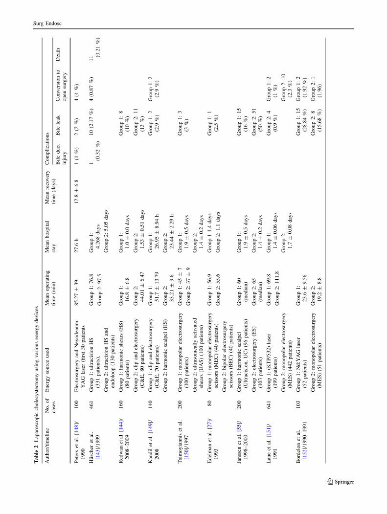
Comparison study
In a comparative study, it is important to understand the
use of energy devices in the context of individual proce-
dures. In this paper, we consider the LC procedure as an
example (Table 2). In LC the common energy sources used
are laser, electrosurgery, and ultrasonic energy. Because
many of the case studies do not explicitly state injuries
occurred while using the energy device, for LC, parame-
ters, such as mean operating time, mean hospital stay,
mean recovery time along with complications, such as bile
duct injury, bile leak, conversion rate to open surgery, and
any reported death, were collected for each study and
represented in Table 2. From the data, HC and UAS per-
formed better than clip and electrosurgery methods based
on operating time and bile duct injuries. Huscher et al.
[143] sates that the main advantage of UAS is the low-risk
dissection in the proximity of biliary structures. Moreover,
with UAS both cystic duct and vessels can be separated
without the need for ligature. The relative bloodless field of
view when using UAS also helps to discriminate anatom-
ical structures. Redwan et al. [144] states that when com-
paring HS and clip and electrosurgery for LC, HS
performed better with lesser operating time and the absence
of major or minor bile leaks. No clear conclusion can be
made regarding laser, because the operating time and
injuries compared with clip and electrosurgery varied
among the different studies and no direct comparison study
exists between laser and ultrasonically activated devices.
Between monopolar and bipolar electrosurgery, although
both had comparable operating times, monopolar electro-
surgery had more complications.
Discussion
A comparative study of the literature shows that the pre-
ferred source of energy in laparoscopic surgery in the early
1990s was the monopolar electrosurgery, whereas bipolar
and laser was used much less frequently. However, in
recent times, even with the advent of new tools in elec-
trosurgery, the preference is gradually shifting toward
ultrasonic energy due to its many advantages in laparos-
copy. Laparoscopic laser cholecystectomy, once a popular
procedure, is rarely performed and HS has been used more
often in recent times. Fiber optic cables have made lasers
more readily available in laparoscopy. The use of lasers is
most dominant in gynecological treatments, such as
endometriosis. ABC is a very effective technique to attain
hemostasis, and despite the large number of deaths and
intraoperative complications, it remains in use.
Despite significant developments, the search for an ideal
energy device that will result in perfect hemostasis with
minimum damage to surrounding tissue in the most effi-
cient manner posing the minimum threat to the patient in
terms of short- and long-term complications remains elu-
sive. Each energy method has advantages and disadvan-
tages, and a thorough knowledge of each device is essential
to decide which energy source should be used for a specific
Surg Endosc
123
Ta
ble
2L
apar
osc
op
icch
ole
cyst
ecto
my
usi
ng
var
iou
sen
erg
yd
evic
es
Au
tho
r/ti
mel
ine
No
.o
f
case
s
En
erg
yso
urc
eu
sed
Mea
no
per
atin
g
tim
e(m
in)
Mea
nh
osp
ital
stay
Mea
nre
cov
ery
tim
e(d
ays)
Co
mp
lica
tio
ns
Bil
ed
uct
inju
ry
Bil
ele
akC
on
ver
sio
nto
op
ensu
rger
y
Dea
th
Pet
ers
etal
.[1
48]/
19
90
10
0E
lect
rosu
rger
yan
dN
eyo
den
um
:
YA
Gla
ser
(firs
t3
0p
atie
nts
85
.27
±3
92
7.6
h1
2.8
±6
.81
(1%
)2
(2%
)4
(4%
)
Hu
sch
eret
al.
[14
3]/
19
99
46
1G
rou
p1
:u
ltra
cisi
on
HS
(33
1p
atie
nts
),
Gro
up
2:
ult
raci
sio
nH
San
d
end
olo
op
(13
0p
atie
nts
)
Gro
up
1:
76
.8
Gro
up
2:
97
.5
Gro
up
1:
4.2
68
day
s
Gro
up
2:
5.0
5d
ays
1
(0.3
2%
)
10
(2.1
7%
)4
(0.8
7%
)1
1 (0.2
1%
)
Red
wan
etal
.[1
44]/
20
08
–2
00
9
16
0G
rou
p1
:h
arm
on
icsh
ears
(HS
)
(80
pat
ien
ts)
Gro
up
2:
clip
and
elec
tro
surg
ery
(C&
E,
80
pat
ien
ts)
Gro
up
1:
16
.8±
6.8
Gro
up
2:
44
.01
±6
.47
Gro
up
1:
1.0
±0
.0d
ays
Gro
up
2:
1.5
3±
0.5
1d
ays
Gro
up
1:
8
(10
%)
Gro
up
2:
11
(13
%)
Kan
dil
etal
.[1
49]/
20
08
14
0G
rou
p1
:cl
ipan
del
ectr
osu
rger
y
(C&
E,
70
pat
ien
ts)
Gro
up
2:
har
mo
nic
scal
pel
(HS
)
Gro
up
1:
51
.7±
13
.79
Gro
up
2:
33
.21
±9
.6
Gro
up
1:
26
.95
±8
.94
h
Gro
up
2:
23
.44
±2
.29
h
Gro
up
1:
2
(2.9
%)
Gro
up
1:
2
(2.9
%)
Tsi
mo
yia
nn
iset
al.
[15
0]/
19
97
20
0G
rou
p1
:m
on
op
ola
rel
ectr
osu
rger
y
(10
0p
atie
nts
)
Gro
up
2:
ult
raso
nic
ally
acti
vat
ed
shea
rs(U
AS
)(1
00
pat
ien
ts)
Gro
up
1:
45
±7
Gro
up
2:
37
±9
Gro
up
1:
1.9
±0
.5d
ays
Gro
up
2:
1.4
±0
.2d
ays
Gro
up
1:
3
(3%
)
Ed
elm
anet
al.
[27
]/
19
93
80
Gro
up
1:
mo
no
po
lar
elec
tro
surg
ery
scis
sors
(ME
C)
(40
pat
ien
ts)
Gro
up
2:
bip
ola
rel
ectr
osu
rger
y
scis
sors
(BE
C)
(40
pat
ien
ts)
Gro
up
1:
56
.9
Gro
up
2:
55
.6
Gro
up
11
.4d
ays
Gro
up
2:
1.1
day
s
Gro
up
1:
1
(2.5
%)
Jan
ssen
etal
.[5
5]/
19
98
–2
00
0
20
0G
rou
p1
:h
arm
on
icsc
alp
el
(Ult
raci
sio
n,
UC
)(9
6p
atie
nts
)
Gro
up
2:
elec
tro
surg
ery
(ES
)
(10
3p
atie
nts
)
Gro
up
1:
60
(med
ian
)
Gro
up
2:
65
(med
ian
)
Gro
up
1:
1.9
±0
.5d
ays
Gro
up
2:
1.4
±0
.2d
ays
Gro
up
1:
15
(16
%)
Gro
up
2:
51
(50
%)
Lan
eet
al.
[15
1]/
19
91
64
1G
rou
p1
:(K
TP
/53
2)
lase
r
(19
9p
atie
nts
)
Gro
up
2:
mo
no
po
lar
elec
tro
surg
ery
(ME
S)
(44
2p
atie
nts
)
Gro
up
1:
69
.8
Gro
up
2:
11
1.8
Gro
up
1:
1.4
±0
.06
day
s
Gro
up
2:
1.7
±0
.08
day
s
Gro
up
2:
4
(0.9
%)
Gro
up
1:
2
(1%
)
Gro
up
2:
10
(2.3
%)
Bo
rdel
on
etal
.
[15
2]/
19
90
–1
99
1
10
3G
rou
p1
:N
d:Y
AG
lase
r
(52
pat
ien
ts)
Gro
up
2:
mo
no
po
lar
elec
tro
surg
ery
(ME
S)
(51
pat
ien
ts)
Gro
up
1:
23
.6±
9.5
6
Gro
up
2:
19
.2±
8.8
Gro
up
1:
15
(28
.84
%)
Gro
up
2:
8
(15
.68
%)
Gro
up
1:
2
(1.9
2%
)
Gro
up
2:
1
(1.9
6)
Surg Endosc
123

procedure. Relative advantages and disadvantages of
existing energy methods are presented in Table 3.
In a 2004 paper, Harrell et al. [145] mention that in a
survey of 500 surgeons in 1993, 18 % (of more than 500
respondents) reported to have caused electrosurgical injury
during laparoscopy and 54 % reported to have known of
another surgeon who has caused similar injury. Deaths
were more common during early in era of laparoscopy. A
brief search of the literature shows numerous instances of
deaths in the 1980s using various energy sources. The
number of deaths and complications of laparoscopy has
been greatly reduced in recent times. For example, com-
plications in LC, which were reported to be approximately
2–4 % in 1994 [146], decreased to approximately 0.4 % by
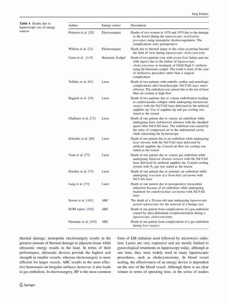
2005 [147]. Table 4 shows the various deaths reported in
laparoscopy using energy sources.
In general, most of the studies suggest that the effect of
any laparoscopic procedure depends on the skill and
familiarity of the surgeon with the surgical tools. An
interesting observation while reviewing studies performed
to test various instruments or methods is that some of the
results seem contradicting. Devices that have been rated
high by some studies have been rated low in others. Sur-
gical skill and familiarity with the particular device may
certainly be a possible contributor to this apparent paradox.
Furthermore, devices that are shown to work well in the
laboratory may not work the same way in an actual lapa-
roscopic procedure. Hence, there is a need to develop
uniform training regimens across surgical specialties under
clinical conditions. Particularly, the fundamental under-
standing of how each of the energy devices work and their
effect on the tissues is very important. For example, in
electrosurgery, the understanding of different power set-
tings and its effect on tissues is very important. Moreover,
the knowledge of safety issues with each of the devices
also should be known so that appropriate precautions could
be used to minimize injury. Solid communication and team
coordination in high fire-risk setting must be introduced
into practice. Examples are the high temperature and low
cooling rate of ultrasonically activated devices even after
switched off, risk of air embolism in laser from high flow
rate gas cooling, and venous gas embolism while using
ABC. A standardized curriculum or manual with working
principles of various energy devices and their safety issues
as envisioned by the FUSE program would be a valuable
tool to increase patient safety in surgical procedures using
energy devices.
Conclusions
Each of the energy devices reviewed in this work had its
own advantages and disadvantages. When consideringTa
ble
2co
nti
nu
ed
Au
tho
r/ti
mel
ine
No
.o
f
case
s
En
erg
yso
urc
eu
sed
Mea
no
per
atin
g
tim
e(m
in)
Mea
nh
osp
ital
stay
Mea
nre
cov
ery
tim
e(d
ays)
Co
mp
lica
tio
ns
Bil
ed
uct
inju
ry
Bil
ele
akC
on
ver
sio
nto
op
ensu
rger
y
Dea
th
El
Nak
eeb
etal
.
[15
3]/
20
08
–2
00
9
12
0G
rou
p1
:h
arm
on
icsh
ears
(HS
)
(60
pat
ien
ts)
Gro
up
2:
clip
and
elec
tro
surg
ery
(C&
E)
(60
pat
ien
ts)
Gro
up
1:
45
.17
±1
0.5
4
Gro
up
2:
69
.71
±1
3.0
1
Gro
up
1:
1
(1.6
%)
Gro
up
2:
2
(3.3
%)
Gro
up
1:
2
(3.3
%)
Gro
up
2:
3
(5%
)
Am
aral
etal
.[3
3]/
19
91
–1
99
2
20
0U
ltra
son
ical
lyac
tiv
ated
scal
pel
(UA
S)
49
.7±
1.5
(wit
ho
ut
cho
lan
gio
gra
ph
y)
61
.4±
2.0
(wit
h
cho
lan
gio
gra
ph
y)
1.4
±0
.1d
ays
1(0
.5%
)
So
uth
ern
Su
rgeo
ns
Clu
b[1
54]/
19
90
1,5
18
KT
Pla
ser
(31
4p
atie
nts
)
Nd
:YA
Gla
ser
(12
7p
atie
nts
)
Ele
ctro
surg
ery
(1,0
77
pat
ien
ts)
90
7
(0.4
6%
)
72
(4.7
%)
1
(0.0
7%
)
Ku
rau
chi
etal
.
[15
5]/
19
91
–1
99
3
1,4
08
Ty
pe
of
mo
dal
ity
no
tm
enti
on
ed1
2 (0.9
%)
5(0
.3%
)8
4(6
%)
Surg Endosc
123
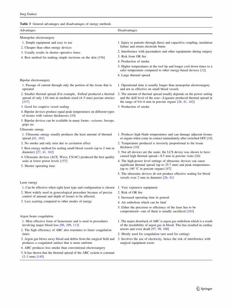
Table 3 General advantages and disadvantages of energy methods
Advantages Disadvantages
Monopolar electrosurgery
1. Simple equipment and easy to use
2. Cheaper than other energy devices
3. Usually results in shorter operative times
4. Best method for making simple incisions on the skin [156]
1. Injury to patients through direct and capacitive coupling, insulation
failure and return electrode burns
2. Interference with pacemakers and other equipments during surgery
3. Risk from OR fire
4. Production of smoke
5. Higher temperatures at the tool tip and longer cool down times to a
safer temperature compared to other energy-based devices [12].
6. Large thermal spread
Bipolar electrosurgery
1. Passage of current through only the portion of the tissue that is
operated
2. Smaller thermal spread (For example, -EnSeal produced a thermal
spread of only 1.84 mm in medium sized (4–5 mm) porcine arteries
[157]
3. Good for coaptive vessel sealing
4. Bipolar devices produce equal peak temperatures on different types
of tissues with various thicknesses [10]
5. Bipolar devices can be available in many forms—scissors, forceps,
grips etc
1. Operational time is usually longer than monopolar electrosurgery
and not as effective on small blood vessels
2. The amount of thermal spread usually depends on the power setting
and the skill level of the user—Ligasure produced thermal spread in
the range of 0.6–6 mm in porcine organs [26, 41, 162]
3. Production of smoke
Ultrasonic energy
1. Ultrasonic energy usually produces the least amount of thermal
spread [41, 161]
2. No smoke and only mist due to cavitation effect
3. Best energy method for sealing small blood vessels (up to 2 mm in
diameter) [27, 41, 162]
4. Ultrasonic devices (ACE, Wave, CS14C) produced the best quality
seals at lower power levels [157]
5. Shorter operating time
1. Produces high blade temperatures and can damage adjacent tissues
or organs when come in contact immediately after switched OFF [10]
2. Temperature produced is inversely proportional to the tissue
thickness [10]
3. Not all devices are the same; the LCS device was shown to have
caused high thermal spread—8.5 mm in porcine veins [26]
4. The high-power level settings of ultrasonic devices can cause
significant thermal spread (up to 25.7 mm) and peak temperatures
(up to 140 �C in porcine organs) [47]
5. The ultrasonic devices do not produce effective sealing for blood
vessels over 2 mm in diameter [26, 41]
Laser energy
1. Can be effective when right laser type and configuration is chosen
2. Most widely used in gynecological procedure because of precise
control of amount and depth of tissues to be affected,
3. Less scarring compared to other modes of energy
1. Very expensive equipment
2. Risk of OR fire
3. Increased operating time in general
4. Air embolism which can be fatal
5. Either the precision or efficiency of the laser has to be
compromised—one of them is usually sacrificed [163]
Argon beam coagulation
1. Most effective form of hemostasis and is used in procedures
involving major blood loss [96, 109, 113]
2. The high efficiency of ABC also translates to faster coagulation
times
3. Argon gas blows away blood and debris from the surgical field and
produces a coagulated surface that is more uniform
4. ABC produces less smoke than conventional electrosurgery
5. It has shown that the thermal spread of the ABC system is constant
(2–3 mm) [145]
1. The major drawback of ABC is argon gas embolism which is a result
of the insolubility of argon gas in blood. This has resulted in cardiac
arrests and even death [97, 98, 100]
2. Mostly used for coagulation (not used for cutting)
3. Involves the use of electricity, hence the risk of interference with
surgical equipment exists
Surg Endosc
123
thermal damage, monopolar electrosurgery results in the
greatest amount of thermal damage to adjacent tissue while
ultrasonic energy results in the least. In terms of their
performance, ultrasonic devices provide the highest seal
strength in smaller vessels, whereas electrosurgery is more
efficient for larger vessels. ABC results in the most effec-
tive hemostasis on irregular surfaces; however, it also leads
to gas embolism. In electrosurgery, RF is the most common
form of EM radiation used followed by microwave radia-
tion. Lasers are very expensive and are mostly limited to
gynecological treatments in laparoscopy today, although at
one time, they were widely used in many laparoscopic
procedures, such as cholecystectomy. In blood vessel
sealing, the effectiveness of an energy device is dependent
on the size of the blood vessel. Although there is no clear
winner in terms of operating time, in the series of studies
Table 4 Deaths due to
laparoscopic use of energy
sources
Author Energy source Description
Peterson et al. [20] Electrosurgery Deaths of two women in 1978 and 1979 due to the damage
to the bowel during the laparoscopic sterilizationprocedure using monopolar electrocoagulation. The
complications were postoperative
Willson et al. [21] Electrosurgery Death due to thermal injury to the colon occurring beyond
the field of view during laparoscopic cholecystectomy
Curro et al. [119] Harmonic Scalpel Death of two patients (one with severe liver failure and one
with sepsis) due to the failure of laparoscopiccholecystectomy in treatment of Child Pugh C cirrhosis
using the harmonic scalpel. The result is more of the case
of ineffective procedure rather than a surgical
complication
Tellides et al. [81] Laser Death of two patients with embolic cardiac and neurologic
complications after bronchoscopic Nd:YAG laser tumorablation. The embolism was caused due to the use of laser
fiber air coolant at high flow
Baggish et al. [70] Laser Death of two patients due to venous embolization leading
to cardiovascular collapse while undergoing intrauterinesurgery with the Nd:YAG laser delivered by the artificial
sapphire tip. Use of sapphire tip and gas cooling was
stated as the reason
Challener et al. [71] Laser Death of one patient due to venous air embolism while
undergoing laser endometrial ablation with the sheathed
quartz fiber Nd:YAG laser. The embolism was caused by
the entry of compressed air to the endometrial cavity
while reinserting the hysteroscope
Schroder et al. [80] Laser Death of one patient due to air embolism while undergoing
laser thermia with the Nd:YAG laser delivered by
artificial sapphire tip. Coaxial air flow for cooling was
stated as the reason
Yuan et al. [77] Laser Death of one patient due to venous gas embolism while
undergoing bilateral choanal stenosis with the Nd:YAG
laser delivered by artificial sapphire tip. Coaxial cooling
system with N2 gas was stated as the reason
Peachey et al. [75] Laser Death of one patient due to systemic air embolism while
undergoing resection of a bronchial carcinoma with
Nd:YAG laser
Lang et al. [73] Laser Death of one patient due to postoperative myocardial
infarction because of air embolism while undergoing
treatment for endobronchial carcinoma with Nd:YAG
laser
Sezeur et al. [101] ABC The death of a 20-year-old man undergoing laparoscopicpartial splenectomy for the removal of a benign cyst
ECRI report. [102] ABC Death of one patient from complications of a gas embolism
caused by intra-abdominal overpressurization during a
laparoscopic cholecystectomy
Ousmane et al. [103] ABC Death of one patient from complications of a gas embolism
during liver surgery
Surg Endosc
123

that were reviewed in this work, the HS has been shown to
have reduced overall time compared with other energy
sources in MIS. In terms of death from complications,
lasers and ABC have more reported cases than the other
methods and surgeons should be familiar with their use.
Electrosurgery is still very popular in MIS due to its
familiarity with surgeons and the development of various
enhanced safety features to minimize injuries.
Disclosures Drs. Ganesh Sankaranarayanan, Rajeswara Resapu,
Daniel B. Jones, Steven Schwaitzberg, and Suvranu De have no
conflict of interest or financial ties to disclose.
Funding NIBIB/NIH Grants # R01EB010037, # R01 EB009362,
# R01 EB005807 and # R01 EB014305.
References
1. Wu M-P, Ou C-S, Chen S-L, Yen EYT, Rowbotham R (2000)
Complications and recommended practices for electrosurgery in
laparoscopy. Am J Surg 179(1):67–73
2. Feldman L, Fuchshuber P, Jones D, Mischna J, Schwaitzberg S,
Force FT (2012) Surgeons don’t know what they don’t know
about the safe use of energy in surgery. Surg Endosc 26(10):
2735–2739. doi:10.1007/s00464-012-2263-y
3. Cushing H, Bovie WT (1928) Electrosurgery as an aid to the
removal of intracranial tumors. Surg Gynecol Obstet 47:
751–784
4. Albert M, Ellis CN, Fleshman J, Margolin D, Ng PC, Podnos
YD (2010) The effective use of the ENSEAL system in colo-
rectal surgery
5. Lee J (2002) Update on electrosurgery. Outpatient Surg 3(2)
6. Govekar H, Robinson T, Varosy P, Girard G, Montero P, Dunn
C et al (2012) Effect of monopolar radiofrequency energy on
pacemaker function. Surg Endosc 26(10):2784–2788. doi:10.10
07/s00464-012-2279-3
7. Montero P, Robinson T, Weaver J, Stiegmann G (2009) Insu-
lation failure in laparoscopic instruments. Surg Endosc 24(2):
462–465. doi:10.1007/s00464-009-0601-5
8. Vancaillie TG (1998) Active electrode monitoring. Surg Endosc
12(8):1009–1012. doi:10.1007/s004649900769
9. Valleylab. (1999) Basics of bipolar electrosurgery. Clin Inform
Hotline News
10. Kim F, Chammas M, Gewehr E, Morihisa M, Caldas F, Hay-
acibara E et al (2008) Temperature safety profile of laparoscopic
devices: harmonic ACE (ACE), Ligasure V(LV), and plasma
trisector (PT). Surg Endosc 22(6):1464–1469
11. Box GN, Lee HJ, Abraham JB, Deane LA, Elchico ER,
Abdelshehid CA et al (2009) Comparative study of in vivo
lymphatic sealing capability of the porcine thoracic duct using
laparoscopic dissection devices. J Urol 181(1):387–391
12. Sutton PA, Awad S, Perkins AC, Lobo DN (2010) Comparison
of lateral thermal spread using monopolar and bipolar dia-
thermy, the Harmonic ScalpelTM and the LigasureTM. Br J Surg
97(3):428–433
13. Tulikangas PK, Smith T, Falcone T, Boparai N, Walters MD
(2001) Gross and histologic characteristics of laparoscopic
injuries with four different energy sources. Fertil Steril
75(4):806–810
14. Gol M, Kizilyar A, Eminoglu M (2007) Laparoscopic hyster-
ectomy with retroperitoneal uterine artery sealing using
LigaSure: Gazi hospital experience. Arch Gynecol Obstet
276(4):311–314
15. Kriplani A, Garg P, Sharma M, Lal S, Agarwal N (2008) A
review of total laparoscopic hysterectomy using LigaSure uter-
ine artery-sealing device: AIIMS experience. J Laparoendosc
Adv Surg Tech A 18(6):825–829
16. Polychronidis A, Tsaroucha AK, Karayiannakis AJ, Perente S,
Efstathiou E, Simopoulos C (2005) Delayed perforation of the
large bowel due to thermal injury during laparoscopic chole-
cystectomy. J Int Med Res 33:360–363
17. Siperstein A, Garland A, Engle K, Rogers S, Berber E, Forou-
tani A et al (2000) Local recurrence after laparoscopic radio-
frequency thermal ablation of hepatic tumors. Ann Surg Oncol
7(2):106–113
18. Darai E, Ackerman G, Bazot M, Rouzier R, Dubernard G (2007)
Laparoscopic segmental colorectal resection for endometriosis:
limits and complications. Surg Endosc 21(9):1572–1577
19. Berber E, Siperstein A (2008) Local recurrence after laparo-
scopic radiofrequency ablation of liver tumors: an analysis of
1032 tumors. Ann Surg Oncol 15(10):2757–2764
20. Peterson HB, Ory HW, Greenspan JR Jr, Tyler CW Jr (1981)
Deaths associated with laparoscopic sterilization by unipolar
electrocoagulating devices, 1978 and 1979. Am J Obstet
Gynecol 139(2):141–143
21. Willson PD, McAnena OJ, Peters EE (1994) A fatal complica-
tion of diathermy in laparoscopic surgery. Minim Invasive Ther
Allied Technol 3(1):19–20. doi:10.3109/13645709409152989
22. Agarwal B, Gupta M, Agarwal S, Mahajan K (2007) Anatomical
footprint for safe laparoscopic cholecystectomy without using
any energy source: a modified technique. Surg Endosc 21(12):
2154–2158
23. Matthews B, Nalysnyk L, Estok R, Fahrbach K, Banel D, Linz H
et al (2008) Ultrasonic and nonultrasonic instrumentation: a
systematic review and meta-analysis. Arch Surg 143(6):
592–600. doi:10.1001/archsurg.143.6.592
24. Hubner M, Demartines N, Muller S, Dindo D, Clavien PA,
Hahnloser D (2008) Prospective randomized study of monopolar
scissors, bipolar vessel sealer and ultrasonic shears in laparo-
scopic colorectal surgery. Br J Surg 95(9):1098–1104
25. Targarona EM, Balague C, Marin J, Neto RB, Martinez C,
Garriga J et al (2005) Energy sources for laparoscopic colec-
tomy: a prospective randomized comparison of conventional
electrosurgery, bipolar computer-controlled electrosurgery and
ultrasonic dissection. Operative outcome and costs analysis.
Surg Innov 12(4):339–344. doi:10.1177/155335060501200409
26. Hruby GW, Marruffo FC, Durak E, Collins SM, Pierorazio P,
Humphrey PA et al (2007) Evaluation of surgical energy devices
for vessel sealing and peripheral energy spread in a porcine
model. J Urol 178(6):2689–2693
27. Edelman DS, Unger SW (1995) Bipolar versus monopolar
cautery scissors for laparoscopic cholecystectomy: a random-
ized, prospective study. Surg Laparosc Endosc 5(6):459–462
28. Campagnacci R, de Sanctis A, Baldarelli M, Rimini M, Lezoche
G, Guerrieri M (2007) Electrothermal bipolar vessel sealing
device vs. ultrasonic coagulating shears in laparoscopic colec-
tomies: a comparative study. Surg Endosc 21(9):1526–1531
29. Demirturk F, Aytan H, Caliskan AC (2007) Comparison of the
use of electrothermal bipolar vessel sealer with harmonic scalpel
in total laparoscopic hysterectomy. J Obstet Gynaecol Res 33:
341–345
30. Lamberton GR, Hsi RS, Jin DH, Lindler TU, Jellison FC,
Baldwin DD (2008) Prospective comparison of four laparo-
scopic vessel ligation devices. J Endourol 22(10):2307–2312
31. Dodde R, Gee JS, Geiger JD, Shih AJ (2010) Monopolar elec-
trosurgical thermal management system to reduce lateral ther-
mal damage during surgery. J Med Device 4(2):1–027505
Surg Endosc
123
32. Davison T, Amaral J, Geis P, Swansrom L, Sinha UK (2008)
The science of ultrasonic energy-based dissection. General
Surgery News.
33. Amaral JF (1995) Laparoscopic cholecystectomy in 200 con-
secutive patients using an ultrasonically activated scalpel. Surg
Laparosc Endosc 5(4):255–262
34. O’Daly BJ, Morris E, Gavin GP, O’Byrne JM, McGuinness GB
(2008) High-power low-frequency ultrasound: a review of tissue
dissection and ablation in medicine and surgery. J Mater Process
Technol 200(1–3):38–58
35. Smith R, Pasic R (2008) The role of vessel sealing technologies
in laparoscopic surgery. Surg Technol Int 17:208–212
36. Kinoshita T, Kanehira E, Omura K, Kawakami K, Watanabe Y
(1999) Experimental study on heat production by a 23.5-kHz
ultrasonically activated device for endoscopic surgery. Surgical
Endoscopy 13(6):621–625
37. Lee SJ, Park KH (1999) Ultrasonic energy in endoscopic sur-
gery. Yonsei Med J 40(6):545–549
38. Liano C (2004) Curbing laparoscopy burns. The Washington
Times website. http://www.upi.com/Science_News/2004/06/21/
Feature-Curbing-laparoscopy-burns/UPI-52511087842993/.
Accessed 39 June 2004
39. Ghosh P (1999) Ultrasonic devices in cardiac surgery. Asian
Cardiovasc Thorac Ann 7(4):333–338
40. Clements R, Palepu R (2007) In vivo comparison of the coag-
ulation capability of SonoSurg and Harmonic Ace on 4-mm and
5-mm arteries. Surg Endosc 21(12):2203–2206
41. Harold KL, Pollinger H, Matthews BD, Kercher KW, Sing RF,
Heniford BT (2003) Comparison of ultrasonic energy, bipolar
thermal energy, and vascular clips for the hemostasis of small-,
medium-, and large-sized arteries. Surg Endosc 17(8):
1228–1230
42. Wolfe BM, Gardiner BN, Leary BF, Frey CF (1991) Endoscopic
cholecystectomy: an analysis of complications. Arch Surg
126(10):1192–1198. doi:10.1001/archsurg.1991.01410340030005
43. Kadesky KM, Schopf B, Magee JF, Blair GK (1997) Proximity
injury by the ultrasonically activated scalpel during dissection.
J Pediatr Surg 32(6):878–879
44. Awwad JT, Isaacson K (1996) The harmonic scalpel: an intra-
operative complication. Obstet Gynecol 88(4):718–720
45. McNally ML, Erturk E, Oleyourryk G, Schoeniger L (2001)
Laparoscopic cyst decortication using the harmonic scalpel for
symptomatic autosomal dominant polycystic kidney disease.
J Endourol 15(6):597–599
46. Kim JS, Hattori R, Yamamoto T, Yoshino Y, Gotoh M (2010)
How can we safely use ultrasonic laparoscopic coagulating
shears? Int J Urol 17:377–381
47. Emam TA, Cuschieri A (2003) How safe is high-power ultra-
sonic dissection? Ann Surg 237(2):186–191
48. Kinoshita T, Kanehira E, Omura K (1999) Basic experiments to
evaluate the safety of a fine caliber ultrasonically activated
device. JSES 4:473–478.
49. Schwartz RO (1994) Total laparoscopic total hysterectomy with
the harmonic scalpel. J Gynecol Surg 10(1):33–34
50. Giannopoulos T, Chipchase J, Tailor A, Butler-Manuel S (2005)
The use of harmonic shears (Ultracision) for laparoscopic
lymphadenectomies in women with gynaecological malignan-
cies. Gynecol Surg 2(2):97–100
51. Jansen FW, Trimbos-Kemper T, Baptist Trimbos J (2002)
Ultrasonic scalpel in laparoscopic gynaecological surgery: an
observational study in 354 cases. Gynaecol Endosc 11(1):47–51
52. Lin J, Zhang X, Xu K (2001) Application of ultrasonic scalpel in
gynecologic operative laparoscopy. Chin Med J 114(12):
1283–1285
53. Amarin NS (2008) Harmonic scalpel and clipless cholecystec-
tomy. World J Laparosc Surg 1(2):6–8
54. Gelmini R, Franzoni C, Zona S, Andreotti A, Saviano M (2010)
Laparoscopic cholecystectomy with harmonic scalpel. JSLS
14(1):14–19
55. Janssen IMC, Swank DJ, Boonstra O, Knipscheer BC, Kli-
nkenbijl JHG, van Goor H (2003) Randomized clinical trial of
ultrasonic versus electrocautery dissection of the gallbladder in
laparoscopic cholecystectomy. Br J Surg 90(7):799–803
56. Elattar OM, Naga AAE, Maged H (2005) Laparoscopic appen-
dectomy by ultrasonically activated scalpel: a prospective study.
Egypt J Surg 24(3):164–167
57. Lin PC, Thyer A, Soules MR (2004) Intraoperative ultrasound
during a laparoscopic myomectomy. Fertil Steril 81(6):1671–1674
58. Stringer NH (1994) Laparoscopic myomectomy with the har-
monic scalpel: a review of 25 cases. J Gynecol Surg 10(4):
241–245
59. Msika S, Deroide G, Kianmanesh R, Iannelli A, Hay J-M,
Fingerhut A et al (2001) Harmonic scalpel in laparoscopic
colorectal surgery. Dis Colon Rectum 44(3):432–436
60. Vetere PF, Apostolis C (2010) Ureteral injury due to a harmonic
scalpel during laparoscopic salpingo-oophorectomy. JSLS
14(1):115–119
61. Chachan S, Waters N, Kent A (2011) Laparoscopic management
of cornual heterotopic pregnancy with the use of Harmonic
ACE�—a case report. Gynecol Surg 8: 243–246
62. Tanaka T, Ueda K, Hayashi M, Hamano K (2009) Clinical
application of an ultrasonic scalpel to divide pulmonary vessels
based on laboratory evidence. Interact Cardiovasc Thorac Surg
8(6):615–618. doi:10.1510/icvts.2008.200584
63. Driscoll P (2006) Energy-based devices in general surgery.
MedMarket Diligence
64. Bruhat M, Mage C, Manhes M (1979) Use of carbon dioxide
laser via laparoscopy. Laser Surgery III Proceedings of the Third
Congress for the International Society for Laser Surgery, Kaplan
65. Sutton C (1986) Initial experience with carbon dioxide laser
laparoscopy. Lasers Med Sci 1(1):25–31
66. Hamman BL, Theologes TT (2009) Surgical treatment of atrial
fibrillation with diode-pumped laser. Baylor Univ Med Center
Proc 22:218–220
67. Williams MR, Casher JM, Russo MJ, Hong KN, Argenziano M,
Oz MC (2006) Laser energy source in surgical atrial fibrillation
ablation: preclinical experience. Ann Thorac Surg 82(6):
2260–2264
68. Welch AJ, Torres JH, Cheong W-F (1989) Laser physics and
laser–tissue interaction. Tex Heart Inst J 16(3):141–149
69. Gerber GS (1995) Lasers in the treatment of benign prostatic
hyperplasia. Urology 45(2):193–199
70. Baggish MS, Daniell JF (1989) Death caused by air embolism
associated with neodymium:yttrium–aluminum–garnet laser
surgery and artificial sapphire tips. Am J Obstet Gynecol 161(4):
877–878
71. Challener RC, Kaufman B (1990) Fatal venous air embolism
following sequential unsheathed (bare) and sheathed quartz fiber
Nd:YAG laser endometrial ablation. Anesthesiology 73(3):
548–551
72. Greville AC, Clements EAF, Erwin DC, McMillan DL, Well-
wood JM (1991) Pulmonary air embolism during laparoscopic
laser cholecystectomy. Anaesthesia 46:113–114
73. Lang NP, Wait GM, Read RR (1991) Cardio-cerebrovascular
complications from Nd:YAG laser treatment of lung cancer. Am
J Surg 162(6):629–632
74. Messiant F, Marquette C, Neviere R, Ramon P, Duverger D,
Mathieu D (1995) Systemic air embolism after laser resection of
a tracheal tumor. Intensive Care Med 21(2):192–193
75. Peachey T, Eason J, Moxham J, Jarvis D, Driver M (1988)
Systemic air embolism during laser bronchoscopy. Anaesthesia
43(10):872–875
Surg Endosc
123
76. Ross DJ, Mohsenifar Z, Potkin RT, Roston WL, Shapiro SM,
Alexander JM (1988) Pathogenesis of cerebral air embolism
during neodymium-YAG laser photoresection. Chest 94(3):
660–662
77. Yuan HB, Poon KS, Chan KH, Lee TY, Lin CY (1993) Fatal gas
embolism as a complication of Nd-YAG laser surgery during
treatment of bilateral choanal stenosis. Int J Pediatr Otorhino-
laryngol 27(2):193–199
78. Baggish MS, Daniell JF (1989) Catastrophic injury secondary to
the use of coaxial gas-cooled fibers and artificial sapphire tips
for intrauterine surgery: a report of five cases. Lasers Surg Med
9(6):581–584
79. Kelly M, Mathews HML, Weir P (1997) Carbon dioxide
embolism during laser endometrial ablation. Anaesthesia 52(1):
65–67
80. Schroder TM, Puolakkainen PA, Hahl J, Ramo OJ (1989) Fatal
air embolism as a complication of laser-induced hyperthermia.
Lasers Surg Med 9(2):183–185
81. Tellides G, Ugurlu BS, Kim RW, Hammond GL (1998) Path-
ogenesis of systemic air embolism during bronchoscopic
Nd:YAG laser operations. Ann Thorac Surg 65(4):930–934
82. Genyk YS, Keller FS, Halpern NB (1994) Hepatic artery
pseudoaneurysm and hemobilia following laser laparoscopic
cholecystectomy. Surg Endosc 8(3):201–204
83. Ewen S, Sutton CJG (1995) Complications of laser laparoscopy:
eleven years experience. Minim Invasive Ther Allied Technol
4(1):27–29. doi:10.3109/13645709509152749
84. Reddick EJ, Olsen DO (1989) Laparoscopic laser cholecystec-
tomy. Surg Endosc 3(3):131–133
85. Goldenberg M, Bider D et al (1994) Pregnancy: laparoscopic
laser surgery of primary ovarian pregnancy. Hum Reprod
9(7):1337–1338
86. Langebrekke A, Kirschner R, SkA¥r OJ, SA¸rnes T, Urnes A
(1991) Treatment of tubal pregnancy by laparoscopic laser
surgery. Acta Obstet Gynecol Scand 70(4–5):331–334.
doi:10.3109/00016349109007882
87. Langebrekke A, Sarnes T, Urnes A (1993) Fertility outcome
after treatment of tubal pregnancy by laparoscopic laser surgery.
Acta Obstet Gynecol Scand 72(7):547–549. doi:10.3109/
00016349309058161
88. Sutton CJG, Ewen SP, Jacobs SA, Whitelaw NL (1997) Laser
laparoscopic surgery in the treatment of ovarian endometriomas.
J Am Assoc Gynecol Laparosc 4(3):319–323
89. Weather LJ (1988) Carbon dioxide laser laparoscopy in treat-
ment of infertility and disorders associated with pelvic pain.
J Natl Med Assoc 80(2):185–187
90. Shumalinsky D, Lobik L, Cytron S, Halpern M, Vasilyev T,
Ravid A et al (2004) Laparoscopic laser soldering for repair
of ureteropelvic junction obstruction in the porcine model.
J Endourol 18(2):177–181. doi:10.1089/089277904322959833
91. Karaman Y, Bingol B, Gunenc Z (2006) Prevention of com-
plications in laparoscopic hysterectomy: experience with 1120
cases performed by a single surgeon. J Minim Invasive Gynecol
14(1):78–84
92. Liu CY (1992) Laparoscopic hysterectomy. A review of 72
cases. J Reprod Med 37(4):351–354
93. Wishnow KI, Johnson DE, Cromeens DM, Ro JY (1989) Laser
photoirradiation of the canine ureteral orifice: comparison
between contact and noncontact techniques. Lasers Surg Med
9(5):485–489
94. Ward PH, Castro DJ, Ward S (1989) A significant new contri-
bution to radical head and neck surgery: the argon beam coag-
ulator as an effective means of limiting blood loss. Arch
Otolaryngol Head Neck Surg 115(8):921–923. doi:10.1001/
archotol.1989.01860320031013
95. Low RK, Moran ME (1993) Laparoscopic argon beam coagulation:
implications for reconstructive urology. Minim Invasive Ther
Allied Technol 2(2):75–78. doi:10.3109/13645709309152671
96. Bowling DM (2002) Argon beam coagulation for post-tonsillec-
tomy hemostasis. Otolaryngol Head Neck Surg 126(3):316–320
97. Cornejo A, Liao L, Kenneth W (2010) Argon gas embolism with
the use of argon beam coagulation during open hepatic resec-
tion. Internet J Surg 22(2)
98. Ikegami T, Shimada M, Imura S, Nakamura T, Kawahito S,
Morine Y et al (2009) Argon gas embolism in the application of
laparoscopic microwave coagulation therapy. J Hepatobiliary
Pancreat Surg 16(3):394–398
99. Bateman SN, Noorily AD, McGuff HS (1996) Sharp dissection,
electrosurgery, and argon-enhanced electrosurgery in porcine
skin flaps. Otolaryngol Head Neck Surg 114(3):435–442
100. Kono M, Yahagi N, Kitahara M, Fujiwara Y, Sha M, Ohmura A
(2001) Cardiac arrest associated with use of an argon beam
coagulator during laparoscopic cholecystectomy. Eur J Cardio-
thorac Surg 87(4):644–646. doi:10.1093/bja/87.4.644
101. Sezeur A, Partensky C, Chipponi J, Duron J–J (2008) Death
during laparoscopy: can 1 gas push out another? Danger of
argon electrocoagulation. Surg Laparosc Endosc Percutan Tech
18(4):395–397
102. (1994) Fatal gas embolism caused by overpressurization during
laparoscopic use of argon enhanced coagulation. Health Devices
23(6):257–259
103. Ousmane ML, Fleyfel M, Vallet B (2002) Venous gas embolism
during liver surgery with argon-enhanced coagulation. Eur J
Anaesthesiol 19(3):225
104. Kizer N, Zighelboim I, Rader JS (2009) Cardiac arrest during
laparotomy with argon beam coagulation of metastatic ovarian
cancer. Int J Gynecol Cancer 19(2):237–238. doi:10.1111/
IGC.0b013e31819c54db
105. Mastragelopulos N, Sarkar MR, Kaissling G, Bahr R, Daub D(1992) Argon gas embolism in laparoscopic cholecystectomy
with the Argon Beam One coagulator. Chirurg 63(12):1053–1054
106. Reddy C, Majid A, Michaud G, Feller-Kopman D, Eberhardt R,
Herth F et al (2008) Gas embolism following bronchoscopic
argon plasma coagulation. Chest 134(5):1066–1069. doi:10.1378/
chest.08-0474
107. Stojeba N, Mahoudeau G, Segura P, Meyer C, Steib A (1999)
Possible venous argon gas embolism complicating argon gas
enhanced coagulation during liver surgery. Acta Anaesthesiol
Scand 43(8):866–867
108. Veyckemans F, Michel I (1996) Venous gas embolism from an
argon coagulator. Anesthesiology 85(2):443–444
109. Kwon AH, Inui H, Kamiyama Y (2001) Successful laparoscopic
haemostasis using an argon beam coagulator for blunt traumatic
splenic injury. Eur J Surg 167(4):316–318
110. Lurie S, Gur D, Glezerman M (2002) Argon beam coagulation
during caesarean section. BJOG 109(12):1415
111. Lucioni A, Orvieto MA, Zorn KC, Lotan T, Gong EM, Stein-
berg GD et al (2008) Efficacy of the argon beam coagulator
alone in obtaining hemostasis after laparoscopic porcine hemi-
nephrectomy: a pilot study. Can J Urol 15(3):4091–4096
112. Meyer-Junghaenel L, Giest H, Waldschmidt Jr (2001) Laparo-
scopic argon-beam coagulation in relapsing congenital dyson-
togenetic splenic cysts. Pediatr Endosurg Innov Tech 5(3):
315–318. doi:10.1089/10926410152634466
113. Okada T, Sasaki F, Kurauchi N, Kubota M, Itoh T, Honda S et al
(2007) Laparoscopic liver biopsy using cup-shaped punch
biopsy forceps and argon beam coagulator in children. Pediatr
Surg Int 23(10):947–951
114. Dowling RD, Ochoa J, Yousem SA, Peitzman A, Udekwu AO
(1991) Argon beam coagulation is superior to conventional
Surg Endosc
123
techniques in repair of experimental splenic injury. J Trauma
31(5):717–721
115. Palmer M, Miller CW III, Orton EC (1993) Venous gas embo-
lism associated with argon-enhanced coagulation of the liver.
J Invest Surg 6(5):391–399
116. Falbo B, Vene M, Visini R, Grottola T, Di Paolo S, Picardi N
(2004) Experimental study of the effect of the argon beam
coagulator on organic tissues from the viewpoint of surgical
utilization. Ann Ital Chir 75(1):59–62
117. Bobbio A, Ampollini L, Internullo E, Caporale D, Cattelani L,
Bettati S et al (2006) Thoracoscopic parietal pleural argon beam
coagulation versus pleural abrasion in the treatment of primary
spontaneous pneumothorax. Eur J Cardiothorac Surg 29(1):6–8.
doi:10.1016/j.ejcts.2005.10.034
118. Beyer E, Lee R, Lam B-K (2009) Minimally invasive bipolar
radiofrequency ablation of lone atrial fibrillation: early multi-
center results. J Thorac Cardiovasc Surg 137(3):521–526
119. Curro G, Iapichino G, Melita G, Lorenzini C, Cucinotta E
(2005) Laparoscopic cholecystectomy in Child-Pugh class C
cirrhotic patients. JSLS 9(3):311–315
120. Saad EB, Marrouche NF, Saad CP, Ha E, Bash D, White RD
et al (2003) Radiofrequency treatment of abnormal heart rhythm
can damage the vessels that return blood from the lungs to the
heart. Ann Intern Med 138(8):1
121. Keltner JR, Donegan E, Hynson JM, Shapiro WA (2001) Acute
renal failure after radiofrequency liver ablation of metastatic
carcinoid tumor. Anesth Analg 93:587–589
122. Kocovic DZ, Harada T, Shea JB, Soroff D, Friedman PL (1993)
Alterations of heart rate and of heart rate variability after
radiofrequency catheter ablation of supraventricular tachycardia.
Delineation of parasympathetic pathways in the human heart.
Circulation 88(4):1671–1681
123. Curley SA, Marra P, Beaty K et al (2004) Early and late com-
plications after radiofrequency ablation of malignant liver
tumors in 608 patients. Ann Surg 239(4):450–458
124. Protsenko DE, Zemek A, Wong BJF (2007) Simulation of laser
induced thermo-mechanical changes in tissue using RF heating
method. In: Thermal treatment of tissue: energy delivery and
assessment IV; Proc. SPIE 6440, San Jose, CA, USA.
125. Koffron AJ, Stein JA (2008) Laparoscopic liver surgery:
parenchymal transection using saline-enhanced electrosurgery.
HPB (Oxford) 10(4):225–228
126. Yim APC, Rendina EA, Hazelrigg SR, Chow LTC, Lee T-W,
Wan S et al (2002) A new technological approach to nonana-
tomical pulmonary resection: saline enhanced thermal sealing.
Ann Thorac Surg 74(5):1671–1676
127. Gozen AS, Teber D, Rassweiler JJ (2007) Principles and initial
experience of a new device for dissection and hemostasis.
Minim Invasive Ther Allied Technol 16(1):58–65. doi:10.1080/
13645700701191537
128. Zarebczan B, Mohanty D, Chen H (2011) A comparison of the
LigaSure and harmonic scalpel in thyroid surgery: a single
institution review. Ann Surg Oncol 18(1):214–218
129. Munver R, Del Pizzo J, Sosa R (2003) Adrenal-preserving
minimally invasive surgery: the role of laparoscopic partial
adrenalectomy, cryosurgery, and radiofrequency ablation of the
adrenal gland. Curr Urol Rep 4(1):87–92
130. Smulders J, de Hingh I, Stavast J, Jackimowicz J (2007)
Exploring new technologies to facilitate laparoscopic surgery:
creating intestinal anastomoses without sutures or staples, using
a radio-frequency-energy-driven bipolar fusion device. Surg
Endosc 21(11):2105–2109
131. Soon PSH, Yeh MW, Sywak MS, Sidhu SB (2006) Use of the
Ligasure vessel sealing system in laparoscopic adrenalectomy.
ANZ J Surg 76(9):850–852
132. Hope WW, Burns JM, Newcomb WL, Heniford BT, Sing RF
(2009) Safety and efficacy of the electrothermal bipolar vessel
sealer in trauma. J Endourol 40(5):564–566
133. Kimura M, Baba S, Polascik TJ (2010) Minimally invasive
surgery using ablative modalities for the localized renal mass.
Int J Urol 17(3):215–227
134. Oefelein MG (2006) Delayed presentation of urinoma after
radiofrequency ablation-assisted laparoscopic partial nephrec-
tomy. J Endourol 20(1):27–30. doi:10.1089/end.2006.20.27
135. Onishi T, Nishikawa K, Hasegawa Y et al (2007) Assessment of
health-related quality of life after radiofrequency ablation or
laparoscopic surgery for small renal cell carcinoma: a prospec-
tive study with medical outcomes study 36-item health survey
(SF-36). Jpn J Clin Oncol 37(10):750–754
136. Bachellier P, Ayav A, Pai M, Jean-Christopher W, Edoardo R,
Daniel J et al (2007) Laparoscopic liver resection assisted with
radiofrequency. Am J Surg 193(4):427–430
137. Hompes D, Aerts R, Penninckx F, Topal B (2007) Laparoscopic
liver resection using radiofrequency coagulation. Surg Endosc
21(2):175–180
138. Tan BJ, El-Hakim A, Nora M, Yavor S, Arthur S, Benjamin R
(2004) Comparison of laparoscopic saline infused to dry radio
frequency ablation of renal tissue: evolution of histological
infarct in the porcine model. J Urol 172(5):2007–2012
139. Asahina Y, Nakanishi H, Izumi N (2009) Laparoscopic radio-
frequency ablation for hepatocellular carcinoma. Dig Endosc
21(2):67–72
140. Sadiq TS, Shrestha R, Weeks S, Gerber DA (2004) Laparo-
scopic radiofrequency ablation provides local control of hepa-
tocellular carcinoma in patients awaiting liver transplant. J Am
Coll Surg 199(3):16
141. van den Bos R, Lidia A, Michael K, Martino N, Tamar N (2009)
Endovenous therapies of lower extremity varicosities: a meta-
analysis. J Vasc Surg 49(1):230–239
142. Weil L Jr, Glover JP, Weil LS Sr (2008) A new minimally invasive
technique for treating plantar fasciosis using bipolar radiofre-
quency: a prospective analysis. Foot Ankle Spec 1(1):13–18
143. Huscher CGS, Lirici MM, Di Paola M, Crafa F, Napolitano C,
Mereu A et al (2003) Laparoscopic cholecystectomy by ultra-
sonic dissection without cystic duct and artery ligature. Surg
Endosc 17(3):442–451
144. Redwan AA (2010) Single-working-instrument, double-trocar,
clipless cholecystectomy using harmonic scalpel: a feasible,
safe, and less invasive technique. J Laparoendosc Adv Surg
Tech 20(7):597–603. doi:10.1089/lap.2009.0375
145. Harrell AG, Kercher KW, Heniford BT (2004) Energy sources
in laparoscopy. Surg Innov 11(3):201–209. doi:10.1177/
107155170401100310
146. Berry SM, Ose KJ, Bell RH, Fink AS (1994) Thermal injury of
the posterior duodenum during laparoscopic cholecystectomy.
Surg Endosc 8(3):197–200
147. Giger U, Michel JM, Vonlanthen R, Becker K, Kocher T,
Krahenbuhl L (2005) Laparoscopic cholecystectomy in acute
cholecystitis: indication, technique, risk and outcome. Langen-
becks Arch Surg 390(5):373–380
148. Peters JH, Ellison EC, Innes JT, Liss JL, Nichols KL, Lomano
JM et al (1991) Safety and efficacy of laparoscopic cholecys-
tectomy. A prospective analysis of 100 initial patients. Ann Surg
213(1):3–12
149. Kandil T, El Nakeeb A, El Hefnawy E (2010) Comparative
study between clipless laparoscopic cholecystectomy by har-
monic scalpel versus conventional method: a prospective ran-
domized study. J Gastrointest Surg 14(2):323–328
150. Tsimoyiannis EC, Jabarin M, Glantzounis G, Lekkas ET, Siakas
P, Stefanaki-Nikou S (1998) Laparoscopic cholecystectomy
Surg Endosc
123
using ultrasonically activated coagulating shears. Surg Laparosc
Endosc 8(6):421–424
151. Lane GE, Lathrop JC (1993) Comparison of results of KTP/532
laser versus monopolar electrosurgical dissection in laparo-
scopic cholecystectomy. J Laparoendosc Surg 3(3):209–214
152. Bordelon BM, Hobday KA, Hunter JG (1993) Laser vs elec-
trosurgery in laparoscopic cholecystectomy. A prospective ran-
domized trial. Arch Surg 128(2):233–236
153. El Nakeeb A, Askar W, El Lithy R, Farid M (2010) Clipless
laparoscopic cholecystectomy using the Harmonic scalpel for
cirrhotic patients: a prospective randomized study. Surg Endosc
24(10):2536–2541
154. (1991) A prospective analysis of 1518 laparoscopic cholecys-
tectomies. N Engl J Med 324(16):1073–1078. doi:10.1056/
NEJM199104183241601
155. Kurauchi N, Kamii N, Kazui K, Saji Y, Uchino J (1998) Lap-
aroscopic cholecystectomy: a report on the community hospital
experience in Hokkaido. Surg Today 28(7):714–718. doi:
10.1007/bf02484617
156. Hideki N, Masato N, Norihiro Y et al (2003) Electrosurgical
devices for abdominal surgery: comparison between monopolar
devices and bipolar scissors. Surg Ther 89(1):8–13
157. Mantke R, Halangk W, Habermann A, Peters B, Konrad S,
Guenther M et al (2011) Efficacy and safety of 5-mm-diameter
bipolar and ultrasonic shears for cutting carotid arteries of the
hybrid pig. Surg Endosc 25(2):577–585
158. Richter S, Kollmar O, Schilling M, Pistorius G, Menger M
(2006) Efficacy and quality of vessel sealing. Surg Endosc
20(6):890–894
159. Carbonell AM, Joels CS, Kercher KW, Matthews BD, Sing RF,
Heniford BT (2003) A comparison of laparoscopic bipolar
vessel sealing devices in the hemostasis of small-, medium-, and
large-sized arteries. J Laparoendosc Adv Surg Tech 13(6):
377–380
160. Goldstein SL, Harold KL, Lentzner A, Matthews BD, Kercher
KW, Sing RF et al (2002) Comparison of thermal spread after
ureteral ligation with the laparo-sonic ultrasonic shears and the
Ligasure system. J Laparoendosc Adv Surg Tech 12(1):61–63.
doi:10.1089/109264202753486957
161. Phillips CK, Hruby GW, Durak E, Lehman DS, Humphrey PA,
Mansukhani MM et al (2008) Tissue response to surgical energy
devices. Urology 71(4):744–748
162. Newcomb W, Hope W, Schmelzer T, Heath J, Norton H,
Lincourt A, et al. (2009) Comparison of blood vessel sealing
among new electrosurgical and ultrasonic devices. Surg Endosc
23(1):90–6.
163. Wetter PA, Kavic MS et al (1999) Prevention and management
of laparoendoscopic surgical complications. Society of Lapa-
roendoscopic Surgeons, Boston
Surg Endosc
123