Combined Osteotome-Induced Ridge Expansion and Guided ...
14
Combined Osteotome-Induced Ridge Expansion and Guided Bone Regeneration Simultaneous with Implant Placement: A Biometric Study Roni Kolerman, DMD;* Joseph Nissan, DMD; † Haim Tal, DMD, PhD ‡ ABSTRACT Purpose: To evaluate the long-term outcome of a single-step ridge expansion osteotome procedure and implant placement combined with guided bone regeneration in patients presenting narrow maxillary alveolar ridges. Materials and Methods: During the period 1999 to 2010, 41 patients aged 19 to 77 years (18 males; 23 females) suffering from partial or full edentulism associated with horizontal resorption of the maxillary ridges (2.5–5 mm) were treated using the combined ridge expansion and guided bone-regeneration techniques to obtain an improved bony base for implant placement. Implant survival, bone width measurements, clinical and radiologic implant success, and clinical complications were recorded and analyzed. Results: Achievement of primary stability of the implant was impossible at six sites; these were recorded as failures. In the remaining 35 patients, one hundred sixteen endosseous titanium implants were simultaneously placed. Follow-up time varied between 6 and 144 months (mean 52.4); of these, 36% were followed up for periods of time longer than 60 months. Implant diameter and lengths varied between 3.3 to 4.8 and 12 to 16 mm, respectively. In the 35 successful procedures (one hundred sixteen implants), the overall implant survival rate was 100%. An average gain in ridge width was 3.5 1 0.93 (p < .0001) and an average enlargement of the buccal bone was 1.91 1 0.6 (p < .0001). The mean vertical mesial bone loss was 1.81 mm 1 1.07 (ranging from 0.3 to 4.2 mm), and the mean vertical distal bone loss was 1.74 mm 1 1.12 (ranging from 0.4 to 4.5 mm). In eight patients (32%), at least one implant presented bone loss of 33 mm. Conclusions: Within the limitations of this study, we suggest that the combined osteotome-induced ridge expansion and guided bone regeneration simultaneous with implant placement is a reliable procedure with reduced morbidity and may offer an alternative in suitable situations. KEY WORDS: allograft, bone expansion, guided bone regeneration, implant, marginal bone loss INTRODUCTION Alveolar ridge resorption following tooth loss is an inevitable and irreversible process, often occurring as early as 6 months after extraction or tooth loss. 1,2 Bone resorption associated with loss of teeth is evident mainly at the expense of the buccal aspect of the jaw, leading to the development of severe ridge deformities. 2,3 Edentu- lous alveolar ridge resorption, also referred to as “disuse atrophy,” often results in reduced ridge width, which may preclude placement of endosseous dental implants unless properly prepared. 3 Among the methods for alveolar ridge augmen- tation, guided bone regeneration (GBR) has been the focus of significant clinical research. 4–7 Favorable data have been accumulated using tissue barriers, particularly resorbable collagen membranes, and different augmen- tation biomaterials such as allografts, 8 alloplasts, 9 and xenogrfts. 10 Another commonly used technique that can expand the narrow ridge is the ridge expansion osteotomy (REO) 11 ; this procedure has the advantage of enabling simultaneous ridge expansion and placement *Instructor, Department of Periodontology and Dental Implantol- ogy, The Maurice and Gabriela Goldschleger School of Dental Medi- cine, Tel-Aviv University, Tel-Aviv, Israel; † professor, Department of Oral Rehabilitation, The Maurice and Gabriela Goldschleger School of Dental Medicine, Tel-Aviv University, Tel-Aviv, Israel; ‡ professor and head, Department of Periodontology and Dental Implantology, The Maurice and Gabriela Goldschleger School of Dental Medicine, Tel-Aviv University, Tel-Aviv, Israel Reprint requests: Dr. Roni Kolerman, Department of Periodontology and Dental Implantology, The Maurice and Gabriela Goldschleger School of Dental Medicine, Tel-Aviv University, Tel-Aviv 69978, Israel; e-mail: [email protected] © 2013 Wiley Periodicals, Inc. DOI 10.1111/cid.12041 1
-
Upload
khangminh22 -
Category
Documents
-
view
1 -
download
0
Transcript of Combined Osteotome-Induced Ridge Expansion and Guided ...

Combined Osteotome-Induced Ridge Expansionand Guided Bone Regeneration Simultaneous withImplant Placement: A Biometric StudyRoni Kolerman, DMD;* Joseph Nissan, DMD;† Haim Tal, DMD, PhD‡
ABSTRACT
Purpose: To evaluate the long-term outcome of a single-step ridge expansion osteotome procedure and implant placementcombined with guided bone regeneration in patients presenting narrow maxillary alveolar ridges.
Materials and Methods: During the period 1999 to 2010, 41 patients aged 19 to 77 years (18 males; 23 females) sufferingfrom partial or full edentulism associated with horizontal resorption of the maxillary ridges (2.5–5 mm) were treated usingthe combined ridge expansion and guided bone-regeneration techniques to obtain an improved bony base for implantplacement. Implant survival, bone width measurements, clinical and radiologic implant success, and clinical complicationswere recorded and analyzed.
Results: Achievement of primary stability of the implant was impossible at six sites; these were recorded as failures. In theremaining 35 patients, one hundred sixteen endosseous titanium implants were simultaneously placed. Follow-up timevaried between 6 and 144 months (mean 52.4); of these, 36% were followed up for periods of time longer than 60 months.Implant diameter and lengths varied between 3.3 to 4.8 and 12 to 16 mm, respectively. In the 35 successful procedures (onehundred sixteen implants), the overall implant survival rate was 100%. An average gain in ridge width was 3.5 1 0.93(p < .0001) and an average enlargement of the buccal bone was 1.91 1 0.6 (p < .0001). The mean vertical mesial bone losswas 1.81 mm 1 1.07 (ranging from 0.3 to 4.2 mm), and the mean vertical distal bone loss was 1.74 mm 1 1.12 (rangingfrom 0.4 to 4.5 mm). In eight patients (32%), at least one implant presented bone loss of 33 mm.
Conclusions: Within the limitations of this study, we suggest that the combined osteotome-induced ridge expansion andguided bone regeneration simultaneous with implant placement is a reliable procedure with reduced morbidity and mayoffer an alternative in suitable situations.
KEY WORDS: allograft, bone expansion, guided bone regeneration, implant, marginal bone loss
INTRODUCTION
Alveolar ridge resorption following tooth loss is an
inevitable and irreversible process, often occurring as
early as 6 months after extraction or tooth loss.1,2 Bone
resorption associated with loss of teeth is evident mainly
at the expense of the buccal aspect of the jaw, leading to
the development of severe ridge deformities.2,3 Edentu-
lous alveolar ridge resorption, also referred to as “disuse
atrophy,” often results in reduced ridge width, which
may preclude placement of endosseous dental implants
unless properly prepared.3
Among the methods for alveolar ridge augmen-
tation, guided bone regeneration (GBR) has been the
focus of significant clinical research.4–7 Favorable data
have been accumulated using tissue barriers, particularly
resorbable collagen membranes, and different augmen-
tation biomaterials such as allografts,8 alloplasts,9 and
xenogrfts.10 Another commonly used technique that
can expand the narrow ridge is the ridge expansion
osteotomy (REO)11; this procedure has the advantage of
enabling simultaneous ridge expansion and placement
*Instructor, Department of Periodontology and Dental Implantol-ogy, The Maurice and Gabriela Goldschleger School of Dental Medi-cine, Tel-Aviv University, Tel-Aviv, Israel; †professor, Department ofOral Rehabilitation, The Maurice and Gabriela Goldschleger Schoolof Dental Medicine, Tel-Aviv University, Tel-Aviv, Israel; ‡professorand head, Department of Periodontology and Dental Implantology,The Maurice and Gabriela Goldschleger School of Dental Medicine,Tel-Aviv University, Tel-Aviv, Israel
Reprint requests: Dr. Roni Kolerman, Department of Periodontologyand Dental Implantology, The Maurice and Gabriela GoldschlegerSchool of Dental Medicine, Tel-Aviv University, Tel-Aviv 69978, Israel;e-mail: [email protected]
© 2013 Wiley Periodicals, Inc.
DOI 10.1111/cid.12041
1

of implants in a previously relatively narrow ridge.
The REO results in mechanical progressive ridge ex-
pansion using a series of osteotomes, until the desired
dimensions, are achieved.12 The REO also enables
implant placement without ostectomy, thus saving bone
otherwise drilled out via the osteotomy preparation.
Although the advantage of the REO technique has pre-
viously been described in different clinical studies and
case reports,12–16 there is still lack of data regarding the
long-term results of the procedure.
Endosseous dental implants should be installed
within the bony envelop.17 The achievement of success-
ful aesthetic results however requires an ideal three-
dimensional implant position within optimal bone
configuration and dimensions,18,19 particularly that of
the buccal and interproximal bone associated with the
implant surface.18,19 Implant positioning in relation to
the bucco-oral and mesio-distal dimensions of the
alveolar ridge is a factor thought to influence the degree
of bone remodeling.20 Such remodeling may negatively
influence the soft-tissue topography and aesthetic
outcome of implant therapy.21 It has also been claimed
that the plate of bone buccal to the implant surface
should measure at least 2 mm.22 These biological con-
cepts have led to several clinical guidelines regarding
the correct implant positioning in relation to bucco-
oral and mesio-distal bone dimensions.18,19 This study
was undertaken to retrospectively evaluate the long-
term outcome of a single-step technique including (1)
implant placement following osteotomy preparation
using a series of osteotomes with progressive diameters
and (2) GBR procedure aiming to increase the width
of the buccal plate of bone to at least 2 mm.18,19,22 The
study was based on data collected from 35 individuals
who were followed up over a 6- to 144-month period
of time.
To our knowledge, a combined REO and GBR tech-
nique, aiming to augment the maxillary atrophic ridge
and to achieve a critical bone width with concomitant
placement of implants, has previously never been
described.
MATERIALS AND METHODS
Case Selection
The files of patients who underwent the combined REO-
GBR procedure during the period 1999 to 2010 were
screened. All patients were operated by the senior author
(R.K.). Cases that met the following inclusion criteria
were selected:
1 Patients presented with partial or full edentulism in
the upper jaw.
2 The width of the alveolar ridge varied between 2.5
and 5.0 mm, i.e., less than the optimal diameter of
the chosen implant (3.3–5.0 mm) plus 2 mm.
3 The alveolar process contours presented a topo-
graphy that would have led to bone fenestration
or dehiscence associated with the implant surface,
unless enlarged.
4 In the aesthetic zone, an increase of the vestibular–
palatal dimensions was indicated.
Exclusion criteria were the following:
1 Severely atrophic ridges (alveolar ridge width less
than 2.5 mm).
2 Coexisting vertical defect that required additional
corrective intervention.
3 Heavy use of tobacco (more than 20 cigarettes per
day).
4 A history of radiotherapy to the head and neck,
treatment with bisphosphonates, uncontrolled dia-
betes, and continuous endocrine therapy.
The surgical procedure and its advantages and limita-
tions, as well as a thorough explanation about the com-
bined technique and the alternative treatment plans,
were explained to the patients; each patient signed an
informed consent as part of an agreement to perform
the treatment. Patients that smoked less than 20 ciga-
rettes per day committed to a smoking cessation proto-
col starting at least 1 week before surgery and ending not
earlier than 1 month after surgery.23
All patients signed an informed consent and agreed
that the data from their medical file be used for this
research project.
A thorough presurgical evaluation including full
mouth periodontal chart, occlusal analysis, study of the
mounted casts, and diagnostic wax-up was made. Initial
periodontal therapy including oral hygiene instructions
and training, scaling, and root planning wherever indi-
cated were carried out, followed by additional periodon-
tal therapy aimed to reduce periodontal probing depth
(PD) and bleeding on probing until a plaque index
(PI)24 of less than 10% was achieved. Surgical templates
were used at the time of implant placement. Panoramic
radiographs or periapical radiographs were obtained
2 Clinical Implant Dentistry and Related Research, Volume *, Number *, 2013

before and immediately after implant placement, as well
as 6 months later.
Preoperatively, computed tomogram was obtained
to evaluate the three-dimensional morphology of the
alveolar process and ridge, the presence of cancellous
bone between the buccal and palatal plates, and the
existence of bony undercuts and horizontal defects.
From a total of five hundred thirty-five files,
41 cases (18 males; 23 females) aged 19 to 77 years old
were found suitable (Table 1). In these, one hundred
twenty-two combined REO-GBR procedures were per-
formed. The distribution of the implant sites is pre-
sented in Table 2. Ridge width was measured using a
caliper (Iwanson spring caliper, Hu-Friedy, Chicago, IL,
USA). Measurements were taken at the midimplant/
osteotomy line, 2 mm apical to the crestal margin.
Measurements were identically repeated at implant
exposure, 6 months later. All measurements were
recorded in the patients’ files. The change in width
was calculated by subtracting the values for the initial
ridge width measurement from the corresponding
values. The width of the buccal plate of bone was
measured using a 1-mm calibrated periodontal probe
(Hu-Friedy).25 Buccal bone plate measurements were
taken immediately after ridge expansion, after implant
placement, and 6 month later at the time of implant
exposure.25,26
At the time of screening the files, whenever possible,
bone level mesial and distal to the implants was radio-
graphically measured using computerized dental radi-
ography based on parallel periapical x-rays. The mesial
and distal alveolar bone crest to implant shoulder dis-
tance was digitally (Schick Technologies, Long Island,
NY, USA) measured using computerized dental radiog-
raphy based on parallel periapical x-rays. Radiographic
distortion was calculated by dividing the radiographic
implant length by the actual one.
Surgical Technique
Before surgery, the patients were premedicated with
8 mg of dexamethasone (Rekah Pharmaceutical Pro-
ducts Ltd., Holon, Israel)27 and 875 mg of amoxicillin
and clavulanate potassium (Augmentin, Glaxo Smith
Klein, Brentford, UK). Penicillin-sensitive patients
were premedicated with clindamycin hydrochloride
(Dalacin-C, Pfizer NV/SA, Puurs, Belgium) 150 mg bid
starting 1 hour before surgery. Patients rinsed their
mouths with chlorhexidine 0.2% (Tarodent mouth-
wash, Taro Pharmaceutical Industries Ltd., Haifa, Israel)
solution for 1 minute before the procedure was initiated.
The surgical technique followed the method
described by Hahn.13 The procedure started with pre-
paring the implant bed by drilling a narrow osteotomy,
followed by increasing its diameter using osteotomes,
progressively increasing in size, until the desired expan-
sion was achieved. The procedure was made under
local anesthesia, full thickness flap elevation through
a midcrestal incision, and mesial and distal releasing
incisions were indicated (Figure 1A). After ridge mea-
surements, osteotomy preparation started using a pilot
drill 2.0 mm in diameter (MIS, Shlomi, Israel). Drilling
was followed by the insertion of a percussion of an
expansion osteotome 2 mm in diameter (Steri-Oss,
Yorba Linda, CA, USA) (Figure 1B). A larger osteo-
tome measuring 2.7 mm in diameter was then applied
(Figure 1C), followed by a successively larger instru-
ment measuring 3.25 and 3.7 whenever applicable.
The expansion osteotomes were inserted manually, and
pressed and rotated at the same time; once the desired
depth had been reached, a 30-second break was taken
to allow bone adaptation to the tension produced
and to let bony microfractures to form and dilate and
compact the adjacent bone.13
Implants were placed immediately after expan-
sion and completion of osteotomy preparation,
preventing osteotomy collapse. Implant shoulders were
placed flush with the bone (Figure 1D). After implant
placement, the buccal cortical plate was perforated
using a 1 mm in diameter round bar (Strauss Co.,
Raanana, Israel) (Figure 1E); this was followed by
grafting the buccal aspect of the buccal plate using
mineralized ground cortical bone allograft (Oragraft,
Life Net, Virginia Beach, VA, USA) stabilized with
a collagen membrane (Bio-Gide, Geistlisch Pharma
AG, Wolhusen, Switzerland) (Figure 1, F and G) freely
adapted to or peripherally fixated with the implant
cover screws (Figure 1G). The wound was closed with
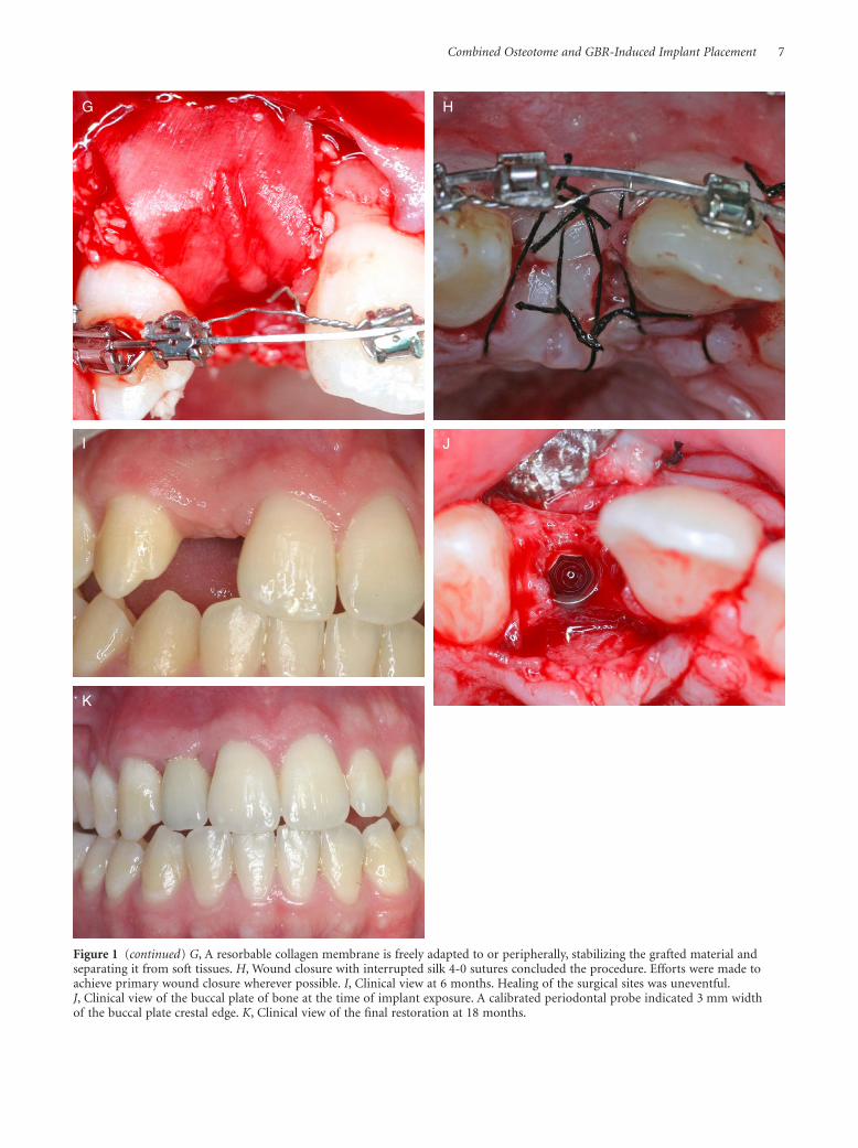
interrupted 4-0 silk sutures (Figure 1H); before closure,
releasing periosteal incisions where made wherever
needed.
If primary implant stability could not be achieved
by the end of the procedure, cases were recorded as a
failure (for the study purposes), and implant placement
was postponed for 4 to 6 months, but the augmentation
procedure was completed as described. Sutures were
removed 7 to 10 days postoperatively.
Combined Osteotome and GBR-Induced Implant Placement 3

TABLE 1 Characteristics of Patients (Successful Implants)
SmokingPeriodontal
Status a Age SexFollow-Up(months) Implant Site Length Width
1 No Severe 53 F 12 *12,22 13 3.3
2 Yes Severe 44 F 12 *12,22 16 3.3
3 No Severe 57 F 18 *24 16 3.75
4 No Moderate 65 F 30 ‡24 15 4
5 No Gingivitis 19 M 6 *11 16 3.75
6 Yes Severe 57 M 12 *24,26,27 12.5 3.75
7 No Moderate 61 M 42 *13,14 13 3.3
8 No Mild 58 F 120 *23,24,25 16 3.9
9 No Gingivitis 32 F 54 ‡11,22 15 3.75
10 No Gingivitis 68 F 144 *14,15,16 12.6 4.3
11 No Severe 56 M 131 *13,14,15,16 16 3.9
12 No Severe 71 F 120 *11,21,22 13.3 4.1
13 No Severe 61 F 72 *15,12,11,21,22,25 13.5 3.9
14 No Severe 56 F 96 §24,25,26 15 4
15 No Severe 63 M 108 ‡15,14,13,11,21,23,24,25 13.8 3.5
16 Yes Severe 58 F 108 ‡17,16,15,14,24,25,26,27 14.6 3.9
17 No Severe 71 M 102 ‡13,12,21,23,24 13.2 3.5
18 Yes Severe 60 M 66 ‡22,23,25,26 15 4
19 No Severe 67 M 60 *16,14,13,12,22,23,24,26 14.8 3.95
20 No Gingivitis 30 M 120 ‡11 15 4
21 No Severe 71 M 12 †22,24,25 14 3.7
22 No Severe 61 F 12 †15,14,12,22,24,25 13.6 3.7
23 No Moderate 56 M 6 *14 13 3.3
24 No Severe 68 M 6 *25 13 3.75
25 No Severe 58 M 24 †15,16,17 13 4.3
26 Yes Severe 60 F 12 †14,13,12,22,23,24,25 15.1 3.9
27 No Severe 69 F 12 †22,23 16 3.7
28 No Mild 39 F 72 ‡12,22 15 3.5
29 No Severe 50 M 72 ‡11,12,21,22 15 3.5
30 Yes Severe 77 F 48 *12,22,23 16 3.9
31 No Gingivitis 29 F 48 *21 16 3.75
32 No Mild 46 F 6 †11,22 16 3.7
33 No Severe 52 F 60 ‡12,11,21,22 15 3.5
34 No Severe 49 F 6 *15,14,24,25 13.3 3.6
35 No Severe 54 M 6 *23,24,25 15 3.75
Mean 55.6 52.4 14.5 3.8
SD 13.2 44.5 1.2 0.3
Min 19 6 12.5 3.3
Max 77 144 16 4.3
% male 43%
*Sandblasted acid-etched internal hex (Seven\BioCom\Lans), MIS, Shlomi, Israel.†Soluble blast media internal hex (Legacy), Implant Direct Malibu Hills Road, Calabasas Hills, CA, USA.‡Resorbable blast texture (Maesto\Prodigy), Bio-Horizons, South Birmingham, AL, USA.§Screw-Vent noncoated microtextured surface, Zimmer Dental Inc, Carlsbad, CA, USA.a Lindhe J, Lang NP, Karring T. Clinical periodontology and implant dentistry. Fifth edition, p. 423.F = female; M = male; SD = standard deviation.
4 Clinical Implant Dentistry and Related Research, Volume *, Number *, 2013

Healing of the surgical sites was uneventful
(Figure 1I). At 6 months, implants were exposed using
a midcrestal incision or a paracrestal (palatal) incision,
intending to maintain at least 3 mm of keratinized
masticatory mucosa. At this stage, crestal width was
remeasured using a caliper 2 mm apical to the exposed
implant shoulders, and marginal width of the buccal
plate of bone was remeasured using a calibrated peri-
odontal probe (Figure 1J). The bone measurements
(periodontal probe and caliper) were thus directly
made at the time of implant placement, immediately
after ridge expansion, but before graft placement, and
were repeated 6 month later at the time of implant
exposure.
TABLE 2 Treatment Effect
PatientBuccalPreop
BuccalPostop
BuccalIncrease
RidgePreop
RidgePostop
RidgeIncrease
Follow-Up(months)
MesialBone Loss
DistalBone Loss
1 0.5 2 1.5 3.25 6.3 3.05 12 1.00 0.70
2 0.5 2 1.5 4.05 6.3 2.25 12 3.25 3.35
3 1 2 1 4.1 6.2 2.1 18 3.50 3.70
4 1 2 1 3.5 6 2.5 30
5 1 3 2 2.5 6.1 3.6 6
6 1 3 2 2.9 8.1 5.2 12 0.33 0.47
7 0.5 2 1.5 3.15 6.3 3.15 42
8 1 3 2 3.63 7.7 4.07 120 2.33 2.23
9 1 2 1 4.2 6.1 1.9 54 2.35 1.35
10 1 3 2 3.97 8.1 4.13 144 1.33 1.40
11 1 3 2 4 7.7 3.7 131 0.35 0.43
12 1 4 3 3.17 8.5 5.33 120 0.70 0.70
13 1 4 3 4.83 8.4 3.57 72
14 1 3 2 4 7.5 3.5 96 2.43 2.37
15 0.75 2 1.25 3.6 6.5 2.9 108
16 0.75 2 1.25 3.5 6.4 2.9 108 2.19 2.03
17 0.5 3 2.5 2.86 6.1 3.24 102 1.96 1.78
18 1 3 2 3.3 7.8 4.5 66 1.60 1.85
19 0.81 3 2.19 4.49 7.95 3.46 60 2.21 3.04
20 1 3 2 4.8 7.8 3 120
21 1 3 2 3.37 7.6 4.23 12 1.83 1.57
22 1 3 2 3.53 7.5 3.97 12
23 1 3 2 2.5 7.4 4.9 6 0.60 0.50
24 0.5 3 2.5 3 7.2 4.2 6 1.50 1.30
25 1 4 3 4.17 8.5 4.33 24 0.67 0.83
26 0.79 2.21 1.43 3.2 7.11 3.91 12 3.41 3.03
27 1 3 2 3.4 6.8 3.4 12 1.85 2.15
28 1 4 3 5 8.5 3.5 72 0.55 0.55
29 1 2 1 5.03 6.5 1.48 72
30 0.5 2.67 2.17 3.97 7.57 3.6 48 4.23 4.53
31 1 3 2 3.2 7.7 4.5 48 1.00 0.40
32 1 3 2 4.05 7.25 3.2 6 2.80 1.90
33 1 2 1 4.6 6.5 1.9 60 1.28 1.33
34 0.5 2 1.5 4.23 6.6 2.38 6
35 0.5 3 2.5 3.6 7.2 3.6 6
Mean 0.86 2.80 1.91 3.73 7.19 3.50 52.43 1.81 1.74
SD 0.21 0.64 0.59 0.67 0.80 0.93 44.48 1.07 1.12
Range 0.5–1.0 2.0–4.0 1.0–3.0 2.5–5.0 6.0–8.5 1.5–5.3 6.0–144.0 0.3–4.2 0.4–4.5
SD = standard deviation.
Combined Osteotome and GBR-Induced Implant Placement 5

A B
C D
E F
Figure 1 A, The alveolar process at an implant site is exposed showing a narrow crestal ridge measuring 3 mm in width. B, Osteotomypreparation started using a pilot drill 2.0 mm in diameter followed by no. 1 osteotome (2.0 mm) inserted to the full depth of the initialosteotomy. C, Applying a 2.7 mm in diameter osteotome to further enlarge the osteotomy; the crestal ridge shows signs of expansion,with no fractures observed. D, A 3.75 mm–diameter implant placed immediately after ridge expansion and osteotomy preparation wascompleted, preventing osteotomy collapse. The implant is placed within the bony envelop with the implant shoulders flush with thesurrounding bone. E, Perforation of the buccal plate of bone using a 1 mm in diameter round bar increases bleeding and exposes thebone marrow to the healing site. F, Grafting the buccal aspect of the buccal plate using bone allograft is performed to further increasethe width of the alveolar bone process and ridge.
6 Clinical Implant Dentistry and Related Research, Volume *, Number *, 2013
G H
I
K
J
Figure 1 (continued) G, A resorbable collagen membrane is freely adapted to or peripherally, stabilizing the grafted material andseparating it from soft tissues. H, Wound closure with interrupted silk 4-0 sutures concluded the procedure. Efforts were made toachieve primary wound closure wherever possible. I, Clinical view at 6 months. Healing of the surgical sites was uneventful.J, Clinical view of the buccal plate of bone at the time of implant exposure. A calibrated periodontal probe indicated 3 mm widthof the buccal plate crestal edge. K, Clinical view of the final restoration at 18 months.
Combined Osteotome and GBR-Induced Implant Placement 7

Postoperative Management
Following surgery, patients were administered with
875 mg of amoxicillin with clavulanate potassium
(Augmentin, Glaxo Smith Klein) bid. Penicillin-sensitive
patients were administered with clindamycin HCL
(Dalacin-C, Pfizer NV/SA) 150 mg qid. Antibiotic
therapy was continued during the first week postopera-
tively. Dexamethasone, 4 mg/day, was prescribed for the
first, second, and third days postsurgery. Whenever
needed, analgetic/anti-inflammatory drug (naproxen
sodium 275 mg) was given twice a day. Chlorhexidine
0.2% mouth rinse was prescribed twice daily for 1
minute over a 3-week period of time. Patients were
instructed to avoid the use of a removable prosthetic
devises until after the sutures were removed, i.e., 10 to
14 days postoperatively. In full mouth cases or in cases
where removable prosthesis use was unavoidable, com-
plete release of the buccal flange was performed.
Healing Time
Six months after placement, implants were exposed and
restored with implant-supported fixed crowns or den-
tures (Figure 1K). Implants were considered successful
if they fulfilled the criteria set up by Albrektsson and
colleagues.28
Statistical Analysis
A paired sample Student’s t-test was used to evaluate
the significance of changes in ridge and buccal plate
width. An independent sample t-test was used to test for
differences in the outcome measures between male and
female patients. The Pearson’s correlation coefficient
was used to test for correlation between age and
outcome measures. All tests were two sided. p-value <.05
was considered significant.
Follow-Up and Criteria for Success
Patients were clinically followed up by the same investi-
gator (R.K.) at 3, 6, and 12 months postoperatively and
then annually. Patients were instructed individually
regarding personal hygiene programs depending on
their periodontal status and were seen and/or treated
every 3 to 6 months.
Periodical maintenance visits performed by a dental
hygienist supervised by R.K. included PI and PD mea-
surements and recording. At the annual evaluation of
the peri-implant soft-tissue condition, the assessment of
PI and PD was made using the following indices: PI was
determined using the O’leary Index.24 PD was measured
to the nearest millimeter using a periodontal probe
(Hu-Friedy) at the same surfaces.29
RESULTS
The files of 41 patients (23 woman; 18 men) met the
inclusion criteria. In these, one hundred twenty-two
sites were prepared for implant placement. Age range
was 19 to 77 years (mean 54.0 1 13.8). Follow-up from
time from the day of implantation varied between 6 and
144 months (mean 52.4), with 36% of the patients being
followed up for periods of time longer than 60 months.
The relevant details of the study group includ-
ing periodontal diagnosis, smoking status, follow-up
period, site, and type of each implant are shown in
Table 1. Implants’ diameter varied between 3.3 and
4.8 mm (average 3.80), and implants length between
12 and 16 mm (average 14.50).
At six sites(4.9%), it was impossible to establish
primary stability of the implant due to fractures of the
buccal plates; these were recorded as failures, omitted
from the statistical analysis, and were augmented by
the same GBR technique, but implant placement was
delayed for by 4 to 6 months. In the one hundred sixteen
successful surgical sites, the initial width of the alveolar
ridge ranged from 2.5 to 5.0 mm (mean = 3.7 1 0.67)
(Table 2). The final width in the successful sites ranged
from 6 to 8.5 mm (mean = 7.2 1 0.8). The difference in
ridge width averaged 3.5 1 0.93 (p < .0001) (Table 2).
The initial buccal bonny plate after implant installation
varied between 0.5 and 1 mm (mean = 0.86 1 0.21). At
6 months, the width of the buccal plates ranged between
2 and 4 mm (average 2.80 1 0.64). The average differ-
ence in width was 1.90 1 0.59 (p < .0001) (Table 2).
Table 3 presents a comparison between male and
female patients in ridge and buccal plate increase, and
mesial and distal bone loss. No significant differences
were found between male and female patients in any of
these outcome measures. Table 4 presents the Pearson’s
correlations between age and the outcome measures. No
significant positive correlations were found between age
and distal or mesial bone loss.
Healing was uneventful with no infection noted
during the postoperative phase. At 6 months, all im-
plants were successfully osseointegrated and loaded.
Follow-up period from the day of implant placement
ranged from 6 to 144 months (average 52.4 1 44.5).
8 Clinical Implant Dentistry and Related Research, Volume *, Number *, 2013

At the time of implant exposure, all one hundred
sixteen implants fulfilled our clinical and radiographic
success criteria.28 Patients were successfully restored
with implant-supported fixed ceramic crowns or bridges
(Figure 1J).
Clinical Observations
At the time of implant exposure, no remnants of
the collagen membranes were observed. In 40 implants
(34%), the cover screws were covered by bone to
a certain expense, demanding bone removal before
healing abutment could be properly placed (Figure 1J).
Complications
Fractures of the buccal plate (green stick) or dehis-
cence’s occurred in 21 (17.2%) implant sites (Figure 2,
A–E). In six sites (4.9%), these were associated with
failure to achieve primary stability, postponing implant
placement by 4 to 6 month; these were recorded as fail-
ures. In the remaining 15 (12.3%) sites, primary stability
was achieved, enabling successful completion of the
combined procedure. Spontaneous exposure occurred
in 18 (15.5%) implants (Figure 2F). Spontaneous expo-
sures were treated by replacement of the cover screw
with healing abutments. In cases where there was insuf-
ficient buccal band of keratinized gingiva, masticatory
mucosa pedicle flap was displaced from the palate adja-
cent to the implant (Figure 2, H and I).
Membrane Exposure
Membrane exposure occurred at five sites in five differ-
ent patients (14%). No exposure demanded premature
removal of the membrane because the exposed portions
of the membrane absorbed shortly after. When a mem-
brane became exposed, the subject was instructed to
topically apply 0.12% chlorhexidine Gel (Corsodyl
Dental Gel, Glaxo Smith Klein) twice a day.
PD
Eighty implants in 25 patients (71.4%) were available for
the final follow-up.
PD ranged from 1 to 8 mm, with two hundred
forty-five (76.6%) sites measuring 24 mm. Mean PD per
implant was 3.44 mm (standard deviation [SD] 1 1.5).
Bleeding on probing was recoreded in 49% of the
examined sites.
PI
At the time of data collection, PI24 ranged between 5 and
40% with a mean of 15%.
Radiographic bone level analysis (Table 2)
(Figure 2I) provided mean mesial bone loss of
1.81 1 1.07 mm (range 0.3–4.2 mm), and mean distal
bone loss was 1.74 1 1.12 mm (range 0.4–4.5 mm).
At implant level, the mean peri-implant bone loss
was 1.9 mm (median 1.8 mm; SD 1.1 mm; range 0.2–
5.6 mm). The most pronounced bone loss at an implant
was 5.6 mm.
Subject-Level Analyses
Smokers (six patients) presented a significantly higher
bone loss at implants’ distal aspects compared with
nonsmokers (m = 2.54 1 1.4 and 1.5 1 0.92, respec-
tively, p = .041) with but only marginally significant
higher mesial bone loss (m = 2.5 1 1.41 and 1.6 1 0.87,
respectively, p = .068).
DISCUSSION
A single-step technique for bone expansion using
cylindroconical osteotomes with gradually escalating
diameters and immediate placement of implants has
TABLE 3 Differences between Male and FemalePatients (Mean 1 SD) in Outcome Measures
Male Female p-Value
Mesial bone loss
(9 M, 16 F)
1.23 1 0.74 2.12 1 1.10 .038
Distal bone loss
(9 M, 16 F)
1.31 1 0.86 1.98 1 1.20 NS
Ridge increase
(15 M, 20 F)
3.70 1 0.92 3.28 1 0.92 NS
Buccal increase
(15 M, 20 F)
2.03 1 0.51 1.82 1 0.65 NS
F = female; NS = not significant; M = male; SD = standard deviation.
TABLE 4 Pearson Correlations between Age andOutcome Measures
Age p-Value
Mesial bone loss (n = 25) 0.242 NS
Distal bone loss (n = 25) 0.326 NS
Ridge increase (n = 35) 0.267 NS
Buccal increase (n = 35) 0.170 NS
NS = not significant.
Combined Osteotome and GBR-Induced Implant Placement 9

A
B
D E
C
Figure 2 A, Radiographic (computerized tomography) presentation of a narrow edentulous alveolar ridge at the upper left premolarregion. B, A 2-mm-wide osteotome is inserted into a 2-mm-wide initial osteotomy. C, Green stick fracture of the buccal plate at thefirst premolar region following insertion of a 3.5-mm-wide osteotome. D, Three implants placed into the expanded ridge withprimary stability. E, A native collagen membrane is fixated and stabilized by a single cover screw.
10 Clinical Implant Dentistry and Related Research, Volume *, Number *, 2013

previously been described.11–16 The technique, originally
reported by Summers,11 takes advantage of the fact that
bone is a viscoelastic material that can be compressed
and manipulated. Unlike drills, osteotomes do not excise
bone during osteotomy preparation; rather, they exert
lateral compression, which increases bone density
and encourages primary retention and implant initial
stability.11–16 The technique is particularly useful in
the upper maxilla, due to the lower bone density and
thinner cortical buccal plate in this jaw, qualities that
allow lateral compression and expansion. Our results
have shown that bony expansion using osteotomes is a
reliable and a relatively less invasive way of widening
narrow ridges. We further enhanced the technique
aiming to achieve critical thickness of more than 2 mm
of the buccal plate.22 It was claimed that this “critical
thickness” reduces the incidents and amount of facial
bone loss.22 Our implant survival rate (100%) is note-
worthy especially because in the present study implants
were placed in expanded ridges rather than in native
bone.
In comparison with the split-crest procedure that
can be performed when the ridge width is as narrow
as 1.5 mm,30 ridge expansion using osteotomes could
be applied only in cases in which ridge width was at
least 2.5 mm. Nevertheless, the added bony volume
in the present technique was similar (3.35 mm) to
that achieved with the ultrasonic bone surgery split
osteotomy technique,31 where an intentional fracture of
the buccal plate was produced aiming to create a “green
stick” fracture. In the present study, fissures of bone
or green stick fractures occurred only in 21 out of
one hundred twenty-two sites (17.2%). In 15 sites
(12.3%), primary stability was possible, thus enabling
F G
H I
Figure 2 (continued) F, Clinical view at 6 months. Implants were spontaneously exposed 2 months after placement. There is a lack ofbuccal keratinized gingiva. G, Clinical view of the exposed implants during grafting of masticatory mucosa shows complete healingof the previously fractured buccal bone. Mild bone recession is associated with the early exposed implants. H, Twelve years of clinicalview of the final ceramometal implant-supported bridge presents healthy keratinized gingival at the restored site. I, Final x-ray(12-year follow-up).
Combined Osteotome and GBR-Induced Implant Placement 11

the combined procedure. The gradual use of escalating
diameter osteotomes allowed a more gradual widening
of the ridge in contrast with the split-crest technique
in which the fractured buccal plate may reduce the
surgeon’s ability to achieve primary stability of the
simultaneously placed implant.
The osteotome procedure is limited only to ridges
that present spongy bone between the cortical plates.11,32
Another limitation is the need of at least 2.5-mm ridge
width or else, performing GBR procedure as previously
described necessary, postponing implant placement by
4 to 6 months.
Collagen was the first bioabsorbable membrane
material to be widely used in GBR. Collagen membranes
have been used in combination with autogenous bone,33
xenografts,34 hydroxyapatite,9 and other bone substitutes35
and have been associated with success rates comparable
with those achieved with nonresorbable expanded poly-
tetrafluoroethylene (e-PTFE) membranes.36–38 Such find-
ings support the use of a bioabsorbable barrier membrane
for the regeneration of bone. Moreover, the use of a bio-
absorbable membrane decreases the risk for membrane
infection if a soft-tissue dehiscence occurs postopera-
tively39,40 (14% in our study). The premature exposure
of these membranes that were managed by an infection
prevention protocol using local application of 0.12%
chlorhexidine did not lead to diminished results. Collagen
membranes have some limitations especially their rela-
tively short survival, thus losing their barrier function
within 2 or 3 months7; therefore, it may not provide
sufficient time for completion of the bone-regeneration
process. There is also some evidence that collagen does not
adequately exclude soft tissue.37
Our results may be compared with previous clinical
studies,6,33 which evaluated changes in ridge width after
GBR. In a series of 40 subjects, Buser and colleagues6
used corticocancellous autografts, bone chips, and non-
resorbable e-PTFE membranes to augment the ridge in
66 potential implantation sites narrower than 5 mm.
Before the augmentation procedure, the mean ridge
width, measured 2 mm apical to the alveolar crest, was
3.5 mm (range 2–4.5 mm). When the e-PTFE mem-
branes were removed 7 to 13 months later, the mean
width was 7 mm (range 5–10 mm). Subsequent place-
ment of titanium implants was successful in all 40
subjects. The mean increase in ridge width achieved by
Buser and colleagues6 was comparable with the present
study (3.50 mm); however, their technique required
a bone-harvesting procedure and frequently a second
operation for membrane removal. In the study con-
ducted by Von Arx and Buser,33 autogenous block grafts,
anorganic bovine bone mineral, and collagen mem-
branes were used for horizontal ridge augmentation of
58 sites in 42 subjects. The mean ridge width at the crest
was 3 mm (range 0.5–5 mm) before the GBR procedure
and 8 mm (range 6–10 mm) after an average of 5.8
months (range 4.5–13.5 months). Although their mean
gain in ridge width (4.6 mm)33 was greater than that
attained in our study (3.50 mm), their augmentation
technique was much more complex than the present one
and required harvesting of block grafts from the sym-
physis or the retromolar area. Moreover, we consider the
final ridge width obtained in our series (7.2 mm 1 0.80
at the crest) adequate for endosseous implant place-
ment. It is worth noting that the former value achieved
in our cases is lower than the ridge width (7.9 mm)
achieved by Nissan and colleagues25 who used cancellous
block allograft for lateral and vertical augmentation in
the posterior mandible. Using similar cancellous bone
block allografts in the anterior maxilla resulted in a
mean horizontal bone gain of 5 mm, i.e., higher than
that achieved in the present study (3.50 mm 1 0.93).26
However, the technique used in our study is easier for
both the operator and the patient, cheaper, less time-
consuming, and enabled simultaneous placement of
implants.
Marginal bone loss to a certain degree is considered
unavoidable soon after two-stage dental implants are
uncovered, loaded, and placed into function.41,42 This
phenomenon is believed to be a sequel of the reestab-
lishment of the “peri-implant biological width,” i.e.,
soft-tissue attachment characterized by junctional
epithelium and suprabony connective tissue adhesion;
these are being noted regardless the type of implant43 or
surgical technique.44 Therefore, 1 year after loading,
crestal bone level stabilized at 1.5 to 2 mm below the
implant/abutment junction is considered part of the
normal healing.45 Our data regarding mean peri-
implant bone loss followed-up period from 0.5 to 12
years (average 4.3 years) were 1.9 mm, thus falling
within the expected figures. These data are similar to
those presented in the longitudinal studies of Albrekt-
sson and colleagues28 and Attard and Zard,46 who
reported mean bone loss of 2 mm 1 year following
loading, but are higher than those claimed by Jemt and
Johansson,47 who reported a mean peri-implant bone
12 Clinical Implant Dentistry and Related Research, Volume *, Number *, 2013
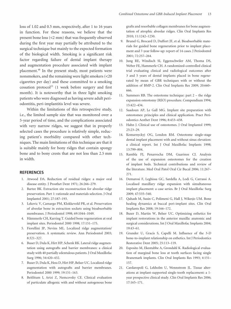
loss of 1.02 and 0.5 mm, respectively, after 1 to 16 years
in function. For these reasons, we believe that the
present bone loss (<2 mm) that was frequently observed
during the first year may partially be attributed to the
surgical technique but mainly to the expected formation
of the biological width. Smoking is a significant risk
factor regarding failure of dental implant therapy
and augmentation procedure associated with implant
placement.48 In the present study, most patients were
nonsmokers, and the remaining were light smokers (<20
cigarettes per day) and these committed to a smoking
cessation protocol23 (1 week before surgery and first
month). It is noteworthy that in three light smoking
patients who were diagnosed as having severe adult peri-
odontitis, peri-implantitis level was severe.
Within the limitations of this retrospective study,
i.e., the limited sample size that was monitored over a
5-year period of time, and the complications associated
with very narrow ridges, we suggest that in properly
selected cases the procedure is relatively simple, reduc-
ing patient’s morbidity compared with other tech-
niques. The main limitations of this technique are that it
is suitable mainly for bony ridges that contain spongy
bone and to bony crests that are not less than 2.5 mm
in width.
REFERENCES
1. Atwood DA. Reduction of residual ridges: a major oral
disease entity. J Prosthet Dent 1971; 26:266–279.
2. Bartee BK. Extraction site reconstruction for alveolar ridge
preservation. Part 1: rationale and materials selection. J Oral
Implantol 2001; 27:187–193.
3. Lekovic V, Camargo PM, Klokkevold PR, et al. Preservation
of alveolar bone in extraction sockets using bioabsorbable
membranes. J Periodontol 1998; 69:1044–1049.
4. Hämmerle CH, Karring T. Guided bone regeneration at oral
implant sites. Periodontol 2000 1998; 17:151–175.
5. Fiorellini JP, Nevins ML. Localized ridge augmentation/
preservation. A systematic review. Ann Periodontol 2003;
8:321–327.
6. Buser D, Dula K, Hirt HP, Schenk RK. Lateral ridge augmen-
tation using autografts and barrier membranes: a clinical
study with 40 partially edentulous patients. J Oral Maxillofac
Surg 1996; 54:420–432.
7. Buser D, Dula K, Hess D, Hirt HP, Belser UC. Localized ridge
augmentation with autografts and barrier membranes.
Periodontol 2000 1999; 19:151–163.
8. Beitlitum I, Artzi Z, Nemcovsky CE. Clinical evaluation
of particulate allogenic with and without autogenous bone
grafts and resorbable collagen membranes for bone augmen-
tation of atrophic alveolar ridges. Clin Oral Implants Res
2010; 11:1242–1250.
9. Brunel G, Brocard D, Duffort JF, et al. Bioabsorbable mate-
rials for guided bone regeneration prior to implant place-
ment and 7-year follow-up: report of 14 cases. J Periodontol
2001; 72:257–264.
10. Jung RE, Windisch SI, Eggenschwiler AM, Thoma DS,
Weber FE, Hammerle CH. A randomized-controlled clinical
trial evaluating clinical and radiological outcomes after
3 and 5 years of dental implants placed in bone regene-
rated by mean of GBR techniques with or without the
addition of BMP-2. Clin Oral Implants Res 2009; 20:660–
666.
11. Summers RB. The osteotome technique: part 2 – the ridge
expansion osteotomy (REO) procedure. Compendium 1994;
15:422–434.
12. Saadoun AP, Le Gall MG. Implant site preparation with
osteotomes: principles and clinical application. Pract Peri-
odontics Aesthet Dent 1996; 8:453–458.
13. Hahn J. Clinical use of osteotomes. J Oral Implantol 1999;
25:23–29.
14. Komarnyckyj OG, London RM. Osteotome single-stage
dental implant placement with and without sinus elevation:
a clinical report. Int J Oral Maxillofac Implants 1998;
13:799–804.
15. Rambla FJ, Penarrocha DM, Guarinos CJ. Analysis
of the use of expansion osteotomes for the creation
of implant beds. Technical contributions and review of
the literature. Med Oral Patol Oral Cir Bucal 2006; 11:267–
271.
16. Demarosi F, Leghissa GC, Sardella A, Lodi G, Carrassi A.
Localised maxillary ridge expansion with simultaneous
implant placement: a case series. Br J Oral Maxillofac Surg
2009; 47:535–540.
17. Qahash M, Susin C, Polimeni G, Hall J, Wikesjo UM. Bone
healing dynamics at buccal peri-implant sites. Clin Oral
Implants Res 2008; 19:166–172.
18. Buser D, Martin W, Belser UC. Optimizing esthetics for
implant restorations in the anterior maxilla: anatomic and
surgical considerations. Int J Oral Maxillofac Implants 2004;
19:43–61.
19. Grunder U, Gracis S, Capelli M. Influence of the 3-D
bone-to-implant relationship on esthetics. Int J Periodontics
Restorative Dent 2005; 25:113–119.
20. Esposito M, Ekestubbe A, Grondahl K. Radiological evalua-
tion of marginal bone loss at tooth surfaces facing single
Branemark implants. Clin Oral Implants Res 1993; 4:151–
157.
21. Cardaropoli G, Lekholm U, Wennstrom JL. Tissue alter-
ations at implant-supported single-tooth replacement: a 1-
year prospective clinical study. Clin Oral Implants Res 2006;
17:165–171.
Combined Osteotome and GBR-Induced Implant Placement 13

22. Spray JR, Black CG, Morris HF, Ochi S. The influence
of bone thickness on facial marginal bone response: stage 1
placement through stage 2 uncovering. Ann Periodontol
2000; 5:119–128.
23. Bain CA. Smoking and implant failure – benefits of a
smoking cessation protocol. Int J Oral Maxillofac Implants
1996; 11:756–759.
24. O’leary TJ, Drake RB, Naylor JE. The plaque control record.
J Periodontol 1972; 43:38.
25. Nissan J, Ghelfan O, Mardinger O, Calderon S, Chaushu G.
Efficacy of cancellous block allograft augmentation prior
to implant placement in the posterior atrophic mandible.
Clin Implant Dent Relat Res 2011; 13:279–285.
26. Nissan J, Mardinger O, Calderon S, Romanos GE,
Chaushu G. Cancellous bone block allografts for the aug-
mentation of the anterior atrophic maxilla. Clin Implant
Dent Relat Res 2011; 13:104–111.
27. Schwartz D, Levin L. Intraoral autogenous block onlay bone
grafting for extensive reconstruction of atrophic maxillary
alveolar ridges. J Periodontol 2005; 76:636–641.
28. Albrektsson T, Zarb G, Worthington P, Eriksson AR. The
long-term efficacy of currently used dental implants: a
review and proposed criteria of success. Int J Oral Maxillofac
Implants 1986; 1:11–25.
29. Mombelli A, Lang NP. Clinical parameters for the evaluation
of dental implants. Periodontol 2000 1994; 4:81–86.
30. Blus C, Szmukler-Moncler S. Split-crest and immediate
implant placement with ultra-sonic bone surgery: a 3 year
life-table analysis with 230 treated sites. Clin Oral Implants
Res 2006; 17:700–707.
31. Anitua E, Begona L, Orive G. Clinical evaluation of split-
crest technique with ultrasonic bone surgery for narrow
ridge expansion: status of soft and hard tissues and
implant success. Clin Implant Dent Relat Res 2011. DOI:
10.1111/j.1708-8208.2011.00340.x.
32. Sethi A, Kaus T. Maxillary ridge expansion with simul-
taneous implant placement: 5-year results of an ongoing
clinical study. Int J Oral Maxillofac Implants 2000; 15:491–
499.
33. Von Arx T, Buser D. Horizontal ridge augmentation using
autogenous block grafts and the guided bone regeneration
technique with collagen membranes: a clinical study with 42
patients. Clin Oral Implants Res 2006; 17:359–366.
34. Zitzmann NU, Scharer P, Marinello CP, Schüpbach P,
Berglundh T. Alveolar ridge augmentation with Bio-Oss: a
histologic study in humans. Int J Periodontics Restorative
Dent 2001; 21:288–295.
35. Wang HL, Boyapati L. “PASS” principles for predictable bone
regeneration. Implant Dent 2006; 15:8–17.
36. Hurzeler MB, Kohal RJ, Naghshbandi J, et al. Evaluation of a
new bioresorbable barrier to facilitate guided bone regene-
ration around exposed implant threads. An experimental
study in the monkey. Int J Oral Maxillofac Surg 1998;
27:315–320.
37. Bunyaratavej P, Wang HL. Collagen membranes: a review.
J Periodontol 2001; 72:215–229.
38. Schwartzmann M. Use of collagen membranes for guided
bone regeneration: a review. Implant Dent 2000; 9:63–66.
39. Stavropoulos F, Dahlin C, Ruskin JD, Johansson C. A
comparative study of barrier membranes as graft protectors
in the treatment of localized bone defects. An experimental
study in a canine model. Clin Oral Implants Res 2004;
15:435–442.
40. Buser D, Dula K, Lang NP, Nyman S. Long-term stability
of osseointegrated implants in bone regenerated with the
membrane technique. 5-year results of a prospective study
with 12 implants. Clin Oral Implants Res 1996; 7:175–183.
41. Hermann JS, Buser D, Schenk RK, Cochran DL. Crestal bone
changes around titanium implants. A histometric evaluation
of unloaded non-submerged and submerged implants in the
canine mandible. J Periodontol 2000; 71:1412–1424.
42. Hermann JS, Buser D, Schenk RK, Higginbottom FL,
Cochran DL. Biologic width around titanium implants.
A physiologically formed and stable dimension over time.
Clin Oral Implants Res 2000; 11:1–11.
43. Abrahamsson I, Berglundh T, Wennström J, Lindhe J.
The peri-implant hard and soft tissues at different implant
systems. A comparative study in the dog. Clin Oral Implants
Res 1996; 7:212–219.
44. Abrahamsson I, Berglundh T, Moon IS, Lindhe J. Peri-
implant tissues at submerged and non-submerged titanium
implants. J Clin Periodontol 1999; 26:600–607.
45. Cochran DL, Nummikoski PV, Schoolfield JD, Jones AA,
Oates TW. A prospective multicenter 5-year radiographic
evaluation of crestal bone levels over time in 596 dental
implants placed in 192 patients. J Periodontol 2009; 80:725–
733.
46. Attard NJ, Zard GA. Long-term treatment outcomes
in edentulous patients with implant-fixed prostheses: the
Toronto study. Int J Prosthodont 2004; 17:417–424.
47. Jemt T, Johansson J. Implant treatment in the edentulous
maxillae: a 15-year follow-up study on 76 consecutive
patients provided with fixed prostheses. Clin Implant Dent
Relat Res 2006; 8:61–69.
48. Strietzel FP, Reichart PA, Kale A, Kulkarni M, Wegner B,
Kuchler I. Smoking interferes with the prognosis of dental
implant treatment: a systematic review and meta-analysis.
J Clin Periodontol 2007; 34:523–544.
14 Clinical Implant Dentistry and Related Research, Volume *, Number *, 2013