Research Article Applied Artificial Bee Colony Optimization ...
Upload
khangminh22Category
view
3download
0
Citation: Won, H.; Kim, S.-H.; Yang,
J.-Y.; Jung, K.; Jeong, J.; Oh, J.-H.; Lee,
J.-H. Colony-Forming Efficiency
Assay to Assess Nanotoxicity of
Graphene Nanomaterials. Toxics 2022,
10, 236. https://doi.org/10.3390/
toxics10050236
Academic Editor:
Avelino Núñez-Delgado
Received: 6 April 2022
Accepted: 27 April 2022
Published: 5 May 2022
Publisher’s Note: MDPI stays neutral
with regard to jurisdictional claims in
published maps and institutional affil-
iations.
Copyright: © 2022 by the authors.
Licensee MDPI, Basel, Switzerland.
This article is an open access article
distributed under the terms and
conditions of the Creative Commons
Attribution (CC BY) license (https://
creativecommons.org/licenses/by/
4.0/).
toxics
Article
Colony-Forming Efficiency Assay to Assess Nanotoxicity ofGraphene NanomaterialsHansol Won † , Sung-Hyun Kim † , Jun-Young Yang, Kikyung Jung, Jayoung Jeong, Jae-Ho Oh and Jin-Hee Lee *
Division of Toxicological Research, National Institute of Food and Drug Safety Evaluation, Ministry of Food andDrug Safety, 187, Osongsaengmyeong 2-Ro, Cheongju 28159, Korea; [email protected] (H.W.);[email protected] (S.-H.K.); [email protected] (J.-Y.Y.); [email protected] (K.J.); [email protected] (J.J.);[email protected] (J.-H.O.)* Correspondence: [email protected]; Tel.: +82-43-719-5106; Fax: +82-43-719-5100† These authors contributed equally to this work.
Abstract: The nano-market has grown rapidly over the past decades and a wide variety of productsare now being manufactured, including those for biomedical applications. Despite the widespreaduse of nanomaterials in various industries, safety and health effects on humans are still controversial,and testing methods for nanotoxicity have not yet been clearly established. Nanomaterials havebeen reported to interfere with conventional cytotoxicity tests due to their unique properties, such aslight absorption or light scattering. In this regard, the colony-forming efficacy (CFE) assay has beensuggested as a suitable test method for testing some nanomaterials without these color-interferences.In this study, we selected two types of GNPs (Graphene nanoplatelets) as test nanomaterials andevaluated CFE assay to assess the cytotoxicity of GNPs. Moreover, for further investigation, includingexpansion into other cell types, GNPs were evaluated by the conventional cytotoxicity tests includ-ing the 3-(4,5-dimethylthiazol-2-yl)-5-(3-carboxymethoxyphenyl)-2-(4-sulfophenyl)-2H-tetrazolium(MTS), Cell Counting Kit-8 (CCK-8), and Neutral red uptake (NRU) assay using MDCK, A549 andHepG2 cells. The results of CFE assay suggest that this test method for three cell lines can be appliedfor GNPs. In addition, the CFE assay was able to evaluate cytotoxicity regardless more accurately ofcolor interference caused by residual nanomaterials.
Keywords: graphene; cytotoxicity; CFE; interference
1. Introduction
In recent years, due to the rapid growth of nanotechnology, various manufacturingnanomaterials are being produced, and nanomaterials are used in various industries suchas batteries, electrodes, cosmetics, displays and biomedical engineering [1–3]. The growthof the nano-industry affects our lives in a more prosperous manner and contributes to itby providing various benefits, however like a ‘double-edged sword’ it has the potentialto induce human toxicity, both large and small when exposed to the body. Therefore, it iscrucial to develop an accurate nanotoxicity evaluation method to understand the toxicityof these nanomaterials.
The Organization for Economic Cooperation and Development (OECD), EuropeanUnion (EU) and other organizations stipulate the following for manufactured nanoma-terials: ‘Materials with a size less than 100 nm made for this purpose’ [4,5]. As such,nanomaterials are nanoscopic in size, and the risk of nanomaterial products stem fromits small size and the unique physicochemical properties of the nanomaterial. Taking thephysical ‘shape’ as an example, carbon nanotube (CNT) nanomaterials with acicular struc-tures such as asbestos or glass fibers have risks such as cancer-causing potential [6,7]. Inaddition, high surface reaction power and surface charge, due to their very small size, cancontribute to allowing nanomaterials to be easily grouped and accumulated into cells [8,9].
Toxics 2022, 10, 236. https://doi.org/10.3390/toxics10050236 https://www.mdpi.com/journal/toxics
Toxics 2022, 10, 236 2 of 10
Unlike general chemicals, nanomaterials have unique properties that make them nearlynon-soluble; thus, during measurements, solid nanomaterials are detected in particularareas such as the bottom of a well, deposition on cell membranes and during intracellularuptake, which can interfere with the system and generate unreliable data [10]. Guadagniniet al. [11] also reported significant differences observed in nanomaterials interference forcytotoxicity analysis depends on the nature of nanomaterials. These problems can make itdifficult to determine the exact cytotoxicity of nanomaterials, so a solution to the effects ofnanomaterial interference is needed.
Colony forming efficiency (CFE) assay used to measure cellular ability to form colonieswas described in OECD detailed review paper and Joint Research Centre (JRC) reports [12,13].This in vitro assay can be used to determine cytotoxicity induced by nanomaterials. It can beperformed with any adherent cells that are able to form colonies including human adult lowcalcium high temperature (HaCaT), Madin Darby canine kidney cell line (MDCK), humanlung cancer cells (A549), human liver carcinoma cell line (HepG2) and immortalized mousefibroblast cell line (Balb/3T3) cells. The great advantage of CFE assay is a label-free test thatreduces the possibility of the incident of nanomaterials interferences. This testing methodcalculates cell viability by comparing the number of colonies in the vehicle control aftertreatment with toxicants.
We conducted a test to analyze the colony formation efficacy of various nanomaterialsin MDCK cells through the OECD-JRC report [13]. In the present study, we tried to evaluatethe CFE assay method for GNPs, a kind of carbon nanomaterial. In addition, we want toestablish a GNP’s CFE assay protocol based on A549 and HepG2 cells, which is differentfrom the CFE conditions of MDCK cells proposed by the OECD.
In this study, the applicability of carbon-based graphene nanomaterials to a total ofthree cell lines was evaluated through CFE assay, an in vitro test method independent ofthe effect of color interference. Also, we aimed to compare the cytotoxicity assay of theconventional colorimetric cytotoxicity assay with that of the CFE assay.
2. Materials and Methods2.1. Graphene Nanomaterials
Two types of GNPs (product No. 06-0225, product No. 06-0230) materials were pur-chased from Strem Chemicals (Newburyport, MA, USA). Their morphological images wereconfirmed by transmission electron microscopy (TEM; JEM-1200EX II, JEOL, Tokyo, Japan).The zeta potentials of the GNPs were measured using a Zetasizer Nano ZS instrument(Malvern Instruments, Malvern Hills, UK). To confirm the unique physicochemical prop-erties of GNPs, this assay was measured in PBS and culture medium (DMEM containedwith 10% FBS). In order to evaluate the dispersion stability of the test substance, the finalworking time was measured up to 72 h. To evaluate the dispersion stability of GNPs, adynamic light scattering (DLS) was measured using a Zetasizer (Malvern). The levels ofendotoxin were evaluated using an Endpoint Chromogenic Limulus Amoebocyte Lysate(LAL) QCL-1000 assay (Cambrex, Walkersville, MD, USA). Endotoxins were measuredaccording to the protocols provided by the kit’s manufacturer.
2.2. Preparation of Nanomaterials Suspensions
GNP’s suspension was prepared by slightly modifying described methods [14,15].Briefly, the GNPs stock (10× fold) solutions were dispersed in PBS and sonicated at 40 kHzwith 100 W output power for 30 min in an ultra-sonicator (Saehan-Sonic, Seoul, Korea).Thereafter, Dulbecco’s Modified Eagle’s Medium (DMEM) (Life Technologies, Grand Island,NY, USA) supplemented with 10% fetal bovine serum (FBS) (Life Technologies), 100 U/mLpenicillin (Life Technologies), and 100 µg/mL streptomycin (Life Technologies) was addedto different working concentrations (Table 1). Initially, the concentration of GNPs was setbased on a JRC report, which was previously evaluated by reference to the concentration ofthe same single-wall carbon nanotubes (swCNTs) [13]. The concentrations of two GNPswere finally set by performing a preliminary toxicity assessment based on the concentration
Toxics 2022, 10, 236 3 of 10
of the above carbon nanotubes and up-adjusting the test concentration according to theresults.
Table 1. Test concentration of the two different carbon nanomaterials.
Nanomaterials CAS RN Test Concentration * (µg/mL)
GNPs-1 (300 m2/g) 7782-42-5 1, 10, 100, 200, 400, 800
GNPs-2 (500 m2/g) 7782-42-5 1, 10, 100, 200, 400, 800* The concentration of GNPs was set based on a JRC report [13]. GNPs = graphene nanoplatelets.
2.3. Cell Culture
MDCK (product No. CCL-34), A549 (product No. CCL-185), and HepG2 (productNo. HB-8065) cell lines were purchased from American Type Culture Collection (ATCC;Manassas, VA, USA). The cells were cultured in DMEM medium supplemented with 10%FBS, 100 U/mL penicillin and 100 µg/mL streptomycin. Three types of cells were sub-cultured every 2–4 days at about 80% confluence. For the experiments, cell density wasadjusted according to the conditions of each cytotoxicity method and seeded on the 96 wellculture plates or 60 × 15-mm Petri dish. Then, culture medium was replaced with a freshmedium and incubated in a humidified atmosphere condition of 5% CO2 at 37 ◦C.
2.4. Colony Forming Efficiency Assay Methods
CFE assay was performed as previously described to study the cytotoxicity inducedby two types of GNPs [13]. The cells were seeded at a density of 200 cells/dish (MDCK) in3 mL complete culture medium at least in three replicates for each treatment. In the sameprocedure, 400 cells/dish for A549 cells and 200 cells/dish for HepG2 cells were inoculated,respectively. After 24 h, the treatment suspensions of nanomaterials and positive control(sodium chromate, Na2CrO4, product No. 307831, Sigma-Aldrich, St. Louis, MO, USA)were added to the cells. After 72 h of exposure, the medium was changed with a freshcomplete culture medium. Considering the growth cycle of each cell line colony, after5 days (MDCK), 8 days (HepG2) and 10 days (A549), each cell was fixed for 20 min with3.7% (v/v) of formaldehyde solution (Sigma-Aldrich) in PBS without calcium, magnesiumand sodium bicarbonate (Life Technologies, product No. 14190-250), and stained for 30 minwith 0.4% (v/v) Giemsa solution (Sigma-Aldrich, product No. GS500) in ultrapure water.Colonies were manually scored under a stereomicroscope. The results were expressed asCFE (%) = [(average of treatment colonies/average of control colonies) × 100] and thecorresponding standard error means [SEM% = SD/
√(number of treatments)].
Colony Forming Efficiency (%) =Average of treatment colonies× 100
Average of control colonies(1)
2.5. Cytotoxicity Measurement of Colorimetric Based Assay
To confirm the difference between the existing tests measuring cytotoxicity of thetest substance and the CFE assay, two types of GNPs were evaluated using three com-monly used colorimetric cytotoxicity assays. The MTS assay; each cell was seeded at3 × 104 cells/well in 96-well plates and cultured overnight and incubated for another 24 h.The conversion of MTS tetrazolium salt into its reduced formazan form was assessed withthe CellTiter 96 AQueous Non-Radioactive Cell Proliferation Assay kit (Promega, Madison,WI, USA) following the manufacturer’s protocol. The absorbance was read at 450 nm on aSynergy HT Multimode Microplate Reader (Bio-Tek Instruments, Winooski, VT, USA).
Cell Counting Kit-8 (CCK-8) assay; to evaluate the cell viability, cells were seededinto 96-well plates at a density of 1 ×10⁴ cells/mL and incubated overnight to reachapproximately 80% confluence. Followed by the addition of suspension containing eithernanomaterials or positive control and then were incubated for 24 h at each different dose.After 24 h, the cell viability was measured using a CCK-8 assay kit (Dojindo MolecularTechnologies, Gaithersburg, MD, USA).
Toxics 2022, 10, 236 4 of 10
Neutral red uptake (NRU) assay is a dye exclusion assay (Sigma-Aldrich, Cat #N2889).Briefly, each cell was seeded at 5 × 10³ cells/well in 96-well plates for 24 h prior to thetreatments. After nanomaterials suspension treatment, plates were incubated for 2 h witha supplemented medium containing 40 µg/mL of neutral red. Cells were subsequentlywashed twice with Dulbecco’s Phosphate Buffered Saline (DPBS) and the dye was ex-tracted with 200 µL destaining solution (ethanol, deionized water, and glacial acetic acid,50:49:1 v/v). The absorbance was read at 540 nm using a microplate reader. Cell viability interms of percentage of control was expressed in the same manner as for the MTS assay. Allfour types of test result data were expressed as mean ± SEM (n = 3) using GraphPad PrismV5.0 (GraphPad Software, San Diego, CA, USA).
3. Results3.1. Physicochemical Characteristics of Graphene Nanomaterials
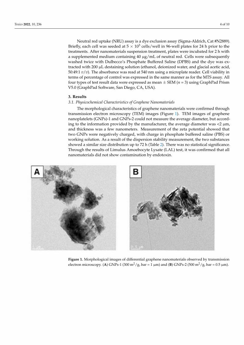
The morphological characteristics of graphene nanomaterials were confirmed throughtransmission electron microscopy (TEM) images (Figure 1). TEM images of graphenenanoplatelets (GNPs)-1 and GNPs-2 could not measure the average diameter, but accord-ing to the information provided by the manufacturer, the average diameter was <2 µm,and thickness was a few nanometers. Measurement of the zeta potential showed thattwo GNPs were negatively charged, with charge in phosphate buffered saline (PBS) orworking solution. As a result of the dispersion stability measurement, the two substancesshowed a similar size distribution up to 72 h (Table 2). There was no statistical significance.Through the results of Limulus Amoebocyte Lysate (LAL) test, it was confirmed that allnanomaterials did not show contamination by endotoxin.
Toxics 2022, 10, x FOR PEER REVIEW 4 of 10
Cell Counting Kit-8 (CCK-8) assay; to evaluate the cell viability, cells were seeded
into 96-well plates at a density of 1 ×10⁴ cells/mL and incubated overnight to reach ap-
proximately 80% confluence. Followed by the addition of suspension containing either
nanomaterials or positive control and then were incubated for 24 h at each different dose.
After 24 h, the cell viability was measured using a CCK-8 assay kit (Dojindo Molecular
Technologies, Gaithersburg, MD, USA).
Neutral red uptake (NRU) assay is a dye exclusion assay (Sigma-Aldrich, Cat
#N2889). Briefly, each cell was seeded at 5 × 10³ cells/well in 96-well plates for 24 h prior
to the treatments. After nanomaterials suspension treatment, plates were incubated for 2
h with a supplemented medium containing 40 μg/mL of neutral red. Cells were subse-
quently washed twice with Dulbecco’s Phosphate Buffered Saline (DPBS) and the dye was
extracted with 200 μL destaining solution (ethanol, deionized water, and glacial acetic
acid, 50:49:1 v/v). The absorbance was read at 540 nm using a microplate reader. Cell via-
bility in terms of percentage of control was expressed in the same manner as for the MTS
assay. All four types of test result data were expressed as mean ± SEM (n = 3) using
GraphPad Prism V5.0 (GraphPad Software, San Diego, CA, USA).
3. Results
3.1. Physicochemical Characteristics of Graphene Nanomaterials
The morphological characteristics of graphene nanomaterials were confirmed
through transmission electron microscopy (TEM) images (Figure 1). TEM images of gra-
phene nanoplatelets (GNPs)-1 and GNPs-2 could not measure the average diameter, but
according to the information provided by the manufacturer, the average diameter was <2
μm, and thickness was a few nanometers. Measurement of the zeta potential showed that
two GNPs were negatively charged, with charge in phosphate buffered saline (PBS) or
working solution. As a result of the dispersion stability measurement, the two substances
showed a similar size distribution up to 72 h (Table 2). There was no statistical signifi-
cance. Through the results of Limulus Amoebocyte Lysate (LAL) test, it was confirmed
that all nanomaterials did not show contamination by endotoxin.
Figure 1. Morphological images of differential graphene nanomaterials observed by transmission
electron microscopy. (A) GNPs-1 (300 m2/g, bar = 1 μm) and (B) GNPs-2 (500 m2/g, bar = 0.5 μm).
Table 2. Physicochemical characterization of the graphene nanomaterials.
Characterization GNPs-1 GNPs-2
Average diameter (nm) * <2 μm <2 μm
(a thickness of a few nanometers)
Figure 1. Morphological images of differential graphene nanomaterials observed by transmissionelectron microscopy. (A) GNPs-1 (300 m2/g, bar = 1 µm) and (B) GNPs-2 (500 m2/g, bar = 0.5 µm).
Toxics 2022, 10, 236 5 of 10
Table 2. Physicochemical characterization of the graphene nanomaterials.
Characterization GNPs-1 GNPs-2
Average diameter (nm) *<2 µm <2 µm
(a thickness of a few nanometers)
Surface area (m2/g) * 300 500
Zeta potential (mV) in PBS ** −30.01 ± 4.30 −33.32 ± 4.91
Zeta potential (mV) in DMEM ** −26.82 ± 0.69 −25.78 ± 0.81
Endotoxin (EU/mL) <0.1 <0.1
Dispersion stability measurement using DLS (nm) **0 h 3018.00 ± 213.55 2929.00 ± 66.4724 h 3116.67 ± 684.92 3074.50 ± 303.3572 h 3416.00 ± 823.07 3467.00 ± 934.80
pHIn DMEM 10.81
In working solution ** 8.87 8.56Data are expressed as mean ± standard error of the mean (n = 6). * This data used material informationprovided by the manufacturer. ** Working concentration was 800 µg/mL (Measurement was performed bydiluting× 100 fold in DW at the highest concentration). GNPs = graphene nanoplatelets, PBS = phosphate bufferedsaline, DMEM = Dulbecco’s modified Eagle’s medium, EU = endotoxin unit, DLS = Dynamic light scattering.
3.2. Cytotoxicity Evaluation of GNPs Using CFE Assay for Selected Cell Lines
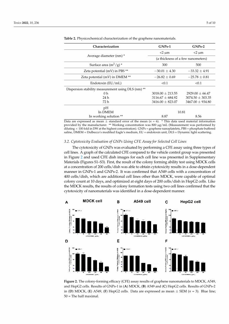
The cytotoxicity of GNPs was evaluated by performing a CFE assay using three types ofcell lines. A graph of the calculated CFE compared to the vehicle control group was presentedin Figure 2 and used CFE dish images for each cell line was presented in SupplementaryMaterials (Figures S1–S3). First, the result of the colony forming ability test using MDCK cellsat a concentration of 200 cells/dish was able to obtain cytotoxicity results in a dose-dependentmanner in GNPs-1 and GNPs-2. It was confirmed that A549 cells with a concentration of400 cells/dish, which are additional cell lines other than MDCK, were capable of optimalcolony count at 10 days, and optimized at eight days of 200 cells/dish in HepG2 cells. Likethe MDCK results, the results of colony formation tests using two cell lines confirmed that thecytotoxicity of nanomaterials was identified in a dose-dependent manner.
Toxics 2022, 10, x FOR PEER REVIEW 5 of 10
Surface area (m2/g) * 300 500
Zeta potential (mV) in PBS ** −30.01 ± 4.30 −33.32 ± 4.91
Zeta potential (mV) in DMEM ** −26.82 ± 0.69 −25.78 ± 0.81
Endotoxin (EU/mL) <0.1 <0.1
Dispersion stability measurement using
DLS (nm) **
0 h 3018.00 ± 213.55 2929.00 ± 66.47
24 h 3116.67 ± 684.92 3074.50 ± 303.35
72 h 3416.00 ± 823.07 3467.00 ± 934.80
pH
In DMEM 10.81
In working solution ** 8.87 8.56
Data are expressed as mean ± standard error of the mean (n = 6). * This data used material infor-
mation provided by the manufacturer. ** Working concentration was 800 μg/mL (Measurement was
performed by diluting × 100 fold in DW at the highest concentration). GNPs = graphene nanoplate-
lets, PBS = phosphate buffered saline, DMEM = Dulbecco’s modified Eagle’s medium, EU = endo-
toxin unit, DLS = Dynamic light scattering.
3.2. Cytotoxicity Evaluation of GNPs Using CFE Assay for Selected Cell Lines
The cytotoxicity of GNPs was evaluated by performing a CFE assay using three types
of cell lines. A graph of the calculated CFE compared to the vehicle control group was
presented in Figure 2 and used CFE dish images for each cell line was presented in Sup-
plementary Materials (Figure S1–S3). First, the result of the colony forming ability test
using MDCK cells at a concentration of 200 cells/dish was able to obtain cytotoxicity re-
sults in a dose-dependent manner in GNPs-1 and GNPs-2. It was confirmed that A549
cells with a concentration of 400 cells/dish, which are additional cell lines other than
MDCK, were capable of optimal colony count at 10 days, and optimized at eight days of
200 cells/dish in HepG2 cells. Like the MDCK results, the results of colony formation tests
using two cell lines confirmed that the cytotoxicity of nanomaterials was identified in a
dose-dependent manner.
Figure 2. The colony-forming efficacy (CFE) assay results of graphene nanomaterials to MDCK, A549,and HepG2 cells. Results of GNPs-1 in (A) MDCK, (B) A549 and (C) HepG2 cells. Results of GNPs-2in (D) MDCK, (E) A549, (F) HepG2 cells. Data are expressed as mean ± SEM (n = 3). Blue line;50 = The half maximal.
Toxics 2022, 10, 236 6 of 10
3.3. Differences of Cytotoxicity between Colorimetric Assays
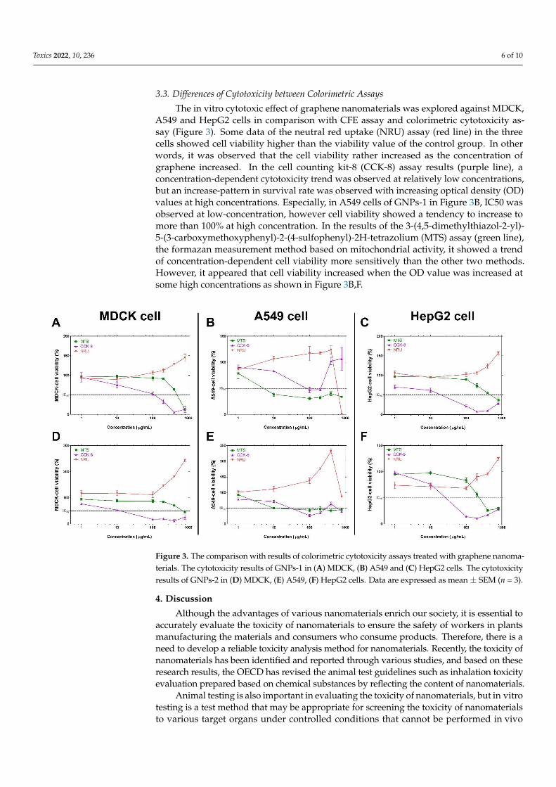
The in vitro cytotoxic effect of graphene nanomaterials was explored against MDCK,A549 and HepG2 cells in comparison with CFE assay and colorimetric cytotoxicity as-say (Figure 3). Some data of the neutral red uptake (NRU) assay (red line) in the threecells showed cell viability higher than the viability value of the control group. In otherwords, it was observed that the cell viability rather increased as the concentration ofgraphene increased. In the cell counting kit-8 (CCK-8) assay results (purple line), aconcentration-dependent cytotoxicity trend was observed at relatively low concentrations,but an increase-pattern in survival rate was observed with increasing optical density (OD)values at high concentrations. Especially, in A549 cells of GNPs-1 in Figure 3B, IC50 wasobserved at low-concentration, however cell viability showed a tendency to increase tomore than 100% at high concentration. In the results of the 3-(4,5-dimethylthiazol-2-yl)-5-(3-carboxymethoxyphenyl)-2-(4-sulfophenyl)-2H-tetrazolium (MTS) assay (green line),the formazan measurement method based on mitochondrial activity, it showed a trendof concentration-dependent cell viability more sensitively than the other two methods.However, it appeared that cell viability increased when the OD value was increased atsome high concentrations as shown in Figure 3B,F.
Toxics 2022, 10, x FOR PEER REVIEW 6 of 10
Figure 2. The colony-forming efficacy (CFE) assay results of graphene nanomaterials to MDCK,
A549, and HepG2 cells. Results of GNPs-1 in (A) MDCK, (B) A549 and (C) HepG2 cells. Results of
GNPs-2 in (D) MDCK, (E) A549, (F) HepG2 cells. Data are expressed as mean ± SEM (n = 3). Blue
line; 50 = The half maximal.
3.3. Differences of Cytotoxicity between Colorimetric Assays
The in vitro cytotoxic effect of graphene nanomaterials was explored against MDCK,
A549 and HepG2 cells in comparison with CFE assay and colorimetric cytotoxicity assay
(Figure 3). Some data of the neutral red uptake (NRU) assay (red line) in the three cells
showed cell viability higher than the viability value of the control group. In other words,
it was observed that the cell viability rather increased as the concentration of graphene
increased. In the cell counting kit-8 (CCK-8) assay results (purple line), a concentration-
dependent cytotoxicity trend was observed at relatively low concentrations, but an in-
crease-pattern in survival rate was observed with increasing optical density (OD) values
at high concentrations. Especially, in A549 cells of GNPs-1 in Figure 3B, IC50 was ob-
served at low-concentration, however cell viability showed a tendency to increase to more
than 100% at high concentration. In the results of the 3-(4,5-dimethylthiazol-2-yl)-5-(3-car-
boxymethoxyphenyl)-2-(4-sulfophenyl)-2H-tetrazolium (MTS) assay (green line), the
formazan measurement method based on mitochondrial activity, it showed a trend of
concentration-dependent cell viability more sensitively than the other two methods. How-
ever, it appeared that cell viability increased when the OD value was increased at some
high concentrations as shown in Figure 3B,F.
Figure 3. The comparison with results of colorimetric cytotoxicity assays treated with graphene na-
nomaterials. The cytotoxicity results of GNPs-1 in (A) MDCK, (B) A549 and (C) HepG2 cells. The
cytotoxicity results of GNPs-2 in (D) MDCK, (E) A549, (F) HepG2 cells. Data are expressed as mean
± SEM (n = 3).
4. Discussion
Although the advantages of various nanomaterials enrich our society, it is essential
to accurately evaluate the toxicity of nanomaterials to ensure the safety of workers in
plants manufacturing the materials and consumers who consume products. Therefore,
there is a need to develop a reliable toxicity analysis method for nanomaterials. Recently,
Figure 3. The comparison with results of colorimetric cytotoxicity assays treated with graphene nanoma-terials. The cytotoxicity results of GNPs-1 in (A) MDCK, (B) A549 and (C) HepG2 cells. The cytotoxicityresults of GNPs-2 in (D) MDCK, (E) A549, (F) HepG2 cells. Data are expressed as mean± SEM (n = 3).
4. Discussion
Although the advantages of various nanomaterials enrich our society, it is essential toaccurately evaluate the toxicity of nanomaterials to ensure the safety of workers in plantsmanufacturing the materials and consumers who consume products. Therefore, there is aneed to develop a reliable toxicity analysis method for nanomaterials. Recently, the toxicity ofnanomaterials has been identified and reported through various studies, and based on theseresearch results, the OECD has revised the animal test guidelines such as inhalation toxicityevaluation prepared based on chemical substances by reflecting the content of nanomaterials.
Animal testing is also important in evaluating the toxicity of nanomaterials, but in vitrotesting is a test method that may be appropriate for screening the toxicity of nanomaterialsto various target organs under controlled conditions that cannot be performed in vivo
Toxics 2022, 10, 236 7 of 10
testing. Oberdorster et al. propose a portal-of-entry toxicity test for target organ toxicitysuch as lung, skin, endothelium, liver and kidney as an in vitro technique [16]. The OECD-JRC report exemplifies some test cases of nanomaterials, and a preemptive study wasconducted [13]. Through this work, the OECD established and proposed a “test protocol”for a colony-forming efficacy assay based on MDCK cells. However, since the test methodwas verified only for MDCK cells, research was needed to apply it to various cells. Toutilize the colony test for a variety of cell lines, Ponti et al. reported studies applyingvarious adherent cells such as Caco-2, HepG2, etc., [17]. In our study, we establishedthe applicability of the CFE assay using the alveolar epithelial cell line A549 and thehepatocyte cell line HepG2 cells as organ-derived cell lines closely related to the exposureand accumulation of nanomaterials. Based on the test results, different time-points arerequired to prepare a colony optimized for cells, so there is a limitation that a ‘standardizednumber of cells’ cannot be specified. However, in the end, it was confirmed that these celllines could be used in the CFE test.
Other studies involving the testing of graphene cytotoxicity with CFE assayin vitro [18–20]. Our study performs validation of cell line expansion of swCNTs(Figure S4), and also reports the CFE evaluation results of carbon-based graphene nano-materials that have not been reported in previous studies [13]. GNP is a type of car-bon nanomaterial, which has recently attracted great attention in various fields includingbiomedical [21]. In general, as well known, nanomaterials were insoluble in almost solvents,and most readily form agglomerates as confirmed by transmission electron microscopy(Figure 1) [22]. Since the nanotoxicity can be accurately evaluated only when the nanoma-terials used are uniformly dispersed, we tried to find a dispersion method optimized fornanomaterials. According to the papers reported on dispersion, ‘serum protein’ is known asa very useful dispersing agent [23,24]. In fact, it has been reported that the large aggregationof nanomaterials in a solvent was reduced by gentle aggregation in the final solution afterdispersion using serum [25,26]. Therefore, by applying this method, the protein-coronacoating operation was performed using FBS on the nanomaterial stock solution to inducethe most homogeneous dispersion of the particles. In addition, since the mechanical disper-sion operation of the ultrasonic disperser can contribute to the homogenization of the testmaterial, the operation was additionally reflected [23].
The contrasting difference between the CFE assay and the conventional cytotoxicityassay for GNPs substances may suggest that the CFE assay was a reliable in vitro toxicityassay for GNPs [10,11,27]. Nanomaterials are either absorbed by cells or deposited on cellmembranes or wells. These particles interact with cells or remain in culture plate wellsdespite multiple washing operations [17,28]. Traditional colorimetric-cytotoxicity assaysuse absorbance to evaluate the toxicity of test materials by calculating the OD values ofthe control and test groups and calculating the cytotoxicity in ‘percentage (%)’ [29–32]. Ifconventional absorbance-based colorimetric measurements were performed in the presenceof such residual nanomaterials, incorrect results may be obtained through distortion ofOD values. According to Guadagnini et al. (2015) [11], TiO2 nanoparticles can cause false-negative results because they have the property of increasing absorbance when measuredby a colorimetric method. Moreover, Wörle-Knirsch et al., Casey et al., Monteiro-Riviereet al. reported that the evaluation of carbon-based nanomaterials, which was also usedin our study, may not be suitable for cytotoxicity evaluation due to color interferenceof the material [33–35]. As such, absorbance-based tests using nanomaterials are highlylikely to cause distortion of results, such as increased cell viability in the presence ofcolor interference. Additionally, if strong washing is performed to remove the remainingnanomaterials, there is a possibility that it may cause loss of cells attached to the bottom ofthe well and lower the viability of the original cells.
In order to avoid distortion of the measurement result due to residual nanomaterials,a method of transferring the supernatant to a new plate may be considered [36]. Whenthis method is applied, the uptake state in the cell or the substances strongly attached tothe outer membrane are excluded from the absorbance measurement so that distortion
Toxics 2022, 10, 236 8 of 10
is not induced. However, in the case of nanomaterials that dissolve rapidly and releasemetal ions, it can affect the color of the medium and induce distortion in the supernatantitself. For example, in the case of CuO nanoparticles, Cu ions chemically inactivate theintracellular formazan formation cascade in LDH analysis, which is one of the color-metricassays, resulting in false-negative results [26]. Therefore, the CFE assay is a label-free assaythat counts the number of colonies in evaluating these kinds of nanomaterials that canaffect the supernatant itself, so it can be a good alternative in vitro assay [37,38].
In conclusion, we reported CFE results using three types of cells for (GNPs, which havenot been evaluated so far. This study successfully established applicability by applying theGNP’s CFE assay to MDCK, A549 and HepG2 cells. Of course, nanomaterials have differentshapes, sizes, colors, etc., so the appropriate test methods may be different, respectively.Because our study applied only two GNPs, there may be some limitations. Therefore, it isthought that more data accumulation of nanomaterials for the CFE test method is needed.
5. Conclusions
In this study, the toxicity evaluation of nano-graphene in three cell lines was success-fully confirmed, and the optimal time zone was confirmed for each cell line. Currently, theCFE test method of nanomaterials is being prepared for OECD guidelines, so it is judgedthat these cell-specific established model studies can contribute to international standardsor guidelines. However, further investigation is needed because a better understandingof the toxicity of these NP requires more information about immune activity and ROSgeneration potential.
Supplementary Materials: The following are available online at https://www.mdpi.com/article/10.3390/toxics10050236/s1, Figure S1: The culture dish images for Colony Forming Efficiency (CFE)evaluation of MDCK cell line treated with graphene nanomaterials, Figure S2: The culture dish imagesfor Colony Forming Efficiency (CFE) evaluation of A549 cell line treated with graphene nanomaterials,and Figure S3: The culture dish images for Colony Forming Efficiency (CFE) evaluation of HepG2cell line treated with graphene nanomaterials, Figure S4: The validation data for Colony FormingEfficiency (CFE) evaluation of (A) MDCK, (B) A549, and (C) HepG2 cell line treated with single wallcarbon nanotubes (swCNTs).
Author Contributions: Conceptualization & experiments and analysis, H.W., J.-H.L. and S.-H.K.;with help (investigation etc.) of J.-Y.Y. and K.J.; writing—original draft preparation, H.W. and S.-H.K.;writing—review & editing, J.-H.L. and S.-H.K.; project administration and supervision, J.J. and J.-H.O.All authors have read and agreed to the published version of the manuscript.
Funding: This research was funded by the Ministry of Food and Drug Safety of Korea, grant number20181MFDS401.
Institutional Review Board Statement: Not applicable.
Informed Consent Statement: Not applicable.
Data Availability Statement: The original contributions presented in the study are included in thearticle/Supplementary Material, further inquiries can be directed to the corresponding authors.
Conflicts of Interest: The authors declare that the research was conducted in the absence of anycommercial or financial relationships that could be construed as a potential conflict of interest.
References1. McNeil, S.E. Nanotechnology for the biologist. J. Leukoc. Biol. 2005, 78, 585–594. [CrossRef] [PubMed]2. Zang, X.; Wang, T.; Han, Z.; Li, L.; Wu, X. Recent advances of 2D nanomaterials in the electrode materials of lithium-ion batteries.
Nano 2019, 14, 1930001. [CrossRef]3. Katz, L.M.; Dewan, K.; Bronaugh, R.L. Nanotechnology in cosmetics. Food Chem. Toxicol. 2015, 85, 127–137. [CrossRef] [PubMed]4. Rasmussen, K.; Rauscher, H.; Kearns, P.; González, M.; Riego Sintes, J. Developing OECD test guidelines for regulatory testing of
nanomaterials to ensure mutual acceptance of test data. Regul. Toxicol. Pharmacol. 2019, 104, 74–83. [CrossRef] [PubMed]5. Rauscher, H.; Rasmussen, K.; Sokull-Klüttgen, B. Regulatory aspects of nanomaterials in the EU. Chem. Ing. Tech. 2017, 89,
224–231. [CrossRef]
Toxics 2022, 10, 236 9 of 10
6. Abdelgied, M.; El-Gazzar, A.M.; Alexander, W.T.; Numano, T.; Iigou, M.; Naiki-Ito, A.; Takase, H.; Hirose, A.; Taquahashi,Y.; Kanno, J. Carcinogenic effect of potassium octatitanate (POT) fibers in the lung and pleura of male Fischer 344 rats afterintrapulmonary administration. Part. Fibre Toxicol. 2019, 16, 34. [CrossRef]
7. Kasai, T.; Umeda, Y.; Ohnishi, M.; Mine, T.; Kondo, H.; Takeuchi, T.; Matsumoto, M.; Fukushima, S. Lung carcinogenicity ofinhaled multi-walled carbon nanotube in rats. Part. Fibre Toxicol. 2015, 13, 53. [CrossRef]
8. Jiang, J.; Oberdörster, G.; Biswas, P. Characterization of size, surface charge, and agglomeration state of nanoparticle dispersionsfor toxicological studies. J. Nanoparticle Res. 2009, 11, 77–89. [CrossRef]
9. Braakhuis, H.M.; Park, M.V.; Gosens, I.; De Jong, W.H.; Cassee, F.R. Physicochemical characteristics of nanomaterials that affectpulmonary inflammation. Part. Fibre Toxicol. 2014, 11, 18. [CrossRef]
10. Kroll, A.; Pillukat, M.H.; Hahn, D.; Schnekenburger, J. Interference of engineered nanoparticles with in vitro toxicity assays. Arch.Toxicol. 2012, 86, 1123–1136. [CrossRef]
11. Guadagnini, R.; Halamoda Kenzaoui, B.; Walker, L.; Pojana, G.; Magdolenova, Z.; Bilanicova, D.; Saunders, M.; Juillerat-Jeanneret,L.; Marcomini, A.; Huk, A. Toxicity screenings of nanomaterials: Challenges due to interference with assay processes andcomponents of classic in vitro tests. Nanotoxicology 2015, 9, 13–24. [CrossRef] [PubMed]
12. OECD Detailed Review Paper on Cell Transformation Assays for Detection of Chemical Carcinogens. 2007. Available online:http://www.oecd.org/chemicalsafety/testing/37863750.pdf (accessed on 28 November 2016).
13. Kinsner-Ovaskainen, A.; Ponti, J.; Norlén, H.; Altmeyer, S.; Andreoli, C.; Bogni, A.; Chevillard, S.; De Angelis, I.; Chung, S.; Eom,I. Interlaboratory comparison study of the Colony Forming Efficiency assay for assessing cytotoxicity of nanomaterials. Jt. Res.Cent. 2014, EUR27009EN, 1–80. [CrossRef]
14. Cho, W.; Duffin, R.; Bradley, M.; Megson, I.L.; MacNee, W.; Lee, J.K.; Jeong, J.; Donaldson, K. Predictive value of in vitro assaysdepends on the mechanism of toxicity of metal oxide nanoparticles. Part. Fibre Toxicol. 2013, 10, 55. [CrossRef] [PubMed]
15. Jeong, J.; Kim, S.; Lee, S.; Lee, D.; Han, Y.; Jeon, S.; Cho, W. Differential contribution of constituent metal ions to the cytotoxiceffects of fast-dissolving metal-oxide nanoparticles. Front. Pharmacol. 2018, 9, 15. [CrossRef]
16. Oberdörster, G.; Maynard, A.; Donaldson, K.; Castranova, V.; Fitzpatrick, J.; Ausman, K.; Carter, J.; Karn, B.; Kreyling, W.; Lai, D.Principles for characterizing the potential human health effects from exposure to nanomaterials: Elements of a screening strategy.Part. Fibre Toxicol. 2005, 2, 8. [CrossRef]
17. Ponti, J.; Colognato, R.; Rauscher, H.; Gioria, S.; Broggi, F.; Franchini, F.; Pascual, C.; Giudetti, G.; Rossi, F. Colony formingefficiency and microscopy analysis of multi-wall carbon nanotubes cell interaction. Toxicol. Lett. 2010, 197, 29–37. [CrossRef]
18. Wang, J.; Wang, P.; He, Y.; Liu, X.; Wang, S.; Ma, C.; Tian, X.; Wang, J.; Wu, X. Graphene oxide inhibits cell migration and invasionby destroying actin cytoskeleton in cervical cancer cells. Aging 2020, 12, 17625–17633. [CrossRef] [PubMed]
19. Wu, J.; Yang, R.; Zhang, L.; Fan, Z.; Liu, S. Cytotoxicity effect of graphene oxide on human MDA-MB-231 cells. Toxicol. Mech.Methods 2015, 25, 312–319. [CrossRef]
20. Elkhenany, H.; Amelse, L.; Lafont, A.; Bourdo, S.; Caldwell, M.; Neilsen, N.; Dervishi, E.; Derek, O.; Biris, A.S.; Anderson, D.; et al.Graphene supports in vitro proliferation and osteogenic differentiation of goat adult mesenchymal stem cells: Potential for bonetissue engineering. J. Appl. Toxicol. 2015, 35, 367–374. [CrossRef]
21. Brownson, D.A.; Banks, C.E. Graphene electrochemistry: An overview of potential applications. Analyst 2010, 135, 2768–2778.[CrossRef]
22. Zhang, Y.; Chen, Y.; Westerhoff, P.; Hristovski, K.; Crittenden, J.C. Stability of commercial metal oxide nanoparticles in water.Water Res. 2008, 42, 2204–2212. [CrossRef] [PubMed]
23. Bihari, P.; Vippola, M.; Schultes, S.; Praetner, M.; Khandoga, A.G.; Reichel, C.A.; Coester, C.; Tuomi, T.; Rehberg, M.; Krombach,F. Optimized dispersion of nanoparticles for biological in vitro and in vivo studies. Part. Fibre Toxicol. 2008, 5, 14. [CrossRef][PubMed]
24. Anders, C.B.; Chess, J.J.; Wingett, D.G.; Punnoose, A. Serum proteins enhance dispersion stability and influence the cytotoxicityand dosimetry of ZnO nanoparticles in suspension and adherent cancer cell models. Nanoscale Res. Lett. 2015, 10, 448. [CrossRef][PubMed]
25. Jeong, J.; Lee, S.; Kim, S.; Han, Y.; Lee, D.; Yang, J.; Jeong, J.; Roh, C.; Huh, Y.S.; Cho, W. Evaluation of the dose metric for acutelung inflammogenicity of fast-dissolving metal oxide nanoparticles. Nanotoxicology 2016, 10, 1448–1457. [CrossRef] [PubMed]
26. Han, Y.; Lee, D.; Kim, S.; Lee, S.; Jeon, S.; Cho, W. High inflammogenic potential of rare earth oxide nanoparticles: The NewHazardous Entity. Nanotoxicology 2018, 12, 712–728. [CrossRef]
27. Holder, A.L.; Goth-Goldstein, R.; Lucas, D.; Koshland, C.P. Particle-induced artifacts in the MTT and LDH viability assays. Chem.Res. Toxicol. 2012, 25, 1885–1892. [CrossRef]
28. Costa, C.; Brandão, F.; Bessa, M.J.; Costa, S.; Valdiglesias, V.; Kiliç, G.; Fernández-Bertólez, N.; Quaresma, P.; Pereira, E.; Pásaro,E. In vitro cytotoxicity of superparamagnetic iron oxide nanoparticles on neuronal and glial cells. Evaluation of nanoparticleinterference with viability tests. J. Appl. Toxicol. 2016, 36, 361–372. [CrossRef]
29. Aslantürk, Ö.S. In Vitro Cytotoxicity and Cell Viability Assays: Principles, Advantages, and Disadvantages; InTech: London, UK, 2018;Volume 2.
30. Han, X.; Gelein, R.; Corson, N.; Wade-Mercer, P.; Jiang, J.; Biswas, P.; Finkelstein, J.N.; Elder, A.; Oberdörster, G. Validation of anLDH assay for assessing nanoparticle toxicity. Toxicology 2011, 287, 99–104. [CrossRef]
31. Kamiloglu, S.; Sari, G.; Ozdal, T.; Capanoglu, E. Guidelines for cell viability assays. Food Front. 2020, 1, 332–349. [CrossRef]

Toxics 2022, 10, 236 10 of 10
32. Stockert, J.C.; Horobin, R.W.; Colombo, L.L.; Blázquez-Castro, A. Tetrazolium salts and formazan products in Cell Biology:Viability assessment, fluorescence imaging, and labeling perspectives. Acta Histochem. 2018, 120, 159–167. [CrossRef]
33. Wörle-Knirsch, J.; Pulskamp, K.; Krug, H. Oops they did it again! carbon nanotubes hoax scientists in viability assays. Nano Lett.2006, 6, 1261–1268. [CrossRef] [PubMed]
34. Casey, A.; Herzog, E.; Davoren, M.; Lyng, F.; Byrne, H.; Chambers, G. Spectroscopic analysis confirms the interactions betweensingle walled carbon nanotubes and various dyes commonly used to assess cytotoxicity. Carbon 2007, 45, 1425–1432. [CrossRef]
35. Monteiro-Riviere, N.; Inman, A.; Zhang, L. Limitations and relative utility of screening assays to assess engineered nanoparticletoxicity in a human cell line. Toxicol. Appl. Pharmacol. 2009, 234, 222–235. [CrossRef] [PubMed]
36. Kim, S.; Lee, D.H.; Choi, S.; Yang, J.; Jung, K.; Jeong, J.; Oh, J.H.; Lee, J.H. Skin Sensitization Potential and Cellular ROS-InducedCytotoxicity of Silica Nanoparticles. Nanomaterials 2021, 11, 2140. [CrossRef]
37. Herzog, E.; Casey, A.; Lyng, F.M.; Chambers, G.; Byrne, H.J.; Davoren, M. A new approach to the toxicity testing of carbon-basednanomaterials—The clonogenic assay. Toxicol. Lett. 2007, 174, 49–60. [CrossRef] [PubMed]
38. Gellein, K.; Hoel, S.; Gellein, K.; Hoel, S.; Evje, L.; Syversen, T. The colony formation assay as an indicator of carbon nanotubetoxicity examined in three cell lines. Nanotoxicology 2009, 3, 215–221. [CrossRef]
Copyright © 2022 FDOKUMEN