Collectins and ficolins: sugar pattern recognition molecules of the mammalian innate immune system
14
Review Collectins and ficolins: sugar pattern recognition molecules of the mammalian innate immune system Jinhua Lu a, * , Cathleen Teh a , Uday Kishore b,c , Kenneth B.M. Reid b a National University Medical Institutes, Blk MD11, 10 Medical Drive, Singapore 117597, Singapore b MRC Immunochemistry Unit, Department of Biochemistry, University of Oxford, South Parks Road, Oxford OX1 3QU, UK c Weatherall Institute of Molecular Medicine, John Radcliffe Hospital, University of Oxford, Headington, Oxford OX3 9DS, UK Received 26 March 2002; accepted 19 June 2002 Abstract Collectins and ficolins represent two important groups of pattern recognition molecules, which bind to oligosaccharide structures on the surface of microorganisms, leading to the killing of bound microbes through complement activation and phagocytosis. Collectins and ficolins bear no significant sequence homology except for the presence of collagen-like sequences over the N-terminal halves of the polypeptides that enable the assembly of these molecules into oligomeric structures. Collectins and ficolins both contain lectin activities within the C-terminal halves of their polypeptides, the C-type carbohydrate recognition domain (CRDs) and fibrinogen h/g (homology) (FBG) domain, respectively. These domains form trimeric clusters at the ends of the collagen triple helices emanating from a central hub, where the N- terminal ends of the polypeptides merge. The collectins and ficolins seem to have evolved to recognize the surface sugar codes of microbes and their binding, to these arrays of cell surface carbohydrate molecules, targets the microbe for subsequent clearance by phagocytic cells. D 2002 Elsevier Science B.V. All rights reserved. Keywords: Collectin; Ficolin; Lectin; Innate immunity; Complement; Pattern recognition 1. Introduction Clonal immunity is equipped with lymphocyte antigen receptors that are able to recognize diverse and highly specific antigens. However, these receptors only effectively detect antigens after the raw antigens, for example, microbes, are processed and presented on antigen-present- ing cells (APCs) as short antigenic epitopes. The APCs, for example, macrophages and dendritic cells, do not possess the antigen receptors that are expressed by lymphocytes. Instead, these phagocytes directly or indirectly employ a limited number of germline-encoded receptors or soluble molecules for pathogen recognition [1]. These molecules appear to have evolved to distinguish microbial pathogens from self-antigens through the recognition of molecular arrays, called pathogen-associated molecular patterns (PAMPs), which are essential to the survival of certain microbial groups, for example, Gram-positive vs. Gram- negative, and are, therefore, highly conserved among the different classes of pathogens. These pathogen recognition molecules are called ‘pattern recognition receptors’ (PRRs) [1]. PAMPs can be protein, lipid, nucleic acid, and carbo- hydrate. The Toll-like receptors (TLRs) are a large family of PRRs that recognize PAMPs of diverse chemical nature. An increasing number of mammalian lectins form another class of PRRs that bind specifically to the unique carbohydrate moieties on microbes [1–7]. The collectins belong to the Ca 2+ -dependent (C-type) lectin superfamily characterized by the presence of the C- type carbohydrate recognition domain (CRD) [2,6]. The ficolins possess a different type of lectin domain, called the fibrinogen-like [fibrinogen h/g (homology) or FBG] domain [8,9]. Collectins bind to a wide range of sugar residues in a Ca 2+ -dependent manner. Ficolins also bind to sugar residues that are rich on microbial surface, for example, N-acetyl-D-glucosamine (GlcNAc) in a calcium- independent manner [10–13]. The collectins and ficolins 0304-4165/02/$ - see front matter D 2002 Elsevier Science B.V. All rights reserved. PII:S0304-4165(02)00320-3 Abbreviations: CRD, carbohydrate recognition domain; FBG, fibrino- gen h/g (homology); PAMP, pathogen-associated molecular patterns; LPS, lipopolysaccharide; LTA, lipoteichoic acid; PRR, pattern recognition receptor; TLR, Toll-like receptor; MBL, mannan-binding lectin; SP-A, surfactant protein A; SP-D, surfactant protein D; MASP, MBL-associated serine protease * Corresponding author. Tel.: +65-874-8060; fax: +65-773-5461. E-mail address: [email protected] (J. Lu). www.bba-direct.com Biochimica et Biophysica Acta 1572 (2002) 387 – 400
Transcript of Collectins and ficolins: sugar pattern recognition molecules of the mammalian innate immune system
Review
Collectins and ficolins: sugar pattern recognition molecules of the
mammalian innate immune system
Jinhua Lu a,*, Cathleen Teh a, Uday Kishore b,c, Kenneth B.M. Reid b
aNational University Medical Institutes, Blk MD11, 10 Medical Drive, Singapore 117597, SingaporebMRC Immunochemistry Unit, Department of Biochemistry, University of Oxford, South Parks Road, Oxford OX1 3QU, UK
cWeatherall Institute of Molecular Medicine, John Radcliffe Hospital, University of Oxford, Headington, Oxford OX3 9DS, UK
Received 26 March 2002; accepted 19 June 2002
Abstract
Collectins and ficolins represent two important groups of pattern recognition molecules, which bind to oligosaccharide structures on the
surface of microorganisms, leading to the killing of bound microbes through complement activation and phagocytosis. Collectins and ficolins
bear no significant sequence homology except for the presence of collagen-like sequences over the N-terminal halves of the polypeptides that
enable the assembly of these molecules into oligomeric structures. Collectins and ficolins both contain lectin activities within the C-terminal
halves of their polypeptides, the C-type carbohydrate recognition domain (CRDs) and fibrinogen h/g (homology) (FBG) domain,
respectively. These domains form trimeric clusters at the ends of the collagen triple helices emanating from a central hub, where the N-
terminal ends of the polypeptides merge. The collectins and ficolins seem to have evolved to recognize the surface sugar codes of microbes
and their binding, to these arrays of cell surface carbohydrate molecules, targets the microbe for subsequent clearance by phagocytic cells.
D 2002 Elsevier Science B.V. All rights reserved.
Keywords: Collectin; Ficolin; Lectin; Innate immunity; Complement; Pattern recognition
1. Introduction
Clonal immunity is equipped with lymphocyte antigen
receptors that are able to recognize diverse and highly
specific antigens. However, these receptors only effectively
detect antigens after the raw antigens, for example,
microbes, are processed and presented on antigen-present-
ing cells (APCs) as short antigenic epitopes. The APCs, for
example, macrophages and dendritic cells, do not possess
the antigen receptors that are expressed by lymphocytes.
Instead, these phagocytes directly or indirectly employ a
limited number of germline-encoded receptors or soluble
molecules for pathogen recognition [1]. These molecules
appear to have evolved to distinguish microbial pathogens
from self-antigens through the recognition of molecular
arrays, called pathogen-associated molecular patterns
(PAMPs), which are essential to the survival of certain
microbial groups, for example, Gram-positive vs. Gram-
negative, and are, therefore, highly conserved among the
different classes of pathogens. These pathogen recognition
molecules are called ‘pattern recognition receptors’ (PRRs)
[1]. PAMPs can be protein, lipid, nucleic acid, and carbo-
hydrate. The Toll-like receptors (TLRs) are a large family of
PRRs that recognize PAMPs of diverse chemical nature. An
increasing number of mammalian lectins form another class
of PRRs that bind specifically to the unique carbohydrate
moieties on microbes [1–7].
The collectins belong to the Ca2 + -dependent (C-type)
lectin superfamily characterized by the presence of the C-
type carbohydrate recognition domain (CRD) [2,6]. The
ficolins possess a different type of lectin domain, called
the fibrinogen-like [fibrinogen h/g (homology) or FBG]
domain [8,9]. Collectins bind to a wide range of sugar
residues in a Ca2 + -dependent manner. Ficolins also bind to
sugar residues that are rich on microbial surface, for
example, N-acetyl-D-glucosamine (GlcNAc) in a calcium-
independent manner [10–13]. The collectins and ficolins
0304-4165/02/$ - see front matter D 2002 Elsevier Science B.V. All rights reserved.
PII: S0304 -4165 (02 )00320 -3
Abbreviations: CRD, carbohydrate recognition domain; FBG, fibrino-
gen h/g (homology); PAMP, pathogen-associated molecular patterns; LPS,
lipopolysaccharide; LTA, lipoteichoic acid; PRR, pattern recognition
receptor; TLR, Toll-like receptor; MBL, mannan-binding lectin; SP-A,
surfactant protein A; SP-D, surfactant protein D; MASP, MBL-associated
serine protease* Corresponding author. Tel.: +65-874-8060; fax: +65-773-5461.
E-mail address: [email protected] (J. Lu).
www.bba-direct.com
Biochimica et Biophysica Acta 1572 (2002) 387–400

bear no significant similarity in amino acid sequences.
However, the two groups of proteins have similar domain
organizations and assemble into similar oligomeric struc-
tures. Functionally, both collectins and ficolins mediate
microbial killing through similar effector systems. This
review will discuss the basic structural and functional
properties of these two groups of lectins. A number of
reviews have been published on various aspects of collectins
and ficolins [3–8,14,15]. Three other articles in this issue
are dedicated respectively to overviews on animal lectins,
the C-type CRD and a specific account of the clinical
aspects of the collectin mannan-binding lectin (MBL)
[16–18].
2. General features
The collectin family has five well-characterized members,
that is, MBL, lung surfactant protein A (SP-A) and D (SP-D),
bovine conglutinin and collectin-43 (CL-43) [3–6]. Recently,
another novel collectin, called collectin liver 1 or CL-L1, has
been cloned which is expressed in most tissues except
skeletal muscle [19]. The ficolin family consists of three
members, that is, H-ficolin initially known as Hakata auto-
antigen [13], L-ficolin also known as p35 [10], and M-ficolin
[12]. The tissues of origin and presentation and carbohydrate
binding properties of these molecules are summarized in
Table 1. It should be noted that bovine conglutinin and CL-
43 are only detected in the bovidae [20,21].
MBL is synthesized in the liver and secreted to the blood
circulation. The serum concentrations of MBL are highly
variable in healthy populations [22,23] and this is genet-
ically controlled by the polymorphism/mutations in both the
promoter and coding regions of the MBL gene [24–26]. As
an acute phase protein, the expression of MBL is further
upregulated during inflammation [27]. Both SP-A and SP-D
are mostly synthesized in the lungs by type II alveolar cells
and Clara cells and are secreted into the alveolar space. SP-
Awas initially identified in the lung surfactant as a relatively
hydrophilic protein component, which also appears in
amniotic fluid [28,29]. Recently, SP-A or SP-A-like proteins
have been detected at mRNA and/or protein levels in
extrapulmonary tissues such as prostate, thymus, intestinal
mucosae, the Eustachian tube, middle ear, paranasal sinuses,
mesothelium and synovium [6,30–32]. SP-D was initially
identified from primary culture medium of type II alveolar
cells [33]. SP-D association with the lung surfactant is
probably mediated by SP-D binding to the surfactant lipid,
phosphatidylinositol, via its CRD and it can be eluted from
the surfactant with EDTA [34,35]. In fact, a large fraction of
Table 1
Expression and sugar specificity of collectins and ficolins
Other names Tissues of origin Tissues of presentation Sugar specificity
Collectins
MBL MBP, RaRF(p28) liver (hepatocytes) serum GlcNAc>mannose/L-fucose>
ManNAcHmaltose>glucose
SP-A SAP-35, SP28–35 lung epithelium, prostate, thymus,
intestinal mucosa, the Eustachian
tube, middle ear, paranasal sinuses,
mesothelium, synovium
bronchus, alveolus,
mucosal surfaces,
semen
ManNAc>L-fucose/maltose>
glucose>mannose/galactose (?)
SP-D CP4 lung epithelium, gastrointestinal
epithelium, and other tissues [33]
bronchus Maltose>L-fucose>mannose>
glucoseHglucosamine
conglutinin bovine liver serum GlcNAcHmannosamine>
L-fucose/mannose>glucose>
ManNAc/glucosamine>Maltose
CL-43 bovine liver serum Mannose/manNAc>L-fucose>
GlcNAc>glucose/maltose>
galactose>lactoseHGalNAc
CL-L1 tissues except skeletal muscle n.d. mannose
CL-P1 vascular endothelial cells, placenta endothelial cells n.d.
Ficolins
H-ficolin Hakata antigen liver (hepatocytes and bile epithelium),
type II alveolar cells, ciliated bronchial
epithelial cells
serum, bronchus,
alveolus, bile
GlcNAc, GalNAc, glucose
L-ficolin p35, huculin, EBP-35 liver serum GlcNAc
M-ficolin ficolin-1, ficolin-a,
ficolin-huterus, monocytes uterus membrane,
monocyte surface
GlcNAc
GlcNAc, N-acetyl-D-glucosamine; GalNAc, N-acetyl-D-galactosamine; ManNAc, N-acetyl-D-mannosamine. n.d., not determined. (?) indicates controversial
reports on binding of SP-A to galactose. Like the collectins, CL-P1 contains collagen-like sequences and a C-type CRD [134]. However, it is distinct from
collectins in that it is membrane-anchored and it forms a triple coiled-coil at the end of the collagen triple helix that is opposite the CRD. In collectins, a
relatively shorter triple coiled-coil forms between the collagen helix and CRD. Compared with the permanent oligomers seen in collectins, CL-P1 can
potentially form transient clusters at the pathogen–host cell interfaces like the mannose receptor.
J. Lu et al. / Biochimica et Biophysica Acta 1572 (2002) 387–400388

soluble SP-D is found in the lung lavage. In contrast, SP-A
is more tightly associated with the surfactant lipids and can
only be effectively separated from the surfactant lipids by
organic solvent extraction or chaotropic treatment. Like SP-
A, SP-D is also expressed in many extrapulmonary tissues
including the gastrointestinal mucosa [6,36]. Bovine con-
glutinin was the first member of the collectin family to be
characterized, being initially identified as a bovine serum
component that could agglutinate horse serum-reacted
guinea pig erythrocytes [37]. It is probably also the first
animal protein that was demonstrated to have lectin activity
when Leon and Yokohari [38] showed that binding of
conglutinin to complement-reacted erythrocytes was inhib-
itable with acetamido sugars such as GlcNAc. Conglutinin
is synthesized in the liver but it is only found in the bovidae.
CL-43 is also found exclusively in the bovidae as a serum
protein synthesized in the liver [39].
Ficolins were first purified from porcine uterus as mem-
brane-associated proteins that bound to transforming growth
factor h1 (TGF-h1) [9]. The lectin activity of the ficolins
was first described for L-ficolin, or p35, based on its affinity
for the yeast cell wall carbohydrate structure, mannan [10].
H-ficolin was initially identified as the Hakata autoantigen,
recognized by autoantibodies in the sera of systemic lupus
erythematosus patients, and its lectin activity was consistent
with its structural similarity to L- and M-ficolin [13]. H-
ficolin is synthesized in the liver by hepatocytes and the bile
duct epithelial cells and is secreted into both the blood
circulation and bile [40]. It is also synthesized in the lung by
ciliated bronchial epithelial and Type II alveolar cells and
secreted into bronchus and alveolus [40]. M-ficolin is so
named for its predominant expression in circulating blood
monocytes [41]. The tissue presentation of M-ficolin
remains to be defined. However, it is detectable on the
surface of monocytes despite the lack of typical membrane
anchoring signals [42].
3. Structure
3.1. Primary structural features
The structures of collectins and ficolins are primarily
defined by the unique domain organization of the polypep-
tide sequences. A collectin polypeptide consists of four
distinct regions. This includes a short N-terminal segment
of 7–28 amino acid residues in which 1–3 cysteine residues
are found, followed by a 53–177 residues collagen-like
region characterized by the (–Gly–Xaa–Yaa–)n repeating
sequence (Fig. 1). MBL and SP-A are much shorter than SP-
D, conglutinin and CL-43 over this region. The collectin
polypeptides end at the C-terminus with C-type CRDs that
are connected to the collagen-like region through a short
‘neck’ region of 24–28 residues. The collagen-like regions
form triple helices and the ‘neck’ regions form stable,
trimeric, a-helical coiled-coils [43].
The ficolin polypeptides have similar domain organiza-
tions except the lack of the ‘neck’ sequence. At their C-
terminal ends, ficolins have the FBG domain. Ficolins
possess an N-terminal segment of 23–28 residues and 2
cysteine residues are found in this segment. Ficolins typi-
cally have shorter collagen-like sequences (11–17 Gly–
Xaa–Yaa repeats) than collectins ranging from 33–51
residues or 11–17 Gly–Xaa–Yaa repeats. MBL has the
shortest collagen-like region in collectins, which is 60
residues or 20 Gly–Xaa–Yaa repeats. This difference in
the length of the collagen-like regions of collectins and
ficolins are reflected in the different dimensions of the
molecules (Fig. 1).
3.2. Tertiary structure
The tertiary structure of collectins and ficolins resembles
to that of complement protein, Clq in overall structure
[44,45]. The oligomeric nature of these molecules was
mostly evident from the mobility of the proteins on SDS-
PAGE under reducing and non-reducing conditions. All
collectins and ficolins migrate as high molecular weight
bands under non-reducing conditions and are, upon inter-
ruption of disulfide linkages, reduced to single polypeptide
species of 30–45 kDa [10,13,21,46–48]. The degree of
inter-chain disulfide bonding, for example, complete or
partial, can be estimated by comparing the molecular
weights of the molecules, under non-reducing conditions,
with that of a single polypeptide.
Electron microscopy has been used to determine the
overall structure of collectin and ficolin molecules. Con-
glutinin was the first collectin found to contain collagen-like
sequences and to assume a highly oligomerized structure as
viewed under the electron microscope [46,49]. Conglutinin,
when examined by SDS-PAGE, is a disulfide-linked
oligomer, larger than 300 kDa under non-reducing condi-
tions and it migrates as a 45-kDa polypeptide under reduc-
ing conditions [46,47]. A 43-kDa peptide is often observed
which is now known to be a degradation product of the 45-
kDa polypeptide due to a single cleavage in the collagen-
like region [47]. These clipped conglutinin molecules are
observed as monomers showing a large globular region at
one end, corresponding to a cluster of three CRDs, whereas
an intact conglutinin molecule consists of four such struc-
tures with the smaller ends merging into a central knob and
the large clusters of CRDs radiating toward the periphery
[47]. Under non-reducing conditions, in SDS-PAGE, the
degraded conglutinin molecules were observed as a 43-kDa
polypeptide showing that inter-chain disulfide bonds were
only formed over the N-terminal ends. The size of a single
SP-D polypeptide is indistinguishable from that of conglu-
tinin. However, it migrates as a much smaller protein
(approximately 160 kDa) than conglutinin on SDS-PAGE
under non-reducing conditions [47]. This implies that SP-D
is oligomerized using different disulfide linkages from
conglutinin. While conglutinin is likely to consist of 12
J. Lu et al. / Biochimica et Biophysica Acta 1572 (2002) 387–400 389
polypeptide chains assembled into four triple-helical struc-
tures that are completely disulfide-linked at the N-terminal
ends, the 160-kDa SP-D molecule could represent a single
trimer of three polypeptides. The SP-D molecule is also
observed as a tetramer of the triple-helical structures merged
at the N-terminal ends, which could be assembled from
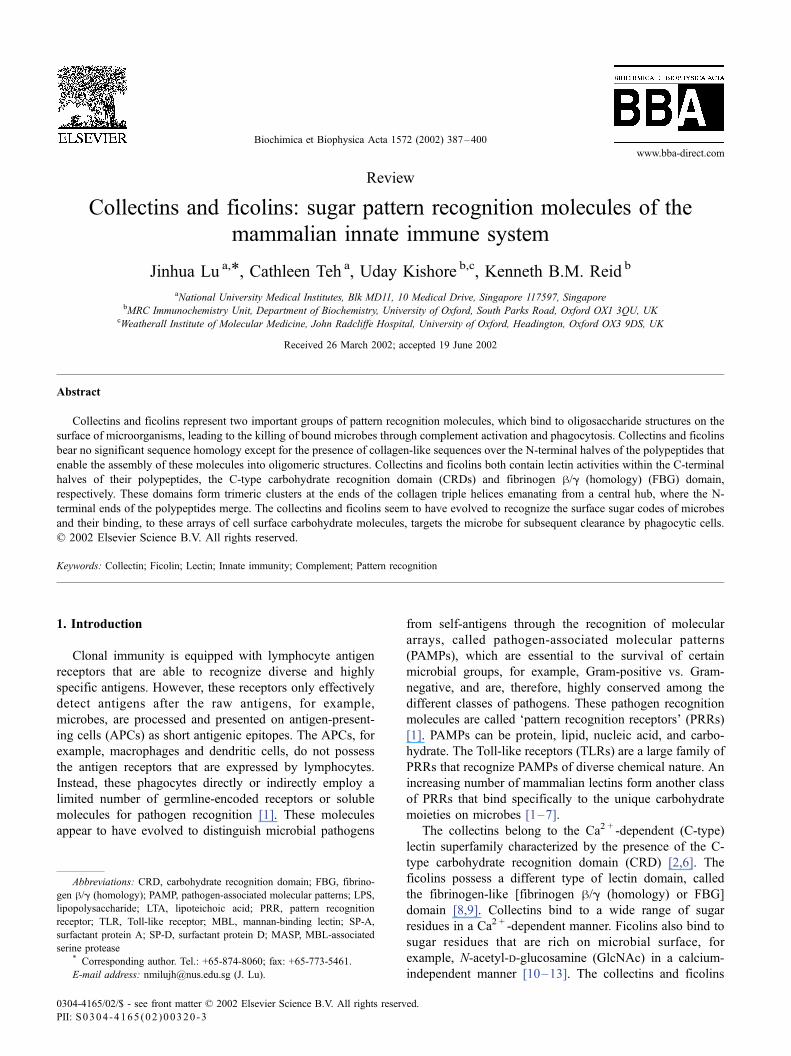
Fig. 1. Primary and tertiary structures of collectins and ficolins. The domain organization of the collectin polypeptides is illustrated. The N-terminal segment
containing cysteine residues (–SH) is followed by the collagen-like, Gly–Xaa–Yaa repeating sequences, and then by a a-helical, coiled-coil sequence and a C-
terminal C-type CRD (A). Three such polypeptides form a triple helix over the collagen-like region and a triple coiled-coil structure over the neck region
bringing three CRDs at the C-termini together as a cluster. The N-terminal segment form inter-chain disulfide bonds leading to higher oligomeric forms (B).
SP-D and conglutinin are predominantly tetramers of the triple-helical structural unit. CL-43 is only observed as monomers and SP-A is assembled into
hexamers. MBL is found in a series of oligomers, that is, from monomers to hexamers and here only hexameric MBL is presented. Ficolins lack the triple
coiled-coil neck region and have shorter collagen-like sequences. Therefore, ficolins are assembled into molecules with decreased dimensions compared with
collectins. Trimers and tetramers have been observed for H- and L-ficolins, respectively, which may further dimerize non-covalently [10,55].
J. Lu et al. / Biochimica et Biophysica Acta 1572 (2002) 387–400390

4� 160 kDa structures [46,47,50]. Further aggregation of
the SP-D tetramers through the central knob region has also
been observed [50]. SP-D is very similar to conglutinin in
domain organization, size and amino acid sequence. How-
ever, conglutinin contains an additional cysteine residue in
the fifth Gly–Xaa–Yaa repeat of the collagen-like region,
which is not present in SP-D [15]. It is possible that this
third cysteine residue in conglutinin is essential for the
higher degree disulfide-linked association over the N-termi-
nal segments including the first five Gly–Xaa–Yaa repeats.
Although conglutinin has a collagen-like region largely
similar to that of SP-D in length, the triple helices of
conglutinin observed under electron microscope are signifi-
cantly shorter than that of SP-D, that is, 37 vs. 46 nm. CL-
43 has a collagen-like region that is shorter than that of
conglutinin and SP-D but longer than that in MBL and SP-
A. It has so far only been observed as a monomer composed
of three disulfide-linked polypeptide chains [39]. As in CL-
43, the N-terminal segment of SP-D also contains two
cysteine residues. Therefore, it is probably the secondary
structures formed by the N-terminal segment of SP-D that
provide additional, non-covalent interactions between the
N-terminal ends of the collagen triple-helices.
The collagen-like sequences of SP-D and CL-43 are
continuous so that they form rigid and straight triple helices.
The cysteine residue within the fifth Gly–Xaa–Yaa repeat
of conglutinin appears likely to merge to the five Gly–Xaa–
Yaa repeats into a large knob with the N-terminal segments.
The collagen-like sequences in MBL and SP-A are both
interrupted by irregular sequences. In MBL, the eigthth
Gly–Xaa–Yaa repeat is not complete (Gly–Gln) [51].
However, the triple helices are apparently straight as viewed
under the electron microscope [21]. SP-A has a more
obvious interruption between the 13th and 14th Gly–
Xaa–Yaa repeat, that is, Pro–Cys–Pro–Pro [52]. The
electron microcopy image of SP-A shows a significant bent
in the middle of the triple helices corresponding to the
interrupted region [53]. The largest oligomers of human
MBL and SP-A consist of six triple helices merged over
their N-terminal region, and therefore, each molecule of
MBL or SP-A contains 18 constituent polypeptides [21,53].
Lower oligomers, ranging between monomers and pentam-
ers, are also consistently observed in purified MBL and
these different oligomers are completely disulfide-linked
[21]. However, these lower oligomers are not found in
SP-A [53]. The arrangement of the disulfide bonding in
SP-A is not clear. One cysteine residue is found in the N-
terminal segment and a second cysteine is present in the
middle of the collagen-like sequence within the Pro–Cys–
Pro–Pro interruption [52]. The electron microscopy images
of SP-A suggest that the six triple helices are organized into
three pairs, with each pair being disulfide-linked through the
cysteine residue within the Pro–Cys–Pro–Pro sequence
merging the N-terminal halves of the two helices and
allowing the C-terminal halves of the two helices to bend
away from the central axis of the triple helix pair [53] (Fig.
1). A complete SP-A molecule can be formed from three
such helix pairs that merge at the N-terminal ends partially
or completely through disulfide-linkages involving the N-
terminal cysteine residue. This results in an overall structure
of SP-A that is indistinguishable from that of Clq [41,53].
Considering the influence of the number of cysteine resi-
dues over the N-terminal regions of the collectins on the
assembly of the collectin molecules, it can be deduced that
three cysteines are required to assemble collectins into
completely disulfide-linked oligomers, for example, in
MBL and conglutinin.
Ficolins are similarly assembled into triple helices over
their collagen-like region and are also observed as disulfide-
linked high molecular weight oligomers on SDS-PAGE. L-
ficolin migrates with a molecular mass corresponding to 320
kDa under non-reducing conditions [10,54,55]. In the elec-
tron microscope, L-ficolin molecules are clearly observed as
tetramers of the triple helices merged at their N-terminal
ends [55]. The electron micrographs of H-ficolin are less
clear due to the lack of top view images, but show large
FBG domains and short collagen helices [13]. It has been
proposed as a trimer of triple helices that can further
dimerize non-covalently because it elutes as a molecule of
650 kDa by gel-filtration equivalent to approximately 18 H-
ficolin polypeptides [13]. L-ficolin also undergoes further
non-covalent dimerization that involves inter-molecular
interactions between the FBG domains [55]. Electron micro-
scopy images have not been obtained for M-ficolin.
4. Sugar specificity
The lectin activity of collectins is located in the C-
terminal, C-type CRDs, which can be divided into two
classes based on a three-residue motif in the CRDs: Glu–
Pro–Asn that enables the CRD to bind mannose with a
higher affinity than galactose, or Gln–Pro–Asp that makes
the CRD a galactose-binding domain [56]. All collectins,
except for SP-A, contain the Glu–Pro–Asn mannose-spe-
cific motif. SP-A has an Arg (in dog) or an Ala (in man)
residue in place of the mannose-specific Asn residue [56].
Collectins, including SP-A, have been shown to bind to a
range of sugar residues including mannose, fucose, glucose,
maltose, GlcNAc and N-acetyl-D-mannosamine (ManNAc),
although individual collectins display different preferences
for certain sugar residues over the others (Table 1). MBL
binds to sugars in the order of GlcNAc>D-mannose/L-
fucose>ManNAcHmaltose>glucose [57]. Conglutinin dis-
plays high affinity for GlcNAc with lower affinity in the
order of mannosamine>L-fucose/mannose>glucose>Man-
NAc/glucosamine>maltose [15]. Conglutinin is not ex-
pressed in species other than the bovidae and its closest
homologue is SP-D [15]. Despite the high degree of
structural similarity between SP-D and conglutinin, SP-D
displays quite different sugar specificity from that of con-
glutinin [15]. SP-D has little affinity for GlcNAc, the best
J. Lu et al. / Biochimica et Biophysica Acta 1572 (2002) 387–400 391

sugar ligand for conglutinin. It binds sugars in the following
order, that is, maltose>L-fucose>mannose>glucoseHglu-
glucosamine, showing the highest affinity for maltose, a
very weak sugar ligand for conglutinin [15]. CL-43 binds
sugars in the following order, that is, mannose/ManNAc>
fucose>GlcNAc>glucose and maltose>galactose>lactoseH
GalNAc, which is similar to MBL, although structurally it
resembles conglutinin and SP-D [19]. Discrepancy has been
reported on the affinity of SP-A for the galactose residue.
By affinity chromatography, Haagsman et al. [58] showed
that SP-A bound to mannose, L-fucose, galactose, and
glucose. In another study, Haurum et al. [57] demonstrated
binding of SP-A to a similar list of sugars, that is, Man-
NAc>L-fucose, maltose>glucose>mannose, but not galac-
tose.
The lectin activity of L- and M-ficolin, and possibly that
of H-ficolin, appears to reside in the FBG domains [11,38].
L-ficolin was identified based on its affinity for immobilized
yeast mannan [10]. However, L-ficolin was found to bind to
cyanogen bromide-activated agarose which did not have
mannan coupled to it, but had simply been inactivated with
Tris [54]. All three ficolins bind to GlcNAc. L-ficolin shows
no obvious affinity for sugars such as mannose, glucose,
galactose and lactose. H-ficolin displays affinity for GalNAc
and D-glucose as well as GlcNAc. A wider range of sugars
needs to be examined to determine the sugar specificity of
ficolins. A critical difference between the C-type CRD and
the FBG domain in sugar binding is that Ca2 + is essential to
the lectin activity of C-type CRDs, whereas it is not required
for the lectin activity of the FBG domains of L- and M-
ficolins [54]. It is not clear whether this observation is also
true for H-ficolin.
5. Microbial targets
The lectin domains of collectins and ficolins undergo two
grades of clustering during assembly. Primary clustering is
brought about through the formation of collagen-like triple
helices and, in collectins, the formation of the triple coiled-
coil ‘neck’ structure (Fig. 1). The clustering of three CRDs
in close proximity significantly increases the affinity for
small sugar clusters. Further clustering occurs through the
merging of multiple triple-helical structures at the opposite
ends of the helices. The impact of this further clustering can
be many-fold. First, it probably ensures that these molecules
only bind with high affinity to dense sugar arrays, typically
found on the surface of microbes, for example, fungi and
bacteria. Secondly, it provides a large enough docking
surface for effector molecules. Selectins function to support
leukocyte rolling on endothelium for which low-affinity
interaction with carbohydrate structures is essential [59].
Accordingly, selectins only contain a single C-type CRD
and the receptors are not constitutively clustered. Like the
collectins, the mannose receptor also recognizes microbial
surface carbohydrate structures. Accordingly, each mannose
receptor contains eight CRDs, which are clustered in the
form of tandem repeats [60,61]. Further clustering of the
CRDs can occur when multiple mannose receptors are
recruited to phagocyte–microbial interfaces. Therefore, the
collectins and ficolins are ideally assembled for microbial
recognition and represent a unique class of soluble PRRs.
Common microbial structures that are recognized by the
collectins are lipopolysaccharides (LPS) and mannan-like
high-mannose structures. LPS is a major glycolipid compo-
nent on the outer membrane of Gram-negative bacteria and
Table 2
Microorganisms recognized by collectins and ficolins
Bacteria Fungi/yeast Viruses
MBL Staphylococcus aureus, h-hemolytic
group A streptococci, Escherichia coli,
Klebsiella species, Haemophilus influenzae
type b, h-hemolytic group B streptococci,
Streptococcus pneumoniae, Staphylococcus
epidermidis
Saccharomyces cerevisiae,
Aspergillus fumigatus,
Candida albicans, acapsular
Cryptococcus neoformans
HIV, IAV, RSV
SP-A Staphylococcus aureus, type 25 pneumococci,
Haemophilus influenzae, Streptococcus
pneumoniae, Group A Streptococcus,
Escherichia coli, J5, Mycoplasma pulmonis,
Klebsiella pneumoniae
Saccharomyces cerevisiae,
Aspergillus fumigatus,
acapsular Cryptococcus neoformans,
Pneumocystis carinii
IAV, RSV, HSV-1
SP-D Mycobacterium tuberculosis bacilli, Acapsular
Klebsiella pneumoniae, Pseudomonas aeruginosa,
Escherichia coli
Saccharomyces cerevisiae,
Aspergillus fumigatus,
Candida albicans, acapsular
Cryptococcus neoformans,
Pneumocystis carinii
IAV, RSV, rotavirus
Conglutinin acapsular Cryptococcus neoformans IAV, rotavirus
CL-43 acapsular Cryptococcus neoformans rotavirus
CL-P1 Escherichia coli, Staphylococcus aureus
L-ficolin Salmonella typhimurium
HIV, human immunodeficiency virus; IAV, influenza A virus; RSV, respiratory syncytial virus; HSV-1, herpes simplex virus type 1.
J. Lu et al. / Biochimica et Biophysica Acta 1572 (2002) 387–400392

fungi are covered with rigid matrices of polysaccharides.
These are ideal ligands for collectins. Collectins have been
reported to bind to a wide range of microbes including
bacteria, fungi and viruses [62–65]. Ficolins have also been
shown to bind to the surface or LPS of bacteria that involve
the lectin activities [10,13,37]. The microbes that are rec-
ognized by each individual collectin and ficolin are listed in
Table 2. A number of fungal/yeast species is recognized by
multiple collectins including Saccharomyces cerevisiae,
Aspergillus fumigatus, Candida albicans, acapsular Crypto-
coccus neoformans and Pneumocystis carinii [65–71].
Collectins also bind selectively to several types of virus
including the human immunodeficiency virus (HIV-I), influ-
enza A virus (IAV), respiratory syncytial virus (RSV), and
rotavirus [72–79] although binding of SP-D to RSV is
controversial [77,78]. SP-A enhances phagocytosis of her-
pes simplex virus-1 (HSV-1) by alveolar macrophages
though there is no evidence that SP-A binds to HSV-1
[80]. SP-D binds to several different bacteria including
Mycobacterium tuberculosis bacilli, acapsular Klebsiella
pneumoniae, Pseudomonas aeruginosa and Escherichia coli
[81–84]. SP-A binds to Staphylococcus aureus, type 25
pneumococci, Haemophilus influenzae, Streptococcus pneu-
moniae, Group A Streptococcus with a preference for Gram-
positive bacteria [64,85,86]. In fact, some Gram-positive
bacteria are also recognized by MBL and SP-D. It is
interesting to note that MBL and SP-D have been shown
to bind, in addition to LPS, to PAMPs which are character-
istic of Gram-positive bacteria, that is, lipoteichoic acid
(LTA) and/or peptidoglycan [87,88]. The wide range of
microbes that are recognized by the collectins highlights
the importance of collectins in innate immunity against these
pathogens. Ficolins bind to sugar structures on bacteria. H-
ficolin binds to LPS derived from Salmonella typhimurium
and Salmonella minnesota [13]. L-ficolin also binds to S.
typhimurium [10]. There is no direct evidence that M-ficolin
binds to microbes. However, it has been shown to promote
phagocytosis of E. coli K-12 by monocytes [12].
6. Functional mechanisms
Binding of collectins and ficolins to microbes through
the lectin domains leads to activation of multiple immuno-
logical processes such as complement activation and phag-
ocytosis. Collectins have also been shown to modulate
leukocyte chemotaxis, proliferation and differentiation.
Some of these processes are not necessarily dependent on
the lectin activity of these molecules.
6.1. Complement activation
The complement system is an important arm of innate
immunity and is found predominantly in the blood circu-
lation as a serine protease cascade that can be triggered by
different mechanisms [89]. Complement activation on the
surface of a microorganism kills microbes through lysis and
then opsonizes microbial skeletons for effective phagocy-
tosis through deposition of complement fragments. The
complement cascade can be activated through three distinct
pathways, that is, the classical, alternative and lectin path-
ways. The activation of the classical pathway requires pre-
sensitization of microbes with antibodies that are recognized
by C1q. C1q is associated with the C1r and C1s serine
protease proenzymes as a complex called C1 and, upon
binding of C1q to antigen-bound IgG and IgM, C1r and C1s
are activated which subsequently recruit and activate C4 and
C2. Activated C4 and C2 form C3 convertase that recruits
and activates C3 leading to the formation of membrane
attack complexes and microbial lysis. The activated C3 and
C4, that is, C3b and C4b, are covalently deposited on the
microbial surface which are recognized by phagocytic
receptors on multiple phagocytes (Fig. 2).
The alternative and lectin pathways are independent of
clonal immunity. The alternative pathway is activated on the
surface of certain microbes directly through the amplification
of tick-over C3 activation, whereas the lectin pathway is
activated after direct binding of MBL and L-ficolin to the
surface sugar residues on microbes [3–7,14]. Ikeda et al.
[90] first demonstrated that, like C1q, MBL could activate
the complement system upon binding to yeast mannan-
coated erythrocytes in a C4-dependent manner, implying
the activation of the classical pathway. Independently, Ji et
al. [91] identified a protein complex in mouse serum, called
the ‘Ra-reactive factor’ which bound specifically to Ra
chemotype strains of Salmonella, and could activate comple-
ment in a C4/C2-dependent and C1-independent manner.
This protein complex includes two components: P28 and
P100; P28 was later shown to be mouse MBL [92,93]. In
vitro studies demonstrated that MBL could interact with and
activate C1r and C1s upon binding to carbohydrate ligands
[23,94]. However, a group of three C1r/C1s-like serine
proteases, called MBL-associated serine proteases (MASPs),
and one non-serine protease protein (MAp19), but no C1r/
C1s, have been found to form complexes with MBL and are
considered to be involved with MBL-mediated complement
activation in vivo [95–99]. The P100 component of RaRF
was also shown to be mouse MASP [93].
The three MASPs, and the MAp19, are expressed from
two MASP genes, that is, MASP-1 [95], MASP-2 [96],
MAp19 [98] and MASP-3 [99], with MAp19 and MASP-3
being alternatively spliced isoforms of MASP-2 and
MASP-1. Recently, these MASPs have been reported to
form complexes preferentially with different MBL
oligomers [99]. MBL is assembled from a structural unit
that consists of three identical polypeptides into trimer to
hexamers of this structural unit through disulfide linkages
over the N-terminal ends (Fig. 1). Differential oligomeriza-
tion has not been observed in other collectins and the
mechanism that leads to the different degrees of MBL
oligomerization is not clear. Trimers and tetramers are
consistently the predominant oligomers in purified MBL
J. Lu et al. / Biochimica et Biophysica Acta 1572 (2002) 387–400 393

(f 80%) [23]. MASP-1 and MAp19 form a complexes
with smaller MBL oligomers and then appear, via MASP-1,
to activate the complement C3 directly, although the
physiological significance of the low levels of C3 activa-
tion in such in vitro studies has been questioned [100].
MASP-2 forms a complex with larger MBL oligomers to
activate C4 and C2 and MASP-3, which also forms a
complex with the larger MBL oligomers, appears to act
as an antagonist of MASP-2 activity in C4 and C2 cleavage
[99]. This implies that the different MBL oligomers, in
association with different MASPs, are able to activate
complement along the classical and, potentially the alter-
native, pathway and the MBL–MASP2 complex is most
closely related to the C1 complex. The association between
MASP-1 and smaller MBL oligomers may involve distinct
inter-molecular bonds since activated MASP-1 forms strong
non-covalent interactions with MBL which cannot be
interrupted by EDTA [101]. This type of interaction has
not been detected between MASP-2 and MBL which is
likely to be Ca2 +-dependent. The MASP-1 that co-purified
with MBL in previous studies, which were historically
considered an artificial dimer of the MBL polypeptide on
SDS-PAGE under reducing conditions, may have contrib-
uted to the C1s activation observed in vitro [23,94,101].
However, the physiologically relevant in vivo targets, for
MASP-1 and MASP-3, remain to be established.
The MBL–MASP pathway of complement activation is
an important innate immune mechanism. Deficiency in
MBL expression results in an opsonic defect, characterized
by the inability of patient serum to promote microbial
phagocytosis due to insufficient complement deposition on
microbes [22,102]. Recently, L-ficolin has also been shown
to complex with MASPs and, upon binding to S. typhimu-
rium, to activate the complement system in a similar manner
as MBL [103]. L-ficolin is present in serum at similar
concentrations as MBL [11,102]. Therefore, the ficolin
arm of the lectin pathway is potentially comparable to the
MBL arm in strength. This may partially explain why a
highly prevalent MBL deficiency (5–9%) was tolerated
during the evolution and also raise a question in the assess-
ment of the clinical manifestations associated with MBL
deficiency. Therefore, the question can be asked—‘‘Are
both MBL and L-ficolin deficiencies required to produce a
detectable complement and opsonic defect?’’. The differ-
ence in sugar specificity of L-ficolin from MBL means that
the L-ficolin and MBL arms are complementary and may
target to different, though overlapping, microbial popula-
tions. The Ca2 +-independent nature of ficolin binding to
sugars is another property that distinguishes L-ficolin from
MBL although the functional implications remain to be
determined [54]. Nevertheless, although L-ficolin defi-
ciency has not been reported, the outcome of MBL, or
Fig. 2. Collectin-mediated microbial killing and clearance through complement activation and phagocytosis. Collectins and ficolins enhance phagocytosis
either directly or, in the case of MBL and L-ficolin, indirectly through deposition of complement C3b and C4b on microbial surfaces. Phagocytes express
phagocytic complement receptors CR3 and CR4. Collectins can also activate phagocytes and up-regulate phagocytosis mediated by CR3, Fc receptor (FcR) or
the mannose receptor (MR) in a complement-independent manner.
J. Lu et al. / Biochimica et Biophysica Acta 1572 (2002) 387–400394

potential L-ficolin, deficiency is likely to be determined by
the types of invading microbes recognized by either, or
both, MBL and L-ficolin.
6.2. Phagocytosis
Collectins and ficolins have been reported to promote
phagocytosis of various microorganisms, either directly
after binding to microbes, or indirectly through upregulation
of phagocytosis mediated by other phagocytic receptors.
This has been discussed in a number of review articles
[2,3,6,7,14,104]. MBL and L-ficolin can indirectly opsonize
microbes with complement opsonins C3b and C4b for
enhanced phagocytosis without interaction with a MBL or
ficolin receptor on phagocytes [14,103]. Collectins and
ficolins can also function as opsonins, which require inter-
action of these proteins with phagocyte receptors. Such
receptors have not been identified for ficolins. However, a
number of putative receptors/binding proteins, for the col-
lectins, have been identified [105,106], including calreticu-
lin [107], C1qRp [108], SPR-210 [109], gp340 [110], CD91
[111], CR1 [112] and CD14 [113,114]. Among these
receptors, calreticulin and C1qRp have been shown to bind
to multiple collectins. SPR-210 and gp340 only exhibited
affinity for SP-A and SP-D. Both CD91 and CR1 bind to
MBL. In fact, MBL mediates phagocytosis of apoptotic
cells by binding to calreticulin on apoptotic cells and CD91
on phagocytes [111]. C1qRp is a receptor that mediates
immobilized collectin-induced upregulation of phagocytosis
through complement and Fc receptors [108]. However,
C1qRp (CD93), defined initially as a receptor on the basis
of its involvement in the enhancement of phagocytosis by
monocytes, appears now to be involved in adhesion events,
rather than C1q-mediated phagocytosis [100]. SP-A has also
been reported to promote phagocytosis of M. tuberculosis
indirectly through upregulation of the activity of the man-
nose receptor but, SP-A receptor involved, remains to be
characterized [115]. Soluble CD14 has recently been shown
to bind to SP-A, SP-D and MBL [113,114]. CD14 is a
recognition component of the LPS receptor complex that
elicits inflammatory reactions upon binding to LPS [116].
The collectins may therefore regulate the expression of
cytokines involved in inflammation.
SP-A and SP-D have been shown to interact with
alveolar macrophages, neutrophils, peripheral blood mono-
cytes, monocyte-derived macrophages, and bone-marrow-
derived macrophages, and modulate their functions. SP-A
increases intracellular Ca2 + and inositol triphosphate (IP3)
concentrations in alveolar macrophages. The calcium
response correlates with IP3 generation, which is necessary
for SP-A-mediated phagocytosis. SP-A also stimulates che-
motaxis via cell interaction involving its collagen region. It
also stimulates directional actin polymerization [117]. These
two effects are preceded by receptor binding and trans-
mission of intracellular signals. SP-A can inhibit production
of cytokines in response to LPS and C. albicans in buffy-
coat cells. In contrast, it enhances production of TNF-a and
colony-stimulating factor (CSF) by alveolar macrophages,
and of TNF-a, IL-1a, IL-1h, IL-6 and IFN-g by human
peripheral blood mononuclear cells (PBMCs).
SP-A and SP-D also modulate the production of reactive
oxygen and nitrogen species by alveolar macrophages and
monocytes. SP-A enhances nitric oxide metabolite produc-
tion by IFN-g-activated alveolar macrophages, when chal-
lenged with Mycoplasma pneumoniae. However, SP-A
inhibits nitric oxide production by IFN-g-activated alveolar
macrophages when exposed to M. tuberculosis, suggesting
that the state of activation of phagocytic cells and the type of
pathogen may affect SP-A-mediated inflammatory response.
SP-A and SP-D are also known to enhance chemotactic,
phagocytic and superoxidative burst properties of neutro-
phils [118].
6.3. Interaction of SP-A and SP-D with allergens
SP-A has been shown to bind a variety of allergenic
pollens such as Lombardy poplar, Kentucky blue grass,
cultivated rye and short ragweed, via its CRDs [119]. SP-A,
SP-D and a recombinant fragment of human SP-D com-
posed of homotrimeric neck and CRD regions (rfSP-D) can
also bind house dust mite extract (Dermatophagoides pter-
onyssinus; Derp) in a carbohydrate-specific and Ca2 +-
dependent manner, inhibit specific IgE binding to these
glycoprotein allergens, and block allergen-induced hista-
mine release from basophils isolated from Derp-sensitive
patients [120]. Similarly, SP-A, SP-D and rfSP-D can also
bind to the 3-week culture filtrate (3wcf) of Aspergillus
fumigatus (Afu) as well as purified glycoprotein allergens,
gp55 and gp45, inhibit the ability of Afu-specific IgE to bind
these allergens, and block Afu allergen-induced histamine
release from sensitized basophils isolated from ABPA
patients [115]. SP-A and SP-D have been reported to reduce
the proliferation of PBMCs isolated from mite-sensitive
asthmatic children [121], and SP-D, in particular, has a
suppressive effect on the secretions of IL-2 by PBMCs
[122]. SP-A can attenuate production of IL-8 by eosinophils
in a concentration-dependent manner [123]. Thus, SP-A and
SP-D inhibit histamine release in the early phase of allergen
provocation and suppress lymphocyte proliferation in the
late phase of bronchial inflammation—the two essential
steps in the development of asthmatic symptoms.
Abnormal levels of SP-A and SP-D in the lung lavage
have been reported in the adult respiratory distress syn-
drome (ARDS) and pulmonary infections caused by influ-
enza virus, mycoplasma and P. carinii in AIDS patients,
hypersensitivity lung diseases and cystic fibrosis [124,125].
Asthmatics show increased amounts of SP-A in bronchiolar
and alveolar lavage and SP-D in alveolar lavage as com-
pared with those in controls [126]. Serum SP-D levels for
two allergic patients have been found elevated at diagnosis
which decrease following corticosteroid therapy [127]. The
patients of birch pollen allergy and pulmonary alveolar
J. Lu et al. / Biochimica et Biophysica Acta 1572 (2002) 387–400 395

proteinosis showed a shift toward lower oligomeric forms of
SP-A in comparison to healthy volunteers [128]. Depoly-
merisation of SP-A may lead to loss of binding affinity for
carbohydrate-rich surfaces, with subsequent loss or alter-
ation of biological function. SP-A has been shown to inhibit
the LPS-induced TNF-a and IL-1 production by human
alveolar macrophages in ARDS patients [129].
Recently, human SP-A, SP-D and a recombinant frag-
ment of SP-D containing homotrimeric neck and CRD
regions (rfSP-D), have been shown to dampen immunolog-
ical parameters associated with allergic disorder in a murine
model of allergic bronchopulmonary aspergillosis (ABPA)
[130], induced by allergenic and antigenic extract of A.
fumigatus. The murine model resembled the human disease
immunologically, exhibiting high levels of specific IgG and
IgE, peripheral blood and pulmonary eosinophilia, and a
Th2 cytokine response. Intranasal administration of SP-A,
SP-D and rfSP-D significantly lowered eosinophilia and
specific antibody levels, pulmonary infiltration, reduced
Th2 response and raised Th1 response. Since IgE-dependent
mechanisms are important in the expansion of a Th2
immune response [131], these results suggest that SP-A
and SP-D may not only suppress Th2 responses by scav-
enging antigens and allergens and so preventing IgE-
dependent activation of eosinophils, but may well directly
interact with APCs (DCs) promoting the induction of IL-12-
dependent Th1 responses. A direct interaction between DC
and SP-D or rfSP-D has been observed (Urban et al.,
unpublished data). Both molecules bind to immature human
DC in a Ca2 +-dependent and carbohydrate-independent
manner. Binding of SP-D or rfSP-D is diminished on
LPS-matured DC. In concordance with these observations,
immature but not mature DC express the putative SP-D
receptor, gp340. SP-D has also been shown to mediate
binding and uptake of Esherichia coli by bone-marrow-
derived mouse DC and enhance antigen presentation of E.
coli expressed proteins to T-cell hybridoma [132].
7. Conclusion
Innate immunity is important in pathogen recognition
and killing, antigen processing and the initiation and mod-
ulation of adaptive immunity. PRRs are directly involved in
pathogen recognition by innate immune systems and can be
functionally classified into two types specialized in patho-
gen capturing or sensing. Phagocytic receptors such as Fc,
complement and lectin receptors are primarily antigen
capturing receptors that are potent in the initial killing of
microbes through complement activation and/or phagocy-
tosis. Collectins and ficolins are soluble mammalian lectins
that can recognize the unique ‘sugar code’ on microbes
[133], capture the microbes, and act either as opsonins that
directly present the bound microbes to phagocytes or
indirectly through further opsonization with complement
proteins. Collectins recognize LPS and LTA, both are
PAMPs exposed on the surface of live pathogens. In
contrast, TLRs recognize a wider spectrum of microbial
PAMPs including LPS and LTA but the binding sites are
only effectively exposed after disintegration of microbes, for
example, through complement attack or phagocytosis. There
is no evidence that TLRs play major roles in pathogen
capturing, for example, phagocytosis. Instead, these are
pathogen-sensing receptors that sample the nature of patho-
gens and play a more important role in the coordination of
inflammation and shaping of adaptive immunity. In this
sense, the collectins are in the very front line of innate
immunity.
The collectins and ficolins are found predominantly in
body fluids and at the interface with the environment, for
example, the surfaces of the respiratory and mucosal surfa-
ces. Structurally, these molecules are characterized by the
presence of collagen-like sequences that consistently lead to
highly oligomerized mature molecules. The lectin domains
in these proteins are invariably clustered at the end of triple
helical collagen bundles and multiple clusters are displayed
with dimensions and flexibility to maximize interactions
with the unique sugar arrays over microbial surfaces. The
microbe-bound collectins can then act as opsonins to
enhance phagocytosis and putative collectin receptors or
binding proteins identified should aid in the characterization
of relevant mechanisms. Collectins may also regulate the
activity of other phagocytic receptors through collectin
receptor signaling. The ability of MBL and L-ficolin to
activate the complement system upon binding to microbial
sugar structures can lead not only to microbial killing, but
also complement-dependent microbial opsonization. There-
fore, the collectins and ficolins form an important network
of innate immunity. The ability of SP-A and SP-D to bind to
allergens and to inhibit specific IgE-mediated histamine
release and to suppress lymphocyte proliferation during
bronchial inflammation suggest a therapeutic role of these
two collectins in asthmatic disorders.
Acknowledgements
J.L. and C.T. are supported by the National Medical
Research Council of Singapore, grant numbers R-364-000-
010-213 and R-364-000-014-213. U.K. and K.B.M.R. are
funded by the European Commission—Contract QLK2-
2000-00325, and the Medical Research Council, UK.
References
[1] R. Medzhitov, C. Janeway Jr., Innate immune recognition: mecha-
nisms and pathways, Immunol. Rev. 173 (2000) 89–97.
[2] W.I. Weis, M.E. Taylor, K. Drickamer, The C-type lectin superfamily
in the immune system, Immunol. Rev. 163 (1998) 19–34.
[3] U. Holmskov, R. Malhotra, R.B. Sim, J.C. Jensenius, Collectins: col-
lagenous C-type lectins of the innate immune defense system, Immu-
nol. Today 15 (1994) 67–74.
J. Lu et al. / Biochimica et Biophysica Acta 1572 (2002) 387–400396
[4] H.J. Hoppe, K.B.M. Reid, Collectins—soluble proteins containing
collagenous regions and lectin domains—and their roles in innate
immunity, Protein Sci. 3 (1994) 1143–1158.
[5] J. Epstein, Q. Eichbaum, S. Sheriff, R.A.B. Ezekowitz, The collectins
in innate immunity, Curr. Opin. Immunol. 8 (1996) 29–35.
[6] J. Lu, Collectins: collectors of microorganisms for the innate immune
system, BioEssays 19 (1997) 509–518.
[7] M. Matsushita, T. Fujita, Ficolins and the lectin complement pathway,
Immunol. Rev. 180 (2001) 78–85.
[8] J. Lu, Y. Le, Ficolins and the fibrinogen-like domain, Immunobiol.
199 (1998) 190–199.
[9] H. Ichijo, U. Hellman, C. Wernstedt, L.J. Gonez, L. Claesson-Welsh,
C.H. Heldin, K. Miyazono, Molecular cloning and characterization of
ficolin, a multimeric protein with fibrinogen- and collagen-like do-
mains, J. Biol. Chem. 268 (1993) 14505–14513.
[10] M. Matsushita, Y. Endo, S. Taira, Y. Sato, T. Fujita, N. Ichikawa, M.
Nakata, T. Mizuochi, A novel human serum lectin with collagen- and
fibrinogen-like domains that functions as an opsonin, J. Biol. Chem.
271 (1996) 2448–2454.
[11] Y. Le, S.H. Lee, O.L. Kon, J. Lu, Human L-ficolin: plasma levels,
sugar specificity, and assignment of its lectin activity to the fibrino-
gen-like (FBG) domain, FEBS Lett. 425 (1998) 367–370.
[12] C. Teh, Y. Le, S.H. Lee, J. Lu, M-ficolin is expressed on monocytes
and is a lectin binding to N-acetyl-D-glucosamine and mediates
monocyte adhesion and phagocytosis of Escherichia coli, Immunol-
ogy 101 (2000) 225–232.
[13] R. Sugimoto, Y. Yae, M. Akaiwa, S. Kitajima, Y. Shibata, H. Sato,
J. Hirata, K. Okochi, K. Izuhara, N. Hamasaki, Cloning and
characterization of the Hakata antigen, a member of the ficolin/op-
sonin p35 lectin family, J. Biol. Chem. 273 (1998) 20721–20727.
[14] D.L. Jack, N.J. Klein, M.W. Turner, Mannose-binding lectin: target-
ing the microbial world for complement attack and opsonophago-
cytosis, Immunol. Rev. 180 (2001) 86–99.
[15] K. Hakansson, K.B.M. Reid, Collectin structure: a review, Protein Sci.
9 (2000) 1607–1617.
[16] D.C. Kilpatrick, Animal lectins: an historical introduction and over-
view, Biochim. Biophys. Acta 1572 (2002) 187–197.
[17] R. Loris, Principles of structures of animal and plant lectins, Biochim.
Biophys. Acta 1572 (2002) 198–208.
[18] D.C. Kilpatrick, Mannan-binding lectin: clinical significance and ap-
plications, Biochim. Biophys. Acta 1572 (2002) 401–413.
[19] K. Ohtani, Y. Suzuki, S. Eda, T. Kawai, T. Kase, H. Yamazaki, T.
Shimada, H. Keshi, Y. Sakai, A. Fukuoh, T. Sakamoto, N. Waka-
miya, Molecular cloning of a novel human collectin from liver (CL-
L1), J. Biol. Chem. 274 (1999) 13681–13689.
[20] J. Lu, S.B. Laursen, S. Thiel, J.C. Jensenius, K.B.M. Reid, The cDNA
cloning of conglutinin and identification of liver as a primary site of
synthesis of conglutinin in members of the Bovidae, Biochem. J. 292
(1993) 157–162.
[21] B.L. Lim, A.C. Willis, K.B.M. Reid, J. Lu, S.B. Laursen, J.C. Jen-
senius, U. Holmskov, Primary structure of bovine collectin-43 (CL-
43). Comparison with conglutinin and lung surfactant protein-D, J.
Biol. Chem. 269 (1994) 11820–11824.
[22] M. Super, S. Thiel, J. Lu, R.J. Levinsky, M.W. Turner, Association of
low levels of mannan-binding protein with a common defect of
opsonisation, Lancet 2 (1989) 1236–1239.
[23] J.H. Lu, S. Thiel, H. Wiedemann, R. Timpl, K.B.M. Reid, Binding of
the pentamer/hexamer forms of mannan-binding protein to zymosan
activates the proenzyme C1r2C1s2 complex, of the classical pathway
of complement, without involvement of C1q, J. Immunol. 144
(1990) 2287–2294.
[24] J.A. Summerfield, S. Ryder, M. Sumiya, M. Thursz, A. Gorchein,
M.A. Monteil, M.W. Turner, Mannose binding protein gene muta-
tions associated with unusual and severe infections in adults, Lancet
345 (1995) 886–889.
[25] H.O. Madsen, P. Garred, S. Thiel, J.A. Kurtzhals, L.U. Lamm, L.P.
Ryder, A. Svejgaard, Interplay between promoter and structural
gene variants control basal serum level of mannan-binding protein,
J. Immunol. 155 (1995) 3013–3020.
[26] M.W. Turner, Mannose-binding lectin (MBL) in health and disease,
Immunobiol. 199 (1998) 327–339.
[27] K. Sastry, K. Zahedi, J.M. Lelias, A.S. Whitehead, R.A.B. Ezeko-
witz, Molecular characterization of the mouse mannose-binding pro-
teins. The mannose-binding protein A but not C is an acute phase
reactant, J. Immunol. 147 (1991) 692–697.
[28] R.J. King, D.J. Klass, E.G. Gikas, J.A. Clements, Isolation of apo-
proteins from canine surface active material, Am. J. Physiol. 224
(1973) 788–795.
[29] R.J. King, J. Ruth, E.G. Gikas, A.C.G. Platzker, R.K. Creasy, Ap-
pearance of apoproteins of pulmonary surfactant in human amniotic
fluid, J. Appl. Physiol. 39 (1975) 741–753.
[30] S. Rubio, T. Lacaze-Masmonteil, B. Chailley-Heu, A. Kahn, J.R.
Bourbon, R. Ducroc, Pulmonary surfactant protein A (SP-A) is ex-
pressed by epithelial cells of small and large intestine, J. Biol. Chem.
270 (1995) 12162–12169.
[31] R. Paananen, V. Glumoff, M. Hallman, Surfactant protein A and D
expression in the porcine Eustachian tube, FEBS Lett. 452 (1999)
141–144.
[32] K.R. Khubchandani, J.M. Snyder, Surfactant protein A (SP-A): the
alveolus and beyond, FASEB J. 15 (2001) 59–69.
[33] A. Persson, K. Rust, D. Chang, M. Moxley, W. Longmore, E.
Crouch, CP4: a pneumocyte-derived collagenous surfactant-associ-
ated protein. Evidence for heterogeneity of collagenous surfactant
proteins, Biochemistry (1988) 8576–8584.
[34] A. Persson, D. Chang, E. Crouch, Surfactant protein D is a divalent
cation-dependent carbohydrate-binding protein, J. Biol. Chem. 265
(1990) 5755–5760.
[35] Y. Ogasawara, D.R. Voelker, Altered carbohydrate recognition speci-
ficity engineered into surfactant protein D reveals different binding
mechanisms for phosphatidylinositol and glucosylceramide, J. Biol.
Chem. 270 (1995) 14725–14732.
[36] J. Madsen, A. Kliem, I. Tornoe, K. Skjodt, C. Koch, U. Holmskov,
Localization of lung surfactant protein D on mucosal surfaces in
human tissues, J. Immunol. 164 (2000) 5866–5870.
[37] J. Bordet, F.P. Gay, Sur les relations des sensibilisatrices avec I’alex-
ine, Ann. Inst. Pasteur 20 (1906) 467–498.
[38] M.A. Leon, R. Yokohari, Conglutination: specific inhibition by car-
bohydrates, Science 143 (1964) 1327–1328.
[39] U. Holmskov, S.B. Laursen, R. Malhotra, H. Wiedemann, R. Timpl,
G.R. Stuart, I. Tornoe, P.S. Madsen, K.B.M. Reid, J.C. Jensenius,
Comparative study of the structural and functional properties of a
bovine plasma C-type lectin, collectin-43, with other collectins, Bio-
chem. J. 305 (1995) 889–896.
[40] M. Akaiwa, Y. Yae, R. Sugimoto, S.O. Suzuki, T. Iwaki, K. Izuhara,
N. Hamasaki, Hakata antigen, a new member of the ficolin/opsonin
p35 family, is a novel human lectin secreted into bronchus/alveolus
and bile, J. Histochem. Cytochem. 47 (1999) 777–786.
[41] J. Lu, Y. Le, O.L. Kon, J. Chan, S.H. Lee, Biosynthesis of human
ficolin, an Escherichia coli-binding protein, by monocytes: compar-
ison with the synthesis of two macrophage-specific proteins, C1q
and the mannose receptor, Immunology 89 (1996) 289–294.
[42] C. Teh, Y. Le, S.H. Lee, J. Lu, M-ficolin is expressed on monocytes
and is a lectin binding to N-acetyl-D-glucosamine and mediates
monocyte adhesion and phagocytosis of Escherichia coli, Immunol-
ogy 101 (2000) 225–232.
[43] H.J. Hoppe, P.N. Barlow, K.B.M. Reid, Collectins—soluble proteins
containing collagenous regions and lectin domains—and their roles
in innate immunity, Protein Sci. 3 (1994) 1143–1158.
[44] K.B.M. Reid, R.R. Porter, Subunit composition and structure of
subcomponent C1q of the first component of human complement,
Biochem. J. 155 (1976) 19–23.
[45] H.R. Knobel, W. Villiger, H. Isliker, Chemical analysis and electron
microscopy studies of human C1q prepared by different methods,
Eur. J. Immunol. 5 (1975) 78–82.
J. Lu et al. / Biochimica et Biophysica Acta 1572 (2002) 387–400 397

[46] A.E. Davis 3rd, P.J. Lachmann, Bovine conglutinin is a collagen-like
protein, Biochemistry 23 (1984) 2139–2144.
[47] J. Lu, H. Wiedemann, U. Holmskov, S. Thiel, R. Timpl, K.B.M.
Reid, Structural similarity between lung surfactant protein D and
conglutinin. Two distinct, C-type lectins containing collagen-like
sequences, Eur. J. Biochem. 215 (1993) 793–799.
[48] U. Holmskov, B. Teisner, A.C. Willis, K.B.M. Reid, J.C. Jensenius,
Purification and characterization of a bovine serum lectin (CL-43)
with structural homology to conglutinin and SP-D and carbohydrate
specificity similar to mannan-binding protein, J. Biol. Chem. 268
(1993) 10120–10125.
[49] C.J. Strang, H.S. Slayter, P.J. Lachmann, A.E. Davis 3rd, Ultrastruc-
ture and composition of bovine conglutinin, Biochem. J. 234 (1986)
381–389.
[50] E. Crouch, A. Persson, D. Chang, J. Heuser, Molecular structure of
pulmonary surfactant protein D (SP-D), J. Biol. Chem. 269 (1994)
17311–17319.
[51] M.E. Taylor, P.M. Brickell, R.K. Craig, J.A. Summerfield, Structure
and evolutionary origin of the gene encoding a human serum man-
nose-binding protein, Biochem. J. 262 (1989) 763–771.
[52] T. Voss, K. Melchers, G. Scheirle, K.P. Schafer, Structural comparison
of recombinant pulmonary surfactant protein SP-A derived from two
human coding sequences: implications for the chain composition of
natural human SP-A, Am. J. Respir. Cell Mol. Biol. 4 (1991) 88–94.
[53] T. Voss, H. Eistetter, K.P. Schafer, J. Engel, Macromolecular organ-
ization of natural and recombinant lung surfactant protein SP 28–36.
Structural homology with the complement factor C1q, J. Mol. Biol.
201 (1998) 219–227.
[54] Y. Le, S.M. Tan, S.H. Lee, O.L. Kon, J. Lu, Purification and binding
properties of a human ficolin-like protein, J. Immunol. Methods 204
(1997) 43–49.
[55] T. Ohashi, H.P. Erickson, Two oligomeric forms of plasma
ficolin have differential lectin activity, J. Biol. Chem. 272 (1997)
14220–14226.
[56] K. Drickamer, Engineering galactose-binding activity into a C-type
mannose-binding protein, Nature 360 (1992) 183–186.
[57] J.S. Haurum, S. Thiel, H.P. Haagsman, S.B. Laursen, B. Larsen, J.C.
Jensenius, Studies on the carbohydrate-binding characteristics of hu-
man pulmonary surfactant-associated protein A and comparison with
two other collectins: mannan-binding protein and conglutinin, Bio-
chem. J. 293 (1993) 873–878.
[58] H.P. Haagsman, S. Hawgood, T. Sargeant, D. Buckley, R.T. White,
K. Drickamer, B.J. Benson, The major lung surfactant protein,
SP 28–36, is a calcium-dependent, carbohydrate-binding protein,
J. Biol. Chem. 262 (1987) 13877–13880.
[59] L.A. Lasky, Selectin–carbohydrate interactions and the initiation
of the inflammatory response, Annu. Rev. Biochem. 64 (1995)
113–139.
[60] M.E. Taylor, K. Bezouska, K. Drickamer, Contribution to ligand
binding by multiple carbohydrate-recognition domains in the macro-
phage mannose receptor, J. Biol. Chem. 267 (1992) 1719–1726.
[61] L. East, C.M. Isacke, The mannose receptor family, Biochim. Bio-
phys. Acta 1572 (2002) 364–386.
[62] K.B.M. Reid, Interactions of surfactant protein D with pathogens,
allergens and phagocytes, Biochim. Biophys. Acta 1408 (1998)
290–295.
[63] H.P. Haagsman, Interactions of surfactant protein A with pathogens,
Biochim. Biophys. Acta 1408 (1998) 264–277.
[64] E. Crouch, J.R. Wright, Surfactant proteins A and D and pulmonary
host defense, Annu. Rev. Physiol. 63 (2001) 521–524.
[65] O. Neth, D.L. Jack, A.W. Dodds, H. Holzel, N.J. Klein, M.W.
Turner, Mannose-binding lectin binds to a range of clinically rele-
vant microorganisms and promotes complement deposition, Infect.
Immun. 68 (2000) 688–693.
[66] M.J. Allen, D.R. Voelker, R.J. Mason, Interactions of surfactant
proteins A and D with Saccharomyces cerevisiae and Aspergillus
fumigatus, Infect. Immun. 69 (2001) 2037–2044.
[67] S. Schelenz, R. Malhotra, R.B. Sim, U. Holmskov, G.J. Bancroft,
Binding of host collectins to the pathogenic yeast Cryptococcus neo-
formans: human surfactant protein D acts as an agglutinin for acap-
sular yeast cells, Infect. Immun. 63 (1995) 3360–3366.
[68] P.E. Zimmerman, D.R. Voelker, F.X.McCormack, J.R. Paulsrud ,W.J.
Martin 2nd, 120-kD surface glycoprotein of Pneumocystis carinii is a
ligand for surfactant protein A, J. Clin. Invest. 89 (1992) 143–149.
[69] D.M. O’Riordan, J.E. Standing, K.Y. Kwon, D. Chang, E.C. Crouch,
A.H. Limper, Surfactant protein D interacts with Pneumocystis car-
inii and mediates organisms adherence to alveolar macrophages, J.
Clin. Invest. 95 (1995) 2699–2710.
[70] S.M. Levitz, A. Tabuni, C. Treseler, Effect of mannose-binding pro-
tein on binding of Cryptococcus neoformans to human phagocytes,
Infect. Immun. 61 (1993) 4891–4893.
[71] B.A. van Rozendaal, A.B. van Spriel, J.G. van De Winkel, H.P.
Haagsman, Role of pulmonary surfactant protein D in innate defense
against Candida albicans, J. Infect. Dis. 182 (2000) 917–922.
[72] N. Wakamiya, Y. Okuno, F. Sasao, S. Ueda, K. Yoshimatsu, M. Naiki,
T. Kurimura, Isolation and characterization of conglutinin as an influ-
enza A virus inhibitor, Biochem. Biophys. Res. Commun. 187 (1992)
1270–1278.
[73] K.L. Hartshorn, E.C. Crouch, M.R. White, P. Eggleton, A.I. Tauber,
D. Chang, K. Sastry, Evidence for a protective role of pulmonary
surfactant protein D (SP-D) against influenza A viruses, J. Clin.
Invest. 94 (1994) 311–319.
[74] K.L. Hartshorn, M.R. White, V. Shepherd, K.B.M. Reid, J.C. Jense-
nius, E.C. Crouch, Mechanisms of anti-influenza activity of surfac-
tant proteins A and D: comparison with serum collectins, Am. J.
Physiol. 273 (1997) L1156–L1166.
[75] R.A.B. Ezekowitz, M. Kuhlman, J.E. Groopman, R.A. Byrn, A hu-
man serum mannose-binding protein inhibits in vitro infection by the
human immunodeficiency virus, J. Exp. Med. 169 (1989) 185–196.
[76] M. Saifuddin, M.L. Hart, H. Gewurz, Y. Zhang, G.T. Spear, Inter-
action of mannose-binding lectin with primary isolates of human
immunodeficiency virus type 1, J. Gen. Virol. 81 (2000) 949–955.
[77] R. Ghildyal, C. Hartely, A. Varrasso, J. Meanger, D.R. Voelker, E.M.
Anders, J. Mills, Surfactant protein A binds to the fusion glycopro-
tein of respiratory syncytial virus and neutralizes virion infectivity,
J. Infect. Dis. 180 (1999) 2009–2013.
[78] T.P. Hickling, H. Bright, K. Wing, D. Gower, S.L. Martin, R.B. Sim,
R. Malhotra, A recombinant trimeric surfactant protein D carbohy-
drate recognition domain inhibits respiratory syncytial virus infection
in vitro and in vivo, Eur. J Immunol. 29 (1999) 3478–3484.
[79] P.C. Reading, U. Holmskov, E.M. Anders, Antiviral activity of
bovine collectins against rotaviruses, J. Gen. Virol. 79 (1998)
2255–2263.
[80] J.F. van Iwaarden, J.A. van Strijp, M.J. Ebskamp, A.C. Welmers, J.
Verhoef, L.M. van Golde, Surfactant protein A is opsonin in phago-
cytosis of herpes simplex virus type 1 by rat alveolar macrophages,
Am. J. Physiol. 261 (1991) L204–L209.
[81] S.F. Kuan, K. Rust, E. Crouch, Interactions of surfactant protein D
with bacterial lipopolysaccharides. Surfactant protein D is an Escher-
ichia coli-binding protein in bronchoalveolar lavage, J. Clin. Invest.
90 (1992) 97–106.
[82] J.S. Ferguson, D.R. Voelker, F.X. McCormack, L.S. Schlesinger,
Surfactant protein D binds to Mycobacterium tuberculosis bacilli
and lipoarabinomannan via carbohydrate– lectin interactions result-
ing in reduced phagocytosis of the bacteria by macrophages, J. Im-
munol. 163 (1999) 312–321.
[83] I. Ofek, A. Mesika, M. Kalina, Y. Keisari, R. Podschun, H. Sahly, D.
Chang, D. McGregor, E. Crouch, Surfactant protein D enhances
phagocytosis and killing of unencapsulated phase variants of Kleb-
siella pneumoniae, Infect. Immun. 69 (2001) 24–33.
[84] C.I. Restrepo, Q. Dong, J. Savov, W.I. Mariencheck, J.R. Wright,
Surfactant protein D stimulates phagocytosis of Pseudomonas aeru-
ginosa by alveolar macrophages, Am. J. Respir. Cell Mol. Biol. 21
(1999) 576–585.
J. Lu et al. / Biochimica et Biophysica Acta 1572 (2002) 387–400398

[85] M.J. Tino, J.R. Wright, Surfactant protein A stimulates phagocytosis
of specific pulmonary pathogens by alveolar macrophages, Am. J.
Physiol. 270 (1996) L677–L688.
[86] T.B. McNeely, J.D. Coonrod, Comparison of the opsonic activity of
human surfactant protein A for Staphylococcus aureus and Strepto-
coccus pneumoniae with rabbit and human macrophages, J. Infect.
Dis. 167 (1993) 91–97.
[87] V.Y. Polotsky, W. Fischer, R.A.B. Ezekowitz, K.A. Joiner, Interac-
tions of human mannose-binding protein with lipoteichoic acids,
Infect. Immun. 64 (1996) 380–383.
[88] J.K. van de Wetering, M. van Eijk, L.M. van Golde, T. Hartung, J.A.
van Strijp, J.J. Batenburg, Characteristics of surfactant protein A and
D binding to lipoteichoic acid and peptidoglycan, 2 major cell wall
components of gram-positive bacteria, J. Infect. Dis. 184 (2001)
1143–1151.
[89] K.B.M. Reid, Characteristics of surfactant protein A and D binding
to lipoteichoic acid and peptidoglycan, 2 major cell wall components
of gram-positive bacteria, Biochem. Soc. Trans. 11 (1983) 1–12.
[90] K. Ikeda, T. Sannoh, N. Kawasaki, T. Kawasaki, I. Yamashina, Se-
rum lectin with known structure activates complement through the
classical pathway, J. Biol. Chem. 262 (1987) 7451–7454.
[91] Y.H. Ji, M. Matsushita, H. Okada, T. Fujita, M. Kawakami, The C4
and C2 but not C1 components of complement are responsible for
the complement activation triggered by the Ra-reactive factor, J.
Immunol. 141 (1988) 4271–4275.
[92] S. Ihara, A. Takahashi, H. Hatsuse, K. Sumitomo, K. Doi, M. Ka-
wakami, Major component of Ra-reactive factor, a complement-ac-
tivating bactericidal protein, in mouse serum, J. Immunol. 146
(1991) 1874–1879.
[93] F. Takada, Y. Takayama, H. Hatsuse, M. Kawakami, A new member
of the C1s family of complement proteins found in a bactericidal
factor, Ra-reactive factor, in human serum, Biochem. Biophys. Res.
Commun. 196 (1993) 1003–1009.
[94] M. Ohta, M. Okada, I. Yamashina, T. Kawasaki, The mechanism of
carbohydrate-mediated complement activation by the serum mannan-
binding protein, J. Biol. Chem. 265 (1990) 1980–1984.
[95] M. Matsushita, T. Fujita, Activation of the classical complement
pathway by mannose-binding protein in association with a novel
C1s-like serine protease, J. Exp. Med. 176 (1992) 1497–1502.
[96] S. Thiel, T. Vorup-Jensen, C.M. Strover, W. Schwaeble, S.B. Laursen,
K. Poulsen, A.C. Willis, P. Eggleton, S. Hansen, U. Holmskov,
K.B.M. Reid, J.C. Jensenius, A second serine protease associated with
mannan-binding lectin that activates complement, Nature 386 (1997)
506–510.
[97] S. Thiel, S.V. Petersen, T. Vorup-Jensen, M. Matsushita, T. Fujita,
C.M. Stover, W.J. Schwaeble, J.C. Jensenius, Interaction of C1q and
mannan-binding lectin (MBL) with C1r, C1s, MBL-associated serine
proteases 1 and 2, and the MBL-associated protein MAp19, J. Im-
munol. 165 (2000) 878–887.
[98] C.M. Stover, S. Thiel, M. Thelen, N.J. Lynch, T. Vorup-Jensen, J.C.
Jensenius, W.J. Schwaeble, Two constituents of the initiation com-
plex of the mannan-binding lectin activation pathway of complement
are encoded by a single structural gene, J. Immunol. 162 (1999)
3481–3490.
[99] M.R. Dahl, S. Thiel, M. Matsushita, T. Fujita, A.C. Willis, T. Chris-
tensen, T. Vorup-Jensen, J.C. Jensenius, MASP-3 and its association
with distinct complexes of the mannan-binding lectin complement
activation pathway, Immunity 15 (2001) 127–135.
[100] K.B.M. Reid, M. Colomb, F. Petry, M. Loos, Complement compo-
nent C1 and the collectins—first line defense molecules in innate and
acquired immunity, Trends Immunol. 23 (2002) 115–117.
[101] S.M. Tan, M.C.M. Chung, O.L. Kon, S. Thiel, S.H. Lee, J. Lu,
Improvements on the purification of mannan-binding lectin and dem-
onstration of its Ca2 + -independent association with a C1s-like serine
protease, Biochem. J. 319 (1996) 329–332.
[102] R.J. Lipscombe, Y.L. Lau, R.J. Levinsky, M. Sumiya, J.A.
Summerfield, M.W. Turner, Identical point mutation leading to low
levels of mannose binding protein and poor C3b mediated opsonisa-
tion in Chinese and Caucasian populations, Immunol. Lett. 32 (1992)
253–257.
[103] M. Matsushita, Y. Endo, T. Fujita, Complement-activating complex
of ficolin and mannose-binding lectin-associated serine protease,
J. Immunol. 164 (2000) 2281–2284.
[104] A.J. Tenner, C1q receptors: regulating specific functions of phago-
cytic cells, Immunobiol. 199 (1998) 250–264.
[105] P. Eggleton, A.J. Tenner, K.B.M. Reid, C1q receptors, Clin. Exp.
Immunol. 120 (2000) 406–412.
[106] A.J. Tenner, Membrane receptors for soluble defense collagens, Curr.
Opin. Immunol. 11 (1999) 34–41.
[107] R. Malhotra, S. Thiel, K.B.M. Reid, R.B. Sim, Human leukocyte
C1q receptor binds other soluble proteins with collagen domains,
J. Exp. Med. 172 (1990) 955–959.
[108] R.R. Nepomuceno, A.H. Henschen-Edman, W.H. Burgess, A.J. Ten-
ner, cDNA cloning and primary structure analysis of C1qRp, the
human C1q/MBL/SPA receptor that mediates enhanced phagocytosis
in vitro, Immunity 6 (1997) 119–129.
[109] Z.C. Chroneos, R. Abdolrasulnia, J.A. Whitsett, W.R. Rice, V.L.
Shepherd, Purification of a cell-surface receptor for surfactant pro-
tein A, J. Biol. Chem. 271 (1996) 16375–16383.
[110] U. Holmskov, J. Mollenhauer, J. Madsen, L. Vitved, J. Gronlund, I.
Tornoe, A. Kliem, K.B.M. Reid, A. Poustka, K. Skjodt, Cloning of
gp-340, a putative opsonin receptor for lung surfactant protein D,
Proc. Natl. Acad. Sci. U. S. A. 96 (1999) 10794–10799.
[111] C.A. Ogden, A. deCathelineau, P.R. Hoffmann, D. Bratton, B. Ghe-
brehiwet, V.A. Fadok, P.M. Henson, C1q and mannose binding lectin
engagement of cell surface calreticulin and CD91 initiates macro-
pinocytosis and uptake of apoptotic cells, J. Exp. Med. 194 (2001)
781–795.
[112] I. Ghiran, S.F. Barbashov, L.B. Klickstein, S.W. Tas, J.C. Jensenius,
A. Nicholson-Weller, Complement receptor 1/CD35 is a receptor for
mannan-binding lectin, J. Exp. Med. 192 (2000) 1797–1808.
[113] H. Sano, H. Chiba, D. Iwaki, H. Sohma, D.R. Voelker, Y. Kuroki,
Surfactant proteins A and D bind CD14 by different mechanisms,
J. Biol. Chem. 275 (2000) 22442–22451.
[114] H. Chiba, H. Sano, D. Iwaki, S. Murakami, H. Mitsuzawa, T. Taka-
hashi, M. Konishi, H. Takahashi, Y. Kuroki, Rat mannan-binding
protein A binds CD14, Infect. Immun. 69 (2001) 1587–1592.
[115] C.D. Gaynor, F.X. McCormack, D.R. Voelker, S.E. McGowan,
L.S. Schlesinger, Pulmonary surfactant protein A mediates enhanced
phagocytosis of Mycobacterium tuberculosis by a direct interaction
with human macrophages, J. Immunol. 155 (1995) 5343–5351.
[116] B. Beutler, Tlr4: central component of the sole mammalian LPS
sensor, Curr. Opin. Immunol. 12 (2000) 20–26.
[117] M.J. Tino, J.R. Wright, Interactions of surfactant protein A with epi-
thelial cells and phagocytes, Biochim. Biophys. Acta 1408 (1998)
241–263.
[118] T. Madan, U. Kishore, A. Shah, P. Eggleton, P. Strong, J.Y. Wang,
S.S. Aggrawal, P.U. Sarma, K.B.M. Reid, Lung surfactant proteins A
and D can inhibit specific IgE binding to the allergens of Aspergillus
fumigatus and block allergen-induced histamine release from human
basophils, Clin. Exp. Immunol. 110 (1997) 241–249.
[119] R. Malhotra, J. Haurum, S. Thiel, J.C. Jensenius, R.B. Sim, Pollen
grains bind to lung alveolar type II cells (A459) via lung surfactant
protein A (SP-A), Biosci. Rep. 13 (1993) 79–90.
[120] J.Y. Wang, U. Kishore, B.L. Lim, P. Strong, K.B.M. Reid, Inter-
action of lung surfactant protein A and D with mite (Dermatopha-
agoides pteronyssinus) allergens, Clin. Exp. Immunol. 106 (1996)
367–373.
[121] J.Y. Wang, C.C. Shieh, P.F. You, H.Y. Lei, K.B.M. Reid, Inhibitory
effect of pulmonary surfactant proteins A and D on allergen-induced
lymphocyte proliferation and histamine release in children with asth-
ma, Am. J. Respir. Crit. Care Med. 158 (1998) 510–518.
[122] P.J. Borron, E.C. Crouch, J.F. Lewis, J.R. Wright, F. Possmayer, L.J.
Fraher, Recombinant rat surfactant-associated protein D inhibits hu-
J. Lu et al. / Biochimica et Biophysica Acta 1572 (2002) 387–400 399

man T lymphocyte proliferation and IL-2 production, J. Immunol.
161 (1998) 4599–4603.
[123] G. Cheng, T. Ueda, H. Nakajima, A. Nakajima, S. Kinjyo, S. Mo-
tojima, T. Fukuda, Suppressive effects of SP-A on ionomycin-in-
duced IL-8 production and release by eosinophils, Int. Arch. Allergy
Immunol. 117 (Suppl. 1) (1998) 59–62.
[124] C.G. Baker, T.W. Evans, B.J. Randle, P.L. Haslam, Damage to sur-
factant-specific protein in acute respiratory distress syndrome, Lan-
cet 353 (1999) 1232–1237.
[125] A.D. Postle, A. Mander, K.B.M. Reid, J.Y. Wang, S.M. Wright,
M. Moustaki, J.O. Warner, Deficient hydrophilic lung surfactant
proteins A and D with normal surfactant phospholipid molecular
species in cystic fibrosis, Am. J. Respir. Cell Mol. Biol. 20 (1999)
90–98.
[126] G. Cheng, T. Ueda, T. Numao, Y. Kuroki, H. Nakajima, Y. Fukush-
ima, S. Motojima, T. Fukuda, Increased levels of surfactant protein A
and D in bronchoalveolar lavage fluids in patients with bronchial
asthma, Eur. Respir. J. 16 (2000) 831–835.
[127] H. Tanaka, H. Sugawara, T. Saikai, K. Tsunematsu, H. Takahashi, S.
Abe, Mushroom worker’s lung caused by spores of Hypsizigus mar-
moreus (Bunasjimeji): elevated serum surfactant protein D levels,
Chest 118 (2000) 1506–1509.
[128] T.P. Hickling, R. Malhotra, R.B. Sim, Human lung surfactant protein
A exists in several different oligomeric states: oligomer size distri-
bution varies between patient groups, Mol. Med. 4 (1998) 266–275.
[129] J. Arias-Diaz, I. Garcia-Verdugo, C. Casals, N. Sanchez-Rico, E.
Vara, J.L. Balibrea, Effect of surfactant protein A (SP-A) on the
production of cytokines by human pulmonary macrophages, Shock
14 (2000) 300–306.
[130] T. Madan, U. Kishore, M. Singh, P. Strong, H. Clark, E.M. Hussain,
K.B.M. Reid, P.U. Sarma, Surfactant protein A and D protect mice
against pulmonary hypersensitivity induced by Aspergillus fumigatus
antigens and allergens, J. Clin. Invest. 107 (2001) 467–475.
[131] A.J. Coyle, K. Wagner, C. Bertrand, S. Tsuyuki, J. Bews, C. Heusser,
Central role of immunoglobulin E in the induction of lung eosinophil
infiltration and T helper cell cytokine production: inhibition by a
non-anaphylactogenic anti-IgE antibody, J. Exp. Med. 183 (1996)
1303–1310.
[132] K.G. Brinker, E. Martin, P. Borron, E. Mostaghel, C. Doyle, C.V.
Harding, J.R. Wright, Surfactant protein D enhances bacterial anti-
gen presentation by bone marrow-derived dendritic cells, Am. J.
Physiol.: Lung Cell. Mol. Physiol. 281 (2001) L1453–L1463.
[133] H.-J. Gabius, S. Andre, H. Kaltner, H.-C. Siebert, The sugar
code: functional lectinomics, Biochim. Biophys. Acta 1572 (2002)
165–177.
[134] K. Ohtani, Y. Suzuki, S. Eda, T. Kawai, T. Kase, H. Keshi, Y. Sakai,
A. Fukuoh, T. Sakamoto, H. Itabe, T. Suzutani, M. Ogasawara, I.
Yoshida, N. Wakamiya, The membrane-type collectin CL-P1 is a
scavenger receptor on vascular endothelial cells, J. Biol. Chem.
276 (2001) 44222–44228.
J. Lu et al. / Biochimica et Biophysica Acta 1572 (2002) 387–400400