Cloning and characterization of rat sphingosine kinase 1 with an N-terminal extension
11
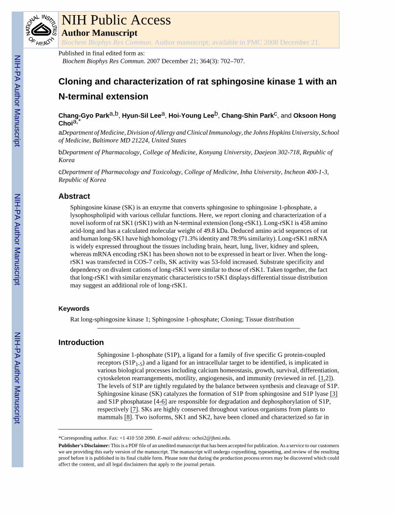
Cloning and characterization of rat sphingosine kinase 1 with an N-terminal extension Chang-Gyo Park a,b , Hyun-Sil Lee a , Hoi-Young Lee b , Chang-Shin Park c , and Oksoon Hong Choi a,* aDepartment of Medicine, Division of Allergy and Clinical Immunology, the Johns Hopkins University, School of Medicine, Baltimore MD 21224, United States bDepartment of Pharmacology, College of Medicine, Konyang University, Daejeon 302-718, Republic of Korea cDepartment of Pharmacology and Toxicology, College of Medicine, Inha University, Incheon 400-1-3, Republic of Korea Abstract Sphingosine kinase (SK) is an enzyme that converts sphingosine to sphingosine 1-phosphate, a lysophospholipid with various cellular functions. Here, we report cloning and characterization of a novel isoform of rat SK1 (rSK1) with an N-terminal extension (long-rSK1). Long-rSK1 is 458 amino acid-long and has a calculated molecular weight of 49.8 kDa. Deduced amino acid sequences of rat and human long-SK1 have high homology (71.3% identity and 78.9% similarity). Long-rSK1 mRNA is widely expressed throughout the tissues including brain, heart, lung, liver, kidney and spleen, whereas mRNA encoding rSK1 has been shown not to be expressed in heart or liver. When the long- rSK1 was transfected in COS-7 cells, SK activity was 53-fold increased. Substrate specificity and dependency on divalent cations of long-rSK1 were similar to those of rSK1. Taken together, the fact that long-rSK1 with similar enzymatic characteristics to rSK1 displays differential tissue distribution may suggest an additional role of long-rSK1. Keywords Rat long-sphingosine kinase 1; Sphingosine 1-phosphate; Cloning; Tissue distribution Introduction Sphingosine 1-phosphate (S1P), a ligand for a family of five specific G protein-coupled receptors (S1P 1-5 ) and a ligand for an intracellular target to be identified, is implicated in various biological processes including calcium homeostasis, growth, survival, differentiation, cytoskeleton rearrangements, motility, angiogenesis, and immunity (reviewed in ref. [1,2]). The levels of S1P are tightly regulated by the balance between synthesis and cleavage of S1P. Sphingosine kinase (SK) catalyzes the formation of S1P from sphingosine and S1P lyase [3] and S1P phosphatase [4-6] are responsible for degradation and dephosphorylation of S1P, respectively [7]. SKs are highly conserved throughout various organisms from plants to mammals [8]. Two isoforms, SK1 and SK2, have been cloned and characterized so far in *Corresponding author. Fax: +1 410 550 2090. E-mail address: [email protected]. Publisher's Disclaimer: This is a PDF file of an unedited manuscript that has been accepted for publication. As a service to our customers we are providing this early version of the manuscript. The manuscript will undergo copyediting, typesetting, and review of the resulting proof before it is published in its final citable form. Please note that during the production process errors may be discovered which could affect the content, and all legal disclaimers that apply to the journal pertain. NIH Public Access Author Manuscript Biochem Biophys Res Commun. Author manuscript; available in PMC 2008 December 21. Published in final edited form as: Biochem Biophys Res Commun. 2007 December 21; 364(3): 702–707. NIH-PA Author Manuscript NIH-PA Author Manuscript NIH-PA Author Manuscript
-
Upload
independent -
Category
Documents
-
view
1 -
download
0
Transcript of Cloning and characterization of rat sphingosine kinase 1 with an N-terminal extension
Cloning and characterization of rat sphingosine kinase 1 with anN-terminal extension
Chang-Gyo Parka,b, Hyun-Sil Leea, Hoi-Young Leeb, Chang-Shin Parkc, and Oksoon HongChoia,*
aDepartment of Medicine, Division of Allergy and Clinical Immunology, the Johns Hopkins University, Schoolof Medicine, Baltimore MD 21224, United States
bDepartment of Pharmacology, College of Medicine, Konyang University, Daejeon 302-718, Republic ofKorea
cDepartment of Pharmacology and Toxicology, College of Medicine, Inha University, Incheon 400-1-3,Republic of Korea
AbstractSphingosine kinase (SK) is an enzyme that converts sphingosine to sphingosine 1-phosphate, alysophospholipid with various cellular functions. Here, we report cloning and characterization of anovel isoform of rat SK1 (rSK1) with an N-terminal extension (long-rSK1). Long-rSK1 is 458 aminoacid-long and has a calculated molecular weight of 49.8 kDa. Deduced amino acid sequences of ratand human long-SK1 have high homology (71.3% identity and 78.9% similarity). Long-rSK1 mRNAis widely expressed throughout the tissues including brain, heart, lung, liver, kidney and spleen,whereas mRNA encoding rSK1 has been shown not to be expressed in heart or liver. When the long-rSK1 was transfected in COS-7 cells, SK activity was 53-fold increased. Substrate specificity anddependency on divalent cations of long-rSK1 were similar to those of rSK1. Taken together, the factthat long-rSK1 with similar enzymatic characteristics to rSK1 displays differential tissue distributionmay suggest an additional role of long-rSK1.
KeywordsRat long-sphingosine kinase 1; Sphingosine 1-phosphate; Cloning; Tissue distribution
IntroductionSphingosine 1-phosphate (S1P), a ligand for a family of five specific G protein-coupledreceptors (S1P1-5) and a ligand for an intracellular target to be identified, is implicated invarious biological processes including calcium homeostasis, growth, survival, differentiation,cytoskeleton rearrangements, motility, angiogenesis, and immunity (reviewed in ref. [1,2]).The levels of S1P are tightly regulated by the balance between synthesis and cleavage of S1P.Sphingosine kinase (SK) catalyzes the formation of S1P from sphingosine and S1P lyase [3]and S1P phosphatase [4-6] are responsible for degradation and dephosphorylation of S1P,respectively [7]. SKs are highly conserved throughout various organisms from plants tomammals [8]. Two isoforms, SK1 and SK2, have been cloned and characterized so far in
*Corresponding author. Fax: +1 410 550 2090. E-mail address: [email protected]'s Disclaimer: This is a PDF file of an unedited manuscript that has been accepted for publication. As a service to our customerswe are providing this early version of the manuscript. The manuscript will undergo copyediting, typesetting, and review of the resultingproof before it is published in its final citable form. Please note that during the production process errors may be discovered which couldaffect the content, and all legal disclaimers that apply to the journal pertain.
NIH Public AccessAuthor ManuscriptBiochem Biophys Res Commun. Author manuscript; available in PMC 2008 December 21.
Published in final edited form as:Biochem Biophys Res Commun. 2007 December 21; 364(3): 702–707.
NIH
-PA Author Manuscript
NIH
-PA Author Manuscript
NIH
-PA Author Manuscript

mammals [9-14]. Although these two enzymes have a high degree of sequence similarity, theyare different in kinetic properties and tissue distribution and originate from different genes[2,15]. In addition, they differ in their physiological functions [16]. While SK1 stimulatesgrowth and survival [17,18], SK2, which contains putative BH3 domain, inhibits cell growthand enhances apoptosis [19].
In addition to the two isoforms, many variants of SK1 and SK2 have been identified [15]. SK1was initially isolated from rat kidney as a 49 kDa protein [13]. Subsequently, two SK1s,SPHK1a and SPHK1b, were cloned from mice [9]. Although these two isoforms are differonly in a few N-terminal amino acid residues, they show different characteristics includingstability, intracellular distribution, and tissue distribution [20]. With respect to human SK1[11,12,14], two additional variants were reported and enzymatic activity of these variants hasbeen shown to be similar to that of SK1 [15]. While sequencing rat genomic DNA and cDNA,Imamura et al. identified 8 alternatively spliced mRNAs with alternative first exons within a3.7-kb CpG island [21,22]. Interestingly, all these spliced isoforms encode the same proteinhomologous to mouse SPHK1a. The transcript homologous to mouse sphk1b is missing in rats[21]. It appears that the presence of the alternatively spliced first exons and a CpG island atthe 5’end is common with both human and mouse SK1 [22].
Here we show cloning of an rSK1 isoform with an additional 75 amino acids at the N-terminalend (long-rSK1). We compared the enzymatic characteristics of long-rSK1 with rSK1 andtissue distribution of mRNA expression of long-rSK1 was determined.
Materials and methodsMaterials
D-erythro-sphingosine, D-erythro-dihydrosphingosine (DHS), DL-threo-dihydrosphingosine(threoDHS), and N,N-dimethylsphingosine (DMS) were from Biomol (Plymouth Meeting,PA); N-acetyl-D-sphingosine from Sigma (St. Louis, MO); [γ-32P]ATP from PerkinElmer(Boston, MA); Silica gel TLC plates from Bodman (Aston, PA). Materials for cell culture wereobtained from Invitrogen (Gaithersberg, MD).
Cloning of long-rSK1In order to obtain predicted mRNA sequences of spliced variants of rSK1, we performed asearch by scanning genomic sequences of rat chromosome 10 (accession no. NW_047343),using Genescan 1.0 program (http://gnen.mit.edu/cgi-bin/genscaw.cgi), The search gave acandidate mRNA, which would encode rSK1 with an N-terminal extension. To confirm theexpression of such mRNA, total RNA was isolated from rat kidney and rat basophilic leukemia(RBL)-2H3, a rat mast cell line, using TRIzol® reagent (Invitrogen). The cDNAs wereproduced using a gene-specific primer (5’-GGGCAGCATCGGTGGCTCCT-3’) and N-terminal portion of long-rSK1 was amplified using two primers (sense (P1): 5’-GGTTCTTCGTCCCAGAGTA-3’, antisense (P2): 5’-AGAGCGCCC-TCGTCATTCCC-3’)(Product #1, Fig. 1C). The rest of rSK1 was cloned using two primers (sense (P3): 5’-ATCTGGCCGCCTCCGGGAAT-3’ antisense (P4): 5’-GGCAGATCCTCATATTGGTTC-3’) and as a template, rSK1f cDNA (accession No.,BC081738), purchased from Invitrogen (Product #2, Fig. 1C). The full-length long-rSK1(Product #3, Fig. 1C) was cloned by PCR using two primers (sense (P5): 5’-TCTGGGAAGCTTATGCAACTG-3’ antisense: P4) and products #1 and #2 as templates. Thecoding region of rSK1 was also cloned by PCR using two primers (sense (P6): 5’-CGGGACAAGCTTATGCAACCA-3’ antisense: P4) and rSK1f as a template (Product #4,Fig. 1C). PCR products were subcloned into the pCR®2.1-TOPO® TA cloning vector. Correctsequences of the PCR products were confirmed by sequencing analysis (Johns Hopkins CORE
Park et al. Page 2
Biochem Biophys Res Commun. Author manuscript; available in PMC 2008 December 21.
NIH
-PA Author Manuscript
NIH
-PA Author Manuscript
NIH
-PA Author Manuscript
DNA Analysis Facility). Finally, cDNAs of long-rSK1 and rSK1 were subcloned into the pFlagvector using the HindIII/EcoRI restriction enzymes.
RT-PCRWister rat tissues were washed twice with ice-cold phosphate buffered saline and quicklyfrozen in liquid N2. Frozen tissues were ground while being kept frozen by continuously addingliquid N2. Total RNA was isolated from these tissues using TRIzol® reagent (Invitrogen). ThecDNAs were produced using a random primer. The N-terminal portion of long-rSK1 (Product#1, Fig. 1C) was amplified using two primers, P1 and P2 (Fig. 1C). β-actin was amplified usinganother pair of primers (sense: 5’-GTGGGCCGGTCTAGGCACCAA-3’, antisense: 5’-CTCTTTG-ATGTGACGCACGA-TTTC-3’).
Cell culture and transfectionCOS-7 cells were maintained as monolayer cultures in Dulbecco’s modified Eagle medium(DMEM) supplemented with 10% fetal bovine serum, penicillin (100 units/ml), andstreptomycin (100 μg/ml). COS-7 cells were transfected with pFlag-long-rSK1 or pFlag-rSK1,using Lipofectamine® according to the manufacturer’s instruction. Expressing cells wereselected with 2 mg/ml of G418 for 12 days before using for experiments. RBL-2H3 cells weremaintained as monolayer cultures in Earle’s modified Eagle’s medium supplemented with 15%fetal bovine serum, 2 mM glutamine, penicillin (1000 units/ml) and streptomycin (100 μg/ml).
Western blot analysisCOS-7 cells transfected with pFlag-long-rSK1 or pFlag-rSK1 were lysed by addition of ice-cold lysis buffer, containing 0.5% Triton X-100, 200 mM boric acid (pH 8.0), 150 mM NaCl,5 mM EDTA, 5 mM NaF, 1 mM Na3VO4, 10 μg/ml aprotinin and pepstatin A and 5 μg/mlleupeptin. Lysates were left on ice for 30 minutes and centrifuged for 30 minutes at 12,000 ×g at 4°C. The supernatant was used for Western blot anlaysis using anti-human SK1 antibodyraised against C-terminal 20 amino acids of human SK1.
In vitro sphingosine kinase assayCytosolic SK activity was measured by incubating various concentration of D-erythro-sphingosine in the reaction mixture containing 0.125% Triton X-100, 10 mM MgCl2, and[γ-32P]ATP (5 μCi, 1 mM) at 30 °C for 20 minutes as previously described [9].
Results and discussionCloning of long-rSK1
As previously mentioned, there has been 8 alternatively spliced mRNAs of rSK1 identified sofar with all but b form encoding the same protein [21,22]. We were interested in finding outwhether there might be an alternatively spliced mRNA that encodes long form of rSK1 as inthe case of human SK1 [15]. We performed a search for spliced variants by scanning genomicsequences of rat chromosome 10, using Genescan 1.0 program. The analysis identified a novelalternatively spliced mRNA, the first two exons of which were derived from the first exon ofisoform f (Fig. 1A). To confirm the expression of such mRNA, two PCR primers (a senseprimer from exon 1 and an antisense primer from exon 2 shown by two arrowheads in Fig. 1A)were designed and RT-PCR was performed using total RNAs obtained from rat kidney or ratmast cell line RBL-2H3. As shown in Fig. 1B, products corresponding to mRNA f and thelong-form were detected. Interestingly, one additional band was produced, which mightsuggest an additional spliced variant. The sequences of the long-rSK1 from rat kidney andRBL-2H3 cells were confirmed to be identical and the same as the predicted one. To clone therest of cDNA of the novel isoform, we performed PCR using primers P3 and P4 and rSK1f
Park et al. Page 3
Biochem Biophys Res Commun. Author manuscript; available in PMC 2008 December 21.
NIH
-PA Author Manuscript
NIH
-PA Author Manuscript
NIH
-PA Author Manuscript
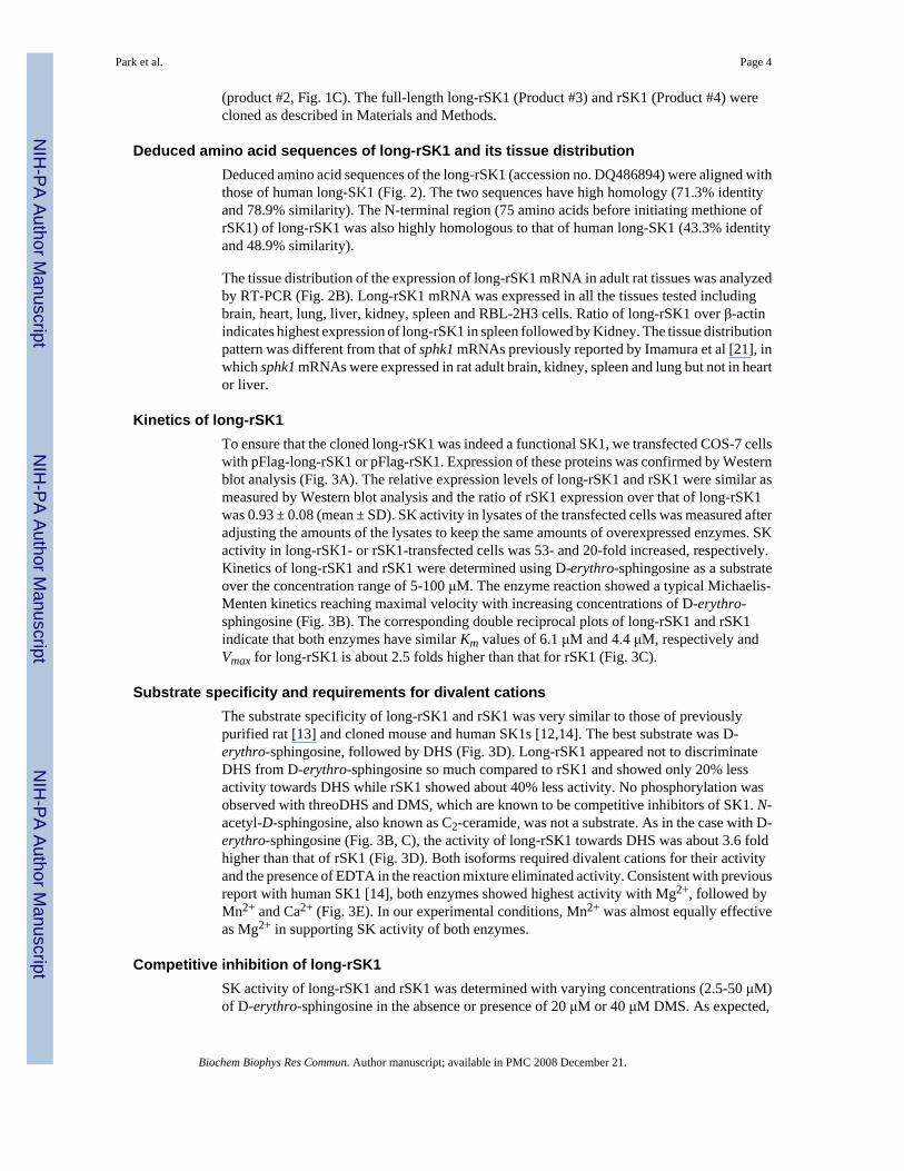
(product #2, Fig. 1C). The full-length long-rSK1 (Product #3) and rSK1 (Product #4) werecloned as described in Materials and Methods.
Deduced amino acid sequences of long-rSK1 and its tissue distributionDeduced amino acid sequences of the long-rSK1 (accession no. DQ486894) were aligned withthose of human long-SK1 (Fig. 2). The two sequences have high homology (71.3% identityand 78.9% similarity). The N-terminal region (75 amino acids before initiating methione ofrSK1) of long-rSK1 was also highly homologous to that of human long-SK1 (43.3% identityand 48.9% similarity).
The tissue distribution of the expression of long-rSK1 mRNA in adult rat tissues was analyzedby RT-PCR (Fig. 2B). Long-rSK1 mRNA was expressed in all the tissues tested includingbrain, heart, lung, liver, kidney, spleen and RBL-2H3 cells. Ratio of long-rSK1 over β-actinindicates highest expression of long-rSK1 in spleen followed by Kidney. The tissue distributionpattern was different from that of sphk1 mRNAs previously reported by Imamura et al [21], inwhich sphk1 mRNAs were expressed in rat adult brain, kidney, spleen and lung but not in heartor liver.
Kinetics of long-rSK1To ensure that the cloned long-rSK1 was indeed a functional SK1, we transfected COS-7 cellswith pFlag-long-rSK1 or pFlag-rSK1. Expression of these proteins was confirmed by Westernblot analysis (Fig. 3A). The relative expression levels of long-rSK1 and rSK1 were similar asmeasured by Western blot analysis and the ratio of rSK1 expression over that of long-rSK1was 0.93 ± 0.08 (mean ± SD). SK activity in lysates of the transfected cells was measured afteradjusting the amounts of the lysates to keep the same amounts of overexpressed enzymes. SKactivity in long-rSK1- or rSK1-transfected cells was 53- and 20-fold increased, respectively.Kinetics of long-rSK1 and rSK1 were determined using D-erythro-sphingosine as a substrateover the concentration range of 5-100 μM. The enzyme reaction showed a typical Michaelis-Menten kinetics reaching maximal velocity with increasing concentrations of D-erythro-sphingosine (Fig. 3B). The corresponding double reciprocal plots of long-rSK1 and rSK1indicate that both enzymes have similar Km values of 6.1 μM and 4.4 μM, respectively andVmax for long-rSK1 is about 2.5 folds higher than that for rSK1 (Fig. 3C).
Substrate specificity and requirements for divalent cationsThe substrate specificity of long-rSK1 and rSK1 was very similar to those of previouslypurified rat [13] and cloned mouse and human SK1s [12,14]. The best substrate was D-erythro-sphingosine, followed by DHS (Fig. 3D). Long-rSK1 appeared not to discriminateDHS from D-erythro-sphingosine so much compared to rSK1 and showed only 20% lessactivity towards DHS while rSK1 showed about 40% less activity. No phosphorylation wasobserved with threoDHS and DMS, which are known to be competitive inhibitors of SK1. N-acetyl-D-sphingosine, also known as C2-ceramide, was not a substrate. As in the case with D-erythro-sphingosine (Fig. 3B, C), the activity of long-rSK1 towards DHS was about 3.6 foldhigher than that of rSK1 (Fig. 3D). Both isoforms required divalent cations for their activityand the presence of EDTA in the reaction mixture eliminated activity. Consistent with previousreport with human SK1 [14], both enzymes showed highest activity with Mg2+, followed byMn2+ and Ca2+ (Fig. 3E). In our experimental conditions, Mn2+ was almost equally effectiveas Mg2+ in supporting SK activity of both enzymes.
Competitive inhibition of long-rSK1SK activity of long-rSK1 and rSK1 was determined with varying concentrations (2.5-50 μM)of D-erythro-sphingosine in the absence or presence of 20 μM or 40 μM DMS. As expected,
Park et al. Page 4
Biochem Biophys Res Commun. Author manuscript; available in PMC 2008 December 21.
NIH
-PA Author Manuscript
NIH
-PA Author Manuscript
NIH
-PA Author Manuscript
the activity of both enzymes was inhibited by DMS (Fig. 4A, B). Double reciprocal plotssuggested that DMS displayed competitive inhibition with increased Km values for long-rSK1to 39.7 μM and 52.0 μM with 20 μM and 40 μM of DMS, respectively and those for rSK1 to16.7 μM and 43.8 μM (Fig. 4C, D). Ki values for long-rSK1 and rSK1 were 5.8 μM and 0.33μM of DMS, respectively.
To summarize, long-rSK1 mRNA is a novel isoform of rat sphk1 that uniquely encodes anadditional 75 amino acids at the N-terminal end of rSK1, whereas the 7 other isoforms encodesrSK1. It is plausible to think that the N-terminal region of long-rSK1 may serve as a regulatorydomain, which receives signals from upstream or play a role in localization of the enzyme indifferent subcellular organelles. In fact, our study with human long-SK1 fused with greenfluorescent protein indicated that long-SK1 is partially associated with the endoplasmicreticulum (ER) and further translocated to the ER upon receptor stimulation (unpublishedobservation, Jung, ID and Choi, OH). This might suggest that long-rSK1 is involved in cellsignaling.
Acknowledgements
This work was supported by National Institutes of Health Grant HL068879 (to O.H.C.) and the Korea ResearchFoundation Grant by the Korean Government (MOEHRD) (KRF-2006-013-E00138) (to C.G.P.).
References1. Spiegel S, Milstien S. Functions of the Multifaceted Family of Sphingosine Kinases and Some Close
Relatives. J Biol Chem 2007;282:2125–2129. [PubMed: 17135245]2. Spiegel S, Milstien S. Sphingosine-1-phosphate: an enigmatic signalling lipid. Nat Rev Mol Cell Biol
2003;4:397–407. [PubMed: 12728273]3. Saba JD, Nara F, Bielawska A, Garrett S, Hannun YA. The BST1 gene of Saccharomyces cerevisiae
is the sphingosine-1- phosphate lyase. J Biol Chem 1997;272:26087–26090. [PubMed: 9334171]4. Mandala SM, Thornton R, Galve-Roperh I, et al. Molecular cloning and characterization of a lipid
phosphohydrolase that degrades sphingosine-1- phosphate and induces cell death. Proc Natl Acad SciU S A 2000;97:7859–7864. [PubMed: 10859351]
5. Ogawa C, Kihara A, Gokoh M, Igarashi Y. Identification and Characterization of a Novel HumanSphingosine-1-phosphate Phosphohydrolase, hSPP2. J Biol Chem 2003;278:1268–1272. [PubMed:12411432]
6. Le Stunff H, Peterson C, Liu H, Milstien S, Spiegel S. Sphingosine-1-phosphate and lipidphosphohydrolases. Biochim Biophys Acta 2002;1582:8–17. [PubMed: 12069805]
7. Saba JD, Hla T. Point-counterpoint of sphingosine 1-phosphate metabolism. Circulation Research2004;94:724–734. [PubMed: 15059942]
8. Leclercq TM, Pitson SM. Cellular signalling by sphingosine kinase and sphingosine 1-phosphate.IUBMB Life 2006;58:467–472. [PubMed: 16916784]
9. Kohama T, Olivera A, Edsall L, Nagiec MM, Dickson R, Spiegel S. Molecular cloning and functionalcharacterization of murine sphingosine kinase. J Biol Chem 1998;273:23722–23728. [PubMed:9726979]
10. Liu H, Sugiura M, Nava VE, et al. Molecular cloning and functional characterization of a novelmammalian sphingosine kinase type 2 isoform. J Biol Chem 2000;275:19513–19520. [PubMed:10751414]
11. Melendez AJ, Carlos-Dias E, Gosink M, Allen JM, Takacs L. Human sphingosine kinase: molecularcloning, functional characterization and tissue distribution. Gene 2000;251:19–26. [PubMed:10863092]
12. Nava VE, Lacana E, POULTON S. Functional characterization of human sphingosine kinase-1. FEBSLett 2000;473:81–84. [PubMed: 10802064]
13. Olivera A, Kohama T, Tu Z, Milstien S, Spiegel S. Purification and characterization of rat kidneysphingosine kinase. J Biol Chem 1998;273:12576–12583. [PubMed: 9575218]
Park et al. Page 5
Biochem Biophys Res Commun. Author manuscript; available in PMC 2008 December 21.
NIH
-PA Author Manuscript
NIH
-PA Author Manuscript
NIH
-PA Author Manuscript

14. Pitson SM, D’andrea RJ, Vandeleur L, et al. Human sphingosine kinase: purification, molecularcloning and characterization of the native and recombinant enzymes. Biochem J 2000;350:429–441.[PubMed: 10947957]
15. Billich A, Bornancin F, Devay P, Mechtcheriakova D, Urtz N, Baumruker T. Phosphorylation of theImmunomodulatory Drug FTY720 by Sphingosine Kinases. J Biol Chem 2003;278:47408–47415.[PubMed: 13129923]
16. Maceyka M, Sankala H, Hait NC, et al. SphK1 and SphK2, Sphingosine Kinase Isoenzymes withOpposing Functions in Sphingolipid Metabolism. J Biol Chem 2005;280:37118–37129. [PubMed:16118219]
17. Olivera A, Kohama T, Edsall L, et al. Sphingosine kinase expression increases intracellularsphingosine-1-phosphate and promotes cell growth and survival. J Cell Biol 1999;147:545–558.[PubMed: 10545499]
18. Olivera A, Rosenfeldt HM, Bektas M, et al. Sphingosine Kinase Type 1 Induces G12/13-mediatedStress Fiber Formation, yet Promotes Growth and Survival Independent of G Protein-coupledReceptors. J Biol Chem 2003;278:46452–46460. [PubMed: 12963721]
19. Liu H, Toman RE, Goparaju SK, et al. Sphingosine Kinase Type 2 Is a Putative BH3-only ProteinThat Induces Apoptosis. J Biol Chem 2003;278:40330–40336. [PubMed: 12835323]
20. Kihara A, Anada Y, Igarashi Y. Mouse Sphingosine Kinase Isoforms SPHK1a and SPHK1b Differin Enzymatic Traits Including Stability, Localization, Modification, and Oligomerization. J BiolChem 2006;281:4532–4539. [PubMed: 16368679]
21. Imamura T, Ohgane J, Ito S, et al. CpG Island of Rat Sphingosine Kinase-1 Gene: Tissue-DependentDNA Methylation Status and Multiple Alternative First Exons. Genomics 2001;76:117–125.[PubMed: 11560121]
22. Imamura T, Miyauchi-Senda N, Tanaka S, Shiota K. Identification of genetic and epigeneticsimilarities of SPHK1/Sphk1 in mammals. J Vet Med Sci 2004;66:1387–1393. [PubMed: 15585953]
Park et al. Page 6
Biochem Biophys Res Commun. Author manuscript; available in PMC 2008 December 21.
NIH
-PA Author Manuscript
NIH
-PA Author Manuscript
NIH
-PA Author Manuscript
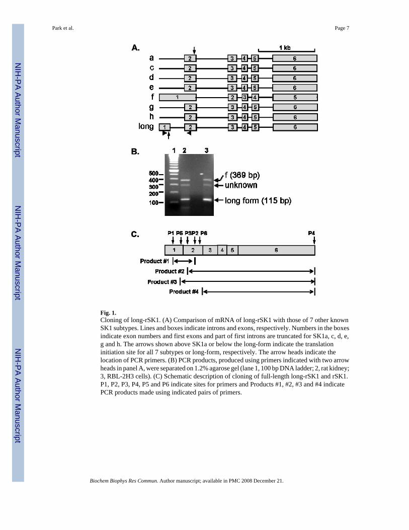
Fig. 1.Cloning of long-rSK1. (A) Comparison of mRNA of long-rSK1 with those of 7 other knownSK1 subtypes. Lines and boxes indicate introns and exons, respectively. Numbers in the boxesindicate exon numbers and first exons and part of first introns are truncated for SK1a, c, d, e,g and h. The arrows shown above SK1a or below the long-form indicate the translationinitiation site for all 7 subtypes or long-form, respectively. The arrow heads indicate thelocation of PCR primers. (B) PCR products, produced using primers indicated with two arrowheads in panel A, were separated on 1.2% agarose gel (lane 1, 100 bp DNA ladder; 2, rat kidney;3, RBL-2H3 cells). (C) Schematic description of cloning of full-length long-rSK1 and rSK1.P1, P2, P3, P4, P5 and P6 indicate sites for primers and Products #1, #2, #3 and #4 indicatePCR products made using indicated pairs of primers.
Park et al. Page 7
Biochem Biophys Res Commun. Author manuscript; available in PMC 2008 December 21.
NIH
-PA Author Manuscript
NIH
-PA Author Manuscript
NIH
-PA Author Manuscript

Fig. 2.Alignment of deduced amino acid sequences of rat and human long-SK1 and the tissuedistribution of the expression of long-rSK1 mRNAs. (A) Sequence alignment of deducedamino acid sequences of rat and human long-SK1 was performed by ClustalW. Identicalsequences were shaded in dark grey and similar sequences in grey background. The initiationmethionines of rat and human SK1s are indicated with *. (B) Tissue distribution of expressionof long-rSK1 mRNAs. Tissue-specific expression of long-rSK1 mRNAs in adult rat tissueswas analyzed by RT-PCR. cDNAs corresponding to 0.3 μg of total RNA of brain, heart, lung,liver, kidney, spleen and RBL-2H3 cells were produced using a random primer. Using gene-specific primers described in the Materials and methods, long-rSK1 (L-rSK1) and β-actin were
Park et al. Page 8
Biochem Biophys Res Commun. Author manuscript; available in PMC 2008 December 21.
NIH
-PA Author Manuscript
NIH
-PA Author Manuscript
NIH
-PA Author Manuscript
amplified. Numbers indicate the ratio of the intensities of long-rSK1 over those of β-actin. Thedepicted data are representative of four experiments with similar results.
Park et al. Page 9
Biochem Biophys Res Commun. Author manuscript; available in PMC 2008 December 21.
NIH
-PA Author Manuscript
NIH
-PA Author Manuscript
NIH
-PA Author Manuscript
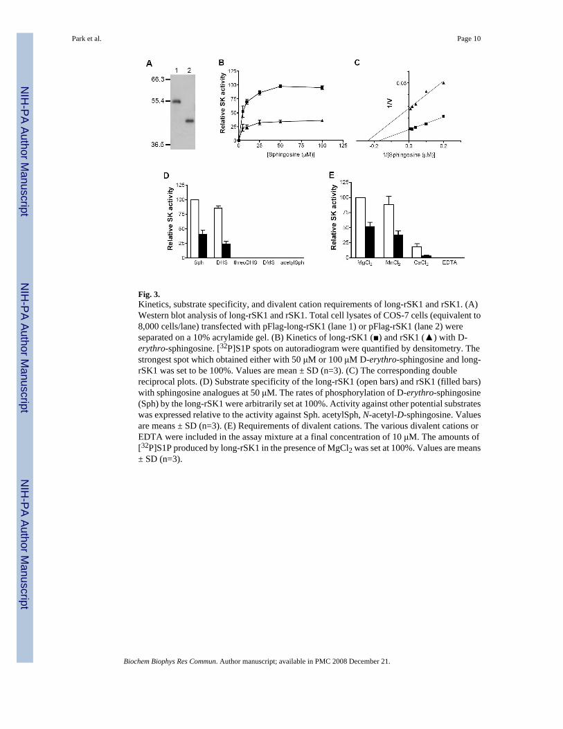
Fig. 3.Kinetics, substrate specificity, and divalent cation requirements of long-rSK1 and rSK1. (A)Western blot analysis of long-rSK1 and rSK1. Total cell lysates of COS-7 cells (equivalent to8,000 cells/lane) transfected with pFlag-long-rSK1 (lane 1) or pFlag-rSK1 (lane 2) wereseparated on a 10% acrylamide gel. (B) Kinetics of long-rSK1 (■) and rSK1 (▲) with D-erythro-sphingosine. [32P]S1P spots on autoradiogram were quantified by densitometry. Thestrongest spot which obtained either with 50 μM or 100 μM D-erythro-sphingosine and long-rSK1 was set to be 100%. Values are mean ± SD (n=3). (C) The corresponding doublereciprocal plots. (D) Substrate specificity of the long-rSK1 (open bars) and rSK1 (filled bars)with sphingosine analogues at 50 μM. The rates of phosphorylation of D-erythro-sphingosine(Sph) by the long-rSK1 were arbitrarily set at 100%. Activity against other potential substrateswas expressed relative to the activity against Sph. acetylSph, N-acetyl-D-sphingosine. Valuesare means ± SD (n=3). (E) Requirements of divalent cations. The various divalent cations orEDTA were included in the assay mixture at a final concentration of 10 μM. The amounts of[32P]S1P produced by long-rSK1 in the presence of MgCl2 was set at 100%. Values are means± SD (n=3).
Park et al. Page 10
Biochem Biophys Res Commun. Author manuscript; available in PMC 2008 December 21.
NIH
-PA Author Manuscript
NIH
-PA Author Manuscript
NIH
-PA Author Manuscript
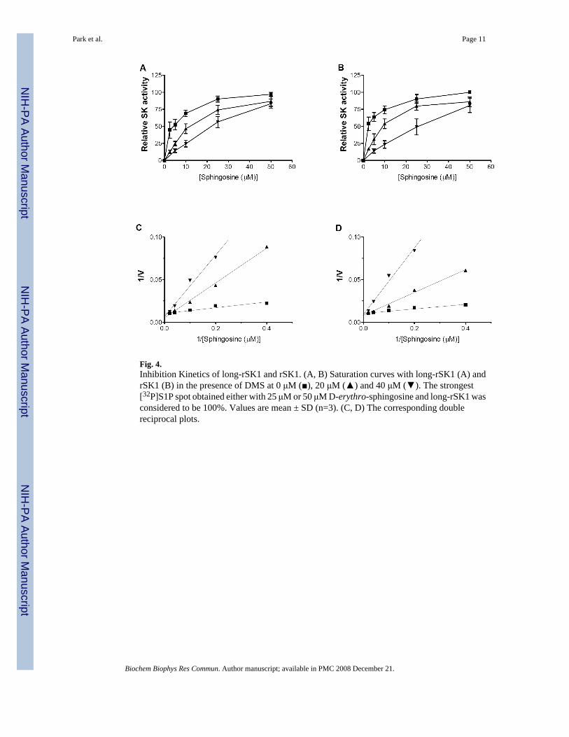
Fig. 4.Inhibition Kinetics of long-rSK1 and rSK1. (A, B) Saturation curves with long-rSK1 (A) andrSK1 (B) in the presence of DMS at 0 μM (■), 20 μM (▲) and 40 μM (▼). The strongest[32P]S1P spot obtained either with 25 μM or 50 μM D-erythro-sphingosine and long-rSK1 wasconsidered to be 100%. Values are mean ± SD (n=3). (C, D) The corresponding doublereciprocal plots.
Park et al. Page 11
Biochem Biophys Res Commun. Author manuscript; available in PMC 2008 December 21.
NIH
-PA Author Manuscript
NIH
-PA Author Manuscript
NIH
-PA Author Manuscript