
Use of near-infrared spectroscopy to identify trends in regional ...
Upload
khangminh22Category
view
0download
0
Clinical application of near-infrared
spectroscopy in perioperative assessment of
cerebral and peripheral tissue oxygenation
Anneliese MoermanGhent UniversityFaculty of Medicine and Health SciencesDepartment of Anaesthesiology

2
Promotor
Prof. Dr. Patrick Wouters
Support committee
Prof. Dr. Stefan De Hert
Prof. Dr. Chris Verborgh
Members of the examination committee
Prof. Dr. Daniel De Wolf
Prof. Dr. Luc Herregods
Prof. Dr. Eric Mortier
Prof. Dr. Gunnar Naulaers
Prof. Dr. Yves Van Belleghem
Prof. Dr. Johan Vande Walle
Prof. Dr. Frank Vermassen
Thesis submitted in fulfillment of the requirements for the degree of Doctor
in Medical Sciences.

3
Father and Daughter: A Father’s Privilege
Child, why would you want to practice medicine?
Late hours, night calls, research projects, administration deadlines
Don’t do medicine, look at my life
Do what makes you happy
Daughter, why are you stressing?
Don’t do this. There are easier ways to make a living
It’s not worth it, go ahead and live
Do what makes you happy
Doctor, don’t you know there is a price for learning?
Nothing worth doing is done easily
The struggle cries there must be an easier way
Do what makes you happy
Dear Heart, you have chosen a difficult road and kept true
In medicine we meet as equals
An old man with eyes that leak now and again
Glad and proud that you do what makes you happy
Spencer S. Kee
This thesis is dedicated to the memory of my father

4

Contents
1 General introduction 9
1.1 Near-infrared spectroscopy . . . . . . . . . . . . . . . 10
1.1.1 Principles of operation . . . . . . . . . . . . . . 11
1.1.2 Assumptions and limitations . . . . . . . . . 15
1.1.3 Validation . . . . . . . . . . . . . . . . . . . . 17
1.2 Clinical considerations . . . . . . . . . . . . . . . . . 18
1.3 Clinical studies . . . . . . . . . . . . . . . . . . . . . 23
1.4 Commercially available NIRS devices . . . . . . . . . 28
1.5 References . . . . . . . . . . . . . . . . . . . . . . . . . 31
2 Objectives of the research 39
3 NIRS and cerebral perfusion 41
3.1 Background . . . . . . . . . . . . . . . . . . . . . . . . 41
3.2 Objective . . . . . . . . . . . . . . . . . . . . . . . . 42
3.3 NIRS monitoring in beach chair position . . . . . . . 43
3.3.1 Introduction . . . . . . . . . . . . . . . . . . 44
3.3.2 Methods . . . . . . . . . . . . . . . . . . . . . 45
3.3.3 Results . . . . . . . . . . . . . . . . . . . . . 47
3.3.4 Discussion . . . . . . . . . . . . . . . . . . . . . 51
3.3.5 References . . . . . . . . . . . . . . . . . . . . 56
3.4 NIRS monitoring in moyamoya disease . . . . . . . . 59
3.4.1 Introduction . . . . . . . . . . . . . . . . . . 59
5
6 CONTENTS
3.4.2 Case report . . . . . . . . . . . . . . . . . . . 60
3.4.3 Discussion . . . . . . . . . . . . . . . . . . . . 64
3.4.4 References . . . . . . . . . . . . . . . . . . . . 67
4 NIRS and regional perfusion 71
4.1 Background . . . . . . . . . . . . . . . . . . . . . . . . 71
4.2 Objective . . . . . . . . . . . . . . . . . . . . . . . . 72
4.3 NIRS during aortic coarctation repair . . . . . . . . 72
4.3.1 Introduction . . . . . . . . . . . . . . . . . . 74
4.3.2 Methods . . . . . . . . . . . . . . . . . . . . . 75
4.3.3 Results . . . . . . . . . . . . . . . . . . . . . 79
4.3.4 Discussion . . . . . . . . . . . . . . . . . . . . 86
4.3.5 References . . . . . . . . . . . . . . . . . . . . 89
4.4 NIRS during aortic aneurysm repair . . . . . . . . . 92
4.4.1 Introduction . . . . . . . . . . . . . . . . . . 93
4.4.2 Case report . . . . . . . . . . . . . . . . . . . 93
4.4.3 Discussion . . . . . . . . . . . . . . . . . . . . 98
4.4.4 References . . . . . . . . . . . . . . . . . . . . 102
5 NIRS as an estimate of SmvO2 105
5.1 Background . . . . . . . . . . . . . . . . . . . . . . . 105
5.2 Objective . . . . . . . . . . . . . . . . . . . . . . . . 107
5.3 NIRS as an estimate of venous saturation . . . . . . 108
5.3.1 Introduction . . . . . . . . . . . . . . . . . . 109
5.3.2 Methods . . . . . . . . . . . . . . . . . . . . . 110
5.3.3 Results . . . . . . . . . . . . . . . . . . . . . 113
5.3.4 Discussion . . . . . . . . . . . . . . . . . . . . 120
5.3.5 References . . . . . . . . . . . . . . . . . . . . 123
6 Physiology of perfusion 127
6.1 Background . . . . . . . . . . . . . . . . . . . . . . . 127
6.2 Objective . . . . . . . . . . . . . . . . . . . . . . . . 128
6.3 NIRS to assess physiology of perfusion . . . . . . . . 128
6.3.1 Introduction . . . . . . . . . . . . . . . . . . 129

CONTENTS 7
6.3.2 Methods . . . . . . . . . . . . . . . . . . . . . 1306.3.3 Results . . . . . . . . . . . . . . . . . . . . . 1346.3.4 Discussion . . . . . . . . . . . . . . . . . . . . 1396.3.5 References . . . . . . . . . . . . . . . . . . . . 143
7 General discussion 147
8 Future perspectives 159
Summary 163
Samenvatting 167
List of abbreviations 169
Dankwoord 173
Curriculum Vitae 177

8 CONTENTS

Chapter 1
General introduction
Adapted from: Moerman A, Wouters P. Near-infrared spec-troscopy (NIRS) monitoring in contemporary anesthesiaand critical care. Acta Anaesth Belg 2010; 61: 185-94
The primary goal in the hemodynamic management of patientsundergoing surgery is to preserve oxygen delivery at a level sufficientto cover all metabolic needs. Nowadays, anesthesiologists can rely ona variety of monitoring tools to quantify cardiovascular performanceand global oxygen delivery. Nonetheless, current standard anesthesiamonitoring still has two major drawbacks. First, it provides a globalassessment of the patient’s status, and as such vital organ ischemiamay go unnoticed until functional organ damage becomes evident.Case reports of dramatic neurologic outcome after minor surgeryin healthy patients [1] point out the compelling need for organ-specific monitoring. A second drawback is that the majority ofvariables monitored in contemporary anesthesia focus on oxygensupply (cardiovascular performance, hemoglobin and arterial oxygencontent) but do not assess imbalances between oxygen supply anddemand. The use of central and mixed venous oxygen measurements
9
10 CHAPTER 1. GENERAL INTRODUCTION
to assess oxygen consumption is gaining interest now in perioperativecare [2]. However, current techniques for assessing venous oxygensaturation are invasive and therefore not routinely incorporated intoclinical anesthesia.
Near-infrared spectroscopy (NIRS) is a non-invasive technologythat continuously monitors regional tissue oxygenation. NIRS wasoriginally introduced in clinical practice in 1985, for the assessmentof cerebral oxygenation in preterm infants [3]. It was also welcomedwith enthusiasm in cardiac and neuro-anesthesia, but its utility,particularly in the latter field of application, was seriously challengedby a series of reports on false positive as well as false negativereadings. The technique was further discredited by anecdotal papersillustrating that NIRS oximeters generated near normal values whenthe probes were placed on an empty human skull filled with blood-soaked gauzes [4] or even on a pumpkin [5]. Even today, these papersare quoted to question the validity of NIRS monitoring. However,a fair understanding of the assumptions and limitations of NIRStechnology suffice to understand that such observations are indeedpossible, yet do not invalidate the use of NIRS to quantify changesin the oxygen status of human tissue. The interest in NIRS as amonitoring tool in anesthesia has revived over the past few years andseveral systems have now been approved for clinical use.
1.1 Near-infrared spectroscopy
The first attempt to monitor human tissue oxygenation non-invasivelydates back to 1874 when the German physiologist Karl Von Vierordtshowed that the amount of red light transmitted through a handdecreased after it was made ischemic [6]. His pioneering studies wereessentially ignored for half a century until it was again reportedthat the variable transmission of red and infrared light through ahuman ear reflected changes in blood oxygenation [7]. The firstsmall portable oximeter was developed in 1942 by Glen Milliken

1.1. NEAR-INFRARED SPECTROSCOPY 11
[8], although the device was used only as an experimental tool inthe aviation and the physiology laboratory. The concept of cerebralnear-infrared spectroscopy originated with the observations of Jobsis[9] who irradiated a cat’s head with near-infrared light and foundthat the intensity of the transmitted light varied with the oxygenmetabolic state of the brain (Fig. 1.1).
Figure 1.1
1.1.1 Principles of operation
The physical and mathematical basis for NIRS is provided by theBeer-Lambert law, which states that the quantity of light absorbedby a substance (A) is directly proportional to the specific absorp-tion coefficient of the substance at a particular wavelength (ε), theconcentration of the substance (c) and the pathlength of the light

12 CHAPTER 1. GENERAL INTRODUCTION
through the solution (l) (A = ε · c · l) [10].
The relative transparency of biological tissues to light in the near-infrared part of the spectrum (700-1000 nm) enables light photonsto pass through the tissues, where they are attenuated due to acombination of absorption and scattering. Because of scatteringby the tissue components, the light does not travel in a straightline. Therefore, the Modified Beer-Lambert law is applied: (A =ε · c · l ·B + k), where B is the differential pathlength factor and k isan additive geometry-dependent term, reflecting scatter loss. Thegeometrical pathlength l has to be multiplied by B to find the trueoptical distance, because light that reaches the detector will have beenscattered multiple times and therefore has travelled a much greaterdistance than the actual light emitter-detector distance. k correctsfor the fact that not all emitted light reaches the detector, becausesome of it is scattered away from the detector, giving scattering losses.Scattering is a function of the tissue composition and the number ofvarious tissue interfaces. Because B and k are unknown factors, noabsolute values can be measured with the Modified Beer-Lambertlaw. NIRS technology is based on the assumption that the quantityof scattering remains constant and that changes in attenuation resultsolely from changes in absorption.
Several biological molecules, termed chromophores, absorb lightin the near-infrared (NIR) spectrum. However, only hemoglobinand cytochrome oxidase are present in variable concentrations,reflecting blood and intracellular oxygenation, respectively. Otherchromophores are assumed to be constant over the period of moni-toring.
The wavelengths of NIR light used in commercial devices areselected to be sensitive to hemoglobin. Cytochroom oxidase has acrucial role in mitochondrial oxidative energy metabolism, and there-fore provides a potential biomarker of the cellular oxygenation state,with substantial physiological and clinical importance. However, it ispresent in much lower concentrations in the tissue than oxygenated

1.1. NEAR-INFRARED SPECTROSCOPY 13
and deoxygenated hemoglobin and its absorption spectrum overlapsthat of these chromophores, and therefore, the validity of cytochroomoxidase measurements is debated, and the signal is not incorporatedinto any clinical monitors yet [11].
Oximetry relies on the fact that absorption of near-infraredlight at specific wavelengths is different in deoxygenated hemoglobin(HHb) when compared with oxygenated hemoglobin (O2Hb) (Fig.1.2).
Figure 1.2: Distinct absorption spectra for water (H2O),lipids, oxygenated (O2Hb) and deoxygenated hemoglobin (HHb).Near-infrared spectroscopy measures light absorption at spe-cific wavelengths to differentiate between O2Hb and HHb.(https://Dosi.bli.uci.edu/userfiles/image/basis spectra.jpg)
Commercial devices generally use wavelengths between 690 and880 nm where the absorption spectra of O2Hb and HHb are max-imally separated and there is minimal overlap with that of waterabsorption (980 nm) (Fig. 1.2). Optical absorption at 1 wavelength

14 CHAPTER 1. GENERAL INTRODUCTION
for each chromophore of interest must be known. NIRS devices usenear-infrared light at two or more specific wavelengths to differentiatebetween O2Hb and HHb. Similar to the principles used in pulseoximetry, the device measures the amount of light absorbed at thesespecific wavelengths and calculates the relative contribution of O2Hband HHb. The resulting ratio of O2Hb to total Hb (THb) (expressedas a percentage) represents the oxygen saturation of tissue under thesensor: O2Hb.k/THb.k.100(%), in which k, the constant reflectingthe scattering, can be neglected. No attempt is made to measureoptical scattering. The scale of measured changes is dependent on theapplication of assumptions of the scattering properties at differentwavelengths, and is incorporated into the algorithm of the respectivedevices. Algorithmic formulae are complex, and their validity iscontingent on the assumptions made. The variability in algorithmsbetween NIRS devices implicates that there are differences in thechromofore concentrations derived, making comparisons betweenoximeters produced by different manufacturers problematic.
Jobsis [9] used transmitted light in his original experiments. Lightwas applied to one side of the body and received on the otherside. However, attenuation of light due to absorption and scat-tering restricts usage of this method to very thin and transparentareas of the body such as the earlobe or finger. For that reason,reflected light rather than transmitted light is being used to studyabsorption of light in larger tissue samples. Reflectance probes locatethe light emitter and detector adjacent to one another. The lighttakes a “banana-shaped” pathway through the tissues, with the depthof photon penetration proportional to the source-detector separation(principle of spatial resolution) (Fig. 1.3). In order to compensatefor superficial tissue, which is not the tissue of interest, differentiallyspaced light detectors are used. Owing to the principle of spatialresolution, the closer receiver will measure more superficial tissuewhile the distal optode measures both superficial and deeper tissue.After subtraction of the interference from superficial tissues – the

1.1. NEAR-INFRARED SPECTROSCOPY 15
mathematical details of which are not provided by the manufacturers– oxygenation in the deeper tissues is derived.
Figure 1.3: Principle of reflectance spectroscopy and spatial resolution(Permission for reproduction obtained from Somanetics Corporation, Troy,MI, USA).
1.1.2 Assumptions and limitations
As with any monitoring system it is essential for the clinician tounderstand its underlying technology, including the limitations andthe assumptions made to translate a physical quantity into a clinicallymeaningful parameter.
First of all, NIRS does not quantify oxygen molecules, but cal-culates the ratio of light absorbencies at predefined wavelengths.External light sources may cause significant artefacts and carefulshielding of the probes is therefore important. Theoretically anysubstance to which NIRS is applied can generate a value on themonitor. In fact, the technology is being used for more than 20 yearsnow in the agricultural, chemical, and pharmaceutical industries, e.g.to determine the freshness of food.

16 CHAPTER 1. GENERAL INTRODUCTION
Second, the algorithms used to calculate oxygen saturationassume a fixed distance for light to travel through the sampledarea (the optical pathlength). However, different tissue componentsproduce very different amounts of photon scattering and absorption.As a result, variations in probe positioning [12] as well as interindi-vidual variations in the composition of tissue may result in 10 to15% variability of the true optical pathlength measurement [13]. Infact, for cerebral NIRS, the influence of extracranial tissue and bloodon the optical pathlength is not known [14]. It is therefore impos-sible to completely eliminate the potential interference of changesin extracranial flow on cerebral NIRS readings [15]. The significantinter-individual biological variability in tissue composition causes awide variation in ‘normal’ baseline values of volunteers. Therefore,NIRS devices are best used as trend monitors. Rather than to basetherapeutic decisions on absolute numbers, it is safer to rely onproportional changes of an individual’s baseline value as a basis forclinical decision making. Although individual manufacturers claimthat some monitors provide reliable absolute values which can beapplied universally, this statement lacks any scientific basis. In fact,to subscribe such a statement, validation studies would be requiredto compare NIRS data to invasive measurements of oxygen saturationin tissue samples obtained directly from the brain. Such studies havenot yet been performed.
Third, the physiological correlate to which tissue saturation mea-surements obtained with NIRS relate, remains a matter of debate.NIRS measurements are continuous, i.e. not time-gated with respectto the cardiac cycle. Furthermore, the interrogated tissue samplecontains all the different vascular components and represents a mix-ture of arterial, capillary and venous oxygen saturations. In contrast,pulse oximetry incorporates the variation in optical density duringthe cardiac cycle which enables it to define the systolic fraction ofthe signal. It calculates the ratio between systolic and diastolicabsorption values to determine arterial oxygen saturation (SaO2). In

1.1. NEAR-INFRARED SPECTROSCOPY 17
contrast to NIRS, pulse oximeters have been subjected to a calibra-tion procedure. During calibration, readings from pulse oximeters arebeing compared to simultaneously obtained arterial blood samplesfrom volunteers who undergo a controlled desaturation (down tovalues of 70% SaO2). For NIRS measurement, the precise contribu-tion from the various vascular beds is not known, but is assumed torepresent a 30/70 ratio (or 20/80) of arterial to venous components[16]. However, relative changes in blood volume of the venous orarterial compartment can influence cerebral saturation independently,without a true change in saturation of either. A simple exampleof this is a change from the head-elevated Fowlers’ position to thehead-down Trendelenburg position [17].
Finally, one of the major criticisms against the use of NIRS asa neuromonitor is that marked decreases in cerebral oximetry mayoccur without apparent resultant neurological damage. It should beclear that low cerebral saturations reflect an oxygenation imbalance,indicating a potential risk of ischemia, but does not necessarilyindicate tissue damage. The transition to irreversible injury dependson both the severity and duration of hypoxia. On the other hand, themeasurements obtained with NIRS are regional, and strictly confinedto the zone beneath the sensor. Clinically relevant focal cerebralischemia in a brain area remote from the monitored area may easilygo unnoticed. These limitations undoubtedly explain the relativelylow sensitivity and specificity reported for carotid endarterectomy[18].
1.1.3 Validation
Several difficulties arise in the validation of cerebral oximetry, becausethere is no invasive direct measurement possible of the true oxygensaturation in the interrogated brain tissue. The accuracy of NIRShas been validated by comparison to internal jugular vein oxygensaturation (SjvO2) [19-22]. However, some factors need to be takeninto account when validating the accuracy of any cerebral oximeter

18 CHAPTER 1. GENERAL INTRODUCTION
with SjvO2: there is a considerable anatomical variation of cerebralvenous drainage [23], NIRS measures regional oxygen saturation,whereas SjvO2 reflects global brain oxygenation, and SjvO2 measuresvenous oxygen saturation, whereas NIRS represents a mixture ofarterial, capillary and venous oxygen saturation, of which the precisecontribution of each compartment is not known [16]. Because of theirfundamentally different sampling character, an expectation of closeagreement between these measures is unjustified.
1.2 Clinical considerations in cerebral NIRSmonitoring
Figure 1.4: Illustration of placement of NIRS sensors (Permission forreproduction obtained from Somanetics Corporation, Troy, MI, USA).
To obtain an individual reference baseline value, NIRS monitoringis best initiated before preoxygenation and anesthesia induction.Self-adhesive sensors containing the infrared light source and lightdetectors are fixed on one or both sides of the forehead (Fig. 1.4).The values reported for regional cerebral oxygen saturation (rScO2)
1.2. CLINICAL CONSIDERATIONS 19
are 71 ± 6% in young healthy adults [20] compared with 67 ± 10%in cardiac surgery patients [24].
In most clinical studies cerebral desaturation is defined as a 20%reduction from baseline values or an absolute decrease below 50%[24]. However, the risk of irreversible tissue damage increases withthe duration of cerebral desaturation. The degree and duration ofreduction in rScO2 associated with development of permanent neu-rologic injury was defined in experimental and clinical studies. In apiglet model, rScO2 reductions to 30-45% produced immediate neu-rophysiologic impairment, manifested by increased lactate, decreasedbrain adenosine triphosphate (ATP), and altered synaptic activity[25], and by injury of mitochondria in the neurons [26]. If a rScO2
of 35% lasted more than 2 hours, the incidence of structural braindamage increased linearly by about 15% per hour [27]. In neonateswith hypoplastic left heart syndrome, prolonged low postoperativecerebral oximetry (<45% for >180 minutes) was associated with thedevelopment of new or worsened ischemia on postoperative magneticresonance imaging [28]. Slater et al. identified a desaturation score of3000 %.sec (calculated by the product of length of time and depth ofrScO2 <50%) to be an independent predictor of postoperative cogni-tive decline and extended hospital length of stay [29]. It is importantto note that thresholds for the onset of cerebral impairment dependon the ‘cerebral blood flow reserve’, which is patient and diseasespecific, and which is also modified by factors, such as anesthesia,vasoactive drugs, and tissue temperature [30]. Therefore, there maybe a much smaller margin for error with respect to cerebral ischemiathan anticipated.
Figure 1.5 illustrates NIRS monitoring during anesthesia fordefibrillator implantation. During anesthesia induction (Fig. 1.5(A))an increase in rScO2 is seen due to preoxygenation and suppressionof cerebral metabolism. To test proper functioning of the defibrilla-tor, ventricular fibrillation was induced, resulting in immediate andprofound cerebral desaturation (Fig. 1.5(B)). After restoration of

20 CHAPTER 1. GENERAL INTRODUCTION
circulation by defibrillation, rScO2 instantly normalized.
Figure 1.5: Time plot of cerebral oxygen saturation (rScO2) from theleft (blue line) and right (pink line) frontal cortex during implantation of adefibrillator. A: Anesthesia induction. B: Ventricular fibrillation.
A clinical algorithm to correct for decreases of rScO2 values isdepicted in table 1.1 on page 22. In case of decrease, first step isto rule out technical or mechanical causes. Verify that the sensorsare well positioned, because an inappropriately applied sensor willcapture ambient light and may display a wrong value [31]. Thenrule out technical and mechanical causes of hypoperfusion. Duringextracorporeal circulation, a malpositioned arterial or venous cannulamay compromize cerebral perfusion pressure, resulting in immediatecerebral desaturation. Proper repositioning of the cannula instanta-neously leads to effective restoration of rScO2 (Fig. 1.6). Since theintroduction of NIRS in cardiac surgery, it turned out that NIRSis often the first and only indicator of cannula malpositioning [32],which strongly suggests that the incidence and impact of cannulamisplacement have been underestimated in the past.
Once technical and mechanical problems have been excluded,the next step is to optimize those factors that influence NIRS

1.2. CLINICAL CONSIDERATIONS 21
Figure 1.6: Time plot of cerebral oxygen saturation (rScO2) from theleft (blue line) and right (pink line) frontal cortex. Malpositioning of thevenous cannula in the superior caval vein (VCS) induces immediate cerebraldesaturation (arrow).
values. A change in NIRS values can be caused by a wide varietyof pathophysiological conditions since every parameter that affectsoxygen balance, both supply and demand, will change tissue oxy-gen saturation. Cerebral oxygen delivery can be increased byoptimizing cardiac output and increasing arterial oxygen content(PaO2, hemoglobin (Hb)). Considering Hb is an important deter-minant of tissue oxygenation, low rScO2 values may be related tolow Hb levels. Some investigators have proposed using rScO2 asa transfusion trigger [33,34]. However, transfusion will not invari-ably increase rScO2 [35], considering Hb is only one determinantof tissue oxygenation and since blood processing may reduce Hboxygen-carrying capacity by up to 90% [36].
Because the cerebral circulation is very responsive to changes incarbon dioxide (CO2), deliberate hypercapnia will increase rScO2.In a patient with normal CO2 reactivity, cerebral blood flow changes1-2 ml/100g/min per mmHg CO2 change [37].

22 CHAPTER 1. GENERAL INTRODUCTION
Table 1.1: Algorithm to correct cerebral oxygen desaturation
Step 1:Check, check, check - Check oximeter/sensors
- Check equipment (ventilator/pump)- Check head/cannula position
Step 2:Optimize cerebral oxygen supply - If reduced or maldistributed cardiac
output: volume infusions, inotropics,vasodilators, etc...- If PaO2 too low : Raise FiO2/PaO2
- If Ht<25% : Blood transfusion- If PaCO2<45 mmHg : Raise PaCO2
- Raise perfusion pressureStep 3:Decrease cerebral oxygen - Control hyperthermiaconsumption - Increase anesthetic depth
- Neuroprotective agent
PaO2: arterial oxygen partial pressure, FiO2: fractional inspired oxygen, Ht:hematocrit, PaCO2: arterial carbon dioxide partial pressure.
Theoretically, cerebral blood flow should be constant when cere-bral perfusion pressures range between 50 and 150 mmHg. However,this concept of cerebral autoregulation is increasingly questioned,since there is an enormous individual variation in the autoregulationlimits [30], and multiple causes during surgery might impair cerebralautoregulation such as hypothermia, vasoactive drugs, anesthetics,endothelial dysfunction and inflammatory responses [38,39]. It wasshown that NIRS has the potential to identify impaired cerebralautoregulation and to detect otherwise unnoticed cerebral hypoper-fusion [40-42].
If a reduction in rScO2 values is observed despite optimization ofcerebral oxygen delivery, steps to decrease cerebral oxygen consump-tion can be taken such as brain cooling and increasing anesthetic

1.3. CLINICAL STUDIES 23
depth.
In summary, decreasing NIRS values undeniably reflect a deterio-ration of the oxygen delivery-demand balance which should triggera search for potential causes and provide an early opportunity fortherapeutic correction.
1.3 Clinical studies
There is an exhaustive and still growing body of literature concerningthe use of NIRS in clinical anesthesia. Whereas initial researchfocused on the use of NIRS as a mere brain monitor in neurosurgeryand cardiovascular surgery, now interest extends to other surgicalareas and to the evaluation of oxygenation of tissues other than thebrain.
Neurosurgery/neurointensive care/interventional neuroradiology
The reliability of NIRS in the setting of neurosurgery and neu-rointensive care has been seriously questioned. In conditions wherethe brain is threatened, light absorption and scattering is highlyvariable, making accurate quantification impossible [43]. Also, NIRSvalues vary greatly after a neurological insult, depending on thesecondary pathophysiological processes, defying interpretation [44].Importantly, Maeda et al. [45] found cerebral oxygenation valuesranging from 0.3% to 95% in 214 human cadavers. The variationwas dependent on the total hemoglobin content, cause of death, andcadaver-storage conditions. Obviously, these data indicate that NIRSwould not qualify to assess cerebral death.
NIRS monitoring may have a useful role in the detection andmanagement of adverse events during neuroradiologic endovascularprocedures. Bhatia et al. demonstrated that vasospasms as detectedfrom angiography were strongly associated with reductions in ipsi-lateral NIRS values [46]. NIRS monitoring enabled prompt diagnosisand management of adverse intraoperative events in a study of 28

24 CHAPTER 1. GENERAL INTRODUCTION
endovascular embolization procedures [47]. However, using NIRS inthis specific setting requires accurate interpretation of measurements,with careful understanding of the ongoing step of the neuroradiologicprocedure, and considering the influence of extrinsic factors thatcan affect results, such as patient systemic variables (blood pressure,hemoglobin concentration, arterial oxygen concentration, arterialcarbon dioxide tension, cerebral metabolic rate). It is also impor-tant to note that while decreases in rScO2 are sensitive indicatorsof potentially threathening changes, the converse does not apply,i.e. due to the regional measurement strictly confined to the zonebeneath the sensor, largely stable rScO2 values do not guaranteecerebral integrity.
Cardiac surgery
As the incidence of neurologic complications is particularly highin patients undergoing cardiac operations [48,49], the potential tomonitor the brain in a simple, non-invasive way was appealing foranesthesiologists managing cardiac surgery patients.
In congenital heart surgery, most centres have adopted NIRSvery quickly as standard of care. Because changes in cerebralhemodynamics and oxygenation are common during pediatric cardiacsurgery, putting these children at risk for brain damage, real-timeneurological monitoring is considered as an integral part of neuropro-tective strategies for pediatric cardiac patients. A growing number ofcase reports describe the early detection of potentially catastrophicevents by NIRS monitoring, which likely prevented brain injury[50-52]. In addition, the potential for instantaneous hemodynamicevaluation and timely intervention has been proven invaluable duringhigh risk pediatric cardiac surgery [53,54].
The interest in NIRS extended soon from congenital to adultcardiac surgery. In several observational studies, routine use of periop-erative cerebral oximetry monitoring in patients undergoing cardiacsurgery has been demonstrated to reduce neurological complications
1.3. CLINICAL STUDIES 25
[55-57] and to shorten hospital stay [58]. However, to justify newtechnology it is important to prove that interventions based on thistechnology effectively improve clinical outcome. Currently, threeinterventional trials in the domain of cardiac surgery and anesthesiahave addressed this question. The first one, from Goldman et al., com-pared 1245 patients who underwent cardiac surgery before cerebraloximetry was incorporated, with 1034 patients in whom rScO2 wasmaintained near to each patient’s pre-induction baseline [59]. Thelatter group had fewer permanent strokes (0.97% vs 2.5%), shorterventilation times, and decreased hospital stay. The weakness of thisstudy is its non-randomized and retrospective design. In the secondinterventional trial, Murkin examined perioperative major organ mor-bidity in a prospective, randomized, blinded study of 200 coronaryartery bypass patients [60]. Hundred patients were randomized tointraoperative cerebral saturation monitoring with an active displayand treatment intervention protocol, and 100 patients underwentblinded cerebral saturation monitoring. Significantly more majororgan dysfunction (death, ventilation >48 h, stroke, myocardial in-farction) was observed in the control group versus the interventiongroup. In the most recent interventional trial, Slater et al. foundthat patients with a higher desaturation score (a score accounting forboth depth and duration of desaturation) had a significantly higherrisk of early postoperative cognitive decline and prolonged hospitalstay [29]. Due to poor compliance to the treatment protocol, Slaterwas not able to demonstrate that treatment of cerebral desaturationresulted in better outcome.
Carotid endarterectomy (CEA)
Perioperative stroke is a major risk of CEA. Stroke may be causedby hypoperfusion during cross-clamping of the internal carotid arteryif collateral blood supply is insufficient, or by embolism duringinsertion of a shunt. Many studies investigated the usefulness ofNIRS to detect patients developing cerebral ischemia during cross-

26 CHAPTER 1. GENERAL INTRODUCTION
clamping, trying to define the indication for a shunt. Although severalstudies showed that rScO2 during carotid cross-clamping decreasedsignificantly more in patients who developed neurological symptoms[18,61], defining a meaningful cutoff for decline in rScO2 associatedwith neurologic threat is very difficult [62]. A 20% reduction in rScO2
from baseline is widely used as the threshold for shunt placement.With this cutoff value, sensitivity between 30% and 83% was reported,and specificity ranged between 25% and 98%, with a high negativepredictive value (98%), but an unacceptable low positive predictivevalue (37%), hence cerebral oximetry cannot be relied on for decisionmaking about placement of a shunt during CEA [61,63,64]. Despitethe uncertainty as to the exact NIRS-derived threshold for the iden-tification of critical ischemia, the body of evidence suggests broadequivalence with other modalities for identifying critical cerebralischemia [61]. Although cerebral oximetry cannot be relied on fordecision making about placement of a shunt during CEA, NIRSis currently used in many centres as the primary cerebral monitorduring carotid surgery to guide systemic physiological managementin order to optimize cerebral perfusion and oxygenation.
Noncardiac surgery
A large multicenter study (International Study of Post-OperativeCognitive Dysfunction (ISPOCD) [65] showed that after major non-cardiac surgery, postoperative cognitive dysfunction was present in26% and 10% of the patients respectively at 1 week and 3 monthsafter surgery. Although the etiology is not completely clear, it isassumed that unrecognized cerebral hypoperfusion can be impli-cated in a significant number of perioperative brain damage. Casatiprospectively monitored rScO2 in 122 elderly patients undergoingmajor abdominal surgery [66]. Twenty % of the patients experienceda decrease in rScO2 below 75% of baseline. Correcting low rScO2
was associated with a lower incidence of immediate postoperativeconfusion and an earlier hospital discharge.

1.3. CLINICAL STUDIES 27
Other organ applications
The interest for using NIRS as a monitor of oxygen status intissues other than the brain is growing. Regional saturation monitor-ing at somatic sites has been advocated as an early warning systemfor changes in the oxygen supply-demand balance. As cardiac outputfalls, the sympathetic stress response raises vascular resistance, re-distributing blood flow to the brain and heart, leaving other tissues– typically kidneys, liver, and intestines - at increased risk for silentischemia. Currently, rSO2 monitoring of kidneys [67-71], liver tissue[72,73], splanchnic tissue [68,74] and muscles [68,75,76] are extensivelybeing studied to evaluate their potential to detect perfusion deficits.Other promising applications of NIRS are prediction of splanchnicischemia in neonates [77,78], diagnosis of compartment syndrome [79],assessment of peripheral vascular disease [80], monitoring of free flaps[81,82], and monitoring of spinal ischemia during thoracoabdominalaneurysm repair [83].
For all these applications it is important to realize that the meandepth of light penetration is proportional to the light source-detectordistance, however the exact depth of penetration of near-infraredlight is not known [84]. For a number of applications there is littleevidence yet to guarantee that the device truly interrogates theorgan of interest. One of the concerns with these new applicationsis that changes in the oxygen status of non-vital organs may be atoo sensitive marker for hemodynamic compromise and result in alarge number of unnecessary interventions. Future work is needed toidentify which of these applications are of benefit in clinical practice.

28 CHAPTER 1. GENERAL INTRODUCTION
1.4 Commercially available NIRS devices
Figure 1.7: Commercially available NIRS devices measuring cerebraloxygen saturation.
Several NIRS devices for measuring cerebral oxygen saturationare commercially available (Fig. 1.7), three of which are FDA-approved: INVOS 5100 (Somanetics Corporation, Troy, MI, USA),Foresight (CAS Medical Systems, Branford, CT, USA) and Equanox7600 (Nonin Medical Inc., Minneapolis, MN, USA). NIRO-200NX(Hamamatsu Phototonics Corp, Tokyo, Japan) is not FDA approved.Despite the identical basic technology using near-infrared wavelengths

1.4. COMMERCIALLY AVAILABLE NIRS DEVICES 29
to detect changes in the concentration of O2Hb and HHb, there areseveral technical differences, which are summarized in table 1.2.NIRO employs the technique of Spatially Resolved Spectroscopy(SRS, multiple closely spaced detectors to measure light attenuationas a function of source-detector separation) to measure tissue oxygensaturation and change in hemoglobin. Independently of the SRSmethod, NIRO measures changes in concentration of O2Hb, HHband THb using the Modified Beer-Lambert method. INVOS, Fore-sight and Equanox use the Modified Beer-Lambert law to measuretissue oxygen saturation, and eliminate the contribution of extra-cerebral tissue by using the principle of Spatial Resolution (depth ofphoton penetration proportional to the source-detector separation).The NIRS devices also differ significantly in applied computationalalgorithms to derive oxygen saturation values. Metz et al. evaluatedthe effect of basic assumptions in the algorithm on the generatedNIRS values, and demonstrated that slight differences in the assump-tions made, and the tissue investigated, have a relevant influence onthe final NIRS value [85]. Penetration depth could differ dependingon the wavelength and intensity of the emitted light, the sensitivityof the light detector, and the spacing between the light emitter andlight detectors. The different devices also have a variable sensitivityto extracranial tissue contamination [15]. Therefore the comparabil-ity between the different NIRS devices is not clear. Since no realreference value exists for rScO2, it is not possible to state if onemonitor is more valid than another.

30 CHAPTER 1. GENERAL INTRODUCTION
Table 1.2: Technical specifications of NIRS devices
NIRO INVOS Foresight Equanox200NX 5100 7600
Measurement TOI (%)items nTHI
∆O2Hb rSO2 (%) SctO2 (%) rSO2 (%)∆HHb∆THb
Measurement MBL MBL MBL MBLmethods SRS SR SR* SR
Light source LED LED Laser LED
Wavelengths 735,810, 730,810 690,778, 730,760,(nm) 850 800,850 810,880
Detectors spacing n.a. 3 and 4 cm 1.5 and 5 cm 2 and 4 cm
Sensors Reusable Single use Single use Single use
TOI: Tissue Oxygenation Index, nTHI: normalized Tissue Hemoglobin Index(arbitrary unit), ∆O2Hb: change in oxygenated hemoglobin (µmol/l), ∆HHb:
change in deoxygenated hemoglobin (µmol/l), ∆THb: change in totalhemoglobin (µmol/l), rSO2: regional oxygen saturation, SctO2: cerebral tissueoxygen saturation, MBL: Modified Beer-Lambert law, SRS: Spatially Resolved
Spectroscopy, SR: Spatial Resolution, LED: Light-emitting diode, n.a.: notapplicable, *applied in the adult sensors only.

1.5. REFERENCES 31
1.5 References
1. Pohl A, Cullen DJ. Cerebral ischemia during shoulder surgery in theupright position: a case series. J Clin Anesth 2005; 17: 463-92. Shepherd SJ, Pearse RM. Role of central and mixed venous oxygensaturation measurement in perioperative care. Anesthesiology 2009; 111:649-563. Brazy JE, Lewis DV, Mitnick MH, Jobsis vander Vliet FF. Noninvasivemonitoring of cerebral oxygenation in preterm infants: preliminary obser-vations. Pediatrics 1985; 75: 217-254. Schwarz G, Litscher G, Kleinert R, Jobstmann R. Cerebral oximetry indead subjects. J Neurosurg Anesthesiol 1996; 8: 189-935. Litscher G, Schwarz G. Transcranial cerebral oximetry - is it clinicallyuseless at this moment to interpret absolute values obtained by the INVOS3100 cerebral oximeter? Biomed Tech 1997; 42: 74-76. von Vierordt K. Die quantative Spektranalyse in ihrer Anwendung aufPhysiologie. Chemie und Technologie, H. Laupp, Tubingen, Germany, 1876.7. Matthes K, Gross F. Fortlaufende Registrierung der Lichtabsorption desBlutes inzwer verschiedenen Spectralbezirken. Arch Exp Pathol Pharmacol1939; 191: 381-908. Milliken GA. The oximeter: an instrument for measuring continuouslyoxygen-saturation of arterial blood in man. Rev Sci Instrum 1942; 13:434-449. Jobsis FF. Noninvasive, infrared monitoring of cerebral and myocardialoxygen sufficiency and circulatory parameters. Science 1977; 198: 1264-710. Wahr JA, Tremper KK, Samra S, Delpy DT. Near-infrared spectroscopy:theory and applications. J Cardiothorac Vasc Anesth 1996; 10: 406-1811. Kakihana Y, Matsunaga A, Yasuda T, Imabayashi T. Brain oxymetryin the operating room: current status and future directions with particularregard to cytochrome oxidase. J Biomed Opt 2008; 13: 033001 1-1412. Kishi K, Kawaguchi M, Yoshitani K, Nagahata T, Furuya H. Influenceof patient variables and sensor location on regional cerebral oxygensaturation measured by INVOS 4100 near-infrared spectrophotometers.J Neurosurg Anesthesiol 2003; 15: 302-613. Duncan A, Meek JH, Clemence M, et al. Optical pathlength measure-ments on adult head, calf and forearm and the head of the newborn infantusing phase resolved optical spectroscopy. Phys Med Biol 1995; 40: 295-304

32 CHAPTER 1. GENERAL INTRODUCTION
14. Samra SK, Stanley JC, Zelenock GB, Dorje P. An assessment of contri-butions made by extracranial tissues during cerebral oximetry. J NeurosurgAnesthesiol 1999; 11: 1-515. Davie SN, Grocott HP. Impact of extracranial contamination on regionalcerebral oxygen saturation: a comparison of three cerebral oximetry tech-nologies. Anesthesiology 2012; 116: 834-4016. Ito H, Kanno I, Fukuda H. Human cerebral circulation: positron emis-sion tomography studies. Ann Nucl Med 2005; 19: 65-7417. Kalmar AF, Foubert L, Hendrickx JF, et al. Influence of steepTrendelenburg position and CO2 pneumoperitoneum on cardiovascular,cerebrovascular, and respiratory homeostasis during robotic prostatectomy.Br J Anaesth 2010; 104: 433-918. Rigamonti A, Scandroglio M, Minicucci F, Magrin S, Carozzo A,Casati A. A clinical evaluation of near-infrared cerebral oximetry in theawake patient to monitor cerebral perfusion during carotid endarterectomy.J Clin Anesth 2005; 17: 426-3019. Abdul-Khaliq H, Troitzsch D, Berger F, Lange PE. Comparison ofregional transcranial oximetry with NIRS and jugular venous bulb oxygensaturation. Biomed Tech (Berl) 2000; 45: 328-3220. Kim MB, Ward DS, Cartwright CR, Kolano J, Chlebowski S,Henson LC. Estimation of jugular venous O2 saturation from cerebraloximetry or arterial O2 saturation during isocapnic hypoxia. J Clin MonitComput 2000; 16: 191-921. Nagdyman N, Ewert P, Peters B, Miera O, Fleck T, Berger F. Compar-ison of different near-infrared spectroscopic cerebral oxygenation indiceswith central venous and jugular venous oxygenation saturation in children.Paediatr Anaesth 2008; 18: 160-622. Daubeney PE, Pilkington SN, Janke E, Charlton GA, Smith DC,Webber SA. Cerebral oxygenation measured by near-infrared spectroscopy:comparison with jugular bulb oximetry. Ann Thorac Surg 1996; 61: 930-423. Beards SC, Yule S, Kassner A, Jackson A. Anatomical variation of cere-bral venous drainage: The theoretical effect in jugular bulb blood samples.Anaesthesia 1998; 53: 627-3324. Edmonds HL, Jr., Ganzel BL, Austin EH, 3rd. Cerebral oximetry forcardiac and vascular surgery. Semin Cardiothorac Vasc Anesth 2004; 8:147-66

1.5. REFERENCES 33
25. Kurth CD, Levy WJ, McCann J. Near-infrared spectroscopy cerebraloxygen saturation thresholds for hypoxia-ischemia in piglets. J Cereb BloodFlow Metab 2002; 22: 335-4126. Hou X, Ding H, Teng Y, Zhou C, Tang X, Li S. Research on therelationship between brain anoxia at different regional oxygen saturationsand brain damage using near-infrared spectroscopy. Physiol Meas 2007; 28:1251-6527. Kurth CD, McCann JC, Wu J, Miles L, Loepke AW. Cerebral oxygensaturation-time threshold for hypoxic-ischemic injury in piglets. AnesthAnalg 2009; 108: 1268-7728. Dent CL, Spaeth JP, Jones BV, Schwartz SM, Glauser TA, Hallinan B,Pearl JM, Khoury PR, Kurth CD. Brain magnetic resonance imagingabnormalities after the Norwood procedure using regional cerebral perfu-sion. J Thorac Cardiovasc Surg 2006; 131: 190-729. Slater JP, Guarino T, Stack J, et al. Cerebral oxygen desaturationpredicts cognitive decline and longer hospital stay after cardiac surgery.Ann Thorac Surg 2009; 87: 36-4430. Drummond JC. The lower limit of autoregulation: time to revise ourthinking? Anesthesiology 1997; 86: 1431-331. Moerman A, De Hert S. Are cerebral oximeters designed to measureambient light? Br J Anaesth 2011; 106: 753-532. Orihashi K, Sueda T, Okada K, Imai K. Malposition of selective cerebralperfusion catheter is not a rare event. Eur J Cardiothorac Surg 2005; 27:644-833. Torella F, Haynes SL, McCollum CN. Cerebral and peripheral oxygensaturation during red cell transfusion. J Surg Res 2003; 110: 217-2134. Torella F, McCollum CN. Regional haemoglobin oxygen saturationduring surgical haemorrhage. Minerva Med 2004; 95: 461-735. Raj A, Bertolone SJ, Mangold S, Edmonds HL, Jr. Assessment ofcerebral tissue oxygenation in patients with sickle cell disease: effect oftransfusion therapy. J Pediatr Hematol Oncol 2004; 26: 279-8336. Marik PE, Sibbald WJ. Effect of stored-blood transfusion on oxygendelivery in patients with sepsis. JAMA 1993; 269: 3024-937. Brian JE, Jr. Carbon dioxide and the cerebral circulation.Anesthesiology 1998; 88: 1365-8638. Dagal A, Lam AM. Cerebral autoregulation and anesthesia. Curr OpinAnaesthesiol 2009; 22: 547-52

34 CHAPTER 1. GENERAL INTRODUCTION
39. Joshi B, Brady K, Lee J, et al. Impaired autoregulation of cerebralblood flow during rewarming from hypothermic cardiopulmonary bypassand its potential association with stroke. Anesth Analg 2010; 110: 321-840. Brady KM, Lee JK, Kibler KK, et al. Continuous time-domain analysisof cerebrovascular autoregulation using near-infrared spectroscopy. Stroke2007; 38: 2818-2541. Steiner LA, Pfister D, Strebel SP, Radolovich D, Smielewski P, CzosnykaM. Near-infrared spectroscopy can monitor dynamic cerebral autoregulationin adults. Neurocrit Care 2009; 10: 122-842. Brady K, Joshi B, Zweifel C, Smielewski P, Czosnyka M, Easley RB,Hogue CW Jr. Real-time continuous monitoring of cerebral blood flowautoregulation using near-infrared spectroscopy in patients undergoingcardiopulmonary bypass. Stroke 2010; 41: 1951-643. Buchner K, Meixensberger J, Dings J, Roosen K. Near-infrared spec-troscopy - not useful to monitor cerebral oxygenation after severe braininjury. Zentralbl Neurochir 2000; 61: 69-7344. Damian MS, Schlosser R. Bilateral near infrared spectroscopy in space-occupying middle cerebral artery stroke. Neurocrit Care 2007; 6: 165-7345. Maeda H, Fukita K, Oritani S, Ishida K, Zhu B.L. Evaluation of post-mortem oxymetry with reference to the causes of death. Forensic Sci Int1997; 87: 201-1046. Bhatia R, Hampton T, Malde S, Kandala NB, Muammar M, Deasy N,Strong A. The application of near-infrared oximetry to cerebral monitoringduring aneurysm embolization: a comparison with intraprocedural angiog-raphy. J Neurosurg Anesthesiol 2007; 19: 97-10447. Mazzeo AT, Di Pasquale R, Settineri N, Bottari G, Granata F, Farago G,Pitrone A, Longo M, Santamaria LB. Usefulness and limits of near infraredspectroscopy monitoring during endovascular neuroradiologic procedures.Minerva Anestesiol 2012; 78: 34-4548. Menache CC, du Plessis AJ, Wessel DL, Jonas RA, Newburger JW.Current incidence of acute neurologic complications after open-heart opera-tions in children. Ann Thorac Surg 2002; 73: 1752-849. Newman MF, Kirchner JL, Phillips-Bute B, et al. Longitudinal assess-ment of neurocognitive function after coronary-artery bypass surgery. NEngl J Med 2001; 344: 395-40250. Sakamoto T, Duebener LF, Laussen PC, Jonas RA. Cerebral ischemiacaused by obstructed superior vena cava cannula is detected by near-infraredspectroscopy. J Cardiothorac Vasc Anesth 2004; 18: 293-303
1.5. REFERENCES 35
51. Han SH, Kim CS, Lim C, Kim WH. Obstruction of the superior venacava cannula detected by desaturation of the cerebral oximeter. J Cardio-thorac Vasc Anesth 2005; 19: 420-152. Gottlieb EA, Fraser CD Jr., Andropoulos DB, Diaz LK. Bilateralmonitoring of cerebral oxygen saturation results in recognition of aorticcannula malposition during pediatric congenital heart surgery. PaediatrAnaesth 2006; 16: 787-953. Hoffman GM. Neurologic monitoring on cardiopulmonary bypass: whatare we obligated to do? Ann Thorac Surg 2006; 81: S2373-8054. Kussman BD, Wypij D, DiNardo JA, et al. Cerebral oximetry duringinfant cardiac surgery: evaluation and relationship to early postoperativeoutcome. Anesth Analg 2009; 108: 1122–3155. Yao FS, Tseng CC, Ho CY, Levin SK, Illner P. Cerebral oxygen desatu-ration is associated with early postoperative neuropsychological dysfunctionin patients undergoing cardiac surgery. J Cardiothorac Vasc Anesth 2004;18: 552-856. Olsson C, Thelin S. Regional cerebral saturation monitoring withnear-infrared spectroscopy during selective antegrade cerebral perfusion:diagnostic performance and relationship to postoperative stroke. J ThoracCardiovasc Surg 2006; 131: 371-957. Orihashi K, Sueda T, Okada K, Imai K. Near-infrared spectroscopyfor monitoring cerebral ischemia during selective cerebral perfusion. Eur JCardiothorac Surg 2004; 26: 907-1158. Hong SW, Shim JK, Choi YS, Kim DH, Chang BC, Kwak YL. Pre-diction of cognitive dysfunction and patients’ outcome following valvularheart surgery and the role of cerebral oximetry. Eur J Cardiothorac Surg2008; 33: 560-559. Goldman S, Sutter F, Ferdinand F, Trace C. Optimizing intraoperativecerebral oxygen delivery using noninvasive cerebral oximetry decreases theincidence of stroke for cardiac surgical patients. Heart Surg Forum 2004; 7:E376-8160. Murkin JM, Adams SJ, Novick RJ, et al. Monitoring brain oxygensaturation during coronary bypass surgery: a randomized, prospective study.Anesth Analg 2007; 104: 51-8
36 CHAPTER 1. GENERAL INTRODUCTION
61. Moritz S, Kasprzak P, Arlt M, Taeger K, Metz C. Accuracy of cerebralmonitoring in detecting cerebral ischemia during carotid endarterectomy: acomparison of transcranial Doppler sonography, near-infrared spectroscopy,stump pressure, and somatosensory evoked potentials. Anesthesiology 2007;107: 563-962. Pennekamp CW, Bots ML, Kappelle LJ, Moll FL, de Borst GJ. TheValue of Near-Infrared Spectroscopy Measured Cerebral Oximetry DuringCarotid Endarterectomy in Perioperative Stroke Prevention. A Review.Eur J Vasc Endovasc Surg 2009; 38: 539-4563. Samra SK, Dy EA, Welch K, Dorje P, Zelenock GB, Stanley JC. Evalu-ation of a cerebral oximeter as a monitor of cerebral ischemia during carotidendarterectomy. Anesthesiology 2000; 93: 964-7064. Stilo F, Spinelli F, Martelli E, et al. The sensibility and specificityof Cerebral Oximetry, measured by INVOS-4100, in patients undergoingcarotid endarterectomy compared with awake testing. Minerva Anestesiol2012; 78: 1126-3565. Moller JT, Cluitmans P, Rasmussen LS, et al. Long-term postoperativecognitive dysfunction in the elderly ISPOCD1 study. ISPOCD investigators.International Study of Post-Operative Cognitive Dysfunction. Lancet 1998;351: 857-6166. Casati A, Fanelli G, Pietropaoli P, et al. Continuous monitoring ofcerebral oxygen saturation in elderly patients undergoing major abdominalsurgery minimizes brain exposure to potential hypoxia. Anesth Analg 2005;101: 740-767. Hoffman GM, Ghanayem NS, Tweddell JS. Noninvasive assessment ofcardiac output. Semin Thorac Cardiovasc Surg Pediatr Card Surg Annu2005; 8: 12-2168. Chakravarti SB, Mittnacht AJ, Katz JC, Nguyen K, Joashi U,Srivastava S. Multisite near-infrared spectroscopy predicts elevated bloodlactate level in children after cardiac surgery. J Cardiothorac Vasc Anesth2009; 23: 663-769. Rao RP, Danduran MJ, Loomba RS, Dixon JE, Hoffman GM. Near-infrared spectroscopic monitoring during cardiopulmonary exercise testingdetects anaerobic threshold. Pediatr Cardiol 2012; 33: 791-670. Ortmann LA, Fontenot EE, Seib PM, Eble BK, Brown R, Bhutta AT.Use of near-infrared spectroscopy for estimation of renal oxygenation inchildren with heart disease. Pediatr Cardiol 2011; 32: 748-53

1.5. REFERENCES 37
71. Owens GE, King K, Gurney JG, Charpie JR. Low renal oximetrycorrelates with acute kidney injury after infant cardiac surgery. PediatrCardiol 2011; 32: 183-872. Weiss M, Schulz G, Fasnacht M, et al. Transcutaneously measurednear-infrared spectroscopic liver tissue oxygenation does not correlate withhepatic venous oxygenation in children. Can J Anaesth 2002; 49: 824-973. Mitsuta H, Ohdan H, Fudaba Y, et al. Near-infrared spectroscopicanalysis of hemodynamics and mitochondrial redox in right lobe grafts inliving-donor liver transplantation. Am J Transplant 2006; 6: 797-80574. Kaufman J, Almodovar MC, Zuk J, Friesen RH. Correlation ofabdominal site near-infrared spectroscopy with gastric tonometry in infantsfollowing surgery for congenital heart disease. Pediatr Crit Care Med 2008;9: 62-875. Cohn SM. Near-Infrared Spectroscopy: Potential Clinical Benefits inSurgery. J Am Coll Surg 2007; 205: 322-3276. Creteur J. Muscle StO2 in critically ill patients. Curr Opin Crit Care2008; 14: 361-677. Fortune PM, Wagstaff M, Petros AJ. Cerebro-splanchnic oxygenationratio (CSOR) using near infrared spectroscopy may be able to predictsplanchnic ischaemia in neonates. Intensive Care Med 2001; 27: 1401-778. Stapleton GE, Eble BK, Dickerson HA, Andropoulos DB, Chang AC.Mesenteric oxygen desaturation in an infant with congenital heart diseaseand necrotizing enterocolitis. Tex Heart Inst J 2007; 34: 442-479. Tobias JD, Hoernschemeyer DG. Near-infrared spectroscopy identifiescompartment syndrome in an infant. J Pediatr Orthop 2007; 27: 311-380. Vardi M, Nini A. Near-infrared spectroscopy for evaluation of peripheralvascular disease. A systematic review of literature. Eur J Vasc EndovascSurg 2008; 35: 68-7481. Repez A, Oroszy D, Arnez ZM. Continuous postoperative monitoringof cutaneous free flaps using near infrared spectroscopy. J Plast ReconstrAesthet Surg 2008; 61: 71-782. Whitaker IS, Pratt GF, Rozen WM, et al. Near infrared spectroscopyfor monitoring flap viability following breast reconstruction. J ReconstrMicrosurg 2012; 28: 149-5483. Moerman A, Van Herzeele I, Vanpeteghem C, Vermassen F,Francois K, Wouters P. Near-Infrared Spectroscopy for Monitoring SpinalCord Ischemia during Hybrid Thoracoabdominal Aortic Aneurysm Repair.J Endovasc Ther 2011; 18: 91-5

38 CHAPTER 1. GENERAL INTRODUCTION
84. Mansouri C, L’Huillier JP, Kashou NH, Humeau A. Depth sensitivityanalysis of functional near-infrared spectroscopy measurement using three-dimensional Monte Carlo modelling-based magnetic resonance imaging.Lasers Med Sci 2010; 25: 431-885. Metz AJ, Biallas M, Jenny C, Muehlemann T, Wolf M. The effect ofbasic assumptions on the tissue oxygen saturation value of near infraredspectroscopy. Adv Exp Med Biol 2013; 765: 169-75

Chapter 2
Objectives of the research
The present work aims to address the applicability of NIRS for thedetection of perioperative compromized tissue perfusion, and exploresits potential to assess the physiological determinants of tissue oxygensaturation in different patients groups. The ultimate goal is theoptimization of patient care in daily anesthesia practice.
Chapter 1 briefly reviews the basic principles of operation, theinherent limitations of the technology and the clinical data that havebeen acquired with NIRS monitoring in the broad field of acuteclinical medicine.
Chapter 2 describes the objectives of the research project.
Chapter 3 discusses the use of NIRS monitoring for detection ofglobal cerebral perfusion deficit in patients at high risk for cerebrovas-cular incidents, and also in patients at low risk for cerebrovascularincidents, but undergoing surgery with a relatively high risk foradverse neurological outcome. Physiological changes that were asso-ciated with cerebral oxygen desaturation were identified.
39

40 CHAPTER 2. OBJECTIVES OF THE RESEARCH
Chapter 4 reports the applicability of NIRS to monitor regionalorgan perfusion. The use of NIRS is examined during aortic cross-clamping, where the perfusion of the left brain, the peripheral tissues,and the spinal cord has been temporarily interrupted. Determinantsof oxygenation debt are described.
Chapter 5 evaluates the feasibility of NIRS to provide a non-invasive estimate of systemic venous oxygen saturation.
Chapter 6 explores the impact of different physiologicaldeterminants of tissue oxygen saturation on cerebral and whole bodyoxygen saturation.
Chapter 7 discusses the main findings and conclusions reportedin this thesis.
Chapter 8 summarizes future perspectives and proposals forfurther research.

Chapter 3
NIRS for monitoringglobal cerebral perfusion
3.1 Background
Despite its vital importance, the human brain remains a poorlymonitored organ in clinical anesthesia. Several clinical conditionsroutinely encountered in our daily practice have the potential todisrupt the balance between the cerebral oxygen supply and demand,exposing to the risk of adverse postoperative neurological outcome.These alterations in oxygen balance might remain unnoticed if wedo not specifically monitor it.
Central nervous system monitoring is technically difficult anddemanding, and therefore its use is not commonly incorporated intoroutine anesthesia monitoring. Direct monitoring of brain oxygena-tion can be obtained by direct measurement of brain tissue oxygentension (PtO2), or by monitoring the jugular venous oxygen satu-ration (SjvO2). Limitations of these techniques are that PtO2 is afocal measurement and therefore identification of ischemia is cruciallydependent on the position of the probe, whereas SjvO2 is a globalmeasurement which may miss regional ischemia. Furthermore, these
41

42 CHAPTER 3. NIRS AND CEREBRAL PERFUSION
techniques are invasive, may be associated with severe complications,and are not usually available outside specialist centres. Transcranialdoppler ultrasonography (TCD) allows a non-invasive assessment ofthe adequacy of cerebral blood flow by measuring blood flow velocityin the proximal portions of large intracranial arteries. However, itsuse is technically demanding and operator dependent, and unlessthe diameter of the vessel is established by some other means it isnot possible to determine the actual blood flow. Electrical activityin the brain can be measured non-invasively with electroencephalo-graphy (EEG), allowing detection of brain ischemia. However, EEGis difficult to interpret by non-specialists. Moreover, neither TCDnor EEG provide direct information on the adequacy of brain tissueoxygenation.
There is therefore the need for a non-invasive, easy to use, bed-side monitor that provides a reliable and real-time assessment ofalterations in the cerebral oxygen balance. NIRS provides a non-invasive, continuous method to measure cerebral oxygen saturation.This technique has been demonstrated repeatedly to provide an earlywarning sign of cerebral hypoperfusion during procedures with highrisk for adverse neurological outcomes.
3.2 Objective
The aim of the present studies was to evaluate NIRS monitoring inpatients where global cerebral perfusion might be compromized andto identify the physiological changes that were associated with cere-bral oxygen desaturation. Our study population comprised patientsundergoing shoulder surgery in the beach chair position, becausecases of dramatic neurological outcome in relatively healthy patientsundergoing this surgery, questioned the adequacy of brain perfusionduring beach chair position. We also evaluated NIRS monitoring in apatient with moyamoya disease, in whom this underlying pathologyseverely compromizes blood supply to the brain.
3.3. NIRS MONITORING IN BEACH CHAIR POSITION 43
3.3 NIRS monitoring in beach chair position
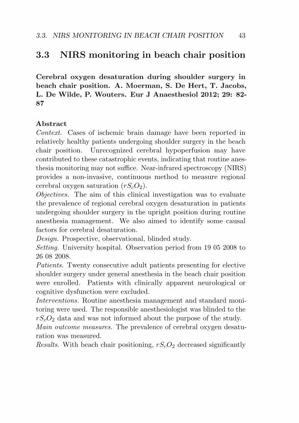
Cerebral oxygen desaturation during shoulder surgery inbeach chair position. A. Moerman, S. De Hert, T. Jacobs,L. De Wilde, P. Wouters. Eur J Anaesthesiol 2012; 29: 82-87
AbstractContext. Cases of ischemic brain damage have been reported inrelatively healthy patients undergoing shoulder surgery in the beachchair position. Unrecognized cerebral hypoperfusion may havecontributed to these catastrophic events, indicating that routine anes-thesia monitoring may not suffice. Near-infrared spectroscopy (NIRS)provides a non-invasive, continuous method to measure regionalcerebral oxygen saturation (rScO2).Objectives. The aim of this clinical investigation was to evaluatethe prevalence of regional cerebral oxygen desaturation in patientsundergoing shoulder surgery in the upright position during routineanesthesia management. We also aimed to identify some causalfactors for cerebral desaturation.Design. Prospective, observational, blinded study.Setting. University hospital. Observation period from 19 05 2008 to26 08 2008.Patients. Twenty consecutive adult patients presenting for electiveshoulder surgery under general anesthesia in the beach chair positionwere enrolled. Patients with clinically apparent neurological orcognitive dysfunction were excluded.Interventions. Routine anesthesia management and standard moni-toring were used. The responsible anesthesiologist was blinded to therScO2 data and was not informed about the purpose of the study.Main outcome measures. The prevalence of cerebral oxygen desatu-ration was measured.Results. With beach chair positioning, rScO2 decreased significantly

44 CHAPTER 3. NIRS AND CEREBRAL PERFUSION
from 79 ± 9 to 57 ± 9% on the left side and from 77 ± 10 to 59 ± 10%on the right side (P<0.001). A relative decrease in rScO2 of morethan 20% occurred in 80% of patients when the beach chair positionwas adopted. Postural decreases in cerebral oxygenation were relatedto blood pressure (r=0.60, P=0.007) and end-tidal carbon dioxideconcentration (r=0.47, P=0.035).Conclusion. The high prevalence of significant cerebral oxygendesaturation during shoulder surgery in the upright position un-derlines the need for close monitoring. NIRS might constitute avaluable technique to detect cerebral hypoperfusion in this high-riskgroup of patients.
3.3.1 Introduction
A series of case reports reporting dramatic adverse neurologicaloutcomes in patients after shoulder surgery in the upright positionhave recently alarmed the surgical and anesthesic communities. Thecomplications ranged from cranial nerve injury [1] to visual loss [2] andcerebral infarction [3,4], occurring in relatively healthy middle-agedpatients considered to be at low risk for cerebrovascular incidents.Although the exact pathogenesis of these events remains largelyunexplained, it has been assumed that the specific positioning of thepatient for shoulder surgery, that is, the beach chair position, maybe responsible for these complications. The beach chair position maybe associated with malrotation of the head and result in mechanicalobstruction of the cerebral vessels. It has also been suggested thatsuch positioning may induce unfavourable hemodynamic alterationswith cerebral tissue hypoperfusion related to the gravitational effectof upright tilting. Finally, management of shoulder surgery oftenincludes general anesthesia with controlled hypotension, which mayfurther compromize cerebral blood flow. These reports mandate thatmore attention should be paid to the effects of patient positioning oncerebral perfusion and that in these circumstances, routine anesthesiamonitoring may not suffice.

3.3. NIRS MONITORING IN BEACH CHAIR POSITION 45
Near-infrared spectroscopy (NIRS) provides a non-invasive, con-tinuous method of measuring cerebral tissue oxygen saturation. Thistechnique has been demonstrated repeatedly to provide an earlywarning sign of cerebral hypoperfusion during procedures with ahigh risk of adverse neurological outcomes [5]. Recent reports havesuggested that cerebral oximetry monitoring with NIRS may be auseful indicator of cerebral hypoperfusion during shoulder surgery inthe beach chair position [6,7].
The aim of this prospective, observational, blinded study wasto evaluate the prevalence of regional cerebral oxygen desaturationin patients undergoing shoulder surgery in the beach chair positionwhen routine anesthesia management and standard monitoring areused. We also aimed to identify potential physiological changes thatmight be associated with postural cerebral oxygen desaturation.
3.3.2 Methods
Ethical approval for this observational study (Ethical CommitteeN◦ 2008/191) was provided by the Ethical Committee of the GhentUniversity Hospital, Gent, Belgium (Chairperson Prof Dr R. Rubens)on 30 April 2008. After written informed consent, 20 consecutiveunpremedicated adult patients scheduled for elective shoulder surgeryin the beach chair position were included. Exclusion criteria wereclinically apparent neurological or cognitive dysfunction.
Standard monitoring was used throughout the procedure, includ-ing ECG, pulse oximetry (SpO2), end-tidal oxygen, carbon dioxideand sevoflurane concentrations and non-invasive blood pressure mea-surement (AS3; Datex, Helsinki, Finland). Blood pressure measure-ments were performed at 3-min intervals with a non-invasive cuffplaced on the arm opposite the operated side. Disposable NIRSsensors were applied on each side of the forehead for continuousregistration of the regional cerebral oxygen saturation (rScO2) of thecorresponding brain hemisphere (INVOS 5100; Somanetics Corpora-tion, Troy, MI, USA).

46 CHAPTER 3. NIRS AND CEREBRAL PERFUSION
Anesthesia was induced with sufentanil (0.1-0.3 µg/kg), propo-fol (2-3 mg/kg) and cisatracurium (0.1 mg/kg). Anesthesia wasmaintained with sevoflurane (1.5-2.5% end-tidal concentration) inan oxygen/air mixture (50% oxygen) and additional doses of sufen-tanil (0.1-0.2 µg/kg), if needed. All patients were raised to a 60-to 70-degree sitting position. The head of the patient was fixed inthe mid-line. Management of anesthesia and hemodynamics wereleft completely to the discretion of the attending anesthesiologist,who was blinded to the rScO2 data, and who also was not informedabout the purpose of the study. Routine clinical practice was usedto maintain blood pressure. No deliberate hypotension was used.Systolic arterial pressure (SAP) of <80 mmHg or heart rate of <50beats/min were usually treated.
Heart rate, non-invasive blood pressure, in- and end-expiratorygas tensions, SpO2 and bilateral rScO2 were recorded continuouslywith RUGLOOP (Demed, Temse, Belgium). Blood loss was estimatedand types and volumes of all fluids administered were recorded, aswell as doses of all drugs given. At the postoperative visit by theresponsible anesthesiologist on the evening of surgery, the patient wasassessed neurologically with a gross motor and sensory neurologicalevaluation and a gross cognitive evaluation (orientation in time andspace, recall of name, date of birth, and address). Any side-effectswere recorded.
The rScO2 values were compared at different time moments(awake, last value before position change from supine to upright posi-tion, 5 min after position change to beach chair, and at the minimumrScO2). Changes in cerebral oxygen saturation were described inabsolute terms (absolute rScO2 <50%) and in relative terms (>20%decrease in rScO2 compared to the value before position change). ArScO2 desaturation score was calculated by multiplying rScO2 below50% with the duration of this event (in seconds) [8]. Hence, the rScO2
desaturation score generated is an area under the curve measurement,which accounts for both severity and duration of desaturation.

3.3. NIRS MONITORING IN BEACH CHAIR POSITION 47
Sample size calculation was based on the assumption that arelative decrease in saturation of 20% (the smallest effect to beclinically important [9]) would be detected. Based on the study ofKim et al. [10], a mean and standard deviation (SD) of 71% and6% respectively were chosen. For a power of 0.8 and an α of 0.05, asample size of 20 patients was calculated to be appropriate to detecta clinically relevant decrease in cerebral oxygen saturation. Statis-tical analysis was performed using the statistical software PASWStatistics 18 (SPSS Inc., Chicago, IL, USA). Data were tested for nor-mal distribution using the Shapiro-Wilk test. Normally distributedcontinuous data are presented as mean ± SD.
The rScO2 values at different time points were compared using ananalysis of variance for repeated measurements (ANOVA). Post-testpairwise comparison was performed with the Tukey test. Possible re-lationships between rScO2 and physiological variables were analyzedusing linear regression analysis and quantified using the Spearman’scorrelation tests. A value of P<0.05 was considered statisticallysignificant.
3.3.3 Results
Patient demographics are presented in Table 3.1. One patient hadundergone a carotid endarterectomy 4 years earlier, and anotherpatient suffered from a transient ischemic attack (TIA) 8 yearsearlier; neither patient had any residual symptoms.
Table 3.2 shows details of surgery and anesthesia. All patientsreceived cristalloid solutions. If more than 1000 ml of cristalloidswere needed, colloids were administered (four patients). The surgicalprocedures did not necessitate the use of deliberate hypotension and,therefore, routine clinical practice was used. Seven patients receivedephedrine (5-10 mg) and one patient required phenylephrine 100 µgto correct a SAP of <80 mmHg. Three patients needed atropine 0.5mg for bradycardia (heart rate <50 beats/min).
48 CHAPTER 3. NIRS AND CEREBRAL PERFUSION
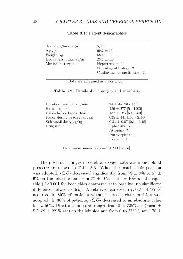
Table 3.1: Patient demographics
Sex, male/female (n) 5/15Age, y 60.2 ± 13.4Weight, kg 68.6 ± 17.6Body mass index, kg/m2 25.2 ± 4.8Medical history, n Hypertension: 11
Neurological history: 2Cardiovascular medication: 11
Data are expressed as mean ± SD
Table 3.2: Details about surgery and anesthesia
Duration beach chair, min 78 ± 45 [20 - 151]Blood loss, ml 186 ± 277 [5 - 1000]Fluids before beach chair, ml 187 ± 186 [50 - 650]Fluids during beach chair, ml 625 ± 444 [150 - 2100]Sufentanil dose, µg/kg 0.24 ± 0.07 [0.1 - 0.38]Drug use, n Ephedrine: 7
Atropine: 3Phenylephrine: 1Urapidil: 1
Data are expressed as mean ± SD [range]
The postural changes in cerebral oxygen saturation and bloodpressure are shown in Table 3.3. When the beach chair positionwas adopted, rScO2 decreased significantly from 79 ± 9% to 57 ±9% on the left side and from 77 ± 10% to 59 ± 10% on the rightside (P<0.001 for both sides compared with baseline, no significantdifference between sides). A relative decrease in rScO2 of >20%occurred in 80% of patients when the beach chair position wasadopted. In 30% of patients, rScO2 decreased to an absolute valuebelow 50%. Desaturation scores ranged from 0 to 725%.sec (mean ±SD: 89 ± 221%.sec) on the left side and from 0 to 3360%.sec (178 ±

3.3. NIRS MONITORING IN BEACH CHAIR POSITION 49
Table 3.3: Postural changes in cerebral oxygen saturation and in bloodpressure
LeftrScO2 RightrScO2 SAP/DAP(%) (%) (mmHg)
Awake 69 ± 6 68 ± 6 156 ± 29/76 ± 20Before position change 79 ± 9 77 ± 10 130 ± 32*/67 ± 205 min position change 65 ± 10‡ 66 ± 11‡ 110 ± 24*‡/64 ± 24Minimum value 57 ± 9*‡ 59 ± 10*‡ 84 ± 22*‡/46 ± 11*‡
Data are expressed as mean ± SD. rScO2: regional cerebral oxygen saturation.*p<0.05 vs awake value. ‡ p<0.05 vs value before position change.
750%.sec) on the right side. One patient had a desaturation score ofmore than 3000%.sec.
The patient who had undergone carotid endarterectomy had amaximum relative decrease in rScO2 of 12.7% (minimum rScO2 55%).The patient with a history of TIA had a relative decrease of 30.4%(minimum rScO2 64%).
At all time points, rScO2 was negatively correlated with age (Fig.3.1 (upper panel)), but the magnitude of the decrease in rScO2 withposition change was independent of age (Fig. 3.1 (lower panel)).
Before the position change, there was a positive correlation be-tween rScO2 and end-tidal carbon dioxide (EtCO2) (r = 0.53, P =0.016). No correlation was found between rScO2 and SAP (r = 0.28,P = 0.086). Five minutes after the change in position, a positivecorrelation was found between rScO2 and EtCO2 (r = 0.56, P =0.013), but no correlation was found between rScO2 and SAP (r= 0.29, P = 0.076). At the minimum rScO2, there were positivecorrelations between rScO2 and EtCO2 (r = 0.47, P = 0.035) andbetween rScO2 and SAP (r = 0.60, P = 0.007).
50 CHAPTER 3. NIRS AND CEREBRAL PERFUSION
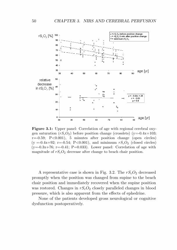
Figure 3.1: Upper panel: Correlation of age with regional cerebral oxy-gen saturation (rScO2) before position change (crosslets) (y=-0.4x+103;r=-0.59; P<0.001), 5 minutes after position change (open circles)(y =-0.4x+92; r=-0.54; P<0.001), and minimum rScO2 (closed circles)(y=-0.3x+76; r=-0.41; P=0.033). Lower panel: Correlation of age withmagnitude of rScO2 decrease after change to beach chair position.
A representative case is shown in Fig. 3.2. The rScO2 decreasedpromptly when the position was changed from supine to the beachchair position and immediately recovered when the supine positionwas restored. Changes in rScO2 closely paralleled changes in bloodpressure, which is also apparent from the effects of ephedrine.
None of the patients developed gross neurological or cognitivedysfunction postoperatively.

3.3. NIRS MONITORING IN BEACH CHAIR POSITION 51
Figure 3.2: Representative case. Changes in regional cerebral oxygensaturation (rScO2) at the start (1) and the end (4) of the beach chairposition and after administration of atropine (2) and ephedrine (3).
3.3.4 Discussion
In the present study, we observed cerebral desaturation (a relativedecrease in rScO2 of >20%) in 80% of patients when an uprightposition was adopted during shoulder surgery. Postural decreases incerebral oxygenation were consistent and related to blood pressureand EtCO2.
Recently, three reports have described the value of NIRS inmonitoring the adequacy of cerebral perfusion during shoulder surgeryin the beach chair position. Fischer et al [6] reported a case showingcausality between rScO2, mean arterial pressure (MAP) and EtCO2.Murphy et al. [7] evaluated the incidence of cerebral desaturationduring shoulder surgery in the beach chair position compared tothe lateral decubitus position. They used standardized anestheticmanagement and aimed to optimize cerebral perfusion by maintainingMAP within 20% of baseline values and controlling EtCO2 between30 and 34 mmHg. When cerebral oxygen desaturation was observed,they used a predetermined management protocol to increase rScO2.

52 CHAPTER 3. NIRS AND CEREBRAL PERFUSION
In the study of Tange et al. [11], perioperative management includedsupport stockings, fluid administration at a rate of 10 ml/kg/hthroughout the study period, gradual head-up tilt position, andcareful blood pressure management to maintain MAP above 60mmHg. All these measures aimed to minimize the impact of positionchange. Our study is different in that the objective was to evaluatethe actual prevalence of regional cerebral oxygen desaturation inpatients undergoing surgery in the beach chair position when standardanesthesia management and routine anesthesia monitoring (whichcurrently does not include cerebral oximetry) were employed. Forthis reason, anesthetic management was left to the discretion of theattending clinicians who did not participate in the study and wereblinded to the rScO2 data.
It is assumed that the upright position induces significant hemo-dynamic changes that may impair cerebral circulation. Comparedto the supine position, adopting an upright position has been shownto decrease systolic and mean arterial pressure, stroke volume andcardiac output, inducing a cerebral blood flow decrease of 12% [12].In conscious individuals, these effects are compensated for by anincrease in systemic vascular resistance, but during anesthesia thisautonomic response may be attenuated or blocked. The combina-tion of the sitting position and general anesthesia may, therefore,be potentially deleterious to cerebral perfusion. Reports describingcerebral ischemia in the beach chair position have, therefore, stressedthe risk of hypotension [13]. It is often suggested that a systemicMAP between 50 and 150 mmHg lies within the range of cerebralautoregulation and, therefore, guarantees adequate cerebral perfusion.However this assumption has been challenged. First, the concept ofcerebral autoregulation is questioned, because there seems to be aconsiderable individual variability in the autoregulation limits [14].Second, it has been claimed that blood pressure measured at thebrachial artery may overestimate the pressure at the level of thebrain when the sitting position is adopted. Some authors, there-

3.3. NIRS MONITORING IN BEACH CHAIR POSITION 53
fore, propose an arithmetic correction of blood pressure to determinepressure at the level of the brain (1 mmHg for each 1.25 cm differencein height between the external meatus and the middle of the bloodpressure cuff) [13]. Applying this assumption to the present study,the mean difference between the brain and the site of the bloodpressure cuff on the arm of 31 ± 3 (24-38) cm, would overestimatearterial pressure at the level of the head by 24 ± 2 (18-29) mmHg.Third, cerebral perfusion pressure (CPP) depends not only on inflowpressure but also on outflow pressure: CPP = MAP- central venouspressure (CVP) or CPP = MAP- intracranial pressure (ICP) if ICPis more than CVP. In the upright position, the jugular veins eitherpartly or fully collapse, and the vertebral venous plexus becomes themain pathway for venous drainage [12]. Flow through this plexusmight be impeded during head rotation and head tilt.
Although intuitively one would suppose that older patients havean increased risk of cerebral desaturation, our data showed nodifference in the response to position change with age, which isconsistent with the results of Gatto et al. [15] and Edlow et al. [16].
The cerebral circulation is very responsive to changes in CO2. Ina patient with normal CO2 reactivity, cerebral blood flow changes1-2 ml/100g/min per mmHg change in CO2 [17]. This was also clearin our study, which showed a lower rScO2 with lower EtCO2.
Given the positive correlations between rScO2 and blood pressureand EtCO2, a simple recommendation could be to avoid hypotensionand low EtCO2. The study by Tange et al. [11] demonstrated that,with appropriate measures, cerebral desaturation can be avoided.However, three caveats should be considered. First, the ability ofthe circulatory system to compensate for sudden position changesvaries considerably between patients and is unpredictable. Second, ifdeliberate hypotension is required, the impact on cerebral perfusionis unknown unless specific monitoring is employed. Finally, the highprevalence of cerebral desaturation in this study, together with thedramatic case reports, indicate that there is a compelling need for

54 CHAPTER 3. NIRS AND CEREBRAL PERFUSION
more wariness and for more specific monitoring. Due to the advantageof simple, continuous and non-invasive monitoring, NIRS could havethe potential to optimize patient care in these situations.
Several limitations should be considered. First, becauseNIRS technology does not distinguish between arterial and venoushemoglobin saturation, changes in the proportion of cerebral arterialand venous blood volume may confound measurements [18]. Changesin body position affect both arterial and venous pressures and canalter the ratio of arterial to venous compartments in the cerebralcirculation. The measured changes in rScO2 in the upright positionmay, therefore, be a consequence of true changes in tissue oxygentension, or may be the effect of the changes in the relative frac-tions of arterial and venous blood in the cerebral tissue. Real-timemeasurements of changes in cerebral blood flow using transcranialDoppler might be helpful in solving this question, but is difficult toestablish properly during head-up table tilting and during surgicalmanipulation of the patient. Second, the prevalence of desaturationis dependent on the baseline value and the threshold used to definecerebral desaturation. In most clinical studies, a 20% reduction fromawake values or an absolute decrease below 50% oxygen saturationis used as the threshold [9]. Slater et al. [8] also took the durationof desaturation into account by introducing the term ‘desaturationscore’. Third, the relevance of the changes in cerebral oxygenationparameters might be questioned because of the absence of grosspostoperative neurological dysfunction. However, without exten-sive neurocognitive monitoring, subtle changes induced by cerebralhypoxia may go unnoticed until functional organ damage becomesevident. Several randomized controlled trials have demonstrated thatdetection and treatment of cerebral oxygen desaturation results inbetter clinical outcome [8, 19-21].
Finally, there is the interesting controversy with regard to theexact determination of CPP and its relationship with flow. Thepractice of correcting the blood pressure for position (raising the

3.3. NIRS MONITORING IN BEACH CHAIR POSITION 55
transducer or adjusting arterial pressure estimation to allow forthe height between the heart and the brain) is dictated by theassumption that the heart lifts the blood to the elevated brain withouta corresponding and equivalent venous return limb. In a closed loopsystem, such as the intact circulation, there is a continuous andbalanced fluid column and no net work is performed against theeffect of gravity [22]. Therefore, as long as the hydrostatic gradientfrom the measurement site to the brain remains the same for inflowand outflow pressures, there is no significant flow related pressuredrop between the measurement site and the brain [23]. Hence, thereis no need for adjustment of pressure measurements if the assumptionis correct that venous outflow depends only on the outflow pressure,although this does not take into account the more complex natureof the venous resistance and the possibility of venous collapse whenassuming the upright position. Therefore, it has been proposed thatthe CPP formula should be adapted to take into account the effectof atmospheric pressure on the jugular veins. It has been suggestedthat measurement level adjustments are probably not necessary ifCVP can be maintained above 18 mmHg [24].
In conclusion, the results of the present study showed an incidenceof 80% of cerebral oxygen desaturation when the beach chair positionwas adopted in patients undergoing shoulder surgery. Together withthe high prevalence of cerebral desaturation also found in otherstudies, this underlines the need for awareness of this problem andsuggests the need for perioperative monitoring of cerebral tissueoxygenation in this type of surgery. Monitoring, of course, shouldnot preclude all customary measures being taken to avoid an abruptdecrease in blood pressure with position change.

56 CHAPTER 3. NIRS AND CEREBRAL PERFUSION
3.3.5 References
1. Boisseau N, Rabarijaona H, Grimaud D, Raucoules-Aime M. Tapia’s
syndrome following shoulder surgery. Br J Anaesth 2002 ; 88: 869-70
2. Bhatti MT, Enneking FK. Visual loss and ophthalmoplegia after shoulder
surgery. Anesth Analg 2003; 96: 899-902
3. Pohl A, Cullen DJ. Cerebral ischemia during shoulder surgery in the
upright position: a case series. J Clin Anesth 2005; 17: 463-9
4. Verbrugge SJ, Klimek M, Klein J. A cerebral watershed infarction after
general anaesthesia in a patient with increased anti-cardiolipin antibody
level. Anaesthesist 2004; 53: 341-6
5. Casati A, Spreafico E, Putzu M, Fanelli G. New technology for
non-invasive brain monitoring: continuous cerebral oximetry. Minerva
Anestesiol 2006; 72: 605-25
6. Fischer GW, Torrillo TM, Weiner MM, Rosenblatt MA. The use of
cerebral oximetry as a monitor of the adequacy of cerebral perfusion in
a patient undergoing shoulder surgery in the beach chair position. Pain
Pract 2009; 9: 304-7
7. Murphy GS, Szokol JW, Marymont JH et al. Cerebral oxygen de-
saturation events assessed by near-infrared spectroscopy during shoulder
arthroscopy in the beach chair and lateral decubitus positions. Anesth
Analg 2010; 111: 496-505
8. Slater JP, Guarino T, Stack J et al. Cerebral oxygen desaturation
predicts cognitive decline and longer hospital stay after cardiac surgery.
Ann Thorac Surg 2009; 87: 36-45
9. Edmonds HL Jr, Ganzel BL, Austin EH. Cerebral oximetry for cardiac
and vascular surgery. Semin Cardiothorac Vasc Anesth 2004; 8: 147-66
10. Kim MB, Ward DS, Cartwright CR, Kolano J, Chlebowski S,
Henson LC. Estimation of jugular venous O2 saturation from cerebral
oximetry or arterial O2 saturation during isocapnic hypoxia. J Clin Monit
Comput 2000; 16: 191-9
11. Tange K, Kinoshita H, Minonishi T et al. Cerebral oxygenation in
the beach chair position before and during general anesthesia. Minerva
Anestesiol 2010; 76: 485-90

3.3. NIRS MONITORING IN BEACH CHAIR POSITION 57
12. Alperin N, Hushek SG, Lee SH, Sivaramakrishnan A, Lichtor T. MRI
study of cerebral blood flow and CSF flow dynamics in an upright posture:
the effect of posture on the intracranial compliance and pressure. Acta
Neurochir 2005; 95: 177-81
13. Cullen DJ, Kirby RR. Beach chair position may decrease cerebral
perfusion; catastrophic outcomes have occurred. APSF Newsletter 2007;
22: 25, 27
14. Drummond JC. The lower limit of autoregulation: time to revise our
thinking? Anesthesiology 1997; 86: 1431-1433.
15. Gatto R, Hoffman W, Paisansathan C, Mantulin W, Gratton E,
Charbel FT. Effect of age on brain oxygenation regulation during changes
in position. J Neurosci Methods 2007; 164: 308-11
16. Edlow BL, Kim MN, Durduran T et al. The effects of healthy aging on
cerebral hemodynamic responses to posture change. Physiol Meas 2010; 31:
477-95
17. Brian JE, Jr. Carbon dioxide and the cerebral circulation.
Anesthesiology 1998; 88: 1365-86
18. Pollard V, Prough DS, DeMelo AE, Deyo DJ, Uchida T, Widman
R. The influence of carbon dioxide and body position on near-infrared
spectroscopic assessment of cerebral hemoglobin oxygen saturation. Anesth
Analg 1996; 82: 278-87
19. Goldman S, Sutter F, Ferdinand F, Trace C. Optimizing intraoperative
cerebral oxygen delivery using noninvasive cerebral oximetry decreases the
incidence of stroke for cardiac surgical patients. Heart Surg Forum 2004; 7:
E376-81
20. Casati A, Fanelli G, Pietropaoli P et al. Continuous monitoring of
cerebral oxygen saturation in elderly patients undergoing major abdominal
surgery minimizes brain exposure to potential hypoxia. Anesth Analg 2005;
101: 740-7
21. Murkin JM, Adams SJ, Novick RJ et al. Monitoring brain oxygen
saturation during coronary bypass surgery: a randomized, prospective study.
Anesth Analg 2007; 104: 51-8
22. Munis JR, Lozada LJ. Giraffes, siphons, and Starling resistors: cerebral

58 CHAPTER 3. NIRS AND CEREBRAL PERFUSION
perfusion pressure revisited. J Neurosurg Anesthesiol 2000; 12: 290-6
23. Munis J. The problems of posture, pressure, and perfusion: cerebral
perfusion pressure defined. APSF Newsletter 2008; 22: 82-3
24. Pranevicius M, Pranevicius O. Modified calculation of the cerebral
perfusion pressure in a sitting position: jugular Starling resistor and related
clinical implications. APSF Newsletter 2008; 23: 32-3

3.4. NIRS MONITORING IN MOYAMOYA DISEASE 59
3.4 NIRS monitoring in moyamoya disease
Value of cerebral oxygen saturation monitoring duringcardiopulmonary bypass in an adult patient with moyamoyadisease. De Buysscher P, Moerman A, Bove T, De PauwM, Wouters P, De Hert S. J Cardiothor Vasc Anesth 2013,http://dx.doi.org/10.1053/j.jvca.2011.11.002
AbstractPurpose: To describe the use of near-infrared spectroscopy (NIRS)to identify and reverse cerebral ischemia in a patient in whom themaintenance of adequate cerebral perfusion during cardiopulmonarybypass (CPB) was jeopardized severely because of moyamoya disease.Case report: In an adult patient with moyamoya disease undergoingmitral valve repair, the use of NIRS provided real-time cerebraloxygenation monitoring and enabled close monitoring of alltherapeutic actions taken. The decrease in rScO2 with the initiationof CPB was reversed succesfully by the institution of pulsatile CPBflow, whereas other standard interventions, such as the increase ofperfusion pressure and pump flow had failed, suggesting that thesepatients depending on extensive collateral network brain perfusionmay benefit from preserved pulsatility during CPB.Conclusion: This case report shows that real-time cerebraloxygenation monitoring during CPB provides a clinical usefuladditional monitoring tool, especially in patients at risk forneurological damage by a cerebrovascular disease like moyamoya.
3.4.1 Introduction
Moyamoya disease (MMD) is a progressive, chronic cerebrovascularocclusive disease occurring predominantly in the Asian population.As described by Suzuki and Takaku [1] in 1969, it affects the internalcarotid arteries and anterior and middle cerebral arteries, resulting

60 CHAPTER 3. NIRS AND CEREBRAL PERFUSION
in compensatory collateral networks at the base of the brain. Thesenetworks can be observed in cerebral angiography as a ‘puff of smoke’(moyamoya means ‘cloud’ in Japanese). The clinical appearance ismarked by various cerebrovascular incidents, including intracranialhemorrhage, transient ischemic attack (TIA), and recurrent smallstrokes [2].
In MMD, the blood supply to the brain is severely compromized,which represents a considerable challenge to the anesthesiologistduring the perioperative period. Although the management ofpatients with MMD during cerebral revascularization surgery isdescribed widely, reports on the management of patients with MMDundergoing cardiac surgery with cardiopulmonary bypass are limited[3-8]. The use of near-infrared spectroscopy (NIRS) as a monitoringtool of cerebral oxygen saturation during cardiac surgical proceduresis gaining wide acceptance; however, its potential value in the peri-operative management of MMD patients is unknown. In this casereport, the use of NIRS to identify and reverse cerebral ischemia ina patient with MMD during cardiac surgery is described.
3.4.2 Case report
A 26-year-old man with MMD was referred to the authors’ universitycenter for the treatment of grade IV mitral valve insufficiency causedby anterior leaflet prolapse. His medical history included one TIAand recurrent episodes of transient headaches over the past tenyears. His brother had a history of malignant hyperthermia. Be-cause of increasing exercise intolerance and recent symptoms ofcongestive heart failure, he was proposed for mitral valve surgery.Echocardiography showed a significant prolapse of the anterior mitralvalve leaflet, resulting in a huge eccentric jet with flow reversal intothe pulmonary veins. Preoperative investigation of the associatedcerebrovascular disease by brain magnetic resonance imaging (Fig.3.3) showed bilateral distal occlusion of the internal carotid arteries,occlusion of the left ophthalmic artery, and a widely disrupted circle

3.4. NIRS MONITORING IN MOYAMOYA DISEASE 61
of Willis with an extensive collateral network. Cerebral cortex per-fusion seemed to be best compensated in the left occipital and leftfrontal cortex. Prophylactic cerebral revascularization surgery wasnot done, and cardiac surgery with mitral valve repair was chosen.
Figure 3.3: Cerebral angiography demonstrating abnormal collateralnetworks at the base of the brain.
Intraoperative monitoring included 5-lead ECG, invasive bloodpressure measured in the right radial artery, central venous pressuremeasured via the right internal jugular vein, temperature (esophagealand rectal), capnography and regular arterial blood gas sampling.Additional transesophageal echocardiography completed the cardio-vascular monitoring. We applied a bispectral index (BIS) electrode(BIS XP; Aspect Medical Systems, Newton, MA, USA) to monitorthe depth of anesthesia and 2 NIRS sensors on the left and rightforehead (INVOS 5100; Somanetics Corporation, Troy, MI, USA) tonon-invasively measure regional cerebral oxygen saturation (rScO2).
Before induction, awake rScO2 values were 62% and 60% atthe left and right side, respectively. With preoxygenation, rScO2

62 CHAPTER 3. NIRS AND CEREBRAL PERFUSION
increased to 70% on both sides (Fig. 3.4). Anesthesia was inducedwith diazepam 0.1 mg/kg and fentanyl 10 µg/kg. Intubation wasfacilitated with rocuronium 0.9 mg/kg and the lungs were ventilatedwith an air/oxygen mixture to keep arterial carbon dioxide partialpressure (PaCO2) around 40 mmHg. Anesthesia was maintainedwith additional fentanyl and diazepam doses (total dose 22 µg/kgand 10 mg, respectively) and a continuous infusion of propofol, ofwhich the dosing was guided to maintain BIS values between 35 and50 (dose range 3-5 mg/kg/h). Throughout surgery, the blood glucoselevel was kept between 80 and 120 mg/dl with a continuous insulininfusion. Between the induction of anesthesia and the initiation ofcardiopulmonary bypass (CPB), NIRS indicated a bilateral rScO2
between 57% and 75% (Fig. 3.4).
Figure 3.4: Intraoperative plot showing real-time readings of bilateralcerebral oxygen saturation and bispectral index. rScO2: regional cerebraloxygen saturation, BIS: bispectral index.

3.4. NIRS MONITORING IN MOYAMOYA DISEASE 63
CPB was performed with a nonpulsatile flow of 2.4 l/min/m2,with hemodilution to a minimal hematocrit of 28%. Nasopharyngealtemperature was kept between 35◦C and 36◦C. PaCO2 and PaO2 weremaintained at 40 mmHg and 350 mmHg, respectively. Alpha-statregulation of pH values was adopted.
A few minutes after starting CPB, a sudden decrease in rScO2
was observed on both sides to 25 to 40% (Fig. 3.5). BIS valuesshowed a parallel decrease, but to a lesser extent (Fig. 3.4). Mechan-ical causes were ruled out, and intended values of hematocrit, BIS,PaCO2, and PaO2 were confirmed. Increases in CPB flow as wellas increases in perfusion pressure with phenylephrine failed to raiserScO2. Therefore we decided to decrease nasopharyngeal tempera-ture to 33◦C. Initially, rScO2 seemed to improve; however, this wasvery short-lasting. Finally, we converted from nonpulsatile flow topulsatile flow, which resulted in a gradual rScO2 increase (Fig. 3.5).
Figure 3.5: Real-time readings of bilateral cerebral oxygen saturationand arterial blood pressure during cardiopulmonary bypass. Institution ofpulsatile flow resulted in gradual increase in cerebral oxygen saturation.rScO2: regional cerebral oxygen saturation, BP: blood pressure, SAP:systolic arterial blood pressure, DAP: diastolic arterial blood pressure.

64 CHAPTER 3. NIRS AND CEREBRAL PERFUSION
After successful weaning from CPB, bilateral rScO2 valuesstabilized around 50 to 60%. Postoperatively, the patient sufferedfrom prolonged somnolence and a transient paresis of the left arm,which improved over the following days without any sequelae. At post-operative day 10, magnetic resonance imaging of the brain revealedno new defects.
3.4.3 Discussion
This case report shows the utility of NIRS monitoring in a MMDpatient in whom the maintenance of adequate cerebral perfusionduring CPB was jeopardized severely because of the underlyingpathology.
Denault et al. [9] introduced a useful treatment algorithm tocorrect decreases in cerebral saturation values, which the authorshave adopted for many years. When rScO2 decreased, technical ormechanical causes (eg, adhesion of the NIRS sensors, head position,and positioning of the arterial and venous cannulae) were first ruledout. Then, those factors that affect cerebral oxygen balance, such asblood pressure [10], PaCO2 [10], temperature [11], hematocrit [12],depth of anesthesia [13] and pump flow, were verified and optimized.Real-time cerebral oxygenation monitoring enabled us to monitorclosely the effects of each therapeutic action. Despite correction forall possible confounding factors, a dramatic decrease in rScO2 valuesduring CPB was still observed. Finally, the decrease in rScO2 wasreversed successfully by initiating pulsatile CPB flow, as suggestedby Denault et al. [9].
The benefit of pulsatile versus nonpulsatile flow during CPBremains an intensely debated topic. The controversy is likely becauseof the heterogeneity of the study populations and the lack of quantifi-cation of generated pressure-flow waveforms [14]. A series of studieshave documented that at the same flow and pressure conditionspulsatile perfusion generates more hemodynamic energy in the bloodflow than nonpulsatile perfusion. This additional energy resulted in

3.4. NIRS MONITORING IN MOYAMOYA DISEASE 65
better vital organ perfusion [15,16]. Pulsatile CPB has been shownto improve cerebral blood flow and brain tissue oxygenation [17].Only 3 clinical reports documented the influence of pulsatile CPBon rScO2 measured with NIRS. Zhao et al. [18] and Su et al. [19]showed smaller decreases in rScO2 levels in the pulsatile versus non-pulsatile group during pediatric CPB. In contrast, Grubhofer et al.[20] found no benefit of pulsatile perfusion on cerebral oxygenationduring hypothermic CPB in adult patients. In the present case,a steady increase in rScO2 was observed when pulsatile flow wasinitiated, supporting the observations of Zhao et al. [18] and Su etal. [19].
Considering the family history of malignant hyperthermia, weavoided the use of volatile anesthetics. Several studies showed nosignificant difference in outcome or perioperative complications ofinhaled anesthesia versus total intravenous anesthesia. However,two studies [21,22] compared the effect of sevoflurane and propofolon regional cortical blood flow and intracranial pressure duringrevascularization procedures in patients with MMD and concludedthat with propofol cortical blood flow was significantly higher andintracranial pressure was lower.
Two case reports in which BIS monitoring was applied [3,4]and one report with NIRS monitoring [5] during cardiac surgeryin patients suffering from MMD were found, yet none used bothBIS and NIRS. In this case, dramatic decreases in NIRS valueswere found, which was only mildly reflected in the associated BISvalues. Although BIS and NIRS offer distinct indices for perioperativeneurological monitoring, both are actually complementary. BISreflects the functional state of the brain, and therefore BIS couldhelp to determine the significance of a rScO2 decrease. By contrast,because rScO2 assesses cerebral oxygen balance, it helps to identifythe cause of a BIS change. In this case report, the decrease ofBIS values could have been interpreted as an increase in depth ofanesthesia or as a decrease in cerebral metabolic rate by lowering body

66 CHAPTER 3. NIRS AND CEREBRAL PERFUSION
temperature. However, the associated decrease in NIRS suggestedthat the reduction in BIS was caused by a cerebral oxygenationdeficit.
The limitations of NIRS are inherent to its underlying technologyand are described in detail elsewhere [23,24]. Specifically for this case,some limitations of NIRS are highlighted. First, although the algo-rithm used to calculate oxygen saturation assumes a fixed distancefor light to travel through the sampled area (the optical path length),its validity remains questionable in the rapidly changing conditionoccurring during CPB (eg, a decrease in hematocrit or a changein viscosity). Second, Tyree et al. [25] showed that measurementsbased on hemoglobin saturation (which is the case for NIRS) do notalways accurately reflect oxygenation at the cellular level, providinga false sense of safety. Finally, the measurements obtained with NIRSare regional and strictly confined to the zone underneath the sensor.Consequently, clinically relevant focal ischemia in a brain area remotefrom the monitored area may stay unnoticed, in particular in patientswith a diffuse occlusive cerebrovascular disease like MMD.
This case report shows that real-time cerebral oxygenation moni-toring during CPB provides a clinical useful additional monitoringtool, especially in patients at risk for neurological damage by acerebrovascular disease like MMD. The successful reversal of rScO2
decrease by the institution of pulsatile CPB flow, whereas otherstandard interventions, such as the increase of perfusion pressureand pump flow had failed, suggests that these patients dependingon extensive collateral network brain perfusion may benefit frompreserved pulsatility during CPB.
3.4. NIRS MONITORING IN MOYAMOYA DISEASE 67
3.4.4 References
1. Suzuki J, Takaku A. Cerebrovascular ”moyamoya” disease. Disease
showing abnormal net-like vessels in base of brain. Arch Neurol 1969; 20:
288-99
2. Burke GM, Burke AM, Sherma AK, et al. Moyamoya disease: a summary.
Neurosurg Focus 2009; 26: E11
3. Al-Naimi KT, Mediratta NK, Pennefather SH. Hypothermic cardiopul-
monary bypass in a patient with moyamoya disease. J Cardiothorac Vasc
Anesth 2009; 23: 206-7
4. Han SH, Kim SD, Ham BM, et al. Normothermic cardiopulmonary
bypass in a patient with moyamoya disease. J Cardiothorac Vasc Anesth
2005; 19: 499-500
5. Jimi N, Shin T. Anesthetic management of a patient with Moyamoya
disease undergoing open heart surgery. Pediatr Anesth 2006; 16: 1195-6
6. Gatti G, Benussi B, Gon L, et al. Aortic valve replacement in an
adult white male with moyamoya disease and coronary artery fistulas.
J Cardiothorac Vasc Anesth 2007; 21: 166-8
7. Wang N, Kuluz J, Barron M, et al. Cardiopulmonary bypass in a patient
with moyamoya disease. Anesth Analg 1997; 84: 1160-3
8. Kashima I, Inoue Y, Takahashi R. The use of intra-aortic balloon pump
as cerebral protection in a patient with moyamoya disease undergoing
coronary artery bypass grafting. Interact Cardiovasc Thorac Surg 2008; 7:
522-3
9. Denault A, Deschamps A, Murkin JM. A proposed algorithm for the
intraoperative use of cerebral near-infrared spectroscopy. Semin Cardiotho-
rac Vasc Anesth 2007; 11: 274-81
10. Iwama T, Hashimoto N, Yonekawa Y. The relevance of hemodynamic
factors to perioperative ischemic complications in childhood moyamoya
disease. Neurosurgery 1996; 38: 1120-6
11. Parray T, Martin TW, Siddiqui S. Moyamoya disease: a review of the
disease and anesthetic management. J Neurosurg Anesthesiol 2011; 23:
100-9
12. Sato K, Shirane R, Yoshimoto T. Perioperative factors related to the

68 CHAPTER 3. NIRS AND CEREBRAL PERFUSION
development of ischemic complications in patients with moyamoya disease.
Childs Nerv System 1997; 13: 68-72
13. Kansha M, Irita K, Takahashi S, et al. Anesthetic management of
children with Moyamoya disease. Clin Neurol Neurosurg 1997; 99: S110-3
14. Undar A. Benefits of pulsatile flow during and after cardiopulmonary
bypass procedures. Artif Organs 2005; 29: 688-90
15. Haines N, Wang S, Undar A, et al. Clinical outcomes of pulsatile and
non-pulsatile mode of perfusion. J Extra Corpor Technol 2009; 41: 26-9
16. Akcevin A, Alkan-Bozkaya T, Qiu F, et al. Evaluation of perfusion
modes on vital organ recovery and thyroid hormone homeostasis in pedi-
atric patients undergoing cardiopulmonary bypass. Artif Organs 2010; 34:
879-84
17. Undar A. Pulsatile versus nonpulsatile cardiopulmonary bypass proce-
dures in neonates and infants: from bench to clinical practice. ASAIO J
2005; 51: VI-X
18. Zhao J, Yang J, Liu J, et al. Effects of pulsatile and nonpulsatile perfu-
sion on cerebral regional oxygen saturation and endothelin-1 in tetralogy
of fallot infants. Artif Organs 2011; 35: E54-8
19. Su XW, Guan Y, Barnes M, et al. Improved cerebral oxygen satura-
tion and blood flow pulsatility with pulsatile perfusion during pediatric
cardiopulmonary bypass. Pediatr Res 2011; 70: 181-5
20. Grubhofer G, Mares P, Rajek A, et al. Pulsatility does not change
cerebral oxygenation during cardiopulmonary bypass. Acta Anaesthesiol
Scand 2000; 44: 586-91
21. Sato K, Shirane R, Kato M, et al. Effect of inhalation anesthesia on
cerebral circulation in moyamoya disease. J Neurosurg Anesthesiol 1999;
11: 25-30
22. Kikuta K, Takagi Y, Nozaki K, et al. Effects of intravenous anesthesia
with propofol on regional cortical blood flow and intracranial pressure in
surgery for moyamoya disease. Surg Neurol 2007; 68: 421-4
23. Edmonds HL Jr, Ganzel BL, Austin EH 3rd. Cerebral oximetry for
cardiac and vascular surgery. Semin Cardiothorac Vasc Anesth 2004; 8:
147-66

3.4. NIRS MONITORING IN MOYAMOYA DISEASE 69
24. Murkin JM, Arango M. Near-infrared spectroscopy as an index of brain
and tissue oxygenation. Br J Anaesth 2009; 103 Suppl 1:i3-i13
25. Tyree K, Tyree M, DiGeronimo R. Correlation of brain tissue
oxygen tension with cerebral near-infrared spectroscopy and mixed ve-
nous oxygen saturation during extracorporeal membrane oxygenation.
Perfusion 2009; 24: 325-31

70 CHAPTER 3. NIRS AND CEREBRAL PERFUSION

Chapter 4
NIRS for monitoringregional tissue perfusion
4.1 Background
The amount of blood delivered to an organ determines oxygen deliveryto that particular organ. Under physiological conditions, differentorgans receive a relatively predictable blood flow depending on theirmetabolic demand. However, when cardiac output decreases, thesympathetic stress response raises vascular resistance, diverting bloodflow from less important tissues (skin, muscle, gastrointestinal tract,kidneys) to vital organs (the brain and the heart). Monitoring themacrocirculation using measures of blood pressure and arterial oxygensaturation, do not necessarily predict adequate perfusion and oxygena-tion of individual tissues. The use of NIRS for monitoring regionaloxygen saturation at somatic sites has been advocated as an earlywarning system for changes in the oxygen supply-demand balance,thereby allowing an early marker of silent regional hypoperfusion.
71

72 CHAPTER 4. NIRS AND REGIONAL PERFUSION
4.2 Objective
The aim of the present studies was to explore the potential of NIRSto monitor regional organ perfusion. In the first study, the effect ofaortic cross-clamping on cerebral, renal, and muscle oxygen saturationwas evaluated in children undergoing aortic coarctation repair. In thesecond study, the applicability of NIRS to estimate oxygen saturationof the spinal cord was explored during aortic cross-clamping in apatient undergoing thoracoabdominal aneurysm repair.
4.3 NIRS during aortic coarctation repair
The effect of blood pressure regulation during aorticcoarctation repair on brain, kidney and muscle oxygensaturation measured by near-infrared spectroscopy: arandomized, clinical trial. Moerman A, Bove T, Francois K,Jacobs S, Deblaere I, Wouters P, De Hert S. Anesth Analg2013; 116: 760-6
AbstractBackground. In this study, we compared the effects of three frequentlyused blood pressure-regulating agents on brain (rScO2), renal (SrO2),and muscle (SmO2) oxygen saturation, during aortic coarctationrepair in children. Based on the reported adverse effect of sodiumnitroprusside (SNP) on left-sided rScO2 during aortic coarctationrepair, we tested the hypothesis that the alterations in left rScO2
occurring with SNP would not be present with sevoflurane andnitroglycerin (NTG). Additionally, we explored the effects of bloodpressure regulation with SNP, NTG or sevoflurane on right-sidedrScO2, SrO2, and SmO2.Methods. Children with isolated aortic coarctation undergoingsurgical repair through a left thoracotomy without the use ofcardiopulmonary bypass were considered eligible for the study.
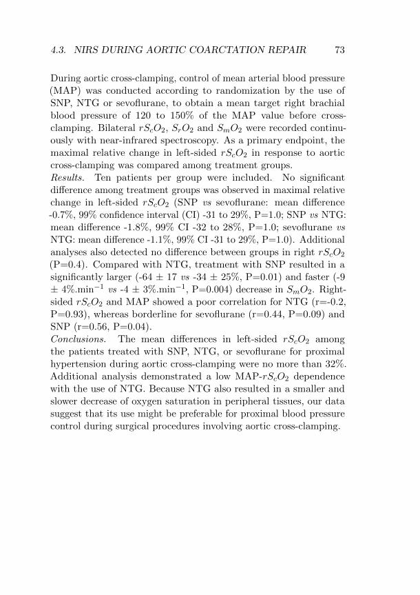
4.3. NIRS DURING AORTIC COARCTATION REPAIR 73
During aortic cross-clamping, control of mean arterial blood pressure(MAP) was conducted according to randomization by the use ofSNP, NTG or sevoflurane, to obtain a mean target right brachialblood pressure of 120 to 150% of the MAP value before cross-clamping. Bilateral rScO2, SrO2 and SmO2 were recorded continu-ously with near-infrared spectroscopy. As a primary endpoint, themaximal relative change in left-sided rScO2 in response to aorticcross-clamping was compared among treatment groups.Results. Ten patients per group were included. No significantdifference among treatment groups was observed in maximal relativechange in left-sided rScO2 (SNP vs sevoflurane: mean difference-0.7%, 99% confidence interval (CI) -31 to 29%, P=1.0; SNP vs NTG:mean difference -1.8%, 99% CI -32 to 28%, P=1.0; sevoflurane vsNTG: mean difference -1.1%, 99% CI -31 to 29%, P=1.0). Additionalanalyses also detected no difference between groups in right rScO2
(P=0.4). Compared with NTG, treatment with SNP resulted in asignificantly larger (-64 ± 17 vs -34 ± 25%, P=0.01) and faster (-9± 4%.min−1 vs -4 ± 3%.min−1, P=0.004) decrease in SmO2. Right-sided rScO2 and MAP showed a poor correlation for NTG (r=-0.2,P=0.93), whereas borderline for sevoflurane (r=0.44, P=0.09) andSNP (r=0.56, P=0.04).Conclusions. The mean differences in left-sided rScO2 amongthe patients treated with SNP, NTG, or sevoflurane for proximalhypertension during aortic cross-clamping were no more than 32%.Additional analysis demonstrated a low MAP-rScO2 dependencewith the use of NTG. Because NTG also resulted in a smaller andslower decrease of oxygen saturation in peripheral tissues, our datasuggest that its use might be preferable for proximal blood pressurecontrol during surgical procedures involving aortic cross-clamping.

74 CHAPTER 4. NIRS AND REGIONAL PERFUSION
4.3.1 Introduction
Surgical repair of aortic coarctation through left thoracotomy involvesproximal aortic arch cross-clamping, resulting in temporary hypoper-fusion of the tissues distal to the cross-clamp. This manoeuver mayalso be associated with an abrupt increase in ventricular afterloadand immediate proximal (to the aortic cross-clamp) hypertension,often necessitating pharmacologic control [1]. Currently, the use ofpotent and short-acting vasodilators such as sodium nitroprusside(SNP) and nitroglycerin (NTG) is preferred for treating hypertensionduring aortic cross-clamping for this procedure. However, Azakieet al. [2] have raised concerns regarding the effect of SNP on left-sided cerebral oxygen saturation (rScO2) during aortic coarctationrepair. In addition, it has been demonstrated that SNP may worsenthe already impaired oxygen balance of tissues below the aorticcross-clamp during surgery on the thoracic aorta [3-5].
The effects of different antihypertensive agents on tissue oxygensaturation during aortic cross-clamping remain largely unexplored.The purpose of the present study was to test the hypothesis that thealterations in left rScO2 described with SNP would not be presentwith other blood pressure-regulating agents. Second, we wanted tocompare the effects of blood pressure-regulating drugs during aorticcoarctation repair on the oxygen saturation of the right-sided brainand the peripheral tissues.
Three agents frequently used for perioperative blood pressurecontrol during cardiac surgery [6] were included: SNP and NTGas intravenous vasodilating agents, and sevoflurane as an inhaledanesthetic with vasodilating effect. We hypothesized that thealterations in oxygen saturation of the brain and peripheral tissuesreported with SNP would represent an agent-specific phenomenonthat is not present with NTG or sevoflurane.

4.3. NIRS DURING AORTIC COARCTATION REPAIR 75
4.3.2 Methods
This study was a single-center, prospectively randomized, controlledtrial, conducted from October 2007 to March 2011. Thetrial was registered at ClinicalTrials.gov, on September 25, 2007(NCT00535808). After Institutional Review Board approval, writteninformed consent was obtained from the parents or legal guardian ofthe child.
All children with isolated aortic coarctation undergoing surgicalcorrection through a left thoracotomy without the use of cardiopul-monary bypass were considered eligible. Patients with associatedcardiac defects resulting in a hemodynamically significant intra-cardiac shunt were excluded. To assure uniformity of the impactof aortic cross-clamping on cerebral perfusion, the experimentalprotocol involved cross-clamping of the left carotid artery in allpatients. If cross-clamping of the left carotid artery was surgicallynot indicated, patients were excluded from the study protocol. As-signment to the groups was determined by random drawing of sealedenvelopes containing the labels ‘sevoflurane’, ‘sodium nitroprusside’or ‘nitroglycerin’.
On the morning of the surgery, patients were allowed to take theirnormal daily medication. The uniform protocol of anesthesia includedinduction with sufentanil 0.5 µg/kg, midazolam 100 µg/kg, androcuronium 0.9 mg/kg. Anesthesia was maintained with sevofluraneinhalation (1 MAC) and additional doses of sufentanil. Mechanicalventilation was adjusted in order to obtain an arterial carbon dioxidepartial pressure (PaCO2) of 40 mmHg. Fractional inspired oxygen(FiO2) was set at 0.5. The body temperature was kept constant at35.0-36.0 ◦C by using a warming mattress underneath the patient(Blanketrol R© II, Cincinnati Sub-Zero Products Inc., Cincinnati, OH)and a pediatric forced-air warming blanket surrounding the patient(Bair Hugger R© Therapy, Arizant Healthcare Inc., Eden Prairie, MN).

76 CHAPTER 4. NIRS AND REGIONAL PERFUSION
Two disposable near-infrared spectroscopy (NIRS) sensors wereapplied on each side of the forehead for continuous registration ofthe rScO2 of the corresponding brain hemisphere (INVOS 5100,Somanetics Corporation, Troy, MI). Additionally, a NIRS sensor wasplaced over the right flank below the costovertebral angle overlyingthe kidney (T10-L2) to estimate renal oxygen saturation (SrO2), andone over the right thigh to measure muscle oxygen saturation (SmO2).Arterial blood pressure was recorded continuously via an arterial linein the right brachial artery. A blood pressure cuff was placed on theleft lower extremity. Central venous pressure was monitored throughright internal jugular vein access.
The surgical procedure was performed in a standardized manner,with the patient in the right lateral decubitus position, using aleft lateral thoracotomy approach. After extensive dissection ofthe aortic arch, supra-aortic vessels and descending aorta down tofour intercostal levels, the ductus arteriosus or ligament was ligated.Subsequent to systemic heparin administration (1 mg/kg), the aorticarch was cross-clamped proximally between the innominate arteryand the left carotid artery, and distally on the descending aorta belowthe first intercostal artery. Surgical repair consisted of resection ofthe coarctation segment and direct end-to-end anastomosis.
Control of blood pressure during aortic cross-clamping wasperformed according to the randomization sequence by the use ofsevoflurane, SNP or NTG. Drug dose was titrated to obtain a meantarget right brachial blood pressure of 120 to 150% of the meanblood pressure value before aortic cross-clamping. The vasodilatortherapy was started immediately after heparin administration (startof administration of SNP/NTG in the SNP and NTG groups, respec-tively, or increasing the concentration of sevoflurane in the sevofluranegroup). Two minutes before anticipated completion of the anasto-mosis, administration of the vasoactive treatment was stopped toattenuate the hypotensive response upon clamp release.

4.3. NIRS DURING AORTIC COARCTATION REPAIR 77
Heart rate, invasive systemic blood pressure and central venouspressure, in- and end-expiratory gas tensions, pulse oximetry andNIRS data (bilateral rScO2, SrO2, SmO2) were recorded continu-ously and integrated digitally with the RUGLOOP software (Demed,Temse, Belgium). Analysis of hematocrit, arterial and venous bloodgas partial pressures, and lactate levels was performed just beforesurgical incision, after heparin administration, 5 min after aorticcross-clamping, 5 min after aortic declamping, and postoperativelyat arrival in the intensive care unit. Types and volumes of all fluidsadministered, doses of any drugs given, and blood loss were recorded.
Data description
In the present study, we compared the effects of three bloodpressure-regulating agents on rScO2, SrO2, and SmO2. Because nodata from the literature were available allowing sample size calcula-tion for changes in SrO2 and SmO2 during aortic cross-clamping, wehad to rely for the expected treatment effects and the sample sizecalculation on reported rScO2 changes during aortic cross-clamping.Azakie et al. [2] demonstrated a relative decrease of left-sided rScO2
of 10.4% with the use of SNP during aortic cross-clamping. Assum-ing a baseline mean rScO2 and standard deviation of 71% and 6%,respectively [7], a sample size of 7 patients per group was calculatedto detect a minimal difference of 10% in left rScO2 among treatmentgroups with a power of 0.8 and an α of 0.05. To account for possibledropouts, 10 patients per group were included.
Absolute NIRS values have a wide range of variability amongpatients when measured with the INVOS technology [8]. Therefore,only measures of the relative changes in NIRS variables were analyzed,defined as the relative percentage of the lowest (in case of decrease)or highest (in case of increase) value during the cross-clamp periodversus the value just before aortic cross-clamping.
The primary outcome variable of the present study was themaximal relative change in left rScO2 after aortic cross-clamping.

78 CHAPTER 4. NIRS AND REGIONAL PERFUSION
Secondary NIRS variables included the maximal relative change inright rScO2, SrO2 and SmO2 after aortic cross-clamping. Furthersecondary analyses included the rate of decrease in SrO2 and SmO2
in each treatment group, and the integrated rScO2, SrO2, and SmO2
for each treatment group. The rate of decrease in SrO2 and SmO2
was determined by the average rate of decay, calculated by dividingthe maximal change in SrO2 or SmO2 by the time to reach its nadir,expressed in % per minute. The integrated rScO2, SrO2, or SmO2,or area under the curve (AUC), was calculated with reference to thevalue just before aortic cross-clamping, using measurement intervalsof 5 seconds, and is expressed in %.second. Subsequently, the AUCaccounts for both depth and duration of change in oxygen saturation.
Since rScO2 and blood pressure have been reported to becorrelated [9], we also evaluated the relationship between right-sidedrScO2 and MAP in the 3 treatment groups.
The extent of collateral circulation has been shown to be age-related [10], therefore the relationship between age and changes inSrO2 and SmO2 was also evaluated.
Statistical analysisStatistical analysis was performed using the statistical software
SPSS Statistics 20 (SPSS Inc., Chicago, IL). The raw data weretested for normality using the Shapiro-Wilk test and were considerednormally distributed if P > 0.05. Homogeneity of variances wastested with Levene’s test. Additionally, residuals were tested as well,and normality has been confirmed. Normally distributed continuousdata are presented as mean ± SD. Non-parametric data are presentedas median [range].
Comparison of demographic data among the 3 treatment groupswas performed with the Kruskal-Wallis test for continuous data andwith the chi-square test for categorical data.

4.3. NIRS DURING AORTIC COARCTATION REPAIR 79
Intraoperative data, absolute rScO2, SrO2 and SmO2 values, andblood gas values among the 3 treatment groups were compared withanalysis of variance. For pairwise comparisons among groups, Tukeycorrection was used with 99% confidence intervals (CIs).
Between-group comparisons of maximal relative changes in rScO2,SrO2, and SmO2 were done in a general linear model with adjustmentfor baseline oxygen saturations, and Bonferroni correction for multiplecomparisons (3 in the Bonferroni denominator). The same statisticalmethod was applied for analysis of the rate of decay for SrO2 andSmO2.
AUCs for rScO2, SrO2 and SmO2 were analyzed with the Kruskal-Wallis test. Pairwise differences in AUC among treatment groupswere examined for significance by using Mann-Whitney U test.
For the correlation between changes in right rScO2 and MAP,Kendall’s tau test of concordance was used. The relationship betweenage and changes in SrO2 and SmO2 was evaluated by simple linearregression analysis.
The level of statistical significance was set at corrected 2-sidedP-value <0.05 for the primary analyses. A P-value of <0.01 wasconsidered significant for secondary endpoints regarding multiplecomparisons after Bonferroni correction with denominator 3.
4.3.3 Results
Thirty-one patients were considered eligible. One patient wasexcluded before randomization to a treatment group because cross-clamping of the left carotid artery was surgically not indicated.Patient characteristics are summarized in Table 4.1. There were nodifferences among groups in the listed variables.
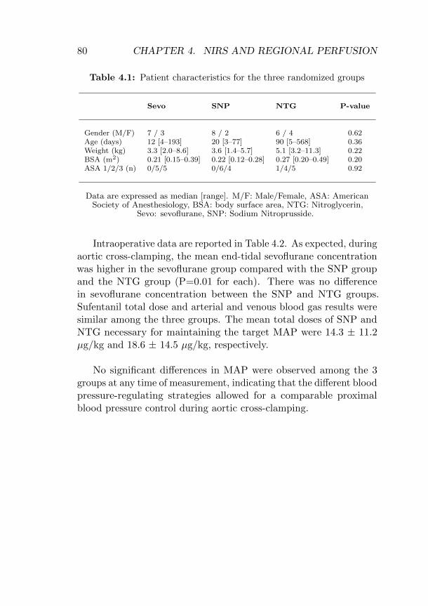
80 CHAPTER 4. NIRS AND REGIONAL PERFUSION
Table 4.1: Patient characteristics for the three randomized groups
Sevo SNP NTG P-value
Gender (M/F) 7 / 3 8 / 2 6 / 4 0.62Age (days) 12 [4–193] 20 [3–77] 90 [5–568] 0.36Weight (kg) 3.3 [2.0–8.6] 3.6 [1.4–5.7] 5.1 [3.2–11.3] 0.22BSA (m2) 0.21 [0.15–0.39] 0.22 [0.12–0.28] 0.27 [0.20–0.49] 0.20ASA 1/2/3 (n) 0/5/5 0/6/4 1/4/5 0.92
Data are expressed as median [range]. M/F: Male/Female, ASA: AmericanSociety of Anesthesiology, BSA: body surface area, NTG: Nitroglycerin,
Sevo: sevoflurane, SNP: Sodium Nitroprusside.
Intraoperative data are reported in Table 4.2. As expected, duringaortic cross-clamping, the mean end-tidal sevoflurane concentrationwas higher in the sevoflurane group compared with the SNP groupand the NTG group (P=0.01 for each). There was no differencein sevoflurane concentration between the SNP and NTG groups.Sufentanil total dose and arterial and venous blood gas results weresimilar among the three groups. The mean total doses of SNP andNTG necessary for maintaining the target MAP were 14.3 ± 11.2µg/kg and 18.6 ± 14.5 µg/kg, respectively.
No significant differences in MAP were observed among the 3groups at any time of measurement, indicating that the different bloodpressure-regulating strategies allowed for a comparable proximalblood pressure control during aortic cross-clamping.

4.3. NIRS DURING AORTIC COARCTATION REPAIR 81
Table 4.2: Intraoperative data for the three randomized groups
Sevo SNP NTG P-value
Duration of CX 15 ± 5 13 ± 5 15 ± 10 0.79(min)Total sufentanil dose 1.5 ± 0.3 1.7 ± 0.6 1.9 ± 0.8 0.34(µg/kg)Mean ETsevo 3.1 ± 1.1 1.8 ± 0.6* 1.8 ±0.5* 0.01during CX (%)Mean MAP during CX 61 ± 11 69 ± 15 70 ± 19 0.43(mmHg)Mean HR during CX 123 ± 10 128 ± 17 126 ± 17 0.73(beats/min)Hct during CX 31 ± 4 32 ± 4 31 ± 6 0.94(%)PaO2 during CX 145 ± 42 151 ± 67 156 ± 67 0.93(mmHg)PvO2 during CX 41 ± 14 46 ± 11 50 ± 14 0.34(mmHg)PaCO2 during CX 44 ± 5 47 ± 8 44 ± 9 0.76(mmHg)Lactate during CX 19 ± 5 18 ± 4 15 ± 4 0.35(mg/dl)
Data are expressed as mean ± SD. Analysis of hematocrit, blood gas partialpressures and lactate was performed 5 minutes after start of aortic
cross-clamping (CX). *P=0.01 for the difference with the sevoflurane group. CX:aortic cross-clamping, ETsevo: end-tidal concentration of sevoflurane, Hct:
hematocrit, HR: heart rate, MAP: mean arterial pressure, NTG: Nitroglycerin,Pa: arterial partial pressure, Pv: venous partial pressure, Sevo: sevoflurane,
SNP: Sodium Nitroprusside.

82 CHAPTER 4. NIRS AND REGIONAL PERFUSION
No difference among groups was observed in mean left-sidedrScO2 values before aortic cross-clamping (72 ± 10%, 77 ± 13%,and 77 ± 11% for sevoflurane, SNP and NTG, respectively, P=0.57).Three possible responses to aortic cross-clamping were observed inleft rScO2 measurements: a decrease of left rScO2 in 2 patients ofeach group (maximal relative decrease of -5 and -17% for sevoflu-rane, -5 and -47% for SNP, -28 and -33% for NTG, P=0.58 betweengroups), no change in left rScO2 in 1 patient in the sevoflurane group,2 patients in the SNP group, and in 2 patients in the NTG group,whereas an increase in left rScO2 was observed in the remainingpatients (sevoflurane, n= 7, 10 ± 7%; SNP, n=6, 13 ± 4%; NTG,n=6, 17 ± 19%, P=0.32 between groups). No differences amonggroups were observed in the maximal relative changes in left rScO2
values in response to aortic cross-clamping (SNP vs sevo: meandifference -0.7%, 99% CI -31 to 29%, P=1.0; SNP vs NTG: meandifference -1.8%, 99% CI -32 to 28%, P=1.0; sevo vs NTG: meandifference -1.1%, 99% CI -31 to 29%, P=1.0).
The following additional observations were made regardingchanges in right rScO2, SrO2 and SmO2. No differences amonggroups were observed in mean right rScO2 values before aortic cross-clamping (P=0.69) and in maximal relative change in right rScO2
values in response to aortic cross-clamping (P=0.4) (Table 4.3). SrO2
and SmO2 showed a rapid and significant decrease after aortic cross-clamping in all groups, reaching a plateau phase for SrO2 within8.6 minutes, 7.4 minutes and 10.0 minutes for sevoflurane, SNP, andNTG, respectively (P=0.08 among groups) and within 9.4 minutes,6.8 minutes and 9.9 minutes for SmO2 for sevoflurane, SNP, and NTG,respectively (P=0.02 among groups). All tissue oxygen saturationsrecovered promptly after release of the aortic cross-clamp. For SmO2,the maximal relative changes were larger and the rate of decay wasfaster in the SNP group compared to the NTG group (Table 4.3).

4.3. NIRS DURING AORTIC COARCTATION REPAIR 83
Table 4.3: Absolute values before aortic cross-clamping and maximalrelative change during aortic cross-clamping for right cerebral (rScO2), renal(SrO2) and muscle oxygen saturation (SmO2) for the three randomizedgroups. Rate of decay during aortic cross-clamping for SrO2 and SmO2 forthe three randomized groups
Sevo SNP NTG P-value
Absolute right rScO2
value (%) before CX 70 ± 13 72 ± 12 74 ± 8 0.69Maximal relative changein right rScO2 (%) 18 ± 18 21 ± 14 14 ± 12 0.40
Absolute SrO2 value (%)before CX 77 ± 8 82 ± 9 70 ± 16 0.13Maximal relative changein SrO2 (%) -43 ± 19 -59 ± 13 -33 ± 22 0.04Rate of decay SrO2
(%.min−1) -6 ± 3 -8 ± 3* -4 ± 3 0.01
Absolute SmO2 value (%)before CX 81 ± 13 78 ± 13 79 ± 16 0.92Maximal relativechange in SmO2 (%) -55 ± 19 -64 ± 17* -34 ± 25 0.01Rate of decay SmO2
(%.min−1) -6 ± 2 -9 ± 4* -4 ± 3 0.004
Data are expressed as mean ± SD. CX: aortic cross-clamping, NTG:Nitroglycerin, Sevo: sevoflurane, SNP: Sodium Nitroprusside.
*Significantly different (p<0.01) from NTG group.

84 CHAPTER 4. NIRS AND REGIONAL PERFUSION
AUC for oxygen saturation from each site is depicted in Fig.4.1. No differences were observed among the 3 treatment groupsin AUC for left-sided and right-sided rScO2 (P=0.74 and P=0.17,respectively). For renal AUC, there was a difference between theNTG and the SNP group (P=0.009). There were no differencesin muscle AUC among the treatment groups (P=0.05 for NTG vssevoflurane, and P=0.03 for NTG vs SNP).
Figure 4.1: Box-and-whisker plots of the area under the curve (AUC) forcerebral, renal and muscle oxygen saturation. NTG: Nitroglycerin, Sevo:sevoflurane, SNP: Sodium Nitroprusside. ◦: outliers of the 5% and 95%-centiles. P-values presented in the figure below each measurement indicatebetween groups differences analyzed with the Kruskal-Wallis test. Pairwisedifferences between treatment groups were analyzed with Mann-WhitneyU test (*SNP vs NTG, p=0.009).

4.3. NIRS DURING AORTIC COARCTATION REPAIR 85
Right-sided rScO2 and MAP showed a poor correlation forNTG (r=-0.2, P=0.93), whereas the correlation was borderline forsevoflurane (r=0.44, P=0.09) and SNP (r=0.56, P=0.04).
An effect of age was observed on the maximal relative decreaseof SrO2 (unstandardized β=0.06, CI 99% -0.02 to 0.14, P=0.06) andSmO2 (unstandardized β=0.07, CI 99% 0.01 to 0.13, P=0.03). Thiseffect was independent of the study product used (unstandardizedβ=1.5, CI 99% -12.4 to 15.4, P=0.76 for SrO2 and unstandardizedβ=6.3, CI 99% -7.9 to 20.6, P=0.23 for SmO2).
Analysis of hematocrit, blood gas partial pressures, and lactatelevels performed 5 minutes after aortic cross-clamp removal ispresented in Table 4.4. There were no differences among thetreatment groups. In all patients, clinical recovery was unevent-ful. Postoperative lactate levels and blood creatinine levels werecomparable among groups.
Table 4.4: Analysis of blood gas values performed 5 minutes after aorticcross-clamp removal for the three randomized groups
Sevo SNP NTG P-value
pH 7.26 ± 0.07 7.27 ± 0.12 7.33 ± 0.09 0.24Base excess (mmol/L) -4.4 ± 2.3 -3.2 ± 4.0 -2.5 ± 3.3 0.45Bicarbonate (mmol/L) 22.1 ± 1.2 23.9 ± 3.3 23.3 ± 2.9 0.31Hct (%) 31 ± 4 32 ± 3 32 ± 5 0.78PaO2 (mmHg) 172 ± 89 175 ± 88 164 ± 63 0.95PvO2 (mmHg) 51 ± 8 49 ± 6 47 ± 8 0.54PaCO2 (mmHg) 51 ± 6 55 ± 15 46 ± 9 0.20Lactate (mg/dl) 29 ± 8 24 ± 8 21 ± 7 0.11
Data are expressed as mean ± SD. Hct: hematocrit, NTG: Nitroglycerin, PaO2:partial pressure of oxygen, arterial, PvO2: partial pressure of oxygen, venous,
Sevo: sevoflurane, SNP: Sodium Nitroprusside.

86 CHAPTER 4. NIRS AND REGIONAL PERFUSION
4.3.4 Discussion
In this randomized, clinical study, the effects of blood pressure-regulating strategies with SNP, NTG, or sevoflurane on cerebral,renal and muscle oxygen saturation were investigated during aorticcross-clamping in children undergoing aortic coarctation repair. Nosignificant differences in cerebral oxygen saturation values were ob-served among the three strategies. Decreases in renal and muscleoxygen saturation were larger and had a faster rate of decay inSNP-treated patients. For both SNP and sevoflurane MAP-rScO2
dependence was higher than for NTG.
Effects on cerebral oxygen saturation
In this study, aortic cross-clamping always involved temporaryocclusion of the left carotid artery. This manoeuver was associatedwith a variable response of the left-sided rScO2, ranging from adecrease to an increase in rScO2. Confidence intervals for pairwisecomparisons between groups were wide. Although our study designdid not allow us to comment on possible underlying mechanisms, itis conceivable that this phenomenon merely reflects the functionaladequacy of the circle of Willis.
In a study on 18 patients undergoing aortic coarctation repair,Azakie et al. [2] observed a pronounced decrease in left rScO2
in 2 patients treated with SNP, in whom the left carotid arterywas clamped during the intervention. They attributed this findingto a SNP-induced disruption of cerebral autoregulation. However,because in that study no simultaneous recording of both left andright hemispheres was obtained, it cannot be determined whetherthis rScO2 decrease is the result of an agent-specific effect or rathera deficient circle of Willis.
We observed no significant differences in right rScO2 values amongthe different blood pressure-regulating strategies; however, with NTGtreatment, changes in rScO2 were less dependent on changes in MAPthan with SNP and sevoflurane. A higher correlation between MAP

4.3. NIRS DURING AORTIC COARCTATION REPAIR 87
and rScO2 has been reported to be indicative of impaired cerebralautoregulation, whereas values around zero and negative values couldbe considered as representative of intact autoregulation [11,12]. Inline with this reasoning, the higher correlation between MAP andrScO2 in the SNP and sevoflurane groups compared with the NTGgroup would suggest that SNP and sevoflurane may interfere to alarger extent with cerebral autoregulation than NTG. This is inaccordance with previous work reporting on dose-dependent impair-ment of cerebral autoregulation with SNP [13], whereas there wasno significant impairment with NTG [14,15]. Data on the effectof sevoflurane on cerebral autoregulation are controversial. It isgenerally assumed that cerebral autoregulation is maintained at lowconcentrations of sevoflurane, whereas higher doses seem to decreaseautoregulatory capacity [16].
Effects on renal and muscle oxygen saturationConcerning the effects on peripheral tissue oxygen saturation, a
larger and faster decrease in both renal and muscle oxygen saturationwas observed in children treated with SNP. Previous studies havesuggested that SNP-induced hypotension might worsen the impairedoxygen balance of tissues below the aortic cross-clamp [3-5]. Thesefindings could not be readily explained. Of interest, an experimen-tal study on striated hamster muscle conducted by Endrich et al.[17] demonstrated that SNP dilated preferentially precapillaries andcaused a consistent increase in intravascular pressure within thevenules. Consequently, the arteriolar-venular pressure gradient wasreduced and functional capillary density decreased, leading to skeletalmuscle tissue hypoxia. In contrast, NTG dilated both arteriolesand venules, leaving the functional capillary density and local pO2
unchanged. Our findings are in accordance with the results of Endrichet al. [17] and suggest that NTG may be preferable to SNP in termsof tissue oxygenation.

88 CHAPTER 4. NIRS AND REGIONAL PERFUSION
In all patients, both SrO2 and SmO2 declined to a plateau phase,which indicated that oxygen delivery no longer met metabolic oxygenrequirements, likely resulting in anaerobic metabolism. The durationof the nadir of oxygenation has been demonstrated to be directlyrelated to the extent of tissue injury [18,19]. Sakamoto et al.[18]demonstrated that nadir times of less than 25 minutes did not inducetissue injury. It can therefore be expected that the nadir times inour study were too short to translate into changes of biomarkersindicative for tissue injury, such as serum lactate, base excess, andcreatinine levels.
Study limitationsThe results of the present study should be interpreted within
the constraints of the methodology. First, because of absence ofevident adverse clinical events due to the short aortic cross-clamptimes, the clinical implications of the current study on outcomeremain to be determined. Second, the current study populationcomprised only young patients. Because the extent of collateralcirculation has been shown to be age-related [10], the validity ofthe current results in older patients with a possibly more developedcollateral circulation needs to be confirmed. Third, the reliability ofNIRS to measure specific renal oxygen saturation can be questionedbecause of the uncertainty related to the depth of penetration of thenear-infrared light in relation to the kidney. However, a number ofstudies have suggested that placement of a NIRS sensor over theflank might indeed reflect renal oxygen saturation. Ortmann et al.[20] demonstrated a strong correlation between flank NIRS valuesand renal vein saturation in children weighing <10 kg. Also, lowrenal tissue oxygen saturations measured with NIRS were associatedwith renal dysfunction after pediatric cardiac surgery [21] and afteraortic coarctation repair [22].

4.3. NIRS DURING AORTIC COARCTATION REPAIR 89
In conclusion, no differences were observed in rScO2 valuesbetween the different blood pressure regulating strategies. How-ever, with NTG treatment, changes in rScO2 were less dependenton changes in MAP than with SNP and sevoflurane. Decreasesin SrO2 and SmO2 were larger and had a faster rate of decay inSNP-treated patients. Based on the lower MAP-rScO2 dependenceand the smaller and slower decreases in SrO2 and SmO2, our datasuggest that NTG might be preferable for blood pressure controlduring surgical procedures involving aortic cross-clamping.
4.3.5 References
1. Gelman S. The pathophysiology of aortic cross-clamping and unclamping.Anesthesiology 1995; 82: 1026-602. Azakie A, Muse J, Gardner M, Skidmore KL, Miller SP, Karl TR,McQuillen PS. Cerebral oxygen balance is impaired during repair of aorticcoarctation in infants and children. J Thorac Cardiovasc Surg 2005; 130:830-63. Gelman S, Reves JG, Fowler K, Samuelson PN, Lell WA, Smith LR.Regional blood flow during cross-clamping of the thoracic aorta and infusionof sodium nitroprusside. J Thorac Cardiovasc Surg 1983; 85: 287-914. Marini CP, Grubbs PE, Toporoff B, Woloszyn TT, Coons MS,Acinapura AJ, Cunningham JN. Effect of sodium nitroprusside on spinalcord perfusion and paraplegia during aortic cross-clamping. Ann ThoracSurg 1989; 47: 379-835. Cernaianu AC, Olah A, Cilley JH Jr, Gaprindashvili T, Gallucci JG,DelRossi AJ. Effect of sodium nitroprusside on paraplegia during cross-clamping of the thoracic aorta. Ann Thorac Surg 1993; 56: 1035-86. Vuylsteke A, Feneck RO, Jolin-Mellgard A, Latimer RD, Levy JH,Lynch C 3rd, Nordlander ML, Nystrom P, Ricksten SE. Perioperative bloodpressure control: a prospective survey of patient management in cardiacsurgery. J Cardiothorac Vasc Anesth 2000; 14: 269-73

90 CHAPTER 4. NIRS AND REGIONAL PERFUSION
7. Kim MB, Ward DS, Cartwright CR, Kolano J, Chlebowski S, Henson LC.
Estimation of jugular venous O2 saturation from cerebral oximetry or arte-
rial O2 saturation during isocapnic hypoxia. J Clin Monit Comput 2000;
16: 191-9
8. McNeill S, Gatenby JC, McElroy S, Engelhardt B. Normal cerebral, renal
and abdominal regional oxygen saturations using near-infrared spectroscopy
in preterm infants. J Perinatol 2011; 31: 51-7
9. Moerman A, De Hert S, Jacobs T, De Wilde L, Wouters P. Cerebral
oxygen desaturation during beach chair position. Eur J Anaesthesiol 2012;
29: 82–7
10. Berens RJ, Stuth EA, Robertson FA, Jaquiss RD, Hoffman GM,
Troshynski TJ, Staudt SR, Cava JR, Tweddell JS, Bert Litwin S. Near
infrared spectroscopy monitoring during pediatric aortic coarctation repair.
Paediatr Anaesth 2006; 16: 777-81
11. Steiner LA, Pfister D, Strebel SP, Radolovich D, Smielewski P,
Czosnyka M. Near-infrared spectroscopy can monitor dynamic cerebral
autoregulation in adults. Neurocrit Care 2009; 10: 122-8
12. Brady K, Joshi B, Zweifel C, Smielewski P, Czosnyka M, Easley RB,
Hogue CW Jr. Real-time continuous monitoring of cerebral blood flow
autoregulation using near-infrared spectroscopy in patients undergoing
cardiopulmonary bypass. Stroke 2010; 41: 1951-6
13. Stange K, Lagerkranser M, Sollevi A. Nitroprusside-induced hypoten-
sion and cerebrovascular autoregulation in the anesthetized pig. Anesth
Analg 1991; 73: 745-52
14. Endoh H, Honda T, Ohashi S, Hida S, Shibue C, Komura N. The
influence of nicardipine-, nitroglycerin-, and prostaglandin E(1)-induced
hypotension on cerebral pressure autoregulation in adult patients during
propofol-fentanyl anesthesia. Anesth Analg 2002; 94: 169-73
15. Moppett IK, Sherman RW, Wild MJ, Latter JA, Mahajan RP. Effects
of norepinephrine and glyceryl trinitrate on cerebral haemodynamics:
transcranial Doppler study in healthy volunteers. Br J Anaesth 2008;
100: 240-4

4.3. NIRS DURING AORTIC COARCTATION REPAIR 91
16. Conti A, Iacopino DG, Fodale V, Micalizzi S, Penna O, Santamaria LB.
Cerebral haemodynamic changes during propofol-remifentanil or sevoflurane
anaesthesia: transcranial Doppler study under bispectral index monitoring.
Br J Anaesth 2006; 97: 333-9
17. Endrich B, Franke N, Peter K, Messmer K. Induced hypotension:
action of sodium nitroprusside and nitroglycerin on the microcirculation.
Anesthesiology 1987; 66: 605-13
18. Sakamoto T, Hatsuoka S, Stock UA, Duebener LF, Lidov HG,
Holmes GL, Sperling JS, Munakata M, Laussen PC, Jonas RA. Prediction
of safe duration of hypothermic circulatory arrest by near-infrared spec-
troscopy. J Thorac Cardiovasc Surg 2001; 122: 339-50
19. Huang JH, Su ZK, Wang SM. Near-infrared spectroscopy monitoring
of brain oxygen in infant cardiac surgery. Asian Cardiovasc Thorac Ann
2007; 15: 194-9
20. Ortmann LA, Fontenot EE, Seib PM, Eble BK, Brown R, Bhutta AT.
Use of near-infrared spectroscopy for estimation of renal oxygenation in
children with heart disease. Pediatr Cardiol 2011; 32: 748-53
21. Owens GE, King K, Gurney JG, Charpie JR. Low renal oximetry
correlates with acute kidney injury after infant cardiac surgery. Pediatr
Cardiol 2011; 32: 183-8
22. Fuentes-Garcia D, Carceles-Baron MD, Lopez-Lopez R, Roques-Escolar
V. Non-invasive oximetry for early detection of cerebral and somatic
ischaemia during corrective surgery for aortic coarctation in paediatric
patients. Br J Anaesth 2010; 104: 780-2

92 CHAPTER 4. NIRS AND REGIONAL PERFUSION
4.4 NIRS during aortic aneurysm repair
Near-Infrared Spectroscopy for Monitoring Spinal CordIschemia during Hybrid Thoracoabdominal AorticAneurysm Repair. Moerman A, Van Herzeele I,Vanpeteghem C, Vermassen F, Francois K, Wouters P.J Endovasc Ther 2011; 18: 91-5
Abstract
Purpose: To describe a simple, non-invasive technique to detectchanges in oxygen saturation at the level of the spinal cord and tosuggest its suitability for individualized blood pressure managementduring and after thoracoabdominal aneurysm repair.Case report: Near-Infrared Spectroscopy (NIRS) sensors were appliedover the tenth thoracic vertebra for continuous monitoring of tissueoxygen saturation (SsO2) during endovascular repair of a post-dissection aneurysm of the aortic arch and descending aorta. Afterstent-graft deployment mean SsO2 decreased significantly. Moreoverthe relationship between SsO2 and arterial blood pressure becamelinear, reflecting pressure-dependency of spinal cord perfusion afterstent deployment.Conclusion: These data show that NIRS monitors changes inSsO2 after aortic endograft deployment which were strongly relatedto arterial blood pressure. Regional NIRS monitoring at thevertebral level may function as a valuable non-invasive guide to themanagement of blood pressure during thoracoabdominal aneurysmrepair, both intra- and postoperatively.

4.4. NIRS DURING AORTIC ANEURYSM REPAIR 93
4.4.1 Introduction
During thoracoabdominal aortic aneurysm (TAAA) repair, spinalcord perfusion is precarious and vulnerable to hemodynamiccompromise, eventually leading to neurologic injury [1,2]. Closemonitoring is mandatory to detect spinal cord ischemia in time totreat it. Near-Infrared Spectroscopy (NIRS) is a non-invasive contin-uous monitoring technique to measure tissue oxygen saturation [3].Whereas initial research focused on the use of NIRS for monitoringcerebral saturation, interest has now extended to evaluating theoxygenation of tissues other than the brain. To date, research onthe use of NIRS to monitor spinal cord oxygenation is scarce: Onlyanimal studies [4,5] and 3 clinical reports [6-8] have examined thefeasibility of NIRS to monitor spinal cord perfusion.
We present a case where tissue oxygen saturation at the level ofthe spinal cord (SsO2) decreased significantly after aortic endograftdeployment and became strongly related to arterial blood pressure.
4.4.2 Case report
A 53-year old man with an extensive medical history, including re-placement of the aortic valve and ascending aorta (Bentall procedure)for acute aortic dissection in 1998 and replacement of the ascendingaorta and proximal aortic arch for acute aortic dissection in 2008,presented with a large TAAA due to chronic dissection from thedistal aortic arch into the iliac arteries. The patient was scheduledfor a staged hybrid repair. In the first phase, extra-anatomical revas-cularization of the supra-aortic arteries from the proximal ascendingaorta was done, followed by placement of endografts in the distalascending aorta, aortic arch, and descending aorta. In the secondphase, open abdominal repair with retrograde perfusion of the celiac,mesenteric, and renal arteries was planned, with further exclusion ofthe TAAA by stent-grafting. Local Ethics’ Committee approval andwritten informed consent from the patient were obtained.

94 CHAPTER 4. NIRS AND REGIONAL PERFUSION
The hybrid arch procedure was performed under general anesthe-sia with full hemodynamic monitoring. A cerebrospinal fluid (CSF)drain was positioned at a height of 13.5 cmH2O above the externalmeatus, which equated to a pressure of 10 mmHg within the spinalcanal. CSF was allowed to drain continuously. Disposable NIRSsensors were applied to the right and the left side of the forehead,over the tenth thoracic vertebra (T10), and over the right calf, andtissue oxygen saturation was monitored continuously during theentire procedure (INVOS 5100; Somanetics Corporation, Troy, MI,USA). All hemodynamic, respiratory and tissue oxygen saturationdata were recorded continuously with RUGLOOP (Demed, Temse,Belgium).
The extra-anatomical revascularization of the arch vessels wascarried out through a median sternotomy. The arch vessels wereanastomosed to a bifurcated graft that was sewn proximally onto theascending aortic tube graft. Epicardial ventricular pacemaker wireswere placed to allow induction of hypotension by rapid ventricularpacing during deployment of the stent-grafts. Subsequently, theExcluder TAG devices (W. L. Gore & Associates, Inc, Flagstaff,AZ, USA) were delivered via the right common femoral artery. ATAG (37 × 200 mm) was introduced and deployed distal to the archvessels origin; a second stent-graft (37 × 150 mm) was deployed withsufficient overlap in the descending thoracic aorta, but during removalof the delivery device, this stent-graft was accidentally withdrawn.Retrieval of the misdeployed stent-graft (still connected to the releasewire and the inner shaft of the delivery device) was accomplished viaan infrarenal aortotomy. The infrarenal abdominal aortic aneurysmwas repaired with a bifurcation graft (Dacron 18 × 9 mm) connectedto both common iliac arteries. Finally, a TAG (37 × 150 mm) wasdeployed uneventfully in the residual descending thoracic aorta withsufficient overlap, terminating proximal to the diaphragm.

4.4. NIRS DURING AORTIC ANEURYSM REPAIR 95
Mean SsO2 decreased significantly from 76 ± 2.6% (from startof measurement until deployment of first stent-graft) to 61 ± 5.6%(from deployment of last stent-graft until the end of the operation)(P<0.001) (Fig. 4.2).
Figure 4.2: Relationship between spinal saturation (SsO2) and meanarterial blood pressure (MAP) before and after stent-graft deploymentduring first operation.
Prior to stent-graft deployment, mean SsO2 fluctuated only mildlywith mean arterial blood pressure (MAP), while after stent-graftdeployment, small decreases in arterial blood pressure provoked majordecreases in SsO2. This pressure dependency is quantified in Figure4.3: the correlation between MAP and SsO2 was clearly linear afterdeployment of the devices, whereas there was no correlation beforestent-graft deployment.

96 CHAPTER 4. NIRS AND REGIONAL PERFUSION
Figure 4.3: Correlation between spinal saturation (SsO2) and meanarterial blood pressure (MAP) before and after stent-graft deploymentduring first operation.
Twelve days later, the second part of the hybrid procedure wasperformed. A spinal drain was inserted and NIRS sensors were placedover the left and right forehead and at 2 locations over the spinalcolumn: at T10 and at the second lumbar vertebra (L2). The visceralvessels were revascularized by the implantation of a bifurcated Dacrongraft anastomosed to the previously implanted bifurcated graft. Theright limb was connected to the common hepatic artery and tunnelledbehind the pancreas; the left limb was anastomosed to the superiormesenteric trunk. Finally, a Dacron graft originating from the rightlimb was connected in an end-to-end fashion to the right renal artery.The remaining TAAA was excluded by deployment of 3 stent-graftswith sufficient overlap (TAG 28×150 mm, 31×150 mm and 37×150mm). Hypotension to prevent movement of the stent-grafts duringdeployment was accomplished with nitroprusside. The devices weredeployed in a retrograde fashion and positioned proximal to thebifurcated graft perfusing the visceral arteries.
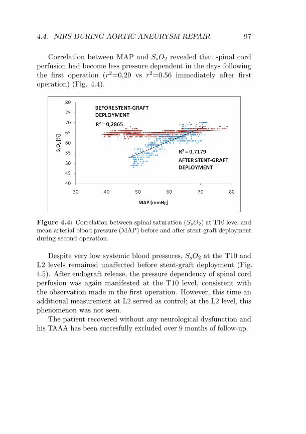
4.4. NIRS DURING AORTIC ANEURYSM REPAIR 97
Correlation between MAP and SsO2 revealed that spinal cordperfusion had become less pressure dependent in the days followingthe first operation (r2=0.29 vs r2=0.56 immediately after firstoperation) (Fig. 4.4).
Figure 4.4: Correlation between spinal saturation (SsO2) at T10 level andmean arterial blood pressure (MAP) before and after stent-graft deploymentduring second operation.
Despite very low systemic blood pressures, SsO2 at the T10 andL2 levels remained unaffected before stent-graft deployment (Fig.4.5). After endograft release, the pressure dependency of spinal cordperfusion was again manifested at the T10 level, consistent withthe observation made in the first operation. However, this time anadditional measurement at L2 served as control; at the L2 level, thisphenomenon was not seen.
The patient recovered without any neurological dysfunction andhis TAAA has been succesfully excluded over 9 months of follow-up.

98 CHAPTER 4. NIRS AND REGIONAL PERFUSION
Figure 4.5: Relationship between spinal saturation (SsO2) at the T10 andL2 level and mean arterial blood pressure (MAP) during second operation.Asterisk (*) represents stent-graft deployment.
4.4.3 Discussion
During endovascular TAAA repair, tissue oxygen saturation at theT10 level dropped significantly after endograft deployment. Further-more, a significant linear relationship was noted between SsO2 andarterial blood pressure after stent-graft deployment, which was notpresent before stent-graft deployment. As NIRS seems to reflectpressure dependence of spinal cord perfusion, its utility as a guideto optimize perioperative blood pressure management during TAAArepair deserves further investigation.
NIRS exploits the fact that biological tissues, including bone,are transparent in the near-infrared range [3]. Specific wavelengthsof near-infrared light are emitted through the tissues lying belowa sensor. Depending on the amount of light reflected back to light

4.4. NIRS DURING AORTIC ANEURYSM REPAIR 99
detectors built into the same sensor, regional oxygen saturation ofthe underlying tissue can be determined. The mean depth of lighttransmission is proportional to the emitter-detector separation by afactor of approximately one third [9]. The use of 2 photodetectorsallows selective measurement of deeper tissue, a process known asspatial resolution: the closer receiver measures superficial tissue whilethe distal optode measures both superficial and deeper tissue. Afteradjusting for interference from the superficial tissues, oxygenation ofdeeper tissues is derived.
Initial animal studies demonstrated the feasibility of NIRS tomonitor the spinal cord. Macnab et al. [4] exposed the posteriorelements of the spine in 3 pigs; they sutured NIRS optodes directed to-ward the spinal cord over the T9 laminae or on the spinous processesof T9 and T10. Interventions included decreasing the arterial oxygensaturation and distracting the spine at T9-T10, which reduced bloodflow to the spinal cord. Each intervention resulted in NIRS changesthat were observed immediately and resolved promptly, independentof the position of the optodes. LeMaire et al. [5] sequentially ligatedsegmental arteries from T6 through L1 in pigs, while regional oxygensaturation was monitored with NIRS in the upper (T6-T7) and lowercord (T9-T11). After ligation, regional oxygen saturation in the lowercord was significantly less than in the upper cord. This regional dif-ference in the degree of ischemia was confirmed microscopically, withsignificantly more pronounced ischemic changes and more ischemicneurons in the lower-cord sections.
To date, 3 reports have described the use of NIRS for monitoringspinal cord oxygenation in humans. Macnab et al. [6] used NIRSin an infant requiring release of a congenitally tethered spinal cord.Subsequently, in a series of 9 patients undergoing TAAA repair,Edmonds and Ganzel [7] observed a greater decline in spinal sat-uration measured at T10 in patients who experienced lower limbmotor deficit compared to patients without deficit. This declinewas consistent with the results of simultaneously performed spinal

100 CHAPTER 4. NIRS AND REGIONAL PERFUSION
cord transmission measurements. Mitchell et al. [8] noted a signifi-cant drop in perispinal regional oxygen saturation at the T10 levelduring coarctation repair in neonates. Removal of the aortic clampimmediately restored the perispinal saturation to baseline values.
In NIRS monitoring of spinal cord ischemia, the optically inter-rogated region is not well-defined, which may pose a problem. Oneconcern is that oxygenation in the surrounding tissues, such as muscleand subcutaneous tissue, is measured instead of oxygenation in thespinal cord. Although the spatial resolution reduces the influence ofouter layers, not all extraspinal structures may have been excluded.Nevertheless, NIRS data on regional oxygenation may adequatelyreflect spinal cord perfusion, albeit indirectly, as a collateral networkensures the shared blood supply between the spinal cord and thesurrounding tissues [10]. In order to prove this concept, LeMaire etal. [5] injected indocyanine green dye into the subarachnoid spaceand found an immediate change in light absorption, suggesting thatat least some of the photons were reaching the spinal cord.
In our case, 2 observations support the viewpoint that saturationat the spinal cord level measured by NIRS (directly or indirectly)reflects spinal cord perfusion. First, SsO2 at T10, which was ini-tially pressure independent, became strongly influenced by arterialblood pressure after endograft deployment. This is consistent withpublished reports that have described the treatment of spinal cordischemia after endovasculair repair by increasing the arterial pres-sure [2,11]. In addition, spinal cord perfusion became less pressuredependent in the days following the first operation, which Etz etal. [12] also observed in their study. Spinal cord perfusion pressurespontaneously returned to baseline values after ∼ 48 hours.
Second, after deployment of stent-grafts from the diaphragm tothe level of the bifurcated graft perfusing the visceral arteries, NIRSvalues at T10 were significantly decreased by changes in arterialblood pressure, whereas at the L2 level, SsO2 remained stable (Fig.4.5). This may be explained by the segmental nature of spinal
4.4. NIRS DURING AORTIC ANEURYSM REPAIR 101
perfusion and may suggest that SsO2 measurement includes thespinal vasculature.
Somatosensory-evoked potentials (SSEPs) and motor-evokedpotentials (MEPs) monitoring are primarily used to monitor spinalcord perfusion, although no prospective studies have proven itsefficacy to reduce spinal cord injury. Nevertheless, several reportshave demonstrated that spinal cord ischemia detected with SSEPsand/or MEPs could be corrected with appropriate interventions[13,14]. Still, SSEPs and MEPs monitoring is not widely used be-cause of its limitations. NIRS could be useful as an adjunctive toolto assess the entire spinal cord perfusion. Or, it may offer a simplealternative for monitoring spinal cord ischemia when SSEPs and/orMEPs are unavailable, especially in the postoperative period in whichSSEP/MEP monitoring is generally impractical or impossible.
ConclusionTo our knowledge, no one has demonstrated the dependence of
oxygen saturation at the level of the spinal cord on blood pressureafter thoracic stent-graft deployment. These preliminary data suggestthat NIRS may be employed for individualized management ofblood pressure during TAAA repair, both intra- and postopera-tively. Further investigations are required to compare NIRS withSSEPs and MEPs monitoring and to evaluate whether measuringand intervening on the values obtained by NIRS make a difference inthe neurological outcome of patients who undergo repair of extensivethoracoabdominal aneurysms.

102 CHAPTER 4. NIRS AND REGIONAL PERFUSION
4.4.4 References
1. Etz CD, Luehr M, Kari FA, et al. Paraplegia after extensive thoracic and
thoracoabdominal aortic aneurysm repair: Does critical spinal cord ischemia
occur postoperatively? J Thorac Cardiovasc Surg 2008; 135: 324-30
2. Azizzadeh A, Huynh TT, Miller CC III, et al. Postoperative risk factors
for delayed neurologic deficit after thoracic and thoracoabdominal aortic
aneurysm repair: A case-control study. J Vasc Surg 2003; 37: 750-4
3. Murkin JM, Arango M. Near-infrared spectroscopy as an index of brain
and tissue oxygenation. Br J Anaesth 2009; 103 (Suppl. 1): i3-i13
4. Macnab AJ, Gagnon RE, Gagnon FA. Near Infrared Spectroscopy for
intraoperative monitoring of the spinal cord. Spine 2002; 27: 17-20
5. LeMaire SA, Ochoa LN, Conklin LD, et al. Transcutaneous near-infrared
spectroscopy for detection of regional spinal ischemia during intercostal
artery ligation: Preliminary experimental results. J Thorac Cardiovasc
Surg 2006; 132: 1150-5
6. Macnab AJ, Gagnon RE, Gagnon FA, et al. NIRS monitoring of brain
and spinal cord – detection of adverse intraoperative events. Spectroscopy
2003; 17: 483-90
7. Edmonds Jr H, Ganzel B. Spinal oximetry for ischemia detection during
thoracoabdominal aneurysm repair [abstract]. Anesth Analg 2004; 98: SCA
72
8. Mitchell ME, Sehic A, Edmonds Jr HL, et al. Non-invasive peri-spinal
oximetry for aortic coarctation repair [abstract]. Clinical Neurophysiology
2006; 117: S238
9. Germon TJ, Evans PD, Barnett NJ, et al. Cerebral near infrared
spectroscopy: emitter-detector separation must be increased. Br J Anaesth
1999; 82: 831-7
10. Griepp RB, Griepp EB. Spinal cord perfusion and protection during
descending thoracic and thoracoabdominal aortic surgery: The collateral
network concept. Ann Thorac Surg 2007; 83: S865-9
11. Cheung AT, Pochettino A, McGarvey ML, et al. Strategies to manage
paraplegia risk after endovascular stent repair of descending thoracic aortic
aneurysms. Ann Thorac Surg 2005; 80: 1280-9

4.4. NIRS DURING AORTIC ANEURYSM REPAIR 103
12. Etz CD, Homann TM, Plestis KA, et al. Spinal cord perfusion after
extensive segmental artery sacrifice: can paraplegia be prevented? Eur J
Cardiothorac Surg 2007; 31: 643-8
13. Jacobs MJ, de Mol BA, Elenbaas Ted, et al. Spinal cord blood supply
in patients with thoracoabdominal aortic aneurysms. J Vasc Surg 2002; 35:
30-7
14. Schurinck GW, Nijenhuis RJ, Backes WH, et al. Assessment of spinal
cord circulation and function in endovascular treatment of thoracic aortic
aneurysms. Ann Thorac Surg 2007; 83: S877-81

104 CHAPTER 4. NIRS AND REGIONAL PERFUSION

Chapter 5
NIRS as an estimate ofvenous oxygen saturation
5.1 Background
Initially, the interest in the use of cerebral oximetry focused on thedetection of asymmetric changes, indicating a potentially catastrophiccerebral event. In recent years, a growing understanding of thephysiological principles of NIRS led to a more complete appreciationof its potential.
NIRS measures regional oxygen saturation in the cerebral cortex,which is a predominantly venous bed [1,2]. Therefore, cerebral oxygensaturation measured with NIRS primarily reflects venous oxygensaturation. When cardiac output decreases progressively, cerebralautoregulation and preferential distribution of cardiac output tothe brain preserves perfusion to the brain longer than perfusionto any other organ system. Consequently, decreases in cerebralvenous oxygen saturation usually occur when global venous oxygensaturation is also deteriorating. Not surprisingly, NIRS-measuredcerebral oxygen saturation was shown not only to correlate withoxygen saturation in blood obtained from the internal jugular vein
105
106 CHAPTER 5. NIRS AS AN ESTIMATE OF SMVO2
[3], but also from the pulmonary artery [4] and superior caval vein[3,4].
Even though perfusion of the brain is momentarily preservedwhen cardiac output declines, the brain appears to offer a “window”on global hemodynamics and NIRS seems to provide a non-invasivemonitoring tool to assess global tissue perfusion. This notion issupported by the results of Murkin [5] who showed that optimizingcerebral NIRS also enhanced global tissue oxygenation, resulting insignificantly less major (non-cerebral) organ morbidity. Recently,NIRS has been suggested as a potentially valuable biomarker intracking the status of heart failure patients [6]. Heringlake et al.demonstrated that preoperative cerebral oxygen saturation is reflec-tive of global cardiopulmonary function, associated with short- andlong-term mortality and morbidity in cardiac surgery patients [7].
Mixed venous oxygen saturation (SmvO2) is generally accepted asan indicator of adequacy of systemic oxygen balance, and its use forguidance of therapy has been demonstrated to improve outcome [8].However, its obligatory need for more invasive instrumentation pre-cludes its use as a standard routine monitoring in perioperative care.The availability of an easy to use, reliable, and non-invasive monitorof global oxygen balance would benefit a much wider spectrum ofpatients at risk for anesthesia and surgery.
References1. Ito H, Kanno I, Fukuda H. Human cerebral circulation: positron emissiontomography studies. Ann Nucl Med 2005; 19: 65-742. Watzman HM, Kurth CD, Montenegro LM, Rome J, Steven JM, NicolsonSC. Arterial and venous contributions to near-infrared cerebral oximetry.Anesthesiology 2000; 93: 947-53

5.2. OBJECTIVE 107
3. Nagdyman N, Ewert P, Peters B, Miera O, Fleck T, Berger F. Comparison
of different near-infrared spectroscopic cerebral oxygenation indices with
central venous and jugular venous oxygenation saturation in children.
Paediatr Anaesth 2008; 18: 160-6
4. Tortoriello TA, Stayer SA, Mott AR, et al. A noninvasive estimation
of mixed venous oxygen saturation using near-infrared spectroscopy by
cerebral oximetry in pediatric cardiac surgery patients. Paediatr Anaesth
2005; 15: 495-503
5. Murkin JM, Adams SJ, Novick RJ, et al. Monitoring brain oxygen
saturation during coronary bypass surgery: a randomized, prospective study.
Anesth Analg 2007; 104: 51-8
6. Rifai L, Winters J, Friedman E, Silver MA. Initial description of cerebral
oximetry measurement in heart failure patients. Congest Heart Fail 2012;
18: 85-90
7. Heringlake M, Garbers C, Kabler JH. Preoperative cerebral oxygen
saturation and clinical outcomes in cardiac surgery. Anesthesiology 2011;
114: 58-69
8. Polonen P, Ruokonen E, Hippelainen M, Poyhonen M, Takala J. A
prospective, randomized study of goal-oriented hemodynamic therapy in
cardiac surgical patients. Anesth Analg 2000; 90: 1052-9
5.2 Objective
The aim of the study was to evaluate the feasibility of NIRS as anon-invasive monitor of global oxygen balance, providing a reliableestimate of systemic venous oxygen saturation.

108 CHAPTER 5. NIRS AS AN ESTIMATE OF SMVO2
5.3 NIRS as an estimate of venous saturation
Relation between mixed venous oxygen saturation and cere-bral oxygen saturation measured by absolute and rela-tive near-infrared spectroscopy during off-pump coronaryartery bypass grafting. Moerman A, Vandenplas G, BoveT, Wouters P, De Hert S. Br J Anaesth 2013; 110: 258-65
AbstractBackground. We hypothesized that previously reported contradictoryresults regarding the equivalence of mixed venous (SmvO2 ) andcerebral (rScO2) oxygen saturation might be related to time delayissues and to measurement technology. In order to explore these twofactors, we designed a prospective clinical study comparing SmvO2
with relative (INVOS) and absolute (Foresight) rScO2 measurements.Methods. Forty-two consenting patients undergoing elective off-pumpcoronary artery bypass grafting were included. Two INVOS andtwo Foresight sensors continuously registered rScO2. SmvO2 wasmeasured continuously via a pulmonary artery catheter. Data wereassessed by within- and between-group comparisons and correlationanalysis.Results. A similar time delay of 19 (4) and 18 (4) s was found forSmvO2 compared with rScO2 measurements by Foresight and INVOS,respectively, during hemodynamic changes. After adjusting for thistime delay, the correlation between SmvO2 and rScO2 increased fromr=0.25 to 0.75 (P<0.001) for Foresight, and from r=0.28 to 0.73(P<0.001) for INVOS. Comparison of Foresight and INVOS revealedsignificant differences in absolute rScO2 values (range 58–89% forForesight and 28–95% for INVOS). Changes in rScO2 in response toacute hemodynamic alterations were significantly more pronouncedwith INVOS compared with Foresight (P<0.001).Conclusions. Considering the important time delay with SmvO2,rScO2 seems to reflect more appropriately acute hemodynamic
5.3. NIRS AS AN ESTIMATE OF VENOUS SATURATION 109
alterations. This might suggest its use as a valid alternative toinvasive monitoring of tissue oxygen saturation. Relative and ab-solute rScO2 measurements demonstrated significant differences inmeasured rScO2 values and in the magnitude of rScO2 changes duringhemodynamic alterations.
5.3.1 Introduction
A primary goal of hemodynamic management of patients undergoingsurgery is to provide adequate tissue oxygenation. Mixed venousoxygen saturation (SmvO2) is generally accepted as an indicator ofadequacy of systemic oxygen balance, and its use for guidance oftherapy has been demonstrated to improve outcome [1]. Owing to itsinvasiveness, this variable is not commonly incorporated into routineanesthesia monitoring. The availability of an easy to use, reliable,and non-invasive monitor of global oxygen balance would benefit amuch wider spectrum of patients at risk for anesthesia and surgery.
Near-infrared spectroscopy (NIRS) measures regional cerebraloxygen saturation (rScO2) in the cerebral cortex, which is apredominantly venous bed [2]. The findings of Murkin and colleagues[3] suggest that cerebral oximetry might be useful as an index forglobal oxygen delivery and consumption, indicating that NIRS mightoffer a continuous, non-invasive alternative for SmvO2.
Previous studies found contradictory results regarding theequivalence and interchangeability of SmvO2 and rScO2 data [4-12].We hypothesized that this inconsistency might be related to twofactors. First, SmvO2 values were recorded intermittently, providingonly isolated epochs for comparison such that time delay issues maynot have been taken into account. Secondly, rScO2 measurementswere all performed with the INVOS cerebral oximeter (SomaneticsCorporation, Troy, MI, USA), which is approved by the Food andDrug Administration (FDA) as a relative cerebral oximeter or a trendmonitor [K051274 FDA 510(k) premarket notification]. Hence, thereported poor correlations might be the result of inherent limitations

110 CHAPTER 5. NIRS AS AN ESTIMATE OF SMVO2
of the measurement technology.In order to explore these two hypotheses, we designed a
prospective observational clinical study comparing continuously mea-sured SmvO2 with both relative and absolute rScO2 measurements.Because we wanted to evaluate both experimental questions in a widerange of hemodynamic changes, we chose a model of off-pump coro-nary artery bypass (OPCAB) surgery, as the course of this procedureentails significant hemodynamic alterations during surgical exposureand cardiac manipulation. The Foresight cerebral oximeter (CASMedical Systems, Branford, CT, USA) was chosen as comparativeNIRS technology, since it claims to be an absolute cerebral oximeter(www.casmed.com), providing absolute rScO2 values [13,14].
5.3.2 Methods
This trial is registered at ClinicalTrials.gov (NCT01673841). AfterInstitutional Ethics Committee approval and written informed con-sent, 42 patients undergoing elective OPCAB surgery for at leastthree-vessel coronary artery disease were included. Patients witharteriovenous shunts, intracardiac shunts, a previous history of cere-brovascular accident, or stenosis of the internal carotid artery of>60% were excluded.
Description of NIRS devicesThe physical principles upon which NIRS is based have been
described [15]. In general terms, NIRS utilizes the absorption andreflectance spectra of near-infrared light to quantify oxygenation oftissues underlying the sensor. Both INVOS and Foresight use theModified Beer-Lambert law to measure tissue oxygen saturation, andeliminate the contribution of extracerebral tissue by using the princi-ple of spatial resolution (depth of photon penetration proportional tothe source-detector separation). However, some important technicaldifferences exist. The INVOS 5100 generates two wavelengths of lightat 730 and 810 nm from light emitting diodes, while Foresight uses

5.3. NIRS AS AN ESTIMATE OF VENOUS SATURATION 111
laser-emitting diodes to generate light at four different wavelengths(690, 778, 800 and 850 nm). The spacing of the light detectors fromthe light emitter, which influences the sample volume and depth ofpenetration, is at 3 and 4 cm distance for INVOS 5100, and at 1.5and 5 cm for Foresight. There is also a difference in assumed cerebralarterial:venous ratio upon which the oximetry values are calculated(assumed ratio of 25:75 for INVOS and 30:70 for Foresight). Owing tothe technical differences and the different computational algorithmsfor the calculation of tissue saturation, the comparability betweenINVOS 5100 and Foresight is not clear.
Study design
On the morning of surgery, subjects were allowed to take theirroutine medication, except for angiotensin-converting enzyme in-hibitors. Four disposable oxygenation sensors [two INVOS sensors(INVOS 5100, Somanetics Corporation) and two Foresight sensors(CAS Medical Systems)] were applied bilaterally on the forehead forcontinuous registration of rScO2. The sensors of one monitor wereplaced just above the eyebrows, and the sensors of the other monitorwere placed just above the former sensors. Sensor placement was de-termined at random by a computerized randomization list. Baselinevalues were obtained at least 1 minute after placement of sensorsuntil values were stabilized. Anesthesia was induced with diazepam0.1 mg/kg, fentanyl 5 µg/kg, propofol 1 mg/kg, and rocuronium 1mg/kg. The lungs were ventilated mechanically with oxygen enrichedair (fractional inspired oxygen 0.6) adjusted to keep the end-tidalcarbon dioxide (EtCO2) around 4.7 kPa. Anesthesia was maintainedwith boluses of fentanyl up to a total dose of 25-35 µg/kg and sevoflu-rane at a minimum concentration of 1.5 vol%. Standard monitoringwas used throughout the procedure, including ECG, pulse oxime-try, capnography, invasive arterial, central venous and pulmonaryartery pressure monitoring, continuous SmvO2 monitoring [Swan-Ganz CCOmbo pulmonary artery catheter (Edwards lifesciences,

112 CHAPTER 5. NIRS AS AN ESTIMATE OF SMVO2
Irvine, CA, USA)], and transoesophageal echocardiography.Hemodynamic management was at the discretion of the at-
tending anesthetist. All variables were recorded continuously withRUGLOOP (Demed, Temse, Belgium) and were analysed at twoseconds intervals. Types and volumes of all fluids were recorded, aswell as doses of any drugs given.
To explore interference when Foresight and INVOS were operat-ing simultaneously, we included a methodological evaluation of thereliability of our data. During preparation of the internal thoracicartery - which includes a hemodynamic stable period - the monitorswere switched off one at a time for 5 min. During these periods,the mean rScO2 and standard deviation (SD) were measured foreach monitor separately and compared with mean (SD) obtainedduring the 5 min preceding this intervention when both monitorswere operating simultaneously.
The response of rScO2 to major hemodynamic disturbances wasexplored during placement of deep pericardial stitches. Duringevery heart retraction, which induced a substantial decrease in meanarterial pressure (MAP), relative changes in MAP, rScO2 and SmvO2
were calculated at 5 (T1) and 10 s (T2) after start of heart retraction,at the minimum value of MAP (T3), and at 5 (T4), 10 (T5), 15 (T6)and 20 (T7) s after release of heart retraction. Relative change wasdefined as the percentage difference between the value just beforeheart retraction and the value at the different time moments. Theintegrated value or area under the curve (AUC) for rScO2 duringplacement of the pericardial stitches was also explored to evaluatethe response of rScO2 to major hemodynamic disturbances . TheAUC was calculated by taking the difference between the currentvalues and the value just before heart retraction, multiplied by thecorresponding time in seconds, and is expressed in %.sec.

5.3. NIRS AS AN ESTIMATE OF VENOUS SATURATION 113
Statistical analysisSample size calculation was based on an aimed correlation
coefficient of 0.5 between SmvO2 and rScO2 values [5,6]. Acceptinga two-tailed α error of 0.01 and a β error of 0.8, 42 subjects wererequired. Statistical analysis was performed using SPSS Statistics20 (SPSS Inc., Chicago, IL, USA). Distribution of data was testedfor normality using the Shapiro-Wilk test. Normally distributedcontinuous data are presented as mean (SD). Non-parametric dataare presented as median [range]. Comparison of data between Fore-sight and INVOS were performed by the Mann-Whitney U analysis.Correlation analysis was performed by the Pearson or Spearman’s ρtesting as appropriate. P-values <0.05 were considered significant.
5.3.3 Results
Thirty male and 12 female subjects with an average age of 72 (8) yr,weight of 78 (16) kg, and height of 166 (8) cm were included. AwakerScO2 values were 70 (4)% and 69 (3)% for Foresight on the left andright side, respectively, and 64 (10)% and 64 (9)% for INVOS onthe left and right side, respectively (P<0.001 between Foresight andINVOS). For further analysis, right and left cerebral saturations ofthe corresponding monitoring system were averaged.
Mutual interference of monitorsWith both monitors running simultaneously, Foresight exhibited
more variability in rScO2 than when INVOS was switched off (0.9(0.4)% compared with 0.5 (0.2)%, P=0.004) (Fig. 5.1). INVOSperformance was not affected by the presence of Foresight (0.6 (0.2)%vs 0.7 (0.2)% Foresight on-off, respectively, n.s.). Simultaneousoperation did not affect the mean rScO2 values for either monitor(INVOS mean values 72 (9)% compared with 70 (15)% with Foresighton and off, respectively; Foresight mean values 70 (4)% comparedwith 70 (4)% with INVOS on and off, respectively).

114 CHAPTER 5. NIRS AS AN ESTIMATE OF SMVO2
Figure 5.1: Representative example illustrating higher variability of Fore-sight regional cerebral oxygen saturation (rScO2) values when Foresightand INVOS monitors were operating simultaneously.
Hypothesis 1: time delay issues are responsible for reported poorcorrelations
To explore this hypothesis, SmvO2, rScO2 and MAP were dis-played graphically for each subject separately. Figure 5.2 illustrates arepresentative case during the placement of deep pericardial stitches,which was associated with substantial hemodynamic alterations.MAP decreased briefly every time the heart was retracted to allowthe placement of the pericardial stitches. NIRS values followed imme-diately, while SmvO2 values obviously lagged behind. This resultedin a poor correlation between SmvO2 and rScO2 at the moment ofplacement of the pericardial stitches: r=0.25 [0.01-0.67] for Foresightand r=0.28 [0.10-0.63] for INVOS, with no significant differencebetween monitors.

5.3. NIRS AS AN ESTIMATE OF VENOUS SATURATION 115
Figure 5.2: Representative case showing substantial changes in meanarterial blood pressure (MAP) (red line) associated with retraction of theheart during placement of deep pericardial stitches. Changes in MAP wereclosely followed by changes in cerebral oxygen saturation measured byForesight (pink and yellow lines) and INVOS (light blue and violet lines),while mixed venous oxygen saturation (SmvO2) (dark blue line) obviouslylagged behind.
The mean time delay between SmvO2 and MAP or rScO2
(calculated as the number of seconds between minimum SmvO2 andminimum MAP or minimum rScO2) was 18 (4), 19 (4) and 18 (4)seconds for MAP, Foresight and INVOS respectively. After adjust-ing for this time delay, taking the nadir value for each variable,the correlation between SmvO2 and rScO2 increased from r=0.25to r=0.75 [0.15-0.88] (P<0.001) for Foresight, and from r=0.28 tor=0.73 [0.30-0.87] (P<0.001) for INVOS, with no significant differencebetween monitors.

116 CHAPTER 5. NIRS AS AN ESTIMATE OF SMVO2
Hypothesis 2: role of the NIRS measurement technologyIn order to investigate the hypothesis that the measurement tech-
nology might be responsible for the conflicting results reported inprevious studies [4-12], we determined whether the absolute (Fore-sight) and relative (INVOS) NIRS monitors reported identical rScO2
values, and whether relationships between rScO2 and SmvO2 weresimilar. We also analyzed the differences in response to importanthemodynamic disturbances.
The mean Foresight values were significantly higher (70 (4)%)than the mean INVOS values (66 (10)%) (P<0.001). A histogramof the distribution of all rScO2 data (9604 data pairs) is presentedin figure 5.3, indicating that INVOS measurements showed a widerrange of values [28-95%] than corresponding Foresight measurements[58-89%]. As a consequence, the correlation between the individualForesight and INVOS rScO2 values was poor (r = 0.31).
Figure 5.3: Frequency distribution of the regional cerebral oxygen satura-tion (rScO2) data measured with Foresight (left panel) and with INVOS(right panel) (n=9604), demonstrating a wider range of absolute rScO2
values with INVOS [28-95%] than with Foresight [58-89%].

5.3. NIRS AS AN ESTIMATE OF VENOUS SATURATION 117
Correlation analysis of all collected data pairs (n = 8338) revealeda poor correlation between SmvO2 and rScO2 for both devices (r=0.37for Foresight and r=0.39 for INVOS) (Fig. 5.4). The linear regressionwas described by y = 0.22x+ 53 for Foresight, and y = 0.62x+ 20for INVOS, indicating that for each % change in SmvO2, there is a0.22% change in rScO2 for Foresight and a 0.62% change in rScO2 forINVOS (P<0.001 between monitors). However, this analysis includesan unequal number of data pairs per subject, which could potentiallyresult in an imbalanced statistical weight of subjects with exceedinglygood or bad correlations between SmvO2 and rScO2. Therefore, wealso calculated the correlation between SmvO2 and rScO2 for eachsubject individually, resulting in a median correlation coefficient ofr=0.73 [0.05-0.92] and r=0.72 [0.12-0.92] for Foresight and INVOS,respectively (no significant difference between monitors).
Figure 5.4: Correlation analysis of all collected data pairs (n=8338)demonstrating a poor correlation between mixed venous oxygen saturation(SmvO2) and regional cerebral oxygen saturation (rScO2) for Foresight(r=0.37) (left panel) and INVOS (r=0.39) (right panel). The slope of rScO2
versus SmvO2 was significantly more positive for INVOS (P<0.001 versusForesight).

118 CHAPTER 5. NIRS AS AN ESTIMATE OF SMVO2
Figure 5.5: Relative changes in mean arterial pressure (MAP) (red line),mixed venous oxygen saturation (SmvO2) (dark blue line) and regionalcerebral oxygen saturation (rScO2) measured with Foresight (FS) (pink line)and INVOS (light blue line) during placement of the pericardial stitches(96 data sets). Changes in MAP are immediately followed by changes inrScO2, while changes in SmvO2 lagged behind. Changes in rScO2 weresignificantly more pronounced when measured with INVOS compared toForesight (* Significant difference between Foresight and INVOS, p<0.001).BL: baseline, just before heart retraction, T1: 5 seconds after start ofheart retraction, T2: 10 seconds after start of heart retraction, T3: at theminimum value of MAP, T4: at 5 seconds after release of heart retraction,T5: at 10 seconds after release of heart retraction, T6: at 15 secondsafter release of heart retraction, T7: at 20 seconds after release of heartretraction.
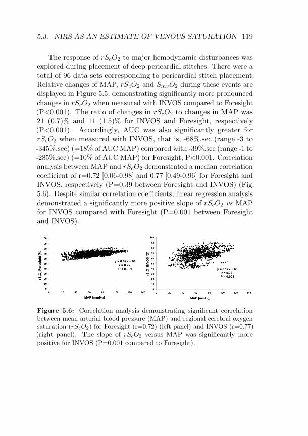
5.3. NIRS AS AN ESTIMATE OF VENOUS SATURATION 119
The response of rScO2 to major hemodynamic disturbances wasexplored during placement of deep pericardial stitches. There were atotal of 96 data sets corresponding to pericardial stitch placement.Relative changes of MAP, rScO2 and SmvO2 during these events aredisplayed in Figure 5.5, demonstrating significantly more pronouncedchanges in rScO2 when measured with INVOS compared to Foresight(P<0.001). The ratio of changes in rScO2 to changes in MAP was21 (0.7)% and 11 (1.5)% for INVOS and Foresight, respectively(P<0.001). Accordingly, AUC was also significantly greater forrScO2 when measured with INVOS, that is, -68%.sec (range -3 to-345%.sec) (=18% of AUC MAP) compared with -39%.sec (range -1 to-285%.sec) (=10% of AUC MAP) for Foresight, P<0.001. Correlationanalysis between MAP and rScO2 demonstrated a median correlationcoefficient of r=0.72 [0.06-0.98] and 0.77 [0.49-0.96] for Foresight andINVOS, respectively (P=0.39 between Foresight and INVOS) (Fig.5.6). Despite similar correlation coefficients, linear regression analysisdemonstrated a significantly more positive slope of rScO2 vs MAPfor INVOS compared with Foresight (P=0.001 between Foresightand INVOS).
Figure 5.6: Correlation analysis demonstrating significant correlationbetween mean arterial blood pressure (MAP) and regional cerebral oxygensaturation (rScO2) for Foresight (r=0.72) (left panel) and INVOS (r=0.77)(right panel). The slope of rScO2 versus MAP was significantly morepositive for INVOS (P=0.001 compared to Foresight).

120 CHAPTER 5. NIRS AS AN ESTIMATE OF SMVO2
5.3.4 Discussion
Our results indicate a substantial time delay between SmvO2 andrScO2 measurements during hemodynamic alterations, which isresponsible for a poor correlation between the individual valuesof these two variables. Comparison of data obtained with Foresightand INVOS cerebral oximetry revealed a similar correlation betweenSmvO2 and rScO2. However, the devices demonstrated significantdifferences in measured rScO2 values and in the magnitude of rScO2
changes during hemodynamic alterations.
We also evaluated mutual interference by examining the variabilityof rScO2 values when both monitors were running simultaneouslycompared with the values when each monitor was working separately.Our data demonstrated that INVOS performance was not affected bythe presence of Foresight, whereas Foresight rScO2 values exhibitedsignificant higher variability when INVOS was also switched on,indicating interference of the Foresight measurements by the beam oflight coming from the INVOS device. However, because the inducedvariability was very small and the mean Foresight rScO2 values didnot change when both devices were operating simultaneously, weconsidered the observed interference as clinically irrelevant and ourdata acquisition to be reliable to address the experimental question.
SmvO2 is often used in anesthesia and in intensive care to evaluatethe balance between tissue oxygen delivery and oxygen consump-tion. However, its obligatory need for more invasive instrumentationprecludes its use as a standard routine measurement. Since it canbe expected that measurements of brain and systemic tissue oxygenbalances vary in a similar direction, the measurement of rScO2 byNIRS could offer a promising non-invasive alternative [3]. Severalauthors have compared rScO2 and SmvO2 by focusing on interchange-ability, using SmvO2 as the standard reference [4-10]. However, rScO2
and SmvO2 measure different entities, and we should not expect thatboth methods are interchangeable. While rScO2 reflects oxygensaturation in a small region of the frontal cortex, SmvO2 repre-

5.3. NIRS AS AN ESTIMATE OF VENOUS SATURATION 121
sents a measure of venous blood oxygen saturation from all organs.This notion is supported by the observation that SmvO2 primarilyrepresents oxygen status in the lower body and poorly reflects brainoxygenation [16]. Therefore, in the present study, we did not focus oninterchangeability, but on the ability of SmvO2 and rScO2 to reflecthemodynamic changes in a similar way.
In accordance with our data, Paarmann and colleagues [4] alsodemonstrated poor correlation between SmvO2 and rScO2 duringperiods of hemodynamic instability compared to hemodynamic stableperiods. They attributed this finding to retention of CO2, leadingto an increase in rScO2, without a concomitant change in SmvO2.The importance of CO2 in the correlation analysis of SmvO2 andrScO2 has been demonstrated during moderate hypothermic car-diopulmonary bypass [5]. The present study design does not allowto comment on potential influences of CO2 on SmvO2 and rScO2.Throughout the study, EtCO2 was maintained at 4.7 kPa and one canassume that the hemodynamic disturbances occurring were too short-lasting to result in effective alterations in arterial CO2 concentrations.
We hypothesized that time delay issues might contribute to thepoor correlation between SmvO2 and rScO2. We demonstrated asubstantial time delay between changes in MAP and concomitantchanges in SmvO2 of 18 (4) seconds, while no time delay was observedbetween changes in MAP and changes in rScO2. After adjustingfor the time delay, the correlation between SmvO2 and rScO2 in-creased significantly for both Foresight and INVOS, confirming ourhypothesis.
Most monitors use a time-weighted average to compute the valuedisplayed, and each device updates and displays the value at adifferent rate, which might confound time delay analyses. In thepresent study, MAP was displayed beat-to-beat, Foresight and SmvO2
both had a data update rate of 2 s, while the INVOS monitor had theslowest data update rate (3.3 s). Therefore, the time delay observedwith SmvO2 compared with MAP and rScO2 cannot be attributed

122 CHAPTER 5. NIRS AS AN ESTIMATE OF SMVO2
to inherent device-related time delays. We speculate that the 18 sdelay in SmvO2 compared with rScO2 reflects the circulation time.rScO2 directly measures regional oxygen balance, whereas SmvO2 isa reflection of the global oxygen balance, from venous blood returningfrom the superior and inferior cavae and from the coronary sinus,and is therefore subject to time delay due to the circulation time.Of interest, the time delay between SmvO2 and rScO2 found in ourstudy perfectly accords with previously described circulation times[17]. Our data imply that rScO2 might represent a prompter morereliable variable than SmvO2 to assess the effect of changes in MAP ontissue perfusion, because it can be assumed that when hemodynamicdeterioration leads to impaired brain perfusion, other vital organswill also be compromized [18].
The range of rScO2 values is significant larger when measuredwith INVOS compared with Foresight. This can be related to the useof four narrow laser wavelengths (bandwidth <1 nm) in the Foresighttechnology compared with the use of two broader wavelengths (band-width 30-40 nm) in the INVOS technology (www.perfusion.com/cgi-bin/absolutenm/articlefiles/chen2008/chen2008.pdf), improving mea-surement accuracy [19]. However, the differences in absolute valuesmight also be due to different computational algorithms for thecalculation of rScO2 of both NIRS devices. Similarly, penetrationdepth could differ depending on the wavelength and intensity of theemitted light, the sensitivity of the light detector, and the spacingbetween the light emitter and light detectors. Variable sensitivityto extracranial tissue contamination is another possibility for theobserved differences [20].
We demonstrated an identical correlation between rScO2 andboth SmvO2 and MAP for the two NIRS devices. However, despitesimilar correlation coefficients, the slopes of rScO2 vs SmvO2 orMAP were significantly more positive for INVOS, suggesting a betterreflection of SmvO2 and MAP changes. Likewise the response ofrScO2 to acute hemodynamic alterations was more pronounced when

5.3. NIRS AS AN ESTIMATE OF VENOUS SATURATION 123
measured with INVOS.The underlying reasons for the differences between INVOS and
Foresight measurements cannot be identified from the present study.Either INVOS data show a greater variability due to less accuratemeasurement technology, or alternatively, Foresight data show lessvariability because of a more pronounced signal attenuation tech-nology, providing rScO2 values that do not as readily reflect truephysiological changes. Clearly, more studies are needed to clarifythis issue.
In summary, our data suggest that the conflicting and poor resultsdescribed previously when comparing rScO2 with SmvO2 can to someextent be attributed to a time delay between measurements. rScO2
might represent a prompter more reliable variable than SmvO2 toassess the effect of changes in MAP on tissue perfusion. We couldnot confirm that the type of cerebral oximeter also contributed to thepoor correlation as previously reported. Both technologies respondedsimilar to hemodynamic changes, although responses of rScO2 tochanges in MAP seemed to be more pronounced when measured withINVOS compared to Foresight.
5.3.5 References
1. Polonen P, Ruokonen E, Hippelainen M, Poyhonen M, Takala J. A
prospective, randomized study of goal-oriented hemodynamic therapy in
cardiac surgical patients. Anesth Analg 2000; 90: 1052-9
2. Watzman HM, Kurth CD, Montenegro LM, Rome J, Steven JM, Nicolson
SC. Arterial and venous contributions to near-infrared cerebral oximetry.
Anesthesiology 2000; 93: 947-53
3. Murkin JM, Adams SJ, Novick RJ, et al. Monitoring brain oxygen
saturation during coronary bypass surgery: a randomized, prospective study.
Anesth Analg 2007; 104: 51-8
4. Paarmann H, Heringlake M, Heinze H, et al. Non-invasive cerebral
oxygenation reflects mixed venous oxygen saturation during the varying
haemodynamic conditions in patients undergoing transapical transcatheter

124 CHAPTER 5. NIRS AS AN ESTIMATE OF SMVO2
aortic valve implantation. Interact Cardiovasc Thorac Surg 2012; 14: 268-72
5. Baraka A, Naufal M, El-Khatib M. Correlation between cerebral and
mixed venous oxygen saturation during moderate versus tepid hypothermic
hemodiluted cardiopulmonary bypass. J Cardiothorac Vasc Anesth 2006;
20: 819-25
6. Dullenkopf A, Baulig W, Weiss M, Schmid ER. Cerebral near-infrared
spectroscopy in adult patients after cardiac surgery is not useful for moni-
toring absolute values but may reflect trends in venous oxygenation under
clinical conditions. J Cardiothorac Vasc Anesth 2007; 21: 535-9
7. Tortoriello TA, Stayer SA, Mott AR, et al. A noninvasive estimation
of mixed venous oxygen saturation using near-infrared spectroscopy by
cerebral oximetry in pediatric cardiac surgery patients. Paediatr Anaesth
2005; 15: 495-503
8. Bhutta AT, Ford JW, Parker JG, et al. Noninvasive cerebral oximeter
as a surrogate for mixed venous saturation in children. Pediatr Cardiol
2007; 28: 34-41
9. Paarmann H, Schon J, Schmidt W, et al. Agreement of noninvasive
cerebral oxygenation with mixed venous oxygen saturation in patients un-
dergoing ECMO-therapy. Appl Cardiopulm Pathophysiol 2011; 15: 55-61
10. Schon J, Heringlake M, Berger K, Volker Groesdonk H, Sedemund-Adib
B, Paarmann H. Relationship between mixed venous oxygen saturation and
regional cerebral oxygenation in awake, spontaneously breathing cardiac
surgery patients. Minerva Anestesiol 2011; 77: 952-8
11. Tyree K, Tyree M, DiGeronimo R. Correlation of brain tissue oxygen
tension with cerebral near-infrared spectroscopy and mixed venous oxygen
saturation during extracorporeal membrane oxygenation. Perfusion 2009;
24: 325-31
12. Cua CL, Hoffman TM, Taeed R, et al. Cerebral saturations trend with
mixed venous saturations in patients undergoing extracorporeal life support.
Perfusion 2004; 19: 171-6
13. Hemmerling TM, Bluteau MC, Kazan R, Bracco D. Significant decrease
of cerebral oxygen saturation during single-lung ventilation measured using
absolute oximetry. Br J Anaesth 2008; 101: 870-5

5.3. NIRS AS AN ESTIMATE OF VENOUS SATURATION 125
14. Tang L, Kazan R, Taddei R, Zaouter C, Cyr S, Hemmerling TM.
Reduced cerebral oxygen saturation during thoracic surgery predicts early
postoperative cognitive dysfunction. Br J Anaesth 2012; 108: 623-9
15. Moerman A, Wouters P. Near-infrared spectroscopy (NIRS) monitoring
in contemporary anesthesia and critical care. Acta Anaesth Belg 2010; 61:
185-94
16. Redlin M, Koster A, Huebler M, et al. Regional differences in tissue
oxygenation during cardiopulmonary bypass for correction of congenital
heart disease in neonates and small infants: relevance of near-infrared
spectroscopy. J Thorac Cardiovasc Surg 2008; 136: 962-7
17. Pearce ML, Lewis AE, Kaplan MR. The factors influencing the circula-
tion time. Circulation 1952; 5: 583-8
18. McDaniel LB, Zwischenberger JB, Vertrees RA, et al. Mixed venous
oxygen saturation during cardiopulmonary bypass poorly predicts regional
venous saturation. Anesth Analg 1995; 80: 466-72
19. Fischer GW, Silvay G. Cerebral oximetry in cardiac and major vascular
surgery. HSR Proceedings in Intensive Care and Cardiovascular Anesthesia
2010; 2: 249-56
20. Davie SN, Grocott HP. Impact of extracranial contamination on re-
gional cerebral oxygen saturation: a comparison of three cerebral oximetry
technologies. Anesthesiology 2012; 116: 834-40

126 CHAPTER 5. NIRS AS AN ESTIMATE OF SMVO2

Chapter 6
NIRS to assess thephysiologicaldeterminants of tissueoxygen saturation
6.1 Background
Vasopressors are frequently used during anesthesia when bloodpressure is challenged. However, vasoconstrictors may impair regionalorgan perfusion, which will go unnoticed when relying solely on bloodpressure, until functional organ damage becomes evident. Bothpressure and flow are considered important variables for preventionof perfusion deficit and the resulting end organ failure. However, therelative importance of each is less established.
Since NIRS provides an estimation of systemic venous saturation,its use offers the opportunity to evaluate physiological determinantsof global tissue perfusion.
127
128 CHAPTER 6. PHYSIOLOGY OF PERFUSION
6.2 Objective
The aim of the present study was to determine the effect of flow andpressure on tissue oxygen saturation.
6.3 NIRS to assess physiology of perfusion
Influence of variations in systemic blood flow andpressure on cerebral and systemic oxygen saturation incardiopulmonary bypass patients. Moerman A, Denys W,De Somer F, Wouters P, De Hert S. Br J Anaesth 2013, inpress
AbstractBackground. Although both pressure and flow are consideredimportant determinants of regional organ perfusion, the relativeimportance of each is less established. The aim of the present studywas to evaluate the impact of variations in flow and/or pressure oncerebral and whole-body oxygen saturation.Methods. Thirty-four consenting patients undergoing electivecardiac surgery on cardiopulmonary bypass were included. Usinga randomized cross-over design, four different hemodynamic stateswere simulated: 1) 20% flow decrease, 2) 20% flow decrease withphenylephrine to restore baseline pressure, 3) 20% pressure decreasewith sodium nitroprusside (SNP) under baseline flow, and 4) in-creased flow with baseline pressure. The effect of these changes wasevaluated on cerebral (ScO2) and systemic (SvO2) oxygen saturation,and on systemic oxygen extraction ratio (OER). Data were assessedby within- and between-group comparisons.Results. Decrease in flow was associated with a decrease in ScO2
6.3. NIRS TO ASSESS PHYSIOLOGY OF PERFUSION 129
(from 63.5 (7.4) to 62.0 (8.5)%, P<0.001). When blood pressure wasrestored with phenylephrine during low flow, ScO2 further decreasedfrom 61.0 (9.7) to 59.2 (10.2)%, P<0.001. Increase in flow was associ-ated with an increase in ScO2 from 62.6 (7.7) to 63.6 (8.9)%, P=0.03,while decreases in pressure with the use of SNP did not affect ScO2.SvO2 was significantly lower (P<0.001) and OER was significantlyhigher (P<0.001) in the low flow arms.Conclusions. In the present elective cardiac surgery population, ScO2
and SvO2 were significantly lower with lower flow, regardless of sys-temic blood pressure. Moreover, phenylephrine administration wasassociated with a reduced cerebral and systemic oxygen saturation.
6.3.1 Introduction
Decreases in blood pressure during anesthesia are often managedby vasopressor use. However, vasoconstrictors may impair regionalorgan perfusion, which might go undetected when monitoring solelyblood pressure [1]. Cerebral oximetry, a noninvasive technologyusing near infrared spectroscopy (NIRS), enables an estimation ofsystemic venous saturation, thereby providing a means for real-timemonitoring of adequacy of organ perfusion [2,3].
In a proposed algorithm to correct for decreases in NIRS-derivedcerebral oxygen saturation (ScO2), increasing mean arterial pressure(MAP) with the use of vasopressors was suggested as one of the initialmeasures to correct for low ScO2 [4]. However, recent published datademonstrated that vasopressors such as phenylephrine may negativelyaffect ScO2 [5-9]. This negative effect on ScO2 was not observed whenincrease in blood pressure was obtained by vasopressor agents whichalso increase cardiac output, such as ephedrine [6,10]. Also, studiesin healthy subjects demonstrated an increase in ScO2 during exercise[7,11], whereas in patients not capable of increasing cardiac output,such as in patients with heart failure, the ability to augment ScO2
during exercise was limited [12]. These data suggest that cardiac

130 CHAPTER 6. PHYSIOLOGY OF PERFUSION
output might contribute to the preservation of cerebral oxygenation.The aim of the present study was to determine the impact of
variations in flow, in pressure, and in both variables at the same timeon cerebral and whole-body oxygen saturation. We hypothesizedthat not only pressure, but also flow would have a major contributionin preservation of cerebral and systemic oxygenation.
A major problem in evaluating physiologic processes is thatpressure and flow are intertwined and modifications to one alsoalter the other. Cardiopulmonary bypass (CPB) represents a uniqueclinical circumstance in which different aspects of perfusion can bemodified independently and in a controlled manner. Therefore wechose CPB as the model to test our hypothesis. To separate theeffect of flow and pressure on cerebral and systemic oxygenation, weindependently modified these parameters in patients on CPB.
6.3.2 Methods
This prospective clinical study was approved by the InstitutionalEthics Committee and written informed consent was obtained from allsubjects. The trial is registered at ClinicalTrials.gov (NCT01424800).Thirty-four adult patients scheduled for elective cardiac surgery(coronary artery bypass grafting and/or valve surgery) on moderatelyhypothermic CPB without blood transfusion were recruited. Patientswith history of cerebrovascular disease or significant carotid arterystenosis (>60%) and patients necessitating vasopressor or inotropictherapy before surgery were excluded.
On the morning of surgery, patients were allowed to take theirroutine medication, except for angiotensin-converting enzyme in-hibitors. Patients were premedicated with oral diazepam (5-10 mg).Standard monitoring was used throughout the procedure, includingECG, pulse oximetry, end-tidal oxygen, carbon dioxide and sevoflu-rane concentrations, bispectral index (BIS), invasive arterial andcentral venous pressure measurement, and temperature measure-ment (AS3; Datex, Helsinki, Finland). Arterial blood pressure was

6.3. NIRS TO ASSESS PHYSIOLOGY OF PERFUSION 131
recorded continuously via the right radial artery catheter. Twodisposable NIRS sensors were applied on each side of the foreheadfor continuous registration of ScO2 of the corresponding brain hemi-sphere (INVOS 5100; Somanetics Corporation, Troy, MI, USA). Alldata were recorded continuously and integrated digitally with theRUGLOOP (Demed, Temse, Belgium).
Anesthesia was induced with fentanyl 5 µg/kg, diazepam 0.1mg/kg, and rocuronium 1 mg/kg. The lungs were ventilated me-chanically with oxygen-enriched air (fractional inspired oxygen 0.6)adjusted to keep the end-tidal carbon dioxide (EtCO2) around 5 kPa.Anesthesia was maintained with boluses of fentanyl up to a totaldose of 25-35 µg/kg and sevoflurane at a minimum concentration of1.5 vol%.
CPB was performed with a roller pump (Stockert S5; Soringroup, Munchen, Germany) providing non-pulsatile flow. Thepriming consisted of 1200 ml colloids (Geloplasma R©, Fresenius Kabi,Schelle, Belgium), heparin 5000 IU and mannitol 0.5 g/kg. Sys-temic heparinization maintained an activated clotting time of >480seconds. Moderately hypothermic CPB (blood temperature 30 ◦C)was initiated at flow rates of 2.5 L/min/m2. During CPB PaO2
and PaCO2 were maintained around 25 kPa and 5 kPa respectively.Arterial blood gases were measured at 37 ◦C, independent of bodytemperature (alpha-stat blood gas management). Blood was sampledfrom the pump oxygenator after 3 minutes during steady state, I1and I3. Temperature, PaCO2, PaO2, haemoglobin and sevofluraneconcentrations were kept constant during the measurements.
Interventions
The study used a randomized cross-over design where the subjectsserved as their own controls. Subjects were randomly allocated, basedon computer generated codes, to start with the flow related inter-ventions or with the pressure related interventions. In all subjects,response to variations in flow, in pressure, and to the combined

132 CHAPTER 6. PHYSIOLOGY OF PERFUSION
variation of flow and pressure was investigated. With the interven-tions, a change of 20% in pressure and/or flow was aimed. Changes inblood pressure were obtained by the use of vasoactive agents, sodiumnitroprusside (SNP) for blood pressure decrease and phenylephrinefor blood pressure increase. Flow was regulated by control of thepump flow.
Baseline (BL) values of mean arterial blood pressure (MAP), flow,ScO2 and systemic oxygen saturation (SvO2) were determined atsteady state. Steady state was defined as the presence of a stable(<10% change) MAP over a period of 5 minutes on CPB. After reach-ing steady state, four different hemodynamic states were simulated:20% flow decrease (I1), 20% flow decrease with administration ofphenylephrine to restore baseline MAP (I2); then hemodynamicswere allowed to return to BL values after which SNP was admin-istered until 20% MAP decrease under baseline flow (I3), followedby restoration of baseline MAP by increasing pump flow (I4). Theorder of variations in pressure and flow was assigned randomly bythe use of a computer generated randomization code. Subjects wererandomly assigned to undergo first the flow related interventions andthen the pressure related interventions (group F), or first the pressurerelated interventions and then the flow related interventions (groupP). All changes were sustained for 5 minutes. In group F the sequenceof changes was BL, I1, I2, BL, I3, I4. In group P the sequence ofchanges was BL, I3, I4, BL, I1, I2 (Fig. 6.1). Interventions wereseparated by a time period of about 2 minutes for finalizing computerdata registration and preparation of the next intervention.
Outcome variables
To analyze the changes in ScO2, right and left ScO2 were averaged.We calculated both the change in absolute values in ScO2, as therelative change in ScO2, defined as the percentage difference betweenthe ScO2 value at the start of the intervention and the value exactly5 minutes later, at the end of the intervention. To account for both

6.3. NIRS TO ASSESS PHYSIOLOGY OF PERFUSION 133
Figure 6.1: Graphic presentation of the study design. Sequence of inter-ventions in group F (first the flow related interventions) and group P (firstthe pressure related interventions).
magnitude and duration of changes in ScO2, the area under the curve(AUC) was also calculated (expressed in %.sec).
To evaluate the effect of changes in flow and pressure on whole-body oxygen balance, SvO2 was measured, and systemic oxygendelivery (DO2) and oxygen extraction ratio (OER) were calculatedaccording to standard formulae. Arterial oxygen content: CaO2 =1.34∗Hb∗SaO2+0.003∗PaO2, where Hb = hemoglobin concentration,SaO2 = arterial oxygen saturation, PaO2 = arterial partial pressureof oxygen. DO2 = Q ∗ CaO2, where Q = pump flow. OER =

134 CHAPTER 6. PHYSIOLOGY OF PERFUSION
(SaO2 − SvO2)/SaO2, where SvO2 = venous oxygen saturation.
Statistical analysisLucas and co-workers assessed the influence of pharmacological-
induced changes in blood pressure on cerebral oxygenation, andindicated an absolute change in ScO2 of -1.8% per 10 mmHg changein MAP, with a reduction approximating 14% during the higherrange of MAP [8]. In the present protocol we aimed at a change of20% in pressure with the interventions. We therefore accepted anabsolute change in rScO2 of 5% with alterations in pressure or flowas a clinically relevant change. Based on the reported mean ScO2
of 64% with a SD of 10% [3], and accepting a two-tailed α error of0.05 and a β error of 0.8, 34 patients were calculated to be required.Statistical analysis was performed using the statistical software SPSSStatistics 20 (SPSS Inc., Chicago, IL, USA). Distribution of thedata was tested for normality using the Shapiro-Wilk test. Theassumption of normality was fulfilled and data are presented as mean(SD). Comparisons between group F and group P were made withStudent’s t-test. Variables during the different interventions werecompared using repeated measures analysis of variance (ANOVA),with Tukey correction for multiple post-hoc comparisons. For eachintervention, differences between pre- and post-intervention ScO2
values were tested using a paired data Student’s t-test. A value ofP<0.05 was taken as the level of significance.
6.3.3 Results
Nine female and 25 male subjects with an average age of 62 (14)years, weight of 80 (16) kg and height of 171 (9) cm were enrolledin the study. Demographics did not differ between the F and the Pgroup.
Temperature, sevoflurane concentrations, PaO2, PaCO2 andhemoglobin showed no differences between the different interventions,neither between the F and the P group (Table 6.1).

6.3. NIRS TO ASSESS PHYSIOLOGY OF PERFUSION 135
BL I1 I2 I3 I4
Temp (◦C)F group 30.5 (1.3) 30.7 (1.4) 31.0 (1.1) 30.7 (1.3) 30.7 (1.3)P group 30.0 (1.5) 30.1 (1.2) 30.2 (1.6) 30.3 (1.4) 30.5 (1.4)
Sevo (%)F group 1.1 (0.5) 1.2 (0.5) 1.2 (0.5) 1.2 (0.5) 1.3 (0.5)P group 1.0 (0.3) 1.4 (0.7) 1.3 (0.7) 1.1 (0.4) 1.1 (0.4)
PaO2 (kPa)F group 33 (9) 30 (5) 30 (6)P group 30 (7) 28 (7) 27 (7)
PaCO2 (kPa)F group 5.8 (0.5) 5.5 (0.6) 5.7 (0.2)P group 5.6 (0.6) 5.0 (0.4) 5.0 (0.5)
Hb (g/dl)F group 9.8 (0.9) 9.9 (1.1) 10.1 (0.9)P group 9.5 (1.4) 10.0 (1.2) 9.5 (1.3)
Table 6.1: Blood temperature, sevoflurane concentration, and blood gas valuesduring the different interventions, demonstrating no differences between the interven-tions, neither between the F group (flow related interventions were performed beforethe pressure related interventions) and the P group (pressure related interventions wereperformed before the flow related interventions). Data are presented as mean (SD). P-value between F and P group >0.05; P-value between interventions >0.05. BL: baseline,(I1) 20% flow decrease, (I2) 20% flow decrease with administration of phenylephrine torestore baseline MAP, (I3) baseline flow with administration of sodium nitroprusside un-til 20% MAP decrease, (I4) restoration of baseline MAP by increasing pump flow; PaO2:arterial oxygen partial pressure, PaCO2: arterial carbon dioxide partial pressure, Hb:hemoglobin.

136 CHAPTER 6. PHYSIOLOGY OF PERFUSION
The intended targets of changes in flow and MAP were reachedin all subjects (Table 6.2). The changes in flow and MAP were notdifferent between the F and the P group, indicating that the sequenceof interventions did not bias the data. Therefore, in order to analyzethe effect of changes in flow and pressure, we pooled the data of bothgroups.
BL I1 I2 I3 I4
Flow (L/min)F group 4.5 (0.5) 3.6 (0.4) 3.6 (0.4) 4.5 (0.5) 5.0 (0.5)P group 4.4 (0.4) 3.6 (0.3) 3.6 (0.3) 4.4 (0.4) 4.9 (0.5)
MAP (mmHg)F group 67 (5) 57 (8) 69 (8) 57 (7) 65 (8)P group 70 (9) 63 (9) 71 (9) 57 (7) 68 (8)
Table 6.2: Endpoints of changes in flow and pressure during the different interven-tions, indicating that the intended targets of changes in flow and pressure were reachedin all subjects. Data are presented as mean (SD). P-value between F and P group>0.05; P-value between interventions <0.001. BL: baseline, (I1) 20% flow decrease, (I2)20% flow decrease with administration of phenylephrine to restore baseline MAP, (I3)baseline flow with administration of sodium nitroprusside until 20% MAP decrease, (I4)restoration of baseline MAP by increasing pump flow.
DO2 was significantly lower during low flow (I1) (263 (62)ml/min/m2) compared to normal flow (I3) (320 (63) ml/min/m2),P=0.001.
The changes in MAP and flow with their concomitant effects onScO2 are illustrated in Figure 6.2. The changes in ScO2 and systemicoxygen balance parameters between the interventions are displayedin Table 6.3.

6.3. NIRS TO ASSESS PHYSIOLOGY OF PERFUSION 137
Figure 6.2: Changes in MAP and flow with their concomitant effectson ScO2 for each intervention: (I1) 20% flow decrease, (I2) 20% flowdecrease with administration of phenylephrine to restore baseline MAP, (I3)baseline flow with administration of sodium nitroprusside until 20% MAPdecrease, (I4) restoration of baseline MAP by increasing pump flow. Dataare presented as mean values. *p<0.05 between pre- and postinterventionScO2.

138 CHAPTER 6. PHYSIOLOGY OF PERFUSION
BL I1 I2 I3 I4
AUC rScO2 N.A. -522(323)* -249(424)* 145(687) 232(379)(%.sec)Rel. change N.A. -2.8(3.7)* -3.2(3.8)* 1.5(5.1) 1.6(3.3)in rScO2 (%)SvO2 (%) 86(3) 82(4)‡ 82(5)‡ 85(4) 87(4)
OER 0.14(0.03) 0.18(0.04)‡ 0.17(0.05)‡ 0.14(0.04) 0.12(0.04)
Table 6.3: Cerebral oxygen saturation and oxygen consumption parameters duringthe different interventions, demonstrating lower cerebral (ScO2) and systemic (SvO2)oxygen saturation and a higher oxygen extraction ratio (OER) during low flow (I1 andI2) compared to baseline (BL), normal flow (I3), and high flow (I4). Data are presentedas mean (SD). *Significantly different from I3 and I4; ‡ Significantly different fromBL, I3 and I4. (I1) 20% flow decrease, (I2) 20% flow decrease with administration ofphenylephrine to restore baseline MAP, (I3) baseline flow with administration of sodiumnitroprusside until 20% MAP decrease, (I4) restoration of baseline MAP by increasingpump flow. AUC ScO2: area under the curve for cerebral oxygen saturation, N.A.: notapplicable.
Responses to decrease in flow
Intervention 1 : With 20% flow decrease, MAP decreased from 65(9) to 60 (11), indicating the accompanying decrease in MAP withflow decrease. ScO2 decreased from 63.5 (7.4) to 62.0 (8.5)%, P<0.001(Fig. 6.2). Decreases in ScO2 were significantly more pronouncedcompared to interventions with normal (I3) and high flow (I4) (Table6.3). SvO2 was significantly lower and OER was significantly highercompared to baseline and compared to interventions with normaland high flow (Table 6.3).
Intervention 2 : With 20% flow decrease and administration ofphenylephrine to restore baseline MAP, MAP increased from 57 (10)to 70 (8). ScO2 decreased from 61.0 (9.7) to 59.2 (10.2)%, P<0.001(Fig. 6.2). Decreases in ScO2 were significantly more pronouncedcompared to interventions with normal (I3) and high flow (I4) (Table6.3). SvO2 was significantly lower and OER was significantly higher

6.3. NIRS TO ASSESS PHYSIOLOGY OF PERFUSION 139
compared to baseline and compared to interventions with normaland high flow (Table 6.3).
Responses to decrease in pressureIntervention 3 : With 20% MAP decrease (from 66 (12) to 57 (7)),
obtained by administration of SNP while maintaining baseline pumpflow, ScO2 did not change significantly (61.7 (8.4) to 62.6 (8.4)%,P=0.13) (Fig. 6.2). SvO2 was significantly higher and OER wassignificantly lower in conditions with low blood pressure obtained bySNP (I3) compared to low blood pressure caused by low flow (I1)(Table 6.3).
Responses to increase in flowIntervention 4 : When increasing pump flow until restoration of
baseline MAP (from 61 (11) to 67 (8)), the increase in flow was11% (from 4.5 (0.5) to 5.0 (0.5) L/min). ScO2 increased from 62.6(7.7) to 63.6 (8.9)%, P=0.03) (Fig. 6.2). ScO2 values were notdifferent between conditions with high flow and baseline pressure(I4) compared to baseline flow and low pressure by SNP (I3) (Table6.3). SvO2 was significantly higher and OER was significantly lowercompared to conditions with low flow (I1 and I2) (Table 6.3).
6.3.4 Discussion
The debate on the best strategies for prevention of perfusion deficitand the resulting end organ failure is ongoing. Both pressure andflow are considered important variables [13]. However, the relativeimportance of each is less established. Under the conditions of thepresent study, changes in flow affected cerebral and systemic venousoxygen saturations more than changes in MAP. ScO2 and SvO2 weresignificantly higher at normal to high flow than at reduced flow,regardless of systemic blood pressure.
In the elective cardiac surgery population, used in this study,maintaining flow and thus DO2 seemed to be more important than

140 CHAPTER 6. PHYSIOLOGY OF PERFUSION
maintaining pressure. This finding is in accordance with data demon-strating that organ injury can be prevented by targeting DO2 levelsabove a critical threshold during cardiopulmonary bypass [14,15].However, the obtained results do not exclude perfusion pressure asan important variable. It is important to note that even during lowpressure, our lowest value of 60 mmHg is higher than the criticalvalue of 50 mmHg as reported in other studies [16].
In the present study increasing blood pressure with phenylephrineinduced a decrease in ScO2. This is in accordance with a number ofrecently published studies [5-9]. The mechanism of this phenomenonis still unknown. Cardiac output has been proposed as the mostimportant factor in preserving ScO2 [5]. However in our study, pumpflow was kept constant during administration of phenylephrine, indi-cating that other factors than cardiac output or pump flow contributeto the decrease in ScO2. Some authors relate the decrease in ScO2
with phenylephrine to direct α-1 adrenergic receptor activation [17]or to indirect cerebral vasoconstriction via reflexively increased sym-pathetic nerve activity [18]. Others refute this mechanism by statingthat the cerebral vasculature lacks significant α- and β-adrenoceptors[19]. Recently it has been suggested that the ScO2 decrease withadministration of phenylephrine indicates a functional pressure au-toregulation mechanism [20]. The phenylephrine-induced increase inperfusion pressure provokes vasoconstriction of the cerebral arteriolesin order to prevent abrupt cerebral hyperperfusion. NIRS calculatesthe oximetry values based on an assumed cerebral arterial to venousblood volume ratio [21]. Autoregulatory vasoconstriction of the cere-bral arterioles induces a smaller arterial and relatively larger venouscontribution to the NIRS signal, causing a decrease in ScO2. Thishypothesis is supported by the study of Ogoh and colleagues whoevaluated arterial and venous cerebral blood flow, and demonstratedan elevated arterial tone and reduced cerebral venous tone duringphenylephrine administration, indicating cerebral autoregulation [9].
The ScO2 increase with SNP-induced hypotension could be readily

6.3. NIRS TO ASSESS PHYSIOLOGY OF PERFUSION 141
explained by the same mechanisms. Either nitrates reduce theresistance in the cerebral vessels, allowing more blood flow to thebrain [22], or the ScO2 increase with administration of SNP couldbe considered as a functional pressure autoregulation mechanism,provoking vasodilation of the cerebral arterioles in order to preventcerebral hypoperfusion.
It might be argued that the cerebral autoregulation-inducedaltered A:V ratio accounts for the observed changes in ScO2, withouta genuine change in cerebral oxygenation. However, the consistentand concordant changes in both ScO2 and systemic oxygen balanceparameters during the different interventions, suggest that ScO2
changes actually reflect oxygen balance changes.
The magnitude of changes in ScO2 in this study was very small(∼ 0.9-1.8% absolute change depending on the intervention) andalthough statistically significant, the clinical relevance of thesechanges in the present study may be debatable. However, our resultscannot be implicitly extrapolated to any other clinical situation. First,our measurements were done during moderate hypothermia whichreduces oxygen consumption, with consequently smaller changes inScO2. Second, we used sevoflurane for maintenance of anesthesia,which might have blunted the decrease in ScO2 with administrationof phenylephrine by its cerebral vasodilatory effect [23,24]. Based onthe same mechanism, the decrease in ScO2 with phenylephrine willbe intensified in case of hypocapnia [20]. Third, flow and pressurewere manipulated within physiological ranges for short periods oftime. In clinical practice larger changes for a longer period are often- deliberately or not - the case.
The results of the present study should be interpreted within theconstraints of the methodology. First, to explore the relative contri-bution of flow and pressure on cerebral oxygenation, and with theaim to separate the effects of both parameters, the present study wasperformed in patients on CPB where both the flow and the pressurecomponent of perfusion can be modified in a controlled manner.

142 CHAPTER 6. PHYSIOLOGY OF PERFUSION
However, as was to be expected, changes in flow were accompaniedby changes in pressure (I1 and I4) (Fig. 6.2). Secondly, based onthe principle of spatially resolved spectroscopy, NIRS devices shouldtheoretically distinguish between absorption of photons returningfrom deep rather than from superficial tissue. However, recently 2reports demonstrated that extracranial contamination significantlyinfluences the NIRS signal [25,26]. Because both vasodilators andvasoconstrictors might affect skin flow directly, changes in skin bloodflow might have influenced the NIRS measurements of cerebral oxy-genation. It has been suggested that the cerebral oximeter usedin the present study is more prone to extracranial contamination[25]. Therefore, administration of vasoactive medication might resultin more pronounced artifactual measurements compared to cere-bral oximeters with less extracranial contribution. Interestingly,we recently demonstrated that ScO2 responses to acute haemody-namic alterations were also more pronounced when measured withINVOS [3]. It is unclear whether this has to be explained by a lessaccurate measurement technology of INVOS, or whether the othercerebral oximetry devices use a more pronounced signal attenua-tion technology, resulting in more stable -but less representative-values for both intra- and extracranial measurements. Third, NIRSmeasures oxygen saturation in a superficial area of the brain directlybelow the sensors, but does not examine the deep brain. As recentlydemonstrated, though a low NIRS value predicts brain hypoperfu-sion, a normal NIRS value may not always imply that perfusion isadequate [27]. Therefore, the utility of NIRS for individualization ofperioperative pressure and blood flow management awaits testing inproperly designed and executed clinical trials [28].
In conclusion, in the elective cardiac surgery population used inthis study, cerebral saturation seemed to be more dependent on flowthan on pressure maintenance. Moreover, blood pressure increasewith phenylephrine elicited reduced cerebral and systemic oxygensaturation.

6.3. NIRS TO ASSESS PHYSIOLOGY OF PERFUSION 143
6.3.5 References
1. O’Dwyer C, Woodson LC, Conroy BP, et al. Regional perfusion
abnormalities with phenylephrine during normothermic bypass. Ann
Thorac Surg 1997; 63: 728-35
2. Murkin JM, Adams SJ, Novick RJ, et al. Monitoring brain oxygen
saturation during coronary bypass surgery: a randomized, prospective study.
Anesth Analg 2007; 104: 51-8
3. Moerman A, Vandenplas G, Bove T, Wouters P, De Hert S. Relation
between mixed venous oxygen saturation and cerebral oxygen saturation
measured by absolute and relative near-infrared spectroscopy during off-
pump coronary artery bypass grafting. Br J Anaesth 2013; 110: 258-65
4. Denault A, Deschamps A, Murkin JM. A proposed algorithm for the in-
traoperative use of cerebral near-infrared spectroscopy. Semin Cardiothorac
Vasc Anesth 2007; 11: 274-81
5. Meng L, Cannesson M, Alexander BS, et al. Effect of phenylephrine
and ephedrine bolus treatment on cerebral oxygenation in anaesthetized
patients. Br J Anaesth 2011; 107: 209-17
6. Nissen P, Brassard P, Jorgensen TB, Secher NH. Phenylephrine but not
ephedrine reduces frontal lobe oxygenation following anesthesia-induced
hypotension. Neurocrit Care 2010; 12: 17-23
7. Brassard P, Seifert T, Wissenberg M, Jensen PM, Hansen CK, Secher
NH. Phenylephrine decreases frontal lobe oxygenation at rest but not during
moderately intense exercise. J Appl Physiol 2010; 108: 1472-8
8. Lucas SJ, Tzeng YC, Galvin SD, Thomas KN, Ogoh S, Ainslie PN. In-
fluence of changes in blood pressure on cerebral perfusion and oxygenation.
Hypertension 2010; 55: 698-705
9. Ogoh S, Sato K, Fisher JP, Seifert T, Overgaard M, Secher NH. The
effect of phenylephrine on arterial and venous cerebral blood flow in healthy
subjects. Clin Physiol Funct Imaging 2011; 31: 445-51
10. Moerman A, De Hert S, Jacobs T, De Wilde L, Wouters P. Cerebral
oxygen desaturation during beach chair position. Eur J Anaesthesiol 2012;
29: 82-7
11. Secher NH, Seifert T, Van Lieshout JJ. Cerebral blood flow and
144 CHAPTER 6. PHYSIOLOGY OF PERFUSION
metabolism during exercise: implications for fatigue. J Appl Physiol 2008;
104: 306-14
12. Koike A, Itoh H, Oohara R, et al. Cerebral oxygenation during exercise
in cardiac patients. Chest 2004; 125: 182-90
13. Murphy GS, Hessel EA 2nd, Groom RC. Optimal perfusion during
cardiopulmonary bypass: an evidence-based approach. Anesth Analg 2009;
108: 1394-417
14. Ranucci M, Romitti F, Isgro G, et al. Oxygen delivery during
cardiopulmonary bypass and acute renal failure after coronary operations.
Ann Thorac Surg 2005; 80: 2213-20
15. Bastien O, Piriou V, Aouifi A, Flamens C, Evans R, Lehot JJ.
Relative importance of flow versus pressure in splanchnic perfusion during
cardiopulmonary bypass in rabbits. Anesthesiology 2000; 92: 457-64
16. Schell RM, Kern FH, Greeley WJ, et al. Cerebral blood flow and
metabolism during cardiopulmonary bypass. Anesth Analg 1993; 76: 849-
65
17. Purkayastha S, Raven PB. The functional role of the alpha-1 adrenergic
receptors in cerebral blood flow regulation. Indian J Pharmacol 2011; 43:
502-6
18. Van Lieshout JJ, Secher NH. Point:counterpoint: Sympathetic activity
does/does not influence cerebral blood flow. Point: Sympathetic activity
does influence cerebral blood flow. J Appl Physiol 2008; 105: 1364-6
19. Strandgaard S, Sigurdsson ST. Point:counterpoint: Sympathetic activ-
ity does/does not influence cerebral blood flow. Counterpoint: Sympathetic
nerve activity does not influence cerebral blood flow. J Appl Physiol 2008;
105: 1366-7
20. Meng L, Gelb AW, Alexander BS, et al. Impact of phenylephrine
administration on cerebral tissue oxygen saturation and blood volume is
modulated by carbon dioxide in anaesthetized patients. Br J Anaesth 2012;
108: 815-22
21. Watzman HM, Kurth CD, Montenegro LM, Rome J, Steven JM, Nicol-
son SC. Arterial and venous contributions to near-infrared cerebral oximetry.
Anesthesiology 2000; 93: 947-53

6.3. NIRS TO ASSESS PHYSIOLOGY OF PERFUSION 145
22. Moppett IK, Sherman RW, Wild MJ, Latter JA, Mahajan RP. Effects
of norepinephrine and glyceryl trinitrate on cerebral haemodynamics: tran-
scranial Doppler study in healthy volunteers. Br J Anaesth 2008; 100:
240-4
23. Conti A, Iacopino DG, Fodale V, Micalizzi S, Penna O, Santamaria LB.
Cerebral haemodynamic changes during propofol-remifentanil or sevoflurane
anaesthesia: transcranial Doppler study under bispectral index monitoring.
Br J Anaesth 2006; 97: 333-9
24. Reinsfelt B, Westerlind A, Ricksten S-E. The effects of sevoflurane on
cerebral blood flow autoregulation and flow-metabolism coupling during
cardiopulmonary bypass. Acta Anaesthesiol Scand 2011; 55: 118-23
25. Davie SN, Grocott HP. Impact of extracranial contamination on regional
cerebral oxygen saturation: a comparison of three cerebral oximetry tech-
nologies. Anesthesiology 2012; 116: 834-40
26. Sorensen H, Secher NH, Siebenmann C, et al. Cutaneous Vasocon-
striction Affects Near-infrared Spectroscopy Determined Cerebral Oxygen
Saturation during Administration of Norepinephrine. Anesthesiology 2012;
117: 263-70
27. Allen BS, Ko Y, Buckberg GD, Tan Z. Studies of isolated global brain
ischaemia: II. Controlled reperfusion provides complete neurologic recovery
following 30 min of warm ischaemia - the importance of perfusion pressure.
Eur J Cardiothorac Surg 2012; 41: 1147-54
28. Grocott HP. Avoid hypotension and hypoxia: an old anesthetic adage
with renewed relevance from cerebral oximetry monitoring. Can J Anaesth
2011; 58: 697-702

146 CHAPTER 6. PHYSIOLOGY OF PERFUSION

Chapter 7
General discussion
The treatment objective for any anesthetist is to provide the bestand most safe treatment with the best possible outcome. Overthe last 25-50 years the incidence of death directly attributableto anesthesia has fallen from 1 in 2500 in the mid-1950s to 1 in200 000 nowadays [1]. Several authors suggested that the incidenceof death and complications could be further reduced by deliber-ately enhancing the balance between tissue oxygen delivery andtissue oxygen consumption, thereby preventing perioperative tissueoxygenation debt (Goal Directed Therapy) [2,3]. However, currentstandard anesthesia monitoring does not give an early indication ofimpending and developing tissue oxygen debt. Improving outcomein our patients will require that anesthetists become more proactivein their monitoring techniques.
Near-infrared spectroscopy (NIRS) is a non-invasive technologythat continuously monitors regional tissue oxygenation. Originallyused for assessment of oxygen saturation of the brain, its use has nowbeen expanded to evaluation of oxygenation of tissues other than thebrain. There is also growing evidence for the larger applicability ofNIRS as an estimate of systemic venous saturation in correspondence
147

148 CHAPTER 7. GENERAL DISCUSSION
with the adequacy of the circulatory status. New and promisingadvances may further this technology to become part of our standardarmamentarium, in order to optimize patient care in daily anesthesiapractice.
The present thesis describes and discusses the feasibility of clinicalmonitoring with NIRS to counter the current shortcomings in anes-thesia monitoring. This thesis reports the potential of NIRS to assessthe oxygen balance in patient groups vulnerable to global or regionalperfusion deficits, thereby enabling optimization of hemodynamicmanagement.
Chapter 1 summarizes the different technical issues of NIRS andhighlights the clinical considerations in cerebral NIRS monitoring.Following that, it is always useful to remember that no monitoringdevice - no matter how insightful its data - will improve patientoutcome unless used with a comprehensive monitoring strategy(Fig. 7.1).
Figure 7.1
149
Applicability of NIRS to assess perioperative global andregional cerebral perfusion
Dramatic case reports of adverse neurological outcome after minorsurgery in relatively healthy patients indicate the shortcomings ofcurrent routine anesthesia monitoring. The technical difficultiesand/or invasiveness of brain monitoring preclude its use as a standardroutine monitoring in perioperative care. As NIRS offers simple,non-invasive, real-time monitoring of cerebral oxygen saturation, itsutility as an additional anesthesia monitoring tool deserves furtherinvestigation. In this thesis we investigated the use of NIRS fordetection of cerebral perfusion deficit, both global and regional, andwe assessed the physiological determinants that influence cerebraloxygen saturation.
The surgical and anesthetic communities have been recentlyalarmed by a series of dramatic neurological outcomes in patientsafter shoulder surgery in the upright position. Although the exactpathogenesis of these events was unknown, malrotation of the headand perioperative hypotension were intuitively implicated as thecausal factors. We hypothesized that cerebral hypoperfusion mightgo unnoticed when relying solely on blood pressure. Therefore weevaluated the prevalence of cerebral oxygen desaturation in 20 con-secutive adult patients undergoing shoulder surgery in the uprightposition. Routine anesthesia management and standard anesthesiamonitoring were used. The responsible anesthetist was not informedabout the purpose of the study and was blinded for the rScO2 data.The study is presented in chapter 3.3. We observed a relativedecrease in rScO2 of more than 20% in 80% of patients when thebeach chair position was adopted, underlining the need for awarenessof this problem and indicating the need for perioperative monitoringof cerebral oxygen saturation in this type of surgery. In the samestudy, we identified blood pressure and carbon dioxide concentrationas physiological determinants for cerebral desaturation.
150 CHAPTER 7. GENERAL DISCUSSION
In chapter 3.4, we demonstrate the utility of NIRS in themanagement of a patient with moyamoya disease, which is a chroniccerebrovascular occlusive disease, severely compromizing blood supplyto the brain. The use of real-time cerebral oxygenation monitoringwith NIRS identified a severe decrease in rScO2 with the initiationof cardiopulmonary bypass. By the use of NIRS, which enabled closemonitoring of all therapeutic actions taken, the decrease in rScO2
could finally be reversed by initiating pulsatile bypass flow.
During repair of aortic coarctation through left thoracotomy,cross-clamping of the left carotid artery is often indicated. Thismight result in temporary hypoperfusion of the left brain. Sinceit has been reported that sodium nitroprusside (a blood pressureregulating agent) has an adverse effect on left-sided rScO2 duringaortic coarctation repair, we aimed to compare the effect of threedifferent blood pressure regulating agents on left-sided rScO2. Thisstudy is presented in chapter 4.3. We observed a variable responseof the left-sided rScO2, ranging from a decrease to an increase inrScO2, independent of the treatment group. Therefore, we concludethat the decrease in left-sided rScO2 is not the result of an agentspecific effect, but most probably reflects a deficient circle of Willis.In this study, we also observed impairment of cerebral autoregulationwith sodium nitroprusside and sevoflurane, whereas autoregulatorycapacity was maintained with nitroglycerin.
With the exception of the patient with moyamoya disease (chapter3.4), who suffered from prolonged somnolence and a transient paresisof the left arm, none of our patients developed gross neurological orcognitive dysfunction postoperatively. In this regard, it is importantto note that it is a time-dependent exposure to a certain degreeof cerebral desaturation (a viability-time threshold) that eventuallyleads to irreversible tissue damage and subsequent functional impair-ment [4,5]. Therefore, the term ‘desaturation score’ was introduced

151
[5], which represents an integrated oxygen saturation, or an areaunder the curve (AUC). This value is calculated by the product oflength of time and depth of rScO2 below a specific threshold, andis expressed in %.sec. To account for both depth and duration ofchange in tissue oxygen saturation, we used the AUC value in manyof the studies presented in this thesis.
For the applicability of NIRS for assessment of perioperativecerebral perfusion deficit, we conclude that monitoring of cerebraloxygen saturation with NIRS allows to identify global and regionalcerebral hypoperfusion, enabling real-time assessment of the effectsof critical events and therapeutic actions. We demonstrated thatchanges in cerebral oxygenation are related to blood pressure andpulsatility, and to changes in CO2, and that cerebral autoregulationmight be impaired by the use of vasodilating drugs.
Applicability of NIRS to assess perioperative tissueperfusion
Initial research focused on the use of NIRS as a monitor of cerebraloxygen saturation, however interest extended soon to applicationsin tissues other than the brain. For a number of applications thereis little evidence yet that the device truly interrogates the organ ofinterest, nor that changes in the oxygen status of non-vital organshave to be intervened upon. Future work is needed to identifywhich applications, and which changes in oxygen status in theseapplications, are of benefit in clinical practice.
In chapter 4.3, we evaluate the effect of different blood pressureregulating agents on renal and muscle oxygen saturation in childrenundergoing aortic coarctation repair. As expected, renal and muscleoxygen saturations showed a rapid and significant drop after aorticcross-clamping, which recovered promptly after release of the aortic

152 CHAPTER 7. GENERAL DISCUSSION
cross-clamp. Interestingly, in this clinical study, we demonstratedthat decreases in renal and muscle oxygen saturation were larger andhad a faster rate of decay in patients treated with sodium nitroprus-side (SNP) compared to nitroglycerin (NTG), which confirms theresults of a previous animal study. Our data suggest that NTG mightbe preferable for blood pressure control during surgical proceduresinvolving aortic cross-clamping.
During thoracoabdominal aortic aneurysm repair, spinal cordperfusion is severly jeopardized. Close monitoring is mandatory todetect spinal cord ischemia in time to treat it. However, due totechnical difficulties and inherent limitations, central nervous systemmonitoring is not widely used in these cases. In chapter 4.4, wepresent preliminary data suggesting that NIRS monitoring at the levelof the spinal cord may identify spinal cord ischemia, and may functionas a valuable perioperative non-invasive guide to the management ofblood pressure in patients undergoing thoracoabdominal aneurysmrepair.
Investigation of the effect of aortic cross-clamping on renal, muscle,and spinal oxygen saturation, demonstrated that NIRS provides real-time assessment of regional tissue perfusion. Its use might optimizeperioperative blood pressure management in surgical proceduresinvolving aortic cross-clamping.
Applicability of NIRS to assess the physiological deter-minants of tissue oxygen saturation
A growing understanding of the physiological principles of NIRSled to an appreciation of its applicability as a monitor of globaloxygen balance, providing a non-invasive estimate of systemic oxygenbalance.

153
In chapter 5, we examine the equivalence and interchangeabilityof mixed venous and cerebral oxygen saturation. The studydemonstrates that mixed venous oxygen saturation has a substantialtime delay between changes in blood pressure and concomitantoxygen saturation changes, whereas cerebral oxygen saturationreacts promptly, without time delay, indicating that cerebral oxygensaturation might represent a prompter more reliable variable thanmixed venous oxygen saturation to assess the effects of changes inblood pressure on tissue perfusion.
Considering the important time delay between SmvO2 and rScO2,correlation analysis is probably not the right test to evaluate theequivalence of these 2 parameters. Though correlation is the sim-plest way to compute the relation between two signals, delayeddynamics between the signals generate smaller correlation coefficients,as demonstrated in our study. Coherence, as correlation, is a measureof strength of a linear relation, but was developed for the analysis oftime-invariant systems [6]. In future studies comparing SmvO2 andrScO2, this time delay has to be accounted for when choosing thestatistical method.
The data in this thesis demonstrate that tissue oxygen satura-tion measured with NIRS provides a prompt responsive monitor ofchanges in perfusion. However, in vivo changes in pressure are accom-panied by changes in flow, and therefore it remained undetermined ifmaintaining pressure and/or flow was the best strategy for preventionof perfusion deficit and the resulting end organ failure. We thereforeexplored the impact of flow and pressure on cerebral and whole bodyoxygen saturation during cardiopulmonary bypass, where flow andpressure can be modified independently and in a controlled manner(chapter 6). In the elective cardiac surgery population used in thatstudy, maintaining flow seemed to be more important than maintain-ing pressure. Moreover, increasing blood pressure with phenylephrineelicited reduced cerebral and systemic oxygen saturation. It is im-portant to note that, throughout all our studies, our lowest mean

154 CHAPTER 7. GENERAL DISCUSSION
blood pressure values were consistently higher than the reportedcritical value of 50 mmHg, and therefore, the obtained results do notexclude perfusion pressure as an important variable. Nevertheless,our results are in accordance with data demonstrating that organinjury can be prevented by targeting oxygen delivery levels above acritical threshold.
Nearly one century ago, Jarisch stated “It was fatal for thedevelopment of our understanding of circulation, that blood flowis relatively difficult to measure, whereas blood pressure is easilymeasured. This is the reason why the blood pressure meter hasgained such a fascinating influence, although most organs do notneed pressure, but blood flow.” (A. Jarisch, “Kreislauffragen” 1928).Our results fully support this statement.
Applicability of NIRS to assess cerebral autoregulation
In chapter 3.3, chapter 4.3 and chapter 6.3, NIRS monitoringwas mentioned in the context of cerebral autoregulation.
In the current standard of care, a systemic MAP of 50-60 mmHg iswidely considered to guarantee adequate cerebral perfusion, becauseof cerebral autoregulation maintaining a constant cerebral blood flow(CBF) in the face of changing cerebral perfusion pressure. However,this practice fails to consider that cerebral autoregulation is derivedbased on limited data that have been seriously questioned, has wideinterindividual variation, and may be altered in specific diseases(e.g. hypertension, diabetes, stroke) and in specific conditions (e.g.changes in CO2, pharmacologic interference) [7]. Studies identifyingthe lower limit of cerebral autoregulation (LLA) in healthy, normoten-sive, nonanesthetized adults demonstrated actual LLA values of 70mmHg or higher [7]. Agents with cerebral vasodilating properties,including anesthetic agents, lower the effective LLA [8,9]. Althoughthis might increase ‘CBF reserve’, it remains unreasonable to accept

155
an empirically chosen LLA for every anesthetized patient in everysituation. So, if one rejects the validity of a fixed LLA to guideclinical management, do we have any alternatives?
Recently, the use of NIRS to monitor cerebral autoregulation hasbecome an area of intense investigation. Cerebral autoregulation ismeasured by quantifying the consequence of changing blood pressureon CBF. It has been demonstrated in laboratory and clinical studiesthat NIRS signals provide an acceptable surrogate for monitoringchanges in CBF for an index of autoregulation [10,11]. Real-timeautoregulation monitoring can be accomplished by the continuouscalculation of the correlation coefficient between MAP and rScO2,generating a novel index of autoregulatory vasoreactivity, the cerebraloximetry index (COx). Blood pressure in the autoregulation range isindicated by a COx that approaches zero, while a COx approaching1 indicates MAP below the LLA or disturbed autoregulation [12-14].The COx has been validated and had good agreement with trans-cranial Doppler derived measurements of pressure autoregulation inpiglets [10] and in adult patients [15-17]. However, analysis of COxrequires complex signal processing and specialized software, limitingits applications mostly to research. Recently, an investigational pro-totype NIRS monitor was validated for automated and continuousmonitoring of cerebral autoregulation [18]. The availability of sucha device offers the opportunity of widespread autoregulation moni-toring, providing effective means for individualizing blood pressuremanagement.
In line with the previously mentioned studies, we evaluatedthe concordance between MAP and rScO2 using simple correlationanalysis. However, cerebral autoregulation is a complex physiologicalsystem, and correlation analysis does not cope with the complexinterplay and the time-varying aspects of the different physiologicalmechanisms [19]. Coherence and transfer function analyses havealso been used to quantify cerebral autoregulation [20,21]. Caicedoanalyzed 4 different measurement models used for cerebral autoregu-

156 CHAPTER 7. GENERAL DISCUSSION
lation assessment (correlation, coherence, modified coherence, andtransfer function) and proposed transfer function gain as the mostrobust method when used for cerebral autoregulation studies [6].Correlation was considered as a robust method, however with sometime delay-related restrictions.
Conclusion. NIRS: When and why should we measure?In an era where clinical outcome is increasingly determined by
optimizing specific target organ function, there is a constant needfor accurate and specific monitoring equipment during the criticalperioperative period. Based on the direct relationship between cere-bral oxygenation and neurological outcome, NIRS has been usedto detect deficits in cerebral perfusion during complex cardiac andcerebrovascular procedures. NIRS also demonstrated its utility forthe detection of tissue hypoperfusion in other surgical areas, wherespecific target organ oxygenation still remains poorly monitored inclinical anesthesia. More importantly, there is growing evidence forthe larger applicability of NIRS as an estimate of systemic venous sat-uration in correspondence with the adequacy of the circulatory status.The information provided by this technology could significantly alterour management of physiologic perturbations during anesthesia. Dueto the advantage of simple, continuous and non-invasive monitoring,NIRS has the potential to become a valuable tool to optimize patientcare in our daily anesthesia practice.

157
References1. Lagasse RS. Anesthesia safety: model or myth? A review of the pub-lished literature and analysis of current original data. Anesthesiology 2002;97: 1609–172. Rivers E, Nguyen B, Havstad S, Ressler J, Muzzin A, Knoblich B,Peterson E, and Tomlanovich M, for the Early Goal-Directed TherapyCollaborative Group. Early Goal-Directed Therapy in the Treatment ofSevere Sepsis and Septic Shock. N Engl J Med 2001; 345: 1368-773. Pearse R, Dawson D, Fawcett J, Rhodes A, Grounds RM, Bennett ED.Early goal-directed therapy after major surgery reduces complications andduration of hospital stay. A randomised, controlled trial. Critical Care2005; 9: R687-R6934. Kurth CD, McCann JC, Wu J, Miles L, Loepke AW. Cerebral oxygensaturation-time threshold for hypoxic-ischemic injury in piglets. AnesthAnalg 2009; 108: 1268-775. Slater JP, Guarino T, Stack J, et al. Cerebral oxygen desaturationpredicts cognitive decline and longer hospital stay after cardiac surgery.Ann Thorac Surg 2009; 87: 36-446. Caicedo A, Naulaers G, Lemmers P, van Bel F, Wolf M, Van HuffelS. Detection of cerebral autoregulation by near-infrared spectroscopy inneonates: performance analysis of measurement methods. J Biomed Opt2012; 17: 117003 1-97. Drummond JC. The lower limit of autoregulation: time to revise ourthinking? Anesthesiology 1997; 86: 1431-38. Stange K, Lagerkranser M, Sollevi A. Nitroprusside-induced hypotensionand cerebrovascular autoregulation in the anesthetized pig. Anesth Analg1991; 73: 745-529. Conti A, Iacopino DG, Fodale V, Micalizzi S, Penna O, Santamaria LB.Cerebral haemodynamic changes during propofol-remifentanil or sevofluraneanaesthesia: transcranial Doppler study under bispectral index monitoring.Br J Anaesth 2006; 97: 333-910. Brady KM, Lee JK, Kibler KK, Smielewski P, Czosnyka M, EasleyRB, Koehler RC, Shaffner DH. Continuous time-domain analysis of cere-brovascular autoregulation using near-infrared spectroscopy. Stroke 2007;38: 2818-2511. Zweifel C, Castellani G, Czosnyka M, Carrera E, Brady KM, Kirk-patrick PJ, Pickard JD, Smielewski P. Continuous assessment of cerebralautoregulation with near-infrared spectroscopy in adults after subarachnoid

158 CHAPTER 7. GENERAL DISCUSSION
hemorrhage. Stroke 2010; 41: 1963-812. Joshi B, Ono M, Brown C, Brady K, Easley RB, Yenokyan G, Gottes-man RF, Hogue CW. Predicting the limits of cerebral autoregulation duringcardiopulmonary bypass. Anesth Analg 2012; 114: 503-1013. Gilmore MM, Stone BS, Shepard JA, Czosnyka M, Easley RB, BradyKM. Relationship between cerebrovascular dysautoregulation and arterialblood pressure in the premature infant. J Perinatol 2011; 31: 722-914. Brady KM, Mytar JO, Lee JK, Cameron DE, Vricella LA, ThompsonWR, Hogue CW, Easley RB. Monitoring cerebral blood flow pressure au-toregulation in pediatric patients during cardiac surgery. Stroke 2010; 41:1957-6215. Steiner LA, Pfister D, Strebel SP, Radolovich D, Smielewski P, CzosnykaM. Near-infrared spectroscopy can monitor dynamic cerebral autoregulationin adults. Neurocrit Care 2009; 10: 122-816. Brady K, Joshi B, Zweifel C, Smielewski P, Czosnyka M, EasleyRB, Hogue CW. Real-time continuous monitoring of cerebral blood flowautoregulation using near-infrared spectroscopy in patients undergoingcardiopulmonary bypass. Stroke 2010; 41: 1951-617. Zheng Y, Villamayor AJ, Merritt W, Pustavoitau A, Latif A, Bhamb-hani R, Frank S, Gurakar A, Singer A, Cameron A, Stevens RD, HogueCW. Continuous cerebral blood flow autoregulation monitoring in patientsundergoing liver transplantation. Neurocrit Care 2012; 17: 77-8418. Ono M, Zheng Y, Joshi B, Sigl JC, Hogue CW. Technical communi-cation: validation of a stand-alone near-infrared spectroscopy system formonitoring cerebral autoregulation during cardiac surgery. Anesth Analg2013; 116: 198-20419. Panerai RB. Cerebral autoregulation: from models to clinical applica-tions. Cardiovasc Eng 2008; 8: 42-5920. Wong FY, Leung TS, Austin T, Wilkinson M, Meek JH, Wyatt JS,Walker AM. Impaired autoregulation in preterm infants identified by usingspatially resolved spectroscopy. Pediatrics 2008; 121: e604-1121. Caicedo A, De Smet D, Naulaers G, Ameye L, Vanderhaegen J, LemmersP, Van Bel F, Van Huffel S. Cerebral tissue oxygenation and regional oxygensaturation can be used to study cerebral autoregulation in prematurelyborn infants. Pediatr Res 2011; 69: 548-53

Chapter 8
Future perspectives
Near-infrared spectroscopy was introduced in our cardiac surgerydepartment in 2006, initially for cerebral monitoring during pediatriccardiac surgery and aortic arch surgery. However, growing clinicalexperience, together with cases where unexpected events weredetected by NIRS, soon led us to adopt NIRS as routine monitor inevery cardiac surgery case. In spite of my extensive clinical experiencewith NIRS, and in spite of this thesis, many questions about theapplicability of NIRS to optimize patient care remain.
Today, I am eager to explore two topics relating to NIRS. Onetopic has arisen from the research for my thesis, the other oneremained in the background in favour of my thesis project. Now thatmy thesis has finished, I am glad that time will come available toexplore these two topics in depth.
First, I would like to investigate more in detail the usefulness ofNIRS to monitor spinal cord perfusion. There is a clear need forimproved spinal cord monitoring during thoracoabdominal aorticsurgery. Currently, evoked potential monitoring is primarily usedto monitor spinal cord perfusion. However, due to its technicalcomplexity and constraints on anesthetic management, it is not widely
159

160 CHAPTER 8. FUTURE PERSPECTIVES
used during thoracoabdominal aortic surgery. Our preliminary datawith NIRS (presented in chapter 4.4) were very promising, howeverfurther investigations are required before a final conclusion can bedrawn. My aim is to compare NIRS with evoked potential monitoring,and to evaluate whether measuring and intervening on the valuesobtained by NIRS makes a difference in the neurologic outcomeof patients who underwent repair of extensive thoracoabdominalaneurysms.
Second, as endothelial dysfunction is the earliest detectable stageof atherogenesis, non-invasive endothelial function testing for thepurpose of cardiovascular risk stratification is very appealing. Oneaspect of endothelial physiology is vasomotor function. The abilityof the endothelium to vasodilate in response to pharmacological andphysiological stimuli has been the most widely used clinical end pointfor assessment of endothelial function [1].
Near-infrared Spectroscopy (NIRS) has been introduced as a wayto assess non-invasively microvascular reactivity in several diseasestates [2]. It enables to quantify endothelium-mediated changes invascular tone, elicited by creating postocclusive reactive hyperemia(PORH). PORH is a reproducible transient increase in blood flowafter release of an arterial occlusion. When the cuff is released, thesurge of blood flow causes an endothelium-dependent Flow MediatedDilatation (FMD) (Fig. 8.1). The speed of flow and the recoveryare mostly determined by the capacity of the microvasculature torecruit arterioles and capillaries, reflecting the integrity of the mi-crocirculation. NIRS proved to provide excellent reproducibility [3],and demonstrated a coherent directional change with simultaneouslyperformed strain gauge plethysmography and radionuclide plethys-mography [4]. Parameters of PORH measured by NIRS were able todistinguish between healthy volunteers and patients with vasculardisorder [5].

161
Figure 8.1: Parameters of postocclusive reactive hyperemia evaluated fromNIRS and TcpO2 signals. HbO2: oxyhemoglobin, TcpO2: transcutaneousoxygen partial pressure, VO2: oxygen consumption, tR: time of recovery,tM: time to peak value, ∆Chip: maximal change of the NIRS signal, HR:maximal hyperemic response, tH: half time of the response. Ann BiomedEng 2001; 29: 311-20
We hypothesize that by means of NIRS we could introduce aneasy-to-use, user-independent method to estimate the cardiovascularrisk profile of patients and, consequently, that we might predict whichpatients have a higher risk for compromized perfusion during thecritical perioperative period.
The purpose of future studies is to determine the applicability ofNIRS for quantification of PORH. In order to fulfill this task we aimto: (1) determine the parameters of PORH measured by NIRS andexamine the reproducibility; (2) compare these results with the digitalpulse amplitude responses measured with the EndoPAT 2000 device(Itamar Medical, Caesarea, Israel), which is widely recognized as thestandard method for non-invasive endothelial function assessment.

162 CHAPTER 8. FUTURE PERSPECTIVES
References
1. Deanfield JE, Halcox JP, Rabelink TJ. Endothelial function and dys-
function: testing and clinical relevance. Circulation 2007; 115: 1285-95
2. Nanas S, Gerovasili V, Renieris P. Non-invasive assessment of the micro-
circulation in critically ill patients. Anaesth Intensive Care 2009; 37: 733-9
3. Lacroix S, Gayda M, Gremeaux V, Juneau M, Tardif JC, Nigam A.
Reproducibility of near-infrared spectroscopy parameters measured during
brachial artery occlusion and reactive hyperemia in healthy men. J Biomed
Opt 2012 Jul; 17(7). doi:10.1117/1.JBO.17.7.077010
4.Harel F, Denault A, Ngo Q, Dupuis J, Khairy P. Near-infrared spec-
troscopy to monitor peripheral blood flow perfusion. J Clin Monit Comput
2008; 22: 37-43
5.Kragelj R, Jarm T, Erjavec T, Presern-Strukelj M, Miklavcic D.
Parameters of postocclusive reactive hyperemia measured by near infrared
spectroscopy in patients with peripheral vascular disease and in healthy
volunteers. Ann Biomed Eng 2001; 29: 311-20

Summary
The primary goal in the hemodynamic management of patientsundergoing surgery is to preserve oxygen delivery at a level sufficientto cover all metabolic needs. Although it has been increasinglydemonstrated that clinical outcome is determined by optimizing tissueoxygenation, specific target organ oxygen saturation still remainspoorly monitored in clinical anesthesia.
Within the last decades, near-infrared spectroscopy (NIRS)is introduced with the potential to continuously assess tissueoxygenation. NIRS is a noninvasive device that measures theabsorption of near-infrared light by tissue chromophores, a techniquesimilar to pulse oximetry. Specific wavelengths of near-infrared lightare emitted through the tissues lying below a sensor. Depending onthe amount of light reflected back to light detectors built into thesame sensor, regional oxygen saturation of the underlying tissue canbe determined.
In the present work, NIRS was used to detect perioperativecompromised perfusion, in order to explore the physiologicaldeterminants of cerebral and peripheral tissue oxygen saturation indifferent patient populations. The ultimate goal is the optimizationof patient management in daily anesthesia practice.
The brain, arguably our most important of organs and despitebeing an obvious site of anesthetic action, ironically is inconsistentlythe focus of targeted monitoring during anesthesia. Moreover,
163

164 SUMMARY
several clinical conditions routinely encountered in our daily medicalpractice have the potential to disrupt the brain oxygen balanceexposing patients to the risk of intraoperative cerebral ischemia. Inthis thesis, we demonstrated that monitoring of cerebral oxygensaturation with NIRS allowed to identify global and regional cerebralhypoperfusion, enabling real-time assessment of the effects of criticalevents and therapeutic actions. Changes in cerebral oxygenationwere demonstrated to be related to blood pressure and pulsatility,and to changes in CO2. We also indicated that the use of vasodilatingdrugs might impair cerebral autoregulation.
The use of NIRS for monitoring regional oxygen saturation atsomatic sites has been advocated as an early warning system forregional hypoperfusion. However, it remains to be identified whichchanges in oxygen status of non-vital organs have to be intervenedupon. The present work demonstrated that NIRS provided real-timeassessment of renal, muscle, and spinal oxygen saturation duringaortic cross-clamping. Changes in tissue oxygen saturation werepressure dependent, and seemed to be negatively affected by theconcomitant use of sodium nitroprusside.
The data in the present thesis demonstrate that tissue oxygensaturation measured with NIRS provides a prompt responsivemonitor of changes in perfusion. However, in vivo changes in pressureare accompanied by changes in flow, and therefore it remainedundetermined if maintaining pressure and/or flow was the beststrategy for prevention of perfusion deficit. We therefore exploredthe impact of flow and pressure on cerebral and whole body oxygensaturation during cardiopulmonary bypass, where flow and pressurecan be modified independently and in a controlled manner. Inthis elective study population, maintaining flow seemed to be moreimportant than maintaining pressure. It is important to note that,throughout all our studies, the lowest mean blood pressure valueswere consistently higher than the reported critical value of 50 mmHg,and therefore, the obtained results do not exclude perfusion pressure

165
as an important variable. Nevertheless, our results are in accordancewith data demonstrating that organ injury can be prevented bytargeting oxygen delivery levels above a critical threshold.
Conclusion. In this thesis, NIRS has demonstrated its utility forassessing specific target organ perfusion. The information providedby this technology could significantly alter our management ofphysiologic perturbations during anesthesia. Due to the advantageof simple, continuous and non-invasive monitoring, NIRS has thepotential to become a valuable tool to optimize patient care in ourdaily anesthesia practice.

166 SUMMARY
Samenvatting
Het doel van het hemodynamisch beleid tijdens anesthesie is omde weefsels van voldoende zuurstof te voorzien. Het is bewezendat de klinische uitkomst beter is wanneer de weefseloxygenatiegeoptimaliseerd wordt, maar omwille van een aantal praktischeproblemen, wordt de zuurstofverzadiging van specifieke organen totop heden niet routinematig gemonitord tijdens anesthesie.
Nabije-infrarood spectroscopie (NIRS) is een niet-invasievetechniek die de mogelijkheid biedt om continu de weefseloxygenatiete meten. Specifieke golflengtes van nabije-infrarood licht wordendoor weefsel gezonden. Afhankelijk van de mate waarin de stralinggeabsorbeerd wordt, kan de zuurstofsaturatie in het weefsel bepaaldworden.
De doelstelling van deze thesis was om door middel van NIRSoxygenatieproblemen ter hoogte van de hersenen en de perifereweefsels te detecteren, en de fysiologische determinanten van deweefselsaturatie te bepalen. Het uiteindelijk doel is de optimalisatievan het hemodynamisch beleid in de dagelijkse anesthesie praktijk.
Meerdere factoren tijdens anesthesie kunnen de zuurstofbalans inde hersenen verstoren, met mogelijks cerebrale letsels tot gevolg.Desondanks worden de hersenen momenteel niet routinematiggemonitord. In deze thesis toonden we aan dat NIRS monitoringzowel globale als regionale cerebrale hypoperfusie kan detecteren.Veranderingen in cerebrale oxygenatie waren gerelateerd aan bloed-
167

168 SAMENVATTING
druk en pulsatiliteit, en aan CO2 veranderingen. We toonden ookaan dat bloedvatverwijdende medicatie de cerebrale autoregulatiekan verstoren.
Tijdens operaties waarbij een aortaklem geplaatst werd, toon-den we aan dat NIRS onmiddellijk veranderingen in de nier-,spier-, en ruggenmergsaturatie detecteerde. De veranderingen in deweefselsaturaties waren drukafhankelijk, en werden negatief beınvloeddoor het gebruik van sodium nitroprusside.
De gegevens in dit proefschrift toonden aan dat de metNIRS gemeten weefseloxygenatie een snelle weergave geeft vandoorbloedingsveranderingen. Echter in vivo gaan bloeddrukveran-deringen steeds gepaard met flowveranderingen, en daarom blijft devraag of druk dan wel flow de beste strategie is om perfusietekortte voorkomen. Daarom onderzochten we het effect van druk enflow op de cerebrale en globale zuurstofsaturatie tijdens cardiopul-monale bypass, waarbij flow en druk gecontroleerd en onafhankelijkvan elkaar kunnen veranderd worden. Uit deze studie bleek datflow belangrijker is dan druk voor het behoud van cerebrale en sys-teem oxygenatie. Het is evenwel belangrijk op te merken dat, in alonze studies, de laagste gemiddelde bloeddrukwaarden steeds hogerwaren dan de algemeen aanvaarde kritische waarde van 50 mmHg,en dat derhalve de verkregen resultaten perfusiedruk niet uitsluitenals een belangrijke variabele. Desalniettemin zijn onze resultatenin overeenstemming met gegevens die aantonen dat orgaanschadekan voorkomen worden door de zuurstofaanvoer boven een bepaaldekritische drempel te houden.
Besluit. In dit proefschrift toonden we aan dat NIRS snel enbetrouwbaar weefseloxygenatieproblemen detecteerde. Door eenbeter begrip van de fysiologische veranderingen tijdens anesthesiekan deze techniek het anesthetisch beleid optimaliseren.

List of abbreviations
AUC area under the curve
BIS bispectral index
CBF cerebral blood flow
CEA carotid endarterectomy
CO2 carbon dioxide
CPB cardiopulmonary bypass
CPP cerebral perfusion pressure
CVP central venous pressure
CX aortic cross-clamping
DO2 systemic oxygen delivery
EtCO2 end-tidal concentration of carbon dioxide
ETsevo end-tidal concentration of sevoflurane
FiO2 fractional inspired oxygen
Hb hemoglobin
HHb deoxygenated hemoglobin
Hct hematocrit
169

170 LIST OF ABBREVIATIONS
HR heart rate
ICP intracranial pressure
LLA lower limit of autoregulation
MAP mean arterial blood pressure
MMD moyamoya disease
NTG nitroglycerin
OER oxygen extraction ratio
O2Hb oxygenated hemoglobin
OPCAB off-pump coronary artery bypass
PaCO2 arterial carbon dioxide partial pressure
PaO2 arterial oxygen partial pressure
PtO2 tissue oxygen tension
PvCO2 venous carbon dioxide partial pressure
PvO2 venous oxygen partial pressure
Q pump flow
rScO2 regional cerebral oxygen saturation
SaO2 arterial oxygen saturation
SAP systolic arterial blood pressure
ScO2 cerebral oxygen saturation
Sevo sevoflurane
SjvO2 jugular vein oxygen saturation
SmO2 muscle oxygen saturation

171
SmvO2 mixed venous oxygen saturation
SNP sodium nitroprusside
SpO2 pulse oximetry
SsO2 spinal oxygen saturation
SR Spatial Resolution
SrO2 renal oxygen saturation
SRS Spatially Resolved Spectroscopy
SvO2 venous oxygen saturation
TAAA thoracoabdominal aortic aneurysm
THb total hemoglobin

172 LIST OF ABBREVIATIONS

Dankwoord
A closed mind is a good thing to lose. . . Dit proefschrift opendevoor mij een stukje van de “black box” van het menselijk lichaam.Maar ik leerde niet enkel bij over de fysiologische veranderingentijdens het perioperatieve gebeuren. Ik leerde evenzeer dat een thesisniet de verdienste is van een enkele persoon, maar een proces, eenproject, waarin velen zowel rechtstreeks als onrechtstreeks bijdragen.Daarom een oprecht dankwoord voor ieder die heeft bijgedragen totde verwezenlijking van deze thesis.
Als allereerste wil ik de mensen danken waarmee alles begonnenis, mijn ouders. Zij hebben mij alle vrijheid en kansen gegeven, endaar ben ik hun zeer dankbaar voor. Zij gaven mij ook een aantalwaarden mee, die ervoor gezorgd hebben dat ik hier nu sta. Voor dewarme familie die ze gecreeerd hebben, en voor de manier waarop zemij opgevoed hebben en steeds nabij zijn geweest, heb ik veel respect.Bedankt pa en ma!
Vervolgens wil ik diegene danken die mij besmet heeft met demicrobe voor anesthesie, ook al is hij er zich zelf niet van bewust.Lang geleden, op mijn allereerste stage heelkunde in het ziekenhuisin Wetteren, was ik danig onder de indruk van de sereniteit en derust die van de anesthesist uitging. Voor mij was gans het gebeurenrond anesthesie pure “magie”, en daar in Wetteren is mijn interesse
173

174 DANKWOORD
in anesthesie gewekt. Dus bedankt, Dr. Donadoni!
Ik dank mijn toenmalig diensthoofd, Prof Dr. G. Rolly, die mij demogelijkheid heeft geboden om mijn opleiding anesthesie te starten,en mij 5 jaar later steunde in mijn droom om mij verder te bekwa-men in de cardio-anesthesie. Ik ben zeer veel verschuldigd aan mijntoenmalige collega’s cardio-anesthesie, Yvon Deryck, Luc Foubert,Luc Herregods en Jan Poelaert, die mij als compleet groentje nietalleen de “grote” knepen van het vak geleerd hebben, maar ook hungebetenheid en hun enthousiasme voor de nuances van de cardio-vasculaire fysiologie met mij deelden. Nu nog, als het oplijnen vaneen neonaat probleemloos verloopt, denk ik met plezier terug aan degouden raad van Yvon, die ik hier om redenen van censuur, jammergenoeg niet kan/durf neerschrijven.
Uiteraard zou dit proefschrift er niet gekomen zijn zonder denodige wetenschappelijke begeleiding. Eerst en vooral wil ik mijnpromotor en diensthoofd, Prof. Dr. Patrick Wouters, danken voor depraktische en professionele ondersteuning, voor de kritische reflectiesen verbeteringen (de cardiovasculaire fysiologie heeft voor hem echtgeen geheimen), en last but not least om mij de mogelijkheid te gevenvrijgesteld te worden van het klinisch werk zodat ik ongestoord aanmijn project kon werken.Ik dank mijn beide co-promotoren, Prof. Dr. Stefan De Hert enProf. Dr. Chris Verborgh, voor de wetenschappelijke begeleiding.In het bijzonder wil ik Prof. De Hert bedanken om mij letterlijk opte leiden in de wetenschap. Hij heeft mij systematisch leren werkenen schrijven, met een scherp afgelijnde vraagstelling, een sluitendemethodiek, heldere resultaten, en (dixit) “de discussie komt danvanzelf”. Sinds zijn komst op onze dienst heeft mijn project een boostgekregen, niet alleen dankzij zijn ruime wetenschappelijke ervaringen zijn sterk analytische geest, maar ook omdat hij ondanks zijneigen drukke bezigheden, er toch steeds in slaagde om mijn werk

175
voor te nemen, en in een minimum van tijd zijn opmerkingen en/ofverbeteringen terug te sturen. Los van de wetenschap, hebben zijnvermogen om te relativeren en zijn advies om geen negatieve energiete verspillen, menigmaal bij mij de “stoom van de ketel” gehaald.Stef, bedankt, ik hoop samen met jou nog veel projecten uit tewerken.Verder gaat mijn dank uit naar de leden van de doctoraatscommissie,Prof. Dr. Daniel De Wolf, Prof. Dr. Luc Herregods, Prof. Dr. EricMortier, Prof. Dr. Gunnar Naulaers, Prof. Dr. Yves Van Belleghem,Prof. Dr. Johan Vande Walle en Prof. Dr. Frank Vermassen,voor hun bereidheid om mijn proefschrift te beoordelen en voor hunwaardevolle bemerkingen en suggesties bij het nalezen van dit werk.
Ik dank de co-auteurs van mijn publicaties, voor hun hulp bijhet recruteren van patienten, en voor hun kritische bemerkingenbij het uitwerken van studieprotocols, het verwerken van data, enhet uitschrijven van resultaten. Hun inbreng, hun interesse en onzetalrijke discussies waren een verrijking en een stimulans voor mijnonderzoek. Een deel ervan gebeurde in samenwerking met assis-tenten anesthesie. Pieter, Wouter, Arif, jullie leverden prima werk,bedankt! Verder ben ik veel verschuldigd aan Marc De Buyzere,die mij doorheen een extreem moeilijke statistische revisie geholpenheeft, en die mij met een eindeloos geduld het verschil in “slang” enin denkpatroon tussen clinici en statistici heeft uitgelegd.
Een belangrijk deel van mijn onderzoek gebeurde in samenwerkingmet de perfusionisten. En het mag gezegd worden, onze perfusionistenzijn gewoonweg fantastisch! Grondige vakkennis, wetenschappelijksterk, geınteresseerd, open-minded, conscientieus, en . . . altijd invoor een grap (Ik citeer: “ook al zijn ze daar wat te oud voor”). Filip,Dirk, Martin, Daniel, Korneel, het is een plezier om met jullie samente werken!

176 DANKWOORD
Mijn collega’s anesthesisten, in het bijzonder mijn cardio-collega’sStef, Isabel en Katelijne, dank ik voor hun collegiale ondersteuningen voor het compenseren van mijn afwezigheden in de kliniek. Ikwil hierbij ook mijn ex-collega Stefan Jacobs niet vergeten en be-danken voor zijn oprechte bezorgdheid dat door zijn vertrek mijnthesis tijdelijk in de diepvriezer zou terecht komen. Stef, nog eensbedankt voor je gouden raad bij je vertrek, ik probeer het nog elkedag opnieuw . . .
Ann (DB), Ann (DP), Barbara, Brigitte, Frieda, Geert, Hilde,Jan, Sjoert, alias “Het secretariaat anesthesie”, dank voor jullievoortdurende bereidwilligheid, ondanks het vele werk, en voor deonontbeerlijke professionele ondersteuning.
Bart, mijn “allerliefste” schoonbroer, bedankt voor je profes-sionele hulp bij de lay-out van de kaft. Broer Koen, je hebt een extraglas wijn tegoed!
Als allerlaatste wil ik tenslotte de personen danken die als geenander betrokken geweest zijn bij mijn proefschrift, mijn kinderenKatrien en Frederik, en mijn echtgenoot Erik. Deze thesis is net zogoed in hun vrije tijd geschreven als in de mijne, en hun begrip enbetrokkenheid hebben mij gesteund in het schrijfproces. Katrien,dank je voor je immer zonnig gemoed en je warme knuffels. Hetis een plezier om jou rondom mij te weten. Fredje, dank je om jecomputer-dummy van een moeder regelmatig uit de nood te helpen,om mijn Endnote-bibliotheek meermaals van een gewisse dood teredden, en om mij te introduceren in LaTeX. Je computerkennis isvan onschatbare waarde geweest voor deze thesis. Erik, dank je ommij de ruimte te gunnen die ik mij toe-eigende voor het schrijven vandit doctoraat, bedankt om lief en leed met mij te delen. Het leuksteaan elke dag is nog altijd ’s avonds bij jou thuis komen.

Curriculum Vitae
PersonaliaFamily Name: MOERMANFirst Name: Anneliese, Thea, Agnes, Albert, MariaPlace and date of birth: Oostakker, november 9, 1963Private Address: P. Meirestraat 11, 9030 MariakerkeE-mail: [email protected]
Education- Secondary education: Sint-Bavo humaniora, Gent- Doctor in Medicine, Surgery and Obstetrics (Cum laude), University ofGhent, 1988- Specialist in Anesthesiology, University of Ghent, 1 november 1993- Certificate Special Studies in Anesthesiology (Magna cum laude), 1990
Professional activities- 01.08.1988 - 31.10.1993: Assistant in training, University Hospital Ghent- 01.11.1993 - 31.12.1994: Part time anesthesiologist in the Group Anes-thesia, Intensive and Urgency Medicine and Pain Care in the MunicipialHospital of Roeselare- 29.11.1993 - 31.12.1994: Part time consultant in the Pain Clinic of theUniversity Hospital of Ghent- 01.01.1995: Resident cardiac anesthesia at the Department ofAnesthesiology, University Hospital Ghent- Since 01.08.1998: Staff member cardiac anesthesia at the Department ofAnesthesiology, University Hospital Ghent
177

178 CURRICULUM VITAE
Publications
Infrared thermographie evaluation of the effect of spinal cord stimulation.Devulder J, Moerman A, Castille F, de Thibault de Boesinghe L.Thermologie Osterreich 1994, juli, pp.113-20
Awareness during anaesthesia for implantable cardioverter defibrillatorimplantation. Moerman A, Herregods L, Foubert L, Poelaert J,Jordaens L, D’Hont L, Rolly G. Anaesthesia 1995; 50: 733-5
Comparative study of limited intentional normovolaemic haemodilution inpatients with left main stenosis. Herregods L, Foubert L, Moerman A,Francois K, Rolly G. Anaesthesia 1995; 50; 950-3
Relevance of Epidurography and Epidural Adhesiolysis in Chronic FailedBack Surgery Patients. Devulder J, Bogaert L, Castille F, Moerman A,Rolly G. Clin J Pain 1995; 11: 147-50
Limited Intentional Normovolemic Hemodilution, ST-Segment Changesand Use of Homologous Bloodproducts in Patients with Left Main Stenosis.Herregods L, Moerman A, Foubert L, Den Blauwen N, Mortier E,Poelaert J, Struys M. J Cardiothor Vasc Anesth 1997; 11: 18-23
Clinical usefulness of the bispectral index for titrating propofol targeteffect-site concentration. Struys M, Versichelen L, Byttebier G, Mortier E,Moerman A, Rolly G. Anaesthesia 1998; 53: 4-12
Influence of anaesthesia on defibrillation threshold. Moerman A,Herregods L, Tavernier R, Jordaens L, Struys M, Rolly G. Anaesthesia1998; 53: 1156-9
Comparative efficacy and safety of remifentanil and fentanyl in fasttrack coronary artery bypass graft surgery: a randomized, double-blindstudy. Mollhoff T, Herregods L, Moerman A, Blake D, MacAdams C,Demeyere R, Kirno K, Dybvik T, Shaikh S, and the Remifentanil StudyGroup. Br J Anaesth 2001; 87: 718-26

179
Remifentanil infusion for cardiac catheterisation in children with congenitalheart disease. Foubert L, Reyntjens K, De Wolf D, Suys B, Moerman A,Mortier E. Acta Anaesth Scand 2002; 46: 355-60
The female coronary patient. Moerman A. Acta Anaesthesiologica Belgica2002; 53: 319-20
Propofol versus Remifentanil for monitored anaesthesia care duringcolonoscopy. Moerman A, Foubert L, Herregods L, Struys M, De Wolf D,De Looze D, De Vos M, Mortier E. Eur J Anaesthesiol 2003; 20: 461-6
Ambulatory electrical external cardioversion with propofol or etomidate.Herregods L, Bossuyt G, De Baerdemaeker L, Moerman A, Struys M,Den Blauwen N, Tavernier R, Mortier E. J Clin Anesth 2003; 15: 91-6
Ability of the bispectral index, autoregressive modelling with exogenousinput-derived auditory evoked potentials, and predicted propofolconcentrations to measure patient responsiveness during anesthesia withpropofol and remifentanil. Struys M, Vereecke H, Moerman A, JensenEW, Verhaeghen D, De Neve N, Dumortier F, Mortier E. Anesthesiology2003; 99: 802-12
Off-pump coronary surgery: surgical strategy for the high-risk patient.Van Belleghem Y, Caes F, Maene L, Van Overbeke H, Moerman A,Van Nooten G. Cardiovasc Surg 2003; 11: 75-9
Bovine glue (BioGlue) is catabolized by enzymatic reaction in the vasculardog model. Van Belleghem Y, Forsyth R, Narine K, Moerman A,Taeymans Y, Van Nooten G. Ann Thorac Surg 2004; 77: 2177-81;discussion 2181
Remifentanil used to supplement propofol does not improve qualityof sedation during spontaneous respiration. Moerman A, Struys M,Vereecke H, Herregods L, De Vos M, Mortier E. J Clin Anesth 2004; 16:237-43

180 CURRICULUM VITAE
Outcome analysis of major cardiac operations in low weight neonates.Bove T, Francois K, De Groote K, Suys B, De Wolf D, Verhaaren H,Matthys D, Moerman A, Poelaert J, Vanhaesebroeck P, Van Nooten G.Ann Thorac Surg 2004; 78: 181-7
Estimation of myocardial performance in CABG patients. Poelaert J,Heerman J, Schupfer G, Moerman A, Reyntjens K, Roosens C.Acta Anaesthesiol Scand 2004; 48: 973-9
An assessment of computer-assisted personalized sedation: a sedationdelivery system to administer propofol for gastrointestinal endoscopy.Pambianco D, Whitten C, Moerman A, Struys MMRF, Martin J.Gastrointestinal endoscopy 2008; 68: 542-7
Manual versus Target-Controlled Infusion Remifentanil Administration inSpontaneously Breathing patients. Moerman A, Herregods L, De Vos M,Mortier E, Struys MMRF. Anesth Analg 2009; 108: 828-34
Near-infrared spectroscopy (NIRS) monitoring in contemporary anesthesiaand critical care. Moerman A, Wouters P. Acta Anaesth Belg 2010; 61:185-94
Near-infrared spectroscopy for monitoring spinal cord ischemia duringhybrid thoracoabdominal aortic aneurysm repair. Moerman A,Van Herzeele I, Vanpeteghem C, Vermassen F, Francois K, Wouters P.J Endovasc Ther 2011; 18: 91-5
Are cerebral oximeters designed to measure ambient light? Moerman A,De Hert S. Br J Anaesth 2011; 106: 753-5
Cerebral oxygen desaturation during shoulder surgery in beach chairposition. Moerman A, De Hert S, Jacobs T, De Wilde L, Wouters P.Eur J Anaesthesiol 2012; 29: 82-7
Critical cerebral oxygen desaturation: how should we define baseline satu-ration? Moerman A, De Hert S. Eur J Anaesthesiol 2012; 29: 352

181
Utility of Near-infrared Spectroscopy for Assessing Cerebral OxygenSaturation during Beach Chair Position. Moerman A, De Hert S.Anesthesiology 2012; 117: 1395-6
Value of cerebral oxygen saturation monitoring during cardiopulmonarybypass in an adult patient with moyamoya disease. De Buysscher P,Moerman A, Bove T, De Pauw M, Wouters P, De Hert S. J CardiothorVasc Anesth 2013; http://dx.doi.org/10.1053/j.jvca.2011.11.002
Relation between mixed venous oxygen saturation and cerebral oxygensaturation measured by absolute and relative near-infrared spectroscopyduring off-pump coronary artery bypass grafting. Moerman A,Vandenplas G, Bove T, Wouters P, De Hert S. Br J Anaesth 2013; 110:258-65
The effect of blood pressure regulation during aortic coarctation repairon brain, kidney, and muscle oxygen saturation measured by near-infrared spectroscopy: a randomized, clinical trial. Moerman A, Bove T,Francois K, Deblaere I, Jacobs S, Wouters P, De Hert S. Anesth Analg2013, 116: 760-6
Influence of variations in systemic blood flow and pressure on cerebraland systemic oxygen saturation in cardiopulmonary bypass patients.Moerman A, Denys W, De Somer F, Wouters P, De Hert S. Br J Anaesth2013, in press
Implementation of a structured information transfer checklist improvespostoperative data transfer after congenital cardiac surgery. Karakaya A,Moerman A, Peperstraete H, Francois K, Wouters P, De Hert S. Eur JAnaesthesiol 2013, in press
The effect of retrograde autologous priming volume on haemodilution andtransfusion requirements during cardiac surgery. Vandewiele K, Bove T, DeSomer F, Dujardin D, Vanackere M, De Smet D, Moerman A, Bouchez S,Francois K. Interact CardioVasc Thorac Surg 2013; doi:10.1093/icvts/ivt085
Copyright © 2022 FDOKUMEN