Tnrs Index contains no reference to the Introductory Tables ...
Upload
independentCategory
view
1download
0
Cleome, a genus closely related to Arabidopsis, containsspecies spanning a developmental progression from C3 to C4
photosynthesis
Diana M. Marshall1, Riyadh Muhaidat2, Naomi J. Brown1, Zheng Liu1, Susan Stanley1, Howard Griffiths1, Rowan F. Sage2 and
Julian M. Hibberd1,*
1Department of Plant Sciences, Downing Street, University of Cambridge, Cambridge, CB2 3EA, UK, and2Department of Botany, University of Toronto, 25 Willcocks Street, Toronto, Ontario M5S 3B2, Canada
Received 13 April 2007; revised 10 May 2007; accepted 10 May 2007.
*For correspondence (fax þ44 1223 333953; e-mail [email protected]).
Summary
C4 photosynthesis involves alterations to leaf development, cell biology and biochemistry. Different lineages of
C4 plants use varying mechanisms to generate the C4 pathway. Although the biochemistry of C4 photosyn-
thesis was described around 20 years ago, the phylogenetic distance between Arabidopsis and the traditional
C4 models has not facilitated the transfer of knowledge from Arabidopsis research to understanding C4
systems. We show that Cleome, a genus closely related to Arabidopsis, contains species spanning a
developmental progression from C3 to C4 photosynthesis. The majority of species we assessed are C3 plants
but have increased venation in leaves. Three C3 species have both increased venation and enlarged bundle
sheath cells, and there is also a tendency to accumulate proteins and transcripts needed for C4 photosynthesis.
Cleome gynandra shows all the characteristics needed for efficient C4 photosynthesis, including alterations to
leaf biochemistry, cell biology and development, and belongs to the NAD-dependent malic enzyme subtype.
Combined with its phylogenetic proximity to Arabidopsis, the developmental progression from C3 to C4
photosynthesis within the genus provides a potentially excellent new model to increase our understanding of
C4 photosynthesis, and provide insights into its evolution.
Keywords: C4 photosynthesis, evolution, leaf development, Cleome, Arabidopsis.
Introduction
C4 plants are amongst the most productive on the planet,
and this is associated with high rates of photosynthesis and
efficient use of water and nitrogen (Brown, 1999). Modifi-
cations to leaf morphology, cell biology and biochemistry
allow CO2 to be concentrated around the active site of rib-
ulose 1,5-bisphosphate carboxylase oxygenase (RuBisCO).
This reduces competition between CO2 and O2 at the active
site of RuBisCO (Jordan and Ogren, 1984), and therefore
increases photosynthesis, particularly in warmer climates.
In most C4 plants, there are more veins (V), bundle sheath
(BS) cells are enlarged, and mesophyll (M) cells form a ring
around each BS. This generates a repeated patterning of
cells in the leaf, consisting of V–BS–M–M–BS–V (Dengler and
Nelson, 1999; Langdale and Nelson, 1991). To allow efficient
transfer of metabolites between M and BS cells, plasmodes-
matal linkage in C4 species is higher between M and BS cells
than in C3 species. In M cells of C4 plants, chloroplast
development is reduced, while in BS cells chloroplasts are
larger and undergo additional rounds of division (Hatch,
1987). Development of the V–BS–M–M–BS–V arrangement
of cells in the C4 leaf is probably convergent, as veins form in
reticulate and parallel orientations respectively, in dicoty-
ledons and monocotyledons, and the C4 pathway has arisen
in both sets of plants.
Superimposed on the morphological alterations to leaves
of C4 plants are changes to the biochemistry of photo-
synthesis (Edwards et al., 2004; Hatch, 1987; Sage, 2004).
Typically in C4 plants, initial fixation of HCO�3 by phos-
phoenolpyruvate carboxylase (PEPC) and re-fixation of CO2
by RuBisCO are compartmented into different cell types,
although segregation can occur within individual cells
(Reiskind et al., 1997; Voznesenskaya et al., 2001, 2002).
886 ª 2007 The AuthorsJournal compilation ª 2007 Blackwell Publishing Ltd
The Plant Journal (2007) 51, 886–896 doi: 10.1111/j.1365-313X.2007.03188.x

Key features of any C4 system include initial fixation of
bicarbonate by an enzyme that does not possess oxygenase
activity, the subsequent release of CO2 in a separate
compartment where it is re-fixed by RuBisCO, and then
regeneration of the primary CO2 acceptor. In most C4 plants,
PEPC fixes HCO�3 in the cytosol of M cells, and does not react
with oxygen. An organic acid then diffuses into BS cells
where it is decarboxylated by one of three alternative four-
carbon decarboxylase enzymes. These enzymes are NAD-
dependent malic enzyme (NAD-ME), NADP-dependent malic
enzyme (NADP-ME) and phosphoenolpyruvate carboxy-
kinase (PCK), and different lineages of C4 plants preferentially
use one of these decarboxylases. High activities of NAD-ME,
NADP-ME and PCK are found in cells associated with the
veins of C3 plants (Hibberd and Quick, 2002), and this may be
the reason that each has been recruited into the C4 pathway.
Use of the different four-carbon decarboxylases represents
convergent evolution, as it indicates that more than one
method can be used to release CO2 around RuBisCO.
Unless closely related species are being assessed, the
consequence of the C4 pathway comprising more than one
route is that comparing the regulation of orthologous genes
in C3 and C4 species is unlikely to provide insight into how
they have altered as C4 evolved. To date, no C4 species that is
closely related to the most widely used C3 species Arabido-
psis thaliana has been used as a model. This is despite the
fact that a number of the genes encoding proteins that are
required for the C4 pathway have been studied in Arabidop-
sis (Dedonder et al., 1993; Gerrard-Wheeler et al., 2005;
Gousset-Dupont et al., 2005; Parsley and Hibberd, 2006;
Penfield et al., 2004; Taniguchi et al., 2002). Most of these
genes belong to multi-gene families, and it is not clear which
member of each gene family is likely to be recruited into C4
photosynthesis. In the case of the Arabidopsis NADP-ME
gene family, cell-specific expression can be generated by
promoters of two members of the family (Gerrard-Wheeler
et al., 2005). However, the mechanisms by which the
promoters of these NADP-ME genes generate cell-specific
expression in Arabidopsis may be irrelevant to the evolution
of C4 photosynthesis if one of the other decarboxylases (i.e.
NAD-ME or PCK) is more likely to be recruited into the C4
pathway in lineages closely related to Arabidopsis. In
summary, because no species of C4 plant that is phylo-
genetically close to the Brassicaceae has been studied, the
results obtained in Arabidopsis currently cannot easily be
used to increase our understanding of the C4 pathway
(Brown et al., 2005).
The Cleomoideae is sister to the Brassicaceae (Hall et al.,
2002), and is reported to contain C4 species (Raghavendra
and Das, 1978; Sage, 2004). We report here that, within
Cleome, C4 photosynthesis may have evolved more than
once, and that Cleome gynandra is a bone fide C4 plant
belonging to the NAD-ME subtype of C4 photosynthesis. In
addition, we show that members of the genus span a
developmental progression to C4 photosynthesis. These
findings will facilitate our understanding of the molecular
processes that have occurred as C4 photosynthesis evolved,
and, in addition, provide a system that will facilitate iden-
tification of factors controlling leaf development in C4 plants.
Results
Leaf development in Cleome
Leaves of Cleome were assessed to determine the extent to
which they develop leaf morphology associated with the C4
pathway. C. gynandra possessed more veins per unit area
of leaf than the C3 species C. spinosa (Figure 1a,b), and so
the distance between veins was reduced. Compared to
C. Spinasa the eight other species of Cleome also showed
increased venation (Figure 1c). To determine whether in-
creased venation in these species is related to cells being
smaller throughout the leaf or to proliferation of veins,
transverse sections of leaves were analysed. In C. spinosa,
veins were separated from one another by many layers of
M cells (Figure 2a), while C. gynandra displayed close vein
spacing separated by few M cells as expected for a C4
species (Figure 2i). The BS cells of C. gynandra were large,
and chloroplast proliferation within the BS cells was
apparent (Figure 2i). The other eight species within the
genus that we investigated showed varying degrees of leaf
development associated with C4 photosynthesis. For
example, in all species, vein spacing was reduced com-
pared with C. spinosa (Figures 1 and 2), while in C. africa-
na, C. foliosa and C. paradoxa, BS cells are enlarged
compared to C. spinosa (Figure 2 and 3a). In some cases,
vein spacing was reduced because intervening M cells were
smaller than those in C. spinosa (Figure 2c and 3b), while
in others it was reduced because M cells were fewer in
number (Figure 2g,h and 3c). In addition to C. gynandra,
C. paradoxa showed proliferation of centripetally arranged
chloroplasts within the BS (Figure 2h). To determine
whether variation in vein spacing and BS cell size is also
found in genera containing only C3 species, we assessed
transverse sections of leaves from four species of Arabid-
opsis. We found no evidence for proliferation of veins or
larger BS cells in these species (Figure S1). This indicates
that leaf morphology is more variable in Cleome than in the
closely related C3 genus Arabidopsis.
In order to provide insight into chloroplast placement and
ultrastructure within BS cells of C. gynandra, sections were
viewed using transmission electron microscopy. In C. afri-
cana and C. gynandra, BS cells were larger than in the C3
species C. spinosa (Figure 3a). Chloroplasts in the BS cells
of C. spinosa were arranged centrifugally (around the out-
side of the cell), while in C. gynandra they were clustered
close to the vein (Figure 3d,e). Chloroplasts and mitochon-
dria were close to each other in BS cells of C. gynandra
C4 photosynthesis in Cleome 887
ª 2007 The AuthorsJournal compilation ª 2007 Blackwell Publishing Ltd, The Plant Journal, (2007), 51, 886–896

(Figure 3g), and it was also noticeable that chloroplasts in
the BS of C. gynandra were larger than those of C. spinosa,
and that granal stacking is reduced in the C4 species
(Figure 3f,g). In both C. gynandra and C. paradoxa, BS
cells contain more centripetally arranged mitochondria
(Figure 3g,h).
Overall, based on the species we assessed, there appears
to be inherent capacity within Cleome to generate several of
the key developmental traits needed in leaves of C4 plants.
These include increased venation, large BS cells, prolifer-
ation of chloroplasts in the BS, and reduced numbers of
mesophyll cells between veins.
Photosynthesis in Cleome
To determine the impact of these alterations in leaf devel-
opment on photosynthesis in Cleome, the response of the
photosynthetic rate to the concentration of CO2 within the
leaf was determined. This allows several critical photosyn-
thetic parameters that differ between C3 and C4 plants to be
quantified. For example, in C4 plants, the intercellular con-
centration of CO2 at which the photosynthetic rate equals the
rate of respiration is lower than that in C3 plants. This is
known as the CO2 compensation point, and it was clearly
lower in C. gynandra than in the C3 species C. spinosa
(Table 1). There were no obvious reductions in compensa-
tion point of the other species studied (Table 1). Although
C. hirta displayed a significantly higher compensation point
than C. spinosa (P < 0.05), this may simply be due to dif-
ferences in the age of the leaf material that was assessed
(Hibberd et al., 1996). Carboxylation efficiency may be
determined from the initial slope of the response between
the rate of photosynthesis and intercellular CO2 concentra-
tion. In C4 plants, the slope is characteristically steeper
because, at cellular pH, HCO�3 is supplied to PEPC faster than
CO2 is to RuBisCO. Carboxylation efficiency was significantly
higher in C. gynandra than in C. spinosa (Table 1, P < 0.05).
In C. africana and C. foliosa, carboxylation efficiency
approaches that of C. gynandra (Table 1); however, these
species also have thicker leaves, and so more CO2 is fixed
per unit area of leaf. To investigate whether accumulation of
the enzymes needed for C4 photosynthesis contributed to
variation in carboxylation efficiency, and to determine the
subtype of C4 photosynthesis that C. gynandra uses, we
carried out coupled enzyme assays and assessed the abun-
dance of proteins by immunoblotting.
Biochemical and molecular determinants of C4 photosyn-
thesis in Cleome
The PEPC protein was easily detected by immunoblotting in
C. gynandra (Figure 4), and its activity was comparable to
that in other C4 plants (Table 2). PEPC protein was also
detectable in C. africana, although the activity was not
0
2
4
6
8
10
12
(a)
Mea
n ve
in le
ngth
(m
m m
m–2
leaf
)C. s
pino
saC. m
arsh
alli
C. afri
cana
C. vio
lace
a
C. hirt
a
C. iso
mer
is
C. fol
iosa
C. gyn
andr
a(b)
(c)
C. par
adox
a
* *** *
Figure 1. Species of Cleome have highly venated leaves, a key character-
istic of C4 photosynthesis.
(a, b) Veins in a leaf of (a) Cleome spinosa (C3) and (b) Cleome gynandra
(C4). Both are viewed from above and are taken with dark-field micros-
copy. Scale bar = 100 lm.
(c) The length of vein per unit area of leaf (mm per mm2) in the nine
species of Cleome studied. Asterisks indicate statistically significant
increases (P < 0.05) in vein length compared to C. spinosa. Data are
derived from between three and five leaves from different plants for each
species.
888 Diana M. Marshall et al.
ª 2007 The AuthorsJournal compilation ª 2007 Blackwell Publishing Ltd, The Plant Journal, (2007), 51, 886–896

significantly increased compared to C. spinosa (Figure 4,
and data not shown). Enzyme-linked assays showed that the
activity of two four-carbon decarboxylases, NAD-ME and
PCK, was enhanced in C. gynandra compared with
C. spinosa, but that NAD-ME was the most active of the two
enzymes (Table 2). Pyruvate orthophoshate dikinase (PPDK),
which regenerates PEP, was detectable in C. gynandra
by immunoblotting (Figure 4). These data indicate that
C. gynandra belongs to the NAD-ME photosynthetic sub-
type, and possesses significant activity of other enzymes
needed for the C4 cycle.
For C4 photosynthesis to be efficient, in addition to the
high activity of these enzymes, they need to be segregated
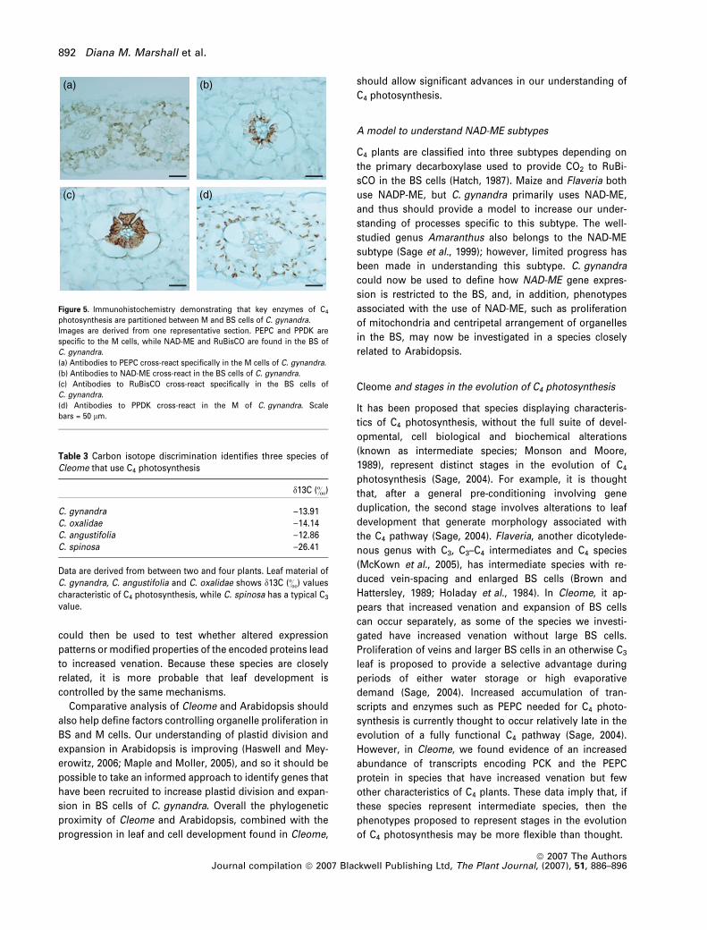
within or between cells. Immunohistochemistry of cross-
sections of C. gynandra leaves showed that PEPC was
abundant in the M cells (Figure 5a), while NAD-ME and
RuBisCO were specifically located in BS cells (Figure 5b,c),
and PPDK was most abundant in M cells (Figure 5d). In
C. gynandra, glycine decarboxylase accumulated in BS cells,
but we found no evidence for this in C. foliosa (data not
shown).
RNA blots were carried out to determine whether accu-
mulation of enzymes needed for C4 photosynthesis correla-
ted with the abundance of their RNA transcripts, and, in
addition, to test whether species of Cleome other than
C. gynandra accumulated transcripts encoding the enzymes
needed for C4 photosynthesis. Using probes generated from
full-length cDNAs of A. thaliana, transcripts for PEPC were
detectable in C. gynandra, and a higher molecular weight
band to which the PEPC probe hybridized was seen in
C. africana (Figure 4). Transcripts for NAD-ME were detected
in most species, but appeared most abundant in C. gynan-
dra, while transcripts encoding PCK were detectable in most
species (Figure 4). PPDK transcripts were only detectable in
C. gynandra (Figure 4). These data indicate there is a
tendency for transcripts encoding the biochemical compo-
nents of C4 photosynthesis to accumulate in Cleome.
Because our analysis of transcript abundance is between
(b)
(a)
(d)
(e)
(f)
(g)
(c)
(h)
(i)
Figure 2. Within Cleome, in addition to vein spacing commonly being reduced, several species have enlarged BS cells.
Transverse sections of mature leaves from nine species of Cleome.
(a) C. spinosa (C3) has a large inter-veinal distance and small BS cells.
(b) C. marshallii has the most similar venation and BS cell arrangement to C. spinosa.
(c) C. violacea has increased venation, and leaves are thicker due to an increased layer of palisade M cells.
(d) C. isomeris has increased venation, but little change in size of the BS.
(e) C. hirta has thinner leaves, and increased venation.
(f) C. foliosa shows increased venation compared to C. spinosa, and enlarged BS cells.
(g) C. africana has thicker leaves with increased venation, larger BS cells and additional layers of palisade M with reduced intercellular air spaces.
(h) C. paradoxa has increased venation, enlarged bundle sheath cells with increased numbers of chloroplasts, and lacks spongy mesophyll cells.
(i) C. gynandra (C4) shows reduced spacing between veins, increased BS cell size, and clear proliferation of chloroplasts in the BS. Scale bars = 100 lm.
C4 photosynthesis in Cleome 889
ª 2007 The AuthorsJournal compilation ª 2007 Blackwell Publishing Ltd, The Plant Journal, (2007), 51, 886–896

species, variation in hybridization of the probes to RNA from
each species could be caused by alterations in the sequence
of each gene as well as alterations in the amount of each
transcript. To minimize these confounding effects, previous
workers have used full-length cDNA probes from a relatively
distantly related species, and this has shown clear gradients
in accumulation of PEPC transcripts between C3 and C4
species of Flaveria and Alternanthera (Engelmann et al.,
2003; Gowik et al., 2006). We generated probes from full-
length Arabidopsis cDNAs, and were able to detect high
levels of transcripts encoding PEPC, NAD-ME and PPDK in
the C4 species C. gynandra.
Because the species of Cleome we assessed span a
developmental progression from C3 to C4 leaves, and there
is also evidence that the RNA transcripts and proteins
needed for the pathway accumulate more than in strict C3
species, we assessed herbarium specimens of Cleome using
stable isotope analysis in order to test whether there are
more C4 plants in this genus. Analysis of the d13C content of
leaves identified at least two new species of C4 plants within
Cleome (Table 3). While C. gynandra and C. angustifolia
originate from Africa, C. oxalidae is Australian, and so it
is possible that C4 photosynthesis has evolved more than
once within the genus. This supports the notion that
Table 1 Photosynthetic parameters derived from the relationshipbetween the rate of net photosynthesis and the internal concentra-tion of CO2 within the leaf
SpeciesCompensation point(lmol mol)1)
Carboxylation efficiency(mol m)2 sec)1)
C. spinosa 50 � 2 0.17 � 0.01C. marshallii 52 � 3 0.24 � 0.03C. africana 65 � 4 0.65 � 0.07C. violacea 51 � 4 0.2 � 0.01C. hirta 123 � 14 0.14 � 0.02C. isomeris 49 � 2 0.25 � 0.02C. foliosa 40 � 2 0.46 � 0.11C. gynandra 9 � 4 0.93 � 0.31
Data are derived from analysis of four or five leaves from differentplants of each species. All of the species except the C4 speciesC. gynandra show compensation points characteristic of C3 plants.C. africana and C. foliosa both have higher carboxylation efficienciesthan C. spinosa. Data are means � SEM.
(d) (e) (f) (g) (h)
0
10
20
30
40
50
0
5
10
15
20
25
30
0
2
4
6
8
10
Wid
th B
S c
ells
(µ
m)
Wid
th M
cel
ls (
µm
)
Num
ber
M c
ells
** * ***C. s
pino
sa
C. mar
shal
lii
C. afri
cana
C. vio
lace
aC. h
irta
C. iso
mer
isC. f
olio
sa
C. gyn
andr
a
C. par
adox
a
C. spi
nosa
C. mar
shal
lii
C. afri
cana
C. vio
lace
a
C. hirt
a
C. iso
mer
isC. f
olio
sa
C. gyn
andr
a
C. par
adox
a
C. spi
nosa
C. mar
shal
lii
C. afri
cana
C. vio
lace
a
C. hirt
a
C. iso
mer
isC. f
olio
sa
C. gyn
andr
a
C. par
adox
a
(a) (b) (c)
Figure 3. Quantification of BS and M cell size, chloroplast arrangement and ultrastructure in BS cells in Cleome.
(a) C. africana and C. gynandra show a significantly increased width of BS cells compared to C. spinosa (P < 0.05). BS width was assessed by measuring the axis
separating mesophyll and vein cells.
(b) In C. foliosa and C. paradoxa, closer veins are associated with a reduced width of M cells (P < 0.01; measured by assessing the axis separating BS cells).
(c) In C. paradoxa and C. gynandra, closer veins are associated with fewer mesophyll cells between veins (P < 0.01).
(d) BS cells of C. spinosa contain chloroplasts arranged centrifugally.
(e) BS cells of C. gynandra contain larger chloroplasts arranged centripetally.
(f) BS chloroplasts from C. spinosa with granal stacks clearly visible.
(g) BS chloroplasts from C. gynandra with reduced granal stacking; increased numbers of mitochondria are also visible.
(h) BS cells from C. paradoxa have increased numbers of mitochondria. Data are derived from at least three sections from each species. Scale bars = 20 lm (d, e) and
2 lm (f–h).
890 Diana M. Marshall et al.
ª 2007 The AuthorsJournal compilation ª 2007 Blackwell Publishing Ltd, The Plant Journal, (2007), 51, 886–896

Cleome possesses traits that pre-condition it to evolve C4
photosynthesis.
Discussion
Within Cleome, there appears to remarkable flexibility in leaf
development, cell biology and to some extent the accumu-
lation of enzymes associated with the C4 pathway. In addi-
tion to the bone fide C4 species C. gynandra, the genus also
contains species that possess only increased venation (e.g.
C. violacea), and others with more veins as well as one or
more additional characteristics needed for the C4 pathway.
For example, C. africana possesses more veins, larger BS
cells and more PEPC protein, while C. paradoxa displays
increased venation and larger BS cells containing more
centripetally arranged chloroplasts and mitochondria. In
other words, species in Cleome appear to deviate from the
‘normal C3 state’ to varying degrees.
Cleome and C4 leaf development
Almost all work on C4 plants has been conducted with
genera that are phylogenetically distant to the model
A. thaliana (Brown et al., 2005). Combined with the com-
plexity of C4 photosynthesis and the relatively large
genomes and long lifecycles of C4 models such as maize and
Flaveria, their phylogenetic distance from A. thaliana has
not helped in the translation of knowledge from Arabidopsis
into C4 systems. For example, insights into the control of leaf
development in Arabidopsis are unlikely to be useful in
understanding the development of leaves in maize because
leaf development is mediated by different mechanisms in
these lineages. In Arabidopsis, YABBY genes are expressed
in abaxial cells (Siegfried et al., 1999), whereas in maize they
are expressed in adaxial cells (Juarez et al., 2004). In addi-
tion, Arabidopsis axillary buds are clonally related to the
subtending leaf, whilst in maize they are clonally related to
the internode above (Irish and Sussex, 1992). This indicates
that different developmental processes can generate the
same structures in distantly related species. Flaveria has
been used as a model for understanding C4 photosynthesis
for several decades, and has provided key insights into the
regulation of genes that were already known to be important
for C4 pathway (Ali and Taylor, 2001; Gowik et al., 2004; Patel
et al., 2006). However, probably due to phylogenetic dis-
tance, it has not been possible to extend our understanding
of processes governing leaf and cell development from
Arabidopsis to Flaveria.
In contrast, because Arabidopsis and Cleome are closely
related and their genomes show significant synteny (Sch-
ranz and Mitchell-Olds, 2006), it should be possible to isolate
orthologues to Arabidopsis genes from Cleome gynandra
that control vein patterning (Carland and Nelson, 2004;
Casson et al., 2002; Cnops et al., 2006). These candidates
PEPC
LSU
PPDK
RNA
PPDK
RbcS
PCK
NAD-ME
PEPC
C. spi
nosa
C. afri
cana
C. vio
lace
aC. h
irta
C. iso
mer
isC. f
olio
saC. g
ynan
dra
Figure 4. Immunoblot and RNA blot analysis from one representative
experiment shows that C. gynandra accumulates proteins and RNA tran-
scripts required for C4 photosynthesis, and in addition there is some
accumulation of the PEPC protein and RNAs in the other species of Cleome.
(a) Accumulation of phosphoenolpyruvate carboxylase (PEPC) in C. gynandra,
and also a small amount in C. africana.
(b) Members of the genus accumulate lower amounts of the large subunit of
RuBisCO than C. spinosa.
(c) Pyruvate orthophosphate dikinase (PPDK) is detectable in C. gynandra, but
not in the other species.
(d) Large amounts of PEPC transcripts are detectable in C. gynandra.
(e) NAD-ME transcripts are abundant in C. gynandra.
(f) PCK transcripts are detected in C. gynandra, and also C. spinosa,
C. africana and C. violacea.
(g) RbcS transcripts are detectable in all species.
(h) PPDK transcripts are detectable in C. gynandra, but not the other species.
The agarase blot at the bottom shows RNA loading.
Table 2 C. gynandra possesses enzyme activities characteristic ofC4 photosynthesis and belongs to the NAD-ME subtype
PEPC NAD-ME PCK NADP-ME
C. gynandra 12.2 � 0.6 2.60 � 0.15 1.03 � 0.49 0.10 � 0.01C. spinosa 0.7 � 0.03 0.06 � 0.01 0.06 � 0.04 0.12 � 0.03
All enzyme activities were determined using enzyme-linked assays,and are expressed as lmol per min per unit chlorophyll. Data arederived from at least five leaf samples originating from differentplants. PEPC, phosphoenolpyruvate carboxylase; NAD-ME, NAD-dependent malic enzyme; PCK, phosphoenolpyruvate carboxykinase;NADP-ME, NADP-dependent malic enzyme. Data are means � SEM.
C4 photosynthesis in Cleome 891
ª 2007 The AuthorsJournal compilation ª 2007 Blackwell Publishing Ltd, The Plant Journal, (2007), 51, 886–896
could then be used to test whether altered expression
patterns or modified properties of the encoded proteins lead
to increased venation. Because these species are closely
related, it is more probable that leaf development is
controlled by the same mechanisms.
Comparative analysis of Cleome and Arabidopsis should
also help define factors controlling organelle proliferation in
BS and M cells. Our understanding of plastid division and
expansion in Arabidopsis is improving (Haswell and Mey-
erowitz, 2006; Maple and Moller, 2005), and so it should be
possible to take an informed approach to identify genes that
have been recruited to increase plastid division and expan-
sion in BS cells of C. gynandra. Overall the phylogenetic
proximity of Cleome and Arabidopsis, combined with the
progression in leaf and cell development found in Cleome,
should allow significant advances in our understanding of
C4 photosynthesis.
A model to understand NAD-ME subtypes
C4 plants are classified into three subtypes depending on
the primary decarboxylase used to provide CO2 to RuBi-
sCO in the BS cells (Hatch, 1987). Maize and Flaveria both
use NADP-ME, but C. gynandra primarily uses NAD-ME,
and thus should provide a model to increase our under-
standing of processes specific to this subtype. The well-
studied genus Amaranthus also belongs to the NAD-ME
subtype (Sage et al., 1999); however, limited progress has
been made in understanding this subtype. C. gynandra
could now be used to define how NAD-ME gene expres-
sion is restricted to the BS, and, in addition, phenotypes
associated with the use of NAD-ME, such as proliferation
of mitochondria and centripetal arrangement of organelles
in the BS, may now be investigated in a species closely
related to Arabidopsis.
Cleome and stages in the evolution of C4 photosynthesis
It has been proposed that species displaying characteris-
tics of C4 photosynthesis, without the full suite of devel-
opmental, cell biological and biochemical alterations
(known as intermediate species; Monson and Moore,
1989), represent distinct stages in the evolution of C4
photosynthesis (Sage, 2004). For example, it is thought
that, after a general pre-conditioning involving gene
duplication, the second stage involves alterations to leaf
development that generate morphology associated with
the C4 pathway (Sage, 2004). Flaveria, another dicotylede-
nous genus with C3, C3–C4 intermediates and C4 species
(McKown et al., 2005), has intermediate species with re-
duced vein-spacing and enlarged BS cells (Brown and
Hattersley, 1989; Holaday et al., 1984). In Cleome, it ap-
pears that increased venation and expansion of BS cells
can occur separately, as some of the species we investi-
gated have increased venation without large BS cells.
Proliferation of veins and larger BS cells in an otherwise C3
leaf is proposed to provide a selective advantage during
periods of either water storage or high evaporative
demand (Sage, 2004). Increased accumulation of tran-
scripts and enzymes such as PEPC needed for C4 photo-
synthesis is currently thought to occur relatively late in the
evolution of a fully functional C4 pathway (Sage, 2004).
However, in Cleome, we found evidence of an increased
abundance of transcripts encoding PCK and the PEPC
protein in species that have increased venation but few
other characteristics of C4 plants. These data imply that, if
these species represent intermediate species, then the
phenotypes proposed to represent stages in the evolution
of C4 photosynthesis may be more flexible than thought.
(a) (b)
(c) (d)
Figure 5. Immunohistochemistry demonstrating that key enzymes of C4
photosynthesis are partitioned between M and BS cells of C. gynandra.
Images are derived from one representative section. PEPC and PPDK are
specific to the M cells, while NAD-ME and RuBisCO are found in the BS of
C. gynandra.
(a) Antibodies to PEPC cross-react specifically in the M cells of C. gynandra.
(b) Antibodies to NAD-ME cross-react in the BS cells of C. gynandra.
(c) Antibodies to RuBisCO cross-react specifically in the BS cells of
C. gynandra.
(d) Antibodies to PPDK cross-react in the M of C. gynandra. Scale
bars = 50 lm.
Table 3 Carbon isotope discrimination identifies three species ofCleome that use C4 photosynthesis
d13C (&)
C. gynandra )13.91C. oxalidae )14.14C. angustifolia )12.86C. spinosa )26.41
Data are derived from between two and four plants. Leaf material ofC. gynandra, C. angustifolia and C. oxalidae shows d13C (&) valuescharacteristic of C4 photosynthesis, while C. spinosa has a typical C3
value.
892 Diana M. Marshall et al.
ª 2007 The AuthorsJournal compilation ª 2007 Blackwell Publishing Ltd, The Plant Journal, (2007), 51, 886–896

However, it is also possible that, although intermediate
species possess characteristics of C4 photosynthesis, the
traits have been generated by changes to loci that have not
occurred in bone fide C4 species. This would mean that
these intermediates represent evolutionary dead ends, and
although they share some phenotypes with C4 species, the
mechanisms responsible for these alterations are incom-
patible with evolution of the full pathway. The presence of
C4 photosynthesis in a genus closely related to Arabidop-
sis and the range of developmental intermediates we
describe in Cleome may allow these proposals to be
tested. For example, once loci responsible for increased
venation in C. gynandra have been identified, it should be
possible to determine whether the same alterations have
occurred in the developmental intermediates such as
C. foliosa, C. africana and C. paradoxa. If the same mech-
anisms control increased BS cell size in C. gynandra and
C. foliosa, C. africana and C. paradoxa, this would imply
they are true intermediates that have not fully evolved C4
photosynthesis. In contrast, if increased BS cell size is
controlled by different loci in C. gynandra, compared to
C. foliosa, C. africana and C. paradoxa, these species may
represent dead ends in terms of the evolution of C4
photosynthesis.
Overall, our results indicate that, in addition to C. gyn-
andra being a bone fide C4 species, other species in the
genus appear to be pre-conditioned to evolve characteris-
tics associated with C4 photosynthesis. These include
increased venation, enlarged BS cells, proliferation of both
mitochondria and chloroplasts, and accumulation of tran-
scripts and proteins needed for C4 photosynthesis. These
alterations include many of the most important ones
required in a C4 leaf, and so, combined with its phylo-
genetic proximity to Arabidopsis, Cleome should be a
good model to increase our understanding of the evolution
of C4 photosynthesis.
Experimental procedures
Plant material and analysis of leaf structure and
ultra-structure
Plants for anatomical, ultra-structural, physiological and biochemi-cal analysis were grown in a greenhouse with temperatures con-trolled at 24�C during the day and 20�C at night, with a photoperiodof 12 h light, 12 h dark. Sunlight was supplemented with metalhalide bulbs providing a minimum of 200 lmol m)2 sec)1. Theplants were grown in Levington M3 potting compost mixed withslow-release nitrogen pellets.
To assess vein lengths per unit area, fully expanded leaves werecleared in 70% ethanol at 65�C and 5% w/v NaOH at roomtemperature. Tissue was then stored in 70% ethanol and stainedin 1% w/v safranin in 50% ethanol. Samples were examined using alight microscope, and images were taken using a Nikon digitalcamera (http:www.nikon.co.uk). Vein lengths were quantified fromfour leaves using Image J software (http://rsb.info.nih.gov/ij/).
To determine the arrangement of cells within the leaf, trans-verse sections were produced from fully expanded tissue. Tissuewas fixed by immersion in 4% w/v glutaraldehyde containing2 mM CaCl2 in 0.1 M PIPES, pH 7.4, and 100 ll 33% H2O2 was addedto each 10 ml aliquot immediately before use. Immersion was for4–6 h at 4�C, after which tissue was washed twice in 0.1 M PIPESbuffer and stored at 4�C. Samples were post-fixed in 1% w/vosmium ferricyanide for 1 h, rinsed three times in H2O, and bulk-stained in 2% w/v uranyl acetate for 1 h. Samples were rinsed inH2O, and dehydrated in an ascending series of ethanol solutions to100% ethanol, rinsed twice in acetonitrile, and embedded inSpurr’s epoxy resin. Histological sections for light microscopywere cut 1–2 lm thick and stained with methylene blue. Fortransmission electron microscopy, 50 nm thick sections were cutwith a Leica Ultracut UCT, stained with saturated uranyl acetate in50% ethanol and lead citrate, and viewed in a FEI Philips CM100operated at 80 kV.
Infra-red gas analysis and analysis of proteins
Photosynthesis was measured using a PP Systems (www.PPSystems.co.uk) CIRAS-1 infra-red gas analyser with a standardleaf cuvette held at 25�C, and supplied with 1500 lmolphotons m)2 sec)1. Varying the amount of CO2 in the chamberallowed the response of net photosynthesis to the internal con-centration of CO2 to be determined. The results are means ofthree to seven replicates �one standard error of the mean.
In order to assess the accumulation of proteins needed for C4
photosynthesis, 0.1–0.2 g fresh weight of tissue was ground in400 ll extraction buffer containing 50 mM Tris, pH 8.0, 2 mM EDTA,2% w/v PVPP, 5% w/v PEG 4000, 25 mM dithiothreitol (DTT), 2 mM
PMSF, 25 lg ml)1 leupeptin, 25 lg ml)1 aprotinin and 25 lg ml)1
pepstatin A. Samples were ground in a pestle and mortar with sand,and the homogenate was centrifuged at 13 000 g for 10 min toobtain a clear supernatant. Protein quantity in the extract wasdetermined using Bio-Rad protein assay reagent (http://www.bio-rad.com/) according to the method described by Bradford(1976).
Prior to SDS–PAGE, samples were placed at 99�C for 2 min in70 mM Tris–HCl, pH 6.8, 0.06% w/v glycerol, 10% w/v SDS, 0.03% w/v2-b-mercaptoethanol and 0.05% w/v bromophenol blue. Sampleswere then resolved on a denaturing acrylamide gel with a 5%stacking gel and a 12% resolving gel. Total soluble protein (2, 5 and10 lg aliquots) was loaded per lane and used to detect LSU,phosphoenol pyruvate carboxylase (PEPC) and PPDK, respectively.After electrophoresis, gels were agitated in transfer buffer (48 mM
Tris–HCl, pH 9.2, 39 mM glycine, 20% methanol) for 30 min beforetransfer to nitrocellulose membrane at 10 V for 1 h in a Bio-Radsemi-dry blotter. Gels were Coomassie-stained to check transferwas complete, and the membrane was rinsed overnight in blockingbuffer (PBS, 1% w/v skimmed milk and 1% Tween-20). Primaryantibody was diluted in blocking buffer: anti-LSU antibody (1:3500)was incubated for 1 h with shaking at room temperature, anti-PEPCantibody (1:5000) was incubated for 1 h with shaking at roomtemperature, and anti-PPPDK antibody (1:10 000) was incubated for3 h with shaking at room temperature. Membrane was then washedthree times in blocking buffer for 5 min each, and incubated in thesecondary antibody, anti-rabbit biotinylated Ig (Amersham Bio-sciences; http://www5.amershambiosciences.com/), at a 1:1000dilution for 2 h at room temperature. Membranes were washedthree times for 5 min each before incubation with streptavidinbiotinylated horseradish peroxidase complex (AmershamBiosciences) at a 1:1000 dilution for 30 min at room temperature.Membranes were washed a further three times in blocking
C4 photosynthesis in Cleome 893
ª 2007 The AuthorsJournal compilation ª 2007 Blackwell Publishing Ltd, The Plant Journal, (2007), 51, 886–896

buffer without milk before detection using the Western Light-ning Chemiluminescence Reagent Plus kit (Perkin Elmer, http://perkinelmer.com). Chemiluminescence was detected using KonikaMinolta (http://www.konicaminolta.com) film with exposures ofbetween 10 sec and 1 min.
Preparation of tissue samples for immunolocalization was per-formed as described by Wang et al. (1992) and Dengler et al. (1995).Approximately 3–4 mm2 of leaf tissues were fixed in a 3:1 v/vethanol:glacial acetic acid solution overnight at room temperature.Tissues were then dehydrated through graded ethanol–tertiarybutyl alcohol mixtures and embedded in Paraplast (Fisher Scientific,http://www.fishersci.com). Cross-sections of 7 lm thickness werecut using a Spencer AO rotary microtome (Reichert histostat, http://www.reichert.com, 820H), mounted on poly-L-lysine-coated slidesand dried overnight. Following de-waxing, sections were re-hydra-ted through ethanol series and air-dried. They were then rinsed withTris-buffered saline, pH 7.6 (Dakocytomation Inc., http://www.dako.com), covered with 3% H2O2 for 10 min, and subsequentlywashed with the same buffer. To prevent random, non-specificbinding reactions, sections were pre-incubated with a universalserumfree blocking reagent (Dakocytomation Inc.) for 15–30 min.
Sections were incubated for 30 min with each antibody, dilutedto 1:2000 for the anti-maize PEPC IgG, and 1:200 for anti-RuBisCOIgG, anti-PPDK IgG and anti-NAD-ME IgG in antibody-dilutingbuffer, pH 7.6 (Dakocytomation Inc.). Control slides treated withdiluting buffer lacking antisera were run with each experiment.Following incubation, all sections were rinsed and washed, andplaced for 20–30 min in a 1:10 dilution of the sensitive EnVision+(Dakocytomation Inc., http://www.dako.com) detection polymersecondary antibody. Binding reactions were visualized by incu-bating sections with liquid 3,3-diaminobenzidine tetrahydrochlo-ride (DAB) and chromogen (Dakocytomation Inc.) for 8 min.Subsequently, the slides were rinsed with H2O, and counter-stained in 1% aqueous azure B. Following a serial dehydrationthrough ethanol, sections were mounted in Permount(www.pbio.com), examined under bright-field microscopy andphotographed using a digital camera and ACT-1 software(Nikon). Polyclonal antisera used were anti-Nicotiana tabacumRuBisCO (holoenzyme provided by N.G. Dengler, University ofToronto, Canada), anti-maize PEPC and anti-Amaranthushypochondriacus NAD-ME (obtained from J. Berry, State Univer-sity of New York, USA), and anti-maize PPDK (obtained fromH. Sakakibara, Plant Science Center, Riken, Japan). The specificityof each antiserum was verified by running positive-controlimmunolabeling experiments on cross-sections of paraffin-embedded leaf tissues of Amaranthus edulis (C4 NAD-ME),Melinis minutiflora (C4 PCK), F. ramossisima (C3–C4) and F. pring-lei (C3) in which the enzymes are known to accumulate in specificcells (Edwards et al., 2001).
To assess the biochemical subtype of Cleome gynandra, enzyme-linked assays were conducted according to the method describedby Ashton et al. (1990). For each assay, leaf tissue from at least fiveseparate plants was ground in sand and four volumes of extractionbuffer with a mortar and pestle, and centrifuged at 13 000 g prior torunning assays at 25�C and monitoring absorbance at 340 nm. PCKwas extracted in 200 mM Bicine-KOH, pH 9.0, 20 mM MgCl2, 5 mM
DTT. The assay buffer was 80 mM MES-NaOH, pH 6.7, 0.35 mM
NADH, 5 mM DTT, 2 mM MnCl2, 2 mM ADP, 50 mM KHCO3. Theassay was initiated with PEP (2 mM) and 6 units of malatedehydrogenase. NAD-ME was extracted in 50 mM HEPES-NaOH,pH 7.2, 50 mM Tricine, 2 mM MnCl2, 5 mM DTT, 0.25% w/v PVP-40000, 0.5% Triton X-100. NAD-ME assay buffer contained 25 mM
HEPES-NaOH, pH 7.2, 5 mM L-malic acid, 2 mM NAD, 5 mM DTT,0.2 mM EDTA. MnCl2 (24 mM) and coenzyme A to 0.1 mM were used
to initiate the reaction. NADP-ME was extracted in 50 mM HEPES-KOH, pH 8.3, 50 mM Tricine, 5 mM DTT, 0.1 mM EDTA. The assaybuffer contained 25 mM Tricine-KOH, pH 8.3, 5 mM L-malic acid,0.5 mM NADP, 0.1 mM EDTA, and the assays were initiated with2 mM MgCl2.
RNA isolation and RNA blot analysis
Total RNA was extracted from mature leaves using Tripure reagent(Roche, http://www.roche.com). Equal amounts of RNA for eachspecies were separated by electrophoresis on formaldehyde/1.2%agarose gels, and then blotted onto GeneScreen Plus membranes(NEN http://www.pertinelmer.co.uk) by capillary blotting.
Radiolabeled probes, prepared by the random-primer method(Feinberg and Vogelstein, 1983), were produced from fragmentscorresponding to full-length coding regions amplified by PCR fromArabidopsis thaliana leaf cDNA prepared using an oligo(dT) primer(Invitrogen Life Technologies; http://www.invitrogen.com/) andM-MLV reverse transcriptase (Promega; http://www.promega.com/).Hybridizations were carried out overnight at 60�C in 250 mM
Na2HPO4, 2.5 mM EDTA, 7% w/v SDS, pH 7.2 (Rosche and Westhoff,1995).
Filters were washed sequentially at 60�C for 15 min in 1· SSC/1%w/v SDS, 0.5· SSC/1% w/v SDS, 0.2· SSC/0.5% w/v SDS and 0.1·SSC/0.5% w/v SDS, and finally rinsed for 5 min at room temperaturein 0.1· SSC (http://www.saranbrands.com). The membranes werewrapped in Saran film and placed on a phosphor screen (MolecularDynamics, http://www.amersham.com), and exposed for between6 h and 5 days. Filters shown are from one representative experi-ment.
Isotope analysis of herbarium specimens
Specimens were sampled from the herbarium at the Royal BotanicGardens, Kew (London, UK). Leaf samples were assayed for d13C atthe Isotope Facility of the University of California, Davis, using a PeeDee limestone standard. For each species, at least two sampleswere analysed. The specimen labels were as follows: C. gynandra –Bartma 78 (Niger), Hein & Pircher 388 (West Africa), van SlagenenMSAA450 (Yemen), van der Maesen 3314 (India); C. oxalidae –Fryxell 3958 (West Australia), Symon 12109 (West Australia);C. angustifolia – Powys 69 (Kenya), Swinderen M22 (Kenya), Wood1171 (Yemen); C. spinosa – Archer 8063 (Brazil), Eiten 10301 (Brazil).
Acknowledgements
We thank Mark Chase of the Royal Botanic Gardens, Kew, forallowing us to sample Cleome specimens in the herbarium, ananonymous referee for helpful comments, and the Biotechnologyand Biology Sciences Research Council and the Leverhulme Trustfor funding.
Supplementary material
The following supplementary material is available for this articleonline:Figure S1. Within Arabidopsis, there is little evidence for variation inmorphological characteristics of leaves associated with the C4
pathway.This material is available as part of the online article from http://www.blackwell-synergy.com
894 Diana M. Marshall et al.
ª 2007 The AuthorsJournal compilation ª 2007 Blackwell Publishing Ltd, The Plant Journal, (2007), 51, 886–896

References
Ali, S. and Taylor, W.C. (2001) Quantitative regulation of theFlaveria Me1 gene is controlled by the 3¢-untranslated regionand sequences near the amino terminus. Plant Mol. Biol. 46,251–261.
Ashton, A.R., Burnell, J.N., Furbank, R.T., Jenkins, C.L.D. and Hatch,
M.D. (1990) Enzymes of C4 photosynthesis. In Methods in PlantBiochemistry, volume 3 (Lea, P.J., ed.). London: Academic Press,pp. 39–72.
Bradford, M.M. (1976) Rapid and sensitive method for quantitationof microgram quantities of protein utilizing principle of protein–dye binding. Anal. Biochem. 72, 248–254.
Brown, H.A. (1999) Agronomic implications of C4 photosynthesis. InC4 Plant Biology (Sage, R.F. and Monson, R.K., eds). San Diego,CA: Academic Press, pp. 473–508.
Brown, R.H. and Hattersley, P.W. (1989) Leaf anatomy of C3–C4
species as related to evolution of C4 photosynthesis. Plant Phys-iol. 91, 1543–1550.
Brown, N.J., Parsley, K. and Hibberd, J.M. (2005) The future of C4
research – maize, Flaveria or Cleome? Trends Plant Sci. 10, 215–221.
Carland, F.M. and Nelson, T. (2004) COTYLEDON VASCULARPATTERN2-mediated inositol (1,4,5) triphosphate signal trans-duction is essential for closed venation patterns of Arabidopsisfoliar organs. Plant Cell, 16, 1263–1275.
Casson, S.A., Chilley, P.M., Topping, J.F., Evans, I.M, Souter, M.A.
and Lindsey, K. (2002) The POLARIS gene of Arabidopsis encodesa predicted peptide required for correct root growth and leafvascular patterning. Plant Cell, 14, 1705–1721.
Cnops, G., Neyt, P., Raes, J. et al. (2006) The TORNADO1 andTORNADO2 genes function in several patterning processes dur-ing early leaf development in Arabidopsis thaliana. Plant Cell, 18,852–866.
Dedonder, A., Rethy, R., Fredericq, H., Van Montagu, M. and
Krebbers, E. (1993) Arabidopsis RbcS genes are differentiallyregulated by light. Plant Physiol. 101, 801–808.
Dengler, N.C. and Nelson, T. (1999) Leaf structure and developmentin C4 plants. In C4 Plant Biology (Sage, R.F. and Monson, R.K.,eds). San Diego, CA: Academic Press, pp. 133–172.
Dengler, N.G., Dengler, R.R., Donnelly, P.M. and Filosa, M.F. (1995)Expression of the C4 pattern of photosynthetic enzyme accumu-lation during leaf development in Atriplex rosea (Chenopodia-ceae). Am. J. Bot. 82, 318–327.
Edwards, G.E., Franceschi, V.R., Ku, M.S.B., Voznesenskaya, E.V.,
Pyankov, V.I. and Andreo, C.S. (2001) Compartmentation ofphotosynthesis in cells and tissues of C4 plants. J. Exp. Bot. 52,577–590.
Edwards, G.E., Franceschi, V.R. and Voznesenskaya, E.V. (2004)Single-cell C4 photosynthesis versus the dual-cell (Kranz)paradigm. Annu. Rev. Plant Biol. 55, 173–196.
Engelmann, S., Blasing, O.E., Gowik, U., Svensson, P. and
Westhoff, P. (2003) Molecular evolution of C4 phosphoenolpyru-vate carboxylase in the genus Flaveria – a gradual increase fromC3 to C4 characteristics. Planta, 217, 717–725.
Feinberg, A.P. and Vogelstein, B. (1983) A technique for radiolabe-ling DNA restriction endonuclease fragments to high specificactivity. Anal. Biochem. 132, 6–13.
Gerrard-Wheeler, M. C., Tronconi, M. A., Drincovich, M. F., Andreo,
C. S., Flugge, U.-I. and Maurino, V. G. (2005) A comprehensiveanalysis of the NADP-malic enzyme gene family of Arabidopsis.Plant Physiol. 139, 39–51.
Gousset-Dupont, A., Lebouteillei, B., Monreal, J., Echevarria, C.,
Pierre, J.N., Hodges, M. and Vidal, J. (2005) Metabolite and post-
translational control of phosphoenolpyruvate carboxylase fromleaves and mesophyll cell protoplasts of Arabidopsis thaliana.Plant Sci. 169, 1096–1101.
Gowik, U., Burscheidt, J., Akyildiz, M., Schlue, U., Koczor, M.,
Streubel, M. and Westhoff, P. (2004) cis-regulatory elements formesophyll specific gene expression in the C4 plant Flaveriatrinervia, the promoter of the C4 phosphoenolpyruvate carboxy-lase gene. Plant Cell, 16, 1077–1090.
Gowik, U., Engelmann, S., Blasing, O.E., Raghavendra, A.S. and
Westhoff, P. (2006) Evolution of C4 phosphoenolpyruvate carb-oxylase in the genus Alternanthera: gene families and the enzy-matic characteristics of the C4 isozyme and its orthologues in C3
and C3/C4 Alternantheras. Planta, 223, 359–368.Hall, J.C., Sytsma, K.J. and Iltis, H.H. (2002) Phylogeny of Cappar-
aceae and Brassicaceae based on chloroplast sequence data. Am.J. Bot. 89, 1826–1842.
Haswell, E.S. and Meyerowitz, E.M. (2006) MscS-like proteins con-trol plastid size and shape in Arabidopsis thaliana. Curr. Biol. 16,1–11.
Hatch, M.D. (1987) C4 photosynthesis – a unique blend of modifiedbiochemistry, anatomy and ultrastructure. Biochim. Biophys.Acta, 895, 81–106.
Hibberd, J.M. and Quick, W.P. (2002) Characteristics of C4 photo-synthesis in stems and petioles of C3 flowering plants. Nature,415, 451–454.
Hibberd, J.M., Richardson, P., Whitbread, R. and Farrar, J.F. (1996)Effects of leaf age, basal meristem and infection with powderymildew on photosynthesis in barley grown in 700 lmol mol)1
CO2. New Phytol. 134, 317–325.Holaday, A.S., Lee, K.W. and Chollet, R. (1984) C3–C4 intermediate
species in the genus Flaveria – leaf anatomy, ultrastructure, andthe effect of O2 on the CO2 compensation concentration. Planta,160, 25–32.
Irish, V.F. and Sussex, I.M. (1992) A fate map of the Arabidopsisembryonic shoot apical meristem development. Development,115, 745–753.
Jordan, D.B. and Ogren, W.L. (1984) The CO2–O2 specificity ofribulosebisphosphate-1,5-carboxylase/oxygenase – dependenceon ribulosebisphosphate concentration, pH and temperature.Planta, 161, 308–313.
Juarez, M.T., Twigg, R.W. and Timmermans, M.C.P. (2004) Speci-fication of adaxial cell fate during maize leaf development.Development, 131, 4533–4544.
Langdale, J.A. and Nelson, T. (1991) Spatial regulation of photo-synthetic development in C4 plants. Trends Genet. 7, 191–196.
Maple, J. and Moller, S.G. (2005) An emerging picture of plastiddivision in higher plants. Planta, 223, 1–4.
McKown, A.D., Moncalvo, J.M. and Dengler, N.G. (2005) Phylogenyof Flaveria (Asteraceae) and inference of C4 photosynthesis evo-lution. Am. J. Bot. 92, 1911–1928.
Monson, R.K. and Moore, B.D. (1989) On the significance of C3–C4
intermediate photosynthesis to the evolution of C4 photosyn-thesis. Plant Cell Environ. 12, 689–699.
Parsley, K. and Hibberd, J.M. (2006) The Arabidopsis ppdk gene istranscribed from two promoters whose transcripts differ inexpression patterns. Plant Mol. Biol. 62, 339–349.
Patel, M., Siegel, A.J. and Berry, J.O. (2006) Untranslated regions ofFbRbcS1 mRNA mediate bundle sheath cell-specific geneexpression in leaves of a C4 plant. J. Biol. Chem. 281, 25485–25491.
Penfield, S., Rylott, E.L., Gilday, A.D., Graham, S., Larson, T.R. and
Graham, I.A. (2004) Reserve mobilization in the Arabidopsisendosperm fuels hypocotyl elongation in the dark, is independentof abscisic acid, and requires PHOSPHOENOLPYRUVATECARBOXYKINASE1. Plant Cell, 16, 2705–2718.
C4 photosynthesis in Cleome 895
ª 2007 The AuthorsJournal compilation ª 2007 Blackwell Publishing Ltd, The Plant Journal, (2007), 51, 886–896

Raghavendra, A.S. and Das, V.S.R. (1978) The occurrence of C4
photosynthesis: a supplementary list of C4 plants reported duringlate 1974 mid 1977. Photosynthetica, 12, 200–208.
Reiskind, J.B., Madsen, T.V., VanGinkel, L.C. and Bowes, G. (1997)Evidence that inducible C4-type photosynthesis is a chloroplasticCO2 concentrating mechanism in Hydrilla, a submersed monocot.Plant Cell Environ. 20, 211–220.
Rosche, E. and Westhoff, P. (1995) Genomic structure and expres-sion of the pyruvate, orthophosphate dikinase gene of the dicot-yledonous C4 plant Flaveria trinervia (Asteraceae). Plant Mol.Biol. 29, 663–678.
Sage, R.F. (2004) The evolution of C4 photosynthesis. New Phytol.161, 341–370.
Sage, R.F., Li, M. and Monson, R.K. (1999) The taxonomic distribu-tion of C4 photosynthesis. In C4 Plant Biology (Sage, R.F. andMonson, R.K., eds). San Diego, CA: Academic Press, pp. 551–584.
Schranz, M.E. and Mitchell-Olds, T. (2006) Independent ancientpolyploidy events in the sister families Brassicaceae and Cleom-aceae. Plant Cell, 18, 1152–1165.
Siegfried, K.R., Eshed, Y., Baum, S.F., Otsuga, D., Drews, G.N.
and Bowman, J.L. (1999) Members of the YABBY gene familyspecify abaxial cell fate in Arabidopsis. Development, 126,4117–4128.
Taniguchi, M., Taniguchi, Y., Kawasaki, M., Takeda, S., Kato, T.,
Sato, S., Tahata, S., Miyake, H. and Sugiyama, T. (2002) Identi-fying and characterizing plastidic 2-oxoglutarate/malate anddicarboxylate transporters in Arabidopsis thaliana. Plant CellPhysiol. 43, 706–717.
Voznesenskaya, E.V., Franceschi, V.R., Kiirats, O., Freitag, H. and
Edwards, G.E. (2001) Kranz anatomy is not essential for terrestrialC4 plant photosynthesis. Nature, 414, 543–546.
Voznesenskaya, E.V., Franceschi, V.R., Kiirats, O.A., Elena, G.,
Freitag, H. and Edwards, G.E. (2002) Proof of C4 photosynthesiswithout Kranz anatomy in Bienertia cycloptera (Chenopodiaceae).Plant J. 31, 649–662.
Wang, J.L., Klessig, D.F. and Berry, J.O. (1992) Regulation of C4
gene-expression in developing Amaranth leaves. Plant Cell, 4,173–184.
896 Diana M. Marshall et al.
ª 2007 The AuthorsJournal compilation ª 2007 Blackwell Publishing Ltd, The Plant Journal, (2007), 51, 886–896
Copyright © 2022 FDOKUMEN