Directional motion of foreign plasmid DNA to nuclear HP1 foci
European Journal of Plant Pathology 110: 163–174, 2004.© 2004 Kluwer Academic Publishers. Printed in the Netherlands.
Chromosomal and plasmid diversity of agrobacterium strains isolated fromFicus benjamina tumors
Aida Raio1, Raffaele Peluso2, Xavier Nesme3 and Astolfo Zoina2
1Istituto per la Protezione delle Piante, Sezione di Portici (Fax: +390817755320; E-mail: [email protected]);2Dipartimento di Arboricoltura, Botanica e Patologia Vegetale – Universita di Napoli Federico II – ViaUniversita, 100 – 80055 Portici (NA), Italy; 3Laboratoire de Ecologie Microbienne/UMR CNRS 5557/UniversiteClaude Bernard – Lyon 1 F-69622 Villeurbanne Cedex, France
Accepted 11 August 2003
Key words: Agrobacterium larrymoorei, plasmid profile, population structure, pTi, ribotype
Abstract
Agrobacteria were previously isolated from tumors developing on branches and aerial and hypogeous roots ofweeping fig plants in Italy and in The Netherlands. A representative group of 48 strains was analyzed by PCR–RFLP of 16S and 16S + IGS ribosomal regions, PCR–RFLP of six Ti plasmid (pTi) regions and characterized forplasmid content. Two groups of agrobacteria were separated by cluster analysis of PCR–RFLP profiles of rrs gene:seventeen strains were similar to the new species Agrobacterium larrymoorei, while the remaining strains wereincluded within the agrobacterium biovar 1 group. Sixteen different plasmid profiles from one to five plasmids wereobserved. In addition, 21 ribotypes and 20 pTi structures were arranged in many different combinations, showing thatfig agrobacteria were characterized by a wide heterogeneity. A general lack of correlation between strain ribotypesand plasmid content was observed.
Introduction
Agrobacteria are soil-borne Gram negative bacteria,responsible for the induction of a neoplastic disease(crown gall) or for abnormal root proliferation (hairyroot) on different host plants. On the basis of physiolog-ical and biochemical characters most of the members ofthe genus Agrobacterium were distinguished into threebiovars (Kersters and De Ley, 1984), two of whichwere recognized as different species: A. rhizogenes(formerly biovar 2) (Sawada et al., 1993) and A. vitis(formerly biovar 3) (Ophel and Kerr, 1990). Studiesbased on DNA–DNA homology (Popoff et al., 1984)indicated that biovar 1 does not correspond to a singlespecies of Agrobacterium, but it includes at least ninegenomic groups that have not yet received acceptednomenspecies status. Classification and nomenclatureof the species belonging to the genus Agrobacterium isstill under discussion and most recently the inclusion
of the Agrobacterium species in the genus Rhizobiumhas been proposed (Young et al., 2001).
Most of the pathogenic determinants of agrobacteriaare born on a large extra chromosomal replicon termedthe Ti plasmid (pTi). During the infection process,part of this plasmid (the T-DNA) is transferred to thewounded plant cells where it is integrated in the nucleargenome (Chilton et al., 1977). The expression of theT-DNA genes in the transformed cells is responsiblefor their proliferation and leads to the formation oftumors. T-DNA genes also encode the synthesis of lowmolecular weight compounds termed opines that arespecifically metabolized by the bacteria responsible forthe induction of the disease. Genetically complex pop-ulations of agrobacteria can generate as a consequenceof conjugative transfer of pTi to non-pathogenicagrobacteria (Kerr, 1971; Petit et al., 1978), acquisitionor loss of sequences (Otten et al., 1992) and nucleotidechanges within the pTi (Belanger et al., 1995). The
164
study of both pTi and chromosomal characteristics of agiven population is thus very important to understandthe type of agrobacteria involved in a disease outbreak.
The sequence of the 16S genes is highly conservedwithin strains belonging to the same genomic species(Nazaret et al., 1991) and can be used to differentiate thespecies, while the intergenic spacer (IGS) between 16Sand 23S rDNA genes is sufficiently variable to allowthe comparison of closely related strains (Navarro et al.,1992). Oger et al. (1998) showed that PCR–RFLP anal-ysis of 16S and 16S + IGS ribosomal regions is a fastand reliable method to perform the phylogenetic studiesof microorganisms. PCR–RFLP can also be employedto study pTi diversity as highly conserved sequencesof vir and T-DNA regions of pTi can be used to designprimers for PCR (Ponsonnet and Nesme, 1994).
Agrobacteria isolated from weeping fig trees inItaly and The Netherlands showed a wide variabilityfor several physiological and biochemical character-istics and were grouped into two main classes. Thefirst included agrobacteria belonging to biovar 1 andto an intermediate biovar and the second includedstrains which were similar to the proposed new speciesAgrobacterium larrymoorei (Zoina et al., 2001). Ananalysis of the diversity of agrobacteria provides anunderstanding of the ecological relationships betweenstrains within the same genus and between agrobacte-ria and other microorganisms. Moreover, the diversityof agrobacterium populations is considered a criticalfactor in the management of crown gall disease (Mooreand Canfield, 1996). Forty-eight agrobacteria includingrepresentative strains from Italy and The Netherlandswere analyzed in this study for their 16S and 16S+IGSribosomal regions, plasmid content and pTi character-istics. The main purpose of this study was to determinethe genetic relationship between the two kinds ofagrobacteria responsible for the disease outbreaks thatwere found constantly associated in fig tumors, and tostudy the phylogenetic similarities of Italian and Dutchisolates with the new species A. larrymoorei (Bouzaret al., 1995; Bouzar and Jones, 2001).
Materials and methods
Bacterial strains
The strains of Agrobacterium used are listed inTable 1. Biovar attribution was performed by biochem-ical and physiological tests while the identificationof Agrobacterium species was done by the BIOLOG
ML1™ system (Zoina et al., 2001). Strains isolatedfrom Ficus benjamina originated from 30 differentplants at one nursery in Salerno (Italy), one plant from aprivate house in Latina (Italy) and 10 plants from a trop-ical glasshouse in Arnhem (the Netherlands) and wereindicated as Fb, LT and NL, respectively (Zoina et al.,2001). The 48 strains from F. benjamina that are consid-ered in this study were chosen on the basis of morpho-logical, physiological and biochemical characteristicsas representative of a collection of 241 fig agrobac-teria (Zoina et al., 2001). All strains belong to thecollection of phytopathogenic bacteria of the Depart-ment of Botany, Horticulture and Plant Pathology ofthe University of Naples ‘Federico II’ (Italy).
PCR–RFLP analysis of 16S and16S + IGS ribosomal regions
Amplifications were carried out in a volume of 50 µlcontaining 100 ng of DNA extracted by the DNeasySystem (Qiagen). Primers FGPS6 and FGPS1509′were used to amplify the ribosomal region of 1479 bprepresenting 99.5% of the 16S rDNA.
Primers FGPS6 and FGPL132′ were used to obtainthe amplification of the ribosomal region of 2500–2700 bp including 16S rDNA and the IGS betweenthe 16S and 23S rDNA (IGS) plus 132 bp of the 23SrDNA (Ponsonnet and Nesme, 1994). Preparation ofPCR reaction mixtures and conditions of amplificationboth for 16S and 16S+IGS were the same as describedby Ponsonnet and Nesme (1994).
Ten microliters of the PCR products were digestedwith 10 U of each restriction enzyme (r.e.). Digestionsof 16S rDNA region was performed using AccI (Bio-labs), AluI (Biolabs), BfaI (Biolabs), HaeIII (GibcoBRL) and HpaII (Biolabs). CfoI (GibcoBRL), HaeIII(GibcoBRL) and NdeII (GibcoBRL) were used todigest the 16S + IGS region.
Restriction fragments obtained after 1 h of diges-tion were separated by horizontal electrophoresis inTBE buffer on a 2.5% (w/v) Nusieve agarose gel con-taining 1 µg/ml ethidium bromide. 1 Kb and 123 bpladders (Gibco BRL) were used as molecular weightstandards. Gels were run at 2.3 v/cm for 3.5 h and pho-tographed under UV light using Polaroid film type 57.Fragments of less than 100 bp were not considered forthe comparison of restriction patterns.
The different ribotypes were indicated with the let-ters ‘L’ (for A. larrymoorei) and ‘T’ (for all other figagrobacteria) followed by a number.

165
Table 1. Agrobacterium strains used in this study
Strains Biovar/species1 Host plant Origin
Fb272, Fb522, Fb80, Fb1193, 1 Ficus benjamina ItalyFb122, Fb1233, Fb1263, Fb1943,Fb2083, Fb2513, Fb2523, Fb2583,Fb2623, Fb276, Fb2833, Fb2902,
Fb2943, Fb3133
(Strains were isolated from12 different plants)
Fb215, Fb3083, Fb3102, Fb312, Intermediate Ficus benjamina ItalyFb321, Fb3323, Fb342(Strains were isolated from seven
different plants)
Fb23, Fb33, Fb46, Fb55, Fb60,Fb72, Fb86, Fb99, Fb190,
Fb233, Fb238, LT3, LT4(Strains were isolated from
nine different plants)Intermediate (putative
A. larrymoorei)Ficus benjamina Italy
NL13, NL3.5, NL373, NL2, 1 Ficus benjamina the NetherlandsNL32, NL3.7 Intermediate Ficus benjamina the Netherlands(Strains were isolated from
five different plants)
NL7, NL25, NL292, NL360 Intermediate (putativeA. larrymoorei)
Ficus benjamina the Netherlands
Strains were isolated fromthree different plants)
B6 1 Undetermined Colorado (USA)C58 1 Prunus cerasus New York (USA)At 20n5 2 Prunus persica ItalyK84 2 Soil AustraliaK305 A. vitis Vitis vinifera AustraliaATCC13335 A. rubi Rubus ursinus USAAF3.44 A. larrymoorei Ficus benjamina Florida (USA)AF3.10 A. larrymoorei Ficus benjamina Florida (USA)AF1.52 1 Ficus benjamina Florida (USA)AF3.51 1 Ficus benjamina Florida (USA)
1Tests for biovar and species identification are described in a previous work (Zoina et al., 2001).2Avirulent strains.3Nopaline catabolizing strains. The remaining strains were unable to utilize mannopine, nopaline or octopine(Zoina et al., 2001).
Plasmid profiles
Plasmid DNA was isolated following the cell lysisand alkaline denaturation procedure of Currier andNester (1976). DNA was loaded onto a 0.5% ultrapure agarose gel (GibcoBRL) and separated by hori-zontal electrophoresis at 4 v/cm for 5 h. After stainingin a 0.5 µg/ml solution of ethidium bromide, the gelswere photographed on a short-wave UV transillumi-nator with Polaroid film type 57. Strains SW2 andSS104 of Erwinia stewartii were used as references forplasmid size determination (Coplin et al., 1981).
The relative mobility (Rm) of each plasmid was esti-mated and the molecular weight (MW) was determinedon the basis of the correlation: log MW = f (log Rm)as described by Meyers et al. (1976). All plasmids werelisted in decreasing order according to their size andnamed in alphabetical order. Different profiles wereobtained by the combination of one or more letters.
PCR–RFLP analysis of pTi
Total DNA was extracted by the DNeasy tissuekit (Qiagen). Primers used to amplify pTi regions
166
were: FGPtmr530 and FGPtmr701′ for amplifying tmr(171 bp), FGPtms194′ and FGPtms46′ for amplifyingtms (587 bp), FGPnos1236′ and FGPnos975 for ampli-fying nos (256 bp), FGPvirA2275 and FGPvirB2164′for amplifying vir (1673 bp), FGPvirB11 + 21and FGPvirG15′ for amplifying vir (246 bp),ANTvirB11887 and FGPvirG15′ for amplifying vir(418 bp) as described by Nesme et al. (1995) andPionnat et al. (1999). Preparation of PCR mixturesand conditions of amplification were those describedby Pionnat et al. (1999). Analysis of restriction frag-ments was performed on tms (587 bp) with CfoI andDdeI, vir (370/418 bp) with MseI and HpaII and vir(1673 bp) with CfoI.
Data analysis
Similarities between strains were computed fromthe presence or absence of restriction fragments ofamplicons and genetic distances were calculatedaccording to Nei and Li (1979) using the DistAFLPsoftware (http://pbil.univ-lyon1.fr/ADE-4/microb),which delivers a matrix of pairwise comparisons (i.e.rate of nucleotide substitution). A phylogenetic treewas constructed by the neighbor-joining method (Ogeret al., 1998).
Results
PCR–RFLP analysis of 16S and16S + IGS ribosomal regions
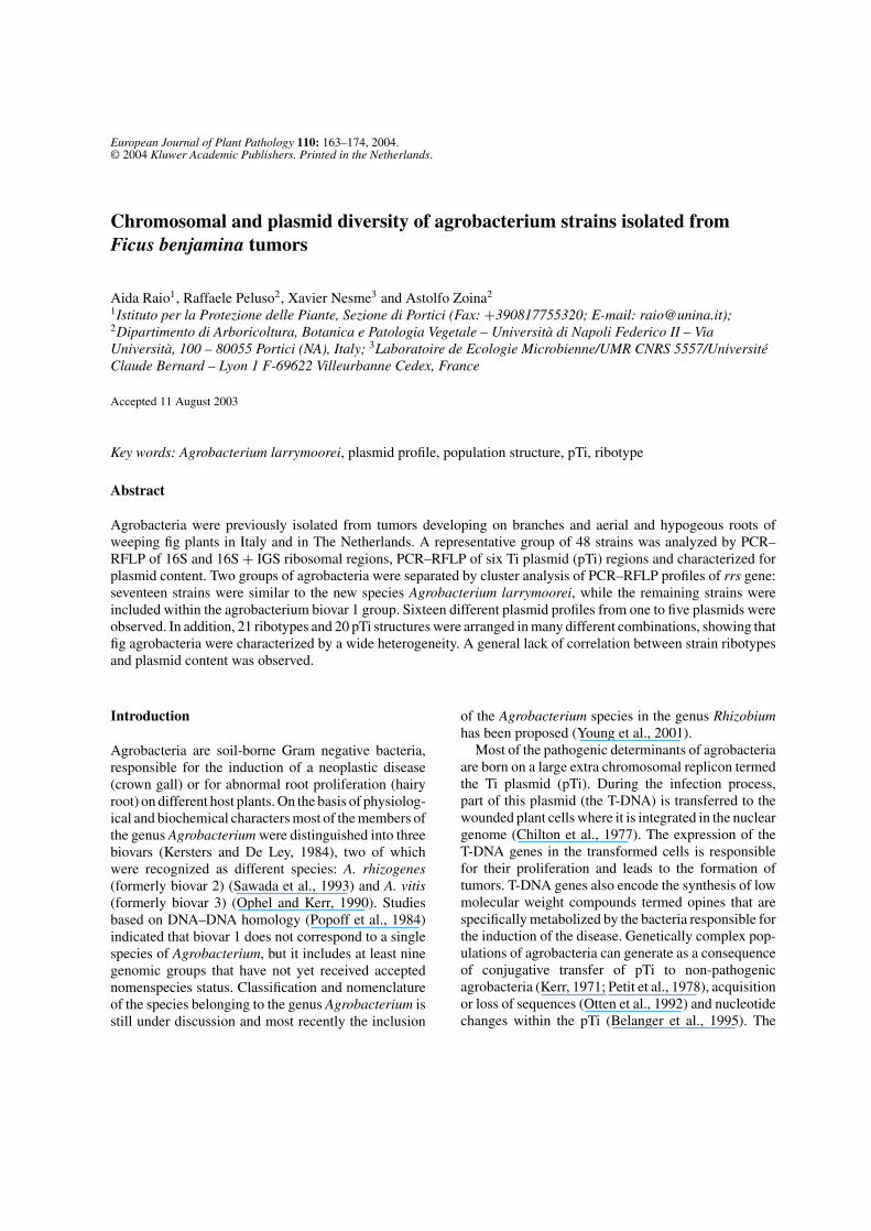
The ribosomal region 16S of 1500 bp was amplifiedin PCR with primers FGPS6 and FGPS1509′ in allthe tested strains. Amplified DNA was digested withAccI, AluI, BfaI, HaeIII and HpaII and the length ofDNA fragments of the agrobacterium reference strainswas identical to that expected according to the 16SrDNA sequence (Willems and Collins, 1993), thusshowing that amplified DNAs corresponded to the16S gene. Strains of A. larrymoorei from fig weredistinguished from the biovar 1 reference strains, theother fig agrobacteria and A. vitis by RFLP using theenzymes AluI, HaeIII and HpaII. Distinction betweenA. larrymoorei and Agrobacterium rubi was obtainedby digesting the 16S amplicons with AccI, BfaI andHaeIII. Cluster analysis of the restriction profiles ofrrs gene separated the fig agrobacteria into two groups(Figure 1): Seventeen strains were clustered withA. larrymoorei reference strains AF3.10 and AF3.44,
the remaining strains, clustered with agrobacteriumbiovar 1 reference strains. Only Fb194 belonged tothe genomic group G6–G8 as C58 reference strainwhile all other strains belonged to the genomic groupG3–G4 as B6 (Figure 1). Since PCR–RFLP analysisof rrs gene does not allow the distinction betweenG3 and G4 and between G6 and G8 genomic groups,results of this analysis show that at least two of thenine hybridization groups included within the biovar 1were found among fig strains.
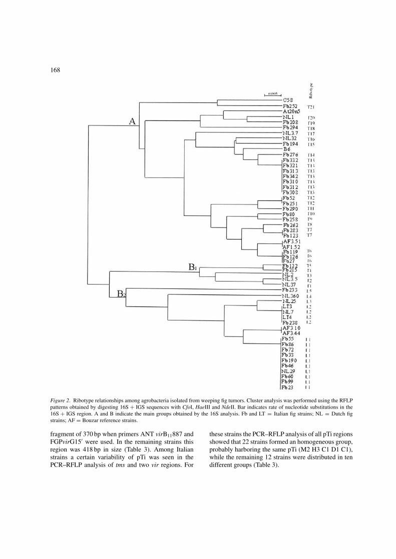
Amplification with primers FGPS6 and FGPL132′gave a product of about 2700 bp as expected foragrobacteria strains (Ponsonnet and Nesme, 1994). Thedendrogram obtained by the cluster analysis of PCR–RFLP profiles of 16S + IGS region is reported inFigure 2. The 17 strains shown to be A. larrymooreiin this study were grouped into the cluster B2; withinthis group of strains five different ribotypes were dis-tinguished (L1–L5). Clusters A and B1 included theremaining 31 strains that were distinguished into 21different ribotypes (T1–T21). All Italian and Dutchstrains were distinguishable from Bouzar’s referencestrain AF3.10.
Plasmid profiles
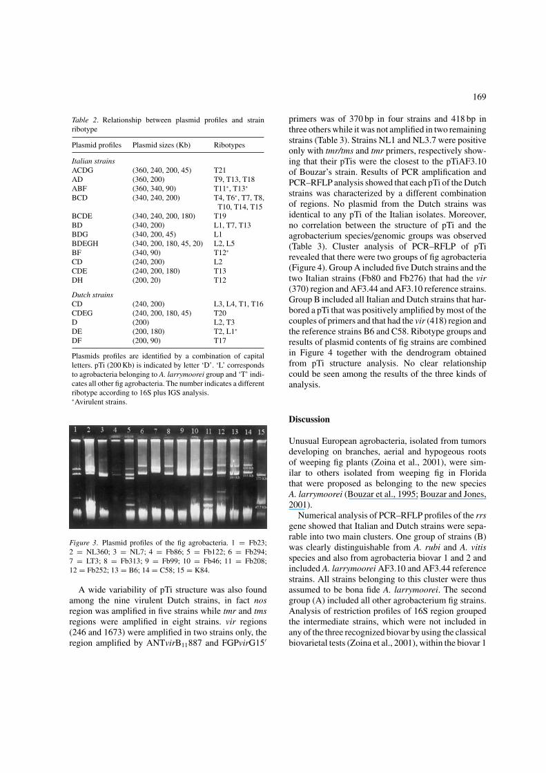
The analysis of plasmid profiles showed a high vari-ability for this character among fig agrobacteria: eightplasmids ranging from 20 to 360 Kb in size were com-bined in 16 different profiles (Table 2). Some examplesof plasmid profiles are reported in Figure 3. No definitecorrelation between plasmid profile and agrobacteriumspecies was found. In fact, the different combinationof plasmids were found both among the A. larrymooreigroup and the other fig agrobacteria. The highest vari-ability was found among the Italian strains of ribotypegroup ‘T’ that showed nine different profiles whileonly four profiles were found within the A. larrymooreigroup (Table 2). Profiles of Italian strains were char-acterized by the association of a minimum of two toa maximum of five plasmids. Among the plasmid pro-files exhibited by the Italian agrobacteria nine of themincluded a cryptic plasmid of 360 or 340 Kb that wasnever detected within the Dutch strains. Within theselatter strains, those belonging to the 16S + IGS group‘T’ harbored 1–4 plasmids combined in five differentprofiles, while Dutch A. larrymoorei strains showedonly two types of profiles made of one and two plas-mids, respectively. The profile characterized by twoplasmids of 240 and 200 bp was the only one that
167
Figure 1. Dendrograms of phylogenetic relatednes between agrobacterial strains isolated from fig tumors based upon RFLP analysis ofrrs PCR products obtained with AccI, AluI, BfaI, HaeIII and HpaII. Strains B6, C58 and AF3.10 are respectively members of the genomicspecies G4, G8 and A. larrymooreii. The bar indicates the scale of the genetic distance given in rate of nucleotide substitution.
was found both in Italian and Dutch strains (Table 2).Hybridization with pTi probes was done on 18 strainsindicating in all instances that the 200 Kb plasmid wasthe pTi (data not shown). Combined analysis of plas-mid profiles and 16S + IGS restriction patterns of the48 isolates showed that isolates belonging to a givenribotype could harbor different combinations of plas-mids and that genetically different strains could harborthe same plasmid combination (Table 2).
PCR–RFLP analysis of pTi
PCR–RFLP analysis of pTi regions of 43 tumorigenicstrains allowed the distinction of 20 different pTi pro-files. Amplification products of tmr, tms, nos and threevir regions of pTi where obtained from the majority ofthe Italian strains.
Moreover Fb80 and Fb276 were the only Italianstrains that, like the B6 reference strain, produced a
168
Figure 2. Ribotype relationships among agrobacteria isolated from weeping fig tumors. Cluster analysis was performed using the RFLPpatterns obtained by digesting 16S + IGS sequences with CfoI, HaeIII and NdeII. Bar indicates rate of nucleotide substitutions in the16S + IGS region. A and B indicate the main groups obtained by the 16S analysis. Fb and LT = Italian fig strains; NL = Dutch figstrains; AF = Bouzar reference strains.
fragment of 370 bp when primers ANT virB11887 andFGPvirG15′ were used. In the remaining strains thisregion was 418 bp in size (Table 3). Among Italianstrains a certain variability of pTi was seen in thePCR–RFLP analysis of tms and two vir regions. For
these strains the PCR–RFLP analysis of all pTi regionsshowed that 22 strains formed an homogeneous group,probably harboring the same pTi (M2 H3 C1 D1 C1),while the remaining 12 strains were distributed in tendifferent groups (Table 3).
169
Table 2. Relationship between plasmid profiles and strainribotype
Plasmid profiles Plasmid sizes (Kb) Ribotypes
Italian strainsACDG (360, 240, 200, 45) T21AD (360, 200) T9, T13, T18ABF (360, 340, 90) T11∗, T13∗
BCD (340, 240, 200) T4, T6∗, T7, T8,T10, T14, T15
BCDE (340, 240, 200, 180) T19BD (340, 200) L1, T7, T13BDG (340, 200, 45) L1BDEGH (340, 200, 180, 45, 20) L2, L5BF (340, 90) T12∗
CD (240, 200) L2CDE (240, 200, 180) T13DH (200, 20) T12
Dutch strainsCD (240, 200) L3, L4, T1, T16CDEG (240, 200, 180, 45) T20D (200) L2, T3DE (200, 180) T2, L1∗
DF (200, 90) T17
Plasmids profiles are identified by a combination of capitalletters. pTi (200 Kb) is indicated by letter ‘D’. ‘L’ correspondsto agrobacteria belonging to A. larrymoorei group and ‘T’ indi-cates all other fig agrobacteria. The number indicates a differentribotype according to 16S plus IGS analysis.∗Avirulent strains.
Figure 3. Plasmid profiles of the fig agrobacteria. 1 = Fb23;2 = NL360; 3 = NL7; 4 = Fb86; 5 = Fb122; 6 = Fb294;7 = LT3; 8 = Fb313; 9 = Fb99; 10 = Fb46; 11 = Fb208;12 = Fb252; 13 = B6; 14 = C58; 15 = K84.
A wide variability of pTi structure was also foundamong the nine virulent Dutch strains, in fact nosregion was amplified in five strains while tmr and tmsregions were amplified in eight strains. vir regions(246 and 1673) were amplified in two strains only, theregion amplified by ANTvirB11887 and FGPvirG15′
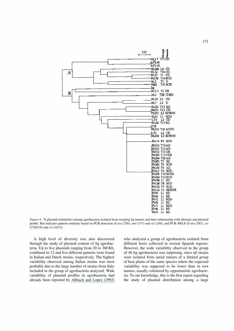
primers was of 370 bp in four strains and 418 bp inthree others while it was not amplified in two remainingstrains (Table 3). Strains NL1 and NL3.7 were positiveonly with tmr/tms and tmr primers, respectively show-ing that their pTis were the closest to the pTiAF3.10of Bouzar’s strain. Results of PCR amplification andPCR–RFLP analysis showed that each pTi of the Dutchstrains was characterized by a different combinationof regions. No plasmid from the Dutch strains wasidentical to any pTi of the Italian isolates. Moreover,no correlation between the structure of pTi and theagrobacterium species/genomic groups was observed(Table 3). Cluster analysis of PCR–RFLP of pTirevealed that there were two groups of fig agrobacteria(Figure 4). Group A included five Dutch strains and thetwo Italian strains (Fb80 and Fb276) that had the vir(370) region and AF3.44 and AF3.10 reference strains.Group B included all Italian and Dutch strains that har-bored a pTi that was positively amplified by most of thecouples of primers and that had the vir (418) region andthe reference strains B6 and C58. Ribotype groups andresults of plasmid contents of fig strains are combinedin Figure 4 together with the dendrogram obtainedfrom pTi structure analysis. No clear relationshipcould be seen among the results of the three kinds ofanalysis.
Discussion
Unusual European agrobacteria, isolated from tumorsdeveloping on branches, aerial and hypogeous rootsof weeping fig plants (Zoina et al., 2001), were sim-ilar to others isolated from weeping fig in Floridathat were proposed as belonging to the new speciesA. larrymoorei (Bouzar et al., 1995; Bouzar and Jones,2001).
Numerical analysis of PCR–RFLP profiles of the rrsgene showed that Italian and Dutch strains were sepa-rable into two main clusters. One group of strains (B)was clearly distinguishable from A. rubi and A. vitisspecies and also from agrobacteria biovar 1 and 2 andincluded A. larrymoorei AF3.10 and AF3.44 referencestrains. All strains belonging to this cluster were thusassumed to be bona fide A. larrymoorei. The secondgroup (A) included all other agrobacterium fig strains.Analysis of restriction profiles of 16S region groupedthe intermediate strains, which were not included inany of the three recognized biovar by using the classicalbiovarietal tests (Zoina et al., 2001), within the biovar 1

170
Table 3. PCR–RFLP analysis of pTi
Italian strains Species orpresumptivegenomicgroup
Amplifications Restriction profiles
nos tmr tms vir vir vir tms vir vir(256) (171) (587) (246) (370∗/
418)(1673) (587) (370/418) (1673)
CfoI DdeI MseI HpaII CfoI
Fb33, Fb46,Fb55, Fb60
A.L.1 + + + + + + C1 D1 M2 H3 C1
Fb72, Fb86,Fb99, Fb190
A.L. + + + + + + C1 D1 M2 H3 C1
Fb122, Fb123,Fb126, Fb208
G3 or G42 + + + + + + C1 D1 M2 H3 C1
Fb215, Fb258,Fb262, Fb283
G3 or G4 + + + + + + C1 D1 M2 H3 C1
Fb294, Fb308,Fb312, Fb313
G3 or G4 + + + + + + C1 D1 M2 H3 C1
Fb332 G3 or G4 + + + + + + C1 D1 M2 H3 C1Fb194 G6 or G82 + + + + + + C1 D1 M2 H3 C1Fb23 A.L. + + + + + + C2 D1 M2 H2 C1Fb119 G3 or G4 + + + + + + C1 D1 M2 H3 C2Fb233 A.L. + + + + + + C1 D1 M1 H3 C2Fb251 G3 or G4 + + + + + + C1 D1 M1 H3 C2Fb80 G3 or G4 + + + + +∗ − C1 D5 M3 H1 −Fb238, LT3 A.L. + + + + + + C1 D1 M2 H3 C4Fb252 G3 or G4 + + + + + + C1 D1 M2 H3 C3Fb276 G3 or G4 − + + + +∗ − C1 D3 M3 H1 −Fb321 G3 or G4 + + + + + + C1 D4 M1 H3 C2Fb342 G3 or G4 + − + + + − C1 D1 M2 H3 −LT4 A.L. + + + + + + C1 D1 M2 H2 C4
Dutch strainsNL1 G3 or G4 − + + − − − C1 D1 − − −NL2 G3 or G4 + + + − +∗ − C1 D1 M3 H1 −NL3.5 G3 or G4 + + + − + − C3 D7 M2 H4 −NL3.7 G3 or G4 − + − − − − − − − − −NL7 A.L. + + + + + + C1 D1 M4 H4 C4NL25 A.L. + + + + + + C1 D6 M4 H4 C4NL32 A.L. + − + − +∗ − C1 D3 M3 H1 −NL37 G3 or G4 − + + − +∗ − C1 D3 M3 H1 −NL360 A.L. − + + − +∗ − C2 D3 M3 H1 −Reference strainsAF3.10, AF3.44 A.L. − − − − − − − − − − −B6 G3 or G4 − + + − +∗ − C1 D2 M3 H1 −C58 G6 or G8 + + + + + + C1 D1 M2 H3 C3
1A.L.: A. larrymoorei.2Groups G3, G4, G6 and G8 are defined by the 16S sequences of the genomic groups described by Mougel et al. (2002).
cluster showing that the identification of agrobacteriawas more precise and reliable by using the moleculartechnique.
PCR–RFLP analysis of 16S and the IGS between16S and 23S, was performed to determine the geneticdifferences within the two Agrobacterium groups.Twenty-six different genotypes were identified using
three restriction enzymes showing a wide genetic diver-sity among fig agrobacteria. High genetic variabilitywas observed among the Dutch strains, since each oneshowed a unique restriction profile of the 16S + IGSribosomal region, even though a limited number ofstrains from The Netherlands was included in the groupof agrobacteria analyzed.
171
Figure 4. Ti plasmid similarities among agrobacteria isolated from weeping fig tumors and their relationship with ribotype and plasmidprofile. Bar indicates pattern similarly based on PCR detection of nos (256), tmr (171) and vir (246), and PCR–RFLP of tms (587), vir(370/418) and vir (1673).
A high level of diversity was also discoveredthrough the study of plasmid content of fig agrobac-teria. Up to five plasmids ranging from 20 to 360 Kb,combined in 12 and five different patterns were foundin Italian and Dutch strains, respectively. The highestvariability observed among Italian strains was mostprobably due to the large number of strains from Italyincluded in the group of agrobacteria analyzed. Widevariability of plasmid profiles in agrobacteria hadalready been reported by Albiach and Lopez (1992)
who analyzed a group of agrobacteria isolated fromdifferent hosts collected in several Spanish regions.However, the wide variability observed in the groupof 48 fig agrobacteria was surprising, since all strainswere isolated from aerial tumors of a limited groupof host plants of the same species where the expectedvariability was supposed to be lower than in roottumors, usually colonized by opportunistic agrobacte-ria. To our knowledge, this is the first report regardingthe study of plasmid distribution among a large
172
number of agrobacteria isolated from one single plantspecies and from three different locations.
In agreement with other studies (Albiach and Lopez,1992; Pulawska et al., 1998) no correlation betweenplasmid profile and geographical origin, virulence,biovar or bacterial ribotypes was found. The casual dis-tribution of plasmids among the fig strains was alsorevealed by comparing the results of plasmid profiletyping with PCR–RFLP analysis of 16S + IGS ribo-somal region. In fact, a given plasmid profile wasobserved in different ribotypes, moreover some strainsthat were identical at 16S + IGS analysis harboreddifferent plasmid combinations.
Conjugal transfer of plasmids can be the mostimportant source of heterogeneity for plasmid con-tent (Genetello et al., 1997; Stockwell et al., 1990).Probably plasmid exchange occurred between virulentand avirulent fig agrobacteria (Genetello et al., 1997;Hooykaas et al., 1977) or between agrobacteria andother bacteria living as endophytes inside fig plants(Zoina et al., 2002) since transfer of pTi to otherbacterial species has been demonstrated among soilmicroflora (Teyssier-Cuvelle et al., 1999; van Veenet al., 1988).
Fig strains showed some differences when analyzedfor some pTi-borne characters such as opine utilization,host range and agrocine 84 sensitivity (Zoina et al.,2001). These observations led us to hypothesize thatsome differences could exist among the pTi of dif-ferent strains. PCR–RFLP analyses of nos, tmr, tmsand three vir regions of pTi demonstrated that mostof the strains harbored a pTi nopaline type and thatamong the strains that were positive at the amplifica-tion with nos primers 14 different pTi profiles weredistinguished. The most frequent of them was sharedby a group of 22 Italian strains that included a part ofA. larrymoorei strains; within these 22 isolates, eightbelonged to the same ribotype and could then be con-sidered isolates of the same strain (Tenover et al., 1995).However, nopaline pTis were closely related to oneanother and probably originated from a common ances-tor and were subjected to some minor changes thattook place during their respective evolution. Our find-ings are in agreement with previous studies where ahigh degree of homogeneity was found within nopa-line strains (Knauf et al., 1983; Michel et al., 1990).Homology studies have shown that pTis are composedof homologous and non-homologous sequences thatmost likely are acquired by horizontal gene transfersince several sequences of octopine and nopaline-type
plasmids have been found in other pTi types and evenin other bacterial species.
This sequence exchange can explain the evolution ofpTi as a mosaic structure (Otten et al., 1992). Moreover,part of pTi variability is due to the insertion elementsthat are known to produce deletion, rearrangements,amplifications and changes in gene expression (Ottenet al., 1992). Tumors are the preferred sites where DNAexchanges take place and it is well known that theopines secreted in tumor tissues induce the conjuga-tive transfer of pTi (Dessaux et al., 1992; Ellis et al.,1982; Moore et al., 1997). This could explain boththe association of identical pTis with several differentgenotypes, even belonging to different Agrobacteriumspecies, and the combination of one genotype withdifferent nopaline pTi structures that were found inF. benjamina galls.
No pTi of Italian and Dutch strains was identical tothe novel plasmid pTiAF3.10 of A. larrymoorei ref-erence strain that was negative in all amplifications,confirming its peculiar properties (Bouzar et al., 1995;Vaudequin-Dransart et al., 1995). Heterogeneity of pTiwas high within the Dutch group where each strainshowed a unique pTi pattern, each one different fromthose of the Italian strains. Moreover, amplification ofvir (370/418) showed that the size of 370 bp was morefrequent among the Dutch isolates, while only twoItalian strains out of the 34 analyzed, had this smallersized vir region.
Results of cluster analysis of PCR–RFLP of pTisshowed that Italian and Dutch strains could be sepa-rated into two groups that were distinguished mainlyby the difference in size of the vir (370/418) region. Anepidemiological study regarding agrobacteria isolatedfrom rose plants showed that strains harboring this virregion of 370 bp in size were able to utilize opines otherthan nopaline (Pionnat et al., 1999). Similarly, some ofthe agrobacteria isolated from weeping fig plants couldharbor a pTi belonging to an uncommon opine group.This putative uncommon pTi was harbored by differ-ent ribotypes belonging to biovar 1 and A. larrymooreistrains, thus evidencing that among these fig strainsno correlation between pTi structure and ribotype wasfound. Additional analyses are planned to explore therelatedness of these plasmids with the pTiAF3.10 andthe other pTi types in a more exhaustive way. Ottenet al. (1996) found a strong correlation between pTitype and ribotype among A. vitis strains. In this case,bacterial strains were from different geographic ori-gins and were selected on the basis of their different
173
opine type plasmids. Nopaline, octopine/cucumopineand vitopine pTi resulted strictly associated to givenchromosomal backgrounds. The majority of strainsthat were analyzed in the present study were isolatedfrom fig galled plants growing in one single nurserythat were multiplied by cutting techniques: a homo-geneous population of agrobacteria was most likelyexpected. However overall results showed that the bac-terial population was composed at least of two differentAgrobacterium species, with several ribotypes. More-over, most of the strains harbored a pTi nopaline typethat showed some variability in the oncogenic regionsanalyzed. This variability appeared unrelated to thechromosomal background of fig strains, thus suggest-ing that the changes occurred within the pTis wereindependent from the chromosomal characteristics ofstrains. The occurrence of agrobacteria belonging totwo separate rrs genotypes in fig tumors indicates thatthe disease initiation was likely due to at least two out-break strains (Tenover et al., 1995). Genetic exchangesthat may have taken place between these agrobacteriaand other species of bacteria living endophytically inthe plants (Zoina et al., 2002), could have produceda certain variability inside the population and conse-quently a lack of correlation between ribotypes andplasmid content. Endophytic behavior of agrobacteriaseems to be a key point in the epidemiology of crowngall on F. benjamina (Zoina et al., 2002). Lehoczky(1968) reported that propagation of crown gall diseaseon grapevines was caused by the use of systemicallyinfected scions and rootstocks. Similarly, it is possiblethat the cutting technique used to propagate the plantsin the Italian nursery may have allowed the dissemi-nation of agrobacteria from the mother plants to thecuttings and consequent disease outbreaks.
Acknowledgements
The authors wish to thank Dr. Christophe Mougeland Mrs. Marilyn L. Miller for the fruitful scientificdiscussions and technical support and Mrs. DiannaPickens for language reviewing of the manuscript.This work was part of INCO-DC European projectERB1C18CT970198 (‘Integrated control of crown gallin Mediterranean countries’).
References
Albiach MR and Lopez MM (1992) Plasmid heterogeneity inSpanish isolates of Agrobacterium tumefaciens from thirteen
different hosts. Applied and Environmental Microbiology 58:2683–2687
Belanger C, Canfield ML, Moore LW and Dion P (1995) Geneticanalysis of nonpathogenic Agrobacterium tumefaciens mutantsarising in crown gall tumors. Journal of Bacteriology 177:3752–3757
Bouzar H, Chilton WS, Nesme X, Dessaux Y, Vaudequin V,Petit A, Jones JB and Hodge NC (1995) A new Agrobacteriumstrain isolated from aerial tumors on Ficus benjamina L.Applied and Environmental Microbiology 61: 65–73
Bouzar H and Jones JB (2001) Agrobacterium larrymoorei sp.nov. is proposed for the pathogenic strains isolated from aerialtumors of Ficus benjamina. International Journal of Systematicand Evolutionary Bacteriology 51: 1023–1026
Chilton MD, Drummond MH, Merio DJ, Sciaky D, Montoya AL,Gordon MP and Nester EW (1977) Stable incorporation ofplasmid DNA into higher plant cells: the molecular basis ofcrown gall tumorigenesis. Cell 11: 263–271
Coplin DL, Rowan RG, Chisholm DA and Whitmoyer RE (1981)Characterization of plasmids in Erwinia stewartii. Applied andEnvironmental Microbiology 42: 599–603
Currier TC and Nester EW (1976) Evidence for diverse typesof large plasmids in tumor-inducing strains of Agrobacterium.Journal of Bacteriology 126: 157–165
Dessaux Y, Petit A and Tempe J (1992) Opines in Agrobacteriumbiology. In: Verma DPS (ed) Molecular Signals in Plant–Microbe Communications (pp 109–136) CRC Press, Inc, BocaRaton, FL
Ellis JG, Kerr A, Petit A and Tempe J (1982) Conjugal transfer ofnopaline and agropine Ti-plasmid. The role of agrocinopines.Molecular and General Genetic 186: 269–274
Genetello C, van Larebeke N, Holsters M, de Picker A,van Montagu M and Schell J (1977) Ti plasmids ofAgrobacterium as a conjugative plasmids. Nature 265: 561–563
Hooykaas PJJ, Klapwijk PM, Nuti MP, Schilperoort RA andRorssch A (1977) Transfer of the Agrobacterium tumefaciensTi plasmid to avirulent agrobacteria and to Rhizobium ex planta.Journal of General Microbiology 98: 477–484
Kerr A (1971) Acquisition of virulence by non-pathogenicisolates of Agrobacterium radiobacter. Physiology and PlantPathology 1: 241–246
Kersters K and De Ley J (1984) Genus III. Agrobacterium Conn1942. In: Krieg NR and Holt JG (eds) Bergey’s Manual ofSystematic Bacteriology, Vol 1 (pp 244–254) The Williams &Wilkins Co., Baltimore
Knauf VC, Panagopoulos CG and Nester EW (1983) Comparisonof Ti plasmids from three different biotypes of Agrobacteriumtumefaciens isolated from grapevines. Journal of Bacteriology153: 1535–1542
Lehoczky J (1968) Spread of Agrobacterium tumefaciens in thevessels of grapevine after natural infection. PhytopathologischeZeitschrift 63: 239–246
Meyers JA, Sanchez D, Elwell LP and Falkow S (1976) Simpleagarose gel electrophoretic method for the identification andcharacterization of plasmid deoxyribonucleic acid. Journal ofBacteriology 127: 1529–1537
Michel MF, Brasileiro Miranda AC, Depierreux C, Otten L,Delmotte F and Jouanin L (1990) Identification of different

174
Agrobacterium strains isolated from the same forest nursery.Applied and Environmental Microbiology 56: 3537–3545
Moore LW and Canfield ML (1996) Biology of Agrobacteriumand management of crown gall disease. In: Hall R (ed)Principles and Practice of Managing Soilborne Plant Pathogens(pp 153–191) APS Press, St. Paul, Minnesota
Moore LW, Chilton WS and Canfield ML (1997) Diversity ofopines and opine-catabolizing bacteria isolated from naturallyoccurring crown gall tumors. Applied and EnvironmentalMicrobiology 63: 201–207
Mougel C, Thioulouse J, Perriere G and Nesme X (2002)A mathematical method for determining genome divergenceand species delineation using AFLP. International Journal ofSystematic and Evolutionary Microbiology 52: 573–586
Navarro E, Simonet P, Normand P and Bardi R (1992)Characterization of natural population of Nitrobacter spp. usingPCR–RFLP analysis of ribosomal intergenic spacer. Archivesof Microbiology 157: 107–115
Nazaret S, Counoyer B, Normand P and Simonet P (1991)Phylogenetic relationships among Frankia genomic speciesdetermined by use of amplified 16S rDNA sequences. Journalof Bacteriology 173: 1456–1471
Nei M and Li WH (1979) Mathematical model for studyinggenetic variation in terms of restriction endonucleases.Proceedings of the National Academy of Sciences USA 76:5269–5273
Nesme X, Picard C and Simonet P (1995) Specific DNAsequences for detection of soil bacteria. In: Trevors JT and vanElsas JD (eds) Nucleic Acids in the Environment (pp 110–139)Springer Lab Manual
Nesme X, Ponsonnet C, Picard C and Normand P (1992)Chromosomal and pTi genotypes of Agrobacterium strainsisolated from Populus tumors in two nurseries. FEMSMicrobiology Ecology 101: 189–196
Oger P, Dessaux Y, Petit A, Gardan L, Manceau C, Chomel C andNesme X (1998) Validity, sensitivity and resolution limit of thePCR–RFLP analysis of the rrs (16S rRNA gene) as a tool toidentify soil-borne and plant-associated bacterial population.Genetics Selection Evolution 30: 311–332
Ophel K and Kerr A (1990) Agrobacterium vitis sp. nov. for strainsof Agrobacterium biovar 3 from grapevines. InternationalJournal of Systematic Bacteriology 40: 236–241
Otten L, Canaday J, Gerard JC, Fournier P, Crouzet P and Paulus F(1992) Evolution of agrobacteria and their Ti plasmids –a review. Molecular Plant–Microbe Interaction 5: 279–287
Otten L, de Ruffray P, Momol EA, Momol MT and Burr TJ(1996) Phylogenetic relationships between Agrobacterium vitisisolates and their Ti plasmids. Molecular Plant–Microbe Inter-actions 9: 782–786
Popoff MY, Kersters K, Kiredjian M, Miras I and Coynault C(1984) Position taxonomique de souches de Agrobacteriumd’origene hospitalier. Annals of Microbiology (Inst. Pasteur)135: 427–442
Petit A, Tempe J, Kerr A, Holsters M, van Montagu M andSchell J (1978) Substrate induction of conjugative activ-ity of Agrobacterium tumefaciens Ti plasmids. Nature 271:570–572
Pionnat S, Keller H, Hericher D, Bettachini A, Dessaux Y,Nesme X and Poncet C (1999) Ti plasmid from Agrobacteriumcharacterize rootstock clones that initiated a spread ofcrown gall disease in Mediterranean countries. Applied andEnvironmental Microbiology 65: 4197–4206
Ponsonnet C and Nesme X (1994) Identification ofAgrobacterium strains by PCR–RFLP analysis of pTi andchromosomal regions. Archives of Microbiology 161: 300–309
Pulawska J, Malinowski T and Sobiczewski P (1998) Diversityof plasmids of Agrobacterium tumefaciens isolated from fruittrees in Poland. Journal of Phytopathology 146: 465–468
Sawada H, Ieki H, Oyaizu H and Matsumoto S (1993) Proposal forrejection of Agrobacterium tumefaciens and revised descrip-tions for the genus Agrobacterium and for Agrobacteriumradiobacter and Agrobacterium rhizogenes. InternationalJournal of Systematic Bacteriology 43: 694–702
Stockwell VO, Kawalek MD, Moore LW and Loper JE (1990)Plasmid transfer between Agrobacterium tumefaciens in thegenus Pinus. Plant Physiology 92: 1226–1232
Tenover FC, Arbeit RD, Goering RV, Mickelsen PA, Murray BE,Persing DH and Swaminathan B (1995) Interpretingchromosomal DNA restriction patterns produced by pulsed-field gel electrophoresis: Criteria for bacterial strain typing.Journal of Clinical Microbiology 33: 2233–2239
Teyssier-Cuvelle S, Mougel C and Nesme X (1999) Directconjugal transfers of Ti plasmid to soil microflora. MolecularEcology 8: 1273–1284
Vaudequin-Dransart V, Petit A, Poncet C, Ponsonnet C, Nesme X,Jones JB, Bouzar H, Chilton WS and Dessaux Y (1995) NovelTi plasmid in Agrobacterium strains isolated from fig treeand Chrysanthemum tumors and their opinelike molecules.Molecular Plant–Microbe Interaction 8: 311–321
van Veen RJM, den Dulk-Ras Bisseling HT, Schilperoort RAand Hooykaas PJJ (1988) Crown gall tumor and root noduleformation by the bacterium Phyllobacterium myrsinacearumafter the introduction of an Agrobacterium Ti plasmid or aRhizobium Sym plasmid. Molecular Plant–Microbe Interaction1: 231–234
Willems A and Collins D (1993) Phylogenetic analysis ofRhizobia and Agrobacteria based on 16S rRNA genesequences. International Journal of Systematic Bacteriology43: 305–313
Young JM, Kuykendall LD, Martinez-Romero E, Kerr A andSawada H (2001) A revision of Rhizobium Frank 1889, withan emended description of the genus, and the inclusion ofall species of Agrobacterium Conn 1942 and Allorhizobiumundicola de Lajudie et al. 1998 as new combinations:Rhizobium radiobacter, R. rhizogenes, R. rubi, R. undicola andR. vitis. International Journal of Systematic and EvolutionaryMicrobiology 51: 89–103
Zoina A, Raio A and Peluso R (2002) Endophytic behaviourof agrobacteria in Ficus benjamina L. In: Proceedings of‘L’endofitismo di funghi e batteri patogeni in piante arboree edarbustive’, 20–21 May (pp 127–136) Sassari, Italy
Zoina A, Raio A, Peluso R and Spasiano A (2001)Characterization of agrobacteria from weeping fig. PlantPathology 50: 620–627
Copyright © 2022 FDOKUMEN