Bovine sperm-oviduct interactions are characterized ... - Nature
Upload
independentCategory
view
0download
0
daf
U
Developmental Biology 214, 429–443 (1999)Article ID dbio.1999.9428, available online at http://www.idealibrary.com on
Cholesterol Efflux-Mediated Signal Transductionin Mammalian Sperm: Cholesterol Release Signalsan Increase in Protein Tyrosine Phosphorylationduring Mouse Sperm Capacitation
Pablo E. Visconti,*,1,4 XiaoPing Ning,*,1,2 Miguel W. Fornes,*,3
Juan G. Alvarez,† Paula Stein,*,‡ Stephanie A. Connors,*,§and Gregory S. Kopf**Center for Research on Reproduction & Women’s Health and ‡Department of Biology,University of Pennsylvania Medical Center, Philadelphia, Pennsylvania 19104-6080;†Department of Obstetrics and Gynecology, Beth Israel Deaconess Medical Center,Harvard Medical School, Boston, Massachusetts 02215; and §Department of Cell andDevelopmental Biology, University of Pennsylvania, Philadelphia, Pennsylvania 19104
We previously demonstrated that mouse sperm capacitation is accompanied by a time-dependent increase in protein tyrosinephosphorylation that is dependent on the presence of BSA, Ca21, and NaHCO3, all three of which are also required for thismaturational event. We also demonstrated that activation of protein kinase A (PK-A) is upstream of this capacitation-associatedincrease in protein tyrosine phosphorylation. BSA is hypothesized to modulate capacitation through the removal of cholesterolfrom the sperm plasma membrane. In this report, we demonstrate that incubation of mouse sperm medium containing BSAresults in a release of cholesterol from the sperm plasma membrane to the medium; release of this sterol does not occur inmedium devoid of BSA. We next determined whether cholesterol release leads to changes in protein tyrosine phosphorylation.Blocking the action of BSA by adding exogenous cholesterol-SO4
2 to the BSA-containing medium inhibits the increase in proteintyrosine phosphorylation as well as capacitation. This inhibitory effect is overcome by (1) the addition of increasingconcentrations of BSA at a given concentration of cholesterol-SO4
2 and (2) the addition of dibutyryl cAMP plus IBMX.High-density lipoprotein (HDL), another cholesterol binding protein, also supports the capacitation-associated increase in proteintyrosine phosphorylation through a cAMP-dependent pathway, whereas proteins that do not interact with cholesterol have noeffect. HDL also supports sperm capacitation, as assessed by fertilization in vitro. Finally, we previously demonstrated thatHCO3
2 is necessary for the capacitation-associated increase in protein tyrosine phosphorylation and demonstrate here, byexamining the effectiveness of HCO3
2 or BSA addition to sperm on protein tyrosine phosphorylation, that the HCO32 effect is
ownstream of the site of BSA action. Taken together, these data demonstrate that cholesterol release is associated with thectivation of a transmembrane signal transduction pathway involving PK-A and protein tyrosine phosphorylation, leading tounctional maturation of the sperm. © 1999 Academic Press
Key Words: mouse; sperm; capacitation; cholesterol; cAMP; protein tyrosine phosphorylation.
cpkCadisz1sz
INTRODUCTION
Mammalian sperm do not possess the ability to fertilize theegg immediately upon ejaculation, but acquire fertilization
1 The first two authors should be considered equal contributorso this work.
2 Current address: Wyeth Ayerst Laboratories, Princeton, NJ.3 Current address: Instituto de Histologıa y Embriologıa, Fac-
ltad de Ciencias Medicas, Universidad Nacional de Cuyo, Casillae Correo 56, Mendoza 5500, Argentina.
4 Current address: Department of Cell Biology and Anatomy,
iniversity of Virginia, Charlottesville, Virginia.0012-1606/99 $30.00Copyright © 1999 by Academic PressAll rights of reproduction in any form reserved.
ompetence following residence in the female tract for a finiteeriod. This time-dependent acquisition of fertilizing ability isnown as “capacitation” and was first observed by bothhang (1951, 1955) and Austin (1951, 1952); capacitation canlso be accomplished in vitro using defined media. Theefinition of capacitation has been modified over the years tonclude the acquisition of the ability of the acrosome-intactperm to undergo the acrosome reaction in response to theona pellucida (Florman and Babcock, 1991; Kopf and Gerton,991; Ward and Storey, 1984) or to other physiological stimuliuch as progesterone (Osman et al., 1989; Wistrom and Mei-el, 1993).Although capacitation can be mimicked in vitro by
t
ud
ncubating epididymal or ejaculated sperm in a defined
429

bi1tttrpt11adat
smtplceicamctitm
cmmcp
ttasBripto
fli
saa
ecWmiB
p
430 Visconti et al.
medium, the exact composition of media that supportcapacitation is variable for different species. Usually, thesemedia represent balanced salt solutions containing appro-priate concentrations of electrolytes, metabolic energysources, Ca21, HCO3
2, and a protein source that usually isovine serum albumin (BSA); this composition in manynstances approximates that of oviduct fluid (Yanagimachi,994). The function of these different medium componentso support capacitation in vitro is not well understood athe molecular level. Work in several species has suggestedhat the presence of serum albumin in the medium isesponsible for the removal of cholesterol from the spermlasma membrane which is known to occur during capaci-ation (Davis, 1976, 1981; Davis et al., 1979; Go and Wolf,985; Langlais and Roberts, 1985; Suzuki and Yanagimachi,989). It also was proposed that this loss of cholesterolccounts for the membrane fluidity changes that have beenocumented in many species during capacitation (Wolf etl., 1986b; Yanagimachi, 1994). The consequence of choles-erol loss on sperm function, however, is poorly understood.
Capacitation has also been correlated with changes inperm intracellular ion concentrations, metabolism, andotility (Harrison, 1996; Yanagimachi, 1994). Although
hese changes have been known for many years to accom-any the process of capacitation, the molecular basis under-ying these events is poorly understood. Remarkably, sinceapacitation in vitro can occur in the absence of any specificxternal stimulus, it is likely that the sperm cell canntrinsically control capacitation and that aspects of thisontrol lie within the sperm plasma membrane. For ex-mple, changes in the properties of the plasma membraneight lead to a derepression of a set of preprogrammed
ellular events ultimately necessary for the development ofhe capacitated state. This, however, does not preclude themportance of potential positive and/or negative modula-ion of capacitation in vivo by factors associated with theale and female reproductive tracts.Previously we demonstrated that incubation conditions
onducive to capacitation in vitro of cauda epididymalouse sperm promote the tyrosine phosphorylation ofultiple sperm proteins (Visconti et al., 1995a). This in-
rease in protein tyrosine phosphorylation, as well as ca-acitation, requires the presence of BSA, Ca21, and NaHCO3
in the medium (Visconti et al., 1995a). Interestingly, thecapacitation-associated up-regulation of protein tyrosinephosphorylation is regulated by cAMP/protein kinase A(PK-A) (Visconti et al., 1995b, 1997) through the stimula-ion of a tyrosine kinase and/or the inhibition of a proteinyrosine phosphatase. This unique crosstalk between PK-And tyrosine kinase signaling pathways appears unique toperm (Visconti et al., 1998). Although the requirement ofSA for capacitation is postulated to be responsible for theemoval of cholesterol from the sperm plasma membrane, its not clear how BSA functions to increase protein tyrosinehosphorylation. Our working hypothesis is that BSA func-ions as a sink for cholesterol and that changes in the levels
f plasma membrane cholesterol lead to changes in theCopyright © 1999 by Academic Press. All right
uidity of the plasma membrane and a subsequent increasen the permeability of the sperm to Ca21 and HCO3
2 (Yanagi-machi, 1994). The resultant increases in the levels of Ca21
and HCO32 would ultimately stimulate the activity of the
perm adenylyl cyclase, leading to an increase in cAMP andn increase in protein tyrosine phosphorylation (Visconti etl., 1995b, 1997).In the present study, we set out to determine whether the
ffect of BSA on protein tyrosine phosphorylation andapacitation in mouse sperm involved cholesterol removal.e demonstrate that cholesterol was released into theedium from sperm only when these cells were incubated
n the presence of BSA. Abrogation of cholesterol binding toSA by addition of cholesterol-SO4
2 to the medium blocksboth the increase in protein tyrosine phosphorylation andinhibits capacitation. As predicted, increasing concentra-tions of BSA, as well as cAMP agonists, overcame thischolesterol-SO4
2 inhibition on protein tyrosine phosphory-lation. Another cholesterol binding protein, namely high-density lipoprotein (HDL), replaced BSA in media to pro-mote protein tyrosine phosphorylation and fertilization invitro. On the other hand proteins that do not bind choles-terol are not able to increase protein tyrosine phosphoryla-tion. These data, taken together, demonstrate that choles-terol removal from the plasma membrane is linked to aunique signal transduction pathway that involves crosstalkbetween PK-A and tyrosine kinase signaling pathways lead-ing to protein tyrosine phosphorylation and functionalmaturation of the sperm.
MATERIALS AND METHODS
Materials
Cholesterol and cholesterol-SO42, BSA (Fraction V; fatty acid
poor), desmosterol, filipin, pregnenolone-SO42, androstenedione-
SO42, cholesterol oxidase (C-8153), protease inhibitors, ovalbumin,
cytochrome c, histone H2A, myelin basic protein, and kemptidewere obtained from Sigma. HDL that had been purified according toPomerantz and Hajjar (1990) was a gift from Ken Pomerantz,Cornell Medical College. Anti-phosphotyrosine antibody (clone4G10) was from UBI (Lake Placid, NY). Organic solvents were fromEM Sciences (chromatographic grade). Unibond RP18/silica geldiphasic plates (10 3 12 cm, 250-mm thickness) were obtained fromAnaltech, Inc. (Newark, DE). HP-K silica gel plates (10 3 10 cm,250-mm thickness) and P-81 phosphocellulose papers were pur-chased from Whatman, Inc. (Clifton, NJ). [g-32P]ATP (3000 Ci/mmol) and ECL reagents were from Amersham. Immobilon P waspurchased from Millipore (Bedford, MA) and EcoLite scintillationfluid was from ICN (Costa Mesa, CA).
Culture MediaThe basic medium used throughout these studies for the prepa-
ration of sperm was a modified Krebs–Ringer bicarbonate medium(HMB-Hepes buffered), as described by Lee and Storey (1986). Thismedium was first prepared in the absence of Ca21, BSA, andyruvate, sterilized by passage through a 0.20-mm filter (Nalgene),
and frozen at 220°C in aliquots for single use. Working “complete”
media were prepared by adding Ca21 (1.7 mM), pyruvate (1 mM),s of reproduction in any form reserved.

7
suw
mprcs(c(p
vW
t1t31
apcpssPctsa(
ss
7rtei
431Cholesterol Release during Sperm Capacitation
and BSA (3 mg/ml), followed by gassing with 5% CO2 in air to pH.3. HM medium was derived from HMB by replacing the NaHCO3
with NaCl, while maintaining the same pH. In some experiments,the concentrations of BSA or NaHCO3 were adjusted, and the pHwas maintained at 7.3.
Preparation of Sperm
Caudal epididymal mouse sperm were collected from CD1retired breeder males by placing one minced cauda epididymis in0.5 ml of medium HM without Ca21 or BSA. After 5 min the spermuspension was washed in 10 ml of the same medium by centrif-gation at 800g for 10 min at room temperature (24°C). The spermere resuspended to a final concentration of 5–10 3 107 cells/ml
and diluted 10 times in the appropriate medium depending on theexperiment performed. After incubation for 1.5 h, except whereindicated, the sperm were concentrated by centrifugation at 5000gfor 1 min (24°C), and the sperm pellet was washed in 1 ml ofphosphate-buffered saline (PBS), resuspended in sample buffer(Laemmli, 1970) without 2-mercaptoethanol, and boiled for 5 min.After centrifugation at 5000g for 3 min, the supernatant wasremoved, 2-mercaptoethanol was added to a final concentration of5%, and the samples were boiled for 5 min and then subjected toSDS–PAGE as described below.
Solubilization of Steroids
All of the steroid sulfates were solubilized using DMSO to a finalstock concentration of 100 mM. In contrast, cholesterol wasdirectly added to the incubation medium to the appropriate con-centration and the solution then sonicated. The final concentrationof DMSO was never higher than 0.5%; this concentration has noeffect on capacitation or the increase in protein tyrosine phosphor-ylation. The standard solutions of the different steroid–sulfatesused in these experiments were diluted to various final concentra-tions as stated in the designated experiment, added to the mediumcontaining BSA, and preincubated for 30 min prior to the additionof the sperm suspension.
Steroid Measurements
Sperm (5 3 106 cells) were incubated for 1.5 h in 0.5 ml ofedium in the absence or presence of 3 mg/ml BSA. After this
eriod, each sample was centrifuged (10,000g; 10 min) and theesultant sperm pellets and supernatants were assayed for sterolontent as described previously (Alvarez and Storey, 1995). Briefly,perm pellets were extracted with 20 vol of chloroform:methanol1:1, v/v) and sperm supernatants were extracted with 6 vol ofhloroform:methanol (2:1, v/v). The samples were then vortexed10 s) and centrifuged at 800g (3 min) and the resultant organichases evaporated to dryness under N2. The evaporated organic
phases were then dissolved in 20 ml of chloroform:methanol (1:1,/v) and aliquots of 4 ml applied to silver nitrate-impregnatedhatman HP-K silica gel microplates. Aliquots (4 ml) of choles-
terol, desmosterol, cholesterol sulfate, and desmosterol standardswere applied on separate lanes as reference standards. The plateswere predeveloped in chloroform:methanol (1:1, v/v) to 1 cm fromthe lower edge of the plate. This predevelopment step is used tominimize eddy diffusion that results in band broadening and lowerresolution. Following predevelopment, the plates were thoroughlydried and then developed in chloroform:acetone (95:5, v/v) in the
same dimension. Following development, the plates were thor- iCopyright © 1999 by Academic Press. All right
oughly dried, dipped in a 10% solution of copper sulfate in 8%phosphoric acid, and placed on a CAMAG Plate Heater III at 185°Cfor 5 min. The resulting bands were scanned at 400 nm in thereflectance mode using a Shimadzu CS-9000 spectrodensitometer(Shimadzu Scientific Instruments, Columbia, MD). The integratedareas obtained for the unknowns were interpolated with thestandard curves obtained for the respective cholesterol, desmos-terol, and cholesterol sulfate standard curves, and the values wereexpressed as nanograms per 106 cells.
To control for nonspecific release of cholesterol from the spermdue to the centrifugation step itself under the different incubationconditions (6BSA), the following controls were performed. Spermsuspensions were incubated in medium in either the absence or thepresence of BSA as described above but, prior to centrifugation, 50ml of a 4 mg/ml solution of cholesterol oxidase in an isotonic buffer(pH 7.4) was added to 500 ml of the sperm suspension (5 3 106
sperm). The suspension was incubated at 24°C for 10 min. Enzymereactions were terminated by the addition of 50 ml of 3 N HCl andhe tubes were then vortexed for 5 s. Following centrifugation at0,000g for 10 min, the sperm pellet and the sperm-free superna-ant were assayed for the presence of cholesterol and 4-cholestene--one (the product of cholesterol oxidation) (Alvarez and Storey,995).
Identification of Filipin–Cholesterol Complexesin Mouse Sperm
Cauda epididymal sperm were obtained as described above.Aliquots of 106 sperm were then incubated in HMB medium in thebsence or presence of 3 mg/ml BSA for 1.5 h. After this incubationeriod, sperm were analyzed for the presence of plasma membraneholesterol by monitoring the formation of filipin–sterol com-lexes by freeze–fracture electron microscopy as previously de-cribed (Diaz-Fontdevila et al., 1992). Briefly, the sperm suspen-ions were centrifuged at 500g for 10 min and washed twice withBS. The final sperm pellet was resuspended in a fixative solutionontaining 3% glutaraldehyde in 0.1% cacodylate buffer, pH 7.4, inhe presence of 0.02% filipin for 1 h at 4°C. Filipin was initiallyolubilized in DMSO as a 1% solution. An aliquot of sperm waslso fixed in the absence of filipin and served as a control. Aliquots500 ml) of fixed sperm were centrifuged in a microfuge at maxi-mum speed for 1 min and the sperm pellets were resuspended andincubated in 500 ml of 25% glycerol in PBS. After 2 h, theuspensions were centrifuged and the pellets mounted on goldupports, precooled in supercooled liquid N2, and fractured using a
double replica device in a Balzers BAF-301 apparatus with a stagetemperature of 2100°C. The specimens were coated with platinum(3–5 nm) and carbon (25 nm). The surface replicas were cleanedwith Clorox and washed in double-distilled water. Specimens werethen observed using a Siemens Elmiskop I at 80 kV.
SDS–PAGE and Immunoblotting
SDS–PAGE (Laemmli, 1970) was performed in 10% gels. Elec-trophoretic transfer of proteins to Immobilon P in all experimentswas carried out according to the method of Towbin et al. (1979) at0 V (constant) for 2 h at 4°C. Immunodetection was carried out atoom temperature as described previously (Kalab et al., 1994) usinghe 4G10 monoclonal antibody against anti-phosphotyrosine andnhanced chemiluminescence detection using an ECL kit accord-ng to the manufacturer’s instructions. Where appropriate, changes
n protein tyrosine phosphorylation were quantified by densito-s of reproduction in any form reserved.

t
Ttspwps
Mw
432 Visconti et al.
metric integration of the band of interest in the scanned film imageusing the NIH Image software.
Assay of Protein Kinase A Activity
Protein kinase A activity was measured as previously described(Visconti et al., 1997) using kemptide as a specific peptide sub-strate. Briefly, sperm were adjusted to a final concentration of 107
cells/ml and incubated for various periods of time either in theabsence or in the presence of 3 mg/ml BSA or 1 mg/ml HDL. At theappropriate time points, 10 ml of the sperm suspension was addedo 10 ml of 23 assay cocktail. The final concentrations of the
components in the assay cocktail after sperm addition were 100mM kemptide, [g-32P]ATP (3000 Ci/mmol) (3 3 106 cpm/assay), 100mM ATP, 1% (v/v) Triton X-100, 1 mg/ml BSA, 10 mM MgCl2, 40mM b-glycerophosphate, 5 mM p-nitrophenyl phosphate, 10 mMTris–HCl, pH 7.4, 10 mM aprotinin, and 10 mM leupeptin. Note thatthe BSA present in the assay cocktail was added for the purposes ofcoprecipitation of the proteins following the enzymatic assay ofPK-A. The samples then were incubated for 5 min at 37°C; theassay was linear at this time point using a variety of sperm proteinconcentrations. The reactions were stopped by adding 20 ml 20%
CA, cooled on ice for 20 min, and centrifuged at room tempera-ure for 3 min at 10,000g. Thirty microliters of the resultantupernatant was then spotted onto 2 3 2-cm Whatman P81hosphocellulose papers. The phosphocellulose papers wereashed 53 5 min in 5 mM phosphoric acid with agitation, dried,laced in vials with 2.5 ml of EcoLite scintillation cocktail, andubjected to liquid scintillation counting.
Coomassie Blue Assay for the Acrosome Reaction
As an endpoint of capacitation, we analyzed the zona pellucida(ZP)-induced acrosome reaction, based on the premise that onlycapacitated sperm undergo exocytosis in response to the ZP. Thepercentage of acrosome reactions was measured using CoomassieBlue G250 as described by Thaler and Cardullo (1995). Briefly,sperm were incubated under the desired experimental conditionsfor 1.5 h, followed by the addition of 5 ZP/ml or buffer, and thesperm were then incubated for an additional 30 min. ZP wereisolated and solubilized as previously described (Visconti et al.,1995a). An equal volume of 23 fixative solution (7.5% formalde-hyde in 10 mM PBS) was then added to each tube. After 10 min, thesperm were centrifuged for 2 min at 10,000g and resuspended in 0.1
ammonium acetate, pH 9. After centrifugation, the sperm pelletas resuspended in 20 to 50 ml of the same buffer, spread onto
poly-L-lysine-coated slides, and air dried. The slides were thenstained with Coomassie Blue G250 [0.04% (w/v) in 3.5% (v/v)perchloric acid]. After 10 min, the slides were gently rinsed withdeionized H2O until the slides appeared blue; the slides were thenair dried and mounted with Permount. To calculate the percentageof acrosome reactions at least 200 sperm were counted per experi-mental condition. The data presented are the average of at leastthree individual experiments.
In Vitro Fertilization
In vitro fertilization of metaphase II-arrested mouse eggs wasperformed as previously described (Visconti et al., 1995a). Polyvi-nyl alcohol (0.01%) was used to maintain the isotonic conditionswhen sperm were cultured in media devoid of BSA prior to
insemination. When HDL was included at different concentrationsCopyright © 1999 by Academic Press. All right
in the capacitation media, the medium containing the capacitatedsperm was diluted 100-fold prior to insemination of the eggs. Spermwere capacitated in the different media for a period of 2 h and thenincubated with the eggs for 3 h. When HDL was present in thecapacitation media, HDL was also present in the inseminationmedia. All in vitro fertilizations were performed using metaphase-arrested eggs retrieved at approximately 14 h post-hCG injection.Phase-contrast optics were used to evaluate fertilization by lookingfor the presence of the second polar body and the formation of boththe male and the female pronuclei.
Statistical Analyses
Statistical analyses were performed using a Student t test asdescribed by Zar (1996).
RESULTS
Cholesterol Is Released from the Sperm PlasmaMembrane under Conditions Conduciveto Capacitation
Previously we demonstrated in mouse sperm that bothprotein tyrosine phosphorylation and capacitation requiredthe presence of BSA in the incubation medium. The role ofBSA in capacitation has been postulated to involve theremoval of cholesterol from the sperm membrane (seeIntroduction). To determine if BSA, indeed, mediated cho-lesterol removal and led to the capacitation-associatedincreases in tyrosine phosphorylation, we first determinedwhether BSA was involved in the release of cholesterolfrom the sperm plasma membrane under our experimentalconditions. Two different approaches were used to test thishypothesis. First, we directly measured the cholesterolreleased into the incubation medium, as well as thatremaining in the sperm under conditions that do or do notsupport capacitation. Second, we visualized the cholesterolremaining in the plasma membrane of sperm incubated inthe absence or the presence of BSA, using the polyeneantibiotic filipin.
Cauda epididymal sperm were incubated in mediumHMB in the presence or in the absence of 3 mg/ml BSA.After 1.5 h of incubation, cholesterol released to the incu-bation medium as well as the cholesterol remaining withthe sperm under these two experimental conditions wasdetermined. A significant increase in medium cholesterolwas observed in sperm suspensions incubated in mediumcontaining BSA compared to sperm suspensions incubatedin medium devoid of BSA (Table 1), suggesting that mediumthat supports capacitation promotes the release of sperm-associated cholesterol. This observation was correlatedwith a decrease in the cholesterol associated with theresultant sperm pellets. Incubation of sperm in mediumcontaining BSA also increased the release of desmosterolinto the incubation medium, but did not affect the releaseof cholesterol-SO4
2 to a measurable extent (Table 1).In order to rule out the possibility that the release of
cholesterol from sperm incubated in medium containing
BSA was due, either in part or completely, to a nonspecifics of reproduction in any form reserved.

tvmf
433Cholesterol Release during Sperm Capacitation
release as a consequence of the centrifugation process,cholesterol oxidase was added to the sperm suspensionsafter the incubation but prior to the centrifugation (seeMaterials and Methods). The enzyme was then inactivatedprior to centrifugation. Membrane-bound cholesterol wouldnot serve as a substrate for this enzyme but, in contrast, anycholesterol released during the incubation period would bequantitatively converted to cholestene-3-one. Under theseconditions, any cholesterol subsequently released during
TABLE 1BSA-Induced Release of Cholesterol, Desmosterol, andCholesterol-SO4
2 from Mouse Sperm
Treatment Fraction
Steroidsa,b
C D CSO42
2BSA Medium 5 6 1.6 3 6 1 NDSperm 416 6 21 277 6 12 19 6 3
1BSA Medium 59 6 5.6* 43 6 3.3* NDSperm 363 6 19** 238 6 14** 17 6 2.7
a Mean 6 SEM (n 5 10) sterol content (ng/106 cells) for spermand supernatant fractions after a 1.5-h incubation in the absence orpresence of BSA (3 mg/ml). Media not containing sperm had nodetectable sterol content (data not shown).
b Abbreviations used: C, cholesterol; D, desmosterol; CSO42,
cholesterol sulfate; ND, not detectable. For incubations in mediumcontaining BSA, sperm contain less (**P , 0.01) and the mediumcontains more (*P , 0.0005) cholesterol and desmosterol, com-pared to incubations in medium devoid of BSA.
FIG. 1. Identification of filipin–cholesterol complexes in mouse sor in the presence (B) of 3 mg/ml BSA for 1.5 h. After this incubatcholesterol by monitoring the formation of filipin–cholesterol cMaterials and Methods. Note that these complexes appear as spher
times with similar results.Copyright © 1999 by Academic Press. All right
he centrifugation step and after cholesterol oxidase inacti-ation would be measured as cholesterol. Using this experi-ental approach, all of the cholesterol released was in the
orm of cholestene-3-one (50 6 3.2 ng sterol/106 cells) andthe cholesterol remaining associated with the sperm re-mained as cholesterol (270 6 24 ng sterol/106 cells). Incontrast when the sperm were incubated in media devoid ofBSA neither cholestene-3-one nor cholesterol was found inthe medium and all of the sperm-associated sterol was inthe form of cholesterol (320 6 23 ng sterol/106 cells). Nocholesterol-3-one was detected when cholesterol oxidasewas not added (data not shown). These data demonstratethat the cholesterol released into the medium during theincubation does not occur as a consequence of the centrif-ugation step.
Changes in the cholesterol content of the sperm plasmamembrane were also monitored by exploiting thecholesterol-binding properties of the polyene antibioticfilipin (Diaz-Fontdevila et al., 1992; Lin and Kan, 1996;Suzuki, 1988; Suzuki and Yanagimachi, 1989; Toshimori etal., 1985). Using freeze–fracture electron microscopic anal-ysis, the cholesterol–filipin complexes were visualized asspherical particles (Fig. 1). In contrast to sperm incubated inmedium devoid of BSA (Fig. 1A), the density and number ofcholesterol–filipin complexes were significantly lower insperm incubated in the presence of BSA (Fig. 1B). In theseexperiments, approximately 80% of the cells examinedunder noncapacitating conditions (i.e., 2BSA) displayed apattern similar to that seen in Fig. 1A. In contrast, approxi-mately 90% of the cells examined under capacitating con-ditions (i.e., 1BSA) displayed a pattern similar to that seen
. Sperm were incubated in HMB medium either in the absence (A)eriod, sperm were analyzed for the presence of plasma membraneexes by freeze–fracture electron microscopy as described understructures in freeze fracture. The experiment was performed three
permion pomplical
s of reproduction in any form reserved.
cbacitcc
IfiM
rsscS
M
Pioe lts; s
434 Visconti et al.
in Fig. 1B. Although the decrease in cholesterol–filipincomplexes is obvious in the head region of the sperm (Fig.1), it was also possible to observe a decrease in cholesterolin the plasma membrane surrounding the principal piece ofthe sperm (data not shown). Taken together, these experi-ments demonstrate that there is a release of cholesterolfrom the plasma membrane surrounding the sperm anteriorhead and tail, and the data are consistent with previousreports of cholesterol release in sperm (Suzuki and Yanagi-machi, 1989) as well as modifications in the properties ofthe plasma membrane that are observed during capacitation(Wolf and Cardullo, 1991). These observations also raise thepossibility that changes in plasma membrane cholesterolcould affect other processes during capacitation related toflagellar movement, such as hyperactivation of motility.
Inhibition of Protein Tyrosine Phosphorylationand Capacitation by Cholesterol-SO4
2
To investigate whether the effect of BSA on thecapacitation-related changes in protein tyrosine phosphor-ylation and on capacitation was due to cholesterol removalfrom the plasma membrane, we determined whether thesetwo aforementioned parameters were affected by the addi-
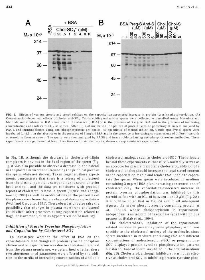
FIG. 2. Effects of various sterols and sterol sulfates on the capConcentration-dependent effects of cholesterol-SO4. Cauda epidid
ethods and incubated in HMB medium in the absence (2BSA)concentrations of cholesterol-SO4
2 as shown. After 1.5 h of incubaAGE and immunoblotted using anti-phosphotyrosine antibodiesncubated for 1.5 h in the absence or in the presence of 3 mg/ml BSr steroid sulfates as shown. The sperm were then analyzed by PAxperiments were performed at least three times with similar resu
tion to the media of increasing concentrations of a soluble
Copyright © 1999 by Academic Press. All right
holesterol analogue such as cholesterol-SO42. The rationale
ehind these experiments is that if BSA normally serves asn acceptor for plasma membrane cholesterol, addition of aholesterol analog should increase the total sterol contentn the capacitation media and render BSA unable to capaci-ate the sperm. When sperm were incubated in mediumontaining 3 mg/ml BSA plus increasing concentrations ofholesterol-SO4
2, the capacitation-associated increase inprotein tyrosine phosphorylation was inhibited by thissteroid sulfate with an IC50 of between 1 and 2 mM (Fig. 2A).t should be noted that in Fig. 2A and in all subsequentgures, the major phosphotyrosine-containing protein at
r 116,000 whose phosphorylation is capacitation-independent is an isoform of hexokinase type I with uniqueproperties (Kalab et al., 1994).
The cholesterol-SO42 inhibition of the capacitation-
elated increase in protein tyrosine phosphorylation waspecific to the cholesterol moiety of the molecule, sinceperm incubated in medium containing BSA and differentoncentrations of androstenedione-SO4
2 or pregnenolone-O4
2 displayed protein tyrosine phosphorylation patternssimilar to those of sperm incubated in the control medium(Fig. 2B). Cholesterol, although inhibitory, was not as effec-
tion-associated increase in protein tyrosine phosphorylation. (A)l mouse sperm were collected as described under Materials andthe presence of 3 mg/ml BSA and in the presence of increasingthe pattern of protein tyrosine phosphorylation was analyzed bySpecificity of steroid inhibition. Cauda epididymal sperm were
d in the presence of increasing concentrations of different steroidsnd immunoblotted using anti-phosphotyrosine antibodies. Thesehown are representative experiments.
acitaymaor intion. (B)A anGE a
tive as cholesterol-SO42 in inhibiting protein tyrosine phos-
s of reproduction in any form reserved.

i
S
d
w
diica
ai
435Cholesterol Release during Sperm Capacitation
phorylation, likely due to the limited solubility of thissteroid in the capacitation medium.
If the effects of cholesterol-SO42 were due to the inhibi-
tion of BSA action leading to an increase in protein tyrosinephosphorylation, addition of higher concentrations of BSAto the incubation medium containing a fixed concentrationof cholesterol-SO4
2 should bypass the inhibitory effect ofcholesterol-SO4
2 on protein tyrosine phosphorylation, andthis was observed to be the case (Fig. 3A). Since thisexperiment was performed by incubating the sperm in thepresence of greater concentrations of BSA using a fixedconcentration of cholesterol-SO4
2 (5 mM), the ability of BSAto overcome the cholesterol-SO4
2 inhibition of protein ty-rosine phosphorylation could be explained in two ways.First, if the concentration of BSA were increased, therebyincreasing the effective concentration of cholesterol bind-ing sites available in the medium, BSA would no longer belimiting in terms of its ability to sequester sperm choles-terol and would therefore support protein tyrosine phos-phorylation. Alternatively, the addition of increasing con-centrations of BSA would quench the available cholesterol-SO4
2 in the medium and in this way would block the
FIG. 3. Effect of increasing concentrations of BSA on the chphosphorylation. (A) Cauda epididymal sperm were collected as des1.5 h in the absence or in the presence of increasing concentrations
control with 3 mg/ml BSA and 0.5% DMSO. (B) Cauda epididymncubated in the absence or in the presence of 3 mg/ml BSA and/or
concentration of 30 mg/ml to some of the sperm suspensions andexperiment was performed using HDL and is shown. In both expersamples was analyzed by PAGE and immunoblotting using anti-photimes with similar results. Shown are representative experiments.
nhibitory action of cholesterol-SO42. To distinguish be- a
Copyright © 1999 by Academic Press. All right
tween these two possibilities, we incubated the sperm inmedium containing 3 mg/ml BSA and 5 mM cholesterol-O4
2 for 1 h, which inhibits protein tyrosine phosphoryla-tion (as shown in Figs. 2 and 3). After 1 h incubation,additional BSA was added to a final concentration of 30mg/ml, and the sperm were further incubated for variousperiods of time. The addition of BSA bypassed the inhibi-tory effect of cholesterol-SO4
2 (Fig. 3B). Although these datao not demonstrate that cholesterol-SO4
2 is blocking therelease of cholesterol from the sperm, they demonstrate: (1)that the inhibition by cholesterol-SO4
2 is reversible and canbe overcome with increasing concentrations of BSA and (2)that cholesterol-SO4
2 is not irreversibly toxic to the sperm.We previously demonstrated that a cAMP pathway thatas downstream of the action of BSA, Ca21, and NaHCO3
mediated the capacitation-associated changes in proteintyrosine phosphorylation (Visconti et al., 1995b). The evi-ence supporting this conclusion is based on the use of twonhibitors of PK-A activity (i.e., H-89 and RpcAMPS) whichnhibit the enzyme by totally different mechanisms (Vis-onti et al., 1995b). To further investigate whether thection of BSA on protein tyrosine phosphorylation is medi-
erol-SO42 inhibition of capacitation-associated protein tyrosine
d under Materials and Methods and incubated in HMB medium forA and/or 5 mM cholesterol-SO4
2 as shown. The asterisk representserm were collected as described under Materials and Methods and
cholesterol-SO42 as shown. After 1.5 h, BSA was added to a final
sperm were incubated for an additional 1, 2, and 3 h. A similarts, the pattern of protein tyrosine phosphorylation in the differenttyrosine antibodies. The experiments were performed at least three
olestcribeof BSal sp5 mMthe
imenspho
ted by a cAMP/PK-A pathway, we measured PK-A activity
s of reproduction in any form reserved.

p1
iieiVip
tosuZri
dm
c
436 Visconti et al.
in media containing or devoid of 3 mg/ml BSA at differentincubation times. PK-A activity is significantly higherwhen sperm are incubated in BSA-containing media com-pared to BSA-free media (Fig. 4A). Moreover, if thecholesterol-SO4
2 effects on protein tyrosine phosphorylationwere due to an abrogation of BSA function, cAMP agonistsshould overcome the inhibitory effect of cholesterol-SO4
2 onrotein tyrosine phosphorylation. As predicted, addition ofmM dibutyryl cAMP plus 100 mM IBMX, an inhibitor of
cyclic nucleotide phosphodiesterases, induced an increasein protein tyrosine phosphorylation in medium containingBSA plus 5 mM cholesterol-SO4
2 (Fig. 4B).To further analyze the correlation between the increase
n protein tyrosine phosphorylation and capacitation, wenvestigated the action of cholesterol-SO4
2 on a biologicalndpoint of capacitation, namely the spontaneous and ZP-nduced acrosome reactions (Florman and Babcock, 1991;isconti et al., 1998; Yanagimachi, 1994). Sperm were
ncubated for 1 h in complete medium (3 mg/ml BSA) in theresence or the absence of 5 mM cholesterol-SO4
2. After thisperiod, buffer or solubilized ZP (5 ZP/ml) were added, thesperm were incubated for an additional 30 min, and the
FIG. 4. Role of cAMP and protein kinase A in mediating the BSA(A) BSA-induced changes in PK-A activity during mouse sperm capathe absence (h) or in the presence (E) of 3 mg/ml BSA for the timescribed under Materials and Methods. This experiment was peeans 6 SEM (n 5 9; *P , 0.05; **P , 0.01 from the same time in
on the cholesterol-SO42 inhibition of the capacitation-associated i
were collected as described under Materials and Methods and incumg/ml BSA and in some cases 5 mM cholesterol-SO4
2, as indicated.AMP and 100 mM IBMX as indicated. The pattern of protein tyrosi
immunoblotted using anti-phosphotyrosine antibodies. This experepresentative experiment is shown.
percentage acrosome reactions was assessed as described
Copyright © 1999 by Academic Press. All right
under Materials and Methods. Cholesterol-SO42 inhibited
he spontaneous acrosome reaction to levels similar to thatbserved in medium devoid of BSA (Fig. 5). Moreover,perm incubated in the presence of cholesterol-SO4
2 did notndergo the acrosome reaction following treatment withP (Fig. 5), the physiological inducer of the acrosome
eaction, demonstrating that cholesterol-SO42 treatment
nhibited capacitation.
Promotion of Protein Tyrosine Phosphorylationand Capacitation by HDL
HDL is a complex of lipids and proteins that mediatescholesterol transfer in vivo. If BSA functions to increasesperm protein tyrosine phosphorylation and capacitation asa consequence of its ability to bind cholesterol, one wouldexpect HDL to substitute for BSA in promoting theseposttranslational modifications and capacitation. In con-trast, proteins that do not bind cholesterol should not beable to increase protein tyrosine phosphorylation. To testthis hypothesis, sperm were incubated for 1.5 h in HMBmedium devoid of BSA but containing 3 mg/ml each of
ced changes in protein tyrosine phosphorylation of mouse sperm.ion. Caudal epididymal sperm were incubated in HMB medium iniods indicated on the abscissa. PK-A activity was then assayed ased three times in triplicate, and the values shown represent therespective 2BSA control). (B) Effects of dibutyryl cAMP and IBMXse in protein tyrosine phosphorylation. Cauda epididymal spermin HMB medium for 1.5 h in the absence or in the presence of 3
e of the treatments were also supplemented with 1 mM dibutyrylosphorylation in the different samples was analyzed by PAGE andt was performed at least three times with similar results, and a
-inducitat
e perrform
thencreabatedSomne phrimen
HDL, ovalbumin, cytochrome c, histone H2A, and myelin
s of reproduction in any form reserved.

aeBcptotc
t
6c
ur1l
t
dltth
ttiyiBsw
amMC
a
437Cholesterol Release during Sperm Capacitation
basic protein. As shown in Fig. 6A, BSA and HDL were theonly proteins that supported protein tyrosine phosphoryla-tion, consistent with their abilities to bind cholesterol.HDL increased protein tyrosine phosphorylation in aconcentration-dependent manner with a maximum effectat 1 mg/ml (Fig. 6B). Moreover, the PK-A inhibitor H-89blocked the effect of HDL on protein tyrosine phosphory-lation (Fig. 6C), suggesting that the signaling pathwayactivated by HDL-induced cholesterol release from thesperm membrane leads to a PK-A up-regulation of proteintyrosine phosphorylation, similar to that seen with BSA(Visconti et al., 1995b). Effects of HDL on sperm PK-Activity were confirmed by measuring the activity of thisnzyme in media containing HDL as was performed withSA in Fig. 4A. HDL-containing media supported an in-rease in PK-A activity compared to media devoid of thisrotein (data not shown), and these effects were similar tohose observed with BSA-containing media (Fig. 4A). More-ver, as shown in Fig. 3B, HDL was observed to overcomehe inhibition of protein tyrosine phosphorylation byholesterol-SO4
2, similar to that observed with BSA. Finally,HDL supported sperm capacitation as demonstrated by itsability to replace BSA in an in vitro fertilization assay (Fig.6D). Heparin, which has been demonstrated to supportprotein tyrosine phosphorylation and capacitation in bo-vine sperm (Galantino-Homer et al., 1997), did not support
FIG. 5. Effect of cholesterol-SO42 on the percentage of sperm
undergoing spontaneous and zona pellucida-induced acrosome re-actions. Cauda epididymal sperm were collected as described underMaterials and Methods and incubated in HMB medium in theabsence or in the presence of 3 mg/ml BSA and in some cases of 5mM cholesterol-SO4
2. After a 1-h incubation, solubilized ZP wereadded where indicated to a final concentration of 5 ZP/ml. After andditional 30 min of incubation, the status of the acrosome wasonitored using the Coomassie blue method as described underaterials and Methods. Data represent the means 6 SEM, n 5 5.holesterol-SO4
2 inhibited significantly the ZP-induced acrosomereaction (*P , 0.01), compared to the respective ZP control in thebsence of cholesterol-SO4
2.
hese posttranslational modifications in mouse sperm (Fig.
Copyright © 1999 by Academic Press. All right
A), consistent with the inability of heparin to supportapacitation in this species.
Recovery of Protein Tyrosine Phosphorylationafter Incubation of Sperm in MediaLacking NaHCO3 or BSA
Previously we demonstrated that mouse sperm incubatedin the absence of NaHCO3 or in the absence of BSA did not
ndergo the capacitation-dependent changes in protein ty-osine phosphorylation or capacitation (Visconti et al.,995a). It is known that following incubation in mediumacking BSA or NaHCO3 for 1 h, the sperm can recover their
ability to be capacitated if the component lacking is addedback to the medium (Neill and Olds-Clarke, 1987). Todetermine if this recovery was correlated with an increasein protein tyrosine phosphorylation, sperm were incubatedin the absence of BSA for 1 h followed by the addition ofBSA to a final concentration of 3 mg/ml. Under theseconditions, the sperm undergo capacitation and proteintyrosine phosphorylation after the additional 90 min ofincubation in this new medium (data not shown). A similarresult was observed when the sperm were incubated in theabsence of NaHCO3 for 1 h followed by the addition back tohe medium of 10 mM NaHCO3 (data not shown).
Since sperm incubated in the absence of NaHCO3 or BSAid not display an increase in protein tyrosine phosphory-ation, we chose to investigate the effect of reintroducinghese medium components on the kinetics of phosphoryla-ion. The aim of these experiments was to elucidate theierarchy of BSA and NaHCO3 in this signal transduction
cascade. After adding back BSA, the increase in proteintyrosine phosphorylation is slow (Fig. 7A, right) with kinet-ics similar to the increase in phosphorylation generallyobserved when noncapacitated sperm are incubated directlyin a complete medium that would support capacitation(Visconti et al., 1995a). In contrast, the effect of adding backNaHCO3 on protein tyrosine phosphorylation is signifi-cantly more rapid than that seen with the BSA (Fig. 7A,left). To compare the kinetics of these effects, we quantifiedthe extent of protein tyrosine phosphorylation of a Mr
95,000 protein (position of protein designated by the smalldot between the two blots in Fig. 7A) as a marker for thepattern of protein tyrosine phosphorylation (Fig. 7B). Thesedata clearly demonstrate that the NaHCO3 effect on proteinyrosine phosphorylation is more rapid and suggest that thearget of BSA action may be the rate-limiting step in thenitiation of signaling leading to protein tyrosine phosphor-lation. This is supported by the observation that increas-ng concentrations of NaHCO3 added to medium devoid ofSA can overcome the inability of BSA-free medium toupport protein tyrosine phosphorylation (Fig. 7C, left),hereas the opposite is not true (Fig. 7C, right).
DISCUSSION
We previously demonstrated that mouse, bull, and hu-
man sperm capacitation in vitro is tightly correlated withs of reproduction in any form reserved.

ciauci(pefFm
438 Visconti et al.
FIG. 6. Effect of HDL, other proteins, and heparin on the capacitation-associated increase in protein tyrosine phosphorylation andapacitation of mouse sperm. (A) Cauda epididymal mouse sperm were collected as described under Materials and Methods and incubatedn HMB medium devoid of BSA, but in the presence of 3 mg/ml of the different proteins or heparin listed. Medium containing BSA serveds a positive control. After 1.5 h of incubation the pattern of protein tyrosine phosphorylation was analyzed by PAGE and immunoblottedsing anti-phosphotyrosine antibodies. (B) Sperm were incubated in HMB medium devoid of BSA and in the absence or presence of differentoncentrations of HDL as listed. One of the samples was incubated in the presence of 3 mg/ml BSA as a positive control. After 1.5 h ofncubation the pattern of protein tyrosine phosphorylation was analyzed by PAGE and immunoblot using anti-phosphotyrosine antibodies.C) Sperm were incubated in HMB medium containing 3 mg/ml HDL and increasing concentrations of H-89. After 1.5 h of incubation, theattern of protein tyrosine phosphorylation was analyzed by PAGE and immunoblot using anti-phosphotyrosine antibodies. Thexperiments were performed at least three times with similar results. Shown are representative experiments. (D) Sperm were capacitatedor 2 h in media containing the indicated concentrations of either BSA or HDL and then incubated with metaphase II-arrested eggs for 3 h.ollowing insemination the eggs were observed for signs of fertilization as described under Materials and Methods. Data represent theeans 6 SEM of five independent experiments in which a minimum of 30 eggs was used for each experimental condition. *P , 0.001
compared to the control without addition of protein.
Copyright © 1999 by Academic Press. All rights of reproduction in any form reserved.

wttalppt
nltp1Rcts(1
pamtob
c
m
ocIcbt
d
o
ith si
439Cholesterol Release during Sperm Capacitation
an increase in protein tyrosine phosphorylation of a subsetof proteins. Moreover, the up-regulation of protein tyrosinephosphorylation by cAMP and PK-A represents a uniquemode of signal transduction crosstalk that, to date, has onlybeen observed in sperm. Since sperm capacitation in manyspecies can be accomplished in vitro in defined medium
ithout the addition of biological fluids, it is likely thathere is an intrinsic regulatory component of capacitationhat involves preprogrammed membrane, transmembrane,nd/or intracellular signaling events that, once initiated,ead to the capacitated state. This does not rule out theossibility of higher order regulation of this “intrinsic”rocess(es) during capacitation in vivo by components ofhe male and female reproductive tracts.
In the present study we further examined the mecha-isms by which these tyrosine phosphorylations are regu-ated, specifically with respect to how the BSA present inhe capacitation medium regulates these phosphorylationrocesses and capacitation. Several authors (Davis, 1976,981; Davis et al., 1979; Go and Wolf, 1985; Langlais andoberts, 1985) have suggested that BSA is necessary in theapacitation medium for the removal of cholesterol fromhe sperm plasma membrane, and other investigations havehown that cholesterol loss can occur during capacitationCross, 1996; Cross and Razy-Faulkner, 1997; Lin and Kan,
FIG. 7. Recovery of protein tyrosine phosphorylation after incubatmouse sperm were collected as described under Materials and MetNaHCO3 (NaHCO3) for 1 h. After 1 h of incubation, BSA and NaHrespectively, to the medium lacking the respective component, andanalyzed by PAGE and immunoblotted using anti-phosphotyrosinsimilar results, and a representative experiment is shown. The smphosphotyrosine-containing protein. (B) The Mr 95,000 band in Aepicted in arbitrary units. Circles represent BSA supplementation
concentrations of NaHCO3 and of BSA on the pattern of protein tyrf BSA or NaHCO3. Cauda epididymal mouse sperm were collect
medium devoid of BSA (2BSA) and increasing concentrations of Naincreasing concentrations of BSA. The sperm were then analyantibodies. This experiment was performed at least three times w
996; Suzuki and Yanagimachi, 1989). Using different ap-
Copyright © 1999 by Academic Press. All right
roaches, we demonstrate here that capacitation is associ-ted with a loss of cholesterol from the sperm plasmaembrane and that BSA, in some way, promotes the loss of
his steroid. The loss of this steroid leads to the activationf this aforementioned unique signal transduction cascadey an, as yet, undefined mechanism.In this report we demonstrated that the presence of
holesterol-SO42 blocked the ability of BSA to initiate the
capacitation-associated increases in protein tyrosinephosphorylation. This effect was specific for the cholesterolmoiety of the cholesterol-SO4
2 molecule, since preg-nenolone-SO4
2 and androstenedione-SO42 were not able to
imic the cholesterol-SO42 effect even at high concentra-
tions. Although cholesterol was able to reduce the BSA-dependent increase in protein tyrosine phosphorylation, itis likely that the limited solubility of cholesterol accountedfor its reduced effectiveness compared to the more solublecholesterol-SO4
2. The observation that additional BSAcan overcome the cholesterol-SO4
2-induced inhibitionf protein tyrosine phosphorylation demonstrates thatholesterol-SO4
2 is not irreversibly affecting sperm viability.n addition, cAMP agonists such as IBMX and dibutyrylAMP are also able to overcome the cholesterol-SO4
2 inhi-ition of protein tyrosine phosphorylation, demonstratinghat the cholesterol-SO4
2 effect is upstream of PK-A activa-
f sperm in medium lacking NaHCO3 or BSA. (A) Cauda epididymaland incubated in HMB medium devoid of BSA (BSA) or devoid of
3 were added to the final concentrations of 3 mg/ml and 10 mM,ncubation continued for the times indicated. The sperm were thenibodies. This experiment was performed at least three times withdot between the two blots indicates the location of a Mr 95,000scanned and quantified using NIH Image software. The data areares represent NaHCO3 supplementation. (C) Effect of increasing
e phosphorylation of sperm incubated, respectively, in the absences described under Materials and Methods and incubated in HMB3 or incubated in HM medium devoid of NaHCO3 (2NaHCO3) andby SDS–PAGE and immunoblotted using anti-phosphotyrosinemilar results, and a representative experiment is shown.
ion ohods
COthe i
e antall
was; squosined aHCOzed
tion in this signal transduction cascade.
s of reproduction in any form reserved.

smcaS1apOdcptbtbtmtCiNtH
440 Visconti et al.
We also demonstrated that cholesterol-SO42 inhibits both
the spontaneous as well as the ZP-induced acrosome reac-tion. This result further supports the correlation betweenthe activation of this unique signal transduction pathwayand those processes associated with capacitation. We mustemphasize that although these two events are tightly cor-related, we have not demonstrated that protein tyrosinephosphorylation is necessary and/or sufficient for capacita-tion. The tight correlation between these two events hasalso been demonstrated in sperm from other species such asbull (Galantino-Homer et al., 1997), human (Carrera et al.,1996; Leclerc et al., 1996; Luconi et al., 1996), horse(Rosenberger et al., 1998), pig (Kalab et al., 1998), andhamster (Visconti et al., 1999b). These results suggest thatthe PK-A induced up-regulation of protein tyrosine phos-phorylation associated with capacitation may be universalfor mammalian sperm.
These data represent one of the first reports describingthe regulation of an intracellular signal transduction path-way by cholesterol. Stulnig et al. (1997) have demonstratedthat signal transduction via CD59 and CD48 in Jurkat Tcells leading to an increase in intracellular calcium isregulated, in some manner, by cellular cholesterol. Thiseffect, however, appears to be independent of effects onmembrane dynamics, which is unlike the case that wereport here. The aforementioned reduction in sperm mem-brane cholesterol accompanying capacitation has beendemonstrated to result in a decrease in the cholesterol/phospholipid ratio as assessed by a variety of criteria (Hoshiet al., 1990; Tesarik and Flechon, 1986). Such changes likelyaccount for the observed alterations in sperm membranefluidity (Wolf et al., 1986a), the aggregation of intramem-branous particles and formation of particle-free patches(Koehler and Gaddum-Rose, 1975), and the documentedmembrane protein redistributions reported with lectins(Cross and Overstreet, 1987) and antibodies (Rochwergerand Cuasnicu, 1992; Shalgi et al., 1990) observed during thismaturational event. From the standpoint of cell signaling,this change in membrane dynamics may have profoundeffects on transmembrane signaling and may representsome of the “intrinsic” control of capacitation describedabove. Transmembrane signaling may be initiated bychanges in ion channel activity and/or the activity ofmembrane-associated enzymatic and nonenzymatic pro-teins. In addition, this dramatic change in plasma mem-brane lipid architecture could also be functionally impor-tant, as it may ultimately prime the membrane for fusionwith the outer acrosomal membrane during the acrosomereaction, an endpoint of capacitation and a prerequisite tosuccessful fertilization. Since cholesterol efflux appears tobe the driving force behind these changes in membranedynamics during capacitation, a clear understanding of themechanism by which this steroid moves within the plasmamembrane and out of the plasma membrane in response toan appropriate acceptor is critical to a molecular under-standing of this maturational event.
Our lab has demonstrated that this release of cholesterol
Copyright © 1999 by Academic Press. All right
is, in some manner, tied to changes in protein tyrosinephosphorylation. The initiation of signal transduction path-ways that result in the activation of tyrosine kinases andprotein tyrosine phosphorylation normally involves plasmamembrane receptors. These receptors could be tyrosinekinases or could be receptors that associate with tyrosinekinases. Sperm represents a unique case in which theincrease in protein tyrosine phosphorylation is regulatedthrough a cAMP and PK-A pathway. We have examined thispathway in greater detail in this report and demonstratethat PK-A activity is significantly higher when the spermare incubated in the presence of BSA. How cholesterolremoval regulates such a pathway is not known. One couldspeculate that the removal of cholesterol from the spermplasma membrane could alter membrane dynamics andincrease the permeability of the sperm to certain ions, suchas HCO3
2 and/or Ca21, which are capable of stimulating theperm adenylyl cyclase. The sperm adenylyl cyclase hasany regulatory properties that set it apart from somatic
ell adenylyl cyclases, one notable difference being itsbility to be directly activated by HCO3
2 (Garty andalomon, 1987; Okamura et al., 1985; Visconti et al., 1990,995b). We and others have demonstrated that these ionsre necessary for both the increase in protein tyrosinehosphorylation and the capacitation process (Neill andlds-Clarke, 1987; Visconti et al., 1995a), and we have alsoemonstrated that the effect of these ions is upstream of theAMP/PK-A pathway (Visconti et al., 1995b, 1997). In theresent work we demonstrated that the increase in proteinyrosine phosphorylation recovered slowly after addingack BSA to sperm incubated for 1 h in medium devoid ofhis protein and recovered almost immediately after addingack HCO3
2 to sperm incubated for 1 h in medium devoid ofhis anion. These observations could be explained by aodel in which cholesterol removal by BSA would prime
he membrane for an increased permeability to HCO32.
onsistent with this model is the fact that when sperm arencubated in the presence of high concentrations ofaHCO3, we observed a BSA-independent increase in pro-
ein tyrosine phosphorylation, suggesting again that theCO3
2 site of action is downstream of the BSA effect. Thismodel, if correct, could open new questions regarding therole of cholesterol in the regulation of ion movements. Inthis respect it is interesting to note that capacitation hasalso been correlated with the hyperpolarization of theplasma membrane (Arnoult et al., 1999; Zeng et al., 1995).It is not yet known if this hyperpolarization is dependent orindependent of cholesterol removal by BSA. Since plasmamembrane hyperpolarization could be either upstream ordownstream of the increased permeability to HCO3
2, thepossibility that anion channels could be regulated by mem-brane potential is an important consideration for futureresearch.
Yet another question pertains to the mechanisms regu-lating sperm cholesterol efflux. In our in vitro system, BSAis functioning as an extracellular acceptor for sperm plasma
membrane cholesterol. Based on the results of severals of reproduction in any form reserved.

d
aTfs
awscaai
espPotcl
A
A
A
C
C
C
C
C
C
441Cholesterol Release during Sperm Capacitation
investigators studying cholesterol efflux in sperm duringcapacitation, one could postulate that such an effluxmechanism might bear some similarities to reverse choles-terol transport observed in somatic cells, where the firststep is the efflux of cellular cholesterol to an appropriateextracellular acceptor, usually HDL. It is generally acceptedthat cholesterol efflux occurs by an aqueous diffusionmechanism in which the cholesterol molecules de-adsorbfrom the plasma membrane into the aqueous phase, diffuse,and are then solubilized by an acceptor molecule. SinceHDL can induce the increase in protein tyrosine phosphor-ylation through a cAMP/PK-A pathway in sperm (thisreport) and can support capacitation (this report; Lane et al.,1999; Therien et al., 1997), it is likely that BSA and HDLmay function similarly as cholesterol acceptors in spermleading to capacitation in vitro.
Recently, our laboratory has demonstrated that thecholesterol-binding heptasaccharides, methyl-b-cyclo-extrin and OH-propyl-b-cyclodextrin, promote the release
of cholesterol from the mouse sperm plasma membrane inmedia devoid of BSA, promote protein tyrosine phosphory-lation, and support capacitation and in vitro fertilization(Visconti et al., 1999a). Both of these b-cyclodextrins werealso demonstrated to increase protein tyrosine phosphory-lation in the absence of BSA in bovine (Visconti et al.,1999a) and human (Osheroff et al., 1999) sperm. Indepen-dently, Choi and Toyoda (1998) and Cross (1999) demon-strated that b-cyclodextrins were able to capacitate mousend human sperm, respectively, in the absence of BSA.hese data provide additional support for the existence,
unction, and physiological role of cholesterol release inperm cell signaling.If one accepts the notion that capacitation in vivo is also
ssociated with cholesterol loss, it is important to considerhat component of the female reproductive tract might
erve as a cholesterol acceptor in vivo. It is clear that theomposition of the fluids of the female tract arise, in part, as
transudate of the serum so that serum-derived sterolcceptors/binding proteins could function in vivo. Thedentity of such acceptors remains to be clarified.
In conclusion, this work demonstrates that cholesterolfflux from the sperm plasma membrane can initiate aignal transduction pathway that leads to an increase inrotein tyrosine phosphorylation through the activation ofK-A. Our efforts will be directed toward the identificationf the molecular mechanisms involved in the regulation ofhis unique signal transduction pathway as well as to theharacterization of the role of protein tyrosine phosphory-ation in regulating sperm function.
ACKNOWLEDGMENTS
This work was supported by grants to G.S.K. (NIH HD 06274 andHD 22732). P.E.V. was supported by the Rockefeller Foundationand by NIH HD06274. X.P.N. was supported by NIH HD06274.M.W.F. was supported by a Fogarty International Training Grant
and by the National Council of Sciences from ArgentinaCopyright © 1999 by Academic Press. All right
(CONICET). J.G.A. was supported by NIH HD-36146 and a grantfrom the Cystic Fibrosis Foundation. P.S. was supported by NIHHD22681. S.A.C. was supported by a training grant from the NIH(T32-HD 07305). We also thank Dr. Ken Pomerantz for providing uswith the purified HDL and insightful comments.
REFERENCES
Alvarez, J. G., and Storey, B. T. (1995). Differential incorporation offatty acids into and peroxidative loss of fatty acids from phos-pholipids of human spermatozoa. Mol. Reprod. Dev. 42, 334–346.rnoult, C., Kazam, I. G., Visconti, P. E., Kopf, G. S., Villaz, M., andFlorman, H. M. (1999). Control of the low voltage-activatedcalcium channel of mouse sperm by egg ZP3 and by membranehyperpolarization during capacitation. Proc. Natl. Acad. Sci.USA 96, 6757–6762.ustin, C. R. (1951). Observations on the penetration of the sperminto the mammalian egg. Aust. J. Sci. Res. 4, 581–596.ustin, C. R. (1952). The “capacitation” of the mammalian sperm.Nature 170, 326.arrera, A., Moos, J., Ning, X. P., Gerton, G. L., Tesarik, J., Kopf,G. S., and Moss, S. B. (1996). Regulation of protein tyrosinephosphorylation in human sperm by a calcium/calmodulin-dependent mechanism: Identification of A kinase anchor pro-teins as major substrates for tyrosine phosphorylation. Dev. Biol.180, 284–296.hang, M. C. (1951). Fertilizing capacity of spermatozoa depositedinto the fallopian tubes. Nature 168, 697–698.hang, M. C. (1955). Development of fertilizing capacity of rabbitspermatozoa in the uterus. Nature 175, 1036–1037.hoi, Y. H., and Toyoda, Y. (1998). Cyclodextrin removes choles-terol from mouse sperm and induces capacitation in a protein-free medium. Biol. Reprod. 59, 1328–1333.ross, N. L. (1996). Human seminal plasma prevents sperm frombecoming acrosomally responsive to the agonist, progesterone:Cholesterol is the major inhibitor. Biol. Reprod. 54, 138–145.ross, N. L. (1999). Effect of methyl-b-cyclodextrin on the acroso-mal responsiveness of human sperm. Mol. Reprod. Dev. 53,92–98.
Cross, N. L., and Overstreet, J. W. (1987). Glycoconjugates of thehuman sperm surface: Distribution and alterations that accom-pany capacitation in vitro. Gamete Res. 16, 23–25.
Cross, N. L., and Razy-Faulkner, P. (1997). Control of human spermintracellular pH by cholesterol and its relationship to the re-sponse of the acrosome to progesterone. Biol. Reprod. 56, 1169–1174.
Davis, B. K. (1976). Inhibitory effect of synthetic phospholipidvesicles containing cholesterol on the fertilizing ability of rabbitspermatozoa. Proc. Soc. Exp. Biol. Med. 152, 257–261.
Davis, B. K. (1981). Timing of fertilization in mammals: Spermcholesterol/phospholipid ratio as a determinant of the capacita-tion interval. Proc. Natl. Acad. Sci, USA 78, 7560–7564.
Davis, B. K., Byrne, R., and Hangund, B. (1979). Studies on themechanism of capacitation. II. Evidence for lipid transfer be-tween plasma membrane of rat sperm and serum albumin duringcapacitation in vitro. Biochim. Biophys. Acta. 558, 257–266.
Diaz-Fontdevila, M., Bustos-Obregon, E., and Fornes, M. (1992).Distribution of filipin–sterol complexes in sperm membranesfrom hypercholesterolaemic rabbits. Andrologia 24, 279–283.
Florman, H. M., and Babcock, D. F. (1991). Progress towards
understanding the molecular basis of capacitation. In “CRCs of reproduction in any form reserved.
G
G
H
H
K
K
K
K
L
P
R
R
S
S
S
S
T
T
T
T
T
V
V
V
442 Visconti et al.
Uniscience—Chemistry of Fertilization” (P. M. Wassarman, Ed.),Vol. II, pp. 105–132. CRC Press, Boca Raton, FL.
Galantino-Homer, H., Visconti, P. E., and Kopf, G. S. (1997).Regulation of protein tyrosine phosphorylation during bovinesperm capacitation by a cyclic adenosine 39,59-monophosphate-dependent pathway. Biol. Reprod. 56, 707–719.arty, N., and Salomon, Y. (1987). Stimulation of partially purifiedadenylate cyclase from bull sperm by bicarbonate. Fed. Eur.Biochem. Soc. 218, 148–152.o, K. J., and Wolf, D. P. (1985). Albumin-mediated changes insperm sterol content during capacitation. Biol. Reprod. 32,145–153.arrison, R. A. P. (1996). Capacitation mechanisms, and the role ofcapacitation as seen in eutherian mammals. Reprod. Fertil. Dev.8, 581–594.oshi, K., Aita, T., Yanagida, K., Yoshimatsu, N., and Sato, A.(1990). Variation in the cholesterol/phospholipid ratio in humanspermatozoa and its relationship with capacitation. Hum. Re-prod. 5, 71–74.alab, P., Peknicova, J., Geussova, G., and Moos, J. (1998). Regula-tion of protein tyrosine phosphorylation in boar sperm through acAMP-dependent pathway. Mol. Reprod. Dev. 51, 304–314.alab, P., Visconti, P., Leclerc, P., and Kopf, G. S. (1994). p95, themajor phosphotyrosine-containing protein in mouse spermato-zoa, is a hexokinase with unique properties. J. Biol. Chem. 269,3810–3817.oehler, J. K., and Gaddum-Rose, P. (1975). Media induced alter-ations of the membrane associated particles of the guinea pigsperm tail. J. Ultrastruct. Res. 51, 106–118.opf, G. S., and Gerton, G. L. (1991). The mammalian spermacrosome and the acrosome reaction. In “Elements of Mamma-lian Fertilization” (P. M. Wassarman, Ed.), Vol. 1, pp. 153–203.CRC Press, Boca Raton, FL.
aemmli, U. K. (1970). Cleavage of structural proteins during theassembly of the head of bacteriophage T4. Nature 227, 680–685.
Lane, M. E., Therien, I., Moreau, R., and Manjunath, P. (1999).Heparin and high-density lipoprotein mediates bovine spermcapacitation by different mechanisms. Biol. Reprod. 60, 169–175.
Langlais, J., and Roberts, K. D. (1985). A molecular membranemodel of sperm capacitation and the acrosome reaction ofmammalian spermatozoa. Gamete Res. 12, 183–224.
Leclerc, P., De lamirande, E., and Gagnon, C. (1996). Cyclicadenosine 39,59 monophosphate-dependent regulation of proteintyrosine phosphorylation in relation to human sperm capacita-tion and motility. Biol. Reprod. 55, 684–692.
Lee, M. A., and Storey, B. T. (1986). Bicarbonate is essential forfertilization of mouse eggs: Mouse sperm require it to undergothe acrosome reaction. Biol. Reprod. 34, 349–356.
Lin, Y., and Kan, F. W. (1996). Regionalization and redistribution ofmembrane phospholipids and cholesterol in mouse spermatozoaduring in vitro capacitation. Biol. Reprod. 55, 1133–1146.
Luconi, M., Lraisz, C., Forti, G., and Baldi, E. (1996). Extracellularcalcium negatively modulates tyrosine phosphorylation and ty-rosine kinase activity during capacitation of human spermato-zoa. Biol. Reprod. 55, 207–216.
Neill, J., and Olds-Clarke, P. (1987). A computer-assisted assay formouse sperm hyperactivation demonstrates that bicarbonate butnot bovine serum albumin is required. Gamete Res. 18, 121–140.
Okamura, N., Tajima, Y., Soejima, Y., Madua, H., and Sugita, Y.(1985). Sodium bicarbonate in seminal plasma stimulates themotility of mammalian spermatozoa through direct activation of
adenylate cyclase. J. Biol. Chem. 260, 9699–9705.Copyright © 1999 by Academic Press. All right
Osheroff, J. E., Visconti, P. E., Valenzuela, J. P., Travis, A. J.,Alvarez, J. A., and Kopf, G. S. (1999). Regulation of human spermcapacitation by a cholesterol efflux-stimulated signal transduc-tion pathway leading to protein kinase A-mediated up-regulationof protein tyrosine phosphorylation. Mol. Human Reprod. 5, Inpress.
Osman, R. A., Andria, M. L., Jones, A. D., and Meizel, S. (1989).Steroid induced exocytosis: The human sperm acrosome reac-tion. Biochem. Biophys. Res. Commun. 160, 828–833.
omerantz, K. B., and Hajjar, D. P. (1990). High-density-lipoprotein-induced cholesterol efflux from arterial smoothmuscle cell derived foam cells: Functional relationship of thecholesteryl ester cycle and eicosanoid biosynthesis. Biochemis-try 29, 1892–1899.
ochwerger, L., and Cuasnicu, P. S. (1992). Redistribution of a ratsperm epididymal glycoprotein after in vitro and in vivo capaci-tation. Mol. Reprod. Dev. 31, 34–41.
osenberger, A., Meyers, S. A., Galantino-Homer, H., and Kopf,G. S. (1998). Tyrosine phosphorylation of stallion sperm duringin vitro capacitation. Biol. Reprod. 58, 76.
halgi, R., Matityahn, A., Gaunt, S. J., and Jones, R. (1990).Antigens on rat spermatozoa with a potential role in fertilization.Mol. Reprod. Dev. 25, 286–296.
tulnig, T. M., Berger, M., Sigmund, T., Stockinger, H., Horejsi, V.,and Waldhausl, W. (1997). Signal transduction via glycosylphosphatidylinositol-anchored proteins in T cells is inhibited bylowering cellular cholesterol. J. Biol. Chem. 272, 19242–19247.
uzuki, F. (1988). Changes in the distribution of intramembranousparticles and filipin–sterol complexes during epididymal matu-ration of golden hamster spermatozoa. J. Ultrastruct. Mol. Struct.Res. 100, 39–54.
uzuki, F., and Yanagimachi, R. (1989). Changes in the distributionof intramembranous particles and filipin-reactive membranesterols during in vitro capacitation of golden hamster spermato-zoa. Gamete Res. 23, 335–347.
esarik, J., and Flechon, J. E. (1986). Distribution of sterols andanionic lipids in human sperm plasma membrane: Effects of invitro capacitation. J. Ultrastruct. Res. 97, 227–237.haler, C. D., and Cardullo, R. A. (1995). Biochemical character-ization of a glycosylphosphatidylinositol-linked hyaluronidaseon mouse sperm. Biochemistry 34, 7788–7795.
herien, I., Soubeyrand, S., and Manjunath, P. (1997). Major pro-teins of bovine seminal plasma modulate sperm capacitation byhigh-density lipoprotein. Biol. Reprod. 57, 1080–1088.oshimori, K., Higashi, R., and Oura, C. (1985). Distribution ofintramembranous particles and filipin–sterol complexes inmouse sperm membranes: Polyene antibiotic filipin treatment.Am. J. Anat. 174, 455–470.
owbin, H., Staehelin, T., and Gordon, J. (1979). Electrophoretictransfer of proteins from polyacrylamide gels to nitrocellulosesheets: Procedure and some applications. Proc. Natl. Acad. Sci.USA 76, 4350–4354.
isconti, P., Muschietti, J. P., Flawia, M. M., and Tezon, J. G.(1990). Bicarbonate dependence of cAMP accumulation inducedby phorbol esters in hamster spermatozoa. Biochim. Biophys.Acta 1054, 231–236.isconti, P. E., Bailey, J. L., Moore, G. D., Pan, D., Olds-Clarke, P.,and Kopf, G. S. (1995a). Capacitation of mouse spermatozoa. I.Correlation between the capacitation state and protein tyrosinephosphorylation. Development 121, 1129–1137.
isconti, P. E., Galantino-Homer, H., Moore, G. D., Bailey, J. L.,Ning, X. P., Fornes, M., and Kopf, G. S. (1998). The molecular
basis of sperm capacitation. J. Androl. 19, 242–248.s of reproduction in any form reserved.
V
V
W
W
W
W
W
Y
Z
Z
443Cholesterol Release during Sperm Capacitation
Visconti, P. E., Galantino-Homer, H., Ning, X., Moore, G. D.,Valenzuela, J. P., Jorgez, C. J., Alvarez, J. G., and Kopf, G. S.(1999a). Cholesterol efflux-mediated signal transduction inmammalian sperm. b-Cyclodextrins initiate transmembrane sig-naling leading to an increase in protein tyrosine phosphorylationand capacitation. J. Biol. Chem. 274, 3235–3242.
Visconti, P. E., Johnson, L. R., Oyaski, M., Fornes, M., Moss, S. B.,Gerton, G. L., and Kopf, G. S. (1997). Regulation, localization,and anchoring of protein kinase A subunits during mouse spermcapacitation. Dev. Biol. 192, 351–363.
isconti, P. E., Moore, G. D., Bailey, J. L., Leclerc, P., Connors,S. A., Pan, D., Olds-Clarke, P., and Kopf, G. S. (1995b). Capaci-tation of mouse spermatozoa. II. Protein tyrosine phosphoryla-tion and capacitation are regulated by a cAMP-dependent path-way. Development 121, 1139–1150.
isconti, P. E., Stewart-Savage, J., Blasco, A., Battaglia, L., Miranda,P., Kopf, G. S., and Tezon, J. G. (1999b). Roles of bicarbonate,cAMP, and protein tyrosine phosphorylation on capacitation andthe spontaneous acrosome reaction of hamster sperm. Biol.Reprod. 61, 76–84.ard, C. R., and Storey, B. T. (1984). Determination of the timecourse of capacitation in mouse spermatozoa using a chlortetra-cycline fluorescence assay. Dev. Biol. 104, 287–296.istrom, C. A., and Meizel, S. (1993). Evidence suggesting involve-ment of a unique human sperm steroid receptor/Cl2 channelcomplex in the progesterone-initiated acrosome reaction. Dev.
Biol. 159, 679–690.Copyright © 1999 by Academic Press. All right
olf, D. E., and Cardullo, R. A. (1991). Physical properties of themammalian sperm plasma membrane. In “Comparative Sperma-tology 20 Years After” (B. Baccetti, Ed.), Vol. 75, pp. 599–604.Raven Press, New York.olf, D. E., Hagopian, S. S., and Isojima, S. (1986a). Changes insperm plasma membrane lipid diffusibility after hyperactivationduring in vitro capacitation in the mouse. J. Cell Biol. 102,1372–1377.olf, D. E., Hagopian, S. S., Lewis, R. G., Voglmayr, J. K., andFairbanks, G. (1986b). Lateral regionalization and diffusion of amaturation-dependent antigen in the ram plasma membrane.J. Cell Biol. 102, 1826–1831.
anagimachi, R. (1994). Mammalian fertilization. In “The Physi-ology of Reproduction” (E. Knobil and J. D. Neill, Eds.), pp.189–317. Raven Press, New York.ar, J. H. (1996). “Biostatistical Analysis.” Prentice Hall, UpperSaddle River, NJ.eng, Y., Clark, E. N., and Florman, H. M. (1995). Sperm membranepotential: Hyperpolarization during capacitation regulates zonapellucida-dependent acrosomal secretion. Dev. Biol. 171, 554–563.
Received for publication June 15, 1999Revised July 23, 1999
Accepted August 2, 1999
s of reproduction in any form reserved.
Copyright © 2022 FDOKUMEN