STUDIES ON CAPACITATION AND THE EFFECTS ... - CORE
285
STUDIES ON CAPACITATION AND THE EFFECTS OF COOLING AND LOW TEMPERATURE STORAGE ON STALLION SPERM FUNCTION by Mohd Azam Khan bin Goriman Khan (DVM, Malaysia) Thesis submitted for the degree of Doctor of Philosophy in the Faculty of Veterinary Medicine University of Glasgow February, 1998 Department of Veterinary Anatomy, University of Glasgow
-
Upload
khangminh22 -
Category
Documents
-
view
3 -
download
0
Transcript of STUDIES ON CAPACITATION AND THE EFFECTS ... - CORE

STUDIES ON CAPACITATION AND THE
EFFECTS OF COOLING AND LOW
TEMPERATURE STORAGE ON
STALLION SPERM FUNCTION
by
Mohd Azam Khan bin Goriman Khan
(DVM, Malaysia)
Thesis submitted for the degree of Doctor of Philosophy in the
Faculty of Veterinary Medicine University of Glasgow
February, 1998
Department of Veterinary Anatomy,
University of Glasgow

ProQuest Number: 13818618
All rights reserved
INFORMATION TO ALL USERS The quality of this reproduction is dependent upon the quality of the copy submitted.
In the unlikely event that the author did not send a com p le te manuscript and there are missing pages, these will be noted. Also, if material had to be removed,
a note will indicate the deletion.
uestProQuest 13818618
Published by ProQuest LLC(2018). Copyright of the Dissertation is held by the Author.
All rights reserved.This work is protected against unauthorized copying under Title 17, United States C ode
Microform Edition © ProQuest LLC.
ProQuest LLC.789 East Eisenhower Parkway
P.O. Box 1346 Ann Arbor, Ml 48106- 1346

GLASGOW UN I VFRSITyUrSASV
GLASGOW I CNivEEsirr ILIBBABT I

LIST OF CHAPTERS
Abstract i
List of Chapters iii
List of Tables xi
List of Figures xvi
Acknowledgements xxi
Dedication xxii
Declaration xxiii

ABSTRACT
This study was undertaken to assess stallion sperm function under two sets of
conditions 1) liquid storage at 5°C and 2) incubation under conditions which support
capacitation. Two aspects of sperm function were studied; motility, using a
computerised motility analyser capable of objective and detailed analysis of1
movement patterns, and the ability to regulate intracellular Ca , and assessment of
the functional competence of the sperm plasma membrane, using the fluorescent dye
Fura-2AM.
It was determined that after cooling and storage at 5°C there was a decline in
the percentage of live spermatozoa and a decline in the velocity parameters of the live
spermatozoa. A change in the pattern of motion to non-progressive motility was also• 2+detected. Concurrently, an increase in intracellular Ca was detected; this was
believed to indicate a reduction in the functional competence of the sperm plasma
membrane. This increase was most marked after cooling and further experiments2+ •indicated that intracellular Ca increased during the cooling process, most markedly
2+below about 15°C. Examination of the Ca regulating ability of individual cells2+confirmed that mean intracellular Ca for the whole population increased but, more
importantly, that there was a discrete subpopulation of sperm cells still able to
regulate calcium at pre-cooled levels, despite cooling and storage.
Incubation conditions capable of supporting capacitation were established for
stallion spermatozoa, as assessed using the dual fluorescent stain chlortetracycline and
Hoechst 33258. This involved incubation for 300 minutes in a TALP-milk medium.
Expression of hyperactivated motility was examined to evaluate the usefulness of this
motility pattern as a biomarker of capacitation. The drug pentoxifylline appeared to
accelerate the capacitation process, judged by dual staining, but had no detectable
stimulatory effect on motility. Instead, pentoxifylline promoted head-to-head
agglutination of the sperm cells in a dose dependent manner.2+
Further experiments showed that an increase in intracellular Ca occurred
during prolonged incubation in a TALP-milk medium with or without pentoxifylline.
This may be a necessary component of capacitation. Binding studies indicated the
presence of progesterone receptors on the plasma membrane of both human and

stallion spermatozoa. A progesterone-mediated calcium influx was stimulated by
adding progesterone to washed human spermatozoa. The same effect was only
generated with stallion spermatozoa after pre-incubation for 300 minutes under
capacitating conditions, in the presence of pentoxifylline. The latter observation
would indicate that the progesterone receptor on stallion spermatozoa is only active
after pre-incubation under capacitating conditions.

LIST OF CHAPTERS
CHAPTER 1 - LITERATURE REVIEW
1.1 The Preservation of Equine Spermatozoa for Artificial Insemination - An
Historical Perspective 1
1.2 Pregnancy Rates Using Preserved Semen 3
1.3 Factors Associated with the Use of Artificial Insemination 5
1.4 Reproductive Anatomy and Physiology of the Stallion 6
1.5 The Stallion Spermatozoon 10
1.5.1 Basic structure of the spermatozoon 10
1.5.2 Sperm metabolism 13
1.5.3 Sperm transport in the female reproductive tract 14
1.6 Sperm Physiology 16
1.6.1 Capacitation 16
1.6.1.1 In vivo studies 17
1.6.1.2 In vitro studies 17
1.6.1.3 Measurement of capacitation 18
1.6.2 The acrosome reaction 19
1.6.2.1 In vivo studies of the acrosome reaction 20
1.6.2.2 In vitro studies of the acrosome reaction 21
1.6.2.3 Measurement of the acrosome reaction 21
1.6.3 Capacitation related motility changes - hyperactivation 22
1.6.3.1 In vivo studies of hyperactivation 23
1.6.3.2 In vitro studies of hyperactivation 24
1.6.3.3 Measurement of hyperactivation 25
1.6.4 Calcium and sperm function 26
1.6.4.1 Calcium and motility 27
1.6.4.2 Calcium and capacitation 27
1.6.4.3 Calcium and acrosome reaction 28
1.6.4.4 Methods for measuring calcium 29
iii

1.7 Semen Processing 32
1.7.1 Semen collection 32
1.7.2 Semen extenders 33
1.7.3 Semen separation techniques 34
1.7.4 Packaging of semen 36
1.7.5 Preservation of stallion semen 36
1.7.5.1 Centrifugation of semen 36
1.7.5.2 Dilution of semen 37
1.7.5.3 Cooling rates 38
1.7.5.4 The effect of rapid cooling - ‘cold shock’ 38
1.7.5.5 Problems associated with re warming - ‘warm shock’ 40
1.8 Evaluation of Stallion Semen 42
1.8.1 Visual assessment of stallion semen motility 42
1.8.2 Microscopic examination of semen 45
1.8.3 Biochemical examination 46
1.8.4 Functional examination 47
1.8.5 Computerised motility analysis 47
1.9 Objectives of the Study 50
1.10 Structure of the Thesis 51
CHAPTER 2 - MATERIALS AND METHODS
2.1 Experimental Animals 52
2.2 Semen Collection 53
2.2.1 Human semen samples 53
2.2.2 Stallion semen samples 53
2.3 Visual Motility Assessment 55
2.3.1 Motility 55
2.3.2 Assessment of “head to head” agglutination 55
2.4 Medium 56
2.4.1 Kenney’s medium 56

2.4.2 Graham TALP 56
2.4.3 BavisterTALP 57
2.4.4 TALP-egg yolk medium 57
2.4.5 TEST-yolk medium 57
2.4.6 Culture medium for human spermatozoa 58
2.5 Tissue Culture Ware 58
2.6 Chemicals 58
2.6.1 Pentoxifylline [l-(5-oxyhexyl)-3-7-dimethylxanthine] 58
2.6.2 Progesterone (4-Pregnene-3, 20-dione) 59
2.6.3 Ionophore A23187 59
2.6.4 Fura-2AM ({l-[2-(5-Carboxyoxazol-2-yl)-6-aminobenzofuran-5-
oxy] -2-2 ’ -amino-5 ’ -methy lphenoxyethane-N,N,N ’ ,N ’ -
tetraaceticacid pentaacetoxymethyl ester}) 59
2.7 Cooling of Semen 60
2.8 Semen Preparation Methods 60
2.8.1 Sperm concentration 60
2.8.2 Swim-up migration 61
2.8.3 Removal of seminal plasma by centrifugation 61
2.9 Viability Staining 63
2.9.1 Eosin-nigrosin staining 63
2.9.2 Dual staining with Chlortetracycline and Hoechst 33258 63
2.9.3 Computerised motility analysis . 65
2.9.3.1 Cell chambers 65
2.9.3.2 Recording procedure 65
2.9.3.3 Computerised analyser 65
2.9.3.4 Parameter settings 66
2.9.4 Intracellular calcium measurements using Fura-2AM 69
2.9.4.1 Loading sperm cells with intracellular dye - ‘Fura
loading’ 69
2.9.4.2 Intracellular calcium measurements - population studies 69
2.9.4.3 Intracellular calcium measurements - single cell studies 70
v

1
2.9.4.4 Preparation of sperm suspension and slide chamber for
single cell measurements 71
2.10 Radioligand Binding Assay Studies - Principle and Procedure 73
2.11 Statistical Analysis 74
CHAPTER 3 - DEVELOPMENT OF METHODS
3.1 Introduction 76
3.2 Validation of the Hamilton-Thome Computerised Motility System for
the Analysis of Stallion Spermatozoa 76
3.2.1 Experiment 1 - Setting “Main Gates” for equine sperm motility
analysis 77
3.2.1.1 Results 78
3.2.2 Experiment 2 - Establishing lower velocity thresholds to minimise
the misidentification of immotile cells as motile 78
3.2.2.1 Results 79
3.2.3 Experiment 3 - Testing the effect of imposing low velocity
thresholds 81
3.2.3.1 Results 81
3.2.4 Experiment 4 - Repeated analysis of the same videotape 83
3.2.4.1 Results 83
3.3 Separation of Spermatozoa from Seminal Plasma 84
3.3.1 Preparation of Percoll gradients 84
3.3.2 Experiment 1 - Separation of human and equine spermatozoa
from seminal plasma by three different methods 85
3.3.2.1 Results 86
3.3.3 Experiment 2 - Separation of stallion spermatozoa through
different Percoll gradients 91
3.3.3.1 Results 91
3.4 Intracellular Calcium Measurements Using the Fluorescent Calcium
Indicator Fura-2AM 92
vi

3.4.1 Experiment 1 - Fura ‘loading’ of dead spermatozoa 92
3.4.1.1 Results 93
3.4.2 Experiment 2 - Effects of changes in sperm concentration on
Fura-2AM loading 94
3.4.2.1 Results 94
3.4.3 Experiment 3 - Effect of semen separation method upon
Fura -2AM loading 95
3.4.3.1 Results 95
3.4.4 Experiment 4 - Effect of medium composition upon Fura-2AM
loading 96
3.4.4.1 Results 96
3.4.5 Immobilisation of spermatozoa for single cell studies 97
3.5 Comparison of Eosin-Nigrosin Viability Staining With
Chlortetracycline/Hoechst 33258 Staining 99
3.5.1 Experiment 1 99
3.5.1.1 Results 99
3.6 Discussion 101
CHAPTER 4 - CHANGES IN STALLION SPERM
FUNCTION DURING SLOW COOLING AND LOW
TEMPERATURE STORAGE
4.1 Introduction 106
4.2 Materials and Methods 107
4.3 Experiments 107
4.3.1 Experiment 1 - Stallion sperm motility parameters after cooling
and storage at 5°C for up to 72 hours 107
4.3.1.1 Results 1082+4.3.2 Experiment 2 - Measurement of [Ca ], in the sperm suspension
after cooling and storage at 5°C for 48 hours 113
4.3.2.1 Results 113
vii

4.3.3 Experiment 3 - Calcium changes during cooling to 5°C 115
4.3.3.1 Results 1152”b •4.3.4 Experiment 4 - Measure of [Ca ]j in single cells after cooling and
storage at 5°C for 48 hours 116
4.3.4.1 Results 116
4.4 Discussion 119
CHAPTER 5 - CHARACTERISATION OF
CAPACITATION
5.1 Introduction 124
5.2 Materials and Methods 125
5.3 Experiments 125
5.3.1 Experiment 1 - Selection of an incubation system 125
5.3.1.1 Results 126
5.3.2 Experiment 2 - Chlortetracycline fluorescent patterns under
different incubation conditions 128
5.3.2.1 Results 128
5.3.3 Experiment 3 - Analysis of movement parameters under different
incubation conditions 133
5.3.3.a Experiment 3a - Analysis of human sperm movement in
seminal plasma, post swim-up and after incubation with
pentoxifylline 133
5.3.3.b Experiment 3b - Analysis of stallion sperm movement
inseminal plasma, post swim-up and after incubation
with pentoxifylline 133
5.3.3.1 Results (Experiments 3a and 3b) 134
5.3.3.C Experiment 3c - Analysis of stallion sperm movement
during incubation for up to 300 minutes 135
5.3.3.2 Results (Experiments 3c) 136

5.3.3.d Experiment 3d - Hyperactivation of stallion spermatozoa
- reanalysis of the data 138
5.3.3.3 Results (Experiments 3d) 138
5.4 Discussion 141
CHAPTER 6 - CALCIUM REGULATION DURING
CAPACITATION
6.1 Introduction 147
6.2 Materials and Methods 148
6.3 Calcium Transients Experiments 148
6.3.1 Experiment 1 - Intracellular calcium changes during incubation
under capacitating and non-capacitating conditions 148
6.3.1.1 Results 149
6.3.2. Experiment 2 - The induction of calcium transients in human
spermatozoa using progesterone 150
6.3.2.1 Results 150
6.3.3 Experiment 3 - The induction of calcium transients in stallion
spermatozoa using progesterone 151
6.3.3.1 Results 151
6.3.4 Experiment 4 - The induction of calcium transients in stallion
spermatozoa using progesterone, after incubation 155
6.3.4.1 Results 155
6.3.5 Experiment 5 - The induction of calcium transients in stallion
spermatozoa using progesterone after incubation under
capacitating and non-capacitating conditions 158
6.3.5.1 Results 158
6.4 Progesterone Binding Site Experiments 161
6.4.1 Experiment 1- Identification of specific binding sites on human
spermatozoa 161
6.4.1.1 Results 162
ix

6.4.2 Experiment 2 - Identification of specific binding sites on stallion
spermatozoa 162
6.4.2.1 Results 163
6.4.3 Experiment 3 - Effect of varying the concentration of tritiated
progesterone 163
6.4.3.1 Results 163
6.4.4 Experiment 4 - Identification of specific progesterone binding
sites on stallion spermatozoa after 0 or 300 minutes of incubation 164
6.4.4.1 Results 165
6.4.5 Experiment 5 - Relationship between specific progesterone
binding sites on human and stallion spermatozoa and numbers of
live spermatozoa 168
6.4.5.1 Results 168
6.5 Discussion 171
CHAPTER 7 - GENERAL DISCUSSIONS
7.1 General Discussions 174
APPENDIX A 181
BIBLIOGRAPHY 182
x

4
4
43
44
44
68
77
78
83
85
87
xi
LIST OF TABLES
Summarised pregnancy rate data from 7 trials using liquid,
chilled semen.
Summarised pregnancy rate data from 13 trials using
cryopreserved semen.
Mean (SD or SEM) of stallion seminal characteristics reported
in several studies (nr = not reported).
Morphologic classification systems used for stallion sperm
(Adapted from Jasko, 1992).
Seminal characteristics of fertile stallion reported in several
studies (Adapted from Jasko, 1992).
Machine settings used for motility analyses performed using the
Hamilton-Thome motility analyser. The derivation of parameter
settings for stallion spermatozoa is described in Chapter 3.
Summary of the “Main Gate” settings examined in
Experiment 1.
Summary of the type and percentage of error observed for each
of the settings evaluated.
Measurement of ALH, VAP, VCL and VSL in triplicate for 200
cells from the same section of videotape.
Percoll dilutions were made by mixing 100% Percoll with the
appropriate volume of medium as shown below.
Sperm count (millions/ml), percentage live and percentage
recovered from three human donors after separation of human
sperm from seminal plasma by one of three different methods.

I
Table 3.6 Sperm count (millions/ml), percentage live and percentage
recovered after separation of equine sperm from seminal plasma
by one of three different methods. Mean and (SEM) are also
shown. 88
Table 3.7 Mean and SEM for amplitude of lateral head deviation
(ALH, pm) and velocities (VCL, VAP, and VSL; pm/s) of
human spermatozoa before and after separation from seminal
plasma by one of three different methods. 89
Table 3.8 Mean and SEM for amplitude of lateral head deviation
(ALH, pm) and velocities (VCL, VAP, and VSL; pm/s) of
equine spermatozoa before and after separation from seminal
plasma by one of three different methods. 90
Table 3.9 Summary of the percentage of live spermatozoa in fresh
ejaculates (n = 3) and after separation through three
discontinuous Percoll gradients. 91
Table 3.10 Ratio of intensity (f34(/f38o) of Fura-2 (mean ± SEM, n = 3),
determined for live stallion spermatozoa incubated at 37°C
(Treatment 2), 5°C (Treatment 3) or killed using liquid nitrogen
and incubated at 37°C (Treatment 5) and 5°C (Treatment 6).
Treatments 1 and 4 represent autofluorescence controls. 93
Table 3.11 The effect of varying the sperm concentration within the cuvette
upon the intensities, the ratio of intensities and the average
intracellular calcium (mean ± SD for 3 experiments) is shown
below. 3̂40 and f38o are the fluorescence intensities at 340 and
380 nm respectively, and R is the ratio of these intensities. 95
Table 3.12 Fluorescence intensities of loaded and unloaded sperm prepared
by swim-up migration or centrifugation. 96
Table 3.13 Fluorescence intensities of loaded and unloaded sperm extended
in Kenney’s, TALP-Milk and TALP-egg yolk media. 97
xii

t
Table 3.14
Table 5.1
Table 5.2
Table 5.3
Table 5.4
Table 5.5
Table 5.6
Table 5.7
Table 5.8
Table 5.9
Comparisons of percentage live (%; Mean ± SEM; n = 5
ejaculates) stained by eosin/nigrosin or the CTC/H258 dual
staining method. 100
Mean + SD of percent motile, gross motility and percent live of
stallion spermatozoa extended under different incubation
systems and evaluated at 0, 2, 4, 6 and 8 hours, respectively. 127
The percentage of live cells (Mean ± SD) for 100 spermatozoa
in six different treatments, evaluated at specific time points 0,
120 and 300 minutes of incubation. 129
Motion parameters of human and stallion spermatozoa extended
in seminal plasma, post swim-up and after addition of
pentoxifylline for 15 minutes (Mean ± SD). 135
Subjective assessment of occurrence of head to head
agglutination (clumping) of stallion spermatozoa incubated in
Bavister-Milk medium containing 0, 0.01, 0.1 or 1 mg/ml
pentoxifylline after 15, 45, 90, 120, 180 and 300 minutes of
incubation. 136
Motility parameters (Mean ± SD) of stallion spermatozoa
incubated in Bavister-Milk medium supplemented with different
levels of PF analysed at specific time points over a 300 minute
incubation period. 137
Numbers of hyperactivated cells detected in human samples
(observed versus computer sorted). 138
Numbers of hyperactivated cells detected in stallion samples
(observed versus computer sorted). 139
Characteristic of sperm trajectories for stallion semen;
Mean ± SD (Range). 139
A comparison of numbers of hyperactivated cells selected using
the criteria of Burkman (1991), visual observation and the newly
derived criteria for stallion spermatozoa. The percentage of
hyperactivated spermatozoa (% HA) was calculated using the
xiii

I
Table 6.1
Table 6.2
Table 6.3
Table 6.4
Table 6.5
Table 6.6
Table 6.7
Table 6.8
criteria of Burkman (1991) or the criteria derived for stallion
spermatozoa in Experiment 3d. 140
Intracellular calcium concentration (Mean ± SD; n = 3
ejaculates) of stallion spermatozoa measured after 0 and
300 minutes of incubation. 149
Intracellular calcium measured prior to addition of progesterone,
for 0-60 seconds after addition of 1 pg/ml progesterone or 0.1%
DMSO and new plateau level for human spermatozoa
(Mean ± SD; n = 6 ejaculates). 151
Intracellular calcium measured prior to addition of progesterone,
for 0-60 seconds after addition of 1 pg/ml progesterone or 0.1%
DMSO and new plateau level for stallion spermatozoa
(Mean ± SD; n = 3 ejaculates). 152
Intracellular calcium concentration (nM) of stallion spermatozoa
incubated in Bavister-Milk medium with the addition of 0 or
1 mg/ml pentoxifylline (PF) and challenged by the addition of
1 jig/ml progesterone (P4) or 0.1% DMSO after 0 or
300 minutes of incubation (Mean ± SEM, n = 3 ejaculates). 156
Intracellular calcium concentration (nM) of stallion spermatozoa
incubated in three different medium and evaluated after 0 or
300 minutes of incubation after the addition of 1 pg/ml
progesterone (Mean ± SEM, n = 3 ejaculates). 159
Radioligand binding results of one ejaculate from one stallion
using fixed unlabelled progesterone (1 mg/ml) and increasing
radioligand concentrations. 164
Radioligand binding assay results for stallion spermatozoa
(n=3 ejaculate) incubated in three different media for 0 and
300 minutes. 165
Radioligand binding (cpm) and percentage live (Mean % (SD)
for human spermatozoa (n = 3 ejaculates) before and after a
swim-up migration. 169
xiv

Table 6.9 Radioligand binding assay results for stallion spermatozoa
(n = 3 ejaculates) incubated in three different media for 0 to
300 minutes. 170
xv

I
Figures
Figure 2.1
Figure 2.2
Figure 2.3
Figure 2.4
Figure 2.5
Figure 2.6
Figure 2.7
Figure 2.8 -
Figure 2.10
Figure 3.1a
Figure 3.1b
Figure 3.1c
Figure 3.2a
LIST OF FIGURES
Page
Semen collection. 54
Cambridge model artificial vagina. 54
Programmable freezing unit (Kryo 10 Series). 62
Hamilton-Thome motility analyser set-up with video
and computer. 62
Spermatozoal motility pattern - forward progressive. 67
Spermatozoal motility pattern - transitional phase. 67
Spermatozoal motility pattern - hyperactivated. 67
2.9 Perkin - Elmer LS-3B dual wavelength spectrofluorimeter
with water bath (a) set in order to regulate temperature of
cuvette held in the cuvette housing (b) at 37°C. 72
A schematic diagram of the set-up for measurement of
intracellular calcium in single sperm cell. 75
Distribution plot of curvilinear velocity (VCL) of
immotile cells or debris misidentified as motile cells. The
91% of cells had a VCL of less than 40 pm/sec. 80
Distribution plot o f average path velocity (VAP) of
immotile cells or debris misidentified as motile cells. The
95% of cells had a VAP of less than 30 pm/sec. 80
Distribution plot o f straight line velocity (VSL) of
immotile cells or debris misidentified as motile cells. The
96% of cells had a VSL of less than 20 pm/sec. 80
Distribution plot of straight-line velocity (VSL) for 200 cells
analysed before the application of low velocity thresholds.
Note the high proportion of cells (54%) with velocities of less
xvi

Figure 3.2b
Figure 4.1a
Figure 4.1b
Figure 4.2a
Figure 4.2b
Figure 4.3a
Figure 4.3b
Figure 4.4a
than 20 pm/sec. Visual inspection confirmed that these cells
were immotile, stuck to the slide or were moving debris. 82
Distribution plot of straight-line velocity (VSL) for 200 cells
analysed from the same videotaped sample after the
application of low velocity thresholds. The mean VSL
increased after motile artefacts were deleted from the
analysis. 82
Percentage live of stallion spermatozoa (%; Mean ± SEM;
n = 5) cooled from 37°C to 5°C using Cooling Protocol A
and analysed at 0, 24, 48 and 72 hours after the cooling
process. 109
Percentage live of stallion spermatozoa (%, Mean ± SEM;
n = 5) cooled from 37°C to 5°C using Cooling Protocol B
and analysed at 0, 24, 48 and 72 hours after the cooling
process. 109
Velocities of stallion spermatozoa (pms'1; Mean ± SEM;
n=5) cooled from 37°C to 5°C using Cooling Protocol A and
analysed at 0, 24,48 and 72 hours after the cooling process. 110
Velocities of stallion spermatozoa (pms’1; Mean ± SEM;
n = 5) cooled from 37°C to 5°C using Cooling Protocol B
and analysed at 0, 24, 48 and 72 hours after the cooling
process. 110
ALH of stallion spermatozoa (pm; Mean ± SEM; n = 5)
cooled from 37°C to 5°C using Cooling Protocol A and
analysed at 0, 24,48 and 72 hours after the cooling process. I l l
ALH of stallion spermatozoa (pm; Mean ± SEM; n = 5)
cooled from 37°C to 5°C using Cooling Protocol A and
analysed at 0,24,48 and 72 hours after the cooling process. I l l
LIN of stallion spermatozoa (%; Mean ± SEM; n = 5) cooled
from 37°C to 5°C using Cooling Protocol A and analysed at
0,24,48 and 72 hours after the cooling process. 112
xvii

Figure 4.4b
Figure 4.5a
Figure 4.5b
Figure 4.6
Figure 4.7
Figure 5.1a -
Figure 5.1g
Figure 5.1h
Figure 5.1i
Figure 5.1j
LIN of stallion spermatozoa (%; Mean ± SEM; n = 5) cooled
from 37°C to 5°C using Cooling Protocol A and analysed at
0, 24,48 and 72 hours after the cooling process. 112
Intracellular calcium (nM; Mean ± SEM; n = 3) of stallion
spermatozoa cooled from 37°C to 5°C using Cooling
Protocol A and analysed at 0, 24, 48 and 72 hours after
cooling. 114
Intracellular calcium (nM; Mean ± SEM; n = 3) of stallion
spermatozoa cooled from 37°C to 5°C using Cooling
Protocol B and analysed at 0, 24, 48 and 72 hours after
cooling. 114
Intracellular calcium (nM; Mean ± SEM; n = 3) of stallion
spermatozoa during cooling from 37°C to 5°C. 117
Ratio of intensity (f3 4(/f3 8o) measured from 250 individual
stallion spermatozoa cooled from 37°C to 5°C, measured
before cooling and at 0, 24 and 48 hours after the cooling
process. Mean values for R were 0.36, 0.48, 0.54 and 0.58
respectively. 118
Percentage (Mean ± SEM, n=3) of ‘F’ H , ‘B’ □ , and
‘AR’ ■ pattern of stallion spermatozoa incubated in six
different treatment and evaluated at specific time points of
incubation. The symbol (*) denotes significant increase in
the ‘B’ pattern in comparisons to time 0 (* at p<0.05 and **
atp<0.01). 130
Stallion sperm showing uncapacitated ‘F* pattern. 131
Decapitated sperm stained by H258, indicating dead sperm. 131
Stallion sperm (enlarged) showing capacitated ‘B’ pattern.
Note the lesser fluorescence band (arrow) in the acrosomal
region. 132
(i) ‘B* pattern stallion sperm showing exocytosis changes in
the acrosome, and (ii) sperm showing acrosome loss. 132
xviii

Figure 6.1a
Figure 6.1b
Figure 6.2a
Figure 6.2b
Figure 6.3a -
Figure 6.3c -
Figure 6.3e - f
Figure 6.4a -
Figure 6.4c -
Graph showing intracellular calcium changes (nM; Mean ±
SEM, n = 6) in human sperm suspension in response to
DMSO. DMSO was added at time 0. 153
Graph depicting typical changes in intracellular calcium
(nM; Mean ± SEM, n = 6) seen in human sperm suspension
in response to progesterone. Progesterone (1 jig/ml final
concentration) was added at time 0. 153
Graph showing intracellular calcium changes (nM; Mean ±
SEM, n = 3) in stallion sperm suspension in response to
DMSO. DMSO was added at time 0. 154
Graph depicting typical changes in intracellular calcium
(nM; Mean ± SEM, n = 3) seen in stallion sperm suspension
in response to progesterone. Progesterone (1 pg/ml final
concentration) was added at time 0. 154
> Spermatozoa incubated in Bavister-Milk. Intracellular
calcium (nM; Mean ± SEM) in response to progesterone,
assessed after 0 and 300 minutes of incubation. 157
I Spermatozoa incubated in Bavister-Milk. Intracellular
calcium (nM; Mean ± SEM) in response to DMSO, assessed
after 0 and 300 minutes of incubation. 157
Spermatozoa incubated in Bavister-Milk supplemented with
1 mg/ml pentoxifylline. Intracellular calcium (nM; Mean ±
SEM)in response to progesterone, assessed after 0 and 300
minutes of incubation. 157
t Spermatozoa incubated in Bavister-Milk. Intracellular
calcium (nM; Mean ± SEM; n = 3) in response to
progesterone, assessed after 0 and 300 minutes of incubation. 160
[ Spermatozoa incubated in homologous seminal plasma.
Intracellular calcium (nM; Mean ± SEM; n = 3) in response
to progesterone, assessed after 0 and 300 minutes of
incubation. 160
xix

Figure 6.4e-f
Figure 6.5a
Figure 6.5b
Figure 6.6a
Figure 6.6b
Spermatozoa incubated in Bavister-Milk supplemented with
1 mg/ml pentoxifylline. Intracellular calcium (nM; Mean ±
SEM; n = 3) in response to progesterone, assessed after 0 and
300 minutes of incubation. 160
A logarithmic plot. % of total binding against increasing
concentration of labelled progesterone; human spermatozoa. 166
A logarithmic plot. % of total binding against increasing
concentration of labelled progesterone; stallion spermatozoa. 166
Total binding, non-specific binding and specific binding
(counts per minute; cpm) against increasing concentration of
labelled progesterone. 167
Specific binding as calculated percentage of total labelled
progesterone added, against increasing concentration of
labelled progesterone. 167
xx

ACKNOWLEDGEMENTS
I take this opportunity to convey my heartfelt thanks to my supervisor Dr
Lindsay Robertson, without whom the successful completion of this thesis would
not have been achieved in the stipulated time. It is my hope that this will mark the
beginning of many years of fruitful association to come, and will benefit us and the
institutions we represent. In the pursuit of this research training, I have been
extremely fortunate to have met Dr Francis Burton for we became good friends. His
talents are not confined to cardiac research, for he has also expertly instructed me on
training the stallions used in this thesis to the dummy. He also dutifully helped me
with semen collection on all occasions during the course of my work. Through him
I was able to use the facilities in the Institute of Biological and Life Sciences for the
calcium flux studies in my thesis. I thank Dr W. Wilson and Dr Shahidullah for
allowing me access to the Perkin-Elmer Spectrofluoremeter for calcium
measurement. I would like to acknowledge Dr Peter O’Shaughnessy for his advice
on the radiobinding experiment and all in the Department of Physiology for their
tolerance of my incursions into their lab. I am grateful to Dr Paul Baker for his
contribution to the fulfilment of this work. I express gratitude to Mr Pat Toner who
took care of my horses. I also owe thanks to the chief technician in the Department
of Veterinary Anatomy Mr Alan Reid, who helped in sourcing out the materials
needed in my research. I extend my thanks to the auxiliary staff Miss Patricia
Wilson who made sure that I am in constant supply of sterilised equipment. I
convey my thanks to all in the Department of Veterinary Anatomy for helping me
“fit in”.
I also wish to thank my wife, who had a mammoth task of managing a
family whilst pursuing her PhD. I must mention my children who had to put up
with many weekends indoors and the numerous occasions that they were sent off to
bed early so that I could do my work.
My sincere thanks to the Faculty of Veterinary Medicine, University of
Glasgow for accepting my application to pursue an education in Scotland. Lastly I
extend my deepest gratitude to my alma mater, University Pertanian Malaysia for
providing me the scholarship to pursue my PhD.
xxi

DEDICATION
I dedicate this work to my father Mr Goriman Khan bin Kalandar Khan and my mother Puan Tamnah Khatoon bt Abdul Kadir

DECLARATION
I certify that this thesis does not contain any material previously published or written
by any other person, except when referred to in the text. I certify that the work o f
which this thesis is a record, is my work, and has not previously been submitted in any
form to any institution for an award of a degree.
Mohd Azam Khan bin Goriman Khan
xxiii
CHAPTER 1
LITERATURE REVIEW

1.1 The Preservation of Equine Spermatozoa for Artificial
Insemination - An Historical Perspective
Reference has been made to equine artificial insemination as early as 1322,
when it was reported that an owner served his mare with semen stolen from a stallion
owned by a hostile neighbour (Watson, 1990). However, the first documented
scientific research was not published until 1780, when an Italian clergyman called
Spallanzani reported the artificial insemination of a bitch (Watson, 1990) and later
evaluated the procedure in horses (Brinsko and Varner, 1992). Spallanzani is also
credited with the discovery of cryopreservation through his experiments on cooling
stallion, human and frog semen with ice (Watson, 1990). In the late 1800’s the
French veterinarian, Repiquet, advised the use of artificial insemination for the
treatment of infertility in mares (Brinsko and Varner, 1992). By the early part of the
present century the use of artificial insemination was expanding through the work of
Professor Iwanoff, Director of the State Institute of Veterinary Experimental Medicine
in Moscow. Due to his influence, by 1928, one hundred and twenty thousand mares
had been artificially inseminated in Russia (Iwanoff, 1930). The widespread use of
frozen semen was predicted by Mantegazza in 1866, who saw the potential for
transporting frozen semen rather than live animals (Watson, 1990). However, the first
foal was not bom from cryopreserved semen until 1957 (Barker and Gandier, 1957).
While breeding horses by artificial insemination has become popular in the
United States, China and Japan, use of the technique is still limited overall, and the
use of cryopreserved semen is very limited. The exception appears to be China which
has developed an extensive artificial insemination programme, with six hundred
thousand mares inseminated in 1959 alone (Brinsko and Varner, 1992) and eleven
hundred thousand mares inseminated with cryopreserved semen in between 1980 and
1985 (Amann and Picket, 1987).
Methods for semen collection evolved over time. Semen was recovered from
the vagina of the mare either directly or using an intravaginal sponge, or collected
from the stallion using “Breeder’s Bags” or mbber condoms. In the early 1930’s, an
artificial vagina was constmcted by Milovanov to facilitate semen collection, but it
1

was reported to be cumbersome and difficult to handle (Frank, 1950). In 1938,
Berliner produced a more pliable version which could be pressed close to the mare
during collection (Berliner, 1940). A modified version of this artificial vagina is the
Missouri model which is in common use today (Love, 1992).
Despite the early interest in artificial insemination, use of the technique has
never been as widespread as in cattle. In the 1950’s, successful cryopreservation
methods were developed for bovine semen which facilitated the rapid expansion of
cattle artificial insemination (Polge and Rowson, 1952). Comparable conception rates
using cryopreserved stallion semen have still not been achieved. The long period of
oestrus and the difficulty of predicting ovulation in the mare makes the correct timing
of insemination difficult. However, the principal reason for slow technological
progress is the attitude of most horse Breed Societies, which has been to either restrict
or prohibit the use of artificial insemination.
2

r
1.2 Pregnancy Rates Using Preserved Semen
There are relatively few publications reporting pregnancy rates from artificial
insemination of the mare. Pregnancy data from artificial insemination using chilled
liquid semen is summarised in Table 1.1 and using cryopreserved semen in Table 1.2.
Trials using chilled liquid semen reported pregnancy rates of between 50% and 91%.
Using cryopreserved semen pregnancy rates ranged between 23% and 67%.
This huge variation in pregnancy rate data can be attributed to a number of
factors including selection of stallion, selection of mares and evaluation of only a
small number of animals. Differences in insemination dose and in timing of
insemination relative to ovulation will affect the outcome, as will the choice of
extending medium and semen storage method. The method of data presentation,
which may be as a per cycle pregnancy rate or as an end o f season pregnancy rate,
often makes direct comparison of data impossible. Methods of assessing pregnancy
also differ. In some trials pregnancy may be established by manual examination and
in others, by foaling. Moreover, many of these studies were conducted before
ultrasonography was commonly used to determine the time of ovulation.
If attention is focused on the more recent pregnancy rate data which is
reported as single cycle pregnancy rates, rates for chilled semen vary from 59-87%
(Douglas-Hamilton et al., 1987; Heiskanen et al., 1994). Results for cryopreserved
semen vary from 29-56% (Cristanelli et al., 1984; Amann and Pickett, 1987). It
appears that results for liquid chilled semen compare favourably with natural service
but there is still room for improvement in the results obtained using cryopreserved
semen.
3
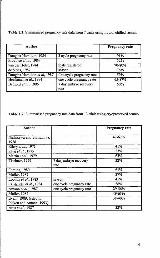
Table 1.1: Summarised pregnancy rate data from 7 trials using liquid, chilled semen.
Author Pregnancy rate
Douglas-Hamilton, 1984 3 cycle pregnancy rate 91%Province et al., 1984 52%van der Holst, 1984 foals registered 70-80%deVries, 1987 season 76%Douglas-Hamilton et al, 1987 first cycle pregnancy rate 59%Heiskanen et al., 1994 one cycle pregnancy rate 65-87%Bedford et al., 1995 7 day embryo recovery
rate50%
Table 1.2: Summarised pregnancy rate data from 13 trials using cryopreserved semen.
Author Pregnancy rate
Nishikawa and Shinomiya, 1976
47-67%
Ellery et al., 1971 41%Klug et al., 1975 23%Martin et al., 1979 63%Tischner, 1979 7 day embryo recovery
rate32%
Fomina, 1980 61%Muller, 1982 37%Loomis et al., 1983 season 45%Cristanelli et al., 1984 one cycle pregnancy rate 56%Amann et al., 1987 one cycle pregnancy rate 29-56%Muller, 1987 49-63%Evain, 1989; (cited in Pickett and Amann, 1993).
38-40%
Ams et al., 1987 32%
4

1.3 Factors Associated with the Use of Artificial
Insemination
The principle advantage of artificial insemination is the increased utilisation
of a selected stallion. A single ejaculate can be divided and used to inseminate a
number of mares. This allows more mares to be booked to an individual stallion
without overuse occurring. It is possible for breeding to continue while a stallion is
competing at shows or performance events or while recovering from injuries or
illness. Semen from prized stallions will still be available in the event of death of the
animal. Other advantages include early detection of stallion infertility through routine
semen analysis and control of the transmission of bacterial diseases where the male is
the carrier, by addition of antibiotics to seminal extenders. With subfertile stallions,
the supportive and protective nature of seminal extenders may improve pregnancy
rates. In cases where the stallion is not able to achieve full tumescence or penetration
because of injury, artificial insemination may be used to reinforce natural service.
Risk of injury to stallion and the mare is decreased in artificial insemination and the
selection of available semen is widened giving mare owners access to genetically
superior stallions.
There are also disadvantages associated with artificial insemination.
Successful management of an Al programme requires a higher degree of knowledge
and skill than the management of a natural service programme. If not applied
properly, techniques for processing, cooling, freezing and packaging can reduce
semen quality. Poor insemination technique will result in low pregnancy rates.
Within the breeding industry as a whole, some stud owners would face loss of income
from stud fees and mare boarding fees. Most importantly, from the viewpoint of
certain breed societies, increased production of offspring from popular stallions would
reduce the monetary value of the individual animals.
5

1.4 Reproductive Anatomy and Physiology of the Stallion
The male reproductive system consists of two testis, each suspended by a
spermatic cord and external cremaster muscle, two epididymides, two deferent ducts
each with an ampulla, paired vesicular glands, a prostate gland, paired bulbourethral
glands, the penis and the associated urethralis, ischiocavemosus, bulbospongiosus and
retractor penis muscles. The reproductive system is supported within the pelvic cavity
by the hammock-like genital fold and externally by the scrotum and prepuce (Amann,
1981).
The scrotum comprised two lateral pouches, which serve to cover, protect
and thermoregulate the testis by vascular transfer of heat and changes in position of
the testis relative to the abdominal wall. This function is dependent upon the
combined action of the scrotum, pampiniform plexus, tunica dartos muscle and
external cremaster muscles (Amann, 1981).
The testis are slightly laterally compressed ovoid structures, each situated in
a separate pouch within the scrotum. Their primary functions are the production of
spermatozoa (spermatogenesis) and the production of steroid hormones
(steroidogenesis). In most animals the testis descend from their point of origin, near
the kidney, into the scrotum before birth. The testicular parenchyma consists of
seminiferous tubules surrounded by interstitial tissue. These seminiferous tubules are
lined with Sertoli cells and developing germ cells. The developing germ cells are
spermatogonia, which undergo mitotic division and the primary spermatocytes,
secondary spermatocytes and spermatids, cell which divide meiotically to produce the
haploid spermatid. Spermatids do not divide further but undergo morphological
changes to tranform from round cells to the more familiar elongated shape of the
spermatozoa. The spermatid acquires an acrosomal cap, the nucleus condenses, a
flagellum forms and mitochondria spiral round the cranial portion of the flagellum, to
form the midpiece. The different stages of spermatogenesis are enclosed in
invaginations of the Sertoli cell. Finally, the spermatozoon is released into the lumen
of the seminiferous tubule (Amann, 1981). Further maturation occurs during travel
through the epididymis. The interstitial tissues contains steroidogenic Leydig cells,
6

fibroblasts, lymphocytes, blood vessels and lymphatics.
Rapid growth of the testis occurs at about 18 months of age when the
production of androgen by the Leydig cells and spermatogenesis gradually begin.
This development culminates in puberty, after which both the quantity and quality of
the spermatozoa produced increase over time. Some studies suggest that the testis
continues to grow throughout life (Thompson et al., 1979; Johnson and Neaves, 1981;
El Wishy et al., 1982) while others suggest that it attains maximum size by 5 to 7
years of age (Amann et al., 1979; Johnson and Thompson, 1983).
The epididymis is a highly convoluted duct, which is divided into the head or
caput, body or corpus and the tail or cauda. It originates at the convergence of the
efferent ductules leaving the testis and terminates at the beginning of the deferent
duct. It is characteristically highly convoluted, and very long (Amann, 1981).
Epididymal functions include concentration, maturation, storage and transport of
spermatozoa. Spermatozoa are conducted to the pelvic urethra via the deferent ducts.
The penis of the stallion is of the musculo-cavemous type and consists of
three parts: the root, the body and the glans. It is anchored at the roots by paired crura
and the ischiocavemosus muscles and suspended from the ventral surface of the pelvis
by two short, strong suspensory ligaments (Amann, 1981), which provide the lateral
stability necessary for normal intromission of the erect penis. The penile musculature
on the caudal or ventral surface of the penile urethra comprises paired
bulbospongiosus muscles. These muscles evacuate the penile urethral contents during
urination or ejaculation. Rhythmic pulsation of the bulbospongiosus muscle indicate
ejaculation during semen collection or natural service. The pulsations are palpable as
a thrill of turbulent fluid coursing along the ventrum of the penis and are usually
accompanied by simultaneous “flagging” of the tail. The retractor penis muscles also
lie on the ventral surface of the penis for most of its length. These muscles are
responsible for returning and maintaining the detumescent penis within the prepuce.
The glans of the stallion penis consists of the mushroom shaped corona glandis. The
neck of the glans is termed the collum glandis. The urethral process extends beyond
the surface of the glans and is surrounded by the fossa glandis.
The prepuce serves to protect the non-erect penis and consists of an internal
and an external preputial fold. The external fold forms the preputial orifice, whilst the
7

internal fold forms the preputial ring. When the penis is erect the preputial orifice is
situated at the base of the penis and the preputial ring is represented by a mid-shaft
fold of skin.
The seminal vesicles, ampullae of the deferent ducts, prostate glands and the
bulbourethral glands make up the accessory sex glands (Setchell, 1977). Secretions
from these glands, the seminal plasma, constitute approximately 95% of the ejaculate
volume (Pickett et al., 1983) and are known to contain factors which may inhibit
(Bass et al., 1983; Padilla and Foote, 1991) or stimulate (Muller and Kirchner, 1978;
Overstreet et al., 1980) spermatozoal function, and are thought to be beneficial,
though not essential, to fertility (Amann, 1981). The seminal plasma functions as a
vehicle of sperm transport (Rodger, 1975; Varner et al., 1987a; Yanagimachi, 1988)
and may also serve antioxidant, bactericidal and immunoprotective functions (Mann,
1975; Rodger, 1975). However, prolong exposure is detrimental to sperm survival.
Semen collected using the artificial vagina should thus be mixed with an extender
containing nutrients to prolong survival during in vitro storage.
Several studies concerning semen storage at 5°C show a negative impact of
seminal plasma on the viability of spermatozoa (Pickett et al., 1987; Jasko et al.,
1992a), demonstrated by a decline in sperm motility in whole ejaculates compared to
samples in which all or a high fraction of seminal plasma is removed by
centrifugation. Other studies in which contact of spermatozoa with seminal fluids was
prevented by vesiculectomy (Webb et al., 1990) or by collection of only the sperm
rich fraction of the ejaculate (Varner et al., 1987a) showed improved motility in
frozen-thawed and cooled samples and the phenomenon of sperm agglutination was
reduced.
The natural act of mating or copulation in the stallion normally occurs with
two physiologically distinct events, termed emission and ejaculation. Emission is the
release of spermatozoa and accessory gland fluids into the pelvic urethra. Ejaculation
is the forceful expulsion of the combined fluids from the pelvic and penile urethra.
The reflex emission follows a series of seven to nine intravaginal thrusts of the erect
penis. Smooth muscle of the cauda epididymides, deferent ducts, ampullae, vesicular
glands, prostate and possibly bulbourethral glands contract with resultant release of
spermatozoa into the urethra. At the same time the bladder neck contracts, keeping
8

the urethral orifice of the bladder tightly closed. These actions are primarily alpha-
adrenergically mediated (McDonnell, 1992).
Ejaculation of semen from the urethra is mediated via the pudendal and
sacral segment at the spinal cord. It results in rhythmic contraction of the
ischiocavemosus, bulbospongiosus, urethralis and other striated pelvic musculature.
Simultaneously, the anal sphincter contracts rhythmically and ejaculation in stallion
is a series of between five to ten jets of semen, each jet expelled with a subsequent
decrease in pressure, volume and sperm concentration (Kosiniak, 1975; McDonnell,
1992). The ejaculate is composed of three fractions. The first, or pre-sperm fraction,
is watery in appearance and contains no spermatozoa. This is followed by several jets
of milky white the ‘spermatozoa rich’ fraction. The third fraction contains large
quantities of gel and citric acid but few spermatozoa.
9

1.5 The Stallion Spermatozoon
1.5.1 Basic structure of the spermatozoon
Spermatozoa exhibit diversity of form, size and structure. A typical
eutharian spermatozoa can easily be distinguished into three regions, the head, neck
and tail, with the latter further subdivided into the middle piece, the principle piece
and the end piece (Bedford and Hoskins, 1990)
The head of the spermatozoon consists of a nucleus with its nuclear
envelope, the acrosome and post-acrosomal region and a plasma membrane. The
nucleus contains highly condensed chromatin, DNA complexed with protamine, and
is enclosed by a double-layered nuclear envelope containing nuclear pores. The
rostral portion of the nucleus is overlain by the acrosome, a membrane limited vesicle
which houses several hydrolytic enzymes. The acrosome, in turn, is overlain by the
plasma membrane. Although the plasma membrane is continuous over the surface of
the spermatozoon except after the acrosome reaction, or with senescence and death,
the nature and function of the plasma membrane differs regionally (Bedford and
Hoskins, 1990).
The neck connects the head and the middle piece. It comprises a connecting
piece, the proximal centriole, several small mitochondria and a redundant nuclear
envelope (Bedford and Hoskins, 1990). The middle piece is characterised by the
presence of numerous mitochondria arranged end to end in a continuous double spiral,
and extends from the caudal end of the neck distally. Central to the mitochondria are
nine dense fibres, extending from their origin in the neck of the spermatozoon
throughout the length of the middle piece and most of the principle piece. These
fibres gradually taper away in the caudal principle piece and are not present in the end
piece.
Central to the dense fibres is the axoneme. The axoneme is the propulsive
element of the spermatozoon. It consists of a central pair of microtubules surrounded
by a ring of 9 doublets. The role of the central pair is unclear. Each doublet is
composed of two subfibres A and B. The principle structural protein of the
10
microtubules is tubulin. Tubulin molecules are arranged in rows to form
protofilaments. Subfibre A contains 13 such protofilaments and subfibre B contains 9
or 10 such protofilaments. Subfibre A also contains two longitudinal rows of arms
pointing to the next doublet. These arms contain dynein, which is rich in ATPase and
transduces chemical energy to mechanical motion. The entire doublet is about 70%
tubulin and 15% dynein. The doublets are interconnected by nexin links and a series
of 9 radial spokes extend from the central pair to the doublets. The nexin links
maintain symmetry during motion and the spokes probably provide structural support.
The dense fibres and axoneme of the middle piece continue through the principle
piece, although the dense fibres becomes narrower and terminate in the caudal
principal piece. The doublets and central pair of the axoneme continue intact about
halfway through the end piece and then taper out over a short distance of 1 to 2 pm.
The 9 + 2 pattern is lost in the caudal end of the end piece (Bedford and Hoskins,
1990).
The stallion spermatozoon has a similar basic structure to spermatozoa of
other mammals. However, some features, the asymmetric head, small volume of the
acrosome and the abaxial position of the tail (Bielanski and Kaczmarski, 1979)
distinguishes it from spermatozoa of bull (Jones, 1975), ram and boar (Bielanski and
Kaczmarski, 1979).
Measurements of stallion spermatozoa gave the following average
dimensions (± s.d); head: length - 5.75 ± 0.47 pm; maximum width - 2.93 ± 0.34 pm;
length of post-nuclear cap - 1.65. ± 0.39 pm; width at the base - 1.45 ± 0.22 pm;
surface area - 12.86 ±2.17 pm ; midpiece: length including the neck - 10.50 + 1.27
pm; diameter - 0.60 ± 0.11 pm; main piece: length - 67.16 ± 3.99 pm; terminal piece:
length 2.79 ± 0.90 pm (Bielanski and Kaczmarski, 1979).
When a spermatozoon leaves the testis the neck is surrounded by remnants of
spermatid cytoplasm, the cytoplasmic droplet. Depending on the position of the
cytoplasmic droplet on the tail in relation to the neck, it can be classified as a
proximal or distal cytoplasmic droplet. Normally when spermatozoa pass through the
epididymis, the cytoplasmic droplet will move down the mid-piece and is completely
lost by the time of ejaculation. This movement and complete stripping of the
cytoplasmic droplet is part of the maturation process and the presence of large
11

numbers of spermatozoa with protoplasmic droplets in an ejaculate is an indication of
a failure of the maturation process (Dott, 1975).
The sperm plasma membrane is similar to other cell membranes in terms of
the lipid bilayer arrangement of hydrophilic head groups and hydrophobic tail groups,
but varies in its composition of lipids (mainly phospholipids and cholesterol) and
proteins (peripheral and integral). Proteins make up more then 50% of the total
weight of the spermatozoon. Most are histone protein involved in DNA packaging.
Others are in the form of enzymes in the acrosome and receptors on the plasma
membrane or participate in ion and carbohydrate transport during normal membrane
function. Other proteins make up the cytoskeletal structure which gives shape to the
head and contributes to the filaments of the tail (Mann, 1964)
About 14% of the total dry weight of the stallion spermatozoon is lipid.
Even though this value is not different from the bull or boar, the composition is
characteristically different, with cholesterol contributing 23% to sperm lipid in the
stallion compared with 14% and 13% in the bull and boar, respectively (Mann, 1975).
In stallion spermatozoa the cholesterol to phospholipid ratio of the plasma membrane
is 0.36. The composition, distribution and nature of the fatty acyl side chain of the
phospholipids also differ within certain regions of the plasma membrane and this
determines the fluidity of the membrane. Under certain conditions, such as cooling,
the lipids may change into crystalline arrays and proteins become aggregated, making
membranes unstable and possibly irreversibly damaged (Hammerstedt et al., 1990).
This change in physical structure is termed ‘lipid phase transition’.
The biochemical characteristics of stallion semen are similar to that of other
domestic species. The pH of normal stallion semen ranges between 6.2 and 7.8, with
osmolality between 300 and 334 mOsm/kg. Stallion semen differs from bull or ram
in that it contains considerably higher concentrations of glucose (Polakoski and
Kopta, 1982), sorbitol and lactic acid (Mann, 1975). Semen contains glyceryl
phosphoryl choline (GPC), ergothioneine and citric acid. Glycerol phosphoryl
choline is secreted mainly by the epididymis and may play a role in the sperm
maturation process (Mann, 1975). Citric acid is mainly secreted by the seminal
vesicles and the concentration found in semen is influenced by seasonal fluctuations.
It is thought that the function of citric acid may be to bind with calcium and contribute
12

to the buffering capacity of semen. Ergothionein is secreted by the ampullary glands,
and may play a role in protecting spermatozoa from the immobilising action of
oxidising or peroxidising agents (Mann, 1964; Amann and Graham, 1993).
1.5.2 Sperm metabolism
The two main metabolic processes within semen are glycolysis and
respiration; the rate of these processes being determined by the number of
spermatozoa and the degree of sperm motility (Mann, 1964). Under conditions of
semen storage for artificial insemination, that is in the absence of oxygen,
spermatozoa rely primarily on exogenous substances; glucose, fructose, lactic acid,
glycerol, fatty acids and amino acids to meet their energy requirements (Mann, 1964).
There are some species specific differences in the ability of spermatozoa to utilise the
metabolic pathways. Bull and ram spermatozoa are able to make use of anaerobic
respiration at a higher rate than stallion spermatozoa. Stallion spermatozoa readily
metabolise glucose but are limited in their capacity to use fructose. They are equally
incapable of utilising sorbitol which is a major component of equine seminal plasma.
Glucose breakdown provides the major source of adenosine tri-phosphate (ATP) in
the stallion spermatozoon (Amann and Graham, 1993). The utilisation of ATP has
been studied in bull spermatozoa and about 60% of ATP is used to maintain motility
(Hammerstedt, 1983). The aerobic pathway generates 36 ATP per glucose molecule
(Amann and Graham, 1993), but the oxygen dissolved in the extender or seminal
plasma is soon exhausted, forcing the spermatozoa to rely on anaerobic metabolism.
This occurs, for example when semen is cooled from body temperature to 5°C.
Whilst oxygen is essential, ‘excess’ oxygen causes peroxidative damage.
Extensive lipid peroxidation destroys the structure of the lipid matrix resulting in
membrane instability (Holland and Storey, 1981; Alvarez and Storey, 1984; Aitken
and Clarkson, 1988). After oxygen is depleted, only the glycolytic pathway is used,
and the end product of anaerobic metabolism, lactic acid accumulates and reduces the
pH of the medium (Mann, 1964). This may cause a decrease in metabolism and ATP
production which eventually results in loss of motility. Maintaining the appropriate
13

intracellular pH may seem like the most likely means of sustaining motility, but this is
complicated by the fact that the pH of the extender is affected by the temperature,
buffers in the extender, buffer capacity, changes in ionic strength of the medium and
cryoprotectants present.
1.5.3 Sperm transport in the female reproductive tract
The site of deposition of the ejaculate varies between species; it may be
deposited in the vagina, cervix or uterus. In the stallion the ejaculate is deposited
directly into the uterus. Whatever the site of deposition, the ability of spermatozoa to
migrate through the female reproductive tract to the site of fertilisation and eventual
ability to fertilise the egg is controlled by (i) the properties of the female reproductive
tract and egg investments and (ii) sperm kinematics and surface properties (Katz et
al., 1989).
Sperm movement through the female reproductive tract involves both passive
and active transport mechanisms. Generally, sperm must pass through the cervix,
uterus, uterotubal junction and oviductal isthmus before reaching the oocyte in the
oviductal ampulla. Different regions of the female tract have physiologically different
environments which influence capacitation and may affect fertilisation by influencing
sperm movement (Suarez et al., 1983; Katz et al., 1989).
In those species where the ejaculate is deposited intravaginally the cervix
provides the first barrier to sperm transport. Cervical mucus serves to exclude
seminal plasma and is said to selectively exclude sperm cells showing morphological
and functional abnormalities (Hanson and Overstreet, 1981). The cervix also provides
a sperm reservoir (Katz et al., 1989).
In the mare, semen is deposited directly into the uterus at copulation. The
only other large domestic animal in which this occur is the pig. The result is that in
this species, spermatozoa and male accessory sex gland secretion are rapidly
distributed in the uterine horns. Therefore, unlike many other domestic animals in
which capacitation and selection of spermatozoa takes place during the transport from
cervix through the uterus, it is believed that this function must occur at the level of the
uterotubal junction in the mare and sow (Boyle et al., 1987).
14

The uterotubal junction has a narrow diameter and the walls are highly folded
(Katz et al., 1989; Suarez et al., 1990). On reaching the junction spermatozoa must
swim actively through. Those which succeed become inactive at the lower isthmus of
the oviduct (Overstreet and Cooper, 1975; Hunter et al., 1982; Smith et al., 1987), and
it is at this site that spermatozoa reside, attached to the wall of the oviduct, forming
another reservoir (Smith and Yanagimachi, 1990; Pollard et al., 1991). The duration
over which spermatozoa remain inactive and attached to the oviductal wall depends
on the timing of mating or insemination in relation to ovulation (Smith et al., 1987).
At about the time of ovulation, spermatozoa detach from the walls of the oviduct to
start swimming actively towards the ampulla. Of the millions of spermatozoa in each
ejaculate, thousands will reach the isthmus, but only tens may reach the ampulla
(Suarez et al., 1990).
In most domestic species secondary oocytes are released at ovulation.
However in the mare, and the bitch, immature primary oocytes are released. These
oocytes undergo further division after ovulation, before fertilisation. Oocytes are
fertile for about 24 hours and spermatozoa survive within the reproductive tract for
between 6 to 48 hours in most species and between 130 and 144 hours in dogs and
horses (Hunter, 1988). For successful fertilisation to occur, spermatozoa must first
become capacitated, and in most species this process can occur between 1 to 6 hours
in the reproductive tract (Hunter, 1988).
15

1.6 Sperm Physiology
1.6.1 Capacitation
The original independent observations of Austin and Chang in 1951 and
subsequent studies confirmed that the environment of the female reproductive tract
changes the spermatozoon, conferring on it the ability to penetrate the vestments of
egg, and eventually fertilise it (Austin, 1951; Chang, 1951; Bedford, 1983; Florman
and Babcock, 1991). These changes, now collectively termed capacitation, comprise
events which involve both the surface and intracellular components of the
spermatozoon. These changes are not restricted to the sperm head only, but also
involve the sperm tail (Vernon et al., 1985). It is believed that the changes involve
dissociation or neutralisation of negative regulators on the sperm surface membrane,
and gradual removal, modification and redistribution of surface membrane proteins
and lipids (Johnson and Hunter, 1972; Snider and Clegg, 1975; O’Rand, 1979; Llanos
and Meizel, 1983; Myles et al., 1984; Langlais and Roberts, 1985; Go and Wolf,
1985; Wolf et al., 1986) as well as redistribution of lectin binding sites (Koehler, 1981
and Ahuja, 1985). The term “capacitation” is now generally accepted as comprising
all prefertilisation changes occurring in the spermatozoon prior to the acrosome
reaction.
David and Aitken, (1989) showed capacitation to be associated with a
progressive rise in cAMP concentrations which precedes the onset of hyperactivation,
and that the rise in cAMP concentrations is severely attenuated in the absence of
exogenous calcium. These changes in intracellular Ca2+ are important for the
transition from the uncapacitated to the capacitated state, and for triggering acrosomal2+exocytosis. Low traces of Ca , at micromolar concentrations, are sufficient to
promote capacitation of guinea pig sperm (Fraser, 1995), slightly higher, but still
micromolar concentrations, will support the capacitation of mouse spermatozoa
(Fraser, 1987) while millimolar concentrations are needed to capacitate human
spermatozoa (Stock and Fraser, 1989; Das Gupta et al., 1993). In contrast, all the
mammalian species investigated required millimolar concentrations of Ca2+ for
16

maximal acrosomal exocytosis {guinea pig - Yanagimachi and Usui, 1974; ram -
Shams-Borhan and Harrison, 1981; hamster - Yanagimachi, 1982; mouse - Fraser,
1987; human - Thomas and Meizel, 1988).
1.6.1.1 In vivo studies
Capacitation is a post-ejaculatory modification of sperm function which
occurs in the female genital tract. The site at which it occurs varies depending on
where semen is deposited at coitus. In species in which semen is deposited in the
vagina at coitus (rabbit, human) capacitation may begin as spermatozoa passes
through the cervix or cervical mucus. When it is deposited in the uterus (rodents, pig,
horses) the principal site of capacitation may be the oviduct (Yanagimachi, 1994).
More recent research has provided evidence that the isthmic region of the oviduct may
be the major site for capacitation. These studies, using hamsters, indicated that only
uncapacitated spermatozoa attached to oviduct mucosa, and that once capacitated they
lost this affinity for the mucosa and were released, implying that some component of
oviduct cell secretion is responsible for capacitation (Smith and Yanagimachi, 1991).
1.6.1.2 In vitro studies
In 1963, Yanagimachi and Chang reported that hamster epididymal
spermatozoa, which had never been exposed to the female tract could fertilise eggs in
vitro, indicating the possibility of in vitro capacitation (Yanagimachi and Chang,
1963). Early studies used oviduct and follicular fluids to induce in vitro capacitation
until the development of synthetic media (Yanagimachi, 1994). Media commonly
used today for in vitro capacitation are modified Tyrodes or Krebs-Ringer solution
supplemented with energy sources (glucose, pyruvate and lactate) and albumin
(Chang, 1984). Commercially available tissue culture media (e.g. TC 199 and Hams
F-10) supplemented with serum are also commonly used for human and pig. There is,
however, no known medium capable for supporting capacitation for all species. For
instance, it has been reported that Krebs-Ringer solution supplemented with glucose,
17

pyruvate and bovine serum albumin supports mouse sperm capacitation and
fertilisation but hamster spermatozoa incubated in the same medium die within a few
hours. For the capacitation of hamster spermatozoa the medium requires
supplementation with taurine and hypotaurine (Comen and Meizel, 1978). Other
agents such as heparin (bull; Parrish et al., 1988) and caffeine are also known to
facilitate capacitation (Mrsny et al., 1979).
The time required for completion of capacitation in vitro varies depending
on the species. Notable differences have been reported between individual human
sperm donors (Perreault and Rogers, 1982). However, other workers reported that
the usual time required for capacitation of human spermatozoa is between 2.5 and 5
hours (McMaster et al., 1978). Minimum capacitation times for rabbit, golden
hamster and mouse spermatozoa were estimated to be about 5 to 6 hours, 2 hours and
1 hour respectively (cited in Yanagimachi, 1988).
Another factor which appears to influence in vitro capacitation is the
concentration of spermatozoa incubated in chemically defined media. A
concentration of 5 million/ml has been recommended for human spermatozoa
(McMaster et al., 1978).
1.6.1.3 Measurement of capacitation
It has proven difficult to measure capacitation because there are no obvious
morphological alterations to the spermatozoa, and because capacitation is defined as
events occurring before, but not including the acrosome reaction. Bioassays have
been developed to determine the ability of the spermatozoa to penetrate the zona
pellucida of non-living oocytes or zona-free hamster eggs (Yanagimachi et al., 1976;
Overstreet et al., 1980) or to fertilise cumulus-free oocytes (Wang et al., 1995). Both
of these assays are difficult to perform and correlate weakly with fertilising ability. In
many other studies the ability of the spermatozoa to respond to inducers of the
acrosome reaction have been used to quantify capacitation. Spermatozoa do not
naturally undergo the acrosome reaction unless capacitated, hence the acrosome
reaction can be used as a reliable indicator of successful capacitation. However,
18

capacitation can be bypassed by the use of inducers like Ionophore A23187 or, in
some situations, capacitated spermatozoa may be prevented from undergoing the
acrosome reaction (Yanagimachi, 1988).
A more recent development is the use of chlortetracycline fluorescence
patterns to assess the capacitation state (Ward and Storey, 1984; Fraser and
McDermott, 1992; Das Gupta et al., 1993; Fraser, 1995; Wang et al., 1995). This
fluorescent staining technique appears to produce three distinct staining patterns
characteristic of uncapacitated acrosome intact cells, capacitated acrosome intact cells
and acrosome reacted cells. This technique is discussed in greater detail later
(Chapters 2, 3 and 5).
1.6.2 The acrosome reaction
At the time of fertilisation the mammalian spermatozoa must undergo an
acrosome reaction. This involves fusion of the plasma membrane overlying the
acrosome with the outer acrosomal membrane. Vesiculation occurs and there is a
release of acrosomal enzymes. The inner acrosomal membrane is exposed, and with
this, new receptor sites (Yanagimachi, 1988; Yudin et al., 1988). This occurs,
physiologically, on the zona pellucida of the egg and is triggered, it is believed, by
zona receptors (Bleil and Wassarman, 1983; Cross et al., 1988; Florman and First,
1988). It has been shown clearly that the acrosome reaction is dependent on an influx
of calcium ions (Yanagimachi and Usui, 1974).
The central role of calcium in the events of membrane fusion was first
reported in 1974 (Yanagimachi and Usui, 1974). The mechanism by which the
plasma membrane and outer acrosomal membrane fuse has been studied intensively.
Several calcium dependent consequences leading to a modification of the
phospholipid composition of membranes occur. These include activation of
phospholipase C (Roldan and Harrison, 1989), activation of phospholipase A2
(Roldan and Fragio, 1993), activation of protein kinase C (Breitbart et al., 1992), and
activation of enzymes involved in cAMP metabolism (De Jonge et al., 1991). Since
calcium is known to bind to the outer acrosomal membrane (Chandler and Battersby,
19

1976; Berrutti et al., 1986; Watson and Plummer, 1986) it has been proposed that
calcium may interact with the phospholipid polar head groups and allow the close
approximation of two membranes or it may cause condensation of polar phospholipid
head groups, thus increasing the hydrophobic attraction forces between the
membranes (Niles and Cohen, 1991).
Although acrosome morphology varies from species to species, the basic
structure and function are similar. It is evident in all species studied so far that the
successful completion of the acrosome reaction is an absolute prerequisite to
fertilisation. Individuals whose sperm display mutations of the acrosome are infertile
(Jeyendran et al., 1985; Sotomayor and Handel, 1986).
1.6.2.1 In vivo studies of the acrosom e reaction
In studies where spermatozoa were recovered from naturally mated or
artificially inseminated females, the majority of free swimming spermatozoa
recovered from the ampullary region of the oviduct were still acrosome intact
(Overstreet and Cooper, 1979; Cummins and Yanagimachi, 1982; Suarez et al.,
1983). Spermatozoa already associated with the cumulus complex were either
unreacted or undergoing the acrosome reaction (Yanagimachi, 1970; Bedford, 1972).
Spermatozoa associated with the zona pellucida were undergoing acrosomal change or
had completed the acrosome reaction (Cummins and Yanagimachi, 1982).
Premature occurrence of the acrosome reaction will result in failure of the
individual spermatozoon to fertilise. Studies have found spermatozoa undergoing
acrosomal exocytosis in different regions of the female reproductive tract. This
premature occurrence, while having no significance in the penetration of the zona
pellucida, is thought to be an important selection process ensuring that only the most
viable, acrosome intact spermatozoa, with optimal fertilising potential, arrive at the
zona pellucida (Suarez et al., 1983, 1984; Storey et al., 1984; Cherr et al., 1986;
Cummins and Yanagimachi, 1986; Corselli and Talbot, 1986, 1987).
20

r
1.6.2.2 In vitro studies of the acrosome reaction
There has been a great deal of interest in assessing the ability of substances to
induce acrosomal exocytosis, in order to study the cohort of capacitated sperm. This
has led researchers to examine the induction of acrosomal loss by physiologically
available substances such as follicular fluids (Tesarik, 1985), cumulus oophorus cells
(Tesarik, 1985; Siiteri et al., 1988), mural granulosa cells (Siiteri et al., 1988),
progesterone and 17a-hydroxyprogesterone (Osman et al., 1989; Thomas and Meizel,
1989). Other approaches have utilised the calcium ionophore A23187 (Talbot et al.,
1976; Ward and Storey, 1984; Wolf et al., 1985; Aitken et al., 1994) and the
fusogenic agent lysophosphatidylcholine (Green, 1978; Fleming and Yanagimachi,
1981; Byrd and Wolf, 1986; Hochi et a l, 1996).
It has been suggested that progesterone, found in high concentrations in the
cumulus matrix, follicular fluids and in the oviducts around the time of ovulation,
may act as a physiological stimulus for the acrosome reaction in human (Meizel et al.,
1990) and other spermatozoa (Shi and Roldan, 1995; Libersky and Boatman, 1995).
Progesterone has been shown to enhance acrosome loss (Osman et al., 1989) and is
said to affect sperm motility (Mbizvo et al., 1990). Recent work has shown the
presence of progesterone receptors on the sperm plasma membrane; these receptors
mediate a progesterone induced increase in intracellular calcium in human and other
spermatozoa (Blackmore et al., 1990; Meizel and Turner, 1991; Tesarik et al., 1992).
Whilst some reports indicate that progesterone may influence motility as shown by a
rise in hyperactivation (Uhler et al., 1992; Mbizvo et al., 1990), other workers have
reported an increased proportion of acrosomal loss without a corresponding rise in
hyperactivation (Kay et al., 1994).
1.6.2.3 Measurement of the acrosome reaction
Because the acrosome reaction is essential for fertilisation, information
concerning the acrosome status is important in semen quality control for artificial
insemination and fertilisation studies. Several techniques have been described for the
21

assessment of the acrosomal status. Most involve visualisation of the acrosome under
the microscope. The acrosome of hamster and guinea pigs can be assessed using
phase contrast microscopy (Yanagimachi, 1981), but the acrosome of most
mammalian species is too small to be examined directly by phase contrast
microscopy. Consequently, techniques for staining the acrosome have been
developed.
Many stains have been used for acrosomal visualisation, such as Giemsa
(Saacke and Marshall, 1968; Watson, 1975; Didion et al., 1989), naphtol-yellow
S/erythromycin B (Bryan and Akruk, 1977) and rose bengal/bismarck brown (Talbot
and Chacon, 1981). Techniques using fluoresceinated lectins (Talbot and Chacon,
1980), monoclonal antibodies and polyclonal antisera have also been developed
(reviewed in Cross and Meizel, 1989). The fix vital stain technique (de Leeuw et al.,
1991) allowed differentiation between physiological and degenerative acrosome loss,
by the addition of the supravital stain H33258 which only stains the nuclei of non-
viable spermatozoa. Chlortetracycline (CTC) fluorescence assays have also been
described (Ward and Storey, 1984; Das Gupta et al., 1993; Fraser et al., 1995). A
recent development is the use of flow cytometry, but though this has been described
as an accurate and efficient way to differentiate acrosome intact spermatozoa from
acrosome free (Miyazaki et al., 1990), visual methods are simpler and more widely
practiced in the clinical andrology laboratory.
1.6.3 Capacitation related motility changes - hyperactivation
In the terminal stages of the capacitated state, spermatozoa of many different
species express an altered pattern of motility termed “hyperactivation” (reviewed
Yanagimachi, 1981, 1988). In 1970, Yanagimachi first described a change in the
motility of hamster spermatozoa capacitated in vitro, which he termed ‘activation’.
He later changed the terminology to ‘hyperactivation’ to differentiate from the
initiation of motility at ejaculation (Yanagimachi, 1981). Since then, hyperactivated
spermatozoa have been observed directly in the oviductal ampulla of hamsters (Katz
and Yanagimachi, 1980) and rats (Saling and Philips, 1988). This pattern has also
22

been observed in spermatozoa recovered from the oviducts of rabbits (Cooper et al.,
1979a; Suarez et al., 1983), hamsters (Cummins and Yanagimachi, 1982), mice
(Olds-Clarke, 1986; Suarez, 1987; Suarez and Osman, 1987) and sheep (Cummins,
1982). Hyperactivation has also been observed in human (Burkman, 1984), dog
(Mahi and Yanagimachi, 1976), bull (Singh et al., 1983), boar (Nagai et al., 1984) and
stallion spermatozoa (Ellington et al., 1993a, 1993b) during culture in vitro.
In this change to an activated state, the spermatozoa adopted a highly
conspicuous and vigorous motility pattern, with increased flexion of the flagellum,
increased amplitude of flagellar waves, and reduced frequency of beats. Episodes of
progressive motility may alternate with episodes of non-progressive motility, and the
pattern of motility has been described as "boobing" (Gwatkin and Anderson, 1969),
"high amplitude" (Yanagimachi, 1970), "serpentine" (Yanagimachi, 1972),
"whiplash" (Cooper et al., 1979b), "figure-of-eight" (Fraser, 1977), and "darting"
(Corselli and Talbot, 1986). This pattern of motility may generate forces which
facilitate sperm detachment from oviduct cells and migration within the oviductal
lumen (Katz et al., 1989; DeMott and Suarez, 1992) and may increase contact and
penetration of the cumulus or zona pellucida (Suarez et al., 1991; Suarez and Dai,
1992). It has been suggested that hyperactivated motility is typical of capacitated
spermatozoa and that it shortly precedes the acrosome reaction (Robertson et al.,
1988).
1.6.3.1 In vivo studies of hyperactivation
As stated above, hyperactivated spermatozoa have been observed in the
oviductal ampulla of hamsters (Katz and Yanagimachi, 1980) and rats (Saling and
Philips, 1988). This has been possible due to the transparent nature of the oviducts of
these species, allowing direct visualisation after transillumination. In vivo studies of
mouse spermatozoa in the female reproductive tract showed that spermatozoa in the
uterus, uterotubal junction and isthmus appeared to adhere to the epithelium most of
the time, only breaking away occasionally. When not attached to the epithelium, they
often appeared to be hyperactivated, because their motility was vigorous and not
23

linear. Sperm samples from the same male, after in vivo and in vitro incubation,
displayed the same acute flagella bends after a period of incubation (Suarez and
Osman, 1987).
Reported evidence suggests that hyperactivation offers an advantage to
spermatozoa in terms of detaching from the oviductal mucosa (Suarez et al., 1983;
Suarez, 1987) and for penetrating viscoelastic substances occupying the oviductal
lumen and the hyaluronate matrix of the cumulus oophorus (Suarez et al., 1993). It
has been calculated that hyperactivation also increases the amount of thrust that the
spermatozoon can develop against the zona pellucida, hence conferring an advantage
during penetration of the zona (Drobnis et al., 1988).
The mechanisms regulating hyperactivation, which entails an increase in
flagellar-bend amplitude (Suarez et al., 1991) and flagellar beat asymmetry (Aoki et
al, 1994) remains obscure but are believed be linked to calcium (Yanagimachi and
Usui, 1974; Fraser, 1977; Neill and Olds-Clarke, 1987) and cAMP (Morton and
Albagli, 1973; Fraser, 1981; Mrsny and Meizel, 1990).
1.6.3.2 In vitro studies of hyperactivation
Hyperactivation in vitro is dependent on glycolytic metabolism (Fraser and
Quinn, 1981) and on the presence of calcium ions (Yanagimachi and Usui, 1974).
Hamster and mouse spermatozoa can be hyperactivated in defined solutions lacking
protein or cAMP, but they do not acrosome react or fertilise under such conditions
(Bavister, 1981; Fraser, 1981). Conversely, acrosome reactions were induced using
calcium ionophore (Green, 1978), but hyperactivation did not normally occur unless
serum albumin was added to remove excess ionophore (Shams-Borhan and Harrison,
1981). Mohri and Yanagimachi (1980) were able to induce hyperactivated
movements in uncapacitated, demembranated guinea pig, hamster and human
spermatozoa simply by exposing them to suitable concentrations of ATP and cAMP,
thus implying that hyperactivation occurs as a result of changes in plasma membrane
consequent upon capacitation.
Specific incubation conditions which support hyperactivation in vitro have
24

been developed for different species. Factors of importance in the initiation of
hyperactivation are calcium (Fraser, 1977), potassium and sodium (Fraser, 1983),
bicarbonate (Neill and Olds-Clarke, 1987) and albumin (Yanagimachi, 1970; Mack et
al., 1989). A range of commercially available media including TCM 199 (Blottner et
al, 1989), modified BWW medium (Grunert et al., 1990), Hams F-10 (Robertson et
al, 1988) and other media used for in vitro fertilisation support hyperactivation.
These media are usually supplemented with an albumin source such as fetal calf,
bovine or human serum albumin. Supplementing the medium with other substances is
also common; addition of calcium ionophore (Grunert et al., 1990;) caffeine, taurine
and prolactin (Burkman, 1984), progesterone (Mbizvo et al., 1990) and pentoxifylline
(Kay et al., 1993) have all been shown to increase hyperactivation in human
spermatozoa. Short incubation periods of 4 hours (hamsters), or 3 hours (mice) at
37°C in medium capable of supporting capacitation, are sufficient to induce
hyperactivation (Neill and Olds-Clarke, 1987), although the optimal incubation period
is not known for other species.
Under in vitro conditions hyperactivation has been associated with the
penetration of zona-free eggs and fertilisation of zona-intact eggs (Fraser, 1981,
Lambert, 1981; Fleming and Yanagimachi, 1981; Cummins, 1982).
1.6.3.3 Measurement of hyperactivation
Hyperactivated motility is considered an integral part of the process of
capacitation and thus, a prerequisite for fertilisation. It has been shown to correlate
with fertilising ability (Coddington et al, 1991), suggesting that hyperactivation may
be a useful biomarker of functional ability. This is particularly relevant to the
diagnosis of infertility in the medical field, and to semen preservation studies in
medical and veterinary fields. To this end techniques were developed for the
objective measurement of hyperactivated spermatozoa, utilising computerised motility
analysis (Robertson et. al., 1988; Burkman, 1991).
Early studies of representative motility patterns were made using high speed
microcinematography (Cummins, 1982), videomicrography (Johnson et al, 1981),
25

stroboscopic illumination (Cooper and Woolley, 1982) and videomicrography with
computer assistance (Neill and Olds-Clarke, 1987). These techniques were time
consuming, assessed relatively small numbers of cells and were not readily applicable
to routine clinical evaluation. The advent of computerised motility has allowed the
rapid measurement of a range of motility parameters in a large number of individual
sperm. It has also facilitated the detection of individual hyperactivated spermatozoa,
recognised by a combination of certain motility parameters. The development of
algoriths for the detection of hyperactivation and their incorporation into motility
analyser software allows the rapid detection and measurement of hyperactivated
subpopulations of cells. This is discussed further in section 1.8.5, in computerised
motility analysis.
1.6.4 Calcium and sperm function
Although many cations (Ca2+, Na+, K+ and H+) have been implicated in the
modulation of sperm function (Fraser, 1995), current evidence suggests that Ca2+
plays a critical role. Calcium ions are involved in promoting sperm motility
(Yanagimachi, 1982; Garbers, 1989; Lindemann and Kanous, 1989), capacitation
(White and Aitken, 1989; Zhou et al., 1990; Das Gupta et al., 1993), hyperactivation
(Yanagimachi, 1982; White and Aitken, 1989; Suarez et al., 1993), the acrosome
reaction (Yanagimachi, 1982; Thomas and Meizel, 1988), sperm-egg fusion and egg
activation and also plays a central role in the activation of phospholipases and in2+membrane fusion. Ca ions act as a trigger and second messenger for co-ordinating
2+the above events. These effects, in turn, are regulated by Ca ions crossing the sperm
plasma membrane.
26

1.6.4.1 Calcium and motility
The precise mechanisms underlying the modulation of sperm motility are
still obscure, but is known that calcium in the external medium plays a regulatory role
(Singh et al., 1978; Triana et al., 1980; Singh et al., 1980). Optimal external
concentrations vary from species to species. In ram, mouse, rabbit and guinea pig the
absence or presence of calcium at less than 5 mM had little effect on motility (Morita
and Chang, 1970; Quinn et al., 1970; Bredderman and Foote, 1971; Hyne and
Garbers, 1981). With chimpanzee and bull spermatozoa (McGrady et al., 1974)
motility was inhibited in a dose dependent manner at calcium concentrations above
1 mM. At a concentration of 1 mM hamster sperm motility was stimulated (Morita
and Chang, 1970; Morton et a l , 1974). At 1.7 mM the motility of rat sperm was
optimal; both higher and lower concentrations were inhibitory (Davis, 1978). In the
1-10 mM range which prevails both in sea water and in mammalian tissue fluid a
steep inward calcium concentration gradient exists. Since the intracellular free
calcium of the spermatozoon is about 0.1 to 1 pM (Breitbart and Rubinstein, 1983)
the sperm plasma membrane is responsible for maintaining low intracellular levels by
on-going active extrusion of Ca2+.
1.6.4.2 Calcium and capacitation
The development of culture systems which support in vitro fertilisation,
hence capacitation in vitro, has enabled studies of the specific ionic requirements for
the stages of sperm function to be carried out. Most attention has focused on cations,
particularly Ca2+, Na+, K+, and H+ ions. The requirement for Ca2+ has been discussed
earlier. In studies of sperm function using CTC, it has been shown that in Ca2+-
deficient medium mouse sperm fail to complete capacitation and are non-fertilising.
Chlortetracycline patterns indicated that the majority of spermatozoa remained in the
uncapacitated state (Fraser, 1982; Fraser and McDermott, 1992). Preincubation of• • • • • 9+mouse sperm suspensions in medium with higher than normal concentration of Ca -
(3.6 mM vs. 1.8 mM) stimulated acrosomal exocytosis, hyperactivated motility and
27

fertilising ability in vitro (Fraser, 1987; Fraser and McDermott, 1992; Das Gupta et
al., 1993). Bull spermatozoa showed similar staining patterns in response to
ionophore (Fraser et al., 1995). Other studies (not using CTC) have also shown the
involvement of increased concentrations of intracellular calcium (bovine: Parrish et
al., 1989). Newer types of calcium mdictors allow the determination of Ca
concentration in small cells (Tsien, 1988; Grynkiewicz et al., 1985). These indicators
have been used to measure calcium influx during capacitation (White and Aitken,
1989; Zhou et al., 1990) or in the assessment of calcium influx after manipulation by
compounds which affect its movement (Simpson and White, 1988).
1.6.4.3 Calcium and the acrosome reaction
In early studies the acquisition of direct evidence of an increased intracellular2+concentrations of Ca was impeded by the lack of detector systems for cells such as
spermatozoa. Experiments demonstrating that the calcium ionophore A23187, in the
presence of calcium, would induce the acrosome reaction (Summers et al., 1976;
Reyers et al., 1977; Green, 1978) led to the assumption that the immediate cause of
acrosome reaction was an increase in the cytoplasmic free calcium concentration
(Green, 1978). Specific extracellular concentrations required to support capacitation
and the acrosome reaction have been listed earlier. Some of this external extracellular^ I
Ca must be internalised, and this has been demonstrated by using A23187.
Induction of acrosomal exocytosis using A23187 has been described in a number of
studies (Talbot et al., 1976; Jamil et al., 1982; Varner et al., 1987b). When
millimolar concentrations of extracellular calcium are present, ionophore is said to
promote both capacitation and exocytosis. In the presence of micromolar
concentrations, however, it is said that capacitation proceeds but neither the acrosome
reaction nor in vitro fertilisation is supported, judged by CTC staining patterns
(mouse - Fraser, 1987; Fraser and McDermott, 1992). Although it has been suggested
by some researchers that treatment with ionophore bypasses capacitation, CTC
analysis reveals that the fluorescence patterns change from the ‘uncapacitated’ to
‘capacitated-acrosome intact’ to ‘acrosome reacted’ state and not ‘uncapacitated’ to2+the ‘acrosome reacted’ state directly. Thus, the initial rise in Ca following
* 28
ionophore introduction leads to an increase in the number of ‘capacitated-acrosome2+
intact’ cells and as intracellular Ca continues to increase it passes the critical
threshold to trigger acrosomal exocytosis (mouse - Fraser and McDermott, 1992;
human - Das Gupta et al., 1993; bull - Fraser et al., 1995; ram - Perez et al., 1996).
1.6.4.4 Methods for measuring calcium
Methods which are used to measure calcium in a wide range of biological systems
can be broadly categorised into indirect and direct. Examples of indirect methods are:
(i) use of radioisotopes such as 45Ca2+ (Robertson and Watson, 1987) (ii) estimates
based on physiological responses (Portzhel et al., 1964) (iii) using electrical methods
(Adams and Gage, 1979) (iv) by biochemical means (Means and Dedman, 1980) (v)
pharmacological methods (Kauffman et al., 1980) and (vi) ultrastructural methods
(Carafoli and Crompton, 1976).2+Direct method studies entail inserting some form of a Ca sensor into a cell.
The first of these used photoproteins, for example, aequorin (Ridgway and Ashley,
1967), to measure intracellular calcium. Photoproteins which were obtained from the
luminescent organs of coelenterates emitted light in a reaction which was catalysed by
Ca2+. This method was first used to measure intracellular calcium by Ridgway and2+Ashley (1967). Subsequently, two other major methods have developed; Ca
2+ • •sensitive microelectrodes (reviewed in Thomas, 1982) and the Ca sensitive dyes or
metallochromic indicators.2+Metallochromic indicators were first used to measure free Ca in 1963,
when Ohnishi and Ebashi utilised murexide (a first generation calcium indicator) to
monitor the uptake of Ca by isolated sarcoplasmic reticulum (Ohnishi and Ebashi,
1963) but its use was never particularly widespread. Several different groups of
workers subsequently worked with arsenazo III and antipyrylazo III (second
generation indicators) and established their effectiveness as intracellular calcium
indicators (Brown et al., 1975; Thomas and Gorman, 1977). It was, however, difficult2+to monitor dynamic changes in [Ca ]; in individual cells accurately until the synthesis
of Quin-2, Fura-2AM and Indo-1 by Tsien and colleagues (Tsien, 1980, Tsien et al.,
1982, Grynkiewicz et al., 1985)
29
Quin-2 was the first of the third generation of calcium indicators, developed
by Tsien in 1980. Quin-2 has been used for the direct measurement of intracellular
calcium in a number of cell types (Grynkiewicz et al., 1985). Although much
important biological information was revealed using Quin-2, severe limitations were
acknowledged. The short excitation wavelength of 339 nm excited significant
autofluorescence from cells, and penetrated microscope optics poorly. It required
high loading concentrations, which in some cell types dampened the response to
changes in intracellular calcium. Quin-2 signals calcium shift by an increase in
fluorescence intensity without much shift in either excitation or emission
wavelengths. It was realised that a better indicator would be one which responded to
calcium by shifting wavelengths while maintaining strong fluorescence intensity.
Fura-2AM and Indo-1 were considered to be an improvement over Quin-2 for this
reason (Grynkiewicz et al., 1985).
Since their development Indo-1 and Fura-2AM have been used widely to2+estimate intracellular cytoplasmic free calcium [(Ca ){] in various cell types (Blatter
and Wier, 1990). Fura-2AM has become the most popular fluorescent probe for
monitoring dynamic changes in cytosolic free calcium in intact living cells (Roe et al.,
1990). The fluorescent dyes (Fura-2AM or Indo-1) are combined with acetoxymethyl
ester groups to form a lipophilic derivative which can cross cell membranes. Inside
the cells Fura-2AM is hydrolysed by cytosolic esterases to form the fluorescent dye.
The assessment of fluorescence signals at specific wavelengths, which depended on
the choice of fluorescent dye used, can be used to interpret calcium distribution within
the cytosol of sperm cells. For Fura-2AM, the peak excitation wavelength changes2+
when Fura-2 binds Ca . As a consequence, measurement of fluorescence at the two2+excitation wavelengths (340 and 380 nm) can be used to obtained a measure of [Ca ]j
which is independent of dye concentration, cell thickness, excitation light intensity
and camera sensitivity. The ratio of the fluorescence intensity of the two excitation2+
wavelengths may be used to calculate [Ca ]j according to the equation originally
derived by Grynkiewicz et al., (1985) (explained further in Chapter 2).
There are, however, recognised limitations to the use of Fura-2AM and
many of these problems are cell-type specific. Among these problems are (a) an
incomplete hydrolysis of Fura-2 AM by cytosolic esterases, and the presence of
30

unidentified contaminants in some commercial preparations, (b) sequestration of Fura-
2AM in non-cytoplasmic compartments, (c) active or passive dye loss from loaded
cells, (d) quenching of Fura-2 fluorescence by heavy metals, (e) photobleaching and
photochemical formation of fluorescent non-calcium sensitive Fura-2 species, (f)
shifts in the absorption and emission spectra as a function of either polarity, viscosity,
ionic strength or temperature of the probe environment and (g) accurate calibration of
Fura-2 signal within the cell.
The fluorescent indicators, Quin-2, Indo-1 and Fura-2AM have been used to
measure intracellular free calcium in various mammalian sperm species (Irvine and
Aitken, 1986; Mahanes et al., 1986; Rigoni and Deana, 1986; Babcock and Pfeiffer,
1987; Simpson and White, 1988; Blackmore et al., 1990; Meizel and Turner, 1991;
Ashizawa et al., 1992; Bailey and Buhr, 1993; Mendoza and Tesarik, 1993; Shimuzu
et al., 1993; Bailey et al., 1993; Weyand et al., 1994; Bonaccorsi et al., 1995; Foresta
et al., 1995; Mendoza et al., 1995; Zhao and Buhr, 1995). These studies measured
intracellular calcium using dual emission spectrofluorimeters, and hence were useful
for mapping changes which occurred in the population of sperm cells within a cuvette.
Individual cell responses can also be measured using a detection system built around
an inverted microscope with an epifluorescence port for the incoming excitation light
and a side port for collecting the emitted fluorescence. At this time very few studies
have been carried out using single sperm cells (Suarez et al., 1993; Plant et al., 1995).
31

1.7 Semen Processing
1.7.1 Semen collection
The development of the artificial vagina (AV) for use in semen collection has
allowed for the collection of an ejaculate representative of that emitted during natural
service. An AV is the most common method of semen collection and most stallions
can be trained to ejaculate into an AV. In general, the AV consists of an outer casing
and an inner latex liner. Warm water, and in some cases air, is infused between the
casing and the liner to provide the proper pressure and temperature for collection. A
collection receptacle is placed on the end of the liner to receive the semen during
collection. Once assembled, the AV is filled up with warm water and the temperature
allowed to equilibrate to between 44 and 48°C. The pressure within the AV is
sufficient to allow contact and stimulation of the penis without hindering penetration
or expansion of the penis to its fully erect size. Just before collection the mouth of the
AV is lubricated with a small amount of non-spermicidal lubricant. Prior to
collection, the erect penis should be washed with clean, warm water to reduce the
contamination of semen with bacteria and debris from the surface of the penis. The
stallion is allowed to mount a hobbled oestrual mare or a phantom. Once the stallion
has mounted the mare or the phantom, the penis is deflected to the side and directed
into the artificial vagina. During collection, the hand of the collector is placed on the
underside of the penis to detect urethral pulsations. When ejaculation begins the AV
is tilted to allow semen to flow into the receptacle and prevent exposure of the
ejaculate to temperatures above 43°C. Exposure of semen to temperatures above
43 °C results in a decline in sperm motility. After collection the water in the AV is
released to allow semen trapped between the walls of the liner to drain into the
collecting receptacle (Pickett, 1993b).
32

1.7.2 Semen extenders
Many different extenders have been formulated for the dilution and storage
of equine semen. The most commonly used are TRIS extender (Pickett and Voss,
1975), Cream-Gel, Skim Milk and Skim Milk Gel extenders (Voss and Pickett, 1976),
Dried Milk Solids-Glucose extender (Kenney et al., 1975), variations of the Eastern
Europe extenders (cited in Pickett and Amann, 1993), H-20 extenders used in Japan
(cited in Amann and Pickett, 1987) and Lactose-EDTA-egg yolk extenders. All have
some common components such as milk, egg yolk, various sugars, electrolytes,
antimicrobial agents, osmolarity and pH regulators. Simple sugars, for example
glucose, serve as an energy source for spermatozoa. Sodium citrate and TRIS act as
buffers to compensate for changes in pH caused by lactic acid formation. Egg yolk
minimises damage to sperm membranes during rapid temperature changes. Sugars
like trehalose, mannose, raffinose and lactose also serve as non-penetrating
cryoprotectants when semen is frozen. Antibiotics may be added to minimise the
proliferation of bacteria (Kenney et al., 1975).
Extender composition varies depending on type of storage; liquid versus
frozen. Freezing extenders generally have glycerol as a cryoprotecting component,
though dimethylsulfoxide (DMSO) and propylene glycol have also been used (Pickett
and Amann, 1993).
Milk extenders have been evaluated extensively for liquid storage of stallion
semen (Kenney et al., 1975; Province et al., 1984; Douglas-Hamilton, 1984; Province
et al., 1985; Heiskanen et al., 1987; Squires et al., 1988; Padilla and Foote, 1991;
Wockener et al., 1992; Heiskanen et al., 1994). The protein fraction of milk in milk
based diluents acts as a buffer against changes in pH (Jones, 1969; Watson, 1979) as
well as a chelating agent for heavy metal ions (Jones, 1969). It also protects
spermatozoa during dilution (Blackshaw, 1953) and cooling for storage (Choong and
Wales, 1962).
Recently the addition of bovine serum albumin to equine extenders has been
shown to improve the maintenance of sperm motility (Goodeaux and Kreider, 1978;
Kreider et al., 1985). Other studies using equine relaxin as an additive, showed
improved motility of cryopreserved equine semen post-thaw (Casey et al., 1991).
33

1.7.3 Semen separation techniques
Semen separation procedures are performed in order to remove seminal
plasma, to isolate motile spermatozoa from dead sperm and cellular debris and to
produce a fraction of sample as homogeneous as possible with respect to both motility
and morphology.
Sperm separation methods involve either centrifugation, be it through
medium or gradients, or utilisation of motility to separate highly motile from less
motile cells. Repeated washing by centrifugation is a time honoured practice
(Harrison and White, 1972; Jones and Holt, 1974). Centrifugation through gradients
has utilised Ficoll (Harrison, 1976; Bongso et al., 1989) continuous and discontinuous
Percoll gradients (Drobnis et al., 1991) and Nycodenz (Gellart-Mortimer et al., 1988;
Serafmi et al., 1990). Separation by utilising motility encompasses swim-up
techniques (Parrish et al., 1988; Bendvold, 1989), glass wool (Paulson et al., 1979),
Sephadex columns (Drobnis et al., 1991), migration-sedimentation technique (Lucena
et al., 1989; Yemer et al., 1990) and columns of glass beads (Lui et al., 1979; Daya et
a l 1987). The 'classical' sperm preparation technique involves multiple cycles of
sperm washing (Harrison and White, 1972; Jones and Holt, 1974). This can be
followed by a swim-up step to prepare a subpopulation of morphologically
homogeneous and highly motile spermatozoa (Aitken and Clarkson, 1988 ).
Results with the numerous methods employed had been contradictory and
confusing. Comparisons between the swim-up technique and a discontinuous Percoll
gradient (Guerin et al., 1989) applied to human semen showed no significant
differences in the concentration of spermatozoa after treatments, with both treatments
showing improved curvilinear (VCL) and straight line (VSL) velocities when
compared to fresh extended semen. A similar comparison by McClure et al., (1989)
showed a significant improvement in sperm morphology, numbers of motile sperm,
and motility with the Percoll method. He noted that Percoll density gradients give
good sperm recovery in normal and abnormal samples. In another publication from
Drobnis and co-workers (Drobnis et al., 1991) which compared the separation of
cryopreserved spermatozoa using Sephadex columns, washing and Percoll gradients,
it was noted that recovery of motile spermatozoa was highest with Sephadex and
34

lowest with washing. Sperm motility however, was highest with Percoll and lowest
with washing but Sephadex and washing were able to maintain motility better than
Percoll.
Other reports have also indicated successful recovery of enhanced
populations of spermatozoa following migration through Percoll gradients (Pousett et
al., 1986, Ord et al., 1990). Percoll prepared spermatozoa were more successful in
achieving hamster egg penetration (Berger et al., 1985: Ford et al, 1992). Other
workers have also shown that Percoll appeared to improve the fertilising capacity of
normal human spermatozoa (Forster et al., 1983; Guerin et al, 1989). In agreement
with this, Van Der Zwalmen et al., (1991) demonstrated a higher pregnancy rate with
Percoll separated semen compared to swim-up procedures. Some reports suggests
that Percoll separation may result in alterations to sperm function, such as motility
changes similar to hyperactivation and early fusion with zona-free hamster oocytes.
Arcidiacono et al., (1983) reported membrane swelling and enzyme leakage following
selection of motile human spermatozoa on continuous Percoll gradients. It has been
proposed that these changes may exacerbate damage done by cryopreservation.
Several of the procedures, for example, glass wool and Sephadex filtration
(Samper et al., 1991), discontinuous Percoll gradient (Del Campo et al., 1990; Buzby
et al., 1993), sodium hyaluronate migration, semen filtration column and swim-up
(Buzby et al., 1993) have been use to isolate equine spermatozoa. Based on the
results from these studies, it was concluded that the success of these selection
procedures was dependent on stallion individuality. Separation using Percoll
gradients resulted in the highest proportion of progressively motile spermatozoa
harvested post-separation, but the spermatozoa became agglutinated within 30
minutes. More work is required to evaluate these methods of separation for stallion
semen before a suitable one can be used routinely in any stallion semen assisted
reproductive programme.
35

1.7.4 Packaging of semen
A number of packaging systems have been used for stallion spermatozoa,
among them glass ampoules (Pace and Sullivan, 1975), or vials capable of holding
from 1 to 10 ml of extended semen (cited in Pickett and Amann, 1993), plastic bags
(Ellery et al., 1971), plastic straws (0.5, 1.0 and 4.0 ml) (cited in Pickett and Amann,
1993), polypropylene straws (Aliev, 1981), vinyl straws (Nishikawa, 1972, 1975;
Nishikawa and Shinomiya, 1976; Loomis et al., 1983; Cristanelli et al., 1984 ) and
aluminium packets (Aliev, 1981; Muller, 1982,1987).
| 1.7.5 Preservation of stallion semen
Stallion semen can be stored either in the frozen or liquid state. Regardless
of the state in which it was stored, successful preservation will depend on; (1) mixing
semen with an appropriate extender (2) cooling to around 4°C for liquid storage or to
-196°C if frozen, at optimal cooling rates, (3) choice of packaging and (4) rewarming
rate. A number of problems are associated with these processes and are elaborated
below.
1.7.5.1 Centrifugation o f semen
For all types of packaging, centrifugation of semen is a routine part of the
processing procedure. Centrifugation of spermatozoa will (1) eliminate most or
virtually all of the seminal plasma present in the ejaculate and (2) provide a
suspension of high concentration which will allow packaging such that the volume
required in one insemination dose is minimised. The process of centrifugation,
however, is deleterious (Pickett et al., 1975; Baumgartl et al., 1980) but damage may
be minimised by using low centrifugal forces (Nishikawa and Shinomiya, 1972,1976)
or by dilution of semen before centrifugation (Buell, 1963; Rajamanan et al., 1968;
Nishikawa and Shinomiya, 1972, 1976; Martin et al., 1979; Cochran et al., 1984). It
has been reported that spermatozoa from certain individual stallions may be
36

excessively damaged by centrifugation (Cochran et al., 1983). More recent studies
(Aitken and Clarkson, 1988) indicated that techniques which involved the repeated
centrifugation of unselected sperm populations generated cell suspensions with
significantly reduced motility and an impaired capacity for fertilisation. Shannon and
Curson (1972) stated that dead bovine spermatozoa were toxic to living cells because
of an amino acid oxidase which became active only after the death of the cell. Jones
and Mann (1973) reported their findings of a peroxidase that was aerobically
produced during centrifugation. The centrifugation step may be omitted from
processing, if only the sperm rich fraction of the ejaculate is collected (Tischner,
1979; Varner et a l, 1987a).
1.7.5.2 Dilution of semen
No specific guidelines are available regarding the extent to which stallion
semen can be extended. The effect of extensive dilution was a subject of great interest
in the early years of sperm research (Blackshaw, 1953; Tampion and Gibbons, 1963)
and it was shown that high dilution ratios produced damaging effects (Bredderman
and Foote, 1971; Harrison et al., 1982). Mammalian spermatozoa respond to high
dilution rates by an initial increase in activity, followed by a rapid loss of motility
(Mann, 1964). This loss of motility is an effect of cell injury attributed to leaching of
intracellular components, as a consequence of the dilution of a "protective agent" in
seminal fluid (Watson, 1979) or the absence of proteinaceous motility stimulants from
the seminal plasma (Harrison et al., 1982). The data of Ashworth et al., (1994), from
experiments performed on ram spermatozoa, suggested that the effect of dilution was
dependent on the type of dilution medium used. Those workers found that when ram
spermatozoa were diluted in a sucrose based medium or in saline medium
supplemented with 10% seminal plasma, survival of spermatozoa was greatly
improved. The protective effect of seminal plasma, it was suggested, resided in a 5-10
kDa fraction. They concluded that spermatozoa tended to die following washing and
high rates of dilution in simple isotonic saline-based medium.
Whilst excessive dilution has been shown to decrease sperm viability, low
37

rates of dilution have been shown to improve sperm motility. One trial indicated that
progressive dilution of semen increased sperm motility, and dilution to a
concentration of 25 million per millilitre prolonged motility (Varner et al., 1987a).
Impairment of sperm velocity generally did not occur until the concentration fell
below 20 million per millilitre.
1.7.5.3 Cooling rates
Liquid storage of spermatozoa involves cooling extended ejaculates from
body temperature to a storage temperature of 5°C and it has become increasingly
obvious that the rate of cooling affects the subsequent fertility of the samples.
Previous studies have shown that ‘cold shock’ (see below) damages plasma
membranes (Plummer and Watson, 1985; Watson and Plummer, 1985; Watson et al.,
1987) while studies focusing on motility showed that there is a significant decline in
stallion sperm motion after rapid cooling (Moran et al., 1992). Data comparing the
effect of cooling rates of 0.5°C, 0.3°C, 0.05°C and 0.012°C per minute on stallion
sperm motility showed that cooling rates of 0.05°C and 0.012°C per minute affected
motility to a similar extent and that those cooling rates were superior to cooling at
0.5°C and 0.3°C per minute (Douglas-Hamilton et al., 1987; Kayser et al., 1992;
Pickett, 1993a). Kayser (1990) has shown that no immediate or latent damage occurs
to the motility of equine spermatozoa cooled rapidly from 37°C to 20°C (cited in
Pickett, 1993a). Bedford et al., (1995) were not able to demonstrate a difference in
motion characteristics of spermatozoa cooled in milk extender whether cooled slowly
or rapidly.
1.7.5.4 The effect of rapid cooling - ‘cold shock’
Spermatozoa are susceptible to damage if rapidly cooled to low temperatures.
This phenomenon was termed 'temperature shock' by Milovanov in 1934 (cited by
Watson, 1990 ) and is now commonly called ‘cold shock’. The severity of damage
caused by rapid cooling varies between species. Bull, ram, boar and stallion
38

spermatozoa are highly susceptible to cold shock; other species, such as human, less
so (reviewed Watson, 1981). The plasma membrane is considered to be the site at
which injury due to cooling is initiated (Morris, 1987).
Maintenance of plasma membrane integrity is crucial to the spermatozoon, as
a number of independent aspects of sperm function involve the internal organelles.
The motility apparatus of the tail relies on controlled membrane permeability, and the
process of capacitation appears to involve extensive modification of both integral and
surface components of the plasma membrane. Ultimately the membrane fusion events
of the acrosome reaction require the participation of the plasma membrane. Therefore
the preservation of spermatozoa by cooling should ideally cause minimal damage to
the plasma membrane, allowing it to resume normal function when required.
Early studies showed that cooling led to an increased incidence of plasma
membrane swelling, acrosomal swelling and mitochondrial disintegration (Jones and
Martin, 1973). Other effects included loss of membrane-selective permeability and
membrane fluidity (Canvin and Buhr, 1989; Buhr et al., 1989), leakage of cellular
components such as phospholipids and proteins, disruption of metabolism,2+irreversible loss of motility (Watson, 1981), increased uptake of [Ca ]
(Karagiannidis, 1976; Simpson et al., 1986, 1987; Robertson et al., 1988,1990) and
decreased fertilising ability (Watson, 1979). More recent evidence has shown that the
severity of these changes is dependent on the rate of cooling, being generally most
severe in the range of 2°C to 12°C (Watson, 1995), and that grossly there was very
little cellular change associated with cooling at a slow, controlled rate (Holt and
North, 1984).
Early studies had shown many changes associated with cold shock;
diminished anaerobic glycolysis (Chang and Walton 1940; Mayer et al., 1951;
Blackshaw and Salisbury, 1957; Blackshaw, 1958), diminished levels of ATP (Mann
and Lutwak-Mann, 1955), leakage of intracellular enzymes (Murdoch and White,
1968, Pursel et al., 1970; Harrison and White, 1972; Moore et al., 1976) and loss of
phospholipids (Blackshaw and Salisbury, 1957; Pickett and Komarek, 1967). Cold
shock was also shown to induce cation movements, with Na+ and Ca2+ gained and K+
and Mg2+ lost by spermatozoa (Quinn and White, 1966; Hood et al., 1970; Robertson
and Watson, 1987).
39

Other changes attributed to cooling are the lateral redistribution and
regionalisation of lipids and proteins in the post acrosomal region (Holt and North,
1984). Freeze fracture studies have shown that these changes are consistent with a
lipid phase transition (Drobnis et al., 1993). Marked effects of cooling were also
observed on the flagellum where extensive regions of particle-free plasma membrane
were formed in the middle and principle piece. It is said that the rearrangement of
membrane components which occur upon cooling below the phase transition
temperature would alter the biological and biophysical properties of a membrane.
These effects might be explicable in terms of increased membrane permeability (Holt
and North, 1984). It appears that these membrane changes are not completely
reversible upon restoration of the physiological temperature and might then interfere
with sperm motility, or with later events of capacitation and acrosome reaction.
More recent studies using the fluorescent indicators Indo-1 showed that• • 2+ significant rates of calcium uptake occur during cooling and at 5°C, and that Ca was
extruded during rewarming (Zhoa and Buhr, 1995). This may be due to the calcium
pumps being less active at low temperatures, thus leading to an accumulation of
intracellular calcium or may also be an effect of protein clustering which may
adversely affect the function of these pumps. It has been suggested that upon the
rewarming of plasma membranes the lateral phase separations induced by cooling are
reversed (de Leeuw et al., 1990, 1991) and the calcium pumps are then able to extrude
the accumulated calcium (Zhoa and Buhr, 1995).
1.7.5.5 Problems associated with rewarming - ‘warm shock’
Rewarming exerts effects on sperm survival in a manner similar to cooling
and this also appears to be rate dependent. Rapid warming of slowly cooled cells is
said to induce a form of osmotic shock. Bamba and Cran (1985, 1988) showed that
warming caused visible sperm plasma membrane surface changes and acrosomal
damage in bull, boar and rabbit spermatozoa. These changes were describe as
roughening or wrinkling of sperm head surface, invagination of acrosomal membranes
into their matrix, the formation of small punctuate ruptures through which acrosin
may be released, or the removal of surface membranes with the loss of contents. The
40

loss of plasma membrane integrity, a decline in motility and leakage of ATP have also
been reported (Holt et al., 1992).
These consequences of rewarming are likely to affect fertility. However, it is
difficult to compare the thawing procedures employed in different studies because of
variation in sperm concentration, packaging material, package volumes, cooling
methods and rates, type of freezing medium used, thawing rates and assessment
I protocols (Ams et al., 1987; Padilla and Foote, 1991; Casey et al., 1991).
41

1.8 Evaluation of Stallion Semen
A detailed semen analysis would consist of a record of volume, sperm
concentration, a visual estimate of motility and an assessment of morphology. It
might also comprise examination of the acrosome, other membrane staining patterns,
biochemical tests of sperm function and computerised analysis of motility patterns
(Watson, 1990). Fresh stallion semen should appear watery, opaque and near-white
in colour. This appearance varies with sperm concentration. The ejaculate volume
varies with age and season, but generally falls between 15 and 100 ml. Concentration
may vary from 50 to 700 x 106 spermatozoa per ml with total number of spermatozoa
per ejaculate ranging from 1 to 20 x 109. Semen longevity should change little during
the first 30 minutes of incubation. Sperm pH immediately after collection should lie
between 7.2 and 7.6. Morphological abnormalities not exceeding 10% of total is
acceptable (Jasko, 1992). Seminal characteristics reported in several studies are
summarised in Table 1.3. Morphological classification systems used for stallion
sperm are summarised in Table 1.4 and seminal characteristics of fertile stallions
summarised in Table 1.5.
1.8.1 Visual assessment of stallion semen motility
A grading system for visual gross motility assessment has not been defined
for stallion spermatozoa. Long et a l (1993) scored stallion spermatozoal motility in
neat semen as follows; 1 (excellent), 2 (good), 3 (fair) and 4 (poor). What each score
describes (whether rapid, sluggish or immotile) was not indicated.
To determine the percentage of progressively motile spermatozoa, neat
semen was first diluted in non-fat dried skim milk extender (1:20). Extension of
semen prevents clumping of spermatozoa (agglutination) of sperm and reduces the
influence of sperm concentration and seminal pH on the subjective appraisal of sperm
motility. Suspensions are restricted to a depth of between 10 and 20pl can be
prepared by the placement of either a 6 or lOpl drop of semen on a slide and placing
either a 18 or 22 mm coverslip, respectively, over the drop (Jasko, 1992).
42

Tabl
e 1.3
M
ean
(SD
or SE
M)
of sta
llion
se
min
al c
hara
cter
istic
s re
porte
d in
seve
ral
studi
es
(nr
= no
t re
porte
d).
Refe
renc
e
6
Mea
n (S
EM)
60.6
(7.4
)
199
(32.
7)
9.8
(1.1
)
nr 48.8
(4.0
)
nr 48.2
(3.5
)
I
5
Mea
n (S
EM)
42 (3
.4)
264
(38.
9)
8.3
(0.9
)
nr 55.9
(3.4
)
nr 38.4
(3.4
)
l
4
Mea
n (S
D)
44.1
(27.
8)
168.
8(10
8.6)
5.8
(4.8
)
36.7
(13.
3)
53.3
(17.
3)
70.5
(7.1
)
nr 28.6
(12.
8)
3
Mea
n (S
D)
58 (2
4)
173
(118
)
9.2
(5.9
)
70(1
7)
53 (2
4)
nr 55(1
9)
nr
2
Mean
(S
D)
45 (3
1)
178(
168)
7.2
(6.9
)
nr 72 (1
6)
nr nr nr
1
Mea
n (S
D)
45 (3
0)
335
(232
)
11.9
(9.0
)
nr 53 (1
5)
nr 51(1
5)
nr
Cha
ract
eris
tics
Gel
free
volu
me
(ml)
Sperm
co
ncen
tratio
n (1
07m
l)
Total
spe
rm/e
jacu
late
(1
0*)
Mot
ile
sperm
(%
)
Prog
ress
ivel
y m
otile
(%
)
Perc
enta
ge
live
(%)
Mor
phol
ogic
ally
no
rmal
(%
)
Mor
phol
ogic
ally
ab
norm
al (
%)
m
ONO n
Ocoa
GOcoC<J<uCO
bQ,G*3OJ 3
GOcoa<DcobQc*3<d2jaCD
o a
T3CD-4->£T3<i
m
ooON
3c§
coO nO n
c*>bOGO
coONON
boGO►JIT) NO

Table 1.4. Morphologic classification systems used for stallion sperm. (Adapted from
Jasko, 1992).
ReferenceKenney et al., 1983 Dowsett et al., 1984 Jasko et al., 1990
Normal sperm Abaxial mid-pieces Normal spermAbnormal acrosome Abnormal heads Abnormal headsAbnormal heads Loose heads Detached headsDetached heads Proximal droplets Proximal dropletsProximal droplets Distal droplets Distal dropletsDistal droplets Abnormal mid-pieces Abnormal mid-piecesBent or coiled mid-pieces Abnormal tails Abnormal tailsOther mid-pieceabnormalitiesHairpin or bent tailsCoiled tails
Table 1.5. Seminal characteristics of fertile stallions reported in several studies
(Adapted from Jasko, 1992).
References Seminal CharacteristicsBielanski, 1956 Minimal requirements: volume, 16 ml; motile, 40% in raw
semen; sperm concentration, 50 million/ml
Bielanski, 1975 Morphologic features: 1% abnormalities,<10%; in vitro survival time of semen diluted 1:3 in 7% glucose at 4°C for a minimum of 24 hr
Bielanski, 1982 Morphologic features: normal sperm,>80%; droplets, <10%; tail loops, <3%; loose heads, 3%; and other abnormalities, <1%
Dowsett and Pattie, 1982 Minimal requirements: gel-free volume, 25 ml; sperm concentration, 20 million/ml; total sperm per ejaculate, 1.3 billion; total live sperm, 1.1 billion
Kenney, 1975 Minimal requirements for single ejaculate collected between April and July: total sperm, 4 billion; progressive motility, 60%; morphologically normal sperm, 60%; longevity of motility, raw semen maintain-10% progressive motility for 6 hours at 23 °C
Kenney etal., 1983 Minimal requirements for the second of two ejaculates collected an hour apart in each month of the year after 1 week of sexual rest: 1 billion morphologically normal, progressive motile sperm.
44
1.8.2 Microscopic examination of semen
A number of techniques have been developed to estimate sperm viability and
membrane integrity. These techniques are used to examine unstained specimens with
differential interference (DIC) microscopy or stained specimens using standard light
microscopy.
The examination of unstained specimens under DIC optics was developed for
assessing acrosomal damage to bull semen (Saacke and Marshall, 1968). The method
involved the preparation of sperm suspensions by mixing a few drops of raw semen in
buffered formol-saline or gluteraldehyde based fixatives. This type of microscopy
reveals the structure of the acrosome and mid-pieces clearly, and has been used
successfully for examining stallion spermatozoa. (Colenbrander et al., 1992a). The
main advantage of the method is that no artifactual damage is caused to the sperm
membrane by the sample preparation method.
The nigrosin-eosin stain is the most common staining technique for
distinguishing live and dead spermatozoa in a dried smear (Hancock, 1951; Campbell
et al., 1956; Dott and Foster, 1972). Other established staining methods include
Blom’s or Wells-Awa’s procedure (Harasymowycz et al., 1976), eosiniphilic staining
(Johnson et al., 1980) and Spermac stain (Oettle, 1986).
Other staining techniques were developed in order to distinguish the presence
or absence of an acrosome. Giemsa stain has been used to detect changes in the
acrosomes of ram spermatozoa (Watson, 1975). The dye combination ‘napthol
yellow S and erythrosin B’ is reported to stain the acrosome but fails to distinguish
live from dead spermatozoa (Bryan and Akruk, 1977). A triple stain technique, using
Trypan blue, Bismark brown and Rose Bengal stains, was designed to evaluate the
acrosomal status of live spermatozoa while differentiating live from dead spermatozoa
(Talbot and Chacon, 1981).
Fluorescent probes have been used to evaluate plasma membrane integrity.
One type detects intracellular acrosome-associated material and therefore requires that
the cell be permeabilised before labelling. The second type can be use without
permeabilising the cells. Lectins are used in the former manner, binding to
glycoconjugates of the acrosomal matrix or outer acrosomal membrane (Saacke and
45

Marshall, 1968; Blanc et al., 1991). Others used in this manner include dyes such as
carboxyfluorescein diacetate (CFDA) and propidium iodide (PI) (Gamer et al., 1986;
Harrison and Vickers, 1990), certain monoclonal antibodies (Wolf et al., 1985;
Moore et al., 1987; Blach et al., 1989; Wockener et al., 1992,) and Hoechst 33258
(Cross et al., 1986).
The second group of probes consists of reagents used without permeabilising
the cells; certain monoclonal antibodies (Saling and Lakoski, 1985; Saling et al.,
1985; Blach et al., 1989) and chlortetracycline (CTC) staining (Fraser and
McDermott, 1992; Das Gupta et al., 1993).
Transmission and scanning electron microscopy have been used in many
studies of sperm morphology (Plummer and Watson, 1985; de Leeuw et al., 1990;
Magistrini and Palmer, 1991). While supplying much useful information, electron
microscopy is a specialist technique with use confined to the research laboratory.
1.8.3 Biochemical examination
Cells with damaged membranes are not able to maintain the required
intracellular concentrations of ions and solutes and lose essential metabolites and
enzymes into the extracellular medium. Biochemical examinations entail
measurement of the components of the entire intracellular or extracellular
compartment. A measure of the distribution of hyaluronidase (Mancini et al., 1964;
Gould and Bernstein, 1973), acrosin (Johnson and Pursel, 1974; Church and Graves,
1976), arylsulphatases and aspartate-aminotransferase (Bower et al., 1973; Larsson et
al., 1976; Moore et al., 1976; Colenbrander et al., 1992b) and rate of influx of Ca2+
ions into spermatozoa can provide information on sperm membrane integrity. The
enzyme asparatate aminotransferase, which plays a role in the biochemical processes
connected with sperm motility, was evaluated as an indicator of stallion semen
freezability (Kosiniak and Bittmar, 1991). They concluded that poor freezability of an
ejaculate is related to high enzyme activity. Their study agreed with an earlier
observation (Amann et al, 1987) that asparatate aminotransferase concentrations may
help determine the freezability of stallion semen.
46

1.8.4 Functional examination
Functional tests are usually performed to see if an intact plasma membrane is
capable of carrying out normal function. Common methods of functional assessment
are the hypo-osmotic swelling test (Check and Check, 1991; Colenbrander et al.,
1992a), zona pellucida binding (Berger et al., 1989; Ams et al., 1991; Yoshizawa et
al., 1994) and the zona-free hamster oocyte penetration test (Yanagimachi, 1984;
Blasco, 1984) The latter test is potentially useful as an indicator of sperm fertility
(Yanagimachi et al., 1976; Berger and Parker, 1989) and it was found to be highly
correlated with the fertility of bull spermatozoa used for artificial insemination (Davis
et al., 1987; Graham et al., 1987). This test is said to monitor the ability of
spermatozoa to undergo capacitation, the acrosome reaction and penetration of an egg
(Yanagimachi, 1984). Brackett and co-workers first reported the penetration of zona
free hamster oocytes by in vitro capacitated stallion spermatozoa in 1982 (Brackett et
al., 1982). More recent work using zona-free hamster oocyte penetration test as a
means to assess sperm functional capacity has been reported by several groups
(Graham et al., 1987; Okolski et al., 1987; Blue et al., 1989; Sampar et al., 1989;
Zhang et al., 1989; Padilla et al., 1991).
1.8.5 Computerised motility analysis
Early studies of pattern and velocity of sperm motion were performed
manually (Yanagimachi, 1970; Phillips, 1972). This involved examination of sperm
preparations with dark field or dark medium-phase contrast objectives. Observations
were recorded cinematographically; the film was then projected frame by frame and
consecutive positions of individual spermatozoon drawn on paper. Tracings of the
points showed the pattern of movement. This method was very time consuming.
Since then, much progress has been made in the automation of these procedures. An
immediate improvement over the manual method was the use of semi-automated
systems. In essence, the semi-automated analysis was similar to the conventional
method, in that sperm tracks were traced manually by the operator, but utilised a
47

digitising tablet, and usually video-recorded images or polaroid photographs system
(Overstreet et al., 1979; Katz and Yanagimachi, 1981). The digitiser relayed the
measurements to a computer for data processing, which increased the speed of
analysis. With the more recent computerised image analysis systems, image detection
and data input are carried out automatically.
Several automatic computer-assisted systems (CASA) are currently available
e.g. Cellsoft (Cryo Resources Ltd, New York, NY), CellTrak (Motion analysis Corp.,
Santa Rosa, CA), Hamilton-Thome ( Hamilton-Thome Research Inc., Beverly, MA,
USA), Autosperm (Amsaten, NVSA., Corp., Deprite, Belgium) and Hobson (Sense
and Vision, Electronic System Ltd, Sheffield, UK) motility analysers.
Computerised sperm motility analysis (CASA) has been used to determine
sperm motion characteristics in semen from human (Mack et al., 1988; Mortimer et
al, 1988), bull (Budworth et al., 1988), stallion (Amann, 1987; Jasko et al., 1988;
Blach et al., 1989; Varner et al., 1991; Palmer and Magistrini, 1992) and a range of
laboratory species. Using CASA, several parameters can be analysed from the
individual trajectory. Examples of these parameters are:
(i) curvilinear velocity (VCL, pms’1) which is the total distance between each head
point for a given cell during the acquisition period, divided by the time elapsed
(ii) straight line velocity (VSL, pms'1) which is the straight line distance between the
first point and the last head point divided by the acquisition time
(iii) average path velocity (VAP, jams'1) a smoothed path constructed by averaging
several neighbouring head positions (5 or 9 points) on the track and joining the
averaged positions
(iv) amplitude of lateral head displacement (ALH, pm), intended to give a measure of
sperm head oscillation and calculated from the maximum sperm head departure from
the average path in pm, and since this represents the departure from the path in only
one direction this figure is doubled to give the full width amplitude
(v) beat cross frequency (BCF, Hz), determined by measuring the frequency with
which the sperm track crosses the average path in either direction
(vi) linearity (LIN, %) which measures departure from linear progression and is
defined as (VSL/VCL)*100 with 100% representing an absolute straight track and
(vii) straightness (STR, %) measured using the ratio (VAP/VCL) * 100.
48

During incubation of human spermatozoa in synthetic media three distinct
patterns of motility have been identified; (i) forward progressive or non
hyperactivated motility, essentially a forward straight progressive trajectory with
uniform flagellar waves, (ii) hyperactivated or “star-spin” motility, characterised by a
non-progressive trajectory with large flagellar waves, causing sperm cells to undergo
successive rapid flexions and (iii) ‘transitional’ motility which is intermediate
between the first two described (Burkman, 1984; Mortimer et al., 1984).
Robertson et al. (1988) defined hyperactivated motility as sperm cells
showing a curvilinear velocity (VCL) of > 80 pms'1, a linearity (LIN) of < 19% and a
dancemean (mean ALH/LIN x 100%) > 17 pm. Burkman (1991) defined
hyperactivation according to the criteria, VCL > 100 pms’1, ALH > 7.5 pm and LIN <
65%. This definition of Burkman is now widely used to identify hyperactivation,
mainly because the Hamilton-Thome motility analyser on which the definition was
derived, is currently the most widely used analyser. This definition can be pre
programmed into an analyser, facilitating automatic identification and quantification
of hyperactivated cells.
The use of CASA has overcome some of the long standing limitations of
visual semen analysis, namely, observer bias and subjective judgement. CASA has
also superseded the time consuming analysis using manual or semi-automated
systems, allowing more fields and cells to be analysed per sample. However, it has
become apparent that motility parameters vary depending on (i) the set-up parameters
used (Knuth et al, 1987) e.g the threshold grey level setting or brightness and
velocity setting (Mack et al, 1988), length of the trajectory analysed (Aanesen and
Bendvold, 1990), sample chamber depth (Mack et al, 1988), temperature and
composition of medium (LeLannou et al, 1992;), concentration of sample (Mortimer
et al, 1988; Mack et al, 1989), viscosity (Suarez et al, 1991), dilution rate (Farrell et
al, 1996) and video frequency used (Mortimer et al, 1988; Mortimer and Swan,
1995; Morris et al, 1996). These limitations can be overcome with careful instrument
calibration, quality control and standardisation of protocol e.g. by defining a
universally accepted value for normal and abnormal sperm motion, and establishing a
standard of CASA practice and performance. The use of CASA in semen assessments
should also be in corroboration with other techniques, as expression of motility is not
49

an entirely reliable parameter of sperm integrity.
1.9 Objectives of the Study
Research involving equine spermatozoa has generally been directed towards
the practical improvement of techniques for preservation, focusing on areas such as
extender formulation and cooling procedures. In comparison to other species,
relatively little basic research has been carried out. For example there is no widely
used in vitro fertilisation system; reports of successful IVF exist but are isolated
(Palmer et al., 1991). Consequently, basic functions such as capacitation and
fertilisation have not been as intensively studied as they have in human, bovine or
laboratory species. Nor has stallion sperm function been examined in detail after
preservation in liquid, chilled or cryopreserved form. Given the paucity of basic
information this study was undertaken to assess stallion sperm function under two sets
of conditions (1) liquid storage at 5°C and (2) incubation under conditions which
support capacitation.
Two aspects of sperm function were studied: motility, using a computerised
motility analyser capable of objective and detailed analysis of movement patterns, and2+ability to regulate intracellular Ca , an assessment of the functional ability of the
sperm plasma membrane.
Further, the studies were designed to test the hypothesis that the ejaculate is
composed of a heterogeneous cell population, within which there may be discrete
subpopulations of physiological importance.
50

1.10 Structure of the Thesis
This thesis is divide into two experimental sections with the first, described
in Chapter 4, investigating the effects of cooling and storage of stallion spermatozoa
at 5°C, upon sperm function. The second experimental section, encompassing
Chapters 5 and 6 are studies of aspects of sperm capacitation. Detailed discussion of
the individual experiments is presented at the end of the appropriate chapters. An
overall discussion is presented in Chapter 7.

CHAPTER 2
MATERIALS AND METHODS
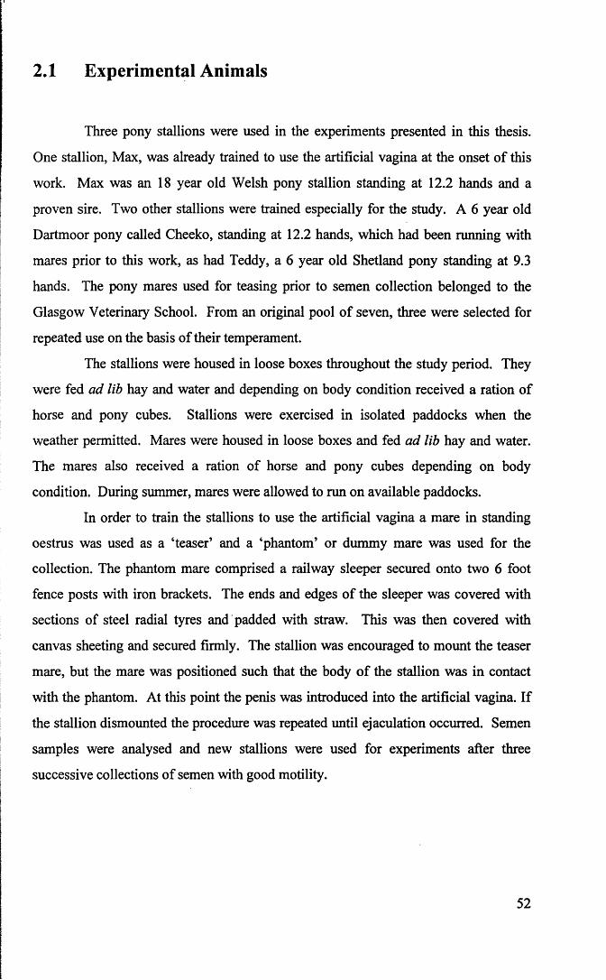
2.1 Experimental Animals
Three pony stallions were used in the experiments presented in this thesis.
One stallion, Max, was already trained to use the artificial vagina at the onset of this
work. Max was an 18 year old Welsh pony stallion standing at 12.2 hands and a
proven sire. Two other stallions were trained especially for the study. A 6 year old
Dartmoor pony called Cheeko, standing at 12.2 hands, which had been running with
mares prior to this work, as had Teddy, a 6 year old Shetland pony standing at 9.3
hands. The pony mares used for teasing prior to semen collection belonged to the
Glasgow Veterinary School. From an original pool of seven, three were selected for
repeated use on the basis of their temperament.
The stallions were housed in loose boxes throughout the study period. They
were fed ad lib hay and water and depending on body condition received a ration of
horse and pony cubes. Stallions were exercised in isolated paddocks when the
weather permitted. Mares were housed in loose boxes and fed ad lib hay and water.
The mares also received a ration of horse and pony cubes depending on body
condition. During summer, mares were allowed to run on available paddocks.
In order to train the stallions to use the artificial vagina a mare in standing
oestrus was used as a ‘teaser’ and a ‘phantom’ or dummy mare was used for the
collection. The phantom mare comprised a railway sleeper secured onto two 6 foot
fence posts with iron brackets. The ends and edges of the sleeper was covered with
sections of steel radial tyres and padded with straw. This was then covered with
canvas sheeting and secured firmly. The stallion was encouraged to mount the teaser
mare, but the mare was positioned such that the body of the stallion was in contact
with the phantom. At this point the penis was introduced into the artificial vagina. If
the stallion dismounted the procedure was repeated until ejaculation occurred. Semen
samples were analysed and new stallions were used for experiments after three
successive collections of semen with good motility.
52

2.2 Semen Collection
Semen used in the experiments described in this thesis was acquired from
either human and equine donors.
2.2.1 Human semen samples
Samples were obtained from healthy male volunteers. Fresh samples were
collected by masturbation into sterile, wide-mouth plastic containers provided by the
laboratory. Samples were allowed to liquefy for approximately 30 minutes at room
temperature, and were processed immediately after liquefaction.
2.2.2 Stallion semen samples
Semen was collected using a Cambridge model artificial vagina (AV) (Figure
2.1). This AV comprised an outer and an inner rubber liner supported by a rigid
plastic casing (Figure 2.2). The plastic casing and rubber liner together formed a
water jacket. Before collection the AV was filled with hot water (>60°C), such that
the luminal temperature was in the range 44°C to 48°C. The luminal pressure of the
AV was adjusted to provide uniformly good contact around the penis, without
interfering with penile penetration. This was achieved by filling the water jacket with
hot water then pushing a hand through the AV to expel excess water through the
filling cap. The inner liner was lubricated with non-spermicidal KY jelly, (Johnson
and Johnson Ltd, Maidenhead, United Kingdom). The collecting receptacle was a
clean plastic bag, fitted with a milk filter to strain the ejaculate, separating gel and
minimising contamination with cellular and non-cellular debris. This collecting
receptacle was kept warm in a cover. After collection the sample was extended in
Kenney ’ s medium at a ratio of 1:1.
53

Figure 2.1 Semen collection.
Figure 2.2 Cambridge model artificial vagina.
54


2.3 Visual Motility Assessment
2.3.1 Motility
After semen collection all samples were inspected visually for motility.
Poorly motile samples were not used for experimental purposes. Where visual
inspection was used within an experiment, sperm motility was assessed by
microscopic examination of a drop of diluted semen at low power (lOOx) on an
Olympus BH2 light microscope. The drop was examined using pre-warmed
glassware, under a coverslip, on a heated microscope stage at 37°C. Vigour of the
motility was scored on a scale of 0-4 as described below;
Score Criteria
4 rapid motility, many forward progressive
3 rapid motility, mostly circling
2 sluggish motility, forward or circling
progressive
1 sluggish motility
0 tail beating without motility
and the percentage of motile cells estimated to the nearest 10%. In order to avoid
bias, two operators were involved in the motility assessment. The slides were
prepared and randomised by the first operator and assessed by the second operator.
2.3.2 Assessment of “head to head” agglutination
‘Head to head’ agglutination or clumping was observed when stallion
spermatozoa were extended and incubated in medium (Bavister TALP-Milk)
supplemented with pentoxifylline. The agglutination was scored as described below:
55

no clumping.+ majority - sperm heads attached in pairs.++ majority - between 2 and 5 sperm heads agglutinated. +++ majority - more than 5 sperm heads agglutinated.
2.4 Medium
2.4.1 Kenney’s medium
Kenney’s medium is a non-fat, dried skimmed milk-glucose extender that
consists of 2.4 g of non-fat dried skim milk (Sanalac™, Premier Beverages, Knighton
Adbaston Stafford, UK), 0.272 M glucose, 2 ml of 7.5% NaHC03 and 2 ml of
gentamycin sulfate (50 mg/ml) in 100 ml of millicure water (Kenney et al., 1975).
During preparation the antibiotic was added after mixing the other chemicals to
prevent curdling of the milk due to the acidic nature of gentamycin. The final pH of
this medium was 6.9.
2.4.2 Graham TALP
This medium was recommended by Dr J. K. Graham (personal
communication). The optically clear nature of the medium made it suitable for the
measurement of fluorescence intensity after Fura-2 ‘loading’. Graham’s TALP
medium comprised 37 mM NaCl, 10.6 mM KC1, 1.2 mM KH2P 0 4, 35.7 mM
NaHC03, 1.2 mM MgS04, 1.7 mM CaCl2.2H20 , 9.2 mM HEPES, 84.4 mM Fructose,
5.5 mM Glucose, 0.18 mM Na pyruvate, 0.37 millilitres Na lactate (60% syrup) and 3
mg/ml bovine serum albumin. The final osmotic pressure was 322 mOsmol/kg and
the pH was adjusted to 7.3. Where required Graham’s TALP was mixed 1:1 (v/v)
with Kenney’s medium.
56

2.4.3 Bavister TALP
The optically clear nature of this medium also made it suitable for the
measurement of fluorescence intensity after Fura-2 ‘loading’. Bavister TALP medium
consisted of 114 mM NaCl, 3.16 mM KC1, 2 mM CaCl2.2H20 , 0.5 mM MgCl2.6H20 ,
10 mM Na lactate syrup (60%), 0.3 mM NaH2P 04.H20 , 5 mM Glucose, 25 mM
NaHC03, 10 mM HEPES, 0.25 mM sodium pyruvate and 6mg/ml bovine serum
albumin (Bavister, 1989). The final osmotic pressure was 302 mOsm/kg and the pH
adjusted to 7.3. Where required Bavister Milk medium was made by mixing Bavister
TALP and Kenney’s medium, 1:1 (v/v).
2.4.4 TALP-egg yolk medium
TALP-egg yolk medium was made by mixing Bavister TALP and TEST-yolk
medium at 1:1 (v/v), as described in Meyers et al., (1995). This medium was reported
to support stallion sperm capacitation in vitro.
2.4.5 TEST-yolk medium
This medium was prepared as described in Bolanos et al., 1983. It consisted
of 188 mM N-Tris(hydroxymethyl) methyl-2-aminoethane sulfonic acid (TES),
824 mM hydroxymethyl aminomethane (Tris), 1.11 mM dextrose, 0.013 g
streptomycine sulfate and 0.008 g Penicillin-G, dissolved in 50 ml of water twice
distilled in glass. The yolk from a chicken egg was then added to make a 20% egg
yolk solution. This medium was then centrifuged at 840g for 10 minutes, after which
the pellet formed was discarded. The pH of the supematent was adjusted to between
7.35 and 7.45 by the addition of Tris. The medium was then modified as in Meyers et
al., (1995), by heat treatment (56°C, 30 minutes) to inactivate the proteases, and by
centrifugation (840g, 20 minutes) and filtration (Millel - HV disposable filter,
Millipore (UK) ltd.) to remove the egg yolk granules.
57

2.4.6 Culture medium for human spermatozoa
Medicult culture medium number 1031 (Medicult Ltd, Copenhagen,
Denmark) was used for human sperm incubations. It consisted of Earle’s balanced
salt solution, a defined synthetic serum replacement, 0.8 mM sodium pyruvate, 2.2 g/1
sodium bicarbonate, 50 IU/ml penicillin, 50 fig/ml streptomycin and 1% human serum
albumin with an osmolarity of 280 mOsm/kg and pH 7.4. This culture medium was
routinely used at the Glasgow Royal Infirmary in the preparation of spermatozoa for
IVF and associated techniques.
2.5 Tissue Culture Ware
Unless otherwise stated 14 ml polystyrene Falcon Grade centrifuge tubes
(Falcon 2095, Becton Dickinson Labware, New Jersey) were used for all sperm
preparation procedures such as centrifugation, swim-up preparation, Fura-2AM
loading and incubation throughout the study. Flat bottom, tissue culture treated
polystyrene cell wells (Cell Wells, Coming, Coming Glass Works, Coming, New
York) were used for incubation of stallion semen in Chapter 5, section 5.2.1.
2.6 Chemicals
2.6.1 Pentoxifylline [l-(5-oxyhexyl)-3-7-dimethylxanthine]
A stock solution of pentoxifylline (Sigma Chemical Company, Poole, Dorset,
England) was made by dissolving 20 mg of pentoxifylline in 10 ml Bavister TALP to
give a concentration of 2 mg/ml. This solution was further diluted in medium to give
58

the concentrations required in individual experiments. Fresh solutions were made on
the day of use.
2.6.2 Progesterone (4-Pregnene-3, 20-dione)
A stock solution was made by dissolving 1 mg of progesterone (Sigma
Chemical Company, Poole, Dorset, England) in 1 ml of dimethylsulphoxide (DMSO;
Sigma Chemical Company, Poole, Dorset, England) to give a concentration of 1
mg/ml and stored at -70°C in eppendorf tubes. The stock solutions were further
diluted as described for individual experiments.
2.6.3 Ionophore A23187
A stock solution was made by dissolving 10 mg of Ionophore A23187
(Sigma Chemical Company, Poole, Dorset, England) in 5 ml of DMSO. This was
stored as 50 pi aliquots in foil-wrapped eppendorf tubes at -70°C. This stock solution
was diluted in TALP medium to a working concentration of 0.5 pM immediately
before use.
2.6.4 Fura-2AM ({l-[2-(5-Carboxyoxazol-2-yl)-6-aminobenzofuran-
5-oxy]-2-2,-amino-5’-methylphenoxyethane-N,N,N,N ’
tetraaceticacid pentaacetoxymethyl ester})
A stock solution of Fura-2AM was made by sonicating 0.25 g Pluronic F I27
(Molecular Probes Inc., Eugene, USA) in 1 ml of DMSO until the detergent dissolves
completely. One ml of this solution was then transferred into a light-shielded bottle
containing Fura-2AM (1000 pg; Calbiochem-Novabiochem (U.K) Ltd, Beeston,
Nottingham), mixed and dispensed into eppendorf tubes in 50 pi aliquots of 1 mM
Fura-2AM. These tubes were stored at -70°C, in the dark, and thawed individually for
use.
59

To form the working solution 50 pi aliquots of stock solution were mixed
with 5 ml of medium (Graham’s TALP or Bavister TALP) to give a working Fura-
2AM concentration of 10 pM. This was mixed 1:1 with sperm suspension.
2.7 Cooling of Semen
A programmable freezing unit (Kryo 10 Series, Planer Biomed, Planer
Products Ltd, Sunbury-ON-THAMES, England) (Figure 2.3) was used to cool semen
samples. Plastic straws of 0.5 ml capacity were filled with sperm suspension, sealed
with P.V.P powder (IMV, rue Clemenceau, Casou, France) and maintained in a water
bath at 37°C. The straws were then transferred onto prewarmed racks which were
placed in the cooling chamber of the programmable freezer. The machine was
programmed to lower the temperature from a selected starting temperature to 5°C at a
rate of 0.05°C per minute. Straws were then removed from the straw racks, immersed
in a waterbath and maintained at 5°C in a refrigerator for up to 72 hours.
2.8 Semen Preparation Methods
2.8.1 Sperm concentration
Sperm concentration was determined using a haemocytometer as described in
the World Health Organization Laboratory Manual (WHO, 1987) and adjusted as
required for individual experiments by centrifugation (300g for 10 minutes) and
resuspension in the appropriate medium. For counting, semen samples were diluted
1:10 in a counting solution consisting of 5 g of sodium bicarbonate and 5 ml of
formalin in 500 ml of distilled water. The sample was mixed thoroughly and 10 pi
aliquots placed in each counting chamber and allowed to settle for 1 minute. The
60

spermatozoa in 5 large squares were counted in each chamber and, in order to
determine the concentration of the original sample in millions/ml the sperm count was
divided by two.
2.8.2 Swim-up migration
The swim-up migration was performed by layering 500 pi of liquefied semen
(human) or extended semen (stallion) at the bottom of a 14 ml test tube containing 1
ml of culture medium, using a lcc syringe and 18G needle. After 45 minutes of
incubation at 37°C in 5% C 02 in humidified air, the upper layer was carefully
removed without disturbing the interface.
2.8.3 Removal of seminal plasma by centrifugation
Unless otherwise stated, for the majority of stallion sperm experiments,
seminal plasma was removed by centrifugation at 300g for 5 minutes after 1:1 dilution
in Kenney’s medium. Seminal plasma was removed and the pellet re-suspended in
medium. Removal of seminal plasma by centrifugation through discontinuous Percoll
gradients is described in detail in Chapter 3.
61

Figure 2.3 Programmable freezing unit (Kryo 10 Series).
Figure 2.4 Hamilton-Thome motility analyser set-up with video and
computer.
62


2.9 Viability Staining
Sperm viability was assessed using either an eosin-nigrosin stain (Hancock,
1951) or the dual staining method described below.
2.9.1 Eosin-nigrosin staining
The eosin-nigrosin stain consisted of 0.67 g of eosin Y and 10 g nigrosin
dissolved in 100 ml of water. The stain was mixed with an equal volume of sperm
suspension for 1 minute. A drop was then transferred to an alcohol-cleaned slide,
smeared and allowed to air dry. Slides were randomised and at least 100 spermatozoa
were examined on each slide under oil immersion. Unstained spermatozoa were
classified as ‘live’ and those showing any degree of pink, indicating that the stain had
permeated the plasma membrane, were classified as ‘dead’.
2.9.2 Dual staining with Chlortetracycline and Hoechst 33258
A stock solution of Hoechst 33258 (2’-[4-Hydroxyphenyl]-5-[-4-methyl-l-
piperazinyl]-2,5’-bi-lH-benzimidazole); (Sigma Chemical Company, Poole, Dorset,
England) was prepared by dissolving 4 mg of Hoechst 33258 in 1 ml of millicure
water. Aliquots (10 pi) were then stored in foiled wrapped eppendorfs at -70°C. A
working solution was prepared by diluting stock solution in 10 ml of medium
(Medicult for human, TALP for stallion) in foiled wrapped tubes. For experiments
using human spermatozoa the medium used was Medicult. TALP was used for
experiments with stallion spermatozoa.
Phosphate buffered saline (PBS) containing 2% polyvinylpyrrolidone-40
(PVP-40, Sigma Chemical Company, Poole, Dorset, England) was prepared by
mixing 2 g PVP-40 in 100 ml PBS and adjusting the pH to 7.4. This solution was
stored as 10 ml aliquots in Falcon grade tubes at -70°C and warmed to 37°C prior to
use.
Chlortetracycline buffer was prepared by mixing 20 mM Tris
63

(hydroxymethyl)methylamine[2-amino-2-(hydroxymethyl)propane-1,3-diol]), 130
mM NaCl and 5 mM cysteine at pH 7.8 and stored in 50 ml aliquots at -70°C.
Chlortetracycline powder (CTC; C22H23CIN20 8.HC1; Sigma Chemical Company,
Poole, Dorset, England) was weighed into tin foil packets in portions of 19.5 mg and
stored at -70°C. The working stain solution was prepared by adding the contents of a
foil packet to 50 ml buffer.
Paraformaldehyde fixative was prepared by adding 1.25 g paraformaldehyde
(Sigma Chemical Company, Poole, Dorset, England) to 10 ml of 0.5 M Tris-HCL
buffer at pH 7.4 to give a 12.5% solution. Aliquots of 1 ml were stored at -70°C.
DABCO (l,4-Diazabicyclo[2.2]octane) fluorescence retarder was prepared by adding
0.2469 g of DABCO to 10 ml of glycerol to give a final concentration of 0.22 M and
stored as 1 ml aliquots at -70°C.
Staining M ethod
One aliquot of Hoechst 33258 (H258) stock solution was diluted in Bavister
TALP medium to give a working concentration of 4 pg/ml. This working solution
was kept foil wrapped and held at the same temperature as the samples. To stain the
sperm cells a 50 pi sample of sperm suspension was mixed with an equal volume of
H258 solution for 2 minutes. The sperm suspension was than washed free of dye by
centrifugation through 1.5 ml of 2% PVP-40 in PBS at 900g for 5 minutes. The
supernatant was removed and the pellet resuspended in 20 pi of medium. This
solution was mixed with 20 pi of CTC working solution for 30 seconds. After 30
seconds, 1.6 pi of 12.5% paraformaldehyde and 5 pi of DABCO were added. A 5 pi
aliquot was then placed on a clean warm slide, and a coverslip carefully applied. The
coverslip was squashed gently to exclude extra liquid, and the edges sealed with nail
varnish. Slides were stored in a light-protected container and evaluated immediately
or up to 2 days later if kept at -70°C, protected from light.
64

2.9.3 Computerised motility analysis
2.9.3.1 Cell chambers
Aliquots of sperm suspension (5 pi) were used to make preparations of 32 pm
depth as described by Mack et al. (1988). Cell chambers were made by applying dry
transfer circles (13 mm diameter, Chartpak # 49, Leeds, MA 01053-9732) to
microscope slides, previously cleaned with absolute alcohol and layered with 10%
bovine serum albumin (BSA), for the analysis of stallion spermatozoa, or 1%
collodion nitrocellulose in amyl acetate for the analysis of human spermatozoa. The
latter step was designed to minimise sperm adhesion to glass. To ensure correct
chamber depth, 5 pi of the sample was placed in the middle of the circle and a
coverslip applied to force the contents to fill the chamber and spill over the sides of
the O-ring.
2.9.3.2 Recording procedure
For each experiment, recordings of at least 200 spermatozoa from two or
more slides were made on a video recorder (Ferguson Videostar Model FV 32L).
Videotapes were stored for subsequent analysis.
2.9.3.3 Computerised analyser
The Hamilton Thome Motility Analyzer model 2030, version 7.1, (Hamilton-
Thome Research Inc., Beverly, MA, USA) was used to measure motility parameters
(Figure 2.4). The analyser comprised an internal microscope and an automated heated
microscope stage, which positioned the sample while maintaining a user defined
temperature. The optical assembly used phase contrast optics. Images were acquired
by a CCD sensor and digitised. The digitised image was then analysed by a
microprocessor and displayed on a colour graphics monitor.
65
I
2.9.3.4 Param eter settings
The parameter setting used for both human and stallion spermatozoa
throughout this study are summarised in Table 2.1. The parameters for human sperm
were determined previously (Kay et al., 1993). Derivation of parameters for stallion
sperm is described in Chapter 3. For each sperm track a set of movement
characteristics was determined. These were amplitude of lateral head displacement
(ALH, pm), linearity (LIN, %), curvilinear velocity (VCL, pms'1), average path
velocity (VAP, pms"1) and straight line velocity (VSL, pms'1). Individual cell data
generated by the HTM analyser were captured by a portable personal computer
(Toshiba T3100) using a HDATA software (HDATA Communications Package for
HTM motility analyser). Descriptions of these movement parameters are given more
fully in Chapter 1. Using visual assessment and CASA systems it had been possible
to identify three general patterns of stallion sperm motility similar to that reported for
human spermatozoa (Robertson et al., 1988):
1) Forward progressive or non-hyperactivated motility involves an essentially straight
progressive trajectory with uniform flagellar waves (Figure 2.5)
2) Transitional phase motility also involves an essentially straight progressive
trajectory, though flagellar waves are larger and irregular (Figure 2.6)
3) Hyperactivated or ‘star-spin’ motility is characterised by non-progressive trajectory
with large flagellar waves, which cause the cells to undergo successive rapid flexions
(Figure 2.7)
Initial measurements of hyperactivated motility were made using the criteria of
Burkman (1984) i.e. ALH > 7.5 jun, VCL > 100 urns'1 and LIN < 65%.
66

Figure 2.5 Spermatozoa! motility pattern - forward progressive.
Figure 2.6 Spermatozoa! motility pattern - transitional progressive.
Figure 2.7 Spermatozoal motility pattern - hyperactivated.
67

EXPANDED U I E U

Table 2.1. Machine settings used for motility analyses performed using the Hamilton-
Thome motility analyser. The derivation of parameter settings for stallion
spermatozoa is described in Chapter 3.
Parameter Human Stallion
Temperature 37°C 37°C
Image type Phase contrast Phase contrast
Field Selection Manual Manual
Calculate ALH yes yes
Frames (min) 13 13
Frames (max) 20 20
Acquisition rate 25 Frames/sec 25 Frames/sec
Minimum contrast 6 pixels 5 pixels
Minimum size 6 pixels 4 pixels
Low size gates 0.4 0.5
High size gates 1.6 1.2
Low intensity gates 0.5 contrast units 0.5 contrast units
High intensity gates 2.0 contrast units 1.2 contrast units
Non motile head size 8 pixels 12 pixels
Non-motile intensity 201 170
Magnification factor 2.17 2.17
Minimum VCL (pms*1) 0 pms’1 40 pms'1
Minimum VAP (pins’1) 5 pms'1 30 pms'1
Minimum VSL (pms'1) 0 pms’1 20 pms"1
2.9.4 Intracellular calcium measurements using Fura-2AM
2.9.4.1 Loading sperm cells with intracellular dye - ‘Fura loading’
In order to measure intracellular free calcium the sperm concentration was
adjusted to 100 million sperm per ml and aliquots of 2 ml were prepared. To each
aliquot was added an equal volume of 0 or 10 pM Fura-2AM in medium. The aliquot
containing 0 pM Fura-2AM served as a control sample for sperm autofluorescence.
Experimental samples contained a final concentration of 5 pm Fura-2AM in a volume
of 4 ml. This suspension was incubated in a foil wrapped test tube at 37°C for 30
minutes to allow Fura-2AM to penetrate into cells. At the end of the incubation
period, samples were centrifuged (300g, 10 minutes) and the supernatant discarded to
remove extracellular dye. The pellet was then re-extended in either Graham TALP or
Bavister TALP medium to give a final sperm suspension of 50 million/ml in a 4 ml
cuvette.
2.9.4.2 Intracellular calcium measurement - population studies
The fluorescence intensity of sperm samples within a cuvette was measured
using a dual-wavelength spectrofluorimeter (diffraction grating monochromator,
Perkin Elmer LS-3B, Perkin-Elmer Corporation, Norwalk, USA; Figures 2.8 and 2.9).
Cuvettes were placed into a sample chamber which was maintained at 37°C by a
circulating water jacket. Sperm suspensions were stirred continuously by a magnetic
flea in the cuvette. The excitation wavelengths of the spectrofluorimeter were set to
340 and 380 nm, respectively, and the emitted fluorescence was recorded at 510 nm.
The machine was set to make measurements of intensity (f) at a rate of 1 reading every
3.5 seconds alternately for each of the excitation wavelengths (f340 and f380). Prior to
reading each experimental sample, background intensity was established by reading
the autofluorescence control at 3.5 second intervals for approximately 1 minute. The
period of reading of experimental samples is detailed in the appropriate Chapter.
The ratio of the fluorescence intensities (R), was obtained after first
69

subtracting background intensity (sperm autofluorescence) as shown irfthe following
formula;
_ (^340(loaded) ~ ^340(un loaded))
(^380(loaded) ^380(un loaded))
2+Intracellular free Ca was calculated by incorporating the ratio of intensity
from the experiment into the equation described by Grynkiewicz et al., (1985). The
dissociation constant Kd of 225 nmol I"1 and p = 7.3 are standard calibration factors.
( R ” R m i n ) * v a *[C a2+1 = K * mi"; * Kd L Ji (R max- R)
R max ° f 16.3 and Rmin of 0.81 are values related to maximum and minimum
intracellular calcium from previous calibration. Rmax was obtained by adding 8 pM
ionomycin, a calcium ionophore, to cell suspension with known concentration of
extracellular calcium (1.8 mM), to saturate the dye within the cell; this was followed
by adding 40 mM EDTA to sequester all calcium, hence Rmin is the limiting value that2+R can have at zero [Ca ].
For example; if the fluorescence intensity after subtracting background
intensity is 579 and 346 at 340 nm and 380 nm respectively, then the ratio of
intensities (derived by dividing 579 by 346) is 1.67. This value of 1.67 is then
incorporated into the formula, thus;
[ca2 q = (1 .6 7 -0 .8 1 )(1 6 .3 -1 .6 7 )
= 97 nM
* 225 nM *7.3
2.9.4.3 Intracellular calcium measurement - single cell studies
The measurement of the intracellular free calcium of individual sperm cells
was performed using an inverted fluorescence microscope (Nikon Diaphot, Tokyo,
70

Japan). A schematic diagram is shown in Figure 2.10. An intense broad-spectrum
light was supplied by a xenon lamp (a) and a low voltage shutter (b) limited the
illumination time by allowing illumination to be turned on and off without the need to
reignite the lamp. A spinning wheel (c) (Caim Research, Sittingboume, Kent)
equipped with six filters, all having half-bandwidths of 10 nm and alternated at 120
Hz, reduced the spectrum to narrow bands and allowed rapid sequential measurement
at two (or more) excitation wavelengths. After passing through the filter, the
excitation light was reflected by a dichroic mirror (d). This had a half-pass
wavelength of 400 nm for Fura-2. The reflected light was focused onto the
preparation by the lOOx Fluor objective of the inverted microscope (e). The longer
wavelength light emitted by the fluorescent indicators was transmitted by dichroic
mirror. The light path passed a sliding mirror (f) which directed the light to the eye
pieces of the microscope (g) or allowed it to pass through an adjustable rectangular
diaphragm (h) which could be positioned to ensure that light only from the region of
interest was measured. The light then passed through a short pass filter (half pass
wavelength 700 nm) mounted at 45° which allowed light emitted by the fluorescent
indicator to go to the photomultiplier (j) and ensured that the much longer visible
wavelength light from the normal microscope illumination did not reach the
photomultiplier but was directed to the video camera (k) used for monitoring the
sperm and adjusting the diaphragm. The rapidly switched signals from the
photomultiplier were integrated using timing pulses derived from the spinning wheel.
An output voltage corresponding, to intensity ratio was produced with a hardware
divider circuit. Both intensity signals and the ratio signal were recorded on paper
chart for later analysis.
2.9.4.4 Preparation of sperm suspension and slide chamber for single cell
measurements
Preliminary experiments were required to establish a means of immobilising
spermatozoa and a protocol for the measurement of intracellular calcium. This
process is described in Chapter 3.
71

Figure 2.8 - 2.9 Perkin - Elmer LS-3B dual wavelength spectrofluorimeter
with water bath (a) set in order to regulate temperature of cuvette held in the
cuvette housing (b) at 37°C.
72


2.10 Radioligand Binding Assay Studies - Principle and
Procedure
The principle of this assay is that radioligand will bind to both the receptor
and non-receptor sites. Binding to a receptor site occurs in a saturable manner
whereas binding to non-receptor sites is in a less saturable manner. Thus, the amount
of isotope adhering to tissue in the absence of unlabelled radioligand represents the
total binding (receptor + non-receptor). When tissues are incubated in a fixed
concentration of radiolabelled ligand in the presence of a saturating concentration of
unlabelled ligand, the unlabelled ligand will selectively inhibit radiolabelled binding
to the ligand but not influence binding to other membrane constituents. This
represents the blank. The difference between total and blank is taken as a measure of
the isotope attached to the receptor (Enna, 1985; Motulsky, 1995).
The radiolabelled ligand used in these studies was [1,2,6,7-3H] progesterone
(Amersham, Life Science, Amersham International, Little Chalfont, Bucks, England).
The stock radioactive concentration was 1.0 mCi/ml which was further diluted in
ethanol to give a concentration (expressed as counts per minute) of 18,250 cpm/pl.
This was stored at -79°C in a freezer used for storing radioactive material only.
The binding assay involved layering a 20 pi aliquot of radiolabelled
progesterone onto 20 pi of DMSO in 1.5 ml eppendorf tubes, and air drying the
mixture for 30 minutes to saturate the DMSO with labelled ligand. This was carried
out for both total and blank designated tubes. One hundred microlitres of sperm
suspension was then added to the total binding and the non-specific binding tubes. In
addition, 10 pi of 1 mg/ml unlabelled progesterone was added to the non-specific
binding tubes to give a final concentration 3.18 pM. The tubes were mixed well and
incubated at 37°C for 45 minutes. At the end of the incubation period, the tubes were
centrifuged (Jouan microcentrifuge, Model A 14, Saint-Herblain, France) at 14000 G
for 5 minutes. After centrifugation the supernatant was discarded and the pellet re
extended in 100 pi of medium. Centrifugation was repeated and the pellet formed by
this second washing step re-extended in 50pl medium, transferred into scintillation
vial inserts (Packard Instrument Company, Meriden) and topped up to 4 ml with
73
scintillation medium (Ecoscint A, National Diagnostics, Atlanta, Georgia, 30336).
The radioactivity in the tubes was read using a Liquid Scintillation Analyser (Model
1600TR, Packard Instrument Company, Meriden). The same procedure was used for
human and stallion spermatozoa.
2.11 Statistical Analysis
Statistical analysis was carried out using the statistical package GraphPad
InStat. When comparing data from populations with equal standard deviation (SD) an
ordinary Analysis of Variance (ANOVA) was performed. When comparing data from
population exhibiting different SD, a Kruskal Wallis non-parametric ANOVA was
performed. With only two sets of data, a Student’s t test was performed. Conversely,
an alternate Welch t test was carried out if SD of the two set of data were significantly
different. Where appropriate a correlation analysis was performed to examine the
nature o f the relationship between variables.
74

Figure 2.10 A schematic diagram of the set-up for measurement of
intracellular calcium in single sperm cell.
75

Figu
re
2.10
: A
sche
mati
c dia
gram
of
the
set-u
p for
mea
sure
men
t of
intra
cellu
lar
calci
um
in sin
gle
sperm
ce
ll.f -
• o

CHAPTER 3
DEVELOPMENT OF METHODS

3.1 Introduction
The experiments reported in this Chapter were performed to validate
techniques which had not previously been evaluated for use with stallion spermatozoa
or were modified for use in this study.
3.2 Validation of the Hamilton-Thorne Computerised
Motility System for the Analysis of Stallion
Spermatozoa
Three experiments were conducted to validate the Hamilton-Thorne system
for analysis of equine spermatozoa. It was necessary to optimise the detection of
motile spermatozoa by the appropriate setting of the “Main Gates” (Experiment 1).
Setting the “Main Gates” involved modifying certain parameters in order to optimise
the correct identification of motile spermatozoa and minimise misidentification of
debris as spermatozoa. The “Main Gate” parameters were the size of the object, in
pixels, and contrast of the object, in arbitrary units of brightness above background
intensity. It was also necessary to set “Lo/Hi” size gates, which set size limits and
“Lo/Hi” intensity gates which set intensity limits for immotile cells. Default values
were used to define immotile cells if there were fewer than five motile cells in a field.
Having selected the appropriate “Main Gate ” settings, minimum velocity
thresholds were imposed to minimise the mis-identification of moving particles as
motile cells (Experiment 2). Finally, the repeatability of the analysis was tested
(Experiment 3). These experiments are described below.
76

t
3.2.1 Experiment 1 - Setting “Main Gates” for equine sperm
motility analysis
Semen from a single ejaculate was used in this experiment. Collection of
semen was as described in Chapter 2. The concentration was determined using a
haemacytometer and semen extended in Kenney’s medium to a final concentration of
25 x 106 / ml. At this concentration between 5 and 25 spermatozoa were visualised on
the Hamilton-Thorne screen per field. Five microlitre samples were loaded into slide
chambers as described in Chapter 2 and placed onto the slide stage of the Hamilton-
Thorne H2030 unit. The digitised image of sperm motion was observed on the screen
of the analyser, and recorded onto a video tape for repeated analysis.
A range of settings were examined and these settings are tabulated in Table
3.1. Approximately 1000 sperm cells were examined for each setting using the video
PLAYBACK facility on the Hamilton-Thorne unit. For this experiment velocity
parameters were set at minimum values. On digitising the image the centroid of
sperm cells is marked with a red point if motile or a blue point if immotile. The
number of cells correctly marked, incorrectly marked or omitted from the analysis
were recorded.
Table 3.1: Summary of the “Main Gate” settings examined in Experiment 1.
Parameter Set-up 1 2 3 4 5
Motile Cells
Minimum size 5 5 4 4 6
Minimum contrast 4 5 6 5 5
Immotile Cells
Minimum (LO) size 0.5 0.5 0.5 0.5 0.4
Maximum (HI) size 1.2 1.2 1.2 1.2 1.8
Minimum (LO) intensity 0.5 0.5 0.5 0.5 0.3
Maximum (HI) intensity 1.2 1.2 1.2 1.2 1.5
Default size 12 12 12 12 11
Default intensity 170 170 170 170 175
77

3.2.1.1 Results
Numbers of spermatozoa which were correctly marked, mismarked either as
immotile or incorrectly identified as motile, motile but omitted and immotile but
omitted are summarised in Table 3.2. Overall, Setting 5 generated the lowest number
of total errors when motile and immotile cells were considered (6.3%). Setting 4,
however, generated the lowest number of errors involving motile spermatozoa, i.e.
incorrectly marked immotile and missed motile and omitted (3.2%). This parameter
set-up was selected for use in all subsequent stallion semen motility analysis
presented in this thesis.
Table 3.2: Summary of the type and percentage of errors observed for each of the settings evaluated.
SetClassification
1 2 3 4 5
correctly identified 1007 1012 1012 884 942
incorrectly marked immotile 102 140 202 14 6
missed motile 3 1 6 14 42
missed immotile 30 6 76 47 11
% error 13.4 14.5 28.1 10.3 6.3
% incorrectly marked immotile and missed motile
10.2 13.9 20.5 3.2 5.1
3.2.2 Experiment 2 - Establishing lower velocity thresholds to
minimise the misidentiflcation of immotile cells as motile
It was apparent from Experiment 1 that misidentification of immotile cells as
motile or classification of debris as motile cells was a considerable problem. One
hundred of these incorrectly identified cells were counted using the video
PLAYBACK facility and EDIT TRACK module which displayed motility
measurements for each individual cell. Data were recorded onto a personal computer
78

linked to the HTMA and the velocity distribution of the curvilinear, average path and
straight-line velocities (VCL, YAP, VSL; pms*1) of these cells were plotted.
3.2.2.1 Results
Velocity distribution plots are shown in Figures 3.1a-c. It was observed that
91% of moving debris and cells showed a VCL of < 40 pins’1, 95% a VAP < 30 pms'1
and 96% a VSL < 20 pms’1.
79

Figure 3.1a: Distribution plot of curvilinear velocity (VCL) of immotile
cells or debris misidentified as motile cells. The 91% of cells had aVCL of
less than 40 pm/sec.
Figure 3.1b: Distribution plot of average path velocity (VAP) of immotile
cells or debris misidentified as motile cells. The 95% of cells had a VAP of
less than 30 pm/sec.
Figure 3.1c: Distribution plot of straight line velocity (VSL) of immotile
cells or debris misidentified as motile cells. The 96% of cells had a VSL of
less than 20 pm/sec
80

Num
ber
of sp
erm
atoz
oa
Num
ber
of sp
erm
atoz
oa
Num
ber
of sp
erm
atoz
oa
3.1a
15
10
5
0
VCL - pms"1
3.1b
20
15
10
5
0
VAP - pms"1
3.1c
20
15
10
5
00 5 10 15 20 25 30 35 40 45 50 55 60
VSL - pms"1
- "
-
-
-
PI1
_
n In . . n . . . . .
40
40 50

3.2.3 Experiment 3 - Testing the effect of imposing low velocity
thresholds
Using the low velocity thresholds derived in section 3.2.2, sections of
videotape were analysed with or without the implementation of the thresholds.
Approximately 200 cells were analysed for each parameter set-up.
3.2.3.1 Results
Distribution plots were constructed for VCL, VAP and VSL for the first 200
cells analysed on a section of videotape. When low velocity thresholds were not
omitted 55, 51 and 54% of cells fell into the low velocity categories for VCL, VAP
and VSL, respectively. Visual inspection confirmed that this population of cells
consisted of ‘moving’ immotile cells and debris or cells stuck to glass and showing
very poor motility. These cells were eliminated from analysis by the application of
the low velocity thresholds. Mean values of VCL, VAP and VSL increased because
immotile cells were eliminated from the analysis. Velocity distribution plots for VSL
with and without thresholds are shown in Figure 3.2a-b.
81

Figure 3.2a: Distribution plot of straight-line velocity (VSL) for 200 cell
analysed before the application of low velocity thresholds. Note the high
proportion of cells (54%) with velocities of < 20 pm/sec. Visual inspection
confirmed that these cells were immotile, stuck to the slide or were moving
debris.
Figure 3.2b: Distribution plot of straight-line velocity (VSL) for 200 cells
analysed from the same videotaped sample after the application of low
velocity thresholds. The mean VSL increased after motile artefacts were
deleted from the analysis.
82

Num
ber
of sp
erm
atoz
oa
Num
ber
of sp
erm
atoz
oa
40
20
0 . . . .
0 10 20 30 40 50 60 70 80 90 100
VSL - i^ms1
3.2b60
4 0 -
2 0 -
00 10 20 30 40 50 60 70 80 90 100
VSL - Jims' 1
Mean VSL = 41,8|im/sec (SD = 10.5)
Low velocity threshold
3.2a
Mean VSL = 22.8nm/sec (SD = 17.6)

3.2.4 Experiment 4 - Repeated analysis of the same videotape
This experiment was performed to determine the accuracy of repeated
analysis using the settings derived in Experiments 3.2.1 and 3.2.2. A total of
approximately 200 cells, with a minimum of 13 trackpoints, from the same section of
videotape were analysed in triplicate and the coefficient of variation (CV, %) of the
three analyses was calculated.
3.2.4.1 Results
Table 3.3: Measurement of ALH, VAP, VCL and VSL in triplicate for 200 cells
from the same section of videotape.
Tracksrecorded
ALH VAP VCL VSL
1st analysis 206 7.65 115.5 131.7 93.62nd analysis 203 7.48 114.9 132.0 90.73rd analysis 202 7.92 116.1 134.6 91.2Mean 7.69 115.5 132.8 91.8Std Deviation 0.23 0.60 1.61 1.56
CV (%) 2.99 0.52 1.21 1.70
83

3.3 Separation of Spermatozoa from Seminal Plasma
Because seminal plasma is believed to affect capacitation and calcium
regulatory mechanisms preliminary experiments were carried out to test three
different methods of separating spermatozoa from seminal plasma. One experiment
was carried out to assess three different separation methods, centrifugation, swim-up
from seminal plasma and the use of a discontinuous Percoll gradient for separation of
human and equine spermatozoa from seminal plasma. Efficacy of separation was
judged by the percentage of the initial sample which was viable after separation and
the quality of the motility. An additional experiment was carried out to assess three
different discontinuous Percoll gradients with ejaculates from one individual stallion
because of failure to produce a motile sample from this animal after separation
through a 50/100% gradient.
3.3.1 Preparation of Percoll gradients
A stock solution of Percoll was purchased (Pharmacia, Uppsala, Sweden) and
an isotonic solution, termed 100% Percoll, was prepared by mixing 45 ml of Percoll
with 5 ml of lOx Earles balanced salt solution (Sigma Chemical Company, Poole,
Dorset, England), 0.75 ml of 4.5% BSA, 0.185 ml Na lactate (60% syrup) and 1.5 mg
Na pyruvate. Further Percoll dilutions were made by diluting 100% Percoll with
medium as shown in Table 3.4, to give dilutions ranging from 100% to 40% Percoll.
Medicult culture medium was used for all human sperm experiments and TALP was
used for all equine experiments.
84

Table 3.4: Percoll dilutions were made by mixing 100% Percoll with the appropriate
volume of medium as shown below.
100% 90% 80% 70% 50% 40%
100% Percoll 10 ml 9 ml 8 ml 7 ml 5 ml 4 ml
Medium 0 ml 1ml 2 ml 3 ml 5 ml 6 ml
3.3.2 Experiment 1 - Separation of human and equine spermatozoa
from seminal plasma by three different methods
One ejaculate was collected from each of 3 human donors and the initial
concentrations were determined using a haemocytometer as described in Chapter 2.
Three aliquots from each ejaculate were treated as follows. A swim-up was prepared
as described in Chapter 2 by layering 500 pi of liquefied semen beneath 1 ml of
Medicult and incubating for 45 minutes. The supernatant was then recovered. A
500 pi aliquot of semen was diluted by the addition of 1.5 ml Medicult, centrifuged at
300g for 5 minutes, the supernatant discarded and the pellet reconstituted to 500 pi. A
2-step discontinuous Percoll gradient was formed by carefully layering 3 ml of 50%
Percoll over 3 ml of 100% Percoll. A measured aliquot of 500-1000 pi of semen was
layered above the Percoll and the sample centrifuged at 500g for 20 minutes. The
supernatant was then discarded and the pellet formed below the 100% Percoll was
washed by centrifugation at 200g for 6 minutes. Again the supernatant was discarded
and the pellet reconstituted to the original volume. Duplicate sperm counts were
performed on aliquots of the separated samples, duplicate eosin-nigrosin smears were
made and samples were videorecorded for motility analysis.
This experiment was then repeated for one ejaculate from 3 different
stallions. For equine spermatozoa Bavister-TALP was used as the diluting medium.
85

3.3.2.1 Results
The sperm count (millions/ml), percentages live and percentage recovered
after separation of seminal plasma by one of three different methods are summarised
in Tables 3.5 and 3.6. For human samples there was no difference in the percentage
recovered after centrifugation or Percoll separation. Recovery after swim-up was
significantly (p<0.05) lower than either, at 21% compared with 68 and 62%,
respectively. The percentage live was unchanged by centrifugation or Percoll
separation but was numerically higher by swim-up preparation.
For equine samples the highest recovery was seen after centrifugation and
recovery after swim-up was significantly (p<0.05) lower at 22% than after separation
by the other two methods. No enhancement of the percentage live was observed for
any of separation methods.
Movement analysis of human samples generally showed an increase in ALH
and velocity after separation from seminal plasma regardless of method (Table 3.7).
This increase was most marked using ‘swim-up’. There was no clear increase in
movement parameters of equine spermatozoa after separation. No motility was
recorded for one stallion (Table 3.8, stallion 2) after separation through Percoll due to
sperm agglutination.
86

Table 3.5. Sperm count (millions/ml), percentage live and percentage recovered from
three human donors after separation of human sperm from seminal plasma by one of
three different methods.
Donor Count % live % recover
Fresh 1 125 53 -2 122 80 -3 162 78.5 -Mean (SEM) 136.3 (12.8) 70.5 (8.8)“ -
Centrifuge 1 100 62 80%2 83 77 68%3 91.5 74 56%Mean (SEM) 91.5 (4.9) 71 (4.6) “ 68% (6.9) “
Swim-up 1 24 89 19%2 20.5 99 17%3 45.5 99.5 28%Mean (SEM) 30(7.8) 95.8 (3.4) “ 21% (3.4) b
Percoll 1 67 62 53%2 65 91 53%3 130 85 80%Mean (SEM) 87.3 (21.3) 79.3 (8.8) “ 62% (9.0) “
different letters a, b between rows s lows significant difference (p < 0.05)
87
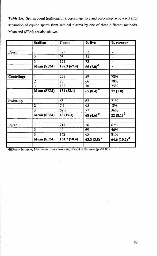
Table 3.6. Sperm count (millions/ml), percentage live and percentage recovered after
separation of equine sperm from seminal plasma by one of three different methods.
Mean and (SEM) are also shown.
Stallion Count % live % recover
Fresh 1 325 52 -
2 95 73 -
3 175 73 -
Mean (SEM) 198.3 (67.4) 66 (7.0)a -
Centrifuge 1 255 59 78%2 75 66 78%3 132 70 75%Mean (SEM) 154 (53.1) 65 (8.4) “ 77 (1.0)“
Swim-up 1 68 62 21%2 7.5 65 8%3 62.5 77 36%Mean (SEM) 46 (19.3) 68 (4.6) a 22 (8.1) b
Percoll 1 218 56 67%2 44 69 46%3 142 65 81%Mean (SEM) 134.7 (50.4) 63.3(3.8)“ 64.6 (10.2)“
different letters a, b between rows s lows significant difference (p < 0.05)
88

Table 3.7. Mean and SEM for amplitude of lateral head deviation (ALH, pm) and
velocities (VCL, VAP and VSL; pm/s) of human spermatozoa before and after
separation from seminal plasma by one of three different methods.
Donor ALH VCL VAP VSL
Fresh 1 5.1 73.8 59.9 51.12 8.3 116.8 85.1 75.13 5.9 89.7 72.8 64.9Mean (SEM) 6.4 (1.0) 93.4 (22.1) 72.6 (7.3) 63.7 (7.0)
Centrifuge 1 5.5 82.7 68.7 60.22 8.5 121.6 86.7 75.43 7.1 107.3 85.7 77.6Mean (SEM) 7.0 (0.9) 103.9(11.4) 80.4 (5.8) 71.1 (5.5)
Swim - up 1 6.9 102.1 83.1 70.42 9.5 137.5 97.2 85.93 7.7 112.0 90.0 79.7Mean (SEM) 8.0 (0.8) 117.2(10.5) 90.1 (4.1) 78.7 (4.5)
Percoll 1 5.2 76.1 61.3 53.32 9.0 114.3 76.4 63.53 7.8 104.9 80.0 68.1Mean (SEM) 7.3 (1.1) 98.4 (11.5) 72.5 (5.7) 61.6 (4.4)
89

Table 3.8. Mean and SEM for amplitude of lateral head deviation (ALH, jim) and
velocities (VCL, VAP and VSL; pm/s) of equine spermatozoa before and after
separation from seminal plasma by one of three different methods.
Horse ALH VCL VAP VSL
Fresh 1 8.0 105.1 86.0 62.62 7.6 81.5 53.3 34.23 4.1 63.7 53.2 46.7Mean (SEM) 6.5 (1.2) 83.5 (12) 64.1 (10.9) 47.8 (8.2)
Centrifuge 1 6.9 87.6 71.3 49.32 8.3 84.4 57.5 35.03 5.5 70.0 58.3 48.0Mean (SEM) 6.9 (0.8) 80.6 (5.4) 62.3 (4.5) 44.1 (4.6)
Swim - up 1 7.1 108.3 92.8 65.72- 8.3 89.2 72.4 40.03 4.3 67.4 55.4 44.2Mean (SEM) 6.6 (1.2) 88.3 (11.8) 73.5 (10.8) 49.9 (8.0)
Percoll 1 7.4 110.6 93.0 62.72 - - - -
3 5.3 80.0 68.2 55.2Mean (SEM) 6.4 (1.1) 95.0 (15.1) 80.5 (12.4) 59.0 (3.8)
90

3.3.3 Experiment 2 - Separation of stallion spermatozoa through
different Percoll gradients
A second experiment was conducted to test 3 ejaculates from Stallion 2
centrifuged through three different discontinuous Percoll gradients. A 50/100%
gradient was prepared as before. A two-step 40/80% gradient was constructed using
the dilutions in Table 3.4. A six-step discontinuous gradient was formed by layering
successive 1 ml aliquots of 100%, 90%, 80%, 70%, 50% and finally 40% in a Falcon
tube. Two ml of semen was layered over this gradient and samples centrifuged and
washed as before. Duplicate eosin-nigrosin smears were made and live/dead counts
are shown in Table 3.9.
3.3.3.1 Results
The percentage live was significantly (p<0.001) reduced when compared to
fresh ejaculates after centrifugation through any of the Percoll gradients. No motility
recordings were made after centrifugation because all spermatozoa were agglutinated
and essentially immotile.
Table 3.9: Summary of the percentage of live spermatozoa in fresh ejaculates (n = 3)
and after separation through three discontinuous Percoll gradients.
Eosin-nigrosin
Ejaculate Fresh 50/100%
Percoll
40/80% Percoll 6-step Percoll
1 81.5 46.2 49 56.7
2 79.5 48.5 53.5 52
3 72 41.5 44.5 45
Mean + SEM 77.7 (2.9) 45.4 (2.1) * 49 (2.6) * 51.2 (3.4) *
* - significantly different from fresh (p<0.001)
91
3.4 Intracellular Calcium Measurements Using the
Fluorescent Calcium Indicator Fura 2-AM
A series of experiments were carried out to validate the techniques used for
intracellular loading of Fura-2AM. Specifically, the ability of dead sperm to load
Fura-2AM was examined. Further, the effect upon fluorescence intensities of sperm
concentration, method of sperm separation from seminal plasma and the suspension
medium was assessed.
3.4.1 Experiment 1- Fura ‘loading9 of dead spermatozoa
A preliminary series of experiments was performed to test whether the
fluorescent Calcium indicator Fura 2-AM ‘loaded* into dead spermatozoa. Semen
was collected from one stallion on three different days, prepared by centrifugation
and re-extended in Kenney’s medium to give a final concentration of 100 million
spermatozoa per millilitre and split into two 8 ml aliquots. One aliquant was
repeatedly immersed in liquid nitrogen. Eosin-nigrosin staining indicated that all
spermatozoa from this aliquant stained pink, and hence was classified as dead. The
second fraction was maintained in a water bath at 37°C and was classified as live.
Six treatments were evaluated. Samples 1 and 4 were not loaded with Fura-
2AM and represented sperm autofluorescence or background intensity. Treatments 2
and 3 were live sperm samples, ‘loaded’ and maintained at 37°C or 5°C, respectively.
Treatments 5 and 6 were dead sperm samples, treated as for samples 2 and 3.
Fura-2AM ‘loading’ was carried out as described in Chapter 2.
The values of emitted intensities read at 340 nm and 380 nm were recorded
as described in Chapter 2. The ratio of the intensities for the different treatments is
shown in Table 3.10.
92

3.4.1.1 Results
The mean values for R, ratio of fluorescence intensity, for treatments 5 and 6,
killed spermatozoa, were not significantly different from Treatment 1 (control, sperm
autofluorescence) control indicating that dead spermatozoa did not load Fura-2AM.
Treatments 2 and 3 showed significantly (p<0.001) higher ratios of 1.17 and 1.39
respectively, indicating Fura-2AM ‘loading’. The higher ratio of intensity seen with
treatment 3 than treatment 2 suggested that intracellular calcium was higher when the
spermatozoa were held at 5°C than at 37°C.
Table 3.10: Ratio of intensity (f34(/f"3 8o) ° f Fura-2 (mean ± SEM, n = 3), determined
for live stallion spermatozoa incubated at 37°C (Treatment 2), 5°C (Treatment 3) or
killed using liquid nitrogen and incubated at 37°C (Treatment 5) and 5°C
(Treatment 6). Treatments 1 and 4 represent autofluorescence controls.
Treatment Ratio o f Intensities (R)
1 autofluorescence 0.953 ± . 01 “
2 live (37°C) 1.170 ± .03 b
3 live (5°C) 1.390 ± .0 4 c
4 autofluorescence 0.995 ±. 01 “
5 killed (37°C) 0.998 ±. 01 “
6 killed (5°C) 1.000 ± . 0 3 “
abc Different letters within column means value differ (p<0.001)
93
3.4.2 Experiment 2 - Effect of changes in sperm concentration on
Fura-2AM loading
This experiment was performed to test if the ability of sperm cells to regulate2+[Ca ]j was affected by changes in sperm concentration. Semen was collected from
one stallion on three different days, prepared by centrifugation and re-extended in
Kenney’s medium to give a final concentration of 500 million spermatozoa per
millilitre. Fura-2AM ‘loading’ and measurement of fluorescence intensities was
conducted as described in Chapter 2. A serial dilution was carried out to give aliquots
containing 500, 250, 125, 63, 31 and 16 million spermatozoa per ml. These aliquots
were diluted to 4 ml in cuvettes with Grahams TALP, giving final sperm2+
concentrations of 125, 63, 31, 16, 8, and 4 million/ml. The [Ca ]{ was measured as
described in Section 2.9.4.2.
3.4.2.1 Results
Results are summarised in Table 3.11. The fluorescence intensities declined
with the number of spermatozoa in the cuvette declined. The ratio of intensities was
stable 125 and 16 million/ml after which a small decline was evident. When2+transformed to nM [Ca ]\ there was no significant difference across the range of
concentrations.
94

Table 3.11: The effect of varying the sperm concentration within the cuvette upon the
intensities, the ratio of intensities and the average intracellular calcium (mean ± SD
for 3 experiments) is shown below. f340 and f380 are the fluorescence intensities at
340 and 380 nm respectively, R is the ratio of these intensities.
Concentrationmillion/ml
^340 f380 R(f34(/f38o)
Intracellular calcium (nM)
125 579 346 1.67 97 ± 8 fl63 307 188 1.63 92 ± 7°31 173 104 1.66 95 ± 10 “16 100 59 1.69 99 ± 1 0 “8 67 53 1.55 82 ± 7 “4 63 43 1.47 73 ± 1 0 “a Letter within column means values do not significantly differ
3.4.3 Experiment 3 - Effect of semen separation method upon
Fura-2AM loading
Duplicate aliquots from one ejaculate from three stallions were treated by
swim-up migration and centrifugation as described previously, to separate the sperm
fraction from seminal plasma. Fura-2AM loading was then carried out as described in
section 2.9.4.
3.4.3.1 Results
The results of the experiment are summarised in Table 3.12. The
fluorescence intensities of loaded samples prepared by swim-up migration did not
exceed background intensity in any of the samples. In contrast, the fluorescence
intensities at both wavelengths, of all samples separated by centrifugation were 2-3
fold higher than background autofluorescence intensities.
95

I
Table 3.12: Fluorescence intensities of loaded and unloaded sperm prepared by swim-
up migration or centrifugation.
(«) (b) (c) (d) (cM») (<»Mb) [(c)- ( a ) ]
[(d)-(b)]SeparationMethod
Ejaculate Unloadedfua
Unloaded(me
LoadedIja
Loadedfwo
(Loaded fM )- (b/ground f1M)
(Loaded (uoh (b/ground r]M) Ratio
1 29 32 28 24 * *Swim-up 2 27 27 27 27 * *
3 23 24 25 25 * *
1 62 64 231 181 169 117 1.44Centrifuge 2 49 49 194 154 145 105 1.38
3 69 70 328 191 259 171 2.14
* - the fluorescence intensities o f loaded samples were close to that o f sperm autofluorescence
3.4.4 Experiment 4 - Effect of medium composition upon
Fura-2AM loading
This experiment was performed to evaluate Fura-2AM “loading” o f
spermatozoa suspended in three different medium. Triplicate aliquots from one
ejaculate from each of three stallions were suspended to 100 million per ml in either
TALP-egg, TALP-milk or Kenney’s medium. Fura-2AM loading and measurement
of intracellular calcium was carried out as described in Sections 2.9.4 and 2.9.4.2
respectively.
3.4.4.1 Results
The fluorescence intensities measured at 340 nm and 380 nm were
consistently higher for spermatozoa suspended in TALP-milk and Kenney’s medium
when compared to intensities measured in TALP-egg yolk medium. This’difference
was significant for TALP-milk (p<0.05). The ratio of intensity, however, was not
significantly different between the three medium. These results are summarised in
Table 3.13.
96

Table 3.13: Florescence intensities of loaded and unloaded sperm extended in
Kenney’s, TALP-Milk and TALP-egg yolk media.
(•) (b) (C) («*) (cM») (dHb) Key- (a ) ] [(<*)-(*)!
Medium Ejaculate Unloadedfiu
Unloadedt,m
Loadedtwo
Loadedfj»o
(Loaded L*,)- (b/ground f„o)
(Loaded fM)- (b/gronnd f3M) Ratio
Bavister
TALP-egg
yolk
1 60 61 157 131 97 70 1.382 68 68 161 134 93 66 1.403 78 78 183 146 105 68 1.54
Bavister
TALP-milk1 59 58 235 186 176 128 1.372 48 48 224 158 176 110 1.603 69 70 314 215 245 145 1.69
Kenney’s
medium
1 63 64 206 159 143 95 1.502 57 57 208 152 151 95 1.593 71 69 305 210 234 141 1.66
3.4.5 Immobilisation of spermatozoa for single cell studies
It was necessary to immobilise spermatozoa for intracellular calcium
measurements in the single cell studies to allow stable ratio measurements to be made.
The approach taken by other workers (Plant et al., 1995) was to immobilise human
spermatozoa in 10% gelatine. However, it was evident that gelatine had a greater
intrinsic fluorescence than TALP. It was therefore necessary to use the lowest
concentration of gelatine which could immobilise equine spermatozoa while
maintaining low background fluorescence. Empirical experimentation indicated that
spermatozoa were immobilised at a concentration of 10% gelatine provided the
gelatine was cooled to room temperature. For all single cell studies the following
protocol was used. An equal volume of sperm suspension in TALP and 20% gelatine
were mixed to give a final sperm concentration of 25 million/ml and a final gelatine
concentration of 10%. Five microlitre aliquots were transferred onto alcohol cleaned
microscope slides to which dry transfer circles had been applied as previously
97

described. A coverslip was carefully applied to avoid formation of air pockets and
excess fluid removed. Intracellular calcium measurements were made at room
temperature (23-25°C); in this temperature range the gelatine solidifies and
immobilises the spermatozoa. To assist in location of Fura-2AM loaded spermatozoa,
the raw intensity signals were amplified and played through a loudspeaker. By
moving the microscope stage, a loaded cell was moved into the field of measurement
and initially identified by an increase in sound volume, hence, facilitating rapid
measurements of cells. A ploughwise transversal of the slide ensured that cells were
measured only once. Care was taken to measure only the head region of the cell by
adjusting a diaphragm to exclude background. Clumps of cells were avoided.
98
3.5 Comparison of Eosin-Nigrosin Viability Staining With
Chlortetracycline/Hoechst 33258 Dual Staining
A preliminary experiment was carried out to compare sperm viability
estimation using the established Eosin-nigrosin staining method with a
Chlortetracycline/Hoechst 33258 dual staining method.
3.5.1 Experiment 1
Aliquots from one or two ejaculates from three stallions were extended in
Kenny’s medium then plunged into liquid nitrogen to supply samples of dead
spermatozoa. A second aliquot from each ejaculate was maintained at 37°C to supply
samples of live spermatozoa. Live and dead spermatozoa were then mixed in various
proportions to approximate 0, 25, 50, 75 and (nominally) 100 percent live
spermatozoa. Duplicates subsamples were then stained with eosin-nigrosin or the
combined CTC/H258 fluorescent stain as described in Chapter 2. Slides were
randomly coded and for each slide 100 spermatozoa were evaluated. For eosin-
nigrosin staining spermatozoa were classified as “dead” if stained pink. For dual
staining spermatozoa were classified as “dead” if the nucleus stained bright blue.
3.5.1.1 Results
The results are summarised in Table 3.14. The dual staining method
correlated strongly with eosin-nigrosin staining (r = 0.92).
99

Table 3.14: Comparisons of percentage live (%; Mean ± SEM; n = 5 ejaculates)
stained by eosin/nigrosin or the CTC/H258 dual staining method.
% Live Eosin/Nigrosin Dual Stain0 3.2 ±1.6 3.4 ±2.025 22.3 ±2.6 24.1_± 1.750 41.2 ±2.9 34.2 + 2.975 47.1 ±2.9 54.9 ±2.9100 61.9 ±2.8 68.7 ±4.1
100

3.6 Discussion
The advent of computerised sperm motility analysis has provided a new
opportunity to study sperm movement using a rapid, objective method of semen
analysis. While this represents a major advance in the objective assessment of semen
quality, accuracy of data collection is dependent upon setting the machine parameters
appropriately. Other workers have highlighted differences between machines from
different manufactures (Mahony et al., 1988). More importantly it has become
apparent that different results are obtained according to the setting of sperm detection
parameters (Aanesen and Benevold, 1990) or according to the videoframing rate in
use by the machine (Morris et al., 1996). The same type of machine used under
controlled conditions in multicentre trials still produced variable results. Satisfactory
calibration was only achieved when a single videotape was used as a calibration
control by the different centres (Davis et al., 1992). Machine setting parameters for
use with stallion spermatozoa on a HTM have been suggested by others (Jasko et al.,
1988; Varner et al., 1991; Palmer and Magistrini 1992) including the manufacturer
(Hamilton Thome Research, Beverly, MA, USA). However, these settings were not
always appropriate for an individual machine with specific software or hardware
upgrades. For this reason a series of experiments were carried out to determine
optimal parameters for the detection of motile stallion spermatozoa using this
individual machine.
The first step in setting parameters was to set Main Gates to optimise sperm
detection. The machine software is designed to track any moving object of a pre-set
minimum size and brightness. If minimum size or intensity gates were set at too high
a level, objects not meeting minimum values for these parameters would not be
considered for analysis. Conversely, if gates were set too low, a single cell could be
included twice, detected once for the head signal and a second time for the blur caused
by movement. The parameters selected in Experiment 1 optimised the detection of
motile spermatozoa at the expense of immotile sperm for two reasons. Firstly,
immotile sperm were irrelevant to movement analysis and secondly, it is accepted that
this type of machine is poor at detecting immotile cells. Recent versions from the
101

same manufacturer include fluorescent optics and advocate fluorescent staining of
cells to facilitate total sperm counts, acknowledging inadequacies in the ability to
detect immotile sperm (Hamilton Thome I VOS version 10, Hamilton Thome
Research, Beverly, MA, USA).
Visual inspection of the selected motile cells revealed a high proportion of
slow moving debris and immotile cells. This artefact was usually caused by collisions
with motile cells and was particularly obvious in milk diluent. This problem has been
identified with other species (bovine - Robertson and Middleton, 1992) and the
approach taken was to impose low velocity thresholds designed to eliminate slow
moving debris. In Experiment 2, a database of moving debris and immotile cells was
collected and velocity distributions plotted. Thresholds which eliminated
approximately 95% of debris were selected. These thresholds were tested on other
samples and markedly reduced skewing of the data towards the low velocity end of
the measurement scale.
Finally, after optimising the “Gate” settings and applying low velocity
thresholds, a triplicate analysis of the same section of the videotape was carried out.
The analysis generated low coefficient of variation values, reflecting a low variation
between the repeated measurement of the same videotapes. These settings were used
for all further movement analysis.
Because later Chapters involved incubation of spermatozoa under conditions
which would support capacitation it was necessary to select a method for removal of
spermatozoa from seminal plasma. Three methods were evaluated, centrifugation,
swim-up migration from seminal plasma and centrifugation through a discontinuous
Percoll gradient. The principal of swim-up migration is that of isolating a self-
selecting population of highly motile spermatozoa. Percoll is a medium composed of
colloidal silica particles coated with polyvinylpyrrolidone. The principle of using
Percoll gradients is to isolate motile spermatozoa in an area of a specific density while
trapping debris and dead cells in gradient interfaces of a different densities. Efficacy
of separation was judged by the proportion of the original sample recovered, by the
percentage viable and by movement analysis of the motile fraction. The separation
techniques were used firstly on human ejaculates because these techniques were well
documented for spermatozoa of this species and because the techniques were already
102

in use in this laboratory.
Results for human ejaculates indicated that recovery after centrifugation and
Percoll separation was comparable (68 vs. 62%) and that there was a slight
enhancement of the percentage viable after Percoll separation. However, as might
have been expected the greatest enhancement of percentage live spermatozoa was for
the swim-up treatment where samples were up to 99% live. For these samples,
however, the percentage of the original sample recovered was very low, with a mean
of only 21%. Movement analysis showed a trend towards an increase in mean
velocities and ALH after separation from seminal plasma. This was most marked for
the swim-up but not consistent for samples centrifuged through Percoll.
For equine spermatozoa centrifugation yielded the highest percentage
recovery and swim-up yielded the lowest at 22%. There was a wide variability in
recovery after Percoll separation ranging from 46-81%. No enhancement of the
percentage live was observed for any separation method. Nor was there a consistent
increase in the velocities or the ALH after separation from seminal plasma. One
observation was that the sperm sample of one stallion was agglutinated after
separation through Percoll. Further investigation using three different discontinuous
Percoll gradients confirmed that for this individual animal the percentage of live
spermatozoa was actually reduced by centrifugation through the Percoll gradients and
that the recovered samples live were consistently immotile. For all further
experiments, unless stated otherwise, sperm samples were separated from seminal
plasma by centrifugation since this method gave the most consistent results for equine
spermatozoa.
Fluorescent calcium indicators have been used in a number of sperm studies,
as described in Chapter 1. However, this technique has not been applied to equine
sperm studies nor has it been used extensively for examining spermatozoa preserved
at low temperatures, so a number of problems had to be addressed prior to the
experimental work. Because the experiments in Chapter 4 involved, prolonged low
temperature storage, a decline was predicted in the percentage of live spermatozoa
present during the experiment. Therefore, it was necessary to establish that
Fura-2AM did not load into dead spermatozoa. Secondly, it was necessary to
establish that fluorescence intensities were still detectable when live sperm
103

concentrations were low, such as after 72 hours of incubation. Data from the
experiment described in section 3.4.1, indicated that dead spermatozoa did not load
Fura-2AM since there was no difference in fluorescence intensities between the killed
sperm samples and the autofluorescence control samples, which were not exposed to
Fura-2AM. Results from the experiment described in section 3.4.2 indicated that2+[Ca ]j measurements were unchanged over a concentration range from 125 to 16
million sperm/ml and stable down to 4 million/ml.
Since experiments in Chapter 5 involved sperm capacitation it was also
necessary to derive a method for removing sperm from seminal plasma without
affecting the subsequent measurement of fluorescence intensities. A comparison was
made between sperm separation using swim-up migration and centrifugation. It was
apparent that the fluorescence intensities of samples loaded after swim-up were not
different from sperm autofluorescence. This was in contrast to centrifugation, where
the fluorescence intensity at both wavelengths was consistently several fold higher
than background. Given the previous experiments which indicated that only 36% or
less of the original sperm numbers were recovered after swim-up this finding was
attributed to low sperm numbers in the sample cuvette, producing fluorescence
intensities at levels too low to be consistently detected by the spectrofluorometer. For
all subsequent experiments spermatozoa were removed from seminal plasma by
centrifugation, a method which consistently supplied sperm samples with
fluorescence intensities within the detection limits of the equipment.
Finally an experiment was conducted to determine the effect of different
types of media, Kenney’s, TALP-Milk and TALP-egg yolk on Fura-2AM ‘loading’.
This experiment showed a consistently lower measure of fluorescence intensity in
sperm extended in TALP-egg yolk medium in comparison to TALP-milk or Kenney’s
medium. A significantly (p<0.05) higher fluorescence intensity was measured in
sperm extended in TALP-milk medium in comparison to TALP-egg yolk medium.
This would tend to suggest that some component of the egg yolk based medium
dampened the fluorescent signal emitted by the indicator. The ratio of intensity (R),
which is a function of intracellular calcium measurement however was not
significantly different between the three media, suggesting that the signal was equally
affected at both wavelengths. However, the decision was taken not to use egg yolk-
104

based cryopreservative diluents in any further experiments.
For the single cell calcium measurement studies it was necessary to
immobilise the spermatozoa prior to making intensity measurements. The approach
taken was that of Plant et al., (1995) who used 10% gelatine to render human sperm
immotile. The TALP solutions used for population or cuvette studies had a low
intrinsic fluorescence. Unfortunately, gelatine was found to have a significant
intrinsic fluorescence. To minimise background fluorescence, it was therefore
desirable to use the lowest gelatine concentration required to immobilise the
spermatozoa. At 37°C gelatine remained fluid at unworkably high concentrations, so
it was also necessary to reduce the temperature to accelerate cross-linking within the
gelatine matrix. The gelatine/sperm suspension was therefore allowed to cool to room
temperature (23-25°C). At this temperature 10% gelatine was found to be sufficient
to halt sperm forward movement, though limited rotational movement was still
apparent in some spermatozoa. With this degree of immobilisation individual sperm
heads were kept within a small rectangular aperture for long enough to allow a level
reading to be taken.
Hoechst 33258 (H258), a fluorescent DNA binding dye with limited
membrane permeability, was used in combination with chlortetracycline (CTC)
staining to assess changes associated with capacitation and the acrosome reaction in
live spermatozoa. The idea of dual staining with H258 and a second stain was
developed by others (Wolf et al., 1985; Cross et al., 1986) for the purpose of detecting
dead spermatozoa, excluding them from consideration, and thus differentiating
between ‘physiological’ and ‘degenerative’ acrosome reactions in human sperm
populations. This dual H258/CTC protocol was developed in this laboratory for use
with human spermatozoa (Kay, et al., 1994); this specific protocol has not been
applied to stallion spermatozoa before. For this reason the experiment detailed in
section 3.5.1 was performed to compare sperm viability counts using H258/CTC dual
staining with eosin-nigrosin staining, a recognised technique used to identify the
proportion of live and dead spermatozoa (Hancock, 1951; World Health Organisation,
1987). The study indicated that results from these two methods were strongly
correlated (r=0.92) and that H258/CTC dual staining was able identify non-viable
cells, allowing their exclusion from the assay.
105

CHAPTER 4
CHANGES IN STALLION SPERM FUNCTION DURING SLOW
COOLING AND LOW TEMPERATURE
STORAGE

4.1 Introduction
Fertility results using cryopreserved stallion semen are poor (see Table 1.1).
Accordingly, artificial insemination has mainly utilised semen stored in the liquid
form. Liquid semen is commonly diluted in a preservative extender and cooled to 5°C
for storage and transport. Treated in this manner, stallion semen retains fertilising
ability for up to 96 hours and reasonable pregnancy rates have been reported (see
Table 1.2). Despite these promising results there is limited information available
concerning stallion sperm function during cooling and low temperature storage. The
following experiments were undertaken to gain more detailed information on the
effects of cooling and low temperature storage on two different aspect of sperm2+function; motility and the ability to regulate intracellular free calcium [Ca ]r In
Experiment 1, the effect of cooling to 5°C and storage for up to 72 hours on motility
was examined. Two cooling protocols were assessed. In Experiment 2, the ability of
the live population of cells to regulate intracellular calcium after cooling and storage
was studied. Experiment 3 was designed to monitor intracellular calcium
accumulation during the cooling process. Recognising the heterogeneous nature of
the live sperm population, Experiment 4 was undertaken to examine the ability of2+single cells to regulate [Ca ]j under the test conditions.
106

4.2 Materials and Methods
Except for Experiment 3, semen used in other Experiments in this Chapter
was collected from a single stallion. Methods for stallion semen collection and
preparation, media preparation, viability staining, computerised motility analysis,
Fura-2AM loading and intracellular calcium measurement are described in Chapter 2.
Plastic straws of 0.5 ml capacity were filled with spermatozoa suspended in Kenney’s
medium to a concentration of 50 million/ml.
4.3 Experiments
4.3.1 Experiment 1 - Stallion sperm motility parameters after
cooling and storage at 5°C for up to 72 hours
For Cooling Protocol A, filled straws were cooled from 37°C to 22°C at
approximately 0.5°C/minute, then cooled from 22°C to 5°C at 0.05°C/minute using
the controlled rate freezer. For Cooling Protocol B straws were cooled from 37°C to
5°C at 0.05°C/minute using the controlled rate freezer. Straws were then maintained
at 5°C in a water-bath in a refrigerator for up to 72 hours. Samples were analysed
prior to cooling, immediately after cooling and at the end of 24, 48 and 72 hours of
storage. For motility analysis, two straws were removed from the refrigerator (5°C)
and placed in a water bath (37°C) for 1 minute. The content of the straws were pooled
in a pre-warmed test-tube and maintained at 37°C for a further 15 minutes prior to
motility analysis. Motility parameters were measured for approximately 200
spermatozoa from each of 5 ejaculates, for both Cooling Protocol A and B.
107

4.3.1.1 Results
Cooling Protocol A: The percentage of live spermatozoa (Figure 4.1a) declined
significantly from 81% before cooling, to 68% immediately after cooling and
continued to decline to 61%, 57% and 55% after 24, 48 and 72 hours respectively
(p<0.01). Analysis of the movement parameters of the living cells show a significant
decline in the mean velocity of the parameters VCL, VAP and VSL after cooling and
with subsequent low temperature storage (Figure 4.2a). The greatest decline was in
VSL, which had declined by 14% after cooling (p<0.01). The other velocity
parameters showed a similar though smaller decline (VCL 11%, VAP 11%; p<0.01).
Further decline in mean velocity parameters were observed during subsequent storage
(range 0-5%). Mean ALH also decreased after cooling and during subsequent storage
(Figure 4.3a). Linearity was essentially unaffected by cooling or storage (Figure
4.4a).
Cooling Protocol B: The percentage of live spermatozoa (Figure 4.1b) declined
significantly from 75% before cooling, to 64% immediately after cooling and
continued to decline to 55% ,42% and 35% after 24, 48 and 72 hours respectively
(p<0.01). Analysis of the movement parameters of the living cells show a significant
decline in the mean velocity of the parameters VCL, VAP and VSL after cooling and
with subsequent low temperature storage (Figure 4.2b). The greatest decline was in
VSL, which had declined by 25% after cooling (p<0.01). The other velocity
parameters showed a similar though smaller decline (VCL 6%, VAP 11%; p<0.01).
Further decline in mean velocity parameters were observed during subsequent storage
(range 0-9%). Mean ALH increased slightly after cooling ( + 1%) but showed a
marked decline of 21% during the first 24 hours of storage (p<0.01) with no change
thereafter (Figure 4.3b). Trajectories were also less linear immediately after cooling
(61% compared with 71%) (Figure 4.4b).
108

Figure 4.1a Percentage live of stallion spermatozoa (%; Mean ± SEM;
n = 5) cooled from 37°C to 5°C using Cooling Protocol A and analysed at 0,
24, 48 and 72 hours after the cooling process.
Figure 4.1b Percentage live of stallion spermatozoa (%, Mean ± SEM;
n = 5) cooled from 37°C to 5°C using Cooling Protocol B and analysed at 0,
24,48 and 72 hours after the cooling process.
109

4.1a
4.
1bC
oolin
g M
etho
d A
Coo
ling
Met
hod
B
oCM ooCO
oCO
oCM
ooCO
Percentage Live
Before
After
24 hr
48 hr
72 hr
Before
After
24 hr
48 hr
72 hr
Coolin
g Coo
ling
Coolin
g Coo
ling

Figure 4.2a Velocities of stallion spermatozoa (jams*1; Mean ± SEM;
n=5) cooled from 37°C to 5°C using Cooling Protocol A and analysed at 0,
24,48 and 72 hours after the cooling process.
Figure 4.2b Velocities of stallion spermatozoa (jams*1; Mean ± SEM; n =
5) cooled from 37°C to 5°C using Cooling Protocol B and analysed at 0, 24,
48 and 72 hours after the cooling process.
110

JDCsJ
Only 3 out of 5 ejaculate with spermatozoa still motile after 72 hours of storage
C D"DO
JO0 O’
CDC"ooo
bH
or-oCO
oCM
o oooCO
CM
CO
CM
CDC .
<5 o «= o < O
£ f•§ § CD O
Velocities - jam/sec
- CM
COM"~o
M".CMG)
CD
< O
O)
CQ ( J
oo oCO
oCO
o o05
Velocities - p.m/sec

Figure 4.3a ALH of stallion spermatozoa (pm; Mean ± SEM; n = 5)
cooled from 37°C to 5°C using Cooling Protocol A and analysed at 0, 24, 48
and 72 hours after the cooling process.
Figure 4.3b ALH of stallion spermatozoa (pm; Mean ± SEM; n = 5)
cooled from 37°C to 5°C using Cooling Protocol A and analysed at 0, 24, 48
and 72 hours after the cooling process.
I l l

4.3a
4.
3bC
oolin
g M
etho
d A
Coo
ling
Met
hod
BOnly 3 out of 5 ejaculate with spermatozoa still
CM
motile after 72 hours of storage
bH CO
CM
CT>
< O
Ol
m o
o O) CO N- co
ALH - |im
- CM
- CO
O)
< o
CD
CD O
o cn oo co
ALH - }im

Figure 4.4a LIN of stallion spermatozoa (%;Mean ± SEM; n = 5) cooled
from 37°C to 5°C using Cooling Protocol A and analysed at 0, 24,48 and 72
hours after the cooling process.
Figure 4.4b LIN of stallion spermatozoa (%;Mean ± SEM; n = 5) cooled
from 37°C to 5°C using Cooling Protocol A and analysed at 0, 24,48 and 72
hours after the cooling process.
112

4.4a
4.
4b
Co
olin
g M
etho
d A
Cool
ing
Met
hod
BOnly 3 out of 5 ejaculate with sperm atozoa still motile after 72 hours of storage
oCO
oco o■'j- oCnJ
o
LIN - %
o oCM
LIN - %
Befo
re
After
24
hr 48
hr 72
hr Be
fore
Af
ter
24 hr
48 hr
72 hr
Co
olin
g Co
olin
g C
oolin
9 C
oolin
9

2+4.3.2 Experiment 2 - Measurement of [Ca ]j in the sperm
suspension after cooling and storage at §°C for 48 hours
Plastic straws of 0.5 ml capacity and filled with sperm suspension as
described previously were cooled from 37°C to 5°C at 0.05°C/minute using either
Cooling Protocol A or B. Calcium measurements were made prior to cooling, after
cooling to 5°C and after 24 and 48 hours of storage. At the sampling times 8-10
straws were re-warmed to 37°C, and their contents transferred to a test-tube. This
suspension was mixed well and further divided into two, 2 ml aliquots (final
concentration 50 million/ml) for Fura-2AM loading and intracellular calcium
measurement. Measurements were made for suspensions from 3 ejaculates cooled by
each of Cooling Protocol A and B.
4.3.2.1 Results
Cooling Protocol A: Measurement of [Ca2+]j in cell suspensions indicated that
[Ca2+]| increased by 25% over the cooling period. The greatest increase in [Ca2+]j was
seen after 24 hours of storage at 5°C. Intracellular calcium concentrations continued
to increase during storage up to 48 hours (Figure 4.5a).
Cooling Protocol B: Measurement of [Ca2+]j in cell suspensions indicated that2+ 2+[Ca ]j increased by 152% over the cooling penod. The increase in [Ca ]; continued
during the first 24 hours of storage, reaching a plateau with little further increase
during the second 24 hours of storage at 5°C (Figure 4.5b).
113

Figure 4.5a Intracellular calcium (nM; Mean ± SEM; n = 3) of stallion
spermatozoa cooled from 37°C to 5°C using Cooling Protocol A and
analysed at 0,24,48 and 72 hours after cooling.
Figure 4.5b Intracellular calcium (nM; Mean ± SEM; n = 3) of stallion
spermatozoa cooled from 37°C to 5°C using Cooling Protocol B and
analysed at 0, 24,48 and 72 hours after cooling.
114

4.5a
4.5b
Coo
ling
Met
hod
A Co
olin
g M
etho
d B
o o o0 0 0 ( 0cm ■— -r-
Intracellular calcium - (nM)
ooCM
oco
oCO
oM- OCM
oc OCO
cCM
o o
00
T3-CM
G) i - C 0 or
cC O < ° CJ
0 ovTl CO —
s §CD O
Intracellular Calcium (nM)
CO
TCM
CD i - C0 or
CC C < ° o
0 O ul C O ■—o0
CD CJ

4.3.3 Experiment 3 - Calcium changes during cooling to 5°C
To test the hypothesis that intracellular calcium increased during the cooling
process, rather than during storage at 5°C, an experiment was carried out to directly
monitor calcium concentrations during cooling from 37°C to 5°C. Three ejaculates,
one from each of three stallions were used in this experiment. Sperm preparation,
Fura-2AM loading, and measurement of fluorescence intensities was as described
previously. Cooling was achieved by adding ice to the waterbath which regulated the
water jacket surrounding the cuvettes within the spectrofluorimeter. In this manner,
the temperature of the cuvette was reduced from 37°C to 5°C over a period of 115
minutes. The cooling rate averaged 0.3°C/minute. Temperature within a cuvette was
monitored using an electronic thermometer (Radio Spares Components Limited,
Glasgow).
4.3.3.1 Results
2 1Figure 4.6 is a graphical representation of the increase in [Ca ]{ which
occurred during cooling. During cooling from 37°C to about 16°C, intracellular
calcium concentrations were in the range of 72 nM to 87 nM. Below 16°C a gradual
increase in intracellular calcium occurred, which became more rapid when the
temperature dropped below 13°C. At 5°C intracellular calcium concentrations were
significantly higher than at 37°C (186 nM compared with 80 nM)
115

4.3.4 Experiment 4 - Measurement of [Ca2+]j in single cells after
cooling and storage at 5°C for 48 hours
Three ejaculates from a single stallion were used in this experiment. Straws
were filled as in previous experiments and cooled from 37°C to 5°C at 0.05°C/minute
and maintained at 5°C for up to 48 hours. The contents of two straws were pooled
and analysed prior to cooling, immediately after cooling and at the end of 24 and 48
hours of storage. At these time rewarmed sperm suspensions were loaded with Fura-
2AM as described previously. For single cell studies spermatozoa were immobilised2“Fin 20% gelatine for fluorescence microscopy. Measurement of sperm [Ca ]j was
made for a total of 250 individual cells from 3 ejaculates.
4.3.4.1 Results
The ratio (R) of the intensities (340/380 nm) for any single cell is an
indication of the free intracellular calcium concentration for that cell. Results are
summarised in Figure 4.7. Before cooling the majority of spermatozoa (91%) were
regulating intracellular Ca at R < 0.5. The population of cells was essentially
normally distributed around a mean value of R = 0.36. After the cooling, the mean R
increased to 0.48, increasing again to 0.54 and 0.58 after 24 and 48 hours of storage,2+respectively. The percentage of cells regulating [Ca ]j at an R < 0.5 reduced from
91% to 70% after cooling and 60% during subsequent storage. The proportion of2 1
cells regulating [Ca ]j at an R = 0.36, the mean value prior to cooling, fell from 68%
to 42% after cooling and 33% then 18% with 24 and 48 hours storage, respectively.
116

Figure 4.6 Intracellular calcium (nM; Mean ± SEM; n = 3) of stallion
spermatozoa during cooling from 37°C to 5°C.
117t

220
- m
_ o
_ oCM
_CM
OCO
_CO
OOCM
OOCO
OOCD
OCO
o oCM
OO OCD
OO
(l/\|u) uunpieo je|n||8 0BJiu|
Tem
pera
ture
-(de
gree
C
elci
us)
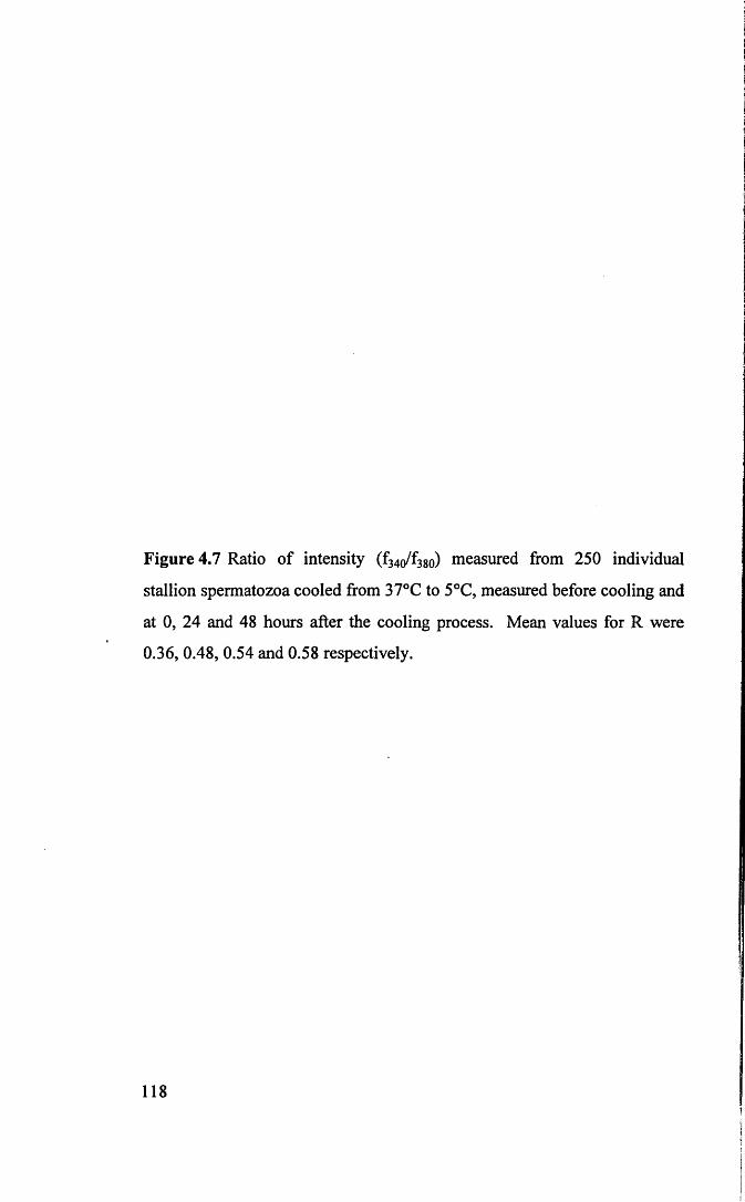
Figure 4.7 Ratio of intensity (f3 4o/f3 so) measured from 250 individual
stallion spermatozoa cooled from 37°C to 5°C, measured before cooling and
at 0, 24 and 48 hours after the cooling process. Mean values for R were
0.36, 0.48, 0.54 and 0.58 respectively.
118

Num
Der
ors
perm
Moz
oa
60
R = 0.5
40 - Mean R = 0.36
20 -
Before Cooling
R = 0.540 -
Mean R = 0.48
20 -
0 hrs After Cooling
p — ■= f— i— r
R = 0.5
40 - Mean R = 0.54
20 -
24 hrs After Cooling
60
R = 0.540 -
Mean R = 0.58
20 -
48 hrs After Cooling
1.00.2 0.4 0.6 0.8 1.20.0 1.4
Ratio of intensity
4.4 Discussion
Because of the poor fertility which results from using cryopreserved semen,
equine artificial insemination most commonly utilises liquid semen chilled to 5°C.
Stallion semen is usually diluted in a dried skim milk-glucose extender prior to
cooling (Kenney et al., 1975; Douglas-Hamilton, 1984; Douglas-Hamilton et al.,
1987; Varner et al., 1989; Jasko et al., 1992a; Malmgren et al., 1994). Cooling can
then be achieved using a controlled rate freezer or simpler, commercially available,
cooling flasks (e.g. Equitainer, Hamilton Thome Research). It has been reported that
cooling at a linear cooling rate of less than 0.05°C/minute between 20°C and 5°C
maintained maximal percentage of motile spermatozoa and that stallion spermatozoa
cooled thus provided acceptable fertility rates (Douglas-Hamilton, 1984; Douglas-
Hamilton et al., 1987). Reports have indicated that careful use of controlled rate
cooling between the temperatures of 21-22°C and 5°C is necessary for successful
storage (Kayser, 1992) but less emphasis has been placed on controlled rate cooling
between 37°C and 22°C.
This study utilised the milk-based extender in most common use, Kenney’s
Medium, (Kenney et al., 1975) and two cooling protocols. The first, called Cooling
Protocol A, used bench top cooling from 37°C to 22°C, with controlled rate cooling
from 22°C to 5°C (Moran et al., 1992; Bedford et al., 1995). The second protocol,
Cooling Protocol B, used controlled rate cooling from 37°C to 5°C. It is worth noting
that Protocol A took approximately five and a half hours and Protocol B eleven hours
to reach 5°C.
Viability staining of spermatozoa after cooling and during storage indicated a
decline in the number of viable spermatozoa after cooling, which continued during
storage. The greatest decline in numbers viable was detected after cooling. No
difference was observed between Cooling Protocol A and B. The motility of the
viable spermatozoa also changed after cooling and during storage. Mean velocities
consistently declined; the greatest decline was immediately after cooling. The
numerically greatest decline was a 23% reduction in mean straight line velocity after
cooling by Protocol B. The much higher decline in VSL as compared to VAP and
119

VCL immediately after cooling indicated that the majority of sperm cells were
moving in circles. This circular motion may be a manifestation of the cooling injury
to stallion sperm during the cooling procedure, although the effect of rewarming must
also be considered. It is possible that cooling and rewarming may damage the
axonemal elements of the tail, though this has generally been considered resistant to
the effects of cooling and rewarming. Holt et al., (1988), in cryomicroscopic studies
of cold shock, found that on cooling to 16°C, 50% of ram spermatozoa exhibited a
rigid bowing of the midpiece with loss of all progressive motility below this
temperature. These workers reported that a number of spermatozoa also acquired a
localised swelling of the flagellum, in the region of the midpiece and endpiece.
Spermatozoa with swollen endpieces were motile on rewarming, but their flagellar
beat was impaired.
To summarise, a decline in sperm viability and mean velocity of the live
fraction was observed after cooling and storage at 5°C. Extending the period of
cooling at the slower rate of 0.05°C/minute to cover the entire cooling period from
37°C to 5°C (Protocol B) did not result in a marked conservation of viability or
velocity, instead, it appeared to increase the proportion of motile cells showing non
progressive motility. One interesting observation was of the maintenance of high
mean ALH after cooling by Protocol B. High amplitude lateral head movement is
associated with hyperactivated motility, a motility pattern which is regulated by
increases in intracellular calcium. It has also been reported that high ALH was
correlated with increased intracellular calcium in cryopreserved bull spermatozoa, as a
consequence of freezing; in this situation high intracellular calcium, in turn, correlated
with reduced fertility (Bailey et al., 1993). The circumstantial evidence, then,
associates high ALH with increased intracellular calcium and this is corroborated by
the experimental evidence from the fluorescence measurements discussed below.
These experiments applied the fluorescent indicator Fura-2AM to the study
equine spermatozoa for the first time, and found that the ability of the stallion
spermatozoa to regulate [Ca ]t was clearly compromised by cooling and low2_j_
temperature storage. With Cooling Protocol A, [Ca ]j had increased by 25% at the
end of cooling and by 64% after the first 24 hours of storage. Using Protocol B, the
120

internalised calcium concentrations measured immediately after cooling showed a
massive increase of 152% compared to precooled samples, with only a small increase
during subsequent storage.
Since Fura-2AM does not load into dead spermatozoa (see Chapter 3, Section
3.4.1) the increase in intracellular calcium could not be attributed to a rise in the
percentage dead, but instead to a huge increase in the [Ca ] {of the live population. In
Experiment 4.3.3 it was possible to directly observe [Ca ]j increase during the
process of cooling. While it was not possible to replicate Cooling Protocol A or B
because cooling within the cuvettes could not be controlled to a rate of 0.05°C/minute
over 11 hours, it was possible to cool at a rate of 0.3°C/minute. Figure 4.6 shows that2+[Ca ]j was maintained at a fairly steady rate until a temperature of around 15°C was
2+reached. Below this temperature the [Ca ]{increased markedly.
Freeze fracture studies have demonstrated that cooling of ram or blackbuck
spermatozoa from 30°C to 5°C (Holt and North, 1984) or those of bulls or boars from
38°C to 0°C (de Leeuw et al., 1990) resulted in morphological membrane changes
consistent with a lipid phase transition. Fluorescence polarisation studies with sperm
plasma membrane vesicles (Holt and North, 1986; Canvin and Buhr, 1989) have
demonstrated changes in viscosity of the plasma during cooling. More recent studies
using Fourier transform infrared spectroscopy to measure lipid phase transition in
shrimp, boar and human spermatozoa during cooling, provided direct evidence that
lipid phase transitions occurred during cooling (Drobnis et al., 1993). In boar sperm,
major phase shifts were seen between 23°C and 14°C, centred at approximately 18°C.
In shrimp sperm (more resistant to cold shock than boar sperm), the lipid phase
transition shifts occurred between 13°C and -2°C, centred at 6°C. These lipid phase
transition shifts were not detected in human spermatozoa, a species more resistant to2+
cold shock than either boar or shrimp. It is interesting to note that the [Ca ]j of
stallion spermatozoa increased rapidly below 15°C, perhaps indicating a lipid phase
transition around this temperature. Moran et al., (1992) demonstrated that stallion
spermatozoa were most sensitive to cold shock between 19°C and 8°C which may be
considered as further evidence that stallion spermatozoa undergo a phase transition in
this temperature range.
121

Summary of the data from the single cell studies clearly supports the findings
of the whole population studies, of an accumulation of intracellular calcium
immediately after cooling, continuing during low temperature storage. Interestingly,
however, the data from the single cell studies showed that while the mean R value
(ratio of intensity 340 nm: 380 nm) of the 250 cells sampled increased during cooling
and with subsequent storage, there were individual cells or a discrete sub-population
still capable of regulating calcium across their membranes at a level comparable to2+precooled levels, while other individual cells showed extremely high levels of [Ca ]j.
2+Proper control of [Ca ]j flow across the spermatozoa membrane is crucial to2+successful fertilisation. The ability to regulate [Ca ]j may be compromised in two
ways during cooling. The accumulation of calcium during the cooling process may be
due to the occurrence of lipid phase transitions, as discussed above, which render the2_j_
plasma membrane ‘leaky’ and unable to exclude extracellular Ca and/or cooling
induced damage to intracellular calcium regulatory mechanisms. It has been
demonstrated during investigation of other aspects of sperm function (the acrosome
reaction) that intracellular calcium concentrations in spermatozoa are maintained by a
Ca2+-ATPase (Vijayasarathy et al., 1980; Breitbart and Rubenstein, 1983; Breitbart et
al., 1984) and a Na+/Ca2+ antiporter (Babcock et al., 1979; Rufo et al., 1984) which2+extrude [Ca ] from the cells and maintain low intracellular concentrations (nM
2+concentrations compared with pM or mM extracellular concentrations). The Ca -
ATPase function is highly dependent on the physico-chemical properties of the lipid
environment surrounding the ATPase. A reduction in temperature would be likely to2+change the nature of the lipid environment and reduce the efficiency of [Ca ]
extrusion. Function of the ATPase is said to be entirely reversed by hydrolysis of
membrane phospholipids (Garrahan and Rega, 1990). The hydrolysis of
phospholipids has been implicated in cold induced membrane damage by other
workers (Robertson et al., 1990; Das et al., 1991). Together these factors would2+
result in inappropriately high [Ca ]j which would affect fertilising ability.
In conclusion, the work in the Chapter indicates that irreversible damage is2+initiated during cooling to 5°C. This damage results in inappropriately high [Ca ]j
levels, a decline in motility and a decline in viability. It also appears, from single cell
122

studies, that despite cooling and storage a discrete sub-population of cells is resistant2+to thermal damage and retains full ability to regulate[Ca ]r
123

CHAPTER 5
CHARACTERISATION OF CAPACITATION

5.1 Introduction
Sperm capacitation is relatively poorly studied in the equine species when
compared to human, bovine and certain laboratory species for which in vitro
fertilisation systems are in common use. It is said to be difficult to reliably and
repeatedly capacitate equine spermatozoa in vitro (Ellington et al., 1993b), although
successes have been reported (Blue et al., 1989; Palmer et al., 1991). The principal
problem with capacitation studies is lack of morphological evidence that the
spermatozoon is undergoing the process. To circumvent this problem studies have
evaluated chemical induction of the acrosome reaction, using the rationale that
capacitation must be complete to allow the acrosome reaction to proceed (Graham et
al., 1987; Samper et al., 1989; Zhang et al., 1991). Alternatively, fertilisation, studied
in vitro, has been used to confirm that capacitation has taken place. Neither approach
overcomes the problem of failing to monitor the process of capacitation, only the end
point. In addition, it is well recognised that only a proportion, or subpopulation, of
any studied population is undergoing capacitation at any one time even under ideal
conditions. The magnitude of this subpopulation appears to vary between species,
with 60-80% of rodent sperm cultured under capacitating conditions showing
capacitation synchronously (Yanagimachi, 1970; Katz and Yanagimachi, 1980) or as
little as 20% of human spermatozoa (Burkman, 1984). Certain in vitro laboratory
techniques have been used to try to identify capacitating subpopulations e.g.
identifying and quantifying hyperactivated motility, monitoring surface membrane
changes with CTC and using pharmacological substances to accelerate or synchronise
capacitation (Ward and Storey, 1984; Byrd and Wolf, 1986; Lee et al., 1987; Parrish
et al., 1988; Tesarik et al., 1990; Fraser and McDermott, 1992; Das Gupta et al., 1993;
Ellington et al., 1993a; Kervancioglu et al., 1994; Mattioli et al., 1996; Perez et al.,
1996). This would seem to be the best approach given the current technology.
This chapter describes a series of experiments conducted to characterise the
capacitation of stallion spermatozoa. Because the capacitation process required
prolonged incubation in vitro it was be necessary to establish a system that would
support sperm viability during incubation. In Experiment 1, a pilot study was carried
124

out to evaluate the ability of different culture media and containers to maintain
motility and viability of stallion sperm during incubation for up to 8 hours. Further
experiments were directed towards identifying capacitating subpopulations. In
Experiment 2, sperm plasma membrane changes were monitored using a combined
chlortetracycline (CTC) and Hoechst 33258 fluorescent staining protocol designed to
differentiate between uncapacitated, capacitated and acrosome reacted cells. In
Experiment 3, the movement characteristics of spermatozoa incubated under
capacitating conditions were examined in detail, with particular attention to
hyperactivated motility. A preliminary study was performed using human samples
because hyperactivated motility is well characterised in this species. A study of
motility under incubation conditions used in Experiment 2 was also conducted.
5.2 Materials and Methods
Semen collection and preparation, media preparation, staining procedures and
motility assessment are as described in Materials and Methods in Chapter 2.
5.3 Experiments
5.3.1 Experiment 1 - Selection of an incubation system
After initial preparation, one ejaculate from each of three stallions was
extended in three different media, Graham TALP, Bavister TALP and Bavister-Milk
medium, to a concentration of 50 million per ml. Five hundred microlitre of each
treatment was aliquoted into 14 ml Falcon grade and 16 mm flat bottom tissue culture
wells and incubated at 38.5°C in 5% C 02 and humidified air over a period of 8 hours.
125

Aliquots from each treatment were evaluated for gross motility, percentage motile and
percentage live using a double blind procedure and scoring system as described in
Section 2.3.1 of Chapter 2.
5.3.1.1 Results
The results of the experiment are summarised in Table 5.1. A significant
decline in percentage motile when compared to the initial assessment, was seen by the
second hour of incubation for Grahams and Bavister TALP in both tubes and wells,
but this decline was significant with Bavister-Milk medium only after 4 hours of
incubation. Gross motility declined significantly after 6 hours for all treatments.
Spermatozoa extended in Bavister-Milk medium, however, exhibited a more vigorous
motility at the start of the incubation. The decline in percentage live was significant
after 8 hours of incubation in Bavister-Milk medium as opposed to between 4 and 6
hours of incubation in the other two media. By the eighth hour of incubation, gross
motility had declined below 1 for all treatments, percentage motile ranged from 2 to
8% and percentage live from between 23 and 29%. No significant difference
(p=0.354 for gross motility; p=0.82 for percentage motile and p=0.083 for percentage
live) was detected in the measured parameters, whether incubation was carried out in
tubes or wells.
126

Tabl
e 5.1
. M
ean
± SD
of pe
rcen
t m
otile
, gr
oss
mot
ility
and
pe
rcen
t liv
e of
stalli
on
sper
mat
oza
exten
ded
unde
r di
ffere
nt i
ncub
atio
n sy
stem
s an
d
evalu
ated
at 0,
2, 4,
6 and
8
hour
s,res
pect
ivel
y.
p
J3oo
***(N
+1
(N
***(N©+1fS©
***VOvd+1o<N 6.7
± 5.2
**
* |
0.6±
0.6
***
|
***
©
+l
oo(N
***vq(N+1cqrd 0.3
±0
.3**
* |
29.0
± 16
.0 *
|
***oocd-HCN
***m©+1
©
***
vd+lt"-vq
(N
***©(N+1OO
0.4
±0.4
***
|
26.3
±6.4
***
|
***VO
CO
+1
cqoo 0.5
8 ±0
.49*
**
|23
.33
±7.1
2*
|
jq9
***
si<q(N 0.8
±0
.3
**35
.7 ±
13.7
*
12.5
±2.
7***
1.2±
0.4
40.2±
7.8
**
***
vd+lr~~vd
***
O+1
42.5
± 8.
7
10.8
±4.9
**
*
0.8±
0.3
***
40.0
± 11
**
12.2±
6.3
*
**
0.7±
0.3
***
28.5
±6.3
**
*
13.3
±9.3
**
*
0.8
±0.5
*
**
46.5
± 13
.1
4 hr
20.0
±3.2
**
*
1.0
±0.5
*
43.2
± 6.
2
25.0
±4.5
**
*
1.2±
0.3
**
Ov+1
vdm 25
.8 ±5
.9**
*1.3
±0
.3
***
48.8
± 12
.4
***
+1rq
(N 1.5
±0.4
39.5
±6.3
**
***r-;(N+1«/■>cs<N 0.9
±
0.2
**
29.8
± 7.0
**
*
33.3
±8.8
**1.6
± 0.
446
.8 ±
19.3
2 hr
36.7
+ 4.1
**
*
1.3
±0.3
45.0
± 9.
7
30.0
***
1.3±
0.3
46.2±
8.
1
43.3
±6.1
1.9
±0.2
51.0
± 21
.4
*#*«n
-H©«nm 1.3
±0
.345
.2±
± 13
.7
36.7±
8.2
**
1.33
±0.6
043
.17
± 10
.19
45.00
±
5.5
Z'O+l’Z 44
.3 ±
13.5
0 hr
+ irq
1.9
±0.4
53.8
±5.8
51.7
±5.2
1.9
±0.4
58.0
±5.6
oo 2.3
± 0.
357
.8 ±
10.3
50.0
± 3.
21.9
±0
.550
.5 ±
6.3
50.0
±3.2
1.75
±0.2
748
.33
±3.5
6
©©© 2.2
± 0.
2650
.5 ±
7.7
Para
met
er e
valu
ated
Perc
ent
mot
ileGr
oss
mot
ility
Perc
ent
live
Perc
ent
mot
ileGr
oss
mot
ility
Perc
ent
live
Perc
ent
mot
ileGr
oss
mot
ility
Perc
ent
live
Perc
ent
mot
ileGr
oss
mot
ility
Perc
ent
live
Perc
ent
mot
ileGr
oss
mot
ility
Perc
ent
live
Perc
ent
mot
ileGr
oss
mot
ility
Perc
ent
live
| In
cuba
tion
syst
em
| Tu
be
Gra
ham
| Tu
be
Bav
iste
r
| Tu
be
Bavi
ster
Milk
| W
ell G
raha
m
| W
ell B
avis
ter
| W
ell B
avist
er M
ilk
signi
fican
t di
ffere
nce;
* (p
<0.0
5),
** (p
<0.0
1) a
nd
***
(p<0
.001
) in
com
paris
on
to in
itial
(time
0)
obse
rvat
ion.

5.3.2 Experiment 2 - Chlortetracycline fluorescent patterns under
different incubation conditions
This experiment was conducted to examine the functional state of equine
spermatozoa during incubation using the dual CTC/Hoechst staining protocol
described in Section 2.9.2 of Chapter 2. One ejaculate from each of three stallions
was used in this experiment. After determination of initial concentration, the ejaculate
was centrifuged, the supernatant discarded and the pellet re-extended into six different
treatments. Treatment 1, comprised spermatozoa diluted in homologous seminal
plasma, selected to give an uncapacitated control. Treatments 2-5, consisted of
spermatozoa centrifuged to remove seminal plasma and diluted in Bavister-Milk
medium supplemented with 0, 0.01, 0.1 or 1 mg/ml pentoxifylline, selected to
promote capacitation, and treatment 6, to which 10 nm ionophore A23187 was added
for 1 minute prior to assessment. Treatment 6 was selected to promote acrosomal
loss. Five hundred microlitre of each treatment (concentration 50 million/ml) was
placed into 16 mm flat bottom tissue culture wells and incubated at 38.5°C in 5% C 0 2
in humidified air for 5 hours. Samples were taken for percentage live estimation and
dual staining after 0, 120 and 300 minutes of incubation.
5.3.2.1 Results
There was a decline in the percentage of live spermatozoa with time in all
Treatments (Table 5.2). This decline was greatest in Treatment 1, semen in seminal
plasma, by 300 minutes of incubation.
128

Table 5.2: The percentage of live cells (Mean ± S.D) for 100 spermatozoa in six
different treatments, evaluated at specific time points 0, 120 and 300 minutes of
incubation.
Treatment State 0 120 min 300 minFresh ejaculate % live 74 ± 9 53 ± 1 4 7 ± 6 * *Bavister-Milk % live 78 ± 1 0 55 ± 1 0 27 ± 8 * *BM(.01 mg/ml PF) % live 76 ± 5 47 ± 1 6 * 33 ± 2 * *B M (.l mg/ml PF) % live 83 ± 1 2 61 ± 2 * 27 ± 9 * *BM (1.0 mg/ml PF) % live 81 ± 1 4 58 ± 7 26 ± 5 * *Ionophore (10 nM) % live 79 ± 9 59 ± 9 34 ± 7 * ** - significant difference (p<0.05) to time 0; ** significant (p<0.01) to time 120 min, a significant difference (p<0.01)at 300 minutes of incubation is also seen observed between treatment 1 (seminal plasma) and other treatment.
Figure 5.1a-f are graphical representation of staining patterns evaluated after
0, 120 and 300 minutes of incubation. The majority of cells in Treatment 1 showed
the ‘uncapacitated’ (F) pattern throughout the 300 minute incubation. By 300 minutes
only ‘uncapacitated’ (F) cells were detected in the live population. Treatments 2 to 5
showed a decline in ‘uncapacitated’ (F) pattern and an increase (significant for
treatments to which pentoxifylline and ionophore were added) in ‘capacitated’ (B)
pattern with incubation. For Treatments 1-4 only low levels of ‘acrosome reacted’
(AR) were detected at any time point (less than 5%). Treatment 5, with 1 mg/ml PF,
showed the highest percentage of ‘capacitated’ (B) pattern after 300 minutes of
incubation (mean value of 70%) and the highest percentage of ‘acrosome reacted’
(AR) after 120 minutes of incubation. Acrosome loss was seen in treatment 6 at all
three time points. The highest levels were detected at 300 minutes (mean value 10%).
Figures 5.1 g-j, show examples of equine spermatozoa stained by the
combined CTC/H258 staining technique.
129

Figure 5 .1 a -f Percentage (Mean ± SEM, n=3) of ‘F’̂ , ‘B’D , and
‘AR’H pattern of stallion spermatozoa incubated in six different treatment
and evaluated at specific time points of incubation. The symbol (*) denotes
significant increase in the ‘B’ pattern in comparisons to time 0. (* at p<0.05
and ** atp<0.01).
130

Figure 5.1a - Fresh100
80
60
40
20
00 mifl 120 min 300 min
Incubation period
Figure 5.1 c Bavister-Milk (+0.01 PF)
<■> 6 0 -
cn
300 min120 min0 minIncubation period
Figure 5.1 e Bavister-Milk (+1.0 PF)
100
80
60
O)40
20
00 min 120 min 300 min
Incubation period
Figure 5.1 b - Bavister-Milk (0 PF)100 ------------------------------------------------------------------------------------
0 min 120 min 300 min
Incubation period
Figure 5.1 dBavister-Milk (+0.10 PF)
O)
0 min 120 min 300 min
Incubation period
Figure 5.1 fBavister-Milk (+A23187)
O 6 0 -
o>
0 min 120 min 300 min
Incubation period

Figure 5.1g Stallion sperm showing uncapacitated ‘F’ pattern.
Figure 5.1h Decapitated sperm stained by H258, indicating dead sperm.

Figure 5.1i Stallion sperm (enlarged) showing capacitated ‘B’ pattern. Nste
the lesser fluorescence band (arrow) in the acrosomal region.
Figure 5.1j (i) ‘B’ pattern stallion sperm showing exocytosis changes in
acrosome, and (ii) sperm showing acrosome loss.
132

5.3.3 Experiment 3 - Analysis of movement parameters under
different incubation conditions
5.3.3.a Experiment 3a - Analysis of human sperm movement in seminal
plasma, post swim-up and after incubation with pentoxifylline
One ejaculate from each of three human donors were collected as described
in Material and Methods. Initial samples were prepared by diluting 50 pi of neat
semen in homologous seminal plasma (1:1 or 1:2). Seminal plasma was isolated by
centrifugation of 400 pi of semen sample at 16,000 rpm for 10 minutes. Concurrently,
a swim-up preparation (as described in Section 2.8.2 of Chapter 2) was made using
1 ml of semen and incubated for 45 minutes. The upper interface was then removed
and divided into two aliquots. These were centrifuged (300g, 5 minutes) and
re-extended in 90 pi medium. To one aliquot was added 10 pi of stock pentoxifylline
(final concentration 1 mg/ml), and to the control sample 10 pi of medium. Motility
analysis of sperm in seminal plasma, post-swim-up and after 15 minutes in
pentoxifylline was carried out. Motility parameters (ALH, LIN, VAP, VCL and VSL)
were analysed after 15 minutes of incubation at 37°C in 5% C 02 in humidified air.
The number of spermatozoa showing hyperactivated motility was determined using
the criteria of Burkman (1991) i.e. ALH > 7.5 pm, LIN < 65% and VCL > 100 pms’1.
5.3.3.b Experiment 3b - Analysis of stallion sperm movement in seminal
plasma, post swim-up and after incubation with pentoxifylline
One ejaculate from each of three stallions was collected as described in
Chapter 2. Concentration was determined using a hemacytometer and initial samples
were extended in homologous seminal plasma to 25 million per ml (a concentration
that gives between 5-25 spermatozoa per field for motility analysis). The rest of the
sample collected were prepared by swim-up as described previously and the upper
interface was harvested and divided into two aliquots. These were centrifuged (300g,
5 minutes) and re-extended in 450 pi medium. To one aliquot was added 50 pi
133

pentoxifylline (final concentration 1 mg/ml), and to the control sample 50 jil medium.
Samples were incubated for 15 minutes at 37°C in 5% C 02 in humidified air. Motility
parameters (ALH, LIN, VAP, VCL, VSL) of sperm in semen, post swim-up and after
15 minutes incubation with pentoxifylline was carried out. The number of
spermatozoa showing hyperactivated motility was determined using the criteria used
for human sperm.
5.3.3.1 Results (Experiment 3a and 3b)
The results for Experiment 3a and 3b are summarised in Table 5.3. For
human spermatozoa a significant increase in ALH and VAP and VCL and a
significant decline in LIN was observed after the swim-up procedure compared to
those parameters in seminal plasma. ALH and VCL increased further after incubation
in 1 mg/ml pentoxifylline for 15 minutes. There was no change to VSL with
treatment. A mean of 8% of spermatozoa extended in seminal plasma showed
hyperactivated activity (range 6-11%). After swim-up, the incidence of
hyperactivation increased to 20% (range 16-22%) and after incubation in
pentoxifylline increased further to 32% (range 21-47%). For stallion spermatozoa
extended in seminal plasma 15% showed hyperactivated activity (range 13-19%).
After swim-up, the incidence of hyperactivation did not change significantly (mean
14%; range 10-18%) and incubation in medium supplemented with pentoxifylline did
not affect the incidence of hyperactivation (mean 11%; range 8-13%).
Analysis of stallion spermatozoa showed no significant change in ALH, LIN
or VSL. However, VCL and VAP increased significantly after swim-up (p<0.001).
No further change was observed after pentoxifylline treatment. There was no
difference in the incidence of hyperactivated spermatozoa between treatments, using
the criteria of Burkman (1991).
134

Table 5.3 : Motion parameters of human and stallion spermatozoa extended in
seminal plasma, post swim-up and after addition of pentoxifylline for 15 minutes
(Mean ± SD).
Seminal Plasma
n= 431 sperm tracks
Post Swim-up
n= 433 sperm tracks
1 mg/ml PF; 15 min
n= 424 sperm tracksALH 4.99 ± 2 .92“ 7.08±4.01 b 8.57±4.41 cLIN 80.54±18.10 “ 70.74±25.65 b 65.09±24.05 c*
Human VAP 76.77±22.90 “ 82.51±22.91 b 85.57±20.34 bVCL 89.35±23.65 “ 101.78±26.22 b 112.46±28.30cVSL 71.80±24.5 70.59±27.93 70.48±26.42%HA 8 20 32
Seminal Plasma
n= 426 sperm tracks
Post swim-up
n= 376 sperm tracks
1 mg/ml PF; 15 min
n= 335 sperm tracksALH 7.12+3.51 7.07±3.29 6.74±3.30LIN 58.33±20.53 56.28±20.74 57.78±21.09
Stallion VAP 79.87±31.66“ 92.60±29.69 b 94.46±28.21 bVCL 99.58±35.94 “ 109.67±35.21 b 110.31±31.78 bVSL 57.76±30.41 59.89±27.97 62.17±28.29%HA 15 14 11
abc Values with different letters between columns differ (p<0.001) and be* denote differ(p<.01)
5.3.3.C Experiment 3c - Analysis of stallion sperm movement during
incubation for up to 300 minutes
One ejaculate from each of three stallion was extended after semen preparation as
described previously in Bavister-Milk medium containing 0, 0.01, 0.1 or 1 mg/ml
pentoxifylline. Motility parameters (ALH, LIN, VAP, VCL and VSL) were analysed
after 15, 45, 90, 120, 180 and 300 minutes of incubation. Because it was noted in
Experiment 5.2.2 that stallion spermatozoa in medium supplemented with
pentoxifylline had a tendency to clump together, a subjective assessment of this
phenomenon was also performed (as described in Section 2.3.2) in addition to the
motility analysis.
135

5.3.3.2 Results (Experiment 3c)
The results of movement analysis for Experiment 3c summarised in Table
5.5, indicated that stallion spermatozoa motility was maintained for up to 300 minutes
in Bavister-Milk medium. The addition of pentoxifylline did not stimulate motility
significantly. In fact, spermatozoa were observed to undergo head to head
agglutination (“clumping”) after 90 minutes of incubation in Bavister-Milk medium
(Table 5.4), which became more obvious with time. This “clumping” was accelerated
by the addition of pentoxifylline in a dose dependent manner. The addition of
1 mg/ml of pentoxifylline to the medium made it impossible to evaluated motility on
the Hamilton Thom motility analyser because of the severe clumping seen with this
treatment. The incidence of hyperactivation using the detection criteria of Burkman
(1991) was high throughout the incubation period. The incidence was numerically
highest at 90 minutes of incubation for all treatments. The numbers of spermatozoa
showing hyperactivated motility declined after 90 minutes of incubation.
Table 5.4: Subjective assessment of occurrence of head to head agglutination
(clumping) of stallion spermatozoa incubated in Bavister-Milk medium containing 0,
0.01, 0.1 or 1 mg/ml Pentoxifylline after 15, 45, 90, 120, 180 and 300 minutes of
incubation.
PF mg/ml 15 min 45 min 90 min 120 min 180 min 300 minBM (0 ) - - + + ++ ++BM (0.01 ) + + ++ ++ ++ ++BM (0 .1 0 ) + ++ +++ ++ ++ ++BM (1 .0 0 ) +++ +++ +++ +++ ++ ++
no clumping.+ majority with two sperm heads attached to one another.++ majority between 2 to 5 sperm heads clumped together.+++ majority with more than 5 sperm heads clumped together.
136

Table
5.5
: Mot
ility
pa
ram
eter
s (M
ean
± SD
) of
stalli
on
sper
mat
ozoa
in
cuba
ted
in Ba
viste
r-Milk
me
dium
supp
lem
ente
d wi
th di
ffere
nt l
evels
of
PF
analy
sed
at sp
ecifi
c tim
epoi
nts
over
a 30
0 m
inut
e in
cuba
tion
perio
d.
i
300
min
|8.0
±4
.3
|66
.8 ±2
0.1
|12
3.8
±46.
4 |
141.4
±5
2.3
|91
.3 ±4
1.0
| I OZ
ON 7.4
± 3.6
|
66.7
±21.
0 |
114.0
±4
8.2
|13
0.5
±53.
3 |
83.7
±40.
8 |
22 |
CO
180
min
9.6
±4.5
63.1
±20.
613
5.2
±51.
910
0.0
± 59
.410
0.4
± 50
.025 o 9.6
±4
.968
.9 ±
20.9
132.0
±5
6.4
154.4
±6
5.3
101.6
±5
8.5
20 ON
120
min
9.6
±4.8
65.0
± 19
.413
6.2
±48.
815
9.7
±55.
410
2.5
±45.
230 CO 10
.3±
5.6
64.0
± 19
.913
3.4
±53.
715
8.9
±62.
210
1.2
±50.
830 CO
90 m
in10
.1 ±5
.3
Z'ZZ +
6Z9 13
7.4
±51.
716
2.0
± 57
.510
0.5
± 50
.733 NO 9.7
±
5.5
65.0
± 19
.912
8.3
± 54
.315
3.6
±65.
297
.3 ±
50.9
30 9.7
± 5.
060
.9 ±2
1.2
126.6
±
52.6
150.9
±6
1.2
87.6
± 44
.440 o o
45 m
in10
.2 ±4
.867
.1 ±
18.8
149.2
±
52.7
173.6
±
59.3
116.1
±5
2.0
36 VO 8.9
± 4.
670
.2 ±
19.6
137.7
±5
2.1
158.0
±5
7.5
101.2
±5
0.8
(N o o 9.7
± 5.
066
.0 ±
19.6
130.0
±
52.7
154.3
±6
2.3
100.0
±
50.0
26 (N
15 m
in9.8
±4
.563
.8 ±
20.9
145.0
±4
7.7
167.5
±5
1.4
105.1
±4
7.1
32 10.4
± 5.
269
.3 ±1
8.1
144.3
±
50.7
170.5
±5
8.5
118.0
±5
1.1
23 CO 10.6
± 5.
271
.9 ±1
6.6
142.8
±4
9.9
169.9
±
59.4
121.3
±5
0.4
o o (N
ALH
LIN
VA
PV
CL IS
A
% H
A VH
%
ALH
LIN
VA
PV
CL IS
AV
H%
%H
A
ALH
LIN
VA
PV
CL IS
AV
H%
VH
%
| B
Mon
ly
| B
urkm
an
crite
ria
| D
eriv
ed
crite
ria
| BM
+
PF|
(O.O
lmg/
ml)
| B
urkm
an
crite
ria
| D
eriv
ed
crite
ria
CL,+
CQ
| (O
.lOm
g/m
l)
| B
urkm
an
crite
ria
| D
eriv
ed
crite
ria
r-m

5.3.3.d Experiment 3d - Hyperactivation of stallion spermatozoa -
re-analysis of the data
The results for movement analysis and percentage hyperactivation presented
in Table 5.3 for human spermatozoa were similar to results from previous work in this
laboratory (Kay et al., 1993; Morris et al, 1996) and from other workers using similar
approaches (Burkman, 1984; Robertson et al., 1988; Burkman, 1991). The criteria
selecting hyperactivated cells, when applied to stallion spermatozoa detected high
proportions in seminal plasma, a medium widely considered to be non-capacitating,
with no apparent increase in hyperactivation during incubation under conditions
believed to support capacitation. For these reasons videotapes were re-examined
visually, utilising the playback facility on the analyser. Two hundred sperm tracks
were examined and categorised into (1) non-hyperactivated trajectory, (2) transitional
phase and (3) star spin trajectory. The procedure was first carried out on human
spermatozoa. Subsequently the process was repeated with approximately 400 stallion
sperm tracks.
5.3.3.3 Results (Experiment 3d)
The results for human spermatozoa are shown in Table 5.6. and for stallion
spermatozoa in Table 5.7.
Table 5.6: Numbers of hyperactivated cells detected in human samples (observed
versus computer sorted).
Human Spermatozoa Seminal Plasma Post Swim-up PF 1 mg/ml
Total counted 200 200 200(1). Non Hyperactivated 181 158 143(2). Transition Phase 13 21 41(3). Star-Spin Pattern 5 17 16%HA (total observed) 9 19 28%HA(computer sorted) 8 19 25Difference -1% +0% +3%
138

Table 5.7: Numbers of hyperactivated cells detected in stallion samples (observed
versus computer sorted).
Stallion Spermatozoa Seminal Plasma Post Swim-up PF 1 mg/ml
Total counted 405 347 382(1). Non Hyperactivated 295 238 265(2). Transition Phase 9 7 13(3). Star-Spin Pattern 1 3 4%HA (total observed) a 3 3 5%HA (computer sorted) b 15 14 11Difference +12% +10% +5%ab different letters between rows (observed values versus computer sorted) differ (p<0.003)
On re-analysis of videotaped human spermatozoa there was no significant difference
between numbers of hyperactivated trajectories identified visually and hyperactivated
trajectories sorted by computer according to the criteria of Burkman (1991); results are
shown in Table 5.6. In contrast, there was a significantly lower visual count of
hyperactivated trajectories in stallion semen, in comparison to counts sorted by
computer using the criteria of Burkman; results shown in Table 5.7. On re-analysis, it
was possible to identify 29 transitional phase trajectories and 8 star spin trajectories
from a total of 1,134 trajectories. Values for the ALH, LIN and VCL of these
individual cells are summarised in Table 5.8.
Table 5.8: Characteristic of sperm trajectories for stallion semen; Mean ± SD
(Range).
Stallion Spermatozoa Transitional phase (N=29) Star spin (N=8)
ALH (pm) 17.1+3.5 (12.4-25.2) 14.5 ±3.6 (12.2-20.0)LIN (%) 53.7 ±7.5 (41.9-65.0) 18.0 ±7.5 (13.1-32.7)VCL (pm/sec) 188.8 ±24.6 (156-235) 173.2 ±28.2 (141 -217)
Based on these results a new definition of hyperactivation was derived for stallion
spermatozoa (termed derived criteria) which was ALH > 12.5 pm, LIN < 65% and
139

VCL > 140 pm/sec. These values encompassed the lowest VCL and ALH and the
highest LIN of the observed hyperactivated cells. These new parameters were
programmed into the “Sort Facility” of the computer, and the track data of Experiment
3c (summarised in Table 5.7) was reanalysed using these criteria. The results are
shown in Table 5.9.
Table 5.9: A comparison of numbers of hyperactivated cells selected using the
criteria of Burkman (1991), visual observation and the newly derived criteria for
stallion spermatozoa. The percentage of hyperactivated spermatozoa (%HA) was
calculated using the criteria of Burkman (1991) or the criteria derived for stallion
spermatozoa in Experiment 3d.
Stallion Spermatozoa Seminal Plasma Post Swim-up PF 1 mg/ml
Total counted 405 347 382%HA (Burkman) a 15 14 11%HA (observed) b 3 3 5%HA (derived criteria) b 4 5 2ab different letters between rows differ (p<0.001)
The derived criteria (ALH > 12.5 pm, LIN < 65% and VCL > 140 pm/sec) were also
used to reanalyse tracks recorded for experiment 5.2.3c. The number of spermatozoa
showing hyperactivated motility declined by 50% as a result of incorporating the new
criteria. The data is shown in Table 5.5.
140

5.4 Discussion
The optimal requirements for the capacitation of stallion spermatozoa are
unknown, and developing reliable methods for capacitation has been a major obstacle
in the advancement of equine in vitro fertilisation. The most commonly used media
are modified Tyrode solutions (TALP) which have been used by many authors (Blue
et al., 1989; Samper et al., 1989; Padilla and Foote, 1991; Zhang et al., 1991;
Ellington et al., 1993a; Hochi et al., 1996). The composition of TALP used by these
authors varies and is presented in Appendix 1. Because of poor sperm viability, recent
interest has turned to co-culturing spermatozoa with uterine tube epithelial cells
(Ellington et al., 1993b), uterine tube explants (Pollard et al., 1991) or medium
conditioned with oviduct epithelial cells (Ellington et al., 1992). These methods are
reported to support viability, capacitation and the acrosome reaction. An alternative
approach has been supporting viability by mixing milk media with capacitating media,
in different proportions. A mixture of 35% skim-milk media and 65% capacitating
media preserved the motility of centrifuged stallion spermatozoa well for 72 hours,
despite cooling (Padilla and Foote, 1991; Webb and Ams, 1995).
In this study several media were examined prior to the reported experiments.
The unsuccessful results achieved using three types of TALP (Parrish et al, 1988;
Blue et al., 1989; Padilla and Foote, 1991) were not repeated in this laboratory.
Promising preliminary results were achieved using Graham TALP (Dr Graham,
Boulder Colorado; personal communication) and Bavister TALP (Zhang et al., 1991).
Promising preliminary results were also achieved by mixing Bavister TALP 1: 1 with
Kenney’s medium. These media were tested further in Experiment 1.
The data from this study suggested that the media examined supported
stallion sperm viability reasonably well, in comparison to starting values, for between
4 and 6 hours. It was noted that the time 0 values (50% motile, mean 55% live) were
lower than would be expected for domestic species such as ram and bull. Motility was
maintained less well but this must be considered together with the information from
later motility studies. Media containing milk maintained the highest percentage
motile for the longest period. From 4 hours onwards the percentage live greatly
141

exceeded the percentage motile. The observation has been made by other workers
(Robertson and Middleton, 1992) and explained as the presence of viable but immotile
spermatozoa. No difference was detected between tubes and tissue culture wells in
this study, although other workers reported improved sperm longevity (Ellington et
a l, 1993b).
Sperm functional state during incubation in Bavister-Milk medium was
assessed using a dual chlortetracycline (CTC) and Hoechst 33258 stain. The use of
CTC to assess capacitation in stallion sperm had been evaluated by a number of
authors (Varner et a l, 1987b; Ward et al., 1988). The major criticism of the technique
is that membranes are stained regardless of viability and it has long been recognised
that one change which can occur during sperm degeneration is acrosome loss (Cross et
al., 1986) which cannot be distinguished from acrosomal loss due to physiological
reasons using histological dyes (Talbot and Chacon, 1981), lectins (Talbot and
Chacon, 1980; Cross et al., 1986; Mortimer et al., 1987) or monoclonal antibodies
(Byrd and Wolf, 1986; Moore et a l, 1987). In this study, a dual staining technique
developed for use with human spermatozoa (Kay et a l, 1994), was used to evaluate
stallion sperm functional state for the first time. The important point about dual
staining is that the Hoechst 33258 dye stains dead sperm; this dye is excluded by live
sperm. Fluorescent CTC patterns are then assessed only for live spermatozoa.
Three distinct CTC patterns similar to those described in mouse (Fraser,
1987) and human (Das Gupta et al., 1993; Kay et al., 1994) were observed. Hence the
same nomenclature: ‘F’ or fresh, a uniform fluorescence over the head, characteristic
of uncapacitated, acrosome intact cells; ‘B’ or banded, a fluorescence free band in the
post acrosomal region, characteristic of capacitated, acrosome-intact cells; and ‘AR’,
with dull or no fluorescence on the head, characteristic of acrosome reacted cells, has
been used. For spermatozoa incubated in seminal plasma the majority of cells showed
the ‘F’ pattern. By 300 minutes only the ‘F’ pattern was present. This was concurrent
with a drop from 70 to 7% live. This decline in percentage live far exceeded that seen
in any culture medium but since it is well recognised that prolonged contact with
seminal plasma is detrimental to sperm survival this finding was not unexpected.
What is more difficult to explain is why there were ‘B’ and ‘AR’ patterns in seminal
plasma.
142

Incubation in Bavister-Milk medium without additives showed an increase in
‘B’ pattern with time, increasing from 10% at time 0 to 60% by 300 minutes, with a
corresponding decline in the percentage of ‘F* pattern. Negligible levels of ‘AR’
pattern were detected. This is consistent with other work indicating that spontaneous
acrosome loss occurs at a low level (5%) ( Stock and Fraser, 1989). The shift from the
uncapacitated to the capacitated state increased with the addition of pentoxifylline in a
dose dependent manner, with the highest levels of capacitated pattern seen after 300
minutes of incubation in medium supplemented with 1 mg/ml pentoxifylline. A high
proportion of acrosome loss was observed after 120 minutes with this treatment (up to
50%). The occurrence of acrosome loss had declined by 300 minutes (mean 10%).
This pattern suggests that cells die rapidly after acrosome loss. The sixth treatment in
this experiment, addition of ionophore, was a positive control, designed to induce the
acrosome reaction. Essentially this treatment was similar to treatment 2, incubation
with 0 pg/ml pentoxifylline, with the late addition of ionophore to induce acrosomal
loss. The proportion of induced acrosome reactions increased with incubation time.
While it is tempting to believe A23187 selectively induces acrosome reactions in
capacitated cells the volume of literature does not support this. It was believed at one
point that A23187 selectively induced acrosomal exocytosis in capacitated cells (Byrd
et al., 1989), however, it is now commonly believed that A23187 induces acrosomal
exocytosis regardless of the capacitation status of the cell (Shams-Borhan and
Harrison, 1981; Varner etal., 1987b; Magistrini and Palmer, 1991).
To further test the hypothesis that capacitated sub-populations were present
within the whole population, an attempt was made to identify stallion sperm
hyperactivation using the Hamilton Thom motility analyser. Because studies of
hyperactivated motility are well established in human spermatozoa (Burkman, 1984;
Robertson et al., 1988; Zhu et al., 1994), initial experiments looked at quantifying
human sperm, hyperactivation under noncapacitating and capacitating conditions. The
results were as expected (Table 5.3). There was an overall increase in ALH, VAP and
VCL, with a decline in linearity (LIN) after swim-up. The numbers of hyperactivated
spermatozoa identified, according to the criteria of Burkman (1991), increased two
fold after swim-up and increased further to four-fold that of the starting value, when
extended in medium supplemented with 1 mg/ml pentoxifylline.
143

The criteria defined by Burkman (1991) were also used to automatically
identify hyperactivated stallion spermatozoa. It was observed that stallion
spermatozoa typically exhibited a wide amplitude of lateral head displacement and
were less linear than human sperm even when extended in homologous seminal
plasma. Velocities (VAP and VCL; pms’1), increased after swim-up but ALH and
LIN, already high, remained unchanged. A relatively high number of allegedly
hyperactivated spermatozoa were identified in seminal plasma (Table 5.3). Re
analysis of the videotapes showed that the criteria used to identify human sperm
hyperactivation, ALH > 7.5 pm, LIN of < 65% and VCL of >100 pms"1, were
insufficiently discriminating for stallion spermatozoa and caused misidentification of
a large number of spermatozoa fitting the criteria though not necessarily
hyperactivated (i.e. not exhibiting a transitional phase or star spin trajectory as
described for human spermatozoa).
Using the playback facility it was noted that the number of stallion
spermatozoa showing hyperactivated motility (as determined by visual inspection)
was significantly different from the number generated using the computerised sort.
From a total of 1134 trajectories visually examined, and classified into non
hyperactivated, transition phase and star-spin pattern as described in Robertson et al.,
(1988) only 29 transitional phase and 8 star spin trajectories were identified. Based on
this data, new criteria of ALH > 12.5 pm, LIN < 65% and VCL > 140 pms’1 were
derived. The number of stallion spermatozoa showing hyperactivated motility, sorted
by computer using these new criteria, was significantly lower than for the Human
criteria and not different from the numbers determined visually. This approach to
determining the criteria for defining hyperactivation has been used before with other
species (Robertson et al., 1988; Burkman, 1991). However, because of the small
number of cells forming the stallion hyperactivation database this study must be
considered preliminary rather than definitive. A problem was encountered which
contributed to the identification of low numbers of hyperactivated cells. This is
discussed more fully below.
A further experiment was conducted to examine sperm motility in detail,
using the computerised analyser, during a 300 minute incubation in the Bavister-Milk
medium, under the same conditions as studied by the CTC/ dual staining technique.
144

Two treatments were dropped from the experiment. Because of the poor viability of
spermatozoa incubated in seminal plasma (7% live by 300 minutes) this treatment was
excluded. Ionophore treatment (A21837) was included as a positive control for
acrosomal exocytosis in the earlier experiment. Since this was not relevance to
motility this treatment was also excluded. The initial time point was 15 minutes
because of the time required to videotape 3-4 minutes of each treatment sample.
Initial analysis of hyperactivated activity using the criteria of Burkman
(1991) showed a higher incidence of hyperactivation for stallion sperm. This may be
attributed to larger ALH and VCL and less linear trajectories of initial stallion sperm
suspensions. The application of the criteria derived from stallion sperm, reduced the
occurrence of hyperactivation, as sorted by computer, by at least 50%. Incubation in
medium supplemented with pentoxifylline did not significantly increase the
occurrence of hyperactivation.
During this experiment it was observed that stallion sperm exhibited ‘head to
head’ agglutination and clumping after 120 minutes of incubation in Bavister-Milk
medium. This phenomenon was exacerbated by the addition of pentoxifylline, and
occurred in a dose-dependent manner. This phenomenon is possibly an indication of
changes to the sperm plasma membrane. Whether these changes are a component of
capacitation is not known. After 300 minutes of incubation however, the extent of
clumping declined a little, with live spermatozoa (identified by the continuous flagella
beat) still clumped together and individual unclumped sperm completely immotile.
Pentoxifylline is a phosphodiesterase inhibitor which causes an increase in
intracellular cAMP. Its actions are not fully understood but various effects on sperm
function have been demonstrated. A number of authors have shown effects of
pentoxifylline on human sperm motility. It has been shown to increase VCL and ALH
of spermatozoa in suspension (Kay et al., 1993; Lewis et al., 1993). It has been
shown to stimulate hyperactivation in fresh and cryopreserved human spermatozoa
(Kay et al., 1993; Morris et al., 1996). It has been shown to accelerate capacitation of
human spermatozoa in the presence (Kay et al., 1994) and absence of progesterone
(Das Gupta et al., 1994).
Yanagamachi, (1982) reported this phenomena of agglutination in hamster
spermatozoa after between 20 to 60 minutes of incubation in a TALP medium. He
145

reported that the number of spermatozoa in each clump varied between two and one
hundred, and that the agglutinated clumps began to disperse about two hours after
incubation. Concomitant with the dispersion was the appearance of hyperactivated
spermatozoa. This dispersion from the ‘clumped’ state to a hyperactivated state
however was not observed in the present study.
In conclusion, fluorescent membrane patterns corresponding to those seen
during capacitation of other species were detected during incubation of stallion and
increased in the presence of pentoxifylline, an agent believed to accelerate
capacitation. Pentoxifylline caused severe sperm clumping, another surface
membrane change associated with capacitation. Pentoxifylline did not stimulate
hyperactivation of stallion sperm as it did for human. It is impossible to say whether
this is a real observation or an artefact of clumping, removing these cells from motility
analysis.
146

CHAPTER 6
CALCUIM REGULATION DURING CAPACITATION

6.1 Introduction
Fluxes in intracellular calcium are a component of the regulatory
mechanisms of sperm capacitation, hyperactivation and the acrosome reaction. Other
workers have shown a requirement for micromolar levels of extracellular calcium to
support capacitation (Fraser, 1987; Adeoya-Osiguwa and Fraser, 1993; Das Gupta et
al., 1993) and millimolar levels to support the acrosome reaction (Yanagimachi and
Usui, 1974; Shams-Borhan and Harrison, 1981; Yanagimachi, 1982; Fraser, 1987;
Thomas and Meizel, 1988). Similar requirements for calcium have been
demonstrated for hyperactivated motility (Yanagimachi, 1982; White and Aitken,
1989). The presence of calcium pumps and channels have been demonstrated in the
sperm plasma membrane of many species (Blackmore et al., 1990; Guerrero and
Darszon, 1990; Lievana and Darszon, 1995) and it has been hypothesised that during2+the process of capacitation the function of Ca ATPases responsible for extruding
2+ 2+[Ca ]i is modified allowing [Ca ]4 to reach a critical threshold affecting other
components of capacitation such as hyperactivation (Adeoya-Osigawa and Fraser,
1996). Other workers have shown that a gradual increase in [Ca2+]j does occur during
prolonged incubation and have speculated that this is a component of capacitation
(Canvin and Buhr, 1989; Buhr et a l, 1989; Zhou et al., 1990; Zhao and Buhr, 1995).
Recently, the steroids progesterone and 17oc-hydroxyprogesterone have been
shown to stimulate a rapid influx of calcium in human spermatozoa (Thomas and
Meizel, 1989; Blackmore et al., 1990; Baldi et al., 1991). It has since been proposed
that progesterone present in the ovulatory milieu in the fallopian tubes after ovulation
plays a physiological role by facilitating or triggering acrosomal exocytosis when the
oocyte reaches the site of fertilisation (Blackmore and Lattanzio, 1991). This view
was supported by Roldan et al., (1994). In their studies with mouse spermatozoa,
they showed that when spermatozoa were exposed first to progesterone and then to
zona pellucida, exocytosis was enhanced over that seen when the agonists were not
presented together or in the inverse order, suggesting that the steroid exerts a priming
effect.
With this information, and in light of the importance of maintaining the
147
ability to regulate intracellular calcium, as demonstrated in Chapter 4, a series of
experiments were carried out to examine calcium regulation under capacitating
conditions. The incubation conditions used were those tested in Chapter 5 and found,
using the CTC stain, to induce capacitation. The first experiment was designed to
examine intracellular calcium concentrations during incubation for 300 minutes. The
second experiment was designed to establish whether a calcium transient could be
induced by progesterone, as demonstrated by others for human spermatozoa (Thomas
and Meizel, 1989; Blackmore et al., 1990; Baldi et al., 1991; Foresta et al., 1993).
This experiment was initially carried out with human spermatozoa. Finally,
experiments were carried out to identify progesterone receptors in both human and
stallion spermatozoa under different incubation conditions.
6.2 Materials and Methods
Semen collection, preparation of media and details of intracellular calcium
measurement are as described in Chapter 2.
6.3 Calcium Transients Experiments
6.3.1 Experiment 1 - Intracellular calcium changes during
incubation under capacitating and non-capacitating
conditions
One ejaculate from each of three stallions was extended in homologous
seminal plasma, a non-capacitating medium, and Bavister-Milk medium and Bavister-
148

Milk medium supplemented with 1 mg/ml pentoxifylline, treatments shown to support
capacitation. Two 4 ml aliquots from each treatment were incubated in capped Falcon
tubes in 5% C 02 in air at 38.5°C. After 0 and 300 minutes of incubation subsamples
were removed, loaded with Fura-2AM as described previously, and extended in a
cuvette at a final concentration of 50 million/ml. Intracellular calcium measurements
were recorded for each treatment after 0 and 300 minutes of incubation.
6.3.1.1 Results
There was no significant increase in intracellular calcium for spermatozoa
incubated in homologous seminal plasma after 300 minutes of incubation. There was
a significant increase in intracellular calcium after 300 minutes of incubation for
stallion spermatozoa incubated in Bavister-Milk medium containing 0 or 1 mg/ml
pentoxifylline. Mean intracellular calcium was numerically higher when the medium
was supplemented with pentoxifylline. However, this difference was not statistically
significant. Results are summarised in Table 6.1.
Table 6.1: Intracellular calcium concentration (Mean ± SD; n = 3 ejaculates) of
stallion spermatozoa measured after 0 and 300 minutes of incubation.
Time 0 minute 300 minutesMediumSeminal Plasma 87 ± 16 nM a 92 ± 5 nM “Bavister-Milk 50 ±21 nM a 189±60nM **Bavister-Milk (+PF) 82 ±31 nM ° 202 ± 86 nMab Values with different letters within column differ (P<0.001) and * values differ (P 0 .001) from start (time 0)
149

6.3.2 Experiment 2 - The induction of calcium transients in human
spermatozoa using progesterone
The response of human sperm to progesterone is well documented
(Blackmore et al., 1990; Baldi et al., 1991; Foresta et al., 1993; Krauscz et al., 1995).
Therefore preliminary experiments using human spermatozoa were carried out to
validate the technique. Three ejaculates from each of two individuals were prepared
by swim-up as described previously. At the end of the swim-up migration, and
without further incubation, spermatozoa were loaded with Fura-2AM as previously
described, and re-extended in Medicult medium in a cuvette for fluorescence
measurements. Fluorescence intensities were recorded for approximately 40 seconds
to establish a baseline reading, then progesterone at a final concentration of 1 pg/ml or
DMSO, the solvent for progesterone, at a final concentration of 0.1% was added and
measurement of fluorescence intensities continued for up to a further 120 seconds.
6.3.2.1 Results
There was a significant increase in intracellular calcium in response to the
addition of 1 pg/ml progesterone. The mean value for the initial basal concentration
was 49 nM. This increased to peak at 120 nM within 40 seconds of progesterone
addition. This value was transient and a new basal concentration was established at
71 nM. No such response was observed on the addition of 0.1% DMSO. Mean
values are summarised in Table 6.2. Responses are shown graphically in Figure 6.1a
and b.
150

Table 6.2: Intracellular calcium measured prior to addition of progesterone, for 0-60
seconds after addition of 1 jig/ml progesterone or 0.1% DMSO and new plateau level
for human spermatozoa (Mean ± SD; n=6 ejaculates).
Hum an Sperm atozoa Initialconcentration
0-60 sec after addition
Plateauconcentration
Progesterone (1 ng/ml) DMSO 0.1%
49 ±16 nM50 ± 12 nM
93 ± 46 nM * 49 ±11 nM
itie
71 ±26nM 49 ±11 nM
values differ * (P<0.001 and ** (P<0.01)
6.3.3 Experiment 3 - The induction of calcium transients in stallion
spermatozoa using progesterone
Experiment 6.3.2 was repeated using stallion spermatozoa. One ejaculate
from each of three stallions was used in this experiment. Because of the problems
encountered previously when using swim-up migration for separating stallion
spermatozoa from seminal plasma, samples were prepared both by swim-up migration
and centrifugation. Fura-2AM loading and intracellular calcium measurement was
carried out as described previously.
Fluorescence intensities were recorded for approximately 40 seconds, to give
a baseline reading, then progesterone, at a final concentration of 1 pg/ml, was added
and the measurement of fluorescence intensities continued for up to a further 120
seconds.
6.3.3.1 Results
The results of the experiment are as summarised in Table 6.3. The
fluorescence intensities of loaded sample prepared by swim-up were as low as
background intensity, hence deduction of background intensity and measurement of
intracellular calcium were not possible. In contrast, after centrifugation, the
fluorescence intensities at both wavelengths were several-fold higher than
151

background. When progesterone (1 pg/ml) was added to these samples, no transient
rise in intracellular calcium was observed. No response was also observed on the
addition of DMSO (Figure 6.2a-b).
Table 6.3: Intracellular calcium measured prior to addition of progesterone, for 0-60
seconds after addition of 1 pg / ml progesterone or 0.1% DMSO and new plateau level
for stallion spermatozoa (Mean ± SD; n=3 ejaculates).
Stallion Spermatozoa Initialconcentration
0-60 sec after addition
Plateauconcentration
Progesterone (1 pg/ml) DMSO 0.1%
87 ±21 nM 87 ± 20 nM
88 ± 21 nM 88 ±21 nM
89 ± 22 nM 89 ± 22 nM
152

Figure 6.1a Graph showing intracellular calcium changes (nM;
Mean ± SEM, n = 6) in human sperm suspension in response to DMSO.
DMSO was added at time 0.
Figure 6.1b Graph depicting typical changes in intracellular calcium
(nM; Mean ± SEM, n = 6) seen in human sperm suspension in response to
progesterone. Progesterone (1 pg/ml final concentration) was added at
time 0.
153

Intr
acel
ular
calc
ium
-
(nM
) In
trac
ellu
lar
cacli
um
- (n
M)
6.1a
DMSO added
Time - seconds
6.1b200
180-
P4 added160- Peak -120nM (32nM)
140-
1 2 0 -
New plateau
71 nM (11nM)1 0 0 -
80-
basal calcium
49nM (7nM)
60-
4 0 -
2 0 -
100 12020 60 80-20 0 40-40
Time - seconds

Figure 6.2a Graph showing intracellular calcium changes (nM;
Mean ± SEM, n = 3) in stallion sperm suspension in response to DMSO.
DMSO was added at time 0.
Figure 6.2b Graph depicting typical changes in intracellular calcium
(nM; Mean ± SEM, n = 3) seen in stallion sperm suspension in response to
progesterone. Progesterone (1 pg/ml final concentration) was added at
time 0.
154

Intra
cellu
larc
alci
um
-(nM
) In
trace
llula
rcal
cium
-(
nM)
200
180
160
140
120
100
80
60
40
20
0-40 -20 0 20 40 60 80 100 120
Time - seconds
200
180
160
140
120
100
80
60
40
20
0
6 .2 b
P4 added
0 20 40 60
Time - seconds120
6.2a
DMSO added
i 1-----1----->-----1-----1-----1-----1-----1-----1-----1-----1-----1----- 1-----■-----r

6.3.4 Experiment 4 - The induction of calcium transients in
stallion spermatozoa using progesterone, after incubation
One ejaculate from each of three stallions was centrifuged to remove seminal
plasma and extended in Bavister-Milk medium containing 0 or 1 mg/ml pentoxifylline
and incubated for 300 minutes. Subsamples were removed after 0 and 300 minutes of
incubation and loaded with Fura-2AM. After removal of extracellular dye,
spermatozoa were suspended in a cuvette at a final concentration of 50 million/ml.
Fluorescence intensities were recorded for 40 seconds to establish baseline
measurements. Progesterone at a final concentration 1 pg/ml or DMSO at a final
concentration of 0.1% was added and recording of the fluorescence intensities was
continued for a further 80 seconds.
6.3.4.1 Results
A significant increase in basal intracellular calcium was seen after
300 minutes of incubation for each treatment (p<0.001) prior to the addition of
progesterone. No response was induced by the addition of progesterone or DMSO to
samples after 0 incubation. A calcium transient was observed in response to the
addition of progesterone after 300 minutes of incubation in Bavister-Milk medium
supplemented with pentoxifylline. No response was detected after the addition of
progesterone to samples incubated in the absence of pentoxyifylline and on the
addition of DMSO. The mean values are summarised in Table 6.4 and the response to
the addition of substances are presented in Figure 6.3 a-f.
155

Table 6.4: Intracellular calcium concentration (nM) of stallion spermatozoa
incubated in Bavister-Milk medium with the addition of 0 or 1 mg/ml pentoxifylline
(PF) and challenged by the addition of 1 pg/ml progesterone (P4) or 0.1% DMSO
after 0 or 300 minutes of incubation (Mean ± SEM, n = 3 ejaculates).
Incubation in Bavister-Milk
0 min 300 min
Initial basal 34 ± 7 61 ± 11 “*0 mg/ml PF 0-60 secs after
addition33 ± 7 63 ± 1 0 “
P4 added Peak values no response no responsePlateau 32 ± 7 63 ± 1 3Initial basal 42 ± 4 63 ± 8 “*
0 mg/ml PF 0-60 secs after addition
41 ± 6 63 ± 9 “
DM SO added Peak values no response no responsePlateau 40 ± 6 64 ± 9Initial basal 51 ± 1 2 82 ± 15°*
1 mg/ml PF 0-60 sec after addition
49 ± 1 1 1 1 1 ± 1 8 c
P4 added Peak values - 137 ± 9 .6Plateau 47 ± 2 0 97 ± .7 b
abc Values with different letters within column differ (p<0.01) and * values differ (p<0.0001) from start (time 0)
156

Figure 6.3a - b Spermatozoa incubated in Bavister-Milk. Intracellular
calcium (nM; Mean ± SEM) in response to progesterone, assessed after
0 and 300 minutes of incubation.
Figure 6.3c - d Spermatozoa incubated in Bavister-Milk. Intracellular
calcium(nM; Mean ± SEM) in response to DMSO, assessed after 0 and
300 minutes of incubation.
Figure 6.3e - f Spermatozoa incubated in Bavister-Milk supplemented
with 1 mg/ml pentoxifylline. Intracellular calcium (nM; Mean ± SEM)in
response to progesterone, assessed after 0 and 300 minutes of incubation.
157

Intra
cellu
lar
calci
um
- (nM
) In
trace
llula
r ca
lcium
- (
nM)
Intra
cellu
lar
calci
um
- (nM
)
No incubation 300 mins incubation6.3 a 6.3 b
200
150-
1 0 0 -
i * • i - i i i i i i i H i 1 i i ~*~H50-
•20 0 20 40 60 80Time - seconds
200
150-P4
100 -
B-H+H-H-H— f H50-
•20 0 20 40 60 80
Time - seconds
6.3 c 6.3 d200
150-
100 -
50
DMSO
-40 -20 5 ! 20 40. 60 8o"Time - seconds
200
150-DMSO
100 -
5 0 -
-20 0 20 40 60 80
Time - seconds
6.3 e 6.3 f200
150-
P4
1 0 0 -
fH ff lti- tf -H tf+ H -l-H H Ht i m
60 80•20 0 20 40
200
P4transient and peak lavels
150-
new caldum levelbasal calcium level
m m
100 -
5 0 -
20 80•20 0 40 60
Time - seconds Time - seconds

6.3.5 Experiment 5 - The induction of calcium transients in stallion
spermatozoa using progesterone after incubation under
capacitating and non-capacitating conditions
Experiment 6.3.4 was repeated; for this experiment spermatozoa were
suspended in homologous seminal plasma, a non-capacitating treatment, Bavister
TALP medium and Bavister-Milk medium supplemented with 1 mg/ml
pentoxifylline.
6.3.5.1 Results
A significant increase in basal intracellular calcium was observed after 300
minutes of incubation in Bavister TALP and Bavister-Milk supplemented with
pentoxifylline (p<0.001). This was not observed for spermatozoa incubated in
homologous seminal plasma. No response was induced by the addition of
progesterone to samples after 0 minutes of incubation. A calcium transient was
induced in response to the addition of progesterone after 300 minutes of incubation in
Bavister-Milk medium supplemented with pentoxifylline. No response was detected
on the addition of progesterone to samples suspended seminal plasma. A small but
non-significant response was detected on addition of progesterone to samples
suspended in Bavister TALP after 300 minutes. Mean values are summarised in
Table 6.4. Response to the addition of progesterone is presented in Figure 6.4 a-f.
158

Table 6.5: Intracellular calcium concentration (nM) of stallion spermatozoa incubated
in three different medium and evaluated after 0 or 300 minutes of incubation after the
addition of 1 pg/ml progesterone (Mean ± SEM, n = 3 ejaculates).
Medium 0 min 300 minInitial basal 48 ± 9 48 ± 6
Seminal plasma 0-60 secs after addition
50 ±10 53 ±10
P4 added Peak values no response no responsePlateau 51 ±10 53 ± 8Initial basal 39 ±18 85 ± 4 0 *
Bavister TALP 0-60 secs after addition
39 ±19 97 ±48
P4 added Peak values no response 106 ±31Plateau 39 ± 18 93 ±43Initial basal 74 ±30 143 ± 43 fl*
Bavister Milk + PF 0-60 secs after adding P4
75 ±29 209 ± 73 °
P4 added Peak values no response 264± 122Plateau 75 ±29 176 ± 62 4
ab Values with different letters within column differ (P<.030) and * values differ (P<.0001) from start (time 0)
159

Figure 6.4a - b Spermatozoa incubated in Bavister-Milk. Intracellular
calcium (nM; Mean ± SEM; n = 3) in response to progesterone, assessed
after 0 and 300 minutes of incubation.
Figure 6.4c - d Spermatozoa incubated in homologous seminal plasma.
Intracellular calcium (nM; Mean ± SEM; n = 3) in response to progesterone,
assessed after 0 and 300 minutes of incubation.
Figure 6.4e-f Spermatozoa incubated in Bavister-Milk supplemented
with 1 mg/ml pentoxifylline. Intracellular calcium (nM; Mean ± SEM;
n = 3) in response to progesterone, assessed after 0 and 300 minutes of
incubation.
160

Intra
cellu
lar
calci
um
- nM
Intra
cellu
lar
calci
um
- nM
Intra
cellu
lar
calci
um
- nM
No incubation6.4 a
400
350-
300-
250- P4200 -
150-
100 -
50- HfHttHtillilltiHlHHIHHI-20 0 20 40 60 80
Time - seconds
6.4 c400
350-
300-
250 P4200
150
100 -
50-
-20 200 40 60
Time - seconds
6.4 e400
350
300
250
200
150
100
50
0
p
' ti 111 n T TI
4
I i n i i i i i i n i n i l i n T111r i l 111J l i i i l n m m m t i i i i
-20 0 20 40 60 80
Time - seconds
300 min incubation6.4 b
400
350-
300-P4
250-
200 -
150-
100 -
50-
-20 0 20 40 60 80
Time - seconds
6.4 d400
350-
300-
250- P4
200 -
150-
100 -
-20 200 40 60
Time - seconds
6.4 f400
350-
300-
250
200 -
150-
100 -
50-
-20 200 40 60 80
Time - seconds

6.4 Progesterone Binding Site Experiments
The transient rise in intracellular calcium shown by human spermatozoa in
response to exposure to progesterone is well documented (Thomas and Meizel, 1989;
Blackmore et al., 1990; Baldi et al., 1991; Foresta et al., 1993; Krausz et al.,1995)
and has been reproduced in Experiment 1 of this chapter. Other studies have shown
that this response is mediated by non-genomic progesterone receptors (Blackmore and
Lattanzio, 1991; Revelli et al., 1994; Baldi et al., 1995; Alexander et al., 1996).
Experiments were carried out in this series to identify binding sites and characterise
receptor site numbers on the membranes of human and stallion spermatozoa, under
different incubation conditions. The principle underlying radioligand binding studies
is that a radioligand (in this case tritiated progesterone) will bind to receptors in a
saturable manner, whereas attachment to non-receptor components is less saturable.
Therefore unlabelled progesterone will selectively inhibit radioligand binding to the
receptors, but not influence binding to other membrane constituents. The difference
between the amount of radioligand bound in the absence and presence of unlabelled
progesterone is taken as a measure of the amount of isotope, attaching to the receptor.
Two preliminary experiments were carried out to establish firstly, that
specific binding sites existed and, secondly, that binding to these sites was saturable.
This information was used to optimise assay conditions and further experiments were
conducted to examine numbers of specific binding sites under capacitating and non
capacitating conditions.
6.4.1 Experiment 1 - Identification of specific binding sites on
human spermatozoa
Preliminary work was carried out using one ejaculate from three human
donors. Spermatozoa were separated from seminal plasma by swim-up migration and
the concentration was adjusted to 50 million/ml. To a 100 pi aliquot of sperm
suspension was added 20 pi of tritium labelled progesterone and 10 pi of 0, 10, 100
161
and 1000 pg/ml unlabelled progesterone. The tubes were mixed well and incubated at
37°C for 45 minutes. At the end of the incubation period, the suspension was
centrifuged, the supernatant discarded to remove excess isotope and the pellet re-
extended in 50 pi medium which was topped up to 4 ml with scintillation medium
(Ecoscint A, National Diagnostics, Atlanta, Georgia, 30336). The amount of isotope
adhering to the spermatozoa was determined using a Liquid Scintillation Analyser
(Model 1600TR, Packard Instrument Company, Meriden).
6.4.1.1 Results
A logarithmic plot representing the percentage displacement of total binding
plotted against unlabelled [progesterone] is presented graphically in Figure 6.5a.
Numbers of specific binding sites on human spermatozoa declined as the
concentration of unlabelled progesterone added to the suspension was increased,
suggesting the presence of specific receptor sites for progesterone.
6.4.2 Experiment 2 - Identification of specific binding sites on
stallion spermatozoa
The radioligand binding assay was repeated for stallion spermatozoa using
one ejaculate from each of three stallions. Seminal plasma was removed by
centrifugation and the pellet formed after centrifugation re-extended in Bavister TALP
medium to a spermatozoa concentration of 50 million/ml. To a 100 pi of sperm
suspension was added 20 pi of tritium labelled progesterone and 10 pi of 0, 10, 100
and 1000 pg/ml unlabelled progesterone as before. After removing excess isotope,
bound isotope was counted as before.
162

6.4.2.1 Results
The results showed that numbers of specific binding sites on stallion
spermatozoa declined as the concentration of unlabelled progesterone was increased,
giving evidence of the presence of specific progesterone receptors. The percentage
displacement of total binding plotted using logarithmic scale against unlabelled
[progesterone] is presented graphically in Figure 6.5b. The magnitude of the decline
in specific binding was less than that seen with human spermatozoa.
6.4.3 Experiment 3 - Effect of varying the concentration of tritiated
progesterone
A further preliminary experiment was carried out to monitor the effect of
increasing the concentration of radioligand. Derived from the first two experiments
the concentration of unlabelled progesterone was fixed at 1000 pg/ml. The
concentration of tritium-labelled progesterone, added in counts per minute, was
increased from 0.33 pmoles up to 2.05 pmoles in steps of 5 pi volume. The
radioligand binding assay was then repeated as before for stallion spermatozoa from
one ejaculate.
6.4.3.1 Results
The results are summarised in Table 6.6. Figure 6.6a is a graphical
presentation of total binding, non specific binding (blank) and specific binding
(displaceable) over the range of concentrations of tritiated progesterone.
Figure 6.6b represents specific binding, as a percentage of total radioligand
added. The percentage displaced or specific binding was saturated by the addition of
0.73 pmoles tritiate progesterone. Specific binding was relatively stable over the
range of 0.73 - 2.06 pmoles.
163

Based on these preliminary experiments, subsequent assays for radioligand
binding site studies were carried out using 20 pi tritiated progesterone (1.46 pmoles)
and 10 pi (1000 pg/ml) unlabelled progesterone in 100 pi of sperm suspension at a
concentration of 50 million/ml.
Table 6.6: Radioligand binding results of one ejaculate from one stallion using fixed
unlabelled progesterone (1 mg/ml) and increasing radioligand concentrations.
5 pi 10 pi 15 pi 20 pi 25 pi 30 piTotal added (cpm)
61756 135849 204279 272710 328057 383400
Total added(picomoles)
0.33 0.73 1.09 1.46 1.76 2.06
Totalbound(cpm)
14627 55928 96073 128762 147908 158989
Blank(cpm)
10661 14178 22280 30440 37653 30627
Displaced 3965 41750 73793 98332 110255 128362Displaced (% of total)
6 31 36 36 40 33
6.'4.4 Experiment 4 - Identification of specific progesterone binding
sites on stallion spermatozoa after 0 or 300 minutes of
incubation
To test the hypothesis that specific progesterone receptor sites were induced
during capacitation stallion spermatozoa from one ejaculate from three stallions were
extended in homologous seminal plasma, Bavister-Milk medium or Bavister-Milk
medium supplemented with 1 mg/ml pentoxifylline to a final concentration of 50
million/ml and incubated at 38.5°C in 5% C 02 for up to 300 minutes. After 0 and 300
minutes of incubation, duplicate 100 ml aliquots of each treatment were removed for
progesterone receptor radioassay as described in the previous experiments.
164

6.4.4.1 Results
The results are summarised in Table 6.7. Specific binding was detected for
all treatments, confirming the presence of receptor sites. The specific binding,
expressed as a percentage of total isotope added, declined significantly (p<0.05) for
all treatments evaluated, from an initial range of 12-15% to between 4-7% after
incubation for 300 minutes.
Table 6.7: Radioligand binding assay results for stallion spermatozoa (n=3 ejaculates)
incubated in three different media for 0 and 300 minutes.
Medium Incubationtime
Totaladded(cpm)
Totalbinding(cpm)
Nonspecificbinding(cpm)
Specificbinding(cpm)
as a % of total cpm added
seminalplasma
0 min Horse 1 272710 77693 77995 * *Horse 2 272710 66363 33766 32579 12Horse 3 272710 83176 34957 48219 18Mean (SEM) 15 (3.0)"
300 min Horse 1 272710 24546 14336 10210 4Horse 2 272710 21252 14212 7040 3Horse 3 272710 49308 33904 15404 6Mean (SEM) 4.3 (0.8)ft
Bavistermilk
0 min Horse 1 272710 72628 37768 34860 13Horse 2 272710 66901 38872 27219 10Horse 3 272710 71529 31474 40055 15Mean (SEM) 12.7 (1.5)"
300 min Horse 1 272710 38850 26673 12717 5Horse 2 272710 35357 28263 7094 3Horse 3 272710 46357 29691 16666 6Mean (SEM) 4.7 (0.9)6
Bavister Milk + PF
0 min Horse 1 272710 72530 39828 34860 12Horse 2 272710 72932 37408 27219 13Horse 3 272710 53869 20236 40055 12Mean (SEM) 12 (0.3)“
300 min Horse 1 272710 38850 14659 13451 5Horse 2 272710 35357 14212 21792 8Horse 3 272710 46357 33904 21107 8Mean (SEM) 7.0 (1.0)*
* - non specific binding value higher than total binding, hence no specific binding, while
different letters ab within column shows values differ (p<0.05).
165

Figure 6.5a A logarithmic plot. % of total binding against increasing
concentration of labelled progesterone; human spermatozoa.
Figure 6.5b A logarithmic plot. % of total binding against increasing
concentration of labelled progesterone; stallion spermatozoa.
166

Spec
ific
bindin
g site
Sp
ecifi
c bin
ding
site
Pe
rcen
tage
of
tota
l bin
ding
Perc
enta
ge
of to
tal
bind
ing
100
9 0 -Human 1
Human 2
6 0 -
Human 35 0 -
4 0 -
3 0 -
10-
log10 [progesterone]
6.5b100
Horse 1
torsi8 0 -Horse 3
4 0 -
20-
log ̂ [progesterone]

Figure 6.6a Total binding, non-specific binding and specific binding
(counts per minute; cpm) against increasing concentration of labelled
progesterone.
Figure 6.6b Specific binding as calculated percentage of total labelled
progesterone added, against increasing concentration of labelled
progesterone.
167

I
6.6a160000 H
■ Total • Blank A Displaceable140000 -
0) 120000-
£ 100000 - d)Q .£ 80000-
60000 -
40000 -
20000 -
1.50.5 1.0 2.0
[radioligand] - pmoles
6.6b100
90-
-O 80- <D "D"8(0■*—>oM—oo'*
60-
50-
40-d>O™ 30-Q lco
Q 2 0 -
10-
2.01.51.00.5
[radioligand] - pmoles

6.4.5 Experiment 5 - Relationship between specific progesterone
binding sites on human and stallion spermatozoa and
numbers of live spermatozoa
Results of Experiment 4 indicated the presence of progesterone receptors but
did not support the hypothesis that specific progesterone receptor sites were induced
during capacitation. It was hypothesised that a higher binding activity would be seen
as more receptor sites becomes available after incubation in a medium capable of
supporting capacitation. It was reasoned that this may not have been evident because
of the decline in percentage of live sperm after 300 minutes of incubation.
Hence, Experiment 3 was repeated using both human and stallion
spermatozoa as before and the percentage of live spermatozoa was determined before
and after treatment.
The procedure for human spermatozoa, one ejaculate from three donors, was
as described in Experiment 2 in Section 6.3.2; in this experiment a calcium transient
was consistently induced by the addition of progesterone to sperm suspensions after
swim-up migration and Fura-2AM loading. The procedure for stallion sperm was as
described in Experiment 3 in 6.3.3, for one ejaculate from each of three stallions.
Percentage live spermatozoa was estimated using eosin-nigrosin staining as described
previously.
6.4.5.1 Results
The percentage of live spermatozoa did not differ significantly before and
after swim-up in human samples with a mean value of 81% and 85%, respectively
(p=0.2). Specific binding, indicating the presence of progesterone receptors, was
identified. However, no significant increase in percentage of specific binding was
seen in post swim-up samples, with a mean percentage binding of 10% before and
11% after swim-up.
The percentage of stallion spermatozoa declined during incubation for all
treatments (p<0.001). The mean percentage specific binding for samples incubated in
168

seminal plasma declined during incubation; this decline was not significant (p=0.7).
There was a higher mean percentage specific binding for stallion sperm extended in
Bavister Milk with or without pentoxifylline after 300 minutes of incubation than a t )
minutes. However, differences between time or treatments were not statistically
significant. The results for human and stallion spermatozoa are summarised in Table
6.8 and 6.9 respectively.
Table 6.8: Radioligand binding (cpm) and percentage live [Mean % (SD)]for human
spermatozoa (n=3 ejaculates) before and after swim-up migration.
Totaladded(cpm)
Totalbound(cpm)
Nonspecificbinding(cpm)
Specificbinding(cpm)
Specific binding as a % of total added
Mean % live (SD)
Before Human 1 272710 48915 21278 27637 10 81 (3.4)Swim-up Human 2 272710 45659 20267 25392 9
Human 3 272710 50872 22254 28618 10Mean (SEM) 9.7 (0.3) 81 (1.9)
After Human 1 272710 39601 12264 27337 10 85 (3.0)Swim-up Human 2 272710 41425 12464 28961 11
Human 3 272710 44931 10562 34346 13Mean (SEM) 11.3 (0.8) 85 (1.7)
169

Table 6.9: Radioligand binding assay results for stallion spermatozoa (n=3 ejaculates)
incubated in three different media for 0 and 300 minutes.
Medium Incubatio n time
Totaladded(cpm)
Totalbinding(cpm)
Nonspecificbinding(cpm)
Specificbinding(cpm)
as a % of total cpm added
% live(mean)
Seminalplasma
0 min Horse 1 272710 81867 72410 9457 3 57Horse 2 272710 83362 64653 18709 8 63Horse 3 272710 76744 63836 12908 5 70Mean(SEM) 5.3(1.5)“ 63.3(3.8)“
300 min Horse 1 272710 85348 83102 2264 1 17Horse 2 272710 109990 84530 25460 9 13Horse 3 272710 81867 72409 9458 3 21Mean(SEM) 4.3(2.4)“ 17(2.3)c
Bavistermilk
0 min Horse 1 272710 72490 67740 4750 2 53Horse 2 272710 66792 64673 2119 1 66Horse 3 272710 88030 73686 14345 5 73Mean (SEM) 2.7(1.2)“ 64(5.9)“
300 min Horse 1 272710 104042 86156 17886 7 34Horse 2 272710 115919 88853 27066 10 43Horse 3 272710 63284 45874 17410 6 48Mean(SEM) 7.3(1.6)“ 41.7(4.1)"
Bavister milk + PF
0 min Horse 1 272710 81159 71346 9813 4 53Horse 2 272710 74366 64364 10002 4 65Horse 3 272710 63322 56055 7267 3 70Mean(SEM) 3.4(0.3)“ 62.7(5.0)“
300 min Horse 1 272710 103470 83620 19850 7 44Horse 2 272710 119052 89539 29459 11 45Horse 3 272710 77033 72742 4291 2 57Mean(SEM) 6.7(2.6)“ 47.0(2.5)*
abc - Different letters within column differ, ab (p<0.05) and ac (p<0.001)
170

6.5 Discussion
Experiments in this Chapter were designed to examine two facets of2+capacitation-related [Ca ]j regulation, not previously characterised for equine sperm;
a gradual increase in intracellular calcium during incubation and a rapid calcium
transient induced by exposure to progesterone. In these experiments it was shown that
intracellular calcium increased during incubation in Bavister-Milk medium with or
without pentoxifylline. The same phenomenon was observed during incubation in
Bavister TALP. In contrast, the intracellular calcium concentration of stallion
spermatozoa extended in homologous seminal plasma remained unchanged after 300
minutes of incubation. This intracellular calcium rise observed in sperm populations
incubated in medium devoid of seminal plasma is consistent with findings reported
for other species (Buhr et al., 1989) and may reflect gradual changes in the efficacy of
calcium extrusion mechanisms (Fraser et al., 1995).
A progesterone-induced calcium transit was readily produced in human
spermatozoa. This reproduced the work of others (Blackmore et al., 1990; Baldi et
al., 1991; Forresta et al., 1993; Krausz et al., 1995) and indicated that the
measurement system within this laboratory was sufficiently sensitive to detect this
transient peak of calcium. It should be noted that the human sperm preparation
method, a swim-up of 45 minutes in Medicult, is not considered a sufficient period of
incubation to capacitate a large population of cells, although hyperactivated cells and
acrosomal exocytosis have been detected immediately post swim-up (Robertson et al
1988).
This experiment could not be reproduced exactly for equine spermatozoa as
the swim-up preparation yielded such a small population of spermatozoa that no
reliable base-line signal for Fura-2 was established and no response to the addition of
progesterone addition was detected. Hence, this approach was abandoned in favour of
preparation by centrifugation and incubation for up to 300 minutes, using media tested
in Chapter 5 and shown to induce capacitation using the CTC/ dual staining method.
No progesterone-related response was stimulated after 0 minutes incubation in any
medium. Nor was a response induced after 300 minutes in seminal plasma, a
171

treatment deliberately selected as a non-capacitating treatment. A progesterone-
induced calcium transient was stimulated after 300 minutes of incubation in Bavister-
Milk medium in the presence of 1 mg/ml pentoxifylline. But the same response could
not be elicited after incubation in Bavister-Milk without pentoxifylline, a treatment
for which CTC staining indicated capacitation. Nor could it be induced after
incubation in Bavister TALP, a treatment proven to support capacitation in other
species and which it was thought might accelerate capacitation of stallion
spermatozoa over that of Bavister-Milk medium.
A radioassay was set up to try to detect specific progesterone binding which
would indicate the presence of progesterone receptors. The results of the progesterone
receptor binding studies suggested that progesterone binding site were present on both
human and stallion spermatozoa. For human spermatozoa these receptors were
detected on both non-capacitated, prior to swim-up and still in the presence of seminal
plasma, and after swim-up where a proportion, albeit small, are likely to be capacitated.
The specific binding as a percentage of total binding was very similar before and after
swim-up, indicating similar numbers of binding sites.
Stallion spermatozoa were examined under the same incubation conditions
used in previous experiments, of 300 minutes incubation in seminal plasma, Bavister-
milk and Bavister-milk with pentoxifylline; the latter treatment representing the
conditions under which a response to progesterone was elicited. Binding sites were
detected for all treatments after 0 and 300 minutes of incubation. Percentage binding
was stable or increased after 300 minutes of incubation despite a fall in percentage
live spermatozoa. However, there was no significant difference between treatment,
and no difference between the presence or absence of pentoxifylline. It was
concluded from these experiments that progesterone receptors were present on the
membrane of capacitated and uncapacitated human and equine spermatozoa.
Despite the presence of receptors stallion spermatozoa only responded to
progesterone after 300 minutes of incubation in Bavister-Milk medium supplemented
with pentoxifylline. Other workers have suggested that the majority of spermatozoa
are inherently incapable of expressing the receptor or that the receptor function is
under the control of some yet unknown factor (s) switching on the receptor only when
the cells are ready to fulfil a role in fertilisation. Mendoza and Tesarik (1993) suggest
172
that the expression of the active receptor on the sperm cell surface membrane is a2+
dynamic process controlled by the basal level of intracellular free [Ca ]. This
suggestion was supported by other work (Shimuzu et al., 1993).
As discussed previously, pentoxifylline is a phosphodiesterase inhibitor
which will increase intracellular cAMP levels. Several reports indicate that an
increase in cAMP levels is a stage in the capacitation process (Stein and Fraser, 1984;
Fraser and Monks, 1990; De Jonge et a l 1991). It can be speculated that the presence
of pentoxifylline during incubation has accelerated some cAMP mediated component
of capacitation, rendering the stallion spermatozoa responsive to progesterone.
While the mechanisms of progesterone mediated stimulation is still unclear,
the action of progesterone on the human sperm is known to operate at least three
distinct events: opening of a progesterone-activated channel on the plasma membrane
permeable to both [Ca2+] and Na+ hence also promoting plasma membrane
depolarisation (Foresta et al., 1993), opening of a plasma membrane Cl-channel
(Turner et al., 1994), and activation of a protein tyrosine kinase (Mendoza et a l,
1995). Phosphorylation is a widespread mechanisms for ion channel modulation
(Levitan, 1994; Chen and Huang, 1992). An abolition of [Ca2+]j influx by PKC
inhibitors suggests that the phosphorylation state of the progesterone receptor/channel2+modulates [Ca ]j influx, showing high permeability in the phosphorylated state and
low permeability in the dephosphorylated state (Foresta et al., 1995). Since the
phosphorylation state of receptors is cAMP mediated it can be speculated that the
effect of pentoxifylline during incubation is upon the phosphorylation state of the
progesterone receptor.
In conclusion, progesterone receptors were detected on equine spermatozoa
under conditions for which it was not possible to induce a progesterone-mediated
calcium flux. It was only possible to induce a progesterone-mediated calcium
response after incubation for 300 minutes under conditions which supported
capacitation and resulted in an increase in basal intracellular calcium. Moreover, the
presence of the phosphodiesterase inhibitor pentoxifylline was required. This
evidence suggests that raised basal intracellular calcium and raised cAMP levels are
components of a system regulating the capacitation of equine spermatozoa.
173
CHAPTER 7
GENERAL DISCUSSIONS

7.1 General Discussions
As discussed in the Objectives of the Study (1.9) there has been relatively
little basic research carried out with stallion spermatozoa in comparison to other
species. This is due to a combination of factors, with one factor being lack of impetus
and funding from a large artificial insemination and cryopreservation industry such as
there is for the bull. Another factor hindering basic research is that the horse is an
expensive and impractical experimental model. Nor has an easily repeatable in vitro
fertilisation system been established. For these reasons this study was undertaken to
assess basic aspects of sperm function after cooling and storage at 5°C and under
incubation conditions designed to support capacitation. The functional parameters
examined in detail were motility, using a computerised motility analyser to undertake2+detailed analysis of movement patterns, and the ability to regulate [Ca ]j, a reflection
of plasma membrane integrity. Further, studies were designed to test the hypothesis
that the ejaculate is composed of a heterogeneous cell population, within which there
are discrete subpopulations of different physiological importance.
During the study several different techniques were used to assess
independent aspects of sperm function. Computerised movement analysis was used
because this technology can analyse large numbers of motile cells objectively and
give data on specific characteristics of the motility pattern, such as progressive
velocity (VSL) and amplitude of lateral head deviation from the mean path (ALH),
which are thought to be of physiological importance. The reliability of the data
depends on the settings of the equipment so a series of experiments were undertaken
to validate this machine for the equine species. A great deal of the work utilised the
intracellular fluorescent calcium probe Fura-2AM. Again a series of experiments
were run to test the effect of sperm concentration, extending medium and the effect of
the presence of dead cells upon the ability of the fluorescent marker to enter the
intracellular compartments of the living sperm cells in suspension. The marker Fura-
2AM was also used to examine calcium concentrations within individual cells; again
preliminary experiments were required to establish the optimal protocol for
immobilising spermatozoa during ratiometric readings. Changes to the sperm plasma
174

membrane during incubation under capacitating conditions were examined using a
surface membrane fluorescent marker, chlortetracycline. Since it was critical that
only data from living cells was considered and it is known that degenerating cells
show a variety of surface membrane patterns, this fluorescent marker was used in
conjunction with a nuclear stain H33258, which identified dead cells. Further
assessment of surface membrane properties was carried out using a radioligand
binding assay, targeted, in these experiments, at putative progesterone receptors.
Several novel observations were made during the study and these are discussed below.
A comparison of sperm function was made after cooling to 5°C using two
different cooling protocols. Protocol A involved more rapid cooling (0.5°C/min) from
37°C to 22°C, then cooling at 0.05°C/min from 22°C to 5°C. Protocol B maintained
the lower cooling rate from 37°C to 5°C. It might have been expected that prolonged
slow cooling would be beneficial in terms of preserving sperm function. However, all
of the markers used to assess sperm function suggested that the slower rate of cooling
had a deleterious effect. In 2 out of 5 experiments using Protocol B, all sperm were
immotile by 72 hours. It was also observed that velocity parameters declined to a
greater degree after cooling by Protocol B. Two other effects were noted; (1) a
decline in VSL, because sperm assumed non-progressive motility and were circling
and (2) that ALH was high after cooling; high ALH has been related to high
intracellular calcium in other studies (Bailey et al., 1994). Measurement of
intracellular calcium, indeed, showed that the prolonged slow cooling of Protocol B2+resulted in a highly significant increase in intracellular Ca after cooling and a greater
increase than that seen for Protocol A.
The question arises of whether the high intracellular calcium is simply a
marker of loss of regulatory ability or whether it is causal of declining sperm function.
In this study high intracellular calcium can be clearly linked to a decline in motility.
High ALH and low VSL occurred concurrently with very high intracellular calcium.
A marked decline in ALH and maintained low VSL occurred thereafter.
Subsequently, 100% cell death was the most marked feature of Protocol B. This
would tend to suggest that high intracellular calcium caused the decline in ALH, if not
also the decline in velocity parameters. Comparison of cooling protocols A and B
suggests that the greater the increase in intracellular calcium, the greater the decline in
175

sperm velocities and reduction in sperm survival. One importance of this observation
is that efforts to improve cooling methods would be best directed towards controlling2+Ca influx during the cooling process.
There are very few studies of calcium regulation by individual sperm cells
and they have focused on either examining cell response to progesterone (Plant et al.,2+1995) or changes in intracellular Ca during the development of hyperactivation
2+(Suarez et al., 1993; Suarez and Dai, 1995 ). Single cell Ca studies have not been
carried out before with stallion spermatozoa nor with any spermatozoa exposed to
cooling and low temperature storage. The single cell studies in this work supplied
two pieces of information. Firstly, the mean data for all the individual cells confirmed2+the evidence of the population studies, that intracellular Ca increased during cooling
and storage. Secondly, and more importantly, the single cell studies indicated that
despite cooling and storage there still existed a discrete sub-population with plasma
membranes resistant to cooling and still able, even after 48 hours of storage at 5°C, to2+
regulate [Ca ]j at precooled levels. It can be speculated that it is this sub-population
which retains the ability to fertilise despite cooling and storage and it would be
interesting to determine exactly what characteristics render this small sub-population
resistant to an environment which has so severely damaged the majority of the
population.
Subsequent experimental work was directed towards examining calcium-
related event during capacitation. Increases in intracellular calcium are a component
of the regulatory mechanisms of the pre-fertilisation events of capacitation,
hyperactivation and the acrosome reaction. It has been suggested that modification of
the function of Ca2+ATPases responsible for extruding intracellular calcium, to allow
[Ca2+]j to reach threshold levels required to trigger other events, is a component of
capacitation (Adeoya-Osigawa and Fraser, 1996). Work in other species has indicated
that progesterone triggers a rapid influx of calcium which, in turn, regulates acrosomal
exocytosis (Thomas and Meizel, 1989; Blackmore et al., 1990; Baldi et al., 1991;
Blackmore and Lattanzio, 1991; Roldan et al., 1994). This experimental work
examined calcium regulation in equine spermatozoa incubated under conditions
capable of supporting capacitation. However, before this work could be carried out
conditions which supported capacitation had to be defined.
176

Published reports have indicated that it is difficult to capacitate stallion
spermatozoa (Ellington et al., 1993a). It is not clear how long capacitation takes in
vivo takes and studies of in vitro capacitation have acknowledged the difficulty of
maintaining stallion spermatozoa in long term cultures (Ellington et al., 1993b). A
number of different media formulations were tested in preliminary experiments (see
Appendix 1) before three media were tested in Chapter 5. The system which best
maintained motility during incubation in these studies was a 1:1 mixture of TALP and
Kenney’s medium. The inclusion of a preservative (milk), which is by definition non
capacitating, opened to question the effectiveness of the mixture for supporting
capacitation. Evidence from CTC staining, however, indicated that about 46% of the
population showed the capacitated pattern by 300 minutes of incubation. Addition of
pentoxifylline to the medium hastened the appearance of the surface membrane
capacitation-related changes detected by CTC staining.
Attempts to corroborate the CTC evidence of successful capacitation by
measuring the capacitation-related motility pattern of hyperactivation were not very
successful for equine sperm. In Chapters 5 and 6 certain techniques were used for
human sperm prior to use with equine sperm because the successful use of these
techniques had been reported for the former. Two such techniques were the detection
o f hyperactivated human sperm using an algorithm incorporated in the Sort Function
o f the computerised motility analyser, and the stimulation of hyperactivation by the
use of the phosphodiesterase inhibitor pentoxifylline. Attempts to apply the first
technique to equine sperm were confounded by the finding that parameters used to
measure hyperactivated motility in human spermatozoa were not sufficiently
discriminating for stallion spermatozoa. Based on visual observation of
hyperactivated stallion spermatozoa alternative, more discriminating, parameters were
formulated. However, the new algorithm could not be tested extensively because
pentoxifylline caused rapid and severe agglutination of equine sperm, unlike human
spermatozoa in which pentoxifylline stimulated hyperactivation. No correlations
could be made between CTC and hyperactivation because agglutinated spermatozoa
could not be measured. Whether all or only some component parts of capacitation
were actually achieved is not clear. Definitive proof would require successful
fertilisation and even within an in vitro fertilisation system the nature of the individual
177

fertilising spermatozoan is unknown. However, the CTC staining response was
consistent with capacitation in other species (Fraser, 1987; Das Gupta et al., 1993;
Kay et al., 1994).
As a further step towards elucidating regulatory mechanisms, experiments
were carried out to examine calcium dynamics under these incubation conditions. It2+was found that mean [Ca ]j increased during a 300 minute incubation in capacitating
medium; this increase was statistically significant in the presence of pentoxifylline.
No increase was seen during incubation in seminal plasma. This is consistent with the
hypothesis that Ca2+ATPases become less efficient at extruding Ca2+ during• • 2+capacitation resulting in a gradual increase in intracellular Ca (Fraser et al., 1995).
2+ • • •A second Ca mechanism was examined; the activation of non-genomic progesterone• 2+ • 2+receptors resulting in an intracellular Ca influx. A progesterone-induced Ca influx
2+was readily induced in washed human spermatozoa. The same Ca influx could only
be induced in stallion spermatozoa after a 300 minute incubation in the presence of
pentoxifylline.
Radioligand binding studies using tritiated progesterone were carried out to
identify progesterone receptors. Progesterone binding studies suggested that
progesterone receptors were present on both human and stallion spermatozoa. For
both human and equine spermatozoa progesterone binding sites were identified under
non-capacitating and capacitating conditions. With equine spermatozoa, numbers of
binding sites were maintained or increased during 300 minutes of incubation despite
falling numbers of live spermatozoa. Thus there were more receptors present on
fewer live spermatozoa after incubation. It would be necessary to show definitively
that binding to dead spermatozoa was not greater than binding to live but incubation
in seminal plasma, which resulted in greater numbers of dead spermatozoa than
incubation in media, did not result greater numbers binding sites. This observation
may be evidence that further binding sites are unmasked during incubation or
capacitation. For many years workers have believed that removal of ‘decapacitation
factors’ and unmasking of binding sites were a critical component of capacitation
(Austin, 1951; Chang, 1951; Bedford, 1983).
As stated above, after incubation in capacitating medium and in the presence
of pentoxifylline, the cell population showed a progesterone-induced calcium
178

transient. Other workers (Baldi et al., 1991; Mendoza and Tesarik, 1993) have shown2+that progesterone-induced [Ca ]i transients were more pronounced in human cell
2+suspensions with higher basal [Ca ]j levels. This current data suggests that the non-
genomic progesterone receptor, at least of equine spermatozoa, is only activated when2+1) intracellular Ca reaches a certain threshold and 2) and/or some other mechanism,
linked to cAMP and the phosphodiesterase inhibiting action of pentoxifylline, is
triggered. As was discussed in Chapter 6 this may be cAMP-mediated
phosphorylation of the progesterone receptor. A recent review (Meizel, 1997)
suggests that progesterone acts on a sperm gamma-aminobutyric acid (GABA)
receptor-like chloride channel, causing membrane depolarisation and consequent• • • 2+ •activation of voltage-sensitive Ca channels and that tyrosine phosphorylation of the
sperm GABA receptor-like chloride channel is an important component of the
progesterone-initiated acrosome reaction.
It was beyond the limits of the current study to determine whether
progesterone responsiveness was a feature of the whole cell population or whether a
functionally distinct subpopulation existed. Mendozoa and Tesarik (1993) showed
that the increased responsiveness to progesterone of human spermatozoa was
explained by an increase in the size of the subpopulation capable of responding, rather
than an increase in the responsiveness of the whole population. They suggested that
many spermatozoa possess the receptor in a functionally inactive form and that
receptor function is switched on asynchronously during capacitation to supply a
steady stream of spermatozoa primed for acrosomal exocytosis and fertilisation. This
contrasted with the evidence of Plant et al. (1995) who determined that the majority2+
(93%) of human spermatozoa showed a increased [Ca ]i in response to progesterone.
Obviously further work is required to clarify this issue.
It has been suggested by others that cryopreserved or cooled spermatozoa
undergo changes resembling capacitation (Watson, 1995; Fuller and Whittingham,
1997). The work carried out for this study has certainly helped to draw parallels2+between the two situations. This work showed that increased [Ca ]j is common to
both cooled and capacitated cells. It is possible to speculate that the increase in2+intracellular Ca which occurs during cooling may trigger components of the
capacitation sequence. Certainly there is evidence from other species that
179

cryopreserved spermatozoa can penetrate eggs without further incubation (Watson,
1995). It has also been suggested that a premature increase in intracellular calcium is
deleterious to sperm function (Robertson and Watson, 1986; Bailey et al., 1994)
which is consistent with the findings of this study, where a marked decline in motility2+and viability followed high intracellular Ca .
2 jThe objectives of the study were fulfilled in that uncontrolled [Ca ]j levels
were related to a decline in sperm function during cooling and storage. It was shown2+that very specific mechanisms exist to regulate [Ca ]j during capacitation. Several
pieces of evidence supported the hypothesis of the existence of functionally distinct
subpopulations of cells. Analysis of individual cooled cells revealed the existence of2+a subpopulation still capable of [Ca ]{ regulation after cooling and storage.
Chlortetracycline staining showed the presence of a capacitated subpopulation. A
hyperactivated subpopulation was observed but could not be characterised objectively.
Further work is required to clarify whether all or a subpopulation of equine
spermatozoa are able to respond to progesterone. In conclusion, it is clear from this2+
study that the ability to control [Ca ]j is absolutely critical to survival of cooling and
low temperature storage and critical, again, for capacitation and, ultimately,
fertilisation.
180

APPENDIX

APPENDIX 1
Different composition of TALP medium used by different authors in stallion sperm function studies.
-----^^M ediumC om position"^^^^^
1 2 3 4 5
g/100 ml g/100 ml g/100 ml g/100 ml g/100 mlPVA 0.100 - -
Penicillin (sodium salt)
0.003
Penicillin G 10,000IU -
Phenol red 0.001HEPES 0.384* 0.240 a 0.240 0.238NaCl 0.666 0.216“ 0.637 0.420 0.655KC1 0.024 0.075“ 0.024 0.187 0.030k h 2p o 4 - 0.016“ - - -
CaCl2.2H20 0.029 0.025" 0.029 0.029 0.033MgCl2. 6H20 0.010 - 0.010 0008 0.011MgS04.7H20 0.029“ - - -
Na lactate 185 pi 370 nl* 85gl 31gl -
NaH2P 04. H20 0.005 - 0.005 0.005 0.011Glucose 0.090 0.100* - - 0.250Fructose - 1.520* - -NaHC03 0.210 0.300“ 0.017 0.210Na Pyruvate 0.002* 0.002 0.011 0.011BSA 1.000 g* 0.300* 0.400g 0.600g 0.300gpH adjusted to 7.800Osmolality 305
M edium 1 - described in Bavister, (1989) and used by Zhang et al., (1991) with some modifications (*).M edium 2 - suggested by Dr J.K.Graham (personal communication) was made up by mixing Stallion Tyrodes (a) and Stallion TALP (b) at a ratio o f 1:1 (v/v). In Bedford et al., (1995) this medium was used at a ratio o f 1:1 (v/v) with milk medium [Dry skim milk, 9.46g; Glucose, 0.5g deionize water 90 ml]M edium 3 - used by Blue et al., (1989).M edium 4 - TALP, composition described in Padilla and Foote, (1991) and used at 65%:35% (TALP: Kenney’s medium) as extender for long term low temperature storage. The ability o f Medium 4 to maintain stallion sperm motility after cooling and storage at 5°C was also evaluated by Webb and Am s (1995).M edium 5 - described by Brackett and Oliphant, (1975) and used to resuspend and incubate stallion spermatozoa by Hochi et al., (1996)
181

BIBLIOGRAPHY

Aanesen, A. and Bendvold, E. (1990) Studies on requirement for trackpoints in Cellsoft automated
semen analysis. Fertility and Sterility, 54: 910-916.
Adams, D.J. and Gage, P.W. (1979) Ionic currents in response to membrane depolarization in an
A plysia neurone. Journal o f Physiology London, 289: 115-141.
Adeoya-Osiguwa, S. and Fraser, L.R. (1993) A biphasic pattern of 45Ca2+ uptake by mouse
spermatozoa in vitro correlates with changing functional potential. Journal o f Reproduction
an d Fertility, 99: 187-194.
Adeoya-Osiguwa, S. and Fraser, L.R. (1996) Evidence for Ca2+-dependent-ATPase activity,
stimulated by decapitation factor and calmodulin, in mouse sperm. M olecular Reproduction
an d Development, 44: 111-120.
Ahuja, K.K. (1985) Carbohydrate determinants involved in mammalian fertilization. American
Journal o f Anatomy, 174: 207-223.
Aitken, R.J. and Clarkson, J.S. (1988) Significance of reactive oxygen species and antioxidants in
defining the efficacy of sperm preparation techniques. Journal o f Andrology, 9: 367-376.
Aitken, R.J., Buckingham, D. and Harkiss, D. (1994) Analysis of the extent to which sperm
movement can predict the results of ionophore-enhanced functional assays of the acrosome
reaction and sperm oocyte fusion. Human Reproduction, 9(10): 1867-1874.
Alexander, N.J., Kim, H.K., Blye, R.R. and Blackmore, P.F. (1996) Steroid specificity o f the
human sperm membrane progesterone receptor. Steroids, 61(3): 116-125.
Aliev, A.I. (1981) A new method of freezing horse semen in polypropylene tubes. Anim al Breeding
Abstracts, 49: pg 805.
Alvarez, J.G . and Storey, B.T. (1984) Assessment of cell damage caused by spontaneous lipid
peroxidation in rabbit spermatozoa. Biology o f Reproduction, 30: 323-331.
Amann, R.P. (1981) A review of anatomy and physiology of the stallion. Journal o f Equine
Veterinary Science, 1: 83-105.
182

Amann, R.P. (1987) Computerised evaluation of stallion semen. Proceedings o f the American
Association o f Equine Practitioners, 33: 453-457.
Amann, R.P. and Pickett, B.W. (1987) Principles of cryopreservation and a review of
cryopreservation of stallion spermatozoa. Journal o f Equine Veterinary Science, 7: 145-173.
Amann, R.P. and Graham , J.K . (1993) Spermatozoal function In: Equine Reproduction. (Eds A. O.
McKinnon and J. L. Voss.) pp 715-745. (Lea and Febiger, London.)
Amann, R.P., Thompson J r , D.L., Squires, E.L. and Pickett, B.W. (1979) Effects o f age and
frequency of ejaculation on sperm production and extragonadal sperm reserves in stallions.
Journal o f Reproduction and Fertility, Supplement 27: 1-6.
Amann, R.P., Cristanelli, M.J. and Squires, E.L. (1987) Proteins in stallion seminal plasma.
Journal o f Reproduction and Fertility, Supplement 35: 113-120.
Aoki, F., Ishida, K., Okuno, M. and Kohmoto, K. (1994) Analysis of flagellar bending in
hyperactivated hamster and mouse spermatozoa. Journal o f Reproduction and Fertility, 101:
397-403.
Arcidiacono, A., W alt, H., Campana, A. and Balerna, M. (1983) The use of Percoll gradients for
the preparation of subpopulations of human spermatozoa. International Journal o f Andrology,
6: 433-445.
Arns, M.J., Webb, G.W., Kreider, J.L., Potter, G.D. and Evans, J.W . (1987) Use of different
nonglycoslysable sugars to maintain stallion sperm viability when frozen or stored at 37°C
and 5°C in a bovine serum albumin medium. Journal o f Reproduction an d Fertility,
Supplement 35: 135-141.
Arns, M.J., Webb, G.W., Johnson, L., M artin, J., Welsh, T. and Evans, J.W . (1991) Zona
pellucida-induced acrosome reactions in equine spermatozoa. Journal o f Reproduction an d
Fertility, Supplement 44: 738-739.
Ashizawa, K., Hashiguchi, A. and Tsuzuki, Y. (1992) Intracellular free Ca2+ concentration in fowl
spermatozoa and its relationship to motility and respiration in spermatozoa. Journal o f
Reproduction and Fertility, 96: 395-405.
183

Ashworth, P.J.C., Harrison, R.A.P., Miller, N.G.A., Plummer, J.M. and Watson, P.F. (1994)
Survival of ram spermatozoa at high dilution: protective effect of simple constituents o f
culture media as compared with seminal plasma. Reproduction Fertility an d Developm ent, 6:
173-180.
Austin, C.R. (1951) Observations on the penetration of sperm into the mammalian egg. Australian
Journal o f Scientific Research, B4: 581-596.
Babcock, D.F. and Pfeiffer, D.R. (1987) Independent elevation of cytosolic [Ca2+] and pH of
mammalian sperm by voltage dependent and pH sensitive mechanisms. Journal o f B iological
Chemistry, 262: 15041-15047.
Babcock, D.P., Singh, J.P. and Lardy, H.A. (1979) Alteration of membrane permeability to calcium
ions during maturation of bovine spermatozoa. Developm ental Biology, 69: 85-93.
Bailey, J.L and Buhr, M.M. (1993) Cryopreservation alters the Ca2+ flux of bovine spermatozoa.
Canadian Journal o f Animal Science, 74: 45-51.
Bailey, J.L ., Robertson, L. and Buhr, M.M. (1993) Relationship among in vivo fertility, computer-
analysed motility and in vitro Ca2+ flux in bovine spermatozoa. Canadian Journal o f Anim al
Science, 74: 53-58.
Baldi, E., Casano, R., Falsetti, C., Krausz, C., Maggi, M. and Forti, G. (1991) Intracellular
calcium accumulation and responsiveness to progesterone in capacitating human spermatozoa.
Journal o f Andrology, 12(5): 323-330.
Baldi,E., Krausz, C., Luconi, M., Bonaccorsi, L., Maggi, M. and Forti, G. (1995) Actions o f
progesterone on human sperm, a model of non-genomic effects of steroids. Journal o f S teroid
Biochem istry an d M olecular Biology, 53: 199-203.
Bamba, K. and C ran, D.G. (1985) Effect of rapid warming of boar semen on sperm morphology and
physiology. Journal o f Reproduction and Fertility, 75: 133-138.
Bamba, K. and C ran, D.G. (1988) Effect of rapid warming of bull and rabbit semen. Journal o f
Reproduction and Fertility, 82: 501-507.
184

Barker, C.A.V. and Gandier, J.C.C. (1957) Pregnancy rates in a mare resulting from frozen
epididymal spermatozoa. Canadian Journal o f Com parative Medicine, 21: 47-51.
Bass, J.W ., Molan, P.C. and Shannon, P. (1983) Factors in seminal plasma of bulls that affect the
viability and motility of spermatozoa. Journal o f Reproduction and Fertility, 68: 275-280.
Baumgartl, C., Bader, H., Drommer, W. and Luning, I. (1980) Ultrastructural alterations of
stallion spermatozoa due to semen conservation. Proceedings o f the Ninth International
Congress on Animal Reproduction and Artificial Insemination, M adrid, 5: 134-137.
Bavister, B.D. (1981) Substitution of synthetic polymer for protein in a mammalian gamete culture
system. Journal o f Experimental Zoology, 217: 445-451.
Bavister, B.D. (1989) A consistently successful procedure for in vitro fertilization o f golden hamster
eggs. Gamete Research, 23: 139-158.
Bedford, J.M . (1972) An electron microscope study of sperm penetration into rabbit eggs after
natural mating. American Journal o f Anatomy, 133: 213.
Bedford, S.J. (1983) Significance of the need for sperm capacitation before fertilisation in eutherian
mammals. Biology o f Reproduction, 28: 108-120.
Bedford, J.M . and Hoskins, D.D. (1990) The mammalian spermatozoon. In Marshall’s Physiology
of Reproduction. (Eds G.E.Lamming.) Volume 2, pp. 379-569. (Churchill Livingstone,
London.)
Bedford, S.J., G raham , J.K ., Amann, R.P., Squires, E.L. and Pickett, B.W. (1995) Use of two
freezing extenders to cool stallion spermatozoa to 5°C with and without seminal plasma.
Theriogenology, 43: 939-953.
Bend void, E. (1989) Swim-over: An alternative method for harvesting motile spermatozoa. Journal
o f In Vitro Fertilization an d Embryo Transfer, 6: 107-110.
Berger, T. and Parker, K. (1989) Modification of the zona-free hamster ova bioassay of boar sperm
fertility and correlation with in vivo fertility. Gamete Research, 22: 385-397.
185

Berger, T., M arrs, R.P. and Moyer, D.L. (1985) Comparisons of techniques for selection of motile
spermatozoa. Fertility and Sterility, 43(2): 268-272.
Berger, T., Turner, K.O., Meizel, S. and Hedrick, J.L. (1989) Zona pellucida-induced acrosome
reaction in boar sperm. Biology o f Reproduction, 40: 525-530.
Berliner, V.R. (1940) An improved artificial vagina for the collection of stallion and jack semen.
Journal o f American Veterinary M edical Association, 96: 667.
Berrutti, G., Franchi, E. and Camatini, M. (1986) Calcium localization in boar spermatozoa by the
pyroantimonate technique and X-ray microanalysis. Journal o f Experimental Zoology, 237:
257-262.
Bielanski, W. and Kaczmarski, F. (1979) Morphology of spermatozoa in semen from stallions o f
normal fertility. Journal o f Reproduction and Fertility, Supplement 27: 39-45.
Blach, E.L., Amann, R.P., Bowen,R.A. and Frantz, D (1989) Changes in quality of stallion
spermatozoa during cryopreservation: plasma membrane integrity and motion characteristic.
Theriogenology, 31: 283-298.
Blackmore, P.F., Beebe, S.J., Danforth, D.R. and Alexander, N. (1990) Progesterone and 17
hydroxyprogesterone. Novel stimulators of calcium influx in human sperm. Journal o f
Biological Chemistry, 265: 1376-1380.
Blackmore, P.F. and Lattanzio, F.A. ; (1991) Cell surface localization of a novel non-genomic
progesterone receptor on the head of human sperm. Biochemical an d B iophysical Research
Communication, 181: 331-336.
Blackshaw, A.W. (1953) The motility of ram and bull spermatozoa in dilute suspension. Journal o f
G eneral Physiology, 36: 449-462.
Blackshaw, A.W. (1958) The effects of incubation, temperature and cold shock on the metabolism of
ram spermatozoa. Australian Journal o f Biological Science, 11: 581-588.
Blackshaw, A.W. and Salisbury, G.W. (1957) Factors influencing metabolic activity of bull
spermatozoa. II: cold shock and its prevention. Journal o f D airy Science, 40: 1099-1106.
186

Blanc, G., Magistrini, M. and Palmer, E. (1991) Use of concanavalin A for coating the membranes
of stallion spermatozoa. Journal o f Reproduction and Fertility, Supplement 44: 191-198.
Blatter, L.A. and Wier, W.G. (1990) Intracellular diffusion, binding, and compartmentalization o f
the fluorescent calcium indicators indo-1 and fiira-2. Biophysical Journal, 58: 1491-1499.
Bleil, J.D. and W assarman, P.M. (1983) Sperm-egg interactions in the mouse: sequence of events
and the induction of acrosome reaction by a zona pellucida glycoprotein. D evelopm ental
Biology, 95: 317-324.
Blottner, S., Bertram , R. and P itra, C. (1989) Species specific motility patterns of hyperactivated
mammalian spermatozoa and quantitative analysis of the hyperactivation of bull spermatozoa.
Andrologia, 21(3): 204-213.
Blue, B.J., McKinnon, A.O., Squires, E.L., Seidel Jr., G.E. and Muscari, K.T. (1989)
Capacitation of stallion spermatozoa and fertilisation o f equine oocytes in vitro. Equine
Veterinary Journal, Supplement 8: 111-116.
Bonaccorsi, L., Luconi, M., Forti, G. and Baldi, E. (1995) Tyrosine kinase inhibition reduces the
plateau phase of the calcium increase in response to progesterone in human sperm. FEBS
Letters, 364: 83-86.
Bongso, A., Ng, S.C., Mok, H., Lim, M.N., Teo, H.L.,Wong, P.C. and Ratnam , S. (1989)
Improved sperm concentration, motility, and fertilisation rates following Ficoll treatment of
sperm in a human in vitro fertilisation program. Fertiltiy and Sterility, 51(5): 850-854.
Bower, R.E., Crabo, B.G., Pace, M.M. and Graham , E.F. (1973) Effects of dilution and glycerol
on the release of glutamic-oxaloacetic transaminase (GOT) from boar spermatozoa. Journal
o f Anim al Science, 36: 319-324.
Boyle, M.S., C ran, D.G., Allen, W.R. and Hunter, R.H.F. (1987) Distribution o f spermatozoa in
the mare’s oviduct. Journal o f Reproduction and Fertility, Supplement 35: 79-86.
Brackett, B.G. and Oliphant, G. (1975) Capacitation of rabbit spermatozoa in vitro . B iology o f
Reproduction, 12: 260- 274.
187

Brackett, B.G., Cofone, M.A., Boice, M.L. and Bousquet, D. (1982) Use of zona-free hamster ova
to assess sperm fertilising ability of bull and stallion. Gamete Research, 5: 217-227.
Bredderm an, P.J. and Foote, R.H. (1971) Factors stabilizing bull sperm cell volume and prolonging
motility at high dilution. Experimental Cellular Research: 66: 458-464.
B reitbart, H. and Rubenstein, S. (1983) Calcium transport by bull spermatozoa plasma membranes.
Biochimica e t Biophysica Acta, 732: 464-468.
B reitbart, H., Darshan, R. and Rubenstein, S. (1984) Evidence for the presence of ATP-dependent
calcium pump and ATPase activities in bull sperm head membranes. Biochemical and
Biophysical Research Communication, 122: 479-484.
Breitbart, H., Lax, J., Rotem, R. and Naor, Z. (1992) Role of protein kinase C in the acrosome
reaction of mammalian spermatozoa. Biochemical Journal, 281: 473-476.
Brinsko, S.P. and Varner, D.D. (1992) Artificial insemination and preservation o f semen.
Veterinary Clinics o f North America: Equine Practice, 8(1): 205-219.
Brown, J.E ., Cohen, L.B., De Weer, P., Pinto, L.H., Ross, W.N. and Salzberg, B.M. (1975) Rapid
changes o f intracellular free calcium concentration. Detection by metallochromic indicator
dyes in squid giant axon. Biophysic Journal, 15: 1155-1159.
Bryan, J.H.D. and Akruk, S.R. (1977) A naphthol yellow S and erythrosin B staining procedure for
use in studies of acrosome reaction of rabbit spermatozoa. Staining Technique, 52: 47-51.
Budworth, P.R., Amann, R.P. and Chapman, P.L. (1988) Relationships between computerized
measurements of motion of frozen-thawed bull spermatozoa and fertility. Journal o f
Andrology, 9(1): 41-54.
Buell, J.R . (1963) A method for freezing stallion semen and tests of fertility. Veterinary Record, 75:
900-902.
Buhr, M.M., Canvin, A.T. and Bailey, J.L. (1989) Effects of semen preservation on boar
spermatozoa plasma membrane. Gamete Research, 23: 441-449.
188

I
Burkinan, L.J. (1984) Characterization of hyperactivated motility by human spermatozoa during
capacitation: comparison of fertile and oligospermic sperm populations. Archives o f
Andrology, 13: 153-165.
Burkm an, L.J. (1991) Discrimination between nonhyperactivated and classical hyperactivated
motility patterns in human spermatozoa using computerised analysis. Fertility an d Sterility,
55: 363-371.
Buzby, J.D., Arns, M.J. and Pool, K.C. (1993) Evaluation of different sperm separation techniques
for harvesting equine spermatozoa intended for in vitro culture. Journal o f Equine Veterinary
Science, 13(9): 498-501.
Byrd, W. and Wolf, D.P. (1986) Acrosomal status in fresh and capacitated human ejaculated sperm.
Biology o f Reproduction, 34: 859-869.
Byrd, W., Tsu, J . and Wolf, D.P. (1989) Kinetics of spontaneous and induced acrosomal loss in
human sperm incubated under capacitating and uncapacitating condition. Gam ete Research,
22: 109-122.
Campbell, R.C., Dott, H.M. and Glover, T.D. (1956) Nigrosin eosin as a stain for differentiating
live and dead spermatozoa. Journal o f Agriculture Science, 48: 1-8.
Canvin, A.T. and Buhr, M.M. (1989) Effect of temperature on the fluidity o f boar plasma
membranes. Journal o f Reproduction and Fertility, 85: 533-540.
Carafoli, E. and Crompton, M. (1976) Calcium ions and mitochondria. Symposium o f Society
Experimental Biology, 30: 89-115.
Casey, P.J., Lui, I.K.M., Stewart, D.R. and Scott, M.A. (1991) The effect of equine relaxin on
sperm longevity and motility in chilled and cryopreserved sperm. Journal o f Reproduction
an d Fertility, Supplement 44: 645-646.
189

Chandler, J.A. and Battersby, S. (1976) X-ray microanalysis of zinc and calcium in ultra thin
sections of human sperm cells using the pyroantimonate technique. Journal o f Histochem istry
an d Cytochemistry, 24: 740-748.
Chang, M.C. (1951) Fertilizing capacity of spermatozoa deposited into the fallopian tubes. Nature,
168: 697-698.
Chang, M.C. (1984) The meaning of sperm capacitation. Journal o f Andrology, 5: 45-50.
Chang, M.C. and Walton, A. (1940) The effects of low temperature and acclimatization on the
respiratory activity and survival of ram spermatozoa. Proceedings o f the Royal Society Series
B, 129: 517-527.
Check, M.L. and Check, J.H. (1991) Poor hypo-osmotic swelling test result from ciyopreserved
sperm despite preservation of sperm motility. Archives o f Andrology, 26: 37-41.
Chen, L. and Huang, L.Y.M. (1992) Protein kinase C reduces Mg2+ block of NMDA-receptor
channels as a mechanism of modulation. Nature, 356: 521-523.
Cherr, G.N., Lambert, H., Meizel, S. and Katz, D.F. (1986) In vitro studies of golden hamster
sperm acrosome reaction: completion of the zona pellucida and induction by homologous
soluble zona pellucidae. Developm ental Biology, 114: 119-122.
Choong, C.H. and Wales, R.G. 1962) The effect of cold shock on spermatozoa. Australian Journal
o f Biological Sciences, 15: 543^541.
Church, K.E. and Graves, C.N. (1976) Loss of acrosin from bovine spermatozoa following cold
shock: protective effects of seminal plasma. Cryobiology, 13, 341-346.
Cochran, J.D., Amann, R.P., Squires, E.L. and Pickett, B.W. (1983) Fertility o f frozen-thawed
stallion semen extended in EDTA-lactose-egg yolk and packaged in straws. Journal o f
Anim al Science, 56: 687-693.
Cochran, J.D., Amann, R.P., Froman, D.P. and Pickett, B.W. (1984) Effect o f centrifugation,
glycerol level, cooling at 5°C, freezing rate and thawing rate on the post-thaw motility of
equine sperm. Theriogenology, 22: 25-38.
190

Coddington, C.C., Franken, D.R., Burkman, L.J., Oosthuizen, W.T., Kruger, T. and Hodgen,
G.D. (1991) Functional aspects of human sperm binding to the zona pellucida using the
hemizona assay. Journal o f Andrology, 12: 1-8.
Colenbrander, B., Fazeli, A.R., Van Buiten, A., Parlevliet, J. and Gadella, B.M. (1992a)
Assessment of cell membrane integrity in the horse. A cta Veterinaria Scandinavia,
Supplement 88: 49-58.
Colenbrander, B., Puyk, H., Zandee, A.R. and Parlevliet, J. (1992b) Evaluation of the stallion for
breeding. A cta Veterinaria Scandinavia, Supplement 88: 29-37.
Cooper, G.W., Overstreet, J.W . and Katz, D.F. (1979a) The motility of rabbit spermatozoa
recovered from the female reproductive tract. Gamete research, 2: 35-42.
Cooper, G.W., Overstreet, J.W . and Katz, D.F. (1979b) Sperm transport in the reproductive tract of
the female rabbit; motility patterns and flagellar activity of spermatozoa. Gam ete Research ,
13, 293-308.
Cooper, T.G. and Woolley, D.M. (1982) Stroboscopic illumination for the assessment o f
hyperactivated motility of mouse spermatozoa. Journal o f Experimental Zoology, 223: 291-
294.
Cornen, L.E. and Meizel, S. (1978) Stimulation of in vitro activation and the acrosome reaction of
hamster spermatozoa by catecholamines. Proceedings o f the National Academ ic Sciences o f
the United States o f America, 10: 4954-4958.
Coronel, C.E. and Lardy, H.A. (1987) Characterisation of Ca2+ uptake by guinea pig epididymal
spermatozoa. Biology o f Reproduction, 37: 1097-1107.
Corselli, J. and Talbot, P. (1986) An in-vitro technique to study penetration o f hamster oocyte
cumulus complexes by using physiological numbers o f sperm. Gamete Research , 13, 293-
308.
Corselli, J. and Talbot, P. (1987) In vitro penetration of hamster oocyte cumulus complexes using
physiological numbers of spermatozoa. Developm ental Biology, 122: 227-241.
191

Cristanelli, M.J., Squires, E.L., Amann, R.P. and Pickett, B.W. (1984) Fertility o f stallion semen
processed, frozen and thawed by a new procedure. Theriogenology, 22: 39-45.
Cross, N.L. and Meizel, S. (1989) Methods for evaluating acrosomal status of mammalian sperm.
Biology o f Reproduction, 41: 635-641.
Cross, N.L., Morales, P., Overstreet, J.W . and Hanson, F.W. (1986) Two simple methods for
detecting acrosome-reacted human spermatozoa. Gamete Research, 15: 213-226.
Cross, N.L., Morales, P., Overstreet, J.W. and Hanson, F.W. (1988) Induction of acrosome
reaction by the human zona pellucida. Biology o f Reproduction, 38: 235-244.
Cummins, J.M. (1982) Hyperactivated motility patterns of ram spermatozoa recovered from the
oviducts of mated ewes. Gamete Research, 6: 53-63.
Cummins, J.M. and Yanagimachi, R. (1982) Sperm-egg ratio and the site of the acrosome reaction
during in vivo fertilization in the hamster. Gamete Research, 5:239.
Cummins, J.M. and Yanagimachi, R. (1986) Development of ability to penetrate the cumulus
oophorus by hamster spermatozoa capacitated in vitro in relation to the timing of the
acrosome reaction Gamete Research, 15: 187-212.
Das, D.K., Iyengar, J., Jones, R.M., Lu, D. and Maity, S. (1991) Protection from a non freezing
cold injury by quinarcine, a phospholipase inhibitor. Cryobiology, 28: 177-184.
Das Gupta, S., Mills, C.L. and Fraser, L.R. (1993) Ca2+-related changes in the capacitation state o f
human spermatozoa assessed by a chlortetracycline fluorescence assay. Journal o f
Reproduction an d Fertility, 99: 135-143.
Das Gupta, S., O ’toole, C., Mills, C.L. and Fraser, L.R. (1994) Effect of pentoxifylline and
progesterone on human sperm capacitation and acrosomal exocytosis. Human Reproduction ,
9: 2103-2109.
David, R.W. and Aitken, R.J. (1989) Relationship between calcium, cyclic AMP, ATP, and
Intracellular pH and the capacity of hamster spermatozoa to express hyperactivated motility.
Gamete Research, 22: 163-177.
192

Davis, B.K. (1978) Effect of calcium on motility and fertilization by rat spermatozoa in vitro.
P roceeding o f the Society fo r Experimental Biology and Medicine, 157: 54-56.
Davis, A.P., G raham , J.K. and Foote, R.H. (1987) Homospermic versus heterospermic
insemination of zona-free hamster eggs to assess fertility of fluorochrome labeled acrosome-
reacted bull spermatozoa. Gamete Research, 17: 343-354.
Davis, R.O., Rothmann, S.A. and Overstreet, J.W. (1992) Accuracy and precision of computer-
aided system analysis in multicentre studies. Fertility an d Sterility, 57: 648-653.
Daya, S., Gwatkin, R.B.L. and Bissessar, H. (1987) Separation of motile human spermatozoa by
means of a glass bead column. Gamete Research, 17: 375-380.
De Jonge, C.D., Han, H.L., Lawrie, H., Mack, S.R. and Zaneveld, L.J.D. (1991) Modulation o f the
human sperm acrosome reaction by effectors of the adenylate cyclase/cyclic AMP second-
messenger pathway. The Journal o f Experimental Zoology, 258: 113-125.
de Leeuw, F.E., Chen, H-C., Colenbrander, B. and Verkleij, A.J. (1990) Cold induced
ultrastructural changes in bull and boar sperm plasma membrane. Cryobiology, 27: 171-183.
de Leeuw, A.M., Den Das, J.H.G. and Woelders, H. (1991) The fix vital stain method:
Simultaneous determination of viability and acrosomal status of bovine spermatozoa. Journal
o f Andrology, 12(2): 112-118.
de Vries, P.J. (1987) Evaluation of the use of fresh extended, transported stallion semen in the
Netherlands. Journal o f Reproduction and Fertility, Supplement 35: pg641.
Del Campo, M.R., Donoso, M.X., Parrish, J .J . and Ginter, O.J. (1990) In vitro fertilization o f in
v/rro-matured equine oocytes. Journal o f Equine Veterinary Science, 10: 18-22.
DeMott, R.P. and Suarez, S.S. (1992) Hyperactivated sperm progress in the mouse oviduct. Biology
o f Reproduction, 46: 779-785.
Didion, B.A., Drobinsky, J.R., Giles, J.R. and Graves, C.N. (1989) Staining procedure to detect
viability and the true acrosome reaction in spermatozoa of various species. Gamete Research,
22: 52-57.
193

Dott, H.M. (1975) Morphology of stallion spermatozoa. Journal o f Reproduction an d Fertility,
Supplement 23: 41-46.
Dott, H.M. and Foster, G.C. (1972) A technique for studying the morphology o f mammalian
spermatozoa which are eosinophilic in a differential live and dead stain. Journal o f
Reproduction and Fertility, 29: 443-446.
Douglas-Hamilton, D.H. (1984) A field study of the fertility of transported equine semen.
Theriogenology, 22: 291-304.
Douglas-Hamilton, D.H., Burns, P.J., Driscoll, D.D. and Viale, K.M. (1987) Fertility and
characteristics of slow cooled stallion semen. Journal o f Reproduction an d Fertility,
Supplement 35: 649-650.
Dowsett, K.F. and Pattie, W.A. (1987) Variation in characteristics of stallion semen caused by
breed, age, season of year and service frequency. Journal o f Reproduction an d Fertility,
Supplement 35: 645-647.
Drobnis, E.Z., Yudin, A.I., Cherr, G.N. and Katz, D.F. (1988) Hamster sperm penetration o f the
zona pellucida; kinematic analysis and mechanical implications. D evelopm ental Biology,
130: 311-323.
Drobnis, E.Z., Zhong, C.Q. and Overstreet, J.W . (1991) Separation of cryopreserved human semen
using sephadex columns, washing, or Percoll gradients. Journal o f Andrology, 12(3): 201-
208.
Drobnis, E.Z., Crowe, L.M., Berger, T., Anchordoguy, T.J., Overstreet, J.W . and Crowe, J.H .
(1993) Cold shock damage is due to lipid phase transitions in cell membranes: A
demonstration using sperm as a model. Journal o f Experimental Zoology, 265: 432-437.
Ellery, J.C., G raham , E.F. and Zemjanis, R. (1971) Artificial insemination o f pony mares with
semen frozen and stored in liquid nitrogen. American Journal o f Veterinary Research , 32:
1693-1698.
Ellington, J.E., Varner, D.D., Mathison, P., Ignotz, G.G., Padilla, A.W., Miller, P.G., Marcucio,
R.S. and Ball, B.A. (1992) Interactions of stallion spermatozoa and mare uterine tube
(oviduct) epithelial cells in a co-culture system. M olecular Andrology, 5: 20-31.
194

I
Ellington, J.E., Ball, B.A. and Yang, X. (1993a) Binding of stallion spermatozoa to the equine zona
pellucida after coculture with oviduct epithelial cells. Journal o f Reproduction an d Fertility,
98: 203-208.
Ellington, J.E ., Ball, B.A., Blue, B.J. and Wilker, C.E. (1993b) Capacitation-like membrane
changes and prolonged viability in vitro of equine spermatozoa cultured with uterine tube
epithelial cells. American Journal o f Veterinary Research, 54(9): 1505-1510.
El Wishy, A.B., AbouAhmed, M.M., Hemeida, N.A. and ElSayed, M.A.I. (1982) Sperm producing
capacity of Arab and native horses in Egypt. Journal o f Reproduction an d Fertility,
Supplement 32: 308-326.
Enna, S.J. (1985) Radioligand binding assays. In: Neurotransmitter Receptor Binding, (eds H.I.
Yamamura., S.J. Enna and M. J. Kuhar.) pp 13-33. (Raven Press, New York.)
Farrell, P.B., Foote, R.H., McArdle., Trouerntrend, V.L. and Tardif, A.L. (1996) Media and
dilution procedures tested to minimize handling effects on human, rabbit, and bull sperm for
computer-assisted sperm analysis (CASA). Journal o f Andrology, 17: 293-300.
Fleming, A.D. and Yanagimachi, R. (1981) Effects of various lipids on the acrosome reaction and
fertilizing capacity of guinea pig spermatozoa, with special reference to the possible
involvement of lysophospholipids in the acrosome reaction. Gamete Research, 4: 253-273.
Florman, H.M and First, N.L. (1988) The regulation of acrosomal exocytosis. I. Sperm capacitation
is required for the induction of acrosome reactions by bovine zona pellucida in vitro.
Developm ental Biology, 128: 453-456.
Florman, H.M., Tombes, R.M., First, N.L. and Babcock, D.F. (1989) An adhesion-associated
agonists from the zona pellucida activates G protein-promoted elevations of internal Ca2+ and
pH that mediate mammalian sperm acrosomal exocytosis. Developm ental Biology, 35: 133-
146.
Florman, H.M. and Babcock, D.F. (1991) Progress towards understanding the molecular basis of
capacitation. In: Elements of Mammalian Fertilization. (Ed P.M. Wassarman.) Volume 1, pp
105-132. (CRC Press, Boca Raton.)
195

Fomina, E. (1980) Promising prospects for the storage of stallion semen. Animal Breeding Abstracts,
48: pg 586.
Ford, W.C.L., McLaughlin, E.A., Prior, S.M., Rees, J.M., W ardle, P.G. and Hull, M.G.R. (1992)
The yield, motility and performance in the hamster egg test of human spermatozoa prepared
from cryopreserved semen by four different methods. Human Reproduction, 7(5): 654-659.
Foresta, C., Rossato, M. and Di Virgilio, F. (1993) Ion fluxes through the progesterone-activated
channel of the sperm plasma membrane. Biochemical Journal, 294: 279-283.
Foresta, C., Rossato, M. and Di Virgilio, F. (1995) Differential modulation by protein kinase C of
progesterone activated response in human sperm. Biochemical and Biophysical Research
Communication, 206(1): 408-413.
Forster, M.S., Smith, W.D., Lee, W.I., Berger, R.E., Karp, L.E. and Stenchever, M.A. (1983)
Selection of human spermatozoa aaccording to their relative motility and their interaction with
zona-free hamster eggs. Fertility and Sterility, 40: 655-660.
Frank, A.H. (1950) Artificial insemination in livestock breeding. United States D epartm ent o f
Agriculture Circular No 567. (Washington DC.)
Fraser, L.R. (1977) Motility patterns in mouse spermatozoa before and after capacitation. Journal o f
Experimental Zoology, 202: 439-444.
Fraser, L.R. (1981) Dibutyryl cyclic AMP decreases capacitation in vitro in mouse spermatozoa.
Journal o f Reproduction and Fertility, 62: 63-72.
Fraser, L.R. (1982) Ca2+ is required for mouse sperm capacitation and fertilization in vitro. Journal
o f Andrology, 3: 412-419.
Fraser, L .R (1983) Pottasium ions modulate expression of mouse sperm fertilizing ability, acrosome
reaction and hyperactivated motility in vitro. Journal o f Reproduction and Fertility, 63: 539-
553.
196

Fraser, L.R. (1986) Both taurine and albumin support mouse sperm motility and fertilizing ability in
vitro but there is no obligatory requirement for taurine. Journal o f Reproduction an d Fertility,
77: 271-280.
Fraser, L.R. (1987) Minimum and maximum extracellular Ca2+ requirements during mouse sperm
capacitation and fertilization in vitro. Journal o f Reproduction and Fertility, 81: 77-79.
Fraser, L.R. (1993) Calcium channels play a pivotal role in the sequence of ionic changes involved in
initiating mouse sperm acrosomal exocytosis. M olecular Reproduction an d Developm ent, 36:
368-376.
Fraser, L.R. (1995) Ionic control of sperm function. Reproduction, Fertility an d Developm ent, 7:
905-925.
Fraser, L.R. and Quinn, P.J. (1981) A glycolytic product is obligatory for initiation o f sperm
acrosome reaction and whiplash motility required for fertilisation in mouse. Journal o f
Reproduction an d Fertility, 61: 25-35.
Fraser, L.R. and Monks, N.J. (1990) Cyclic-Nucleotide and mammalian sperm capacitation.
Journal o f Reproduction an d Fertility, Supplement 42: 9-21.
Fraser, L.R. and McDermott, C.A. (1992) Ca2+-related changes in the mouse sperm capacitation
state: a possible role for Ca2+-ATPase. Journal o f Reproduction and Fertility, 96: 363-377.
Fraser, L.R., Abeydeera, L.R. and Niwa, K. (1995) Ca2+-regulating mechanisms that modulate bull
sperm capacitation and acrosomal exocytosis as determined by chlortetracycline analysis.
M olecular Reproduction and Development, 40: 233-241.
Fuller, S.J. and Whittingham , D.G. (1997) Capacitation-like changes occur in mouse spermatozoa
cooled to low temperatures. M olecular Reproduction an d Developm ent, 46: 318-324.
G arbers, D.L. 1989) The regulation of spermatozoa function by the egg. In: The Molecular Biology
of Fertilization, (eds H, Schatten and G, Schatten.) pp3-19. (Academic Press, New York.)
197

G arner, D.L., Pinkel, D., Johnson, L.A. and Pace, M.M. (1986) Assessment o f spermatozoal
function using dual fluorescent staining and flow cytometric analysis. Biology o f
Reproduction, 34: 127-138.
G arrahan , P.J. and Rega, A.F. (1990) Plasma membrane calcium pump. In: Intracellular Calcium
Regulation, (ed F.Bonner.) pp 271-303. (John Wiley and Sons, New York.)
Gellert-M ortimer, S.T., Clarke, G.N., Baker, H.W.G., Hyne, R.V. and Johnston, W.I.H. (1988)
Evaluation of Nycodenz and Percoll density gradients for the selection o f motile human
spermatozoa. Fertility an d Sterility, 49:335-341.
Go, K.J. and Wolf, D.P. (1985) Albumin mediated changes in the sperm sterol contents during
capacitation. Biology o f Reproduction, 32: 145-153.
Goodeaux, S.D. and Kreider, J.L. (1978) Motility and fertility of stallion spermatozoa isolated in
bovine serum albumin. Theriogenology, 10(5): 405-414.
Gould, S.F. and Bernstein, H.M. (1973) Localization of bull sperm hyaluronidase. Journal o f C ell
Biology, 59: p g ll9 a .
G raham , J.K., Foote, R.H. and Hough, S.R. (1987) Penetration of zona-free hamster eggs by
liposome-treated sperm from the bull, ram, stallion and boar. Biology o f Reproduction, 37:
181-188.
Green, D.P.L. (1978) The mechanism of the acrosome reaction. In: Development in Mammals. (Ed
M.H. Johnson.) Volume 3, pp 65-81. (Amsterdam, North-Holland.)
G runert, J.H ., De Geyter, C. and Nieschlag, E. (1990) Objective identification of hyperactivated
human spermatozoa by computerized sperm motion analysis with the Hamilton-Thom sperm
motility analyser. Human Reproduction, 5(5): 593-599.
Grynkiewicz, G., Ponie, M. and Tsein, R.Y. (1985) A new generation of Ca2+ indicators with
greatly improved fluorescence properties. Journal o f B iological Chemistry, 260: 3440-3450.
198

Guerin, J.F., Mathieu, C., Lornage, J., Pinatel, M.C. and Boulieu, D. (1989) Improvement of
survival and fertilizing capacity of human spermatozoa in IVF programme by selection on
discontinuous Percoll gradients. Human Reproduction, 4(7): 798-804.
Guerrero, A. and Darszon, A. (1990) Evidence for the activation of two different calcium channels
during the egg jelly-induced acrosome reaction of sea urchin sperm. Journal o f B iological
Chemistry, 264: 19593-19599.
Gwatkin, R.B.L. and Anderson, O.F. (1969) Capacitation of hamster spermatozoa by bovine
follicular fluid, Nature, 224: 1111-1112.
Hammerstedt, R.H. (1983) Use of sperm as a model for the study of metabolism. In: Biochemistry
of Metabolic Processes, (eds D.L.F. Lennon., F.W. Stratman and R.N. Zahlten.) pp 29-38.
(Elsevier, New York.)
Hammerstedt, R.H., Graham, J.K. and Nolan, J.P. (1990) Cryopreservation of mammalian sperm:
What we ask them to survive. Journal o f Andrology, 11: 73-78.
Hancock, J.L. (1951) A staining technique for the study of temperature shock in semen. Nature
(London), 167: 323.
Hanson, F.W. and Overstreet, J.W. (1981) The interaction of human spermatozoa with cervical
mucus in vivo. American Journal o f Obstetrics and Gynecology, 140: 173-178.
Harasymowycz, J., Ball, L. and Seidel, G.E. (1976) Evaluation of bovine spermatozoal
morphologic features after staining or fixation. American Journal o f Veterinary Research,
37: 1053-1057.
Harrison, R.A.P. (1976) A highly efficient method for washing mammalian spermatozoa. Journal o f
Reproduction and Fertility, 48: 347-353.
Harrison, R.A.P. and White, I.G. (1972) Some methods for washing spermatozoa from bull, boar
and ram: a comparison using biochemical and other criteria. Journal o f Reproduction and
Fertility, 29: 271-284.
199
Harrison, R.A.P. and Vickers, S.E. (1990) Use of fluorescent probes to assess membrane integrity
in mammalian spermatozoa. Journal o f Reproduction an d Fertility, 88: 343-352.
Harrison, R.A.P., Dott, H.M. and Foster, G.C. (1982) Bovine serum albumin, sperm motility, and
the “dilution effect”. Journal o f Experimental Zoology, 222: 81-88.
Heiskanen, M.L., Pirhonen, A., Koskinen, E. and Maenpaa, P.H. (1987) Motility and ATP content
of extended equine spermatozoa in different storage conditions. Journal o f Reproduction and
Fertility, Supplement 35: 103-107.
Heiskanen, M.L., Huhtinen, M., Pirhonen, A. and Maenpaa, P.H. (1994) Insemination results
with slow cooled stallion semen stored for 70 or 80 hours. Theriogenology, 42: 1043-1051.
Hochi, S., Korosue, K., Choi, Y.H. and Oguri, N. (1996) In vitro capacitation o f stallion
spermatozoa assessed by the lysophosphatidylcholine-induced acrosome reaction and the
penetration rate into in vitro matured, zona free mare oocytes. Journal o f Equine Veterinary
Science, 16: 244-248.
Holland, M.K. and Storey, B.T. (1981) Oxygen metabolism of mammalian spermatozoa. Generation
of hydrogen peroxide by rabbit epididymal spermatozoa. Biochemical Journal, 198: 273-
280.
Holt, W.V. and North, R.D. (1984) Partially irreversible cold-induced lipid phase transitions in
mammalian sperm plasma membrane domains: Freeze fracture study. Journal o f
Experimental Zoology, 230: 473-483.
Holt, W.V. and North, R.D. (1986) Thermotropic phase transitions in the plasma membrane of ram
sperm. Journal o f Reproduction and Fertility, 74: 445-457.
Holt, W.V., M orris, G.J., Coulson, G. and North, R.D. (1988) Direct observation o f cold-shock
effects in ram spermatozoa with the use of a programmable ciyomicroscope. Journal o f
Experimental Zoology, 246: 305-314.
Holt, W.V., Head, M.F. and North, R.D. (1992) Freeze induced membrane damage in ram
spermatozoa is manifested after thawing: observations with experimental cryomicroscopy.
Biology o f Reproduction, 46: 1086-1094.
200

Hood, R.D., Foley, C.W. and M artin, T.G. (1970) Effects of cold shock, dilution, glycerol and
dimethyl sulfoxide on cation concentration in porcine spermatozoa. Journal o f Animal
Science, 30: 91-94.
Hunter, H.R.F. (1988) Human fertilization in-vivo, with special reference to progression, storage and
release of competent spermatozoa. Human Reproduction, 2: 329-332.
Hunter, H.R.F., Barwise, L. and King, R. (1982) Sperm transport, storage and release in the sheep
oviduct in relation to the time of ovulation. British Veterinary Journal, 38: 225-232.
Hyne, R.V. and Garbers, D.L. (1981) Requirement of serum factors for capacitation and the
acrosome reaction of guinea pig spermatozoa in buffered medium below pH 7.8. Biology o f
Reproduction, 24: 257-266.
Irvine, D.S and Aitken, R.J. (1986) Measurement of intracellular calcium in human spermatozoa.
Gamete Research, 15: 57-71.
Iwanoff, E. (1930) Artificial insemination of mammals. Scientific and Zootechnical methods.
Veterinary Record, 10: 25-30.
Jamil, K., White, I.G. and Dwarte, D.M. (1982) Calcium ionophore A23187 as a probe for freeze-
fracture studies of membrane changes in the head of human spermatozoa. Archives o f
Andrology, 8: 1-9.
Jasko, D.J. (1992) Evaluation of stallion semen. Veterinary Clinics o f North America, 8: 129-148.
Jasko, D.J., Little, T.V., Smith, K., Lein, D.H. and Foote, R.H. (1988) Objective analysis o f
stallion sperm motility. Theriogenology: 30: 1159-1167.
Jasko, D.J., Hathaway, J.A., Schaltenbrand, V.L., Simper, W.D. and Squires, E.L. (1992a) Effect
o f seminal plasma and egg-yolk on motion characteristics of cooled stallion spermatozoa.
Theriogenology, 37: 1241-1252.
Jasko, D.J., M artin, J.M . and Squires, E.L. (1992b) Effect of insemination volume and
concentration of spermatozoa on embryo recovery in mares. Theriogenology, 37: 1233-
1239.
201

Jeyendran, R.S., Van der Ven, H.H., Kennedy, W.P., Heath, E., Perez-Pelacz, M., Sobrero, A.J.
and Zaneveld, L.J.D. (1985) Acrosomeless sperm: a primary cause of male infertility.
Andrologia, 17: 31-36.
Johnson, W.I. and Hunter, A.G. (1972) Seminal antigens: Their alteration in the genital tract of
female rabbits and during partial in vitro capacitation with beta-amylase and beta
glucoronidase. Biology o f Reproduction, 7: 332-340.
Johnson, L. and Neaves, W.B. (1981) Age related changes in the Leydig cell population,
seminiferous tubules, and sperm production in stallions. Biology o f Reproduction, 24: 703-
721.
Johnson, L. and Thompson, D.L.J. (1983) Age related seasonal variation in Sertoli cell population,
daily sperm production and serum concentration of follicle-stimulating hormone, leutinising
hormone and testosterone in stallions. Biology o f Reproduction, 29: 777-789.
Johnson, L.A. and Pursel, V.G. (1974) Effect of cold shock and freezing on acrosomal proteinase of
porcine spermatozoa. Cryobiology, 11: 558.
Johnson, L.A, Amann, R.P. and Pickett, B.W. (1980) Maturation of equine epididymal
spermatozoa. American Journal o f Veterinary Research, 41: 1190-1196.
Johnson, L.L., Katz, D.F. and Overstreet, J.W . (1981) The movement characteristic of rabbit
spermatozoa before and after activation. Gamete Research, 4: 275-282.
Jones, R.C. (1969) Studies of the suitability of ewe and cow milk for storing ram spermatozoa at 37,
5and-79°C. Australian Journal o f B iological Sciences, 22: 983-984.
Jones, R.C. (1975) Fertility and infertility in mammals in relation to sperm structure. In: The
Biology of the Male Gamete, (eds J.G. Duckett and P.A. Racey.) pp 343-365. (Academic
Press, London.)
Jones, R.C. and Mann, T. (1973) Lipid peroxidation in spermatozoa. Proceedings o f the Royal
Society o f London, Bulletin 184: 103-107.
202

Jones, R.C. and M artin, I.C.A. (1973) The effects of dilution, egg yolk and cooling to 5°C on the4
ultrastructure of ram spermatozoa. Journal o f Reproduction and Fertility, 35: 311-320.
Jones, R.C. and Holt, W.V. (1974) The effects of washing on the ultrastructure and cytochemistry of
ram spermatozoa. Journal o f Reproduction and Fertility, 41: 159.
Karagiannidis, A. (1976) The distribution of calcium in bovine spermatozoa and seminal plasma in
relation to cold shock. Journal o f Reproduction an d Fertility, 46: 83-90.
Katz, D.F. and Yanagimachi, R. (1980) Movement characteristic o f hamster spermatozoa within the
oviduct. Biology o f Reproduction, 22: 759-764.
Katz, D.F. and Yanagimachi, R. (1981) Movement characteristics of hamster and guinea-pig
spermatozoa upon attachment to the zona pellucida. Biology o f Reproduction, 25: 785-791.
Katz, D.F., Drobnis, E.Z. and Overstreet, J.W . (1989) Factors regulating mammalian sperm
migration through the female reproductive tract and oocyte vestsments. Gamete Research,
22: 443-469.
Kaufmann, R.F., Taylor, R.W. and Pfeiffer, D.R. (1980) Cation transport and specificity of
ionomycin. Journal o f B iological Chemistry, 255: 2735-2739.
Kay, V.J., Coutts, J.R.T. and Robertson, L. (1993) Pentoxifylline stimulates hyperactivation in
human spermatozoa. Human Reproduction, 8: 727-731.
Kay, V.J., Coutts, J.R.T. and Robertson, L. (1994) Effects of pentoxifylline and progesterone on
human sperm capacitation and acrosome reaction. Human Reproduction, 9: 2318-2323.
Kayser, J.P., Amann, R.P., Shideler, R.K., Squires, E.L., Jasko, D.J and Pickett, B.W. (1992)
Effects of linear cooling rate on motion characteristics of stallion spermatozoa.
Theriogenology, 28:773-781.
Kenney, R.M., Bergman, R.V., Cooper, W.L. and Morse, G.W. (1975) Minimal contamination
techniques for breeding mares: Technique and preliminary findings. Proceedings o f the
American Association o f Equine Practitioners, 327-335.
203

Kervancioglu, M.E., Djahanbakhch, O. and Aitken, R.J. (1994) Epithelial cell coculture and the
induction of sperm capacitation. Fertility an d Sterility, 61: 1103-1108.
Klug, E., Treu, H., Hillman, H. and Heinze, H. (1975) Centrifugation of stallion semen and its
storage in large volume straws. Journal o f Reproduction and Fertility, Supplement 23: 107-
110.
Knuth, U.A., Yeung, C.H. and Nieschlag, E. (1987) Computerized semen analysis: objective
measurement of semen characteristics is biased by subjective parameter setting. Fertility and
Sterility, 48: 118-124.
Koehler, J.K . (1981) Lectins as probes of the spermatozoan surfaces. Archives o f Andrology, 6:
197.
Kosiniak, K. (1975) Characteristics of the successive jets of ejaculated semen o f stallions. Journal o f
Reproduction and Fertility, Supplement 23: 59-61.
Kosiniak, K. and Bittmar, A. (1991) Prognosis of stallion semen freezability on the basis of
biochemical tests. Journal o f Reproduction and Fertility, Supplement 44 : 653-654.
Krausz, C., Bonaccorsi, L., Luconi, M., Fuzzi, B., Criscuoli, L., Pelligrini, S., Forti, G. Baldi, E.
(1995) Intracellular calcium increase and acrosome reaction in response to progesterone in
human spermatozoa are correlated with in vitro fertilisation. Human Reproduction, 10: 120-
124.
Kreider, J.L., Tindall, W.C. and Potter, G.D. (1985) Inclusion of bovine serum albumin in semen
extenders to enhance maintenance o f stallion sperm viability. Theriogenology, 23: 399-408.
Lam bert, H. (1981) Temperature dependence of capacitation in bat sperm monitored by zona-free
hamster ova. Gamete Research, 4: 525-533.
Langlais, J . and Roberts, K.D. (1985) A molecular membrane model of sperm capacitation and the
acrosome reaction of mammalian spermatozoa. Gamete Research, 12: 183-224.
204

Larsson, K., Einarsson, S. and Nicander, L. (1976) Influence of thawing diluents on vitality,
acrosome morphology, ultrastructure and enzyme release of deep frozen boar spermatozoa.
A cta Veter inaria Scandinavia, 17: 83-100.
Lee, M.A., Trucco, G.S., Bechtol, K.B., Wummer, N., Kopf, G.S., Blasco, L. and Storey, B.T.
(1987) Capacitation and acrosome reaction in human spermatozoa monitored by a
chlortetracycline fluorescence assay. Fertility and Sterility, 48: 649-657.
LeLannou, D., Griveau, J.F., Lepichon, J.P. and Quero, J.C. (1992) Effects of chamber depth on
the motion pattern of human spermatozoa in semen or in capacitating medium. Human
Reproduction, 7(10): 1417-1421.
Levitan, I.B. (1994) Modulation of ion channels by protein-phosphorylation and dephosphorylation.
Annual Reviews o f Physiology, 56: 193-212.
Lewis, S.E.M., Moohan, J.M . and Thompson, W. (1993) Effects of pentoxifylline on human sperm
motility in normospermic individuals using computer-assisted analysis. Fertility an d Sterility,
59: 418-423.
Lievano, A. and Darszon, A. (1995) Ion channels in sperm. C ell Physiology Source Book, 330-339.
Libersky, E.A. and Boatman, D.E. (1995) Progesterone concentration in serum, follicular fluid, and
oviductal fluid of the golden hamster during the preovulatory period. Biology o f
Reproduction, 53: 477-482.
Lindemann, C.B. and Kanous, K.S. (1989) Regulation of mammalian sperm motility. Archives o f
Andrology, 23: 1-22.
Llanos, M. and Meizel, S. (1983) Phospholipid methylation increases during capacitation o f golden
hamster sperm in vitro. Biology o f Reproduction, 28: 1043-1051.
Long, P.L., Pickett, B.W., Sawyer, H.R., Shideler, R.K., Amann, R.P. and Squires, E.L. (1993)
Relationship between morphologic and seminal characteristics of equine spermatozoa. Equine
Veterinary Science, 13(3): 143-149.
205

Loomis, P.R., Amann, R.P., Squires, E.L. and Pickett, B.W. (1983) Fertility of unfrozen and
frozen stallion spermatozoa extended in EDTA-lactose-egg yolk and packaged in straws.
Journal o f Animal Science, 56: 687-693.
Love, C.C. (1992) Semen collection techniques. Veterinary Clinics o f North America: Equine
Practice, 8(1): 111-128.
Lucena, E., Lucena, C., Gomez, M., Ortiz, J.A., Ruiz, J., Arango, A., Diaz, C. and Beuerman, C.
(1989) Recovery of motile sperm using the migration-sedimentation technique in an in-vitro
fertilization-embryo transfer programme. Human Reproduction, 4(2): 163-165.
Lui, C.W., Mrsny, R.J. and Meizel, S. (1979) Procedures for obtaining high percentages o f viable in
vitro capacitated hamster sperm. Gamete Research, 2: 207-211.
Mack, S.O., Wolf, D.P. and Tash, J.S. (1988) Quantitation of specific parameters of motility in large
numbers of human sperm by digital image processing. Biology o f Reproduction, 38: 270-
281.
Mack, S.O., Tash, J.S. and Wolf, D.P. (1989) Effect of measurement conditions on quantification of
hyperactivated human sperm subpopulations by digital image analysis. B iology o f
Reproduction, 40: 1162-1169.
Magistrini, M. and Palmer, E. (1991) Motility, triple stain and electron microscopic analysis o f
spermatozoa treated with ionophore A23187 for in vitro fertilization. Journal o f Reproduction
and Fertility, Supplement 44: 661-663.
Mahanes, M.S., Ochs, D.L. and Eng, L.A. (1986) Cell calcium of ejaculated rabbit spermatozoa
before and following in vitro, capacitation. Biochemical and Biophysical Research
Communication, 134: 664-670.
Mahi, C.A. and Yanagimachi, R. (1976) Maturation and sperm penetration o f canine ovarian
oocytes in vitro. Journal o f Experimental Zoology, 196: 189-196.
Mahony, M.C., Alexander, N.J. and Swanson, R.J. (1988) Evaluation of semen parameters by
automated sperm motion analysers. Fertility and Sterility, 49: 876-880.
206

Malmgren, L., Op den Kamp, B., Wockener, A., Boyle, M. and Colenbrander, B. (1994)
Motility, velocity and acrosome integrity of equine spermatozoa under different conditions.
Reproduction Dom estic Animal, 29, 469-476.
Mancini, R.E., Alonson, A., Barquet, J., Alvarez, B. and Nemirovsky, M. (1964)
Histoimmunological localization of hyaluronidase in bull testis. Journal o f Reproduction and
Fertility, 8: 325-330.
Mann, T. (1964) Fructolysis, respiration and sperm energetics. In: The Biochemistry o f Semen and of
the Male Reproductive Tract. (Eds T. Mann.) pp. 265-307. (Barnes and Noble, New York.)
M ann, T. (1975) Biochemistry of stallion spermatozoa Journal o f Reproduction an d Fertility,
Supplement 23: 25-28.
M artin, J.C., Klug, E. and Gunzel, A.R. (1979) Centrifugation of stallion semen and its storage in
large volume straws. Journal o f Reproduction and Fertility, Supplement 27: 47-51.
Mattioli, M., Barboni, B., Lucidi, P. and Seren, E. (1996) Identification of capacitation in boar
sperm by chlortetracycline staining. Theriogenology, 45: 373-381.
Mayer, D.T., Squires, C.D., Bogart, R. and Oloufa, M.M. (1951) The technique for characterising
mammalian spermatozoa as dead or living by differential staining. Journal o f Animal Science,
10: 226-235.
Mbizvo, M.T., Burkman, L.J. and Alexander, N.J. (1990) Human follicular fluid stimulates
hyperactivated motility in human sperm. Fertility and Sterility, 54: 708-712.
McClure, R.D., Nunes, L. and Tom, R. (1989) Semen manipulation: improved sperm recovery and
function with a two-layer Percoll gradient. Fertility an d Sterility, 51(5): 874-877.
McDonnell, S.M. (1992) Ejaculation; physiology and dysfunction. Veterinary Clinics o f N orth
America, Equine Practice 8: 57
McGrady, A.V., Nelson, L. and Ireland, M. (1974) Ionic effects on the motility o f bull and
chimpanzee spermatozoa. Journal o f Reproduction an d Fertility, 40: 71-76.
207

I
M cMaster, R., Yanagimachi, R. and Lopata, A. (1978) Penetration of human eggs by human
spermatozoa in vitro. Biology o f Reproduction, 19: 212-216.
Means, A.R. and Ded man, J.R . (1980) Calmodulin - an intracellular calcium receptor. Nature, 285:
73-77.
Meizel, S. (1997) Amino acid neurotransmitter receptor/chloride channels of mammalian sperm and
the acrosome reaction. Biology o f Reproduction, 56: 569-574.
Meizel, S., Pillai, M.C., Diaz-Perez, E. and Thomas, P. (1990) Initiation of the human sperm
acrosome reaction by components of the human follicular fluid and cumulus secretions
including steroids. In: Fertilization in Mammals. (Eds B.D. Bavister., J. Cummins and E.R.S.
Roldan.) pp 205-222. (Norwell, M.A, Serono Symposia.)
Meizel, S. and Turner, K.O. (1991) Progesterone acts at plasma membrane level of human sperm.
M olecular and Cellular Endocrionolgy, 11: R1-R5.
Mendoza, C. and Tesarik, J. (1990) Effect of follicular fluid on sperm movement characteristics.
Fertility and Sterility, 54: 1135-1139.
Mendoza, C. and Tesarik, J. (1993) The plasma-membrane progesterone receptor in human sperm is
switched on by increasing intracellular free calcium. FEBS Letters, 330(1): 57-60.
Mendoza, C., Soler, A. and Tesarik, J. (1995) Nongenomic steroid action: independent targeting o f
a plasma membrane calcium channel and a tyrosine kinase. Biochemical an d Biophysical
Research Communications, 210(2): 518-523.
Meyers, S.A., Overstreet, J.W ., Liu, I.K. and Drobnis, E.Z. (1995) Capacitation o f stallion
spermatozoa: comparison of progesterone induced acrosome reactions in fertile and subfertile
mares. Journal o f Andrology, 16(1): 47-54.
Miyazaki, R., Fukuda, M., Takeuchi, H., Itoh, S. and Takada, M. (1990) Flow cytometry to
evaluate acrosome reacted sperm. Archives o f Andrology, 25: 243-251.
208

Mohri, H. and Yanagimachi, R. (1980) Characteristic of motor apparatus in testicular, epididymal
and ejaculated spermatozoa: A study using demembranated sperm models. Experimental
Cellular Research, 127: 191-196.
Moore, Hall, G.A. and Hibbit, K.G. (1976) Seminal plasma proteins and the reaction of
spermatozoa from intact boars and from boars without seminal vesicles to cooling. Journal
o f Reproduction and Fertility, 47: 39-45.
Moore, H.D.M., Smith, C.A., Hartm an, T.D. and Bye, A.P. (1987) Visualization and
characterization of the acrosome reaction of human spermatozoa by immunolocalization with
monoclonal antibody. Gamete Research, 17: 245-259.
Moran, D.M., Jasko, D.J., Squires,E.L. and Amann, R.P. (1992) Determination of temperature and
cooling rate which induce cold shock in stallion spermatozoa. Theriogenology, 38: 999-
1012 .
Morita, Z. and Chang, M.C. (1970) The motility and aerobic metabolism of spermatozoa in
laboratory animals with special references to the effects of cold shock and the importance of
calcium for the motility of hamster spermatozoa. Biology o f Reproduction, 3: 169-179.
M orris, A.R., Coutts, J.R.T. and Robertson, L. (1996) A detailed study of the effect of videoframe
rate of 25, 30 and 60 hertz on human sperm movement characteristics. Human Reproduction,
11: 304-310.
M orris, G.J. (1987) Direct chilling injury. In: The Effects of Low Temperatures on Biological
Systems. (Eds B.W.W. Grout and G.J. Morris.) pp 120-146. (Edward Arnold, London.)
M ortimer, D., Courtot, A.M., Giovangrandi, Y., Jeulin, C and David, G. (1984) Human sperm
motility after migration into, and incubation in, synthetic media. Gamete Research, 9: 131-
144.
M ortimer, D., Curtis, E.F., Miller, R.G. (1987) Specific labeling by peanut agglutinin of the outer
acrosomal membrane of the human spermatozoon. Journal o f Reproduction an d Fertility, 81:
127-135.
209
M ortimer, D., Serres, C., M ortimer, S.T. and Jouannet, P. (1988) Influence o f image sampling
frequency on perceived movement characteristics of progressively motile human spermatozoa.
Gam ete Research, 20: 313-327.
M ortimer, S.T. and Swan, M.A. (1995) Kinematics of capacitating human spermatozoa analysed at
60 Hz. Human Reproduction, 10: 873-879.
M orton, B. and Albagli, L. (1973) Modification of hamster sperm adenyl cyclase by capacitation in
vitro. Biochemical and Biophysical Research Communication, 50: 697-703.
Morton, B., Harrigan-Lum, J., Albagli, L. and Jooss, T. (1974) The activation of motility in
quiescent hamster from the epididymis by calcium and cyclic nucleotides. Biochemical an d
Biophysical Research Communication, 56: 372-379.
Motulsky, H. (1996) The GraphPad Guide to Analyzing Radioligand Binding Data. G raphPad
Software Inc, San D iego, CA, pp 1-40.
Mrsny, R.J., Waxman, L. and Meizel, S. (1979) Taurine maintains and stimulates motility o f
hamster sperm during capacitation in vitro. Journal o f Experimental Zoology, 210: 123-128.
Mrsny, R.L. and Meizel, S. (1990) Evidence suggesting a role for cyclic nucleotides in acrosome
reactions of hamster sperm in vitro. Journal o f Experimantal Zoology, 211: 153-157.
Muller, Z. (1982) Fertility of frozen equine semen. Journal o f Reproduction an d Fertility,
Supplement 32: 47-51.
Muller, Z. (1987) Practicalities of insemination of mares with deep frozen semen. Journal o f
Reproduction and Fertility, Supplement 35: 121-125.
Muller, B. and Kirchner, C. (1978) Influences of seminal plasma proteins on motility o f rabbit
spermatozoa. Journal o f Reproduction an d Fertility, 45: 167-172.
Murdoch, R.N. and White, I.G. (1968) Studies on the distribution and source o f enzymes in
mammalian semen. Australian Journal o f Biological Science, 21: 483-490.
210

Myles, D.G., Primakoff, P. and Koppel, D.E. (1984) A localized surface protein of guinea pig
sperm exhibits free diffusion in its domain. Journal o f Cellular Biology, 98: 1905-1909.
Nagai, T., Niwa, K. and Iritani, M. (1984) Effect of sperm concentration during preincubation in a
defined medium on fertilization in vitro of pig follicular oocytes. Journal o f Reproduction
and Fertility, 70: 271-275.
Neill, J.M. and Olds-Clarke, P. (1987) A computer assisted assay for mouse sperm hyperactivation
demonstrates that bicarbonate but not bovine serum albumin is required. Gam ete Research,
18: 125-140.
Niles, W.D. and Cohen, F.S. (1991) Videomicroscopy studies of vesicle-planar membrane adhesion
and fusion. Annals N ew York Academ y Sciences, 635: 273-284.
Nishikawa, Y. (1972) Motility and fertilizing ability of frozen horse spermatozoa . Proceedings o f the
International Symposium o f Zootechnology, 155-167.
Nishikawa, Y (1975) Studies on the preservation of raw and frozen horse semen. Journal o f
Reproduction and Fertility, 23: 99-104.
Nishikawa, Y and Shinomiya, S. (1972) Our experimental results and methods o f deep freezing o f
horse spermatozoa. International Congress on Animal Reproduction an d Artificial
Insemination, Volume 2, 1539-1543.
Nishikawa, Y. and Shinomiya, S. (1976) Results of conception tests of frozen horse semen during
the past ten years. Proceedings o f the International Congress on Animal reproduction an d
Artificial Insemination, Volume 4: 1034-1037.
O ’Connor, M.T., Amann, R.P. and Saacke, R.G. (1981) Comparisons of computer evaluation o f
spermatozoal motility with standard laboratory tests and their use for predicting fertility.
Journal o f Animal Science, 53(5): 1368-1376.
O 'Rand, M.G. (1979) Changes in sperm surface properties correlated with capacitation. In: The
Spermatozoon. (Eds D.W. Fawcett and J.M. Bedford.) pp. 195-204. (Urban and
Schwarzenberg, Baltimore.)
211

Oettle, E.E. (1986) Using a new acrosome stain to evaluate sperm morphology. Veterinary Medicine,
81: 263-266.
Ohnishi, S.T. and Ebashi, S. (1963) Spectrophotometrical measurement of instantaneous calcium
binding of the relaxing factor of muscle. Journal o f Biochemistry, 54: 506-511.
Okolski, A., Babusik, P., Tischner, M. and Lietz, W. (1987) Evaluation of mare oocyte collection
methods and stallion sperm penetration of zona-free hamster ova. Journal o f Reproduction
an d Fertility, Supplement 35, 191-196.
Olds-Clarke, P. (1986) Motility characteristics of sperm from the uterus and oviducts of female mice
after mating to congenic mice differing in sperm transport and fertility. Biology o f
Reproduction, 34: 453-467.
Ord, T., Patrizo, P., Marello, E., Balmaceda, J.P. and Asch, R.H. (1990) Mini-Percoll: a new
method of semen preparation for IVF in severe male factor infertility. Human Reproduction,
5(8): 987-989.
Osman, R.A., Andria, M.L., Jones, A.D. and Meizel, S. (1989) Steroid induced exocytosis: the
human sperm acrosome reaction. Biochemical an d Biophysical Research Communications,
160: 828-833.
Overstreet, J.W . and Cooper, G.W. (1975) Reduced sperm motility in the isthmus of the rabbit
oviduct. Nature, 258: 718-719.
Overstreet, J.W. and Cooper, G.W. (1979) The time and location of the acrosome during sperm
transport in the female rabbit. Journal o f Experimental Zoology, 209: 97
Overstreet, J.W ., Katz, D.F., Hanson, F.W. and Fonseca, J.R. (1979) A simple inexpensive
method for objective assessment of human sperm movement characteristic. Fertility and
Sterility, 31: 162-172.
Overstreet, J.W ., Yanagimachi, R., Katz, D., Hayashi, K. and Hanson, F.W. (1980) Penetration
of human spermatozoa into the human zona pellucida and zona-free hamster egg: a study of
fertile donors and infertile patients. Fertility an d Sterility, 33: 534-542.
212

Pace,, M.M. and Sullivan, J .J . (1975) Effect of timing of insemination, numbers of spermatozoa and
extender composition on pregnancy rates in mares inseminated with frozen stallion semen.
Journal o f Reproduction and Fertility, Supplement 23: 115-121.
Padilla, A.W. and Foote, R.H. (1991) Extender and centrifugation effects on the motility patterns of
slow-cooled stallion spermatozoa. Journal o f Animal Science, 69: 3308-3313.
Padilla, A.W., Tobback, C. and Foote, R.H. (1991) Penetration of frozen-thawed, zona-free hamster
oocytes by fresh and slow cooled stallion spermatozoa. Journal o f Reproduction an d Fertility,
Supplement 44: 207-212.
Palm er, E., Bezard, J., Magistrini, M. and Duchamp, G. (1991) In vitro fertilization in horse: a
retrospective study. Journal o f Reproduction and Fertility, Supplement 44: 375-384.
Palm er, E. and Magistrini, M. (1992) Automated analysis of stallion semen post-thaw motility.
A cta Veter inaria Scandinavia, Supplement 88: 137-152.
Parriish, J.J., Susko-Parrish, J., Winer, M.A. and First, N.L. (1988) Capacitation of bovine sperm
by heparin. Biology o f Reproduction, 38: 1171 -1180.
Parriish, J.J., Susko-Parrish, J.L ., Handrow, R.R., Sims, M.M. and First, N.L. (1989)
Capacitation o f bovine spermatozoa by oviductal fluid. Biology o f Reproduction, 40: 1020-
1025.
Paulson, J.D., Polakoski, K.L. and Leto, S. (1979) Further characterization of glass wool column
filtration of human semen. Fertility and Sterility, 32: 125-126.
Perez, L.J., Valcarcel, A., de las Heras, M.A., Moses, D and Baldassarre, H. (1996) In vitro
capacitation and induction of acrosomal exocytosis in ram spermatozoa as assessed by the
chlortetracycline assay. Theriogenology, 45: 1037-1046.
Perreault, S.D. and Rogers, R.J. (1982) Capacitation pattern of human spermatozoa. Fertility and
Sterility, 57: 559-564.
Phillips, D.M. (1972) Comparative analysis of mammalian sperm motility. Journal o f Cellular
Biology, 53: 561-573.
213

I
Pickett, B.W. (1993b) Collection and evaluation of stallion semen for artificial insemination. In
Equine Reproduction. (Eds A.O. McKinnon and J.L. Voss.) pp 705-714. (Lea and Febiger,
London.)
Pickett, B.W. (1993a) Seminal extenders and cooled semen. In Equine Reproduction. (Eds A.O.
McKinnon and J.L. Voss) pp 746-754. (Lea and Febiger, London.)
Pickett, B.W. and Komarek, R.J. (1967) Effect of cold shock and freezing on loss of lipid from
spermatozoa. Journal o f D airy Science, 50: 753-757.
Pickett, B.W. and Voss, J.L. (1975) The effect of semen extenders and sperm number on mare
fertility. Journal o f Reproduction an d Fertility, Supplement 23: 95-98.
Pickett, B.W., Sullivan, J.J., Byers, W.W., Pace, M.M. and Remmenga, E.E. (1975) Effect of
centrifugation and seminal plasma on motility and fertility of stallion and bull spermatozoa.
Fertility and Sterility, 26: 167-174.
Pickett, B.W., Voss, J.L. and Squires, E.L. (1983) Fertility evaluation of the stallion. Compendium
o f Continuing Education, Veterinary Practice, 5: 194-196.
Pickett, B.W., Squires, E.L. and McKinnon, A.O. (1987) Procedures for collection, evaluation and
utilization of stallion semen for artificial insemination. Colorado State University, Animal
Reproduction Laboratory Bulletin, 3: 11-26.
Pickett, B.W. and Amann, R.P (1993) Cryopreservation of semen. In Equine Reproduction. (Eds
A.O. McKinnon and J.L.Voss.) pp 769-789. (Lea and Febiger, London.).
Plant, A., McLaughlin, E.A. and Ford, W.C.L. (1995) Intracellular calcium measurements in
individual human sperm demonstrate that the majority can respond to progesterone. Fertility
and Sterility, 64: 1213-1215.
Plummer, J.M . and Watson, P.F. (1985) Ultrastructural localization o f calcium ions in ram
spermatozoa before and after cold shock as demonstrated by a pyrantimonate technique.
Journal o f Reproduction and Fertility, 75: 255-263.
214

Polakoski, K.L. and Kopta, M (1982) Seminal plasma In: Biochemistry of Mammalian
Reproduction. (Eds L.J.D. Zaneveld and R.T. Chatterton) pp 89-118. (John Wiley and Sons,
New York.).
Polge, C. and Rowson, L.E.A. (1952) Fertilizing capacity of bull spermatozoa after freezing at -
79°C. Nature, 169: 626-627.
Pollard, J.W ., Plante, C., King, W.A., Hansen, P.J., Betteridge, K.J. and Suarez, S.S. (1991)
Fertilizing capacity of bovine sperm may be maintained by binding to oviductal epithelial
cells. Biology o f Reproduction, 44: 102-107.
Portzhel, H., Caldwell, P.C. and Ruegg, J.C. (1964) The dependence of contraction and relaxation
of muscle fibres from the crab M aia squinado on the internal concentration of free calcium
ions. Biochimica e t Biophysica Acta, 79: 581-591.
Pousette, A., Akerlof, E., Rosenborg, L. and Fredricsson, B. (1986) Increase in progressive
motility and improved morphology of human spermatozoa following their migration through
Percoll gradients. International Journal o f Andrology, 9: 1-13.
Prince, W.T., Berridge, M.J. and Rasmussen, H. (1972) Role of calcium and adenosine 3’5’-cyclic
monophosphate in controlling fly salivary gland secretion. Proceeding o f the N ational
Academ ic Science o f the United States o f America, 69: 553-557.
Province, C.A., Amann, R.P., Pickett, B.W. and Squires, E.L. (1984) Extenders for preservation of
canine and equine spermatozoa at 5°C. Theriogenology, 22: 409-415.
Province, C.A., Squires, E.L., Pickett, B.W. and Amann, R.P. (1985) Cooling rates, storage
temperatures and fertility of extended equine spermatozoa. Theriogenology, 23: 925-934.
Pursel, V.G., Johnson, L.A. and Gerrits, R.J. (1970) Distribution of glutamiic oxaloacetic
transaminase and lactic dehydrogenase activities in boar semen after cold shock and freezing.
Cryobiology, 7: 141-144.
Quinn, P.J. and White, I.G. (1966) The effect of cold shock and deep freezing on the concentration
o f major cations in spermatozoa. Journal o f Reproduction and Fertility, 12: 263-270.
215

Quinn, P.J., White, I.G. and Voglmayr, J.K. (1970) The use of a continuous flow dialysis apparatus
to determine the effects of cations, buffers and osmotic pressure on ram spermatozoa. Journal
o f Reproduction and Fertility, 22: 253-260.
Rajam anan, A.H.J., Zemjanis, R. and Ellery, J. (1968) Freezing and fertility studies with stallion
semen. Proceedings o f the International Congress on Animal Reproduction an d Artificial
Insemination, Volume 2 , 1601-1604.
Revelli, A., Modotti M., Piffaretti-Yanez, A., Massobrio, M., Balerna, M. (1994) Steroids receptor
in human spermatozoa. Human Reproduction, 9: 760-766.
Reyers, A., Goicoechea, B. and Rosado, A. (1977) In vitro capacitation of mammalian spermatozoa
by the calcium ionophore A23187. Fertility and Sterility, 28: 356.
Ridgway, E.B. and Ashley, C.C. (1967) Calcium transients in single muscle fibres. Biochem ical an d
Biophysical Research Communications, 229-234.
Rigoni, F. and Deana, R. (1986) Ruthenium red inhibits the mitochondrial Ca2+ uptake in intact
bovine spermatozoa and increases the cystolic Ca2+ concentration. FEBS Letters, 198: 103-
108.
Robertson, L. and Watson P.F. (1987) The effect of egg yolk on the control of intracellular calcium
in ram spermatozoa cooled and stored at 5°C. Animal Reproduction Science, 15: 177-187.
Robertson, L. and Middleton, Y.A. (1992) Use of HTM-2030 computerised motility analyser for the
analysis of ffozen-thawed bull spermatozoa. 12th International Congress on Anim al
Reproduction, 517-519.
Robertson, L., Wolf, D.P. and Tash, J.S. (1988) Temporal changes in motility parameters related to
acrosomal status: identification and characterization of population of hyperactivated human
sperm. Biology o f Reproduction, 39: 797-805.
Robertson, L., Bailey, J.L. and Buhr, M.M. (1990) Effects of cold shock and phospholipase A2 on
intact boar spermatozoa and sperm head plasma membranes. M olecular Reproduction an d
Developm ent, 26: 143-149..
216

Rodger, J.C. (1975) Seminal plasma, an unnecessary evil. Theriogenology, 3: 237-247.
Roe, M.W., Lematers, J .J . and Herman, B. (1990) Assessment of Fura-2 for measurements of
cytosolic free calcium. Cell Calcium, 11: 63-67.
Roldan, E.R.S. and Harrison, R.A.P. (1989) Polyphosphoinositide breakdown and subsequent
exocytosis in the Ca2+/ionophore-induced acrosome reaction of mammalian spermatozoa.
Biochemical Journal, 129: 12-24.
Roldan, E.R.S. and Fragio, C. (1993) Phospholipase A2 activation and subsequent exocytosis in the
Ca2+/ionophore induced acrosome reaction of ram spermatozoa. Journal o f B iological
Chemistry, 268: 13962-13970.
Roldan, E.R.S. , Murase, T. and Shi, Qi-Xian. (1994) Exocytosis in spermatozoa in response to
progesterone and zona pellucid. Science, 226: 1578-1581.
Rousset, H., Chanteloube, P.H., Magistrini, M. and Palmer, E. (1987) Assessment of fertility and
semen evaluation of stallions. Journal o f Reproduction an d Fertility, Supplement 35: 25-
31.
Rufo, G.A., J r ., Schoff, P.K. and Lardy, H.A. (1984) Regulation of calcium content in bovine
spermatozoa. Journal. B iological Chemistry, 259: 2547-2552
Saacke, R.G. and Marshall, C.E. (1968) Observations on the acrosomal cap of fixed and unfixed
bovine spermatozoa. Journal o f Reproduction and Fertility, 81: 127-135.
Saling, P.M. and Lakoski, K.A. (1985) Mouse sperm antigens that participate in fertilization. II.
Inhibition of sperm penetration through the zona pellucida using monoclonal antibodies.
Biology o f Reproduction, 33: 527-536.
Saling, P.M., Irons, G. and Waibel, R. (1985) Mouse sperm antigens that participate in fertilization.
I. Inhibition of sperm fusion with the egg plasma membrane using monoclonal antibodies.
B iology o f Reproduction, 33: 515-526.
217

Saling, R. and Philips, D.M. (1988) Temporal changes in motility parameters related to acrosomal
status: identification and characterization of populations of hyperactivated human sperm.
Biology o f Reproduction, 39: 797-805.
Samper, J.C ., Bhenke, E.J., Byers, A.P., Hunter, A.G. and Crabo, B.G. (1989) In vitro
capacitation of stallion spermatozoa in calcium-free Tyrode’s medium and penetration of
zona-free hamster eggs. Theriogenology, 31(4): 875-884.
Samper, J.C., Hellander, J.C. and Grabo, B.G. (1991) Relationship between the fertility of fresh
and frozen stallion semen and semen quality. Journal o f Reproduction an d Fertility,
Supplement 44: 107-114.
Serafini, P., Blank, W., Tran, C., M ansourian, M., Tan, T. and Batzofin, J. (1990). Enhanced
penetration of zona-free hamster ova by sperm prepared by Nycodenz and Percoll gradient
centrifugation. Fertility and Sterility, 53:551-555.
Setchell, B.P. (1977) Male reproductive organs and semen. In: Reproduction in Domestic Animals.
(Eds H.H.Cole and P.T. Cupps) pp 229-256. (Academic Press, New York.)
Shams-Borhan, G. and Harrison, R.A.P. (1981) Production, characterisation and use o f ionophore
induced calcium-dependent acrosome reaction in spermatozoa. Gamete Research, 4: 407-
432.
Shannon, P and Curson, B. (1972) Toxic effect and action of dead sperm on diluted bovine semen.
Journal o f D airy Science, 48: 1357-1361.
Shi, Q. and Roldan, E.R.S. (1995) Evidence that a GABAA-like receptor is involved in progesterone-
induced acrosomal exocytosis in mouse spermatozoa. Biology o f Reproduction, 52: 373-381.
Shimuzu, Y., Nord, E.P. and Bronson, R.A. (1993) Progesterone-evoked increase in sperm [Ca2+]j
correlate with the egg penetrating ability of sperm from fertile but not infertile men. Fertility
an d Sterility, 60(3): 526-532.
Siiteri, J.E., Dandekar, P. and Meizel, S. (1988) Human sperm acrosome reaction-initiating activity
associated with the human cumulus oophorus and mural granulosa cells. Journal o f
Experimantal Zoology, 246: 71-80.
218

Simpson, A.M. and White, I.G. (1988) Measurement and manipulation of cytoplasmic free calcium
of ram and boar sperm using quin-2. Cell Calcium, 9: 45-56.
Simpson, A.M., Swan, M.A. and White, I.G. (1986) Action of phosphatidylcholine in protecting
ram sperm from cold shock. Gamete Research, 15: 43-56.
Simpson, A.M., Swan, M.A. and White, I.G. (1987) Inter-relationship between motility, c-AMP,
respiration and calcium uptake of ram and boar sperm. Animal Reproduction Science, 15:
189-207.
Singh, J.P., Babcock, D.F. and Lardy, H.A. (1978) Increased calcium-ion flux is a component of
capacitation of spermatozoa. Biochemical Journal, 172: 549-556.
Singh, J.P., Babcock, D.F. and Lardy, H.A. (1980) Induction of accelerated acrosome reaction in
guinea pig sperm. Biology o f Reproduction, 22: 566-570.
Singh, J.P., Babcock, D.F. and Lardy, H.A. (1983) Motility activation, respiratory stimulation, and
alteration of Ca2+ transport in bovine sperm treated with amine local anesthetic and calcium
transport antagonists. Archives o f Biochemical and. Biophysical, 221: 291-303.
Smith, T.T. and Yanagimachi, R. (1990) The viability of hamster spermatozoa stored in the isthmus
of the oviduct. Biology o f Reproduction, 42: 450-457.
Smith, T.T. and Yanagimachi, R. (1991) Attachment and release of spermatozoa from the caudal
isthmus of the oviduct. Journal o f Reproduction and Fertility, 91: 567-573.
Smith, T.T., Koyanagi, F. and Yanagimachi, R. (1987) Distribution and number o f spermatozoa in
the oviduct of the golden hamster after natural mating and artificial insemination. Biology o f
Reproduction, 37: 225-234.
Snider, D.R. and Clegg, E.D. (1975) Alteration of phospholipids in porcine spermatozoa during in
vitro uterus and oviduct incubation. Journal o f Animal Science, 40: 269-274.
Somlyo, A.V., Shuman, H. and Somlyo, A.P. (1977) Composition of sarcoplasmic reticulum in situ
by electron probe X-ray microanalysis. Nature, 268: 556-558.
219

Sotomayor, R.E. and Handel, M.A. (1986) Failure of acrosome assembly in a male sterile mutant
mouse. Biology o f Reproduction, 34: 171-182.
Spring-Mills, E and Hafez, E.S.E. (1980) Functional anatomy. In Male Accessory Sex Glands. (Eds
E. Spring-Mills and E.S.E. Hafez) pp 68-69. (Elsevier,North Holland)
Squires, E.L., Amann, R.P., McKinnon, O.A and Pickett, B.W. (1988) Fertility of equine
spermatozoa cooled to 5°C or 20°C. Proceedings o f the International Congress o f Animal
Reproduction an d Artificial Insemination, Volume 3: 297-299.
Stein, D.M. and Fraser, L.R. (1984) Cyclic nucleotide meatbolism in mouse epididymal
spermatozoa during capacitation in vitro. Gamete Research, 10: 283-289.
Stock, C.E. and Fraser, L.R. (1989) Divalent cations, capacitation and the acrosome reaction in
human spermatozoa. Journal o f Reproduction and Fertility, 87: 463-478.
Storey, B.T., Lee, M.A., Muller, C., W ard, C.R. and W irtshafter, D.G. (1984) Binding of mouse
spermatozoa to the zona pellucidae of mouse eggs in cumulus: evidence that the acrosomes
remain substantially intact. Biology o f Reproduction, 31: 1119-1128.
Suarez, S.S. (1987) Sperm transport and motility in the mouse oviduct: observations in situ. Biology
o f Reproduction, 36: 203-210.
Suarez, S.S., Katz, D.F. and Overstreet, J.W. (1983) Movement characteristics and acrosomal
status of rabbit spermatozoa recovered at the site and time of fertilization. Biology o f
Reproduction, 29: 1277-1287.
Suarez, S.S., Katz, D.F. and Meizel, S. (1984) Changes in motility that accompany the acrosome
reaction in hyperactivated hamster spermatozoa. Gamete Research: 10: 253-265.
Suarez, S.S. and Osman, R.A. (1987) Initiation of hyperactivated flagellar bending in mouse sperm
within the female reproductive tract. Biology o f Reproduction, 36: 1191-1987.
Suarez, S.S., Drost, M., Redfern, K. and Gottlieb, W. (1990) Sperm motility in the oviduct. In
Fertilization in Mammals. (Eds B.D.Bavister., J.Cummins and E.R.S. Roldan.) pp 111-124
(Serono Symposium, Norwell.)
220
Suarez, S.S., Katz, D.F., Owen, D.H., Andrew, J.B. and Powell, R.L. (1991) Evidence for the
function of hyperactivated motility in sperm. Biology o f Reproduction, 44: 375-381.
Suarez, S.S. and Dai, X.B. (1992) Hyperactivation enhances mouse sperm capacity for penetrating
viscoelastic media. Biology o f Reproduction, 46: 686-691.
Suarez, S.S., Varosi, S.M. and Dai, X.B. (1993) Intracellular calcium increases with hyperactivation
in intact, moving hamster sperm and oscillates with the flagellar beat cycle. Proceedings o f the
National Academ ic Sciences o f the United States o f Am erica, 90: 4660-4664.
Suarez, S.S. and Dai, X.B. (1995) Intracellular calcium reaches different levels o f elevation in
hyperactivated and acrosome-reacted hamster sperm. M olecular Reproduction and
Development, 42: 325-333.
Summers, R.G., Talbot, P., Keough, E.M., Hylander, B.L. and Franklin, L.E. (1976) Ionophore
A23187 induces acrosome reaction in sea urchin and guinea pig spermatozoa. Journal o f
Experimental Zoology, 196: 381-385.
Talbot, P., Summers, R.G., Hylander, B.L., Keogh, E.M. and Franklin, L.E. (1976) The role o f
calcium in the acrosome reaction. An analysis using ionophore A23187. Journal o f
Experimental Zoology, 198: 383-392.
Talbot, P. and Chacon, R.S. (1980) A new procedure for rapidly scoring acrosome reactions of
human sperm. Gam ete Research, 3: 211-226.
Talbot, P. and Chacon, R.S. (1981) A triple stain technique for evaluating normal acrosome
reactions of human sperm. Journal o f Experimental Zoology, 215:201-208.
Tampion, D. and Gibbons, R.A. (1963). Swimming rate of bull spermatozoa in various media and
the effect of dilution. Journal o f Reproduction and Fertility, 5: 259-275.
Tash, J.S. and Means, A.R. (1982) Regulation of protein phosphorylation and motility of sperm by
cyclic adenosine monophosphate and calcium. Biology o f Reproduction, 26: 735-763.
Tash, J.S and Means, A.R. (1983) Cyclic adenosine 3’,5’ monophosphate, calcium and protein
phosphorylation in flagellar motility. Biology o f Reproduction, 28: 75-104.
221

Tesarik, J . (1985) Comparison of acrosome reaction-inducing activities of human cumulus oophorus
follicular fluid and ionophore A23187 in human sperm populations o f proven fertilizing
ability in vitro. Journal o f Reproduction an d Fertility, 74: 383-388.
Tesarik, J ., Mendoza, C. and Testart, J. (1990) Effect of human cumulus oophorus on the
movement characteristics of human capacitated spermatozoa. Journal o f Reproduction and
Fertility, 88 665-675.
Tesarik, J ., Mendoza, C., Moos, J. and C arreras, A. (1992) Selective expression of a progesterone
receptor on the human sperm surface. Fertility an d Sterility, 58: 784-792.
Thomas, M.V. (1982) Techniques in Calcium Research, Biological Techniques Series, Academic
Press, London, New York.
Thomas, M.V. and Gorman, A.L.F. (1977) Internal calcium changes in a bursting pacemaker
neuron measured with arsenazo III. Science, 196: 531-533.
Thomas, P. and Meizel, S. (1988) An influx o f extracellular calcium is required for initiation o f the
human sperm acrosome reaction induced by human follicular fluid. Gamete Research, 20:
397-411.
Thomas, P. and Meizel, S. (1989) Phosphatidylinositol 4,5-biphosphate hydrolysis in human sperm
stimulated with follicular fluid or progesterone is dependent upon Ca2+ influx. Biochemical
Journal, 264: 539-546.
Thompson, D.L. Jr., Pickett, B.W., Squires, E.L. and Amann, R.P. (1979) Testicular
measurements and reproductive characteristics in stallions. Journal o f Reproduction an d
Fertility, Supplement 27: 13-16
Thomson, M.F. and W ishart, G.J. (1989) Elucidation o f the mechanisms responsible for the
temperature-dependent reversible inactivation of the motility o f fowl spermatozoa. British
P oultry Science, 30: 687-692.
Tischner, M. (1979) Evaluation of deep frozen semen in stallions. Journal o f Reproduction and
Fertility, Supplement 27: 53-59.
222

Tischner, M., Kosiniak, K. and Brelanski, W. (1974) Analysis of the pattern o f ejaculation in
stallions. Journal o f Reproduction an d Fertility, 41: 329-335.
Triana, L.R., Babcock, D.F., Lorton., S.P., First, N.L. and Lardy, H.A. (1980) Release of
acrosomal hyaluronidase follows increased permeability to calcium in the presumptive
capacitation sequence for spermatozoa of the bovine and other mammalian species. Biology o f
Reproduction, 23: 47-59.
Tsien, R.Y (1980) New calcium indicators and buffers with a high selectivity against magnesium and
protons: design, synthesis and properties of prototype structures. Biochemistry, 19: 2396
Tsien, R.Y. (1988) Fluorescence measurements and photochemical manipulation of cystolic free
calcium. TINS, 11(10): 419-424.
Tsien, R.Y., Pozzan, T. and Rink, T.J. (1982) Calcium homeostasis in intact lymphocytes:
cytoplasmic free calcium monitored with a new intracellularly trapped fluorescent indicator.
Journal o f C ell Biology, 94: 325-334.
T urner, K.O., Garcia, M.A. and Meizel, S. (1994) Progesterone initiation of the human sperm
acrosome reaction: the obligatory increase in intracellular calcium is independent o f the
chloride requirement. M olecular an d Cellular Endocrinology, 101: 221-225.
Uhler, M.L., Leung, A., Chan, S.W.Y. and Wang, C. (1992) Direct effects of progesterone and
antiprogesterone on human sperm hyperactivated motility and acrosome reaction. Fertility
a n d Sterility, 58(6): 1191-1198.
van D er Holst, W. (1984) Stallion semen production in A.I. program in the Netherlands. In: The
Male in Farm Animal Production. (Ed M. Courot) pp 195-201. (Martinus Nijhoff, Boston.)
Van D er Zwalman, P., Bertin-Segal, G., Geerts, L., Debauche, C. and Schoysman, R. (1991)
Sperm morphology and IVF pregnancy rate: comparison between Percoll gradient
centrifugation and swim-up procedures. Human Reproduction, 6(4): 581-588.
V arner, D.D., Blanchard, T.L., Love, C.L., Garcia, M.C. and Kenney, R.M. (1987a) Effects o f
semen fractionation and dilution ration on equine spermatozoal motility parameters.
Theriogenology, 28: 709.
223

V arner, D.D., W ard, C.R., Storey, B.T. and Kenney, R.M. (1987b) Induction and characterization
of the acrosome reaction in equine spermatozoa American Journal o f Veterinary Research,
48(9): 1383-1389.
V arner, D.D., Blanchard, T.L., Meyers, P.J. and Meyers, S.A. (1989) Fertilizing capacity of
stallion spermatozoa stored for 24 hours at 5°C or 20°C. Theriogenology, 32: 515-525.
V arner, D.D., Vaughan, S.D. and Johnson, L. (1991) Use of a computerised system for evaluation
of equine spermatozoal motility. American Journal o f Veterinary Research, 52(2): 224-230.
Vernon, R.B., Hamilton, M.S. and Eddy, E.M. (1985) Effects of in vivo and in vitro fertilization
environments on the expression o f a surface antigen of the mouse sperm tail. Biology o f
Reproduction, 32: 669-680
Vijayasarathy, S., Shivaji, S. and Balaram, P. (1980) Plasma membrane bound Ca-ATPase activity
in bull sperm. FEBS Letters, 114: 45-47.
Voss, J.L. and Pickett, B.W. (1976) Reproductive management o f the broodmare. Anim al
Reproduction Laboratory, General Series Bulletin, No 961. (Fort Collins, Colorado State
University.)
W ang, W.H., Abeydeera, L.R., Fraser, L.R. and Niwa, K. (1995) Functional analysis using
chlortetracycline fluorescence and in vitro fertilization of ffozen-thawed ejaculated
spermatozoa incubated in a protein-free chemically defined medium. Journal o f Reproduction
and Fertility, 104: 305-313.
W ard, C.R. and Storey, B.T. (1984) Determination of the time course of capacitation in mouse
spermatozoa using a chlortetracycline fluorescence assay. Developm ental Biology, 104: 287-
296.
W ard , C.R., Varner, D.D., Storey, B.T. and Kenney, R.M. (1988) Chlortetracycline fluorescent
probe analysis of equine spermatozoa. 11th International Congress on Animal reproduction
and Artificial Insemination, Dublin, Ireland. 3: 309.
W atson, P.F. (1975) Use of Giemsa stain to detect changes in the acrosome of frozen ram
spermatozoa. Veterinary Record, 97: 12-15.
224

Watson, P.F. (1979) The preservation of semen in mammals. Oxford Reviews o f Reproductive
B iology, 1: 283-350.
Watson, P.F. (1981) The effects of cold shock on sperm cell membrane. In: The Effects of Low
Temperature on Biological Membranes, (eds G.J. Morris and A. Clarke.) pp 189-218.
(Academic Press, London.)
Watson, P.F. (1990) Artificial insemination and the preservation of semen. In: Marshall’s Physiology
of Reproduction. (Eds G.E. Lamming.) Volume 2, pp 747-869. (Chruchill Livingstone,
London.)
Watson, P.F. (1995) Recent developments and concepts in the cryopreservation o f spermatozoa and
the assessment o f their post-thawing function. Reproduction and Fertility Developm ent, 7:
871-891.
Watson, P.F. and Plummer, J.M. (1985) The responses of boar sperm membranes to cold shock and
cooling. In: Deep Freezing of Boar Semen. (Eds L.A. Johnson and K, Larsson.) pp. 113-117.
(Swedish University of Agriculture Sciences, Upssala.)
Watson, P.F. and Plummer, J.M . (1986) Relationship between calcium binding sites and membrane
fusion during the acrosome reaction induced by ionophore in ram spermatozoa. Journal o f
Experimental Zoology, 238: 113-118.
Watson, P.F., Plummer, J.M . and Allen, W.E. (1987) Quantitative assessment of membrane
damage in cold-shocked spermatozoa of stallions. Journal o f Reproduction and Fertility,
Supplement 35: 651-653.
Webb, R.L., Evans, J.W ., Arns, N.J., Webb, G.W., Taylor, T.S. and Potter, G.D. (1990) Effects
of vesiculectomy on stallion spermatozoa. Journal o f Equine Veterinary Science, 10: 218-
223.
Webb, G.W. and Arns, N.J. (1995) Influence of modified Tyrode’s media on motility o f cold-stored
stallion spermatozoa. Journal o f Equine Veterinary Science, 15: 441-444.
Weyand, I., Goddle, M., Frings, S., Weiner, F., Althenhofen, W., W att, H. and Kaupp, U.B.
(1994). Cloning and functional expression of a Cyclic-Nucleotide-gated channel from
mammalian sperm, Nature, 368: 859-863.
225

White, D.R. and Aitken, R.J. (1989) Relationship between calcium, cyclic AMP, ATP and
intracellular pH and the capacity of hamster spermatozoa to express hyperactivated motility.
Gam ete Research, 22: 163-177.
Wockener, A., Malmgren, L., Op den Kamp, B., Boyle, M., Bader, H. and Colenbrander, B.
(1992) Freezing of stallion semen - effects on sperm motility and morphology. 12th
International Congress On Animal Reproduction , The Hague, The Netherlands, 4: 1933-
1935.
Wolf, D.E., Hagopian, S.S. and Ishijima, S. (1986) Changes in sperm plasma membrane lipid
diffusibility after hyperactivation during in vitro capacitation in the mouse. The Journal o f
C ell Biology, 102: 1372-1377.
Wolf, D.P., Boldt, J., Byrd, W. and Bechtol, K.B. (1985) Acrosomal status evaluation in human
ejaculated spermatozoa with monoclonal antibodies. Biology o f Reproduction, 32: 1157-
1162.
World Health Organisation. (1987). Laboratory Manual fo r the Examination o f Human Semen an d
Semen Cervical Mucus Interaction. 2nd edition. (Cambridge University Press, Cambridge.)
Yanagimachi, R. (1970) The movement of golden hamster spermatozoa before and after capacitation.
Journal o f Reproduction and Fertility, 23: 193-196.
Yanagimachi, R. (1972) Fertilization of guinea pig eggs in vitro. Anatomy Record, 174: 9-20.
Yanagimachi, R. (1981) Mechanisms of fertilization in mammals. In: Fertilization and Embryonic
Development In vitro. (Eds L. Mastroianni and J.D. Biggers.) pp 81-182. (Plenum Press,
New York.)
Yanagimachi, R. (1982) Requirement of extracellular calcium ions for various stages of fertilization
and fertilization-related phenomena in the hamster. Gamete Research, 5: 323-344.
Yanagimachi, R. (1984) Zona-free hamster eggs: their use in assessing fertilizing capacity and
examining chromosomes of human spermatozoa. Gamete Research, 10, 187-232.
226

Yanagimachi, R. (1988) Mammalian fertilization. In The Physiology of Reproduction. (Eds E.
Knobil., J. Neill., L.L. Ewing., C.L.Markert., G.S.Greenwald and Pfaff.) Volume 1, pp 135-
185. (Raven Press, New York.)
Yanagimachi, R. (1994) Mammalian fertilization. In: The Physiology of Reproduction. (Eds E.
Knobil and J.D.Neill.) pp 189-317. (Raven Press, New York.)
Yanagimachi, R. and Chang, M.C. (1963) Fertilization of hamster eggs in vitro. Nature, 200: 281-
282.
Yanagimachi, R. and Usui, N. (1974) Calcium dependence of the acrosome reaction and activation
of guinea pig spermatozoa. Experimental Cellular Research, 89: 161-174.
Yanagimachi, R., Yanagimachi, H. and Rodgers, B.J. (1976) The use of zona-free animal ova as a
test system for the assessment of fertilizing capacity of human spermatozoa. B iology o f
Reproduction, 15: 471-476.
Yerner, C., M athur, S. and Parent, B. (1990) Comparisons of two sperm preparation techniques
using automated sperm motion analysis: migration sedimentation versus swim-up. Archives
o f Andrology, 25: 17-20.
Yoshizawa, M., Nagai, T., Yonezawa, N. and Nakano, M. (1994) Native zona pelluida structure is
required for completion of sperm acrosome reaction in porcine fertilization, Theriogenology,
41: 1307-1313.
Yovich, J.L. (1993) Pentoxifylline: Action and applications in assisted reproduction. Human
Reproduction. 8: 1786-1791.
Yudin, A.I., Gottleib, W. and Meizel, S. (1988) Ultrastructural studies of the early events
of the human sperm acrosome reaction as initiated by human follicular fluid. Gam ete
Research, 20: 11-24.
Zhang, J.J., Boyle, M.S. and Allen, W.R. (1989) Effects of pre-incubation conditions on penetration
of zona-free hamster eggs by stallion spermatozoa. Journal o f Reproduction and Fertility.,
Abstract Series, 3: pg 8.
227
Zhang, J.J., Muzs, L.Z. and Boyle, M.S. (1991) Variations in structural and functional changes o f
stallion spermatozoa in response to calcium ionophore A23187. Journal o f Reproduction an d
Fertility, Supplement 44: 199-205.
Zhao, Y. and Buhr, M.M. (1995) Cryopreservation extenders that affect calcium flux in bovine
spermatozoa during a temperature challenge. Journal o f Andrology, 16(3): 278-285.
Zhou, R., Shi, B., Chou, K.C.K., Oswalt, M.D. and Hang, A. (1990) Changes in intracellular
calcium of porcine sperm during in vitro incubation with seminal plasma and a capacitating
medium. Biochemical Biophysical Research Communication, 172: 47-53.
Zhu, J.J ., Pacey, A. A.,Bar rat, C.L.R, and Cooke, I.D. (1994) Computer-assisted measurement of
hyperactivation in human spermatozoa: Differences between European and American version
of the Hamilton-Thom motility analyser. Human Reproduction, 9: 456-462.
GLASGOWuniversityt.TBPARY
228