Chikungunya Virus Arthritis in Adult Wild-Type Mice
12
JOURNAL OF VIROLOGY, Aug. 2010, p. 8021–8032 Vol. 84, No. 16 0022-538X/10/$12.00 doi:10.1128/JVI.02603-09 Copyright © 2010, American Society for Microbiology. All Rights Reserved. Chikungunya Virus Arthritis in Adult Wild-Type Mice † Joy Gardner, 1 Itaru Anraku, 1 Thuy T. Le, 1 Thibaut Larcher, 2 Lee Major, 1 Pierre Roques, 3 Wayne A. Schroder, 1 Stephen Higgs, 4 and Andreas Suhrbier 1,5 * Queensland Institute of Medical Research, Australian Centre for International and Tropical Health, Brisbane, Australia 1 ; Institut National de Recherche Agronomique, Unite ´ Mixte de Recherche 703, Ecole Nationale Ve ´te ´rinaire, Nantes, France 2 ; CEA, Division of Immuno-Virology, Institute of Emerging Diseases and Innovative Therapies, Fontenay-aux-Roses, France 3 ; Department of Pathology, University of Texas Medical Branch, Galveston, Texas 4 ; and Griffith Medical Research College, Griffith University, Brisbane, Australia 5 Received 12 December 2009/Accepted 25 May 2010 Chikungunya virus is a mosquito-borne arthrogenic alphavirus that has recently reemerged to produce the largest epidemic ever documented for this virus. Here we describe a new adult wild-type mouse model of chikungunya virus arthritis, which recapitulates the self-limiting arthritis, tenosynovitis, and myositis seen in humans. Rheumatic disease was associated with a prolific infiltrate of monocytes, macrophages, and NK cells and the production of monocyte chemoattractant protein 1 (MCP-1), tumor necrosis factor alpha (TNF-), and gamma interferon (IFN-). Infection with a virus isolate from the recent Reunion Island epidemic induced significantly more mononuclear infiltrates, proinflammatory mediators, and foot swelling than did an Asian isolate from the 1960s. Primary mouse macrophages were shown to be productively infected with chikungunya virus; however, the depletion of macrophages ameliorated rheumatic disease and prolonged the viremia. Only 1 g of an unadjuvanted, inactivated, whole-virus vaccine derived from the Asian isolate completely protected against viremia and arthritis induced by the Reunion Island isolate, illustrating that protection is not strain specific and that low levels of immunity are sufficient to mediate protection. IFN- treatment was able to prevent arthritis only if given before infection, suggesting that IFN- is not a viable therapy. Prior infection with Ross River virus, a related arthrogenic alphavirus, and anti-Ross River virus antibodies protected mice against chikungunya virus disease, suggesting that individuals previously exposed to Ross River virus should be protected from chikungunya virus disease. This new mouse model of chikungunya virus disease thus provides insights into pathogenesis and a simple and convenient system to test potential new interventions. Chikungunya virus (CHIKV) is a mosquito-borne alphavirus that has caused periodic outbreaks of predominantly rheu- matic disease in Africa and Asia (69). The disease usually involves weeks to months of arthralgia/arthritis and can involve myalgia, fever, and/or a rash (6). During 2004 to 2007 the largest documented outbreak of CHIKV disease occurred in Indian Ocean islands and India. Over 260,000 cases (about one-third of the population) were reported in Reunion Island (France) (56), with 1.39 million cases in India (42) and a small outbreak of 200 cases also occurring in Italy (56, 74). The recent outbreak was associated with the emergence of a new clade of CHIKV viruses within the large East, Central, and South African phylogroup, which is distinct from the more distantly related Asian phylogroup (52, 55, 62). A key mutation in the E1 gene (A226V) is believed to have allowed efficient CHIKV transmission by Aedes albopictus mosquitoes (13, 76, 80), which were the main vector in the outbreak in Reunion Island and in some parts of India (31). The recent epidemic was associated with a low level of asymptomatic infections and appeared to result in an increase in disease severity compared with that of previous epidemics (8, 51). A small percentage of cases resulted in death (42, 72), although in such cases other underlying medical conditions may have contributed to mortality (14). CHIKV has been declared a high-priority pathogen by the U.S. NIH (60). No licensed vaccine or particularly effective drug is available for human use for any alphavirus (60), although an- algesics and nonsteroidal anti-inflammatory drug treatment can provide relief from rheumatic symptoms (48, 68). The development and testing of new interventions are greatly facilitated by the use of mouse models, which can also provide insights into disease pathogenesis (59). Mouse models of CHIKV disease have recently been developed and involve lethal infec- tions of neonatal mice (89) or adult mice defective in the alpha/beta interferon (IFN-/) receptor (9). Such models have been used to illustrate the potential utility of treatment with adoptively transferred anti-CHIKV antibodies (10). A third model used intranasal inoculation of CHIKV but showed no rheumatic signs or symptoms (82). All these models used lethality rather than rheumatic manifestations as disease mea- sures. In humans, arthritis/arthralgia is the main manifestation of CHIKV disease, and disease is only rarely fatal (14). The requirement for young mice makes the testing of prophylactic vaccines difficult, as there is insufficient time for vaccination. Human infants also tend not to develop arthritic symptoms following CHIKV infection (78). The use of mice lacking IFN- / responses complicates the testing of vaccines and other immunological interventions, as the absence of IFN-/ sig- naling can affect both vaccine (26, 77) and virus (60) behaviors. Here we describe the behaviors of two virus isolates of * Corresponding author. Mailing address: Queensland Institute of Medical Research, PO Royal Brisbane Hospital, Queensland 4029, Aus- tralia. Phone: 61-7-33620415. Fax: 61-7-33620107. E-mail: andreasS @qimr.edu.au. † Supplemental material for this article may be found at http://jvi .asm.org/. Published ahead of print on 2 June 2010. 8021
-
Upload
independent -
Category
Documents
-
view
19 -
download
0
Transcript of Chikungunya Virus Arthritis in Adult Wild-Type Mice

JOURNAL OF VIROLOGY, Aug. 2010, p. 8021–8032 Vol. 84, No. 160022-538X/10/$12.00 doi:10.1128/JVI.02603-09Copyright © 2010, American Society for Microbiology. All Rights Reserved.
Chikungunya Virus Arthritis in Adult Wild-Type Mice�†Joy Gardner,1 Itaru Anraku,1 Thuy T. Le,1 Thibaut Larcher,2 Lee Major,1 Pierre Roques,3
Wayne A. Schroder,1 Stephen Higgs,4 and Andreas Suhrbier1,5*Queensland Institute of Medical Research, Australian Centre for International and Tropical Health, Brisbane, Australia1;
Institut National de Recherche Agronomique, Unite Mixte de Recherche 703, Ecole Nationale Veterinaire, Nantes,France2; CEA, Division of Immuno-Virology, Institute of Emerging Diseases and Innovative Therapies,Fontenay-aux-Roses, France3; Department of Pathology, University of Texas Medical Branch, Galveston,
Texas4; and Griffith Medical Research College, Griffith University, Brisbane, Australia5
Received 12 December 2009/Accepted 25 May 2010
Chikungunya virus is a mosquito-borne arthrogenic alphavirus that has recently reemerged to produce thelargest epidemic ever documented for this virus. Here we describe a new adult wild-type mouse model ofchikungunya virus arthritis, which recapitulates the self-limiting arthritis, tenosynovitis, and myositis seen inhumans. Rheumatic disease was associated with a prolific infiltrate of monocytes, macrophages, and NK cellsand the production of monocyte chemoattractant protein 1 (MCP-1), tumor necrosis factor alpha (TNF-�), andgamma interferon (IFN-�). Infection with a virus isolate from the recent Reunion Island epidemic inducedsignificantly more mononuclear infiltrates, proinflammatory mediators, and foot swelling than did an Asianisolate from the 1960s. Primary mouse macrophages were shown to be productively infected with chikungunyavirus; however, the depletion of macrophages ameliorated rheumatic disease and prolonged the viremia. Only1 �g of an unadjuvanted, inactivated, whole-virus vaccine derived from the Asian isolate completely protectedagainst viremia and arthritis induced by the Reunion Island isolate, illustrating that protection is not strainspecific and that low levels of immunity are sufficient to mediate protection. IFN-� treatment was able toprevent arthritis only if given before infection, suggesting that IFN-� is not a viable therapy. Prior infectionwith Ross River virus, a related arthrogenic alphavirus, and anti-Ross River virus antibodies protected miceagainst chikungunya virus disease, suggesting that individuals previously exposed to Ross River virus shouldbe protected from chikungunya virus disease. This new mouse model of chikungunya virus disease thusprovides insights into pathogenesis and a simple and convenient system to test potential new interventions.
Chikungunya virus (CHIKV) is a mosquito-borne alphavirusthat has caused periodic outbreaks of predominantly rheu-matic disease in Africa and Asia (69). The disease usuallyinvolves weeks to months of arthralgia/arthritis and can involvemyalgia, fever, and/or a rash (6). During 2004 to 2007 thelargest documented outbreak of CHIKV disease occurred inIndian Ocean islands and India. Over 260,000 cases (aboutone-third of the population) were reported in Reunion Island(France) (56), with 1.39 million cases in India (42) and a smalloutbreak of �200 cases also occurring in Italy (56, 74). Therecent outbreak was associated with the emergence of a newclade of CHIKV viruses within the large East, Central, andSouth African phylogroup, which is distinct from the moredistantly related Asian phylogroup (52, 55, 62). A key mutationin the E1 gene (A226V) is believed to have allowed efficientCHIKV transmission by Aedes albopictus mosquitoes (13, 76,80), which were the main vector in the outbreak in ReunionIsland and in some parts of India (31). The recent epidemicwas associated with a low level of asymptomatic infections andappeared to result in an increase in disease severity comparedwith that of previous epidemics (8, 51). A small percentage of
cases resulted in death (42, 72), although in such cases otherunderlying medical conditions may have contributed to mortality(14). CHIKV has been declared a high-priority pathogen by theU.S. NIH (60). No licensed vaccine or particularly effective drugis available for human use for any alphavirus (60), although an-algesics and nonsteroidal anti-inflammatory drug treatment canprovide relief from rheumatic symptoms (48, 68).
The development and testing of new interventions are greatlyfacilitated by the use of mouse models, which can also provideinsights into disease pathogenesis (59). Mouse models of CHIKVdisease have recently been developed and involve lethal infec-tions of neonatal mice (89) or adult mice defective in thealpha/beta interferon (IFN-�/�) receptor (9). Such modelshave been used to illustrate the potential utility of treatmentwith adoptively transferred anti-CHIKV antibodies (10). Athird model used intranasal inoculation of CHIKV but showedno rheumatic signs or symptoms (82). All these models usedlethality rather than rheumatic manifestations as disease mea-sures. In humans, arthritis/arthralgia is the main manifestationof CHIKV disease, and disease is only rarely fatal (14). Therequirement for young mice makes the testing of prophylacticvaccines difficult, as there is insufficient time for vaccination.Human infants also tend not to develop arthritic symptomsfollowing CHIKV infection (78). The use of mice lacking IFN-�/� responses complicates the testing of vaccines and otherimmunological interventions, as the absence of IFN-�/� sig-naling can affect both vaccine (26, 77) and virus (60) behaviors.
Here we describe the behaviors of two virus isolates of
* Corresponding author. Mailing address: Queensland Institute ofMedical Research, PO Royal Brisbane Hospital, Queensland 4029, Aus-tralia. Phone: 61-7-33620415. Fax: 61-7-33620107. E-mail: [email protected].
† Supplemental material for this article may be found at http://jvi.asm.org/.
� Published ahead of print on 2 June 2010.
8021

CHIKV, an Asian isolate and a Reunion Island isolate, in anew adult wild-type mouse model of CHIKV arthritis. TheAsian isolate was collected in the 1960s in Thailand, and theReunion Island isolate was collected during the recent out-break (55). The model produced a measurable self-limitingperimetatarsal foot swelling with clear histological signs ofacute and persistent inflammatory disease. We also charac-terize the cells and inflammatory mediators associated withinfection and disease and illustrate the use of the model forstudying vaccines, IFN-� therapy, and cross-protection withRoss River virus (RRV), an Australiasian arthrogenic al-phavirus related to CHIKV (16, 69).
MATERIALS AND METHODS
Ethics statement. All animals were handled in strict accordance with goodanimal practice as defined by the National Health and Medical Research Councilof Australia. All animal work was approved by the Queensland Institute ofMedical Research animal ethics committee.
Virus isolates and preparation. The Asian CHIKV isolate, collected in Asiain the early 1960s, was inoculated into suckling mouse brain and then seriallypassaged nine times in adult C57BL/6 mice. Partial E1 sequencing (GenBankaccession number FJ457921) showed it to be closely related to the Indian isolateIND-63-WB1 and the Thai isolate AF15561. The Reunion Island isolate(LR2006-OPY1) is a primary isolate from the recent outbreak in Reunion Island(55) and was passaged twice in C6/36 cells (ATCC CRL-1660). The ReunionIsland isolate shows �97% amino acid sequence identity with the Indian andThai isolates. Virus was harvested 24 h after infection (multiplicity of infection[MOI] of 0.04 to 0.06) of C6/36 cells cultured in medium comprising RPMI 1640medium supplemented with 5% fetal calf serum (FCS). RRV (T48) was pre-pared as described previously (38). The virus preparations had undetectableendotoxin and mycoplasma contamination as measured by sensitive bioassays(27, 33); titers were determined by a log10 50% cell culture infectivity dose(CCID50) assay on Vero cells, as described previously (38), using eight replicates,and virus preparations were stored frozen in aliquots at �70°C.
Mouse infection and disease monitoring. Female C57BL/6 mice that were atleast 6 weeks old were inoculated with CHIKV (104 CCID50 in 40 �l RPMI 1640medium supplemented with 2% FCS) subcutaneously (s.c.) into the ventral sideof each hind foot, toward the ankle. The inoculation dose was confirmed by aCCID50 assay (using residual material in the syringe) on Vero cells using eightreplicates. The height and width of the perimetatarsal area of the hind feet weremeasured by using Kincrome digital vernier calipers. Infection with RRV wasperformed as described previously (38) with 500 CCID50 administered intraperi-toneally (i.p.) in 500 �l phosphate-buffered saline (PBS) into 6- to 8-week-oldfemale C57BL/6 mice. In animals of this age, RRV infection is asymptomatic.Animals were monitored daily, and no adverse events requiring euthanasia werereported during the experiments.
Measurements of virus titers. The indicated tissues were dissected, weighed,added to medium (�50 to 100 mg tissue/ml RPMI 1640 medium supplementedwith 10% FCS), snap-frozen on dry ice, crushed and macerated while thawingusing metal mesh and a 5-ml syringe barrel, and vortexed, followed by a 30-s spinwith a microcentrifuge at top speed. Supernatants were then assayed for virus by10-fold serial dilutions in 96-well plates in duplicate on C6/36 cells (2 � 104
cells/well). After 3 days of incubation at 28°C, 25 �l from each well was trans-ferred into a parallel well of parallel 96-well plates containing Vero cells (104
cells/well). After incubation for 4 days at 37°C the plates were stained with crystalviolet to visualize cytopathic effects (CPE) (5). Viremia was measured by col-lecting �40 �l of blood from a tail vein into 0.8-ml MiniCollect serum separationtubes (Greiner Bio-One GmbH, Kremsmunster, Austria). The tubes were spunat 7,000 rpm for 2.5 min on a bench-top microcentrifuge. Serum was collectedand diluted 1 in 10 in medium, snap-frozen, and assayed by serial dilution onC6/36 cells as described above. Viral titers were expressed as CCID50 (38) per mlof serum or gram of tissue.
Infection of MEFs. Primary murine embryonic fibroblasts (MEFs) weretrypsinized in low-endotoxin trypsin-EDTA (Gibco), seeded into 24-well (5 �104 cells/well) plates, and infected the next day at an MOI of 0.1 of each of theviral isolates in RPMI 1640 medium supplemented with 2% FCS. After five5-min washes with medium, virus titers in the supernatant were assayed asdescribed above. MEFs were seeded into 96-well plates (104 cells/well) and
infected as described above, and CPE were determined by crystal violet stainingof replicate plates on the indicated days.
Histology and immunohistochemistry. Tissues were fixed in 10% neutral buff-ered formalin, feet were decalcified, tissue was embedded in paraffin wax, and6-�m-thick sections were cut and stained with hematoxylin-eosin-safranin. Forimmunohistochemistry, joints were fixed for 24 h in formalin, decalcified with15% EDTA in 0.1% phosphate buffer over 10 days, and embedded in paraffinwax. Proteinase K antigen recovery and staining were undertaken by using theintelliPATH automated immunostaining machine (Biocare Medical, Concord,CA). Sections were stained with F4/80 (Abcam, Cambridge, MA) and rat probepolymer (Biocare Medical) as the secondary antibody, and color was developedby using DAB (3,3�-diaminobenzidine). Sections were counterstained with May-er’s hematoxylin.
Fluorescence-activated cell sorter (FACS) analysis. At the peak of foot swell-ing, feet were removed (n � 6 to 12 feet per group), tissue was scraped frombone, and the material was digested with collagenase-dispase (Roche, Dee Why,NSW, Australia) for 30 min at 37°C with occasional mixing. The mixture wasspun for 1 min at 8 � g to remove debris, and the supernatant was layered ontoPercoll (GE Healthcare, Sweden) and spun at 500 � g for 30 min. Cells at theinterface were collected and washed once, red blood cells were lysed with ACKbuffer (Sigma), and the cells were washed again. Cells were then stained withanti-CD45-phycoerythrin (PE) (30-F11) (Bio Legend, San Diego, CA) or anti-CD3-PE (145-2C11) (BD Pharmingen, Heidelberg, Germany) and anti-CD4(GK1.5), anti-CD8 (YTS156.7.7), anti-CD19 (ID3), anti-CD11b (M1/70) (BioLegend), anti-NK1.1 (PK136) (BD Pharmingen), anti-B220 (RA3682), anti-PDCA1 (927) (Bio Legend), and/or anti-F4/80 (CI:A3-1) (Serotec, Martinsried,Germany) labeled with fluorescein isothiocyanate (FITC) or allophycocyanin(APC). 2.4G2 was used to block Fc receptors. Isotype control antibodies (BioLegend) were used to set appropriate gates. Cells were analyzed by using aFACSCalibur apparatus (Becton Dickinson, North Ryde, NSW, Australia).Events with low forward scatter and side scatter, previously identified as deadcells and/or cell debris, were excluded from the analysis.
Infection of splenocytes and macrophages. Splenocytes from three mice werepooled and infected for 2 h with the Reunion Island isolate expressing greenfluorescent protein (GFP) (75) (MOI of 2). The cells were then cultured for 24 hcells, stained with anti-F4/80-PE antibody (Serotec), and analyzed by FACS. Toinfect macrophages, splenocytes were cultured overnight, and nonadherent cellswere removed by extensive washing. Cells were cultured for 4 days in RPMI 1640medium supplemented with FCS with undetectable endotoxin contamination(27), infected in triplicate (2 � 104 cells/ml/well) with the Reunion Island isolateat the indicated MOI for 2 h, washed 10 times, and seeded into 24-well plates,and on days 0, 1, 2, and 3, 50 �l of supernatant was then removed and assayedfor viral titers. Cells in these cultures were 85% F4/80 positive (F4/80).
Cytokine/chemokine analyses. Serum cytokine/chemokine protein levels wereanalyzed by using the BD Cytokine Bead Array Bioanalyzer system (BectonDickinson, Franklin Lakes, NJ) according to the manufacturer’s instructions.
Bioactive IFN-�/� was measured by a CPE inhibition bioassay using SemlikiForest virus infection of L-929 cells (ATCC CCL1) (39). The assay was stan-dardized by using recombinant mouse IFN-�A (Sigma-Aldrich, St. Louis, MO).Mouse serum was diluted 1 in 10 in RPMI 1640 medium supplemented with 2%FCS and exposed to 960 mW/cm2 UV-C for 2 h to inactivate CHIKV beforeaddition to L-929 cells (2-fold serial dilution in duplicate) (2 � 104 cells/96 wells).After overnight incubation, Semliki Forest virus was added (100 CCID50/well),and after 4 days, CPE were determined by crystal violet staining.
For real-time reverse transcription (RT)-PCR analysis, feet were cut length-ways, placed into RNAlater solution (Ambion, Austin, TX) for 24 h at 4°C, andfrozen at �70°C. RNA was extracted by using TRIzol reagent (Invitrogen,Carlsbad, CA) according to the manufacturer’s instructions. First-strand totalcDNA synthesis was performed with a 20-�l reaction mixture containing 1 �g oftotal RNA, 500 �M deoxynucleoside triphosphates (dNTPs), 200 ng of randomhexamer oligonucleotides, 1� Superscript first-strand buffer, 10 mM dithiothre-itol (DTT), and 200 U of Superscript III (Invitrogen). Real-time PCR analysisused the following nucleotide primers: 5�-CAGCCAGATGCAGTTAACGC-3�and 5�-CAGACCTCTCTCTTGAGCTTGG-3� for monocyte chemoattractantprotein 1 (MCP-1), 5�-AATTCGAGTGACAAGCCTGTAGC-3� and 5�-AGTAGACAAGGTACAACCCATCG-3� for tumor necrosis factor alpha (TNF-�),5�-ACTGGCAAAAGGATGGTGAC-3� and 5�-GCTGATGGCCTGATTGTCTT-3� for IFN-�, and 5�-GAGGTCGGGTGGAAGTACCA-3� and 5�-TGCATCTTGGCCTTTTCCTT-3� for RPL13A (45). The 20-�l amplification reactionmixture contained 0.1 �g of randomly primed cDNA, 0.5 �M each primer pair,and 10 �l of 2� Platinum SYBR green qPCR Supermix-UDG (Invitrogen).Cycling conditions were as follows: one cycle of 50°C for 2 min and one cycle of95°C followed by 45 cycles of 94°C for 5 s, 60°C for 10 s, and 72°C for 30 s. The
8022 GARDNER ET AL. J. VIROL.

real-time PCR was performed by using a Rotor-Gene 3000 PCR machine (Cor-bett Research, Mortlake, Australia). The data were analyzed with Rotor-Genereal-time analysis software (Corbett Research, Sydney, Australia). Each samplewas analyzed in duplicate and normalized to RPL13A mRNA, as mRNA ex-pression for this housekeeping gene remains constant even under conditions ofwidespread gene induction (45).
Production of inactivated purified CHIKV antigen. CHIKV antigen was kindlyprovided by Inverness Medical Innovations Australia Pty. Ltd., Brisbane,Australia. Briefly, confluent monolayers of C6/36 cells grown in 2-stacker and5-stacker cell factories (Corning Life Sciences Inc., Lowell, MA) were infected ina low volume with the CHIKV Asian isolate at an MOI of 0.04 to 0.06 CCID50
for 90 min at 30°C with 5% CO2. The cultures were topped up with mediumconsisting of RPMI 1640 medium supplemented with 2% FCS (SAFC Bio-sciences, Lenexa, KS), 25 mM HEPES (Invitrogen), and 100 U/ml penicillin–100�g/ml streptomycin (Invitrogen). After incubation for 24 to 28 h, the superna-tants were harvested and clarified by filtration using a 1-�m Polypure capsule(Pall Corporation, Ann Arbor, MI). The virus was inactivated by using 3 mMbinary ethyleneimine (Sigma-Aldrich) for a period of 8 h at 37°C, as describedpreviously (57), and shown to be inactive by three serial passages in C6/36 cellsprior to a CPE assay on Vero cells. The virus was then purified by polyethyleneglycol precipitation as described previously (58). The virus preparation was90% pure by SDS-PAGE and contained 2.8 mg/ml of protein.
Mouse vaccination. The inactivated and purified CHIKV antigen describedabove was used as a vaccine. Mice were vaccinated once s.c. at the base of the tailwith 50 �l of this preparation, which contained the indicated amount of antigendiluted in RPMI 1640 medium. Where indicated, Quil A (Iscotec AB, Lulea,Sweden) (10 �g/mouse) was mixed with the virus preparation prior to injection.
Macrophage depletion with clodronate. Mice were injected on day �1 intra-venously (i.v.) with 200 �l of control liposomes or liposomes containing clodr-onate (79) or PBS. Spleen F4/80 macrophages were depleted by 85% on day0 (data not shown). Clodronate was a gift of Roche Diagnostics GmbH (Mann-heim, Germany). Mice were infected with the Reunion Island isolate on day 0.
Adoptive transfer of antisera. Mice (n � 5 per group) were infected with RRVor the Reunion Island isolate as described above or were mock infected. After 4weeks, serum was harvested and pooled for each group, and 300 �l was injectedi.p. into naïve animals (n � 3 mice per group) 1 day before infection with theReunion Island isolate.
ELISA and ELISPOT. Inactivated and purified CHIKV antigen (describedabove) was coated onto enzyme-linked immunosorbent assay (ELISA) plates(MaxiSorb; Nunc, Rochester, NY) at 2 �g/ml in carbonate buffer (pH 9) over-night, and the plates were blocked with 5% FCS–PBS. Mouse serum was addedin 3-fold serial dilutions, and CHIKV-specific antibodies were detectedwith biotin-conjugated rat anti-mouse IgG2c (R19-15) and IgG1 (A85-1)(BD Pharmingen), streptavidin-horseradish peroxidase (HRP) (Biosource,Camarillo, CA), and ABTS [2,2�-azinobis(3-ethylbenzthiazolinesulfonic acid)]substrate (Sigma-Aldrich, Castle Hill, NSW, Australia). An ex vivo IFN-� enzyme-linked immunospot (ELISPOT) assay was performed as described previously (4) byusing 10 �g/ml of the inactivated and purified virus described above.
Statistics. Statistical analysis was performed by using SPSS for Windows (ver-sion 15.0, 2007; SPSS Inc., Chicago, IL). For comparison of two samples, the ttest was used if the difference in the variances was �4, skewness was �2, andkurtosis was �2; otherwise, the nonparametric Mann-Whitney U test was used.A P value of �0.05 was deemed significant.
RESULTS
Infection of adult C57BL/6 mice with Asian and ReunionIsland isolates of CHIKV resulted in virus replication andrheumatic disease. To develop an adult wild-type mouse model ofCHIKV arthritis, two CHIKV isolates were tested for theirabilities to cause viremia and rheumatic disease. An Asianisolate initially produced inconsistent viremias and was thusserially passaged in C57BL/6 mice nine times before consistentviremias and disease could be obtained by using this virus (datanot shown). The Reunion Island isolate behaved consistentlywithout the requirement for mouse adaptation.
Infection of adult C57BL/6 mice with the Asian or a Re-union Island CHIKV isolate resulted in the development ofclearly visible foot swelling (Fig. 1A). The swelling peaked 6 to
FIG. 1. Disease and virus replication. (A) Pictures of feet at thetime (day 7) of peak swelling after inoculation of medium (control) orthe Asian or the Reunion isolate of CHIKV. (B) Perimetatarsal footswelling over time after inoculations described above (A) (n � 6 to 12feet per group). Peak swelling (day 7 for Reunion isolates and day 8 forthe Asian isolate) is indicated (*) and was �75% higher for theReunion Island isolate (P � 0.024). (C) Peripheral blood viremia (n �6). (D) Virus titers in the foot (n � 6 to 10 mice per group). (E) Virustiters in quadriceps muscle, spleen, inguinal lymph nodes, and liver(n � 3 to 6 mice per group). (F) Replication (*, P � 0.03) andinduction of CPE (*, P � 0.021 and 0.029) following infection ofMEFs by the two CHIKV isolates (MOI of 0.1) (means of data fromfour replicates).
VOL. 84, 2010 CHIKUNGUNYA VIRUS ARTHRITIS IN ADULT WILD-TYPE MICE 8023

8 days after virus inoculation, with the Reunion Island isolateinducing significantly more swelling (Fig. 1B). The injection ofinactivated CHIKV into naïve mice failed to produce signifi-cant foot swelling (data not shown).
The foot swellings were preceded by peripheral blood vire-mias that lasted 4 to 5 days and were similar for the two viralisolates (Fig. 1C). Viral titers in the feet peaked on day 1 afterinfection with the Reunion Island isolate and were on averagehigher than those for the Asian isolate at this time (Fig. 1D).On day 2, Asian isolate-infected mice had foot titers that werean average of 0.7 logs higher than those of Reunion Islandisolate-infected animals (Fig. 1D). Replication-competent vi-rus was detected in the feet until day 9 for both isolates (Fig.1D). Virus was also prominent in muscle, spleen, lymph nodes,and liver but was detected only until day 3 in liver and day 5 inthe remaining tissues (Fig. 1E). No virus was detected in braintissues (data not shown).
Replication and cytopathicity in MEFs. Both the Asian andReunion Island isolates replicated and induced CPE in pri-mary murine embryonic fibroblasts (MEFs) (Fig. 1F). TheReunion Island isolate produced a higher titer on day 1 (Fig.1F, left) and more cytopathicity on days 2 and 3 postinfection(Fig. 1F, right).
Arthritis was associated with a prolific infiltrate of mono-nuclear lymphocytes. A histological examination of feet ofCHIKV-infected animals during the time of peak swellingshowed a prodigious and generalized infiltrate of mononuclearcells, marked subcutaneous edema (Fig. 2), and large foci ofcellular infiltrates in muscle tissues (Fig. 2). Image analysisshowed that the overall level of infiltrate in the feet of miceinfected with the Reunion Island isolate was significantlyhigher than that seen for mice infected with the Asian isolate(see Fig. S1A and S1B in the supplemental material). Clearsigns of arthritis were evident for both isolates with markedmononuclear cellular infiltrates in and around the synovialmembranes, with synovial membrane architecture also beingdisrupted (Fig. 2). Higher magnifications show a disruption ofthe synovial membranes, with normal cohesive cells (Fig. 2)lining the synovial membranes absent in infected animals (Fig.2), although basement membranes were still visible (Fig. 2).Mononuclear cell infiltrates were present in the underlyingconnective tissues, and also evident in the articular spaces werefibrinous exudates, which were prominent in Reunion Islandisolate-infected mice (Fig. 2). Fibrinous exudates were notedpreviously for reactive arthritis models (20, 30). Tenosynovitiswas clearly evident with marked inflammatory cell infiltrationof the connective tissues surrounding the tendons, with inflam-matory cells also present in the capsules and sometimes in thedistended space between the capsules and the tendons (Fig. 2).The tendons appeared normal. Skeletal muscle was severelyaffected. In Asian isolate-inoculated mice, some interstitialinflammatory cells were observed to form hypercellular foci of�300 �m, which surrounded groups of necrotic muscle fibers.In Reunion Island isolate-inoculated mice, inflammatory fociof �1 mm were observed, with the infiltrate severely distendingendomysial and perimysial tissues, and the muscle fibersshowed extensive necrosis (Fig. 2).
Other tissues exhibited milder lesions, with no differencesapparent between the CHIKV isolates. Lymph nodes weremildly hypertrophic with mononuclear cell infiltration of the
cortex. Red pulp of the spleen was also infiltrated withnumerous mononuclear cells, and white pulp exhibited somemoderate hyperplasia of the T-cell-dependent areas (datanot shown).
Long-term persistence of mononuclear infiltrates in feet. AsCHIKV infection has been associated with prolonged illness(35), we undertook a histological examination of tissues fromReunion Island isolate-infected mice at several time pointsafter foot swelling had subsided. Although tibial skeletal mus-cle, liver, spleen, and lymph node histology had returned tonormal by day 14, mononuclear cell infiltrates were observed insubcutaneous and peritendinous connective tissues from days14 to 21 (see Fig. S2A and S2B in the supplemental material).Small focal infiltrates were also observed in the muscle tissueof the feet (Fig. S2C and S2D), although these had decreasedsignificantly from those seen on day 7. Although synovial mem-brane architecture had returned largely to normal by day 14,perisynovial tissues were mildly infiltrated by mononuclearcells, and some mild edema was also observed (Fig. S2E).Although occasionally present, these persistent infiltrates hadlargely resolved by days 30 to 40 (data not shown).
Arthritic infiltrates showed a predominance of monocytes,macrophages, and NK cells. Staining of the infected feet onday 7 with mouse macrophage-specific monoclonal antibodyF4/80 showed a widespread infiltration of macrophages intosynovial and surrounding connective tissues (Fig. 2). The stain-ing was slightly more marked in feet of mice infected with theReunion Island isolate, although this did not reach significance(see Fig. S1C in the supplemental material).
FACS analysis of leukocytes isolated from the swollen feetat the time of peak swelling illustrated a predominance ofmonocytes, macrophages, and NK cells with minor populationsof B cells, T cells, and dendritic cells (Table 1). Results fromcontrol mice are also shown (Table 1), with 31-fold 7.7-fold-more CD45 cells isolated from feet of mice infected with theReunion Island isolate than from control feet.
Depletion of macrophages ameliorated disease. Macro-phage infiltrates are prominent in primates infected withCHIKV (32), and their depletion ameliorated rheumatic dis-ease in a mouse model of RRV infection (37). To investigatetheir role in the current model, mice were treated with clodr-onate liposomes to deplete macrophages and were infected thenext day with the Reunion Island isolate. Foot swelling wassignificantly reduced in mice treated with clodronate comparedwith mice treated with control liposomes or PBS (Fig. 3A).These data support the view that macrophages are importantdrivers of the rheumatic disease induced by alphaviral infec-tions (32, 37) and are consistent with data from F4/80 staining(Fig. 2) and FACS analyses (Table 1). Clodronate treatmentalso significantly prolonged viremia (Fig. 3B), suggesting thatmacrophages are also required for the clearance of virus.
Macrophages were productively infected with CHIKV. Mac-rophages appear to be important targets for CHIKV infectionand replication in primates and humans (32, 66). To ascertainwhether adult wild-type mouse macrophages could also beproductively infected, a Reunion Island isolate virus encodingGFP (75) was used to infect splenocytes ex vivo. The infectedcells were analyzed by FACS, and a population of GFP-posi-tive (GFP) F4/80 cells that was not present in uninfectedcultures was clearly identified (Fig. 4A). After inoculation of
8024 GARDNER ET AL. J. VIROL.

FIG. 2. Histology and immunocytochemistry of feet from control mice or mice infected with the Asian or Reunion Island CHIKV isolate day7 postinfection. The row labeled “Foot” shows subcutaneous edema (*), and foci of inflammatory cell infiltrates in muscle tissue (arrows) areindicated (bars, 1 mm). B, bone; M, muscle. The row labeled “Joint” shows that synovial membranes (arrowheads) were disrupted and containedmononuclear cell infiltrates (bars, 100 �m). SS, synovial space. The row labeled “Synovium” shows that the normal cohesive cells lining the synovialmembrane (control) (arrows) were absent in infected mice, sometimes replaced by moderate amounts of fibrinous exudate (columns labeled“Asian” and “Reunion”) (arrows). The basement membrane was always visible (arrowheads). Numerous inflammatory cells, composed mainly oflarge mononuclear cells, have infiltrated the underlying connective tissue in infected mice (bars, 10 �m). The row labeled “Tendon” shows markedtenosynovitis in CHIKV-infected mice, with inflammatory cells present in the tendon capsule and sometimes in the distended space (arrowhead)between the capsule and the tendon (bars, 100 �m). T, tendon. In the row labeled “Muscle,” extensive inflammatory infiltrate can be seen inCHIKV-infected mice, which was more pronounced in mice infected with the Reunion Island isolate (bars, 100 �m). The row labeled “F4/80”shows metatarsal areas stained with the F4/80 macrophage-specific antibody (bars, 1 mm).
8025
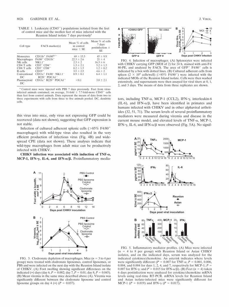
this virus into mice, only virus not expressing GFP could berecovered (data not shown), suggesting that GFP expression isnot stable.
Infection of cultured adherent splenic cells (85% F4/80
macrophages) with wild-type virus also resulted in the veryefficient production of infectious virus (Fig. 4B) and wide-spread CPE (data not shown). These analyses indicate thatwild-type macrophages from adult mice can be productivelyinfected with CHIKV.
CHIKV infection was associated with induction of TNF-�,MCP-1, IFN-�, IL-6, and IFN-�/�. Proinflammatory media-
tors, including TNF-�, MCP-1 (CCL2), IFN-�, interleukin-6(IL-6), and IFN-�/�, have been identified in primates andhumans infected with CHIKV and in other alphaviral arthrit-ides (32, 51, 71). The serum levels of several proinflammatorymediators were measured during viremia and disease in thecurrent mouse model, and elevated levels of TNF-�, MCP-1,IFN-�, IL-6, and IFN-�/� were observed (Fig. 5A). No signif-
TABLE 1. Leukocyte (CD45) populations isolated from the feetof control mice and the swollen feet of mice infected with the
Reunion Island isolate 7 days previouslya
Cell type FACS marker(s)Mean % of cells
in controlmice SE
Mean % of cells7 days
postinfection SE
Monocytes CD11b F4/80lo/� 69 15.5 45 9.9Macrophages F4/80 CD11b 22.3 2.6 21 4NK cells NK1.1 2.3 2 16.3 6CD4 T cells CD3 CD4 1.3 2.1 5.2 1.3CD8 T cells CD3 CD8 0.2 1.5 1.2 0.2B cells CD19 3.5 2.5 4.6 2Conventional
DCCD11c F4/80� NK1.1�
B220� PDCA1�0.9 0.1 6.4 1.1
PlasmacytoidDC
CD11c B220 PDCA1 �0.1 3.8 2.1
a Control mice were injected with PBS 7 days previously. Feet from virus-infected animals contained, on average, 31-fold 7.7-fold-more CD45 cellsthan feet from control animals. Data represent the means of data from two tothree experiments with cells from three to five animals pooled. DC, dendriticcells.
FIG. 3. Clodronate depletion of macrophages. Mice (n � 3 to 4 pergroup) were treated with clodronate liposomes, control liposomes, orPBS and were infected on the next day with the Reunion Island isolateof CHIKV. (A) Foot swelling showing significant differences on theindicated (*) days (day 6, P � 0.002; day 7, P � 0.01; day 8, P � 0.045).(B) Mean viremia in the same mice described above (A). Viremia wassignificantly different between the clodronate liposome and controlliposome groups on day 4 (*) (P � 0.037).
FIG. 4. Infection of macrophages. (A) Splenocytes were infectedwith CHIKV carrying GFP (MOI of 2) for 24 h, stained with anti-F4/80-PE, and analyzed by FACS. The area of GFP F4/80 cells isindicated by a box with dotted lines. (B) Cultured adherent cells fromspleen (2 � 104 cells/well) (85% F4/80) were infected with theindicated MOIs of the Reunion Island isolate. Cells were then washedextensively, and supernatants were then assayed for viral titers at 0, 1,2, and 3 days. The means of data from three replicates are shown.
FIG. 5. Inflammatory mediator profiles. (A) Mice were infected(n � 4 to 8 per group) with Reunion Island or Asian CHIKVisolates, and on the indicated days, serum was analyzed for theindicated cytokines/chemokine. An asterisk indicates where levelswere significantly different (P � 0.007 for TNF-�; P � 0.001, 0.006,0.004, and 0.004 for days 1, 2, 4, and 7, respectively for MCP-1; P �0.007 for IFN-�; and P � 0.015 for IFN-�/�). (B) Feet (n � 4) taken6 days postinfection were analyzed for cytokine/chemokine mRNAlevels using real-time RT-PCR. mRNA levels for Reunion Islandand Asian isolate-infected mice were significantly different forMCP-1 (P � 0.019) and IFN-� (P � 0.017).
8026 GARDNER ET AL. J. VIROL.

icant amount of IL-10 or IL-1� was detected (data not shown).Compared with the Asian isolate, the Reunion Island isolateinduced significantly more serum MCP-1, IFN-�, and IFN-�/�during the viremic period and more TNF-� and MCP-1 duringthe disease period (i.e., period of foot swelling) (Fig. 5A). Thesignificantly higher levels of IFN-�/� induced by the ReunionIsland isolate on day 2 than those induced by the Asian isolate(Fig. 5A) were not associated with lower viremia, as might beexpected (53, 60), but did correlate with lower Reunion Islandvirus titers in the feet on that day (Fig. 1D).
Analysis of the feet by real-time RT-PCR also showed thatduring the disease period, significantly more IFN-� and MCP-1mRNA was present in the feet of Reunion Island-infectedmice than in the feet of Asian isolate-infected mice (Fig. 5B).Elevated TNF-� levels were also seen, although these levelswere not significantly different between isolates. No significantIFN-�4 or IFN-� mRNA levels above those seen in mock-infected animals were detected (data not shown), suggestingthat ongoing elevated levels of IFN-�/� are not required forthe rheumatic inflammatory response (83). The increased lev-els of IFN-� and MCP-1 mRNA induced by infection with theReunion Island isolate were consistent with the increasedrheumatic disease seen with this isolate (Fig. 1A and B). IFN-�and MCP-1 are known to be involved in macrophage activationand recruitment as well as inflammation (19, 59, 71).
CHIKV infection induced high levels of CHIKV-specificIgG2c responses. The level of CHIKV-specific IgG1 and IgG2cantibodies induced after CHIKV infection was determined bystandard ELISA. Infection produced high levels of CHIKV-specific IgG2c responses, with the Reunion Island isolateproducing significantly higher levels than the Asian isolate (P �0.004). The levels of IgG1 induced were very low and notsignificantly different for the two isolates (Fig. 6A). These dataillustrate that CHIKV infection induces strongly biased Th1responses.
An inactivated CHIKV vaccine protected against viremiaand disease. A number of vaccines have been developed forCHIKV (2, 15, 47, 73, 82). However, it was previously notpossible to evaluate the ability of CHIKV vaccines to preventarthritis in an animal model. To illustrate the utility of thecurrent model for testing prophylactic vaccines, mice wereimmunized once s.c. with a whole-virus, binary ethyleneimine-inactivated (7), Asian isolate CHIKV vaccine. A similar strat-egy was used previously for the generation of an experimentalRRV vaccine (1). Ten micrograms of the CHIKV vaccineinduced strong IgG2c and some IgG1 responses, with lowerdoses inducing much weaker responses. The addition of theadjuvant Quil A significantly increased the antibody responses;data are shown for the 0.1-�g dose (P � 0.001) (Fig. 6A).
After challenge with the Reunion Island isolate, mice vac-cinated with 0.01 �g of vaccine had a significantly reducedviremia, and mice that received 0.1 to 10 �g of vaccine showedno detectable viremia (Fig. 6B). Animals receiving the Quil Avaccine also showed no detectable viremia (data not shown).The same mice were assessed for foot swelling, with all vac-cines significantly reducing the level of swelling, although ahint of swelling was still evident at the 0.01-�g vaccine dose.This experiment illustrated not only that a vaccine derivedfrom an old Asian isolate can protect against a virus isolatefrom the recent epidemic but also that vaccinated animals with
relatively low levels of antibody (compared with infected mice)were protected against viremia and disease.
IFN-� treatment before, but not after, infection preventeddisease. IFN-� is used to treat a number of chronic viral in-fections, particularly hepatitis C (63), and has been proposedas a potential treatment for encephalitic alphaviruses (40, 84)and CHIKV (68). Like other alphaviruses (5, 60), CHIKV issensitive to IFN-�/� (66). To determine whether IFN-� mighthave an application in preventing CHIKV arthritis, mice wereinjected with 103 IU IFN-� i.v., an IU/kg dose similar to thatgiven per injection to humans with hepatitis C (43). The nextday, the mice were challenged with the Reunion Island isolate.The mean peak viremia was significantly reduced by �2.5 logsand delayed by 2 days (see Fig. S3A in the supplementalmaterial), and disease was abolished (Fig. S3B). However,when IFN-� treatment was delayed until day 3, there was nosignificant effect on either viral titers in the feet or foot swelling(data not shown), suggesting that IFN-� treatment may havelimited value as a therapeutic intervention.
RRV infection and anti-RRV antiserum protected againstCHIKV. Cross-protection involving alphaviruses that cause le-thal encephalitic disease in mice has been extensively studied
FIG. 6. Vaccination and CHIKV challenge. (A) Fifty percent end-point IgG1 and IgG2c titers 5 weeks after infection (n � 6) with theindicated virus isolate or vaccination with different doses of purifiedinactivated CHIKV vaccine (n � 3 per group). (B) Five weeks aftervaccination, mice were challenged with the Reunion Island isolate, andthe level of viremia was determined over 6 days. Mock vaccination wasdone with medium. (C) Foot swelling of the animals shown in B (n �6 feet per group).
VOL. 84, 2010 CHIKUNGUNYA VIRUS ARTHRITIS IN ADULT WILD-TYPE MICE 8027

(36, 81, 85). However, cross-protection against arthritis be-tween arthrogenic alphaviruses has not been reported. To in-vestigate this phenomenon, mice were mock infected or in-fected with RRV and were challenged with the Reunion Islandisolate 4.5 months later. Mice previously infected with RRVhad a �3- to 4-log-lower mean peak viremia after infectionwith CHIKV (Fig. 7A) and were protected against CHIKVdisease (data not shown). To investigate the role of antibody inthis cross-protection, antiserum from RRV-infected mice wasadoptively transferred into naïve mice. These mice were theninfected with CHIKV and showed a 3-log reduction in peakviremia (data not shown) and were protected against CHIKVdisease (Fig. 7B). As reported previously (10), antisera fromCHIKV-infected mice protected against viremia (data notshown) and disease, whereas sera from naïve mice had noeffect (Fig. 7B). CHIKV and RRV both belong to the SemlikiForest virus antigenic complex (16). Therefore, as expected,serum from RRV-infected mice (prior to CHIKV challenge)cross-reacted with CHIKV (Fig. 7C) and showed CHIKV-
specific IgG1 and IgG2c antibody titers comparable to thoseseen after vaccination with 1 �g of the inactivated CHIKVvaccine (Fig. 6A). T cells from RRV-infected mice also showedsignificant cross-reactivity with CHIKV virus, giving responsescomparable to those seen after CHIKV infection (Fig. 7D).
DISCUSSION
Here we report a simple mouse model of CHIKV arthritis inwild-type adult mice. The model recapitulates the viremia (55)and, importantly, the rheumatic symptoms of CHIKV infectionof humans (6, 28, 54, 55), with clear signs of self-limiting footswelling and histological evidence of acute and persistent ar-thritis, tenosynovitis, and myositis.
In establishing this model of CHIKV infection and disease,we observed that CHIKV obtained from mice infected Veroand BHK cells inconsistently (data not shown), for reasonswhich are currently unclear. To measure virus titers, we thusadopted a method of determining titers of serum and tissueextracts on C6/36 cells, followed by subculture onto parallelplates of Vero cells to determine which of the individual C6/36wells contained virus. We also found that low levels of endo-toxin or mycoplasma contamination can prevent efficient al-phavirus infection in vivo, and we have used sensitive bioassays(27, 33) to ensure the absence of these potential contaminantsin CHIKV preparations. Although s.c. and intradermal (i.d.)injections of CHIKV at the base of the tail produced viremia,they produced no foot swelling (data not shown). The injectionof one hind foot also did not result in significant swelling of theother hind foot (data not shown). We speculate that the de-layed and/or lower CHIKV replication in the noninjected feetin the latter settings precludes the development of significantovert disease. These features probably represent a weakness ofthis mouse model, as mosquito-mediated infection in humansoften results in the swelling of multiple joints (6, 50).
The cellular infiltrates seen in this mouse model of CHIKVdisease correlate well with data from other studies of alphavi-ral arthritides. Pronounced monocyte/macrophage infiltratesin the synovial fluid of RRV-infected patients (18, 65) and in amonkey model of CHIKV infection (32) have been reported.The presence of high numbers of monocytes, compared tomacrophages, has not previously been enumerated. Neverthe-less, the abundance of monocytes/macrophages in the swollenfeet of CHIKV-infected mice and the ability of macrophagedepletion to ameliorate disease reaffirm the importance ofthese cells in the rheumatic disease induced by alphaviral in-fections (37, 46, 59, 71). NK cells were the next most abundantcell type in the swollen feet of CHIKV-infected mice (Table 1),and these cells were identified in the synovial fluid of RRV-infected patients (25) and in muscle tissues in a mouse modelof RRV myositis (46). NK cells were shown previously tocontribute to alphaviral encephalitis (3) and may thus alsocontribute to the immunopathogenesis of alphaviral arthritides(71). The low levels of B and T cells (Table 1) support the viewthat these cells do not play a major role in the immunopatho-genesis in mouse models of alphaviral rheumatic disease (46).CD8 T cells have also been shown to have no protective effectagainst viremia in a mouse model of RRV infection (38).Whether there is a role for T cells in human disease, where Tcells in synovial effusions seem to be slightly more abundant
FIG. 7. Cross-protection with RRV. (A) Mice were infected withRRV or mock infected (n � 6 per group) and 4.5 months later wereinfected with the Reunion Isolate of CHIKV, and CHIKV viremia wasmeasured (an asterisk indicates significant differences [P � 0.029 and0.005]). (B) CHIKV-induced foot swelling of mice that had receivedserum from naïve mice or mice previously infected with RRV or theReunion Island isolate of CHIKV. (C) ELISA using serum from theRRV-infected mice described above (A) prior to CHIKV infection,with titers determined on purified inactivated CHIKV. (D) IFN-�ELISPOT assay using splenocytes from a separate group of miceinfected 4.5 weeks previously with the indicated virus and using puri-fied inactivated CHIKV as an antigen.
8028 GARDNER ET AL. J. VIROL.

(65), remains unclear. The presence of dendritic cells andplasmacytoid dendritic cells is consistent with other studiesshowing the recruitment of these cells into the sites of virusreplication (12, 23).
Macrophages have been shown to be important targets ofinfection in a primate model of CHIKV infection (32), andhuman macrophages have also been shown to be productivelyinfected with CHIKV in vitro (66). Here we show that primaryadult wild-type macrophages are also productively infectedwith CHIKV (Fig. 4C and D). However, clodronate depletionof macrophages significantly prolonged viremia, indicatingthat macrophages and/or their inflammatory products (e.g.,TNF-�) (71) are also involved in promoting the clearance ofvirus. Clodronate treatment was recently reported to increasethe viremia associated with another macrophage-tropic virus,dengue virus (17). Macrophages are thus a target for infection,promote viral clearance, and are important for the develop-ment of rheumatic disease. Given the phenotypic diversity,functional plasticity, and widespread distribution of macro-phages, different types and/or sources of macrophages may beresponsible for each of these activities. Long-term persistentinfection of macrophages was reported previously for primates(32), and such infections may give rise to the chronic diseaseseen for some patients (35). Whether the persistent lesionsseen in the feet in the current model are also due to low-levelpersistent infection of macrophages or are a result of an on-going resolution of tissue damage remains to be established.
The in vivo cytokine/chemokine profiles associated with al-phaviral arthritides have not been extensively studied (37, 51,69). Here we show that CHIKV infection increased serumlevels of MCP-1, IFN-�/�, IFN-�, IL-6, and TNF-� and thatthe serum levels of these mediators peaked during the viremicperiod. Peripheral blood of monkeys infected with CHIKValso showed elevated levels of MCP-1, IFN-�/�, IFN-�, IL-6,and TNF-� (32). A recent study of patients with CHIKV dis-ease identified serum IL-1� and IL-6 as biomarkers of CHIKVdisease severity, with IFN-�, IL-5, IL-7, IL-10, and IL-15 alsobeing detected (51). However, serum MCP-1, IFN-�, andTNF-� levels were no different in CHIKV-infected patientsand healthy controls (51). The current study may provide someinsight into this apparent discrepancy. The peak in serumproinflammatory mediator levels (Fig. 5A) clearly precededthe onset of foot swelling (Fig. 1B), suggesting that once pa-tients present with arthritis/arthralgia, the viremia, and theassociated peripheral inflammatory response, may have abated(51). Importantly, in the arthritic feet of infected mice, thevirus continues to replicate (Fig. 1D), and the levels ofexpression of MCP-1, IFN-�, and TNF-� continue to beelevated (Fig. 5B). The levels of these inflammatory medi-ators are also elevated in a number of viral arthritides andare also key players in rheumatoid arthritis (71). Increasedlevels of MCP-1, IFN-�, and TNF-� were also detected insynovial effusions in RRV-infected patients with polyarthritis(37), with RRV also reported to persist in the joints of thesepatients (65). These observations support the view thatCHIKV arthritis/arthralgia is an inflammatory disease (56).
Preclinical testing of vaccines and immunotherapies forCHIKV-induced arthritis has previously not been possible.Here we illustrate the utility of the new mouse model byevaluating a simple inactivated CHIKV vaccine and IFN-�
treatment. Inactivated alphavirus vaccines have been devel-oped for veterinary use (44) and were described previously forRRV (29). A simple, unadjuvanted, and inactivated CHIKVvaccine derived from the Asian isolate was shown herein to beimmunogenic and protected mice against rheumatic diseaseinduced by the Reunion Island isolate. Antibodies appear to bethe main mediators of protection against alphaviruses (22, 44),including CHIKV (10) (Fig. 7B), and mice with only low levelsof CHIKV-specific antibodies were protected against disease(see Fig. 6A and C at the 1-�g/mouse dose). This finding isconsistent with previous data showing that mice with only lowlevels of anti-RRV antibodies were protected against RRVviremia (87). Taken together, these data suggest that vaccinesagainst arthrogenic alphaviruses may not need to induce highlevels of immunity in order to mediate effective protection.
In contrast to alum (73), which was unable to improve theantibody responses induced by an inactivated RRV vaccine(87), Quil A was very effective at increasing antibody responsesto the inactivated CHIKV vaccine. This adjuvant was shownpreviously to increase antibody responses to several inactivat-ed-virus vaccines (67, 86). Quil A also tends to promote Th1responses, whereas alum biases responses toward Th2 (73).Natural CHIKV infection induced an extraordinarily Th1-bi-ased response, and Th2 bias is generally considered to promotevirus replication (61). Th1-promoting adjuvants are thereforelikely to be more appropriate for CHIKV vaccines.
IFN-� was proposed previously to be a potential treatmentfor encephalitic alphaviruses and CHIKV (40, 68, 84). Weshow here that IFN-� treatment given before infection re-duced viremia and prevented rheumatic disease. However, ifgiven on day 3 postinfection, there was no significant effect onthe levels of virus recovered from the feet or any effect ondisease. This parallels other previous reports that showed thatIFN-� treatment is less effective against alphaviral encephaliticdisease when given later in infection (21, 53). The activation ofsuppressors of cytokine signaling proteins by the inflammatoryresponse (11) and/or disruption of IFN-�/� signaling by viralproteins (64) may be involved in limiting the effectiveness ofIFN-� treatment in an established infection. The value ofIFN-� as a therapy for CHIKV disease is thus probably lim-ited.
RRV infection and anti-RRV antibodies were able signifi-cantly to reduce CHIKV viremia and to protect mice againstCHIKV disease. Although an antibody-dependent enhancementof infection was shown previously for RRV (41, 70), there was noindication that anti-RRV antibodies enhanced CHIKV infection.The adoptive-transfer experiments (Fig. 7B) also confirmedthat antibodies mediate effective protection against CHIKV(10). CD4 T cells may also play a role (36), as CD4 T cells wereshown previously to protect mice against other alphaviruses(88). The high level of cross-reactivity seen for B- and T-cellresponses (Fig. 7C and D) may reflect the 75% sequencehomology (60% sequence identity) between RRV andCHIKV in the structural polyprotein. The putative receptorbinding domains of arthrogenic alphavirus E2 glycoproteinsare also highly conserved (34). The C-terminal two-thirds ofthe capsid proteins of RRV and CHIKV also show 95%sequence homology (90% identity), with this region contain-ing a previously identified T-cell epitope (38). These results
VOL. 84, 2010 CHIKUNGUNYA VIRUS ARTHRITIS IN ADULT WILD-TYPE MICE 8029

suggest that individuals with prior exposure to RRV (mostlyAustralasians [24]) would be protected from CHIKV disease.
A number of differences between the behaviors of the Asianand Reunion Island isolates were evident, which may shedsome light on pathogenesis. The Reunion Island isolate repli-cated faster in feet (Fig. 1D); showed a more pronouncedmononuclear infiltrate (see Fig. S1 in the supplemental mate-rial); induced significantly more serum TNF-�, MCP-1, IFN-�,and IFN-�/� (Fig. 5A) and more MCP-1 and IFN-� mRNA inthe feet (Fig. 5B); generated more virus-specific IgG2c re-sponses (Fig. 6A); and replicated faster and induced moreCPE in MEFs in vitro (Fig. 1F). One might speculate thatfaster initial replication (Fig. 1D) and, perhaps, more CPE (49)induce more early MCP-1, IFN-�/�, and IFN-�, with IFN-�likely derived from activated NK cells (3, 25, 46). The in-creased levels of these proinflammatory mediators may lead tomore infiltration and activation of macrophages and MCP-1and IFN-� production, ultimately resulting in increased rheu-matic inflammation (19, 37, 59, 71).
ACKNOWLEDGMENTS
We thank Luis Mateo and Rebecca Pawliw (Inverness InnovationsAustralia Pty. Ltd., Brisbane, Australia) for the supply of purifiedinactivated CHIKV and Clay Winterford (QIMR) for undertaking theimmunohistochemistry.
Funding was provided by the Australian Centre for Internationaland Tropical Health, the National Health Medical Research Council,the Australian Centre for Vaccine Development, and the French-Australian Science and Technology program of the Department ofInnovation, Industry, Science, and Research (Australia). A.S. is a prin-cipal research fellow with the National Health & Medical ResearchCouncil of Australia.
The funders had no role in study design, data collection and analysis,decision to publish, or preparation of the manuscript.
REFERENCES
1. Aaskov, J., L. Williams, and S. Yu. 1997. A candidate Ross River virusvaccine: preclinical evaluation. Vaccine 15:1396–1404.
2. Akahata, W., Z. Y. Yang, H. Andersen, S. Sun, H. A. Holdaway, W. P. Kong,M. G. Lewis, S. Higgs, M. G. Rossmann, S. Rao, and G. J. Nabel. 2010. Avirus-like particle vaccine for epidemic Chikungunya virus protects nonhu-man primates against infection. Nat. Med. 16:334–338.
3. Alsharifi, M., M. Lobigs, M. M. Simon, A. Kersten, K. Muller, A. Koskinen,E. Lee, and A. Mullbacher. 2006. NK cell-mediated immunopathology dur-ing an acute viral infection of the CNS. Eur. J. Immunol. 36:887–896.
4. Anraku, I., V. V. Mokhonov, P. Rattanasena, E. I. Mokhonova, J. Leung, G.Pijlman, A. Cara, W. A. Schroder, A. A. Khromykh, and A. Suhrbier. 2008.Kunjin replicon-based simian immunodeficiency virus gag vaccines. Vaccine26:3268–3276.
5. Antalis, T. M., M. La Linn, K. Donnan, L. Mateo, J. Gardner, J. L. Dick-inson, K. Buttigieg, and A. Suhrbier. 1998. The serine proteinase inhibitor(serpin) plasminogen activation inhibitor type 2 protects against viral cyto-pathic effects by constitutive interferon alpha/beta priming. J. Exp. Med.187:1799–1811.
6. Brighton, S. W., O. W. Prozesky, and A. L. de la Harpe. 1983. Chikungunyavirus infection. A retrospective study of 107 cases. S. Afr. Med. J. 63:313–315.
7. Brown, F. 2001. Inactivation of viruses by aziridines. Vaccine 20:322–327.8. Charrel, R. N., X. de Lamballerie, and D. Raoult. 2007. Chikungunya out-
breaks—the globalization of vectorborne diseases. N. Engl. J. Med. 356:769–771.
9. Couderc, T., F. Chretien, C. Schilte, O. Disson, M. Brigitte, F. Guivel-Benhassine, Y. Touret, G. Barau, N. Cayet, I. Schuffenecker, P. Despres, F.Arenzana-Seisdedos, A. Michault, M. L. Albert, and M. Lecuit. 2008. Amouse model for Chikungunya: young age and inefficient type-i interferonsignaling are risk factors for severe disease. PLoS Pathog. 4:e29.
10. Couderc, T., N. Khandoudi, M. Grandadam, C. Visse, N. Gangneux, S.Bagot, J. F. Prost, and M. Lecuit. 2009. Prophylaxis and therapy for Chikun-gunya virus infection. J. Infect. Dis. 200:516–523.
11. Dalpke, A., K. Heeg, H. Bartz, and A. Baetz. 2008. Regulation of innateimmunity by suppressor of cytokine signaling (SOCS) proteins. Immunobi-ology 213:225–235.
12. Donaghy, H., L. Bosnjak, A. N. Harman, V. Marsden, S. K. Tyring, T. C.Meng, and A. L. Cunningham. 2009. Role for plasmacytoid dendritic cells inthe immune control of recurrent human herpes simplex virus infection.J. Virol. 83:1952–1961.
13. Dubrulle, M., L. Mousson, S. Moutailler, M. Vazeille, and A. B. Failloux.2009. Chikungunya virus and Aedes mosquitoes: saliva is infectious as soonas two days after oral infection. PLoS One 4:e5895.
14. Economopoulou, A., M. Dominguez, B. Helynck, D. Sissoko, O. Wichmann,P. Quenel, P. Germonneau, and I. Quatresous. 2009. Atypical Chikungunyavirus infections: clinical manifestations, mortality and risk factors for severedisease during the 2005–2006 outbreak on Reunion. Epidemiol. Infect. 137:534–541.
15. Edelman, R., C. O. Tacket, S. S. Wasserman, S. A. Bodison, J. G. Perry, andJ. A. Mangiafico. 2000. Phase II safety and immunogenicity study of livechikungunya virus vaccine TSI-GSD-218. Am. J. Trop. Med. Hyg. 62:681–685.
16. Fields, B. N., D. M. Knipe, and P. M. Howley. 1996. Alphaviruses, 3rd ed.,vol. 1. Lippincott-Raven, Philadelphia, PA.
17. Fink, K., C. Ng, C. Nkenfou, S. G. Vasudevan, N. van Rooijen, and W. Schul.2009. Depletion of macrophages in mice results in higher dengue virus titersand highlights the role of macrophages for virus control. Eur. J. Immunol.39:2809–2821.
18. Fraser, J. R., A. L. Cunningham, B. J. Clarris, J. G. Aaskov, and R. Leach.1981. Cytology of synovial effusions in epidemic polyarthritis. Aust. N. Z. J.Med. 11:168–173.
19. Furtado, G. C., B. Pina, F. Tacke, S. Gaupp, N. van Rooijen, T. M. Moran,G. J. Randolph, R. M. Ransohoff, S. W. Chensue, C. S. Raine, and S. A. Lira.2006. A novel model of demyelinating encephalomyelitis induced by mono-cytes and dendritic cells. J. Immunol. 177:6871–6879.
20. Gardner, D. L., P. N. Skelton-Stroud, and R. J. Fitzmaurice. 1991. Acutemuramyl dipeptide-induced arthritis in the baboon Papio cynocephalus. Z.Rheumatol. 50:86–92. (In German.)
21. Glasgow, L. A. 1970. Transfer of interferon-producing macrophages: newapproach to viral chemotherapy. Science 170:854–856.
22. Griffin, D., B. Levine, W. Tyor, S. Ubol, and P. Despres. 1997. The role ofantibody in recovery from alphavirus encephalitis. Immunol. Rev. 159:155–161.
23. Gulbahar, M. Y., W. C. Davis, H. Yuksel, and M. Cabalar. 2006. Immuno-histochemical evaluation of inflammatory infiltrate in the skin and lung oflambs naturally infected with sheeppox virus. Vet. Pathol. 43:67–75.
24. Harley, D., A. Sleigh, and S. Ritchie. 2001. Ross River virus transmission,infection, and disease: a cross-disciplinary review. Clin. Microbiol. Rev. 14:909–932.
25. Hazelton, R. A., C. Hughes, and J. G. Aaskov. 1985. The inflammatoryresponse in the synovium of a patient with Ross River arbovirus infection.Aust. N. Z. J. Med. 15:336–339.
26. Hensley, S. E., A. S. Cun, W. Giles-Davis, Y. Li, Z. Xiang, M. O. Lasaro, B. R.Williams, R. H. Silverman, and H. C. Ertl. 2007. Type I interferon inhibitsantibody responses induced by a chimpanzee adenovirus vector. Mol. Ther.15:393–403.
27. Johnson, B. J., T. T. T. Le, C. A. Dobbin, T. Banovic, C. B. Howard, F. deMaria Leon Flores, D. Vanags, D. J. Naylor, G. R. Hill, and A. Suhrbier.2005. Heat shock protein 10 inhibits lipopolysaccharide-induced inflamma-tory mediator production. J. Biol. Chem. 280:4037–4047.
28. Kennedy, A. C., J. Fleming, and L. Solomon. 1980. Chikungunya viral ar-thropathy: a clinical description. J. Rheumatol. 7:231–236.
29. Kistner, O., N. Barrett, A. Bruhmann, M. Reiter, W. Mundt, H. Savidis-Dacho, S. Schober-Bendixen, F. Dorner, and J. Aaskov. 2007. The preclinicaltesting of a formaldehyde inactivated Ross River virus vaccine designed foruse in humans. Vaccine 25:4845–4852.
30. Koga, T., K. Kakimoto, T. Hirofuji, S. Kotani, H. Ohkuni, K. Watanabe, N.Okada, H. Okada, A. Sumiyoshi, and K. Saisho. 1985. Acute joint inflam-mation in mice after systemic injection of the cell wall, its peptidoglycan, andchemically defined peptidoglycan subunits from various bacteria. Infect. Im-mun. 50:27–34.
31. Kumar, N. P., R. Joseph, T. Kamaraj, and P. Jambulingam. 2008. A226Vmutation in virus during the 2007 chikungunya outbreak in Kerala, India.J. Gen. Virol. 89:1945–1948.
32. Labadie, K., T. Larcher, C. Joubert, A. Mannioui, B. Delache, P. Brochard,L. Guigand, L. Dubreil, P. Lebon, B. Verrier, X. de Lamballerie, A. Suhrbier,Y. Cherel, R. Le Grand, and P. Roques. 2010. Chikungunya disease innonhuman primates involves long-term viral persistence in macrophages.J. Clin. Invest. 120:894–906.
33. La Linn, M., A. J. Bellett, P. G. Parsons, and A. Suhrbier. 1995. Completeremoval of mycoplasma from viral preparations using solvent extraction.J. Virol. Methods 52:51–54.
34. La Linn, M., J. A. Eble, C. Lubken, R. W. Slade, J. Heino, J. Davies, and A.Suhrbier. 2005. An arthritogenic alphavirus uses the alpha1beta1 integrincollagen receptor. Virology 336:229–239.
35. Larrieu, S., N. Pouderoux, T. Pistone, L. Filleul, M. C. Receveur, D. Sissoko,K. Ezzedine, and D. Malvy. 2010. Factors associated with persistence ofarthralgia among chikungunya virus-infected travellers: report of 42 Frenchcases. J. Clin. Virol. 47:85–88.
8030 GARDNER ET AL. J. VIROL.

36. Latif, Z., D. Gates, C. J. Wust, and A. Brown. 1979. Cross protection amongtogaviruses in nude mice and littermates. J. Gen. Virol. 45:89–98.
37. Lidbury, B. A., N. E. Rulli, A. Suhrbier, P. N. Smith, S. R. McColl, A. L.Cunningham, A. Tarkowski, N. van Rooijen, R. J. Fraser, and S. Ma-halingam. 2008. Macrophage-derived proinflammatory factors contribute tothe development of arthritis and myositis after infection with an arthrogenicalphavirus. J. Infect. Dis. 197:1585–1593.
38. Linn, M. L., L. Mateo, J. Gardner, and A. Suhrbier. 1998. Alphavirus-specific cytotoxic T lymphocytes recognize a cross-reactive epitope from thecapsid protein and can eliminate virus from persistently infected macro-phages. J. Virol. 72:5146–5153.
39. Liu, W. J., X. J. Wang, D. C. Clark, M. Lobigs, R. A. Hall, and A. A.Khromykh. 2006. A single amino acid substitution in the West Nile virusnonstructural protein NS2A disables its ability to inhibit alpha/beta inter-feron induction and attenuates virus virulence in mice. J. Virol. 80:2396–2404.
40. Lukaszewski, R. A., and T. J. Brooks. 2000. Pegylated alpha interferon is aneffective treatment for virulent Venezuelan equine encephalitis virus and hasprofound effects on the host immune response to infection. J. Virol. 74:5006–5015.
41. Mahalingam, S., and B. A. Lidbury. 2002. Suppression of lipopolysaccha-ride-induced antiviral transcription factor (STAT-1 and NF-kappa B) com-plexes by antibody-dependent enhancement of macrophage infection byRoss River virus. Proc. Natl. Acad. Sci. U. S. A. 99:13819–13824.
42. Mavalankar, D., P. Shastri, T. Bandyopadhyay, J. Parmar, and K. V. Ra-mani. 2008. Increased mortality rate associated with chikungunya epidemic,Ahmedabad, India. Emerg. Infect. Dis. 14:412–415.
43. Mazzoran, L., F. Zorat, L. Chemello, L. S. Croce, I. Rigato, L. Cavalletto, E.Bernardinello, C. Tiribelli, A. Alberti, and G. Pozzato. 2001. Human leuco-cyte interferon-alpha in the treatment of chronic hepatitis C. Dig. Liver Dis.33:347–352.
44. Minke, J. M., J. C. Audonnet, and L. Fischer. 2004. Equine viral vaccines:the past, present and future. Vet. Res. 35:425–443.
45. Mogal, A., and S. A. Abdulkadir. 2006. Effects of histone deacetylase inhib-itor (HDACi); trichostatin-A (TSA) on the expression of housekeepinggenes. Mol. Cell. Probes 20:81–86.
46. Morrison, T. E., A. C. Whitmore, R. S. Shabman, B. A. Lidbury, S. Ma-halingam, and M. T. Heise. 2006. Characterization of Ross River virustropism and virus-induced inflammation in a mouse model of viral arthritisand myositis. J. Virol. 80:737–749.
47. Muthumani, K., K. M. Lankaraman, D. J. Laddy, S. G. Sundaram, C. W.Chung, E. Sako, L. Wu, A. Khan, N. Sardesai, J. J. Kim, P. Vijayachari, andD. B. Weiner. 2008. Immunogenicity of novel consensus-based DNA vaccinesagainst Chikungunya virus. Vaccine 26:5128–5134.
48. Mylonas, A. D., A. M. Brown, T. L. Carthew, B. McGrath, D. M. Purdie, N.Pandeya, P. C. Vecchio, L. G. Collins, I. D. Gardner, F. J. de Looze, E. J.Reymond, and A. Suhrbier. 2002. Natural history of Ross River virus-in-duced epidemic polyarthritis. Med. J. Aust. 177:356–360.
49. Nagata, S. 2008. Rheumatoid polyarthritis caused by a defect in DNA deg-radation. Cytok. Growth Factor Rev. 19:295–302.
50. Ng, K. W., A. Chow, M. K. Win, F. Dimatatac, H. Y. Neo, D. C. Lye, and Y. S.Leo. 2009. Clinical features and epidemiology of chikungunya infection inSingapore. Singapore Med. J. 50:785–790.
51. Ng, L. F., A. Chow, Y. J. Sun, D. J. Kwek, P. L. Lim, F. Dimatatac, L. C. Ng,E. E. Ooi, K. H. Choo, Z. Her, P. Kourilsky, and Y. S. Leo. 2009. IL-1beta,IL-6, and RANTES as biomarkers of Chikungunya severity. PLoS One4:e4261.
52. Njenga, M. K., L. Nderitu, J. P. Ledermann, A. Ndirangu, C. H. Logue, C. H.Kelly, R. Sang, K. Sergon, R. Breiman, and A. M. Powers. 2008. Trackingepidemic Chikungunya virus into the Indian Ocean from East Africa. J. Gen.Virol. 89:2754–2760.
53. O’Brien, L., S. Perkins, A. Williams, L. Eastaugh, A. Phelps, J. Wu, and R.Phillpotts. 2009. Alpha interferon as an adenovirus-vectored vaccine adju-vant and antiviral in Venezuelan equine encephalitis virus infection. J. Gen.Virol. 90:874–882.
54. Ozden, S., M. Huerre, J. P. Riviere, L. L. Coffey, P. V. Afonso, V. Mouly, J.de Monredon, J. C. Roger, M. El Amrani, J. L. Yvin, M. C. Jaffar, M. P.Frenkiel, M. Sourisseau, O. Schwartz, G. Butler-Browne, P. Despres, A.Gessain, and P. E. Ceccaldi. 2007. Human muscle satellite cells as targets ofChikungunya virus infection. PLoS One 2:e527.
55. Parola, P., X. de Lamballerie, J. Jourdan, C. Rovery, V. Vaillant, P. Mino-dier, P. Brouqui, A. Flahault, D. Raoult, and R. N. Charrel. 2006. Novelchikungunya virus variant in travelers returning from Indian Ocean islands.Emerg. Infect. Dis. 12:1493–1499.
56. Pialoux, G., B. A. Gauzere, S. Jaureguiberry, and M. Strobel. 2007. Chikun-gunya, an epidemic arbovirosis. Lancet Infect. Dis. 7:319–327.
57. Pyke, A. T., D. A. Phillips, T. F. Chuan, and G. A. Smith. 2004. Sucrosedensity gradient centrifugation and cross-flow filtration methods for theproduction of arbovirus antigens inactivated by binary ethylenimine. BMCMicrobiol. 4:3.
58. Repik, P. M., J. M. Dalrymple, W. E. Brandt, J. M. McCown, and P. K.
Russell. 1983. RNA fingerprinting as a method for distinguishing dengue 1virus strains. Am. J. Trop. Med. Hyg. 32:577–589.
59. Rulli, N. E., A. Guglielmotti, G. Mangano, M. S. Rolph, C. Apicella, A. Zaid,A. Suhrbier, and S. Mahalingam. 2009. Amelioration of alphavirus-inducedarthritis and myositis in a mouse model by treatment with bindarit, aninhibitor of monocyte chemotactic proteins. Arthritis Rheum. 60:2513–2523.
60. Ryman, K. D., and W. B. Klimstra. 2008. Host responses to alphavirusinfection. Immunol. Rev. 225:27–45.
61. Schneider, B. S., and S. Higgs. 2008. The enhancement of arbovirus trans-mission and disease by mosquito saliva is associated with modulation of thehost immune response. Trans. R. Soc. Trop. Med. Hyg. 102:400–408.
62. Schuffenecker, I., I. Iteman, A. Michault, S. Murri, L. Frangeul, M. C.Vaney, R. Lavenir, N. Pardigon, J. M. Reynes, F. Pettinelli, L. Biscornet, L.Diancourt, S. Michel, S. Duquerroy, G. Guigon, M. P. Frenkiel, A. C. Brehin,N. Cubito, P. Despres, F. Kunst, F. A. Rey, H. Zeller, and S. Brisse. 2006.Genome microevolution of chikungunya viruses causing the Indian Oceanoutbreak. PLoS Med. 3:e263.
63. Selmi, C., A. Lleo, M. Zuin, M. Podda, L. Rossaro, and M. E. Gershwin.2006. Interferon alpha and its contribution to autoimmunity. Curr. Opin.Invest. Drugs 7:451–456.
64. Simmons, J. D., L. J. White, T. E. Morrison, S. A. Montgomery, A. C.Whitmore, R. E. Johnston, and M. T. Heise. 2009. Venezuelan equineencephalitis virus disrupts STAT1 signaling by distinct mechanisms indepen-dent of host shutoff. J. Virol. 83:10571–10581.
65. Soden, M., H. Vasudevan, B. Roberts, R. Coelen, G. Hamlin, S. Vasudevan,and J. La Brooy. 2000. Detection of viral ribonucleic acid and histologicanalysis of inflamed synovium in Ross River virus infection. ArthritisRheum. 43:365–369.
66. Sourisseau, M., C. Schilte, N. Casartelli, C. Trouillet, F. Guivel-Benhassine,D. Rudnicka, N. Sol-Foulon, K. Le Roux, M. C. Prevost, H. Fsihi, M. P.Frenkiel, F. Blanchet, P. V. Afonso, P. E. Ceccaldi, S. Ozden, A. Gessain, I.Schuffenecker, B. Verhasselt, A. Zamborlini, A. Saib, F. A. Rey, F. Arenzana-Seisdedos, P. Despres, A. Michault, M. L. Albert, and O. Schwartz. 2007.Characterization of reemerging chikungunya virus. PLoS Pathog. 3:e89.
67. Stittelaar, K. J., H. W. Vos, G. van Amerongen, G. F. Kersten, A. D. Oster-haus, and R. L. de Swart. 2002. Longevity of neutralizing antibody levels inmacaques vaccinated with Quil A-adjuvanted measles vaccine candidates.Vaccine 21:155–157.
68. Stock, I. 2009. Chikungunya fever—expanded distribution of a re-emergingtropical infectious disease. Med. Monatsschr. Pharm. 32:17–26. (In Ger-man.)
69. Suhrbier, A., and M. La Linn. 2004. Clinical and pathologic aspects ofarthritis due to Ross River virus and other alphaviruses. Curr. Opin. Rheu-matol. 16:374–379.
70. Suhrbier, A., and M. La Linn. 2003. Suppression of antiviral responses byantibody-dependent enhancement of macrophage infection. Trends Immu-nol. 24:165–168.
71. Suhrbier, A., and S. Mahalingam. 2009. The immunobiology of viral arthrit-ides. Pharmacol. Ther. 124:301–308.
72. Suryawanshi, S. D., A. H. Dube, R. K. Khadse, S. V. Jalgaonkar, P. S. Sathe,S. D. Zawar, and M. P. Holay. 2009. Clinical profile of chikungunya fever inpatients in a tertiary care centre in Maharashtra, India. Indian J. Med. Res.129:438–441.
73. Tiwari, M., M. Parida, S. R. Santhosh, M. Khan, P. K. Dash, and P. V. Rao.2009. Assessment of immunogenic potential of Vero adapted formalin inac-tivated vaccine derived from novel ECSA genotype of Chikungunya virus.Vaccine 27:2513–2522.
74. Townson, H., and M. B. Nathan. 2008. Resurgence of chikungunya. Trans.R. Soc. Trop. Med. Hyg. 102:308–309.
75. Tsetsarkin, K., S. Higgs, C. E. McGee, X. De Lamballerie, R. N. Charrel, andD. L. Vanlandingham. 2006. Infectious clones of Chikungunya virus (LaReunion isolate) for vector competence studies. Vector Borne Zoonotic Dis.6:325–337.
76. Tsetsarkin, K. A., D. L. Vanlandingham, C. E. McGee, and S. Higgs. 2007.A single mutation in Chikungunya virus affects vector specificity and epi-demic potential. PLoS Pathog. 3:e201.
77. Tudor, D., S. Riffault, C. Carrat, F. Lefevre, M. Bernoin, and B. Charley.2001. Type I IFN modulates the immune response induced by DNA vacci-nation to pseudorabies virus glycoprotein C. Virology 286:197–205.
78. Valamparampil, J. J., S. Chirakkarot, S. Letha, C. Jayakumar, and K. M.Gopinathan. 2009. Clinical profile of Chikungunya in infants. Indian J. Pe-diatr. 76:151–155.
79. van Rooijen, N., and E. van Kesteren-Hendrikx. 2002. Clodronate lipo-somes: perspectives in research and therapeutics. J. Liposome Res. 12:81–94.
80. Vazeille, M., S. Moutailler, D. Coudrier, C. Rousseaux, H. Khun, M. Huerre,J. Thiria, J. S. Dehecq, D. Fontenille, I. Schuffenecker, P. Despres, and A. B.Failloux. 2007. Two Chikungunya isolates from the outbreak of La Reunion(Indian Ocean) exhibit different patterns of infection in the mosquito, Aedesalbopictus. PLoS One 2:e1168.
81. Walton, T. E., M. M. Jochim, T. L. Barber, and L. H. Thompson. 1989.Cross-protective immunity between equine encephalomyelitis viruses inequids. Am. J. Vet. Res. 50:1442–1446.
VOL. 84, 2010 CHIKUNGUNYA VIRUS ARTHRITIS IN ADULT WILD-TYPE MICE 8031

82. Wang, E., E. Volkova, A. P. Adams, N. Forrester, S. Y. Xiao, I. Frolov, andS. C. Weaver. 2008. Chimeric alphavirus vaccine candidates for chikungunya.Vaccine 26:5030–5039.
83. Wenzel, J., S. Zahn, T. Bieber, and T. Tuting. 2009. Type I interferon-associated cytotoxic inflammation in cutaneous lupus erythematosus. Arch.Dermatol. Res. 301:83–86.
84. Wu, J. Q., N. D. Barabe, Y. M. Huang, G. A. Rayner, M. E. Christopher, andF. L. Schmaltz. 2007. Pre- and post-exposure protection against Westernequine encephalitis virus after single inoculation with adenovirus vectorexpressing interferon alpha. Virology 369:206–213.
85. Wust, C. J., R. Crombie, and A. Brown. 1987. Passive protection acrosssubgroups of alphaviruses by hyperimmune non-cross-neutralizing anti-Sind-bis serum. Proc. Soc. Exp. Biol. Med. 184:56–63.
86. Xiao, C., Z. I. Rajput, and S. Hu. 2007. Improvement of a commercialfoot-and-mouth disease vaccine by supplement of Quil A. Vaccine 25:4795–4800.
87. Yu, S., and J. G. Aaskov. 1994. Development of a candidate vaccine againstRoss River virus infection. Vaccine 12:1118–1124.
88. Yun, N. E., B. H. Peng, A. S. Bertke, V. Borisevich, J. K. Smith, J. N. Smith,A. L. Poussard, M. Salazar, B. M. Judy, M. A. Zacks, D. M. Estes, and S.Paessler. 2009. CD4 T cells provide protection against acute lethal en-cephalitis caused by Venezuelan equine encephalitis virus. Vaccine 27:4064–4073.
89. Ziegler, S. A., L. Lu, A. P. da Rosa, S. Y. Xiao, and R. B. Tesh. 2008. Ananimal model for studying the pathogenesis of chikungunya virus infection.Am. J. Trop. Med. Hyg. 79:133–139.
8032 GARDNER ET AL. J. VIROL.