Chemical Engineering Journal - City University of Hong Kong
29
Contents lists available at ScienceDirect Chemical Engineering Journal journal homepage: www.elsevier.com/locate/cej A bifunctional hydrogel incorporated with CuS@MoS 2 microspheres for disinfection and improved wound healing Xingyu Zhang a , Guannan Zhang a , Hongyu Zhang b , Xiaoping Liu a , Jing Shi c , Huixian Shi a , Xiaohong Yao a , Paul K. Chu d , Xiangyu Zhang a, ⁎ a College of Materials Science and Engineering, Taiyuan University of Technology, Taiyuan 030024, China b Second Hospital of Shanxi Medical University, Taiyuan 030024, China c Analytical Instrumentation Center, State Key Laboratory of Coal Conversion, Institute of Coal Chemistry, Chinese Academy of Sciences, Taiyuan 030001, China d Department of Physics and Department of Materials Science and Engineering, City University of Hong Kong, Tat Chee Avenue, Kowloon, Hong Kong, China HIGHLIGHTS • A hybrid hydrogel embedded with CuS@MoS2 microspheres is synthesized. • The hydrogel exhibits excellent antibacterial activity. • The hydrogel can enhance the secretion of HIF-1α and VEGF. • The hydrogel shows no obvious toxicity to organs and promotes wound healing. ARTICLE INFO Keywords: Wound healing CuS@MoS 2 -incorporated hydrogel Photothermal therapy Photodynamic therapy Vascularization ABSTRACT Recent advances in antibacterial technology has made it possible to obviate the needs for antibiotics to combat bacterial infection during would healing. However, few current wound dressings can simultaneously kill bacteria efficiently and promote would healing by facilitating revascularization. Herein, a hybrid hydrogel embedded with CuS@MoS 2 microspheres is synthesized. This hydrogel exhibits outstanding antibacterial activities in a short time and enhances wound healing at the same time. Within 15 min, 99.3% of Escherichia coli (E. coli) and 99.5% of Staphylococcus aureus (S. aureus) are killed due to the synergistic effects rendered by the photodynamic and photothermal antibacterial treatments under co-irradiation of 660 nm visible light (VL) and 808 near in- frared (NIR) light. The synergistic effects rendered by hyperthermia and reactive oxygen species (ROS) generated by the hydrogel during light irradiation improves the bacterial membrane permeability so that the bacterial membrane and protein can be easily destroyed by ROS, leading to rapid killing of bacteria in vitro and in vivo. In addition, the CuS@MoS 2 -incorporated hydrogel expedites wound healing because of promotion of the hypoxia inducible factor-1 (HIF-1α) and vascular endothelial growth factor (VEGF) expression and subsequent vascu- larization at the wound sites. This safe and synergistic therapy system has large potential in bacteria-infected wound healing therapy. 1. Introduction Bacterial infection and related diseases are responsible for one third of the global mortality [1,2]. For instance, S. aureus that can cause lo- calized purulent infection, pneumonia, sepsis, and other systemic in- fections is one of the most common causes of skin tissue infection [3,4]. Skin is an important barrier for the body to resist external infection and it is necessary to prevent skin tissues from bacterial infection and ac- celerate healing of damaged tissues [5,6]. Currently, antibiotic-based therapies are the most common methods to treat infection but excessive use of antibiotics leads to growing drug resistance in bacteria and even super-bacteria [7]. Silver nanoparticles are effective antibacterial agents [8] but a high concentrations of silver is toxic and can also cause bacterial resistance [9]. Traditional antibacterial treatments usually involve mutation and evolution [10], which are time consuming and it is thus important to develop techniques and materials that can kill bacteria in a short time. There is increasing interest in photodynamic therapy (PDT) and https://doi.org/10.1016/j.cej.2019.122849 Received 23 June 2019; Received in revised form 14 August 2019; Accepted 15 September 2019 ⁎ Corresponding author. E-mail address: [email protected] (X. Zhang). Chemical Engineering Journal 382 (2020) 122849 Available online 17 September 2019 1385-8947/ © 2019 Elsevier B.V. All rights reserved. T
-
Upload
khangminh22 -
Category
Documents
-
view
0 -
download
0
Transcript of Chemical Engineering Journal - City University of Hong Kong
Contents lists available at ScienceDirect
Chemical Engineering Journal
journal homepage: www.elsevier.com/locate/cej
A bifunctional hydrogel incorporated with CuS@MoS2 microspheres fordisinfection and improved wound healing
Xingyu Zhanga, Guannan Zhanga, Hongyu Zhangb, Xiaoping Liua, Jing Shic, Huixian Shia,Xiaohong Yaoa, Paul K. Chud, Xiangyu Zhanga,⁎
a College of Materials Science and Engineering, Taiyuan University of Technology, Taiyuan 030024, Chinab Second Hospital of Shanxi Medical University, Taiyuan 030024, Chinac Analytical Instrumentation Center, State Key Laboratory of Coal Conversion, Institute of Coal Chemistry, Chinese Academy of Sciences, Taiyuan 030001, ChinadDepartment of Physics and Department of Materials Science and Engineering, City University of Hong Kong, Tat Chee Avenue, Kowloon, Hong Kong, China
H I G H L I G H T S
• A hybrid hydrogel embedded with CuS@MoS2 microspheres is synthesized.
• The hydrogel exhibits excellent antibacterial activity.
• The hydrogel can enhance the secretion of HIF-1α and VEGF.
• The hydrogel shows no obvious toxicity to organs and promotes wound healing.
A R T I C L E I N F O
Keywords:Wound healingCuS@MoS2-incorporated hydrogelPhotothermal therapyPhotodynamic therapyVascularization
A B S T R A C T
Recent advances in antibacterial technology has made it possible to obviate the needs for antibiotics to combatbacterial infection during would healing. However, few current wound dressings can simultaneously kill bacteriaefficiently and promote would healing by facilitating revascularization. Herein, a hybrid hydrogel embeddedwith CuS@MoS2 microspheres is synthesized. This hydrogel exhibits outstanding antibacterial activities in ashort time and enhances wound healing at the same time. Within 15min, 99.3% of Escherichia coli (E. coli) and99.5% of Staphylococcus aureus (S. aureus) are killed due to the synergistic effects rendered by the photodynamicand photothermal antibacterial treatments under co-irradiation of 660 nm visible light (VL) and 808 near in-frared (NIR) light. The synergistic effects rendered by hyperthermia and reactive oxygen species (ROS) generatedby the hydrogel during light irradiation improves the bacterial membrane permeability so that the bacterialmembrane and protein can be easily destroyed by ROS, leading to rapid killing of bacteria in vitro and in vivo. Inaddition, the CuS@MoS2-incorporated hydrogel expedites wound healing because of promotion of the hypoxiainducible factor-1 (HIF-1α) and vascular endothelial growth factor (VEGF) expression and subsequent vascu-larization at the wound sites. This safe and synergistic therapy system has large potential in bacteria-infectedwound healing therapy.
1. Introduction
Bacterial infection and related diseases are responsible for one thirdof the global mortality [1,2]. For instance, S. aureus that can cause lo-calized purulent infection, pneumonia, sepsis, and other systemic in-fections is one of the most common causes of skin tissue infection [3,4].Skin is an important barrier for the body to resist external infection andit is necessary to prevent skin tissues from bacterial infection and ac-celerate healing of damaged tissues [5,6]. Currently, antibiotic-based
therapies are the most common methods to treat infection but excessiveuse of antibiotics leads to growing drug resistance in bacteria and evensuper-bacteria [7]. Silver nanoparticles are effective antibacterialagents [8] but a high concentrations of silver is toxic and can also causebacterial resistance [9]. Traditional antibacterial treatments usuallyinvolve mutation and evolution [10], which are time consuming and itis thus important to develop techniques and materials that can killbacteria in a short time.
There is increasing interest in photodynamic therapy (PDT) and
https://doi.org/10.1016/j.cej.2019.122849Received 23 June 2019; Received in revised form 14 August 2019; Accepted 15 September 2019
⁎ Corresponding author.E-mail address: [email protected] (X. Zhang).
Chemical Engineering Journal 382 (2020) 122849
Available online 17 September 20191385-8947/ © 2019 Elsevier B.V. All rights reserved.
T
photothermal therapy (PTT) to fight pathogenic bacteria [11–16]. InPDT, the photosensitizer is triggered to produce ROS (%OH, 1O2) uponVL or NIR light irradiation resulting in oxidization of bioactive mole-cules and killing of microorganisms. Some photosensitizers can producehyperthermia when exposed to NIR light causing denaturation of bac-terial proteins and it is classified as PTT. Compared to traditionalbacteria killing methods (such as antibiotics, Ag, Cu nanoparticles andpolymeric composites), PDT and PTT are efficient, have broad-spectrumantibacterial activity, require irradiation for only a short time, and donot lead to the formation of drug-resistant pathogens. The synergisticeffects of hyperthermia produced by PTT and ROS generated by PDTcan decrease the bacterial activity and improve the bacterial membranepermeability, then ROS can more easily permeate the cell wall of thebacteria to oxidize intracellular proteins and disturb bacterial home-ostasis.
Recently, some new materials, such as those designed by mussel-inspired chemistry [17–20], metal–organic framework nanoparticles[21–23], covalent organic frameworks nanoparticles [24], have beenstudied widely for biological applications including the biologicalimaging, drug delivery and antibacterial applications. In addition, or-ganic-inorganic composites have also attracted widespread attentionfor medical applications [25,26]. In this work, an organic-inorganichybrid dressing was prepared for disinfection and improved woundhealing.
Among the various transition metal chalcogen compounds, MoS2has many desirable properties such as the direct bandgap, specialcomponents, and large specific surface area and is widely used in theenergy [27,28] and catalysis fields [29,30]. Previous reports haveshown the MoS2 nanoflakes have a wide range of spectral responsesspanning UV and NIR to generate hyperthermia and ROS [31]. Besides,S and Mo are essential elements in living organisms and MoS2 is anexcellent candidate in biological applications [32]. Similarly, CuS is agood candidate for PTT due to less biological toxicity, low cost, andbiodegradability and can produce ROS when exposed to NIR light [33].In addition, Cu is an essential trace element and takes part in variousmetabolic processes [34]. It has been reported that a suitable con-centration of Cu2+ promotes proliferation and differentiation of en-dothelial cells and enhances vascularization thereby boding well forwound healing [35].
In this work, by taking advantage of the desirable properties ofMoS2 and CuS, 3D CuS@MoS2 microspheres are prepared to improvethe photothermal conversion, photocatalytic performance, and woundhealing efficacy. The 3D structure has enhanced photocatalytic prop-erties due to the large surface area, physical properties, and structuralstability. The CuS@MoS2 microspheres are incorporated into the porousPolyvinyl alcohol (PVA) hydrogel (Scheme 1). Hydrogels can provide amoist environment and have been widely used as wound dressings. PVAis a highly versatile polymer and possesses good film-forming abilities,water solubility and adhesion. PVA is also abundant in source andcheap in price. In addition, PVA is non-toxic, tasteless and non-irritatingto the skin [36]. The hybrid hydrogel reduces the excessive temperatureproduced by the CuS@MoS2 microspheres under light irradiation toabout 50 °C and the hybrid hydrogel generates hyperthermia and ROSunder dual light (660 nm+808 nm) irradiation. Hyperthermia in-creases the bacterial membrane permeability [37] and ROS can easilyaccess the bacterial membrane to accelerate oxidation of antioxidantglutathione (GSH) to disulfide (GSSG) [38–41]. As a result of the sy-nergistic effects of PDA and PTT, the hybrid hydrogel has excellentantibacterial properties against both S. aureus and E. coli under duallight irradiation for only 15min. The hydrogel also promotes pro-liferation and differentiation of endothelial cells, upregulates secretionof HIF-1α and VEGF, facilitates vascularization at wounds, and ex-pedites healing.
2. Experimental section
2.1. Synthesis of PVA hydrogel solution
The PVA (Mw=170,000–220,000) hydrogel solution was preparedby a conventional method. 2 g polyvinyl alcohol were dissolved in50mL of deionized water under stirring and heated at 90 °C for 30minto obtain a homogenous viscous mixture.
2.2. Synthesis of CuS microspheres
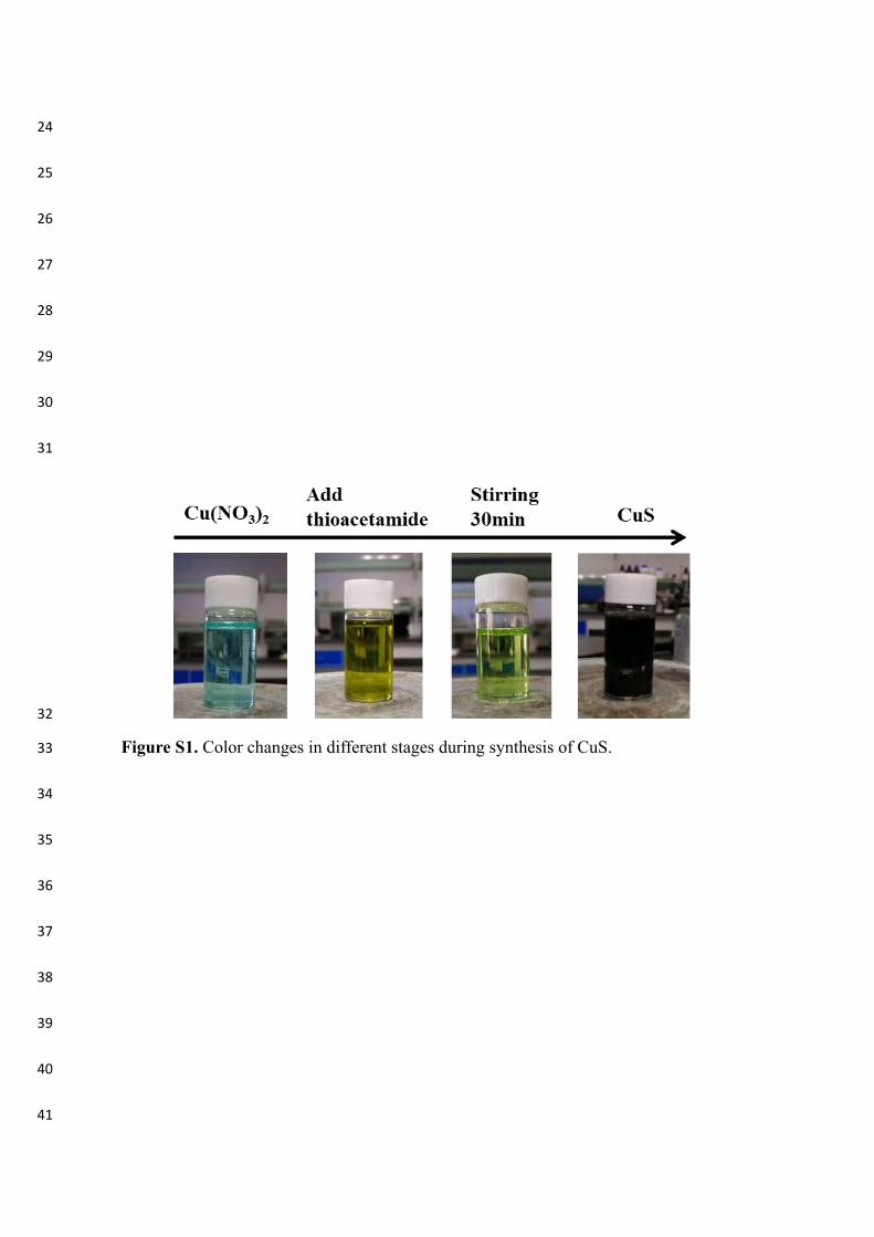
To obtain the 3D hierarchical doughnut-shaped CuS, 0.483 g of Cu(NO3)2 was added to 25mL of dimethyl sulfoxide (DMSO) and stirredvigorously to get a blue solution. 0.4 g of poly (vinyl pyrrolidone) (PVP,Mr=10,000) and 0.3 g of thioacetamide were added under vigorousstirring to get a pale green solution. The solution was transferred to a30mL Teflon-lined stainless steel autoclave that was then sealed andmaintained at 120 °C for 20 h. After the solution cooled to room tem-perature, the black solid products were collected by centrifuging, wa-shed with absolute ethanol and deionized water three times, and driedat 50 °C for 12 h. The color changes in different stages are shown in Fig.S1. The CuS particles were obtained for further use.
2.3. Synthesis of MoS2 nanomaterials
MoS2 nanomaterials, as a comparative control, were also prepared.0.565 g of Na2MoO4 and 3 g of CH4N2S were added to 30mL of deio-nized water under stirring for 30min. The solution was transferred to a30mL Teflon-lined stainless steel autoclave and heated 180 °C for 12 hto obtain MoS2.
2.4. Synthesis of CuS@ MoS2 composites
To synthesize the CuS@MoS2 composites, 0.75 g of sodium mo-lybdate and 1.5 g of thioacetamide were dissolved in 100mL of deio-nized water under stirring and 0.5 g of CuS particles were added. Thesolution was transferred to a 30mL Teflon-lined stainless steel auto-clave and heated 200 °C for 24 h. The solution cooled to room tem-perature and centrifuged at 4000 r/min. The product was washed withabsolute ethanol and deionized water three times and dried at 50 °C for12 h in a vacuum oven.
2.5. Fabrication of CuS and CuS@MoS2-incorporated hydrogels
4mL of the PVA solution prepared previously were put in 12 wells,and 10mg, 30mg and 50mg of CuS and CuS@MoS2 were introduced tosynthesize the 2.5mg/mL, 7.5mg/mL, and 12.5mg/mL CuS and CuS@MoS2-incorporated hydrogels, respectively. The mixtures were stirredfor 30min and frozen at−20 °C for 1 h. Several freezing-thawing cycleswere implemented for physical cross-linking and formation of three-dimensional porous CuS and CuS@MoS2 hydrogel.
2.6. Characterization
The morphology and microstructure of the CuS and CuS@MoS2were analyzed by field-emission scanning electron microscopy (FE-SEM, JSM-7001F, JEOL) and high-resolution transmission electronmicroscopy (HR-TEM, JEM-2100F, JEOL). The phase structure wasanalyzed by X-ray diffractometry (XRD, Shimadzu) system with Cu Kɑradiation using the continuous mode with 2θ from 20° to 70° and a stepsize of 0.02°. X-ray photoelectron spectroscopy (XPS, K-Alpha, Thermo)was used to determine the surface chemical composition of the samples.
2.7. Rheological analysis
Rheological analysis of the hydrogels were performed on a Modular
X. Zhang, et al. Chemical Engineering Journal 382 (2020) 122849
2

Compact Rheometer MCR 302 (Anton Paar Co., Austria) to study theevolution of storage (G′) and loss (G″) modulus and the measuring gapsize is 1.5 mm. The G′ and G″ were measured with the variation of thefrequency increased from 0.1 to 100 rad·s−1 (37 °C, with a shear stressof 1 Pa). The temperature dependence of G′ and G″ were detected from50 to 20 °C at a cooling rate of 5 °Cmin−1. Besides, the viscosity mea-surements as a function of shear rate (0–100 s−1) at 37 °C.
2.8. Compression test
The mechanical properties of the hybrid hydrogels were determinedby electronic universal testing machine (Shenzhen Reger experimentalinstrument Co. Ltd) at room temperature. The sample size was about20mm in diameter and 10mm in height. The compression rate was2mm/min.
2.9. Swelling behavior
To detect the swelling behavior, the hydrogels were freeze driedfirstly, then immersed in PBS at 37 °C. Afterwards, the samples were putout from PBS solution and the specific weights were recorded to con-stant values at particular time points. Results were calculated as swel-ling ratio with the equation: S= (Wt−W0)/W0, where Wt is the weightof recorded hydrogels at particular time points and W0 is the weight ofdried hydrogels.
2.10. Photothermal effects in vitro
To determine the photothermal effects of the hydrogels with dif-ferent concentrations of CuS (2.5 mg/mL, 7.5mg/mL, and 12.5 mg/mL)and CuS@MoS2 (2.5 mg/mL, 7.5mg/mL, and 12.5 mg/mL), the VL(660 nm), NIR light (808 nm), and dual lights (660 nm+808 nm) witha power of 0.6W (power density of 0.2W cm−2) were focused to a spoton the hydrogels with 1mL phosphate buffered saline (PBS), respec-tively. The blank hydrogel was the control. The total irradiation time oneach hydrogel was 15min. The temperature was detected and recordedby a FLIR thermal imaging camera.
2.11. Evaluation of ROS
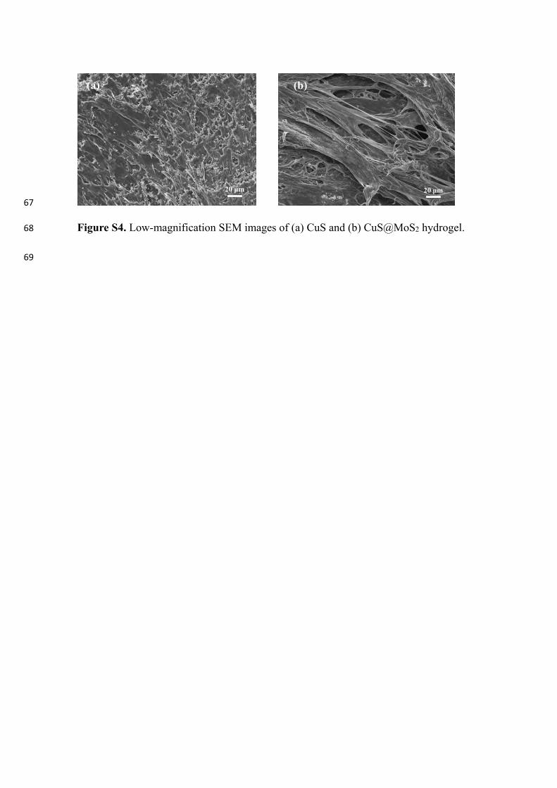
ROS, especially the hydroxyl radical (%OH) and singlet oxygen(1O2), can damage cell membranes, cellular proteins, and DNA. The %
OH measurements were carried out by UV–vis spectrophotometry (UV-1800PC) and methyl violet was used as an indicator to determine thecontent of the %OH. To reach the adsorption/desorption equilibriumprior to the test, the samples were immersed in 3mL of methyl violetsolution in darkness for 5min and then irradiated with the 660 nm,808 nm, and dual lasers. After 15min, the supernatant was measured byUV–vis spectrophotometry under light irradiation. To detect the 1O2, 1,3-diphenylisobenzofuran (DPBF) were used and the method is as de-scribed above for the %OH measurements.
2.12. Detection of glutathione (GSH)
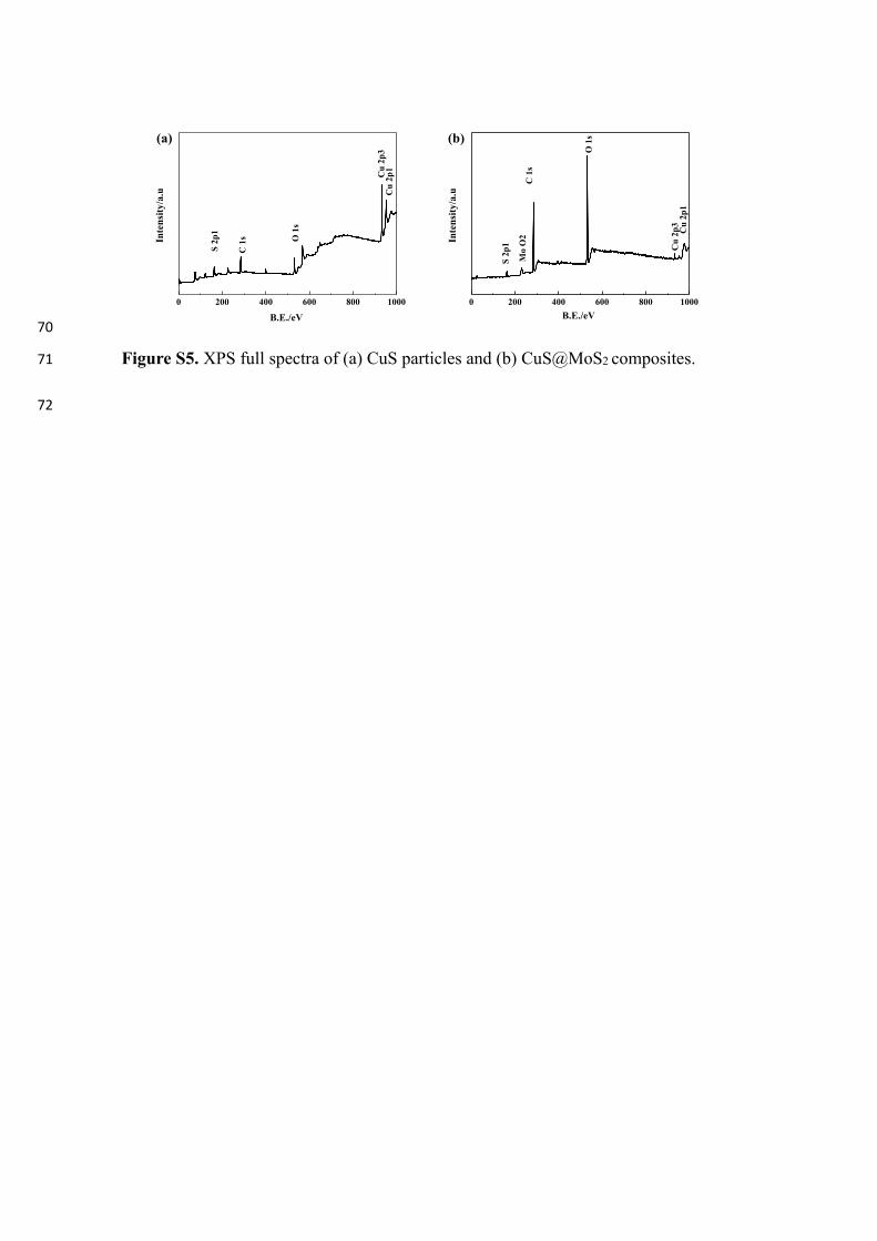
GSH is an important antioxidant and presents at high levels in manybacteria and cannot be monitored to gauge the antibacterial effect. Thehydrogels including the blank, CuS and CuS@MoS2 were put in 675 μLof the GSH bicarbonate buffer solution in a 12-well plate and irradiatedby 660 nm, 808 nm, and dual lasers, respectively. Afterwards, the GSHsolution was mixed with 2.4mL of 0.05 Tris-HCl (pH=8) solution and45 μL of 100× 10−3 M 5,5′-dithiobis (2-nitrobenzoic acid) (Ellman’sreagent) [39]. The 1× 10⁻³ M GSH solution was used as a negativecontrol. The color became lighter after irradiation and loss of GSH wasevaluated by measuring the absorbance at 410 nm by spectro-photometry.
2.13. In vitro antibacterial experiment
E. coli (Gram-negative) and S. aureus (Gram-negative) were used inthe spread plate test using a concentration of about 1.0×10⁵ CFU/mL.The experimental apparatus was sterilized in a high-pressure steamsterilizer before the experiment. 1 mL of 75% alcohol was injected ontoeach hydrogel for 20min and immersed in 1mL of PBS for 10min threetimes. Afterwards, 1 mL of the bacterial suspension in the sterile liquidmedium (1 g of peptone, 0.5 g of beef extract and 0.5 g of NaCl in100mL of deionized water) was added to 12-well plates containingdifferent types of hydrogels. Each sample was divided into four groups
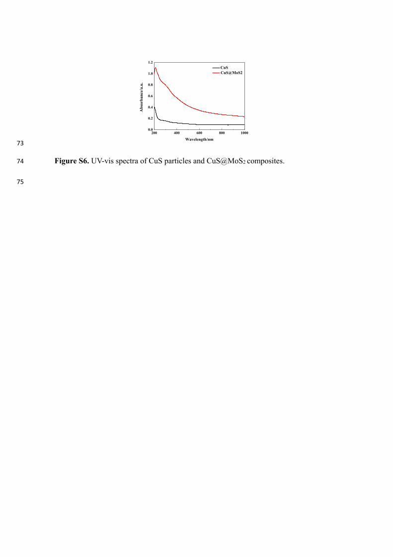
Scheme 1. The synthesis process and schematic illustration of ROS generation mechanism of CuS@MoS2 composites.
X. Zhang, et al. Chemical Engineering Journal 382 (2020) 122849
3
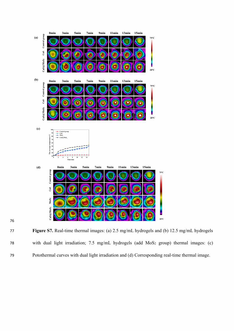
(15min by 660 nm or 808 nm laser, 15min by both lasers, and darkconditions). After irradiation, 1 mL of the bacterial liquid was taken anddiluted 100 times with the liquid medium and 60 μL of the dilutedbacterial solution were put on a culture dish covered with the solidmedium and incubated at 37 °C for 24 h. The bacteria colonies on theplates were photographed and counted and the antibacterial rate wascalculated according to Eq. (1) (N is the number of colonies):Antibacterial ration= (N of the control group-N of the experimentalgroup)/N of the control group×100%. The antibacterial activity assaywas also evaluated by SEM. The bacteria suspensions were put on asilicon wafer, cultured for 4 h, fixed with 3% glutaraldehyde for 2 h,dehydrated in graded ethanol sequentially (20–100%) for 15min, anddried at room temperature. Before imaging, the bacteria were sputter-coated with platinum.
2.14. Cell culture
To detect the biocompatibilities of samples, ECs (EA.hy926) wereused for next series of cell experiments. They were purchased from thecell bank of Shanghai Chinese Academy of Sciences (Catalog number:GNM15 and GNHu39). Based on the instructions of supplier, cells werecultured in Dulbecco’s Modified Eagle Medium (DMEM, Gibco,America) consisting with 10% of FBS and 1% of an anti-biotic–antimycotic solution composed of 100 units/mL penicillin(Weifang Pharmaceutical Co., Ltd., China) and 100mg/mL strepto-mycin (Lukang Pharmaceutical Co., Ltd., China). The external en-vironment of cell culturing in incubator included 37 °C of temperatureand 5% CO2 of atmosphere. When cellular status reached sub-con-fluence, 0.25% trypsin was used to collect and reseeded cells to furtherstudies as cell proliferation, cytotoxicity, and functions. All experimentswere evaluated slides at a density of 1×10⁴ cells/cm2 and the culturemedium was replenished every day.
2.15. Cytotoxicity assay
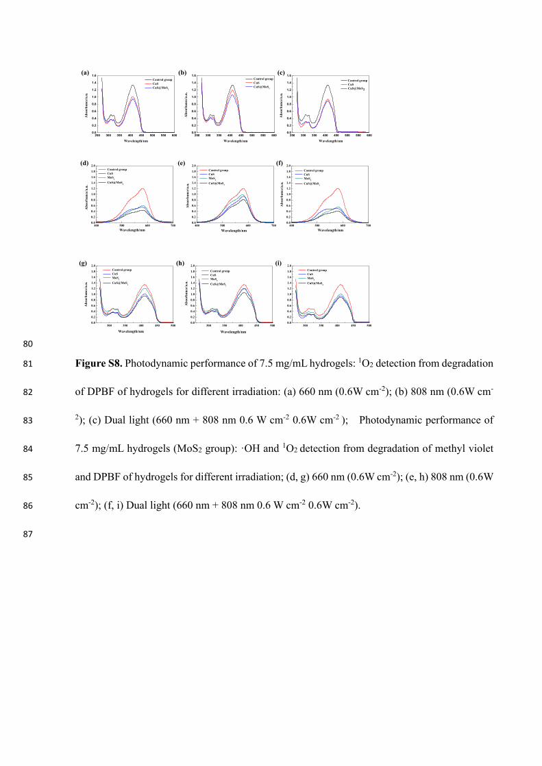
The live/dead viability/cytotoxicity kit for mammalian cells(Invitrogen) was used in the cytotoxicity assay of the different types ofhydrogels according to the manufacturer’s instruction to quantify thecell viability on the blank (group I), 0.75mg/mL CuS hydrogel (groupII), and 0.75mg/mL CuS@MoS2 hydrogel (group III). The samples weresterilized with 75% alcohol for 30min and rinsed with PBS 3 timesbefore 1mL of the complete medium was put onto the samples. Afterincubation for 24 h, 1mL of the complete medium immersed with thesamples was mixed with the cells on a 24-well plate. After culturing for1 and 2 days, the medium was removed and the cell slides were washedthrice with PBS. 50 μL of the stain solution containing 0.5 μL/mL cal-cein AM and 2 μL/mL ethidium homodimer (EthD-1) in PBS were puton the surface and incubated at 37 °C for 1 h in darkness. Finally, thecell slides were washed with PBS and photographed by CLSM to iden-tify the live cells (stained green with calcein AM) and dead ones(stained red with EthD-1). The same method was adopted to evaluatethe cytotoxicity of endothelial cells.
2.16. Cell proliferation assay
In the MTT assay, cell proliferation was evaluated by the 3-(4, 5-dimethylthiazol-2-yl)-2,5-diphenyl tetrazolium bromide (Sigma, USA).After culturing 1 and 2 days, the medium was removed and the cellslides were washed thrice with PBS. On each well, it was added 100 μLof the MTT solution (5mg/mL MTT in PBS) and 900 μL of fresh DMEMculture medium. After cultured with 4 h in incubator, blue formazanformed on the well. Then 100 μL of the DMSO solution was added todissolve formazan to form homogeneous solution. At a wavelength of570 nm, the optical density (OD) value of each sample was measured bymicroplate reader (Infinite F50, TECAN). Three specimens from eachgroup were tested.
2.17. ELISA for VEGF and HIF-1α secretion
To determine the influence of the CuS and CuS@MoS2-incorporatedhydrogels on protein expressions of VEGF and HIF-1α, the VEGF andHIF-1α enzyme-linked immunosorbent assay (ELISA) kits (Abcam, UK)were employed. The endothelial cells were seeded on a 24-well plate ata density of 1×10⁴ per well and cultured with the three different typesof cells with the culture media. According to the manufacturer's spe-cification, after the endothelial cells were seeded on the samples for24 h, the supernatants of the medium were taken out and centrifuged.The results were expressed by the concentrations ere determined from astandard curve as the amount (pg) of VEGF and HIF-1α per mL su-pernatant. The experiments were performed in triplicate.
2.18. In vivo experiment
The male rats (300–350 g body weight) were obtained from ShanxiProvince Center for Disease Prevention & Control and all the animalexperiments and the procedures were approved by the ShanxiProvincial Center for Disease Prevention & Control. The animals weremaintained in accordance with the Animal Management Rules of theMinistry of Health of the People’s Republic of China and the Guidelinesfor the Care and Use of Laboratory Animals of China. The rats wereindividually kept in cages at a standard temperature and divided into 4groups: sterile wound dressing (group I, conventional wound therapy,Qingdao Hainuo Biological Engineering Co., Ltd), blank hydrogel(group II), CuS hydrogel (group III), and CuS@MoS2 hydrogel (groupIV). Anesthesia was performed with 1% sodium pentobarbital and usinghair removal cream, the dorsal area of the rat was depilated to createsquare wounds (length of about 10mm) and treated with differenthydrogels. Afterwards, 50 μL of the diluted bacterial suspension (107
CFU mL−1) were added to the wound for 5min and then treated withsterile wound dressing, blank hydrogel, CuS hydrogel, and CuS@MoS2hydrogel, respectively. Each group was irradiated with dual lasers for15min (except group I) and the wounds were wrapped by sterilemedical tapes. The rats were individually kept in cages at a standardtemperature. Each dressing was changed every 2 days for a total of14 days. On day 2, 6, and 14, the wound condition was observed andphotographed. Meanwhile, on day 2, 4, and 6, 20 μL of the exudatewere collected from the wound and diluted 100 times to spread onplates and incubated at 37 °C for 24 h to form viable colony units. Therats were sacrificed on day 14 and the skin tissue around the wound washarvested and fixed with 4% paraformaldehyde. The central woundsections were fixed on glass slides and stained with Giemsa stain on day2 and hematoxylin and eosin (H&E) on day 14. Giemsa and H&Estaining were used to evaluate the type and amount of adherent bac-teria around the wound and healing progress, respectively. The majororgans including the heart, liver, spleen, lung, and kidney were har-vested and stained with H&E after 14 days.
2.19. Statistical analysis
All the experiments were performed in triplicate and the resultswere presented as mean ± standard deviation based on the SPSS 14.0software. One-way ANOVA combined with a Student–Newman–Keuls(SNK) post hoc test was utilized to determine the level of significance.
3. Results
3.1. Structure characterization
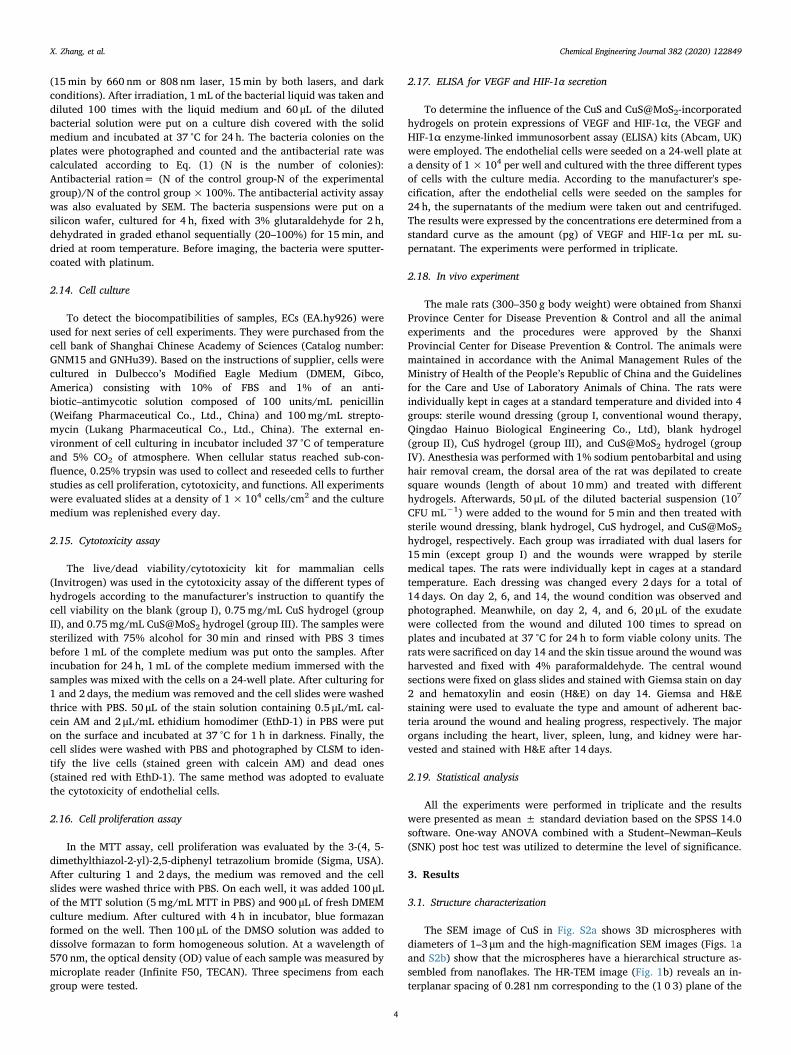
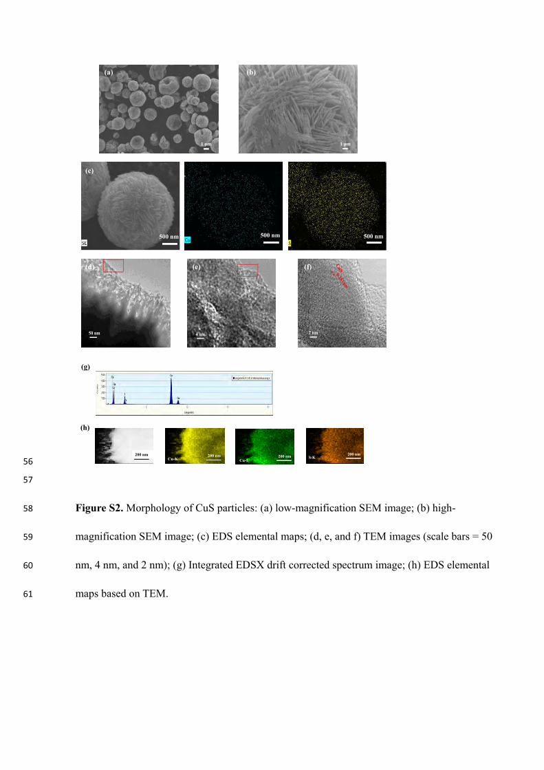
The SEM image of CuS in Fig. S2a shows 3D microspheres withdiameters of 1–3 μm and the high-magnification SEM images (Figs. 1aand S2b) show that the microspheres have a hierarchical structure as-sembled from nanoflakes. The HR-TEM image (Fig. 1b) reveals an in-terplanar spacing of 0.281 nm corresponding to the (1 0 3) plane of the
X. Zhang, et al. Chemical Engineering Journal 382 (2020) 122849
4
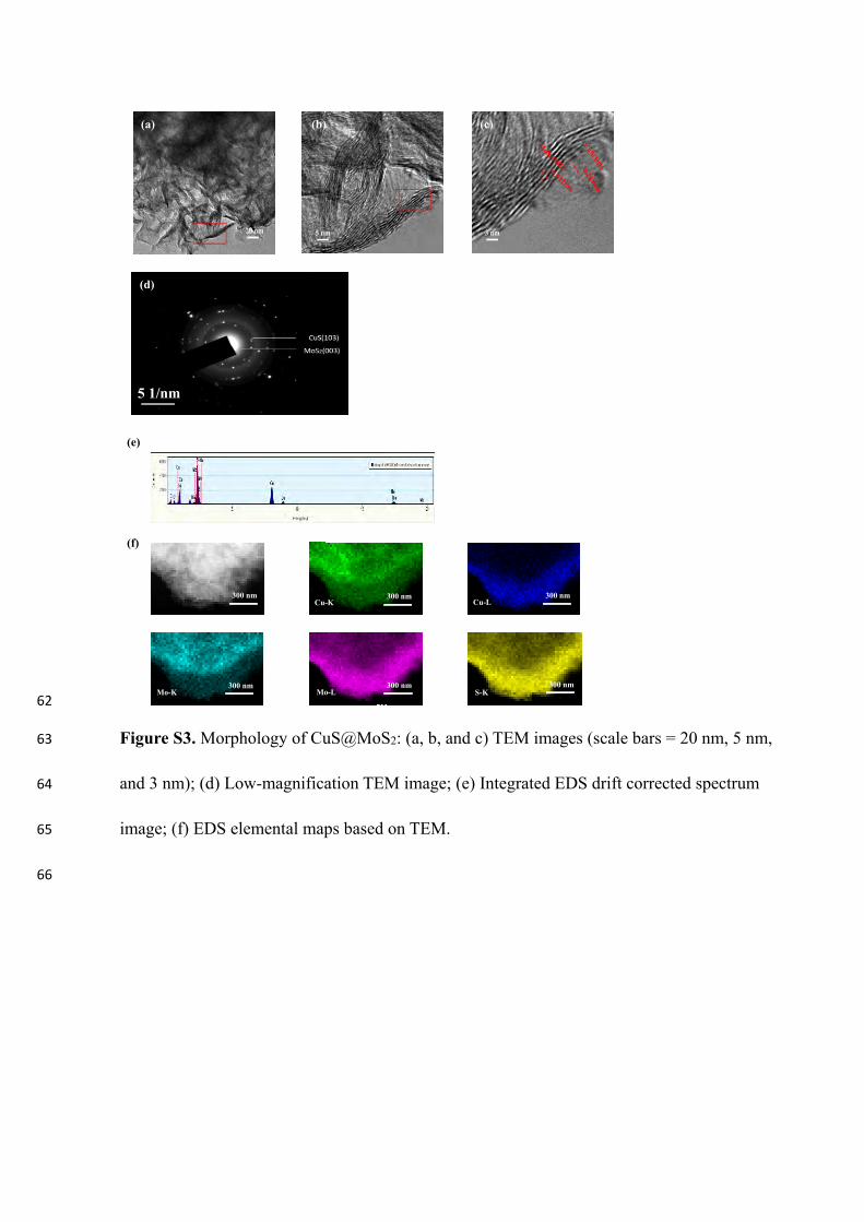
CuS microspheres. The elemental maps in Fig. S2c indicate uniformdistributions of Cu and S and Fig. S2d shows voids in the microspheresand uniform Cu and S distributions around the microspheres. Fig. 1cshows the morphology of CuS@MoS2 composite microspheres. Com-pared with CuS, the 3D microspheres have a coarse surface and thenanoflakes are thick. Figs. S3 and 1d indicate that the ultrathin MoS2nanosheets are distributed uniformly around the CuS@MoS2 micro-spheres and the lattice spacings of 0.281 and 0.612 nm in the inter-connected regions can be attributed to the (1 0 3) face-centered cubicCuS and (0 0 2) of MoS2, respectively. Fig. 1e reveals even distributionsof Cu, S and Mo over the CuS@MoS2 microspheres consistent with theelemental maps (Fig. S3f). Voids also exist in the CuS@MoS2 micro-spheres and the selected-area electron diffraction patterns (SAED) (Fig.
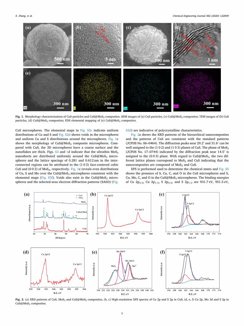
S3d) are indicative of polycrystalline characteristics.Fig. 2a shows the XRD patterns of the hierarchical nanocomposites
and the patterns of CuS are consistent with the standard patterns(JCPDS No. 06–0464). The diffraction peaks near 29.2° and 31.6° can bewell assigned to the (1 0 2) and (1 0 3) planes of CuS. The phase of MoS2(JCPDS No. 17–0744) indicated by the diffraction peak near 14.5° isassigned to the (0 0 3) plane. With regard to CuS@MoS2, the two dif-ferent lattice planes correspond to MoS2 and CuS indicating that thenanocomposites are composed of MoS2 and CuS.
XPS is performed used to determine the chemical states and Fig. S5shows the presence of S, Cu, C, and O in the CuS microspheres and S,Cu, Mo, C, and O in the CuS@MoS2 microspheres. The binding energiesof Cu 2p3/2, Cu 2p1/2, S 2p3/2, and S 2p1/2 are 931.7 eV, 951.5 eV,
Fig. 1. Morphology characterization of CuS particles and CuS@MoS2 composites. SEM images of (a) CuS particles, (c) CuS@MoS2 composites; TEM images of (b) CuSparticles, (d) CuS@MoS2 composites; EDS elemental mapping of (e) CuS@MoS2 composites.
Fig. 2. (a) XRD patterns of CuS, MoS2 and CuS@MoS2 composites, (b, c) High-resolution XPS spectra of Cu 2p and S 2p in CuS; (d, e, f) Cu 2p, Mo 3d and S 2p inCuS@MoS2 composites.
X. Zhang, et al. Chemical Engineering Journal 382 (2020) 122849
5
161.8 eV, and 162.8 eV, respectively, confirming the presence of CuS inCuS microspheres (Fig. 2b and c). As for CuS@MoS2, the high-resolu-tion spectra of Cu 2p and S 2p illustrate the presence of CuS. The twopeaks at 227.0 eV and 232.2 eV in the high-resolution spectra of Mo 3dcan be assigned to Mo 3d3/2 and Mo 3d5/2 for the Mo (IV) oxidationstate and together with the S 2p spectra validate the formation of MoS2in the CuS@MoS2 microspheres (Fig. 2d-2f).
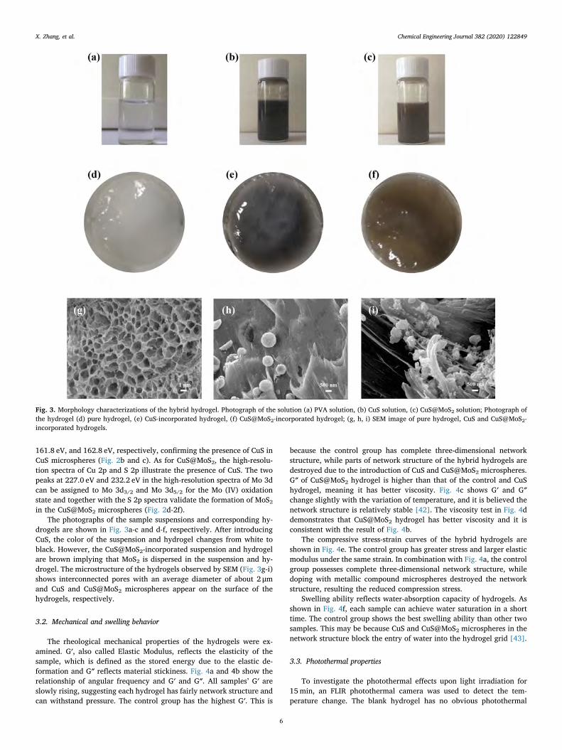
The photographs of the sample suspensions and corresponding hy-drogels are shown in Fig. 3a-c and d-f, respectively. After introducingCuS, the color of the suspension and hydrogel changes from white toblack. However, the CuS@MoS2-incorporated suspension and hydrogelare brown implying that MoS2 is dispersed in the suspension and hy-drogel. The microstructure of the hydrogels observed by SEM (Fig. 3g-i)shows interconnected pores with an average diameter of about 2 μmand CuS and CuS@MoS2 microspheres appear on the surface of thehydrogels, respectively.
3.2. Mechanical and swelling behavior
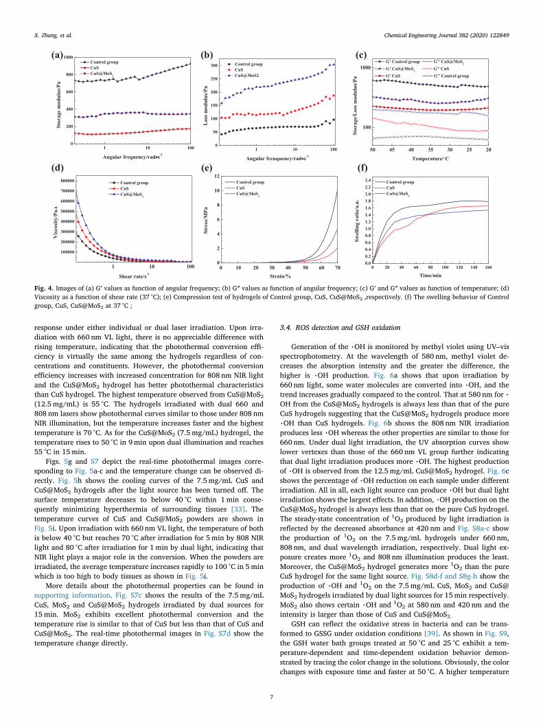
The rheological mechanical properties of the hydrogels were ex-amined. G′, also called Elastic Modulus, reflects the elasticity of thesample, which is defined as the stored energy due to the elastic de-formation and G″ reflects material stickiness. Fig. 4a and 4b show therelationship of angular frequency and G′ and G″. All samples’ G′ areslowly rising, suggesting each hydrogel has fairly network structure andcan withstand pressure. The control group has the highest G′. This is
because the control group has complete three-dimensional networkstructure, while parts of network structure of the hybrid hydrogels aredestroyed due to the introduction of CuS and CuS@MoS2 microspheres.G″ of CuS@MoS2 hydrogel is higher than that of the control and CuShydrogel, meaning it has better viscosity. Fig. 4c shows G′ and G″change slightly with the variation of temperature, and it is believed thenetwork structure is relatively stable [42]. The viscosity test in Fig. 4ddemonstrates that CuS@MoS2 hydrogel has better viscosity and it isconsistent with the result of Fig. 4b.
The compressive stress-strain curves of the hybrid hydrogels areshown in Fig. 4e. The control group has greater stress and larger elasticmodulus under the same strain. In combination with Fig. 4a, the controlgroup possesses complete three-dimensional network structure, whiledoping with metallic compound microspheres destroyed the networkstructure, resulting the reduced compression stress.
Swelling ability reflects water-absorption capacity of hydrogels. Asshown in Fig. 4f, each sample can achieve water saturation in a shorttime. The control group shows the best swelling ability than other twosamples. This may be because CuS and CuS@MoS2 microspheres in thenetwork structure block the entry of water into the hydrogel grid [43].
3.3. Photothermal properties
To investigate the photothermal effects upon light irradiation for15min, an FLIR photothermal camera was used to detect the tem-perature change. The blank hydrogel has no obvious photothermal
Fig. 3. Morphology characterizations of the hybrid hydrogel. Photograph of the solution (a) PVA solution, (b) CuS solution, (c) CuS@MoS2 solution; Photograph ofthe hydrogel (d) pure hydrogel, (e) CuS-incorporated hydrogel, (f) CuS@MoS2-incorporated hydrogel; (g, h, i) SEM image of pure hydrogel, CuS and CuS@MoS2-incorporated hydrogels.
X. Zhang, et al. Chemical Engineering Journal 382 (2020) 122849
6
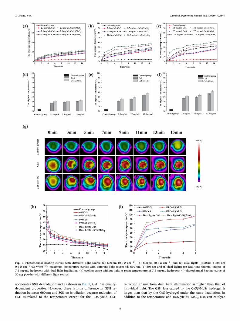
response under either individual or dual laser irradiation. Upon irra-diation with 660 nm VL light, there is no appreciable difference withrising temperature, indicating that the photothermal conversion effi-ciency is virtually the same among the hydrogels regardless of con-centrations and constituents. However, the photothermal conversionefficiency increases with increased concentration for 808 nm NIR lightand the CuS@MoS2 hydrogel has better photothermal characteristicsthan CuS hydrogel. The highest temperature observed from CuS@MoS2(12.5 mg/mL) is 55 °C. The hydrogels irradiated with dual 660 and808 nm lasers show photothermal curves similar to those under 808 nmNIR illumination, but the temperature increases faster and the highesttemperature is 70 °C. As for the CuS@MoS2 (7.5 mg/mL) hydrogel, thetemperature rises to 50 °C in 9min upon dual illumination and reaches55 °C in 15min.
Figs. 5g and S7 depict the real-time photothermal images corre-sponding to Fig. 5a-c and the temperature change can be observed di-rectly. Fig. 5h shows the cooling curves of the 7.5mg/mL CuS andCuS@MoS2 hydrogels after the light source has been turned off. Thesurface temperature decreases to below 40 °C within 1min conse-quently minimizing hyperthermia of surrounding tissues [33]. Thetemperature curves of CuS and CuS@MoS2 powders are shown inFig. 5i. Upon irradiation with 660 nm VL light, the temperature of bothis below 40 °C but reaches 70 °C after irradiation for 5min by 808 NIRlight and 80 °C after irradiation for 1min by dual light, indicating thatNIR light plays a major role in the conversion. When the powders areirradiated, the average temperature increases rapidly to 100 °C in 5minwhich is too high to body tissues as shown in Fig. 5i.
More details about the photothermal properties can be found insupporting information. Fig. S7c shows the results of the 7.5mg/mLCuS, MoS2 and CuS@MoS2 hydrogels irradiated by dual sources for15min. MoS2 exhibits excellent photothermal conversion and thetemperature rise is similar to that of CuS but less than that of CuS andCuS@MoS2. The real-time photothermal images in Fig. S7d show thetemperature change directly.
3.4. ROS detection and GSH oxidation
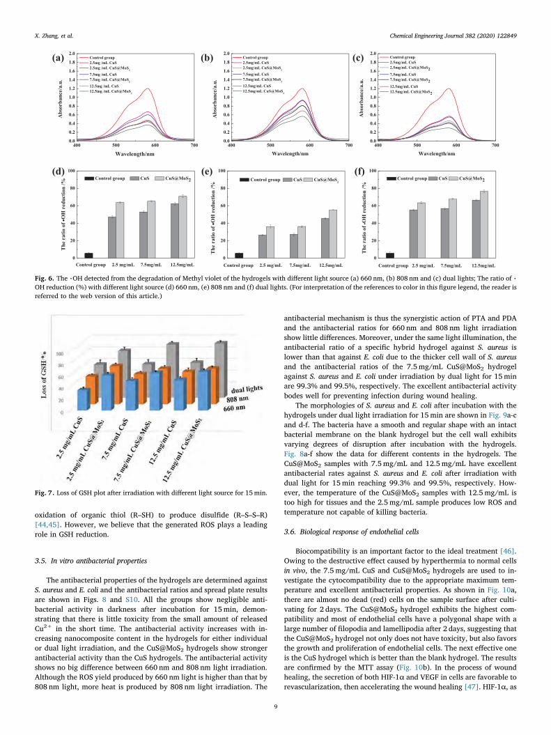
Generation of the %OH is monitored by methyl violet using UV–visspectrophotometry. At the wavelength of 580 nm, methyl violet de-creases the absorption intensity and the greater the difference, thehigher is %OH production. Fig. 6a shows that upon irradiation by660 nm light, some water molecules are converted into %OH, and thetrend increases gradually compared to the control. That at 580 nm for %OH from the CuS@MoS2 hydrogels is always less than that of the pureCuS hydrogels suggesting that the CuS@MoS2 hydrogels produce more%OH than CuS hydrogels. Fig. 6b shows the 808 nm NIR irradiationproduces less %OH whereas the other properties are similar to those for660 nm. Under dual light irradiation, the UV absorption curves showlower vertexes than those of the 660 nm VL group further indicatingthat dual light irradiation produces more %OH. The highest productionof %OH is observed from the 12.5mg/mL CuS@MoS2 hydrogel. Fig. 6cshows the percentage of %OH reduction on each sample under differentirradiation. All in all, each light source can produce %OH but dual lightirradiation shows the largest effects. In addition, %OH production on theCuS@MoS2 hydrogel is always less than that on the pure CuS hydrogel.The steady-state concentration of 1O2 produced by light irradiation isreflected by the decreased absorbance at 420 nm and Fig. S8a-c showthe production of 1O2 on the 7.5 mg/mL hydrogels under 660 nm,808 nm, and dual wavelength irradiation, respectively. Dual light ex-posure creates more 1O2 and 808 nm illumination produces the least.Moreover, the CuS@MoS2 hydrogel generates more 1O2 than the pureCuS hydrogel for the same light source. Fig. S8d-f and S8g-h show theproduction of %OH and 1O2 on the 7.5 mg/mL CuS, MoS2 and CuS@MoS2 hydrogels irradiated by dual light sources for 15min respectively.MoS2 also shows certain %OH and 1O2 at 580 nm and 420 nm and theintensity is larger than those of CuS and CuS@MoS2.
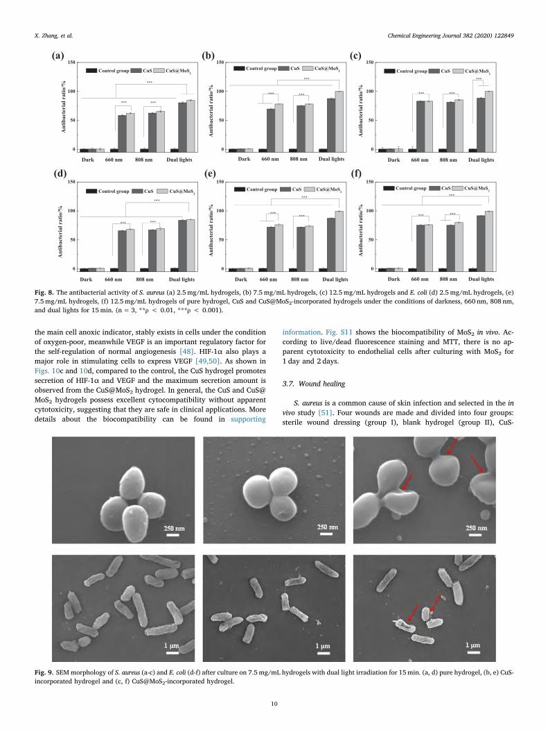
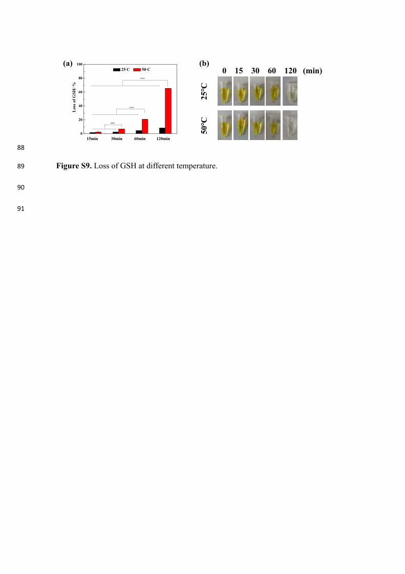
GSH can reflect the oxidative stress in bacteria and can be trans-formed to GSSG under oxidation conditions [39]. As shown in Fig. S9,the GSH water bath groups treated at 50 °C and 25 °C exhibit a tem-perature-dependent and time-dependent oxidation behavior demon-strated by tracing the color change in the solutions. Obviously, the colorchanges with exposure time and faster at 50 °C. A higher temperature
Fig. 4. Images of (a) G′ values as function of angular frequency; (b) G″ values as function of angular frequency; (c) G′ and G″ values as function of temperature; (d)Viscosity as a function of shear rate (37 °C); (e) Compression test of hydrogels of Control group, CuS, CuS@MoS2 ,respectively. (f) The swelling behavior of Controlgroup, CuS, CuS@MoS2 at 37 °C ;
X. Zhang, et al. Chemical Engineering Journal 382 (2020) 122849
7
accelerates GSH degradation and as shown in Fig. 7, GSH has quality-dependent properties. However, there is little difference in GSH re-duction between 660 nm and 808 nm irradiation because reduction ofGSH is related to the temperature except for the ROS yield. GSH
reduction arising from dual light illumination is higher than that ofindividual light. The GSH loss caused by the CuS@MoS2 hydrogel islarger than that by the CuS hydrogel under the same irradiation. Inaddition to the temperature and ROS yields, MoS2 also can catalyze
Fig. 5. Photothermal heating curves with different light source (a) 660 nm (0.6W cm−2), (b) 808 nm (0.6W cm−2) and (c) dual lights ((660 nm+808 nm0.6W cm−2 0.6W cm−2); maximum temperature curves with different light source (d) 660 nm, (e) 808 nm and (f) dual lights; (g) Real-time thermal images of7.5 mg/mL hydrogels with dual light irradiation; (h) cooling curve without light at room temperature of 7.5mg/mL hydrogels; (i) photothermal heating curve of30mg powder with different light source.
X. Zhang, et al. Chemical Engineering Journal 382 (2020) 122849
8
oxidation of organic thiol (R–SH) to produce disulfide (R–S–S–R)[44,45]. However, we believe that the generated ROS plays a leadingrole in GSH reduction.
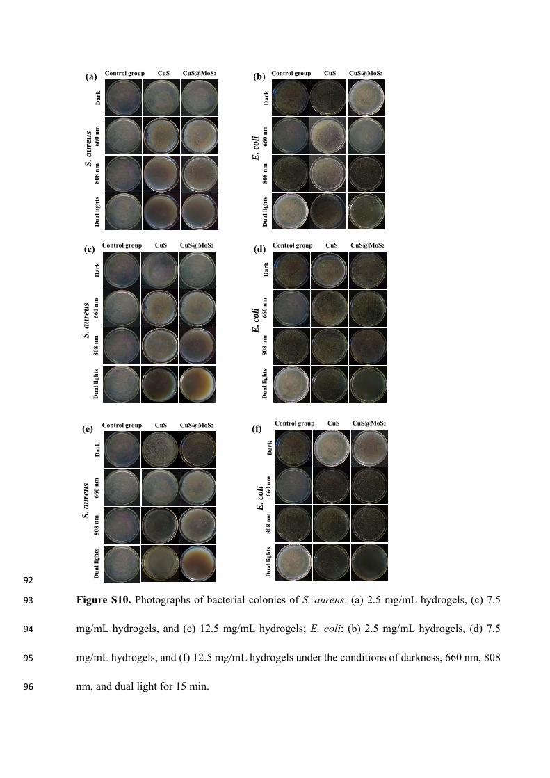
3.5. In vitro antibacterial properties
The antibacterial properties of the hydrogels are determined againstS. aureus and E. coli and the antibacterial ratios and spread plate resultsare shown in Figs. 8 and S10. All the groups show negligible anti-bacterial activity in darkness after incubation for 15min, demon-strating that there is little toxicity from the small amount of releasedCu2+ in the short time. The antibacterial activity increases with in-creasing nanocomposite content in the hydrogels for either individualor dual light irradiation, and the CuS@MoS2 hydrogels show strongerantibacterial activity than the CuS hydrogels. The antibacterial activityshows no big difference between 660 nm and 808 nm light irradiation.Although the ROS yield produced by 660 nm light is higher than that by808 nm light, more heat is produced by 808 nm light irradiation. The
antibacterial mechanism is thus the synergistic action of PTA and PDAand the antibacterial ratios for 660 nm and 808 nm light irradiationshow little differences. Moreover, under the same light illumination, theantibacterial ratio of a specific hybrid hydrogel against S. aureus islower than that against E. coli due to the thicker cell wall of S. aureusand the antibacterial ratios of the 7.5mg/mL CuS@MoS2 hydrogelagainst S. aureus and E. coli under irradiation by dual light for 15minare 99.3% and 99.5%, respectively. The excellent antibacterial activitybodes well for preventing infection during wound healing.
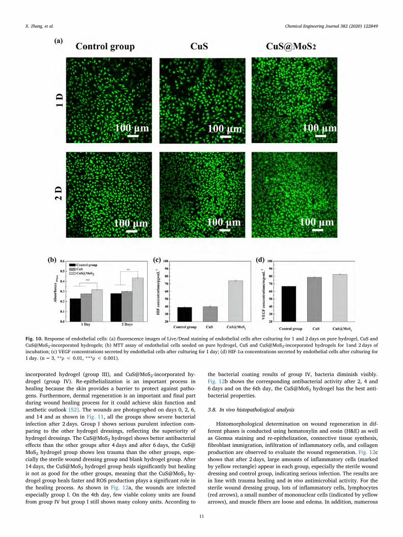
The morphologies of S. aureus and E. coli after incubation with thehydrogels under dual light irradiation for 15min are shown in Fig. 9a-cand d-f. The bacteria have a smooth and regular shape with an intactbacterial membrane on the blank hydrogel but the cell wall exhibitsvarying degrees of disruption after incubation with the hydrogels.Fig. 8a-f show the data for different contents in the hydrogels. TheCuS@MoS2 samples with 7.5 mg/mL and 12.5 mg/mL have excellentantibacterial rates against S. aureus and E. coli after irradiation withdual light for 15min reaching 99.3% and 99.5%, respectively. How-ever, the temperature of the CuS@MoS2 samples with 12.5mg/mL istoo high for tissues and the 2.5 mg/mL sample produces low ROS andtemperature not capable of killing bacteria.
3.6. Biological response of endothelial cells
Biocompatibility is an important factor to the ideal treatment [46].Owing to the destructive effect caused by hyperthermia to normal cellsin vivo, the 7.5 mg/mL CuS and CuS@MoS2 hydrogels are used to in-vestigate the cytocompatibility due to the appropriate maximum tem-perature and excellent antibacterial properties. As shown in Fig. 10a,there are almost no dead (red) cells on the sample surface after culti-vating for 2 days. The CuS@MoS2 hydrogel exhibits the highest com-patibility and most of endothelial cells have a polygonal shape with alarge number of filopodia and lamellipodia after 2 days, suggesting thatthe CuS@MoS2 hydrogel not only does not have toxicity, but also favorsthe growth and proliferation of endothelial cells. The next effective oneis the CuS hydrogel which is better than the blank hydrogel. The resultsare confirmed by the MTT assay (Fig. 10b). In the process of woundhealing, the secretion of both HIF-1α and VEGF in cells are favorable torevascularization, then accelerating the wound healing [47]. HIF-1α, as
Fig. 6. The %OH detected from the degradation of Methyl violet of the hydrogels with different light source (a) 660 nm, (b) 808 nm and (c) dual lights; The ratio of %OH reduction (%) with different light source (d) 660 nm, (e) 808 nm and (f) dual lights. (For interpretation of the references to color in this figure legend, the reader isreferred to the web version of this article.)
Fig. 7. Loss of GSH plot after irradiation with different light source for 15min.
X. Zhang, et al. Chemical Engineering Journal 382 (2020) 122849
9
the main cell anoxic indicator, stably exists in cells under the conditionof oxygen-poor, meanwhile VEGF is an important regulatory factor forthe self-regulation of normal angiogenesis [48]. HIF-1α also plays amajor role in stimulating cells to express VEGF [49,50]. As shown inFigs. 10c and 10d, compared to the control, the CuS hydrogel promotessecretion of HIF-1α and VEGF and the maximum secretion amount isobserved from the CuS@MoS2 hydrogel. In general, the CuS and CuS@MoS2 hydrogels possess excellent cytocompatibility without apparentcytotoxicity, suggesting that they are safe in clinical applications. Moredetails about the biocompatibility can be found in supporting
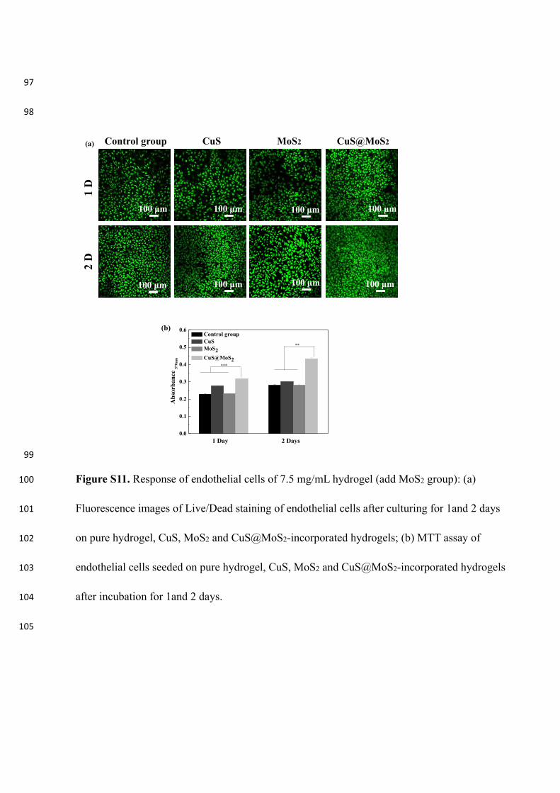
information. Fig. S11 shows the biocompatibility of MoS2 in vivo. Ac-cording to live/dead fluorescence staining and MTT, there is no ap-parent cytotoxicity to endothelial cells after culturing with MoS2 for1 day and 2 days.
3.7. Wound healing
S. aureus is a common cause of skin infection and selected in the invivo study [51]. Four wounds are made and divided into four groups:sterile wound dressing (group I), blank hydrogel (group II), CuS-
Fig. 8. The antibacterial activity of S. aureus (a) 2.5mg/mL hydrogels, (b) 7.5 mg/mL hydrogels, (c) 12.5 mg/mL hydrogels and E. coli (d) 2.5 mg/mL hydrogels, (e)7.5 mg/mL hydrogels, (f) 12.5 mg/mL hydrogels of pure hydrogel, CuS and CuS@MoS2-incorporated hydrogels under the conditions of darkness, 660 nm, 808 nm,and dual lights for 15min. (n=3, **ρ < 0.01, ***ρ < 0.001).
Fig. 9. SEM morphology of S. aureus (a-c) and E. coli (d-f) after culture on 7.5 mg/mL hydrogels with dual light irradiation for 15min. (a, d) pure hydrogel, (b, e) CuS-incorporated hydrogel and (c, f) CuS@MoS2-incorporated hydrogel.
X. Zhang, et al. Chemical Engineering Journal 382 (2020) 122849
10
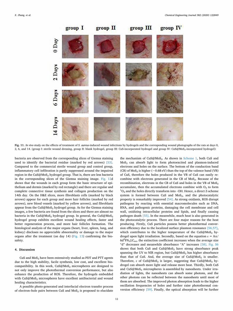
incorporated hydrogel (group III), and CuS@MoS2-incorporated hy-drogel (group IV). Re-epithelialization is an important process inhealing because the skin provides a barrier to protect against patho-gens. Furthermore, dermal regeneration is an important and final partduring wound healing process for it could achieve skin function andaesthetic outlook [52]. The wounds are photographed on days 0, 2, 6,and 14 and as shown in Fig. 11, all the groups show severe bacterialinfection after 2 days. Group I shows serious purulent infection com-paring to the other hydrogel dressings, reflecting the superiority ofhydrogel dressings. The CuS@MoS2 hydrogel shows better antibacterialeffects than the other groups after 4 days and after 6 days, the CuS@MoS2 hydrogel group shows less trauma than the other groups, espe-cially the sterile wound dressing group and blank hydrogel group. After14 days, the CuS@MoS2 hydrogel group heals significantly but healingis not as good for the other groups, meaning that the CuS@MoS2 hy-drogel group heals faster and ROS production plays a significant role inthe healing process. As shown in Fig. 12a, the wounds are infectedespecially group I. On the 4th day, few viable colony units are foundfrom group IV but group I still shows many colony units. According to
the bacterial coating results of group IV, bacteria diminish visibly.Fig. 12b shows the corresponding antibacterial activity after 2, 4 and6 days and on the 6th day, the CuS@MoS2 hydrogel has the best anti-bacterial properties.
3.8. In vivo histopathological analysis
Histomorphological determination on wound regeneration in dif-ferent phases is conducted using hematoxylin and eosin (H&E) as wellas Giemsa staining and re-epithelization, connective tissue synthesis,fibroblast immigration, infiltration of inflammatory cells, and collagenproduction are observed to evaluate the wound regeneration. Fig. 12cshows that after 2 days, large amounts of inflammatory cells (markedby yellow rectangle) appear in each group, especially the sterile wounddressing and control group, indicating serious infection. The results arein line with trauma healing and in vivo antimicrobial activity. For thesterile wound dressing group, lots of inflammatory cells, lymphocytes(red arrows), a small number of mononuclear cells (indicated by yellowarrows), and muscle fibers are loose and edema. In addition, numerous
Fig. 10. Response of endothelial cells: (a) fluorescence images of Live/Dead staining of endothelial cells after culturing for 1 and 2 days on pure hydrogel, CuS andCuS@MoS2-incorporated hydrogels; (b) MTT assay of endothelial cells seeded on pure hydrogel, CuS and CuS@MoS2-incorporated hydrogels for 1and 2 days ofincubation; (c) VEGF concentrations secreted by endothelial cells after culturing for 1 day; (d) HIF-1α concentrations secreted by endothelial cells after culturing for1 day. (n= 3, **ρ < 0.01, ***ρ < 0.001).
X. Zhang, et al. Chemical Engineering Journal 382 (2020) 122849
11
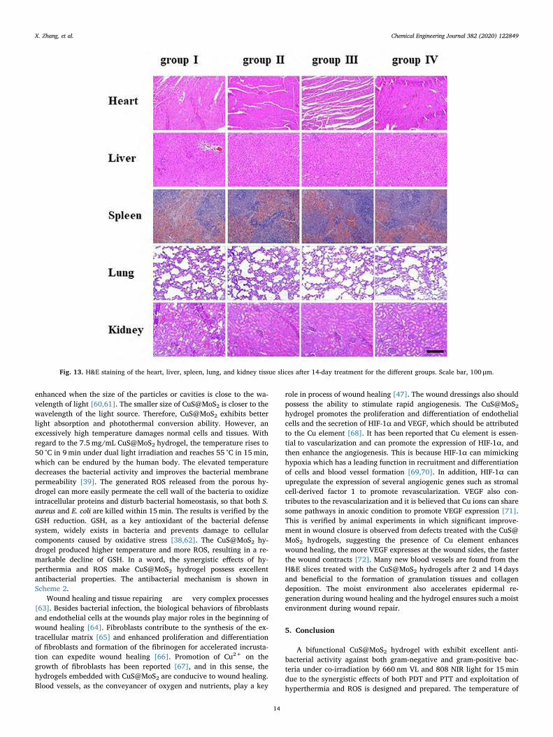
bacteria are observed from the corresponding slices of Giemsa stainingused to identify the bacterial residue (marked by red arrows) [53].Compared to the commercial sterile wound group and control group,inflammatory cell infiltration is partly suppressed around the impairedregion in the CuS@MoS2 hydrogel group. That is, there are less bacteriain the corresponding slices of the Giemsa staining image. Fig. 12dshows that the wounds in each group form the basic structure of epi-thelium and dermis (marked by red rectangle) and there are regular andcomplete connective tissue synthesis and collagen production on the14th day. On the H&E slices, more fibroblasts cells (marked by blackarrows) appear for each group and more hair follicles (marked by redarrows), new blood vessels (marked by yellow arrows), and fibroblastsappear from the CuS@MoS2 hydrogel group. As for the Giemsa stainingimages, a few bacteria are found from the slices and there are almost nobacteria in the CuS@MoS2 hydrogel group. In general, the CuS@MoS2hydrogel group exhibits excellent wound healing effects, faster andbetter regeneration process, and more hair follicles formation. Thehistological analysis of the major organs (heart, liver, spleen, lung, andkidney) discloses no appreciable abnormality or damage to the majororgans after the treatment (on day 14) (Fig. 13) confirming the bio-safety.
4. Discussion
CuS and MoS2 have been extensively studied as PDT and PTT agentsdue to the high stability, facile synthesis, low cost, and excellent bio-compatibility. In this work, CuS@MoS2 microspheres are designed tonot only improve the photothermal conversion performance, but alsoenhance the production of ROS. Therefore, the hydrogels embeddedwith CuS@MoS2 microspheres have excellent antibacterial and woundhealing characteristics.
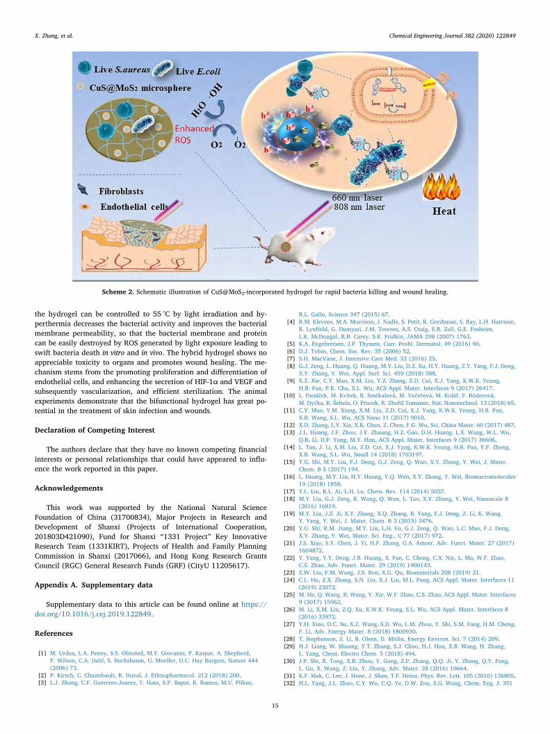
A possible photo-generated and interfacial electron transfer processof electron–hole pairs between CuS and MoS2 is proposed to elucidate
the mechanism of CuS@MoS2. As shown in Scheme 1, both CuS andMoS2 can absorb light to form photoexcited and plasmon-inducedelectrons and holes on the surface. The bottom of the conduction band(CB) of MoS2 is higher (~0.68 eV) than the top of the valence band (VB)of CuS, therefore the holes produced in the VB of CuS can easily re-combine with electrons generated in the CB of MoS2. Because of therecombination, electrons in the CB of CuS and holes in the VB of MoS2accumulate, then the accumulated electrons combine with O2 to form1O2 and the holes directly transform into %OH. Hence, a direct Z-schemesystem is formed between CuS and MoS2, and the photocatalyticproperty is remarkably improved [54]. As strong oxidants, ROS disruptpathogens by reacting with essential macromolecules such as DNA,RNA, and pathogenic proteins, damaging the cell membrane and cellwall, oxidizing intracellular proteins and lipids, and finally causingpathogen death [55]. In the meanwhile, much heat is also generated inthe photocatalytic process. There are four major reasons for the heatproducing. Firstly, CuS particles possess better photothermal conver-sion efficiency due to the localized surface plasmon resonance [56,57],which contributes to the higher temperature of the CuS@MoS2 hy-drogel upon light irradiation. Secondly, based on the equation ε=Aπ/6d3FNALCwt, the extinction coefficient increases when the average size“d” decreases and meanwhile absorbance “A” increases [58]. Fig. S6shows that both CuS and CuS@MoS2 have strong absorbance peakspanning the UV to NIR region, but CuS@MoS2 has higher absorbancethan that of CuS. And, the average size of CuS@MoS2 is smaller.Therefore, ε of CuS@MoS2 is larger, suggesting that CuS@MoS2 hy-drogel can absorb more light and release more heat. Thirdly, both CuSand CuS@MoS2 microspheres is assembled by nanosheets. Under irra-diation of lights, the nanosheets can absorb some photons, and theother photons can be reflected between the nanosheets until most ofthem are absorbed. The improved photon absorption leads to the higheroscillation frequencies of holes and further raise photothermal con-version efficiency [59]. Finally, the optical absorption will be further
Fig. 11. In vivo study on the effects of treatment of S. aureus-induced wound infections by hydrogels and the corresponding wound photographs of the rats at days 0,2, 6, and 14. (group I: sterile wound dressing, group II: blank hydrogel, group III: CuS-incorporated hydrogel and group IV: CuS@MoS2-incorporated hydrogel):
X. Zhang, et al. Chemical Engineering Journal 382 (2020) 122849
12
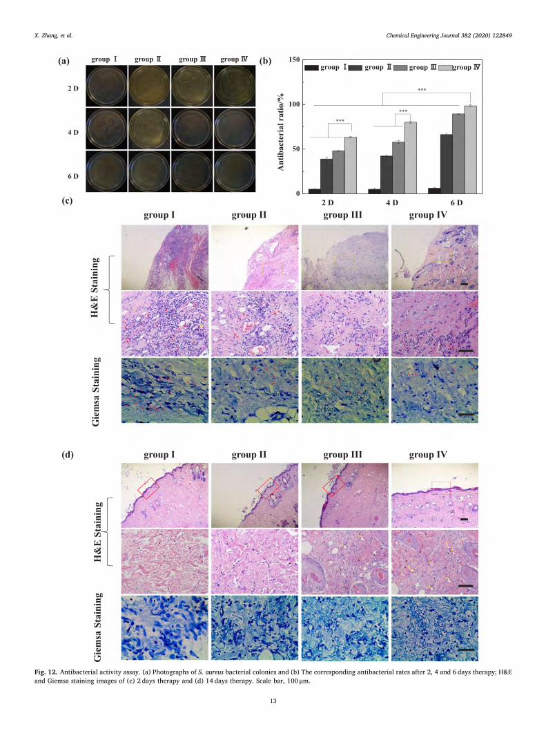
Fig. 12. Antibacterial activity assay. (a) Photographs of S. aureus bacterial colonies and (b) The corresponding antibacterial rates after 2, 4 and 6 days therapy; H&Eand Giemsa staining images of (c) 2 days therapy and (d) 14 days therapy. Scale bar, 100 μm.
X. Zhang, et al. Chemical Engineering Journal 382 (2020) 122849
13
enhanced when the size of the particles or cavities is close to the wa-velength of light [60,61]. The smaller size of CuS@MoS2 is closer to thewavelength of the light source. Therefore, CuS@MoS2 exhibits betterlight absorption and photothermal conversion ability. However, anexcessively high temperature damages normal cells and tissues. Withregard to the 7.5 mg/mL CuS@MoS2 hydrogel, the temperature rises to50 °C in 9min under dual light irradiation and reaches 55 °C in 15min,which can be endured by the human body. The elevated temperaturedecreases the bacterial activity and improves the bacterial membranepermeability [39]. The generated ROS released from the porous hy-drogel can more easily permeate the cell wall of the bacteria to oxidizeintracellular proteins and disturb bacterial homeostasis, so that both S.aureus and E. coli are killed within 15min. The results is verified by theGSH reduction. GSH, as a key antioxidant of the bacterial defensesystem, widely exists in bacteria and prevents damage to cellularcomponents caused by oxidative stress [38,62]. The CuS@MoS2 hy-drogel produced higher temperature and more ROS, resulting in a re-markable decline of GSH. In a word, the synergistic effects of hy-perthermia and ROS make CuS@MoS2 hydrogel possess excellentantibacterial properties. The antibacterial mechanism is shown inScheme 2.
Wound healing and tissue repairing are very complex processes[63]. Besides bacterial infection, the biological behaviors of fibroblastsand endothelial cells at the wounds play major roles in the beginning ofwound healing [64]. Fibroblasts contribute to the synthesis of the ex-tracellular matrix [65] and enhanced proliferation and differentiationof fibroblasts and formation of the fibrinogen for accelerated incrusta-tion can expedite wound healing [66]. Promotion of Cu2+ on thegrowth of fibroblasts has been reported [67], and in this sense, thehydrogels embedded with CuS@MoS2 are conducive to wound healing.Blood vessels, as the conveyancer of oxygen and nutrients, play a key
role in process of wound healing [47]. The wound dressings also shouldpossess the ability to stimulate rapid angiogenesis. The CuS@MoS2hydrogel promotes the proliferation and differentiation of endothelialcells and the secretion of HIF-1α and VEGF, which should be attributedto the Cu element [68]. It has been reported that Cu element is essen-tial to vascularization and can promote the expression of HIF-1α, andthen enhance the angiogenesis. This is because HIF-1α can mimickinghypoxia which has a leading function in recruitment and differentiationof cells and blood vessel formation [69,70]. In addition, HIF-1α canupregulate the expression of several angiogenic genes such as stromalcell-derived factor 1 to promote revascularization. VEGF also con-tributes to the revascularization and it is believed that Cu ions can sharesome pathways in anoxic condition to promote VEGF expression [71].This is verified by animal experiments in which significant improve-ment in wound closure is observed from defects treated with the CuS@MoS2 hydrogels, suggesting the presence of Cu element enhanceswound healing, the more VEGF expresses at the wound sides, the fasterthe wound contracts [72]. Many new blood vessels are found from theH&E slices treated with the CuS@MoS2 hydrogels after 2 and 14 daysand beneficial to the formation of granulation tissues and collagendeposition. The moist environment also accelerates epidermal re-generation during wound healing and the hydrogel ensures such a moistenvironment during wound repair.
5. Conclusion
A bifunctional CuS@MoS2 hydrogel with exhibit excellent anti-bacterial activity against both gram-negative and gram-positive bac-teria under co-irradiation by 660 nm VL and 808 NIR light for 15mindue to the synergistic effects of both PDT and PTT and exploitation ofhyperthermia and ROS is designed and prepared. The temperature of
Fig. 13. H&E staining of the heart, liver, spleen, lung, and kidney tissue slices after 14-day treatment for the different groups. Scale bar, 100 μm.
X. Zhang, et al. Chemical Engineering Journal 382 (2020) 122849
14
the hydrogel can be controlled to 55 °C by light irradiation and hy-perthermia decreases the bacterial activity and improves the bacterialmembrane permeability, so that the bacterial membrane and proteincan be easily destroyed by ROS generated by light exposure leading toswift bacteria death in vitro and in vivo. The hybrid hydrogel shows noappreciable toxicity to organs and promotes wound healing. The me-chanism stems from the promoting proliferation and differentiation ofendothelial cells, and enhancing the secretion of HIF-1α and VEGF andsubsequently vascularization, and efficient sterilization. The animalexperiments demonstrate that the bifunctional hydrogel has great po-tential in the treatment of skin infection and wounds.
Declaration of Competing Interest
The authors declare that they have no known competing financialinterests or personal relationships that could have appeared to influ-ence the work reported in this paper.
Acknowledgements
This work was supported by the National Natural ScienceFoundation of China (31700834), Major Projects in Research andDevelopment of Shanxi (Projects of International Cooperation,201803D421090), Fund for Shanxi “1331 Project” Key InnovativeResearch Team (1331KIRT), Projects of Health and Family PlanningCommission in Shanxi (2017066), and Hong Kong Research GrantsCouncil (RGC) General Research Funds (GRF) (CityU 11205617).
Appendix A. Supplementary data
Supplementary data to this article can be found online at https://doi.org/10.1016/j.cej.2019.122849.
References
[1] M. Urdea, L.A. Penny, S.S. Olmsted, M.Y. Giovanni, P. Kaspar, A. Shepherd,P. Wilson, C.A. Dahl, S. Buchsbaum, G. Moeller, D.C. Hay Burgess, Nature 444(2006) 73.
[2] P. Kirsch, C. Chaimbault, R. Duval, J. Ethnopharmacol. 212 (2018) 200.[3] L.J. Zhang, C.F. Guerrero-Juarez, T. Hata, S.P. Bapat, R. Ramos, M.V. Plikus,
R.L. Gallo, Science 347 (2015) 67.[4] R.M. Klevens, M.A. Morrison, J. Nadle, S. Petit, K. Gershman, S. Ray, L.H. Harrison,
R. Lynfield, G. Dumyati, J.M. Townes, A.S. Craig, E.R. Zell, G.E. Fosheim,L.K. McDougal, R.B. Carey, S.K. Fridkin, JAMA 298 (2007) 1763.
[5] K.A. Engebretsen, J.P. Thyssen, Curr. Probl. Dermatol. 49 (2016) 90.[6] D.J. Tobin, Chem. Soc. Rev. 35 (2006) 52.[7] S.H. MacVane, J. Intensive Care Med. 32 (2016) 25.[8] G.J. Zeng, L. Huang, Q. Huang, M.Y. Liu, D.Z. Xu, H.Y. Huang, Z.Y. Yang, F.J. Deng,
X.Y. Zhang, Y. Wei, Appl. Surf. Sci. 459 (2018) 588.[9] X.Z. Xie, C.Y. Mao, X.M. Liu, Y.Z. Zhang, Z.D. Cui, X.J. Yang, K.W.K. Yeung,
H.B. Pan, P.K. Chu, S.L. Wu, ACS Appl. Mater. Interfaces 9 (2017) 26417.[10] L. Panáček, M. Kvítek, R. Smékalová, M. Večeřová, M. Kolář, F. Röderová,
M. Dyčka, R. Šebela, O. Prucek, R. Zbořil Tomanec, Nat. Nanotechnol. 13 (2018) 65.[11] C.Y. Mao, Y.M. Xiang, X.M. Liu, Z.D. Cui, X.J. Yang, K.W.K. Yeung, H.B. Pan,
X.B. Wang, S.L. Wu, ACS Nano 11 (2017) 9010.[12] X.D. Zhang, L.Y. Xia, X.K. Chen, Z. Chen, F.G. Wu, Sci. China Mater. 60 (2017) 487.[13] J.L. Huang, J.F. Zhou, J.Y. Zhuang, H.Z. Gao, D.H. Huang, L.X. Wang, W.L. Wu,
Q.B. Li, D.P. Yang, M.Y. Han, ACS Appl. Mater. Interfaces 9 (2017) 36606.[14] L. Tan, J. Li, X.M. Liu, Z.D. Cui, X.J. Yang, K.W.K. Yeung, H.B. Pan, Y.F. Zheng,
X.B. Wang, S.L. Wu, Small 14 (2018) 1703197.[15] Y.G. Shi, M.Y. Liu, F.J. Deng, G.J. Zeng, Q. Wan, X.Y. Zhang, Y. Wei, J. Mater.
Chem. B 5 (2017) 194.[16] L. Huang, M.Y. Liu, H.Y. Huang, Y.Q. Wen, X.Y. Zhang, Y. Wei, Biomacromolecules
19 (2018) 1858.[17] Y.L. Liu, K.L. Ai, L.H. Lu, Chem. Rev. 114 (2014) 5057.[18] M.Y. Liu, G.J. Zeng, K. Wang, Q. Wan, L. Tao, X.Y. Zhang, Y. Wei, Nanoscale 8
(2016) 16819.[19] M.Y. Liu, J.Z. Ji, X.Y. Zhang, X.Q. Zhang, B. Yang, F.J. Deng, Z. Li, K. Wang,
Y. Yang, Y. Wei, J. Mater. Chem. B 3 (2015) 3476.[20] Y.G. Shi, R.M. Jiang, M.Y. Liu, L.H. Fu, G.J. Zeng, Q. Wan, L.C. Mao, F.J. Deng,
X.Y. Zhang, Y. Wei, Mater. Sci. Eng., C 77 (2017) 972.[21] J.S. Xiao, S.Y. Chen, J. Yi, H.F. Zhang, G.A. Ameer, Adv. Funct. Mater. 27 (2017)
1604872.[22] Y. Yang, Y.Y. Deng, J.B. Huang, X. Fan, C. Cheng, C.X. Nie, L. Ma, W.F. Zhao,
C.S. Zhao, Adv. Funct. Mater. 29 (2019) 1900143.[23] Z.W. Liu, F.M. Wang, J.S. Ren, X.G. Qu, Biomaterials 208 (2019) 21.[24] C.L. Hu, Z.X. Zhang, S.N. Liu, X.J. Liu, M.L. Pang, ACS Appl. Mater. Interfaces 11
(2019) 23072.[25] M. He, Q. Wang, R. Wang, Y. Xie, W.F. Zhao, C.S. Zhao, ACS Appl. Mater. Interfaces
9 (2017) 15962.[26] M. Li, X.M. Liu, Z.Q. Xu, K.W.K. Yeung, S.L. Wu, ACS Appl. Mater. Interfaces 8
(2016) 33972.[27] Y.H. Xiao, D.C. Su, X.Z. Wang, S.D. Wu, L.M. Zhou, Y. Shi, S.M. Fang, H.M. Cheng,
F. Li, Adv. Energy Mater. 8 (2018) 1800930.[28] T. Stephenson, Z. Li, B. Olsen, D. Mitlin, Energy Environ. Sci. 7 (2014) 209.[29] H.J. Liang, W. Shuang, Y.T. Zhang, S.J. Chao, H.J. Han, X.B. Wang, H. Zhang,
L. Yang, Chem. Electro Chem. 5 (2018) 494.[30] J.P. Shi, R. Tong, X.B. Zhou, Y. Gong, Z.P. Zhang, Q.Q. Ji, Y. Zhang, Q.Y. Fang,
L. Gu, X. Wang, Z. Liu, Y. Zhang, Adv. Mater. 28 (2016) 10664.[31] K.F. Mak, C. Lee, J. Hone, J. Shan, T.F. Heinz, Phys. Rev. Lett. 105 (2010) 136805.[32] H.L. Yang, J.L. Zhao, C.Y. Wu, C.Q. Ye, D.W. Zou, S.G. Wang, Chem. Eng. J. 351
Scheme 2. Schematic illustration of CuS@MoS2-incorporated hydrogel for rapid bacteria killing and wound healing.
X. Zhang, et al. Chemical Engineering Journal 382 (2020) 122849
15

(2018) 548.[33] Q. You, Q. Sun, J.P. Wang, X.X. Tan, X.J. Pang, L. Liu, M. Yu, F.P. Tan, N. Li,
Nanoscale 9 (2017) 3784.[34] L. Zhang, J.Q. Guo, X.Y. Huang, Y.N. Zhang, Y. Han, J. Mater. Chem. B 4 (2016)
3788.[35] M.X. Zong, L. Bai, Y.L. Liu, X. Wang, X.Y. Zhang, X.B. Huang, R.Q. Hang, B. Tang,
Mater. Sci. Eng., C 71 (2017) 93.[36] F. Wang, Y. Wen, T.C. Bai, Mater. Sci. Eng., C 69 (2016) 268.[37] L. Tan, J. Li, X.M. Liu, Z.D. Cui, X.J. Yang, S.L. Zhu, Z.Y. Li, X.B. Yuan, Y.F. Zheng,
K.W.K. Yeung, H.B. Pan, X.B. Wang, S.L. Wu, Adv. Mater. 30 (2018) 1801808.[38] X. Yang, J. Li, T. Liang, C.Y. Ma, Y.Y. Zhang, H.Z. Chen, N. Hanagata, H.X. Su,
M.S. Xu, Nanoscale 6 (2014) 10126.[39] T.I. Kim, B. Kwon, J. Yoon, I.J. Park, G.S. Bang, Y.K. Park, Y.S. Seo, S.Y. Choi, ACS
Appl. Mater. Interfaces 9 (2017) 7908.[40] Q. Gao, X. Zhang, W.Y. Yin, D.Q. Ma, C.J. Xie, L.R. Zheng, X.H. Dong, L.Q. Mei,
J. Yu, C.Z. Wang, Z.J. Gu, Y.L. Zhao, Small 14 (2018) 1802290.[41] Z.Z. Feng, X.M. Liu, L. Tan, Z.D. Cui, X.J. Yang, Z.Y. Li, Y.F. Zheng, K.W.K. Yeung,
S.L. Wu, Small 14 (2018) 1704347.[42] K. Chen, D.K. Zhang, X.T. Cui, Q.L. Wang, RSC Adv. 5 (2015) 24215.[43] J.M. Wang, C. Zhang, Y.Q. Yang, A.L. Fan, R.F. Chi, J. Shi, X.Y. Zhang, Appl. Surf.
Sci. 494 (2019) 708.[44] E.P. Nguyen, B.J. Carey, T. Daeneke, J.Z. Ou, K. Latham, S. Zhuiykov, K. Kalantar-
zadeh, Chem. Mater. 27 (2015) 53.[45] Y. Yang, T. Liu, L. Cheng, G.S. Song, Z. Liu, M.W. Chen, ACS Appl. Mater. Interfaces
7 (2015) 7526.[46] W.Z. Teo, E.L.K. Chng, Z. Sofer, M. Pumera, Chemistry 20 (2014) 9627.[47] S.C. Zhao, L. Li, H. Wang, Y. Zhang, X.G. Cheng, N. Zhou, M.N. Rahaman, Z.T. Liu,
W.H. Huang, C.Q. Zhang, Biomaterials 53 (2015) 379.[48] N. Kong, K. Lin, H.Y. Li, J. Chang, J. Mater. Chem. B 2 (2014) 1100.[49] P. Maxwell, K. Salnikow, Cancer Biol. Ther. 3 (2004) 29.[50] P.H. Maxwell, M.S. Wiesener, G.W. Chang, S.C. Clifford, E.C. Vaux, M.E. Cockman,
C.C. Wykoff, C.W. Pugh, E.R. Maher, P.J. Ratcliffe, Nature 399 (1999) 271.[51] G.J. Moran, A. Krishnadasan, R.J. Gorwitz, G.E. Fosheim, V. Albrecht, B. Limbago,
D.A. Talan, Clin. Infect. Dis. 54 (2012) 1126.[52] Y. Wu, L. Chen, P.G. Scott, E.E. Tredget, Stem Cells 25 (2007) 2648.[53] M. Li, X.M. Liu, L. Tan, Z.D. Cui, X.J. Yang, Z.Y. Li, Y.F. Zheng, K.W.K. Yeung,
P.K. Chu, S.L. Wu, Biomater. Sci. 6 (2018) 2110.[54] C.D. Song, X. Wang, J. Zhang, X.B. Chen, C. Li, Appl. Surf. Sci. 425 (2017) 788.[55] C. Mao, Y.M. Xiang, X.M. Liu, Z.D. Cui, X.J. Yang, Z.Y. Li, S.L. Zhu, Y.F. Zheng,
K.W.K. Yeung, S.L. Wu, ACS Nano 12 (2018) 1747.[56] J. Mou, C.B. Liu, P. Li, Y. Chen, H.X. Xu, C.Y. Wei, L. Song, J.L. Shi, H.R. Chen,
Biomaterials 57 (2015) 12.[57] L.W. Zhang, S. Gao, F. Zhang, K. Yang, Q.J. Ma, L. Zhu, ACS Nano 8 (2014) 12250.[58] R.C. Lv, P.P. Yang, B. Hu, J.T. Xu, W.T. Shang, J. Tian, ACS Nano 11 (2017) 1064.[59] X.C. Wang, F. Lv, T. Li, Y.M. Han, Z.F. Yi, M.Y. Liu, J. Chang, C.T. Wu, ACS Nano 11
(2017) 11337.[60] Q.W. Tian, M.H. Tang, Y.G. Sun, R.J. Zou, Z.G. Chen, M.F. Zhu, S.P. Yang,
J.L. Wang, J.H. Wang, J.Q. Hu, Adv. Mater. 23 (2011) 3542.[61] D.X. Wu, J.F. Duan, C.Y. Zhang, K. Guo, H.T. Zhu, J. Phys. Chem. C 117 (2013)
9121.[62] A. Pompella, A. Visvikis, A. Paolicchi, V.D. Tata, A.F. Casini, Biochem. Pharmacol.
66 (2003) 1499.[63] M.H. Kanzler, D. Gorsulowsky, N. Swanson, J. Dermatol. Surg. Oncol 12 (1986)
1156.[64] N.N. Nissen, P.J. Polverini, A.E. Koch, M.V. Volin, R.L. Gamelli, L.A. DiPietro, Am.
J. Pathol. 152 (1998) 1445.[65] L. Macomber, J.A. Imlay, Proc. Natl. Acad. Sci. U.S.A. 106 (2009) 8344.[66] Q.J. Wu, J.H. Li, W.J. Zhang, H.X. Qian, W.J. She, H.Y. Pan, J. Wen, X.L. Zhang,
X.Y. Liu, X.Q. Jiang, J. Mater. Chem. B 2 (2014) 6738.[67] S. Sonali, X.S. Wen, P. Leoni, K. Naff, C.S.V. Pelt, H. Nakamura, A. Leask,
D. Abraham, G.B. Gharios, B. Crombrugghe, Arthritis Rheum. 62 (2010) 1523.[68] R. Barbucci, S. Lamponi, A. Magnani, F.M. Piras, A. Rossi, E. Weber,
Biomacromolecules 6 (2005) 212.[69] C. Gerard, L.J. Bordeleau, J. Barralet, C.J. Doillon, Biomaterials 31 (2010) 824.[70] S.C. Zhao, H. Wang, Y.D. Zhang, W.H. Huang, M.N. Rahaman, Z.T. Liu, D.P. Wang,
C.Q. Zhang, Acta Biomater. 14 (2015) 185.[71] F. Martin, T. Linden, D.M. Katschinski, F. Oehme, I. Flamme, C.K. Mukhopadhyay,
K. Eckhardt, J. Tröger, S. Barth, G. Camenisch, R.H. Wenger, Blood 105 (2015)4613.
[72] C.K. Sen, S. Khanna, M. Venojarvi, P. Trikha, E.C. Ellison, T.K. Hunt, S. Roy, Am. J.Physiol. Heart Circ. Physiol. 282 (2002) H1821.
X. Zhang, et al. Chemical Engineering Journal 382 (2020) 122849
16
A bifunctional hydrogel incorporated with CuS@MoS2 microspheres for 1
disinfection and improved wound healing 2
3
Xingyu Zhanga, Guannan Zhanga, Hongyu Zhangb, Xiaoping Liua, Jing Shic, Huixian Shia, 4
Xiaohong Yaoa, Paul K. Chud, Xiangyu Zhanga* 5
a College of Materials Science and Engineering, Taiyuan University of Technology, Taiyuan 030024, 6
China 7
b Second Hospital of Shanxi Medical University, Taiyuan 030024, China 8
c Analytical Instrumentation Center, State Key Laboratory of Coal Conversion, Institute of coal 9
chemistry, Chinese Academy of Sciences, Taiyuan 030001, China 10
d Department of Physics and Department of Materials Science and Engineering, City University of 11
Hong Kong, Tat Chee Avenue, Kowloon, Hong Kong, China 12
13
14
*Corresponding Author: E-mail: [email protected] 15
16
17
18
19
20
21
22
23
24
25
26
27
28
29
30
31
32
Figure S1. Color changes in different stages during synthesis of CuS. 33
34
35
36
37
38
39
40
41
56
57
Figure S2. Morphology of CuS particles: (a) low-magnification SEM image; (b) high-58
magnification SEM image; (c) EDS elemental maps; (d, e, and f) TEM images (scale bars = 50 59
nm, 4 nm, and 2 nm); (g) Integrated EDSX drift corrected spectrum image; (h) EDS elemental 60
maps based on TEM. 61
1 μm 1 μm
(a) (b)
500 nm 500 nm 500 nm
(c)
50 nm 4 nm 2 nm
(d) (e) (f)
200 nmCu-K 200 nm
Cu-L200 nm
S-K200 nm
(g)
(h)
62
Figure S3. Morphology of CuS@MoS2: (a, b, and c) TEM images (scale bars = 20 nm, 5 nm, 63
and 3 nm); (d) Low-magnification TEM image; (e) Integrated EDS drift corrected spectrum 64
image; (f) EDS elemental maps based on TEM. 65
66
3 nm5 nm20 nm
5 1/nm
300 nm 300 nmCu-K
300 nmCu-L
300 nmMo-K
300 nmMo-L
300 nmS-K
(a) (b) (c)
(d)
(e)
(f)
67
Figure S4. Low-magnification SEM images of (a) CuS and (b) CuS@MoS2 hydrogel. 68
69
20 μm 20 μm
(a) (b)
70
Figure S5. XPS full spectra of (a) CuS particles and (b) CuS@MoS2 composites. 71
72
0 200 400 600 800 1000
Cu
2p1
Inte
nsity
/a.u
B.E./eV
C 1
s O 1
s
S 2p
1
Cu
2p3
0 200 400 600 800 1000
Inte
nsity
/a.u
C 1
s
O 1
s
S 2p
1
Cu
2p1
Cu
2p3
Mo
O2
B.E./eV
(a) (b)
73
Figure S6. UV-vis spectra of CuS particles and CuS@MoS2 composites. 74
75
200 400 600 800 10000.0
0.2
0.4
0.6
0.8
1.0
1.2
Abs
orba
nce/
a.u.
Wavelength/nm
CuS CuS@MoS2
76
Figure S7. Real-time thermal images: (a) 2.5 mg/mL hydrogels and (b) 12.5 mg/mL hydrogels 77
with dual light irradiation; 7.5 mg/mL hydrogels (add MoS2 group) thermal images: (c) 78
Potothermal curves with dual light irradiation and (d) Corresponding real-time thermal image. 79
20℃
75℃(a)
(b)
20℃
75℃
0min 3min 5min 7min 9min 11min 13min 15min
Con
trol
gro
upC
uSC
uS@
MoS
2C
ontr
ol g
roup
CuS
CuS
@M
oS2
0min 3min 5min 7min 9min 11min 13min 15min
0 2 4 6 8 10 12 1420
30
40
50
60
70
80
90
100 Control group CuS MoS
2
CuS@MoS2
Time/min
The
ave
rage
tem
pera
ture
/C
20℃
75℃
0min 3min 5min 7min 9min 11min 13min 15min
Con
trol
gro
upC
uSC
uS@
MoS
2M
oS2
(c)
(d)
80
Figure S8. Photodynamic performance of 7.5 mg/mL hydrogels: 1O2 detection from degradation 81
of DPBF of hydrogels for different irradiation: (a) 660 nm (0.6W cm-2); (b) 808 nm (0.6W cm-82
2); (c) Dual light (660 nm + 808 nm 0.6 W cm-2 0.6W cm-2 ); Photodynamic performance of 83
7.5 mg/mL hydrogels (MoS2 group): ·OH and 1O2 detection from degradation of methyl violet 84
and DPBF of hydrogels for different irradiation; (d, g) 660 nm (0.6W cm-2); (e, h) 808 nm (0.6W 85
cm-2); (f, i) Dual light (660 nm + 808 nm 0.6 W cm-2 0.6W cm-2). 86
87
400 500 600 7000.0
0.2
0.4
0.6
0.8
1.0
1.2
1.4
1.6
1.8
2.0
Abs
orba
nce/
a.u.
Wavelength/nm
Control group CuS MoS
2
CuS@MoS2
400 500 600 7000.0
0.2
0.4
0.6
0.8
1.0
1.2
1.4
1.6
1.8
2.0
Abs
orba
nce/
a.u.
Wavelength/nm
Control group CuS MoS
2
CuS@MoS2
300 350 400 450 5000.0
0.2
0.4
0.6
0.8
1.0
1.2
1.4
1.6
1.8
2.0
Control group CuS MoS
2
CuS@MoS2
Wavelength/nm
Abs
orba
nce/
a.u.
300 350 400 450 5000.0
0.2
0.4
0.6
0.8
1.0
1.2
1.4
1.6
1.8
2.0 Control group CuS MoS
2
CuS@MoS2
Wavelength/nm
Abs
orba
nce/
a.u.
300 350 400 450 5000.0
0.2
0.4
0.6
0.8
1.0
1.2
1.4
1.6
1.8
2.0 Control group CuS MoS
2
CuS@MoS2
加上二硫化钼
250 300 350 400 450 500 550 6000.0
0.2
0.4
0.6
0.8
1.0
1.2
1.4
1.6
Abs
orba
nce/
a.u.
Wavelength/nm
Control group CuS CuS@MoS
2
250 300 350 400 450 500 550 6000.0
0.2
0.4
0.6
0.8
1.0
1.2
1.4
1.6
Abs
orba
nce/
a.u.
Wavelength/nm
Control group CuS CuS@MoS
2
250 300 350 400 450 500 550 6000.0
0.2
0.4
0.6
0.8
1.0
1.2
1.4
1.6
Abs
orba
nce/
a.u.
Wavelength/nm
Control group CuS CuS@MoS2
(a) (b) (c)
400 500 600 7000.0
0.2
0.4
0.6
0.8
1.0
1.2
1.4
1.6
1.8
2.0 Control group CuS MoS
2
CuS@MoS2
Wavelength/nm
Abs
orba
nce/
a.u.
(d) (e) (f)
(g) (h) (i)
88
Figure S9. Loss of GSH at different temperature. 89
90
91
0
20
40
60
80
100
15min 30min 60min 120min
Los
s of G
SH/ %
25C 50C
25℃
0 15 30 60 120 (min)
50℃
(a) (b)
92
Figure S10. Photographs of bacterial colonies of S. aureus: (a) 2.5 mg/mL hydrogels, (c) 7.5 93
mg/mL hydrogels, and (e) 12.5 mg/mL hydrogels; E. coli: (b) 2.5 mg/mL hydrogels, (d) 7.5 94
mg/mL hydrogels, and (f) 12.5 mg/mL hydrogels under the conditions of darkness, 660 nm, 808 95
nm, and dual light for 15 min. 96
Control group CuS CuS@MoS2
Dar
k66
0 nm
808
nmD
ual l
ight
sS
. a
ure
us
Control group CuS CuS@MoS2
Dar
k66
0 nm
808
nmD
ual l
ight
sS
. a
ure
us
Control group CuS CuS@MoS2
Dar
k66
0 nm
808
nmD
ual l
ight
sE
. co
li
Control group CuS CuS@MoS2
Dar
k66
0 nm
808
nmD
ual l
ight
sS
. a
ure
us
Control group CuS CuS@MoS2
Dar
k66
0 nm
808
nmD
ual l
ight
sE
. co
li(c) (d)
(a) (b)
Control group CuS CuS@MoS2
Dar
k66
0 nm
808
nmD
ual l
ight
sE
. co
li
(e) (f)
97
98
99
Figure S11. Response of endothelial cells of 7.5 mg/mL hydrogel (add MoS2 group): (a) 100
Fluorescence images of Live/Dead staining of endothelial cells after culturing for 1and 2 days 101
on pure hydrogel, CuS, MoS2 and CuS@MoS2-incorporated hydrogels; (b) MTT assay of 102
endothelial cells seeded on pure hydrogel, CuS, MoS2 and CuS@MoS2-incorporated hydrogels 103
after incubation for 1and 2 days. 104
105
100 μm 100 μm 100 μm 100 μm
100 μm 100 μm 100 μm 100 μm
Control group CuS MoS2 CuS@MoS2
0.0
0.1
0.2
0.3
0.4
0.5
0.6
A
bsor
banc
e 57
0nm
1 Day 2 Days
Control group CuS MoS2 CuS@MoS2
1 D
2D
(a)
(b)

106
107
108
Figure S12. In vivo thermal images of the back of the mice under dual light after 15 min: group 109
II: blank hydrogel, group III: CuS-incorporated hydrogel and group IV: CuS@MoS2-110
incorporated hydrogel. 111
112
113
114
115
116
117
118
119