Chemical durability and microstructural analysis of glasses soaked in water and in biological fluids
17
Chemical durability and microstructural analysis of glasses soaked in water and in biological fluids V. Cannillo a , F. Pierli a , I. Ronchetti b , C. Siligardi a, * , D. Zaffe c a Dipartimento di Ingegneria dei Materiali e dell’Ambiente, Universita ` di Modena e Reggio Emilia, Via Vignolese 905, 41100 Modena, Italy b Dipartimento di Scienze Biomediche, Universita ` di Modena e Reggio Emilia, Via Campi 287, 41100 Modena, Italy c Dipartimento di Anatomia ed Istologia, Universita ` di Modena e Reggio Emilia, Via del Pozzo 51, 41100 Modena, Italy Received 2 February 2009; received in revised form 1 March 2009; accepted 20 March 2009 Available online 15 April 2009 Abstract A new glass, obtained from Bioglass 1 BG45S5 original composition by substituting CaO with MgO, was produced and its chemical durability and microstructural characteristics were compared with that of Bioglass 1 . The two glasses (labelled as BG45 and MG45) were soaked up to 4 weeks at physiological temperature in different solutions, i.e. bi-distilled water, Hank’s Buffered Salt Solution 61200 (labelled as HBSS+), Hank’s Buffered Salt Solution 14170 (labelled as HBSS), and Kokubo’s SBF. Moreover, the influence of either flat or flake surfaces was analysed for both glasses. Results showed that the chemical durability of a glass in saline at 37 8C, evaluated through pH and ICP-AES chemical analysis of the leached components, depended mainly on the chemical composition of the soaking solution. Moreover, the MG45 glass never exhibited hydroxyapatite crystal formation on its surface also after soaking in calcium- containing solutions. The apatite crystallisation and deposition mechanism, typical of a bioactive glass, was induced only if the glass itself contained calcium. The contemporaneous presence of calcium in the glass and in the soaking solution improved the reactivity of the glass, as apatite crystals nucleated in a shorter time and grew more quickly. As regards the morphology of the glass surface, rougher surfaces favoured the formation of hydroxyapatite crystals on glasses containing calcium. # 2009 Elsevier Ltd and Techna Group S.r.l. All rights reserved. Keywords: D. Glass; Chemical durability; Calcium; Magnesium; Microstructure 1. Introduction Bioglass 1 is the most accepted bioactive glass used in biomedical applications, especially in bone replacement. Hench [1,2] first demonstrated in the 70s that bioactive glasses are able to bind to bone and to promote bone formation. When bioactive glasses are soaked in physiological media or implanted in vivo, they can bind to living bone trough an apatite layer formed on their surface. The reaction steps first detailed by Clerk and Hench [3] involve the formation of a silica-rich layer and of a Ca–P-rich layer that crystallises in hydroxyl-carbonate apatite (HCA), a mineral phase similar to that of bone. After the development of Bioglass 1 by Hench [1,2], with a composition of 45 wt% SiO 2 , 24.5 wt% CaO, 24.5 wt% Na 2 O and 6 wt% P 2 O 5 , different bioactive glasses have been produced and studied. Most of them contain both CaO and P 2 O 5 as main components [4,5]. Addition of elements, like magnesium or aluminium, may be used to control some physical and chemical properties [6]. It has been found that specific concentrations of magnesium can influence the glass dissolution or the physical–chemical reaction at the glass periphery [7]. Many works analysed bioactive glasses with compositions based on CaO–MgO–P 2 O 5 –SiO 2 systems [8–13]. Bioactive glasses doped with magnesium lead to the formation of a Ca–P–Mg-rich layer on a pure silica layer. For example, Jallot [6] demonstrated that magnesium influences the spontaneous formation and the evolution of the in vivo HCA layer and bone bonding. Moreover, magnesium has a beneficial effect on the crystallinity and on the solubility of apatites. Mg- substituted apatites are more soluble than calcium apatites, and suppress apatite crystallisation in in vivo tests [13,14]. Many works have studied the bioactivity, i.e. the HCA deposition, www.elsevier.com/locate/ceramint Available online at www.sciencedirect.com Ceramics International 35 (2009) 2853–2869 * Corresponding author. Fax: +39 059 2056243. E-mail address: [email protected] (C. Siligardi). 0272-8842/$36.00 # 2009 Elsevier Ltd and Techna Group S.r.l. All rights reserved. doi:10.1016/j.ceramint.2009.03.029
-
Upload
independent -
Category
Documents
-
view
1 -
download
0
Transcript of Chemical durability and microstructural analysis of glasses soaked in water and in biological fluids
Chemical durability and microstructural analysis of glasses
soaked in water and in biological fluids
V. Cannillo a, F. Pierli a, I. Ronchetti b, C. Siligardi a,*, D. Zaffe c
a Dipartimento di Ingegneria dei Materiali e dell’Ambiente, Universita di Modena e Reggio Emilia, Via Vignolese 905, 41100 Modena, Italyb Dipartimento di Scienze Biomediche, Universita di Modena e Reggio Emilia, Via Campi 287, 41100 Modena, Italy
c Dipartimento di Anatomia ed Istologia, Universita di Modena e Reggio Emilia, Via del Pozzo 51, 41100 Modena, Italy
Received 2 February 2009; received in revised form 1 March 2009; accepted 20 March 2009
Available online 15 April 2009
Abstract
A new glass, obtained from Bioglass1 BG45S5 original composition by substituting CaO with MgO, was produced and its chemical durability
and microstructural characteristics were compared with that of Bioglass1.
The two glasses (labelled as BG45 and MG45) were soaked up to 4 weeks at physiological temperature in different solutions, i.e. bi-distilled
water, Hank’s Buffered Salt Solution 61200 (labelled as HBSS+), Hank’s Buffered Salt Solution 14170 (labelled as HBSS�), and Kokubo’s SBF.
Moreover, the influence of either flat or flake surfaces was analysed for both glasses. Results showed that the chemical durability of a glass in saline
at 37 8C, evaluated through pH and ICP-AES chemical analysis of the leached components, depended mainly on the chemical composition of the
soaking solution. Moreover, the MG45 glass never exhibited hydroxyapatite crystal formation on its surface also after soaking in calcium-
containing solutions. The apatite crystallisation and deposition mechanism, typical of a bioactive glass, was induced only if the glass itself
contained calcium. The contemporaneous presence of calcium in the glass and in the soaking solution improved the reactivity of the glass, as apatite
crystals nucleated in a shorter time and grew more quickly. As regards the morphology of the glass surface, rougher surfaces favoured the formation
of hydroxyapatite crystals on glasses containing calcium.
# 2009 Elsevier Ltd and Techna Group S.r.l. All rights reserved.
Keywords: D. Glass; Chemical durability; Calcium; Magnesium; Microstructure
www.elsevier.com/locate/ceramint
Available online at www.sciencedirect.com
Ceramics International 35 (2009) 2853–2869
1. Introduction
Bioglass1 is the most accepted bioactive glass used in
biomedical applications, especially in bone replacement.
Hench [1,2] first demonstrated in the 70s that bioactive glasses
are able to bind to bone and to promote bone formation. When
bioactive glasses are soaked in physiological media or
implanted in vivo, they can bind to living bone trough an
apatite layer formed on their surface. The reaction steps first
detailed by Clerk and Hench [3] involve the formation of a
silica-rich layer and of a Ca–P-rich layer that crystallises in
hydroxyl-carbonate apatite (HCA), a mineral phase similar to
that of bone. After the development of Bioglass1 by Hench
[1,2], with a composition of 45 wt% SiO2, 24.5 wt% CaO,
* Corresponding author. Fax: +39 059 2056243.
E-mail address: [email protected] (C. Siligardi).
0272-8842/$36.00 # 2009 Elsevier Ltd and Techna Group S.r.l. All rights reserve
doi:10.1016/j.ceramint.2009.03.029
24.5 wt% Na2O and 6 wt% P2O5, different bioactive glasses
have been produced and studied. Most of them contain both
CaO and P2O5 as main components [4,5]. Addition of elements,
like magnesium or aluminium, may be used to control some
physical and chemical properties [6]. It has been found that
specific concentrations of magnesium can influence the glass
dissolution or the physical–chemical reaction at the glass
periphery [7]. Many works analysed bioactive glasses with
compositions based on CaO–MgO–P2O5–SiO2 systems [8–13].
Bioactive glasses doped with magnesium lead to the formation
of a Ca–P–Mg-rich layer on a pure silica layer. For example,
Jallot [6] demonstrated that magnesium influences the
spontaneous formation and the evolution of the in vivo HCA
layer and bone bonding. Moreover, magnesium has a beneficial
effect on the crystallinity and on the solubility of apatites. Mg-
substituted apatites are more soluble than calcium apatites, and
suppress apatite crystallisation in in vivo tests [13,14]. Many
works have studied the bioactivity, i.e. the HCA deposition,
d.
V. Cannillo et al. / Ceramics International 35 (2009) 2853–28692854
mostly by analysing what happens at the surface of a bioactive
glass after immersion in a simulated biological fluid (SBF) [15–
19]. However, to the best of the authors’ knowledge, the present
research analyses for the first time the surface reactions in
physiological media of phosphate glasses in which the calcium
oxide is completely substituted by the magnesium oxide.
In fact, in the present work a glass, based on the composition
of Bioglass1 45S5 [1], but modified by the complete substitution
of CaO with MgO, has been produced and characterised in
comparison to the traditional Bioglass1 45S5. Glasses were
soaked in bi-distilled water and in different simulated biological
fluids for 1, 2 and 4 weeks, in order to determine the specific
effects of Ca2+ and Mg2+ ions on the chemical durability and
behaviour of both glasses and the eventual crystals formation on
the surface after incubation in the different media. The physical
and the chemical properties of the two glasses were evaluated by
using various analytical techniques.
2. Experimental
2.1. Glass systems
The glasses studied in this work were labelled as BG45 and
MG45. The BG45 glass had the composition of the 45S5
Bioglass1 reported by Hench [1,2] and was chosen as the
reference glass. MG45 glass was obtained by substituting CaO
by MgO in the composition of BG45. The compositions of the
two glasses in wt% and mol% are reported in Table 1. The batch
mixtures were prepared from pure grade commercial reagents
SiO2, Na2CO3, CaCO3, Na3PO4 and MgCO3 (Carlo Erba,
Italy). The batches were properly dry mixed by using Al2O3
balls for 30 min into porcelain jars. Batches were placed in a
platinum crucible and melted in an electric furnace at 1450 8Cfor 1 h. The melted glasses were then poured into a carbon-
made small container to form bulk glasses. The as-cast glasses
were then annealed at 560 8C and at 500 8C for 1 h for BG45
and MG45, respectively.
Table 1
Chemical composition of glasses in oxide.
BG45 (45S5
Bioglass1) [1,2]
MG45
wt% mol% wt% mol%
SiO2 45 46.13 48.33 46.13
Na2O 24.5 24.35 26.32 24.35
CaO 24.5 26.91 – –
MgO – – 18.91 26.91
P2O5 6 2.60 6.44 2.60
Table 2
Ion concentration (mM) and pH of SBF, HBSS+, HBSS� in comparison with hum
Na+ K+ Mg2+ Ca2+
Human body plasma 142 5 1.5 2.5
SBF 142 5 1.5 2.5
HBSS+ 81 4.6 0.97 1.4
HBSS� 84.5 4.6 – –
Glasses were cut to dimensions of 5 mm � 5 mm � 3 mm,
polished to a 0.5 mm finishing using diamond paste and then
washed with acetone in an ultrasonic bath. To evaluate the
effect of the surface morphology of glasses on their chemical
durability, flake samples were also prepared, without polishing:
in this way the surface was rough. Comparison of the roughness
between flat and flake surfaces was made by means of an optical
profilometer (Conscan Profilometer, CSM Instruments). All
samples were stored in a desiccator until used.
The glasses were characterised by means of DTA analysis
(Netzsch DSC 404 Differential Thermal Analyzer), using
30 mg of powders heated from 20 8C to 1400 8C at 10 8C/min,
in order to obtain the critical temperatures of glasses, such as
glass transition and crystallisation temperatures (accuracy of
DTA = �2 8C). Density measurements were performed
through He picnometry (AccuPyc 1330, Micromeritics) and
the molar volume was calculated in order to evaluate the effect
of replacing CaO with MgO on the structure of glasses. The
molar volume (Vm) was calculated from the density data
obtained by using the following relation [20]:
Vm ¼X
i
Mi Ni
r
where Mi and Ni are the molar weight and the molar ratio of the
oxide, respectively.
2.2. Dissolution study and microstructural
characterisation
The flat glasses were immersed into four different fluids: bi-
distilled water, Hank’s Buffered Salt Solution 61200 (labelled
as HBSS+), Hank’s Buffered Salt Solution 14170 (labelled as
HBSS�) and SBF (Simulated Body Fluid). The SBF was
prepared and used according to the procedure described by
Kokubo and Takadama [21]. HBSS+, HBSS� and SBF have
composition similar to human body fluids, as reported in
Table 2 [21,22]. The HBSS+ solution was characterised by a
little amount of Ca2+ and Mg2+, while the HBSS� solution was
free of Ca2+ and Mg2+. Since a previous work [23] showed that
the surface morphology of bioactive glasses significantly
influences their physiological performance, in the present
study, the in vitro bioactivity of the two glasses with either flat
or rough surfaces was compared after soaking in SBF, as this is
the most accepted solution for in vitro tests [21].
Glasses were soaked in 2 ml of solution (four different tests
for each glass: water, HBSS+, HBSS�, and SBF) for 1, 2 and 4
weeks at a constant temperature of 37 8C in a cell culture
incubator. Tests were carried out in duplicate. Measurements of
an blood plasma.
Cl� HCO3� HPO4
� SO4� pH
103 27 1.0 0.5 7.40
147.8 4.2 1.0 0.5 7.40
85.4 – 1.08 – 6.80
84 3.5 1.08 – 7.81
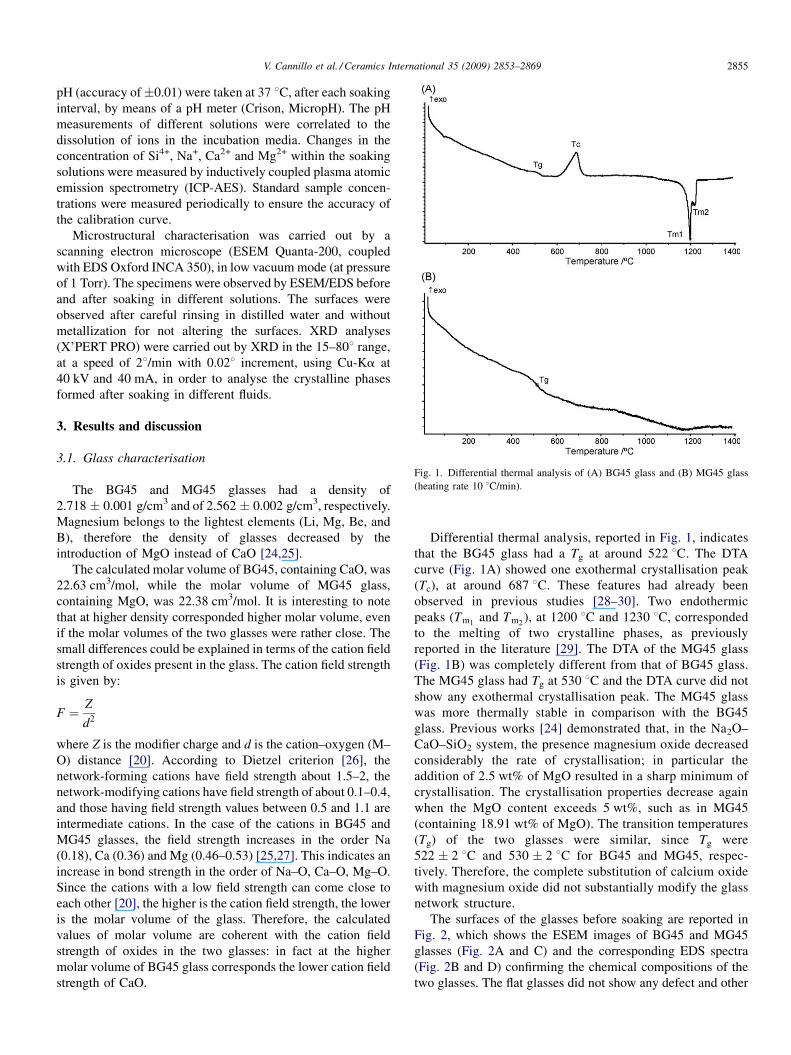
Fig. 1. Differential thermal analysis of (A) BG45 glass and (B) MG45 glass
(heating rate 10 8C/min).
V. Cannillo et al. / Ceramics International 35 (2009) 2853–2869 2855
pH (accuracy of�0.01) were taken at 37 8C, after each soaking
interval, by means of a pH meter (Crison, MicropH). The pH
measurements of different solutions were correlated to the
dissolution of ions in the incubation media. Changes in the
concentration of Si4+, Na+, Ca2+ and Mg2+ within the soaking
solutions were measured by inductively coupled plasma atomic
emission spectrometry (ICP-AES). Standard sample concen-
trations were measured periodically to ensure the accuracy of
the calibration curve.
Microstructural characterisation was carried out by a
scanning electron microscope (ESEM Quanta-200, coupled
with EDS Oxford INCA 350), in low vacuum mode (at pressure
of 1 Torr). The specimens were observed by ESEM/EDS before
and after soaking in different solutions. The surfaces were
observed after careful rinsing in distilled water and without
metallization for not altering the surfaces. XRD analyses
(X’PERT PRO) were carried out by XRD in the 15–808 range,
at a speed of 28/min with 0.028 increment, using Cu-Ka at
40 kV and 40 mA, in order to analyse the crystalline phases
formed after soaking in different fluids.
3. Results and discussion
3.1. Glass characterisation
The BG45 and MG45 glasses had a density of
2.718 � 0.001 g/cm3 and of 2.562 � 0.002 g/cm3, respectively.
Magnesium belongs to the lightest elements (Li, Mg, Be, and
B), therefore the density of glasses decreased by the
introduction of MgO instead of CaO [24,25].
The calculated molar volume of BG45, containing CaO, was
22.63 cm3/mol, while the molar volume of MG45 glass,
containing MgO, was 22.38 cm3/mol. It is interesting to note
that at higher density corresponded higher molar volume, even
if the molar volumes of the two glasses were rather close. The
small differences could be explained in terms of the cation field
strength of oxides present in the glass. The cation field strength
is given by:
F ¼ Z
d2
where Z is the modifier charge and d is the cation–oxygen (M–
O) distance [20]. According to Dietzel criterion [26], the
network-forming cations have field strength about 1.5–2, the
network-modifying cations have field strength of about 0.1–0.4,
and those having field strength values between 0.5 and 1.1 are
intermediate cations. In the case of the cations in BG45 and
MG45 glasses, the field strength increases in the order Na
(0.18), Ca (0.36) and Mg (0.46–0.53) [25,27]. This indicates an
increase in bond strength in the order of Na–O, Ca–O, Mg–O.
Since the cations with a low field strength can come close to
each other [20], the higher is the cation field strength, the lower
is the molar volume of the glass. Therefore, the calculated
values of molar volume are coherent with the cation field
strength of oxides in the two glasses: in fact at the higher
molar volume of BG45 glass corresponds the lower cation field
strength of CaO.
Differential thermal analysis, reported in Fig. 1, indicates
that the BG45 glass had a Tg at around 522 8C. The DTA
curve (Fig. 1A) showed one exothermal crystallisation peak
(Tc), at around 687 8C. These features had already been
observed in previous studies [28–30]. Two endothermic
peaks (Tm1and Tm2
), at 1200 8C and 1230 8C, corresponded
to the melting of two crystalline phases, as previously
reported in the literature [29]. The DTA of the MG45 glass
(Fig. 1B) was completely different from that of BG45 glass.
The MG45 glass had Tg at 530 8C and the DTA curve did not
show any exothermal crystallisation peak. The MG45 glass
was more thermally stable in comparison with the BG45
glass. Previous works [24] demonstrated that, in the Na2O–
CaO–SiO2 system, the presence magnesium oxide decreased
considerably the rate of crystallisation; in particular the
addition of 2.5 wt% of MgO resulted in a sharp minimum of
crystallisation. The crystallisation properties decrease again
when the MgO content exceeds 5 wt%, such as in MG45
(containing 18.91 wt% of MgO). The transition temperatures
(Tg) of the two glasses were similar, since Tg were
522 � 2 8C and 530 � 2 8C for BG45 and MG45, respec-
tively. Therefore, the complete substitution of calcium oxide
with magnesium oxide did not substantially modify the glass
network structure.
The surfaces of the glasses before soaking are reported in
Fig. 2, which shows the ESEM images of BG45 and MG45
glasses (Fig. 2A and C) and the corresponding EDS spectra
(Fig. 2B and D) confirming the chemical compositions of the
two glasses. The flat glasses did not show any defect and other
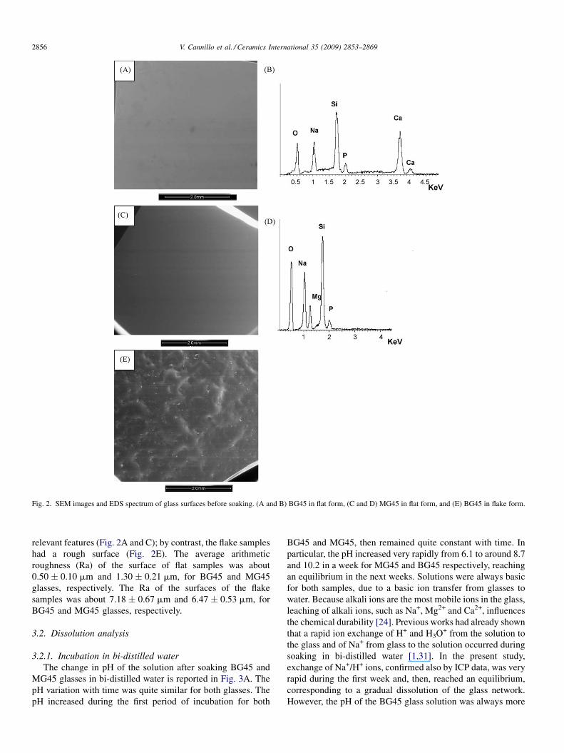
Fig. 2. SEM images and EDS spectrum of glass surfaces before soaking. (A and B) BG45 in flat form, (C and D) MG45 in flat form, and (E) BG45 in flake form.
V. Cannillo et al. / Ceramics International 35 (2009) 2853–28692856
relevant features (Fig. 2A and C); by contrast, the flake samples
had a rough surface (Fig. 2E). The average arithmetic
roughness (Ra) of the surface of flat samples was about
0.50 � 0.10 mm and 1.30 � 0.21 mm, for BG45 and MG45
glasses, respectively. The Ra of the surfaces of the flake
samples was about 7.18 � 0.67 mm and 6.47 � 0.53 mm, for
BG45 and MG45 glasses, respectively.
3.2. Dissolution analysis
3.2.1. Incubation in bi-distilled water
The change in pH of the solution after soaking BG45 and
MG45 glasses in bi-distilled water is reported in Fig. 3A. The
pH variation with time was quite similar for both glasses. The
pH increased during the first period of incubation for both
BG45 and MG45, then remained quite constant with time. In
particular, the pH increased very rapidly from 6.1 to around 8.7
and 10.2 in a week for MG45 and BG45 respectively, reaching
an equilibrium in the next weeks. Solutions were always basic
for both samples, due to a basic ion transfer from glasses to
water. Because alkali ions are the most mobile ions in the glass,
leaching of alkali ions, such as Na+, Mg2+ and Ca2+, influences
the chemical durability [24]. Previous works had already shown
that a rapid ion exchange of H+ and H3O+ from the solution to
the glass and of Na+ from glass to the solution occurred during
soaking in bi-distilled water [1,31]. In the present study,
exchange of Na+/H+ ions, confirmed also by ICP data, was very
rapid during the first week and, then, reached an equilibrium,
corresponding to a gradual dissolution of the glass network.
However, the pH of the BG45 glass solution was always more
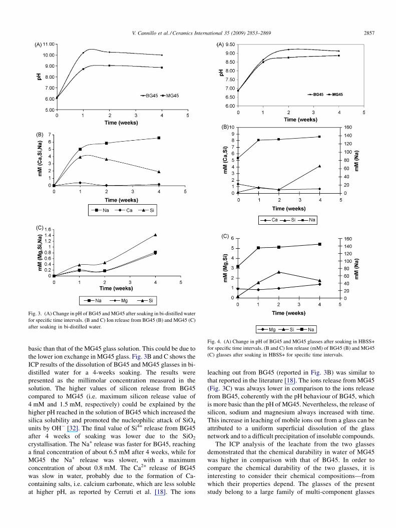
Fig. 3. (A) Change in pH of BG45 and MG45 after soaking in bi-distilled water
for specific time intervals. (B and C) Ion release from BG45 (B) and MG45 (C)
after soaking in bi-distilled water.
Fig. 4. (A) Change in pH of BG45 and MG45 glasses after soaking in HBSS+
for specific time intervals. (B and C) Ion release (mM) of BG45 (B) and MG45
(C) glasses after soaking in HBSS+ for specific time intervals.
V. Cannillo et al. / Ceramics International 35 (2009) 2853–2869 2857
basic than that of the MG45 glass solution. This could be due to
the lower ion exchange in MG45 glass. Fig. 3B and C shows the
ICP results of the dissolution of BG45 and MG45 glasses in bi-
distilled water for a 4-weeks soaking. The results were
presented as the millimolar concentration measured in the
solution. The higher values of silicon release from BG45
compared to MG45 (i.e. maximum silicon release value of
4 mM and 1.5 mM, respectively) could be explained by the
higher pH reached in the solution of BG45 which increased the
silica solubility and promoted the nucleophilic attack of SiO4
units by OH� [32]. The final value of Si4+ release from BG45
after 4 weeks of soaking was lower due to the SiO2
crystallisation. The Na+ release was faster for BG45, reaching
a final concentration of about 6.5 mM after 4 weeks, while for
MG45 the Na+ release was slower, with a maximum
concentration of about 0.8 mM. The Ca2+ release of BG45
was slow in water, probably due to the formation of Ca-
containing salts, i.e. calcium carbonate, which are less soluble
at higher pH, as reported by Cerruti et al. [18]. The ions
leaching out from BG45 (reported in Fig. 3B) was similar to
that reported in the literature [18]. The ions release from MG45
(Fig. 3C) was always lower in comparison to the ions release
from BG45, coherently with the pH behaviour of BG45, which
is more basic than the pH of MG45. Nevertheless, the release of
silicon, sodium and magnesium always increased with time.
This increase in leaching of mobile ions out from a glass can be
attributed to a uniform superficial dissolution of the glass
network and to a difficult precipitation of insoluble compounds.
The ICP analysis of the leachate from the two glasses
demonstrated that the chemical durability in water of MG45
was higher in comparison with that of BG45. In order to
compare the chemical durability of the two glasses, it is
interesting to consider their chemical compositions—from
which their properties depend. The glasses of the present
study belong to a large family of multi-component glasses
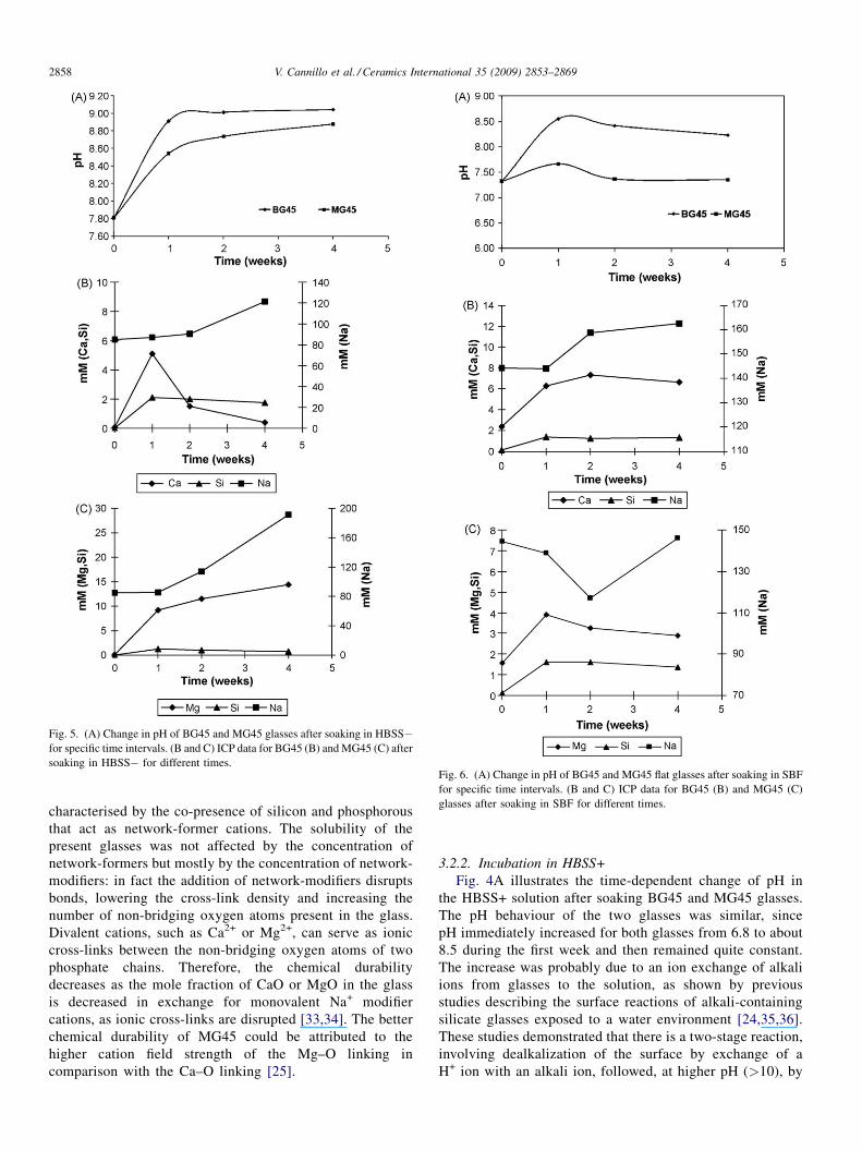
Fig. 5. (A) Change in pH of BG45 and MG45 glasses after soaking in HBSS�for specific time intervals. (B and C) ICP data for BG45 (B) and MG45 (C) after
soaking in HBSS� for different times.Fig. 6. (A) Change in pH of BG45 and MG45 flat glasses after soaking in SBF
for specific time intervals. (B and C) ICP data for BG45 (B) and MG45 (C)
glasses after soaking in SBF for different times.
V. Cannillo et al. / Ceramics International 35 (2009) 2853–28692858
characterised by the co-presence of silicon and phosphorous
that act as network-former cations. The solubility of the
present glasses was not affected by the concentration of
network-formers but mostly by the concentration of network-
modifiers: in fact the addition of network-modifiers disrupts
bonds, lowering the cross-link density and increasing the
number of non-bridging oxygen atoms present in the glass.
Divalent cations, such as Ca2+ or Mg2+, can serve as ionic
cross-links between the non-bridging oxygen atoms of two
phosphate chains. Therefore, the chemical durability
decreases as the mole fraction of CaO or MgO in the glass
is decreased in exchange for monovalent Na+ modifier
cations, as ionic cross-links are disrupted [33,34]. The better
chemical durability of MG45 could be attributed to the
higher cation field strength of the Mg–O linking in
comparison with the Ca–O linking [25].
3.2.2. Incubation in HBSS+
Fig. 4A illustrates the time-dependent change of pH in
the HBSS+ solution after soaking BG45 and MG45 glasses.
The pH behaviour of the two glasses was similar, since
pH immediately increased for both glasses from 6.8 to about
8.5 during the first week and then remained quite constant.
The increase was probably due to an ion exchange of alkali
ions from glasses to the solution, as shown by previous
studies describing the surface reactions of alkali-containing
silicate glasses exposed to a water environment [24,35,36].
These studies demonstrated that there is a two-stage reaction,
involving dealkalization of the surface by exchange of a
H+ ion with an alkali ion, followed, at higher pH (>10), by
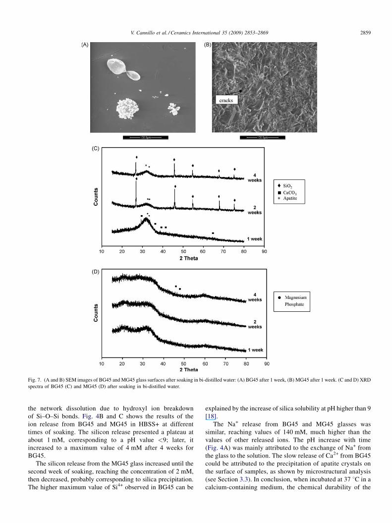
Fig. 7. (A and B) SEM images of BG45 and MG45 glass surfaces after soaking in bi-distilled water: (A) BG45 after 1 week, (B) MG45 after 1 week. (C and D) XRD
spectra of BG45 (C) and MG45 (D) after soaking in bi-distilled water.
V. Cannillo et al. / Ceramics International 35 (2009) 2853–2869 2859
the network dissolution due to hydroxyl ion breakdown
of Si–O–Si bonds. Fig. 4B and C shows the results of the
ion release from BG45 and MG45 in HBSS+ at different
times of soaking. The silicon release presented a plateau at
about 1 mM, corresponding to a pH value <9; later, it
increased to a maximum value of 4 mM after 4 weeks for
BG45.
The silicon release from the MG45 glass increased until the
second week of soaking, reaching the concentration of 2 mM,
then decreased, probably corresponding to silica precipitation.
The higher maximum value of Si4+ observed in BG45 can be
explained by the increase of silica solubility at pH higher than 9
[18].
The Na+ release from BG45 and MG45 glasses was
similar, reaching values of 140 mM, much higher than the
values of other released ions. The pH increase with time
(Fig. 4A) was mainly attributed to the exchange of Na+ from
the glass to the solution. The slow release of Ca2+ from BG45
could be attributed to the precipitation of apatite crystals on
the surface of samples, as shown by microstructural analysis
(see Section 3.3). In conclusion, when incubated at 37 8C in a
calcium-containing medium, the chemical durability of the
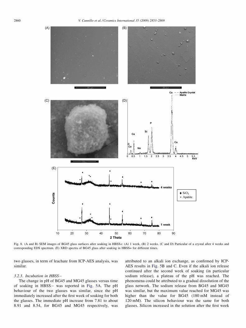
Fig. 8. (A and B) SEM images of BG45 glass surfaces after soaking in HBSS+: (A) 1 week, (B) 2 weeks. (C and D) Particular of a crystal after 4 weeks and
corresponding EDS spectrum. (E) XRD spectra of BG45 glass after soaking in HBSS+ for different times.
V. Cannillo et al. / Ceramics International 35 (2009) 2853–28692860
two glasses, in term of leachate from ICP-AES analysis, was
similar.
3.2.3. Incubation in HBSS�The change in pH of BG45 and MG45 glasses versus time
of soaking in HBSS� was reported in Fig. 5A. The pH
behaviour of the two glasses was similar, since the pH
immediately increased after the first week of soaking for both
the glasses. The immediate pH increase from 7.81 to about
8.91 and 8.54, for BG45 and MG45 respectively, was
attributed to an alkali ion exchange, as confirmed by ICP-
AES results in Fig. 5B and C. Even if the alkali ion release
continued after the second week of soaking (in particular
sodium release), a plateau of the pH was reached. The
phenomena could be attributed to a gradual dissolution of the
glass network. The sodium release from BG45 and MG45
was similar, but the maximum value reached for MG45 was
higher than the value for BG45 (180 mM instead of
120 mM). The silicon behaviour was the same for both
glasses. Silicon increased in the solution after the first week
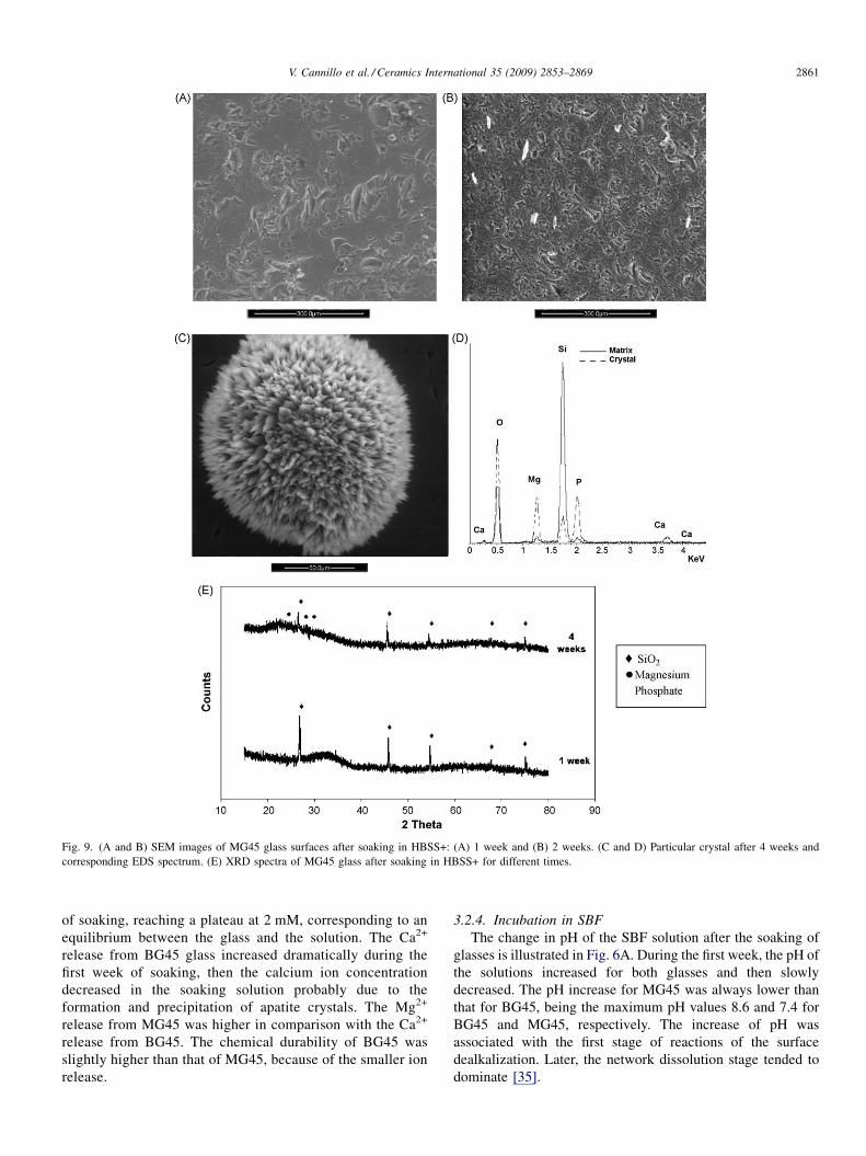
Fig. 9. (A and B) SEM images of MG45 glass surfaces after soaking in HBSS+: (A) 1 week and (B) 2 weeks. (C and D) Particular crystal after 4 weeks and
corresponding EDS spectrum. (E) XRD spectra of MG45 glass after soaking in HBSS+ for different times.
V. Cannillo et al. / Ceramics International 35 (2009) 2853–2869 2861
of soaking, reaching a plateau at 2 mM, corresponding to an
equilibrium between the glass and the solution. The Ca2+
release from BG45 glass increased dramatically during the
first week of soaking, then the calcium ion concentration
decreased in the soaking solution probably due to the
formation and precipitation of apatite crystals. The Mg2+
release from MG45 was higher in comparison with the Ca2+
release from BG45. The chemical durability of BG45 was
slightly higher than that of MG45, because of the smaller ion
release.
3.2.4. Incubation in SBF
The change in pH of the SBF solution after the soaking of
glasses is illustrated in Fig. 6A. During the first week, the pH of
the solutions increased for both glasses and then slowly
decreased. The pH increase for MG45 was always lower than
that for BG45, being the maximum pH values 8.6 and 7.4 for
BG45 and MG45, respectively. The increase of pH was
associated with the first stage of reactions of the surface
dealkalization. Later, the network dissolution stage tended to
dominate [35].
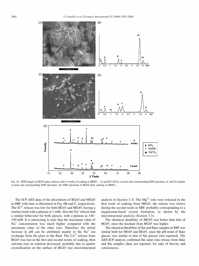
Fig. 10. SEM images of BG45 glass surfaces after 4 weeks of soaking in HBSS�: (A and B) CaCO3 crystals and corresponding EDS spectrum. (C and D) Apatite
crystals and corresponding EDS spectrum. (E) XRD spectrum of BG45 after soaking in HBSS�.
V. Cannillo et al. / Ceramics International 35 (2009) 2853–28692862
The ICP-AES data of the dissolution of BG45 and MG45
in SBF with time is illustrated in Fig. 6B and C, respectively.
The Si4+ release was low for both BG45 and MG45, having a
similar trend with a plateau at 1 mM. Also the Na+ release had
a similar behaviour for both glasses, with a plateau at 140–
150 mM. It is interesting to note that the maximum value of
Na+ concentration was much higher compared with the
maximum value of the other ions. Therefore, the initial
increase in pH can be attributed mainly to the Na+ ion
exchange from the glass to the fluid. The Ca2+ release from
BG45 was fast in the first and second weeks of soaking, then
calcium ions in solution decreased, probably due to apatite
crystallisation on the surface of BG45 (see microstructural
analysis in Section 3.3). The Mg2+ ions were released in the
first week of soaking from MG45; the release was slower
during the second week in SBF, probably corresponding to a
magnesium-based crystal formation, as shown by the
microstructural analysis (Section 3.3).
The chemical durability of MG45 was better than that of
BG45, since the leachate from BG45 was higher.
The chemical durability of flat and flake samples in SBF was
similar both for MG45 and BG45, since the pH trend of flake
glasses was similar to that of flat glasses (not reported). The
AES-ICP analysis confirmed the same ions release from flake
and flat samples (data not reported, for sake of brevity and
conciseness).
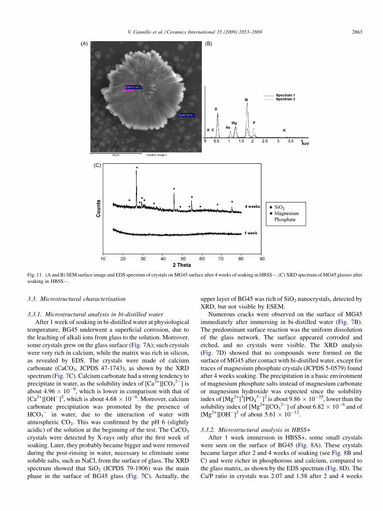
Fig. 11. (A and B) SEM surface image and EDS spectrum of crystals on MG45 surface after 4 weeks of soaking in HBSS�. (C) XRD spectrum of MG45 glasses after
soaking in HBSS�.
V. Cannillo et al. / Ceramics International 35 (2009) 2853–2869 2863
3.3. Microstructural characterisation
3.3.1. Microstructural analysis in bi-distilled water
After 1 week of soaking in bi-distilled water at physiological
temperature, BG45 underwent a superficial corrosion, due to
the leaching of alkali ions from glass to the solution. Moreover,
some crystals grew on the glass surface (Fig. 7A); such crystals
were very rich in calcium, while the matrix was rich in silicon,
as revealed by EDS. The crystals were made of calcium
carbonate (CaCO3, JCPDS 47-1743), as shown by the XRD
spectrum (Fig. 7C). Calcium carbonate had a strong tendency to
precipitate in water, as the solubility index of [Ca2+][CO32�] is
about 4.96 � 10�9, which is lower in comparison with that of
[Ca2+][OH�]2, which is about 4.68 � 10�6. Moreover, calcium
carbonate precipitation was promoted by the presence of
HCO3� in water, due to the interaction of water with
atmospheric CO2. This was confirmed by the pH 6 (slightly
acidic) of the solution at the beginning of the test. The CaCO3
crystals were detected by X-rays only after the first week of
soaking. Later, they probably became bigger and were removed
during the post-rinsing in water, necessary to eliminate some
soluble salts, such as NaCl, from the surface of glass. The XRD
spectrum showed that SiO2 (JCPDS 79-1906) was the main
phase in the surface of BG45 glass (Fig. 7C). Actually, the
upper layer of BG45 was rich of SiO2 nanocrystals, detected by
XRD, but not visible by ESEM.
Numerous cracks were observed on the surface of MG45
immediately after immersing in bi-distilled water (Fig. 7B).
The predominant surface reaction was the uniform dissolution
of the glass network. The surface appeared corroded and
etched, and no crystals were visible. The XRD analysis
(Fig. 7D) showed that no compounds were formed on the
surface of MG45 after contact with bi-distilled water, except for
traces of magnesium phosphate crystals (JCPDS 5-0579) found
after 4 weeks soaking. The precipitation in a basic environment
of magnesium phosphate salts instead of magnesium carbonate
or magnesium hydroxide was expected since the solubility
index of [Mg2+]3[PO43�]2 is about 9.86 � 10�25, lower than the
solubility index of [Mg2+][CO32�] of about 6.82 � 10�6 and of
[Mg2+][OH�]2 of about 5.61 � 10�12.
3.3.2. Microstructural analysis in HBSS+
After 1 week immersion in HBSS+, some small crystals
were seen on the surface of BG45 (Fig. 8A). These crystals
became larger after 2 and 4 weeks of soaking (see Fig. 8B and
C) and were richer in phosphorous and calcium, compared to
the glass matrix, as shown by the EDS spectrum (Fig. 8D). The
Ca/P ratio in crystals was 2.07 and 1.58 after 2 and 4 weeks
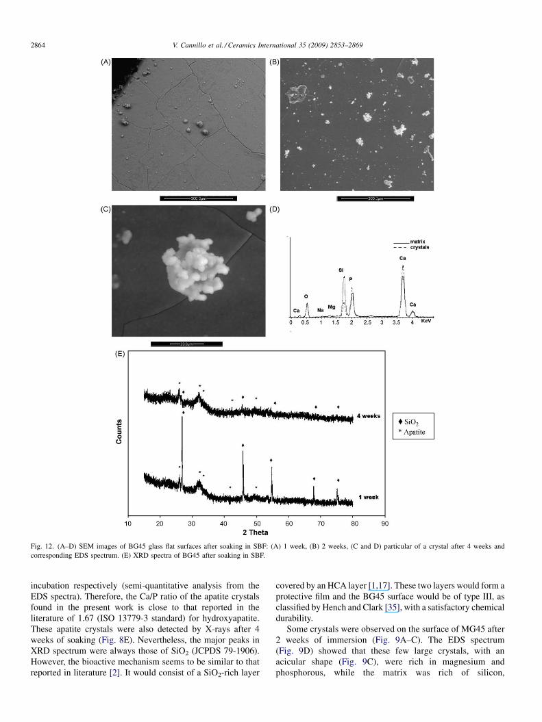
Fig. 12. (A–D) SEM images of BG45 glass flat surfaces after soaking in SBF: (A) 1 week, (B) 2 weeks, (C and D) particular of a crystal after 4 weeks and
corresponding EDS spectrum. (E) XRD spectra of BG45 after soaking in SBF.
V. Cannillo et al. / Ceramics International 35 (2009) 2853–28692864
incubation respectively (semi-quantitative analysis from the
EDS spectra). Therefore, the Ca/P ratio of the apatite crystals
found in the present work is close to that reported in the
literature of 1.67 (ISO 13779-3 standard) for hydroxyapatite.
These apatite crystals were also detected by X-rays after 4
weeks of soaking (Fig. 8E). Nevertheless, the major peaks in
XRD spectrum were always those of SiO2 (JCPDS 79-1906).
However, the bioactive mechanism seems to be similar to that
reported in literature [2]. It would consist of a SiO2-rich layer
covered by an HCA layer [1,17]. These two layers would form a
protective film and the BG45 surface would be of type III, as
classified by Hench and Clark [35], with a satisfactory chemical
durability.
Some crystals were observed on the surface of MG45 after
2 weeks of immersion (Fig. 9A–C). The EDS spectrum
(Fig. 9D) showed that these few large crystals, with an
acicular shape (Fig. 9C), were rich in magnesium and
phosphorous, while the matrix was rich of silicon,
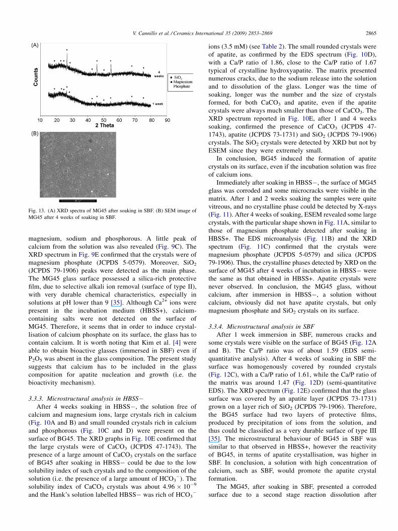
Fig. 13. (A) XRD spectra of MG45 after soaking in SBF. (B) SEM image of
MG45 after 4 weeks of soaking in SBF.
V. Cannillo et al. / Ceramics International 35 (2009) 2853–2869 2865
magnesium, sodium and phosphorous. A little peak of
calcium from the solution was also revealed (Fig. 9C). The
XRD spectrum in Fig. 9E confirmed that the crystals were of
magnesium phosphate (JCPDS 5-0579). Moreover, SiO2
(JCPDS 79-1906) peaks were detected as the main phase.
The MG45 glass surface possessed a silica-rich protective
film, due to selective alkali ion removal (surface of type II),
with very durable chemical characteristics, especially in
solutions at pH lower than 9 [35]. Although Ca2+ ions were
present in the incubation medium (HBSS+), calcium-
containing salts were not detected on the surface of
MG45. Therefore, it seems that in order to induce crystal-
lisation of calcium phosphate on its surface, the glass has to
contain calcium. It is worth noting that Kim et al. [4] were
able to obtain bioactive glasses (immersed in SBF) even if
P2O5 was absent in the glass composition. The present study
suggests that calcium has to be included in the glass
composition for apatite nucleation and growth (i.e. the
bioactivity mechanism).
3.3.3. Microstructural analysis in HBSS�After 4 weeks soaking in HBSS�, the solution free of
calcium and magnesium ions, large crystals rich in calcium
(Fig. 10A and B) and small rounded crystals rich in calcium
and phosphorous (Fig. 10C and D) were present on the
surface of BG45. The XRD graphs in Fig. 10E confirmed that
the large crystals were of CaCO3 (JCPDS 47-1743). The
presence of a large amount of CaCO3 crystals on the surface
of BG45 after soaking in HBSS� could be due to the low
solubility index of such crystals and to the composition of the
solution (i.e. the presence of a large amount of HCO3�). The
solubility index of CaCO3 crystals was about 4.96 � 10�9
and the Hank’s solution labelled HBSS� was rich of HCO3�
ions (3.5 mM) (see Table 2). The small rounded crystals were
of apatite, as confirmed by the EDS spectrum (Fig. 10D),
with a Ca/P ratio of 1.86, close to the Ca/P ratio of 1.67
typical of crystalline hydroxyapatite. The matrix presented
numerous cracks, due to the sodium release into the solution
and to dissolution of the glass. Longer was the time of
soaking, longer was the number and the size of crystals
formed, for both CaCO3 and apatite, even if the apatite
crystals were always much smaller than those of CaCO3. The
XRD spectrum reported in Fig. 10E, after 1 and 4 weeks
soaking, confirmed the presence of CaCO3 (JCPDS 47-
1743), apatite (JCPDS 73-1731) and SiO2 (JCPDS 79-1906)
crystals. The SiO2 crystals were detected by XRD but not by
ESEM since they were extremely small.
In conclusion, BG45 induced the formation of apatite
crystals on its surface, even if the incubation solution was free
of calcium ions.
Immediately after soaking in HBSS�, the surface of MG45
glass was corroded and some microcracks were visible in the
matrix. After 1 and 2 weeks soaking the samples were quite
vitreous, and no crystalline phase could be detected by X-rays
(Fig. 11). After 4 weeks of soaking, ESEM revealed some large
crystals, with the particular shape shown in Fig. 11A, similar to
those of magnesium phosphate detected after soaking in
HBSS+. The EDS microanalysis (Fig. 11B) and the XRD
spectrum (Fig. 11C) confirmed that the crystals were
magnesium phosphate (JCPDS 5-0579) and silica (JCPDS
79-1906). Thus, the crystalline phases detected by XRD on the
surface of MG45 after 4 weeks of incubation in HBSS� were
the same as that obtained in HBSS+. Apatite crystals were
never observed. In conclusion, the MG45 glass, without
calcium, after immersion in HBSS�, a solution without
calcium, obviously did not have apatite crystals, but only
magnesium phosphate and SiO2 crystals on its surface.
3.3.4. Microstructural analysis in SBF
After 1 week immersion in SBF, numerous cracks and
some crystals were visible on the surface of BG45 (Fig. 12A
and B). The Ca/P ratio was of about 1.59 (EDS semi-
quantitative analysis). After 4 weeks of soaking in SBF the
surface was homogenously covered by rounded crystals
(Fig. 12C), with a Ca/P ratio of 1.61, while the Ca/P ratio of
the matrix was around 1.47 (Fig. 12D) (semi-quantitative
EDS). The XRD spectrum (Fig. 12E) confirmed that the glass
surface was covered by an apatite layer (JCPDS 73-1731)
grown on a layer rich of SiO2 (JCPDS 79-1906). Therefore,
the BG45 surface had two layers of protective films,
produced by precipitation of ions from the solution, and
thus could be classified as a very durable surface of type III
[35]. The microstructural behaviour of BG45 in SBF was
similar to that observed in HBSS+, however the reactivity
of BG45, in terms of apatite crystallisation, was higher in
SBF. In conclusion, a solution with high concentration of
calcium, such as SBF, would promote the apatite crystal
formation.
The MG45, after soaking in SBF, presented a corroded
surface due to a second stage reaction dissolution after
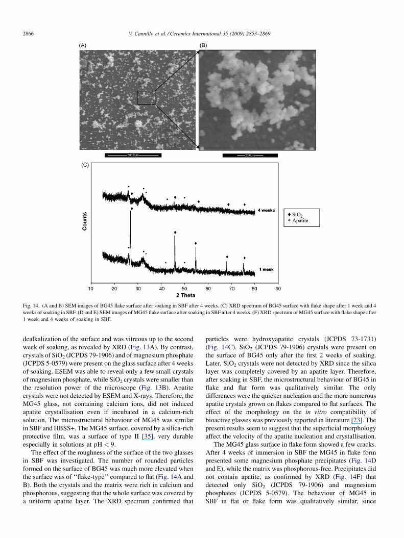
Fig. 14. (A and B) SEM images of BG45 flake surface after soaking in SBF after 4 weeks. (C) XRD spectrum of BG45 surface with flake shape after 1 week and 4
weeks of soaking in SBF. (D and E) SEM images of MG45 flake surface after soaking in SBF after 4 weeks. (F) XRD spectrum of MG45 surface with flake shape after
1 week and 4 weeks of soaking in SBF.
V. Cannillo et al. / Ceramics International 35 (2009) 2853–28692866
dealkalization of the surface and was vitreous up to the second
week of soaking, as revealed by XRD (Fig. 13A). By contrast,
crystals of SiO2 (JCPDS 79-1906) and of magnesium phosphate
(JCPDS 5-0579) were present on the glass surface after 4 weeks
of soaking. ESEM was able to reveal only a few small crystals
of magnesium phosphate, while SiO2 crystals were smaller than
the resolution power of the microscope (Fig. 13B). Apatite
crystals were not detected by ESEM and X-rays. Therefore, the
MG45 glass, not containing calcium ions, did not induced
apatite crystallisation even if incubated in a calcium-rich
solution. The microstructural behaviour of MG45 was similar
in SBF and HBSS+. The MG45 surface, covered by a silica-rich
protective film, was a surface of type II [35], very durable
especially in solutions at pH < 9.
The effect of the roughness of the surface of the two glasses
in SBF was investigated. The number of rounded particles
formed on the surface of BG45 was much more elevated when
the surface was of ‘‘flake-type’’ compared to flat (Fig. 14A and
B). Both the crystals and the matrix were rich in calcium and
phosphorous, suggesting that the whole surface was covered by
a uniform apatite layer. The XRD spectrum confirmed that
particles were hydroxyapatite crystals (JCPDS 73-1731)
(Fig. 14C). SiO2 (JCPDS 79-1906) crystals were present on
the surface of BG45 only after the first 2 weeks of soaking.
Later, SiO2 crystals were not detected by XRD since the silica
layer was completely covered by an apatite layer. Therefore,
after soaking in SBF, the microstructural behaviour of BG45 in
flake and flat form was qualitatively similar. The only
differences were the quicker nucleation and the more numerous
apatite crystals grown on flakes compared to flat surfaces. The
effect of the morphology on the in vitro compatibility of
bioactive glasses was previously reported in literature [23]. The
present results seem to suggest that the superficial morphology
affect the velocity of the apatite nucleation and crystallisation.
The MG45 glass surface in flake form showed a few cracks.
After 4 weeks of immersion in SBF the MG45 in flake form
presented some magnesium phosphate precipitates (Fig. 14D
and E), while the matrix was phosphorous-free. Precipitates did
not contain apatite, as confirmed by XRD (Fig. 14F) that
detected only SiO2 (JCPDS 79-1906) and magnesium
phosphates (JCPDS 5-0579). The behaviour of MG45 in
SBF in flat or flake form was qualitatively similar, since
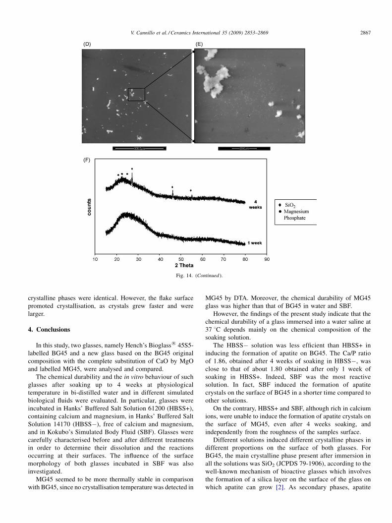
Fig. 14. (Continued ).
V. Cannillo et al. / Ceramics International 35 (2009) 2853–2869 2867
crystalline phases were identical. However, the flake surface
promoted crystallisation, as crystals grew faster and were
larger.
4. Conclusions
In this study, two glasses, namely Hench’s Bioglass1 45S5-
labelled BG45 and a new glass based on the BG45 original
composition with the complete substitution of CaO by MgO
and labelled MG45, were analysed and compared.
The chemical durability and the in vitro behaviour of such
glasses after soaking up to 4 weeks at physiological
temperature in bi-distilled water and in different simulated
biological fluids were evaluated. In particular, glasses were
incubated in Hanks’ Buffered Salt Solution 61200 (HBSS+),
containing calcium and magnesium, in Hanks’ Buffered Salt
Solution 14170 (HBSS�), free of calcium and magnesium,
and in Kokubo’s Simulated Body Fluid (SBF). Glasses were
carefully characterised before and after different treatments
in order to determine their dissolution and the reactions
occurring at their surfaces. The influence of the surface
morphology of both glasses incubated in SBF was also
investigated.
MG45 seemed to be more thermally stable in comparison
with BG45, since no crystallisation temperature was detected in
MG45 by DTA. Moreover, the chemical durability of MG45
glass was higher than that of BG45 in water and SBF.
However, the findings of the present study indicate that the
chemical durability of a glass immersed into a water saline at
37 8C depends mainly on the chemical composition of the
soaking solution.
The HBSS� solution was less efficient than HBSS+ in
inducing the formation of apatite on BG45. The Ca/P ratio
of 1.86, obtained after 4 weeks of soaking in HBSS�, was
close to that of about 1.80 obtained after only 1 week of
soaking in HBSS+. Indeed, SBF was the most reactive
solution. In fact, SBF induced the formation of apatite
crystals on the surface of BG45 in a shorter time compared to
other solutions.
On the contrary, HBSS+ and SBF, although rich in calcium
ions, were unable to induce the formation of apatite crystals on
the surface of MG45, even after 4 weeks soaking, and
independently from the roughness of the samples surface.
Different solutions induced different crystalline phases in
different proportions on the surface of both glasses. For
BG45, the main crystalline phase present after immersion in
all the solutions was SiO2 (JCPDS 79-1906), according to the
well-known mechanism of bioactive glasses which involves
the formation of a silica layer on the surface of the glass on
which apatite can grow [2]. As secondary phases, apatite
V. Cannillo et al. / Ceramics International 35 (2009) 2853–28692868
(JCPDS 73-1731) and calcium carbonate (JCPDS 47-1743)
were detected on the surface of BG45. The apatite
crystallisation could be attributed to the reactions of the
glass with different solutions, while CaCO3 crystallisation
was attributed to atmospheric CO2, after soaking in bi-
distilled water, and to the composition of the solution,
containing HCO3� after immersion in HBSS�. BG45 was
therefore a bioactive material, as known from literature,
since the apatite layer could be seen after soaking in different
simulated biological fluids [2].
An apatite layer was never seen on the surface of MG45
glass even if incubated for 4 weeks in solutions containing
calcium and magnesium. The crystalline phases detected on the
MG45 surface after immersion in different solutions were only
SiO2 (JCPDS 79-1906) and magnesium phosphate (JCPDS 5-
0579). Therefore, in order to induce the formation of apatite on
the surface of a glass as a reactivity of the glass-solution system,
the glass itself should contain calcium.
As regards the effect of the solution used, the bi-distilled
water was the less reactive medium for the BG45 glass.
Moreover, the chemical durability of BG45 was higher in
HBSS� than in HBSS+, since the apatite crystallisation was
more active in HBSS+. The reactivity of BG45 was higher in
SBF than in the other solutions, and SBF favoured the apatite
crystallisation. Therefore, the presence of high quantity of
calcium in the soaking solution, such as SBF, promotes the
formation of apatite crystals on glasses already containing
calcium.
The bioactive mechanism in SBF of BG45 was similar on
both flat and flake samples, even if the amount and the size of
crystals increased on flake samples. Therefore, the present
results confirm that the morphology of the surface of glasses
influenced nucleation and growth of crystals as the roughness
being a promoter of crystallisation.
Acknowledgements
The authors would like to thank Dr. M.A. Croce
(University of Modena and Reggio Emilia, Italy) for the
help in performing the in vitro tests. A. Gupta (Indian
Institute of Technology, Kanpur, India) is acknowledged for
his collaboration. Thanks also to Dr. M. Cannio for help with
the ICP-AES testing.
References
[1] L.L. Hench, Bioceramics: from concept to clinic, Journal of American
Ceramic Society 74 (1991) 1487–1510.
[2] L.L. Hench, Bioceramics, Journal of American Ceramic Society 81 (1998)
1705–1728.
[3] Clerk, L.L. Hench, The influence of surface chemistry on implant inter-
face, Journal of Biomedical Materials Researches 10 (1976) 161–174.
[4] H.M. Kim, F. Miyaji, T. Kokubo, Bioactivity of Na2O–CaO–SiO2 glasses,
Journal of American Ceramic Society 78 (1995) 2405–2411.
[5] E.E. Stroganova, N.Yu. Mikhailenko, O.A. Moroz, Glass-based biomater-
ials: present and future (a review), Glass and Ceramics 60 (2003) 315-.
[6] E. Jallot, Role of magnesium during spontaneous formation of a calcium
phosphate layer at the periphery of a bioactive glass coating doped with
MgO, Applied Surface Science 211 (2003) 89–95.
[7] F. Barrere, C.A. van Blitterswijk, K. De Groot, P. Layrolle, Influence of
ionic strength and carbonate on the Ca–P coating formation from SBF X 5
solution, Biomaterials 23 (2002) 1921.
[8] H.L. Ren, Y. Yue, C.H. Ye, L.P. Guo, J.H. Lei, NMR study of crystal-
lisation in MgO–CaO–SiO2–P2O5 glass–ceramics, Chemical Physics
Letters 2982 (1998) 317–322.
[9] S. Agathopoulos, D.U. Tulyaganov, J.M.G. Ventura, S. Kannan, M.A.
Karakassides, J.M.F. Ferreira, Formation of hydroxyapatite onto glasses of
the CaO–MgO–SiO2 system with B2O3, Na2O, CaF2 and P2O5 additives,
Biomaterials 27 (2006) 1832–1840.
[10] W. Holand, Biocompatible and bioactive glass–ceramics—state of the art
and new directions, Journal of Non-Crystalline Solids 219 (1997) 192–
197.
[11] V.K. Marghussian, A. Sheikh-Mehdi Mesgar, Effects of composition on
crystallization behaviour and mechanical properties of bioactive glass–
ceramics in the MgO–CaO–SiO2–P2O5 system, Ceramics International 26
(2000) 415–420.
[12] V. Banchet, J. Michel, E. Jallot, L. Worthman, S. Bouthors, D. Laurent-
Maquin, G. Balossier, Interfacial reactions of glasses for biomedical
application by scanning transmission electron microscopy and microa-
nalysis, Acta Biomaterialia 2 (2006) 349–359.
[13] J.M. Oliveira, R.N. Correia, M.H. Fernandes, Surface modifications of a
glass and a glass–ceramic of the MgO–3CaO*P2O5–SiO2-system in a
simulated body fluid, Biomaterials 16 (1995) 849–854.
[14] R.Z. Legeros, R. Kijkowska, I. Kattech, M. Jemal, J.P. Legeros, Journal of
Dental Research 65 (1986) 783.
[15] I. Barrios de Arenas, C. Schattner, M. Vasquez, Bioactivity and mechan-
ical properties of Na2O.CaO.SiO2.P2O5 modified glass, Ceramics Inter-
national 32 (2006) 515–520.
[16] W. Cao, L.L. Hench, Bioactive materials, Ceramics International 22
(1996) 493–507.
[17] P.N. De Aza, A.H. De Aza, P. Pena, S. De Aza, Bioactive glass and glass–
ceramics, Ceramica y Vidro Bolletin Sociedad Espanola Ceramica 46 (2)
(2007) 45–55.
[18] M. Cerruti, D. Greenspan, K. Powers, Effect of pH and ionic strength on
the reactivity of Bioglass 45S5, Biomaterials 26 (2005) 1665–1674.
[19] M. Cerruti, C.L. Bianchi, F. Bonino, A. Damin, A. Perardi, C. Morterra,
Surface modifications of bioglass immersed in TRIS buffered solutions. A
multitechnical spectroscopy study, The Journal of Physical Chemistry B
109 (2005) 14496–14505.
[20] A.K. Varshneya, Fundamentals of Inorganic Glasses, Academic Press,
Boston, 1993.
[21] T. Kokubo, H. Takadama, How useful is SBF in predicting in vivo bone
bioactivity? Biomaterials 27 (2006) 2907–2915.
[22] J.H. Hanks, R.E. Wallace, Proceedings of the Society for Experimental
Biology and Medicine 71 (1949) 196.
[23] A.L. Andrade, P. Valerio, A.M. Goes, M.F. Leite, R.Z. Domingues,
Influence of morphology on in vitro compatibility of bioactive glasses,
Journal of Non-Crystalline Solids 2352 (2006) 3508–3511.
[24] M.B. Volf, Chemical Approach to Glass, Elsevier, 1984.
[25] K. Shimoda, T. Nemoto, K. Saito, Local structure of magnesium in silicate
glasses: a 25Mg 3QMAS NMR study, Journal of Physical Chemistry B 112
(2008) 6747–6752.
[26] A.Z. Dietzel, Die Kationenfeldstirken und ihre Beziehungen zu Entgla-
sungsvorgangen, zur Verbindungsbildung und zu den Schmelzpunkten
von Silikaten, Z Elektrochemie 48 (1942) 9–23.
[27] K.-H. Sun, Fundamental condition of glass formation, Journal of the
American Ceramic Society 30 (1947) 277.
[28] H.A. ElBatal, M.A. Azooz, E.M.A. Khalil, A.S. Monem, Y.M. Hamdy,
Characterisation of some bioglass–ceramics, Materials Chemistry and
Physics 80 (2003) 599–609.
[29] L. Lefebre, J. Chevalier, L. Gremillard, R. Zenati, G. Thollet, D.
Bernache-Assolant, A. Govin, Structural transformations of bioactive
glass 45S5 with thermal treatments, Acta Materialia 55 (2007) 3305–
3313.
[30] A.R. Boccaccini, Q. Chen, L. Lefebre, L. Gremillard, J. Chevalier,
Sintering, crystallisation and biodegradation behaviour of Bioglass1-
derived glass–ceramic, Faraday Discussions 136 (2007) 27–44.
V. Cannillo et al. / Ceramics International 35 (2009) 2853–2869 2869
[31] T.R. Zeitler, A.N. Cormack, Interaction of water with bioactive glass
surfaces, Journal of Crystal Growth 294 (2006) 96–102.
[32] R.K. Iler, The Chemistry of Silica, Wiley, New York, 1979.
[33] D.S. Brauer, C. Russel, J. Kraft, Solubility of glasses in the system
P2O5–CaO–MgO–Na2O–TiO2: experimental and modeling using arti-
ficial neural networks, Journal of Non-Crystalline Solids 353 (2003)
263–270.
[34] A.B. Jedlicka, A.G. Clare, Chemical durability of commercial silicate
glasses. I. Material characterisation, Journal of Non-Crystalline Solids 281
(2001) 6–24.
[35] L.L. Hench, D.E. Clark, Physical chemistry of glass surfaces, Journal of
Non-Crystalline Solids 28 (1978) 83.
[36] D.E. Clark, E.L. Yen-Bower, Corrosion of glass surfaces, Surface Science
100 (1980) 53–70.