Characterization of the Substrate Specificity of Human Carboxypeptidase A4 and Implications for a...
38
1 File: CPA4 characterization –v39 Date: April 9, 2010 JBC/2009/060350 –r3 Human carboxypeptidase A4: Characterization of the substrate specificity and implications for a role in extracellular peptide processing Sebastian Tanco ‡ , Xin Zhang § , Cain Morano § , Francesc X. Aviles ‡ , Julia Lorenzo ‡1 and Lloyd D. Fricker §2 ‡ Institut de Biotecnologia i de Biomedicina & Departament de Bioquimica, Universitat Autonoma de Barcelona, Bellaterra (Barcelona), Spain § Department of Molecular Pharmacology, Albert Einstein College of Medicine, Bronx, NY, USA, 10461 1 To whom correspondence should be addressed: Institut de Biotecnologia i de Biomedicina & Departament de Bioquimica, Universitat Autonoma de Barcelona, IBB-Campus de la UAB, Bellaterra (Barcelona), 08193, Spain.Tel.: 34-935811231; Fax: 34-935812011; E-mail: [email protected] 2 To whom correspondence should be addressed: Dept. of Molecular Pharmacology, Albert Einstein College of Medicine, 1300 Morris Park Ave., Bronx, NY 10461. Tel.: 718-430- 4225; Fax: 718-430-8922; E-mail: [email protected] . Carboxypeptidase A4 (CPA4) is a member of the metallocarboxypeptidase family. CPA4 was originally found in a screen of mRNAs upregulated by sodium butyrate- induced differentiation of cancer cells. Further studies suggested a relation between CPA4 and prostate cancer aggressiveness. In the present study, we determined that CPA4 is secreted from cells as a soluble proenzyme (proCPA4) that can be activated by endoproteases such as trypsin. Three complementary approaches were used to study the substrate specificity of CPA4: kinetic analysis was performed using a new series of chromogenic substrates and some biologically relevant peptides, the cleavage of synthetic peptides was tested individually, and the cleavage of a mixture of >100 mouse brain peptides was examined using a quantitative peptidomics mass spectrometry-based approach. CPA4 was able to cleave hydrophobic C-terminal residues with a preference for Phe, Leu, Ile, Met, Tyr and Val. However, not all peptides with C-terminal hydrophobic residues were cleaved, indicating the importance of additional residues within the peptide. Aliphatic, aromatic and basic residues in the P1 position have a positive influence on the cleavage specificity. In contrast acidic residues, Pro, and Gly have a negative influence in the P1 position. Some of the peptides identified as CPA4 substrates (such as neurotensin, granins, and opioid peptides) have been previously shown to function in cell proliferation and differentiation, potentially explaining the link between CPA4 and cancer aggressiveness. Taken together, these studies suggest that CPA4 functions in neuropeptide processing and regulation in the extracellular environment. Running Title: Characterization of human carboxypeptidase A4 Keywords: peptidase, protease, neuropeptide, carboxypeptidase A4, peptidomics http://www.jbc.org/cgi/doi/10.1074/jbc.M109.060350 The latest version is at JBC Papers in Press. Published on April 12, 2010 as Manuscript M109.060350 Copyright 2010 by The American Society for Biochemistry and Molecular Biology, Inc. by guest on September 4, 2016 http://www.jbc.org/ Downloaded from by guest on September 4, 2016 http://www.jbc.org/ Downloaded from by guest on September 4, 2016 http://www.jbc.org/ Downloaded from by guest on September 4, 2016 http://www.jbc.org/ Downloaded from by guest on September 4, 2016 http://www.jbc.org/ Downloaded from by guest on September 4, 2016 http://www.jbc.org/ Downloaded from by guest on September 4, 2016 http://www.jbc.org/ Downloaded from by guest on September 4, 2016 http://www.jbc.org/ Downloaded from
Transcript of Characterization of the Substrate Specificity of Human Carboxypeptidase A4 and Implications for a...
1
File: CPA4 characterization –v39 Date: April 9, 2010 JBC/2009/060350 –r3 Human carboxypeptidase A4: Characterization of the substrate specificity and implications for a role in extracellular peptide processing Sebastian Tanco‡, Xin Zhang§, Cain Morano§, Francesc X. Aviles‡, Julia Lorenzo‡1 and Lloyd D. Fricker§2 ‡Institut de Biotecnologia i de Biomedicina & Departament de Bioquimica, Universitat Autonoma de Barcelona, Bellaterra (Barcelona), Spain §Department of Molecular Pharmacology, Albert Einstein College of Medicine, Bronx, NY, USA, 10461 1 To whom correspondence should be addressed: Institut de Biotecnologia i de Biomedicina & Departament de Bioquimica, Universitat Autonoma de Barcelona, IBB-Campus de la UAB, Bellaterra (Barcelona), 08193, Spain.Tel.: 34-935811231; Fax: 34-935812011; E-mail: [email protected] 2 To whom correspondence should be addressed: Dept. of Molecular Pharmacology, Albert Einstein College of Medicine, 1300 Morris Park Ave., Bronx, NY 10461. Tel.: 718-430-4225; Fax: 718-430-8922; E-mail: [email protected]. Carboxypeptidase A4 (CPA4) is a member of the metallocarboxypeptidase family. CPA4 was originally found in a screen of mRNAs upregulated by sodium butyrate-induced differentiation of cancer cells. Further studies suggested a relation between CPA4 and prostate cancer aggressiveness. In the present study, we determined that CPA4 is secreted from cells as a soluble proenzyme (proCPA4) that can be activated by endoproteases such as trypsin. Three complementary approaches were used to study the substrate specificity of CPA4: kinetic analysis was performed using a new series of chromogenic substrates and some biologically relevant peptides, the cleavage of synthetic peptides was tested individually, and the cleavage of a mixture of >100 mouse brain peptides was examined using a quantitative peptidomics mass spectrometry-based approach. CPA4 was able to cleave hydrophobic C-terminal residues with a preference for Phe, Leu,
Ile, Met, Tyr and Val. However, not all peptides with C-terminal hydrophobic residues were cleaved, indicating the importance of additional residues within the peptide. Aliphatic, aromatic and basic residues in the P1 position have a positive influence on the cleavage specificity. In contrast acidic residues, Pro, and Gly have a negative influence in the P1 position. Some of the peptides identified as CPA4 substrates (such as neurotensin, granins, and opioid peptides) have been previously shown to function in cell proliferation and differentiation, potentially explaining the link between CPA4 and cancer aggressiveness. Taken together, these studies suggest that CPA4 functions in neuropeptide processing and regulation in the extracellular environment. Running Title: Characterization of human carboxypeptidase A4 Keywords: peptidase, protease, neuropeptide, carboxypeptidase A4, peptidomics
http://www.jbc.org/cgi/doi/10.1074/jbc.M109.060350The latest version is at JBC Papers in Press. Published on April 12, 2010 as Manuscript M109.060350
Copyright 2010 by The American Society for Biochemistry and Molecular Biology, Inc.
by guest on September 4, 2016
http://ww
w.jbc.org/
Dow
nloaded from
by guest on September 4, 2016
http://ww
w.jbc.org/
Dow
nloaded from
by guest on September 4, 2016
http://ww
w.jbc.org/
Dow
nloaded from
by guest on September 4, 2016
http://ww
w.jbc.org/
Dow
nloaded from
by guest on September 4, 2016
http://ww
w.jbc.org/
Dow
nloaded from
by guest on September 4, 2016
http://ww
w.jbc.org/
Dow
nloaded from
by guest on September 4, 2016
http://ww
w.jbc.org/
Dow
nloaded from
by guest on September 4, 2016
http://ww
w.jbc.org/
Dow
nloaded from
2
INTRODUCTION Carboxypeptidases (CPs) hydrolyze a single amino acid from the C-terminus of peptides and proteins. MetalloCPs use a catalytic mechanism in which nucleophilic attack on a peptide bond is mediated by a Zn2+-activated water molecule (1). Most metalloCPs are classified based on amino acid sequence, structure and function into subfamilies within clan MC, Family M14 (2). These subfamilies include; M14A, also known as the CPA/CPB subfamily; M14B, also known as the CPN/CPE subfamily; M14C, that includes gamma-D-glutamyl-(L)-meso-diaminopimelate peptidase I from Bacillus sphaericus, and a proposed fourth subfamily including cytosolic CPs (CCPs) (3,4). MetalloCPs have also been divided into subgroups based on substrate specificity: A-like enzymes with preference for hydrophobic C-terminal amino acids; B-like enzymes which cleave C-terminal basic residues; CPs with preference for glutamate in the C-terminal position; and CPs with a broad substrate specificity. Carboxypeptidase A4 (CPA4) belongs to the M14A subfamily of carboxypeptidases. The pancreatic members of this subfamily (CPA1, CPA2 and CPB) act in the degradation of dietary proteins in the digestive tract. Other members of this subfamily display a wide spectrum of physiological roles. Mast cell carboxypeptidase (recently renamed CPA3) is found in the secretory granules of mast cells and plays a role in defense (5). CPA6 is linked to Duane syndrome, a congenital eye-movement disorder, and has been proposed to participate in the regulation of neuropeptides in the extracellular matrix within the olfactory bulb and other parts of the brain (6). CPU, also known as CPB2 and thrombin-activatable fibrinolysis inhibitor (TAFI), participates in fibrinolysis inhibition and possesses anti-inflammatory properties (7), and for these
reasons is of interest to the pharmaceutical industry (8). CPA4 was originally referred to as CPA3 (9), but was renamed CPA4 to reflect the order in which the CPAs were discovered. Huang et al identified CPA4 in a search for mRNAs induced by sodium butyrate in androgen-independent prostate cancer cells (9). The histone deacetylase inhibitor trichostatin A also induced the expression of CPA4 mRNA in PC-3, DU145 and BPH1 human prostate cancer cell lines (9). Huang et al. also reported that CPA4 mRNA expression is associated with hormone-regulated tissues, suggesting that it may have a role in cell growth and differentiation. The human CPA4 gene is located on chromosome 7q32, which is a region in the genome that might contain genes for prostate cancer aggressiveness (10). In addition, the CPA4 gene was found to be maternally imprinted in a tissue-specific manner (11,12). Furthermore, imprinting in adult benign hypertrophic prostate tissue suggests that mutations or aberrant imprinting in CPA4 may be related to prostate cancer aggressiveness (12). Ross et al. studied the association of single-nucleotide polymorphisms (SNPs) on the CPA4 gene and its relation to prostate cancer and found that the nonsynonymous coding SNP (Gly303Cys) rs2171492 was associated with an increased risk of aggressive disease in younger men (13). Despite the potential importance of CPA4 towards prostate and other cancers, no previous studies have examined the substrate specificity of CPA4 in detail. The three-dimensional structure of CPA4 has been determined for the zymogen state (14) and for the active form in complex with latexin, a brain-derived CP inhibitor (15), and in complex with a hexapeptide (16). This latter study also compared the cleavage of a handful of synthetic peptides by CPA4, but no kinetic details were provided. To gain a better understanding of the enzymatic properties of
by guest on September 4, 2016
http://ww
w.jbc.org/
Dow
nloaded from

3
CPA4, we used a variety of approaches to characterize the substrate specificity. A series of di-peptide chromogenic substrates were synthesized and tested with purified CPA4. A number of synthetic peptides were individually tested with purified CPA4; many of these peptides correspond to biologically active peptides. A third approach involved incubating CPA4 with a mixture of over 100 endogenous peptides extracted from mouse brain, using a quantitative peptidomics approach to identify substrates and products. The subcellular distribution, secretion, and pH optimum of CPA4 were also examined. Taken together, these studies provide a complementary and thorough analysis of the cleavage specificity of CPA4 and suggest that this enzyme functions as a soluble extracellular peptidase that removes C-terminal hydrophobic/aliphatic residues from secreted peptides.
EXPERIMENTAL PROCEDURES
Protein Production and Purification. Human proCPA4 (PCPA4) was produced using the vector pPIC9 and the methylotrophic yeast Pichia pastoris as an expression host and purified as described elsewhere (15). The active enzyme was obtained through tryptic activation (at a 1/10 w/w ratio) for 60 min at room temperature, and the resulting product was subsequently purified by anion-exchange chromatography (TSK-DEAE 5PW) using a FPLC-Äkta system with a linear salt gradient from 0 to 30% of 0.4 M ammonium acetate in 20 mM Tris-HCl (pH 10.5). Eluted fractions were analyzed by SDS−PAGE, and the purest samples containing the active enzyme were pooled, desalted, and concentrated to 1 mg/mL by Amicon centrifugal filter devices.
Cell culture and Transfection HeLa (human cervix adenocarcinoma), PC-3, DU145 and LNCaP (all three cell lines are human prostate carcinoma), 1BR3.G (human skin fibroblasts) and HEK 293T (human embryo kidney) cells were purchased from the American Type Culture Collection (Manassas, VA), and were cultured in the recommended growth medium: Eagle's Minimum Essential Medium or Dulbecco’s Modified Eagle’s Medium with 4500 mg/L glucose, GlutaMAX–I and pyruvate supplemented with 10% fetal calf serum (Invitrogen, Grand Island, NY, USA). The full-length cDNA sequence of human proCPA4 was cloned into pcDNA3.1 vector (Invitrogen) and the hemagglutinin (HA) epitope was introduced on the C-terminus of CPA4. The plasmid pDsRed2-ER from Clontech was used for endoplasmic reticulum (ER) localization. Cells were grown in 35 mm dishes and transfected with Lipofectamine 2000 according to the manufacturer’s instructions. Cell fractionation and processing Forty eight hours after transfection, medium was removed, centrifuged and collected on ice. The dishes were gently washed with phosphate-buffered saline (PBS), and cells were removed by pipetting up and down in a mild extraction buffer consisting of 50 mM Tris-HCl pH 8.0. The cell suspension was sonicated and centrifuged at 16,000 g for 10 min at 4°C. The pellet was resuspended in an equivalent amount of 50 mM Tris-HCl pH 8.0, containing 500 mM NaCl and 1% Nonidet P-40 and centrifuged as above to produce a detergent/high salt extract. Following the removal of cells, the plate was washed several times with PBS and the extracellular matrix (ECM) was extracted by adding hot SDS-PAGE sample buffer directly to the plate. All homogenates were frozen and stored at −70°C until analysis. For trypsin activation assays, 200 ng of trypsin (Sigma)
by guest on September 4, 2016
http://ww
w.jbc.org/
Dow
nloaded from
4
were added to 20 µl of the extracellular medium of transfected HEK 293T cells and incubated for 30 min at 37°C. Immunoblot For immunoblotting, equivalent amounts of each lysate or fraction were analyzed by SDS/10% PAGE, and the separated proteins were electrophoretically transferred onto polyvinylidene difluoride (PVDF) filters (Millipore Corp.). Non-specific binding sites on the PVDF filters were blocked by incubation with 5% skim milk for 12 h. The PVDF filters were then incubated with anti-HA antibody (dilution 1:3,000; Sigma-Aldrich) for 1 h at 25°C. After washing with PBS containing 0.05% Tween 20, the filters were incubated with goat anti-mouse IgG-peroxidase (dilution 1:10,000; Pierce) for 1 h. After three rinses, immunoreactive bands were visualized with a chemiluminescence detection kit (ECL; Millipore).
Immunofluorescence HeLa, HEK 293T, PC-3, DU145 or LNCaP cells were co-transfected with HA-tagged PCPA4 and pDsRed2-ER using Lipofectamine 2000 (Invitrogen) according to the manufacturer’s protocol. After 48 h incubation cells were fixed in methanol, permeabilized with 0.1% Triton X-100, and incubated with anti-HA antibody (clone HA-7, Sigma, 1:2,000 dilution), followed by conjugated Alexa-488 anti-mouse-IgG (Molecular Probes, 1:500 dilution) and DAPI (4’,6’-diamidino-2-phenylidole). Coverslips were mounted with Fluoprep mounting medium (Biomerieux), and analyzed with a confocal laser scanning microscope (Leica Microsystems, Bannockburn, IL).
Substrate synthesis Chromogenic substrates were synthesized according to Blumberg et al (17). 3-(2-furyl)acryloyl-Phe-N-hydroxysuccinimide (FA-Phe-Osu) was prepared according to the
following procedure: a solution of 15 mmol FA-Phe and 15 mmol N-hydroxysuccinimide in 60 ml of dioxane-DMF, 1:1, was cooled to 4ºC and 15 mmol dicyclohexylcarbodiimide was added. The solution was stirred at 4ºC for 16 hr. The dicyclohexyl urea was filtered and the solution evaporated in vacuo. Furylacryloyl dipeptides were prepared by reacting FA-Phe-Osu with the appropriate amino acid according to the following procedure: the N-hydroxysuccinimide ester (5 mmol) in 25 ml of dioxane was added at room temperature to a solution of the amino acid (6 mmol) and sodium bicarbonate (12 mmol) in 25 ml of water. The mixture was kept at room temperature for 6 hr, the dioxane evaporated, and the residue acidified with 1N HCI. After the mixture was cooled overnight at 4ºC, the precipitate was collected by filtration and dried.
Kinetic measurements The rate of hydrolysis of different substrates was continuously measured at 25ºC in 50 mM Tris 0.15 M NaCl at the optimal pH for each enzyme. The wavelengths varied slightly among substrates: 336 nm for FA-Phe-Phe, FA-Phe-Ile and FA-Phe-Trp; 338 nm for FA-Phe-Ala, FA-Phe-His and FA-Phe-Leu; and 340 nm for FA-Phe-Val and FA-Phe-Met. Initial rates determined from the first 5–10% of the time-trace of each reaction were obtained at substrate concentrations close to the Km value whenever possible. The kinetic parameters, kcat and Km, were obtained using 6 experimental points by direct fit to a Michaelis–Menten curve using a nonlinear least-squares regression analysis. For those substrates for which the solubility of the substrates limited the measurements to concentrations well below Km, the kcat/Km values were estimated from the slope of the linear portion of saturation curve using the simplified equation v=(kcat/Km).[E0].[S].
by guest on September 4, 2016
http://ww
w.jbc.org/
Dow
nloaded from

5
Measurement of equilibrium dissociation constants (Ki) The method for reversible tight-binding inhibitors described by Bieth (18) was used for Ki determination. Recombinant purified CPA4 at a concentration of 8 nM was assayed against increasing concentrations of inhibitor. Initial rates, determined from the first 5–10% of the time-trace of each reaction, were obtained for each inhibitor concentration using the N-(4-Methoxyphenylazoformyl)-L-phenylalanine (AAFP) substrate (Bachem). Ki values were obtained by direct fitting of fraction velocity versus inhibitor concentration data to the Morrison equation using the Grafit program.
Substrate specificity and kinetic measurements using peptides CPA4 substrate specificity was analyzed using synthetic peptide substrates. Reactions were performed in 50 mM Tris-HCl 100 mM NaCl pH 7.5 at 37ºC using a peptide and enzyme concentrations of 10 µmol and 1 nmol, respectively. Peptide cleavage reactions were stopped at different times (30 minutes, 1h, 2h and 16h) by dropping the pH to 4 using 1% TFA. The reaction products were desalted using a C18 ZipTip (Millipore) following the manufacturer’s protocol. The peptides were eluted with 1 µl of 70% acetonitrile, 0.1% trifluoroacetic acid, mixed with an equal volume of a saturated solution of α-hydroxycinnamic acid in the same solvent, and analyzed in a Bruker Daltonics Ultraflex matrix-assisted laser desorption ionization time-of-flight (MALDI-TOF) mass spectrometer. For the determination of kinetic constants for the hydrolysis of peptides, rates of substrate hydrolysis were determined by reversed-phase high performance liquid chromatography (RP-HPLC) in a Waters Alliance apparatus using a Grace Vydac 218TPTM C18 column (3 µm, 100Å, 4.6 mm
i.d. × 250 mm, Hesperia, CA) with a linear gradient of 5 to 35% acetonitrile and following the absorbance at either 214 or 280 nm. Reactions were performed at 37 °C for 30 minutes in 50mM Tris-HCl 100 mM NaCl pH 7.5 and stopped by the addition of 0.1% TFA. Substrate concentrations ranged from 0.2 to 5 times the Km whenever possible and the enzyme concentration was chosen so that hydrolysis was less than 10% product formed. The kinetic parameters, kcat and Km, were obtained by direct fit to a Michaelis–Menten curve using the Grafit program Peptidomics Peptides were extracted from whole mouse brain (minus the olfactory bulb and cerebellum) as described (19,20). In brief, four mice (C57B6/J, 2-3 months old) were sacrificed by decapitation and the head was immediately irradiated in a conventional microwave oven for 8 seconds to raise the brain temperature to 80°C and inactivate proteases that can rapidly degrade cellular proteins during postmortem processing of the tissue (21). The mouse brain was isolated, the olfactory bulb and cerebellum were discarded, and the remainder of the brain was frozen in dry ice and stored at -70°C until peptide extraction. Peptides were extracted by sonication of each brain in 1.0 mL ice-cold water followed by incubation at 70°C for 20 min, cooling in an ice bath, acidification with 120 µL of 0.1 M HCl to a final concentration of 10 mM HCl, and centrifugation at 13,000 g for 30 min at 4°C. The supernatants (peptide extracts) from each of the four mice were pooled, mixed, and 60 µl aliquots neutralized to pH 8 by 75 mM borate buffer and incubated in the presence of purified CPA4 (20 ng, 670 pg, or 22 pg), or in the absence of enzyme, for 90 minutes. The reactions were stopped by the addition of 2.5 mM benzyl-succinic acid and heat-inactivated at 80°C for 10 min. Labeling with 4-trimethyl-ammoniumbutyrate (TMAB) isotopic tags
by guest on September 4, 2016
http://ww
w.jbc.org/
Dow
nloaded from

6
containing either no (D0), 3 atoms (D3), 6 atoms (D6), or 9 atoms (D9) of deuterium was performed as described (19). After labeling, the TMAB reagent was quenched with glycine, the 4 samples were combined, and peptides were isolated by passage through a Microcon® YM-10 unit (Millipore) to remove proteins >10 kDa. The samples were treated with hydroxylamine to remove TMAB labels from Tyr residues, desalted with a PepCleanTM C-18 spin column (Pierce), and concentrated to ~20 µl in a vacuum centrifuge.
Mass spectrometry was performed as previously described (19,22). In brief, the peptide mixture was trapped and washed on a PepMapTM C18 trapping column (5 µm, 100Å, 300 µm i.d. × 5 mm, LC Packing, Marlton, NJ) and separated on a Grace Vydac MS C18 capillary column (3 µm, 100Å, 75 µm i.d. × 150 mm, Hesperia, CA) at 4 µL/min with an acetonitrile gradient in 0.1% formic acid. The column eluate was analyzed on an API Q-Star Pulsar-iTM quadrupole time-of-flight mass spectrometer (Applied Biosystems/MDS Sciex, Foster City, CA) in information-dependent acquisition mode, with a collision energy for MS/MS (20-45 eV) based on the m/z value and charge state of the ion selected.
Data was analyzed as described (19). In brief, Mascot (http://www.matrix-science.com) was used followed by manual verification; identifications were rejected unless 80% or more of the major fragments observed in MS/MS matched predicted b- or y-series fragments (minimum five matches). Additional criteria included all of the following: a parent mass within 40 ppm (preferably 20 ppm) of the theoretical mass; the observed number of TMAB tags on the peptide matched the predicted number of free amines available (i.e., Lys residue and N-terminus); and the observed charge state(s) of the peptide was consistent with the expected number of positive charges. The intensity of
the TMAB-labeled isotopic peaks were determined by measuring peak heights of the monoisotopic peak and the additional peaks containing 1 and 2 atoms of 13C. The peak intensities of the samples incubated with enzyme were compared to the peak intensity of the sample incubated without enzyme; previous studies examining 4 aliquots of brain extract with the 4 different isotopic tags showed average ratios of 1.00 (19). RESULTS
The sequence analysis of PCPA4 protein reveals the presence of a 16 amino acid-long signal peptide consensus sequence and therefore the protein is predicted to be translocated to the lumen of the ER upon synthesis. From the ER, many options are possible, including retention in the secretory pathway, targeting to lysosomes, or secretion. Because CPA4 lacks any transmembrane domains and other members of this subfamily are secreted, PCPA4 is predicted to also be secreted from cells. Some CPs such as CPA1 remain as soluble proteins in the extracellular environment (23) while others like CPA6 (6) or mouse CPZ (24) are retained within the ECM. In relation to proprotein processing, some proCPs are activated before or upon secretion by proteases present in the secretory pathway (6) while others are activated after secretion by extracellular proteases (23). To study whether PCPA4 is secreted and activated, HEK 293T cells were transiently transfected with HA-tagged PCPA4 cDNA. After 48 h of transfection, cells, media and ECM were collected, fractionated and analysed by Western blot. A band of an apparent molecular mass of 50 kDa corresponding to the full-length PCPA4 was detected predominantly in the cell culture medium (Fig. 1A lane 5). PCPA4 was also detected in two different cell extracts: the zymogen appears in a low salt extract but also
by guest on September 4, 2016
http://ww
w.jbc.org/
Dow
nloaded from
7
at low levels in a detergent/high salt extract (Fig. 1A lanes 1 and 3). The absence of any PCPA4 in the ECM shows that the protein remains as a soluble protein in the extracellular medium. In all cases PCPA4 is found in the full length form indicating that its activating protease is not present or active in sufficient levels in the system. To investigate if this behaviour would be similar in other cell lines, we repeated the transfection of PCPA4 in five different cell lines: DU145 and PC-3, which are prostate cancer cells previously found to express PCPA4 in relatively high levels (9); LNCaP, a prostate cancer cell line that does not express PCPA4; HeLa cells, which arise from a cervical adenocarcinoma; and 1BR.3.G, a human skin fibroblast cell line. In all cases, PCPA4 was predominantly found soluble in the extracellular medium in the zymogen form (data not shown).
To demonstrate that PCPA4 expressed in HEK 293T cells was properly folded and able to produce the enzymatically active form, extracellular medium collected after 48 h of transfection was treated with trypsin. Western blot analysis of these samples shows that trypsin causes the disappearance of the 50 kDa band corresponding to PCPA4 and the appearance of a 35 kDa band corresponding to the expected molecular weight of the active enzyme (Fig. 1B). Enzymatic analysis of these samples shows no detectable CP activity in the extracellular medium of PCPA4 transfected cells, but incubation with trypsin resulted in the appearance of A-type CP enzymatic activity against the AAFP substrate (data not shown). The finding that PCPA4 can be activated with trypsin indicates that it is properly folded and that the absence of detectable zymogen processing in the HEK 293T cells, as well as in various other cell lines, is due to insufficient levels of the activating enzyme and not due to the improper folding of PCPA4 in these cell lines.
The presence of PCPA4 in the cells was further investigated by immunocytochemistry. PCPA4 was found to show some overlap with an ER marker (Fig. 1C), which is consistent with the finding that intracellular PCPA4 is present in the secretory pathway of the cell.
Although there are several reports about CPA4 structure (14,15), expression (9) and relation to prostate cancer aggressiveness (11,12), a detailed analysis of its enzymatic properties has not been previously reported. Recombinant protein obtained in the Pichia pastoris system as previously described (15) was used for this purpose. Mature CPA4 was obtained after treatment of the proenzyme with trypsin and subsequent purification. As expected, the active enzyme shows the ability to remove C-terminal residues from both FA-Phe-Phe and FA-Phe-Ala. No detectable activity towards substrates with basic (FA-Ala-Arg) or acidic (hippuryl-L-glutamic acid) C-terminal residues was found. This is in accordance with predictions based on its sequence and structure that includes CPA4 in the group of A-type CPs. The Pichia-expressed proenzyme shows 3% of residual activity against the AAFP substrate when compared to the active enzyme. Although the proenzyme shows residual activity, it could not be detected in the conditioned media of PCPA4 transfected HEK 293T cells because this activity falls below the limit of detection of the technique.
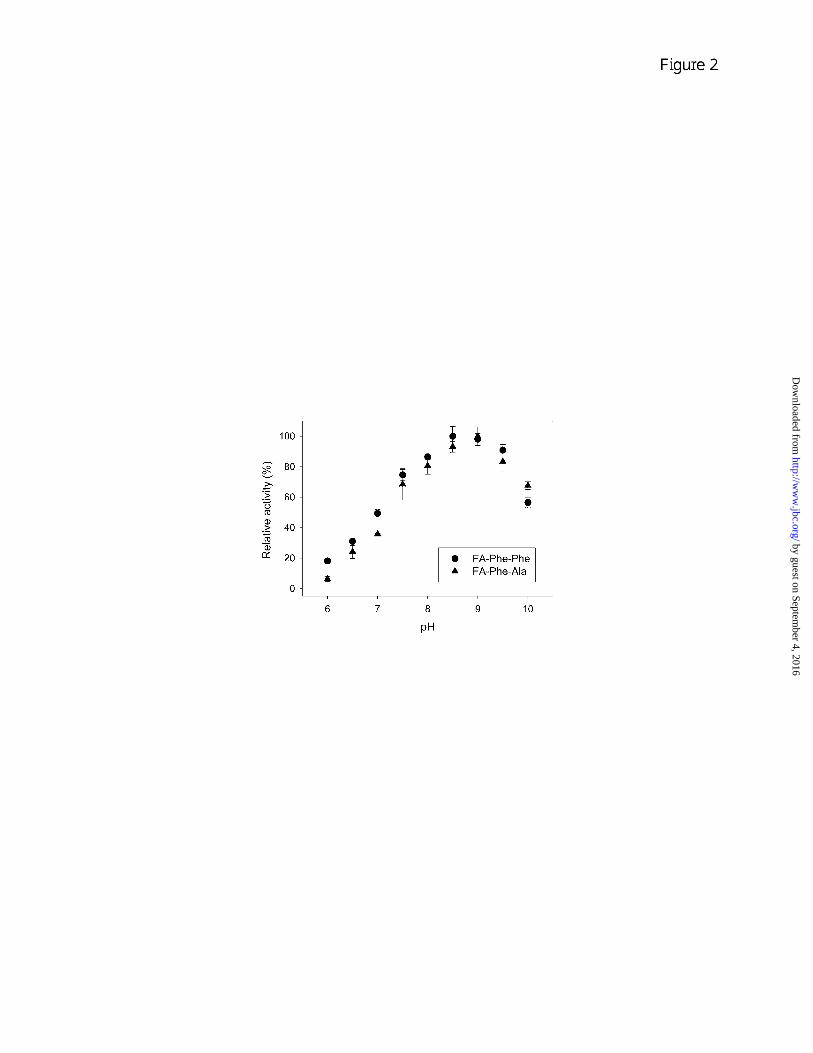
The influence of pH on CPA4 activity was analyzed using both FA-Phe-Phe and FA-Phe-Ala. The optimal pH for cleavage of either substrate is in the range of 8.5-9 with very low activity below pH 6 (Fig. 2). This suggests that CPA4 is maximally active in the extracellular environment after secretion, and has little activity within the acidic environment of the secretory pathway.
The effect of several different CP inhibitors on CPA4 activity was assayed. Benzylsuccinic acid (25), a potent synthetic
by guest on September 4, 2016
http://ww
w.jbc.org/
Dow
nloaded from

8
inhibitor of the A-type CP subfamily, at a concentration of 100 µM produced 30% of inhibition on CPA4 activity and completely inhibited the enzyme when 1 mM concentration of the inhibitor was used. The Zn2+-chelating agent 1,10-phenanthroline (26) showed 70% inhibition of the CP activity after 30 minutes of incubation at 1 mM concentration. CPA4 showed resistance against the action of EDTA, another chelating inhibitor of CPs: complete inhibition of CPA4 activity was only achieved using a 100 mM concentration of the inhibitor in an overnight incubation, while the same inhibitor concentration caused only 60% inhibition after 5 hours of treatment. Recombinant forms of potato CP inhibitor (PCI) (27), leech CP inhibitor (LCI) (28) and tick CP inhibitor (TCI) (29) were tested with CPA4 and found to exhibit equilibrium dissociation constant (Ki) values in the low nanomolar range (Supplementary Table S1). The protein inhibitor with the highest affinity for CPA4 was found to be that from tick Rhipicephalus bursa, for which a constant of 0.8 ± 0.3 nM was obtained (Table S1). The Ki values for CPA4 determined in the present study are generally comparable to values previously published for CPA4 with latexin (15) and Ascaris carboxypeptidase inhibitor (ACI) (30); these results are included in Table S1 for comparison with the present data.
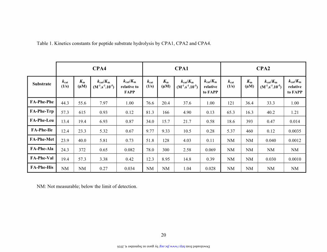
To investigate the substrate specificity of CPA4 a new series of substrates of the type FA-Phe-Xaa were synthesized. These substrates incorporated the furylacryloyl moiety as a chromophore, allowing for continuous monitoring of CP activity at a wavelength that has little interference from proteins or aromatic derivatives (31). This new series of substrates provided a broad range of hydrophobic/aliphatic residues in the C-terminal position so that substrate specificities of A-type CPs could be evaluated. The different amino acids included in the C-terminal position were His, Leu, Ile,
Phe, Trp, Ala, Val and Met. All substrates contain Phe in the penultimate position and are of the same length, allowing a direct comparison of the substrates’ C-terminal residues. Kinetic constants were determined for CPA4 and also for human CPA1 and CPA2 to directly compare these related enzymes with the same substrates. CPA1 and CPA2 were obtained as recombinant proteins in the Pichia pastoris system, purified in their zymogen form, activated with trypsin and further purified as previously described (32,33). kcat, Km and kcat/Km values are shown in Table 1, and indicate that although all three enzymes have overlapping specificities they differ markedly. While CPA1 and CPA4 display similar and broader substrate specificities, CPA2 shows a substrate specificity that is essentially restricted to large aromatic residues in the C-terminal position. CPA4 shows a preference for hydrophobic C-terminal amino acids like Phe, Leu, Met, Val, and Ile, as judged by kcat/Km values. Of all the dipeptide substrates tested with CPA4, FA-Phe-Trp exhibited the highest kcat, but the high Km resulted in a lower kcat/Km for this substrate than for FA-Phe-Phe. Overall, the kcat varied only 5-fold among the substrates that showed measureable kcat values, while the Km varied 32-fold and the kcat/Km varied 12-fold for these substrates (Table 1).
To further characterize the catalytic activity of CPA4, its ability to cleave a series of peptides was studied. Some of the peptides chosen for this analysis are biologically active and are known to be secreted into the extracellular fluid of tissues where CPA4 is expressed. Others are not likely to be biological substrates of CPA4 but provide further information of the ability of this enzyme to cleave peptides. All of these peptides were previously tested against CPA6 (6) so they are useful for comparison purposes. The enzyme, at a concentration of 1 nM, was incubated at 37ºC with the peptides in a final concentration of 10 µM. In order to
by guest on September 4, 2016
http://ww
w.jbc.org/
Dow
nloaded from
9
obtain semi-quantitative information of the cleavage, different times of incubation were analysed by MALDI-TOF MS. While some peptides were extensively cleaved, other peptides were only partially cleaved, and others were not cleaved under the conditions tested (Supplementary Table S2). CPA4 was found to completely cleave PEN and Met-enkephalin-Arg-Phe after 30 minutes of incubation. Big SAAS and neurotensin were also extensively cleaved after 1-2 h of incubation. Partial cleavage of approximately 50% of angiotensin was found after 30 minutes of incubation. Des-Asp1-angiotensin and Leu-enkephalin were partially cleaved in 2 h; and Met-Enkephalin and CPA5 C-terminus needed an overnight incubation for a partial digestion by CPA4. Only a small percentage of cleavage was detected for human little SAAS. No digestion of the aldolase C-terminal peptide was detected, although it was expected to be cleaved because of the C-terminal Tyr. When a 10-fold higher concentration of enzyme (10 nM) was used, substantial cleavage of the C-terminal Tyr was observed after 2 hours of incubation. Neither bradykinin nor des-Arg9-bradykinin were substrates for CPA4 (Table S2). Although des-Arg9-bradykinin has a C-terminal Phe, the presence of a Pro in the penultimate position may explain why CPA4 is not able to cleave this substrate under the conditions tested.
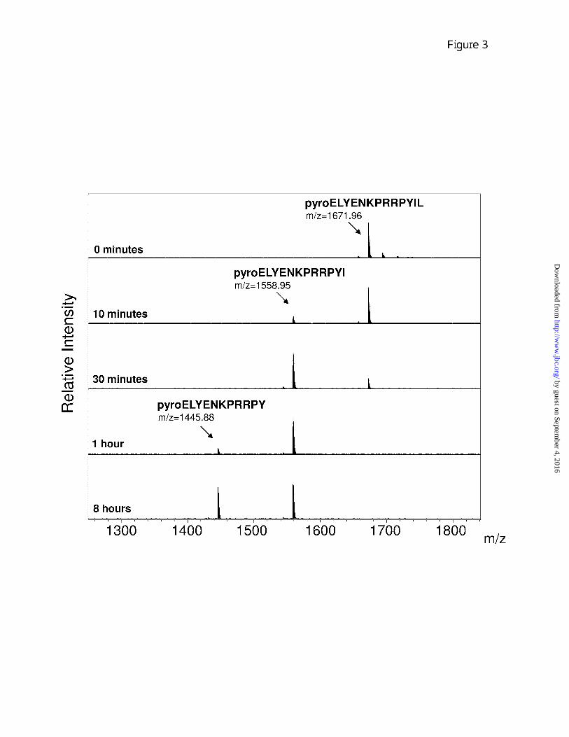
By using higher concentrations of enzyme (20 nM) and substrate (50 µM) it was possible to examine the processivity of CPA4 cleavage. The sequential hydrolysis of amino acids from the C-terminus of neurotensin was observed (Fig. 3). The product of the cleavage of the C-terminal Leu was detected at 10 minutes of incubation, while hydrolysis of the penultimate Ile was observed at 1 hour of incubation (Fig. 3). Further removal of the Tyr was not detected, even upon prolonged incubation, presumably because of the negative influence of the Pro in the P1
position of this peptide (Fig. 3). The action of CPA4 (20 nM) with angiotensin (200 µM) also showed processivity: the removal of C-terminal Leu, penultimate His and antepenultimate Phe is sequentially observed at 10 minutes, 30 minutes and 4 hours of incubation, respectively (data not shown).
Five of the peptides identified as CPA4 substrates were further investigated by determining the kinetic constants for CPA4 catalyzed hydrolysis. The data is summarized in Table 2 and shows a broad variation in Km and kcat values for the different peptides. CPA4 cleaves neurotensin and angiotensin with high efficiency (43.8 µM-1.min-1 and 9.43 µM-1.min-1 respectively) and low Km (0.329 µM and 9.23 µM respectively). To evaluate the ability of CPA4 to cleave a larger number of peptides, a quantitative peptidomics approach using differential isotopic-tagging and MS was used. For this, peptides were extracted from heat-inactivated mouse brains and purified away from proteins by microfiltration (10 kDa filter cut-off), and then incubated with varying amounts of purified CPA4, or in the absence of enzyme, for 1.5 h (Fig. 4). After heat inactivation of CPA4, the various samples were labeled with one of four isotopic tags, combined, and analyzed by liquid chromatography / mass spectrometry (LC/MS). Over 100 peptides were detected in the LC/MS analysis, including many of the brain peptides tested in Table S2 (neurotensin, PEN, big SAAS, Met-enkephalin-Arg-Phe (i.e. the proenkephalin heptapeptide), little SAAS, Leu-enkephalin, Met-enkephalin, and big LEN). Angiotensin and bradykinin were not detected in the brain peptidome, reflecting the low abundance of these peptides in brain. Quantification of the relative peptides levels in the enzyme-treated and control incubations was performed by determining peak intensity; representative data is indicated in Fig. 4. Many of the observed peptides showed roughly equal peak intensities; these are
by guest on September 4, 2016
http://ww
w.jbc.org/
Dow
nloaded from
10
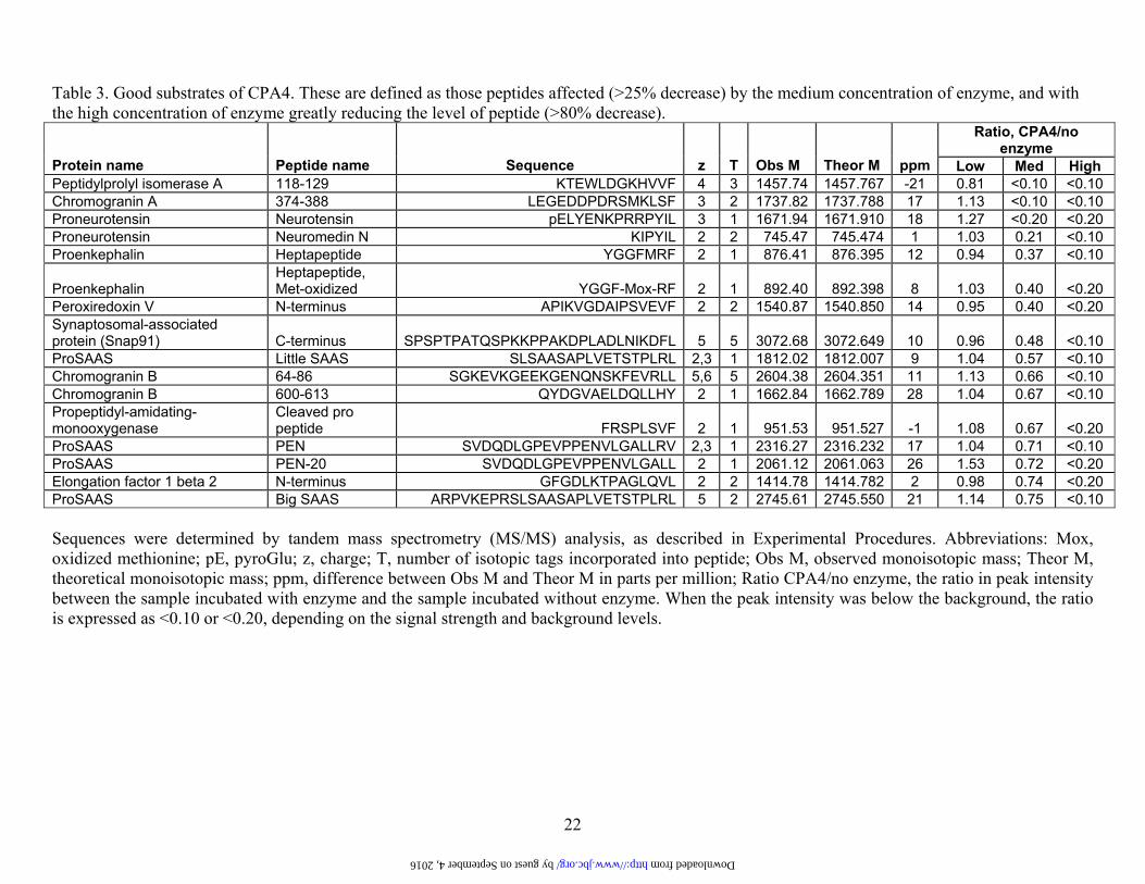
neither substrates nor products of CPA4 under the incubation conditions employed. Some peptides showed a decrease in peak intensity only for the sample incubated with the highest amount of enzyme; these are weak substrates of CPA4. Other peptides showed a large decrease in peak intensity for both the highest and the medium amount of enzyme; these are good substrates of CPA4. Finally, some peptides showed a large increase in peak intensity when incubated with enzyme; these are products of CPA4. Control studies in which aliquots of mouse brain peptides were isotopically tagged and analyzed by LC/MS, without incubation with enzyme, showed 4 equal peaks for all of the peptides observed in the present study (19). Approximately half of the detected peptides could be identified by tandem mass spectrometry (MS/MS) sequence analysis. In addition to these sequenced peptides, several other peptides were tentatively identified based on matches to known or predicted peptides; the criteria for these matches included an observed monoisotopic mass that was within 0.004% of the theoretical mass, a correct charge (equal to the number of basic residues plus the N-terminus), and a correct number of isotopic tags incorporated (based on the number of free amines in the peptide). These tentatively identified peptides are included in Tables 3-5 and S3 with the sequence surrounded by parentheses; all other sequences in these tables were confirmed by MS/MS sequencing (in addition to fitting the above criteria).
Analysis of the C-terminal residue (i.e. the P1’ position) of the 16 best substrates of CPA4 identified from the brain peptidome showed a predominance of Leu and Phe, with 14 of the 16 peptides containing one of these two amino acids (Table 3 and Figure 5). The other two peptides in this group had C-termini of Tyr or Val. Similar analysis of the 24 weak substrates of CPA4 showed these 4 amino acids to be present in the C-terminal position, in addition to Ile, Met, Ala, Thr, and His
(Table 4 and Figure 5). Analysis of the products of CPA4 focused on the downstream residue that was removed; of the 10 products that were identified, 6 required removal of a Leu, and the other four required removal of an Ile, Val, Tyr, or Ala (Table 5). Analysis was also performed on the peptides that were neither substrates nor products of CPA4 (Supplementary Table S3 and Figure 5). Of the 50 peptides in this category, only a small number had C-terminal Leu or Phe residues; the majority of these peptides contained C-terminal residues that were basic (Lys, Arg), acidic (Asp, Glu), polar (Ser, Asn, Gln), or otherwise not represented in the best substrate group (Ala, His, Pro).
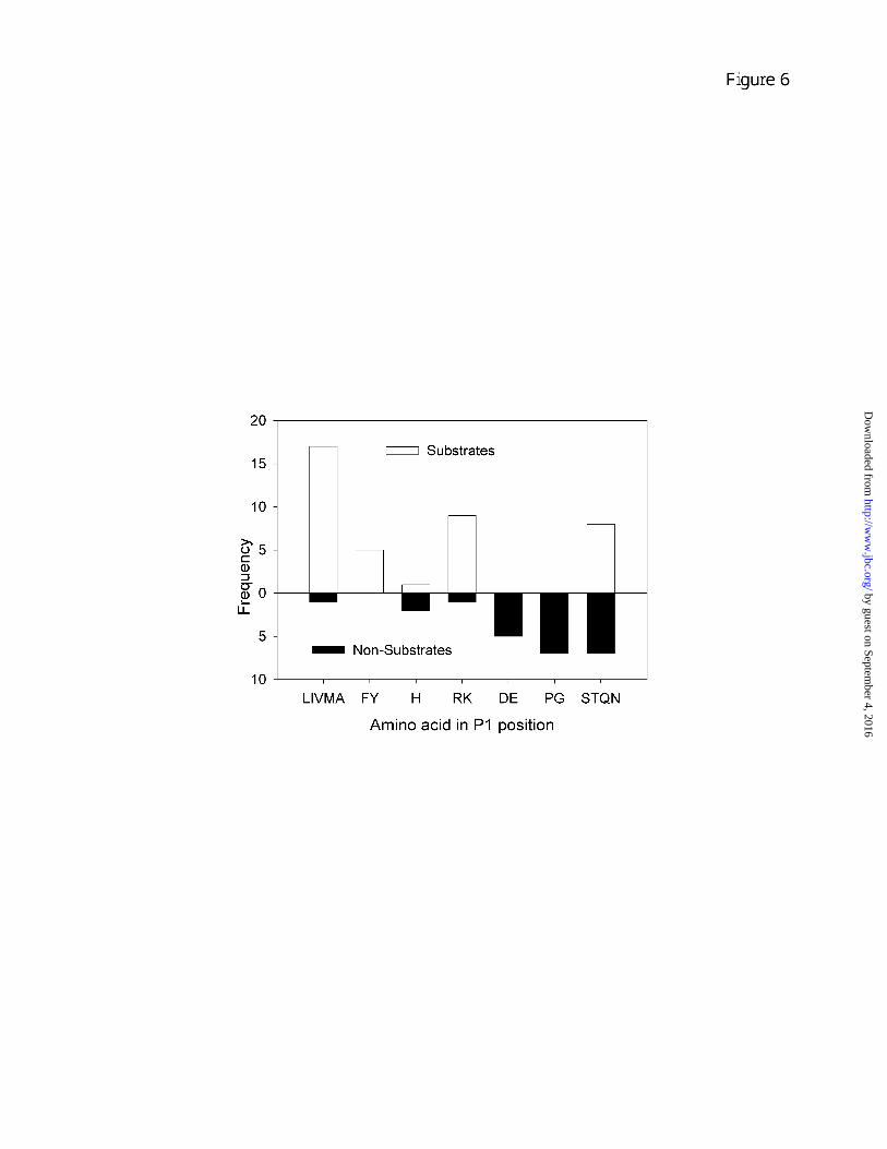
The finding that some, but not all of the peptides with C-terminal aliphatic and aromatic residues could be cleaved by CPA4 under the conditions tested suggested that additional residues contributed to the preference of this enzyme for particular peptides. To analyze this, we examined the P1 residue in all peptides with a C-terminal hydrophobic residue. Altogether, there were 62 peptides with a C-terminal Leu, Ile, Val, Met, Ala, Phe, Tyr, His, or Thr; of these, 40 were found to be substrates and 22 were not cleaved under the conditions tested. The majority of the substrates contained an aliphatic residue (Leu, Ile, Val, Met, Ala), aromatic (Phe, Tyr), or basic (Arg, Lys) residue in the P1 position (Fig. 6). Furthermore, none of the substrates contained an acidic residue (Asp, Glu), a Pro, or a Gly in the P1 position. In contrast, only 2 of the non-cleaved peptides had P1 aliphatic, aromatic, or basic residues (Fig. 6). Furthermore, many of these non-cleaved peptides had acidic or alpha-helix-breaking residues in the P1 position. Thus, it appears that while the C-terminal residue plays a critical role in determining specificity of CPA4, the P1 residue also contributes to the cleavage specificity.
by guest on September 4, 2016
http://ww
w.jbc.org/
Dow
nloaded from
11
DISCUSSION Summary of cleavage specificity of CPA4 The major finding of the present study is the cleavage specificity of CPA4, which has not been previously examined. Because of the potential link between CPA4 and some types of cancer (described in Introduction), it is important to determine the substrate specificity of this enzyme so that predictions can be made regarding its biological role(s). Based on the present results, CPA4 is clearly a CPA-like enzyme with specificity for C-terminal aliphatic and aromatic residues, consistent with the substrate binding pocket predicted from analysis of the crystal structure of CPA4 (14-16). The three types of substrates used in the present study (small chromogenic substrates, selected synthetic peptides, and a mixture of brain peptides) provide complementary information. While the small chromogenic substrates are ideal for kinetic analysis of kcat and Km values, these are not natural substrates and the chromogenic group can potentially affect the cleavage specificity. Synthetic peptides are useful to study precise substrate/product relationships, but are limited by the expense and the time to analyze each peptide. The quantitative peptidomics approach provides a great deal of information regarding which peptides are substrates, which are products, and which are not cleaved by CPA4. However, this approach does not provide kinetic information, and it is not possible to follow individual reactions as can be done with the synthetic peptides. Therefore, these three approaches have overlapping strengths and collectively provide a more complete picture than a single approach. Taken together, the optimal C-terminal residue for CPA4 cleavage is Leu, Ile, Val, Met, Tyr, or Phe (Fig. 7). Other acceptable amino acids in this position include Ala, His, Thr, or Trp. CPA4 does not cleave C-terminal Pro, basic residues (Arg, Lys), acidic residues (Asp, Glu), or polar
residues such as Ser, Asn, or Gln. Although Thr can be considered a polar residue, it is less polar than Ser. No information on Gly or Cys in the C-terminal position is available. The P1 position also has an influence on cleavage specificity, with aliphatic, aromatic, and basic residues favourable in this position (Fig. 7). Certain amino acids have a negative influence in the P1 position, such as acidic residues, Pro, and Gly (Fig. 7). No information on Trp or Cys is available for the P1 position. Comparison among CPAs
The FA-Phe-Xaa series of substrates offer higher values of kcat/Km values than other small synthetic substrates, allowing measurements of CPA2 towards substrates with small hydrophobic C-terminal amino acids, which has not previously been reported. In most cases, the improvement of catalytic efficiency for CPA2 towards FA-Phe-Xaa substrates versus Cbz-(Gly)n-Xaa substrates is due to lower Km values but in some cases an increase in kcat values also contributes to the difference (33). This effect may be explained by the different amino acids in the penultimate position of these substrates (Phe versus Gly); if CPA2 is similar to CPA4 as described above from the peptidomics analysis, Phe is much preferred over Gly in the P1 position. In addition, the FA-blocking group is known to enhance the CP activity (31). The kinetic data for CPA2 fully agree with that reported by Reverter et al., showing a preference for FA-Phe-Trp and FA-Phe-Phe (33). Compared to FA-Phe-Trp, the kcat/Km values are 85-fold and 335-fold lower for FA-Phe-Leu and FA-Phe-Ile, respectively. In contrast with these previous reports we were able to estimate kcat/Km values for FA-Phe-Met and FA-Phe-Val that are approximately 1000 times lower than the one for FA-Phe-Trp.
Although CPA1 has been well studied over the past 50 years, particularly bovine
by guest on September 4, 2016
http://ww
w.jbc.org/
Dow
nloaded from
12
CPA1 (originally referred to as CPA in the literature), the kinetic constants for human CPA1 have not been previously reported. The preference of CPA1 for Phe in the C-terminal position is more marked than that of CPA4, when the kcat/Km values for different substrates are compared. The kcat/Km ratio of CPA1 for FA-Phe-Phe is approximately two-fold greater than that of FA-Phe-Leu and FA-Phe-Val, four times higher than that of FA-Phe-Ile and ten-fold higher than the corresponding values for FA-Phe-Trp, FA-Phe-Met and FA-Phe-Ala. When kcat/Km values are compared between the three enzymes, CPA1 and CPA2 appear to be more efficient than CPA4 in the removal of C-terminal residues from peptides. For example, kcat/Km values for FA-Phe-Phe are approximately the same for CPA1 and CPA2, and four times lower for CPA4. A comparison between CPA4 and CPA6 activities towards the same group of peptides show several differences. CPA6 cleaves both Leu and His from the C-terminus of angiotensin I and des-Asp1 angiotensin I, whereas CPA4 only removes the Leu and not the His under the conditions described in Supplementary Table S2. However, CPA4 can cleave both the Leu and His in angiotensin when higher enzyme and peptide concentrations are used (data not shown). Furthermore, FA-Phe-His is cleaved by CPA4 at a low rate and in the peptidomics study two peptides which contain C-terminal His residues were found to be weak substrates of CPA4. Therefore, CPA4 is able to cleave His from the C-terminus of some substrates. In addition to CPA4 and CPA6, previously it was found that CPA1 (34) and CPA3 (35) can cleave C-terminal His residues. Although His is commonly considered to be a basic residue, at neutral pH values it will be mainly unprotonated and resemble an aromatic/aliphatic residue. Thus, it is not surprising that this residue is cleaved by CPA-like enzymes.
The substrate specificity of CPA4 determined in the present study fits very well with predictions made from structural analysis of this enzyme in complex with a hexapeptide (16), and with results from structural analysis of many CPAs and Bs (1). The specificity of CPA4 for C-terminal hydrophobic residues such as Leu, Ile, Val, Met, Tyr, and Phe, and weaker hydrolysis of Trp, matches more closely the specificity of CPA1 rather than CPA2. The S1’ specificity pocket is delimited by the side chains in positions 194, 203, 207, 243, 247, 250, 253-255, and 268 (using the numbering system of bovine CPA1). The residue in position 255 (Ile for CPA4) is considered to be the major determinant of substrate specificity for carboxypeptidases of the CPA/CPB subfamily (36). In CPA4 the residues in positions 203, 247, and 250 (numbering system of CPA1), which are Met, Val, and Ala respectively, also contribute to the hydrophobicity of the S1’ binding pocket. A comparison of the amino acids that shape the S1’ pocket between CPA1, CPA2 and CPA4 explains the strong preference of hCPA2 for bulkier hydrophobic amino acids in comparison to CPA1 and CPA4 (Supplementary Table S4 and Figure S1). The presence of Ala in position 268 in CPA2, in contrast to the Thr present in CPA1 and CPA4, makes the specificity pocket of CPA2 larger and more hydrophobic, explaining the specificity of CPA2 for large amino acids like Trp (37,38). The aminoacid in position 253 may also be responsible for this difference. The preference of CPA1 for C-terminal Phe is more marked than that of CPA4; this preference can be explained by the presence of a more hydrophobic environment in the S1’ pocket of CPA1 provided by Ile243 and Ile247, compared to the Thr and Val in similar positions in CPA4. In the present study, the S1 subsite was also found to contribute to the specificity of CPA4 (Fig. 7); critical residues in this subsite are the Tyr in position 198 and Ser in
by guest on September 4, 2016
http://ww
w.jbc.org/
Dow
nloaded from
13
position 199 (16). Other residues known to contribute to the shape of the S1 subsite of CPAs include Val 247, Tyr248, Phe279 and Glu270, which form a mainly hydrophobic pocket that may explain the preference of CPA4 for substrates with hydrophobic amino acids in the P1 position. The preference of CPA4 for positively charged amino acids over negatively charged amino acids in the P1 position could be explained by interactions between the side chain of the P1 residue with the polar portion of Tyr198 and Ser199 in the S1 binding site (1). Potential function of CPA4 The finding that PCPA4 is secreted from cells in a soluble form and, once activated, has a neutral pH optimum suggests a function in the extracellular environment rather than within the secretory pathway. Although trypsin was used to activate PCPA4 in the present study, this is not likely to represent the physiological activating enzyme because trypsin is pancreatic while CPA4 has a broader distribution. There are a number of trypsin-like enzymes expressed in brain and other tissues where PCPA4 is found (39). Based on modelling and structural considerations, the conversion of PCPA4 into active CPA4 requires cleavage at a single Arg in the sequence EGQER-SS. It is possible that the activation of PCPA4 may be an important control point that regulates the active form of the enzyme. The distribution of CPA4 provides important clues as to the function of this enzyme. Huang et al (9) found CPA4 mRNA to be expressed at high levels in different prostate cancer cell lines and at extremely low levels in some normal human tissues including prostate and pancreas. CPA4 mRNA was induced in prostate cancer cell lines upon treatment with 10 mM sodium butyrate indicating that its expression is associated with in vitro differentiation. It was also reported that CPA4 expression is
associated with hormone-regulated tissues. Through an imprinting analysis, Kayashima et al (12) found that CPA4 mRNA was also highly expressed in fetal tissues that are growing or differentiating with the transcriptionally active state of chromatin. Using RT-PCR Bentley et al (11) detected CPA4 expression in a wide variety of tissues including placenta, testis, uterus, heart, brain, intestine, kidney, and muscle. In mouse brain, in situ hybridization showed relatively high expression of CPA4 mRNA in the olfactory bulb, specifically in the granular and mitral layers (40), in an expression pattern similar to CPA6 mRNA (41). A US patent using TaqMan analysis of human tissues reported expression of CPA4 in kidney, spinal cord, and brain, with moderate levels in cortex and considerably higher levels in hypothalamus, especially in the arcuate and paraventricular nuclei (42). Taken together, it is clear that CPA4 mRNA is present in many tissues. The association of CPA4 mRNA levels with differentiation and/or cancerous cells and tissues raises the possibility that this enzyme plays an important role in cleaving a factor or factors that are critical to the differentiated state. Many secreted factors are known to function as growth factors or to otherwise enable tissues to grow and metastasize (such as secreted proteases) and CPA4 may contribute to these processes. Some of the peptides found to be substrates in the present study include neurotensin, granins like chromogranin A and B and secretogranin II, and opioid peptides such as Met- and Leu-enkephalin. The cleavage performed by CPA4 would eliminate the binding of these peptides to their receptors. While enkephalin is a well-known neuropeptide that functions in analgesia and the reward pathway, additional roles for enkephalin include synaptogenesis and neurite outgrowth (43,44). Some granin-derived peptides have been postulated to participate as regulators of tissue repair, inflammatory reaction and innate immunity
by guest on September 4, 2016
http://ww
w.jbc.org/
Dow
nloaded from

14
(45). Additional roles for some of these products include the stimulation of proliferation in tumor cells (46) and invasive potential of prostate cancer cells partially through enhancement of cell mobility (47). Similarly, neurotensin is also a well-studied neuropeptide that has recently been proposed to contribute to the progression of several types of cancer including prostate, breast, and pancreatic cancer (48-50). Because the enkephalins, secretogranins and neurotensin are secreted from some of the same tissues where CPA4 is also secreted, it is likely that this enzyme plays a role in the termination of the action of these peptides, as well as many others. A role for CPA4 in the inactivation of peptides that function in cell proliferation would potentially provide a mechanism to account for the previous studies linking CPA4 with aggressiveness of some forms of cancer. Additional research efforts are needed to characterize and understand the biological effects of this enzyme and its SNP variant(s) in the tumoral process.
ACKNOWLEDGMENTS Mass spectrometry was performed in the Laboratory for Macromolecular Analysis and Proteomics of the Albert Einstein College of Medicine, directed by Dr. Ruth Angeletti. The authors acknowledge the technical assistance of the Cell Culture Facility of the Institut de Biotecnologia i de Biomedicina (IBB) and the Microscopy Facility, both from the Universitat Autònoma de Barcelona. Thanks to Dr. Peter Lyons for helpful comments. This work was supported in part by National Institutes of Health grants DK-51271 and DA-04494 (L.D.F.), by European Commission grant FP7-Health-2009-241919-LIVIMODE (F.X.A.), by Spanish Ministry of Science and Technology Grant BIO2007-68046-C02 (F.X.A.) and by XeRBa (Generalitat de Catalunya).
Abbreviations: ACI, Ascaris carboxypeptidase inhibitor; CCP, cytosolic carboxypeptidase; CP, Metallocarboxypeptidase; CPA, A-like carboxypeptidase; CPA1, carboxypeptidase A1; CPA2, carboxypeptidase A2; CPB, carboxypeptidase B; CPA3, carboxypeptidase A3; CPA6, carboxypeptidase A6; CPU, carboxypeptidase U; ECM, extracellular matrix; ER, endoplasmic reticulum; CPA4, carboxypeptidase A4; PCPA4, procarboxypeptidase A4; LCI, leech carboxypeptidase inhibitor; LC/MS, liquid chromatography/mass spectrometry; MALDI-TOF MS, Matrix-assisted laser desorption ionization time-of-flight mass spectrometry; MS/MS, tandem mass spectrometry; PBS, phosphate-buffered saline; PCI, potato carboxypeptidase inhibitor; PVDF, polyvinylidene difluoride; RP-HPLC, reversed-phase high performance liquid chromatography; SNP, single-nucleotide polymorphism; TAFI, thrombin-activatable fibrinolysis inhibitor; TCI, tick carboxypeptidase inhibitor; TFA, trifluoroacetic acid; TMAB, 4-trimethyl-ammoniumbutyrate.
by guest on September 4, 2016
http://ww
w.jbc.org/
Dow
nloaded from
15
LITERATURE CITED 1. Gomis-Ruth, F. X. (2008) Crit Rev Biochem Mol Biol 43, 319-345 2. Rawlings, N. D., Morton, F. R., and Barrett, A. J. (2006) Nucleic Acids Res 34, D270-272 3. Kalinina, E., Biswas, R., Berezniuk, I., Hermoso, A., Aviles, F. X., and Fricker, L. D.
(2007) FASEB J 21, 836-850 4. Rodriguez de la Vega, M., Sevilla, R. G., Hermoso, A., Lorenzo, J., Tanco, S., Diez, A.,
Fricker, L. D., Bautista, J. M., and Aviles, F. X. (2007) FASEB J 21, 851-865 5. Pejler, G., Abrink, M., Ringvall, M., and Wernersson, S. (2007) Adv Immunol 95, 167-
255 6. Lyons, P. J., Callaway, M. B., and Fricker, L. D. (2008) J Biol Chem 283, 7054-7063 7. Leung, L. L., Myles, T., Nishimura, T., Song, J. J., and Robinson, W. H. (2008) Mol
Immunol 45, 4080-4083 8. Arolas, J. L., Vendrell, J., Aviles, F. X., and Fricker, L. D. (2007) Curr Pharm Des 13,
349-366 9. Huang, H., Reed, C. P., Zhang, J. S., Shridhar, V., Wang, L., and Smith, D. I. (1999)
Cancer Res 59, 2981-2988 10. Witte, J. S., Goddard, K. A., Conti, D. V., Elston, R. C., Lin, J., Suarez, B. K., Broman,
K. W., Burmester, J. K., Weber, J. L., and Catalona, W. J. (2000) Am J Hum Genet 67, 92-99
11. Bentley, L., Nakabayashi, K., Monk, D., Beechey, C., Peters, J., Birjandi, Z., Khayat, F. E., Patel, M., Preece, M. A., Stanier, P., Scherer, S. W., and Moore, G. E. (2003) J Med Genet 40, 249-256
12. Kayashima, T., Yamasaki, K., Yamada, T., Sakai, H., Miwa, N., Ohta, T., Yoshiura, K., Matsumoto, N., Nakane, Y., Kanetake, H., Ishino, F., Niikawa, N., and Kishino, T. (2003) Hum Genet 112, 220-226
13. Ross, P. L., Cheng, I., Liu, X., Cicek, M. S., Carroll, P. R., Casey, G., and Witte, J. S. (2009) BMC Cancer 9, 69
14. Garcia-Castellanos, R., Bonet-Figueredo, R., Pallares, I., Ventura, S., Aviles, F. X., Vendrell, J., and Gomis-Ruth, F. X. (2005) Cell Mol Life Sci 62, 1996-2014
15. Pallares, I., Bonet, R., Garcia-Castellanos, R., Ventura, S., Aviles, F. X., Vendrell, J., and Gomis-Ruth, F. X. (2005) Proc Natl Acad Sci U S A 102, 3978-3983
16. Bayes, A., Fernandez, D., Sola, M., Marrero, A., Garcia-Pique, S., Aviles, F. X., Vendrell, J., and Gomis-Ruth, F. X. (2007) Biochemistry 46, 6921-6930
17. Blumberg, S., and Vallee, B. L. (1975) Biochemistry 14, 2410-2419 18. Bieth, J. G. (1995) Methods Enzymol 248, 59-84 19. Morano, C., Zhang, X., and Fricker, L. D. (2008) Anal Chem 80, 9238-9309 20. Che, F. Y., Zhang, X., Berezniuk, I., Callaway, M., Lim, J., and Fricker, L. D. (2007) J
Proteome Res 6, 4667-4676 21. Che, F. Y., Lim, J., Pan, H., Biswas, R., and Fricker, L. D. (2005) Mol Cell Proteomics 4,
1391-1405 22. Zhang, X., Che, F. Y., Berezniuk, I., Sonmez, K., Toll, L., and Fricker, L. D. (2008) J
Neurochem 107, 1596-1613 23. Hamstra, D. A., and Rehemtulla, A. (1999) Hum Gene Ther 10, 235-248 24. Novikova, E. G., Reznik, S. E., Varlamov, O., and Fricker, L. D. (2000) J Biol Chem
275, 4865-4870
by guest on September 4, 2016
http://ww
w.jbc.org/
Dow
nloaded from
16
25. Byers, L. D., and Wolfenden, R. (1973) Biochemistry 12, 2070-2078 26. Felber, J. P., Coombs, T. L., and Vallee, B. L. (1962) Biochemistry 1, 231-238 27. Ryan, C. A., Hass, G. M., and Kuhn, R. W. (1974) J Biol Chem 249, 5495-5499 28. Reverter, D., Vendrell, J., Canals, F., Horstmann, J., Aviles, F. X., Fritz, H., and
Sommerhoff, C. P. (1998) J Biol Chem 273, 32927-32933 29. Arolas, J. L., Lorenzo, J., Rovira, A., Castella, J., Aviles, F. X., and Sommerhoff, C. P.
(2005) J Biol Chem 280, 3441-3448 30. Sanglas, L., Aviles, F. X., Huber, R., Gomis-Ruth, F. X., and Arolas, J. L. (2009) Proc
Natl Acad Sci U S A 106, 1743-1747 31. Peterson, L. M., Holmquist, B., and Bethune, J. L. (1982) Anal Biochem 125, 420-426 32. Pallares, I., Fernandez, D., Comellas-Bigler, M., Fernandez-Recio, J., Ventura, S., Aviles,
F. X., Bode, W., and Vendrell, J. (2008) Acta Crystallogr D Biol Crystallogr D64, 784-791
33. Reverter, D., Garcia-Saez, I., Catasus, L., Vendrell, J., Coll, M., and Aviles, F. X. (1997) FEBS Lett 420, 7-10
34. Garcia, K. C., Tallquist, M. D., Pease, L. R., Brunmark, A., Scott, C. A., Degano, M., Stura, E. A., Peterson, P. A., Wilson, I. A., and Teyton, L. (1997) Proc Natl Acad Sci U S A 94, 13838-13843
35. Lundequist, A., Tchougounova, E., Abrink, M., and Pejler, G. (2004) J Biol Chem 279, 32339-32344
36. Grishin, A. M., Akparov, V., and Chestukhina, G. G. (2008) Protein Eng Des Sel 21, 545-551
37. Faming, Z., Kobe, B., Stewart, C. B., Rutter, W. J., and Goldsmith, E. J. (1991) J Biol Chem 266, 24606-24612
38. Garcia-Saez, I., Reverter, D., Vendrell, J., Aviles, F. X., and Coll, M. (1997) EMBO J 16, 6906-6913
39. Wang, Y., Luo, W., and Reiser, G. (2008) Cell Mol Life Sci 65, 237-252 40. Lein, E. S., Hawrylycz, M. J., Ao, N., Ayres, M., Bensinger, A., Bernard, A., Boe, A. F.,
Boguski, M. S., Brockway, K. S., Byrnes, E. J., et al. (2007) Nature 445, 168-176 41. Fontenele-Neto, J. D., Kalinina, E., Feng, Y., and Fricker, L. D. (2005) Brain Res Mol
Brain Res 137, 132-142 42. White, D. (2004) Use for Carboxypeptidase-A4 in the diagnosis and treatment of
metabolic disorders in U.S. Patent 2004/0077001 A1 by Millennium Pharmaceuticals Inc.
43. Davila-Garcia, M. I., and Azmitia, E. C. (1990) Adv Exp Med Biol 265, 75-92 44. Escobar, M. L., Barea-Rodriguez, E. J., Derrick, B. E., Reyes, J. A., and Martinez, J. L.,
Jr. (1997) Brain Res 751, 330-335 45. Helle, K. B., Corti, A., Metz-Boutigue, M. H., and Tota, B. (2007) Cell Mol Life Sci 64,
2863-2886 46. Mosca, A., Berruti, A., Russo, L., Torta, M., and Dogliotti, L. (2005) J Endocrinol Invest
28, 141-145 47. Nagakawa, O., Ogasawara, M., Murata, J., Fuse, H., and Saiki, I. (2001) Int J Urol 8, 65-
70 48. Dupouy, S., Viardot-Foucault, V., Alifano, M., Souaze, F., Plu-Bureau, G., Chaouat, M.,
Lavaur, A., Hugol, D., Gespach, C., Gompel, A., and Forgez, P. (2009) PLoS One 4, e4223
by guest on September 4, 2016
http://ww
w.jbc.org/
Dow
nloaded from

17
49. Myers, R. M., Shearman, J. W., Kitching, M. O., Ramos-Montoya, A., Neal, D. E., and Ley, S. V. (2009) ACS Chem Biol 4, 503-525
50. Vias, M., Burtt, G., Culig, Z., Veerakumarasivam, A., Neal, D. E., and Mills, I. G. (2007) Prostate 67, 190-202
by guest on September 4, 2016
http://ww
w.jbc.org/
Dow
nloaded from
18
FIGURE LEGENDS
Figure 1. PCPA4 is a soluble secreted protein. A) PCPA4 transfection in mammalian cells. HEK 293T cells were transfected with empty vector (-) or plasmid expressing HA-tagged PCPA4 (+) and different fractions were collected and prepared. Equal amounts of each fraction were analyzed by Western blot using an HA antibody. B) Trypsin processing. Extracellular media was treated with (+) or without (-) trypsin and analyzed by Western blot. C) Subcellular distribution of PCPA4. HEK 293T cells were transfected with HA-tagged PCPA4 and pDsRed-ER cDNAs. After fixation, transfected cells were incubated with mouse anti-HA antibody followed by anti-mouse IgG Alexa 488 (green) and DAPI to detect cell nuclei.
Figure 2. Effect of pH on CPA4 activity. CPA4 activity was determined by measuring initial rates using 100 µM FA-Phe-Phe or FA-Phe-Ala in 50 mM Tris-acetate buffer at the indicated pH at 25ºC. Error bars represent the standard error of the mean for three independent determinations.
Figure 3. MALDI-TOF MS analysis of neurotensin cleavage by CPA4. MALDI-TOF MS spectra of 50 µM neurotensin incubated for different times at 37°C with 20 nM CPA4. Numbers above the major peaks indicate the monoisotopic masses of the MH+ ion (m/z). The peak with mass of 1671.96 corresponds to neurotensin (theoretical monoisotopic mass of neurotensin = 1671.91) and a first cleavage product with a mass of 1558.95 formed in the presence of CPA4 corresponds to the peptide lacking the C-terminal Leu (theoretical mass = 1558.84) and a second cleavage product with mass 1445.88 corresponds to the peptide lacking both C-terminal Leu and Ile (theoretical mass = 1445.75).
Figure 4. Quantitative peptidomics scheme and representative results. Peptides were extracted from microwave-irradiated mouse brain and separated from proteins by microfiltration with a 10 kDa filter. Aliquots were incubated with purified CPA4 (20 ng or 30-fold dilutions) or in the absence of CPA4. After incubation, the samples were labeled with one of 4 stable isotopic TMAB tags that differ in the number of hydrogens and deuteriums. Samples were then pooled and analyzed by liquid chromatography/mass spectrometry (LC/MS). Representative data are shown. Peptides that showed less than a 25% decrease in peak intensity for the sample incubated with the highest concentration of enzyme (the D3 TMAB tag) were considered non-substrates, as shown for the proenkephalin fragment SPGLEDEAKELQ. Peptides which showed a major decrease with the highest concentration of enzyme (>50% decrease) but less than a 25% decrease with the medium concentration of enzyme were considered weak substrates. An example of a weak substrate is the neuropeptide dynorphin A8. Peptides which showed a substantial decrease in peak intensity for the highest concentration of enzyme, and at least a 25% decrease for the medium concentration of enzyme were considered good substrates. An example is little SAAS, an 18-residue peptide with a C-terminal Leu. Peptides that showed an increase in peak intensity for the samples incubated with enzyme were considered products of CPA4. An example is little SAAS 1-17, which lacks the C-terminal Leu of little SAAS.
by guest on September 4, 2016
http://ww
w.jbc.org/
Dow
nloaded from
19
Figure 5. Analysis of the C-terminal residue (P1’) of peptides detected using the quantitative peptidomics technique. The number of times each amino acid was present in the C-terminal position was determined for good substrates, weak substrates, and non-substrates, as defined in the Fig. 4 legend.
Figure 6. Analysis of the P1 residue of peptide containing hydrophobic C-terminal residues. For this analysis, only peptides that were substrates or potential substrates were considered, based on the analysis shown in Fig. 5. Those peptides with acidic, basic, or polar C-terminal residues were excluded. This analysis included 40 substrates (good and weak substrates were combined) and 22 non-substrates with similar C-terminal residues as the substrates (i.e. L, I, V, M, A, F, Y, H). The frequency of the various amino acids in the P1 position of each group were determined and combined into groups based on amino acid type; aliphatic (L, I, V, M, A), aromatic (F, Y), basic (R, K), acidic (D, E), alpha-helix breakers (P, G), and polar (S, T, Q, N). His was placed into its own group, and there were no data for Cys or Trp as these amino acids were not present in the P1 position in any of the peptides analyzed.
Figure 7. Summary of cleavage specificity of CPA4, based on results from synthetic chromogenic substrates, synthetic peptides, and endogenous mouse brain peptides. Those amino acids considered most favorable for cleavage by CPA4 are listed in the top row, and include L, I, V, M, Y, and F in the P1’ position and these same residues plus R and K in the P1 position. Residues that are acceptable in these positions are listed in the middle row, and those residues that appear to be inhibitory for cleavage are listed in the bottom row.
by guest on September 4, 2016
http://ww
w.jbc.org/
Dow
nloaded from
20
Table 1. Kinetics constants for peptide substrate hydrolysis by CPA1, CPA2 and CPA4.
CPA4 CPA1 CPA2
Substrate kcat (1/s)
Km (µM)
kcat/Km (M-1.s-1.10-5)
kcat/Km relative to
FAPP
kcat (1/s)
Km (µM)
kcat/Km (M-1.s-1.10-5)
kcat/Km relative to FAPP
kcat (1/s)
Km (µM)
kcat/Km (M-1.s-1.10-5)
kcat/Km relative to FAPP
FA-Phe-Phe 44.3 55.6 7.97 1.00 76.6 20.4 37.6 1.00 121 36.4 33.3 1.00
FA-Phe-Trp 57.3 615 0.93 0.12 81.3 166 4.90 0.13 65.3 16.3 40.2 1.21
FA-Phe-Leu 13.4 19.4 6.93 0.87 34.0 15.7 21.7 0.58 18.6 393 0.47 0.014
FA-Phe-Ile 12.4 23.3 5.32 0.67 9.77 9.33 10.5 0.28 5.37 460 0.12 0.0035
FA-Phe-Met 23.9 40.0 5.81 0.73 51.8 128 4.03 0.11 NM NM 0.040 0.0012
FA-Phe-Ala 24.3 372 0.65 0.082 78.0 300 2.58 0.069 NM NM NM NM
FA-Phe-Val 19.4 57.3 3.38 0.42 12.3 8.95 14.8 0.39 NM NM 0.030 0.0010
FA-Phe-His NM NM 0.27 0.034 NM NM 1.04 0.028 NM NM NM NM
NM: Not measurable; below the limit of detection.
by guest on September 4, 2016 http://www.jbc.org/ Downloaded from

21
Table 2. Kinetic constants for the hydrolysis of biological peptides by CPA4
Peptide kcat (s-1)
Km (µM)
kcat/Km (µM-1.min-1)
Neurotensin 0.240 ± 0.017 0.329 ± 0.049 43.8 ± 9.5
Met-enkephalin-Arg-Phe 1.45 ± 0.03 9.23 ± 0.64 9.43 ± 0.86
Angiotensin I 8.23 ± 0.34 227 ± 25 2.18 ± 0.31
Leu-enkephalin 2.33 ± 0.11 165 ± 19 0.85 ± 0.13
Met-enkephalin 3.43 ± 0.16 550 ± 52 0.374 ± 0.053
by guest on September 4, 2016 http://www.jbc.org/ Downloaded from
22
Table 3. Good substrates of CPA4. These are defined as those peptides affected (>25% decrease) by the medium concentration of enzyme, and with the high concentration of enzyme greatly reducing the level of peptide (>80% decrease).
Sequences were determined by tandem mass spectrometry (MS/MS) analysis, as described in Experimental Procedures. Abbreviations: Mox, oxidized methionine; pE, pyroGlu; z, charge; T, number of isotopic tags incorporated into peptide; Obs M, observed monoisotopic mass; Theor M, theoretical monoisotopic mass; ppm, difference between Obs M and Theor M in parts per million; Ratio CPA4/no enzyme, the ratio in peak intensity between the sample incubated with enzyme and the sample incubated without enzyme. When the peak intensity was below the background, the ratio is expressed as <0.10 or <0.20, depending on the signal strength and background levels.
Ratio, CPA4/no enzyme
Protein name Peptide name Sequence z T Obs M Theor M ppm Low Med High Peptidylprolyl isomerase A 118-129 KTEWLDGKHVVF 4 3 1457.74 1457.767 -21 0.81 <0.10 <0.10 Chromogranin A 374-388 LEGEDDPDRSMKLSF 3 2 1737.82 1737.788 17 1.13 <0.10 <0.10 Proneurotensin Neurotensin pELYENKPRRPYIL 3 1 1671.94 1671.910 18 1.27 <0.20 <0.20 Proneurotensin Neuromedin N KIPYIL 2 2 745.47 745.474 1 1.03 0.21 <0.10 Proenkephalin Heptapeptide YGGFMRF 2 1 876.41 876.395 12 0.94 0.37 <0.10
Proenkephalin Heptapeptide, Met-oxidized YGGF-Mox-RF 2 1 892.40 892.398 8 1.03 0.40 <0.20
Peroxiredoxin V N-terminus APIKVGDAIPSVEVF 2 2 1540.87 1540.850 14 0.95 0.40 <0.20 Synaptosomal-associated protein (Snap91) C-terminus SPSPTPATQSPKKPPAKDPLADLNIKDFL 5 5 3072.68 3072.649 10 0.96 0.48 <0.10 ProSAAS Little SAAS SLSAASAPLVETSTPLRL 2,3 1 1812.02 1812.007 9 1.04 0.57 <0.10 Chromogranin B 64-86 SGKEVKGEEKGENQNSKFEVRLL 5,6 5 2604.38 2604.351 11 1.13 0.66 <0.10 Chromogranin B 600-613 QYDGVAELDQLLHY 2 1 1662.84 1662.789 28 1.04 0.67 <0.10 Propeptidyl-amidating-monooxygenase
Cleaved pro peptide FRSPLSVF 2 1 951.53 951.527 -1 1.08 0.67 <0.20
ProSAAS PEN SVDQDLGPEVPPENVLGALLRV 2,3 1 2316.27 2316.232 17 1.04 0.71 <0.10 ProSAAS PEN-20 SVDQDLGPEVPPENVLGALL 2 1 2061.12 2061.063 26 1.53 0.72 <0.20 Elongation factor 1 beta 2 N-terminus GFGDLKTPAGLQVL 2 2 1414.78 1414.782 2 0.98 0.74 <0.20 ProSAAS Big SAAS ARPVKEPRSLSAASAPLVETSTPLRL 5 2 2745.61 2745.550 21 1.14 0.75 <0.10
by guest on September 4, 2016 http://www.jbc.org/ Downloaded from
23
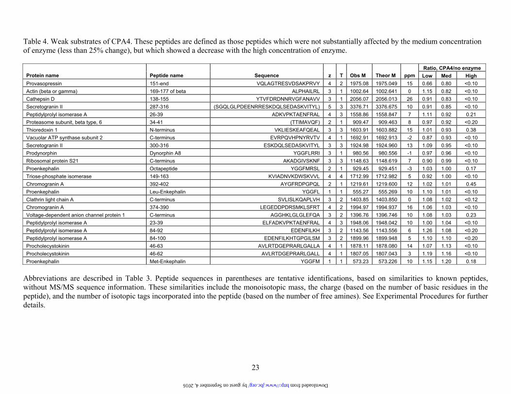
Table 4. Weak substrates of CPA4. These peptides are defined as those peptides which were not substantially affected by the medium concentration of enzyme (less than 25% change), but which showed a decrease with the high concentration of enzyme.
Ratio, CPA4/no enzyme Protein name Peptide name Sequence z T Obs M Theor M ppm Low Med High Provasopressin 151-end VQLAGTRESVDSAKPRVY 4 2 1975.08 1975.049 15 0.66 0.80 <0.10 Actin (beta or gamma) 169-177 of beta ALPHAILRL 3 1 1002.64 1002.641 0 1.15 0.82 <0.10 Cathepsin D 138-155 YTVFDRDNNRVGFANAVV 3 1 2056.07 2056.013 26 0.91 0.83 <0.10 Secretogranin II 287-316 (SGQLGLPDEENRRESKDQLSEDASKVITYL) 5 3 3376.71 3376.675 10 0.91 0.85 <0.10 Peptidylprolyl isomerase A 26-39 ADKVPKTAENFRAL 4 3 1558.86 1558.847 7 1.11 0.92 0.21 Proteasome subunit, beta type, 6 34-41 (TTIMAVQF) 2 1 909.47 909.463 8 0.97 0.92 <0.20 Thioredoxin 1 N-terminus VKLIESKEAFQEAL 3 3 1603.91 1603.882 15 1.01 0.93 0.38 Vacuolar ATP synthase subunit 2 C-terminus EVRPQVHPNYRVTV 4 1 1692.91 1692.913 -2 0.87 0.93 <0.10 Secretogranin II 300-316 ESKDQLSEDASKVITYL 3 3 1924.98 1924.960 13 1.09 0.95 <0.10 Prodynorphin Dynorphin A8 YGGFLRRI 3 1 980.56 980.556 -1 0.97 0.96 <0.10 Ribosomal protein S21 C-terminus AKADGIVSKNF 3 3 1148.63 1148.619 7 0.90 0.99 <0.10 Proenkephalin Octapeptide YGGFMRSL 2 1 929.45 929.451 -3 1.03 1.00 0.17 Triose-phosphate isomerase 149-163 KVIADNVKDWSKVVL 4 4 1712.99 1712.982 5 0.92 1.00 <0.10 Chromogranin A 392-402 AYGFRDPGPQL 2 1 1219.61 1219.600 12 1.02 1.01 0.45 Proenkephalin Leu-Enkephalin YGGFL 1 1 555.27 555.269 10 1.10 1.01 <0.10 Clathrin light chain A C-terminus SVLISLKQAPLVH 3 2 1403.85 1403.850 0 1.08 1.02 <0.12 Chromogranin A 374-390 LEGEDDPDRSMKLSFRT 4 2 1994.97 1994.937 16 1.06 1.03 <0.10 Voltage-dependent anion channel protein 1 C-terminus AGGHKLGLGLEFQA 3 2 1396.76 1396.746 10 1.08 1.03 0.23 Peptidylprolyl isomerase A 23-39 ELFADKVPKTAENFRAL 4 3 1948.06 1948.042 10 1.00 1.04 <0.10 Peptidylprolyl isomerase A 84-92 EDENFILKH 3 2 1143.56 1143.556 6 1.26 1.08 <0.20 Peptidylprolyl isomerase A 84-100 EDENFILKHTGPGILSM 3 2 1899.96 1899.948 5 1.10 1.10 <0.20 Procholecystokinin 46-63 AVLRTDGEPRARLGALLA 4 1 1878.11 1878.080 14 1.07 1.13 <0.10 Procholecystokinin 46-62 AVLRTDGEPRARLGALL 4 1 1807.05 1807.043 3 1.19 1.16 <0.10 Proenkephalin Met-Enkephalin YGGFM 1 1 573.23 573.226 10 1.15 1.20 0.18
Abbreviations are described in Table 3. Peptide sequences in parentheses are tentative identifications, based on similarities to known peptides, without MS/MS sequence information. These similarities include the monoisotopic mass, the charge (based on the number of basic residues in the peptide), and the number of isotopic tags incorporated into the peptide (based on the number of free amines). See Experimental Procedures for further details.
by guest on September 4, 2016 http://www.jbc.org/ Downloaded from
24
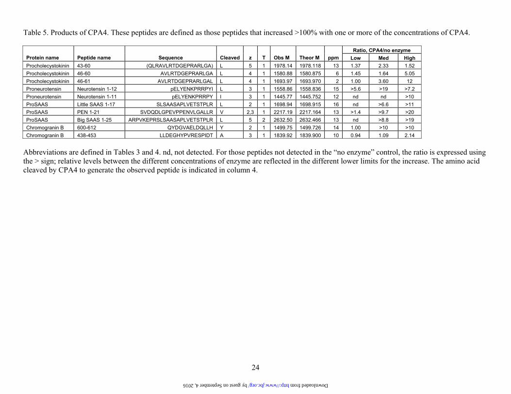
Table 5. Products of CPA4. These peptides are defined as those peptides that increased >100% with one or more of the concentrations of CPA4.
Ratio, CPA4/no enzyme Protein name Peptide name Sequence Cleaved z T Obs M Theor M ppm Low Med High Procholecystokinin 43-60 (QLRAVLRTDGEPRARLGA) L 5 1 1978.14 1978.118 13 1.37 2.33 1.52 Procholecystokinin 46-60 AVLRTDGEPRARLGA L 4 1 1580.88 1580.875 6 1.45 1.64 5.05 Procholecystokinin 46-61 AVLRTDGEPRARLGAL L 4 1 1693.97 1693.970 2 1.00 3.60 12 Proneurotensin Neurotensin 1-12 pELYENKPRRPYI L 3 1 1558.86 1558.836 15 >5.6 >19 >7.2 Proneurotensin Neurotensin 1-11 pELYENKPRRPY I 3 1 1445.77 1445.752 12 nd nd >10 ProSAAS Little SAAS 1-17 SLSAASAPLVETSTPLR L 2 1 1698.94 1698.915 16 nd >6.6 >11 ProSAAS PEN 1-21 SVDQDLGPEVPPENVLGALLR V 2,3 1 2217.19 2217.164 13 >1.4 >9.7 >20 ProSAAS Big SAAS 1-25 ARPVKEPRSLSAASAPLVETSTPLR L 5 2 2632.50 2632.466 13 nd >8.8 >19 Chromogranin B 600-612 QYDGVAELDQLLH Y 2 1 1499.75 1499.726 14 1.00 >10 >10 Chromogranin B 438-453 LLDEGHYPVRESPIDT A 3 1 1839.92 1839.900 10 0.94 1.09 2.14
Abbreviations are defined in Tables 3 and 4. nd, not detected. For those peptides not detected in the “no enzyme” control, the ratio is expressed using the > sign; relative levels between the different concentrations of enzyme are reflected in the different lower limits for the increase. The amino acid cleaved by CPA4 to generate the observed peptide is indicated in column 4.
by guest on September 4, 2016 http://www.jbc.org/ Downloaded from

by guest on September 4, 2016
http://ww
w.jbc.org/
Dow
nloaded from
Lloyd
Figure 1
by guest on September 4, 2016
http://ww
w.jbc.org/
Dow
nloaded from
Lloyd
Figure 2
by guest on September 4, 2016
http://ww
w.jbc.org/
Dow
nloaded from
Lloyd
Figure 3

by guest on September 4, 2016
http://ww
w.jbc.org/
Dow
nloaded from
Lloyd
Figure 4

by guest on September 4, 2016
http://ww
w.jbc.org/
Dow
nloaded from
Lloyd
Figure 5
by guest on September 4, 2016
http://ww
w.jbc.org/
Dow
nloaded from
Lloyd
Figure 6

by guest on September 4, 2016
http://ww
w.jbc.org/
Dow
nloaded from
Lloyd
Figure 7

Supplemental information
Human carboxypeptidase A4: Characterization of the substrate specificity and implications for a role in extracellular peptide processing Sebastian Tanco‡, Xin Zhang§, Cain Morano§, Francesc X. Aviles‡, Julia Lorenzo‡1 and Lloyd D. Fricker§2 ‡Institut de Biotecnologia i de Biomedicina & Departament de Bioquimica, Universitat Autonoma de Barcelona, Bellaterra (Barcelona), Spain §Department of Molecular Pharmacology, Albert Einstein College of Medicine, Bronx, NY, USA, 10461
Contents:
Table S1. Ki values for CPA4 against different proteinaceous carboxypeptidase inhibitors.
Table S2. Analysis of peptide cleavage by CPA4 using MALDI-TOF MS.
Table S3. Brain peptides that are not substrates or products of CPA4. Table S4. Comparison of amino acids that shape the S1’ pocket in different CPs. Figure S1. Structural based model of human CPA4 in complex with a hexapeptide
______________________________________________________________________
Supplementary Table S1. Ki values for CPA4 against different proteinaceous carboxypeptidase inhibitors.
Inhibitor Ki (nM)
Ascaris Carboxypeptidase Inhibitor 23.9 ± 3.9a
Latexin 3.0 ± 0.3b
Potato Carboxypeptidase Inhibitor 1.3 ± 0.1
Leech Carboxypeptidase Inhibitor 7.3 ± 0.4
Tick Carboxypeptidase Inhibitor 0.8 ± 0.3 Taken from: a) Sanglas, L., Aviles, F. X., Huber, R., Gomis-Ruth, F. X., and Arolas, J. L. (2009) Proc Natl
Acad Sci U S A 106, 1743-1747 b) Pallares, I., Bonet, R., Garcia-Castellanos, R., Ventura, S., Aviles, F. X., Vendrell, J., and
Gomis-Ruth, F. X. (2005) Proc Natl Acad Sci U S A 102, 3978-3983

Supplementary Table S2. Analysis of peptide cleavage by CPA4 using MALDI-TOF MS.
Peptidesa Sequence and site of cleavageb
Peptides extensively cleaved (> 70% cleavage)
Neurotensin pyroELYENKPRRPYI ↓ L
PEN (rat) AVDQDLGPEVPPENVLGALLR ↓ V
Big SAAS (rat) ARPVKEPRSLSAASAPLAETSTPLR ↓ L
Met-Enkephalin-Arg-Phe YGGFMR ↓ F
Peptides partially cleaved (< 70% cleavage)
Angiotensin I DRVYIHPFH ↓ L
Des-Asp1 - Angiotensin I RVYIHPFH ↓ L
Little SAAS (human) GLSAASPPLAETGAPRR ↓ F
CPA5 C-terminus CQTIMKHTLNHP ↓ Y
Leu-Enkephalin YGGF ↓ L
Met-Enkephalin YGGF ↓ M
Peptides not cleaved
Aldolase C-terminus (mouse)c LYIANHAY
Big LEN (rat) LENSSPQAPARRLLPP
Bradykinin RPPGFSPFR
Des-Arg9 – Bradykinin RPPGFSPF
YE-17 (rat) YPPENVLGALLRVKRLE
CPE-mid (bovine) YDPDRPPCRKNDD
a) Peptides were incubated at a 10 µM concentration with CPA4 at a 1 nM concentration in 50 mM Tris-HCl 100 mM NaCl pH 7.5 at 37ºC and analyzed for cleavage at different times (0.5, 1, 2, and 16 h). Peptide names are indicated; note that PEN, SAAS, and LEN are names, not abbreviations.
b) Cleavage sites are indicated by arrow.
c) Although no cleavage was detected under the conditions used for all other peptides in this table (1 nM CPA4), extensive cleavage for this peptide was observed when enzyme concentration was increased to 10 nM.

Supp
lem
enta
ry T
able
S3.
Bra
in p
eptid
es th
at a
re n
ot su
bstra
tes o
r pro
duct
s of C
PA4.
Pr
otei
n na
me
Pept
ide
nam
e Se
quen
ce
z T
Obs
M
Theo
r M
ppm
R
atio
, CPA
4/no
enz
yme
Low
M
ed
Hig
h P
hosp
hatid
ylet
hano
lam
ine-
bind
ing
prot
ein
1 30
-46
AG
VTV
DE
LGK
VLT
PTQ
V
3 2
1725
.99
1725
.951
23
0.
80
1.03
0.
74
Vol
tage
-dep
ende
nt a
nion
ch
anne
l pro
tein
2
C-te
rmin
us
AG
GH
KLG
LALE
LEA
3
2 13
77.7
7 13
77.7
62
5 0.
91
0.88
0.
79
Pro
opio
mel
anoc
ortin
P
hosp
hory
late
d C
LIP
R
PV
KV
YPN
VA
EN
E-p
hosp
hoS
-AE
AFP
LEF
3 2
2585
.27
2585
.230
14
1.
09
0.95
0.
79
Pro
Vas
oact
ive
Inte
stin
al
Pep
tide
111-
122
ISS
SIS
ED
PV
PI
2 1
1242
.66
1242
.634
25
1.
32
1.28
0.
80
Pro
enke
phal
in
197-
208
SP
QLE
DE
AK
ELQ
2,
3 2
1385
.68
1385
.667
11
0.
94
1.10
0.
89
VG
F 48
7-50
7 K
KN
AP
PE
PV
PP
PR
AA
PA
PTH
V
5 3
2170
.23
2170
.201
11
1.
00
1.00
0.
86
Chr
omog
rani
n B
43
8-44
6 LL
DE
GH
YPV
2
1 10
41.5
2 10
41.5
21
2 0.
97
1.11
0.
88
Pro
horm
one
conv
erta
se 2
94
-104
IK
MA
LQQ
EG
FD
2 2
1278
.64
1278
.636
6
0.83
0.
67
0.88
C
hrom
ogra
nin
B
438-
454
LLD
EG
HYP
VR
ES
PID
TA
3 1
1910
.94
1910
.930
7
0.82
0.
95
0.90
U
biqu
inol
-cyt
ochr
ome
C
redu
ctas
e hi
nge
prot
ein
N-te
rmin
us
GD
PK
EE
EE
EE
LVD
PLT
TVR
EH
4
2 24
50.2
0 24
50.1
45
21
0.83
1.
14
0.92
U
biqu
inol
-cyt
ochr
ome
c re
duct
ase
com
plex
11
kDa
prot
ein
C-te
rmin
us
Cya
no-C
VA
HK
LFK
NLK
3,
4 3
1324
.73
1324
.749
-1
4 1.
12
0.86
0.
86
Pro
thyr
otro
pin
Rel
easi
ng
Hor
mon
e 83
-106
E
EK
EE
DV
EA
EE
RG
DLG
EV
GA
WR
PH
5
2 27
65.3
0 27
65.2
53
15
1.09
1.
09
0.92
Tu
bulin
bet
a, is
ofor
ms
2 - 6
N
-term
inus
M
RE
IVH
(I/L)
QA
3
1 10
95.6
1 10
95.5
86
20
1.00
1.
00
0.92
P
roth
yrot
ropi
n R
elea
sing
H
orm
one
95-1
06
GD
LGE
VG
AW
RP
H
3 1
1292
.67
1292
.626
33
1.
08
1.15
0.
93
Pro
dyno
rphi
n A
lpha
-neo
endo
rphi
n YG
GFL
RK
YPK
4
3 12
27.6
6 12
27.6
76
-17
1.09
0.
93
0.93
P
roho
rmon
e co
nver
tase
2
C-te
rmin
us
SLQ
SIL
RK
N
3 2
1057
.63
1057
.624
6
1.05
0.
92
0.93
Fi
brin
ogen
, alp
ha p
olyp
eptid
e N
-term
inus
TD
TED
KG
EFL
SE
GG
GV
R
3 2
1795
.85
1795
.822
15
1.
00
0.90
0.
93
Pro
thyr
otro
pin
Rel
easi
ng
Hor
mon
e 25
-50
LLE
AA
QE
EG
AV
TPD
LPG
LEK
VQ
VR
PE
3,
4 2
2787
.50
2787
.483
6
1.10
1.
09
po
Pro
opio
mel
anoc
ortin
C
LIP
R
PV
KV
YPN
VA
EN
ES
AE
AFP
LEF
3 2
2505
.30
2505
.260
14
1.
10
1.12
0.
94
Pro
enke
phal
in
Pho
spho
ryla
ted
238-
261
FAE
SLP
-pho
spho
S-D
EE
GE
NYS
KE
VP
EIE
3
2 25
77.1
1 25
77.0
60
18
0.95
1.
05
0.97
P
rota
chyk
inin
A
72-9
5 D
AD
SS
VE
KQ
VA
LLK
ALY
GH
GQ
ISH
4,
5 3
2565
.34
2565
.319
8
0.86
1.
10
1.00
C
hrom
ogra
nin
B
588-
597
SFA
RA
PQ
LDL
2 1
1116
.60
1116
.590
11
1.
03
0.88
0.
98
Pro
chol
ecys
toki
nin
72-9
4 A
PS
GR
MS
VLK
NLQ
SLD
PS
HR
ISD
5
2 25
07.3
1 25
07.2
91
7 0.
90
1.26
0.
99
VG
F 48
9-50
7 N
AP
PE
PV
PP
PR
AA
PA
PTH
V
3 1
1914
.04
1914
.011
13
0.
97
0.73
1.
00
Cer
ebel
lin 4
64
-77
AN
SK
VA
FSA
VR
STN
3
2 14
50.7
7 14
50.7
53
15
1.05
0.
80
1.00
Thym
osin
bet
a-10
E
ntire
pro
tein
A
c-A
DK
PD
MG
EIA
SFD
KA
KLK
KTE
TQE
KN
TLP
TKE
TIE
QE
KR
SE
IS
9 8
4936
.37
4936
.490
-2
5 1.
21
1.16
1.
00
Pro
opio
mel
anoc
ortin
J-
pept
ide
AE
EE
AV
WG
DG
SP
EP
SP
RE
-am
ide
3 1
1939
.88
1939
.855
12
1.
03
1.16
1.
01
Pro
enke
phal
in
218-
228
VG
RP
EW
WM
DY
Q
2 1
1465
.68
1465
.645
25
1.
23
1.09
1.
01
VG
F 34
8-37
2 (D
LGG
RE
LQE
TQQ
ER
EN
ER
EE
EA
EQ
E)
4 1
3030
.43
3030
.340
29
0.
98
1.00
0.
83
Thym
osin
bet
a-4
Ent
ire p
rote
in
Ac-
SD
KP
DM
AE
IEK
FDK
SK
LKK
TETQ
EK
NP
LPS
KE
TIE
QE
KQ
AG
ES
7
9 49
63.5
1 49
63.5
40
-5
1.15
1.
10
1.02
M
acro
phag
e m
igra
tion
inhi
bito
ry fa
ctor
28
-47
AQ
ATG
KP
AQ
YIA
VH
VV
PD
QL
3 2
2105
.15
2105
.127
10
0.
88
1.08
1.
06
Chr
omog
rani
n B
31
3-33
0 P
SP
KE
SK
EA
DV
ATV
RLG
E
4 3
1911
.98
1911
.990
-5
1.
00
1.04
1.
07
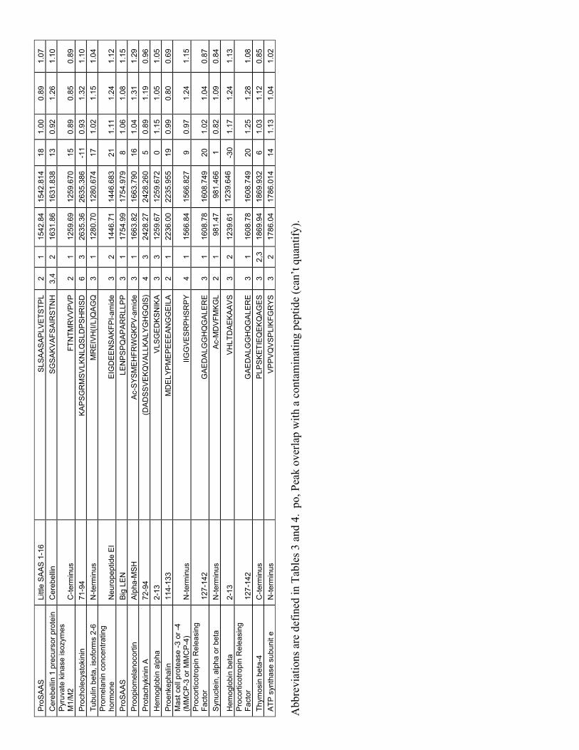
Pro
SA
AS
Li
ttle
SA
AS
1-1
6 S
LSA
AS
AP
LVE
TSTP
L 2
1 15
42.8
4 15
42.8
14
18
1.00
0.
89
1.07
C
ereb
ellin
1 p
recu
rsor
pro
tein
C
ereb
ellin
S
GS
AK
VA
FSA
IRS
TNH
3,
4 2
1631
.86
1631
.838
13
0.
92
1.26
1.
10
Pyr
uvat
e ki
nase
isoz
ymes
M
1/M
2 C
-term
inus
FT
NTM
RV
VP
VP
2
1 12
59.6
9 12
59.6
70
15
0.89
0.
85
0.89
P
roch
olec
ysto
kini
n 71
-94
KA
PS
GR
MS
VLK
NLQ
SLD
PS
HR
ISD
6
3 26
35.3
6 26
35.3
86
-11
0.93
1.
32
1.10
Tu
bulin
bet
a, is
ofor
ms
2-6
N-te
rmin
us
MR
EIV
H(I/
L)Q
AG
Q
3 1
1280
.70
1280
.674
17
1.
02
1.15
1.
04
Pro
mel
anin
con
cent
ratin
g ho
rmon
e N
euro
pept
ide
EI
EIG
DE
EN
SA
KFP
I-am
ide
3 2
1446
.71
1446
.683
21
1.
11
1.24
1.
12
Pro
SA
AS
B
ig L
EN
LE
NP
SP
QA
PA
RR
LLP
P
3 1
1754
.99
1754
.979
8
1.06
1.
08
1.15
P
roop
iom
elan
ocor
tin
Alp
ha-M
SH
A
c-S
YSM
EH
FRW
GK
PV
-am
ide
3 1
1663
.82
1663
.790
16
1.
04
1.31
1.
29
Pro
tach
ykin
in A
72
-94
(DA
DS
SV
EK
QV
ALL
KA
LYG
HG
QIS
) 4
3 24
28.2
7 24
28.2
60
5 0.
89
1.19
0.
96
Hem
oglo
bin
alph
a 2-
13
VLS
GE
DK
SN
IKA
3
3 12
59.6
7 12
59.6
72
0 1.
15
1.05
1.
05
Pro
enke
phal
in
114-
133
MD
ELY
PM
EP
EE
EA
NG
GE
ILA
2
1 22
36.0
0 22
35.9
55
19
0.99
0.
80
0.69
M
ast c
ell p
rote
ase
-3 o
r -4
(MM
CP
-3 o
r MM
CP
-4)
N-te
rmin
us
IIGG
VE
SR
PH
SR
PY
4
1 15
66.8
4 15
66.8
27
9 0.
97
1.24
1.
15
Pro
corti
cotro
pin
Rel
easi
ng
Fact
or
127-
142
GA
ED
ALG
GH
QG
ALE
RE
3
1 16
08.7
8 16
08.7
49
20
1.02
1.
04
0.87
S
ynuc
lein
, alp
ha o
r bet
a N
-term
inus
A
c-M
DV
FMK
GL
2 1
981.
47
981.
466
1 0.
82
1.09
0.
84
Hem
oglo
bin
beta
2-
13
VH
LTD
AE
KA
AV
S
3 2
1239
.61
1239
.646
-3
0 1.
17
1.24
1.
13
Pro
corti
cotro
pin
Rel
easi
ng
Fact
or
127-
142
GA
ED
ALG
GH
QG
ALE
RE
3
1 16
08.7
8 16
08.7
49
20
1.25
1.
28
1.08
Th
ymos
in b
eta-
4 C
-term
inus
P
LPS
KE
TIE
QE
KQ
AG
ES
3
2,3
1869
.94
1869
.932
6
1.03
1.
12
0.85
A
TP s
ynth
ase
subu
nit e
N
-term
inus
V
PP
VQ
VS
PLI
KFG
RYS
3
2 17
86.0
4 17
86.0
14
14
1.13
1.
04
1.02
Abb
revi
atio
ns a
re d
efin
ed in
Tab
les 3
and
4.
po, P
eak
over
lap
with
a c
onta
min
atin
g pe
ptid
e (c
an’t
quan
tify)
.
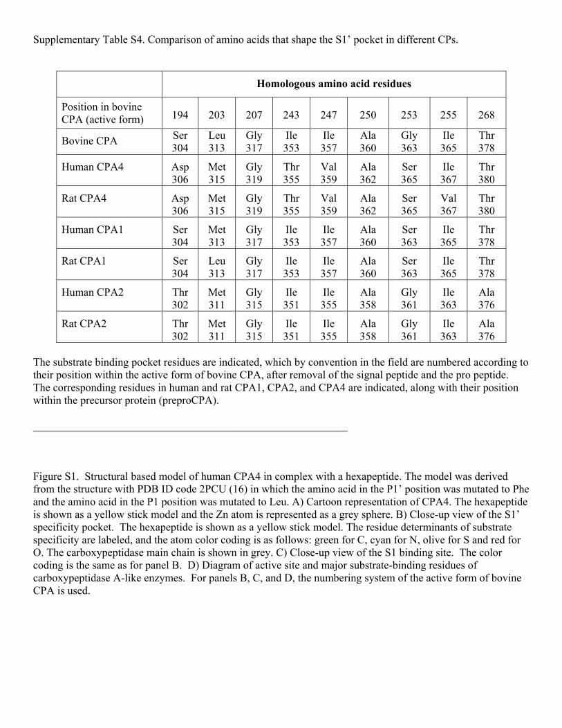
Supplementary Table S4. Comparison of amino acids that shape the S1’ pocket in different CPs.
Homologous amino acid residues
Position in bovine CPA (active form) 194 203 207 243 247 250 253 255 268
Bovine CPA Ser 304
Leu 313
Gly 317
Ile 353
Ile 357
Ala 360
Gly 363
Ile 365
Thr 378
Human CPA4 Asp 306
Met 315
Gly 319
Thr 355
Val 359
Ala 362
Ser 365
Ile 367
Thr 380
Rat CPA4 Asp 306
Met 315
Gly 319
Thr 355
Val 359
Ala 362
Ser 365
Val 367
Thr 380
Human CPA1 Ser 304
Met 313
Gly 317
Ile 353
Ile 357
Ala 360
Ser 363
Ile 365
Thr 378
Rat CPA1 Ser 304
Leu 313
Gly 317
Ile 353
Ile 357
Ala 360
Ser 363
Ile 365
Thr 378
Human CPA2 Thr 302
Met 311
Gly 315
Ile 351
Ile 355
Ala 358
Gly 361
Ile 363
Ala 376
Rat CPA2 Thr 302
Met 311
Gly 315
Ile 351
Ile 355
Ala 358
Gly 361
Ile 363
Ala 376
The substrate binding pocket residues are indicated, which by convention in the field are numbered according to their position within the active form of bovine CPA, after removal of the signal peptide and the pro peptide. The corresponding residues in human and rat CPA1, CPA2, and CPA4 are indicated, along with their position within the precursor protein (preproCPA). _________________________________________________________ Figure S1. Structural based model of human CPA4 in complex with a hexapeptide. The model was derived from the structure with PDB ID code 2PCU (16) in which the amino acid in the P1’ position was mutated to Phe and the amino acid in the P1 position was mutated to Leu. A) Cartoon representation of CPA4. The hexapeptide is shown as a yellow stick model and the Zn atom is represented as a grey sphere. B) Close-up view of the S1’ specificity pocket. The hexapeptide is shown as a yellow stick model. The residue determinants of substrate specificity are labeled, and the atom color coding is as follows: green for C, cyan for N, olive for S and red for O. The carboxypeptidase main chain is shown in grey. C) Close-up view of the S1 binding site. The color coding is the same as for panel B. D) Diagram of active site and major substrate-binding residues of carboxypeptidase A-like enzymes. For panels B, C, and D, the numbering system of the active form of bovine CPA is used.

Lloyd D. FrickerSebastian Tanco, Xin Zhang, Cain Morano, Francesc Xavier Aviles, Julia Lorenzo and
implications for a role in extracellular peptide processingHuman carboxypeptidase A4: Characterization of the substrate specificity and
published online April 12, 2010J. Biol. Chem.
10.1074/jbc.M109.060350Access the most updated version of this article at doi:
Alerts:
When a correction for this article is posted•
When this article is cited•
to choose from all of JBC's e-mail alertsClick here
Supplemental material:
http://www.jbc.org/content/suppl/2010/04/12/M109.060350.DC1.html
http://www.jbc.org/content/early/2010/04/12/jbc.M109.060350.full.html#ref-list-1
This article cites 0 references, 0 of which can be accessed free at
by guest on September 4, 2016
http://ww
w.jbc.org/
Dow
nloaded from