Proteomic characterization of novel histone post-translational modifications
Upload
khangminh22Category
view
2download
0
Characterization of Novel Xenorhabdus-Steinernema Associations and Identification of Novel Antimicrobial Compounds Produced by
Xenorhabdus khoisanae
by
Jonike Dreyer
Thesis presented in partial fulfilment of the requirements for the degree of Master of Science in the Faculty of Science at Stellenbosch University
Supervisor: Prof. L.M.T. Dicks
Co-supervisor: Dr. A.P. Malan
March 2018
ii
Declaration
By submitting this thesis electronically, I declare that the entirety of the work contained therein
is my own, original work, that I am the sole author thereof (save to the extent explicitly
otherwise stated), that reproduction and publication thereof by Stellenbosch University will not
infringe any third party rights and that I have not previously in its entirety or in part submitted
it for obtaining any qualification.
March 2018
Copyright © 2018 Stellenbosch University
All rights reserved
Stellenbosch University https://scholar.sun.ac.za

iii
Abstract
Xenorhabdus bacteria are closely associated with Steinernema nematodes. This is a species-
specific association. Therefore, a specific Steinernema species is associated with a specific
Xenorhabdus species. During the Xenorhabdus-Steinernema life cycle the nematodes infect
insect larvae and release the bacteria into the hemocoel of the insect by defecation. The bacteria
and nematodes produce several exoenzymes and toxins that lead to septicemia, death and
bioconversion of the insect. This results in the proliferation of both the nematodes and bacteria.
When nutrients are depleted, nematodes take up Xenorhabdus cells and leave the cadaver in
search of their next prey. Xenorhabdus produces various broad-spectrum bioactive compounds
during their life cycle to create a semi-exclusive environment for the growth of the bacteria and
their symbionts.
During this study, a molecular approach was used to identify four Xenorhabdus isolates from
Steinernema sacchari SB10T, Steinernema jeffreyense J194T, Steinernema nguyeni F2T and
Steinernema litchii WS9T as Xenorhabdus khoisanae SB10 and J194, Xenorhabdus bovienii
F2 and Xenorhabdus griffiniae WS9, respectively. Steinernema phylogenetics were analyzed
and the X. khoisanae-S. sacchari and X. griffiniae-S. litchii associations proved that
X. khoisanae and X. griffiniae has the ability to switch between different nematode clades.
Antimicrobial compounds produced by X. khoisanae SB10 were purified and analyzed by high-
performance liquid chromatography (HPLC) and liquid chromatography-mass spectrometry
(LCMS), respectively. MS spectra and MSe fragmentation profiles revealed novel
antimicrobial compounds with mass-to-charge ratios of 671.41 m/z, 259.17 m/z, 434.27 m/z
and/or 341.15 m/z. Additionally, this study reports for the first time, the isolation of PAX
peptides, xenocoumacins and xenorhabdins from X. khoisanae.
Stellenbosch University https://scholar.sun.ac.za

iv
Opsomming
Xenorhabdus bakterieë is naby geassosieer met Steinernema nematodes. Hierdie is ‘n spesie-
spesifieke assosiasie. Dit wil sê, ʼn spesifieke Steinernema spesie is geassosieer met ʼn
spesifieke Xenorhabdus spesie. Tydens die Xenorhabdus-Steinernema lewenssiklus infekteer
die nematodes inseklarwes en word die bakterieë in die hemoseel van die insek vrygestel deur
middel van ontlasting. Die bakterieë en nematodes produseer verskeie ekso-ensieme en
toksiene wat lei tot septisemie, dood en bio-omskakeling van die insek. Dit lei tot die
vermeerdering van beide die nematodes en bakterieë. Sodra nutriente uitgeput is, neem
nematodes Xenorhabdus selle op en verlaat die kadawer opsoek na hul volgende prooi.
Xenorhabdus produseer verskeie breë-spektrum bioaktiewe verbindings tydens hul
lewenssiklus om ‘n gedeeltelike eksklusiewe omgewing te skep vir die groei van die bakterieë
en hul simbionte.
Gedurende hierdie studie was ‘n molekulêre benadering gebruik om vier Xenorhabdus isolate
vanaf Steinernema sacchari SB10T, Steinernema jeffreyense J194T, Steinernema nguyeni F2T
en Steinernema litchii WS9T te identifiseer as, Xenorhabdus khoisanae SB10 en J194,
Xenorhabdus bovienii F2 en Xenorhabdus griffiniae WS9, afsonderlik. Steinernema
filogenetika was geanaliseer en die X. khoisanae-S. sacchari en X. griffiniae-S. litchii
assosiasies het bewys dat X. khoisanae en X. griffiniae die vermoë het om te wissel tussen
nematodes van verskillende klades.
Antimikrobiese verbindings geproduseer deur die isolaat, X. khoisanae SB10, was gesuiwer en
geanaliseer deur hoëdrukvloeistofchromatografie (HDVC) en vloeistofchromatografie massa-
spektrometrie (VCMS), afsonderlik. MS spektra en MSe fragementasie profiele het nuwe
antimikrobiese verbindings met massa-tot-lading verhoudings van 671.41 m/z, 259.17 m/z,
434.27 m/z en/of 341.15 m/z onthul. Vêrder rapporteer hierdie studie, vir die eerste keer, dat
PAX peptiede, xenokoumasiene en xenorhabdiene geïsoleer was vanaf X. khoisanae.
Stellenbosch University https://scholar.sun.ac.za

v
Biographical sketch
Jonike Dreyer was born in Cape Town, South Africa on the 10th of March 1993. She
matriculated at Paarl Girls’ High School, South Africa, in 2011. In 2012 she enrolled as B.Sc.
student in Molecular Biology and Biotechnology at the University of Stellenbosch and obtained
her B.Sc (Cum Laude) in 2014. In 2015 she obtained her B.Sc (Hons) in Microbiology, also at
the University of Stellenbosch. In 2016 she enrolled as M.Sc. student in Microbiology.
Stellenbosch University https://scholar.sun.ac.za

vi
Preface
This thesis is represented as a compilation of 6 chapters. Chapters 1, 2, 5 and 6 are written
according to instructions of the Journal of Applied and Environmental Microbiology. Chapters
3 and 4 have been published in Current Microbiology (2017, volume 74:8, pp 938-942) and
Archives of Microbiology (2017, doi: 10.1007/s00203-017-1452-4), respectively.
Chapter 1: General Introduction
Chapter 2: Phenotypic and Genotypic Characteristics of Xenorhabdus Species, Their
Association with Entomopathogenic Nematodes and Production of Antimicrobial Compounds
Chapter 3: Three Novel Xenorhabdus-Steinernema Associations and Evidence of Strains of
X. khoisanae Switching Between Different Clades
Chapter 4: First Report of a Symbiotic Relationship Between Xenorhabdus griffiniae and an
Unknown Steinernema from South Africa
Chapter 5: Novel Antimicrobial Compounds from Xenorhabdus khoisanae SB10, and the
First Report of PAX peptides, Xenocoumacins and Xenorhabdins from this Species
Chapter 6: General Discussion and Conclusions
Stellenbosch University https://scholar.sun.ac.za

vii
Acknowledgements
I would like to sincerely thank the following people and organizations:
Prof. L.M.T. Dicks for granting me this opportunity, and all his support and guidance,
Dr. A.P. Malan for sample collection, support and guidance,
Dr. A.D. van Staden for HPLC assistance, valuable insight and guidance,
Prof. M. Rautenbach for guidance and assistance with HPLC purification and MS analysis,
Riaan de Witt for assistance with processing Whole Genome Sequencing data,
Dr. S.M. Deane for critical reading of the manuscript,
All my co-workers in the lab and Department of Microbiology for insight and support,
The National Research Foundation (NRF) of South Africa for financial support.
Stellenbosch University https://scholar.sun.ac.za

viii
Dedication
Because an acknowledgement simply is not enough,
I dedicate this thesis to:
My mother, Emma Dreyer, for supporting me, no matter what,
Du Toit Kemp, for weekend lab visits, always making me see the bright side of things and
making me laugh,
Leané Dreyer, for freak-out sessions and motivational speeches since 2012,
Elzaan Booysen, for MS-DOS, floppy disk and motivational support,
Dicks lab members, for three years of pizza after lab clean, random mystic’s nights and,
getting told to clean your bench and wash your dishes,
All scientists, who realized that doing something exactly the same for the 77th time will have
a different result,
and finally, to
The greatest God that ever has and ever will exist, for always being there, even when I did
not realize it.
Stellenbosch University https://scholar.sun.ac.za

ix
Table of Contents
Chapter 1 General Introduction 1
General Introduction 2
References 4
Chapter 2 Literature review:
Phenotypic and Genotypic Characteristics of Xenorhabdus Species, Their Association with Entomopathogenic Nematodes and Production of Antimicrobial Compounds 7
Abstract 8
The Genus Xenorhabdus 9
Association of Xenorhabdus with Entomopathogenic Nematodes 10 The Xenorhabdus-Steinernema Life Cycle 14
Stage I 14
Stage II 14
Stage III 15 Synergistic Effect 16
EPNs as Biological Control Agents 17
Xenorhabdus Bioactive Secondary Metabolites 18
Depsipeptides 18 Xenocoumacins 20
Fabclavines 20
Pristinamycin 24
Xenortides 25 Rhabdopeptides 25
Bicornitun 25
PAX peptides 25
Cabanillasin and Nemaucin 26
Dithiolopyrrolone derivatives 26 Indole-containing compounds 26
Unnamed peptides 27
Benzylideneacetone 27
Rhabduscin 27
Stellenbosch University https://scholar.sun.ac.za

x
Bacteriocins 28
Upregulating the Production of Xenorhabdus Antimicrobials 28
Conclusion 30 References 31
Chapter 3
Three Novel Xenorhabdus-Steinernema Associations and Evidence of Strains of X. khoisanae Switching Between Different Clades 43
Abstract 44 Introduction 45
Materials and Methods 45
Isolation and Maintenance of Cultures 45
Biochemical Characterization 46 Antibacterial Activity 47
Phylogenetic Analysis of Isolates 47
Results and Discussion 48
References 52
Supplementary material 55
Chapter 4
First Report of a Symbiotic Relationship Between Xenorhabdus griffiniae and an Unknown Steinernema from South Africa 60
Abstract 61
Introduction 62 Materials and Methods 63
Isolation of Xenorhabdus sp. and Maintenance of Cultures 63
Cell Morphology and Phenotypic Characteristics 63
Genotypic Characterization and Phylogenetic Analysis 64 Testing for Antibacterial Activity 66
Results and Discussion 66
Phenotypic and Biochemical Characteristics 66
Antibacterial Activity 66
Phylogenetic Analysis of Strain WS9 67 Acknowledgements 69
References 70
Online Resources 73
Stellenbosch University https://scholar.sun.ac.za

xi
Chapter 5
Novel Antimicrobial Compounds Produced by Xenorhabdus khoisanae SB10, and the First Report on PAX peptides, Xenocoumacins and Xenorhabdins from this Species 80
Abstract 81 Introduction 82
Materials and methods 83
Bacterial strains, growth media and growth conditions 83
Isolation of antimicrobial compounds 85
Purification of antimicrobial compounds 85 Method A 85
Method B 86
Ultra-performance liquid chromatography (UPLC) and electrospray ionisation mass spectrometry (ESI-MS) 88
Antimicrobial spectrum 88 Temperature stability 88
Results 89
Isolation and partial purification of antimicrobial compounds 89
Method A 89 Method B 94
Antimicrobial spectrum and temperature stability 100
Discussion 100
Method A 100
Method B 102 Comparison of purification methods 103
Antimicrobial spectrum and temperature stability 103
Conclusion 104
References 105 Supplementary material 108
Chapter 6 General Discussion and Conclusions 112
General Discussion 113 Conclusion 115
References 115
Stellenbosch University https://scholar.sun.ac.za

1
Chapter 1
General Introduction
Stellenbosch University https://scholar.sun.ac.za

2
General Introduction
The genus Xenorhabdus consists of Gram-negative bacteria belonging to the
Enterobacteriacaea family (1). These bacteria co-exist in a mutualistic relationship with
pathogenic nematodes of the genus Steinernema (2), this is a species-specific association, i.e.
a single Xenorhabdus sp. is associated with a specific Steinernema sp. (3). However, a single
Xenorhabdus sp. associating with more than one Steinernema sp. has been reported (4–6). The
latter, referred to as switching of hosts, is generally between nematodes in the same clade, but
more recent studies have shown that the switching of hosts may also take place between
nematodes of different clades (4–6).
The mutualistic relationship between Steinernema spp. and Xenorhabdus spp. is critical in the
formation of a tripartite relationship with the host (insect) larvae. Xenorhabdus bacteria are
carried in the receptacle of Steinernema nematodes when in the infective juvenile (IJ) phase
(7, 8). Infective juveniles position themselves near the soil surface (9), enter the host through
natural openings such as respiratory spiracles, mouth or anus and migrate to the hemocoel to
excrete viable cells of Xenorhabdus (10). The host’s immune response is inhibited with
compounds produced by the nematodes and bacterial cells. Exoenzymes and toxins produced
by Xenorhabdus lead to septicemia (11–14) and, ultimately, death of the host within 24-48 h.
The nematodes reproduce sexually and repeat their life cycle until nutrients become depleted.
During this phase, Xenorhabdus cells produce a number of antimicrobial compounds to create
a semi-exclusive environment for themselves and the nematodes (10, 15). Second-phase
juveniles stemming from the mutualistic relationship develop into IJs via the third phase. These
IJs harbour viable Xenorhabdus cells.
Xenorhabdus spp. produce various bioactive compounds throughout their life cycle. These
bacteria are, however, an underestimated and neglected source of novel bioactive compounds.
Biologically active compounds produced by Xenorhabdus spp. have a broad-spectrum of
antimicrobial activity, inhibiting the growth of bacteria, fungi and protozoa, the development
of insects and nematodes, and the formation of cancerous cells (15). The variety of bioactive
compounds produced by Xenorhabdus spp. differ, even between strains of the same species.
Polyketide synthetases (PKS) and non-ribosomal peptide synthetases (NRPS) are responsible
for the production of a diverse group of peptides, e.g. depsipeptides (16–18), xenocoumacins
(19) and PAX peptides (peptide-antimicrobial-Xenorhabdus) (20). Other Xenorhabdus
Stellenbosch University https://scholar.sun.ac.za

3
antimicrobial compounds include benzylideneacetone (21), indole derivatives (22) and
bacteriocins (23, 24).
The efficiency of Xenorhabdus bioactive compounds in the agricultural industry has been
shown with various studies (25–29). Xenorhabdus bacteria, as a source of bioactive
compounds, have exceeding potential, however, not only in the agricultural industry, but also
in the healthcare and food industries. According to genomic analysis done on X. nematophila
DSM 3370T (30), only a fraction of the bioactive compounds that may be produced by
Xenorhabdus spp. have been reported. It is evident that the research already done on
Xenorhabdus bacteria is only the prelude to what is yet to come.
In the first section of this thesis, four Xenorhabdus strains (SB10, J194, WS9 and F2), isolated
from Steinernema sacchari, Steinernema jeffreyense, Steinernema litchii and Steinernema
nguyeni, respectively, are identified to species level. Before this study, the Xenorhabdus
symbionts associated with these nematodes had not been reported. Identification was done by
using biochemical tests, PCR and genome sequencing. The results led to the discovery of
intriguing novel Xenorhabdus-Steinernema associations.
The second section of the study focussed on the antimicrobial activity of Xenorhabdus strain
SB10. Antimicrobial compounds in the cell-free extract of Xenorhabdus khoisanae SB10 was
isolated with XAD-16 beads, purified by high-performance liquid chromatography (HPLC)
and fractions with antimicrobial activity subjected to liquid chromatography–mass
spectrometry (LCMS) for putative identification. Strain SB10 was selected as no previous
studies have been done on the bioactive compounds produced by X. khoisanae. The isolation
and purification methods for antimicrobials produced by strain SB10 was optimized.
Stellenbosch University https://scholar.sun.ac.za

4
References
1. Thomas GM, Poinar GO. 1979. Xenorhabdus gen. nov., a genus of entomopathogenic,
nematophilic bacteria of the family Enterobacteriaceae. Int J Syst Bacteriol 29:352–360.
2. Adams B, Nguyen KB. 2002. Taxonomy and systematics, p 1–34. In Gaugler R (ed),
Entomopathogenic nematology. CABI Publishing, New York.
3. Akhurst RJ. 1982. A Xenorhabdus sp. (Eubacteriales: Enterobacteriaceae) symbiotically
associated with Steinernema kraussei (Nematoda: Steinernematidae). Rev Nematol
5:277–280.
4. Lee M-M, Stock SP. 2010. A multigene approach for assessing evolutionary
relationships of Xenorhabdus spp. (γ-Proteobacteria), the bacterial symbionts of
entomopathogenic Steinernema nematodes. J Invertebr Pathol 104:67–74.
5. Dreyer J, Malan AP, Dicks LMT. 2017. Three novel Xenorhabdus–Steinernema
associations and evidence of strains of X. khoisanae switching between different clades.
Curr Microbiol 74:938–942.
6. Çimen H, Půža V, Nermuť J, Hatting J, Ramakuwela T, Faktorová L, Hazir S. 2016.
Steinernema beitlechemi n. sp., a new entomopathogenic nematode (Nematoda:
Steinernematidae) from South Africa. Nematology 18:439–453.
7. Kim SK, Flores-Lara Y, Patricia Stock S. 2012. Morphology and ultrastructure of the
bacterial receptacle in Steinernema nematodes (Nematoda: Steinernematidae). J
Invertebr Pathol 110:366–374.
8. Snyder H, Stock SP, Kim S-K, Flores-Lara Y, Frost S. 2007. New insights into the
colonization and release processes of Xenorhabdus nematophila and the morphology
and ultrastructure of the bacterial receptacle of its nematode host, Steinernema
carpocapsae. Appl Environ Microbiol 73:5338–5346.
9. Kaya HK, Gaugler R. 1993. Entomopathogenic Nematodes. Annu Rev Entomol 38:181–
206.
10. Poinar GO, Thomas GM. 1966. Significance of Achromobacter nematophilus Poinar
and Thomas (Achromobacteraceae: Eubacteriales) in the development of the nematode,
DD-136 (Neoaplectana sp. Steinernematidae). Parasitology 56:385–390.
11. Brillard J, Ribeiro C, Boemare N, Brehélin M, Givaudan A. 2001. Two distinct
hemolytic activities in Xenorhabdus nematophila are active against immunocompetent
insect cells. Appl Environ Microbiol 67:2515–2525.
12. Cho S, Kim Y. 2004. Hemocyte apoptosis indiced by entomopathogenic bacteria,
Stellenbosch University https://scholar.sun.ac.za

5
Xenorhabdus and Photorhabdus, in Bomyx mori. J Asia Pac Entomol 7:195–200.
13. Dunphy GB, Webster JM. 1984. Interaction of Xenorhabdus nematophilus subsp.
nematophilus with the haemolymph of Galleria mellonella. J Insect Physiol 30:883–
889.
14. Yang J, Zeng H-M, Lin H-F, Yang X-F, Liu Z, Guo L-H, Yuan J-J, Qiu D-W. 2012. An
insecticidal protein from Xenorhabdus budapestensis that results in prophenoloxidase
activation in the wax moth, Galleria mellonella. J Invertebr Pathol 110:60–67.
15. Webster JM, Chen G, Hu K, Li J. 2002. Bacterial metabolites, p 99–114. In Gaugler, R
(ed), Entomopathogenic nematology. CAB International, New York.
16. Zhou Q, Grundmann F, Kaiser M, Schiell M, Gaudriault S, Batzer A, Kurz M, Bode
HB. 2013. Structure and biosynthesis of xenoamicins from entomopathogenic
Xenorhabdus. Chem - A Eur J 19:16772–16779.
17. Kronenwerth M, Bozhüyük KAJ, Kahnt AS, Steinhilber D, Gaudriault S, Kaiser M,
Bode HB. 2014. Characterisation of taxlllaids A-G; natural products from Xenorhabdus
indica. Chem Eur J 20:17478–17487.
18. Lang G, Kalvelage T, Peters A, Wiese J, Imhoff JF. 2008. Linear and cyclic peptides
from the entomopathogenic bacterium Xenorhabdus nematophilus. J Nat Prod 71:1074–
1077.
19. Reimer D. 2013. PhD thesis. Johann Wolfgang Groethe-Universität, Frankfurt,
Germany. Identification and characterization of selected secondary metabolite
biosynthetic pathways from Xenorhabdus nematophila.
20. Fuchs SW, Proschak A, Jaskolla TW, Karas M, Bode HB. 2011. Structure elucidation
and biosynthesis of lysine-rich cyclic peptides in Xenorhabdus nematophila. Org
Biomol Chem 9:3130–3132.
21. Ji D, Yi Y, Kang G-H, Choi Y-H, Kim P, Baek N-I, Kim Y. 2004. Identification of an
antibacterial compound, benzylideneacetone, from Xenorhabdus nematophila against
major plant-pathogenic bacteria. FEMS Microbiol Lett 239:241–248.
22. Sundar L, Chang FN. 1993. Antimicrobial activity and biosynthesis of indole antibiotics
produced by Xenorhabdus nematophilus. J Gen Microbiol 139:3139–3148.
23. Thaler JO, Baghdiguian S, Boemare N. 1995. Purification and characterization of
xenorhabdicin, a phage tail-like bacteriocin, from the lysogenic strain F1 of
Xenorhabdus nematophilus. Appl Environ Microbiol 61:2049–2052.
24. Singh J, Banerjee N. 2008. Transcriptional analysis and functional characterization of a
gene pair encoding iron-regulated xenocin and immunity proteins of Xenorhabdus
Stellenbosch University https://scholar.sun.ac.za

6
nematophila. J Bacteriol 190:3877–3885.
25. Ng KK, Webster JM. 1997. Antimycotic activity of Xenorhabdus bovienii
(Enterobacteriaceae) metabolites against Phytophthora infestans on potato plants. Can J
Plant Pathol 19:125–132.
26. Böszörményi E, Érsek T, Fodor AM, Fodor AM, Földes LS, Hevesi M, Hogan JS,
Katona Z, Klein MG, Kormány A, Pekár S, Szentirmai A, Sztaricskai F, Taylor RAJ.
2009. Isolation and activity of Xenorhabdus antimicrobial compounds against the plant
pathogens Erwinia amylovora and Phytophthora nicotianae. J Appl Microbiol 107:746–
759.
27. Fang XL, Li ZZ, Wang YH, Zhang X. 2011. In vitro and in vivo antimicrobial activity
of Xenorhabdus bovienii YL002 against Phytophthora capsici and Botrytis cinerea. J
Appl Microbiol 111:145–154.
28. Shapiro-Ilan DI, Reilly CC, Hotchkiss MW. 2009. Suppressive effects of metabolites
from Photorhabdus and Xenorhabdus spp. on phytopathogens of peach and pecan. Arch
Phytopathol Plant Prot 42:715–728.
29. Barkai-Golan R. 2001. Postharvest diseases of fruits and vegetables: development and
control, 1st ed. Elsevier, Amsterdam, The Netherlands.
30. Crawford JM, Kontnik R, Clardy J. 2010. Report regulating alternative lifestyles in
entomopathogenic bacteria. Curr Biol 20:69–74.
Stellenbosch University https://scholar.sun.ac.za

7
Chapter 2
Literature review:
Phenotypic and Genotypic Characteristics of
Xenorhabdus Species, Their Association with
Entomopathogenic Nematodes and Production of
Antimicrobial Compounds
Stellenbosch University https://scholar.sun.ac.za

8
Phenotypic and Genotypic Characteristics of Xenorhabdus species, Their Association
with Entomopathogenic Nematodes and Production of Antimicrobial Compounds
J. Dreyer1, A. P Malan2 and L. M. T. Dicks1
1 Department of Microbiology, Stellenbosch University, Private Bag X1, Matieland 7602,
South Africa 2 Department of Conservation Ecology and Entomology, Stellenbosch University, Private Bag
X1, Matieland 7602, South Africa
Abstract
The genus Xenorhabdus belongs to the family Enterobacteriaceae. These bacteria live
mutualistically within entomopathogenic nematodes of the genus Steinernema. This
association is species-specific, however, sharing of a specific Xenorhabdus sp. does occur
between Steinernema hosts. During the Xenorhabdus-Steinernema life cycle, insect larvae are
infected and killed, while both mutualists produce bioactive compounds. These compounds
work synergistically to ensure the reproduction and proliferation of the nematodes and bacteria.
Over the past two decades, the number of studies done on the bioactive compounds produced
by Xenorhabdus spp. have increased drastically. These compounds are often broad-spectrum
with activity against bacteria, fungi, insects, nematodes, protozoa and cancerous cells. It is
evident that this genus is greatly underestimated and neglected in the search for novel bioactive
compounds, especially when taking into consideration the need for novel antibiotics.
Keywords Xenorhabdus, Steinernema, bioactive compounds
Stellenbosch University https://scholar.sun.ac.za

9
The Genus Xenorhabdus
The genus Xenorhabdus consists of 26 species and belongs to the family Enterobacteriaceae
(1). The rod-shaped cells (0.3-2.0 µm x 2.0-10.0 µm) stain Gram-negative, have peritrichous
flagella when motile, do not reduce nitrate and, are oxidase and catalase negative. No
endospores are produced, but crystalline inclusions may form during stationary growth.
Spheroplasts with an average diameter of 2.6 µm may form towards the end of exponential
growth (2). Most members of the genus are mesophilic and grow at 28 °C, although some
strains may grow at 42 °C. Metabolism is respiratory and fermentative and the cells are
classified as facultative anaerobes. Mannose, glycerol and N-acetylglucosamine are usually
fermented. Some species ferment fructose. Most strains produce DNases, proteases and
lipases.
Xenorhabdus spp. live in close association with entomopathogenic nematodes of the family
Steinernematidae. This association directly influences the viable state of the bacterial cells.
Two cell types, referred to as phase variants, have been described. Cells naturally associated
with actively reproducing nematodes are in phase I (form I, or primary form), but change to
phase II cells during later stages of the nematode reproduction cycle, i.e. when nematodes infect
the insect cadaver (3). Conversion to phase II also occurs when cells are repeatedly sub-
cultured in vitro (4). Phase I cells are larger than phase II cells, are motile, have swarming
abilities, form crystalline inclusion bodies, and produce proteases, lipases and antimicrobial
compounds (5–10). Phase I cells absorb certain dyes and can be distinguished from phase II
cells by streaking the cells onto nutrient agar supplemented with 0.025% (w/v) bromothymol
blue and 0.004% (w/v) triphenyltetrazolium chloride (NBTA medium) (11). Colonies on
NBTA medium are dark blue with a red core and are surrounded by a clear zone. Exceptions
to the rule have been reported, i.e. on rare occasions phase I cells do not absorb bromothymol
blue (12).
Xenorhabdus spp. are differentiated based on biochemical characteristics (Table 1), but
identification has to be confirmed using genotypic classification methods. Sequences of genes
encoding recombinase A (recA), DNA gyrase subunit B (gyrB), DNA polymerase III beta chain
(dnaN), initiation factor B (infB), glutamyl-tRNA synthetase catalytic subunit (gltX) and
16S rRNA (16S rDNA) are usually compared to determine the level of genetic relatedness.
Furthermore, DNA-DNA hybridization of the entire genome may be used to confirm the exact
Stellenbosch University https://scholar.sun.ac.za

10
species. The phylogenetic relatedness of Xenorhabdus spp., compiled from sequences in
GenBank, is shown in Fig. 1.
Association of Xenorhabdus with Entomopathogenic Nematodes
Xenorhabdus spp. are closely associated with entomopathogenic nematodes (EPNs) of the
family Steinernematidae Travassos, specifically the genus Steinernema, and they are believed
to increase the virulence and reproduction (thus “fitness”) of the nematode (13). Until recently,
the general assumption was that a specific Xenorhabdus sp. can only infect one
Steinernema species. This has clearly been demonstrated in a study conducted by
Sicard et al. (13). The authors have shown that the fitness of Steinernema carpocapsae
improved when associated with Xenorhabdus nematophila, but not when associated with non-
native Xenorhabdus spp. More recent findings have, however, shown that a single species of
Xenorhabdus may be associated with several Steinernema spp. (Table 2). Murfin et al. (14)
reported an increase in the fitness of Steinernema nematodes when infected with a strain of
Xenorhabdus bovienii native to the nematode, or when associated with a strain from another
Steinernema sp. closely related to the original nematode. Some authors hypothesized that the
association of a specific Xenorhabdus sp. with more than one Steinernema sp. is an indication
that the respective nematodes are phylogenetically related. The findings of Lee and Stock (15)
provided the final answer to this hypothesis. The authors have shown that host switching of
Xenorhabdus spp. have occurred in clades and between clades as many as 17 times.
Steinernema beitlechemie from the Cameroonense-clade (16) is associated with
Xenorhabdus khoisanae. However, X. khoisanae was first isolated from
Steinernema khoisanae that belongs to the Glaseri-clade (17, 18). In a more recent paper,
further evidence of X. khoisanae switching between clades was reported when X. khoisanae
was isolated from Steinernema sacchari of the Cameroonense-clade (19).
Stellenbosch University https://scholar.sun.ac.za

11
Table 1 Carbohydrate fermentation and assimilation by Xenorhabdus spp. Characteristic 1 2 3 4 5 6 7 8 9 10 11 12 13 14 15 16 17 18 19 20 21 22 23 24 25 26
Acid production from:
5-Ketogluconate + v + - - n.a. v w + + + w + w + - - w w + v w w - n.a. +
Aesculin + - - - v - v + + v - + - - - + + + + - v - + + - +
Fructose + v + v v - v + + + + + + + - - - + + + v + + v - +
gluconate + v - - - - - - - - + - - + - - - + w - - w + - - -
Glucose + + + + v + + - + + + + - + + + + + + + + + + + - +
Inositol - n.a. + v v - - - v + v - - w - - - + - v - w + + - -
Maltose + v - v v + v - v - - + + + + + + + + v + + + v - +
mannose + + + + v + + - + + + + + + + + + + + + + + + v - +
N-acetyl-glucosamine + + v + v - v - v v + + + + - - - + + + + + + v - +
Ribose + v - - v - v + v - + w + + - + - + + v - - + v - -
Sorbitol - - - - - - - - - - - - - - - - - - - - - - - + - -
trehalose + v - - v + - - n.a. - v + - v - + + + + v v + - - - +
Assimilation of:
Aesculin + - v - v - v + + v v + - + - v + + + - v - + + - +
Fructose + v + v v + v - v + + v + n.a. - - - + + + v + + - + +
gluconate + + v - + + v + + + + + w + - v - + + v + + + + + +
Glucose + + + + + + + + + + + - + + - + + + + + + + + + + +
Inositol - - - - + - - w v + + - + n.a. - - - + w v - + + + - w
Maltose + + v + + + + + + + - - + + - + + + + + + + + + + +
mannose + + + + v + v + + + + + + + - + + + + + + + + + + +
N-acetyl-glucosamine + + v + + + + + + + + + + + - + + + + + + + + + + +
Ribose + v - - v + v - v - + - - n.a. - + - + + - - - + v - -
Trehalose + + + v + + v + + v + + + n.a. - + + + + + + + - v + +
(1) X. beddingii, (2) X. bovienii, (3) X. budapestensis, (4) X. cabanillasii, (5) X. doucetiae, (6) X. eapokensis, (7) X. ehlersii, (8) X. griffiniae, (9) X. hominickii, (10) X. indica, (11) X. innexi, (12) X. ishibashii, (13) X. japonica, (14) X. khoisanae, (15) X. koppenhoeferi, (16) X. kozodoii, (17) X. magdalenensis, (18) X. mauleonii, (19) X. miraniensis, (20) X. nematophila, (21) X. poinarii, (22) X. romanii, (23) X. stockiae, (24) X. szentirmaii, (25) X. thuongxuanensis, (26) X. vietnamensis. +, 90% of strains positive; -, 90% of strains negative; v, variable reaction; w, weak reaction; n.a., not available. Data obtained from (19–21).
Stellenbosch University https://scholar.sun.ac.za

Figure 1 Phylogenetic tree of known Xenorhabdus species, based on the protein-coding genes,
recombinase A (recA), DNA gyrase subunit B (gyrB), DNA polymerase III beta chain (dnaN),
initiation factor B (infB) and glutamyl-tRNA synthetase catalytic subunit (gltX). Type strains
are indicated by the superscript letter T. Photorhabdus species are used as outgroups. Bootstrap
values above 70% are shown. Gene sequences were obtained from the National Center for
Biotechnology Information (NCBI) and MEGA6.0 (22) was used to construct the phylogenetic
tree.
Stellenbosch University https://scholar.sun.ac.za

13
Table 2 Mutualistic relationships between Steinernema nematodes and Xenorhabdus bacteria. Xenorhabdus Steinernema Source
X. beddingii S. longicaudum (15)
X. bovienii S. affine, S. anatoliense, S. costaricense, S. feltiae, S.
intermedium, S. jollieti, S. kraussei, S. litorale, S. nguyeni,
S. oregonense, S. puntauvense, S. sichaunense, S. weiseri,
S. silvaticum
(5, 15, 19, 23–28)
X. budapestensis S. bicornutum, S. ceratophorum (23, 29)
X. cabanillasii S. riobrave (23)
X. doucetiae S. diaprepesi (23)
X. eapokensis S. eapokense (21)
X. ehlersii S. serratum, S. longicaudum (23, 29)
X. griffiniae S. hermaphroditum (Previously referred to as S. dharanai)
S. litchi, S. khoisanae (See Chapter 4 for clarity)
(23, 30)
X. hominickii S. karii, S. monticolum (23, 31)
X. indica S. thermophilum, S. yirgalemense, S. abbasi (23, 32, 33)
X. innexi S. scapterisci (29)
X. ishibashii S. aciari (20)
X. japonica S. kushidai (34)
X. khoisanae S. khoisanae, S. jeffreyense, S. sacchari, S. beitlechemie (17, 19)
X. koppenhoeferi S. scarabaei (23)
X. kozodoii S. arenarium, S. apuliae, S. boemarei (15, 23, 26)
X. magdalenensis S. austral (35)
X. mauleonii Steinernema sp. (23)
X. miraniensis Nematode from the family Steinernematidae, isolated from
Australia
(26)
X. nematophila S. carpocapsae (previously referred to as S. anatoliense) S.
websteri
(15, 36)
X. poinarii S. glaseri, S. cubanum (25, 37)
X. romanii S. puertoricense (23)
X. stockiae S. siamkayai (23)
X. szentirmaii S. rarum, S. costaricense (15, 29)
X. thuongxuanensis S. sangi (21)
X. vietnamensis S. sangi (38)
Stellenbosch University https://scholar.sun.ac.za

14
The Xenorhabdus-Steinernema Life Cycle
Cognate nematodes and bacteria may be disassociated under laboratory conditions, without
affecting the fitness of the bacteria. However, a decrease in reproduction rate and virulence of
the nematode occurs after a few generations without their symbionts (13, 39). The different
stages in the life cycle of the symbionts are described below.
Stage I. In the first stage of development, Steinernema nematodes are present in the infective
juvenile (IJ) form, also referred to as a special third phase juvenile or dauer juvenile. The IJs
are encased by a double outer cuticle and have a closed mouth and anus (40). IJs are relatively
resistant to environmental stressors and may live for several months without feeding (41).
Xenorhabdus bacteria are carried in a specialized organ of the IJ, called a receptacle (42). This
organ is a modification of the two most anterior cells of the intestine with sizes varying from
8 x 5 µm to 46 x 17 µm (43). IJs of the family Steinernematidae may actively search for insect
hosts, or wait near the soil surface for passing insects (44). Once an insect is in close proximity,
the nematode enters the insect through natural openings, such as the mouth, anus and respiratory
spiracles, and migrates to the hemocoel.
Stage II. Nutrients in the hemocoel of a susceptible host (not fully characterized), trigger the
start of a new phase in the nematode’s life cycle (Fig. 2), referred to as the recovered, feeding
phase (J3). Recovered nematodes start feeding and moult to the fourth phase (J4). During the
recovery phase, the bacteria are released by defecation, as a result of ingestion of the insect
hemolymph. The hemolymph has a sophisticated immune system that protects it from invading
microorganisms (45). However, Steinernema produce proteins that suppress the immune
response (46) and the bacteria start to multiply. Exoenzymes and toxins are released, which
leads to septicemia and bioconversion of the insect host. This results in death of the insect
within 24-48 h. The J4 phase develops into gonochoristic males and females that reproduce by
copulation and production of eggs. The eggs develop into adult nematodes by passing through
juvenile phases J1 to J4. This cycle is repeated for as long as nutrients are available (depending
on the size of the host), generally for up to three generations. The bacteria proliferate and
produce various antimicrobials, including antibiotics and bacteriocins (47). This creates a semi-
exclusive environment for the nematodes and Xenorhabdus by preventing the colonization of
the cadaver by other soil micro-organisms.
Stellenbosch University https://scholar.sun.ac.za

15
Stage III. After one to three generations (depending on the size of the host insect), second phase
juveniles (J2) develop into IJs, special third phase juveniles. An increase in the nematode
population depletes the nutrients and leads to the accumulation of byproducts, such as NH3 (48).
Nematodes take bacterial cells up in their receptacle and cease feeding. They then re-create
their double outer cuticle layer that closes the mouth and anus. At this stage, thousands of IJs
leave the host cadaver in search of their next prey.
Figure 2 The Steinernema life cycle. The infective juvenile (IJ) nematodes infect an insect host
and recover to the feeding phase (J3). J3 nematodes moult into fourth phase (J4) juveniles,
which in turn develop into male and female adults. These adults reproduce and lay eggs. The
eggs hatch as first phase juveniles (J1) which feed and moult to second, third and fourth juvenile
phases (J2-J4), and ultimately into adults. After one to three generations, when nutrients are
depleted, second phase juveniles develop into IJs (special third phase juveniles). Each of the
IJs host Xenorhabdus bacteria in their receptacle. These IJs then leave the cadaver and await a
new prey.
Stellenbosch University https://scholar.sun.ac.za

16
Synergistic Effect
Xenorhabdus spp. produce several compounds that inhibit the immune system and lead to
septicemia of the host. Xenorhabdus nematophila produces UnA, a protein that inhibits the
ability of hemocytes to aggregate and form capsules or nodules around the nematodes and
bacteria (49). Additionally, outer membrane proteins and lipopolysaccharides of
X. nematophila reduce non-self recognition in Galleria mellonella Linnaeus
(Lepidoptera; Phyralidae) hemocytes, which allow the bacteria to avoid adhesion to hemocytes
(50). This inhibits the activation of phenoloxidase, an important enzyme in the insects’ immune
response to foreign organisms (51). In contrast, Xenorhabdus budapestensis D43 produces a
57 kDa insecticidal protein that activates the phenoloxidase cascade and elicits an intense
immune response in G. mellonella larvae (52). This leads to an excessive production of
quinones, which are toxic to the larvae. Xenorhabdus nematophila influences the immune
response of insects by preventing the production of phospholipase A2 (PLA2) and inhibiting its
activity (50, 53–55). PLA2 is partly responsible for the biosynthesis of eicosanoids. Eicosanoids
play a role in mediating hemocyte behavior, thereby regulating the immune response of the
insect. The absence of eicosanoids results in severe immune depression and septicemia of the
insect. Xenorhabdus nematophila, Xenorhabdus japonica, Xenorhabdus kozodoii and
Xenorhabdus beddingii cause apoptosis of insect hemocytes (56–58). The compound
responsible for cytotoxicity of X. nematophila has been identified as protein CyA (cytotoxic
activity). From these studies, it is clear that Xenorhabdus bacteria play an important role in
inhibiting the immune system of insects and in the production of cytotoxins, toxins and
hemolysins killing the insect.
Despite the various bioactive compounds produced by Xenorhabdus spp., few strains cause
infection of insect larvae when taken up orally. It is thus important for the bacteria to be
“vectored” into the insect hemocoel by the nematodes. Apart from spreading Xenorhabdus spp.
amongst insect hosts, nematodes support the survival of the bacteria. Steinernema nematodes
produce specific proteins that inhibit the insects’ antimicrobial compounds (46). This promotes
growth of their respective Xenorhabdus mutualists. The attributes of the nematode in this
tripartite relationship are not merely to vector and protect the bacteria, but to also contribute in
killing of the host insect. Axenic, therefore sterile, S. carpocapsae (59, 60) and S. feltiae (61)
kill G. mellonella larvae. This is likely due to insecticidal compounds produced by Steinernema
nematodes. Steinernema carpocapsae, and most likely also S. feltiae, produce a protein toxic
Stellenbosch University https://scholar.sun.ac.za

17
to G. mellonella larvae (62). However, not all axenic Steinernema nematodes are efficient in
killing G. mellonella larvae.
Akhurst (63) reported that neither Steinernema glaseri, nor its symbiont, Xenorhabdus poinarii,
was able to kill G. mellonella larvae when tested independently. However, when combined,
the symbionts killed all G. mellonella larvae. Similar results were reported for
Steinernema scapterisci and its Xenorhabdus symbiont (64). Steinernema feltiae and
X. bovienii are each virulent on their own, with a mortality rate of 39% and a virulence of LC50 =
15 700 colony forming units, respectively, when Tipula oleracea Linneaus (Diptera; Tipulidae)
was used as host. The combination of both partners however, increased the virulence to a
mortality rate of 90% (61).
It is undeniable, that the nematodes, as well as their bacterial symbionts, are crucial for killing
insect host larvae and neither are especially effecient at doing so without their symbiont. The
efficiency of killing host larvae cannot be attributed solely to an additive effect of the nematode
and bacterial toxins, as virulence increases drastically when these two mutualists act together.
This phenomenon should therefore, rather be described as a synergistic affect, as proposed by
Boemare (65).
EPNs as Biological Control Agents
The early 20th century led to the discovery that EPNs could be useful in agriculture as biological
control agents. Since the 1980’s research on EPNs has expanded rapidly (65) and in the current
day and age, EPNs have been very effective in the treatment of insect pests. Since the
combination of Steinernema nematodes and Xenorhabdus bacteria is highly effective, this
mutualistic relationship has been exploited for biological control purposes. For example,
Steinernema yirgalemense cause a 100% mortality of false codling moth larvae
(Thaumatotibia leucotreta, Meyrick) when as few as 50 IJs per insect were used (66). Other
South African studies have shown these mutualists to be effective against codling moth
(Cydia pomonella, L.) (67), mealy bugs (Planococcus ficus, Signoret) (68), sugarcane stalk
borer (Eldana saccharina, Walker) (69), fruit flies Ceratitis capitate (Wiedemann) and
Ceratitis rosa (Karsch) (70) and many more.
Stellenbosch University https://scholar.sun.ac.za

18
Xenorhabdus Bioactive Secondary Metabolites
Most naturally produced antimicrobial metabolites are produced by bacteria (71), of which
Streptomyces (72), Bacillus (73), cyanobacteria (74), myxobacteria (75) and Pseudomonas (76)
are the best studied. There are, however, some immensely underestimated and neglected
antimicrobial sources. These include species of the bacteria Bulkholderia, Janthinobacterium,
Lysobacter, non-pathogenic clostridia, Photorhabdus and Xenorhabdus. Although a number
of antimicrobial compounds are produced by these bacteria, they have not been studied to the
same extent as the previously mentioned species (77). Xenorhabdus bacteria are known to
produce broad-spectrum compounds with activity against bacteria, fungi, insects, nematodes,
protozoa and cancer cells (47). These activities each play a unique role in the protection and
bioconversion of the host cadaver, and promote reproduction and growth of the nematodes.
Dutky (78) was the first to suggest that the symbiont of Neoaplectana (now known as
Steinernema), could produce antimicrobial compounds. It was only 22 years later that scientists
started to show an interest in these compounds. Paul et al. (79) identified several novel
antibacterial compounds produced by Xenorhabdus spp. Since this discovery, various
additional Xenorhabdus compounds have been described.
Various Xenorhabdus bioactive secondary metabolites are produced by polyketide synthetases
(PKS) and/or non-ribosomal peptide synthetases (NRPSs). The latter are catalysts that use
intricate reactions to assemble diverse peptides without the assistance of ribosomes (80). They
contain a set of modules that are responsible for the stepwise incorporation of amino acids.
These modules, in turn, contain domains that trigger complex reactions leading to production
of the final compound. Secondary metabolites produced by Xenorhabdus spp. include
depsipeptides, xenocoumacins, fabclavines, pristimamycin IIa, xenortides, rhabdopeptides,
bicornitun, PAX peptides, nemaucin, cabanillasin, dithiopyrrolone derivatives, indole-
containing compounds, unnamed peptides, benzylideneacetone, rhabduscin, bacteriocins,
phenethylamine and trypamine derivatives, phenethylamides, chaiyaphumines and
xenofuranones. Chemical structures are shown in Fig. 3.
Depsipeptides. Depsipeptides are peptides with one or more amide group replaced by a
hydroxy acid, leading to the formation of an ester bond. These peptides generally contain
alternating peptide and ester bonds. Five classes of depsipeptides produced by
Stellenbosch University https://scholar.sun.ac.za

19
Xenorhabdus spp. have been characterized. The first class consists of eight
tridecadepsipeptides, named xenoamicins (81). These compounds consist mainly of
hydrophobic amino acids and are produced by Xenorhabdus doucetiae and
Xenorhabdus mauleonii. The gene cluster encoding the biosynthesis of xenoamicins was
identified by using the whole genome of X. doucetiae DSM 17909. The gene cluster consists
of the five NRPSs, XabABCD and the aspartic acid decarboxylase XabE. XabABCD contains
13 modules for the synthesis of xenoamicins, while XabE is suggested to be involved in the
formation of β-alanine. The large number of hydrophobic amino acids suggested that
xenoamicins interact with the cytoplasmic membrane. However, no antibacterial or antifungal
activity was recorded for xenoamicin A, which suggests a different mode of activity. Anti-
protozoal and weak cytotoxic activities have been reported for xenoamicin A, but the target site
has not been identified.
The second depsipeptide class, isolated from Xenorhabdus indica, was characterized by
Kronenwerth et al. (82). These depsipeptides contain an additional fatty acid chain which is
attached to one of the amino acids, which classifies them as lipodepsipeptides. The peptides
are named after their amino acid sequence, T-A-X-L-L-L-A (X = L, F or Y), and are referred
to as taxlllaids (A-G). Seven variants were described, each classified based on the length of the
fatty acid chain, the third amino acid and the overall structure of the molecule, i.e. an open chain
or ring structure. The synthesis of taxlllaids are encoded by a gene cluster consisting of two
NRPSs, TxlA and TxlB, containing four and three modules, respectively. Natural taxlllaid A
and synthetic taxlllaids B-G have antiprotozoal activity, with taxlllaid A also being cytotoxic
to human carcinoma cells (HeLa).
The third class of depsipeptides are classified as the indole-containing xenematides.
Xenematide A was the first example, isolated from X. nematophila (83). The molecule is cyclic,
antibacterial and weakly insecticidal. Three years later, Crawford et al. (84) isolated another
three xenematides (B-D) from X. nematophila and showed that the NRPS, classified as
XNC1_2713, is responsible for the production of xenematide A. This was accomplished by
knocking out the gene that encodes the XNC1_2713 NRPS in X. nematophila. Metabolite
analysis revealed that production of xenematide A was terminated in the mutant strain.
Xenematides are not restricted to X. nematophila or the genus Xenorhabdus, as protein
homologs have been identified in X. bovienii and Photorhabdus asymbiotica.
Stellenbosch University https://scholar.sun.ac.za

20
The final two depsipeptide classes consist of xenobactin and szentiamide (90, 91). Xenobactin
was isolated from the unknown Xenorhabdus sp. strain PB30.3 and szentiamide from
Xenorhabdus szentirmaii. Both compounds have good activity against the causative agent of
malaria, Plasmodium falciparum and some activity against Trypanosoma brucei rhodesiense
and Trypanosoma cruzi. Szentiamide does not have any effect on the growth of bacteria or
yeasts, however, it has an additional weak cytotoxicity against G. mellonella hemocytes.
Contrary to szentiamide, xenobactin has no cytotoxic activity, but is active against
Micrococcus luteus. The antibacterial activity is likely due to the hydrophobic nature of the
peptide and the compound is proposed to interact with the membrane of M. luteus.
Xenocoumacins. These peptides, first described by McInerney (92), have benzopyran
structures and are some of the major antimicrobials produced by X. nematophila. Xcn1 is active
against Gram-positive and Gram-negative bacteria, and has antifungal and antiulcer activity.
Xcn2 has less antibacterial activity and no antifungal activity, but has antiulcer activity. More
recently, Reimer (93) discovered that Xcn2 is produced from Xcn1 through reactions encoded
by genes xcnM and xcnN. In a study conducted by Park et al. (94), the xcnM gene was
inactivated, which led to an increased Xcn1 level, as expected, but it also decreased cell viability
by 20-fold. The conversion of Xcn1 to Xcn2 was therefore, suggested to be a mechanism used
by the bacteria to avoid self-toxicity. Xcn1 is proposed to be the terminal PKS/NRPS product,
which is then modified by various reactions to produce Xcn2-6. Xcn3 to Xcn6 were isolated
from X. nematophila and X. kozodoii (95).
Fabclavines. A class of peptide-polyketide-polyamino products, called fabclavines, have
recently been isolated from X. budapestensis and X. szentirmaii (96). Analysis of the genomes
of the producer bacteria led to the discovery that the fabclavines are produced by a hybrid PKS-
NRPS gene cluster. The peptide moiety is synthesized by the FclI and FclJ NRPSs, while the
PKS, FclK, is responsible for catalyzing the elongation of the peptide moiety’s proline residue.
These compounds have broad-spectrum activity and are active against Gram-positive and
Gram-negative bacteria, Saccharomyces cerevisiae, Plasmodium falciparum,
Trypanosoma brucei and Trypanosoma cruzi. Furthermore, fabclavines and cationic
antimicrobial peptides are structurally very similar. Cationic peptides have massive synergistic
effects when combined with other antibiotics (97). Fabclavines may thus also display
synergistic effects when combined with other antibiotics in the insect cadaver.
Stellenbosch University https://scholar.sun.ac.za

21
Xenobactin
Figure 3 Xenorhabdus bioactive compounds (85–89). Bioactive compounds with unknown
structures include the antibacterial xenoprotec, bicornitun C and D, and the two bacteriocins,
xenorhabdicin and xenocin.
Xenoamicin R1 R2 R3 R4
A Butanoyl Methyl Ethyl 1-methylethyl B Butanoyl Methyl Ethyl 2-methylpropyl C Butanoyl Methyl Ethyl butan-2-yl D Acetoyl Methyl Ethyl 2-methylpropyl butan-2-yl E Butanoyl Methyl Methyl 2-methylpropyl butan-2-yl F Butanoyl H Ethyl 2-methylpropyl butan-2-yl G Pentanoyl Methyl Ethyl 2-methylpropyl butan-2-yl H H Methyl Ethyl 2-methylpropyl butan-2-yl
Taxlllaid n R A 3 1-methylethyl B 3 Phenyl C 3 (ρ-OH) Phenyl D 2 1-methylethyl E 2 Phenyl F 1 1-methylethyl
Xenematide R1 R2
A Indolyl Indolyl B Phenyl Phenyl C Phenyl Indolyl D Indolyl Phenyl
Xenocoumacin R
1 and 5
2 and 6
3
4
Taxlllaids
Xenematide
Xenocoumacins 1-4
Pristinamycin IIA
Xenoamicins
Xenocoumacins 5 and 6
Xenematides
Stellenbosch University https://scholar.sun.ac.za

22
Szentiamide
Rhabdopeptide 2, 4 and 6
Rhabdopeptide 7
Rhabdopeptide 8
Szentiamide
Figure 3 Continued
Fabclavine Compound n R1
Ia 1 2
Ib 2 1
IIa 3 2
IIb 4 1
Xenortide R
A Phenyl B Indolyl C Phenyl D Indolyl
Rhabdopeptide R
1 & 2
3 & 4
5 & 6
Fabclavine 1-4
Fabclavine 5
Xenortide A and B
Xenortide C and D
Rhabdopeptide 1, 3 and 5
A2
A1
Bicornutin A1 and A2
Stellenbosch University https://scholar.sun.ac.za

23
Figure 3 Continued
PAX R1 R2
1 Propylamine (3R)-3-hydroxytetradecanoyl 2 Ethylguanidine (3R)-3-hydroxytetradecanoyl 3 Propylamine (3R)-3-hydroxypentadecanoyl 4 Ethylguanidine (3R)-3-hydroxypentadecanoyl 5 Propylamine (3R,7Z)-3-hydroxytetradec-7-enoyl 6 Ethylguanidine (3R,7Z)-3-hydroxytetradec-7-enoyl 7 Propylamine (3R)-3-hydroxyhexadecanoyl 8 Propylamine (3R)-3-hydroxyoctadecanoyl 9 Ethylguanidine (3R,9Z)-3-hydroxyhexadec-9-enoyl
10 Ethylguanidine (3R)-3-hydroxyhexadecanoyl 11 Ethylguanidine (3R,10Z)-3-hydroxyheptadec-10-enoyl 12 Ethylguanidine (3R)-3-hydroxyheptadecanoyl 13 Ethylguanidine (3R,11Z)-3-hydroxyoctadec-11-enoyl
Xenorhabdin R1 R2
I H Pentyl II H 4-methylpentyl III H Heptyl IV Methyl Pentyl V Methyl 4-methylpentyl
VII Methyl 2-methylpropyl VIII Methyl Propyl
Xenorxide R1 R2
I H Phenyl II H 4-methylpentyl
Xenocyloin R1 R2
A H Methyl B H Ethyl C Acetyl Methyl D Acetyl Ethyl E Propyl Ethyl
PAX peptides
Cabanillasi
Xenorhabdins
Xenorxides
Indole derivatives
Indole Oxindole
Nematophin
Xenocycloins
Benzylideneacetone
Rhabducin
Stellenbosch University https://scholar.sun.ac.za

24
Figure 3 Continued
Pristinamycin. Pristinamycin forms part of the streptogramin A family of antibiotics and was
until recently known to be produced by streptomycetes only. Pristinamycin consists of
approximately 30% pristinamycin I and 70% pristinamycin II. Component II occurs in two
forms, pristinamycin IIA and IIB, of which IIA is the most abundant (98). This streptomycete
antibiotic pristinamycin IIA is, however, also produced by X. nematophila via a hybrid
PKS/NRPS (99). The biosynthetic gene clusters for this compound are very similar in
X. nematophila and Streptomyces pristinaspiralis. Interestingly, further analysis of
X. nematophila showed that it does not contain a gene cluster for the biosynthesis of
pristinamycin IA. The pxn (pristinamycin IIA, X. nematophila) gene cluster, however, is
Phenethylamine and tryptamine derivative
R A/B
1 Benzyl A 2 2-methylpropyl A 3 Pentyl A 4 Hexyl A 5 Heptyl A 6 Octyl A 7 6-methylheptyl A 8 Nonyl A 9 3-decene A 10 Decane A 11 4-undecene A 12 Undecane A 13 5-dodecene A 14 Dodecane A 15 6-tridecene A 16 Tridecane A 17 7-tetradecene A 18 Tetradecane A 19 12-methyltridecane A 20 8-pentadecene A 21 Pentadecane A 22 4-methylpentane B 23 Octane B 24 Decane B 25 4-undecene B 26 5-dodecene B
Phenethylamide R
1 Phenyl 2 Ethyl 3 1-methylethyl
Chaiyaphumine R
1 Phenyl 2 Ethyl 3 Methyl 4 H
Xenofuranone R
A Methyl B H
Phenethylamides
(cytotoxic)
Phenylethylamine and tryptamine derivatives
(cytotoxic)
A
B
Chaiyaphumines
(weak cytotoxic and antiprotozoal)
Xenofuranones
(weak cytotoxic)
Nemaucin
(various variants exist)
Stellenbosch University https://scholar.sun.ac.za

25
associated with transposases, suggesting that the genes were obtained through horizontal gene
transfer. This might explain the absence of a pristinamycin IA gene cluster in X. nematophila.
Xenortides. To date four xenortides, namely xenortides A-D, have been identified from
X. nematophila (83, 84, 100). These peptides are biosynthesized by a gene cluster consisting
of two NRPS genes (xndA and xndB). Xenortides have weak antiprotozoal activity, with the
tryptamides (xenortides B and D) being more active than the phenylethylamides (xenortides A
and C), and xenortide B being the most active (100).
Rhabdopeptides. Rhabdopeptides are linear, nonribosomally produced, and structurally
similar to xenortides. A total of eight rhabdopeptides have been identified, rhabdopeptides one
to four are from X. nematophila, and seven and eight are from Xenorhabdus cabanillasii (101).
Rhabdopeptide 2 has weak cytotoxic activity against myoblasts; 2, 7 and 8 have antiprotozoal
activity, and 7 and 8 are weakly hemotoxic. These peptides are produced at high concentrations
after 4 days of infection but this stagnates after 10 days, suggesting that rhabdopeptides are
important during the stages of insect bioconversion and nematode reproduction. The gene
cluster responsible for the biosynthesis of these peptides consists of a three module NRPS gene,
RdpABC.
Bicornitun. Xenorhabdus budapestensis produce the arginine rich, bioactive compounds,
bicornitun A1, A2, B and C (102). The NRPS responsible for the production of bicornitun A1
was identified as BicA. This was determined by cloning the bicA gene, which encodes BicA,
into an expression vector and heterologously expressing bicornitun A1 in Escherichia coli
(103). Furthermore, the bicornitun complex (a combination of bicornitun A-C) is cytotoxic
towards Phytophthora nicotianae by inhibiting colony formation, as well as mycelial growth.
Erwinia amylovora and Bacillus subtilis is also susceptible to the bicornitun complex.
PAX peptides. PAX peptides 1 to 5 were first identified by Gaultieri et al. (104), as lysine-rich
cyclolipopeptides produced by X. nematophila. These peptides have antifungal and
antibacterial activity, however they do not show cytotoxic activity and did not lead to increased
mortality when injected into insects. An additional eight PAX peptides were identified and
their structure elucidated by Fuchs et al. (105). Three NRPS genes (paxABC) are responsible
for the biosynthesis of the PAX compounds. The three NRPSs, PaxA, PaxB and PaxC contains
three, nine and ten domains, respectively.
Stellenbosch University https://scholar.sun.ac.za

26
Cabanillasin and Nemaucin. More recently, another two peptides were isolated, namely
cabanillasin and nemaucin. These peptides were isolated from X. cabanillasii and have shown
significant bioactivity. Cabanillasin is efficient at inhibiting the growth of human pathogenic
filamentous fungi and yeasts (106). Nemaucin was, however, active against methicillin resistant
Staphylococcus aureus (MRSA). Common genes are proposed to be involved in the production
of these two peptides as both compounds have four units of the amino-1 guanidino-butane
moiety and are produced by the same organism. Nemaucin is, however, structurally more
similar to fabclavine 1a from X. budapestensis, than cabanillasin, and differs only by having a
shorter C-terminal at the peptide moiety (96).
Dithiolopyrrolone derivatives. These derivatives include the two metabolites, xenorhabdins
and xenorxides. Xenorhabdins have a typical heterobicyclic pyrrolinonodithiole core, which is
characteristic of dithiolopyrrolone compounds (107). Xenorxides, in turn, are structurally
similar to xenorhabdins and are produced when the sulphur moiety of xenorhabdins is
oxidized (108). Xenorhabdins and xenorxides have antibacterial, antifungal and insecticidal
activities (109–111). Additionally, some of these dithiopyrrolone derivatives have anticancer
properties. The general mode of action for dithiolopyrrolones has been suggested to be the
inhibition of RNA synthesis (112–116).
Indole-containing compounds. Indole is an aromatic heterocyclic compound, consisting of a
fused pyrrole- and benzene ring (117). Various bacterial species produce indole and indole
derivatives that play a role in the regulation of bacterial physiology (118). Indole derivatives
isolated form X. nematophila and X. bovienii are active against Gram-positive and Gram-
negative bacteria, as well as fungi. Sundar and Chang (119) studied these compounds and
revealed the mechanism of action as the inhibition of RNA synthesis. Growing bacteria have
a relatively narrow range of ppGpp concentrations and indole derivatives increase this
concentration, leading to a reduction in RNA synthesis and ultimately a reduction in growth
rate. Furthermore, Seo et al. (120) identified the indole-containing compound, oxindole, as
well as indole, also produced by X. nematophila. These compounds have weak phospholipase
A2 inhibitory effects. As mentioned previously, phospolipase A2 is an enzyme required for the
production of eicosanoids. Eicosanoids, in turn, are crucial for activating an immune response
in the insect by modulating and mediating hemocyte behaviour (121). Therefore, these
compounds inhibit the immune response of the insect, making it more susceptible to infection
by microorganisms. Furthermore, Proschak et al. (122), identified additional indole derivatives,
Stellenbosch University https://scholar.sun.ac.za
27
called xenocycloins (A-E), also produced by X. bovienii. These compounds have no
antibacterial activities, but xenocycloin B and D are active against G. mellonella hemocytes.
Xenocyloins therefore, also contribute to the insecticidal activity of these bacteria.
Xenematides, previously discussed under depsipeptides, are also known to contain the indole
structure.
Another indole containing compound, nematophin, is highly active against MRSA strains
(123). In a study done by Li, Chen and Webster (124), minimal inhibitory concentrations of
nematophin and its derivatives against S. aureus strains were determined and it was proven that
compounds with an α-carbonyl acyl group inhibited the growth of S. aureus. However,
compounds where the α-carbonyl acyl group was reduced or transferred to a corresponding α-
methoximino acyl group, bioactivity decreased or disappeared. It was therefore suggested, that
this α-carbonyl acyl group is essential for the bioactivity of these compounds.
Unnamed peptides. Two antimicrobial peptides, GP-19 and EP-20, have been isolated from
X. budapestensis (125). These peptides show broad-spectrum antimicrobial activity against
fungi and bacteria, but the mode of action is yet to be unraveled. GP-19 has a neutral charge
and is proposed to cause a disruptive effect on the membrane by mobilizing to the cell surface
and possibly penetrating the membrane. As EP-20 has a negative charge it most likely does not
have the same mode of action. This peptide is proposed to have an intracellular effect, by
inhibiting cell wall, nucleic acid and protein synthesis.
Benzylideneacetone. The moderately hydrophobic compound, benzylideneacetone, isolated
from X. nematophila, is active against Gram-negative plant pathogenic bacteria. This
compound has been used in the industry for various applications, including as a flavouring
additive in soaps, cosmetics, detergents and cigarettes, as well as a food additive in candy,
gelatin, and puddings. Even though it has been used for some time, it was only discovered in
2004 to have antibacterial activity (126). Benzylideneacetone also inhibits phospholipases A2,
which, as described, results in the inhibition of the immune response of the insect (120).
Rhabduscin. Rhabduscin is an insecticidal tyrosine derivative, produced by X. nematophila.
The insecticidal activity of this compound is achieved by inhibiting the enzyme phenoloxidase
to a low nanomolar-level with an IC50 measurement of approximately 64.1 nM. Phenoloxidase
Stellenbosch University https://scholar.sun.ac.za

28
is important in the melanization pathway of the insect’s immune system. Inhibition thereof
leads to inhibition of one of the primary innate immune responses (127).
Bacteriocins. Xenorhabdus bacteria also produce bacteriocins, for example, xenocin, which is
produced by X. nematophila. Interestingly, the antibacterial activity of xenocin was only
observed when bacterial strains were grown in minimal medium and not in enrichment medium
such as Luria or nutrient broth. Xenocin production is triggered by a low iron concentration.
The role of iron depletion has been proposed to be linked to an iron repressed protein, which
may act as a toxin receptor on sensitive bacterial strains. This bacteriocin is therefore, only
produced in the host larva when nutrient concentrations are low and competition intensifies
(128). Another bacteriocin, produced by X. nematophila as well as X. bovienii, the phage tail-
like xenorhabdicin, is bactericidal (87, 129, 130). Xenorhabdus owes its activity against closely
related bacteria to these bacteriocins, which are essential for keeping the environment free of
other Xenorhabdus spp. and its sister genus, Photorhabdus spp. X. beddingii is also able to
produce bacteriocins, however these bacteriocins have not been characterized.
Upregulating the Production of Xenorhabdus Antimicrobials
When producing antibiotics, it is of the utmost importance that the fermentation conditions are
optimal to avoid the squandering of time and money. Antibiotic production in Xenorhabdus has
been optimized at various time periods, mostly by one research group from the Northwest
University of Agriculture and Forestry, China. This group focused on antibiotic production by
X. bovienii YL002 (131, 132) and, X. nematophila TB (133) and YL001 (134), while another
group focused on a specific X. nematophila strain isolated from S. carpocapsae BJ (135).
Factors taken into consideration for these studies were the environmental parameters; initial
medium pH, temperature, rotary speed, inoculation volume, medium volume in flask,
fermentation time, dissolved oxygen levels and growth media.
As expected, the optimization for specific strains varies. There are, however a few trends in the
results of these studies. The optimal fermentation conditions are a pH from 6.0 to 8.24,
temperature of 25-32 °C, rotary speed of 150-220 rpm, inoculation volume of 4-15%, medium
volume of 54-100 ml/250 ml flask and a fermentation time of 54-72 h. The dissolved oxygen
level was tested for only X. nematophila YL001 and was optimal when it was shifted during
fermentation from 70% after the first 18 h to 50% for the remaining 54 h. The optimal growth
Stellenbosch University https://scholar.sun.ac.za

29
media was tested for X. nematophila TB and X. bovienii YL002, however, the ingredients and
amount of each ingredient differs for the respective recipes.
Crawford et al. (136) identified one of the main compounds that leads to increased small
metabolite production in X. nematophila. Xenorhabdus bacteria are known to produce higher
concentrations of bioactive compounds when in G. mellonella hemolymph than grown
in vitro (137). Therefore, it was hypothesized that one or more compounds present in insect
hemolymph are responsible for activating the production of bioactive compounds. This led to
the selective purification of G. mellonella hemolymph, which led to the discovery of proline as
the activating signal. Supplementing bacterial cultures with D-proline did not increase the
production of bioactive compounds, however L-proline did. L-proline is thought to be a generic
activating signal as it is present in various insect larvae.
The addition of L-proline to bacterial cultures led to an increase in xenematide, three indole
derivatives and rhabduscin biosynthesis. Another indole-containing compound that was
affected by an increase in L-proline is nematophin. This L-proline increase led to a decrease in
the production of nematophin but an increase in its reduced derivative. L-proline therefore,
regulates a metabolic shift in this case, rather than an increase in nematophin production.
It is evident that production of bioactive compounds requires optimization of the production
protocol. This is necessary both for use in industry, as well as in research. The optimization
process is however not an easy task and extended research is needed for this process, especially
since the protocol will be specific for each bacterial strain and product desired.
Stellenbosch University https://scholar.sun.ac.za

30
Conclusion
Even though Xenorhabdus is not one of the generally known antimicrobial metabolite sources,
it is clear to see why Pidot et al. (77) refer to it as a neglected antibiotic source. It is evident
that Xenorhabdus bacteria are an excellent source for novel antimicrobial metabolites. Various
studies (102, 138–141), have revealed the significant potential of these bioactive secondary
metabolites not only in vitro, but also in vivo. These studies investigated the use of these
compounds in only the agricultural industry. However, these compounds may also be exploited
in various other industries, including the healthcare and food industries.
A number of papers have been published on Xenorhabdus bacteria and their bioactive
compounds. However, this is only the tip of the iceberg. A study done by Crawford et al. (142),
stated that the X. nematophila DSM 3370T genome contains various gene clusters encoding
small molecule antimicrobial metabolites. The number of potential metabolites estimated to be
produced by this bacterium vastly exceeds the amount of known antibiotic metabolites.
Furthermore, it is generally known that different Xenorhabdus species, and even strains,
produce different bioactive compounds. Therefore, it is clear that the possibilities regarding
novel bioactive compounds produced by Xenorhabdus bacteria are virtually endless.
Furthermore, taking into consideration the current antibiotic resistance crisis, novel antibiotic
discovery is of the essence and Xenorhabdus bacteria might hold the key to human survival in
the 21st century.
Stellenbosch University https://scholar.sun.ac.za

31
References
1. Thomas GM, Poinar GO. 1979. Xenorhabdus gen. nov., a genus of entomopathogenic,
nematophilic bacteria of the family Enterobacteriaceae. Int J Syst Bacteriol 29:352–360.
2. Akhurst RJ, Boemare NE. 2015. Xenorhabdus, p 1–14. In Whitman WB (ed), Bergey’s
Manual of Systematics of Archaea and Bacteria. Hoboken: John Wiley & Sons Ltd,
Chichester, UK.
3. Akhurst R. 1980. Morphological and functional dimorphism in Xenorhabdus spp.,
bacteria symbiotically associated with the insect pathogenic nematodes Neoaplectana
and Heterorhabditis. J Gen Microbiol 121:303–309.
4. Abu Hatab M, Stuart RJ, Gaugler R. 1998. Antibiotic resistance and protease production
by Photorhabdus luminescens and Xenorhabdus poinarii bacteria symbiotic with
entomopathogenic nematodes: variation among species and strains. Soil Biol Biochem
30:1955–1961.
5. Boemare NE, Akhurst RJ. 1988. Biochemical and physiological characterization of
colony form variants in Xenorhabdus spp. (Enterobacteriaceae). J Gen Microbiol
134:751–761.
6. Akhurst RJ. 1982. Antibiotic activity of Xenorhabdus spp., bacteria symbiotically
associated with insect pathogenic nematodes of the families Heterorhabditidae and
Steinernematidae. J Gen Microbiol 128:3061–3065.
7. Givaudan A, Baghdiguian S, Lanois A, Boemare N. 1995. Swarming and swimming
changes concomitant with phase variation in Xenorhabdus nematophilus. Appl Environ
Microbiol 61:1408–1413.
8. Givaudan A, Lanois A, Boemare N. 1996. Cloning and nucleotide sequence of a flagellin
encoding genetic locus from Xenorhabdus nematophilus: phase variation leads to
differential transcription of two flagellar genes (fliCD). Gene 183:243–253.
9. Couche GA, Lehrbach PR, Forage RG, Cooney GC, Smith DR, Gregson RP. 1987.
Occurrence of intracellular inclusions and plasmids in Xenorhabdus spp. J Gen
Microbiol 133:967–973.
10. Couche GA, Gregson RP. 1987. Protein inclusions produced by the entomopathogenic
bacterium Xenorhabdus nematophilus subsp. nematophilus. J Bacteriol 169:5279–5288.
11. Kaya HK, Stock SP. 1997. Techniques in insect nematology, p 281-324. In Lacey, LA
(ed), Manual of Techniques in Insect Pathology. Academic Press, London.
12. Koppenhöfer HS. 2007. Bacterial symbionts of Steinernema and Heterorhabditis, p 735–
Stellenbosch University https://scholar.sun.ac.za

32
808. In Nguyen, KB (ed), Entomopathogenic nematodes: systematics, phylogeny and
bacterial symbionts, 5th ed. Brill, Leiden, The Netherlands.
13. Sicard M, Ferdy J-B, Pagès S, Le Brun N, Godelle B, Boemare N, Moulia C. 2004. When
mutualists are pathogens: an experimental study of the symbioses between Steinernema
(entomopathogenic nematodes) and Xenorhabdus (bacteria). J Evol Biol 17:985–993.
14. Murfin KE, Lee M-M, Klassen JL, McDonald BR, Larget B, Forst S, Stock SP, Currie
CR, Goodrich-Blair H. 2015. Xenorhabdus bovienii strain diversity impacts coevolution
and symbiotic maintenance with Steinernema spp. nematode hosts. mBio 6:e00076-15.
15. Lee M-M, Stock SP. 2010. A multigene approach for assessing evolutionary
relationships of Xenorhabdus spp. (γ-Proteobacteria), the bacterial symbionts of
entomopathogenic Steinernema nematodes. J Invertebr Pathol 104:67–74.
16. Çimen H, Půža V, Nermuť J, Hatting J, Ramakuwela T, Faktorová L, Hazir S. 2016.
Steinernema beitlechemi n. sp., a new entomopathogenic nematode (Nematoda:
Steinernematidae) from South Africa. Nematology 18:439–453.
17. Ferreira T, van Reenen CA, Endo A, Sproër C, Malan AP, Dicks LMT. 2013. Description
of Xenorhabdus khoisanae sp. nov., the symbiont of the entomopathogenic nematode
Steinernema khoisanae. Int J Syst Evol Microbiol 63:3220–3224.
18. Nguyen KB, Malan A, Gozel U. 2006. Steinernema khoisanae n. sp. (Rhabditida:
Steinernematidae), a new entomopathogenic nematode from South Africa. Nematology
8:157–175.
19. Dreyer J, Malan AP, Dicks LMT. 2017. Three novel Xenorhabdus–Steinernema
associations and evidence of strains of X. khoisanae switching between different clades.
Curr Microbiol 74:938–942.
20. Kuwata R, Qiu LH, Wang W, Harada Y, Yoshida M, Kondo E, Yoshiga T. 2013.
Xenorhabdus ishibashii sp. nov., isolated from the entomopathogenic nematode
Steinernema aciari. Int J Syst Evol Microbiol 63:1690–1695.
21. Kämpfer P, Tobias NJ, Ke LP, Bode HB, Glaeser SP. 2017. Xenorhabdus
thuongxuanensis sp. nov. and Xenorhabdus eapokensis sp. nov., isolated from
Steinernema species. Int J Syst Evol Microbiol 67:1107–1114.
22. Tamura K, Stecher G, Peterson D, Filipski A, Kumar S. 2013. MEGA6: Molecular
evolutionary genetics analysis version 6.0. Mol Biol Evol 30:2725–2729.
23. Tailliez P, Pages S, Ginibre N, Boemare N. 2006. New insight into diversity in the genus
Xenorhabdus, including the description of ten novel species. Int J Syst Evol Microbiol
56:2805–2818.
Stellenbosch University https://scholar.sun.ac.za

33
24. Malan AP, Knoetze R, Tiedt LR. 2016. Steinernema nguyeni n. sp. (Rhabditida:
Steinernematidae), a new entomopathogenic nematode from South Africa. Nematology
18:571–590.
25. Akhurst RJ. 1983. Taxonomic study of Xenorhabdus, a genus of bacteria symbiotically
associated with insect pathogenic nematodes. Int J Syst Bacteriol 33:38–45.
26. Boemare NE, Akhurst RJ, Mourant RG. 1993. DNA relatedness between Xenorhabdus
spp. (Enterobacteriaceae), symbiotic bacteria of entomopathogenic nematodes, and a
proposal to transfer Xenorhabdus luminescens to a new genus, Photorhabdus gen. nov.
Int J Syst Bacteriol 43:249–255.
27. Kazimierczak W, Sajnaga E, Skowronek M, Kreft AM, Skrzypek HW, Wiater A. 2016.
Molecular and phenotypic characterization of Xenorhabdus bovienii symbiotically
associated with Steinernema silvaticum. Arch Microbiol 198:995–1003.
28. Kuwata R, Shigematsu M, Yoshiga T, Yoshida M, Kondo E. 2006. Phylogenetic
analyses of Japanese steinernematid nematodes and their associating Xenorhabdus
bacteria. Japanese J Nematol 36:75–85.
29. Lengyel K, Lang E, Fodor A, Szállás E, Schumann P, Stackebrandt E. 2005. Description
of four novel species of Xenorhabdus, family Enterobacteriaceae: Xenorhabdus
budapestensis sp. nov., Xenorhabdus ehlersii sp. nov., Xenorhabdus innexi sp. nov., and
Xenorhabdus szentirmaii sp. nov. Syst Appl Microbiol 28:115–122.
30. Dreyer J, Malan AP, Dicks LMT. 2017. First report of a symbiotic relationship between
Xenorhabdus griffiniae and an unknown Steinernema from South Africa. Arch Microbiol
doi: 10.1007/s00203-017-1452-4.
31. Fischer-Le Saux M, Mauléon H, Constant P, Brunel B, Boemare N. 1998. PCR-
ribotyping of Xenorhabdus and Photorhabdus isolates from the Caribbean region in
relation to the taxonomy and geographic distribution of their nematode hosts. Appl
Environ Microbiol 64:4246–4254.
32. Ferreira T, van Reenen CA, Tailliez P, Pagès S, Malan AP, Dicks LMT. 2016. First
report of the symbiotic bacterium Xenorhabdus indica associated with the
entomopathogenic nematode Steinernema yirgalemense. J Helminthol 90:108–112.
33. Somvanshi VS, Lang E, Ganguly S, Swiderski J, Saxena AK, Stackebrandt E. 2006. A
novel species of Xenorhabdus, family Enterobacteriaceae: Xenorhabdus indica sp. nov.,
symbiotically associated with entomopathogenic nematode Steinernema thermophilum
Ganguly and Singh, 2000. Syst Appl Microbiol 29:519–525.
34. Nishimura Y, Hagiwara A, Suzuki T, Yamanaka S. 1994. Xenorhabdus japonicus sp.
Stellenbosch University https://scholar.sun.ac.za

34
nov. associated with the nematode Steinernema kushidai. World J Microbiol Biotechnol
10:207–210.
35. Tailliez P, Pagès S, Edgington S, Tymo LM, Buddie AG. 2012. Description of
Xenorhabdus magdalenensis sp. nov., the symbiotic bacterium associated with
Steinernema australe. Int J Syst Evol Microbiol 62:1761–1765.
36. Park Y, Kim Y, Yi Y. 1999. Identification and characterization of a symbiotic bacterium
associated with Steinernema carpocapsae in Korea. J Asia Pac Entomol 2:105–111.
37. Fischer-Le Saux M, Arteaga-Henández E, Mrácek Z, Boemare N. 1999. The bacterial
symbiont Xenorhabdus poinarii (Enterobacteriaceae) is harbored by two phylogenetic
related host nematodes: the entomopathogenic species Steinernema cubanum and
Steinernema glaseri (Nematoda: Steinernematidae). FEMS Microbiol Ecol 29:149–157.
38. Tailliez P, Laroui C, Ginibre N, Paule A, Pagès S, Boemare N. 2010. Phylogeny of
Photorhabdus and Xenorhabdus based on universally conserved protein-coding
sequences and implications for the taxonomy of these two genera. Proposal of new taxa:
X. vietnamensis sp. nov., P. luminescens subsp. caribbeanensis subsp. nov., P.
luminescens subsp. hainanensis subsp. nov. P. temperata subsp. khanii subsp. nov., P.
temperata subsp. tasmaniensis subsp. nov., and the reclassification of P. luminescens
subsp. thracensis as P. temperata subsp. thracensis comb. nov. Int J Syst Evol Microbiol
60:1921–1937.
39. Sicard M, Le Brun N, Pages S, Godelle B, Boemare N, Moulia C. 2003. Effect of native
Xenorhabdus on the fitness of their Steinernema hosts: contrasting types of interaction.
Parasitol Res 91:520–524.
40. Poinar GO, Leutenegger R. 1968. Anatomy of the infective and normal third-stage
juveniles of Neoaplectana carpocapsae Weiser (Steinernematidae: Nematoda). J
Parasitol 54:340–350.
41. Poinar GO. 1979. Nematodes for the biological control of insects. CRC Press, Boca
Raton.
42. Snyder H, Stock SP, Kim S-K, Flores-Lara Y, Frost S. 2007. New insights into the
colonization and release processes of Xenorhabdus nematophila and the morphology and
ultrastructure of the bacterial receptacle of its nematode host, Steinernema carpocapsae.
Appl Environ Microbiol 73:5338–5346.
43. Kim SK, Flores-Lara Y, Patricia Stock S. 2012. Morphology and ultrastructure of the
bacterial receptacle in Steinernema nematodes (Nematoda: Steinernematidae). J
Invertebr Pathol 110:366–374.
Stellenbosch University https://scholar.sun.ac.za

35
44. Kaya HK, Gaugler R. 1993. Entomopathogenic Nematodes. Annu Rev Entomol 38:181–
206.
45. Khush RJ, Lemaitre B. 2000. Genes that fight infection: what the Drosophila genome
says about animal immunity. Trends Genet 16:442–449.
46. Götz P, Boman A, Boman HG. 1981. Interactions between insect immunity and an
insect-pathogenic nematode with symbiotic bacteria. Proc R Soc B Biol Sci 212:333–
350.
47. Webster JM, Chen G, Hu K, Li J. 2002. Bacterial metabolites, p 99–114. In Gaugler R
(ed), Entomopathogenic nematology. CAB International, London.
48. San-blas E, Gowen S, Pembroke B. 2008. Steinernema feltiae: Ammonia triggers the
emergence of their infective juveniles. Exp Parasitol 119:180–185.
49. Ribeiro C, Duvic B, Oliveira P, Givaudan A, Palha F, Simoes N, Brehélin M. 1999.
Insect immunity-effects of factors produced by a nematobacterial complex on
immunocompetent cells. J insect Pathol 45:677–685.
50. Dunphy GB, Webster JM. 1991. Antihemocytic surface components of Xenorhabdus
nematophilus var. dutki and their modification by serum nonimmune larvae of Galleria
mellonella. J Invertebr Pathol 58:40–51.
51. Forst S, Dowds B, Boemare N, Stackebrandt E. 1997. Xenorhabdus and Photorhabdus
spp.: Bugs that kill bugs. Annu Rev Microbiol 51:47–72.
52. Yang J, Zeng H-M, Lin H-F, Yang X-F, Liu Z, Guo L-H, Yuan J-J, Qiu D-W. 2012. An
insecticidal protein from Xenorhabdus budapestensis that results in prophenoloxidase
activation in the wax moth, Galleria mellonella. J Invertebr Pathol 110:60–67.
53. Park Y, Kim Y. 2000. Eicosanoids rescue Spodoptera exigua infected with Xenorhabdus
nematophilus, the symbiotic bacteria to the entomopathogenic nematode Steinernema
carpocapsae. J Insect Physiol 46:1469–1476.
54. Shrestha S, Kim Y. 2009. Biochemical characteristics of immune-associated
phospholipase A2 and its inhibition by an entomopathogenic bacterium, Xenorhabdus
nematophila. J Microbiol 47:774–782.
55. Park Y, Kim Y. 2003. Xenorhabdus nematophilus inhibits p-bromophenacyl bromide
(BPB)-sensitive PLA2 of Spodoptera exigua. Arch Insect Biochem Physiol 54:134–142.
56. Brillard J, Ribeiro C, Boemare N, Brehélin M, Givaudan A. 2001. Two distinct
hemolytic activities in Xenorhabdus nematophila are active against immunocompetent
insect cells. Appl Environ Microbiol 67:2515–2525.
57. Cho S, Kim Y. 2004. Hemocyte apoptosis induced by entomopathogenic bacteria,
Stellenbosch University https://scholar.sun.ac.za

36
Xenorhabdus and Photorhabdus, in Bomyx mori. J Asia Pac Entomol 7:195–200.
58. Dunphy GB, Webster JM. 1984. Interaction of Xenorhabdus nematophilus subsp.
nematophilus with the haemolymph of Galleria mellonella. J Insect Physiol 30:883–889.
59. Poinar GO, Thomas GM. 1966. Significance of Achromobacter nematophilus Poinar and
Thomas (Achromobacteraceae: Eubacteriales) in the development of the nematode, DD-
136 (Neoaplectana sp. Steinernematidae). Parasitology 56:385–390.
60. Han R, Ehlers R-U. 2000. Pathogenicity, development, and reproduction of
Heterorhabditis bacteriophora and Steinernema carpocapsae under axenic in vivo
conditions. J Invertebr Pathol 75:55–58.
61. Ehlers R-U, Wulff A, Peters A. 1997. Pathogenicity of axenic Steinernema feltiae,
Xenorhabdus bovienii, and the bacto–helminthic complex to larvae of Tipula oleracea
(Diptera) and Galleria mellonella (Lepidoptera). J Invertebr Pathol 69:212–217.
62. Burman M. 1982. Neoaplectana carpocapsae: Toxin production by axenic insect
parasitic nematodes. Nematologica 28:62–70.
63. Akhurst RJ. 1986. Xenorhabdus nematophilus subsp. poinarii: Its interaction with insect
pathogenic nematodes. Syst Appl Microbiol 8:142–147.
64. Bonifassi E, Fischer-Le Saux M, Boemare N, Lanois A, Laumond C, Smart G. 1999.
Gnotobiological study of infective juveniles and symbionts of Steinernema scapterisci:
A model to clarify the concept of the natural occurrence of monoxenic associations in
entomopathogenic nematodes. J Invertebr Pathol 74:164–172.
65. Boemare N. 2002. Biology, taxonomy and systematics of Photorhabdus and
Xenorhabdus, p 35-56. In Gaugler R (ed), Entomopathogenic Nematology. CAB
International, London.
66. Malan AP, Knoetze R, Moore SD. 2011. Isolation and identification of
entomopathogenic nematodes from citrus orchards in South Africa and their biocontrol
potential against false codling moth. J Invertebr Pathol 108:115–125.
67. de Waal JY, Malan AP, Addison MF. 2011. Efficacy of entomopathogenic nematodes
(Rhabditida: Heterorhabditidae and Steinernematidae) against codling moth, Cydia
pomonella (Lepidoptera: Tortricidae) in temperate regions. Biocontrol Sci Technol
21:1161–1176.
68. le Vieux PD, Malan AP. 2013. The potential use of entomopathogenic nematodes to
control Planococcus ficus (Signoret) (Hemiptera: Pseudococcidae). South African J Enol
Vitic 34:296–306.
69. Pillay U, Martin LA, Rutherford RS, Berry SD. 2009. Entomopathogenic nematodes in
Stellenbosch University https://scholar.sun.ac.za

37
sugarcane in South Africa. Proc South African Sugar Technol Assoc 82:538–541.
70. Malan AP, Manrakhan A. 2009. Susceptibility of the Mediterranean fruit fly (Ceratitis
capitata) and the Natal fruit fly (Ceratitis rosa) to entomopathogenic nematodes. J
Invertebr Pathol 100:47–49.
71. Clardy J, Fischbach MA, Walsh CT. 2006. New antibiotics from bacterial natural
products. Nat Biotechnol 24:1541-1550.
72. Watve M, Tickoo R, Jog M, Bhole B. 2001. How many antibiotics are produced by the
genus Streptomyces? Arch Microbiol 176:386–390.
73. Fickers P. 2012. Antibiotic Compounds from Bacillus: Why are they so Amazing? Am
J Biochem Biotechnol 8:38–43.
74. Welker M, Dittmann E, Von Döhren H. 2012. Cyanobacteria as a Source of Natural
Products, p 23–46. In Hopwood DA (ed), Natural Product Biosynthesis by
Microorganisms and Plants Part C, 1st ed, vol 517. Academic Press,
75. Wenzel SC, Müller R. 2009. Myxobacteria—“microbial factories” for the production of
bioactive secondary metabolites. Mol Biosyst 5:567–574.
76. Gross H, Loper JE. 2009. Genomics of secondary metabolite production by
Pseudomonas spp. Nat Prod Rep 26:1408–1446.
77. Pidot SJ, Coyne S, Kloss F, Hertweck C. 2014. Antibiotics from neglected bacterial
sources. Int J Med Microbiol 304:14–22.
78. Dutky SR. 1959. Insect microbiology. Adv Appl Microbiol 1:175–200.
79. Paul VJ, Frautschy S, Fenical W, Nealson KH. 1981. Antibiotics in microbial ecology.
J Chem Ecol 7:589–597.
80. Cane DE, Walsh C. 1999. The parallel and convergent universes of polyketide
synthetases and nonribosomal peptide synthetases. Chem Biol 6:319–325.
81. Zhou Q, Grundmann F, Kaiser M, Schiell M, Gaudriault S, Batzer A, Kurz M, Bode HB.
2013. Structure and biosynthesis of xenoamicins from entomopathogenic Xenorhabdus.
Chem Eur J 19:16772–16779.
82. Kronenwerth M, Bozhüyük KAJ, Kahnt AS, Steinhilber D, Gaudriault S, Kaiser M,
Bode HB. 2014. Characterisation of taxlllaids A-G; natural products from Xenorhabdus
indica. Chem Eur J 20:17478–17487.
83. Lang G, Kalvelage T, Peters A, Wiese J, Imhoff JF. 2008. Linear and cyclic peptides
from the entomopathogenic bacterium Xenorhabdus nematophilus. J Nat Prod 71:1074–
1077.
84. Crawford JM, Portmann C, Kontnik R, Walsh CT, Clardy J. 2011. NRPS substrate
Stellenbosch University https://scholar.sun.ac.za

38
promiscuity diversifies the xenematides. Org Lett 13:5144–5147.
85. Gaultieri M, Villain-Guillot P, Givaudan A, Pages S. 2012. Nemaucin, an antibiotic
produced by entomopathogenic Xenorhabdus cabanillasii. Patent WO2012085177 A1.
86. Paik S, Park YH, Suh S Il, Kim HS, Lee IS, Park MK, Lee CS, Park SH. 2001. Unusual
cytotoxic phenethylamides from Xenorhabdus nematophilus. Bull Korean Chem Soc
22:372–374.
87. Boemare NE, Boyer-Giglio MH, Thaler JO, Akhurst RJ, Brehelin M. 1992. Lysogeny
and bacteriocinogeny in Xenorhabdus nematophilus and other Xenorhabdus spp. Appl
Environ Microbiol 58:3032–3037.
88. El-Hag HAA, El-Sadawy HA. 2008. Xenoprotec: antimicrobial agent derived from
Xenorhabdus indica. Am Eurasian J Agric Environ Sci 3:568–576.
89. Hwang S-Y, Paik S, Park S-H, Kim H-S, Lee I-S, Kim S-P, Baek W-K, Suh M-H, Kwon
TK, Park J-W, Park J-B, Lee J-J, Suh S-I. 2003. N-phenethyl-2-phenylacetamide isolated
from Xenorhabdus nematophilus induces apoptosis through caspase activation and
calpain-mediated Bax cleavage in U937 cells. Int J Oncol 22:151–157.
90. Grundmann F, Kaiser M, Kurz M, Schiell M, Batzer A, Bode HB. 2013. Structure
determination of the bioactive depsipeptide xenobactin from Xenorhabdus sp. PB30.3.
RSC Adv 3:22072–22077.
91. Nollmann FI, Dowling A, Kaiser M, Deckmann K, Grösch S, ffrench-Constant R, Bode
HB. 2012. Synthesis of szentiamide, a depsipeptide from entomopathogenic
Xenorhabdus szentirmaii with activity against Plasmodium falciparum. Beilstein J Org
Chem 8:528–533.
92. McInerney B V., Taylor WC, Lacey MJ, Akhurst RJ, Gregson RP. 1991. Biologically
active metabolites from Xenorhabdus spp., part 2. Benzopyran-1-one derivatives with
gastroprotective activity. J Nat Prod 54:785–795.
93. Reimer D. 2013. PhD thesis. Johann Wolfgang Groethe-Universität, Frankfurt,
Germany. Identification and characterization of selected secondary metabolite
biosynthetic pathways from Xenorhabdus nematophila.
94. Park D, Ciezki K, van der Hoeven R, Singh S, Reimer D, Bode HB, Forst S. 2009.
Genetic analysis of xenocoumacin antibiotic production in the mutualistic bacterium
Xenorhabdus nematophila. Mol Microbiol 73:938–949.
95. Reimer D, Luxenburger E, Brachmann AO, Bode HB. 2009. A new type of pyrrolidine
biosynthesis is involved in the late steps of xenocoumacin production in Xenorhabdus
nematophila. ChemBioChem 10:1997–2001.
Stellenbosch University https://scholar.sun.ac.za

39
96. Fuchs SW, Grundmann F, Kurz M, Kaiser M, Bode HB. 2014. Fabclavines: bioactive
peptide-polyketide-polyamino hybrids from Xenorhabdus. ChemBioChem 15:512–516.
97. Hancock RE. 2001. Cationic peptides: effectors in innate immunity and novel
antimicrobials. Lancet Infect Dis 1:156–164.
98. Blanc V, Lagneaux D, Didier P, Gil P, Lacroix P, Crouzet J. 1995. Cloning and analysis
of structural genes from Streptomyces pristinaespiralis encoding enzymes involved in
the conversion of pristinamycin IIB to pristinamycin IIA (PIIA ): PIIA synthase and
NADH:riboflavin 5’-phosphate oxidoreductase. J Bacteriol 177:5206–5214.
99. Brachmann AO, Reimer D, Lorenzen W, Alonso EA, Kopp Y, Piel J, Bode HB. 2012.
Reciprocal cross talk between fatty acid and antibiotic biosynthesis in a nematode
symbiont. Angew Chemie Int Ed 51:12086–12089.
100. Reimer D, Nollmann FI, Schultz K, Kaiser M, Bode HB. 2014. Xenortide biosynthesis
by entomopathogenic Xenorhabdus nematophila. J Nat Prod 77:1976–1980.
101. Reimer D, Cowles KN, Proschak A, Nollmann FI, Dowling AJ, Kaiser M, ffrench-
Constant R, Goodrich-Blair H, Bode HB. 2013. Rhabdopeptides as insect-specific
virulence factors from entomopathogenic bacteria. ChemBioChem 14:1991–1997.
102. Böszörményi E, Érsek T, Fodor AM, Fodor AM, Földes LS, Hevesi M, Hogan JS,
Katona Z, Klein MG, Kormány A, Pekár S, Szentirmai A, Sztaricskai F, Taylor RAJ.
2009. Isolation and activity of Xenorhabdus antimicrobial compounds against the plant
pathogens Erwinia amylovora and Phytophthora nicotianae. J Appl Microbiol 107:746–
759.
103. Fuchs SW, Sachs CC, Kegler C, Nollmann FI, Karas M, Bode HB. 2012. Neutral loss
fragmentation pattern based screening for arginine-rich natural products in Xenorhabdus
and Photorhabdus. Anal Chem 84:6948–6955.
104. Gualtieri M, Aumelas A, Thaler J-O. 2009. Identification of a new antimicrobial lysine-
rich cyclolipopeptide family from Xenorhabdus nematophila. J Antibiot (Tokyo)
62:295–302.
105. Fuchs SW, Proschak A, Jaskolla TW, Karas M, Bode HB. 2011. Structure elucidation
and biosynthesis of lysine-rich cyclic peptides in Xenorhabdus nematophila. Org Biomol
Chem 9:3130–3132.
106. Houard J, Aumelas A, Noël T, Pages S, Givaudan A, Fitton-Ouhabi V, Villain-Guillot
P, Gualtieri M. 2013. Cabanillasin, a new antifungal metabolite, produced by
entomopathogenic Xenorhabdus cabanillasii JM26. J Antibiot (Tokyo) 66:617–620.
107. Challinor VL, Bode HB. 2015. Bioactive natural products from novel microbial sources.
Stellenbosch University https://scholar.sun.ac.za

40
Ann N Y Acad Sci 1354:82–97.
108. Webster JM, Li J, Chen G. 1996. Xenorxides with antibacterial and antimycotic
properties. Patent WO1996032396 A1.
109. Li J, Chen G, Webster JM, Czyzewska E. 1995. Antimicrobial metabolites from a
bacterial symbiont. J Nat Prod 58:1081–1086.
110. McInerney B V, Gregson RP, Lacey MJ, Akhurst RJ, Lyons GR, Rhodes SH, Smith
DRJ, Engelhardt LM, White AH. 1991. Biologically active metabolites from
Xenorhabdus spp., part 1. dithiolopyrrolone derivatives with antibiotic activity. J Nat
Prod 54:774–784.
111. Chen G. 1996. PhD thesis. Simon Fraser University, Burnaby, Canada. Antimicrobial
activity of the nematode symbionts, Xenorhabdus and Photorhabdus
(Enterobacteriaceae) and the discovery of two groups of antimicrobial aubstances,
nematophin and xenorxides.
112. Oliva B, O’Neill A, Wilson JM, O’Hanlon PJ, Chopra I. 2001. Antimicrobial properties
and mode of action of the pyrrothine holomycin. Antimicrob Agents Chemother 45:532–
539.
113. Khachatourians GG, Tipper DJ. 1974. In vivo effect of thiolutin on cell growth and
macromolecular synthesis in Escherichia coli. Antimicrob Agents Chemother 6:304–
310.
114. Joshi A, Verma M, Chakravorty M. 1982. Thiolutin-resistant mutants of Salmonella
typhimurium. Antimicrob Agents Chemother 22:541–547.
115. Celmer WD, Solomons IA. 1955. The structures of thiolutin and aureothricin, antibiotics
containing a unique pyrrolinonodithiole nucleus. J Am Chem Soc 77:2861–2865.
116. Ettlinger L, Gäumann E, Hütter R, Keller-Schierlein W, Kradolfer F, Neipp L, Prelog V,
Zähner H. 1959. Stoffwechselprodukte von Actinomyceten 17. Mitteilung Holomycin.
Helv Chim Acta 42:563–569.
117. Sainsbury M. 2001. Benzo[b]pyrrole, benzo[b]furan and benzo[b]thiophene, p 79-114.
In Sainsbury M (ed), Heterocyclic chemistry, vol 8. Royal Society of Chemistry,
Cambridge, UK.
118. Lee J-H, Lee J. 2010. Indole as an intercellular signal in microbial communities. FEMS
Microbiol Rev 34:426–444.
119. Sundar L, Chang FN. 1993. Antimicrobial activity and biosynthesis of indole antibiotics
produced by Xenorhabdus nematophilus. J Gen Microbiol 139:3139–3148.
120. Seo S, Lee S, Hong Y, Kim Y. 2012. Phospholipase A2 inhibitors synthesized by two
Stellenbosch University https://scholar.sun.ac.za

41
entomopathogenic bacteria, Xenorhabdus nematophila and Photorhabdus temperata
subsp. temperata. Appl Environ Microbiol 78:3816–3823.
121. Shrestha S, Kim Y. 2007. An entomopathogenic bacterium, Xenorhabdus nematophila,
inhibits hemocyte phagocytosis of Spodoptera exigua by inhibiting phospholipase A2. J
Invertebr Pathol 96:64–70.
122. Proschak A, Zhou Q, Schöner T, Thanwisai A, Kresovic D, Dowling A, ffrench-Constant
R, Proschak E, Bode HB. 2014. Biosynthesis of the insecticidal xenocyloins in
Xenorhabdus bovienii. ChemBioChem 15:369–372.
123. Li J, Chen G, Webster JM. 1997. Nematophin, a novel antimicrobial substance produced
by Xenorhabdus nematophilus (Enterobactereaceae). Can J Microbiol 43:770–773.
124. Li J, Chen G, Webster JM. 1997. Synthesis and antistaphylococcalactivity of nematophin
and its analogs. Bioorganic Med Chem 7:1349–1352.
125. Xiao Y, Meng F, Qiu D, Yang X. 2012. Two novel antimicrobial peptides purified from
the symbiotic bacteria Xenorhabdus budapestensis NMC-10. Peptides 35:253–260.
126. Ji D, Yi Y, Kang G-H, Choi Y-H, Kim P, Baek N-I, Kim Y. 2004. Identification of an
antibacterial compound, benzylideneacetone, from Xenorhabdus nematophila against
major plant-pathogenic bacteria. FEMS Microbiol Lett 239:241–248.
127. Crawford JM, Portmann C, Zhang X, Roeffaers MBJ, Clardy J. 2012. Small molecule
perimeter defense in entomopathogenic bacteria. Proc Natl Acad Sci 109:10821–10826.
128. Singh J, Banerjee N. 2008. Transcriptional analysis and functional characterization of a
gene pair encoding iron-regulated xenocin and immunity proteins of Xenorhabdus
nematophila. J Bacteriol 190:3877–3885.
129. Thaler JO, Baghdiguian S, Boemare N. 1995. Purification and characterization of
xenorhabdicin, a phage tail-like bacteriocin, from the lysogenic strain F1 of Xenorhabdus
nematophilus. Appl Environ Microbiol 61:2049–2052.
130. Morales-Soto N, Gaudriault S, Ogier JC, Thappeta KRV, Forst S. 2012. Comparative
analysis of P2-type remnant prophage loci in Xenorhabdus bovienii and Xenorhabdus
nematophila required for xenorhabdicin production. FEMS Microbiol Lett 333:69-76.
131. Wang Y, Fang X, An F, Wang G, Zhang X. 2011. Improvement of antibiotic activity of
Xenorhabdus bovienii by medium optimization using response surface methodology.
Microb Cell Fact 10:98.
132. Fang X-L, Han L-R, Cao X-Q, Zhu M-X, Zhang X, Wang Y-H. 2012. Statistical
optimization of process variables for antibiotic activity of Xenorhabdus bovienii. PLoS
One 7:e38421.
Stellenbosch University https://scholar.sun.ac.za

42
133. Fang XL, Feng JT, Zhang WG, Wang YH, Zhang X. 2010. Optimization of growth
medium and fermentation conditions for improved antibiotic activity of Xenorhabdus
nematophila TB using a statistical approach. African J Biotechnol 9:8068–8077.
134. Wang Y-HH, Feng J-TT, Zhang Q, Zhang X. 2008. Optimization of fermentation
condition for antibiotic production by Xenorhabdus nematophila with response surface
methodology. J Appl Microbiol 104:735–744.
135. Xiufen Y, Huaiwen Y, Heng J, Zheng L. 2001. Effect of fermentation conditions on
antibiotic production of Xenorhabdus nematophilus. Wei sheng wu xue tong bao 28:12–
16.
136. Crawford JM, Kontnik R, Clardy J. 2010. Regulating alternative lifestyles in
entomopathogenic bacteria. Curr Biol 20:69–74.
137. Maxwell PW, Chen G, Webster JM, Dunphy GB. 1994. Stability and activities of
antibiotics produced during infection of the insect Galleria mellonella by two isolates of
Xenorhabdus nematophilus. Appl Environ Microbiol 60:715–721.
138. Fang XL, Li ZZ, Wang YH, Zhang X. 2011. In vitro and in vivo antimicrobial activity
of Xenorhabdus bovienii YL002 against Phytophthora capsici and Botrytis cinerea. J
Appl Microbiol 111:145–154.
139. Shapiro-Ilan DI, Reilly CC, Hotchkiss MW. 2009. Suppressive effects of metabolites
from Photorhabdus and Xenorhabdus spp. on phytopathogens of peach and pecan. Arch
Phytopathol Plant Prot 42:715–728.
140. Ng KK, Webster JM. 1997. Antimycotic activity of Xenorhabdus bovienii
(Enterobacteriaceae) metabolites against Phytophthora infestans on potato plants. Can J
Plant Pathol 19:125–132.
141. Barkai-Golan R. 2001. Postharvest diseases of fruits and vegetables: development and
control, 1st ed. Elsevier, Amsterdam, The Netherlands.
142. Crawford JM, Kontnik R, Clardy J. 2010. Report regulating alternative lifestyles in
entomopathogenic bacteria. Curr Biol 20:69–74.
Stellenbosch University https://scholar.sun.ac.za

43
Chapter 3
Three Novel Xenorhabdus-Steinernema
Associations and Evidence of Strains of
X. khoisanae Switching Between Different Clades (Published in the Journal of Current Microbiology)
Stellenbosch University https://scholar.sun.ac.za

44
Three Novel Xenorhabdus-Steinernema Associations and Evidence of Strains of
X. Khoisanae Switching Between Different Clades
Jonike Dreyer1, Antionette P. Malan2 and Leon M. T. Dicks1
1 Department of Microbiology, Stellenbosch University, Private Bag X1, Matieland 7602, South
Africa 2 Department of Conservation Ecology and Entomology, Stellenbosch University, Private Bag
X1, Matieland 7602, South Africa
Abstract
Xenorhabdus species are normally closely associated with entomopathogenic nematodes of the
family Steinernematidae. Strain F2, isolated from Steinernema nguyeni, was identified as
Xenorhabdus bovienii and strains J194 and SB10, isolated from Steinernema jeffreyense and
Steinernema sacchari as Xenorhabdus khoisanae, based on phenotypic characteristics and
sequencing of 16S rRNA and housekeeping genes dnaN, gltX, gyrB, infB and recA. All three
strains produced antimicrobial compounds that inhibited the growth of Gram-positive and
Gram-negative bacteria. This is the first report of associations between strains of the symbiotic
bacteria X. bovienii with S. nguyeni, and X. khoisanae with S. jeffreyense and S. sacchari. This
provides evidence that strains of Xenorhabdus spp. may switch between nematode species
within the same clade and between different clades.
Stellenbosch University https://scholar.sun.ac.za

45
Introduction
The genus Xenorhabdus (Thomas & Poinar, 1979) belongs to the family Enterobacteriaceae.
Xenorhabdus bacteria are Gram-negative, asporogenous, facultative anaerobic rods with an
optimal growth temperature of 28 °C, or less, and are differentiated from other
Enterobateriaceae by the inability to produce oxidase and reduce nitrate [3, 25].
Xenorhabdus spp. produce secondary metabolites that include bacteriocins and antibiotics
[6, 21]. Two cell variants with different physiological and morphological characteristics exist,
and are classified as phase I or phase II. Phase I cells absorb certain dyes and produce protease,
lipase and antibiotics. These characteristics are lost or reduced in phase II cells [2, 5].
Xenorhabdus species are known to be in a mutually beneficial association with free-living
infective juveniles (IJs) of the genus Steinernema. These nematodes are pathogenic to various
insect hosts. When the nematodes locate the host in the soil, they penetrate the insect through
natural openings and the bacteria are released into the hemocoel. Degradation of the insect leads
to septicaemia and the host dies within 24 to 48 h. Compounds released by the bacteria protect
the nematodes against attack from other microorganisms. With completion of its life cycle, the
nematodes take up the symbionts and leave the host cadaver to infect a new host [1]. In general,
the Steinernema-Xenorhabdus association is species-specific and strains of a Steinernema
species are associated with a single Xenorhabdus species only. However, strains of a single
Xenorhabdus species may be associated with more than one Steinernema species [3].
During this study Xenorhabdus bacteria were isolated, for the first time, from
Steinernema nguyeni Malan, Knoetze and Tiedt, Steinernema jeffreyense Malan, Knoetze and
Tiedt and Steinernema sacchari Nthenga, Knoetze, Berry, Tiedt and Malan. Relatedness of the
bacteria and their association with the three Steinernema spp. were studied using phenotypic,
biochemical and molecular methods.
Materials and Methods
Isolation and Maintenance of Cultures
Galleria mellonella L. (Lepidoptera: Phyralidae) larvae were exposed to IJs of S. nguyeni F2
(KP325084) [17], originally collected from soil beneath an Olea europaea subsp. africana L.
Stellenbosch University https://scholar.sun.ac.za

46
tree in the Western Cape, 32°20’23’’S, 18°47’49’’E, S. jeffreyense J194 (KC897093) [18],
isolated from soil underneath a guava tree in the Eastern Cape, 34°02’730’’S, 24°55’610’’E
and S. sacchari SB10 (KC633095) [20] isolated from a sugar cane field in KwaZulu-Natal,
29°0.1’37’’S, 31°35’37’’E. Eighteen hours after exposure to the IJs, the live Galleria larvae
were sterilized with 98% (v/v) alcohol and pricked close to a proleg, by using a sterile syringe
[12]. A drop of haemolymph was spread-plated onto Nutrient Agar (Biolab, Biolab Diagnostics,
Midrand, South Africa), supplemented with 0.025% (w/v) bromothymol blue and 0.004% (w/v)
triphenyltetrazolium chloride (NBTA), and incubated at 30 °C for 48 h [11, 13]. Single colonies
that absorbed bromothymol blue were sub-cultured to obtain pure cultures.
Bacterial isolates F2, J194 and SB10 were cultured in Tryptone Soy Broth (TSB, Biolab) and
stored at -80 °C in the presence of 40% (v/v) glycerol. Escherichia coli DH5α transformants
were cultured in Luria broth (LB, Biolab), supplemented with 100 µg ampicillin/ml. Indicator
strains (Bacillus subtilis subsp. subtilis BD170, Listeria monocytogenes EDGE,
Staphylococcus aureus Xen 5, Pseudomonas aeruginosa Xen 5, Escherichia coli Xen 14 and
Salmonella typhimurium Xen 26 from the culture collection of the Department of Microbiology,
Stellenbosch University) were cultured in Brain Heart Infusion (BHI, Biolab), with the
exception of E. coli Xen 14 that was cultured in LB (Biolab). Escherichia coli DH5α
transformants and indicator strains were cultured in their respective growth media and stored at
-80 °C, in the presence of 40% (v/v) glycerol. All assays were performed by using phase I cells,
i.e. cells that absorbed bromothymol blue from NBTA.
Biochemical Characterization
Gram-reaction, and catalase and oxidase activity were determined as described before [11].
Isolates were cultured at 26, 30, 37 and 42 °C for 24 h in TSB and changes in growth determined
by recording optical cell density (OD) at 600 nm. Media required for optimal growth was
determined by growing cultures in LB (Biolab), Nutrient Broth (NB, Biolab) and TSB (Biolab).
Incubation was at 30 °C. Optical density readings (OD600 nm) were recorded after 24h.
Lecithinase and DNase activity were determined as described by Ferreira et al. [11]. Lipase
activity was determined by a modification of the method used by Ferreira et al. [11]. Briefly,
48 h old cultures were spot-inoculated onto growth media containing 0.1% (w/v) CaCl2 • 2H2O,
5% (w/v) NaCl, 10% (w/v) peptone and 10% (v/v) Tween (40, 60 or 80, respectively).
Stellenbosch University https://scholar.sun.ac.za

47
Biochemical characteristics were determined by using the API 20 NE test strips, according to
instructions of the manufacturer (BioMérieux, Marcy I’Etoile, France). Strips were incubated
for 10 days at 30 °C.
Antibacterial Activity
Xenorhabdus isolates were spot-inoculated onto TSA plates and incubated at 30 °C for 24, 48,
72 and 96 h, respectively. After incubation, colonies were overlaid with BHI agar (1%, w/v),
seeded with a 12 h-old culture of B. subtilis subsp. subtilis BD170 (1%, v/v). The same method
was used to overlay 96 h-old colonies with the remainder of the indicator strains used in this
study. All plates were incubated at 37 °C for 24 h and examined for the presence of growth
inhibition (clear zones surrounding the colonies).
Phylogenetic Analysis of Isolates
A modification of the method described by Crouse and Amorese [9] was used to extract
genomic DNA. Polymerase chain reaction (PCR) was used to amplify the 16S rRNA gene and
housekeeping genes glutamyl-tRNA synthetase catalytic subunit (gltX) and DNA polymerase
III beta chain (dnaN). PCR amplifications were performed by using Q5® High-Fidelity DNA
Polymerase (Thermo Fisher Scientific, Waltham, Massachusetts, USA) or GoTaq® HotStart
DNA Polymerase (Promega Corporation, Madison, Wisconsin, USA) in a SwiftMinipro
thermal cycler (Esco Healthcare, Malaysia). Primers for the housekeeping genes and 16S rRNA
were obtained from Integrated DNA Technologies (Coralville, Iowa, USA) and Inqaba
Biotechnology (Pretoria, South Africa), respectively. Oligonucleotide sequences are listed in
Table S1. Amplified genes were sequenced by the DNA sequencing unit, Central Analytical
Facility (CAF), University of Stellenbosch, South Africa.
A Whole Genome Sequencing (WGS) approach was used to clarify the exact species of isolates
J194 and SB10. Genomic DNA was mechanically fragmented using the Covaris® M220
Focused-Ultrasonicator™ (Thermo Fisher Scientific, Waltham, MA, USA). Sequencing
libraries were prepared by using the NEBNext® Ultra™ DNA Library Prep Kit (New England
Biolabs Inc., Ipswich, MA, USA) and the Illumina® NEBNext® Multiplex Oligos (New
England Biolabs Inc.) before sequencing the genomic DNA on the Illumina® Miniseq
(Illumina, San Diego, California, USA). This service was delivered by the Centre for Proteomic
Stellenbosch University https://scholar.sun.ac.za

48
and Genomic Research, for isolates J194 and SB10, and X. miraniensis Q1T. Whole genome
sequencing for X. khoisanae SF87T was performed on the Ion Proton (CAF). Accession
numbers are SAMN06841127, SAMN06841126, SAMN06841131 and SAMN06841128 for
J194, SB10, X. khoisanae SF87T and X. miraniensis Q1T, respectively.
Illumina PE reads for each strain were de novo assembled with Velvet v1.2.10 [29] and SPAdes
v3.10.1 [4]. SPAdes v3.10.1 yielded better assemblies and larger contigs based on the assembly
statistics [16, 26]. Contigs containing the 16S rDNA, dnaN, gltX, DNA gyrase subunit B (gyrB),
initiation factor B (infB) and recombinase A (recA) genes were identified by BLASTn v2.6.0+
[7, 28] analysis against respective gene reference sequences. Each gene with approximately 500
bp of up and down stream regulatory elements was used to compare isolates SB10 and J194 to
strains, X. khoisanae SF87T and X. miraniensis Q1T by BLASTn analyses.
Additional gene sequences of type strains of Xenorhabdus spp. were obtained from the National
Center for Biotechnology Information (NCBI). These sequences and sequences obtained from
this study were aligned and trimmed by using MEGA6.0 [24]. Distance matrices were
calculated by the Kimura two-parameter model [14]. Phylogenetic trees were constructed by
using the maximum-likelihood method and bootstrapping with 1000 replicates (MEGA6.0).
Translated BLAST searches (BLASTx, NCBI) were performed with protein-coding sequences.
DNA sequences were uploaded to NCBI. Strain F2 with accession numbers KT954036,
KT954029 and KT954034 for genes 16S rRNA, gltX and dnaN, respectively. Strain SB10 with
accession numbers KT954037, KT954026 and KX925557 for genes 16S rRNA, gltX and dnaN,
respectively. Lastly, strain J194 with accession numbers KX925559, KT954027 and KX925558
for genes 16S rRNA, gltX and dnaN, respectively. Gene sequences range from 1371 to 1524,
1009 to 1167 and 995 to 1095 nT for genes 16S rRNA, gltX and dnaN, respectively. Additional
phylogenetic trees are shown in supplementary Figs. S1-S3.
Results and Discussion
All isolates were Gram-negative, rod-shaped, and oxidase and catalase negative. Colonies were
dark blue-green on NBTA. Optimal growth was between 26 and 30 °C. Similar growth was
recorded on NB, LB and TSB. All three strains were negative for lecithinase activity and were
unable to reduce nitrate. Isolate F2 had no DNase or lipase activity, but produced indole from
Stellenbosch University https://scholar.sun.ac.za

49
tryptophan. DNase activity was reported for isolates J194 and SB10. Isolate SB10 showed
lipase activity on plates containing Tween 40. D-glucose, D-mannose, N-acetyl-glucosamine,
D-maltose, potassium gluconate, adipic acid, malic acid, trisodium citrate and phenylacetic acid
were assimilated. Esculin and gelatin were hydrolysed by all three isolates (F2, J194 and SB10).
Maximum antibacterial activity was recorded after 72 h of incubation. The growth of all
indicator strains was inhibited, except P. aeruginosa Xen 5. Most Xenorhabdus spp. produce a
range of bioactive secondary metabolites [27]. This is most likely the case with the three
isolates, as they clearly inhibited the growth of Gram-positive and Gram-negative bacteria.
Growth of P. aeruginosa Xen 5 was not inhibited, most possibly due to the low concentration
of the secondary metabolites produced.
Sequences obtained for 16S rRNA, gltX and dnaN of strain F2 shared 98-99% similarity (97-
99% DNA coverage) with sequences reported for Xenorhabdus bovienii T228T. The
phylogenetic tree constructed from concatenated sequences of 16S rRNA, gltX and dnaN
(Fig. 1) grouped strain F2 with X. bovienii T228T. The same grouping was obtained with
comparison of individual gene sequences and protein sequences (Table S2). This is the first
report of X. bovienii associated with the entomopathogenic nematode S. nguyeni.
Xenorhabdus bovienii is also associated with closely related members of the Feltiae-clade
(based on ITS and D2-D3 regions), which include Steinernema affine (Bovien) Wouts, Mráček,
Gerdin and Bedding, Steinernema feltiae (Filipjev) Wouts Mráček, Gerdin and Bedding,
Steinernema intermedium (Poinar) Mamiya, Steinernema kraussei (Steiner) Travassos,
Steinernema jollieti Spiridonov, Krasomil-Osterfeld and Moens, Steinernema oregonense Liu
and Berry, Steinernema puntauvense Uribe-Lorio, Mora and Stock and Steinernema weiseri
Mráček, Sturhan and Reid [23]. Xenorhabdus bovienii is not only associated with Steinernema
species in the Feltiae-clade, but also with species in the Affine-clade, including
Steinernema affine (Bovien) Wouts, Mráček, Gerdin & Bedding and Steinernema intermedium
(Poinar) Mamiya [23].
Stellenbosch University https://scholar.sun.ac.za

50
Fig. 1 Phylogenetic position of isolates F2, J194 and SB10 based on concatenated sequences of
genes, 16S rDNA, gltX and dnaN. Photorhabdus spp. were used as outgroups and type strains
are indicated by superscript letter T. Bootstrap values below 70% are not shown.
Nucleotide similarity searches suggested that isolates J194 and SB10 belong to the species
X. khoisanae with a similarity of 96-99%. Figure 1 indicates, as supported by the putative
identification based on nucleotide and protein similarities, that isolates J194 and SB10 are
additional strains belonging to the species X. khoisanae. Previous studies have identified five
strains of X. khoisanae, strains SF 87T, SF80, SF362 and 106-C all isolated from S. khoisanae
[10] and strain MCB isolated from an unidentified Steinernema sp. [19]. This study reports that
X. khoisanae is additionally associated with S. jeffreyense and S. sacchari.
Steinernema jeffreyense belongs to the glaseri-group of which the IJs are larger than 1000 µm
and molecularly related to clade V [22]. However, S. sacchari belongs to the new monophyletic
group, the Cameroonian clade (clade III), which is closely related to the feltiae-kraussei-
oregonense group, with IJs between 700-1000 µm [20]. Concluded from these reports,
Stellenbosch University https://scholar.sun.ac.za

51
X. khoisanae has the ability to switch between nematodes of distantly related clades (between
clades V and III). In a recent report, Çimen et al. [8] have shown that Steinernema beitlechemi
Çimen, Půža, Nermu, Hatting, Ramakuwela, Faktorová and Hazir, a new entomopathogenic
nematode from South Africa and a member of the Cameroonense-clade closely related to
S. sacchari, is also a host to X. khoisanae. The authors based their findings on 16S rRNA gene
sequences only. Lee and Stock [15] have shown at least 17 host switches of strains of
Xenorhabdus spp. within clades and even between clades. Stock [23] also reported the
switching of strains of Xenorhabdus nematophila (Poinar & Thomas) from clade IV to a more
distantly related nematode host in clade II.
Stellenbosch University https://scholar.sun.ac.za

52
References
1. Adams BJ, Nguyen KB (2002) Taxonomy and systematics. In: Gaugler R (ed)
Entomopathogenic Nematology. CAB International, London, pp 1-33
2. Akhurst RJ (1980) Morphological and functional dimorphism in Xenorhabdus spp.,
bacteria symbiotically associated with the insect pathogenic nematodes Neoplectana and
Heterorhabditis. J Gen Microbiol 212:303-309
3. Akhurst RJ, Boemare NE (2005) Xenorhabdus. In: Brenner DJ, Krieg NR, Staley JT,
Garrity GM (eds) Bergey’s manual of systematic bacteriology, 2nd edn, vol. 2B. Springer,
New York, pp 831-383
4. Bankevich A, Nurk S, Antipov D, Gurevich AA, Dvorkin M, Kulikov AS, Lesin VM,
Nikolenko SI, Pham S, Prjibelski AD, Pyshkin AV, Sirotkin AV, Vyahhi N, Tesler G,
Alekseyev MA, Pevzner PA (2012) SPAdes: a new genome assembly algorithm and its
applications to single-cell sequencing. J Comput Biol 19:455-477
5. Boemare NE, Akhurst RJ (1988) Biochemical and physiological characterization of colony
form variants in Xenorhabdus spp. (Enterobacteriaceae). J Gen Microbiol 134:751-761
6. Boemare NE, Boyer-Giglio M-H, Thaler J-O, Akhurst RJ, Brehelin M (1992) Lysogeny
and bacteriocinogeny in Xenorhabdus nematophilus and other Xenorhabdus spp. Appl
Environ Microb 58(9):3032-3037
7. Camacho C, Coulouris G, Avagyan V, Ma N, Papadopoulos J, Bealer K, Madden TL
(2009) BLAST+: architecture and applications. BMC Bioinformatics 10:421
8. Çimen H, Půža V, Nermu J, Hatting H, Ramakuwela T, Faktorová L, Hazir S (2016)
Steinernema beitlechemi n. sp., a new entomopathogenic nematode (Nematoda:
Steinernematidae) from South Africa. Nematology 18:439–453
9. Crouse J, Amorese D (1987) Ethanol precipitation: ammonium acetate as an alternative to
sodium acetate. Focus 9:3–5
10. Ferreira T, Van Reenen CA, Endo A, Dicks LMT, Malan AP, Spröer C (2013) Description
of Xenorhabdus khoisanae sp. nov., a symbiont of the entomopathogenic nematode
Steinernema khoisanae. Int J Syst Evol Micribiol 63:3220-3224
11. Ferreira T, Van Reenen CA, Pagès S, Tailliez P, Malan AP, Dicks LMT (2013)
Photorhabdus luminescens subsp. noenieputensis subsp. nov., a symbiotic bacterium
associated with a novel Heterorhabditis species related to Heterorhabditis indica. Int J Syst
Evol Microbiol 63:1853–1858
Stellenbosch University https://scholar.sun.ac.za

53
12. Ferreira T, Addison MF, Malan AP (2016) Development and population dynamics of
Steinernema yirgalemense (Rhabditida: Steinernematidae) and growth characteristics of its
associated Xenorhabdus indica symbiont in liquid culture. J Helminthol 90:364-371
13. Kaya HK, Stock SP (1997) Techniques in insect nematology. Lawrence L (ed) Manual of
Techniques in Insect Pathology. Academic Press, London, pp 281-322
14. Kimura M (1980) A simple method for estimating evolutionary rates of base substitutions
through comparative studies of nucleotide sequences. J Mol Evolut 16:111–120
15. Lee MM, Stock SP (2010) A multigene approach for accessing evolutionary relationships
of Xenorhabdus spp. (gamma-Proteobacteria), the bacterial symbionts of
entomopathogenic Steinernema nematodes. J Invertebr Pathol 104:67-74
16. Magoc T, Pabinger S, Canzar S, Liu X, Su Q, Puiu D, Tallon LJ, Salzberg SL (2013)
GAGE-B: an evaluation of genome assemblers for bacterial organisms. Bioinformatics
29(14):1718-1725
17. Malan AP, Knoetze R, Tiedt TR (2016) Steinernema nguyeni n. sp. (Rhabditida:
Steinernematidae), a new entomopathogenic nematode from South Africa. Nematology 18:
571-590
18. Malan AP, Knoetze R, Tiedt LR (2016) Steinernema jeffreyense n. sp. (Rhabditida:
Steinernematidae), a new entomopathogenic nematode from South Africa. J Helminthol
90:262-278
19. Naidoo S, Featherston J, Gray VM (2015) Draft whole-genome sequence and annotation
of the entomopathogenic bacterium Xenorhabdus khoisanae strain MCB. Genome
Announce 3(4):e00872-15. doi: 10.1128/genomeA.00872-15
20. Nthenga I, Knoetze R, Berry S, Tiedt LR, Malan AP (2014) Steinernema sacchari n. sp.
(Rhabditida: Steinernematidae), a new entomopathogenic nematode from South Africa.
Nematology 16:475–494
21. Poinar GO, Hess R, Thomas GM (1980) Isolation of defective bacteriophages form
Xenorhabdus spp. (Enterobacteriaceae). IRCS J Med Sci 8:141
22. Spiridonov SE, Reid AP, Podrucka K, Subbotin SA, Moens M (2004) Phylogenetic
relationships within the genus Steinernema (Nematoda: Rhabditida) as inferred from
analyses of sequences of the ITSI-5.8S-ITS2 region of rDNA and morphological features.
Nematology 6:547–566
23. Stock P (2015) Diversity, biology and evolutionary relationships. In: Campos-Herrera R
(ed) Sustainability in plant and crop protection: ecology and applied technologies for
sustainable plant and crop protection. Springer, Basel, pp 3-27
Stellenbosch University https://scholar.sun.ac.za

54
24. Tamura K, Stecher G, Peterson D, Filipski A, Kumar S(2013) MEGA6: Molecular
evolutionary genetics analysis version 6.0. Mol Biol Evol :30:2725-2729
25. Thomas GM, Poinar JR (1979) Xenorhabdus gen. nov., a genus of entomopathogenic,
nematophilic bacteria of the family Enterobacteriaceae. Int J Syst Bacteriol 29(4):352-
360 -line 21 & 24
26. Utturkar SM, Klingeman DM, Land ML, Schadt CW, Doktycz MJ, Pelletier DA, Brown
SD (2014) Evaluation and validation of de novo and hybrid assembly techniques to derive
high-quality genome sequences. Bioinformatics 30(19):2709-2716
27. Webster JM, Chen G, Hu K, Li J (2002) Bacterial metabolites. In: Gaugler R (ed)
Entomopathogenic nematology. CAB International, London, pp 35–56
28. Zang Z, Schwartz S, Wagner L, Miller W (2000) A greedy algorithm for aligning DNA
sequences. J Comput Biol 7(1-2):203-14
29. Zerbino DR, Birney E (2008) Velvet: algorithms for de novo short read assembly using de
Bruijn graphs. Genome Res 18(5):821-9
Stellenbosch University https://scholar.sun.ac.za

55
Supplementary material
Table S1 Oligonucleotides Target gene Primer Oligonucleotide sequence (5’ to 3’) Tm (°C) Product
size (bp)
gltX gltX1 (F) GGCACCAAGTCCTACTGGCTA 56.6 1258
gltX2 (R) GGCATRCCSACTTTACCCATA 55.9
dnaN dnaN1 (F) GAAATTYATCATTGAACGWG
46.0 1091
dnaN2 (R) CGCATWGGCATMACRAC
51.4
16S rDNA F8 (F) CAGGCATCCAGACTTTGATYMTGGCT
CAG
55.0 +/- 1600
R1512 (R) GTGAAGCTTACGGYTAGCTTGTTACG
ACTT
55.0
Stellenbosch University https://scholar.sun.ac.za

56
Table S2 Protein similarity of protein-coding genes using BLASTx
Isolate Protein-
coding gene
Nearest Similarity
Organism Protein size
(aa)
Identity (%) Accession number
F2 gltX X. bovienii 471 99 WP_038186822.1
dnaN X. bovienii 366 99 WP_038247914.1
J194 gltX X. khoisanae 171 98 WP 047961607.1
dnaN X. khoisanae 331 99 WP_047962545.1
recA X. khoisanae 358 99 WP_047961634.1
gyrB X. khoisanae 804 99 WP_047962547.1
infB X. khoisanae 923 99 WP_047962383.1
SB10 gltX X. khoisanae 171 98 WP 047961607.1
dnaN X. khoisanae 330 99 WP_047962545.1
recA X. khoisanae 358 100 WP_047961634.1
gyrB X. khoisanae 804 99 WP_047962547.1
infB X. khoisanae 923 99 WP_047962383.1
Stellenbosch University https://scholar.sun.ac.za

57
Fig. S1 Phylogenetic relationship of isolates F2, J194 and SB10 with other Xenorhabdus spp.
based on the 16S rDNA sequence, using the maximum likelihood method. Photorhabdus spp.
were used as outgroups and type strains are indicated by T. Bootstrap values of more than 70%
are shown at branch points.
Stellenbosch University https://scholar.sun.ac.za

58
Fig. S2 Phylogenetic relationship of isolates F2, J194 and SB10 with other Xenorhabdus spp.
based on the gltX protein-coding sequence, using the maximum likelihood method.
Photorhabdus spp. were used as outgroups and type strains are indicated by T. Bootstrap values
of more than 70% are shown at branch points.
Stellenbosch University https://scholar.sun.ac.za

59
Fig. S3 Phylogenetic relationship of isolates F2, J194 and SB10 with other Xenorhabdus spp.
based on the dnaN protein-coding sequence, using the maximum likelihood method.
Photorhabdus spp. were used as outgroups and type strains are indicated by T. Bootstrap values
of more than 70% are shown at branch points.
Stellenbosch University https://scholar.sun.ac.za
60
Chapter 4
First Report of a Symbiotic Relationship Between
Xenorhabdus griffiniae and an Unknown
Steinernema from South Africa (Published in Archives of Microbiology)
Stellenbosch University https://scholar.sun.ac.za

61
First Report of a Symbiotic Relationship Between Xenorhabdus griffiniae and an
Unknown Steinernema from South Africa
Jonike Dreyer1 • Antoinette P. Malan2 • Leon M. T. Dicks1
1Department of Microbiology, University of Stellenbosch, Private Bag X1, 7602 Matieland,
South Africa 2Department of Conservation Ecology and Entomology, University of Stellenbosch, Private
Bag X1, 7602 Matieland, South Africa
Abstract
Strain WS9, a mutualistic-associated bacterium, was isolated from an unknown
entomopathogenic Steinernema nematode, collected from a litchi orchard in Friedenheim,
Mpumalanga, South Africa. Based on phenotypic and phylogenetic data of the 16S rRNA, gltX,
recA, dnaN, gyrB and infB gene sequences, strain WS9 is identified as X. griffiniae. Strain WS9
has antibacterial activity against Gram-positive and Gram-negative bacteria. This is the first
report of an association between X. griffiniae and an unknown Steinernema species from South
Africa.
Keywords Entomopathogenic nematodes • Mutualistic bacterium • Taxonomy • 16S rRNA
Stellenbosch University https://scholar.sun.ac.za

62
Introduction
Members of the genus Xenorhabdus are Gram-negative, asporogenic, fermentative, facultative
anaerobic rods with a respiratory and fermentative metabolism, and belongs to the family
Enterobacteriaceae. They grow optimally at 28 °C, are oxidase and catalase negative and do not
reduce nitrate to nitrite (Thomas and Poinar 1997; Akhurst and Boemare 2005). Acid is
produced from the fermentation of glucose, but without the release of CO2. N-acetyl-
glucosamine, glycerol, fructose and mannose are usually fermented (Akhurst and Boemare
2005). Xenorhabdus species are known to produce two phenotypic variants that differ in
morphology and physiology. Phase I cells are larger than phase II cells, absorb certain dyes and
produce proteases, lipases and antibiotics (Akhurst 1980; Boemare and Akhurst 1988).
Xenorhabdus spp. are closely associated with entomopathogenic nematodes of the family
Steinernematidae that infects insects such as Lepidoptera, Diptera, Orthoptera, Coleoptera and
Hymenoptera (Laumond et al. 1979; Poinar 1979). Although the association between
Xenorhabdus spp. and nematodes is species-specific, a single species may infect different
Steinernema spp. (Fischer-Le Saux et al. 1997; Lee and Stock 2010). More than twenty
Xenorhabdus species have been characterized, with approximately eleven of these species being
associated with more than one Steinernema nematode species (Tailliez et al. 2006; Stock 2015).
Steinernema dauer larvae or non-feeding infective juvenile (IJ) transport Xenorhabdus to the
hemocoel by entering natural openings on the body of the insect. Once in the hemocoel, the IJ
nematodes release an immune-suppressive agent that suppresses the activity of antimicrobial
peptides produced by the insect (Götz et al. 1981). IJ develop into amphimictic females and
males that feeds on the proliferating bacteria. Endo- and exotoxins (e.g. DNases, lipases and
lecithinases) are released by the bacteria, killing the insect within 24 to 48 h. Colonization of
the cadaver by other microorganisms is prevented by the release of antimicrobial compounds
from the respective Xenorhabdus species. The bacteria and bio-converted host tissue supports
the sexual reproduction of the nematodes as long as nutrients are available. The nematodes,
colonized with a few cells of Xenorhabdus are then released into the environment to repeat the
cycle of infection (Adams and Nguyen 2002).
Stellenbosch University https://scholar.sun.ac.za

63
In this study, the bacterial strain WS9 was isolated from an unknown entomopathogenic
nematode Steinernema sp. WS9. The bacteria were identified using phenotypic and genotypic
characteristics.
Materials and Methods
Isolation of Xenorhabdus sp. and Maintenance of Cultures
Galleria mellonella L. (Lepidoptera: Pyralidae) larvae were exposed to IJs of
Steinernema sp. WS9 (KP325086; MF443108) collected from a litchi orchard in Friedenheim
(25°30.927’S 30°58.681’E), Mpumalanga, South Africa. After 18 h, the live Galleria larvae
were dipped in 98% alcohol, bent and pricked close to a proleg, using a sterile syringe. A drop
of hemolymph, was spread-plated onto Nutrient Agar (NA, Biolab, Biolab Diagnostics,
Midrand, South Africa), supplemented with 0.025% (w/v) bromothymol blue and 0.004% (w/v)
triphenyltetrazolium chloride (TTC), known as NBTA (Kaya and Stock 1997; Ferreira et al.
2013). The plates were incubated at 30 ˚C for 48 h. Colonies that absorbed bromothymol blue
were regarded as Xenorhabdus spp. and streaked onto NBTA to obtain pure cultures.
Strain WS9, isolated from one colony, was cultured in Tryptone Soy Broth (TSB, Biolab) and
stored at -80 ˚C in cryotubes with 40% (v/v) glycerol. All experiments were performed with
phase I cells, stored in glycerol, to prevent phase shifting. Phase I cells were differentiated from
phase II cells by the absorption of bromothymol blue from NBTA plates. All assays were
performed with cultures from glycerol to prevent phase shifting. Escherichia coli Xen 14 was
cultured in Luria broth (LB, Biolab) and E. coli DH5α transformants in LB, supplemented with
100 µg ampicillin/ml. Bacillus subtilis subsp. subtilis BD170, Listeria monocytogenes EDGE,
Staphylococcus aureus Xen 5, Pseudomonas aeruginosa Xen 5 and Salmonella typhimurium
Xen 26 were cultured in Brain Heart Infusion (BHI, Biolab). Stock cultures were prepared in
40% (v/v) glycerol and stored at -80 ˚C.
Cell Morphology and Phenotypic Characteristics
Strain WS9 was gram stained and the size of a single cell determined using a Leica DM2000
research microscope (Leica Microsystems), equipped with a camera, computer and digital
image software [Leica Application Suite (LAS), version 3.5.0]. Oxidase activity was
Stellenbosch University https://scholar.sun.ac.za

64
determined using Kovács oxidase reagent. Catalase activity was recorded by suspending a
colony in a drop of 5% (v/v) H2O2. Optimal growth temperature was determined by growing
cultures at 26 °C, 30 °C, 37 °C and 42 °C for 24 h in TSB. Changes in growth were monitored
by recording optical density readings at 600 nm. Lecithinase activity was determined as
described by Ferreira et al. (2013). Lipase activity was determined by streaking 48 h-old
cultures onto media containing 10% (w/v) peptone, 5% (w/v) sodium chloride, 0.1% (w/v)
CaCl2 • 2H2O and either 10% (v/v) Tween 40, Tween 60 or Tween 80. All experiments were
done in triplicate.
Carbohydrate fermentation and assimilation profiles were determined using API 50 CHE and
20 NE test strips, respectively (BioMérieux, Marcy I’Etoile, France), according to the
instructions of the manufacturer. Results were recorded after 10 days of incubation at 30 ˚C.
Genotypic Characterization and Phylogenetic Analysis
Genomic DNA was extracted using a modification of the method described by Crouse and
Amorese (1987). The 16S rDNA and housekeeping genes glutamyl-tRNA synthetase catalytic
subunit (gltX), recombinase A (recA) and DNA polymerase III beta chain (dnaN) were amplified
by using the primers listed in Table 1. 16S rDNA primers were from Inqaba Biotechnology
(Pretoria, South Africa) and all other primers from Integrated DNA Technologies (Coralville,
Iowa, USA). Amplifications were performed using Q5® High-Fidelity DNA Polymerase
(Thermo Fisher Scientific, Waltham, Massachusetts, USA) or GoTaq® HotStart DNA
Polymerase (Promega Corporation, Madison, Wisconsin, USA) and a SwiftMinipro thermal
cycler (Esco Healthcare, Malaysia). Amplified fragments were ligated into plasmids pGEM®T
Easy (Promega Corporation) or pJET1.2 (Thermo Fisher Scientific), according to instructions
of the manufacturer. The plasmids were transformed to E. coli DH5α and cloned cells selected
from colonies on LB agar supplemented with 100 µg/ml ampicillin. Positive clones were
confirmed by sequencing. The sequencing facilities at the DNA sequencing unit, Central
Analytical Facility, University of Stellenbosch, were used.
Whole Genome Sequencing (WGS) was used to determine the precise species of isolate WS9.
Genomic DNA was sequenced on the Illumina Miniseq (Illumina, San Diego, California, USA)
instrument. Before sequencing, genomic DNA was fragmented by using the M220 Focused-
Ultrasonicator (Thermo Fisher, Waltham, MA, USA). The NEBNext® Ultra™ DNA Library
Stellenbosch University https://scholar.sun.ac.za

65
Prep Kit and Illumina® NEBNext® Multiplex Oligos (New England Biolabs Inc., Ipswich,
MA, USA) were used for preparing sequencing libraries. This service was delivered by the
Centre for Proteomic and Genomic Research (CPGR). The accession number for the WGS data
of strain WS9 is SAMN07327420.
Table 1 Primers used for sequencing
Target gene Primer Oligonucleotide sequences (5’-3’) Tm (°C) Product size (bp)
gltX gltX1 GGCACCAAGTCCTACTGGCTA (F) 56.6 1258
gltX2 GGCATRCCSACTTTACCCATA (R) 55.9
recA recA1 GCTATTGATGAAAATAAACA (F) 43.7 893
recA2 RATTTTRTCWCCRTTRTAGCT (R) 48.8
dnaN dnaN1 GAAATTYATCATTGAACGWG (F) 46.0 1091
dnaN2 CGCATWGGCATMACRAC (R) 51.4
16S rDNA F8 (F) CAGGCATCCAGACTTTGATYMTGGCTCAG (F) 55.0 Approx.1600
R1512 GTGAAGCTTACGGYTAGCTTGTTACGACTT (R) 55.0
De novo assemblies of Illumina PE reads were constructed by using SPAdes v3.10.1. Contigs
containing the 16S rDNA and housekeeping genes, gltX, recA, dnaN, DNA gyrase B (gyrB) and
initiation factor B (infB) were identified by using BLASTn v2.6.0+ analysis and compared to
respective gene reference sequences. MEGA6.0 (Tamura et al. 2013) was used to align
sequences. The maximum-likelihood method was used to construct phylogenetic trees from
single gene sequences and concatenated sequences. The Kimura two-parameter model
(Kimura 1980) was used to calculate the distance metrics for aligned sequences. The robustness
of individual branches was determined by bootstrapping with 1000 replicates. Parameters were
kept constant for the construction of all phylogenetic trees. Protein-coding sequences were
compared to previously published sequences by performing translated BLAST searches
(BLASTx, National Center for Biotechnology Information). Gene sequences of closely related
species and outgroups were obtained from the National Center for Biotechnology Information
(NCBI).
Stellenbosch University https://scholar.sun.ac.za

66
Testing for Antibacterial Activity
Antibacterial activity was determined by spotting 5 µl of a 12 h-old culture of WS9 onto TSB
agar (TSA). The plates were incubated at 30 °C for 24, 48, 72 and 96 h, respectively, and then
overlaid with 12 h-old cells of B. subtilis subsp. subtilis BD170 suspended in BHI agar
(1%, w/v). Spot inoculated plates that were incubated for 96 h were overlaid with
L. monocytogenes EDGE, S. aureus Xen 5, P. aeruginosa Xen 5 and S. typhimurium Xen 26,
each suspended in BHI agar (1%, w/v), and E. coli suspended in LB agar (1%, w/v). Results
were recorded after 24 h of incubation at 37 ˚C.
Results and Discussion
Phenotypic and Biochemical Characteristics
Strain WS9 is Gram-negative, rod-shaped and 0.8-1.1 by 3.1-6.2 µm in size. Catalase and
oxidase were not produced. All colonies absorbed bromothymol blue from NBTA plates, which
is characteristic for Xenorhabdus spp. (Kaya and Stock 1997). Optimal growth was recorded at
26 and 30 °C, with slightly better growth at 30 °C. Strain WS9 does not produce lecithinase,
and is negative for the hydrolysis of Tweens 60 and 80, but positive for Tween 40.
Strain WS9 produced acid from the fermentation of ribose, xylose, glucose, fructose, mannose,
dulcitol, N-acetylglucosamine, maltose, melibiose, gentobiose and fucose. Acid production
from glycerol, arabinose, galactose, inositol, manitol, esculin ferric citrate, trehalose, luxose,
arabitol and potassium gluconate was weak. Strain WS9 assimilated glucose, mannose, N-cetyl-
glucosamine, maltose, potassium gluconate, adipic acid, malic acid, trisodium citrate and
phenylacetic acid. No other tested carbohydrates were metabolized. Nitrate was not reduced to
nitrite.
Antibacterial Activity
The antibacterial activity of strain WS9 reached maximum level after 72 h of growth. Colonies
of strain WS9 inhibited the growth of B. subtilis subsp. subtilis BD170, L. monocytogenes
EDGE, S. aureus Xen 5, E. coli Xen 5 and S. typhimurium Xen 26. No antibacterial activity
was recorded against P. aeruginosa Xen 5, which is most likely due to a low secondary
Stellenbosch University https://scholar.sun.ac.za

67
metabolite concentration. Most Xenorhabdus spp. produce one or more antimicrobial
compounds (Webster et al. 2002). Further research is required to identify the antibacterial
compounds produced by strain WS9.
Phylogenetic Analysis of Strain WS9
Strain WS9, grouped with the type strain of X. griffiniae when 16S rDNA, gltX, recA, dnaN,
gyrB and infB sequences were concatenated (Fig. 1). Comparison of 16S rDNA, gltX, dnaN and
gyrB sequences (Online Resources 1 to 4), separately, grouped strain WS9 with X. griffiniae,
but with X. ehlersii and Xenorhabdus sp. TZ01 (JQ687358; JQ687369–JQ687373) when
sequences of recA and infB (Online Resources 5 and 6) were compared. This suggests that strain
WS9 is phylogenetically related to X. griffiniae and, to a lesser extent to X. ehlersii and
Xenorhabdus sp. TZ01. Nucleotide similarity searches produced lower identity percentages for
X. ehlersii and Xenorhabdus sp. TZ01 compared to X. griffiniae, despite the fact that query
coverages were similar, with exception of the recA gene (Online Resource 7). Protein-coding
sequences of gltX, dnaN and gyrB obtained for isolate WS9 were similar, but not identical, to
protein-coding sequences listed for X. griffiniae (Online Resource 7). However, corresponding
to phylogenetic trees, protein-coding sequences for recA and infB, were similar to those of
X. ehlersii and Xenorhabdus sp. TZ01, respectively. Taking into consideration all the above
mentioned, the bacterial isolate WS9 has been identified as a strain belonging to the species
X. griffiniae. Closely related X. ehlersii has been isolated from Steinernema longicaudum Shen
& Wang (syn. Steinernema serratum) (Lengyel et al. 2005; Tailliez et al. 2006), and
Xenorhabdus sp. TZ01 from Steinernema pwaniensis (Půža et al. 2016). The Steinernema sp.
strain WS9 is closely related to S. pwaniensis from Tanzania, and both belongs to the Karii-
clade, while S. longicaudum belongs to the Longicaudum-clade (Spiridonov and Subbotin
2017).
Xenorhabdus griffiniae has previously been isolated from the nematode
Steinernema hermaphroditum Stock, Griffin & Chaerani (Nguyen and Hunt 2007) and more
recently from the nematode Steinernema khoisanae strain BMMCB (Mothupi et al. 2015).
Based on the internal transcribed spacer (ITS) region (KT027382), S. khoisanae BMMCB is
91% similar to the type species, S. khoisanae SF80 (DQ314287), and 92% to Steinernema sp.
strain WS9 (KP325086). According to Nguyen & Hunt (2007) a sequence similarity in the ITS
region of 95% or less to the most closely related species indicate a possible new Steinernema
Stellenbosch University https://scholar.sun.ac.za

68
species. This suggests that strain BMMCB could be neither S. khoisanae nor Steinernema sp.
WS9, but appears to be a new Steinernema species.
Fig. 1 Phylogenetic relationship of strain WS9 with known Xenorhabdus spp. The maximum
likelihood tree was constructed using concatenated sequences of six genes (16S rDNA, gltX,
recA, dnaN, gyrB and infB). Three Photorhabdus spp. were used as outgroups. Type strains are
indicated by a superscript T. Bootstrap values of more than 70% are shown at branch points.
The bar indicates a sequence divergence of 2%.
Previously, Spiridonov et al. (2004) grouped all three nematode species associated with
Xenorhabdus griffiniae in one clade (clade V). According to recent molecular information
(Spiridonov & Subbotin, 2017), the two nematode species associated with X. griffiniae belong
to different clades; Steinernema sp. WS9 to the Karii-clade and S. hermaphroditum to the
Longicaudum-clade. Host switches of the bacterial symbionts are generally observed between
Stellenbosch University https://scholar.sun.ac.za

69
nematodes belonging to the same clade, however increasing reports have shown that host
switches can occur between nematodes from different clades, which is also the case for this
study (Lee and Stock 2010; Çimen et al. 2016; Dreyer et al. 2017).
This study reports, for the first time, an association between X. griffiniae and an unknown
locally isolated Steinernema sp. WS9. Accession numbers for the PCR obtained sequences
16S rDNA, gltX, recA and dnaN are KT954035, KT954028, KT954030 and KT954033,
respectively.
Acknowledgements
The National Research Foundation (NRF, South Africa) for funding and Willem Steyn
(Agricultural Research Council) for the collection of the nematode sample. Finally, this study
would not have been possible without the help of my colleague, Riaan de Witt, with processing
of WGS data.
Stellenbosch University https://scholar.sun.ac.za

70
References
Adams BJ, Nguyen KB (2002) Taxonomy and systematics. In: Gaugler R (ed)
Entomopathogenic Nematology. CAB International, London, pp 1-33
Akhurst RJ (1980) Morphological and functional dimorphism in Xenorhabdus spp., bacteria
symbiotically associated with the insect pathogenic nematodes Neoplectana and
Heterorhabditis. J Gen Microbiol 212:303–309
Akhurst RJ, Boemare NE (2005) Xenorhabdus. In: Brenner DJ, Krieg NR, Staley JT, Garrity
GM (eds) Bergey’s manual of systematic bacteriology, vol 2b, 2nd edn. Springer, New
York, pp 831–838
Boemare NE, Akhurst RJ (1988) Biochemical and physiological characterization of colony
form variants in Xenorhabdus spp. (Enterobacteriaceae). J Gen Microbiol 134:751-761
Çimen H, Půža V, Nermut J, Hatting J, Ramakuwela T, Faktorová L, Hazir S (2016)
Steinernema beitlechemi n. sp., a new entomopathogenic nematode (Nematoda:
Steinernematidae) from South Africa. Nematol 18(4):439-453
Crouse J, Amorese D (1987) Ethanol precipitation: ammonium acetate as an alternative to
sodium acetate. Focus (Madison) 9:3–5
Dreyer J, Malan AP, Dicks LMT (2017) Three novel Xenorhabdus-Steinernema associations
and evidence of strains of X. khoisanae switching between different clades. Curr
Microbiol 74(8):938-942
Ferreira T, van Reenen C, Pagès S, Tailliez P, Malan AP, Dicks LMT (2013) Photorhabdus
luminescens subsp. noenieputensis subsp. nov., a symbiotic bacterium associated with a
novel Heterorhabditis species related to Heterorhabditis indica. Int J Syst Evol Microbiol
63:1853–1858
Fischer-Le Saux M., Maule´on H, Constant P, Brunel B, Boemare N (1997) PCR-ribotyping of
Xenorhabdus and Photorhabdus isolates from the Caribbean region to the taxonomy and
geographic distribution of their nematode hosts. Appl Environ Microbiol 64:4246–4254
Götz P, Boman A, Boman HG (1981) Interactions between insect immunity and an insect-
pathogenic nematode with symbiotic bacteria. Proc R Soc Lond 212:333-350
Griffin CT, Chaerani R, Fallon D, Reid AP, Downes MJ (2000) Occurrence and distribution of
the entomopathogenic nematodes Steinernema spp. and Heterorhabditis indica in
Indonesia. J Helminth 74:143–150
Kaya HK, Stock SP (1997) Techniques in insect nematology. In: Lawrence L (ed) Manual of
Techniques in Insect Pathology. Academic Press, London, pp 281-322
Stellenbosch University https://scholar.sun.ac.za

71
Kimura M (1980) A simple method for estimating evolutionary rates of base substitutions
through comparative studies of nucleotide sequences. J Mol Evol 16:111–120
Laumond C, Maule´on H, Kermarrec A (1979) Donne´es nouvelles sur le spectre d’hoˆ tes et le
parasitisme du ne´matode entomophage Neoplectana carpocapsae. Entomophaga 24:13–
27
Lee MM, Stock SP (2010) A multigene approach for assessing evolutionary relationships of
Xenorhabdus spp. (γ-Proteobacteria), the bacterial symbionts of entomopathogenic
Steinernema nematodes. J Invertebr Pathol 104:67-74
Lengyel K, Lang E, Fodor A, Szállás E, Schumann P, Stackebrandt E (2005) Description of
four novel species of Xenorhabdus, family Enterobacteriaceae: Xenorhabdus
budapestensis sp. nov., Xenorhabdus ehlersii sp. nov., Xenorhabdus innexi sp. nov., and
Xenorhabdus szentirmaii sp. nov. Syst Appl Microbiol 28:115-122
Mothupi B, Featherston J, Gray V (2015) Draft whole-genome sequence and annotation of
Xenorhabdus griffiniae strain BMMCB associated with the South African
Entomopathogenic nematode Steinernema khoisanae strain BMMCB. Genome Announc
3(4) e00785-15
Nguyen KB, Hunt DJ (2007) Entomopathogenic nematodes: systematics, phylogeny and
bacterial symbionts. Brill, The Netherlands
Poinar GO (1979) Nematodes for Biological Control of Insects. CRC Press Inc., Boca Raton,
Florida
Půža V, Nermut J, Mráček Z, Gengler S, Haukeland S (2016) Steinernema pwaniensis n. sp., a
new entomopathogenic nematode (Nematoda: Steinernematidae) from Tanzania. J
Helminth 91:20-34
Spiridonov SE, Subbotin SA (2017) Phylogeography of Heterorhabditis and Steinernema. In:
Hunt DJ, Nguyen KB (eds) Advances in entomopathogenic nematode taxonomy and
phylogeny. Brill, The Netherlands, pp 413-427
Spiridonov SE, Reid AP, Podrucka K, Subbotin SA, Moens M (2004) Phylogenetic
relationships within the genus Steinernema (Nematoda: Rhabditida) as inferred from
analysis of sequences of the ITS-5.8S-ITS2 region of rDNA and morphological features.
Nematology 6:547–566
Stock SP (2015) Diversity, biology and evolutionary relationships. In: Campos-Herrera (ed)
Nematode pathogenesis of insects and other pests. Springer International Publishing,
Switzerland, pp 3-27
Stellenbosch University https://scholar.sun.ac.za
72
Tailliez P, Pagès S, Ginibre N, Boemare N (2006) New insight into diversity in the genus
Xenorhabdus, including the description of ten novel species. Int J Syst Evol Microbiol
56:2805-2818
Tamura K, Stecher G, Peterson D, Filipski A, Kumar S (2013) MEGA6: Molecular evolutionary
genetics analysis version 6.0. Mol Biol Evol 30:2725-2729
Thomas GM, Poinar JR (1979) Xenorhabdus gen. nov., a genus of entomopathogenic,
nematophilic bacteria of the family Enterobacteriaceae. Int J Syst Bacteriol 29(4):352–
360
Webster JM, Chen G, Hu K, Li J (2002) Bacterial metabolites. In: Gaugler R (ed)
Entomopathogenic Nematology. CAB International, New York, pp 99-114
Stellenbosch University https://scholar.sun.ac.za

73
Online Resources
Online Resource 1. Phylogenetic position of strain WS9 with known Xenorhabdus spp. based
on 16S rDNA sequences. Three Photorhabdus spp. were used as outgroups. Type strains are
indicated by a superscript T. Bootstrap values of more than 70% are shown at branch points. A
sequences divergence of 0.5% is indicated by the bar.
Stellenbosch University https://scholar.sun.ac.za

74
Online Resource 2. Phylogenetic position of strain WS9 with known Xenorhabdus spp. based
on gltX protein-coding sequences. Three Photorhabdus spp. were used as outgroups. Type
strains are indicated by a superscript T. Bootstrap values of more than 70% are shown at branch
points. A sequences divergence of 2% is indicated by the bar.
Stellenbosch University https://scholar.sun.ac.za

75
Online Resource 3. Phylogenetic position of strain WS9 with known Xenorhabdus spp. based
on dnaN protein-coding sequences. Three Photorhabdus spp. were used as outgroups. Type
strains are indicated by a superscript T. Bootstrap values of more than 70% are shown at branch
points. A sequences divergence of 5% is indicated by the bar.
Stellenbosch University https://scholar.sun.ac.za

76
Online Resource 4. Phylogenetic position of strain WS9 with known Xenorhabdus spp. based
on gyrB protein-coding sequences. Three Photorhabdus spp. were used as outgroups. Type
strains are indicated by a superscript T. Bootstrap values of more than 70% are shown at branch
points. A sequences divergence of 5% is indicated by the bar.
Stellenbosch University https://scholar.sun.ac.za

77
Online Resource 5. Phylogenetic position of strain WS9 with known Xenorhabdus spp. based
on recA protein-coding sequences. Three Photorhabdus spp. were used as outgroups. Type
strains are indicated by a superscript T. Bootstrap values of more than 70% are shown at branch
points. A sequences divergence of 2% is indicated by the bar.
Stellenbosch University https://scholar.sun.ac.za

78
Online Resource 6. Phylogenetic position of strain WS9 with known Xenorhabdus spp. based
on infB protein-coding sequences. Three Photorhabdus spp. were used as outgroups. Type
strains are indicated by a superscript T. Bootstrap values of more than 70% are shown at branch
points. A sequences divergence of 2% is indicated by the bar.
Stellenbosch University https://scholar.sun.ac.za

79
Online Resource 7. Putative identification of isolate WS9 based on nucleotide and protein-coding similarities, using BLASTn and BLASTx,
respectively.
Gene Nucleotide Protein-coding
Nearest similarity Query
coverage
(%)
Nucleotide
identity
(%)
Accession
number
Nearest similarity Protein
(aa)
Protein
identity
(%)
Accession number
gltX X. griffiniae ID10 100 98 FJ840496.1 X. griffiniae ID10 352 99 ACZ13593.1
recA X. ehlersii DSM16337 99 96 FJ823398.1 X. ehlersii DSM16337 215 100 ACZ13497.1
dnaN X. griffiniae ID10 100 98 FJ831449.1 X. griffiniae ID10 276 99 ACZ13534.1
gyrB X. griffiniae ID10 100 98 EU934525.1 X. griffiniae ID10 287 99 ACL68114.1
infB X. griffiniae ID10 100 98 JF798405.1 Xenorhabdus sp. TZ01 320 100 AFZ40448.1
16S rDNA X. griffiniae ID10 100 99 NR043643.1 Not applicable
Stellenbosch University https://scholar.sun.ac.za
Stellenbosch University https://scholar.sun.ac.za
80
Chapter 5
Novel Antimicrobial Compounds Produced by
Xenorhabdus khoisanae SB10, and the First
Report on PAX peptides, Xenocoumacins and
Xenorhabdins from this Species
Stellenbosch University https://scholar.sun.ac.za

81
Novel Antimicrobial Compounds Produced by Xenorhabdus khoisanae SB10, and the
First Report on PAX peptides, Xenocoumacins and Xenorhabdins from this Species
J. Dreyer1, E. Booysen1, M. Rautenbach2, A.D. van Staden1, S.M. Deane1 and L.M.T. Dicks1
1Department of Microbiology, University of Stellenbosch, Private Bag X1, 7602 Matieland,
South Africa 2 Department of Biochemistry, University of Stellenbosch, Private Bag X1, 7602 Matieland,
South Africa
Abstract
Xenorhabdus species are known to produce various bioactive compounds. Here we report on
antimicrobial compounds produced by Xenorhabdus khoisanae SB10, a strain isolated from
the nematode Steinernema sacchari SB10T. Xenorhabdus khoisanae SB10 was incubated on
solid media and treated with XAD-16 beads to isolate compounds with hydrophobic properties.
Sep-Pak C18 chromatography was used for purification. Further purification was by two
separate methods; FPLC and HPLC, and a two-step HPLC method. Characterization was done
by subjecting the active fractions to UPLC (Ultra-Performance Liquid Chromatography)
coupled to ESI-MS (Electrospray Ionization Mass Spectrometry). Four novel antimicrobial
compounds with mass-to-charge (m/z) ratios of 671.41 m/z, 434.27 m/z, 341.15 m/z and
259.17 m/z were identified. This is the first report of a X. khoisanae strain producing PAX
peptides, xenocoumacins and xenorhabdins.
Keywords Xenorhabdus khoisanae, antimicrobial compounds, FPLC, HPLC, UPLC, ESI-MS
Stellenbosch University https://scholar.sun.ac.za

82
Introduction
Xenorhabdus bacteria are mutualistically associated with Steinernema nematodes and form a
species-specific association, i.e. a specific Steinernema sp. is associated with a specific
Xenorhabdus sp. At the beginning of the Xenorhabdus-Steinernema life cycle, nematodes in
the infective juvenile phase infect the insect host by entering the mouth, anus or respiratory
spiracles. Once inside the insect, the nematodes release the symbiotic bacteria by defecation.
Steinernema nematodes produce proteins that suppress the insect’s immune response, which
allows Xenorhabdus to multiply (1). The release of exoenzymes and toxins by both mutualists
leads to septicemia and the insect dies within 24-48 h after infection (2–4). Nematodes
reproduce sexually by going through phases J1 to J4 until resources are depleted, after which
they return to the infective juvenile state, take up symbionts and leave the cadaver in search of
a new host. During the life cycle, Xenorhabdus produce various antimicrobial compounds to
create a semi-exclusive environment and prevent colonization of the host (insect) by other
microorganisms (5).
In 1959, Dutky (6) was the first to suggest Xenorhabdus bacteria produce antimicrobial
compounds. However, interest in antimicrobial compounds produced by these bacteria only
started to increase 22 years later (7). Numerous bioactive compounds have been identified from
Xenorhabdus spp., which include broad-spectrum compounds with activity against bacteria,
fungi, insects, nematodes, protists and even cancer cells. Active compounds range from being
small, such as xenocoumacins (8), xenortides (9), xenorhabdins (10) and indole derivatives
(11), to larger, more complex compounds such as xenoamicins (12), bicornutin (13) and PAX
peptides (14). Some compounds are cyclic, e.g. taxlllaids (15), xenobactin (16) and szentiamide
(17), while others are linear, e.g. rhabdopeptides (18) and cabanillasin (19). Thus far, the only
bacteriocins characterized for the genus, xenocin and xenorhabdicin, were from Xenorhabdus
nematophila (20, 21) and Xenorhabdus bovienii (22). Bacteriocins have also been isolated from
Xenorhabdus beddingii, but they have not been characterized (23). Antimicrobial compounds
produced by Xenorhabdus bacteria are listed along with their producer species in Table 1.
This study reports on the isolation, purification and identification of antimicrobial compounds
produced by Xenorhabdus khoisanae SB10. This is the first report on antimicrobial compounds
produced by this species. Furthermore, the temperature stability and activity spectrum of Sep-
Pak column partially purified antimicrobials produced by X. khoisanae SB10 was determined.
Stellenbosch University https://scholar.sun.ac.za

83
Table 1 Antimicrobial compounds produced by Xenorhabdus spp. Antimicrobial compound Producer species Source
Depsipeptides
Xenoamicins Xenorhabdus doucetiae and Xenorhabdus mauleonii (12)
Taxlllaids Xenorhabdus indica (15)
Xenematides Xenorhabdus nematophila (9)
Xenobactin Xenorhabdus sp. strain PB30.3 (16)
Szentiamide Xenorhabdus szentirmaii (17)
Xenocoumacins X. nematophila, Xenorhabdus miraniensis and
Xenorhabdus kozodoii
(8, 24)
Fabclavines X. szentirmaii and Xenorhabdus budapestensis (25)
Xenortides X. nematophila (9)
Rabdopeptides X. nematophila and Xenorhabdus cabanillasii (18)
Bicornitun Xenorhabdus budapestensis (13)
PAX peptides X. nematophila (14)
Cabanillasin and Nemaucin X. cabanillasii (19)
Xenorhabdins Xenorhabdus bovienii and Xenorhabdus miraniensis (10, 26)
Xenorxides X. bovienii (27)
Indole containing compounds
Indole derivatives X. nematophila and X. bovienii (11, 28)
Nematophin X. nematophila (29)
Benzylideneacetone X. nematophila (30)
Rhabducin X. nematophila (31)
Bacteriocins
Xenocin X. nematophila (20)
Xenorhabdicin X. nematophila and X. bovienii (21, 22)
Materials and methods
Bacterial strains, growth media and growth conditions. Xenorhabdus khoisanae SB10 was
maintained on NBTA, consisting of nutrient agar supplemented with bromothymol blue
(0.025%, w/v) and triphenyltetrazoliumchloride (TTC, 0.004%, w/v), and incubated at 30 °C.
The bacteria, yeasts and filamentous fungi used as targets in the testing for antimicrobial
activity, and the culturing conditions used for each, are listed in Table 2. All growth media
used in this study were obtained from Biolab Diagnostics (Gauteng, South Africa).
Stellenbosch University https://scholar.sun.ac.za

84
Table 2 Target microorganisms and culturing conditions.
Microorganism Mediab Temp
(°C)
Source
Gram-positive bacteria
Enterococcus sp. BHI 37 DMSUc
Listeria monocytogenes EDGE BHI 37 Caliper Life sciences, Hopkinton,
MA, USA
Staphylococcus epidermidis BHI 37 DMSU
Staphylococcus aureus 30 (MRSAa),
31 (MRSA)
BHI 37 Caliper Life sciences, Hopkinton,
MA, USA
Streptomyces scabiei YPD 30 DPPSUd
Bacillus subtilis subsp. subtilis
BD170
BHI 37 DMSU
Gram-negative bacteria
Escherichia coli Xen 14 BHI 37 DMSU
Salmonella typhimurium Xen 26 BHI 37 DMSU
Pseudomonas aeruginosa Xen 5 BHI 37 DMSU
Pectobacterium carotovorum YPD 30 DPPSU
Agrobacterium vitis Ag119 YPD 30 DPPSU
Agrobacterium tumefaciens Ag120 YPD 30 DPPSU
Yeasts
Cryptococcus neoformans var. grubii
CAB 843
PD 37 DMSU
Candida albicans CAB 392 PD 37 DMSU
Filamentous fungi
Botrytis cinerea PD 26 DVOSUe
Fusarium oxysporum f. sp. cubensis PD 26 DPPSU aMRSA: Methicillin resistant Staphylococcus aureus. bGrowth media; PD: potato dextrose, BHI: brain heart infusion, YPD: yeast peptone dextrose cDMSU: Culture collection of the Department Microbiology, Stellenbosch University. dDPPSU: Culture collection of Department Plant Pathology, Stellenbosch University. eDVOSU: Culture collection of Department Viticulture and Oenology, Stellenbosch
University.
Stellenbosch University https://scholar.sun.ac.za

85
Isolation of antimicrobial compounds. See Fig. 1 for flow chart of antimicrobial isolation
and purification. XAD-16 beads were activated by treating with 80% isopropanol containing
0.1% trifluoroacetic acid (TFA) (v/v). Activated XAD-16 beads were added to tryptic soy broth
(TSB) and placed on an orbital shaker at 100 rpm for 30 min, at 4 °C. The beads were removed
and the medium autoclaved. Xenorhabdus khoisanae SB10 was inoculated into 5 ml TSB and
incubated at 26 °C for 24 h on a rotating wheel. After incubation the culture was added to 5 g
activated XAD-16 beads, spread-plated onto XAD-16-treated TSB agar, using petri dishes with
a diameter of 135 mm, and incubated at 26 °C for 96 h. After incubation, beads were collected
from agar and washed with double distilled water (ddH2O) to remove bacterial cells. Double
distilled water was removed from beads by vacuum suction and 150 ml 30% (v/v) ethanol
added to beads collected from four petri dishes. The suspension with beads was incubated at
4 °C for 15 min on an orbital shaker (100 rpm). Ethanol was removed by vacuum suction. The
beads were washed with ddH2O and hydrophobic compounds eluted from the beads using
70% (v/v) isopropanol containing 0.1% (v/v) TFA (isopropanol-TFA). The eluent was filtered
through a 0.45 µM cellulose nitrate filter and the isopropanol removed by a rotary evaporator
(RotaVapor® R-114, Büchi), connected to a waterbath (Waterbath B-480, Büchi).
Purification of antimicrobial compounds. The eluent was subjected to reverse phase
chromatography on a Sep-Pak C18 column. The column was washed with ddH2O and
hydrophobic compounds eluted by using a stepwise isopropanol-TFA gradient ranging from
10% to 70% isopropanol (with 10% increments). The antimicrobial activity of fractions was
tested using an agar-well diffusion assay. In short, BHI agar (1.0%, w/v), was seeded with a
dense 12-h-old culture of B. subtilis subsp. subtilis BD170 (1.0%, v/v). Wells were made into
the agar and 20 µl of each fraction dispensed into a well. A clear zone surrounding the well
indicated activity. Isopropanol was removed from active fractions (SPC active fractions) by
rotary evaporation, as described previously. Samples were lyophilized and resuspended in 50%
acetonitrile. Two separate methods were used for further purification of the antimicrobial
compounds.
Method A. The SPC active fractions were loaded onto a HiScale column (16 × 100 mm)
packed with 15 RPC resin (GE healthcare, South Africa) fitted to fast protein liquid
chromatography (FPLC, ÄKTA purifier, GE healthcare, South Africa). Fractions were eluted
by a linear gradient of 10% to 55% B1 in 30 min, at a flow rate of 2.5 ml/min (A: MilliQ water
containing 0.1% TFA, v/v, and B1: Acetonitrile containing 0.1% TFA, v/v). Absorbance
Stellenbosch University https://scholar.sun.ac.za

86
readings were recorded at 254 nm. Fractions were tested for antimicrobial activity using the
agar-well diffusion assay as described previously.
The active fraction from the ÄKTA purifier (FPLC active fraction) was lyophilized,
resuspended in 50% (v/v) acetonitrile, and loaded onto a Discovery BIO Wide Pore C18 HPLC
column (10 μm, 250 × 10 mm; Sigma-Aldrich) and eluted by using a linear gradient (25% to
45%) of eluent B1 over 28 min. The flow rate was 2 ml/min. Absorbance readings were
recorded at 254 nm. A Surveyor plus HPLC (Thermo Fisher Scientific, Waltham,
Massachusetts, USA) was used. Peaks were collected and activity tested as described
previously. Active fractions (HPLC-A) were freeze dried and stored at -20 °C, until further use.
Method B. A separate batch of hydrophobic compounds obtained from isolation with XAD-
16 beads was separated by using a Sep-Pak C18 column, the isopropanol evaporated and SPC
active fractions lyophilized as described previously. SPC active fractions were resuspended in
50% (v/v) acetonitrile, loaded onto a Discovery BIO Wide Pore C18 HPLC column and eluted
by a Waters Corporation non-linear, slightly convex gradient of 5 from 30% to 70% B2 over
12.5 min, at a flow rate of 3 ml/min (A: MilliQ water containing 0.1% TFA, v/v, and B2: 90%
Acetonitrile containing 0.01% TFA, v/v). The column was heated to 45 °C to enhance peak
resolution. The chromatographic system consisted of two Waters 510 pumps and a Waters 440
detector. Absorbance readings were recorded at 254 nm. Fractions were tested for antimicrobial
activity as mentioned previously. Active fractions (HPLC-B1) were freeze dried and stored at
-20 °C, until further use.
HPLC-B1 fractions that eluted at between 7 and 9 min, excluding fractions that eluted at 7.66
min to 7.83 min and 8.42 min to 8.58 min, were resuspended in 50% acetonitrile and loaded
onto the same C18 HPLC column as used previously. These fractions had antimicrobial
activity, but peak resolution was poor and therefore further purification was needed. Peaks
were separated by using a linear gradient of 40% to 60% B2 over 12.5 min, at a flow rate of
3 ml/min. The column was heated to 45 °C as mentioned previously. Fractions were tested for
antimicrobial activity as described before. Active fractions (HPLC-B2) were freeze dried and
stored at -20 °C until further use.
Stellenbosch University https://scholar.sun.ac.za

87
Figure 1 Antimicrobial isolation and purification flow chart.
Isolate antimicrobials
(XAD-16 beads and 70% isopropanol containing 0.1% TFA, v/v)
Remove isopropanol by rotary evaporation
Lyophilize
Sep-Pak C18 column purification (Yielding SPC active fractions)
Remove isopropanol by rotary evaporation
FPLC (Yielding FPLC active fraction)
HPLC (Yielding HPLC-B1 active fractions)
HPLC (Yielding HPLC-A active fractions) HPLC (Yielding HPLC-B2 active fractions)
UPLC-MS
Lyophilize
Lyophilize Lyophilize
Lyophilize Lyophilize
UPLC-MS
Method A
Method B
Stellenbosch University https://scholar.sun.ac.za

88
Ultra-performance liquid chromatography (UPLC) and electrospray ionization mass
spectrometry (ESI-MS). All active fractions from method A and B were analyzed by UPLC
and ESI-MS. Analyses were performed at the Central Analytical Facility of Stellenbosch
University (LCMS unit). Active peaks were analyzed by using a Waters Quadrupole Time-of-
Flight Synapt G2 mass spectrometer (Waters Corporation, Miliford, USA) coupled to an
Acquity UPLC. Three microliters of each sample was directly injected for direct mass analysis.
For UPLC-MS analysis, 3 µl of each sample was separated on a UPLC C18 reverse-phase
analytical column (Acquity UPLC® HSS T3, 1.8 µm particle size, 2.1 x 150 mm, Waters
Corporation, Dublin, Ireland), at a flow rate of 0.4 ml/min. Eluents A and B3 were 0.1% formic
acid and 100% acetonitrile, respectively. Compounds were eluted using a linear gradient of 5%
to 100% B3. Capillary voltage, cone voltage and source temperatures were 3 V, 15 V and
120 °C, respectively. Masslynx software version 4.1 (Waters Corporation, Milford, USA) was
used to analyze the data.
Antimicrobial spectrum. Antimicrobial activity against bacteria and yeast (listed in Table 1)
was determined using the agar-well diffusion assay, as described previously. SPC active
fractions were suspended in MilliQ water, containing 0.1% (v/v) TFA to 350 mg/ml. Agar
media, listed in Table 1, were seeded with 1.0% (v/v) actively growing cells of bacteria or
yeast. Wells were made into the seeded agar and filled with 15 µl SPC active fractions. Plates
were incubated for 24-48 h at the temperature listed for each strain (Table 1). The area
surrounding a well was visually inspected for changes in bacterial or yeast growth.
Antimicrobial activity against filamentous fungi was determined by mixing 200 µl SPC active
fractions (350 mg/ml) with 10 ml potato dextrose agar (1% v/v), poured into sterile plates and
allowed to solidify. The same 350 mg/ml suspension with antimicrobial activity was used. An
agar plug with mycelial growth, cut from a 7-day-old culture, was placed on the surface of the
PDA with the incorporated antimicrobial suspension and incubated at 26 °C. Mycelial growth
was recorded after 4 days and compared to the controls.
Temperature stability. SPC active fractions of 350 mg/ml were prepared in MilliQ water,
containing 0.1% (v/v) TFA. The suspension was autoclaved for 20 min and tested for
antimicrobial activity against B. subtilis subsp. subtilis BD170, using the agar-well diffusion
assay as described previously. Plates were incubated at 37 °C for 24 h. The diameter of growth
inhibition zones was recorded and compared to controls. This was done by using the software
program ImageJ (v. 1.48).
Stellenbosch University https://scholar.sun.ac.za
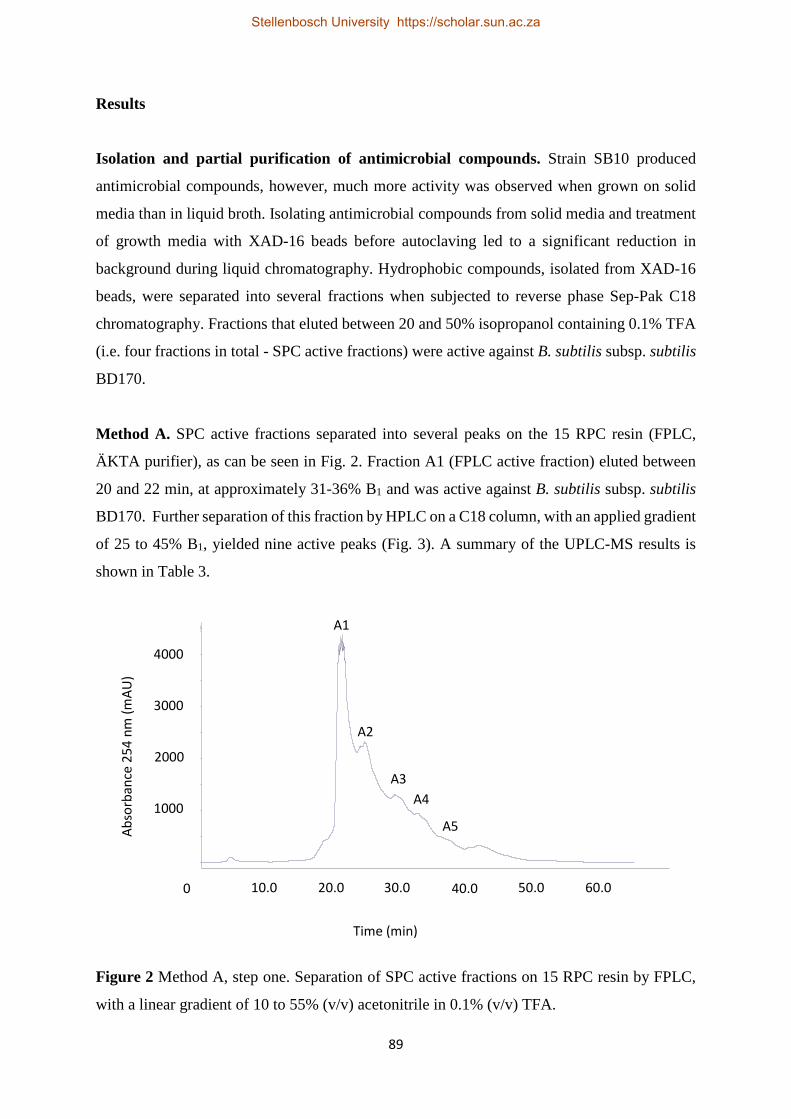
89
Results
Isolation and partial purification of antimicrobial compounds. Strain SB10 produced
antimicrobial compounds, however, much more activity was observed when grown on solid
media than in liquid broth. Isolating antimicrobial compounds from solid media and treatment
of growth media with XAD-16 beads before autoclaving led to a significant reduction in
background during liquid chromatography. Hydrophobic compounds, isolated from XAD-16
beads, were separated into several fractions when subjected to reverse phase Sep-Pak C18
chromatography. Fractions that eluted between 20 and 50% isopropanol containing 0.1% TFA
(i.e. four fractions in total - SPC active fractions) were active against B. subtilis subsp. subtilis
BD170.
Method A. SPC active fractions separated into several peaks on the 15 RPC resin (FPLC,
ÄKTA purifier), as can be seen in Fig. 2. Fraction A1 (FPLC active fraction) eluted between
20 and 22 min, at approximately 31-36% B1 and was active against B. subtilis subsp. subtilis
BD170. Further separation of this fraction by HPLC on a C18 column, with an applied gradient
of 25 to 45% B1, yielded nine active peaks (Fig. 3). A summary of the UPLC-MS results is
shown in Table 3.
Figure 2 Method A, step one. Separation of SPC active fractions on 15 RPC resin by FPLC,
with a linear gradient of 10 to 55% (v/v) acetonitrile in 0.1% (v/v) TFA.
3000
0
1000
2000
Abso
rban
ce 2
54 n
m (m
AU)
Time (min)
4000
10.0 20.0 30.0 40.0 50.0 60.0
A1
A2
A3 A4
A5
Stellenbosch University https://scholar.sun.ac.za

90
According to ESI-MS, compounds 1 and 2 (from peaks AA and AB, respectively; Fig. 3) have
monoisotopic masses of 670.41 Da (671.41 m/z, [M + H]+) and 406.22 Da (407.22 m/z [M +
H]+), respectively. The ESI-MS analyses and MSe fragmentation patterns of the two
compounds are shown in Figs 4 and 5. Several larger compounds with mass-to-charge ratios
of 1052.79 m/z, 1066.81 m/z, 1070.79 m/z, 1078.80 m/z, 1080.82 m/z and 1106.84 m/z were
revealed by ESI-MS spectra. The positive mass spectra obtained for these compounds revealed
a repeating 128.09 m/z neutral loss, as well as a dehydration pattern (Fig. 6). Ions with a mass-
to-charge ratio of 84.08 m/z and 129.10 m/z were repeatedly observed in all these samples.
Figure 3 Method A, step two. C18 HPLC chromatogram of the fraction representing peak A1
in Fig. 2 (FPLC active fraction). A linear gradient from 25-45% acetonitrile containing 0.1%
TFA was used.
Time (min)
Abso
rban
ce 2
54 n
m (m
AU)
8 12 16 20
24
28
36
44
32 4 40 0
0
400
800
1200
1600
2000
2400
2800
3200
Time (min)
Stellenbosch University https://scholar.sun.ac.za
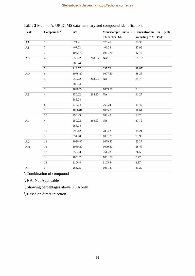
91
Table 3 Method A, UPLC-MS data summary and compound identification. Peak Compound/ a m/z Monoisotopic mass /
Theoretical Mr
Concentration in peak
according to MS (%)c
AA 1 671.41 670.41 93.33
AB 2 407.22 406.22 82.96
3 1052.79 1051.79 12.76
AC 4a 250.22, 268.23,
286.24
NAb 71.12d
5 213.57 637.72 28.87d
AD 6 1078.80 1077.80 38.38
4a 250.22, 268.23,
286.24
NA 35.70
7 1070.79 1069.79 3.01
AE 4a 250.22, 268.23,
286.24
NA 61.27
8 270.24 269.24 11.42
9 1066.81 1065.81 10.64
10 790.43 789.43 8.37
AF
4a 250.22, 268.23,
286.24
NA 57.72
10 790.42 789.42 15.21
3 351.60 1051.81 7.89
AG 11 1080.82 1079.82 83.27
AH
11 1080.82 1079.82 28.45
12 252.23 251.23 26.52
3 1052.79 1051.79 9.77
13 1106.84 1105.84 6.37
AI 3 263.95 1051.81 83.20 a, Combination of compounds b, NA: Not Applicable c, Showing percentages above 3.0% only d, Based on direct injection
Stellenbosch University https://scholar.sun.ac.za

92
Figure 4 (A) ESI-MS analysis and (B) MSe fragmentation pattern of compound 1, 671.41 m/z.
Masses indicated are singly charged species [M + H]+.
A
B
Stellenbosch University https://scholar.sun.ac.za

93
Figure 5 (A) ESI-MS analysis and (B) MSe fragmentation pattern of compound 2, 407.22 m/z.
Masses indicated are singly charged species [M + H]+.
A
B
Stellenbosch University https://scholar.sun.ac.za

94
Figure 6 ESI-MS analysis of compound 3 (1052.79 m/z), as representative of compounds 6, 7,
9, 11 and 13 with similar characteristics. MaxEnt3 was used to generate this positive mass
spectrum. Arrows, asterisks and circles indicate the repeating 128.09 m/z neutral loss pattern,
dehydration and, 84.08 m/z and 129.10 m/z ions, respectively. Masses indicated are singly
charged species [M + H]+.
Method B. During the second purification method, a two-step HPLC protocol was used instead
of using FPLC followed by HPLC. The first step consisted of using a non-linear gradient at a
column temperature of 45 °C. The non-linear gradient condensed compounds that eluted non-
specifically to elute at a more specific time, as shown in Fig. 7, compared to the entire gradient
as in method A (Fig. 3). Altering of the gradient enabled purification of peaks B2 to B9. The
heated column enhanced peak resolution. Antimicrobial activity was observed for fraction B1,
and peaks B2, B3, B4, B5, B8 and B9. Fraction B1 (a combination of the area before peak B2,
between peaks B2 and B3 and between peaks B3 and B4), was used during the second HPLC
purification process as peaks could not be separated sufficiently during the first HPLC step.
Peaks were sufficiently separated during this protocol (Fig. 8). Antimicrobial activity was
observed for all peaks, excluding peak BI. See Table 4 for summary of UPLC-MS data.
∆128.09 ∆128.09 ∆128.09 ∆128.09 ∆128.09 ∆128.09 * * * * **
Stellenbosch University https://scholar.sun.ac.za

95
Figure 7 Method B, step one. HPLC Chromatogram of sample SB10, run on a non-linear,
slightly convex Waters Corporation 5 gradient of 30-70% B (B2: 90% Acetonitrile containing
0.1% TFA). Fraction B1 was collected as the background between peaks B2, B3 and B4.
Figure 8 Method B, step two. HPLC Chromatogram of sample SB10, fraction B1 (excluding
peaks B2 and B3) from the first HPLC step of method B, run on a linear gradient of 40-60% B
(B2: 90% Acetonitrile containing 0.1% TFA).
Time (min)
Time (min)
Abso
rban
ce 2
54 n
m (A
U)
Abso
rban
ce 2
54 n
m (A
U)
Stellenbosch University https://scholar.sun.ac.za

96
Overall, the same dominant compounds were detected in the ESI-MS data of samples from
method B, as were detected in samples from method A. These include the larger compounds,
3 (1051.81 Da) and 11 (1079.84 Da), the combination of compounds with mass-to-charge ratios
of 268.23 m/z, 286.24 m/z and 250.22 m/z, and compounds 1 (671.41 m/z) and 2 (407.22 m/z).
Compound 21 from peak B9 showed significant activity against B. subtilis subsp. subtilis
BD170 and was not detected in samples from method A. This compound with a mass-to-charge
ratio of 285.07 m/z, had a significant bright yellow color and an UV absorbance at 392.12 nm
(Fig. 9). The fragmentation profile (Fig. 10) reveals a daughter ion of 186.99 m/z. Peaks B2
(consisting of compounds 434.27 m/z and 341.15 m/z) and BD (259.17 m/z) had activity. See
Fig. 11, and supplementary Figs S1 and S2, for ESI-MS analyses and MSe fragmentation
patterns of compounds 259.17 m/z, 434.27 m/z and 341.15 m/z, respectively.
Figure 9 Diode array of compound 21, 285.07 m/z, indicating absorbance at 329.12 nm.
Stellenbosch University https://scholar.sun.ac.za

97
Figure 10 (A) ESI-MS analysis and (B) MSe fragmentation profile of compound 21, indicating
the parent and daughter ions, 258.07 m/z and 186.99 m/z, respectively. Masses indicated are
singly charged species [M + H]+.
A
B
Stellenbosch University https://scholar.sun.ac.za

98
Figure 11 (A) ESI-MS analysis and (B) MSe fragmentation profile of compound 24,
259.17 m/z. Masses indicated are singly charged species [M + H]+.
A
B
Stellenbosch University https://scholar.sun.ac.za

99
Table 4 Method B, UPLC-MS data summary and compound identification. Peak Compound/ a m/z Monoisotopic mass
/ Theoretical Mr
Concentration in peak
according to MS (%)c
B2 14 434.27 433.27 70.76
15 341.15 340.15 24.05
1 671.41 670.41 Trace amounts
B3 2 407.22 406.22 53.83
10 790.43 789.43 37.86
B4 4a 250.22, 268.23,
286.24
NAb 26.49
16 266.21 265.21 21.49
12 252.23 251.23 17.85
17 751.41 750.41 15.47
8 270.24 269.24 11.98
B5 12 252.23 251.23 46.81
4a 250.22, 268.23,
286.24
NAb 25.04
B8 20 307.06 306.06 ND
B9 21 285.07 284.07 78.75
4 250.22, 268.23,
286.24
NAb 5.85
BA 3 351.60, 526.91 1051.81 ND
BB 3 351.60, 526.91 1051.81 61.85
23 1124.64 1123.64 12.44
15 341.15 340.15 10.61
BC 14 434.27 433.27 68.76
15 341.15 340.15 22.90
3 351.68, 527.09 1051.81 8.34
BD 24 259.17 258.17 91.41
BE 2 407.22 406.22 71.32
11 360.94, 540.92,
1080.82
1079.84 9.97
BF 10 790.43 789.43 45.28
4a 250.22, 268.23,
286.24
NA 24.83
BG 10 790.43 789.43 29.11
17 751.41 750.41 27.58
11 360.94, 540.92 1079.84 24.78
BH 4a 250.22, 268.23,
286.24
NA 81.50
11 360.94, 540.92 1079.84 9.03 a, Combination of compounds b, NA: Not Applicable c, Showing percentages above 3.0% only
Stellenbosch University https://scholar.sun.ac.za

100
Antimicrobial spectrum and temperature stability. SPC active fractions from strain
X. khoisanae SB10, were tested against filamentous fungi, yeasts and Gram-positive and
Gram-negative bacteria. Growth of all target microorganisms was inhibited. Furthermore,
treating SPC active fractions at 121 °C, for 20 min, did not have a significant effect on activity
against B. subtilis subsp. subtilis BD170.
Discussion
Xenorhabdus bacteria are known to produce various antimicrobial compounds, but it is
however, a highly neglected antimicrobial source that has not been exploited to its full
potential. Therefore, antimicrobial isolation, purification, identification and characterization
have not been done for all species belonging to this genus. This study isolated antimicrobial
compounds from the species, X. khoisanae for the first time. Due to the Xenorhabdus-
Steinernema-insect host tripartite relationship, it was not surprizing to discover the production
of various antimicrobial compounds by this species. Two different methods were used for
purification of these compounds, which led to similar, but not identical purified antimicrobial
compounds.
Method A. Compound 1 (671.41 m/z) was identified as a novel antimicrobial compound. The
MSe fragmentation pattern of this compound revealed a daughter ion with a mass-to-charge
ratio of 473.28 m/z (Fig. 4). Further research needs be done to elucidate the structure.
Compound 2, with a monoisotopic mass of 406.22 Da, was identified in the ESI-MS spectrum
obtained for peak AB. Two daughter ions with mass-to-charge ratios of 158.08 m/z and
250.15 m/z were revealed by the fragmentation pattern (Fig. 5). Two additional species,
176.07 m/z and 232.13 m/z, were observed and determined to be the hydration and dehydration
products of the two daughter ions, 158.08 m/z and 250.15 m/z, respectively. Compound 2 was
identified as xenocoumacin 2 based on the characteristic fragmentation pattern. Fragment
250.15 m/z represents the benzopyran-1-one fragment, while fragment 158.08 m/z represents
the remaining fragment with the pyrrolidine as R group (24) (Fig. 12). This identification was
supported by antiSMASH (32) results of 100% similarity between the genome of strain SB10
and the xenocoumacin biosynthetic gene cluster.
Stellenbosch University https://scholar.sun.ac.za

101
Figure 12 MSe fragmentation pattern and chemical structure of compound 2, xenocoumacin 2,
indicating corresponding fragments. Masses indicated are singly charged species [M + H]+.
A combination of fragments (combination 4), including the two daughter ions of
xenocoumacin 2, was detected in peaks AC, AD, AE and AF. This combination consisted of
singly charged species [M + H]+ with mass-to-charge ratios of 250.22 m/z, 268.23 m/z
(hydration product of 250.22 m/z), 286.24 m/z (hydration product of 268.23 m/z) and
158.09 m/z (See supplementary Fig. S3 for ESI-MS analysis and MSe fragmentation profile).
The benzopyran-1-one fragment of xenocoumacins 2 may be hydrated to produce
xenocoumacin 6 (24) (Fig. 13). This fragment will therefore have a mass-to-charge ratio of
268.22 m/z, which was observed in this sample. However, no xenocoumacin parent ions were
observed when MS data of these peaks were studied, suggesting that these compounds were
degraded or isolated mid-production. Therefore, it is suggested that isolate SB10 also produces
xenocoumacin 6, the hydration product of xenocoumacin 2. The hydration product of
268.23 m/z (286.24 m/z) may indicate the production of another xenocoumacin derivative,
where xenocoumacin 6 is further hydrated.
Xenocoumacin 2
Stellenbosch University https://scholar.sun.ac.za

102
Figure 13 Hydration of xenocoumacin 2 to xenocoumacin 6.
The larger compounds with mass-to-charge ratios of 1052.79 m/z, 1066.81 m/z, 1070.79 m/z,
1078.80 m/z, 1080.82 m/z and 1106.84 m/z revealed repeated dehydration and neutral losses of
128.09 m/z, indicative of lysine residues. Additionally, two ions, 84.08 m/z and 129.10 m/z,
were detected in the ESI-MS spectrum, which are known as lysine associated ions. These traits
were recognized as characteristics of lysine-rich cyclic lipopeptides, called PAX (peptide-
antimicrobial-Xenorhabdus) peptides. These compounds were first characterized by Gaulteri
et al. (14) and to date a total of 13 PAX peptides have been reported (33). The peptides
identified here (compounds 3, 6, 11, 9 and 13), correspond directly with the mass-to-charge
ratios of known PAX peptides; PAX 1 (1052.79 m/z), PAX 6 (1078.78 m/z), PAX 2
(1080.82 m/z), PAX 3 (1066.81 m/z) and PAX 9 (1106.82 m/z). However, a PAX peptide with
a monoisotopic mass of 1069.79 Da, which was identified from peak AD, has not been
reported. This compound exhibits the previously discussed characteristics of PAX peptides
(Fig. S4). This is therefore a novel PAX peptide, PAX 14. PAX peptides have only been
isolated from Xenorhabdus nematophila, therefore, this is the first report of PAX peptides from
another species.
Method B. Four active compounds detected in samples from method B were not detected in
samples from method A. These include compounds 14 (434.27 m/z), 15 (341.15 m/z), 21
(285.07 m/z) and 24 (259.17 m/z) (Figs S1, S2, 10 and 11). Compounds 14 and 15 need to be
purified to determine the active compound, as the active fraction tested in this study contained
both these compounds.
Throughout the isolation process it was noted that the sample from peak B9 was bright yellow,
which suggested this sample contains xenorhabdins, as these compounds have been reported
to be bright yellow (10). This was confirmed when ESI-MS and MSe fragmentation data of
compound 21 was analyzed. Compound 21 has a parent ion with a mass-to-charge ratio of
Xenocoumacin 2 Xenocoumacin 6
Stellenbosch University https://scholar.sun.ac.za

103
285.07 m/z and a daughter ion of 186.99 m/z. This corresponds directly with characteristics of
xenorhabdin 4, and compound 21 was identified accordingly (10).
Comparison of purification methods. Compounds that were detected in the ESI-MS data of
method A and B include members of the PAX family (PAX 1, 1051 Da and PAX 2, 1079 Da),
xenocoumacin 2, fragments of xenocoumacin 6 and compound 1 (671.41 m/z). Method A
proved to produce more pure compounds when comparing concentrations of PAX peptides
(PAX 1 and 2) and xenocoumacin 2 to that of method B (Table 5). Fragments of xenocoumacin
6 were however, more pure when using method B. Method A, therefore, proved to be the better
method for purification when taking all compounds into consideration. However, xenorhabdin
4 (compound 21, peak B9), compound 14, 15 and 21 were detected only in samples purified
using method B. This might be due to the purification process selecting for purification of these
compounds, more than the first method. This is similar for compound 1, which was detected
only in the sample from method A. This phenomenon is however, likely the cause of using two
separate isolation batches for testing of the two purification methods. Direct comparison of
these two methods should be done with the same isolation batch to ensure consistency and
reliability of results.
Table 5 Comparison of active compound concentrations between methods A and B.
Compound Monoisotopic
mass (Da)
Method A Method B
Peak Concentration in
peak (%)
Peak Concentration in
peak (%)
Xenocoumacin 2 406.22 AB 82.96 B3 53.83
Xenocoumacin 6
fragments
250.22,
268.23,
286.24a
AC 71.12 BH 81.50
PAX peptide 1 1051.79 AI 83.20 BB 61.85
PAX peptide 2 1079.82 AG 83.27 BG 24.78 a, Mass-to-charge ratios
Antimicrobial spectrum and temperature stability. SPC active fractions from strain
X. khoisanae SB10 inhibited growth of all target strains tested. Antimicrobial purification from
X. khoisanae SB10, revealed the bioactive compounds produced by this strain as PAX peptides,
xenocoumacins, xenorhabdins and three to four (671.41 m/z, 259.17m/z and 434.27 m/z and/or
Stellenbosch University https://scholar.sun.ac.za

104
341.15 m/z) unidentified compounds. Xenocoumacin 2 has been shown to be active against
both Gram-negative and Gram-positive bacteria (8). PAX peptides 1-5 showed weak activity
against bacteria, but were active against filamentous fungi and yeasts with MIC (minimal
inhibitory concentration) values as low as 1.56 µg/ml against Fusarium oxysporum (14).
Lastly, xenorhabdin 4 has been shown to be active against Micrococcus luteus (10). These
compounds, therefore, all play a role in the overall bioactivity exhibited by these bacteria. It is
proposed that these compounds do not merely have an additive effect, but synergistic as well.
Furthermore, the stability of these compounds at 121 °C, for 20 min, is likely due to the cyclic
nature of these compounds. Stability of antimicrobial compounds is very important in industry
as this increases the ease of working with and storage of compounds. This is therefore a highly
desired characteristic of these compounds.
Conclusion
SPC active fractions produced by X. khoisanae SB10, were shown to have broad-spectrum
activity and stability at 121 °C, for 20 min. Purification of the antimicrobial compounds
produced by strain SB10 revealed the production of various compounds including PAX
peptides, xenocoumacins, xenorhabdins, as well as, unidentified compounds with
monoisotopic masses, 670.41 Da, 258.17 Da, 433.27 Da and 340.15 Da. These compounds
therefore, all play a role in the overall antimicrobial activity of strain SB10. This study reports
on the production of PAX peptides, xenocoumacins and xenorhabdins by X. khoisanae for the
first time. Furthermore, three to four novel antimicrobial compounds have been discovered.
Future studies may be done on the characterization and structure elucidation of these unknown
compounds as it is not covered by the scope of this study.
Stellenbosch University https://scholar.sun.ac.za

105
References
1. Götz P, Boman A, Boman HG. 1981. Interactions between insect immunity and an
insect-pathogenic nematode with symbiotic bacteria. Proc R Soc B Biol Sci 212:333–
350.
2. Dunphy GB, Webster JM. 1991. Antihemocytic surface components of Xenorhabdus
nematophilus var. dutki and their modification by serum nonimmune larvae of Galleria
mellonella. J Invertebr Pathol 58:40–51.
3. Yang J, Zeng H-M, Lin H-F, Yang X-F, Liu Z, Guo L-H, Yuan J-J, Qiu D-W. 2012. An
insecticidal protein from Xenorhabdus budapestensis that results in prophenoloxidase
activation in the wax moth, Galleria mellonella. J Invertebr Pathol 110:60–67.
4. Burman M. 1982. Neoaplectana carpocapsae: Toxin production by axenic insect
parasitic nematodes. Nematologica 28:62–70.
5. Webster JM, Chen G, Hu K, Li J (2002) Bacterial metabolites. In: Gaugler R (ed)
Entomopathogenic nematology. CAB International, London, pp 35–56
6. Dutky SR. 1959. Insect microbiology. Adv Appl Microbiol 1:175–200.
7. Paul VJ, Frautschy S, Fenical W, Nealson KH. 1981. Antibiotics in microbial ecology.
J Chem Ecol 7:589–597.
8. McInerney B V., Taylor WC, Lacey MJ, Akhurst RJ, Gregson RP. 1991. Biologically
active metabolites from Xenorhabdus spp., part 2. Benzopyran-1-one derivatives with
gastroprotective activity. J Nat Prod 54:785–795.
9. Lang G, Kalvelage T, Peters A, Wiese J, Imhoff JF. 2008. Linear and cyclic peptides
from the entomopathogenic bacterium Xenorhabdus nematophilus. J Nat Prod 71:1074–
1077.
10. McInerney B V, Gregson RP, Lacey MJ, Akhurst RJ, Lyons GR, Rhodes SH, Smith
DRJ, Engelhardt LM, White AH. 1991. Biologically active metabolites from
Xenorhabdus spp., part 1. dithiolopyrrolone derivatives with antibiotic activity. J Nat
Prod 54:774–784.
11. Sundar L, Chang FN. 1993. Antimicrobial activity and biosynthesis of indole antibiotics
produced by Xenorhabdus nematophilus. J Gen Microbiol 139:3139–3148.
12. Zhou Q, Grundmann F, Kaiser M, Schiell M, Gaudriault S, Batzer A, Kurz M, Bode
HB. 2013. Structure and biosynthesis of xenoamicins from entomopathogenic
Xenorhabdus. Chem - A Eur J 19:16772–16779.
13. Böszörményi E, Érsek T, Fodor AM, Fodor AM, Földes LS, Hevesi M, Hogan JS,
Stellenbosch University https://scholar.sun.ac.za

106
Katona Z, Klein MG, Kormány A, Pekár S, Szentirmai A, Sztaricskai F, Taylor RAJ.
2009. Isolation and activity of Xenorhabdus antimicrobial compounds against the plant
pathogens Erwinia amylovora and Phytophthora nicotianae. J Appl Microbiol 107:746–
759.
14. Gualtieri M, Aumelas A, Thaler J-O. 2009. Identification of a new antimicrobial lysine-
rich cyclolipopeptide family from Xenorhabdus nematophila. J Antibiot (Tokyo)
62:295–302.
15. Kronenwerth M, Bozhüyük KAJ, Kahnt AS, Steinhilber D, Gaudriault S, Kaiser M,
Bode HB. 2014. Characterisation of taxlllaids A-G; natural products from Xenorhabdus
indica. Chem Eur J 20:17478–17487.
16. Grundmann F, Kaiser M, Kurz M, Schiell M, Batzer A, Bode HB. 2013. Structure
determination of the bioactive depsipeptide xenobactin from Xenorhabdus sp. PB30.3.
RSC Adv 3:22072–22077.
17. Nollmann FI, Dowling A, Kaiser M, Deckmann K, Grösch S, ffrench-Constant R, Bode
HB. 2012. Synthesis of szentiamide, a depsipeptide from entomopathogenic
Xenorhabdus szentirmaii with activity against Plasmodium falciparum. Beilstein J Org
Chem 8:528–533.
18. Reimer D, Cowles KN, Proschak A, Nollmann FI, Dowling AJ, Kaiser M, ffrench-
Constant R, Goodrich-Blair H, Bode HB. 2013. Rhabdopeptides as insect-specific
virulence factors from entomopathogenic bacteria. ChemBioChem 14:1991–1997.
19. Houard J, Aumelas A, Noël T, Pages S, Givaudan A, Fitton-Ouhabi V, Villain-Guillot
P, Gualtieri M. 2013. Cabanillasin, a new antifungal metabolite, produced by
entomopathogenic Xenorhabdus cabanillasii JM26. J Antibiot (Tokyo) 66:617–620.
20. Singh J, Banerjee N. 2008. Transcriptional analysis and functional characterization of a
gene pair encoding iron-regulated xenocin and immunity proteins of Xenorhabdus
nematophila. J Bacteriol 190:3877–3885.
21. Thaler JO, Baghdiguian S, Boemare N. 1995. Purification and characterization of
xenorhabdicin, a phage tail-like bacteriocin, from the lysogenic strain F1 of
Xenorhabdus nematophilus. Appl Environ Microbiol 61:2049–2052.
22. Morales-Soto N, Gaudriault S, Ogier JC, Thappeta KRV, Forst S. 2012. Comparative
analysis of P2-type remnant prophage loci in Xenorhabdus bovienii and Xenorhabdus
nematophila required for xenorhabdicin production. FEMS Microbiol Lett 333:69-76.
23. Boemare NE, Boyer-Giglio MH, Thaler JO, Akhurst RJ, Brehelin M. 1992. Lysogeny
and bacteriocinogeny in Xenorhabdus nematophilus and other Xenorhabdus spp. Appl
Stellenbosch University https://scholar.sun.ac.za

107
Environ Microbiol 58:3032–3037.
24. Reimer D, Luxenburger E, Brachmann AO, Bode HB. 2009. A new type of pyrrolidine
biosynthesis is involved in the late steps of xenocoumacin production in Xenorhabdus
nematophila. ChemBioChem 10:1997–2001.
25. Fuchs SW, Grundmann F, Kurz M, Kaiser M, Bode HB. 2014. Fabclavines: bioactive
peptide-polyketide-polyamino hybrids from Xenorhabdus. ChemBioChem 15:512–516.
26. Li J, Chen G, Webster JM, Czyzewska E. 1995. Antimicrobial metabolites from a
bacterial symbiont. J Nat Prod 58:1081–1086.
27. Chen G. 1996. PhD thesis. Simon Fraser University, Burnaby, Canada. Antimicrobial
activity of the nematode symbionts, Xenorhabdus and Photorhabdus
(Enterobacteriaceae) and the discovery of two groups of antimicrobial aubstances,
nematophin and xenorxides.
28. Proschak A, Zhou Q, Schöner T, Thanwisai A, Kresovic D, Dowling A, ffrench-
Constant R, Proschak E, Bode HB. 2014. Biosynthesis of the insecticidal xenocyloins
in Xenorhabdus bovienii. ChemBioChem 15:369–372.
29. Li J, Chen G, Webster JM. 1997. Nematophin, a novel antimicrobial substance produced
by Xenorhabdus nematophilus (Enterobactereaceae). Can J Microbiol 43:770–773.
30. Ji D, Yi Y, Kang G-H, Choi Y-H, Kim P, Baek N-I, Kim Y. 2004. Identification of an
antibacterial compound, benzylideneacetone, from Xenorhabdus nematophila against
major plant-pathogenic bacteria. FEMS Microbiol Lett 239:241–248.
31. Crawford JM, Portmann C, Zhang X, Roeffaers MBJ, Clardy J. 2012. Small molecule
perimeter defense in entomopathogenic bacteria. Proc Natl Acad Sci 109:10821–10826.
32. Weber T, Blin K, Duddela S, Krug D, Kim HU, Bruccoleri R, Lee SY, Fischbach MA,
Müller R, Wohlleben W, Breitling R, Takano E, Medema MH. 2015. antiSMASH 3.0—
a comprehensive resource for the genome mining of biosynthetic gene clusters. Nucleic
Acids Res 43:W237–W243.
33. Fuchs SW, Proschak A, Jaskolla TW, Karas M, Bode HB. 2011. Structure elucidation
and biosynthesis of lysine-rich cyclic peptides in Xenorhabdus nematophila. Org
Biomol Chem 9:3130–3132.
Stellenbosch University https://scholar.sun.ac.za

108
Supplementary material
Figure S1 (A) ESI-MS analysis and (B) MSe fragmentation profile of compound 14,
434.27 m/z. Masses indicated are singly charged species [M + H]+.
B
A
Stellenbosch University https://scholar.sun.ac.za

109
Figure S2 (A) ESI-MS analysis and (B) MSe fragmentation profile of compound 15,
341.15 m/z. Masses indicated are singly charged species [M + H]+.
B
A
Stellenbosch University https://scholar.sun.ac.za

110
Figure S3 (A) ESI-MS analysis and (B) MSe fragmentation profile of combination 4 revealing
xenocoumacin 2 and xenocoumacin 6 fragments. Masses indicated are singly charged species
[M + H]+.
B
A
Stellenbosch University https://scholar.sun.ac.za

111
Figure S4 ESI-MS analysis of compound 7 (PAX 14), revealing the typical repeating neutral
loss of 128.09 m/z fragments and dehydration of PAX peptides, as well as, lysine related ions,
84.08 m/z and 129.10 m/z. Masses indicated are singly charged species [M + H]+.
Stellenbosch University https://scholar.sun.ac.za

112
Chapter 6
General Discussion and Conclusions
Stellenbosch University https://scholar.sun.ac.za

113
General Discussion
Four Xenorhabdus strains were isolated from four different Steinernema species:
Steinernema sacchari SB10T Nthenga, Knoetze, Berry, Tiedt and Malan;
Steinernema jeffreyense J194T Malan, Knoetze and Tiedt; Steinernema nguyeni F2T Malan,
Knoetze and Tiedt; and Steinernema litchii WS9T Steyn, Knoetze, Tiedt and Malan.
Xenorhabdus isolates were named to correspond to their nematode source, Xenorhabdus
isolates SB10, J194, F2 and WS9, respectively. Biochemical characteristics were determined,
only to identify differences between strains, and a molecular approach was taken for species
identification.
A 98-99% similarity was obtained for 16S rDNA, gltX and dnaN sequences between isolate F2
and Xenorhabdus bovienii T228T. Translated BLAST analysis supported this finding and
isolate F2 was identified as a strain belonging to the species X. bovienii. The association of
X. bovienii with S. nguyeni was expected as S. nguyeni belongs to the Feltiae-clade (1) and
X. bovienii is associated with eight other Steinernema species belonging to this clade.
Sequences obtained for housekeeping genes gltx, recA, dnaN, gyrB and infB, and 16S rDNA
(including upstream and downstream regulatory elements) of isolates J194 and SB10 showed
96-99% similarity to corresponding gene sequences of Xenorhabdus khoisanae SF87T.
Identification of isolates J194 and SB10 as strains of X. khoisanae was supported by protein
similarity searches. Similar to the X. bovienii-S. nguyeni association, the X. khoisanae-
S. jeffreyense association was expected, as X. khoisanae has also been isolated from
Steinernema khoisanae which belongs to the same clade (Glaseri) as S. jeffreyense (2, 3).
However, the X. khoisanae-S. sacchari association was not anticipated. Steinernema sacchari
belongs to the Cameroonense-clade (4), which is closely related to the Feltiae-, and not
Glaseri, -clade, as would be expected for a nematode associated with X. khoisanae. It was,
therefore concluded that X. khoisanae has the ability to switch between nematodes belonging
to distantly related clades (Glaseri and Cameroonense clades).
Sequences obtained for the 16S rDNA and housekeeping genes of isolate WS9 had similarities
of 98-99% to X. griffiniae ID10T, excluding the recA gene which had a 96% similarity to
X. ehlersii DSM16337T. Protein-coding genes were most similar to those of X. griffiniae ID10T,
excluding the recA and infB genes, which were more similar to those of X. ehlersii DSM16337T
Stellenbosch University https://scholar.sun.ac.za

114
and Xenorhabdus sp. TZ01, respectively. This suggests that isolate WS9 is closely related to
X. griffiniae and to a lesser extent to X. ehlersii and Xenorhabdus sp. TZ01. Isolate WS9 was
therefore classified as a member of the X. griffiniae species. It was therefore proven that
X. griffiniae WS9 is associated with S. litchii WS9T from the Karii-clade (5).
Xenorhabdus griffiniae has previously been isolated from Steinernema hermaphroditum
Stock, Griffin and Chaerani, a member of the Longicaudum-clade and an uncharacterized
Steinernema sp. strain BMMCB (6). Therefore, X. griffiniae has the ability, similar to
X. khoisanae, to switch between nematodes belonging to different clades.
Antimicrobials isolated from strain SB10 were purified using two methods and subjected to
ESI-MS coupled to UPLC. Method A revealed eight compounds of interest, including a novel
antimicrobial with a monoisotopic mass of 670.41 Da (671,41 m/z, [M + H]+). This compound
had significant activity against B. subtillis subsp. subtillis BD170.
A second compound with a mass-to-charge ratio of 407.22 m/z, was identified as
xenocoumacin 2, based on the daughter ions revealed by MSe fragmentation. This
identification was supported by antiSMASH results indicating strain SB10 to contain the
xenocoumacin biosynthetic gene cluster. A combination of fragments 250.22 m/z, 268.23 m/z
and 158.09 m/z were detected in several peaks. These are suggested to be fragments of both
xenocoumacin 2 and 6. Since the parent ion was not detected in the sample, isolation of
antimicrobials likely occurred mid-biosynthesis or, alternatively, the compounds had been
degraded. An additional fragment was detected as the hydration product of 268.23 m/z,
suggesting xenocoumacin 6 to be further hydrated to produce a xenocoumacin 6 derivative.
The remaining six compounds detected during method A had mass-to-charge ratios of
1052.79 m/z, 1066.81 m/z, 1070.79 m/z, 1078.80 m/z, 1080.82 m/z and 1106.84 m/z. These
compounds revealed typical characteristics of lysine-rich cyclolipopeptides, called PAX
(peptide-antimicrobial-Xenorhabdus) peptides (7, 8). These characteristics, as well as
corresponding monoisotopic masses, led to the identification of these compounds as PAX
peptides 1, 2, 3, 6 and 9. An additional, novel, PAX peptide, PAX 14 (1069.79 Da), was
identified in this study.
Stellenbosch University https://scholar.sun.ac.za

115
Compounds with activity, from method B only, are compounds 434.27 m/z, 341.15 m/z,
259.17 m/z and 285.07 m/z. The sample containing novel compounds 434.27 m/z and
341.15 m/z showed significant activity. The source of this activity can be either one of these
compounds or both. The compound, 285.07 m/z, was identified as xenorhabdin 4, based on the
MS spectrum and fragmentation profile obtained, as well as the bright yellow color of the
sample.
The same predominant compounds detected in samples from method A were also detected in
samples from method B. These include a novel antimicrobial compound (671.41 m/z),
xenocoumacin 2, fragments of xenocoumacin 6 and PAX peptides (PAX peptides 1 and 2).
Comparison of concentrations obtained for each compound revealed method A to be the better
method for purification, when using two different antimicrobial isolation batches. However,
the same antimicrobial isolation batch needs to be used to compare the two methods directly.
The specific purification method also depends on the specific compound desired to be purified.
Broad-spectrum activity of the SPC active fractions from isolate SB10 was supported by the
previously reported activity spectrum of PAX peptides, xenocoumacins and xenorhabdins.
These compounds all play a part in the overall activity of strain SB10 and are suggested to
work synergistically.
Conclusion
Xenorhabdus bacteria are a neglected and underestimated source of bioactive compounds. In
the current day and age, antibiotic resistance has become an immense crisis. The discovery of
novel antimicrobial compounds is therefore of the essence, as they may be used to treat
infections caused by antibiotic resistant strains. The time has come to investigate
unconventional antimicrobial sources, such as Xenorhabdus, as this might hold the key to
survival of humankind in the 21st century.
During this study, four novel Xenorhabdus-Steinernema associations were discovered,
including proof that X. khoisanae and X. griffiniae have the ability to switch between different
nematode clades. Purification of antimicrobial compounds produced by strain SB10 revealed
the production of known Xenorhabdus compounds, including PAX peptides, xenocoumacins
and xenorhabdins. Three to four novel antimicrobial compounds were identified with mass-to-
Stellenbosch University https://scholar.sun.ac.za

116
charge ratios of 671.41 m/z, 259.17 m/z, 434.27 m/z and/or 341.15 m/z. The latter two
compounds were not sufficiently purified as the active sample contained both compounds.
These compounds need to be further purified to determine which compound is the source of
activity.
Future studies may characterize the three novel compounds found in this study. This
characterization includes determining the activity spectrum, cytotoxicity against cancerous
cells and insect larvae, protease-, storage- and temperature-stability, and chemical structure.
Structure elucidation might be done by NMR. Furthermore, the synergistic effect of all
bioactive compounds identified during this study may be determined.
Stellenbosch University https://scholar.sun.ac.za

117
References
1. Malan AP, Knoetze R, Tiedt LR. 2016. Steinernema nguyeni n. sp. (Rhabditida:
Steinernematidae), a new entomopathogenic nematode from South Africa. Nematology
18:571–590.
2. Malan AP, Knoetze R, Tiedt LR. 2015. Steinernema jeffreyense n. sp. (Rhabditida:
Steinernematidae), a new entomopathogenic nematode from South Africa. J Helminthol
90:262-278.
3. Nguyen KB, Malan A, Gozel U. 2006. Steinernema khoisanae n. sp. (Rhabditida:
Steinernematidae), a new entomopathogenic nematode from South Africa. Nematology
8:157–175.
4. Malan AP, Knoetze R, Tiedt LR, Nthenga I, Berry S. 2014. Steinernema sacchari n. sp.
(Rhabditida: Steinernematidae), a new entomopathogenic nematode from South Africa.
Nematology 16:475–494.
5. Steyn WP, Knoetze R, Tiedt LR, Malan AP. 2017. Steinernema litchii n. sp. (Rhabditida:
Steinernematidae), a new entomopathogenic nematode from South Africa. Nematology
19:1157–1177.
6. Stock SP, Griffin CT, Chaerani R. 2004. Morphological and molecular characterisation
of Steinernema hermaphroditum n. sp. (Nematoda: Steinernematidae), an
entomopathogenic nematode from Indonesia, and its phylogenetic relationships with
other members of the genus. Nematology 6:401–412.
7. Fuchs SW, Proschak A, Jaskolla TW, Karas M, Bode HB. 2011. Structure elucidation
and biosynthesis of lysine-rich cyclic peptides in Xenorhabdus nematophila. Org
Biomol Chem 9:3130–3132.
8. Gualtieri M, Aumelas A, Thaler J-O. 2009. Identification of a new antimicrobial lysine-
rich cyclolipopeptide family from Xenorhabdus nematophila. J Antibiot (Tokyo)
62:295–302.
9. McInerney B V, Gregson RP, Lacey MJ, Akhurst RJ, Lyons GR, Rhodes SH, Smith
DRJ, Engelhardt LM, White AH. 1991. Biologically active metabolites from
Xenorhabdus spp., part 1. dithiolopyrrolone derivatives with antibiotic activity. J Nat
Prod 54:774–784.
Stellenbosch University https://scholar.sun.ac.za
Copyright © 2022 FDOKUMEN