Characterisation of the molybdenum-responsive ModE regulatory protein and its binding to the...
8
Eur. J. Biochem. 246, 119 ~ 126 (1997) 0 FEBS 1997 Characterisation of the molybdenum-responsive ModE regulatory protein and its binding to the promoter region of the modABCD (molybdenum transport) operon of Escherichia coli Lisa A. ANDERSON ’ Department of Biochemistry, Medical Sciences Institute, University of Dundee, Dundee, UK ’ Department of Biological and Molecular Sciences, University of Stirling, Stirling, UK .’ Nitrogen Fixation Laboratory, John Innes Centre, Norwich, UK (Received 23 December 1996/7 March 1997) - EJB 96 189512 Tracy PALMER I, Nicholas C. PRICE’, Stephen BORNEMANN I, David H. BOXER’ and Richard N. PAU’ Molybdenum-dependent repression of transcription of the Escherichia coli modABCD operon, which encodes the high-affinity molybdate transporter, is mediated by the ModE protein. This regulatory protein was purified as an N-terminal His,-tagged derivative and characterised both with and without the N- terminal oligohistidine extension. Equilibrium centrifugation showed that ModE is at least a 57-kDa homodimer. Circular dichroism spectroscopy indicated that when molybdate or tungstate bind to ModE there is little change in its a-helical content, but a major change in the environment of tryptophan and tyrosine residues occurs. Addition of molybdate or tungstate to the protein results in almost 50% quench- ing of the fluorescence attributed to tryptophan. Titration of fluorescence quenching showed that two molecules of molybdenum bind to each dimer of ModE with a Kd of 0.8 pM. DNA mobility-shift assays showed that ModE requires molybdenum, or tungstate, to bind with high affinity (approximate K,, of 30 nM ModE) to the modABCD promoter region. In accord with ModE’s role as a molybdenum-depen- dent transcriptional repressor, DNase 1 footprinting experiments showed that the ModE-molybdenum complex binds to a single 31-bp region around the transcription start of the modABCD promoter. This region contains a 6-base palindromic sequence CGTTAT-N,,-ATAACG. Keywords: molybdenum; transport: regulation : binding : Escherichia coli. Escherichia coli requires molybdenum to synthesise the co- factors of a number of oxidoreductases (Stiefel, 1993). Molyb- doenzymes catalyse reactions at carbon, sulfur and nitrogen, and include nitrate reductase and the formate dehydrogenases that have important roles in anaerobic metabolism (Stewart, 1988). In the molybdenum cofactors of these diverse enzymes, molyb- denum is coordinated to a dithiolene group in a pterin called molybdopterin (Rajagopalan, 1993, 1996) Molybdenum for co- factor biosynthesis enters the cell by more than one route (Pau et al., 1997; Rosentel et al., 1995). When present at low concen- trations, molybdenum is transported into the cell as molybdate via a high-affinity transport system belonging to the ABC family of transporters. The genes for the high-affinity molybdate trans- porter of Escherichia coli are found on a single operon, mod- ABCD (Maupin-Furlow et al., 1995 ; Walkenhorst et al., 1995). The modA gene encodes the periplasmic binding protein, modB the integral membrane protein, and modC the peripheral mem- brane protein that contains an ATP-binding site. The role of inodD is not known. The internal concentration of molybdenum is maintained within a narrow range even when its external concentration var- ies widely (Scott and Amy, 1989). A key factor in this is the control of metal uptake. High concentrations of molybdenum repress expression of the high-affinity molybdenum-transport Correspondence to R. N. Pau, Nitrogen Fixation Laboratory, John Fax: +44 1603 454970. E-mail: [email protected] URL: http ://www.uea.ac.uklnrp/.jic/iid.litin Tnnes Centre, Colney Lane, Norwich NR4 7UH, UK operon (Maupin-Furlow et al., 1995; Miller et al., 1987: Mouncey et al., 1996; Rech et al., 1995). Sensing the internal molybdenum concentration does not involve the molybdenum cofactor because cells that are unable to synthesise molybdo- pterin are able to repress modABCD transcription (Miller et al., 1987; Rech et al., 1995). The regulatory protein ModE has been shown to be responsible for the molybdenum-dependent repres- sion of transcription of the nzodABCD operon (Grunden et al., 1996; Mouncey et al., 1996: Walkenhorst et al., 1995). It is encoded by the gene modE which is immediately upstream, and transcribed divergently from, the E. coli inodABCD operon. The ModE protein has two distinct domains. The C-terminal half consists of a tandem repeat of a sequence similar to the 7 kDa molybdenum-binding protein called Mop from Closiridium pasteurianum (Hinton and Freyer, 1986). Mutational analysis suggests that the C-terminal domain of the ModE protein is in- volved in repressor activity (McNicholas et al., 1996). Deletion or disruption of these domains results in constitutive repression of modABCD transcription. The N-terminal domain is consid- ered to be the DNA-binding domain (Grunden et al., 1996; McNicholas et al., 1996). Mutations in this domain result in con- stitutive expression of the modABCD operon. Genes similar to modE occur in the mod loci of other bacterial species. In Azoto- bacter vinelandii, the ModE protein is likewise involved in re- pression of the operon encoding genes for the high-affinity mo- lybdate transporter (Mouncey et al., 1996). In this species it is encoded by the first gene in the molybdate-transport operon (Mouncey et al., 1995). In Rhodobucter capsulatus, proteins called MopA and MopB, that are similar to the E. coli ModE
Transcript of Characterisation of the molybdenum-responsive ModE regulatory protein and its binding to the...

Eur. J. Biochem. 246, 119 ~ 126 (1997) 0 FEBS 1997
Characterisation of the molybdenum-responsive ModE regulatory protein and its binding to the promoter region of the modABCD (molybdenum transport) operon of Escherichia coli Lisa A. ANDERSON ’ Department of Biochemistry, Medical Sciences Institute, University of Dundee, Dundee, UK ’ Department of Biological and Molecular Sciences, University of Stirling, Stirling, UK .’ Nitrogen Fixation Laboratory, John Innes Centre, Norwich, UK
(Received 23 December 1996/7 March 1997) - EJB 96 189512
Tracy PALMER I , Nicholas C. PRICE’, Stephen BORNEMANN I, David H. BOXER’ and Richard N. PAU’
Molybdenum-dependent repression of transcription of the Escherichia coli modABCD operon, which encodes the high-affinity molybdate transporter, is mediated by the ModE protein. This regulatory protein was purified as an N-terminal His,-tagged derivative and characterised both with and without the N- terminal oligohistidine extension. Equilibrium centrifugation showed that ModE is at least a 57-kDa homodimer. Circular dichroism spectroscopy indicated that when molybdate or tungstate bind to ModE there is little change in its a-helical content, but a major change in the environment of tryptophan and tyrosine residues occurs. Addition of molybdate or tungstate to the protein results in almost 50% quench- ing of the fluorescence attributed to tryptophan. Titration of fluorescence quenching showed that two molecules of molybdenum bind to each dimer of ModE with a Kd of 0.8 pM. DNA mobility-shift assays showed that ModE requires molybdenum, or tungstate, to bind with high affinity (approximate K,, of 30 nM ModE) to the modABCD promoter region. In accord with ModE’s role as a molybdenum-depen- dent transcriptional repressor, DNase 1 footprinting experiments showed that the ModE-molybdenum complex binds to a single 31-bp region around the transcription start of the modABCD promoter. This region contains a 6-base palindromic sequence CGTTAT-N,,-ATAACG.
Keywords: molybdenum; transport: regulation : binding : Escherichia coli.
Escherichia coli requires molybdenum to synthesise the co- factors of a number of oxidoreductases (Stiefel, 1993). Molyb- doenzymes catalyse reactions at carbon, sulfur and nitrogen, and include nitrate reductase and the formate dehydrogenases that have important roles in anaerobic metabolism (Stewart, 1988). In the molybdenum cofactors of these diverse enzymes, molyb- denum is coordinated to a dithiolene group in a pterin called molybdopterin (Rajagopalan, 1993, 1996) Molybdenum for co- factor biosynthesis enters the cell by more than one route (Pau et al., 1997; Rosentel et al., 1995). When present at low concen- trations, molybdenum is transported into the cell as molybdate via a high-affinity transport system belonging to the ABC family of transporters. The genes for the high-affinity molybdate trans- porter of Escherichia coli are found on a single operon, mod- ABCD (Maupin-Furlow et al., 1995 ; Walkenhorst et al., 1995). The modA gene encodes the periplasmic binding protein, modB the integral membrane protein, and modC the peripheral mem- brane protein that contains an ATP-binding site. The role of inodD is not known.
The internal concentration of molybdenum is maintained within a narrow range even when its external concentration var- ies widely (Scott and Amy, 1989). A key factor in this is the control of metal uptake. High concentrations of molybdenum repress expression of the high-affinity molybdenum-transport
Correspondence to R. N. Pau, Nitrogen Fixation Laboratory, John
Fax: +44 1603 454970. E-mail: [email protected] URL: http ://www.uea.ac.uklnrp/.jic/iid.litin
Tnnes Centre, Colney Lane, Norwich NR4 7UH, UK
operon (Maupin-Furlow et al., 1995; Miller et al., 1987: Mouncey et al., 1996; Rech et al., 1995). Sensing the internal molybdenum concentration does not involve the molybdenum cofactor because cells that are unable to synthesise molybdo- pterin are able to repress modABCD transcription (Miller et al., 1987; Rech et al., 1995). The regulatory protein ModE has been shown to be responsible for the molybdenum-dependent repres- sion of transcription of the nzodABCD operon (Grunden et al., 1996; Mouncey et al., 1996: Walkenhorst et al., 1995). It is encoded by the gene modE which is immediately upstream, and transcribed divergently from, the E. coli inodABCD operon. The ModE protein has two distinct domains. The C-terminal half consists of a tandem repeat of a sequence similar to the 7 kDa molybdenum-binding protein called Mop from Closiridium pasteurianum (Hinton and Freyer, 1986). Mutational analysis suggests that the C-terminal domain of the ModE protein is in- volved in repressor activity (McNicholas et al., 1996). Deletion or disruption of these domains results in constitutive repression of modABCD transcription. The N-terminal domain is consid- ered to be the DNA-binding domain (Grunden et al., 1996; McNicholas et al., 1996). Mutations in this domain result in con- stitutive expression of the modABCD operon. Genes similar to modE occur in the mod loci of other bacterial species. In Azoto- bacter vinelandii, the ModE protein is likewise involved in re- pression of the operon encoding genes for the high-affinity mo- lybdate transporter (Mouncey et al., 1996). In this species it is encoded by the first gene in the molybdate-transport operon (Mouncey et al., 1995). In Rhodobucter capsulatus, proteins called MopA and MopB, that are similar to the E. coli ModE

120 Anderson et a]. (ELK J. Biochem. 246)
protein, are also involved in repression of anfA, which encodes an activator required for transcription of the structural genes for the alternative, molybdenum-independent, nitrogenase (Kutshe et al., 1996; Wang et al., 1993). Here we characterise the E. coli ModE protein with respect to its binding of molybdate and modA promoter DNA.
MATERIALS AND METHODS
Bacterial strains and plasmids. The plasmid for overex- pression of ModE was constructed as follows: DNA correspond- ing to the modE ORF was amplified by PCR with the following primers : 5’ GCGCCATATGCAGGCCGAAATCCTTC and 5’ GCGCGGATCCCACGCTTAGCACAGC. The template DNA was plasmid pHW121 which is derived from vector pUC18 and carries the 3.1-kb HindIII-Kpal fragment of pHW843 (Wal- kenhorst et al., 1995). The PCR product was digested with NdeI and BamHI (sites present in the PCR primers above) and cloned between the Ndel and BamHI sites of the N-terminal His-tag, bacteriophage T7 RNA polymerase-dependent, expression vec- tor PET-1% (Novagen) to give pLAA6. The region that contains the cloned DNA was sequenced and shown to correspond ex- actly to that of the EMBL database entry EC07867. The ModE protein was overexpressed in E. coli strain BL21(DE3) [E. coli B F- dcm ompT hsdS(ru-mB-) gal i(DE31 which contains the gene for bacteriophage T7 polymerase (Studier and Moffatt, 1986). Plasmid pSJE350, which contains an 11-kb DNA frag- ment covering the whole of the mod region cloned in the vector pUC9 (Johann and Hinton, 1987), was used as a template for PCR amplification of the modABCD promoter region.
DNA manipulations and sequencing. Standard recombi- nant DNA techniques were used (Sambrook et al., 1989). DNA sequencing was carried out by the dideoxynucleotide terminator cycle sequencing method with an ABI 373 DNA sequencer (Murray, 1989; Sanger and Coulson, 1975). The primers used were 5’ CGTCCTGAGTAATACC, S’GGTATTACTCAGGACG, S’GCCAGTAAGTCATAGAGC and S‘GCTCTATGACTTACT- GGC.
Purification of ModE protein. The plasmid pLAA6 ex- presses a ModE fusion protein under control of a T7Zac pro- moter. The fusion protein consists of the full-length ModE with the N-terminal extension MetGlySerSerHis,SerSerGlyLeuVal- ProArgGlySerHis. A 1-1 culture of E. coli strain BL21(DE3) harbouring plasmid pLAA6 was grown at 37 “C in Luria Bertani medium (Miller, 1972) with 125 pg/ml ampicillin. When growth of the culture reached mid-log phase 0.4 mM isopropyl thio-p- D-galactopyranoside was added, and the cells harvested by cen- trifugation 4 h later. The cells were washed and suspended in S O mM Tris, pH 7.6, 5 mM benzamidine, and broken by passage through a French press. The cell debris was removed by centrif- ugation at 4°C and 18000 g for 15 min. The supernatant was then centrifuged at 120000 g for 1 h 40 min. Further purification of the ModE protein from the supernatant was performed at room temperature. The cell extract was applied to a 5-ml metal- chelatc affinity column (HiTrap ; Pharmacia) charged with nickel. The unbound proteins were washed from the column with 50 mM Tris/HCI, pH 7.6, 250 mM NaCl, 5 mM benzami- dine, and the bound protein eluted with a 0 to 500 mM imidazole gradient in the same buffer. His-tagged ModE eluted between 200-350 mM imidazole. To remove the His, tag, the fractions containing the bound protein were pooled, dialysed overnight in S O mM TridHC1, pH 7.6, and incubated with 8 U thrombin (ICN Pharmaceuticals) and 2.5 mM CaCI, for 1.5 h at 28°C. ModE was separated from the cleaved N-terminal peptide by gel-filtra- tion chromatography on a Superose 12 HR10/30 column (Phar-
macia) eluted with S O mM Tris/HCI, pH 7.6, 250 mM NaCI, 5 mM benzamidine.
Protein and molybdenum analysis. Proteins were analysed by SDS/PAGE using a 14% (masdvol.) separating gel (Laemmli, 1970). Protein for N-terminal sequence analysis was separated by SDS/PAGE and electroblotted onto ProBlot mem- brane (Applied Biosystems) using a Trans Blot semi-dry transfer cell (BioRad) with 25 mM TrisMCl 192 mM glycine, pH 8.3, transfer buffer at 260 niA (constant current) for 30 min. The membranes were stained with Coomassie brilliant blue R and the band containing ModE excised. The bound protein was se- quenced for five cycles on a Beckman 470A gas-phase auto- matic peptide sequencer.
The concentration of purified monomeric ModE protein was determined by its absorbance ( E = 22 190 1 . mol-’ . cm-I) at 280 nm, as calculated from its aromatic amino acid composition (Gill and von Hippel, 1989). Molybdenum was assayed by a modification of the method of Clark and Axley (1955). 7.5 ml 4.8 M HC1 were added to 0.5 ml protein solution, followed by 1 ml of 0.2% (mass/vol.) toluene-3,4-dithiol in 1 % (mass/vol.) NaOH and 1.4% (by vol.) thioglycolic acid. The mixture was incubated for 30 inin at room temperature, after which 1.25 nil of isoamyl acetate was added. The mixture was vortexed and the layers allowed to separate. The upper isoamyl acetate layer of each sample was carefully removed and the absorbance of the Mo-dithiol complex determined at 680 nm. A standard solution of molybdenum (SpectrosoL; Merck) was used to calibrate the assay.
IJKI oxidation of ModE in order to assess molybdopterin content was performed as follows. 1 mg of ModE in 0.5 ml 50 mM Tris/HCl, pH 7.6 was acidified to pH 2 with HCI then mixed with 100 p1 10% SDS and 100 pI of 1% I, (mass/vol.) and 2% KI (niass/vol.). After standing overnight at room tem- perature the solution was spun through a 10-pM filter, and ap- plied to a 2 mi QAE Sephadex column which was then washed with water. The column was eluted with 4 ml S O mM HCI. The eluate was neutralised with ammonia solution, dried under vacu- um, and resuspended in 1 ml 50 mM ammonium acetate, pH 6.8. The solution was examined by fluorescence spectroscopy.
Relative molecular mass determinations were performed by equilibrium centrifugation at 20°C in a Beckman XL-A analyti- cal ultracentrifuge. The loading concentrations of ModA in 50 mM Tris/HCl, pH 7.6, were 1.3, 0.85 and 0.43 mg/ml. Equi- librium distributions were recorded spectrophotometrically at 280 nm, and the baseline determined at the end of each experi- ment by increasing rotor speed to pellet all protein.
Protein-secondary-structure prediction was carried out by the programmes PHD (Rost and Sander, 1994) and TOPITS (Rost, 1995). The PHD prediction was based on a MSF alignment of ModE and ModE-like proteins from E. c d i , A. vinelandii, R. capsulatus and Haemophilus injluenzar produced by the GCG program PILEUP with default settings (Devereux et a]., 1987). The PROSITE (release 13) and BLOCKS (Version 8) databases (Bairoch, 1992; Henikoff and Henikoff, 1994) were searched using the programme MacPattern (Fuchs, 1994).
Circular dichroism spectra. CD spectra of ModE protein were recorded at 25°C on a JASCO 5-600 spectropolarimeter, using cells of 0.5-cm and 0.02-cin pathlengths and protein con- centrations of 1.92 mg/ml and 0.48 mg/ml for the near and far ultraviolet, respectively. Molar ellipticity values were obtained using the value 107.1 for the mean residue mass calculated from the amino acid sequence of the protein.
Fluorescence measurements. Intrinsic tluorescence spectra were recorded using a Perkin Elmer LS5OB spectrofluorimeter maintained at 25°C. Excitation was at 290 nm and the scan rate 100 nmimin, with protein concentrations of 92 pg/ml (1.63 pM
Anderson et al. ( E m J . Rioclzern. 246) 121
dimer) and 0.46 mg/ml (8.15 pM dimer) in 50 mM Tris/HCI, pH 7.6. The slit widths were 5 nm and 2.5 nm for the higher and lower protein concentrations, respectively. The binding of molybdate was studied by addition of small aliquots of sodium molybdate to ModE protein at concentrations of 0.092 mg/ml (1.63 pM dimer), 0.46 mg/ml (8.15 pM dimer) and 1.13 mg/ml (20pM dimer). Fluorescence was measured at an emission wavelength of 353 nm with an excitation wavelength of 290 nm for protein at 0.092 mg/ml and 0.46 mg/ml, and 310 nm for pro- tein at 1.13 mg/ml. Corrections were made for the effects of dilution (never exceeding 3%). It was not necessary to correct for an inner-filter effect as neither molybdate nor tungstate ab- sorbs significantly at the wavelengths used. After addition of each concentration of ligand, the degree of saturation of the binding sites was calculated as the ratio of the observed change in fluorescence relative to the change at a saturating concentra- tion of ligand (0.5 mM). Titrations were performed in duplicate with the variation in the degree of saturation at a given concen- tration of ligand less than 5 % .
DNA mobility-shift assay. 127-bp and 445-bp fragments corresponding to the regions from +27 to -87 and -153 to -598 relative to the start site of transcription of the rnodABCD operon were amplified by PCR using plasmid pHWl21 as the template. The primers used for the 127-bp fragment were 5' CGCGGATCCTGCCGATATTTTTTCTTATCTACC and 5' CCGGAATTCTGGTAACCCCTTAATGTAACGTTATG. The primers for the 445-bp fragment were 5' CACTTAACTGATTC- ATCTCGTTAATGGCATC and S'GTCCGGGCTGGTCTTTT- TTACC. 5 ng of the PCR oligonucleotides were labelled with 4 pCi [Y-'~P]ATP (5000 Ci/mmol) using T4 DNA kinase. The labelled DNA and ModE, or His-tagged ModE, were mixed for 15 min at 22°C in a total volume of 10 p1 containing 20 mM Tris/HCI, pH 7.5, 20 mM NaCI, 0.2 mM dithiothreitol, 0.1 mg/ ml BSA and 0.1 mM sodium molybdate, where required. 100- fold calf thymus DNA was used as an unspecific competitor. The reaction mixtures were analysed by electrophoresis on a 160 mmX240 mm 5 5% (masshol.) polyacrylamide gel contain- ing 890 mM Tris/borate, pH 8.0, which had been run for 0.5 h at 60 V before loading. The gel was run 2.5 h at 220 V. The gel was transferred to 3 MM paper, dried and exposed to Kodak X- Omat AR film overnight at -70°C.
DNase I protection. A 278-bp fragment corresponding to the region from -61 to + lo4 of the start site of transcription of the nzodABCD operon was amplified by PCR using plasmid pSJE350 as the template. The primers used were 5' GGGGATC- CTCACAAAGGTTAGC and 5' GGGGAATTCATCTGCCAG- TGC and incorporate BarnHI and EcoRI sites, respectively. The PCR fragment was digested with EcoRI, and the EcoRI site at its 5' end labelled with [n-'*P]dATP (3000 Cifmrnol) using the Klenow fragment of DNA polymerase I. Binding of His-tagged ModE to the labelled fragment was performed in 20 mM Tris/ HCI, pH 7.5, 20 mM NaCI, 0.2 mM dithiothreitol, 0.1 nig/ml BSA, with 0.1 mM Na,MoO,. Approximately 4 n g DNA and various amounts of ModE in a total reaction volume of 50 pl were incubated at 25°C for 15 min. 50 p1 10 mM MgCl,, 5 mM CaC12 were then added, followed by 3 pI (0.0225 U) of DNase 1 (Pharmacia). After 40 s the reaction was stopped by addition of 90 p1 20 mM EDTA, 200 mM NaCl, 0.1 % SDS (masshol.), 15 mM MgCI, and 250 pg/ml yeast tRNA. The mixture was ex- tracted with phenol/chloroform and the DNA precipitated by ethanol. The fragments were resuspended in formamide loading dye and heated for 2 min at 95°C prior to electrophoresis on a 6 % polyacrylamide gel containing 8 M urea. The gel was trans- ferred to 3 MM paper, dried and exposed to Kodak X-Omat AR film overnight at -70°C. Densitometry was carried out using the Fuji Bioimager system and the programme MacBas.
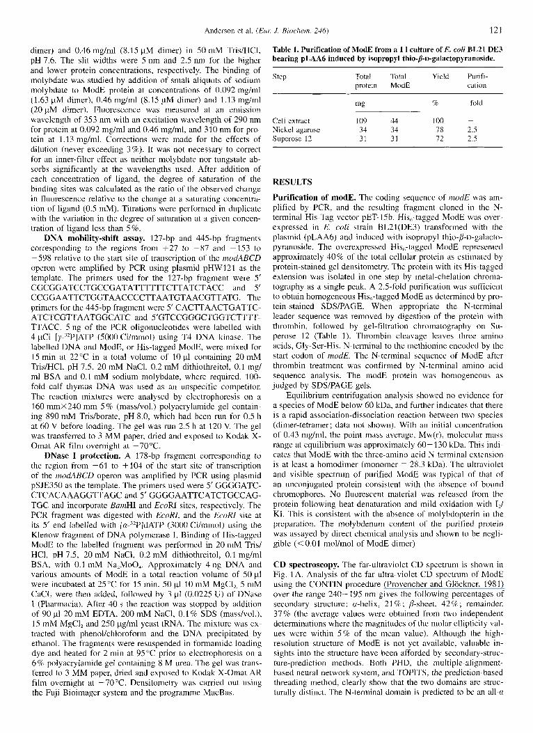
Table 1. Purification of ModE from a 1 1 culture of E. coli BL21 DE3 bearing pLAA6 induced by isopropyl thio-P-D-galactopyranoside.
Step Total Total Yield Purifi- protein ModE cation
Cell extract 109 44 100 ~
Nickel agarose 34 34 78 2.5 Superose 12 31 31 72 2.5
RESULTS
Purification of modE. The coding sequence of modE was am- plified by PCR, and the resulting fragment cloned in the N- terminal His-Tag vector PET- 15b. His,-tagged ModE was over- expressed in E. coli strain BL21(DE3) transformed with the plasmid (pLAA6) and induced with isopropyl thio-/$D-galacto- pyranoside. The overexpressed His,-tagged ModE represented approximately 40% of the total cellular protein as estimated by protein-stained gel densitometry. The protein with its His-tagged extension was isolated in one step by metal-chelation chroma- tography as a single peak. A 2.5-fold purification was sufficient to obtain homogeneous His,-tagged ModE as determined by pro- tein-stained SDS/PAGE. When appropriate the N-terminal leader sequence was removed by digestion of the protein with thrombin, followed by gel-filtration chromatography on Su- perose 12 (Table 1). Thrombin cleavage leaves three amino acids, Gly-Ser-His, N-terminal to the methionine encoded by the start codon of modE. The N-terminal sequence of ModE after thrombin treatment was confirmed by N-terminal amino acid sequence analysis. The modE protein was homogeneous as judged by SDSPAGE gels.
Equilibrium centrifugation analysis showed no evidence for a species of ModE below 60 kDa, and further indicates that there is a rapid association-dissociation reaction between two species (dimer-tetramer; data not shown). With an initial concentration of 0.43 mg/ml, the point mass average, Mw(r), molecular mass range at equilibrium was approximately 60-130 kDa. This indi- cates that ModE with the three-amino-acid N-terminal extension is at least a homodimer (monomer = 28.3 kDa). The ultraviolet and visible spectrum of purified ModE was typical of that of an unconjugated protein consistent with the absence of bound chromophores. No fluorescent material was released from the protein following heat denaturation and mild oxidation with I,/ KI. This is consistent with the absence of molybdopterin in the preparation. The molybdenum content of the purified protein was assayed by direct chemical analysis and shown to be negli- gible (< 0.01 mol/mol of ModE dimer)
CD spectroscopy. The far-ultraviolet CD spectrum is shown in Fig. 1 A. Analysis of the far ultra-violet CD spectrum of ModE using the CONTIN procedure (Provencher and Glockner, 1981) over the range 240- 195 nm gives the following percentages of secondary structure: a-helix, 21 % ; /?-sheet, 42 C/a ; remainder, 37 % (the average values were obtained from two independent determinations where the magnitudes of the molar ellipticity val- ues were within 5% of the mean value). Although the high- resolution structure of ModE is not yet available, valuable in- sights into the structure have been afforded by secondary-struc- ture-prediction methods. Both PHD, the multiple-alignment- based neural network system, and TOPJTS, the prediction-based threading method, clearly show that the two domains are struc- turally distinct. The N-terminal domain is predicted to be an all-a

122 Anderson et al. ( E m J . Biochem. 246)
190 200 220 240 260
Wavelength (nrn)
-20
-30
-40
260 270 280 290 300 310 320
Wavelength (nrn)
Fig. 1. CD spectra of ModE in the absence and presence of ligands. Spectra were recorded using the cell pathlengths and protein concentrations given in Materials and Methods. The traces (-), (---) and (. "...) represent protein i n the absence of ligands and in the presence of 1 mM molybdate or 1 mM tungstate respectively. (a) Far-ultraviolet spectra, (b) near-ultraviolet spectra.
structure and the C-terminal domain an al1-B structure. While there is little doubt about the distinctive character of the two domains, the predicted overall secondary-structure contents are only in general agreement with those derived by CD. Using the subset of the PHD prediction that gives an accuracy of greater than 82% the predicted contents are 22% rr-helix, 21 % p-sheet and 57 % remainder. Determination of the ij-sheet content by CD in the presence of significant a-helix is greatly affected by the level of noise below 200 nm. In addition, it must be emphasised that there are only three sequences similar to the E. coli ModE in the SwissProt database (indexing date 29 November 1996) and thus the accuracy of the PHD prediction may be compro- mised. On addition of 1 mM sodium molybdate or sodium tung- state there are small changes in the spectrum. CONTIN analysis suggests that there may be an increase in a-helix (to 24% and 29 %, respectively) in the presence of molybdate and tungstate, and in @-sheet (to 4 3 % and 53'32, respectively), but these esti- mates should be viewed with caution in view of the level of noise in the spectra below 200 nm.
The near-ultraviolet CD spectrum of ModE (Fig. 1 B) shows a broad negative peak over the range 270-310 nm. On addition of 1 mM sodium molybdate or sodium tungstate, there are very pronounced changes in the spectrum, including a decrease in signal below 290nm and the appearance of a sharp band at 292 nm, characteristic of tryptophan. These changes indicate that the environments of a number of the aromatic side chains and particularly that of one or more of the three tryptophan residues in each subunit have undergone marked changes on binding either of the ligands. Addition of the ligand clearly also causes perturbations in the environment of one or more tyrosine resi- dues as indicated i n the spectrum below 285 nm. There may be small differences in the environment of one or more tyrosine or phenylalanine side chains (260 to 280 nm regions) when molyb- date or tungstate are bound.
Determination of molybdate binding to ModE by the pertur- bation of intrinsic protein fluorescence. The intrinsic fluores- cence emission spectrum of ModE shows an emission wave- length maximum at 353 nm, indicating that the majority of the tryptophan side chains in the protein are highly exposed to solvent (Fig. 2). Under these conditions the emission maximum of the model compound N-acetyltryptophan amide is 358 nm (data not shown). The addition of molybdate or tungstate re- sulted in a shift to a shorter emission wavelength maximum (348 nm) and a quenching of the fluorescence by approximately 50%. Similar changes were observed on the addition of molyb- date or tungstate to the His-tagged protein (data not shown).
500
400
300
200
100
0 300 320 340 360 380 400
Wavelength (nrn)
Fig. 2. Fluorescence spectra of ModE in the absence and presence of ligands. Spectra were collected as described in Materials and Methods. Emission spectra are shown for 8.15 nM ModE diiner in 50 mM Trisl HCI, pH 7.6. The excitation wavelength was 290 nm. The solid curve is the spectrum in the absence of added ligands. The dotted and dashed curves are the spectra in the presence of 0.5 mM molybdate and tung- state, respectively. Addition of 0.5 mM molybdate and tungstate resulted in an approximately 50% quenching of the fluorescence and a 5-nm blue shift.
Fluorescence titrations with molybdate were carried out to determine the number of binding sites and the K, for molybdate binding. For accurate estimates, the concentration of sites should be at least ten times the value of the K , so that at low total ligand concentrations over 90% of the ligand is bound to protein. The titration with 20 pM ModE dimer showed that 2.0 mol Mo bind to 1 mol ModE dimer (Fig. 3). A good fit to the data was ob- tained by assuniing that the K, is 0.8 pM and two binding sites/ dimer. The number of binding sites was confirmed by repeating the experiment at the lower concentration of 8.15 pM, but not at 1.63 pM when the conditions described above are not fulfilled (data not shown). The experiments at lower protein concentra- tions confirmed the K , to be 0.8 pM.
Molybdenum-dependent binding of ModE to the modABCD promoter region. The in vitro binding of ModE protein to the modABCD promoter region was characterised by DNA mobility- shift experiments using a 127-bp fragment corresponding to the region from $27 to -87 of the start site of transcription of the modABCD operon (Fig. 4). In the absence of molybdenum there was no shift in the promoter fragment at concentrations of ModE

Andcrson et al. (Eiir:
-0 Q1
3 m In In Q1
In 0 C 0
0
IA
C E .I-
c. .- Ic
.- c
II!
0 20 40 60 80 100
[Ligand] (total) (pM)
Fig. 3. Molybdenum binding to the ModE protein determined by the change in tryptophan fluorescence as a function of added molybdate. Fluorescence titration was carried out as described in Materials and Methods. The solid squares are experimental data points obtained by the addition of molybdate to 20 pM ModE dimer. The continuous curved line is the theoretical curve obtained assuming 2 binding sites/ModE diiner and a K,, of 0.8 pM molybdate. A straight line is drawn through the data points for lower total molybdate concentrations and extrapolated to full site occupancy.
protein up to 120 nM, and an approximately 50% shift with 300 nM ModE. By contrast, 30 nM ModE protein resulted in 50% binding in the presence of 0.1 mM molybdate. A high con- centration of molybdate was used in the binding reaction as well as the gel and electrophoresis buffers to ensure full saturation of ModE during binding and electrophoretic analysis. A distinct molybdate-independent shift was observed with above 1300 nM ModE protein. The molybdate-dependent binding ModE protein was specific. It was also seen in the presence of 100-fold unla- belled competitor DNA (data not shown). It was also related to the promoter region, and not seen in the a 445 bp (-153 to -598) upstream fragment that includes the divergent modE pro- moter region (data not shown). Consistent with the ability of ModE to bind tungstate, 0.1 mM sodium tungstate was able to substitute for molybdate in the gel-shift assays (data not shown). The tungstate-ModE complex is therefore able to bind to the modA promoter region. Further, His-tagged ModE behaved in an identical manner in the gel-shift experiments.
J . Hiochern. 246) 123
DNase I footprinting was used to determine where the ModE protein binds. A single protected area can be seen from approxi- mately + I 1 to -20 from the transcription-start site (Fig. 5A). This area is approximately centred on the transcription start, and also includes the - 10 binding site for 07"-dependent RNA poly- merase. An adenine bases at +3 and a thymidine base at -20 become hypersensitive to DNase I when the ModE protein is bound (Fig. 5 B).
DISCUSSION
Mutations in ModE abolish molybdate-dependent repression of modABCD transcription. On the basis of sequence similarity the protein has two distinct domains, each of which extends over approximately half the protein. The C-terminal domain contains a tandem repeat of a sequence that is very similar to that of the 7-kDa protein called Mop isolated from C. pasterianum (Hinton and Freyer, 1986). Secondary-structure predictions also distin- guish the two halves of the molecule. The C. pasfeurianum Mop protein was reported to contain 0.9 mol molybdenudmonomer protein (Hinton and Merritt, 1986). It also contained tluorescent material which produced pterin-like fluorescent compounds but no molybdenum cofactor activity could be demonstrated with the Neurospom crassa nit-1 complementation though the presence of molybdopterin in the Mop protein prepa- rations was only ambiguously proven, proteins similar to the Mop protein have been designated molybdopterin-binding pro- teins. The ultraviolet and fluorescence spectra of ModE show no evidence for the presence of a non-protein chromophore. We conclude that ModE as prepared is free of molybdopterin or molybdenum cofactor.
Our results show that a ModE dimer binds two molecules of molybdate in vitro with a Kd of 0.8 pM. A Kd of this magnitude is consistent with any molybdate originally bound to ModE be- ing rapidly dissociated from the protein early in the preparation. The Kd for molybdate binding to ModE is of the same order as the concentration of molybdate in the medium (0.1 -0.5 pM) required to produce 50% repression of transcription of the mo- lybdenum transport operons in E. coli and A. viizelandii (Miller et al., 1987; Mouncey et al., 1996). It is clear from our results that molybdate binds directly to the ModE protein without a requirement for any other compound. In vivo expression studies have also indicated that the ModE-mediated molybdate repres- sion does not have a requirement for molybdopterin (Miller et al., 1987; Rech et al., 1995). Our results show that purified
Fig.4. Binding of the ModE protein to the modABCD promoter region shown by a DNA mobility-shift assay of a 127-bp labelled DNA fragment generated by PCR. (A) A mobility-shift assay in the absence of molybdate. (B) A mobility-shift assay in the presence of 0.1 mM molybdate. In both (A and B), lanes 1-9 show the mobility of the labelled DNA fragment in the presence of increasing concentrations of ModE. and lane 10 shows the mobility in the absence of ModE. The concentrations of ModE are given below the lanes.

124 Anderson et al. (Eur: J. Biochem. 245)
B 3.0 +3 I
0.5 I I I I I 0 50 100 150 200 250
Relative distance
0 4 54 200 562 ModE (nM)
C
E. coli modA
H. influenzae modA
R. cupsulutus mopA
R. cupsulutus unfA
A. vinehdii modE
A. vinelandii modG
Consensus Mo-Box
4 -10 +1 c AGTCGTTATATTGTCGCCTACATAACGT CATTAAGGGGTTACCAATG - -
TTATTTATTT~,~.?.~~ATAATCAAGTAAATAACGTTTAATC ..\\\.\\\.
GAAAAACCATCGCTATTAGTCGGGTCTATATATAACGATCCACCTTCGGACCCTGCCATG .\.\\\\\w ..........
CAGCCTGTCGTTATATGGAATCACTATATAACGGAGTCGGTCATG .......... .......... GTGAATTGCTTTATATAAACTGGATAAATAGATAGATAAATTGCAGGCTACCAGACGATG .......... .......... TTTTACGACAATTTATAGTGCAAATGATATAGCGCTTGTGCC~CCCNi5GAGGAACACAGCATG ......... .-A\\ \..v
(CT) (ATG) (ATC)TAT - N l l / l 2 - ATA(ATG) (AC) (AGT) Fig.5. The binding site of ModE. (A) Localisation of the ModE-binding site in the inodABCD promoter region by DNase I footprinting. The coding strand of a 178-bp PCR fragment was labelled at its 5' end with the Klenow fragment of DNA polymerase I. Prior to DNase I digestion, the labelled fragment was incubated with the amounts of ModE indicated at the bottom of lanes 1-5. Lanes labelled AG are the A and G sequence ladders generated by chemical cleavage DNA sequencing (Maxam and Gilbert, 1980). The transcription start (+1) and positions relative to i t are indicated. The position of the translation-start codon (ATG) is labelled. The protected region is indicated by a vertical bar. (B) Densitometric traces of the footprints corresponding to lanes 1 and 3 of the gel are shown in (A). The solid line represents the density of the lane without ModE, and the broken line represents the lane with 56 nM ModE. The bands which become hypersensitive to DNase I action on addition of ModE are labelled +3 and -20. (C) Alignment of the promoter region of the E. coli modABCD operon with those of the molybdenum transport operons from A. vinelondii, H. i$Ltenzae and R. ccipsutntu.~, and the promoter regions of R. cqxndarus mf;4 and A. viriefnndii modG. The region of the E. cofi morlARCD promoter that is protected by ModE is enclosed by a box. The symmetric dyad within this region is underlined with solid lines. The corresponding regions in the other promoter regions are shown with hatched lines. The sites which become hypersensitive to DNase I digestion when the ModE-molybdenum complex is bound are show by vertical arrows. The consensus sequence is shown below the promoter sequences.
ModE also binds tungstate in v i m , and furthermore the tung- state-ModE complex is able to bind to the ModE promoter re- gion. Miller et al. (1987) showed that E. coli niodABCD expres- sion is repressed by tungstate addition to the growth medium. It seems highly likely that tungstate repression of modA is medi- ated by ModE in E. coli. This was shown to be the case in A. vinelandii (Mouncey et al., 1996).
Molybdate is presumed to bind to the C-terminal domain of ModE because of its sequence similarity with Mop proteins. The changes observed in the near-ultraviolet CD spectroscopy indi- cate that binding produces a major conformational change in the environment of typtophan and tyrosyl residues of the protein. This conformational change does not require binding to DNA. The C-terminal domain contains two of the three tryptophan and

Anderson et al. (Eur: J . Biochem. 246) 125
one of the four tyrosyl residues. The changes in the intrinsic tryptophan fluorescence are indicative of burying of one or more tryptophan residues which are exposed in the unbound state. Tungstate produces similar conformational changes, and the bound and unbound spectra of the His-tagged ModE protein are similar to those of the protein lacking the His tag.
In vitro DNA mobility-shift experiments showed that molyb- date is essential for the ModE protein to be bound with a high affinity to the modA promoter fragment. The molybdate-inde- pendent DNA binding observed at high concentrations of ModE may not be representative of physiological conditions. The clear requirement of molybdate for binding was not observed in pre- viously reported mobility-shift experiments which may have been performed using higher concentrations of ModE which was purified by a different method (Grunden et al., 1996). It has been suggested from comparison of the modA promoter regions of molybdenum-related bacterial genes that the sequence CATAA- N,-CATTAA might be involved in ModE binding (Rech et al., 1995). Mutations in the C and the last two A in the upstream element abolished molybdenum-dependent repression. Mutating bases in the downstream element did not abolish repression. The DNase I protection experiment identified a single 31-bp region that is protected by the ModE protein with bound molybdenum. It is located in a position typical for a repressor in a region around the transcription-start site. This region contains a sym- metric dyad, and its downstream half-site corresponds to the re- gion in which mutations relieved repression by ModE (Recli et al., 1995). The upstream half-site overlaps the -10 hexamer which is required for RNA polymerase binding. The 42-bp DNA fragment which was used for previously reported gel-shift ex- periments only included approximately half of the protected re- gion and the downstream half-site (Grunden et al., 1996). The two 6-base half-sites of the symmetric dyad in the ModE-bind- ing site of E. coli modA promoter are separated by 12 bases. Comparison with the modA promoter regions of other bacteria shows that this spacing can be 11 bases in A. vinelandii and R. capsulutus. The consensus sequence from the promoter regions of the molybdenum transporter operons of four bacterial genera shows that only three bases in each of the E. coli half-sites are conserved (Fig. 5 C). This accounts for previous difficulty in identifying a putative ModE-binding site by sequence compari- son. The consensus sequence identified here is also found, with the correct inter half-site spacing, in the promoters of A. vinelan- dii inodG and R. capsulutus anfA, whose molybdenum-depen- dent repression is mediated by ModE and in R. capsulutus by the ModE-like proteins MopA and MopB (Mouncey et a]. 1996; Kutsche et al., 1996).
In contrast to a previous report which stated that ModE ex- ists as a monomer (McNicholas et al., 1996), the ultracentrifuga- tion analysis reported here showed that ModE with an N-termi- nal extension of three-amino acids is at least a dimer in solution. Further investigations are required to determine the conditions which influence its oligomeric state. The presence of a dyad element in the DNA-binding site for ModE suggests that ModE binds as a dimer. The DNase I protection experiment also showed the presence of two sites which were hypersensitive to DNase 1 digestion when ModE was bound. The hypersensitive sites suggest that the molybdate-dependent binding of ModE bends the DNA (Schultz et al., 1991 ; Ansari et al., 1995). DNA bending is associated with many regulatory proteins, including members of the MerR arid LysR families of ligand-responsive transcriptional regulators (Ansari, 1995). The sequence of the ModE protein does not show a significant match with any family of proteins in the PROSITE database. A search of the BLOCKS database to reveal more distant relationships identified the first 53 residues of ModE to be similar to both the AraC and LysR
families of regulatory proteins. A putative helix-turn-helix DNA-binding motif has been postulated in this region (Grunden et al., 1996). In members of the LysR family the DNA-binding domain is N-terminal and the signal-recognition domain C-ter- minal, while the converse is true for members of the AraC fam- ily (Gallegos et al., 1993 ; Schell, 1993). ModE may therefore be distantly related to the LysR family of transcriptional regulators.
McNicholas et al. (1997) have recently published an account of the characterization of the ModE DNA-binding sites in the control regions of modABCD and moaABCDE of Eschericlzia coli.
We are grateful to R. Eichenlaub, H. M. Walkenhorst and S. Hinton for providing us with plasmids and information, to S. Kelly for the CD spectra, to S. E. Harding and K. Jumel for the equilibrium centrifugation analysis, and to R. G. Sawers for invaluable advice on DNA-binding experiments and critical reading of the manuscript. L. A. Anderson is a Biotechnology and Biological Sciences Research Council CASE re- search student. Circular dichroism spectroscopy was carried out at the Biotechnology and Biological Sciences Research Council-funded Circu- lar Dichroism Facility at the University of Stirling, Scotland. Ultracen- trifugation analysis was carried out at the Biotechnology and Biological Sciences Research Council and the Engineering and Physical Sciences Research Council-funded UK National Centre for Molecular Hydrody- namics at Nottingham University. This work was supported by Biotech- nology and Biological Sciences Research Council Grant (94/P03108) to D. H. Boxer.
REFERENCES Ansari, A. Z., Bradner, J. E. & O’Halloran, T. V. (1995) DNA-bend
modulation in a repressor-to activator switching mechanism, Nuture
Bairoch, A. (1992) PROSITE: a dictionary of sites and patterns in pro- teins, Nucleic Acids Res. 20 (Supplement), 201 3 -201 8.
Clark, L. R. & Axley, J. H. (1955) Molybdenum determination in soils and rocks with dithiol, Anal. Chem. 27, 2000-2003.
Devereux, J. F., Coulson, A. F. W. & Lyall, A. (1987) Protein and nucleic acid sequence database searching: a suitable case for parallel pro- cessing, Nucleic Acids Rex 12, 387-395.
Fuchs, R. (1991) Predicting protein function: a versatile tool for the Apple Macintosh, Comput. Appl. Biosci. 1 0 , 171 ~ 178.
Gallegos, M.-T., Michin, C. & Ramos, J. L. (1993) The XylS/AraC family of regulators, Nucleic Acids Res. 21, 807-810.
Gill, S. C. & von Hippel, P. H. (1989) Calculation of protein extinction coefficients from amino acid sequence data, Anal. Biochem. 182,
Grunden, A.M., Ray, R. M., Rosentel, J. K., Healy, F. G. & Shanmugam, K. T. (1996) Repression of the Escherichiu coli modABCD (molyb- date transport) operon by ModE, J. Bacteriol. 178, 735 -744.
Henikoff, S. & Henikoff, J. G. (1994) Protein family classification based on searching a database of blocks, Genomics 19, 97- 107.
Hinton, S. M. & Freyer, G. (1986) Cloning, expression and sequencing the molybdenum-pterin binding protein (mop) gene of Clostridiuni pasteurianum in Escherichia coli, Nucleic Acids Res. 14, 9371 - 9380.
Hinton, S. M. & Merritt, B. (1986) Purification and characterisation of a molybdenum-pterin-binding protein (Mop) in Clo.w+fium pmtruri- anum W5, J . Bacteriol. 168, 688-693.
Johann, S. & Hinton, S. (1987) Cloning and nucleotide sequence of the chlD locus, J. Bacteriol. 169, 19 11 - 19 16.
Kutshe, M., Leimkiihler, S., Angemuller, S. & Klipp, W. (1996) Prornot- ers controlling expression of the alternative nitrogenase and the mo- lybdenum uptake system in Rhodobacter capsulatus are activated by NtrC, independent of os4, and repressed by molybdenum, 1. Bacte- rial. 178, 2010-2017.
Laemmli, U. K. (1970) Cleavage of structural proteins during the assem- bly of the head of bacteriophage T4, Nuture 227, 680-685.
Maupin-Furlow, J. A., Rosentel, J. K., Lee, J. H., Deppenmeier, U. & Gunsalus, R. P. (1995) Genetic analysis of the madABCD (molybdate transport) operon of Escherichia coli, J . Bacteriol. 177, 4851 -4856.
374, 371-375.
3 19 - 326.

126 Anderson et al. (EUK J . Biochem. 246)
Maxam, A. M. & Gilbert, W. (1980) Sequencing end-labelled DNA with base-specific chemical cleavages, Methods Enzymol. 65, 499- 500.
McNicholas, P. M., Chiang, R. C. & Gunsalus, R. P. (1996) The Escheri- chiu coli modE gene: effects of mutations on molybdate dependent modA expression, FEMS Mirrobiol. Letl. 145, 117- 123.
McNicholas, P. M., Rech, S. A. & Gunsalus, R. P. (1997) Characteriza- tion of the ModE DNA-binding sites in the control regions of mo- dABCD and mouABCDB of Escherichia roli, Mol. Mirrobiol. 23, 515-524.
Miller, J., Scott, D. & Amy, N. (1987) Molybdenum-sensitive transcrip- tional regulation of the chill locus of Escherichia coli, .I. Bacte- rio1.169, 1853- 1860.
Miller, J. H. (1972) Experiments in rnolecular genetics, Cold Spring Har- bor Laboratory Press, Cold Spring Harbor NY.
Mouncey, N. J., Mitchenall, L. A. & Pau, R. N. (1995) Mutational analy- sis of genes of the mod locus involved in molybdenum transport, homeostasis and processing in Azotobacter vinelandii, J . Burteriol. 177,5294-5302.
Mouncey, N. J., Mitchenall, L. A. & Pau, R. N. (1996) The modE gene product mediates molybdenum-dependant expression of genes for the high-affinity molybate transporter and rnodC in Azotahacter vi- nelondii, Microbiology 142, 1997-2004.
Murray, V. (1989) Improved double-stranded DNA sequencing using the lincar polymerase chain reaction, Nucleic Acids Res. 17, 8889.
Pau, R. N., Klipp, W. & Leimkuhler, S. (1997) Molybdenum transport, processing and genc regulation, in Ii-iin ond related trrmrition nzerals i n microbitrl rnetaholism (Winkelmann, G. & Carrano, C. J . , eds) pp. 217-234, Harwood Academic Publishers, Amsterdam.
Provencher, S. W. & Gliickner, J. (1981) Estimation of globular protein secondary structure from circular dichroism, Biochemistn 20, 33 - 37.
Rajagopalan, ti. V. (1993). Biochemistry of the molybdenum cofacors, in Molybdenunz enzymes, cofuctors and niodel systeins (Stiefel, E. I., Coucouvanis, D. & Newton, W. E., eds) pp. 38-49. American Chemical Society. Washington DC.
Rajagopalan, ti. V. (1996). Biosynthesia of the molybdenum cofactor. in Escherichia coli and Salnzonelln typhirnuriunz (Neidhardt, F. C., ed.) pp. 674-679, American Scocicty of Microbiology Press, Washing- ton DC.
Rech, S., Deppenmeier, U. & Gunsalus, R. P. (1995) Regulation of the molybdate tramport opcron, modABCD, of Escherichia coli in re- sponse to molybdate availibility, ./. Bacreriol. 177, 1023 - 1029.
Rosentel. J . K., Healy, F., Maupin-Furlow, J. A,, Lee, J. H. & Shanmu- gam, K. T. (1995) Molybdenum regulation of mod (Molybdate trans- port), ,fdhF, and lzyc (forinate hydrogen lyase) operons in Escherichia coli, .I. Hucteriol. 177, 4857-4864.
Rost, B. ( 1 995) TOPITS: Threading one-dimensional predictions into three-dimensional structures, in The third international conference on intelligent systems,for molecular biology (ISBM), Cambridge U K July 16-19, 1995, (Rawlings, C., Clark, D., Altman, R., Hunter, L., Lengauer, T. & Wodak, S., eds) pp. 314-321, AAAI Press, Menlo Park, CA.
Rost, B. & Sander, C. (1994) Combining evolutionary information and neural networks to predict protein secondary structure, Proteins 19, 55-77.
Sarnbrook, J., Fritsch, E. F. & Maniatis, T. (1989) Molecular Cloning: a l ohora to~ manual, 2nd edn, Cold Spring Harbor Laboratory Press, Cold Spring Harbor NY.
Sanger, F. & Coulson, A. R. (1975) A rapid method for determining sequences in DNA by primed synthesis with DNA polymerase, J . Mid. Biol. 94, 441 -448.
Schell, M. A. (1993) Molecular biology of the LysR family of transcrip- tional regulators, Annu. Re\,. Microhiol. 47, 597-626.
Schultz, S. C., Scheilds, G. C. & Steitz, T. A. Crystal structure of a CAP-DNA complex: The DNA is bent by 90" (1991) Science 253, 1001 - 1007.
Scott, D. & Amy, N. K. (1989) Molybdenum accumulation in chlD mu- tants of Escherichia ccdi, J . Bacreriol. 171, 1284-1287.
Stewart, V. (1 988) Nitrate respiration in relation to facultative metabo- lism in enterobacteria, Microbiol. Rev. 52, 190-232.
Stiefel, E. I. (1993) Molybdenum enzymes, cofactors and chemistry, in Molybrlenuin enzymes, cofuctors and niodel sy.srems (Stiefel, E. I . , Coucouvanis, D. & Newton, W. E., eds) pp. 1 - 18, American Chemi- cal Society, Washington DC.
Studier, F. W. & Moffatt, B. A. (1986) Use of bacteriophage T7 RNA polymerase to direct selective high-level expression of cloned genes, .I. Mol. Biol. 189, 113-130.
Walkenhorst, H. M., Heinscheineier, S. ti. & Eicheniaub, R. (1995) Mo- lecular analysis of the molybdate uptake operon, modABCD, of Escherichia coli and modR,a regulatory gene, Microbiol. Res. 150,
Wang, G., Angermiiller, S. & Klipp, W. (1993) Characterization of Rho- dobacter cappsulurus genes encoding a molybdenum transport system and putative molybdenrrm-pterin-binding proteins, 1. Bacteriol. 175, 3031-3042.
347-361.