Characterisation and In Vitro Cytotoxicity of Triorganophosphinegold(I) 2-Mercaptobenzoate Complexes
10
CHARACTERISATION AND IN VITRO CYTOTOXIClTY OF TRIORGANOPHOSPHINEGOLD(I) 2-MERCAPTOBENZOATE COMPLEXES Dick de Vos , Phil Clements, 2 Simon M. Pyke, 2 Douglas R. Smyth 2 and Edward R.T. Tiekink 2,* Medical Department, Pharmachemie B.V., NL-2003 RN Haarlem, The Netherlands 2 Department of Chemistry, The University of Adelaide, Australia 5005 Abstract The preparation and full NMR (1H,13C and 31p) characterisation of three R3PAu(2mba) complexes, where R Et, Ph and Cy, and 2mba is the anion derived from 2-mercaptobenzoic acid is reported. An interesting solvent dependence in the H spectra is rationalised in terms of competing intra- and inter-molecular hydrogen bonding. An X-ray analysis of the [Ph3PAu(2mba)] species reveals a linear P--Au--S arrangement and association in the lattice via the familar carboxylic acid dimer motif. The in vitro cytotoxicity against seven human tumour lines is also described. The complexes display moderate to very high activity. Particularly noteworthy is their greater activity against the H226 cell line (non-small cell lung cancer) compared with that displayed by a range of cytotoxic drugs. Introduction The use of gold complexes in the treatment of rheumatoid arthritis is well known [1-3]. Two classes of complexes are used in this context, namely, polymeric, water soluble gold thiolates such as myocrisin (sodium aurothiomalate) and solganol (aurothioglucose) as well as monomeric, lipophilic auranofin ([(1-thi--D-gucpyranse-2346-tetraacetat-s)(triethyphsphine)gd()) Given their clinical use, a logical extension of the study of gold complexes is an exploration of their potential use as anti-tumour agents [e.g. 1-3]. As a continuation of a program investigating the anti- tumour activity of phosphinegold(I) thiolates [4,5], i.e. complexes containing the P--Au---S arrangement as found in auranofin, the in vitro cytotoxicity of three R3PAu(2mba) complexes is reported where R Et, Ph and Cy, and 2mba is the anion derived from 2-mercaptobenzoic acid. The preparation of these complexes has been reported in the patent [6,7] and chemical literature [8], and the crystal structure determination of the R Cy complex is also known [8]. Experimental General Analytical grade solvents were used without further purification. 1H (and 1H-1H COSY), 13C and 31p NMR spectra were recorded on a Varian Gemini 2000 spectrometer operating at 300.145, 75.479 and 121.501 MHz, respectively. 1H-13C HSQC and HMBC 2D experiments were measured on a Varian INOVA NMR spectrometer operating at 599.952 and 150.873 MHz for 1H and 13C NMR, respectively. Electrospray mass spectra were obtained from methanol or ethanol solutions (NaOMe was added in the case of R Ph and Cy (in-ve mode) in order to aid ionisation). Samples were injected into a Finagan LCQ mass spectrometer via direct infusion using a syringe pump. Nitrogen was used as the drying and nebulising gas. Elemental analyses were performed by Chemical and Micro Analytical Services Pty Ltd (Belmont, Victoria). Preparations The R3PAuCI precursors were prepared according to the literature method [9] and 2- mercaptobenzoic acid (2mbaH) was purchased from Fluka. The three R3PAu(2mba) complexes were prepared using a similar procedure in each case. To a stirred ethanol solution (20 ml) of * email: [email protected] 31
-
Upload
independent -
Category
Documents
-
view
1 -
download
0
Transcript of Characterisation and In Vitro Cytotoxicity of Triorganophosphinegold(I) 2-Mercaptobenzoate Complexes
CHARACTERISATION AND IN VITRO CYTOTOXIClTY OFTRIORGANOPHOSPHINEGOLD(I)
2-MERCAPTOBENZOATE COMPLEXES
Dick de Vos, Phil Clements,2 Simon M. Pyke,2 Douglas R. Smyth2
and Edward R.T. Tiekink2,*
Medical Department, Pharmachemie B.V., NL-2003 RN Haarlem, The Netherlands2 Department of Chemistry, The University of Adelaide, Australia 5005
AbstractThe preparation and full NMR (1H,13C and 31p) characterisation of three R3PAu(2mba)
complexes, where R Et, Ph and Cy, and 2mba is the anion derived from 2-mercaptobenzoic acidis reported. An interesting solvent dependence in the H spectra is rationalised in terms ofcompeting intra- and inter-molecular hydrogen bonding. An X-ray analysis of the [Ph3PAu(2mba)]species reveals a linear P--Au--S arrangement and association in the lattice via the familarcarboxylic acid dimer motif. The in vitro cytotoxicity against seven human tumour lines is alsodescribed. The complexes display moderate to very high activity. Particularly noteworthy is theirgreater activity against the H226 cell line (non-small cell lung cancer) compared with that displayedby a range of cytotoxic drugs.
IntroductionThe use of gold complexes in the treatment of rheumatoid arthritis is well known [1-3]. Two
classes of complexes are used in this context, namely, polymeric, water soluble gold thiolates suchas myocrisin (sodium aurothiomalate) and solganol (aurothioglucose) as well as monomeric,lipophilic auranofin ([(1-thi--D-gucpyranse-2346-tetraacetat-s)(triethyphsphine)gd())Given their clinical use, a logical extension of the study of gold complexes is an exploration of theirpotential use as anti-tumour agents [e.g. 1-3]. As a continuation of a program investigating the anti-tumour activity of phosphinegold(I) thiolates [4,5], i.e. complexes containing the P--Au---Sarrangement as found in auranofin, the in vitro cytotoxicity of three R3PAu(2mba) complexes isreported where R Et, Ph and Cy, and 2mba is the anion derived from 2-mercaptobenzoic acid.The preparation of these complexes has been reported in the patent [6,7] and chemical literature[8], and the crystal structure determination of the R Cy complex is also known [8].
ExperimentalGeneralAnalytical grade solvents were used without further purification. 1H (and 1H-1H COSY), 13C
and 31p NMR spectra were recorded on a Varian Gemini 2000 spectrometer operating at 300.145,75.479 and 121.501 MHz, respectively. 1H-13C HSQC and HMBC 2D experiments were measuredon a Varian INOVA NMR spectrometer operating at 599.952 and 150.873 MHz for 1H and 13C NMR,respectively. Electrospray mass spectra were obtained from methanol or ethanol solutions (NaOMewas added in the case of R Ph and Cy (in-ve mode) in order to aid ionisation). Samples wereinjected into a Finagan LCQ mass spectrometer via direct infusion using a syringe pump. Nitrogenwas used as the drying and nebulising gas. Elemental analyses were performed by Chemical andMicro Analytical Services Pty Ltd (Belmont, Victoria).
PreparationsThe R3PAuCI precursors were prepared according to the literature method [9] and 2-
mercaptobenzoic acid (2mbaH) was purchased from Fluka. The three R3PAu(2mba) complexeswere prepared using a similar procedure in each case. To a stirred ethanol solution (20 ml) of
* email: [email protected]
31
Vol. 6, No. 1, 1999 Characterisation and In Vitro Cytotoxicity ofTriorganophosphinegold(1)2-Mercaptobenzoate Complexes
R3PAuCI (ca 500 mg) was added an aqueous ethanolic solution (20 ml) containing equimolarquantities of 2mbaH and KOH. The solution was stirred at room temperature for h, the solventevaporated off and the residue recrystallised from acetone solution in each case.[Et3PAu(2mba)]: Yellow crystals, Yield 82 %, m. pt 109- 110C. Found C, 33.23; H, 4.24.C13H20AuO2PS requires, C, 33.34; H, 4.30.[Ph3PAu(2mba)]: Yellow crystals, Yield 83 %, mo pt 157.5- 159C. Found C, 49.18; H, 3.27.C25H20AuO2PS requires, C, 49.03; H, 3.29. Crystals suitable for X-ray crystallography were grownfrom the slow evaporation and cooling of a hot ethanol solution of the complex.[Cy3PAu(2mba)]: Colourless crystals, Yield 81%, m. pt 170- 171C. Found C, 47.68; H, 6.22.C25H38AuO2PS requires, C, 47.62; H, 6.07.
Crystallography for [Ph,PAu(2mba)]Intensity data for a yellow crystal (0.13 x 0.18 x 0.34 mm) were measured at room temperature
on a Rigoaku AFC6R diffractometer fitted with MoKo radiation (graphite monochromator, X0.71073 A) using the o:2e scan technique so that emax was 25.0. No decomposition of the crystaloccurred during the data collection, the data set was corrected for Lorentz and polarization effects[10] and for absorption employing an empirical procedure [11] such that the minimum and maximumtransmission factors were 0.352 and 1, respectively. A total of 4151 data (3942 unique) werecollected and of these, 2792 that satisfied the I_> 3.0c(/) criterion were used in the subsequentanalysis.
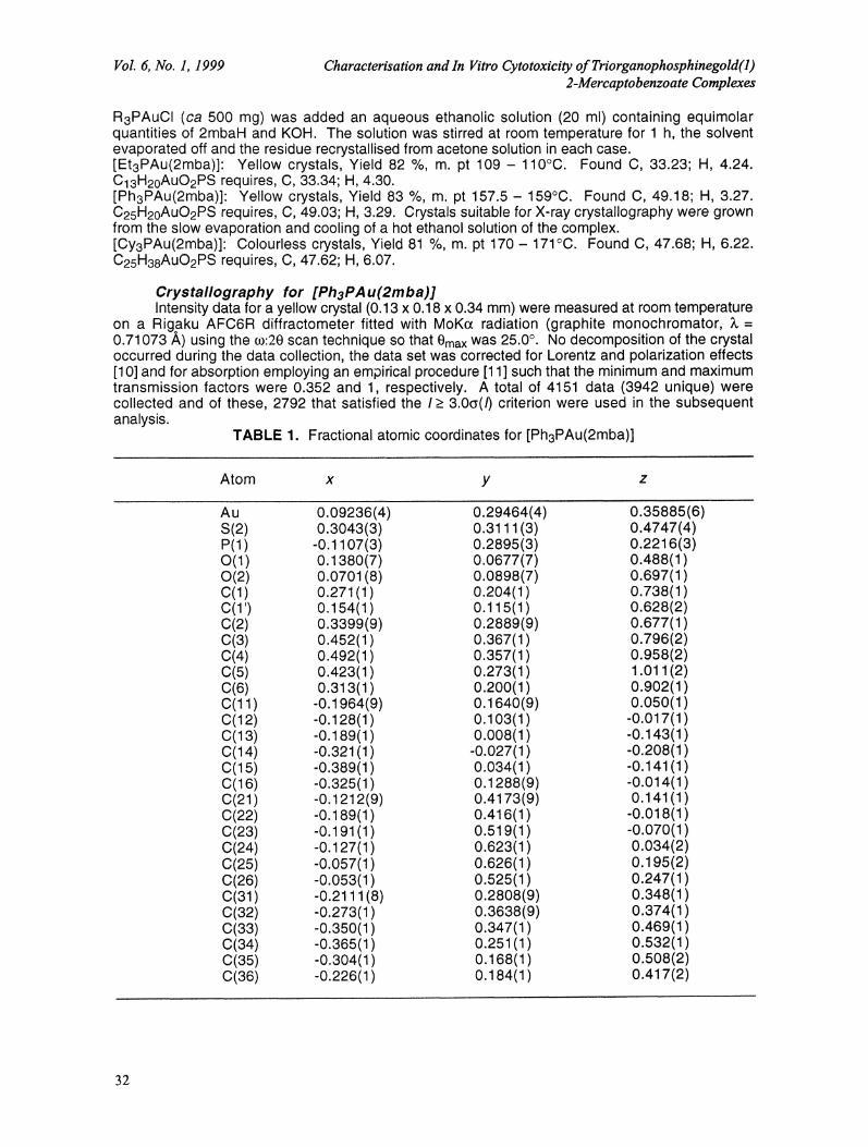
TABLE 1. Fractional atomic coordinates for [Ph3PAu(2mba)]
Atom x y z
Au 0.09236(4) 0.29464(4) 0.35885(6)S(2) 0.3043(3) 0.3111(3) 0.4747(4)P(1) -0.1107(3) 0.2895(3) 0.2216(3)O(1) 0.1380(7) 0.0677(7) 0.488(1)0(2) 0.0701 (8) 0.0898(7) 0.697(1)C(1) 0.271(1) 0.204(1) 0.738(1)C(1’) 0.154(1) 0.115(1) 0.628(2)C(2) 0.3399(9) 0.2889(9) 0.677(1C(3) 0.452(1 0.367(1 0.796(2)C(4) 0.492(1 0.357(1 0.958(2)C(5) 0.423(1) 0.273(1) 1.011(2)C(6) 0.313(1 0.200(1 0.902(1C(11) -0.1964(9) 0.1640(9) 0.050(1)C(12) -0.128(1) 0.103(1) -0.017(1)C(13) -0.189(1 0.008(1 -0.143(1C(14) -0.321 (1) -0.027(1 -0.208(1C(15) -0.389(1) 0.034(1) -0.141(1)C(16) -0.325(1) 0.1288(9) -0.014(1)C(21) -0.1212(9) 0.4173(9) 0.141(1)C(22) -0.189(1) 0.416(1) -0.018(1)C(23) -0.191(1) 0.519(1) -0.070(1)C(24) -0.127(1) 0.623(1) 0.034(2)C(25) -0.057(1) 0.626(1) 0.195(2)C(26) -0.053(1) 0.525(1) 0.247(1)C(31) -0.2111 (8) 0.2808(9) 0.348(1C(32) -0.273(1 0.3638(9) 0.374(1C(33) -0.350(1) 0.347(1) 0.469(1)C(34) -0.365(1 0.251 (1) 0.532(1C(35) -0.304(1) 0.168(1) 0.508(2)C(36) -0.226(1) 0.184(1) 0.417(2)
32
Dick de Vos et al. Metal-Based Drugs
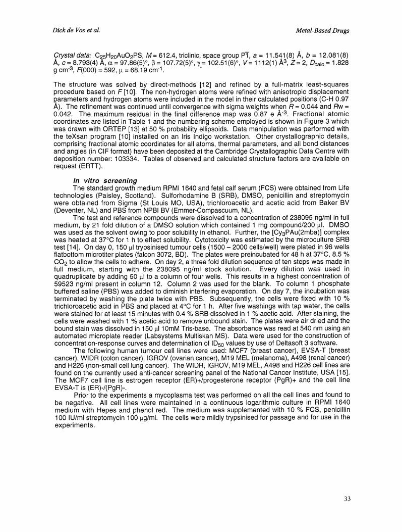
data: C5H20AuO2PS, M= 612.4, triclinic, space group P, a 11 541(8) A, b 12.081(8)c 8.793(4) A, o 97.86(5), I 107.72(5), y 102.51 (6), V 1112(1") A3, Z 2, Dcalc 1.828
g cm"3, F(000) 592, 68.19 cm-1.
The structure was solved by direct-methods [12] and refined by a full-matrix least-squaresprocedure based on F [10]. The non-hydrogen atoms were refined with anisotropic displacementparameters and hydrogen atoms were included in the model in their calculated positions (C-H 0.97A). The refinement was continued until convergence with sigma weights when R 0.044 and Rw0.042. The maximum residual in the final difference map was 0.87 e ,&,-3. Fractional atomiccoordinates are listed in Table and the numbering scheme employed is shown in Figure 3 whichwas drawn with ORTEP [13] at 50 % probability ellipsoids. Data manipulation was performed withthe teXsan program [10] installed on an Iris Indigo workstation. Other crystallographic details,comprising fractional atomic coordinates for all atoms, thermal parameters, and all bond distancesand angles (in CIF format) have been deposited at the Cambridge Crystallographic Data Centre withdeposition number: 103334. Tables of observed and calculated structure factors are available onrequest (ERTT).
In vitro screeningThe standard growth medium RPMI 1640 and fetal calf serum (FCS) were obtained from Life
technologies (Paisley, Scotland). Sulforhodamine B (SRB), DMSO, penicillin and streptomycinwere obtained from Sigma (St Louis MO, USA), trichloroacetic and acetic acid from Baker BV(Deventer, NL) and PBS from NPBI BV (Emmer-Compascuum, NL).
The test and reference compounds were dissolved to a concentration of 238095 ng/ml in fullmedium, by 21 fold dilution of a DMSO solution which contained mg compound/200 #1. DMSOwas used as the solvent owing to poor solubility in ethanol. Further, the [Cy3PAu(2mba)] complexwas heated at 37C for h to effect solubility. Cytotoxicity was estimated by the microculture SRBtest [14]. On day 0, 150 #1 trypsinised tumour cells (1500- 2000 cells/well) were plated in 96 wellsflatbottom microtiter plates (falcon 3072, BD). The plates were preincubated for 48 h at 37C, 8.5 %CO2 to allow the cells to adhere. On day 2, a three fold dilution sequence of ten steps was made infull medium, starting with the 238095 ng/ml stock solution. Every dilution was used inquadruplicate by adding 50 #1 to a column of four wells. This results in a highest concentration of59523 ng/ml present in column 12. Column 2 was used for the blank. To column phosphatebuffered saline (PBS) was added to diminish interfering evaporation. On day 7, the incubation wasterminated by washing the plate twice with PBS. Subsequently, the cells were fixed with 10 %trichloroacetic acid in PBS and placed at 4C for h. After five washings with tap water, the cellswere stained for at least 15 minutes with 0.4 % SRB dissolved in 1% acetic acid. After staining, thecells were washed with 1% acetic acid to remove unbound stain. The plates were air dried and thebound stain was dissolved in 150 1 10mM Tris-base. The absorbance was read at 540 nm using anautomated microplate reader (Labsystems Multiskan MS). Data were used for the construction ofconcentration-response curves and determination of ID50 values by use of Deltasoft 3 software.
The following human tumour cell lines were used: MCF7 (breast cancer), EVSA-T (breastcancer), WlDR (colon cancer), IGROV (ovarian cancer), M19 MEL (melanoma), A498 (renal cancer)and H226 (non-small cell lung cancer). The WIDR, IGROV, M19 MEL, A498 and H226 cell lines arefound on the currently used anti-cancer screening panel of the National Cancer Institute, USA [15].The MCF7 cell line is estrogen receptor (ER)+/progesterone receptor (PgR)+ and the cell lineEVSA-T is (ER)-/(PgR)-.
Prior to the experiments a mycoplasma test was performed on all the cell lines and found tobe negative. All cell lines were maintained in a continuous logarithmic culture in RPMI 1640medium with Hepes and phenol red. The medium was supplemented with 10 % FCS, penicillin100 IU/ml streptomycin 100 l.tg/ml. The cells were mildly trypsinised for passage and for use in theexperiments.
33

Vol. 6, No. 1, 1999 Characterisation and In Vitro Cytotoxicity ofTriorganophosphinegold(l)2-Mercaptobenzoate Complexes
TABLE 2. 1H NMR spectra (5 in ppm, coupling constants, in Hertz, are given in parentheses)recorded in CDCI3 and DMSO-d6 solutions for 2mbaH and R3PAu(2mba). Abbreviations: d
doublet, triplet, dd= doublet of doublets, dt doublet of triplets, dq doublet of quartets, dttdoublet of triplet of triplets, br broad, u unobserved. The atom lables for the thiol/thiolate is
shown in the scheme. The labels o to 5 refer to the nuclei of the phosphine ligands.02H
SH6
5
2mbaH* R Et R Ph R Cy
CDCI3 DMSO-d6 CDCI3 DMSO-d6 CDCI3 DMSO-d6 CDCI3 DMSO-d6
H4
H5
H6
OH
(P) Hc
H
7.31 dd 7.49 dd 7.50 dd 7.65 dd 7.74 dd 7.76 d-br 7.65 dd 7.66 d-br(7.8, 1.2) (8.1, 0.9) (7.7, 1.4) (8.1, 1.2) (7.8, 1.2) (7.5) (7.6, 1.8) (7.8)7.35 ddd 7.35 ddd 7.38 ddd 7.11 ddd 7.30 ddd 7.13 dd- br 7.28 ddd 7.10 ddd(7.5, 7.5, (7.8, 7.8, (7.4, 7.4, (7.6, 7.6, (7.5, 7.5, (7.2) (7.5, 7.5, (7.6, 7.6,
1.4) 1.7) 1.6) 1.6) 1.8) 1.5) 1.5)7.10 ddd 7.16 ddd 7.19 ddd 6.95 ddd 7.20 ddd 6.99 dd- 7.17 ddd 6.95 ddd
br(7.4)(7.5, 7.5, (7.5, 7.5, (7.7, 7.7, (7.4, 7.4, (8.1, 8.1,
1.2) 1.1) 1.3) 1.3) 1.5)8.12 dd 7.91 dd 8.30 dd 7.34 dd 8.32 dd 7.38 dd
([.8, 1.8) (8.0, 1.4) (7.5, 1.2) (7.5, 1.5) (8.0, 1.7) (7.4, 1.4)u 13.01 br u 12.58 br 13.39 br 12.72 br
1.83 dq b 1.92 dq b
(10.1, 7.4) (10.2, 7.5)
1.17 dt c 1.11 dt c 7.43-7.49 7.49-7.56
(18.9, 7.7) (18.7, 7.7) rn m
Hy 7.43-7.49 7.54-7.58m m
(7.5, 7.5, (7.6, 7.6,.5) .5)
8.32 dd 7.36 dd(7.8, 1.6) (7.8, 1.8)13.49 br 12.50 br
1.98 dt-br a 2.17 dtt a
(10.8,. (9.5, 12.0,13.4) 3.0)
1.84d-bre 1.78d-bre(12.6) (12.3)
1.28 m a 1.33 dtt a
(12,.6, 12.6)1.90d-bre 1.95d-bre
(10.2) (10.8)1.39 m a 1.42 dtt a
(12.6) (11.4,12.3, 3.9)
H5 7.50-7.54 7.58-7.62 1.72 d-br e 1.65 d-br e
m m (11.4) (12.0)1.20 m a 1.21 dtt a
(12.6,12.6, 3.2)a, 3JHa-
He), HI (where appropriate): (2JHa.He, 3JHa.Ha), H., (where appropriate): (2JHa.He, 3JHa.Ha, 3JHa.He), HS(where appropriate): (2JHa.He, 3JHa.Ha, 3JHa.He).b (2jH.p 3jR.H).c (3jH.p 3jR.H).SH: 4.61 sharp (CDCI3), 5.33 broad (DMSO-dT).
Results and DiscussionThe three R3PAu(2mba), R Et, Ph and Cy, complexes display IR characteristics consistent
with an earlier report [8]. Full asignment of 1H and 13C chemical shifts, obtained in both CDCI3 and
34
Dick de Vos et al. Metal-Based Drugs
DMSO-d6 solutions, are listed in Tables 2 and 3. The assignments were based on theinterpretation of COSY, HSQC and HMBC methodologies. Interestingly, a solvent dependencewas found. Notably, in the 1H NMR spectra the positions of the chemical shifts due to H4 and H5nuclei were variable as were the C3-C6 resonances in the 13C NMR spectra.
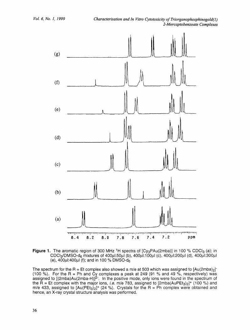
Figure displays major solvent dependence in the chemical shift of H6 in the spectra of[Cy3PAu(2mba)], with only minor solvent dependence observed for H3-H5. In 100 % CDCI3, Figurel(a), H6 appears downfield at 8.32 ppm due to anisotropy of the adjacent carbonyl -bond.However, in 100 % DMSO-d6, Figure l(g), H6 is shifted upfield to 7.36 ppm, a shift of almostppm. This dramatic shift was proven by the gradual titration of DMSO-d6 into a CDCl3 solution of[Cy3PAu(2mba)] as shown in spectra Figure l(b) -l(f). This dramatic shift could be caused by aconformational flip of the -CO2H group induced by introduction of polar DMSO-d6 into the solvationsphere of the [Cy3PAu(2mba)] molecule. Figure 2 shows the proposed structures of[Cy3PAu(2mba)] in 100 % CDCl3 (a) and 100 % DMSO-d6 (b).
In CDCI3 (a), the anisotropic effects of the carbonyl -bond are facilitated by the intramolecularO-H...S hydrogen bonding locking the -CO2H moiety into position. The ppm shift clearly indicatesthat this orientation does not also exist in DMSO-d6, i.e. H6 is no longer in the deshielding region ofthe carbonyl -bond. The polar DMSO-d6 molecules must disrupt the intramoleculr hydrogenbonds with the formation of Q-H...DMSO-d6 intermolecular hydrogen bonds facilitated by bondrotation around the C-C bond joining the phenyl ring to the -CO2H moiety, as shown in Figure 2 (b).This bond rotation may also facilitate a weak Au...O contact as there is no evidence of aconformational flip for 2mba with only a 0.21 ppm solvent dependant shift of H6 observed.However, the possibility of a weak Au...O interaction is difficult to investigate as 197Au (100%abundance) is not a suitable isotope to probe the structure of gold complexes.
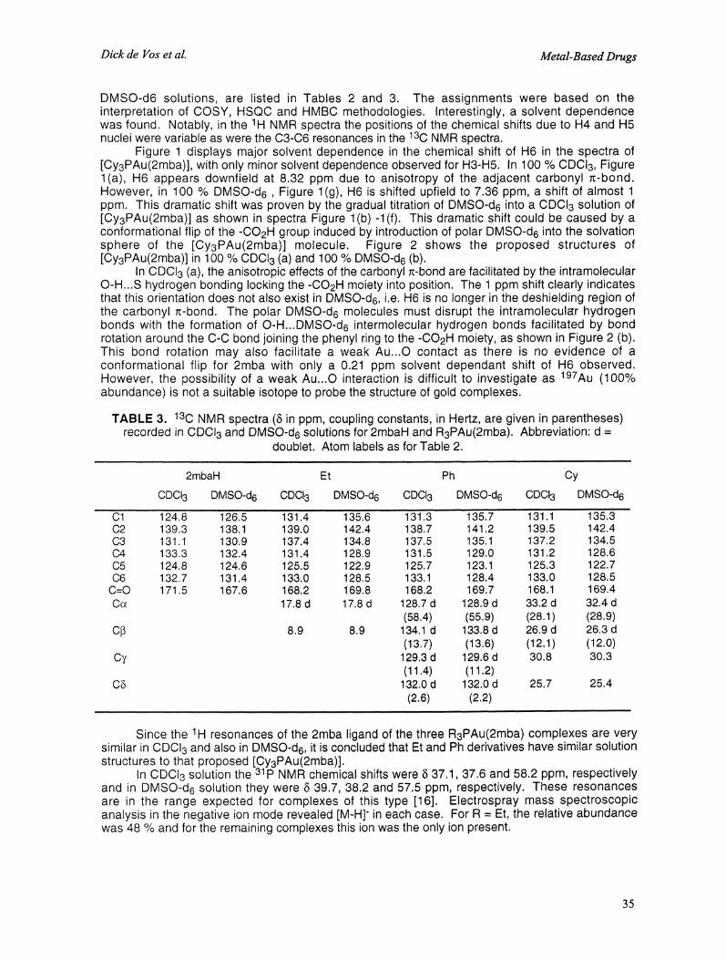
TABLE 3. 130 NMR spectra (5 in ppm, coupling constants, in Hertz, are given in parentheses)recorded in CDCI3 and DMSO-d6 solutions for 2mbaH and R3PAu(2mba). Abbreviation: d
doublet. Atom labels as for Table 2.
2mbaH Et Ph Cy
CDCI3 DMSO-d6 CDCI3 DMSO-d6 CDCI3 DMSO-d6 CDCI3 DMSO-d6
C1 124.8 126.5 131.4 135.6 131.3 135.7 131.1 135.3C2 139.3 138.1 139.0 142.4 138.7 141.2 139.5 142.4C3 131.1 130.9 137.4 134.8 137.5 135.1 137.2 134.5C4 133.3 132.4 131.4 128.9 131.5 129.0 131.2 128.6C5 124.8 124.6 125.5 122.9 125.7 123.1 125.3 122.7C6 132.7 131.4 133.0 128.5 133.1 128.4 133.0 128.5C=O 171.5 167.6 168.2 169.8 168.2 169.7 168.1 169.4Cx 17.8 d 17.8 d 128.7 d 128.9 d 33.2 d 32.4 d
(58.4) (55.9) (28.1) (28.9)C[ 8.9 8.9 134.1 d 133.8 d 26.9 d 26.3 d
(13.7) (13.6) (12.1) (12.0)Cy 129.3 d 129.6 d 30.8 30.3
(11.4) (11.2)C 132.0 d 132.0 d 25.7 25.4
(2.6) (2.2)
Since the 1H resonances of the 2mba ligand of the three R3PAu(2mba) complexes are verysimilar in CDCI3 and also in DMSO-d6, it is concluded that Et and Ph derivatives have similar solutionstructures to that proposed [Cy3PAu(2mba)].
In CDCI3 solution the 31p NMR chemical shifts were 5 37.1, 37.6 and 58.2 ppm, respectivelyand in DMSO-d6 solution they were 5 39.7, 38.2 and 57.5 ppm, respectively. These resonancesare in the range expected for complexes of this type [16]. Electrospray mass spectroscopicanalysis in the negative ion mode revealed [M-H]-in each case. For R Et, the relative abundancewas 48 % and for the remaining complexes this ion was the only ion present.
35
Vol. 6, No. 1, 1999
(g)
(e)
(d)
(c)
(b)
(a)
Characterisation and In Vitro Cytotoxicity ofTriorganophosphinegold(1)2-Mercaptobenzoate Complexes
8.4 8.Z 8.0 7.8 7.5 7.4 7.Z ppm
Figure 1. The aromatic region of 300 MHz H spectra of [Cy3PAu(2mba)] in 100 % CDCI3 (a); inCDCI3/DMSO-d6 mixtures of 4001:501 (b), 400Ltl:100JLtl (C), 400;ul:200pl (d), 4001:3001(e), 4001.L1:400#1 (f); and in 100 % DMSO-d6
The spectrum for the R Et complex also showed a m/e at 503 which was assigned to [Au(2mba)2]-(100 %). For the R Ph and Cy complexes a peak at 249 (91% and 49 %, respectively) wasassigned to [(2mba)Au(2mba-H)]2". In the positive mode, only ions were found in the spectrum ofthe R Et complex with the major ions, i.e. m/e 783, assigned to [2mba(AuPEt3)2]+ (100 %) andm/e 433, assigned to [Au(PEt3)2]+ (24 %). Crystals for the R Ph complex were obtained andhence, an X-ray crystal structure analysis was performed.
36
Dick de Vos et al. Metal-Based Drugs
H6 0
0
PR3(a) CDCI3
H6 0/H,O---S,,,
CD3CD3
u--.PR3
(b) d6-DMSO
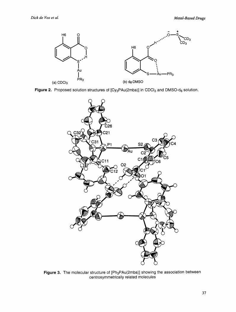
Figure 2. Proposed solution structures of [Cy3PAu(2mba)] in CDCI3 and DMSO-d6 solution.
C26
C21
C3P1 $2 C4
C
Cll C1 C602
s
Figure 3. The molecular structure of [Ph3PAu(2mba)] showing the association betweencentrosymmetrically related molecules
3?
Vol. 6, No. 1, 1999 Characterisation and In Vitro Cytotoxicity ofTriorganophosphinegoM(1)2-Mercaptobenzoate Complexes
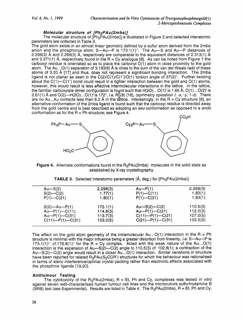
Molecular structure of [Ph3PAu(2mba)]The molecular structure of [Ph3PAu(2mba)].is illustrated in Figure 3 and selected interatomic
parameters are collected in Table 3.The gold atom exists in an almost linear geometry defined by a sulfur atom derived from the 2mbaanion and the phosphorus atom; S--Au--P is 173.1(1). The Au--S and Au--P distances of2.298(3) ,& and 2.269(3) A, respectively are comparable to the equivalent distances of 2.313(1)and 2.271(1) A, respectively found in the R Cy analogue [8]. As can be noted from Figure thecarboxyl residue is orientated so as to place the carbonyl O(1) atom in close proximity to the goldatom. The Au...O(1) separation of 3.193(8) A is close to the sum of the van der Waals radii of theseatoms of 3.20 ,&, [17] and thus, does not represent a significant bonding interaction. The 2mbaligand is not planar as seen in the C(2)/C(1)/C(1’)/O(1) torsion angle of 37(2). Further twistingabout the C(1 )--C(1’) bond could result in a tighter interaction between the gold and O(1) atoms,however, this would result is less effective intermolecular interactions in the lattice. In the lattice,the familiar carboxylate dimer configuration is found such that H(20)...O(1 )i is 1.65/, O(1 )...O(2) is2.61(1) A and O(2)--H(20)...O(1) is 172, i.e. R(8) [18], (symmetry operation i:-x,-y, l-z). Thereare no Au...Au contacts less than 6.0 A in the I,ttice. Interestingly, in the R Cy structure [8], analternative conformation of the 2mba ligand is found such that the carboxyl residue is directed awayfrom the gold centre and is best described as adopting an exo conformation as opposed to a endoconformation as for the R Ph structure; see Figure 4.
Ph3P---Au
HO2C
Cy3P’- Au SCO2H
Figure 4. Alternate conformations found in the R3PAu(2mba) molecules in the solid state asestablished by X-ray crystallography
TABLE 3. Selected interatomic parameters (/, deg.) for [Ph3PAu(2mba)]
Au--S(2) 2.298(3) Au---P(1) 2.269(3)S(2)--C(2) 1.77(1) P(1)---C(11) 1.83(1)P(1)--C(21) 1.80(1) P(1)--C(31) 1.83(1)
S(2)--Au--P(1).Au--P(1 )--C(11Au--P(1 )---C(31)C(11)--P(1)--C(31)
173.1 (1) Au---S(2)--C(2) 110.5(3)114.8(3) Au---P(1 )--C(21) 112.0(3)113.7(3) C(11)--P(1 )---C(21) 107.0(5)103.0(5) C(21 )--P(1)--C(31) 105.5(5)
The effect on the gold atom geometry of the intramolecular Au...O(1) interaction in the R Phstructure is minimal with the major influence being a greater distortion from linearity, i.e. S--Au--P is173.1(1) cf.176.8(1) for the R Cy complex. Allied with the weak nature of the Au...O(1)interaction is the expansion of Au---S(2)--C(2) angle to 110.5(3) cf. 102.8(1); a contraction of theAu--S(2)--C(2) angle would result in a closer Au...O(1) interaction. Similar variations in structurehave been reported for related R3PAu(S2COR’) structures for which the behaviour was rationalisedin terms of steric interference/optimal crystal packing rather than electronic effects associated withthe phosphine ligands [19,20].
Antitumour TestingThe cytotoxicity of the R3PAu(2mba), R Et, Ph and Cy, complexes was tested in vitro
against seven well-characterised human tumour cell lines and the microculture sulforhodamine B(SRB) test (see Experimental). Results are listed in Table 4. The R3PAu(2mba), R Et, Ph and Cy,
38
Dick de Vos et al. Metal-Based Drugs
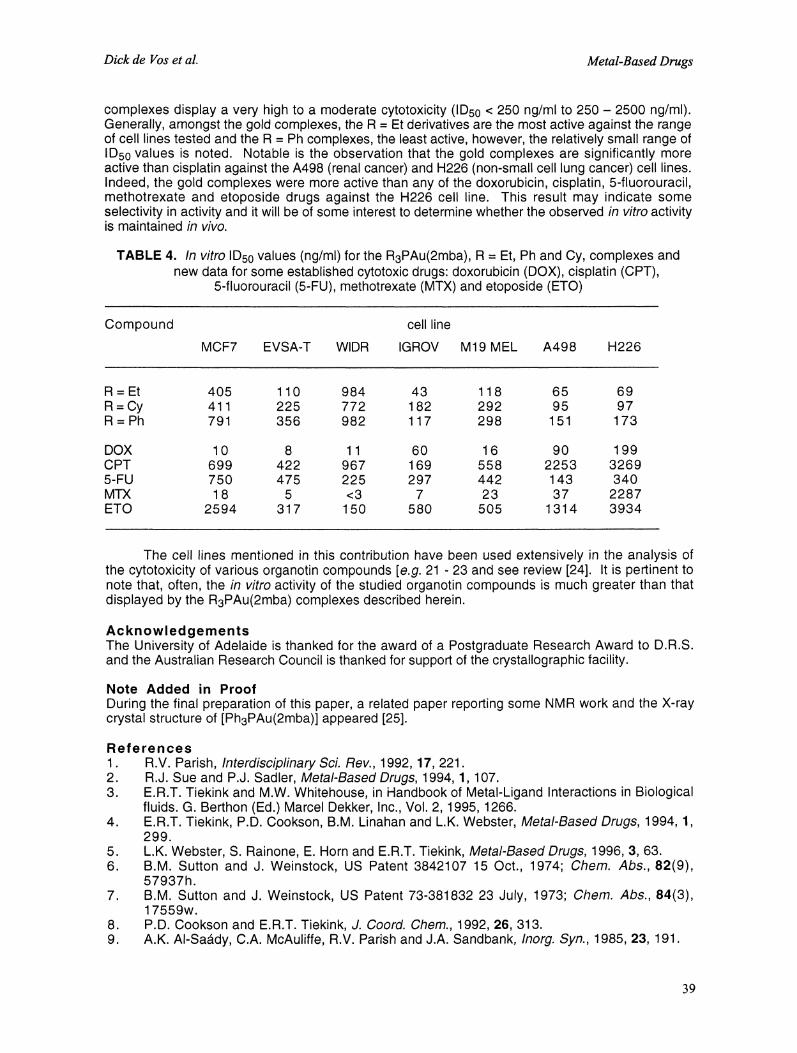
complexes display a very high to a moderate cytotoxicity (ID50 < 250 ng/ml to 250- 2500 ng/ml).Generally, amongst the gold complexes, the R Et derivatives are the most active against the rangeof cell lines tested and the R Ph complexes, the least active, however, the relatively small range ofID5o values is noted. Notable is the observation that the gold complexes are significantly moreactive than cisplatin against the A498 (renal cancer) and H226 (non-small cell lung cancer) cell lines.Indeed, the gold complexes were more active than any of the doxorubicin, cisplatin, 5-fluorouracil,methotrexate and etoposide drugs against the H226 cell line. This result may indicate someselectivity in activity and it will be of some interest to determine whether the observed in vitro activityis maintained in vivo.
TABI..15 4. In vitro ID50 values (ng/ml) for the R3PAu(2mba), R Et, Ph and Cy, complexes andnew data for some established cytotoxic drugs: doxorubicin (DOX), cisplatin (CPT),
5-fluorouracil (5-FU), methotrexate (MTX) and etoposide (ETO)
Compound cell line
MCF7 EVSA-T WIDR IGROV M19 MEL A498 H226
R Et 405 110 984 43 118 65 69R=Cy 411 225 772 182 292 95 97R Ph 791 356 982 117 298 151 173
DOX 0 8 60 6 90 99CPT 699 422 967 69 558 2253 32695-FU 750 475 225 297 442 43 340MTX 8 5 <3 7 23 37 2287ETO 2594 31 7 50 580 505 31 4 3934
The cell lines mentioned in this contribution have been used extensively in the analysis ofthe cytotoxicity of various organotin compounds [e.g. 21 23 and see review [24]. It is pertinent tonote that, often, the in vitro activity of the studied organotin compounds is much greater than thatdisplayed by the R3PAu(2mba) complexes described herein.
AcknowledgementsThe University of Adelaide is thanked for the award of a Postgraduate Research Award to D.R.S.and the Australian Research Council is thanked for support of the crystallographic facility.
Note Added in ProofDuring the final preparation of this paper, a related paper reporting some NMR work and the X-raycrystal structure of [Ph3PAu(2mba)] appeared [25].
References1. R.V. Parish, Interdisciplinary Sci. Rev., 1992, 17, 221.2. R.J. Sue and P.J. Sadler, Metal-Based Drugs, 1994, 1,107.3. E.R.T. Tiekink and M.W. Whitehouse, in Handbook of MetaI-Ligand Interactions in Biological
fluids. G. Berthon (Ed.) Marcel Dekker, Inc., Vol. 2, 1995, 1266.4. E.R.T. Tiekink, P.D. Cookson, B.M. Linahan and L.K. Webster, Metal-Based Drugs, 1994, 1,
299.5. L.K. Webster, S. Rainone, E. Horn and E.R.T. Tiekink, Metal-Based Drugs, 1996, 3, 63.6. B.M. Sutton and J. Weinstock, US Patent 3842107 15 Oct., 1974; Chem. Abs., 82(9),
57937h.7. B.M. Sutton and J. Weinstock, US Patent 73-381832 23 July, 1973; Chem. Abs., 84(3),
17559w.8. P.D. Cookson and E.R.T. Tiekink, J. Coord. Chem., 1992, 26, 313.9. A.K. AI-Sa,dy, C.A. McAuliffe, R.V. Parish and J.A. Sandbank, Inorg. Syn., 1985, 23, 191.
39
Vol. 6, No. 1, 1999 Characterisation and In Vitro Cytotoxicity ofTriorganophosphinegold(1)2-Mercaptobenzoate Complexes
10.
13.
14.
15.16.17.18.19.20.21.
teXsan, Single Crystal structure analysis software, Version 1.6 (1993), Molecular StructureCorporation, The Woodlands, TX, USA.N. Walker and D. Stuart, Acta Crystallogr., 1983, A39,158.P.T. Beurskens, G. Admiraal, G. Beurskens, W.P. Bosman, S. Garcfa-Granda, J.M.M. Smitsand C. Smykalla, The DIRDIF program system. Technical report of the crystallographylaboratory, University of Nijmegen, The Netherlands, 1992.C.K. Johnson, ORTEP. Report ORNL-5138 (1976), Oak Ridge National Laboratory, TN,USA.Y.P. Kepers, G.J. Peters, J. van Ark-Otte, B. Winograd and H.M. Pinedo, Eur. J. Cancer,1991, 27, 897.M.R. Boyd, Principles and Practice of Oncology, 1989, 3, 1.P.D. Cookson, E.R.T. Tiekink and M.W. Whitehouse, Aust. J. Chem., 1994, 47, 577.A. Bondi, J. Phys. Chem., 1964, 68, 441.M.C. Etter, J.C. MacDonald and J. Bernstein, Acta Crystallogr., 1990, B46, 256.G. Siasios and E.R.T. Tiekink, Z. Kristallogr., 1993, 204, 95.G. Siasios and E.R.T. Tiekink, Z. Kristallogr., 1993, 205, 261.D. de Vos, R. Willem, M. Gielen, K.E. van Wingerden and K. Nooter, Metal-Based Drugs,1998, 5, 179.M. Kemmer, M. Gielen, M. Biesemans, Do de Vos and R. Willem, Metal-Based Drugs, 1998, 5,189.E.R.T. Tiekink, M. Gielen, A. Bouhdid, R. Willem, V.I. Bregadze, L.V. Ermanson and S.A.Glazun, Metal-Based Drugs, 1997, 4, 75.M. Gielen, Coord. Chem. Rev., 1996, 1.51, 41.K. Nomiya, N.C. Kasuga, I. Takamori and K. Tsuda, Polyhedron, 1998, 17, 3519.
Received: January 21, 1999 Accepted: January 27, 1999Received in revised camera-ready format: January 29, 1999
4O