
chapter 5 - cell structure and function
26
1 CHAPTER 5 CELL STRUCTURE AND FUNCTION Why are cells small? (surface to volume ratio) LUCA is not the name of a famous scientist in the field; it is shorthand for Last Universal Common Ancestor, a single cell that lived perhaps 3 or 4 billion years ago, and from which all life has since evolved. The genetic code is universal for all life. That the genetic code is universal to all life tells us that everything is related. All life regenerates itself by producing offspring, and over time small changes in the offspring result in small changes to the protein recipes. But because the recipes are written in the same language (the genetic code), it is possible to compare these recipes (and other genes) to build the equivalent of a family tree
-
Upload
khangminh22 -
Category
Documents
-
view
6 -
download
0
Transcript of chapter 5 - cell structure and function
1
CHAPTER 5
CELL STRUCTURE AND FUNCTION
Why are cells small? (surface to volume ratio)
LUCA is not the name of a famous scientist in the
field; it is shorthand for Last Universal Common
Ancestor, a single cell that lived perhaps 3 or 4
billion years ago, and from which all life has since
evolved.
The
genetic
code is
universal for all
life.
That the genetic code is universal
to all life tells us that everything is related. All life regenerates itself
by producing offspring, and over
time small changes in the offspring
result in small changes to the
protein recipes. But because the recipes are written in the same
language (the genetic code), it is
possible to compare these recipes
(and other genes) to build the
equivalent of a family tree

2
I THE THREE DOMAIN SYSTEM OF CLASSIFICATION
Was first proposed by Carl Woese . He based this on
differences in the sequences of cell’s ribosomal RNA
( rRNA) as well as the membrane lipid structure and
sensitivity to antibiotics.
The system proposes that a common ancestor cell gave rise to
three different cell types each represented by a different
domain.
i. Archaea: archaebacteria ( Extremophiles)
Characteristics
a. Prokaryotic cells
b. Membranes composed of branched hydrocarbon
chains
c. Many times antibiotic resistant
d. Cell walls made of polysaccharides **
e. Live in extreme environments
i. Halophiles , hyperthermophiles
ii. Volcanic vents, midatlantic rift, Yellowstone
3
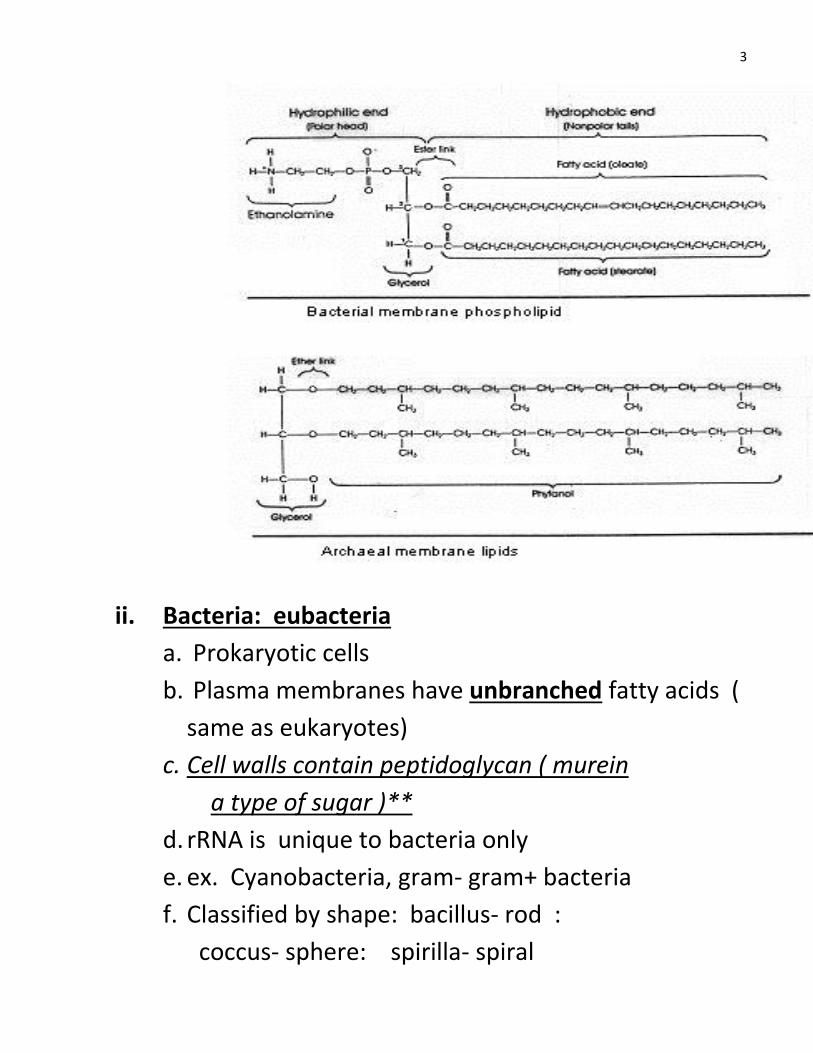
ii. Bacteria: eubacteria
a. Prokaryotic cells
b. Plasma membranes have unbranched fatty acids (
same as eukaryotes)
c. Cell walls contain peptidoglycan ( murein
a type of sugar )**
d. rRNA is unique to bacteria only
e. ex. Cyanobacteria, gram- gram+ bacteria
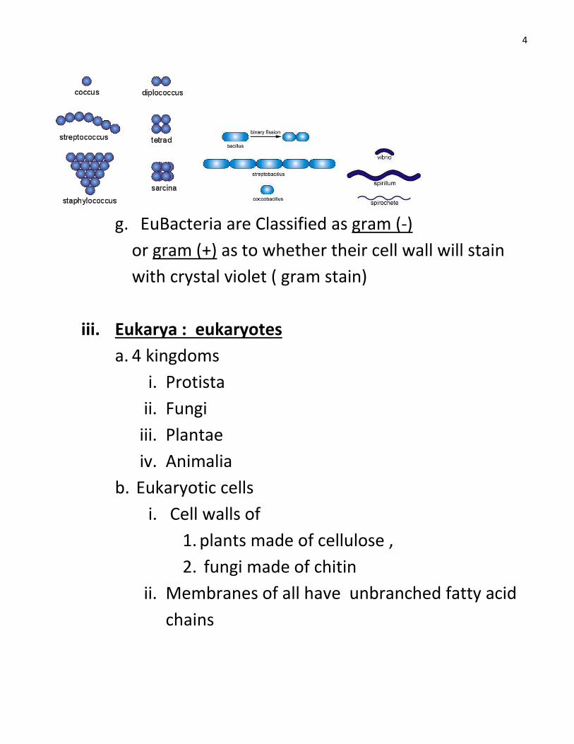
f. Classified by shape: bacillus- rod :
coccus- sphere: spirilla- spiral
4
g. EuBacteria are Classified as gram (-)
or gram (+) as to whether their cell wall will stain
with crystal violet ( gram stain)
iii. Eukarya : eukaryotes
a. 4 kingdoms
i. Protista
ii. Fungi
iii. Plantae
iv. Animalia
b. Eukaryotic cells
i. Cell walls of
1. plants made of cellulose ,
2. fungi made of chitin
ii. Membranes of all have unbranched fatty acid
chains
6
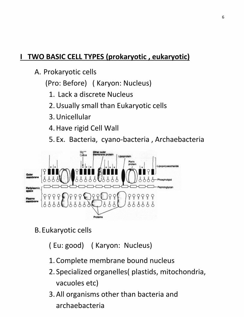
I TWO BASIC CELL TYPES (prokaryotic , eukaryotic)
A. Prokaryotic cells
(Pro: Before) ( Karyon: Nucleus)
1. Lack a discrete Nucleus
2. Usually small than Eukaryotic cells
3. Unicellular
4. Have rigid Cell Wall
5. Ex. Bacteria, cyano-bacteria , Archaebacteria
B. Eukaryotic cells
( Eu: good) ( Karyon: Nucleus)
1. Complete membrane bound nucleus
2. Specialized organelles( plastids, mitochondria,
vacuoles etc)
3. All organisms other than bacteria and
archaebacteria
7
4. Unicellular- Protists
5. Multicellular- plants, fungi, animals
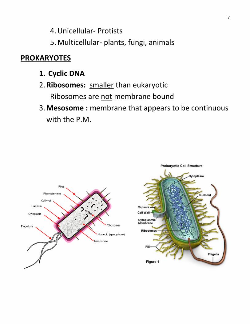
PROKARYOTES
1. Cyclic DNA
2. Ribosomes: smaller than eukaryotic
Ribosomes are not membrane bound
3. Mesosome : membrane that appears to be continuous
with the P.M.
8
4. Thick cell wall:
a. Cell wall of Archaea made of polysaccharides
b. Cell wall of eubacteria made of Peptidoglycan
sugar
( also known as murein an amino-sugar comp.)
Some cell wall substances are toxic + cause diseases.
Antibiotics : such as Penicillin interfere w/ the
construction of the cell wall “amino-sugar” formation
therefore inhibiting bacterial growth.
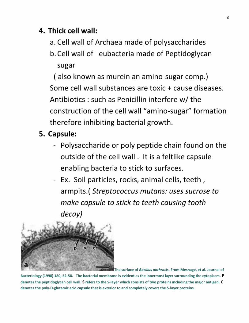
5. Capsule:
- Polysaccharide or poly peptide chain found on the
outside of the cell wall . It is a feltlike capsule
enabling bacteria to stick to surfaces.
- Ex. Soil particles, rocks, animal cells, teeth ,
armpits.( Streptococcus mutans: uses sucrose to
make capsule to stick to teeth causing tooth
decay)
The surface of Bacillus anthracis. From Mesnage, et al. Journal of
Bacteriology (1998) 180, 52-58. The bacterial membrane is evident as the innermost layer surrounding the cytoplasm. P
denotes the peptidoglycan cell wall. S refers to the S-layer which consists of two proteins including the major antigen. C
denotes the poly-D-glutamic acid capsule that is exterior to and completely covers the S-layer proteins.
9
6. Pili: Hundreds of hollow strands of proteins used for
attachment ( Pg78 read + know caption)
7. Flagella: movement of bacteria using long thin spiral
flagellum
8. Gram - appear red or pink
9. Gram + appear blue or purple
EUKARYOTIC CELLS OF HIGHER ORGANISMS
1. Much larger than prokaryotic cells.
2. Many membrane Bound Organelles
a. ( table 5-2) know all of these .
b. Plastids: found only in plants
c. Cell wall: in plants ( Cellulose)
In fungi ( Chitin)
d. Mitochondria
e. Lysosomes
f. Vacuoles ( Contractile Vacuoles : regulate
water in plants)
3. CYTOSOL: The soluble portion of the cytoplasm.
Contains:
a. Ions, molecules , molecular aggregates
b. ½ the cells volume is composed of cytosol
Cytosol= 20% protein
10
COMPONENTS OF EUKARYOTIC CELLS
A. NUCLEUS:
a. Very complex in plants and animals
b. Genetic material (DNA) arranged into
Chromosomes
c. Chromosomes: Long DNA molecule + RNA proteins
d. Chromosomes coil up into short threadlike
structures before and during cell division
e. Most of time nucleus is not not dividing. During this
time chromosome s are uncoiled in loose indistinct
tangle called CHROMATIN.
Chromatin determines what RNA is mad in the
nucleus
f. RNA made in the nucleus travels to the Cytoplasm
to ribosomes where it directs protein synthesis
Note: there are substances in the cytop. That
enter nucleus and influence DNA
g. NUCLEOLI: ( sing. Nucleolus)
Area in the nucleus where ribonsomes are
made.( Disappears during cell division)
h. NUCLEAR ENVELOPE: ( nuclear membrane)
Double membrane surrounding the nucleus
Perforated w/ pores ( disappears during cell
division) ( double membrane)
11
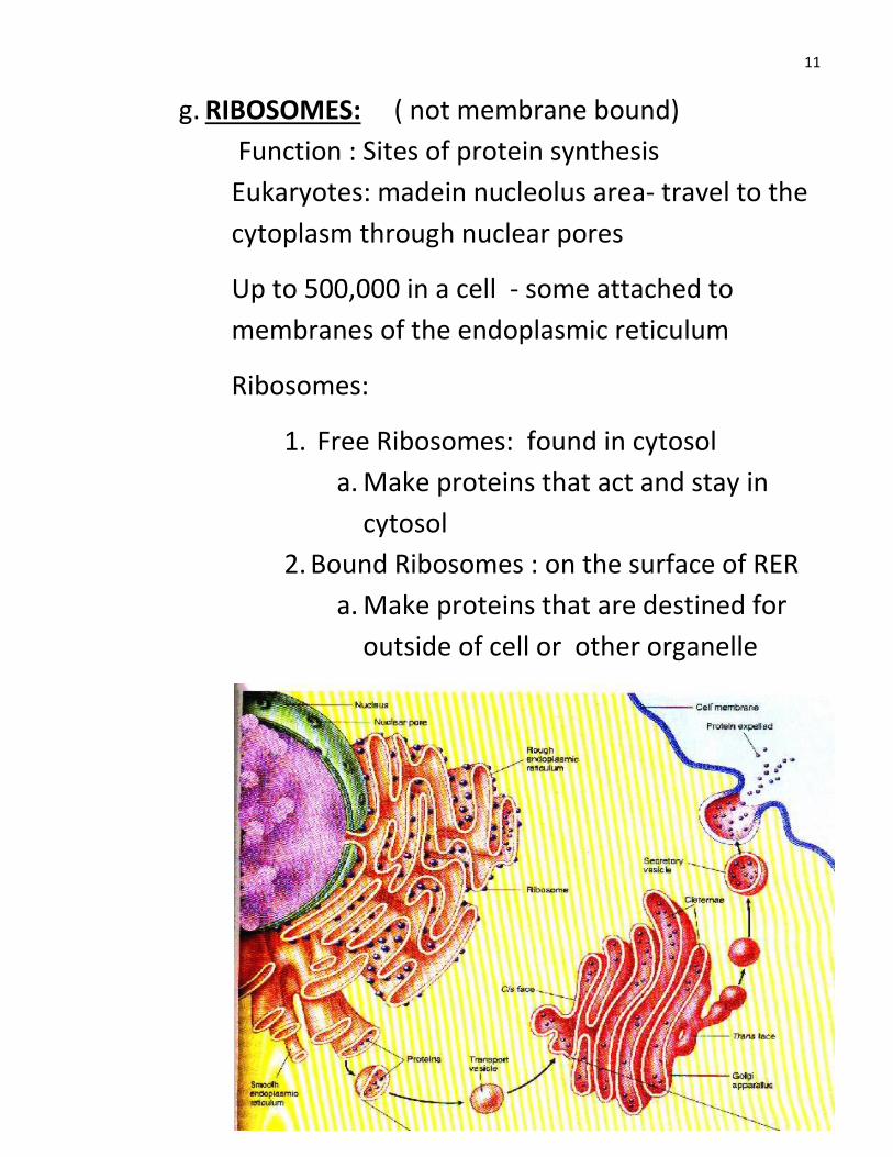
g. RIBOSOMES: ( not membrane bound)
Function : Sites of protein synthesis
Eukaryotes: madein nucleolus area- travel to the
cytoplasm through nuclear pores
Up to 500,000 in a cell - some attached to
membranes of the endoplasmic reticulum
Ribosomes:
1. Free Ribosomes: found in cytosol
a. Make proteins that act and stay in
cytosol
2. Bound Ribosomes : on the surface of RER
a. Make proteins that are destined for
outside of cell or other organelle
12
h. ENDOPLASMIC RETICULUM
( know Fig. 5-8 pg 85)
1. Is a system of membranous tunnels and sacs
found in eukaryotes .
2. Usually lies just outside the nucleus
3. Appears as piles of sacs
4. Lumen: of er provides the cell w/ a
compartment to contain substances that must
be kept separate from the cytosol.
1. Rough Endoplasmic :
Ribosomes Attached to the outer surface give it a
bumpy appearance.
RER: Are predominant in cells that make proteins
that are secreted from the cell.
(Ex. Pancreas: makes insulin)
PROTEIN
Transport ERLumen Sacs called Vesicles Golgi Exocytosis
Out of cell
NOTE: Most of the cells new membrane is produced in the ER.
2. Smooth Endoplasmic
Not much found in most cells
A lot found in cells involved in lipid metabolism and
production of cholesterol and steroids.
13
i. GOLGI COMPLEX : Pg 86
A stack of flattened Membranous sacs.
Around the edges of the stack, swarms of small,
round transport vesicles carry molecules to or
from.
Golgi lies near nucleus
Cells may have one large or hundreds of small
ones
Overall role: Modify, sort and package the cell
From the golgi …..
Molecules exocytosis
New proteins + lipids for P. Mem.
Production of new proteins
Ribo RER Golgi complex Final location
Proteins modified by enzymes
Further modified for transport w/ a transport marker
14
j. Lysosomes: Pg 87
Membrane bound sac
Contains hydrolytic enzymes that were made in
ER.
Function: Digest: food , Disease causing viruses,
damaged organelles, entire cells,
Junk like old clothes and toys
( occurs when another vesicle fuses w/ lysosome)
K. Peroxisomes:
Small sacs containing enzymes that break down
amino acids, fatty acids, and hydrogen peroxide
H2O2 ---- H2O + O2
( many found in liver and kidney cells)
G. MITOCHONDRIA: (Power House) Pg 87
1. Produce almost all of the ATP for cell
2. Make adenosine tri-phosphate from cell
Respiration ( series of rxns that use O2 to
break down glucose into CO2 + H2O + ATP
3. High energy cells have many mito.
Ex. Heart cells, growing root tips, liver
4. ( know structure) has 2 membranes
Outer, and inner ( highly folded)
5. ** Contains own
a. **DNA b. RNA c. Ribosomes
15
b. **Makes some of its own proteins +
membranes
c. **Also reproduces itself.
ENDOSYMBIONT THEORY: Many scientists believe that
Mitoch. Evolved from prokaryotic cells that came to live
inside of a larger cell. Thus becoming an organelle.
Plastids also are believed to have arisen this way from
cyanobacteria that invaded plant cells.
H. PLASTIDS:
** 1. Found only in plants
2. 3 unique structures in plants.
a. plastids
b. cellulose cell wall
c. large vacuole
3. plastids contain:
DNA, RNA, Ribosomes,
4.Can reproduce themselves
5. Endosymbiont theory
6. three types:
a. Chloroplasts: Green
Photosynthesis Contain green pigment
Chlorophyll
16
b.Chromoplasts: Make + store yellow +
orange pigments: (Xanthophylls, Carotenoids
) ( flowers, fruits, roots)
c. Amyloplasts
Store starch
Lots found in tubers, roots,
I. CELL WALL :
Found outside of the plasma
membrane
Cellulose in plants, Chitin in Fungi
Porous enough to allow H20 through
Structural support
J. VACUOLES:
- Sac of fluid surrounded by a
Membrane
- occur in many cells but mostly plant
cells and some protists
- Function: to hold stored food , water
and pigments.
- Also store some toxic materials
17
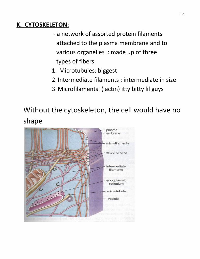
K. CYTOSKELETON:
- a network of assorted protein filaments
attached to the plasma membrane and to
various organelles : made up of three
types of fibers.
1. Microtubules: biggest
2. Intermediate filaments : intermediate in size
3. Microfilaments: ( actin) itty bitty lil guys
Without the cytoskeleton, the cell would have no
shape
18
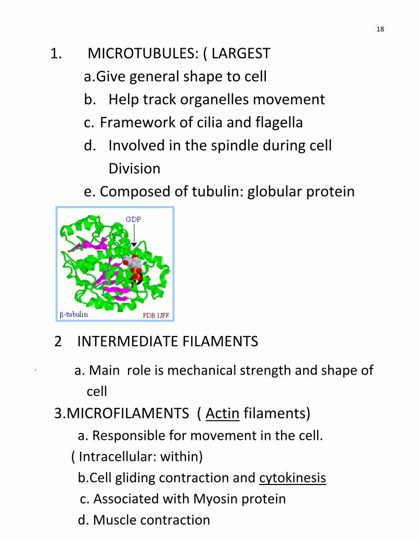
1. MICROTUBULES: ( LARGEST
a. Give general shape to cell
b. Help track organelles movement
c. Framework of cilia and flagella
d. Involved in the spindle during cell
Division
e. Composed of tubulin: globular protein
.
2 INTERMEDIATE FILAMENTS
a. Main role is mechanical strength and shape of
cell
3.MICROFILAMENTS ( Actin filaments)
a. Responsible for movement in the cell.
( Intracellular: within)
b.Cell gliding contraction and cytokinesis
c. Associated with Myosin protein
d. Muscle contraction
19
MICROTUBULES FUNCTIONS
1. Movement of organelles w/in the cell
2. Skeletal framework of cell
3. Largest fiber
4. Role in cell division ( spindle fiber)
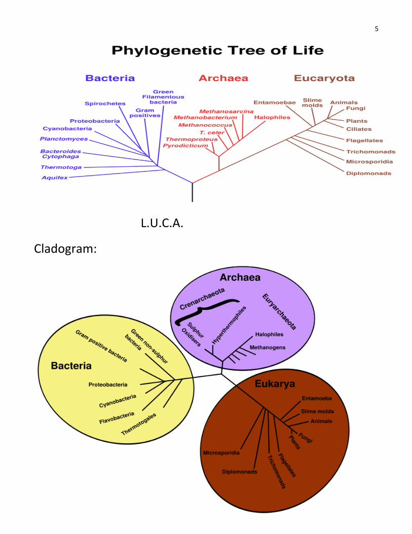
5. Cilia and Flagella: Threadlike organelles present
on the surface of many eukaryotes
6. Cilia short and more numerous than flagella
7. Both function in locomotion
Ex sperm, paramecium covered w/ cilia
Cilia move mucous and debris out of air
passages in humans.
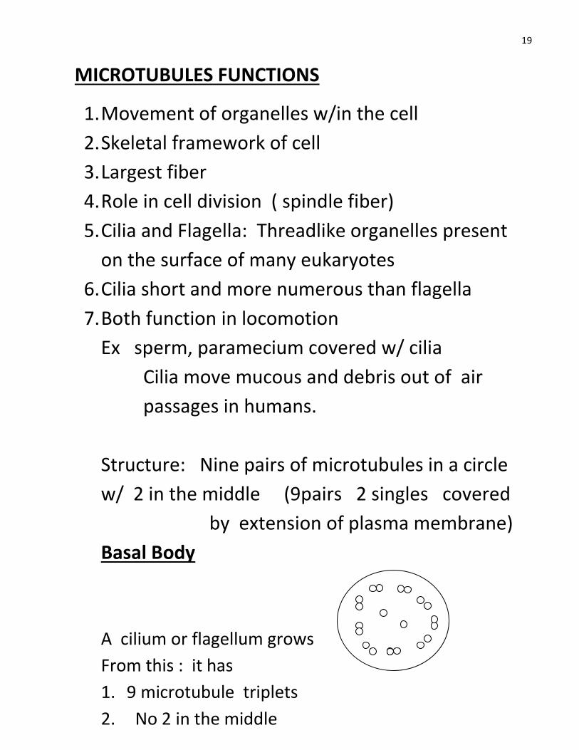
Structure: Nine pairs of microtubules in a circle
w/ 2 in the middle (9pairs 2 singles covered
by extension of plasma membrane)
Basal Body
A cilium or flagellum grows
From this : it has
1. 9 microtubule triplets
2. No 2 in the middle
21
Movement of cilia and flagellum
1. Moves by action of arms of the protein
DYNEIN that extend from one microtubule
of each pair.
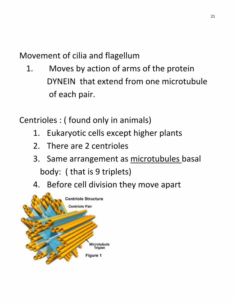
Centrioles : ( found only in animals)
1. Eukaryotic cells except higher plants
2. There are 2 centrioles
3. Same arrangement as microtubules basal
body: ( that is 9 triplets)
4. Before cell division they move apart
22
INTERMEDIATE FILAMENTS:
1. These are protein fibers intermediate in
thickness. Between MT and MF
2. Ropelike polypeptides
3. Function : give cell mechanical strength
MICROFILAMENTS:
1. Thinnest filaments
2. Also called Actin filaments because made of
the protein actin ( found as contractile protein
in muscle cells)
3. Most abundant protein in the cytoplasm
4. FUNCTION
a. Movement of organelles
b. Structure + strength
c. Muscle contraction
d. Cytokinesis
e. Endocytosis/ exocytosis
5. Myosin: Protein associated w/ microF. : it
interacts w/ actin in muscle cell contraction
23
TISSUES AND ORGANS:
Tissue:
A group of cells of one or a few types that
perform a specific job. Ex. Connective tissue,
bone tissue, cardiac tissue, epithelial tissue
Organ : Made up of a group of tissues
Functional unit of the body
Ex. Eyes, kidneys, heart, lungs, brain
System: ?
A group of organs working together to
perform a group of similar functions.
Ex. Nervous system, digestive system,
immune system, circulatory system
24
MAIN ANIMAL TISSUES:
1. Epithelial tissue:
a. Form coverings and linings
b. Line lungs, digestive tract , mouth ,
Outside of body,
2. Connective tissue: ( mostly protein collagen)
Most abundant tissue :
Adipose(fat) , cartilage, bone, fibrous cells
3. Nervous tissue:
a. Consists of nerve cells that conduct
electric currents ( transmit messages)
b. Not contractile
4. Muscle tissue
a. Cells that can both conduct an electric
impulse and contract (* Unique trait)
PLANT TISSUES: FOUR MAIN TYPES
1. Epidermis :
Covers the outside of leaves and stems
2. Vascular tissues:
25
Transports water , food, hormones through
the plant (Xylem ) H20 + minerals, nutrients
(Phloem ) Transports glucose from leaves
elsewhere in the plant
3. Ground tissues:
Fills in spaces between epidermis and
vascular tissues (Parenchyma cells loosely
packed cells)
4. Meristems :
Cells that are ready to divide and
develop Ex. Buds, root tips
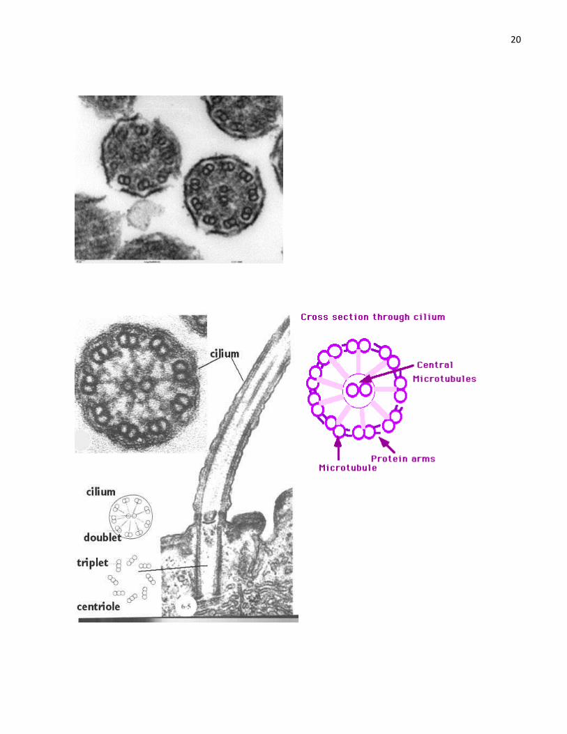
CLADOGRAMS:
We can diagram a tree-like relationship called a
cladogram. The cladogram graphically represents a
hypothetical evolutionary process. Cladograms are
subject to change as new data becomes available.
The terms evolutionary tree, and sometimes
phylogenetic tree are often used synonymously with
cladogram,