Chapter 1 – Radioactivity and Radionuclides - Amazon AWS
13
1 PART 1 Basic Principles Radioactivity and Radionuclides 1 In nuclear medicine, radiopharmaceuticals given to the patient emit the radiation used to create images or perform therapy. In order to understand how these agents perform and what safety considerations are involved in their use, it is necessary to be famil- iar with some basic aspects of the physics behind radioactive decay. is chapter discusses radioactive molecules, different types of radioactive decay, and how these emissions interact with matter. ATOMIC STRUCTURE OF MATTER Electronic Structure of the Nucleus All matter is made up of atoms, which in turn are made up of protons, electrons, and neutrons. Positively charged protons and uncharged neutrons have a similar mass and are known as nucleons because they are located in the nucleus. Although much less massive, electrons orbiting the nucleus possess an opposite negative charge equal in magnitude to that of the pro- tons (Table 1.1). Some properties of atomic particles are listed, along with important constant values, in Table 1.2. e attraction of the opposite charges keeping the electron in orbit around the nucleus is known as an electrostatic force (or coulombic force; the coulomb is the unit for electric charge). On the other hand, there is also a repulsive, electrostatic force in the nucleus from the like-charged protons pushing apart. e nucleus is held together by the attractive strong nuclear force each nucleon exerts on the other nucleons. Although more powerful than electrical forces, these strong forces act only over extremely short distances. e actual atomic mass is less than the sum of the masses of all its nucleons. is difference in mass, or mass deficit, is manifest in the nuclear binding energy hold- ing the nucleus together (as related by the equation E = mc 2 ). Elements are organized in the periodic table of the elements (see Appendix 2). All atoms of the same element have the same number of protons. e proton number is also referred to as the atomic number or Z. us, all carbon atoms have 6 protons, all oxy- gen atoms have 8 protons, and all iodine atoms have 53 protons— that is, they have Z numbers of 6, 8, and 53, respectively. Atoms of a particular element can, however, have a varying number of neu- trons (referred to as the neutron number, N). For example, in addi- tion to their 8 protons, some oxygen atoms have 8 neutrons, and others have 7 or 10 neutrons. e total number of nucleons (Z plus N) is known as the atomic mass or atomic number, A. erefore, in the oxygen example, A would be 15, 16, and 18 for atoms that have 8 protons plus 7, 8, or 10 neutrons, respectively. Unlike an element, which is characterized only by its number of protons (Z), a nuclide is a nuclear entity characterized by a certain nuclear composition of protons and neutrons as well as a certain energy level. Shorthand notation has been agreed on to describe the makeup of specific nuclides: Atomic mass N Z A X Element Neutron number Proton number To illustrate this, consider the element iodine, which has 53 protons (Z = 53). If one particular nuclide of the element iodine has 78 neutrons (N = 78), the atomic mass (A) of 53 + 78 equals 131. It can be written as: N 131 78 53 I A Z Because the atomic number can be inferred by the element’s symbol, and N = A – Z, this can be shortened: 131 53 78 78 131 131 I I I is can also be written as I-131 or iodine-131. e term iso- tope describes nuclides of the same element, that is, nuclides TABLE 1.1 Properties of Atomic Particles Particle Charge Mass (amu or u) a Mass (MeV) b Mass (kg) Proton + 1 1.0073 938.21 1.673 × 10 –27 Neutron 0 1.0087 939.51 1.675 × 10 –27 Electron –1 0.000549 0.511 9.11 × 10 –31 a One amu = 1.661 × 10 –27 kg or 1/12 atomic mass carbon (1 nucleon from carbon-12 atom). b Energy as related by E = mc 2 .
-
Upload
khangminh22 -
Category
Documents
-
view
1 -
download
0
Transcript of Chapter 1 – Radioactivity and Radionuclides - Amazon AWS

1
PART 1 Basic Principles
Radioactivity and Radionuclides
1
In nuclear medicine, radiopharmaceuticals given to the patient emit the radiation used to create images or perform therapy. In order to understand how these agents perform and what safety considerations are involved in their use, it is necessary to be famil-iar with some basic aspects of the physics behind radioactive decay. This chapter discusses radioactive molecules, different types of radioactive decay, and how these emissions interact with matter.
ATOMIC STRUCTURE OF MATTERElectronic Structure of the NucleusAll matter is made up of atoms, which in turn are made up of protons, electrons, and neutrons. Positively charged protons and uncharged neutrons have a similar mass and are known as nucleons because they are located in the nucleus. Although much less massive, electrons orbiting the nucleus possess an opposite negative charge equal in magnitude to that of the pro-tons (Table 1.1). Some properties of atomic particles are listed, along with important constant values, in Table 1.2.
The attraction of the opposite charges keeping the electron in orbit around the nucleus is known as an electrostatic force (or coulombic force; the coulomb is the unit for electric charge). On the other hand, there is also a repulsive, electrostatic force in the nucleus from the like-charged protons pushing apart. The nucleus is held together by the attractive strong nuclear force each nucleon exerts on the other nucleons. Although more powerful than electrical forces, these strong forces act only over extremely short distances. The actual atomic mass is less than the sum of the masses of all its nucleons. This difference in mass, or mass deficit, is manifest in the nuclear binding energy hold-ing the nucleus together (as related by the equation E = mc2).
Elements are organized in the periodic table of the elements (see Appendix 2). All atoms of the same element have the same number of protons. The proton number is also referred to as the atomic number or Z. Thus, all carbon atoms have 6 protons, all oxy-gen atoms have 8 protons, and all iodine atoms have 53 protons—that is, they have Z numbers of 6, 8, and 53, respectively. Atoms of a particular element can, however, have a varying number of neu-trons (referred to as the neutron number, N). For example, in addi-tion to their 8 protons, some oxygen atoms have 8 neutrons, and others have 7 or 10 neutrons. The total number of nucleons (Z plus N) is known as the atomic mass or atomic number, A. Therefore, in the oxygen example, A would be 15, 16, and 18 for atoms that have 8 protons plus 7, 8, or 10 neutrons, respectively.
Unlike an element, which is characterized only by its number of protons (Z), a nuclide is a nuclear entity characterized by a certain nuclear composition of protons and neutrons as well as a certain energy level. Shorthand notation has been agreed on to describe the makeup of specific nuclides:
Atomic mass
NZAX
Element
Neutron numberProton number
To illustrate this, consider the element iodine, which has 53 protons (Z = 53). If one particular nuclide of the element iodine has 78 neutrons (N = 78), the atomic mass (A) of 53 + 78 equals 131. It can be written as:
N
1317853 I
A
Z
Because the atomic number can be inferred by the element’s symbol, and N = A – Z, this can be shortened:
13153 78 78
131 131I I I
This can also be written as I-131 or iodine-131. The term iso-tope describes nuclides of the same element, that is, nuclides
TABLE 1.1 Properties of Atomic Particles
Particle ChargeMass (amu or u)a Mass (MeV)b Mass (kg)
Proton + 1 1.0073 938.21 1.673 × 10⁻²⁷
Neutron 0 1.0087 939.51 1.675 × 10⁻²⁷
Electron –1 0.000549 0.511 9.11 × 10⁻³¹
aOne amu = 1.661 × 10⁻²⁷ kg or 1/12 atomic mass carbon (1 nucleon from carbon-12 atom).bEnergy as related by E = mc2.

2 PART 1 Basic Principles
with the same number of protons (Z) but potentially differing atomic numbers. Radioisotopes are isotopes that undergo radio-active decay. For example, some common isotopes of iodine are as follows:
I I I I78, 72, 71, 70131 125 123124
53 53 53 53
In medicine, different isotopes have varying properties, such as the types of radiation they emit and how long they remain radioactive, which can determine their usefulness. For example, the beta and high-energy gamma emitter 131I (I-131) is used for treating thyroid cancer and performing thyroid uptake mea-surements; 125I (I-125), a low-energy gamma and x-ray emitter, is used in biological assays and prostate cancer brachytherapy; 124I (I-124), a positron emitter, can image thyroid cancer with a positron emission tomography (PET) scanner; and 123I, a mod-erate-energy gamma emitter, is very commonly used to image benign thyroid diseases and thyroid cancers as well as to calcu-late thyroid activity (radioactive iodine uptake).
In addition to isotopes, other special terms include isotones, nuclides with the same number of neutrons (e.g., 8
14 O, 713 N, C12
6 where N = 6); and isobars, those with simi-lar atomic mass numbers (e.g., 14O, 14N, 14C). Nuclides that have the same Z and N numbers (and, therefore, A) but differ in their energy states are called isomers. A well-known example of an isomer in nuclear medicine is technetium-99 (Tc-99) and its metastable state technetium-99m (Tc-99m). Several key terms to know concerning atomic structure are listed in Box 1.1.
Structure of the Orbital ElectronsOur understanding of the atom has evolved, but it is still useful to picture the classic Bohr atom (Fig. 1.1) with electrons arrang-ing themselves into discrete orbital shells (Table 1.3). The inner-most shell is referred to as the K shell, and subsequent shells are referred to as L, M, N, O, and beyond. Each shell holds only a set maximum number of electrons, given by 2n2, where n is the shell number). Based on this, for example, the K shell (n = 1) contains 2 electrons, and the L shell (n = 2) has 8.
Because electrons are bound by the electrical forces, energy is required to remove an electron from an atom. This orbital binding energy (BE) is characteristic for each particular atom, depending on its Z number, as well as which shell is involved (i.e., it is harder to remove an inner-shell electron than an out-er-shell electron; Fig. 1.2).
ELECTROMAGNETIC RADIATIONElectromagnetic (EM) radiations, such as visible light, have long been known to have a duality to their nature: behaving in some situations as a wave and in others as a particle, or photon. The EM spectrum (Fig. 1.3) varies in wavelength and frequency, from low-energy radio waves up to high-energy x-rays and gamma (γ) rays as used in medical imaging and therapy.
The unit of energy typically used in atomic and nuclear phys-ics is the electron volt (eV), which is the amount of energy an electron garners when crossing an electronic potential difference of 1 volt. One eV is equivalent to 1.6 × 10⁻⁹ joules. EM radiations travel at the speed of light (c) with the known relationship:
c = vλ
TABLE 1.2 Summary of Physical ConstantsUnit of charge 1 amp·secCoulomb (C) 6.24 × 10¹⁸ electrons
Electron volt (eV) 1.602 × 10⁻¹⁹ J
Charge of 1 electron –1.6 × 10⁻¹⁹ C
Charge of 1 proton + 1.6 × 10⁻¹⁹ C
Planck’s constant (h) 6.63 × 10⁻³⁴ m2·kg/s
Avogadro’s number 6.02 × 10²³ molecules/g·mole
Calorie (cal) 4.2 Joules
Speed of light in a vacuum 3.0 × 10⁸ m/sec
Angstrom (Å) 10–10 m
Nucleon—components of the atomic nucleus: protons and neutronsAtomic number Number of protons, or ZNeutron number Number of neutrons, or NAtomic mass Sum of the nucleons—protons and neutrons (Z + N)—or
atomic number or AElements Atoms with the same number of protons (Z)Nuclides Nuclear entity comprised of a particular number of protons
(Z) and neutrons (N) as well as energy state of the nucleus
Radionuclides Unstable nuclides: isotopes emitting radiation attempting to reach stability
Isotopes Atoms with the same number of protons: P for proton, P for isotope.
Isotones Atoms with the same number of neutrons: N for isotoneIsobars Atoms with the same atomic number A: A for isobarIsomer Nuclide with same Z and N (so same A) but a different
energy level
BOX 1.1 Important Terms Related to Atomic Matter
M for metastable; M for isomer.
N M L K NucleusZ = 19
Fig. 1.1 Bohr model diagram of the potassium atom. Potassium has an atomic (Z) number of 19; that is, it has 19 protons in the nucleus and 19 orbital electrons.

3ChAPTeR 1 Radioactivity and Radionuclides
where ν is frequency, λ is the wavelength, and c = 3 × 10⁸ m/s.The photon energy (E) is related to the frequency of the EM
wave by
E = hv
where h is Planck’s constant (6.626 × 10⁻³⁴ J/s)
Relating these equations, v = so E = , thus:c hc
E (keV) =12.4
Åwith the λ measured in angstroms Å .
Visible light has energy slightly less than 1 eV, whereas x-rays and gamma rays have energies in the range of several thousand eV (or keV) to tens of millions eV (MeV).
X-rays and gamma-ray photons do not differ in their energy levels but in their origin. X-rays are generated from interactions outside the nucleus, whereas gamma rays are generated by tran-sitions within the nucleus. Once created, nothing distinguishes an x-ray from a gamma ray (e.g., A 100-keV x-ray is absolutely identical and indistinguishable from a 100-keV gamma ray).
Production of X-RaysX-rays are produced in two ways: (1) as a result of the transition of atomic electrons from one orbit to another and (2) from the deacceleration of passing charged particles as they interact with other charged particles, usually as a result of columbic electrical interactions.
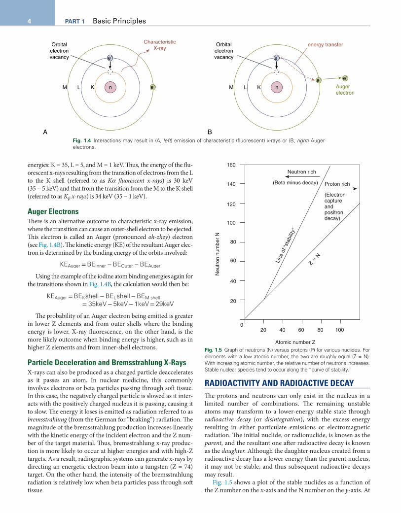
Characteristic X-RaysIn the first instance, excited electrons may be removed from their atomic orbit or elevated to a higher-energy orbit. An electron from an outer orbit can drop down to fill the vacancy, and the excess energy, the difference in the binding energy of the shells, can be emitted as an x-ray photon, a fluorescent x-ray (Fig. 1.4A). This is also known as a characteristic x-ray because it is specific to not only each element but also to the orbital shell from which it originated. Consider the case of fluorescent or characteristic x-rays from elec-tronic transitions within an iodine atom with the following binding
PotentialEnergy
Highest
Highest
orbital shell
N
M
L
K
nucleus
Lowest
Lowest
BindingEnergy
Fig. 1.2 Orbital binding energy.
Photon energyelectron Volt (eV)
106 103 100 101 1010106
1MeV 1keV 1eV
Increasing Wavelength
0.0001 nm 0.01 nm 10 nm 1000 nm 0.01 cm 1 cm 1 m 100 m
Gamma rays X-raysUltra-violet
Infrared Radio waves
Visible light
Radar TV FM AM
Fig. 1.3 Electromagnetic energy spectrum. Photon energies (eV) and wavelengths of x-rays and gamma ultraviolet, visible light, infrared, and radio waves.
TABLE 1.3 Terms Used to Describe electrons
Term Comment
Electron Basic elementary particle
Orbital electron Electron in one of the shells or orbits in an atom
Valence electronElectron in the outermost shell of an atom; responsible
for chemical characteristics and reactivity
Auger electronElectron ejected from an atomic orbit by energy
released during an electron transition
Conversion electron
Electron ejected from an atomic orbit because of internal conversion phenomenon as energy is given off by an unstable nucleus
Photoelectron
Electron ejected from an atomic orbit as a conse-quence of an interaction with a photon (photoelectric interaction) and complete absorption of the photon’s energy
Compton electronElectron ejected from orbit after absorbing a portion of
a photon’s energy during Compton scatter
4 PART 1 Basic Principles
energies: K = 35, L = 5, and M = 1 keV. Thus, the energy of the flu-orescent x-rays resulting from the transition of electrons from the L to the K shell (referred to as Kα fluorescent x-rays) is 30 keV (35 − 5 keV) and that from the transition from the M to the K shell (referred to as Kβ x-rays) is 34 keV (35 − 1 keV).
Auger ElectronsThere is an alternative outcome to characteristic x-ray emission, where the transition can cause an outer-shell electron to be ejected. This electron is called an Auger (pronounced oh-zhey) electron (see Fig. 1.4B). The kinetic energy (KE) of the resultant Auger elec-tron is determined by the binding energy of the orbits involved:
KEAuger = BEInner − BEOuter − BEAuger
Using the example of the iodine atom binding energies again for the transitions shown in Fig. 1.4B, the calculation would then be:
KEAuger = BEKshell − BELshell − BEM shell= 35keV − 5keV − 1keV = 29keV
The probability of an Auger electron being emitted is greater in lower Z elements and from outer shells where the binding energy is lower. X-ray fluorescence, on the other hand, is the more likely outcome when binding energy is higher, such as in higher Z elements and from inner-shell electrons.
Particle Deceleration and Bremsstrahlung X-RaysX-rays can also be produced as a charged particle deaccelerates as it passes an atom. In nuclear medicine, this commonly involves electrons or beta particles passing through soft tissue. In this case, the negatively charged particle is slowed as it inter-acts with the positively charged nucleus it is passing, causing it to slow. The energy it loses is emitted as radiation referred to as bremsstrahlung (from the German for “braking”) radiation. The magnitude of the bremsstrahlung production increases linearly with the kinetic energy of the incident electron and the Z num-ber of the target material. Thus, bremsstrahlung x-ray produc-tion is more likely to occur at higher energies and with high-Z targets. As a result, radiographic systems can generate x-rays by directing an energetic electron beam into a tungsten (Z = 74) target. On the other hand, the intensity of the bremsstrahlung radiation is relatively low when beta particles pass through soft tissue.
RADIOACTIVITY AND RADIOACTIVE DECAYThe protons and neutrons can only exist in the nucleus in a limited number of combinations. The remaining unstable atoms may transform to a lower-energy stable state through radioactive decay (or disintegration), with the excess energy resulting in either particulate emissions or electromagnetic radiation. The initial nuclide, or radionuclide, is known as the parent, and the resultant one after radioactive decay is known as the daughter. Although the daughter nucleus created from a radioactive decay has a lower energy than the parent nucleus, it may not be stable, and thus subsequent radioactive decays may result.
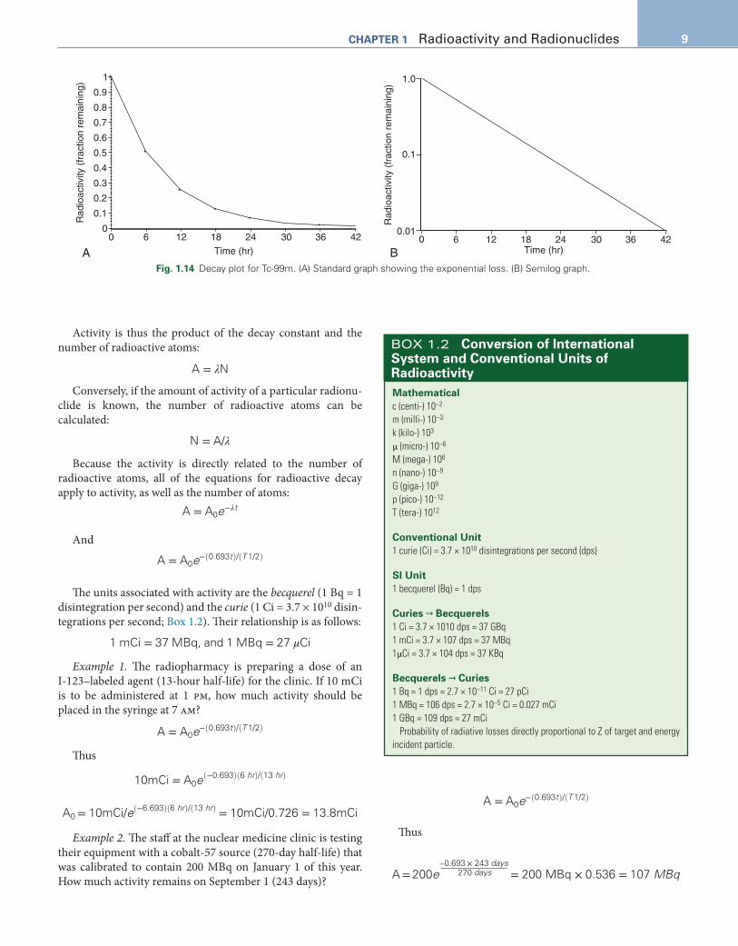
Fig. 1.5 shows a plot of the stable nuclides as a function of the Z number on the x-axis and the N number on the y-axis. At
K nLM
e-
e- K nLM
BA
e-
e- e-
Orbitalelectronvacancy
Orbitalelectronvacancy
CharacteristicX-ray
energy transfer
Augerelectron
Fig. 1.4 Interactions may result in (A, left) emission of characteristic (fluorescent) x-rays or (B, right) Auger electrons.
Proton rich
(Electroncaptureand positrondecay)
Line
of “
stab
ility
”
Z � N
Neutron rich160
140
120
100
80
60
40
20
20
Atomic number Z
Neu
tron
num
ber
N
40 60 80 1000
(Beta minus decay)
Fig. 1.5 Graph of neutrons (N) versus protons (P) for various nuclides. For elements with a low atomic number, the two are roughly equal (Z = N). With increasing atomic number, the relative number of neutrons increases. Stable nuclear species tend to occur along the “curve of stability.”

5ChAPTeR 1 Radioactivity and Radionuclides
low Z numbers, stable elements tend to have equal numbers of protons and neutrons (e.g., carbon-12, nitrogen-14, and oxy-gen-16) and lie along or near the Z = N line. However, as the nucleus becomes larger, the repulsive force of the nuclear pro-tons grows, and more neutrons are necessary in the stable nucleus to provide additional attractive nuclear force. Other factors also contribute to the stability and instability of the nucleus. For example, nuclides with even numbers of protons and neutrons tend to be more stable than those with odd Z and N configurations.
Unstable nuclides fall to either the right or the left of the curve of stability, with those to the right considered proton rich and those to the left neutron rich. As unstable radionuclides decay to entities that are closer to the curve of stability, pro-ton-rich radionuclides tend to decay in a manner that will reduce the Z number and increase the N number, and neu-tron-rich radionuclides tend to decay in a way that decreases the N number and increases the Z number.
Proton-rich radionuclides can be created by bombarding a cer-tain target material with high-energy protons that can overcome nuclear forces. Typically, a particle accelerator such as a cyclotron is used, increasing kinetic energy by accelerating charged particles to high speeds in a spiral path using alternating high-frequency volt-age and electromagnetic fields (Fig. 1.6). Conversely, in artificial production of neutron-rich radionuclides, one typically must use a nuclear reactor to bombard a target with a neutron flux (Fig. 1.7).
Modes of Radioactive DecayA decay scheme is a way to illustrate the transition from parent to daughter nuclides. In a decay scheme, higher energy levels are toward the top of the figure, and higher Z numbers are to the right of the figure. Transitions that lead to a reduction in energy are represented by an arrow pointing down. If it also results in a daughter nuclide with a change in the Z number, the arrow will point to the left with a decrease in the Z number and to the right if Z is increased.
A
A
B C
Fig. 1.6 “Proton-rich” radionuclides that decay by positron emission can be made in a cyclotron or particle accelerator. (A) Varying in size and appearance, cyclotrons may be self-shielded or housed in a thick cement vault (as shown) to reduce radiation exposure. Beam lines (arrow) extending from the central unit direct high-speed charged particles to bombard desired targets. (B) The bottom of the cyclotron contains the accelerating electrodes (short arrows). (C) Electromagnetic fields created by a large magnet (arrowhead) in the upper por-tion of the cyclotron constrain the particles to circular orbits. (Photos courtesy of Anthony F. Zagar, University of Alabama at Birmingham.)

6 PART 1 Basic Principles
Alpha DecayAn unstable heavy atom may decay to a nuclide closer to the curve of stability by emitting an alpha particle (α) consisting of 2 protons and 2 neutrons (essentially an ionized helium atom):
ZAX→ Z−2
A−4Y + α = Z−2
A−4Yjo9 +
2
4He
The daughter nucleus may not be stable, and thus the emis-sion of an alpha particle often will lead to the emission of a series of radiations until the nucleus is stable. The decay scheme for the decay of radium-226 (Ra-226) to radon-222 (Rn-222) is shown in Fig. 1.8.
Beta-Minus DecayNeutron-rich radionuclides tend to stabilize by decreasing the number of neutrons through a radioactive-decay process referred to as beta-minus (β–), also known as negatron or beta decay. Factors such as weak forces between nucleons transfer energy, transforming a neutron into a proton (N – 1 and Z + 1). This is an isobaric transition with no change to the atomic mass (A). An example of the beta-minus decay scheme for I-131 is shown in Fig. 1.9. Excess energy is emit-ted from the nucleus as an antineutrino and a negative beta particle (or negatron). This process can be written as follows:
ZAX
N=
Z+1AY
N−1+ β− −+ υ antineutrino
Fig. 1.7 Fission and neutron capture radionuclide production in a nuclear reactor. Samples can be lowered into the reactor as shown, with water acting as shielding against neutrons. The blue glow is caused by the emission of electrons from the radioactive products; when charged par-ticles move faster than the speed of light in a medium such as water, the emitted radiation is called Cherenkov radiation. (Courtesy of the Uni-versity of Missouri Research Reactor Center.)
�2
�1
22286
Rn (3.8 days)
22688
Ra (1600 yr)
448 keV
186 keV
�
Fig. 1.8 Alpha decay. The emission of an alpha particle (2 protons and 2 neutrons) results in the atomic number (Z) decreasing by 2 and the atomic mass (Z + N) decreasing by 4. Decay of radium-226 to the daugh-ter Rn-222 shows the arrow pointed down, indicating a decrease in energy, and to the left because of the decrease in Z.

7ChAPTeR 1 Radioactivity and Radionuclides
The antineutrino is very difficult to measure because it has virtually no mass or charge associated with it, only energy. The negative beta particle is indistinguishable from an electron with the same mass and electric charge, differing only in that the beta particle is emitted from the nucleus and the electron orbits the nucleus. In addition to the Mo-99 used to make Tc-99m, several β–-emitting radionuclides play an important role in nuclear medicine for therapy applications: I-131, phos-phorus-32 (P-32), yttrium-90 (Y-90), and lutetium-177 (Lu-177).
Beta-Plus (Positron) DecayUnstable proton-rich radionuclides can reduce Z and increase N numbers through either beta-plus decay or electron capture. In beta-plus decay, the parent nucleus emits a positively charged beta particle, a positron (β+). The resulting daughter nucleus has one fewer proton and one more neutron than the parent, an iso-baric transition:
ZAXN = Z−1
AYN+1+ β + + υneutrino
average β + kinetic energy: Eβ + ≈ Emax/3
The positron has the same mass as a beta-minus particle or electron, with a charge of the same magnitude but the opposite. In fact, the positron is the antiparticle of the electron; if they are brought into close contact, they will be annihilated and trans-formed into two 511-keV photons, traveling at 180 degrees in opposite directions. This annihilation process is the basis of PET imaging. The 511-keV value derives from the energy equiv-alence of the mass of the beta particle, similar to the rest mass of an electron (using E = mc2 as previously discussed).
For positron decay to occur, the transition energy must be in excess of a 1022-keV threshold (twice 511 keV) to overcome the production of the positron and addition of an orbital electron to maintain electric neutrality. These radionuclides are typically produced using a cyclotron. Some positron-emitting radionu-clides of interest include fluorine-18 (F-18; Fig. 1.10), nitro-gen-13 (N-13), carbon-11 (C-11), gallium-68 (Ga-68), and rubidium-82 (Rb-82).
Electron Capture. An alternative to beta-plus decay for proton-rich radionuclides is electron capture (EC). In this process, an inner-shell, orbital electron is absorbed into the nucleus, leading to the reduction of Z and increase of N by 1. However, no energy threshold exists for EC to occur. In cases in which the transition energy is less than the 1022-keV threshold, EC is the only possible decay process, but either process is possible when the energy is greater than 1022 keV. For F-18, positron decay occurs 97% of the time, and EC occurs 3% of the time. The capture of an orbital electron leads to an inner-shell vacancy, which in turn leads to the emission of fluorescent x-rays or Auger electrons. Radionuclides that decay through EC exclusively include thallium-201 (Tl-201; Fig. 1.11), gallium-67 (Ga-67), and indium-111 (In-111). These are all produced in a cyclotron.
Isomeric Transition. In some cases, an excited radionuclide decays from one energy level to another while retaining the same Z and N numbers. This is referred to as an isomeric transition because the nuclide decays from one isomer (energy level) to another. This transition may result in the emission of a gamma ray, the energy of which is determined by the energy
� (81%)
13153
I (8 days)
13154
Xe (stable)
364 keV
��
Fig. 1.9 Beta minus (β –) decay scheme for iodine-131 to the daughter Xe-133. β– decay (negatron emission) results in the daughter with one more proton in the nucleus (Z + 1), so the arrow points to the right. Because a neutron is lost (N – 1), this is an isobaric transition with the atomic mass unchanged.
189F (110 min)
188O (stable)
��
Fig. 1.10 Positron (β+) decay results in a loss of 1 proton (Z – 1) in pro-ton-rich radionuclides. Because 1 neutron is gained, the atomic mass of the daughter is unchanged, another example of isobaric transition. F-18 decay by positron emission results in the daughter product, O-18. The arrow points down and to the left, indicating the decrease in Z.
20181Tl (73 hr)
20180 Hg (stable)
EC
167 keV
32 keV� (10%)� (3%)
Fig. 1.11 Electron capture is an alternate transition that can occur to reduce the proton number and does not require that an energy thresh-old be met. Tl-201 decays by electron capture, with the daughter (Hg-201) containing one fewer proton (Z – 1) than the parent.

8 PART 1 Basic Principles
difference of the initial and eventual energy levels. In some cases, an alternate process called internal conversion can occur, resulting in the emission of an orbital electron, a conversion electron. The kinetic energy is calculated as the difference in the two energy levels minus the electron’s binding energy.
Perhaps the most important isomeric transition for nuclear medicine involves technetium. The term metastable (i.e., almost stable) is used if the daughter nucleus remains in the excited state for a considerable amount of time (>1 microsecond, which is long by nuclear standards). Mo-99 decays to an excited, or metastable, Tc-99m that in turn transitions to Tc-99 (Figs. 1.12 and 1.13). Tc-99m has a 6-hour half-life. Tc-99m is so commonly used because of its reasonable half-life, as well as its gamma-ray energy (140 keV) and lack of beta- or alpha-particle emissions. Another example of isomeric transition is seen in the decay scheme of I-131 (see Fig. 1.9). Xenon-131 (Xe-131), formed from the beta-minus decay of I-131, is in an excited state and imme-diately decays by isomeric transition with the emission of a 364-keV gamma ray.
Radioactive Decay CalculationsAtoms in a sample containing a certain number (N) of radioac-tive atoms will not all decay at the same time but with a mean time (Tm) that is characteristic of a particular radionuclide. The
reciprocal of Tm, the fraction of the radioactive atoms that decay per unit time, is referred to as the decay constant, λ:
λ =1
TmThus, the number of atoms (dN) that decay in a short time
interval (dt) is given by:
dN = λdt
Integrating this equation over time leads to:
N = N0e−λt
where N0 is the initial number of radioactive atoms, and N is the number remaining after some time, t.
This equation describes exponential decay in which a certain fraction of the material is lost in a set period. This fraction is referred to as the decay fraction, DF:
DF = e−λt
Thus, the number of radioactive atoms remaining, N, is also given by:
N = N0 × DF
Also, Nd is the number of atoms that have decayed in time, t, and can be calculated with:
Nd = N0 × (1 − DF)
The time necessary for half of the material to decay is defined as the half-life (T1/2). The half-life is related to the mean life and the decay constant by the following equations:
T1/2 = ln (2)Tm =0.693
λ
Alternatively, one can determine the decay constant from the half-life by:
λ =0.693
T1/2
One can also express the radioactive decay equation using the half-life:
N = N0e− 0.693tT1/2
If a sample contains 10,000 radioactive atoms at a partic-ular point in time, one half-life later, there will be 5000 atoms; another half-life later, there will be 2500 atoms; and so on. This process of a certain fraction of the material decaying in a certain time is representative of exponential decay (Fig. 1.14A). When graphed using a log scale on the y-axis (semilog plot), the result is a straight line with the negative slope equal in magnitude to the decay constant (Fig. 1.14B).
The amount of activity (A) is the number of nuclear transfor-mations—decays or disintegrations—per unit time. The activity is characterized by the number of radioactive atoms in the sam-ple, N, divided by the mean time to radioactive decay, Tm:
A = N/Tm
�
99m43Tc (6.01 hr)
9943Tc
140.5 keV (89%)
Fig. 1.12 Isomeric transitions involve a change in the energy state of a radionuclide, such as Tc-99m to Tc-99.
��
�
�
99
0.0
43Tc (2.1 � 105 yr)
140.5 keV
142.7 keV
9942Mo (2.8 days or 66 hr)
Fig. 1.13 Decay scheme of Mo-99. Beta-minus emission to Tc-99m, followed by isomeric transition to Tc-99.
9ChAPTeR 1 Radioactivity and Radionuclides
Activity is thus the product of the decay constant and the number of radioactive atoms:
A = λN
Conversely, if the amount of activity of a particular radionu-clide is known, the number of radioactive atoms can be calculated:
N = A/λ
Because the activity is directly related to the number of radioactive atoms, all of the equations for radioactive decay apply to activity, as well as the number of atoms:
A = A0e−λt
And
A = A0e(− 0.693t)/(T 1/2)
The units associated with activity are the becquerel (1 Bq = 1 disintegration per second) and the curie (1 Ci = 3.7 × 10¹⁰ disin-tegrations per second; Box 1.2). Their relationship is as follows:
1 mCi = 37 MBq, and 1 MBq = 27 μCi
Example 1. The radiopharmacy is preparing a dose of an I-123–labeled agent (13-hour half-life) for the clinic. If 10 mCi is to be administered at 1 pm, how much activity should be placed in the syringe at 7 am?
A = A0e(− 0.693t)/(T 1/2)
Thus
10mCi = A0e(−0.693)(6 hr)/(13 hr)
A0 = 10mCi/e(−6.693)(6 hr)/(13 hr) = 10mCi/0.726 = 13.8mCi
Example 2. The staff at the nuclear medicine clinic is testing their equipment with a cobalt-57 source (270-day half-life) that was calibrated to contain 200 MBq on January 1 of this year. How much activity remains on September 1 (243 days)?
A = A0e(− 0.693t)/(T 1/2)
Thus
A = 200e–0.693 × 243 days
270 days = 200 MBq × 0.536 = 107 MBq
1
0.9
0.8
0.7
0.6
0.5
0.4
0.3
0.2
0.1
00 6 12 18
Time (hr)24 30 36 42
Rad
ioac
tivity
(fr
actio
n re
mai
ning
)
A
1.0
0.1
0.010 6 12 18
Time (hr)24 30 36 42
Rad
ioac
tivity
(fr
actio
n re
mai
ning
)
BFig. 1.14 Decay plot for Tc-99m. (A) Standard graph showing the exponential loss. (B) Semilog graph.
Mathematicalc (centi-) 10–2
m (milli-) 10–3
k (kilo-) 103
μ (micro-) 10–6
M (mega-) 106
n (nano-) 10–9
G (giga-) 109
p (pico-) 10–12
T (tera-) 1012
Conventional Unit1 curie (Ci) = 3.7 × 10¹⁰ disintegrations per second (dps)
SI Unit1 becquerel (Bq) = 1 dps
Curies → Becquerels1 Ci = 3.7 × 10¹⁰ dps = 37 GBq1 mCi = 3.7 × 10⁷ dps = 37 MBq1μCi = 3.7 × 10⁴ dps = 37 KBq
Becquerels → Curies1 Bq = 1 dps = 2.7 × 10⁻¹¹ Ci = 27 pCi1 MBq = 106 dps = 2.7 × 10⁻⁵ Ci = 0.027 mCi1 GBq = 109 dps = 27 mCi
Probability of radiative losses directly proportional to Z of target and energy incident particle.
BOX 1.2 Conversion of International System and Conventional Units of Radioactivity

10 PART 1 Basic Principles
INTERACTIONS BETWEEN RADIATION AND MATTERCharged-Particle Interactions With MatterA charged particle may transfer energy in different ways. First, it can be attracted and slowed by the opposite charge of the nucleus or orbiting electrons in target material atoms. The resulting kinetic energy loss is released as radiation (radiative losses). Bremsstrahlung radiation occurring with a β– emitter is one example of this type of interaction. The energy of radiative losses is directly proportional to the Z number of the target as well as to the incident particle’s energy.
Charged particles can also directly transfer energy to the atom’s orbital electrons, resulting in electron excitations and ionizations. While excited, electrons can temporarily move to a shell farther from the nucleus. As de-excitation occurs, trans-ferred energy leads to the emission of Auger electrons or elec-tromagnetic radiation. This radiation can have a wide range of energies, including visible or ultraviolet radiation for outer-shell transitions and fluorescent x-rays for the inner-shell transitions.
When the energy from charged-particle interactions pro-duces ionized electrons and atoms in tissues, the majority of the ionized electrons are low energy. However, some interactions result in high-energy electrons, referred to as delta rays, which in turn can also cause excitation and ionization. In the energies of practical interest in nuclear medicine, nearly all of the energy from a charged-particle interaction (greater than 99%) is expended in excitation and ionization (or collisional losses) compared with radiative losses.
The rate at which a material causes a charged particle to lose energy (per unit length of the matter) is referred to as its stop-ping power. A related quantity is linear energy transfer (LET), which is the amount of energy deposited locally (i.e., not lost to energetic electrons, delta rays, or radiative loss) per unit length. The stopping power and LET values depend on the type of radi-ation, its energy, and the density of the material through which it travels. Radiation with a higher LET value has been shown to cause more damage to cells. Alpha particles have a higher LET than beta particles or electrons.
Although densely ionizing, alpha particles deposit their energy over a very short distance, a small fraction of a millime-ter in soft tissues. Also, although they are easily stopped by the skin, these high-energy particles cause substantial cell death when internalized, making them both extremely dangerous if accidentally ingested as well as highly effective in therapeutic applications (e.g., Ra-223 in prostate cancer).
Comparatively, β– particles travel for much longer distances, ranging from several millimeters to several centimeters depend-ing on their initial energy. They can be stopped by material such as a thin sheet of aluminum or a few millimeters of soft tissue.
The β– kinetic energy is variable because it shares energy with the antineutrino produced during the decay event. The maxi-mum kinetic energy of the beta particle (Emax) is defined by the difference in the energy levels of the parent and daughter nuclide. However, it is the average kinetic energy that is used when calculating the impact of the β– on cells and tissues, esti-mated as 1/3 of Emax (Eβ ≈ Emax/3), similar to the calculation previously described for positrons.
Photon Interactions in MatterHigh-energy photons (gamma rays, x-rays, bremsstrahlung radiation, and annihilation radiation) can also transfer energy to the electrons, nuclei, or atoms as a whole that they encoun-ter. Unlike charged particles, which directly create ionized atoms, the high-energy photons act indirectly, transferring their energy to charged particles, specifically electrons, which in turn create most of the excitations and ionizations that occur in the matter. Thus they are considered secondary ionizing radiations.
At low energy levels (a few keV), photons are scattered in a manner that does not deposit energy, referred to as Rayleigh scattering. Photons with energy in excess of several MeV can result in pair production of a negatron and a positron (effectively a negative and positive electron). However, in energy ranges most common in nuclear medicine (from several tens of keV to approximately 1 MeV), the two most prominent modes of pho-ton interaction are the photoelectric effect and Compton scatter-ing. Factors involved in the various types of interaction are outlined in Table 1.4.
Photoelectric EffectThe photoelectric effect (Fig. 1.15) occurs when a photon trans-fers all of its energy to an orbital electron, causing it to be ejected from the atom and creating an electron-shell vacancy. The kinetic energy of the liberated photoelectron equals the incident photon’s energy minus the binding energy of the electron’s ini-tial orbital shell. As the shell vacancy is filled by electrons from outer shells, fluorescent x-rays and Auger electrons are also emitted. Paradoxically, the probability of photoelectric interac-tions is highest for tightly bound orbiting electrons (i.e., those in the inner shells of high Z elements). These electrons are most likely the ones in the innermost orbital shell where the binding energy is just under the photon’s energy. In addition, the chance of this interaction dramatically decreases as incident photon energy increases. The probability of the photoelectric interac-tion (or PPE) is given by:
PPE α Z3/E3
where Z = atomic number, and E = incident photon kinetic energy.

11ChAPTeR 1 Radioactivity and Radionuclides
In soft tissues (low Z), the photoelectric effect is much less common than Compton scatter. It is, however, more prevalent in the high-Z materials used for shielding (e.g., lead) or for pho-ton detection (sodium iodide crystals). Photoelectric effect interactions can also occur in the gamma camera’s photomulti-plier tubes, which contain high-Z materials, such as cesium.
Compton ScatterThe incident photon does not disappear in Compton scatter. Rather, it transfers a portion of its energy to an orbital electron (a Compton electron), which is then ejected from the atom. The photon is deflected or scattered, at an angle θ from its original path (Fig. 1.16). The electron and scattered photon may go on to ionize or excite other atoms.
The sum of the kinetic energies of the scattered photon and the Compton electron will equal the initial photon’s energy. With lower-energy incident photons, more energy is transferred to the electron, and there is greater backscatter of the resulting photon (i.e., the angle between the incident and scattered pho-tons tends to be wider), with even 180 degrees of backscatter
occurring. Higher-energy incident photons lose less energy to the electron, and deflection is less significant (i.e., the scatter angle is narrower), such that both the scattered photon and elec-tron tend to travel in a more forward direction.
Whereas the photoelectric effect is important at lower energies and more likely involves inner-shell electrons in high-Z materials, Compton scatter predominates in soft tis-sues in the moderate-energy ranges of gamma-ray and x-ray photons in nuclear medicine imaging and tends to involve outer-shell electrons. Because the energy of the incident pho-ton is much greater than the shell’s binding energy, the colli-sion occurs as if involving a free electron. Compton interactions tend to depend on electron density but are rela-tively independent of the Z number or incident photon energy. Because electron density is fairly consistent among the atoms in soft tissues, the probability increases with increasing material density rather than its Z number. Electron density is higher when hydrogen atoms are present, so tissues with high water content are more affected than anhydrous tissues.
TABLE 1.4 Photon Interactions in Matter
Interaction Occurrenceeffect of Target Material Z
Incident Photon e Range (e0) Target
Resulting Particle emissions
Secondary Photon emissions
Compton Predominant in soft tissues at diagnostic E range
Nearly independent of ZDepends on e– density
(therefore on target density)
Hydrous > anhydrous material
Mid E range (≈26 keV–30 MeV)
Outer-shell e– Recoil e– Scattered photonDegrades image
Photoelectric effect
Predominates in shielding and detector crys-tals/PMT
High-Z materials Low-E photonZ3/E3
Innermost-shell e– possible
PhotoelectronsAuger e–
Characteristic x-ray
e– cascade produces
Characteristic x-ray ↑ with ↑ Z-detector mate-rial, shielding
↑ when weakly bound valence (e.g., PMT photocathode materials)
Auger e– ↑ in soft tissues e– binding E less a factor in tissue
Pair production Not typically seen in ener-gies used in medicine
1.02-MeV minimum but actually
>>1.02 MeV (not present at diagnostic E ranges)
Usually nucleus, sometimes orbital e–
β+ and β–
(or e+ and e–)Annihilation photons (from β+)
Two 5110 keV at 180 degrees
E, Energy; Z, atomic number (number of protons); PMT, photomultiplier tube; e–, electron; β+, positron (or a positive electron); β–, negatron (same as negative electron or beta-minus particle).

12 PART 1 Basic Principles
Incident photon
Nucleus
A
Vacancy
Photoelectron
Nucleus
B
Characteristicx-ray
Vacancy
Electrontransitionbetween
shells
Nucleus
C
Fig. 1.15 Photoelectric absorption. (A) An incident photon interacts with an orbital electron. (B) The electron is ejected from its shell, creating a vacancy. The electron is either ejected from the atom or moved to a shell further from the nucleus. (C) The orbital vacancy is filled by the transition of an electron from a more distant shell. Consequently, a characteristic x-ray is given off.

13ChAPTeR 1 Radioactivity and Radionuclides
SUGGESTED READINGChandra R, Rahmin A. Nuclear Medicine Physics: The Basics. 8th ed.
Philadelphia: Williams & Wilkins; 2012.Cherry SR, Sorenson JA, Phelps ME. Physics in Nuclear Medicine. 4th
ed. Philadelphia: WB Saunders; 2012.Eckerman KF, Endo A. MIRD: Radionuclide Data and Decay Schemes.
2nd ed. Reston, VA: Society of Nuclear Medicine; 2008.Loevinger R, Budinger TF, Watson EE. MIRD Primer for Absorbed
Dose Calculations. Reston, VA: Society of Nuclear Medicine; 1988.Powsner RA, Powsner ER. Essentials of Nuclear Medicine Physics. 3rd
ed. West Sussex, UK: Wiley-Blackwell; 2013.Saha GP. Physics and Radiobiology of Nuclear Medicine. 4th ed. New
York: Springer; 2013.�
Comptonelectron
Compton-scatteredphoton
Incidentphoton
Nucleus
Angleof scatter
Fig. 1.16 Compton scatter. An incident photon interacts with an outer or loosely bound electron, giving up a portion of its energy to the elec-tron and undergoing a change in direction at a lower kinetic energy level.