CHAPTER 1 9 - A ntepartum H aemorrhage - Mount Sinai Hospital
20
178 CHAPTER 19 A ntepartum H aemorrhage JCP Kingdom Antepartum haemorrhage (APH) refers to bleed- ing from the genital tract after 20 weeks of gesta- tion, which is 4 −6 weeks below the lower limit of fetal viability. Establishing the cause of APH is important to distinguish scenarios at risk of substantial haemorrhage, such as major placenta praevia or abruption, from a range of possibili- ties that pose much lower risks (Table 19-1). Cases with serious underlying causes of vaginal bleeding, such as caesarean section scar preg- nancy, 1 may present with vaginal bleeding before 20 weeks. Furthermore, the identification of a benign lower genital tract source of minor vaginal bleeding does not preclude an additional more serious uterine cause of APH. The two most serious causes are placental abruption and placenta praevia; the latter is becoming more common, due in part to a greater prevalence of: previous uterine surgery, assisted reproductive technologies, multifetal pregnancy and advanced maternal age. 2 Major abruption is much less common due to general advances in maternal health, including a large reduction in smoking, and improvements in antenatal care. Histori- cally, the first description of placenta praevia in 1885 was by the Parisian physician, Paul Portal (1630–1703), who was the first to describe the attachment of the placenta to the lower uterine segment. placenta praevia, which still exists in many coun- tries today. 3 The terms ‘unavoidable ’ referring to placenta praevia and ‘accidental ’ referring to placental abruption are attributed from 1775 to Edward Rigby (1747–1821) of Norwich, England. The historical background of placenta praevia has been documented. 4,5 PLACENTA PRAEVIA Placenta praevia occurs when the entire pla- centa, or in part, implants in the lower uterine segment after 20 weeks’ gestation. The inci- dence varies by population but significant disease occurs in about 1/200 deliveries. 6 Perinatal mor- tality is increased almost twofold compared to non-praevia pregnancies adjusted for smoking, maternal age, parity and in vitro fertilization. 6 The risk factors for placenta praevia are sum- marized in Table 19-2. Some of these, in particu- lar multiple previous caesarean deliveries and previous placenta praevia, confer recurrence risks of up to 5%. Finally, the increasing use of 18 −20-week ultrasound examinations to assess fetal anatomy has increased the rate of diagnosis of asymptomatic minor degrees of placenta praevia. 7 An abnormally large placenta surface area predisposes to placenta praevia, the most common cause being multifetal pregnancy; amongst the rare causes, careful consideration of a succenturiate lobe in the lower segment is important, since it is associated with vasa praevia, which, when undiagnosed, may result in fetal mortality. 8 Twin pregnancies have a 50% greater risk of placenta praevia. 9 Classification The classical method describes four types, or degrees, of placenta praevia as illustrated in Figure 19-1. With additional descriptive termi- nology, these are as follows: • Type 1 (low-lying): the lower edge of the placenta is inside the lower uterine segment but does not reach the internal cervical os. FIRST DESCRIPTION OF PLACENTA PRAEVIA ‘I put my fingers into the orifice and felt the after birth which covered the orifice of the matrix from all sides and adhered in all its parts with the exception of the middle’. Paul Portal La Pratique des Accouchements Soutenue d’un Grand Nombre d’Observations. Paris: G. Martin, 1685 . The elegant, yet poignant, drawings of this disease from partially dissected dead women by the Scot, William Hunter (1718 −1783), living in London, are a vivid reminder of the danger of
-
Upload
khangminh22 -
Category
Documents
-
view
0 -
download
0
Transcript of CHAPTER 1 9 - A ntepartum H aemorrhage - Mount Sinai Hospital

178
C H A P T E R 1 9
A ntepartum H aemorrhage JCP Kingdom
Antepartum haemorrhage (APH) refers to bleed-ing from the genital tract after 20 weeks of gesta-tion, which is 4 − 6 weeks below the lower limit of fetal viability. Establishing the cause of APH is important to distinguish scenarios at risk of substantial haemorrhage, such as major placenta praevia or abruption, from a range of possibili-ties that pose much lower risks ( Table 19-1 ). Cases with serious underlying causes of vaginal bleeding, such as caesarean section scar preg-nancy, 1 may present with vaginal bleeding before 20 weeks. Furthermore, the identifi cation of a benign lower genital tract source of minor vaginal bleeding does not preclude an additional more serious uterine cause of APH. The two most serious causes are placental abruption and placenta praevia; the latter is becoming more common, due in part to a greater prevalence of: previous uterine surgery, assisted reproductive technologies, multifetal pregnancy and advanced maternal age. 2 Major abruption is much less common due to general advances in maternal health, including a large reduction in smoking, and improvements in antenatal care. Histori-cally, the fi rst description of placenta praevia in 1885 was by the Parisian physician, Paul Portal (1630–1703), who was the fi rst to describe the attachment of the placenta to the lower uterine segment.
placenta praevia, which still exists in many coun-tries today. 3 The terms ‘unavoidable ’ referring to placenta praevia and ‘accidental ’ referring to placental abruption are attributed from 1775 to Edward Rigby (1747–1821) of Norwich, England. The historical background of placenta praevia has been documented. 4,5
PLACENTA PRAEVIA
Placenta praevia occurs when the entire pla-centa, or in part, implants in the lower uterine segment after 20 weeks ’ gestation. The inci-dence varies by population but signifi cant disease occurs in about 1/200 deliveries. 6 Perinatal mor-tality is increased almost twofold compared to non-praevia pregnancies adjusted for smoking, maternal age, parity and in vitro fertilization. 6 The risk factors for placenta praevia are sum-marized in Table 19-2 . Some of these, in particu-lar multiple previous caesarean deliveries and previous placenta praevia, confer recurrence risks of up to 5%. Finally, the increasing use of 18 − 20-week ultrasound examinations to assess fetal anatomy has increased the rate of diagnosis of asymptomatic minor degrees of placenta praevia. 7 An abnormally large placenta surface area predisposes to placenta praevia, the most common cause being multifetal pregnancy; amongst the rare causes, careful consideration of a succenturiate lobe in the lower segment is important, since it is associated with vasa praevia, which, when undiagnosed, may result in fetal mortality. 8 Twin pregnancies have a 50% greater risk of placenta praevia. 9
Classifi cation The classical method describes four types, or degrees, of placenta praevia as illustrated in Figure 19-1 . With additional descriptive termi-nology, these are as follows:
• Type 1 (low-lying): the lower edge of the placenta is inside the lower uterine segment but does not reach the internal cervical os.
FIRST DESCRIPTION OF PLACENTA PRAEVIA
‘I put my fi ngers into the orifi ce and felt the after birth which covered the orifi ce of the matrix from all sides and adhered in all its parts with the exception of the middle’.
Paul Portal La Pratique des Accouchements Soutenue
d ’ un Grand Nombre d ’ Observations. Paris: G. Martin, 1685 .
The elegant, yet poignant, drawings of this disease from partially dissected dead women by the Scot, William Hunter (1718 − 1783), living in London, are a vivid reminder of the danger of

19 Antepartum Haemorrhage 179
TABLE 19-1 Causes of Antepartum Haemorrhage
Site Diagnosis
Uterine Placenta praeviaAbruptionPlacenta praevia incretaAntepartum fetal death
Cervix Cervical ectropion/congestionCervical pregnancyCancer of the cervix
Lower genital tract
Vulvo-vaginal varicesVulvo-vaginal infectionsMalignanciesTrauma
Unclassifi ed Cervical effacement
TABLE 19-2 Risk Factors for Placenta Praevia
Category Risk Factor
Maternal SmokingAdvanced maternal age
Obstetric history High parity, twinsAssisted reproductive
technologiesPrevious placenta praevia
Uterine Uterine surgeryCaesarean sectionsMyomectomyHysteroscopic surgerySeptumAdhesions
Placental Multifetal pregnanciesEnlarged placentaHigh altitudeChronic fetal anaemiaAbnormal placental
developmentSuccenturiate lobe (vasa
praevia)Bi-lobar placentaPlacenta membranacea
FIGURE 19-1 ■ Classifi cation of placenta praevia.
Internal os
Upperuterinesegment
Loweruterinesegment
Type I Type II Type III Type IV(lateral/low lying) (marginal) (partial) (complete/central)
Minor degree Major degree
• Type 2 (marginal): the lower edge of the placenta extends to but not across the inter-nal os.
• Types 1 and 2 are commonly observed in asymptomatic women at the 18 − 20-week transabdominal fetal anatomy ultrasound examination. The distinction between the two, by transvaginal ultrasound, is unim-portant at this stage, since in both instances, the likelihood of clinically signifi cant pla-centa praevia in the third trimester is very small.
• Type 3 (partial): the lower edge of the pla-centa extends asymmetrically across the internal os; however, since the portion of the placenta covering the internal os is thin, it may pull away with minimal vaginal
bleeding during cervical effacement and dilation to permit safe vaginal delivery.
• Type 4 (complete or central): the placenta is almost centrally placed within the lower uterine segment.
With few exceptions the distinction between type 3 and 4 placenta praevia is not important in high-resource countries, because the risk/benefi t

180 19 Antepartum Haemorrhage
ratio of planned caesarean delivery outweighs that of attempting vaginal delivery. Exceptions would include: previous vaginal deliveries in a highly motivated and well-informed individual; and anticipation of a diffi cult caesarean, for example, due to morbid obesity. In other health-care settings, especially those in which accurate transvaginal ultrasound imaging is not readily available to distinguish these types, the use of the term ‘type 3’ to describe transabdominal ultra-sound fi ndings implies that a proportion of women in labour with a small amount of pla-centa across the os can deliver safely by the vaginal route because during cervical effacement and dilation the small area of disrupted placenta may not bleed signifi cantly.
The wide application of high resolution transabdominal and transvaginal ultrasound has largely obviated the need to describe four cate-gories of placenta praevia, such that the disease is now commonly described as minor praevia (types 1 and 2) and major praevia (types 3 and 4) that respectively do not or do require elective caesarean delivery.
Physiology of the Lower Uterine Segment In the non-pregnant state the uterus is comprised of just two parts, a corpus and cervix, the bound-ary between which is a fi bro-muscular junction described originally by Danforth. 10 The lower uterine segment begins to form in the second trimester, once the gestation sac has fully occu-pied the uterine cavity; thereafter maternal tissue below the apex of the fetal membranes is con-sidered the cervix and the junction is the internal os ( Fig 19-2a ). The lower uterine segment gradu-ally forms, via myometrial growth and thinning, from the tissue above and below the internal os. As such, the cervix gradually shortens as pregnancy advances 11 while failure to do so increases the risk of caesarean section for dys-tocia. 12 Formation of the lower uterine segment provides one-third of uterine volume for fetal growth and is normally occupied by the fetal head from 34 weeks ’ gestation. Sonographically, the upper margin of the lower uterine segment is the refl ection of the utero-vesical peritoneum at the upper edge of the semi-fi lled bladder ( Fig 19-2a ). In labour, the upper active uterine segment (the fundus) provides the driving force for labour, placental detachment and subsequent mechanical haemostasis. By contrast, the lower uterine segment is a passive structure in normal labour; at caesarean section for obstructed labour it may balloon out signifi cantly and be a source of primary postpartum haemorrhage. In the
labour and delivery setting, the lower uterine segment is defi ned pragmatically as that 6 − 8 cm portion of the uterine cavity palpable digitally in women with either regional or general anaes-thesia following delivery of the placenta.
Placental ‘Migration’ Gradual formation of the lower uterine segment is sometimes described as ‘placental migration’. The forces underlying this phenomenon may cause the lower placental edge to bleed, even in women with type 1 or 2 praevia. Nevertheless, this migration means that at least 90% of low-lying placentas will resolve, leaving about 1 in 200 women with clinically signifi cant placenta praevia after 34 weeks. The almost universal use of ultrasound at 19 − 20 weeks has the capacity to over-diagnose minor degrees of placenta praevia in asymptomatic women ( Fig 19-2a ). Advice on safe mode of delivery in this context can all too easily err on the side of caution and caesarean delivery; yet many options, described in Chapter 20 , exist to manage postpartum haemorrhage effectively. The previous generally accepted standard was to recommend caesarean where the lower placental edge is < 2 cm from the internal os in the third trimester. 13 However, the lower uterine segment continues to form, especially with cervical shortening and effacement from Braxton Hicks contractions. More recent evi-dence suggests that the majority of women with no other risk factors can achieve safe vaginal delivery if the lower placental edge is > 1 cm from the internal os. 14 A fl exible policy of serial transvaginal ultrasound in borderline situations can save some women from unnecessary caesar-ean delivery ( Fig 19-2b,c ).
Assessment of Vaginal Bleeding from Placenta Praevia Around 80% of all women with major placenta praevia will have one or more bleeds before delivery. The fi rst is a warning, or ‘sentinel’ bleed. Any subsequent bleeds are likely to be heavier. In general, major degrees of placenta praevia bleed earlier, more frequently and more heavily than do minor degrees. Nevertheless, even a complete placenta praevia may not bleed until either the onset of labour (if undiagnosed) or simply presentation with an oblique or trans-verse lie in the clinic after 34 weeks. Bleeding from placenta praevia is most commonly caused by disruption of small uteroplacental veins as the anchored placenta is gradually stretched. This maternal source of blood may escape through the decidua and enter the myometrium.

19 Antepartum Haemorrhage 181
following the sentinel bleed. Unexpectedly severe vaginal bleeding from a known minor pla-centa praevia may be due to a prominent mar-ginal placental sinus where uterine venous blood drains out of the lower edge of the placenta. Transvaginal ultrasound is a valuable assessment tool in stable women following a sentinel bleed. Where vaginal bleeding with placenta praevia is seen, the rare accompaniment of gross haematu-ria should immediately raise the suspicion of associated invasive placentation (see below and Figures 19-2d and 19-9 ).
The importance of careful evaluation of women with a ‘warning bleed’ from placenta praevia was dramatically illustrated by Munro Kerr in an earlier edition:
Thrombin, the local product of pathological haemorrhage, is a powerful myometrial irritant 15 and may explain why uterine contractions can accompany bleeding from placenta praevia. Maternal tocolysis may arrest ongoing APH and extend the duration of pregnancy. 16
Transvaginal ultrasound may be useful to predict preterm birth in placenta praevia; at 32 − 33 weeks, a cervical length < 30 mm con-ferred a threefold risk of subsequent delivery for haemorrhage and preterm birth. 17 A prominent marginal sinus at the lower edge also predicts the need for caesarean delivery due to vaginal bleed-ing. 18 Transvaginal ultrasound therefore pro-vides useful predictors of recurrent bleeding and thus the need to remain in hospital if undelivered
FIGURE 19-2 ■ Placenta praevia. (a) Ultrasound imaging of the lower uterine segment and saggital view at 18 weeks demonstrating a normal closed cervix (dashed line, length 46 mm) and a posterior placenta (p) that is 2.4 cm from the internal os. The maternal bladder (bl) is full. The lower uterine segment (LUS) is that part of the lower segment above the internal os and behind the bladder. Technically at this stage the lower edge of the placenta is within the LUS though it will migrate upwards as gestation advances. (b) Transvaginal ultrasound demonstrating posterior type 3 placenta praevia (p) at 22 weeks ’ gestation. Arrowheads indicate the cervical canal. Vx, vertex. (c) Corresponding transvaginal image at 28 weeks. Dashed line indicates the cervical canal. Note that the edge of the posterior placenta praevia (p) is now 2.4 cm (* − *) from the internal os (io). Cxa, anterior lip of cervix; Cxp, posterior lip of cervix; Br, breech. (d) Transvaginal ultrasound diagnosis of invasive placenta. Note multiple large lakes (l) in the placenta and expanded vascular myometrium beneath the placental tissue (* − *). Vx, vertex.
a) b)
c) d)
182 19 Antepartum Haemorrhage
placenta praevia and previous uterine surgery.
• Anaesthesia consultation. • Refi ne gestational age for caesarean
delivery based on several factors, includ-ing abnormal lie, recurrent APH, co-morbidities (e.g. hypertension), fetal wellbeing, a prior history of preterm delivery and cervical length. 17
• Consent for surgery in advance, focus-ing on the wisdom of a midline skin incision and classical caesarean section, especially with persistent transverse lie, large fi broids, previous laparotomies and morbid obesity.
• Ensure that women are counselled about the option of tubal ligation.
Copies of such consents should be faxed to the labour ward, given to the patient in an ambulatory setting and/or scanned into the patient ’ s electronic fi le for ready access in emergencies. Asymptomatic women remaining ambulant with major placenta praevia should be counselled specifi cally on the following: to be with other adults at all times, carry a cell-phone, consider not driving, stay in urban areas, avoid fl ying or other travel, avoid sexual inter-course and constipation. Such women should be certifi ed off work on medical grounds in the third trimester, and earlier if any APH occurs. Hospital triage unit contacts should be reviewed.
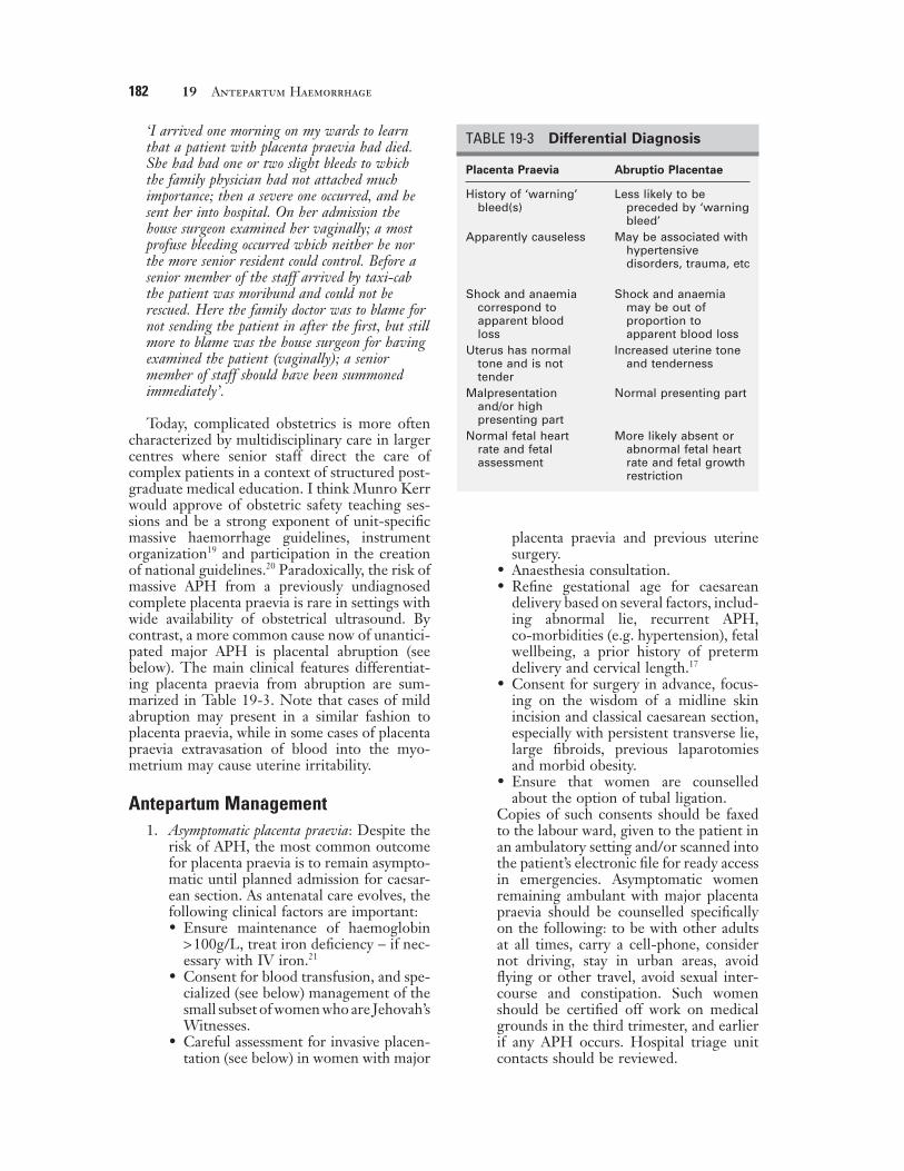
‘I arrived one morning on my wards to learn that a patient with placenta praevia had died. She had had one or two slight bleeds to which the family physician had not attached much importance; then a severe one occurred, and he sent her into hospital. On her admission the house surgeon examined her vaginally; a most profuse bleeding occurred which neither he nor the more senior resident could control. Before a senior member of the staff arrived by taxi-cab the patient was moribund and could not be rescued. Here the family doctor was to blame for not sending the patient in after the fi rst, but still more to blame was the house surgeon for having examined the patient (vaginally); a senior member of staff should have been summoned immediately’.
Today, complicated obstetrics is more often characterized by multidisciplinary care in larger centres where senior staff direct the care of complex patients in a context of structured post-graduate medical education. I think Munro Kerr would approve of obstetric safety teaching ses-sions and be a strong exponent of unit-specifi c massive haemorrhage guidelines, instrument organization 19 and participation in the creation of national guidelines. 20 Paradoxically, the risk of massive APH from a previously undiagnosed complete placenta praevia is rare in settings with wide availability of obstetrical ultrasound. By contrast, a more common cause now of unantici-pated major APH is placental abruption (see below). The main clinical features differentiat-ing placenta praevia from abruption are sum-marized in Table 19-3 . Note that cases of mild abruption may present in a similar fashion to placenta praevia, while in some cases of placenta praevia extravasation of blood into the myo-metrium may cause uterine irritability.
Antepartum Management 1. Asymptomatic placenta praevia : Despite the
risk of APH, the most common outcome for placenta praevia is to remain asympto-matic until planned admission for caesar-ean section. As antenatal care evolves, the following clinical factors are important: • Ensure maintenance of haemoglobin
> 100g/L, treat iron defi ciency − if nec-essary with IV iron. 21
• Consent for blood transfusion, and spe-cialized (see below) management of the small subset of women who are Jehovah ’ s Witnesses.
• Careful assessment for invasive placen-tation (see below) in women with major
TABLE 19-3 Differential Diagnosis
Placenta Praevia Abruptio Placentae
History of ‘warning’ bleed(s)
Less likely to be preceded by ‘warning bleed’
Apparently causeless May be associated with hypertensive disorders, trauma, etc
Shock and anaemia correspond to apparent blood loss
Shock and anaemia may be out of proportion to apparent blood loss
Uterus has normal tone and is not tender
Increased uterine tone and tenderness
Malpresentation and/or high presenting part
Normal presenting part
Normal fetal heart rate and fetal assessment
More likely absent or abnormal fetal heart rate and fetal growth restriction

19 Antepartum Haemorrhage 183
the non-stress test, such women can be admitted to the antenatal unit. Where no formal diagnosis of placenta praevia has been made, elective high-quality transab-dominal and transvaginal ultrasound should be arranged to establish or refute this diagnosis. Women transferred to the labour and delivery area with intermediate-level APH and/or uterine contractions < 32 weeks are at greater risk of delivery in the subsequent 48 hours, and therefore co-care with anaesthesia is important. Some women merit blood transfusion, to stay ahead of blood loss. Tocolysis may be useful in this setting. 16 All women admit-ted in the window 24 − 32 weeks with APH should be given an intramuscular course of antenatal steroids to promote fetal lung maturation. 24 For viable deliveries < 32 weeks women should also be started on a 12-hour IV regimen of magnesium sul-phate for fetal neuro-protection. 25 These two evidence-based interventions make sense in high-resource settings but may be understandably omitted where resources must be focused on survival of the mother and term new-borns.
4. Reassessment of care : Depending on organi-zational structure, women under the care of a midwife or family physician should have their care transferred to an obstetri-cian. Where a diagnosis of major placenta praevia is made following admission to a small birth unit lacking 24/7 in-house support and/or major support services, consideration should be given to transport to a regional centre when stable.
5. Subsequent expectant management : If the patient is < 36 weeks ’ gestation, has no contractions, the bleeding has settled for 48 hours, the fetus is objectively healthy and there are no maternal co-morbidities that direct the need for delivery (e.g. pre-eclampsia), a period of expectant treatment is reasonable in order to gain time for fetal maturation. Documentation of a thin pla-cental edge > 1 cm away from the internal os is reassuring. 17 In borderline situations with major placenta praevia at 34 − 35 weeks, particularly with a new or recurrent APH, amniocentesis in the fasted state for fetal lung maturation makes sense, 26 in order that planned caesarean section can take place when a mature lamellar body count is demostrated – rather than expos-ing women to prolonged hospitalization and the risk of emergency caesarean deliv-ery for a subsequent APH. A positive
2. Admission with major APH : Should this occur, women should be advised to call 911 (or equivalent) and rely on medical services, rather than private transport, in order that they are stabilized (if necessary) and transported to the nearest general hospital with obstetrical services. Ideally, they will arrive with intravenous access. The initial triage assessment should involve ABC (airway/breathing/circula-tion), including vital signs, oxygen satura-tion on air, abdominal examination and a non-stress test. Admission as an emer-gency > 26 weeks with active APH is an indication to perform this initial assess-ment in the operating room, with co-assessment by the anaesthesia team. No pelvic examination should be under-taken; rather, the patient ’ s medical records should be reviewed and an emergency transabdominal ultrasound should be per-formed with portable equipment. Ultra-sound will quickly determine fetal viability (fetal death or bradycardia is rare unless the diagnosis is abruption), fetal lie and presentation, in order to verify the appro-priate skin and likely uterine incision, should the extent of APH dictate immedi-ate caesarean section under general anaes-thesia. In parallel with this obstetric assessment, anaesthesia, supported by nursing and anaesthesia assistants, can establish two large-bore IV access lines to obtain blood samples, start resuscitation measures including infusion of crystalloids + / − colloids and prepare the woman for general anaesthesia (airway assessment, pre-oxygenation) as needed. Initial blood tests are a complete blood count, coagula-tion screen, baseline electrolytes, blood type, antibody screen and cross-match at least two units of packed red cells. Where APH is substantial and immediate general anaesthesia is agreed on, the local policy to alert all systems for massive blood transfusion (often termed ‘code Omega’) should be activated. 22 Surgical and post-operative considerations for women deliv-ered by caesarean section for placenta praevia and major APH are described below. Despite the acute setting, it is important that rhesus-D negative women receive Anti-D. 23
3. Admission with minor APH : Typically in this situation the vaginal bleeding settles during triage assessment of a haemody-namically stable woman. In the absence of any uterine contractility or concerns with

184 19 Antepartum Haemorrhage
data, showing that two-thirds of women can deliver safely vaginally if the lower edge of the placenta is 1 − 2 cm from the internal os. 14 Since the cervix gradually shortens by effacement in the late second and third trimesters, a minor praevia in the context of a normal length ( > 3 cm) cervix is very likely to pull back behind the fetal head, as a result of cervical efface-ment ( Fig 19-2b,c ), or during early normal labour. This apparent movement of the lower placental edge takes place even after 36 weeks; therefore even weekly assess-ments at this stage are worthwhile so as to avoid an unnecessary caesarean section.
7. Fetal wellbeing assessment and placenta praevia : Despite lower implantation, there is no convincing evidence that this results in any direct association with placental dysfunction. 28 Therefore the initial fetal health assessment in the context of placen-tal praevia and an APH should be as follows: fetal biometry, amniotic fl uid, umbilical artery Doppler, biophysical profi le and a non-stress test. Additional Doppler studies should be reserved for specifi c indications: middle cerebral artery Doppler (if the fetus appears growth-restricted), uterine artery Doppler (if the fetus appears growth-restricted or the woman is hypertensive) and anterior lower segment colour Doppler (anterior praevia with previous caesarean section to rule out increta). Since placental function is normal in placenta praevia, fetal growth and tests of wellbeing should follow current advice. 29
Vaginal Delivery in Placenta Praevia The widespread availability of good-quality ultrasound means that most women with minor placenta praevia will have had discussions about mode of delivery with their obstetrician in the antenatal clinic setting. As such, the need to clarify the safety of attempting vaginal delivery in an acute setting is rare. Examples today would be either early normal labour with unusu-ally heavy show and fresh bleeding vaginally, or presentation in labour between appointments where no fi nal decision has been made on mode of delivery. In this context use of the ‘double set-up’ examination is valid, since this was the method used to make such decisions in the pre-ultrasound era in haemodynamically stable women. The components of the process are as follows: transfer to the operating room, co-care with anaesthesia, perform complete blood count
Betke − Kliehauer (BK) test > 10 ml indicat-ing a signifi cant feto-maternal bleed is rare in placenta praevia 27 and would be an indi-cation for delivery > 32 weeks. Likewise, cervical length < 30 mm on transvaginal ultrasound assessment confers a threefold increased risk of subsequent APH and preterm delivery < 37 weeks, which is pre-sumably due to subclinical labour provok-ing bleeding. 17 The main elements of expectant management in hospital are as follows: (a) Admission and bed rest with bath-
room privileges (b) Diagnose and treat anaemia (c) Anaesthesia consultation (d) Continuous cross-match of at least 2
units packed red cells (e) Identify and arrange a date for elective
caesarean and obtain consents – discuss and document type of surgery including desire for tubal ligation
(f) Rhesus-D-negative women with no antibodies should receive Anti-D.
When major placenta praevia is found in the context of an APH and a viable fetus, women are best advised to remain in hos-pital until delivery. If clinically stable, with no further bleeding, they can receive pro-phylactic daily heparin for thrombo-prophylaxis (especially if age > 40 and/or overweight, or have received a blood transfusion), otherwise they can be fi tted with compression stockings. Women with minor placenta praevia, an isolated APH and no unusual considerations (such as poor ambulatory access to care) are often discharged home for ongoing antenatal care. Documenting a normal length cervix is relevant in this context. 17 The subset of women with recurrent APH should remain in hospital, following re-admission, until delivery. Despite the increased adminis-trative pressure to limit antenatal admis-sions, in the context of placenta praevia decisions should err on the side of safety and always include the documented agree-ment of the woman.
6. Follow-up of women with minor placenta praevia : Where women remain undeliv-ered for > 2 weeks in the context of minor (type 1 or 2) placenta praevia, repeat con-sideration should be given to the mode of delivery where vaginal delivery is feasible in the absence of placenta praevia. The modern cut-off for safe vaginal delivery of > 2 cm from the internal os 13 has been challenged by more recent prospective

19 Antepartum Haemorrhage 185
large-bore IV access and two units of blood cross-matched, and be assessed by the anaesthet-ist and by the most senior on-call obstetrician. Pro-active care, using amniotomy and oxytocin infusion, is preferable since cervical effacement and dilation brings the placenta away from the leading edge of the cervix, while descent of the fetal head may compress the lower placental edge. Active bleeding in early labour is an indica-tion for caesarean section, whereas new bleeding in more advanced labour may be a sign of advanced cervical dilatation and thus the possi-bility of vaginal delivery.
Postpartum considerations : Following successful vaginal delivery, the woman is at greater risk of primary postpartum haemorrhage because of increased bleeding from the lower uterine segment that is not capable of strong tetanic contractions. Prophylactic measures should be undertaken as described in Chapter 20 .
Vaginal Delivery with Major (Type 3 or 4) Placenta Praevia In modern obstetric practice, it is occasionally permissible to consider and attempt vaginal delivery when the placenta clearly covers the internal os. The most common situation is with the prenatal diagnosis of a major lethal abnor-mality (e.g. renal agenesis or skeletal dysplasia) or intrauterine fetal death, typically before 24 weeks of gestation. Assuming there are no other considerations (e.g. previous caesarean), the author ’ s group have approached such cases using feticide and pre-induction Gelfoam emboliza-tion of the anterior divisions of the iliac arteries, followed immediately by a high-dose vaginal misoprostol regimen (600 μ g every 4 hours) for induction of labour. Others have approached the challenge in a similar fashion. 30 We would gener-ally not consider this approach after 28 weeks, due to the much greater risk of haemorrhage and the greater likelihood of achieving a lower segment caesarean section.
Abnormal Lie/Malpresentation in Early Labour with Minor Placenta Praevia Given the potential danger of caesarean section in the pre-blood transfusion/antibiotic era, several techniques were developed to achieve maternal survival via vaginal delivery for the non-vertex fetus. These may remain applicable today when the fetus is dead, pre-viable or has a lethal anomaly and in remote areas with limited or unsafe facilities for caesarean section.
(CBC) and coagulation screen, cross-match two units of blood, staff present for immediate cae-sarean section. The major difference today in comparison with former times is that the major-ity of such procedures are done with a full top-up epidural. Women bleeding heavily would proceed faster through the above steps, with the exception of a general anaesthetic. It is useful to have a portable ultrasound machine at hand, which in acute circumstances may be helpful as follows: (1) the placenta may only be a minor praevia with an engaged fetal head – therefore proceed to vaginal examination as the bleeding may only be due to a rapidly dilating cervix; (2) there is major placenta praevia, but the fetus is a back-up transverse lie, mostly above the umbili-cus – therefore use a midline skin entry and be mentally prepared to perform a classical caesar-ean section (see below).
Double set-up examination : Once the epidural is fully functional, the woman is examined abdominally, to determine lie, presentation and engagement. Any clinical doubts should be resolved using portable ultrasound prior to gowning, sterile preparation and adopting the lithotomy position. The procedure is abandoned in favour of caesarean in women with either transverse lie or (most) situations with breech presentation. Next, the bladder is catheterized with a Foley catheter and bag. A sterile fi nger is then inserted vaginally to palpate the fornices. This initial step is done to determine if thick placental tissue is present between the lower uterine segment and the fetal head. If the fetal head is easily palpated through a thin lower uterine segment it is then deemed safe to push the examining 1 − 2 fi ngers through the cervix, to explore the lower uterine segment for any inter-vening placental tissue. Blood clot and placental tissue may be diffi cult to distinguish, though pla-cental tissue is fi rm and may have a gritty feel. If no placenta is found upon digital exploration of the inside of the lower uterine segment then labour can be safely induced with amniotomy and an oxytocin infusion. If placenta praevia is confi rmed, or if there is active bleeding, a cae-sarean section is performed. If the cervix is long and closed, the examination is inconclusive – which is why portable ultrasound is important, so that the woman can leave the operating room with a clear plan for mode of delivery.
Monitoring in labour : Women attempting vaginal delivery with minor placenta praevia should have one-to-one nursing/midwifery care, in a labour room that is in immediate proximity to the operating room. Written informed consent should be obtained for care, including caesarean if needed as an emergency. Women should have

186 19 Antepartum Haemorrhage
BRAXTON HICKS BIPOLAR VERSION
‘Introduce the left hand, with the usual precau-tions, into the vagina, so far as to fairly touch the foetal head, even should it recede an inch. Having passed one or two fi ngers (if only one, let it be the middle fi nger) within the cervix, and resting them on the head, place the right hand on the left side of the breech at the fundus. Employ gentle pressure and slight impulsive movements on the fundus towards the left iliac fossa. In a very short time it will be found that the head is rising and at the same time the breech is descend-ing. The foetus is now transverse; the knee will be opposite the os, and the membranes being ruptured it can be seized and brought into the vagina.’
Bipolar podalic version : Rare circumstances exist where fetal manipulation and assisted vaginal delivery may be the safest maternal option in type 1 − 2 placenta praevia, though at the expense of fetal survival. The Braxton Hicks bipolar podalic version method was developed 150 years ago. 31 The technique demands that the cervix is > 2 cm dilated and the placenta praevia does not cover the internal os – so that the gentle insertion of 1–2 fi ngers can be used to push up the fetal head between contractions while the external hand manipulates the breech in a down-ward direction into the pelvis ( Fig 19-3 ). The fi ngers through the cervix then grasp a foot of the fetus ( Fig 19-4 ) to bring that leg down through the cervix. In this way the breech is used to both dilate the cervix and tamponade the lower placental edge ( Fig 19-5 ). Persistent trac-tion is put on the breech to keep it fi rmly against the placenta: a bandage can be tied to the fetal ankle and a small weight, for example a bag of saline, is attached to provide sustained traction. For small immature fetuses, sponge forceps can be used to grasp a leg.
BRAXTON HICKS BIPOLAR VERSION: USE IN PLACENTA PRAEVIA
‘Anything which gave the practitioner some power of action was to be earnestly welcomed … Turn, and if you employ the child as a plug the danger is over. Then wait for the pains, rally the powers in the interval, and let nature, gently assisted, complete the delivery.’
John Braxton Hicks On a new method of version in abnormal
labour. Lancet 1860; 2:28–30.
WILLETT ’ S SCALP FORCEPS
‘The application of the forceps is easy and they can be applied to the scalp as soon as the os will admit a fi nger, thus ensuring early treatment … a weight varying from 1lb to 2lb, hanging over the end of the bed, is applied to the handles by a tape. Nothing further is done until the head is in the vagina, when the forceps are removed and the patient is allowed to deliver herself without further interference’.
John Willett The treatment of placenta praevia by continu-
ous weight traction – a report of seven cases. Proc R Soc Med 1925; 18:90–94.
Cephalic traction : Vaginal delivery for cephalic presentation with bleeding from minor placenta praevia was described by John Willett using a
specially designed T-forceps to grasp the scalp of the fetus and apply cephalic tamponade to the placenta 32 (Allis forceps or similar can be used). Having passed the forceps through the cervix and grasped the scalp, gentle traction is applied by means of a bandage tied to the handles of the forceps with a light weight hung over the end of the bed ( Fig 19-6 ). The ensuing uterine contractions dilate the cervix to advance labour, while scalp traction provides tamponade on the lower separated edge of the placenta. Deliv-ery is not to be forced by strong traction but should be accomplished by normal uterine contractions.
It is emphasized that the indications for these potentially dangerous techniques are very few. However, the degree of haemostasis produced by these techniques can be impressive and, on the rare occasions it is necessary, life-saving.
Technical Aspects of Caesarean Section for Placenta Praevia The potential for rapid blood loss during caesar-ean section for major placenta praevia demands that senior staff should always attend. In addition to the surgical principles outlined in Chapter 13 the following should be considered specifi cally in the context of placenta praevia:
1. Preoperative preparation : A ‘time out’ pro-cedure 33 should be conducted in the operating room, either following the establishment of regional anaesthesia (combined epidural-spinal is preferable for longer surgery) or prior to induction of general anaesthesia. At least two units of packed red blood cells should be in the operating room and checked. Pro-phylactic IV antibiotics should be given. Two large-bore IVs should be sited. The local ‘code omega’ protocol should ideally be on the wall near the anaesthetist. 22 If

19 Antepartum Haemorrhage 187
FIGURE 19-3 ■ Bipolar version: the fetal head is pushed up with the internal fi nger(s) and the external hand manipulates the breech down to the pelvis.
FIGURE 19-4 ■ Bipolar version: the fetus is turned by combined manipulation and the foot is grasped.
FIGURE 19-5 ■ Bipolar version: the leg of the fetus is pulled through the cervix so that the breech produces tamponade against the placenta and the lower uterine segment.
Placenta

188 19 Antepartum Haemorrhage
FIGURE 19-6 ■ Application of Willett ’ s scalp forceps. Placenta
the fetal lie is abnormal, or the woman is known to have fi broids, portable ultra-sound at this stage is invaluable for the next step.
2. Skin incision : Subumbilical midline skin incision should be used for the following: (a) previous similar scar (b) major praevia with transverse lie and
back-up (the fetus is then mostly above the umbilicus)
(c) women with multiple previous caesar-eans and diffi cult access, multiple adhesions or previous bladder injury
(d) rapid access under general anaesthetic for life-threatening APH.
Pfannenstiel incision is adequate, however, for most controlled placenta praevia situ-ations, especially when the fetus is in a longitudinal lie.
3. Uterine incision : In most cases with pla-centa praevia near term and a longitudinal lie, the lower uterine segment is suffi -ciently developed to allow the standard transverse lower segment incision to be performed. However, due to increased pelvic vascularity and a higher presenting part, a bladder fl ap need not be made, and access is made in the upper portion of the lower segment. There are, however, a number of situations in which a lower uterine segment incision is unwise and a classical uterine incision should be per-formed. These are as follows: (a) abnormal lie with a narrow poorly formed lower uterine segment – this will be easily rec-ognized if the surgeon always uses his/her right hand to explore the uterus to deter-mine the fetal attitude (lie, presentation and high vs. normal level of the presenting part); (b) extremely vascular lateral aspects
of the lower uterine segment; and (c) uterine abnormality or uterine distortion by fi broids. Taking a few moments to review the above is a useful mental strat-egy to anticipate how to best extract the fetus, assuming that excessive bleeding will instantly obscure visual guidance. If in doubt, a well-placed vertical incision is prudent as this will ensure easy and swift delivery of the fetus and placenta. Ulti-mately this approach will result in less blood loss than struggling with a trans-verse incision that either requires a central T extension to deliver the fetus, the risk of excessive bleeding from the uterine arteries or the formation of a broad liga-ment haematoma. Some authors advocate ligating large surface vessels on either side in the line of the proposed line of uterine incision before incising the uterus. A prac-tical alternative is for the surgeon to com-press the upper margin of the transverse uterine incision with the 2nd − 4th fi ngers of his/her left hand on a rolled-up sponge, while the fi rst assistant does the same on the lower margin – this approach also steadies the presenting part. The second assistant should hover a suction tip over the uterine incision to maintain vision during entry.
4. Delivery of the fetus : If the placenta praevia is either anterior or complete it is inevita-ble that placental tissue will be encoun-tered immediately after incising the uterine muscle. No attempt should be made to cut through the placenta; rather, the right hand should separate the pla-centa either upwards or laterally to encounter the fetus palpable through the membranes. The surgeon ’ s left hand is

19 Antepartum Haemorrhage 189
effective action of second-line drugs is impor-tant, and gives the surgeon a sense of control. Meanwhile, discussions can continue with anaes-thesia regarding estimated blood loss and need to commence blood transfusion. Additional sur-gical options to achieve adequate haemostasis include the placement of rectangular absorbable sutures through the lateral parts of the lower uterine segment cavity or the retrograde place-ment of a Bakri balloon, and are described in Chapter 28 . In units with well-developed vascular interventional radiology (VIR), discus-sion of the feasibility to perform Gelfoam occlu-sion of both anterior divisions of the iliac arteries may avoid the necessity for caesarean hysterec-tomy. 36 Ultimately, a variety of factors (blood loss, patient stability, age, parity, practical ability to access VIR, consent for tubal ligation) con-trive to determine if a caesarean hysterectomy should be performed. This is discussed in Chapter 28 .
Jehovah ’ s Witness Patients Routine use of a blood transfusion consent form in tandem with operative consent will serve as a practical step to ensure that Jehovah ’ s Witness patients are identifi ed in advance. Discuss surgery and blood loss issues without extended family present, in order that specifi c informed consent is obtained; this is especially important when discussing surgery with women new to this faith via their partner. Current recommenda-tions on management include safe transfer to a regional centre with a cell saver device that is capable of recycling intraoperative blood back into the woman. 37
PLACENTAL ABRUPTION
Placental abruption refers to partial or complete separation of the normally situated placenta before delivery of the fetus. Since 1980 the population-based incidence in Finland of abrup-tion has reduced by one-third, to approximately 1/300 deliveries. 38 In modern obstetric units maternal death is rare, although abruption increases the risk sevenfold above the general maternal mortality rate. 39 Maternal morbidity can be considerable though, and 10 − 20% of perinatal mortality is attributable to abruption. 39 The recent Finnish cohort study demonstrates the variety of social, medical and obstetrical risk factors for abruption: 38
• advanced maternal age (incremental above age 30)
used in conjunction to ideally attain a lon-gitudinal lie. If the membranes are still intact they are ruptured by the fi rst assist-ant, with persistent fundal pressure by the surgeon – remaining in control. The right hand of the surgeon either guides the fetal pole into the incision, or starts breech extraction. The surgical aspects of pla-centa praevia have been reviewed by Ward. 34
5. Intraoperative management of the third stage : Following delivery of the fetus a bolus and infusion of oxytocin is given to promote a strong sustained contraction of the upper uterine segment. Often the uterus is exte-riorized, but this is not essential and the uterine repair of an uncomplicated pla-centa praevia should take place in situ . 35 The following steps are suggested to min-imize blood loss: (a) Put Green–Armytage clamps on
actively bleeding vessels of the uterine incision
(b) Ligate each uterine angle separately (c) Repair in the normal fashion if the
lower uterine segment has minimal bleeding
(d) If the lower uterine segment oozes signifi cantly, the uterus should be exteriorized to perform compression. The author puts three large sponges behind, inside and in front of the lower uterine segment respectively, inserts the Doyen retractor anterior to these, his left hand behind the poste-rior sponge, then pushes the Doyen retractor with his right hand against his left hand – and holds this for 4 minutes timed ‘by the wall clock’
(e) Should bleeding of concern (‘welling up’) persist, the inside of the lower uterine segment is then explored, with suction to maximize visibility. Any sinusoids bleeding into the cavity are ligated with fi gure-of-eight 2/0 sutures under bimanual control. If the bleeding is generalized, a bladder fl ap should be developed and each uterine artery ligated with a strong No 1 fi gure-of-eight suture midway between the lateral margins of the uterine incision and the bladder angles.
Strategies for ligation of pelvic arteries in surgi-cal postpartum haemorrhage are discussed in detail in Chapter 28 , while additional medical therapies are discussed in Chapter 20 . Massage and compression of the uterus, to await the

190 19 Antepartum Haemorrhage
Pathophysiology Placental separation is initiated by haemor-rhagic disruption of vasculopathic decidual arterioles in the basal plate. 40 Progression of haematoma formation extends the separation, and may be compounded by uterine irritability causing labour since haemorrhage disperses through the myometrium and generates thrombin locally as a powerful contractile agent. 15 Abruption is usually an acute clinical diagnosis, but may be chronic and visible on ultrasound examination. 41 The haematoma adheres to the basal surface and thus facilitates a clinical diagnosis at the third stage of labour. In rare cases of concealed or mixed abruption, the retroplacental extravasation of blood through the myometrium may be so ex-tensive as to reach the serosal surface, causing bruising and discoloration. This is apparent at the time of caesarean section and is known as the Couvelaire uterus, after Alexandre Cou-velaire (1873–1948) of Paris who fi rst described this as ‘uteroplacental apoplexy’. In the past a Couvelaire uterus was often blamed as the cause of uterine atony and postpartum. In fact, in most cases this is due to the associated coagu-lopathy, which refl ects the severity of the process. The combination of arteriolar spasm that accompanies the hypovolaemic shock of severe abruption, coupled with the renal burden of the products of disseminated intravascular coagulation, greatly increases the risk of renal tubular and cortical necrosis and subsequent renal failure.
• hypertensive disorders, particularly severe pre-eclampsia and eclampsia
• increasing parity ( ≥ 3) • smoking (doubles the risk) • prolonged prelabour rupture of the
membranes • extreme preterm delivery (5 − 8% of deliver-
ies 24 − 32 weeks) • multifetal pregnancy (threefold increased
risk) • male pregnancy (55% cases) • sudden decompression of an over-distended
uterus, such as follows uncontrolled rupture of the membranes with polyhydramnios or after delivery of the fi rst twin
• trauma: a fall, domestic violence, car acci-dent or version; overall these are rare causes.
Classifi cation There are three types of abruption illustrated in Figure 19-7 . The most common is revealed where the edge of the placenta separates and blood tracks down between the membranes and the uterine wall to escape through the cervix. In 5–10% of cases the bleeding is concealed, or retroplacental, where the haemorrhage remains trapped between the placenta and the uterus. In this scenario, the mother may complain of con-stant abdominal pain while hypovolaemic shock may occur and the fetus may either be dead, or exhibit signs of acute distress on fetal heart rate monitoring. Sometimes abruption has mixed features.
FIGURE 19-7 ■ Classifi cation of abruptio placentae.
Revealed Concealed Mixed

19 Antepartum Haemorrhage 191
to correct any coagulopathy and to consider interventional radiology techniques as an alter-native to laparotomy and caesarean hysterec-tomy. In most cases of moderate to severe abruption, under-transfusion is common. To avoid hypovolaemia patients require immediate IV crystalloid and blood transfusion to maintain tissue perfusion, especially renal perfusion, which may lessen the chance of disseminated intravascular coagulation (see Chapter 25 ).
Clinical Diagnosis and Management The presentation of abruption can vary widely, from mild revealed bleeding in a stable patient to severe concealed abruption with acute, severe and unrelenting abdominal pain and profound hypovolaemic shock. In the latter situation the uterus is typically hard and tender and the fetus is either dead or exhibits a fetal hear rate pattern consistent with acute asphyxia. Labour has often become established by the time the patient reaches hospital. Initial assessment should focus on maternal resuscitation and collaborative teamwork. Most of the principles outlined above for placenta praevia equally apply to abruption, with a few specifi c differences that can be empha-sized as follows:
1. Acuity of disease : Fetal distress and mater-nal shock can often be out of proportion with revealed blood loss and evolve with alarming speed. Patients with suspected abruption therefore require the full atten-tion of senior staff. Major abruption with a viable fetus in early labour is best managed by a rapid combination of resus-citation, general anaesthesia and caesarean delivery.
2. Potential for vaginal delivery : Provided the fetus is vertex, there is always the potential for successful vaginal delivery in stable women labouring with a minor abruption, especially in multipara. A wise course of action is therefore to manage labour in or very close to an operating room, with amniotomy and attachment of a fetal scalp clip. Co-care with anaesthesia is essential. Conversion to the lithotomy position at around 8cm, subsequent refl ection of the cervix with a contraction, followed by pushing and use of vacuum or forceps to effect delivery when safe, are tips that may be useful to avoid a caesarean section as an abruption is evolving and causing pro-gressive fetal distress.
3. Potential for coagulopathy and PPH : Extrava-sation of maternal blood into the myo-metrium, coupled with rapid progress in labour, are independent risk factors for postpartum haemorrhage. The manage-ment is generally as described for placenta praevia, except that more commonly the woman will have a vaginal delivery.
The emphasis is on ensuring that the uterus is empty and intact and repair of any vaginal trauma, leaving the anaesthetist to manage resuscitation and uterotonic drugs. In this setting use of a uterine tamponade balloon can buy time
COUVELAIRE UTERUS
‘The uterine wall, in the zone of membranous insertion as well as the zone of placental inser-tion, was the site of a tremendous bloody infi ltra-tion separating the muscle bundles … The ovaries were peppered with a punctiform bloody suffusion. The broad ligaments were infi ltrated with blood. This was indeed a true case of utero-placental apoplexy’.
Alexandre Couvelaire Traitement chirurgical des hémorrhagies
utéro-placentaires avec décollement du placenta normalement inséré. Ann Gynécol 1911; 8:591–608
VASA PRAEVIA
Normal placental anatomy includes the insertion of the umbilical cord into the placental disc. In 1 − 2% of pregnancies the placental cord root is either marginal (on the edge − Fig 19-8a ), or more rarely inserted into the fetal membranes and described as velamentous. In both instances, fetal-derived vessels may traverse portions of the membranes. These two situations place the pregnancy at risk of fetally derived vessels running in the membranes over the internal os, termed vasa praevia. The fi rst occurs where the marginal or velamentous cord root is associated with the lower edge of a minor placenta praevia and the second, where fetally derived vessels run from the placental disc to a separate succenturi-ate lobe in the lower uterine segment.
Vasa praevia occurs in about 1/5000 unse-lected singleton pregnancies 42 though higher rates are observed in referral centres due to selection bias. Twins, especially monochorionic, are at increased risk due to the common associa-tion with velamentous cord; as such, universal screening is considered cost-effective in twins. 43 Undiagnosed, vasa praevia may present in labour with vaginal bleeding due to vessel rupture (especially the thin-walled larger veins, see Fig 19-8b − d ) at either spontaneous or artifi cial rupture of the membranes. Fetal distress from vaginal bleeding is typically acute, severe, out of proportion to the amount of vaginal bleeding

192 19 Antepartum Haemorrhage
Another presentation is variable decelerations with intact membranes, where cord presentation may be confused with vasa praevia on vaginal examination.
Rarely, vasa praevia may present more slowly following ruptured membranes in labour, with minor vaginal bleeding and progressive fetal tachycardia in otherwise normal labour.
The rapid bedside ‘Apt test’ quickly detects the presence of fetal haemoglobin in a small sample of collected vaginal blood, based on its resistance to denaturation by alkali compared with adult haemoglobin. A few drops of the vaginal blood are added to 10 ml of 0.1% sodium hydroxide. Adult haemoglobin will turn brown in the solution within 30 seconds but fetal haemoglobin, resisting denaturation by alkali, remains pink. 45
and with no suggestive features of abruption. In this situation, immediate caesarean section under general anaesthesia followed by immediate transfusion of uncross-matched blood may save the infant from hypovolaemic shock, though many cases are fatal ( Fig 19-8d ). Prenatal diag-nosis and planned elective caesarean delivery increase the chance of survival from 47% to 97%. 44
Occasionally, fetally derived chorionic plate arteries may be felt pulsating in the bulging membranes in labour prior to amniotomy; in this case the woman should be transferred to the operating room to establish or refute the diag-nosis by clinical methods (repeat vaginal exami-nation between contractions when the membranes are soft, or amnioscopy) or transvaginal colour Doppler examination ( Fig 19-8b,c ).
FIGURE 19-8 ■ Vasa praevia. (a) Transabdominal ultrasound with colour Doppler demonstrating a velamentous cord insertion (cd) at the placenta (p) with a chorionic plate artery (ar) running towards the inferior wall of the uterus. Transvaginal ultrasound is therefore indicated to fi nd or exclude vasa praevia. (b) Transvaginal ultrasound showing minor posterior placenta praevia (p) that is 2.6 cm from the internal os (io). The dotted line indicates the cervical canal. Cxa, anterior lip of cervix; Cxp, posterior lip of cervix; Cr, free loop of cord. Arrowhead shows a vessel embedded in the amnion that is suspicious for vasa praevia. (c) Corresponding colour and pulsed Doppler view demonstrating continuous venous fl ow (v) from a fetal vasa praevia vein. (d) Consequences of failure to diagnose vasa praevia. Following amniotomy for induction of labour, vaginal bleeding led to acute fetal distress, emergency caesarean section and early neonatal death from consequences of hypovolaemic shock. The rupture point in the cord (asterisk) is shown in the velamentous cord.
a) b)
c) d)

19 Antepartum Haemorrhage 193
Vasa praevia fulfi ls the criteria for an antenatal screening program that can be built into the 19 − 20 week fetal anatomical ultrasound exami-nation, since planned elective caesarean is an effective treatment. In addition to screening for placenta praevia, the transabdominal ultrasound includes both a search for the placental cord root and a succenturiate lobe in the lower uterine segment. Transvaginal ultrasound is reserved for the small subset of women suspicious for vasa praevia ( Fig 19-8a − c ). Presently, one national guideline exists for vasa praevia. 8 A novel new treatment, to avoid an otherwise inevitable cae-sarean section, is fetoscopic laser ablation of the vasa praevia vessels. 46
INVASIVE PLACENTA PRAEVIA
The term placenta accreta refers to excessive adherence of the placenta to the uterine wall. It can occur in the normally implanted site, though the most serious type is invasive placenta praevia, a rare and serious complication of pregnancy that occurs in about 1 in 5000 deliveries. The incidence of all types of placenta accreta is rising, mostly related to pregnancy with multiple prior caesarean deliveries. Advanced maternal age compounds the risk, since older women are at more risk of caesarean section, placenta praevia, dilatation and curettage for spontaneous miscar-riage, myomectomy and pregnancy following endoscopic surgery for Asherman ’ s syndrome. 47
Risk factors in our recent series of 33 women with placenta praevia increta 48 are shown in Table 19-4 . Common to all is injury to the endometrium that transforms to the decidua during implanta-tion. When placental trophoblast makes contact with the decidua it stimulates the formation of a layer of fi brin deposits, Nitabuch ’ s zone – this is the physiological plane of placental separation. 49 Defi cient areas of decidua during implantation, especially in lower segment scars 50 predispose to praevia increta. Pathological placental adherence is patchy, because the underlying decidual damage is non-uniform.
The disease is classifi ed histopathologically as: accreta – loss of Nitabuch ’ s zone and direct
contact of placental villi with myometrium increta – chorionic villi invade the (often defi -
cient) myometrium percreta – placental tissue erodes through the
myometrium to the serosal surface of the uterus, or beyond (parametrium, bladder).
The variable pathology induces a wide spectrum of clinical presentation, ranging from a diffi cult manual removal of placenta and postpartum haemorrhage (focal accreta), through to a total
‘But when I endeavored to extract the placenta it had adhered so strongly to the cervix uteri that it was near an hour and half before I could remove it; nor then without separating the adher-ing part with my hands.’
Edward Rigby An Essay on the Uterine Haemorrhage which
Precedes the Delivery of the Full Grown Fetus: Illustrated with Cases. 6th ed. London: William Hunter, 1822
lack of placental separation at vaginal delivery, to an obvious ‘increta reaction’ on the lower surface of the uterus at laparotomy for planned caesar-ean hysterectomy.
TABLE 19-4 Risk Factors for Placenta Praevia Increta
Risk Factors Outcome (n = 33)
Previous caesarean sections 2 (0 − 4) 0 2 (6.1%) 1 11 (33.3%) 2 13 (39.4%) 3 or more 7 (21.2%)Previous D&Cs 0 (0 − 4) 0 19 (57.6%) 1 10 (30.3%) 2 or more 4 (12.1%)Asherman ’ s syndrome 2 (6.1%)Septal surgery 1 (3.0%)
Data presented as median (range) or n (%), as appropriate. Adapted from Walker MG, Allen L, Windrim RC, Kachura JR, Pollard L, Pantazi S, et al. Multidisciplinary manage-ment of invasive placenta previa. J Obstet Gynaecol Can 2013;35(5):417–25.
Screening for Placenta Praevia Increta Women with a low-lying anterior placenta or obvious major placenta praevia at the 19 − 20 week fetal anatomy ultrasound should have a repeat ultrasound examination at 22 weeks to look for the signs of invasive placentation. The ultrasound features of placenta praevia increta are shown in Figure 19-9a,b . The placenta bulges into the back of the bladder due to pro-gressive myometrial dehiscence. Transvaginal ultrasound may give clearer images and can also evaluate invasion of the cervix ( Fig 19-2d ). Mag-netic resonance imaging (MRI) is not necessary for the diagnosis when ultrasound evaluation is performed and discussed amongst experienced staff. 51 The role of MRI is to defi ne the extent

194 19 Antepartum Haemorrhage
by classical caesarean section. Superfi cially, this is an attractive option since the surgery is techni-cally easy, poses minimal risk of major haemor-rhage and leaves the potential for future pregnancies. This procedure and ultrasound images of gradual spontaneous resolution have been described. 54 However, the risk of signifi cant bleeding remains, with a protracted and uncer-tain period of resolution that can last more than 1 year. 52 Haemorrhage results from the
of myometrial invasion, in particular to identify extrauterine placenta tissue that may involve vital structures, e.g. ureters ( Fig 19-9c ).
Surgical Strategies for Placenta Praevia Increta Elective non-removal of the placenta : We 52 and others 53,54 have evaluated the potential value of elective non-removal of the placenta at delivery
FIGURE 19-9 ■ Invasive placenta praevia. (a) Sagittal transabdominal view of the lower uterine segment demon-strating anterior placenta praevia (p) with the characteristic anterior ‘bulge’ into the bladder (bl) due to myometrial dehiscence. Note several large prominent lakes (l) suggesting placenta increta. (b) Corresponding transvaginal colour Doppler imaging demonstrating bladder wall vascularity (arrowheads) and venous fl ow within the lakes (l). (c) Sagittal magnetic resonance image. Contrast normal anterior placental tissue (np) with placenta increta (pi) above the bladder (bl) that contains dark placental bands corresponding to lakes seen on ultrasound. Arrow-heads indicate increased bladder base vascularity. (d) Preoperative placement of internal iliac artery occlusion balloons (bal). Note fetal spine (s) on maternal left. Arrow indicates insertion point of catheter into the left femoral artery. Arrowheads indicate course to the right iliac artery, via the aortic bifurcation (asterisk). The test arteriogram shows proximal vessel occlusion and distal feeding of the placenta praevia increta (pi). Postoperatively the patient will return to interventional radiology for angiography and Gelfoam slurry embolization of any leaking branches of the anterior division of the internal iliac arteries.
a) b)
c) d)

19 Antepartum Haemorrhage 195
as they facilitate confi dent ureteric identi-fi cation during dissection in the broad ligament. We inform intensive care in advance of the potential need for postop-erative admission. We give preoperative antibiotics and do a ‘time out’ procedure.
3. Anaesthesia : Our preference is for regional epidural anaesthesia for several reasons: fi rst, this gives analgesia for internal iliac catheter placement; second, the partner remains in the room during the operation for support so that both see the new-born; and third, the couple can be informed of progress. In our series, most women remained awake throughout surgery and did not move to intensive care postopera-tively ( Table 19-5 ).
4. Surgery : A general surgery environment is ideal for these cases, since other surgical specialities, in particular urology and vas-cular surgery, then have ready access to their equipment if called to assist. Once the uterus is exposed, we assess the exter-nal appearances of the lower uterus; in cases where the lower uterus bulges for-wards with a major vascular reaction we make the decision, before performing a high midline uterine incision, to leave the placenta in situ, not to infuse oxytocin and to proceed immediately to caesarean hys-terectomy. Typically the fundal incision retracts laterally and does not bleed exces-sively. Therefore, we clamp any bleeding sinusoids with Green − Armytage clamps and proceed with the dissection. We do not regard bladder dome injuries as serious and will repair small openings without consulting urology; indeed, opening the dome of the bladder to identify the trigone and/or catheterize a ureter can be a useful intraoperative strategy in the absence of ureteric catheterization. Once the bladder is dissected adequately and the cardinal ligaments are secured, we decide on the need for total versus subtotal hysterec-tomy, based on: diffi culty with bladder dissection, haemostasis and perceived cer-vical length.
5. Postoperative care : Following postoperative angiography and Gelfoam embolization of both anterior divisions of the internal iliac arteries, one catheter is left overnight and, if stable, the woman is returned to the labour and delivery suite for one-to-one nursing. Epidural analgesia can be contin-ued, which is a distinct advantage over patient-controlled narcotics after general anaestheisa. Where bladder repair or
formation of large arterio-venous fi stulas in decaying placental tissue. Arterio-venous fi stula formation resulted in emergency repeat laparot-omy due to vaginal bleeding in 4/10 women in our series. Despite full resolution, two women requested tubal ligation. Based on this experi-ence, we moved to the preferred option of planned caesarean hysterectomy in 2008. 48 Pres-ently, the indications for conservative manage-ment are: (a) intraoperative diagnosis in a suboptimal setting for emergency caesarean hys-terectomy and (b) patient choice, despite being fully informed of the risks versus benefi ts of defi nitive surgery (e.g. diagnosis after one previ-ous caesarean).
Elective caesarean hysterectomy : Surgery should be scheduled at 34 − 35 weeks 55 balancing prema-turity and risk of emergency surgery due to APH. Given the rarity of this diagnosis and the risk of haemorrhage, women requiring this surgery are best managed by regional teams comprising interested members of the contribut-ing subspecialities. 53 These include surgical obstetrics (or maternal-fetal medicine), pelvic surgery, urology, obstetric anaesthesia, blood transfusion, pathology and critical care. In our recent prospective series of 33 cases, the pro-gressive application of team-based elements of care signifi cantly reduced serious morbidity. 48
Important points to highlight are as follows: 1. Consent : Obtain consent for midline access,
caesarean hysterectomy and (should the placenta separate spontaneously with acceptable bleeding) tubal ligation. Table 19-5 shows the major risks of surgery from our series that should be discussed preoperatively; 10% of women with pla-centa praevia increta did not need a cae-sarean hysterectomy. 48 We document cervical cytology results so as to retain the option of subtotal hysterectomy.
2. Patient preparation : We routinely place balloon catheters in the anterior divisions of the iliac arteries ( Fig 19-9d ) while others favour selective intraoperative ligation of the internal iliac arteries. 53 While intraoperative balloon infl ation, following delivery of the fetus, results in variable degrees of arrest of bleeding, this strategy facilitates immediate postopera-tive angiography and Gelfoam emboliza-tion of aberrant branches of the anterior divisions of the internal iliac arteries. We place a 3-way urinary catheter for intraop-erative methylene blue dye testing of bladder integrity. Ureteric catheters are a wise consideration, especially if lateral placental extension is suspected on MRI,

196 19 Antepartum Haemorrhage
REFERENCES 1. Sinha P , Mishra M . Caesarean scar preganancy: A pre-
cursor of placenta percreta/accreta . J Obstet Gynaecol 2012 ; 32 : 621 – 3 .
2. Rosenberg T , Pariente G , Sergienko R , Wiznitzer A , Sheiner E . Critical analysis of risk factors and outcome of placenta previa . Arch Gynecol Obstet 2011 ; 284 : 47 – 51 .
3. Hunter W . Anatomy of the gravid uterus . London : Bask-erville Press ; 1774 .
4. Baskett TF . Of violent fl oodings in pregnancy: evolution of the management of placenta praevia . In: Sturdee D , Olah K , Keane D , editors. The Yearbook of Obstetrics and Gynaecology . London : RCOG Press ; 2001 . p. 1 – 14 .
5. Baskett TF . Edward Rigby (1747 − 1821) of Norwich and his essay on the uterine haemorrhage . J R Soc Med 2002 ; 95 : 618 – 22 .
6. Norgaard LN , Pinborg A , Lidegaard O , Bergholt T . A Danish national cohort study on neonatal outcome in singleton pregnancies with placenta previa . Acta Obstet Gynecol Scand 2012 ; 91 : 546 – 51 .
7. Blouin D , Rioux C . Routine third trimester ultrasound examination for low-lying or marginal placentas diag-nosed at mid-pregnancy: Is this indicated? J Obstet Gynaecol Can 2012 ; 34 : 425 – 8 .
8. Gagnon R . Guidelines for the management of vasa previa . J Obstet Gynaecol Can 2009 ; 31 : 748 – 53 .
9. Weis MA , Harper LM , Roehl KA , Odibo AO , Cahill AG . Natural history of placenta previa in twins . Obstet Gynecol 2012 ; 120 : 753 – 8 .
10. Danforth DN , Ivy AC . The lower uterine segment: its derivation and physiological behaviour . Am J Obstet Gynecol 1949 ; 57 : 831 – 8 .
11. Bergelin I , Valentin L . Patterns of normal change in cervical length and width during pregnancy in nullipa-rous women: a prospective, longitudinal ultrasound study . Ultrasound Obstet Gynecol 2001 ; 18 : 217 – 22 .
12. Smith GCS , Celik E , To M , Khouri O , Nicolaides KH . Cervical length at mid-pregnancy and the risk of primary cesarean delivery . N Engl J Med 2008 ; 358 : 1346 – 53 .
13. Oppenheimer L . Society of Obstetrics and Gynaecolo-gists of Canada Clinical. Practice Guideline: Diagnosis and Management of Placenta Previa . J Obstet Gynaecol Can 2007 ; 29 : 261 – 6 .
14. Vergani P , Ornaghi S , Pozzi I , Beretta P , Russo FM , Follesa I , et al . Placenta previa: distance to internal os and mode of delivery . Am J Obstet Gynecol 2009 ; 201 : e1 – 5 .
15. O ’ Sullivan C , Allen NM , O ’ Loughlin AJ , Friel AM , Morrison J . Thrombin and PAR1-activating peptide: Effects on human uterine contractility in vitro . Am J Obstet Gynecol 2004 ; 190 : 1098 – 105 .
16. Towers CV , Pircon RA , Heppard M . Is tocolysis safe in the management of third-trimester bleeding? Am J Obstet Gynecol 1999 ; 180 : 1572 – 8 .
17. Stafford IA , Dashe JS , Shiwers SA , Alexander JM , McIntire DD , Leveno KJ . Ultrasonographic cervical length and risk of hemorrhage in pregnancies with pla-centa previa . Obstet Gynecol 2010 ; 116 : 595 – 600 .
18. Ohira S , Kikuchi N , Kobara H , Osada R , Ashida T , Kanai M , et al . Predicting the route of delivery in women with low-lying placenta using transvaginal ultrasonogra-phy: Signifi cance of placental migration and marginal sinus . Gynecol Obstet Invest 2012 ; 72 : 217 – 22 .
19. Baskett TF . Surgical management of severe obstetric hemorrhage: experience with an obstetric hemorrhage equipment tray . J Obstet Gynaecol Can 2004 ; 26 : 805 – 8 .
ureteric stenting has been necessary, we arrange urology co-care for outpatient assessment.
6. Blood transfusion : Based on our experience of blood loss, we do not see the need to routinely deploy a cell saver (unless the woman is a Jehovah ’ s Witness or has complex red cell antibodies). Normovol-aemic haemodilution (crystalloid volume expansion immediately before skin inci-sion) is a useful practical way to reduce red cell loss intraoperatively.
TABLE 19-5 Intraoperative Course and Postoperative Complications
Complication Outcome (n = 33)
Mode of anaesthesia Regional 23 (69.7%) General 4 (12.1%) Conversion from regional
to general6 (18.2%)
Estimated blood loss (ml) 2000 (600 − 10000)Bladder dome injury and
repair10 (30.3%)
Bowel or ureteric injury 0Blood transfusion Intra-/postoperative pRBC
required24 (72.7%)
pRBC (units) 3.5 (0 − 20) Other components
required (FFP, cryoprecipitate, platelets)
11 (33.3%)
Primary surgery Classical caesarean section
and removal of placenta2 (6.1%)
Caesarean hysterectomy 31 (93.9%) Subtotal 16 (48.5%) Total 15 (45.5%)Skin incision Pfannenstiel 3 (9.1%) Midline 30 (90.9%)Operative time (minutes) 107 (68 − 334)Intensive care unit
admission5 (15.2%)
Length of postpartum stay (days)
5 (2 − 13)
Data presented as median (range) or n (%), as appropriate.
FFP, fresh frozen plasma; pRBC, packed red blood cells. Adapted from Walker MG, Allen L, Windrim RC,
Kachura JR, Pollard L, Pantazi S, et al. Multidisciplinary management of invasive placenta previa. J Obstet Gynaecol Can 2013;35(5):417–25.
19 Antepartum Haemorrhage 197
Available at http://www.guideline.gov/content.aspx?id=25668 .
38. Tikkanen M , Riihimaki O , Gissler M , Luukkaala T , Metsaranta M , Andersson , et al . Decreasing incidence of placental abruption in Finland during 1980–2005 . Acta Obstet Gynecol Scand 2012 ; 91 : 1046 – 52 .
39. Tikkanen M . Placental abruption: epidemiology, risk factors and consequences . Acta Obstetr Gynecol Scand 2010 ; 90 : 14014 – 19 .
40. Fitzgerald B , Shannon P , Kingdom J , Keating S . Rounded intraplacental haematomas due to decidual vasculopathy have a distinctive morphology . J Clin Pathol 2011 ; 64 : 729 – 32 .
41. Walker M , Whittle W , Keating S , Kingdom J . Sono-graphic diagnosis of chronic abruption . J Obstet Gynae-col Can 2010 ; 32 : 1056 – 8 .
42. Lee W , Lee VL , Kirk JS , Sloan CT , Smith RS , Com-stock CH . Vasa previa: prenatal diagnosis, natural evolu-tion and clinical outcome . Obstet Gynecol 2000 ; 95 : 572 – 6 .
43. Cipriano LE , Barth WH Jr , Zaric GS . The cost-effectiveness of targeted or universal screening for vasa previa at 18 − 20 weeks of gestation in Ontario . BJOG 2010 ; 117 : 1108 – 18 .
44. Oyelese Y , Catanzarite V , Prefumo F , Lashley S , Schachter M , Tovbin Y , et al . Vasa previa: the impact of prenatal diagnosis on outcomes . Obstet Gynecol 2004 ; 103 : 937 – 42 .
45. Loendersloot EW . Vasa praevia . Am J Obstet Gynecol 1979 ; 135 : 702 – 3 .
46. Chmait RH , Chavira E , Kontopoulos EV , Quintero RA . Third trimester fetoscopic laser ablation of type II vasa previa . J Matern Fetal Neonatal Med 2010 ; 23 : 459 – 62 .
47. Fernandez H , Al-Najjar F , Chauveaud-Lambling A , Frydman R , Gervaise A . Fertility after treatment of Asherman ’ s syndrome stage 3 and 4 . Minimally Invasive Gynecol 2006 ; 13 : 398 – 402 .
48. Walker MG , Allen L , Windrim RC , et al . Multidiscipli-nary management of invasive placenta previa . J Obstet Gynaecol Can 2013 ; 35 ( 5 ): 417 – 25 .
49. Craven CM , Chedwick LR , Ward K . Placental basal plate formation is associated with fi brin deposition in decidual veins at sites of trophoblast cell invasion . Am J Obstet Gynecol 2002 ; 186 : 291 – 6 .
50. Monteagudo A , Carreno C , Timor-Tritsch IE . Saline infusion sonohysterography in nonpregnant women with previous cesarean delivery: The ‘Niche’ in the scar . J Ultrasound Med 2001 ; 20 : 1105 – 15 .
51. Masselli G , Brunelli R , Casciani E , Polettini E , Piccioni MG , Anceschi M , et al . Magnetic resonance imaging in the evaluation of placental adhesive disorders: correla-tion with colour Doppler ultrasound . Eur Radiol 2008 ; 18 : 1292 – 9 .
52. Amsalem H , Kingdom JCP , Farine D , Allen L , Yinon Y , D ’ Souza DL , et al . Planned caesarean hyterectomy versus ‘conserving’ caesarean section in patients with placenta accreta . J Obstet Gynaecol Can 2011 ; 33 : 1005 – 10 .
53. Eller AG , Bennett MA , Sharshiner M , Masheter C , Soisson AP , Dodson M , et al . Maternal morbidity in cases of placenta accreta managed by a multidisciplinary care team compared with standard obstetric care . Obstet Gynecol 2011 ; 117 : 331 – 7 .
54. Wong VV , Burke G . Planned conservative management of placenta percreta . J Obstet Gynaecol 2012 ; 32 : 447 – 52 .
55. Robinson BK , Grobman WA . Effectiveness of timing strategies for delivery of individuals with placenta previa and accreta . Obstet Gynecol 2010 ; 116 : 835 – 42 .
20. RCOG Guidelines . Royal College of Obstetricians and Gynaecologists 2012 . Accessed 5 Nov 2012. Available at http://www.rcog.org.uk/womens-health/clinical-guidance .
21. Khalafallah A , Dennis A , Bates J , Bates G , Robertson IK , Smith L , et al . A prospective randomized, controlled trial of intravenous versus oral iron for moderate iron defi ciency anaemia of pregnancy . J Internal Med 2010 ; 268 : 286 – 95 .
22. Burtelow M , Riley E , Druzin M , Fontaine M , Viele M , Goodnough LT . How we treat: management of life-threatening primary postpartum hemorrhage with a standardized massive transfusion protocol . Transfusion 2007 ; 47 : 1564 – 72 .
23. Fung KF , Eason E . Society of Obstetricians and Gynae-cologists of Canada. Clinical Practice Guidelines: Pre-vention of Rh Alloimmunization . J Obstet Gynaecol Can 2003 ; 25 : 765 – 73 .
24. Roberts D , Dalziel SR . Antenatal corticosteroids for accelerating fetal lung maturation for women at risk of preterm birth . The Cochrane Collaboration 2010 : 1 – 173 .
25. Magee L , Sawchuck D , Synnes A , von Dadelszen P . Society of Obstetricians and Gynaecologists of Canada. Clinical Practice Guideline: magnesium sulphate for fetal neuroprotection . J Obstet Gynaecol Can 2011 ; 33 : 516 – 29 .
26. Torrance HL , Voorbijb HAM , Wijnberger LD , vanBel F , Visser GHA . Lung maturation in small for gestational age fetuses from pregnancies complicated by placental insuffi ciency or maternal hypertension . Early Human Development 2008 ; 84 : 465 – 9 .
27. Wing DA , Paul RH , Millar RK . Usefulness of coagula-tion studies and blood banking in patients with sympto-matic placenta previa . Am J Perinatol 1997 ; 14 : 601 – 4 .
28. Harper LM , Odibo AO , Macones GA , Crane JP , Cahill AG . Effect of placenta previa on fetal growth . Am J Obstet Gynecol 2010 ; 203 : 330e1 – e5 .
29. Lausman A , McCarthy F , Walker M , Kingdom J . Screen-ing, diagnosis, and management of intrauterine growth restriction . J Obstet Gynaecol Can 2012 ; 34 : 17 – 28 .
30. du Passage A , Le Ray C , Grange G , Cabrol D , Tsatsaris V . Termination of pregnancy and placenta previa, inter-est in performing feticide before the labour induction? J Gynecol Obstet Biol (Paris) 2011 ; 40 : 149 – 55 .
31. Hicks JB . On a new method of version in normal labour . Lancet 1860 ; 2 : 28 – 30 .
32. Willett JA . The treatment of placenta previa by continu-ous weight traction: a report of seven cases . Proc R Soc Med 1925 ; 18 : 90 – 4 .
33. Safe Surgery Saves Lives Frequently Asked Questions . World Health Organization , 2012 . Accessed 5 Nov 2012. Available at http://www.who.int/patientsafety/safesurgery/faq_introduction/en/ .
34. Ward CR . Avoiding an incision through the anterior previa at cesarean delivery . Obstet Gynecol 2003 ; 102 : 552 – 4 .
35. Siddiqui M , Goldszmidt E , Fallah S , Kingdom J , Windrim R , Carvalho JCA . Complications of exterior-ized compared with in situ uterine repair at cesarean delivery under spinal anesthesia: A randomized control-led trial . Obstet Gynecol 2007 ; 110 : 570 – 5 .
36. Kirby JM , Katchura JR , Rajan DK , Sniderman KW , Simons ME , Windrim RC , et al . Arterial embolization for primary postpartum hemorrhage . J Vasc Interv Radiol 2009 ; 20 : 1036 – 45 .
37. Placenta praevia, placenta praevia accreta and vasa praevia: diagnosis and management. Agency for Health-care Research and Quality , 2011 . Accessed 5 Nov 2012.