Ch 33 - Woo R; Schulz R; Yang G; Adler J; Krummel T: Robotic Radiosurgery for the General Surgeon
18
375 Abstract General surgery has seen many advances in recent years. In particular, many innovations in surgical technology and technique have been driven by the desire to make operations less invasive and more pre- cise. In today’s operating room, endoscopic, laparo- scopic and robot-assisted technologies are used every day with the goal of providing patients with improved cosmetic results, faster recovery and potentially fewer complications. While laparoscopy has been the dom- inant arena for recent technological developments in general surgery, other surgical disciplines have uti- lized alternative minimally invasive solutions. In the field of neurosurgery, stereotactic surgery and radio- surgery have been used to treat benign and malig- nant lesions of the brain for over 50 and 30 years, respectively. More recently, a frameless stereotactic radio- surgical system has been developed with robotic technology, Figure , that is capable of treating extracranial lesions. As of March 2005, more than 4000 extracranial sites have been treated with the CyberKnife®. More than 000 of these lesions were located in the chest and abdomen—areas that have traditionally been treated by general surgeons. As the scientific understanding and clinical practice of radiosurgery develops, this technology may become an increasingly valuable, minimally invasive option for treating a wide range of general surgical diseases. is chapter reviews the history of stereotactic sur- gery and radiosurgery, introduces the principles of robotic radiosurgery for the general surgeon, com- ments on early results of robotic radiosurgery in a few general surgical applications and discusses the potential for future applications. Introduction Minimally Invasive Surgery, Laparoscopy & Beyond General surgeons have embraced technologies that make procedures both more precise and less inva- sive. During the past two decades, this has been CHAPTER 33 Robotic Radiosurgery for the General Surgeon Russell K. Woo Raymond A. Schulz George P. Yang John R. Adler Jr omas M. Krummel
Transcript of Ch 33 - Woo R; Schulz R; Yang G; Adler J; Krummel T: Robotic Radiosurgery for the General Surgeon

375
Abstract
General surgery has seen many advances in recent years. In particular, many innovations in surgical technology and technique have been driven by the desire to make operations less invasive and more pre-cise. In today’s operating room, endoscopic, laparo-scopic and robot-assisted technologies are used every day with the goal of providing patients with improved cosmetic results, faster recovery and potentially fewer complications. While laparoscopy has been the dom-inant arena for recent technological developments in general surgery, other surgical disciplines have uti-lized alternative minimally invasive solutions. In the field of neurosurgery, stereotactic surgery and radio-surgery have been used to treat benign and malig-nant lesions of the brain for over 50 and 30 years, respectively.
More recently, a frameless stereotactic radio-surgical system has been developed with robotic technology, Figure , that is capable of treating extracranial lesions. As of March 2005, more than
4000 extracranial sites have been treated with the CyberKnife®. More than 000 of these lesions were located in the chest and abdomen—areas that have traditionally been treated by general surgeons. As the scientific understanding and clinical practice of radiosurgery develops, this technology may become an increasingly valuable, minimally invasive option for treating a wide range of general surgical diseases. This chapter reviews the history of stereotactic sur-gery and radiosurgery, introduces the principles of robotic radiosurgery for the general surgeon, com-ments on early results of robotic radiosurgery in a few general surgical applications and discusses the potential for future applications.
Introduction
Minimally Invasive Surgery, Laparoscopy & Beyond
General surgeons have embraced technologies that make procedures both more precise and less inva-sive. During the past two decades, this has been
CHA P T E R
33 Robotic Radiosurgery for the General Surgeon
Russell K. Woo
Raymond A. Schulz
George P. Yang
John R. Adler Jr
Thomas M. Krummel
3 7 6 PA RT V: Future Directions of Robotic Radiosurgery
most evident within general surgery by the develop-ment of minimally invasive, laparoscopic techniques. Although laparoscopic surgery was first described at the beginning of the twentieth century, the wide-spread adoption of these techniques did not occur until the mid-980s and early 990s. This occurred with the development of video-assisted laparoscopy by Dr. Cameron Nezhat,1 followed by the emergence of laparoscopic cholecystectomy, initially performed in 987 by Dr. Philip Moure.2 Since then, the lapa-roscopic approach to cholecystectomy has become standard, and laparoscopic techniques are now rou-tinely utilized to perform a wide range of minimally invasive general surgical operations.
Recent advances in surgical robotics have made possible even less invasive intra-abdominal and tho-racic procedures. Originally developed for use in battlefield surgery,3 tele-operated robots such as the da Vinci® computer-assisted robotic surgical systems (Intuitive Surgical Inc., Sunnyvale, CA), Figure , are
now being used to facilitate a variety of laparoscopic general surgical operations, including: hepatobiliary procedures, bariatric procedures, endocrine proce-dures, colorectal resections, radical prostatectomy and complex foregut operations.3-5
In addition, orthopedic and neurosurgical applica-tions have utilized robots and image guidance for total joint replacement (hip, knee), brain surgery and spine surgery. Some robots, such as Robodoc® (Integrated Surgical Systems Inc., Davis, CA), have had varying degrees of clinical success in this area.
Though the clinical benefits of robotic-assisted surgery have yet to be fully understood or quantified, its use in general surgery has grown because it pro-vides several key technical advantages over standard laparoscopic techniques. These include stereoscopic 3D visualization, motion scaling, tremor reduction, and instrumentation with increased degrees of move-ment.3 Similar to the growth of standard laparos-copy over the last 20 years, the adoption of robotic
Figure . Two robotic surgical systems in use at Stanford University: the da Vinci robotic surgical system (left) and the CyberKnife robotic radiosurgery system (right).

CH A PT ER 33 : Robotic Radiosurgery for the General Surgeon 377
technology into the operating room has been driven by the overarching desire to provide patients with less invasive surgical therapies. In addition, robotic sur-gical systems promise the added advantage of com-puter-enhanced dexterity, which can result in greater operative precision.
While laparoscopy has been the dominant arena for recent technological development in general sur-gery, other surgical disciplines have utilized alter-native minimally invasive solutions. For example, endovascular treatments, such as percutaneous coronary angioplasty, drug-eluting coronary stents, and aortic stent grafts, have revolutionized the man-agement of cardiovascular disease.6-8 In the fields of otolaryngology and neurosurgery, computerized image-guidance systems have been used to accurately register intraoperatively encountered structures with preoperative images.9, 10 In neurosurgery, the develop-ment of robotic radiosurgery merges the fundamental concepts of stereotaxis, stereotactic surgery, real-time image-guided surgery and radiosurgery. It performs bloodless surgery where the surgical instrument is a robotically driven, high-energy image-guided pencil beam of radiation delivered from hundreds of positions—ablating tumors while sparing surround-ing sensitive tissue.
Stereotaxy to Stereotactic Surgery
Stereotactic systems are designed to spatially local-ize a patient’s internal anatomy utilizing a previously acquired anatomical map. This localization is then used to surgically target tissues to achieve a specific objective, such as biopsy, functional stimulation or ablation in a minimally invasive manner. Dittar in 87311 is credited with the first use of a minimally inva-sive frame as a way to target the medulla oblongata in animals, followed by Zernov in 889, who developed an arc-based device designed for operations on the
brain.12 Stereotaxis was coined from the Greek stereo for “three-dimensional” and taxic for “an arrangement (map)” by a neurosurgeon (Horsley) and a mathemati-cian (Clarke), who in 908 were the first to develop a system using 3D cartesian coordinate principles to electrically stimulate the dentate nucleus.13 Clarke submitted the first patent application in 92 for a stereotactic device.14 The most important aspect of Horsley and Clarke’s work was that they developed both the device and anatomical map to go with that device.15–16
Spiegel17 in 947 was the first to develop and use stereotactic frames for use on humans, and various frame designs followed.18 A stereotactic frame for the skull consists of a rigid ring that circles the skull with pins that are screwed into the cranium for rigid fixation. The frame supports two key items: (a) fidu-cial structures that are visible in imaging studies and allow mapping of the target location in the image to its location in the frame’s physical spatial coordinate system, and (b) devices that can accurately and repro-ducibly position and guide instruments into the brain from any location.
In the early days, the corresponding internal landmarks that these frames referenced included the calcified pineal gland, the foramen of Monro and the intercommisural line, all based on intraop-erative X-rays with or without air contrast. Spiegel’s stereotactically positioned rectilinear frame used microdrive-based anterior-posterior and lateral posi-tioning: the electrode was positioned in the superior-inferior position. Leksell19 and others18 developed arc-centered frames such that no matter at what angular location the skull entry position was, the electrode always moved to the center of the arc, which was the target. This scheme later became the basis for his radiosurgical system where all of the beams of radiation converge at one point. All of these frames

3 7 8 PA RT V: Future Directions of Robotic Radiosurgery
were rigidly attached to the skull, via pins in the skull or other rigid fixation schemes, so as to ensure good surgical reproducibility.
The invention of the CT scanner by Godfrey Hounsfield 20 in 968 and its introduction by EMI 21 in 972, revolutionized the ability to target the entire volume of 3D anatomy by providing a digital anatomi-cal map of internal anatomy for the first time. Prior to that development, 3D spatial localization was as best inferred from 2D projections. Clinicians soon had the ability to view the anatomy in any plane through digi-tal manipulation22, 23 of CT data. This wealth of data provides frame-based systems the ability to not only surgically target and deliver therapy to the pathology of interest but, more importantly, to simultaneously spare intervening critical structures with equal sub-millimeter accuracy. 24–27
The next step in the continuum of minimally inva-sive stereotactic surgery was to remove the frame. Frameless stereotaxy was conceptualized by Guthrie,28 developed by Guthrie and Adler in 99229 and per-fected by Bucholz and others shortly thereafter.30–33
In the latest frameless stereotactic surgery systems, surgeons only need to touch skin-affixed fiducials or anatomical landmarks with a special surgical instru-ment, then click on the same point on the 3D image on their computer screen. After four to six of these fiducial localizations are performed the instrument is co-registered to the patient. Frameless systems that recognize the position of the instrument relative to the patient in 3D space typically use infrared optical or magnetic tracking technology. The 3D image (CT-MR fused image) is constantly updated as the instrument is moved through space and into the patient during the operative procedure. These techniques remove the frame-based fixation pins from stereotactic pro-cedures, thereby removing a relatively invasive aspect
of the procedure and providing wider access to condi-tions requiring multi-staged procedures.
Stereotactic Radiosurgery
Stereotactic radiosurgery takes stereotactic surgery one step further by going from minimally invasive surgery to bloodless surgery. It utilizes precision tar-geting, image guidance and hundreds of cross-fired tightly collimated beams of ionizing radiation to non-invasively ablate tissue. Conceptualized in the 950s by Swedish neurosurgeon Lars Leksell, this technol-ogy has been used to treat/ablate a variety of benign and malignant intracranial lesions without any inci-sion.34 Leksell’s Gamma Knife system utilizes 20 beams of gamma radiation distributed over approxi-mately a hemisphere from a 60Co source, all focus-ing at one point. In effect, the Gamma Knife creates a small sphere of ablative radiation at the target, whose size is dependent on the collimation scheme used. The lesioning of non-malignant brain tissue, such as the trigeminal nerve (trigeminal neuralgia), thalamus (tremor) and epileptic foci (intractable seizures), is an additional important clinical application of this technology.34
Stereotactic radiosurgery with the Gamma Knife is confined to cranial lesions. The skull is immobilized using a stereotactic frame that is fixed to the skull via a series of pins. This frame is based on Leksell’s own experience in developing a frame for stereotactic neurosurgery.19 As in stereotactic surgery, accuracy is achieved by identifying the clinical target from the frame-based preoperative imaging study and posi-tioning the center of the target at the therapeutic center of the Gamma Knife. Treatment plans are con-structed so that sensitive tissues are avoided.
Radiosurgery is now generally well established for the treatment of a variety of intracranial conditions. Intracranial contents are relatively static compared

CH A PT ER 33 : Robotic Radiosurgery for the General Surgeon 379
to other tissues in the body. From the viewpoint of radiosurgery, this results in essentially non-mobile target tissues, enabling the delivery of high radiation doses in one or a few treatment stages with minimal risk to adjacent normal tissues. Overall, this has led to the widespread use of stereotactic radiosurgical techniques for the treatment of benign and malignant neurosurgical lesions that are contained within, or closely adjacent to, the rigid confines of the skull.35-39
A number of groups, beginning with Betti 40 and Colombo,41 modified linear accelerator-based sys-tems for the purpose of radiosurgerical lesion abla-tion. These systems have the advantage of being less costly, and provide the opportunity to treat anatomy in regions in the head and neck that were not physi-cally accessible by Gamma Knife. They still require a frame and are somewhat less accurate in targeting and dose conformity, though they typically deliver slightly higher dose homogeneity. Studies from com-mercial versions of these systems have demonstrated radiosurgery to be an important treatment option for many otolaryngologic conditions, such as skull base and head and neck tumors. 42-46
These systems have also been modified for whole-body applications. They have used semi-rigid body frames in an attempt to limit soft tissue move-ment. However, even in these frames, the external body and internal organs have the ability to move several centimeters. Hence, tumor targeting has to account for soft tissue movement, which substantially compromises the ability to have highly conformal radiosurgical targeting to specific anatomy. These systems use the term stereotactic radiosurgery (SRS) and stereotactic radiotherapy (SRT) somewhat inter-changeably, though they do not meet the high treat-ment precision standards of traditional radiosurgery.
Image-Guided Robotic Radiosurgery
Merging the concepts of frameless stereotaxis, radio-surgery and image guidance with robotic surgical technologies, Adler 47 invented a radiosurgery sys-tem in 992 that has become commercially avail-able for extracranial applications since its FDA clearance for those applications in 200 (Accuray Inc, Sunnyvale, CA), Figure . The CyberKnife thus employs robotic technologies familiar to general
Figure 2. Robot accuracy and maneuverability: The CyberKnife robot has six degrees of freedom, a positioning accuracy of 0.5 mm and a positioning repeatability of 0.2 mm. It can thus provide highly conformal delivery of ablative radiation to complex tumor shapes while sparing complex radiosensitive normal tissues.

3 8 0 PA RT V: Future Directions of Robotic Radiosurgery
surgeons and radiosurgery technologies familiar to neurosurgeons, Figure 2, while providing for “stereo-tactic radiosurgery anywhere in the body.” 48 It has been applied to the frameless treatment of both intracranial and extracranial CNS diseases, including spinal tumors.49, 50
Robotic radiosurgery is a superset of stereotactic radiosurgery in that it is frameless and provides the ability to treat whole-body clinical targets. Soft tis-sues in the body are treated using intra-treatment image guidance, which provides high accuracy for relatively static targets. Respiratory motion technol-ogy provides real-time thoracic motion tracking, also resulting in high accuracy for tumors that move with respiration. Many of the newest applications of robotic radiosurgery fall under the traditional realm
of general surgery, including lesions in lung, liver, pancreas, renal and prostate cancers.
The CyberKnife is unique in that, just like a surgeon, it is designed to respond intelligently to changes in the patient position during the (radio)surgical procedure. The CyberKnife, Figure 3, has two imaging-guidance technologies to achieve this: X-ray-based intra-operative image tracking of internal anatomy and infrared camera (IR)-based tracking of respiratory motion. The preparation and procedure incorporat-ing technologies occur with the following basic steps: () Imaging studies are acquired to confirm the lesion target and three to five mm x 4 mm cylindrical gold fiducials are percutaneously placed near or in the clinical target. (2) A planning CT is obtained a week later. (3) The surgeon outlines the tissues to be ablated and those to be spared and the radiation oncologist
Figure 3. CyberKnife image-guided robotic radiosurgery operating room. Key components include: (a) a digital X-ray stereo pair, which is registered to preoperative CT data to provide stereotactic targeting, (b) a high precision robotically driven compact linear accelerator, which provides frameless and bloodless surgery throughout the body, and (c) an IR-LED camera-imaging system, which tracks tumors that move with respiration.
Robotic Manipulator
Treatment couch
Imaging detectors
Linearaccelerator
X‒ray sourcesSynchronycamera
Targeting System
Robotic Delivery System

CH A PT ER 33 : Robotic Radiosurgery for the General Surgeon 38
creates a treatment plan. (4) Once the patient is regis-tered to the system, an on-board pair of digital X-ray images of the patient is compared to the equivalent CT-based digitally reconstructed radiographs (DRR). (5) A compact robotically directed linear accelerator in a step-point-shoot scheme is prepared to direct radiation to the patient from hundreds of directions. (6) When the procedure starts the fiducials are ste-reotactically co-registered on both the CT and the digital stereo pair of images. (7) If the patient moves, within specified limits, the robotic arm compensates for and follows that motion. (8) For organs that move with respiration: (a) LEDs are placed on a vest that the patient wears; (b) an IR camera tracks the motion of the chest; (c) a mathematical model tracks where the tumor is moving in relation to chest motion.
The CyberKnife tracks that motion and keeps in step with breathing, thereby achieving high conformality even for tumors that move several cen-timeters with respiration, Figure 4.
With the ability to target and track soft tissues in the chest and abdomen, robotic radiosurgery may become an increasingly valuable, minimally invasive option for treating a wide range of general surgical diseases. In this chapter, we review the current experience with robotic radiosurgery for the treat-ment of intra-thoracic and intra-abdominal lesions and discuss the potential role of robotic radiosurgery in the treatment of general surgical diseases with par-ticular emphasis on lung, liver, pancreas, kidney and prostate tumors.
General Surgical Applications of Robotic Radiosurgery
The use of radiosurgery for the treatment of extra-cranial lesions, specifically intra-thoracic and intra-abdominal pathologies, is still in its infancy.51-56 To date, there is only a small body of literature regarding the application of robotic radiosurgery for extracranial lesions, much of which is spine and head-neck. The following sections will review the published literature to date regarding the use of robotic radiosurgery for the treatment of intra-thoracic and intra-abdominal pathologies. For liver pathologies, where there is no current literature pertaining to robotic radiosurgery, we reviewed the brief clinical experience of stereo-tactic radiosurgery.
Lung Tissue Ablation
Surgical resection is the standard of care for early primary lung cancers as well as certain metastatic tumors. While this treatment provides the best probability for long-term survival, it is not a viable option for a considerable number of cancer patients.
Figure 4. Tracking tumors that move with respiration. A respiratory motion tracking system employs real-time image-based feedback (inset) to treat tumors moving in multiple directions with respiration. The system has a respiratory motion tumor targeting accuracy of .25 mm for the highly conformal ablation of tumors that move with breathing.

3 8 2 PA RT V: Future Directions of Robotic Radiosurgery
Specifically, resections with curative intent may not be tolerated in patients with impaired pulmonary function, advanced age, poor performance status, acute illness and/or significant comorbidities.55 For these medically inoperable patients, non-surgical therapy consisting of standard fractionated radio-therapy has been employed with survival rates rang-ing from 0-30% in specific populations (stage I non-small cell lung cancers).56 In patients with early primary lung cancers, several studies have reported a benefit with higher radiation doses, suggesting a dose-response relationship for both survival and local control.56 Due to their ability to precisely deliver high dose radiation, the use of stereotactic radiosurgical systems has recently been investigated for the treat-ment of lung cancers. Motion of intra-thoracic organs is quite significant and varies with the respiratory
cycle. Therefore, the use of robotic radiosurgery in the chest has required the development of various tech-niques to compensate for lung motion and allow for accurate tissue ablation while preventing damage to surrounding tissue.
Whyte et al reported the preliminary results of a phase I trial (first phase of a three-stage dose escalation trial) using the CyberKnife system (Accuray Inc., Sunnyvale, CA) to treat 23 patients at two institutions with biopsy-proven lung tumors.57 Eight patients had metastatic tumors and 5 patients had primary lung tumors. Tumor size ranged from -5 cm in maximal diameter and the median treatment time was four hours. The average patient age was 63 years (range 23–87 years). Each patient had two to four cylindrical gold fiducials ( mm x 3 mm) percutaneously placed in the tumor or adjacent to it using local anesthesia
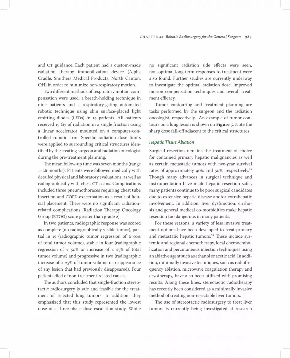
Figure 5. Ablating tumors moving with respiration: by tracking the motion of the chest and internal structures, highly conformal ablative treatments to lung (left) and pancreas (right) tumors are achieved while sparing adjacent sensitive tissue.
CH A PT ER 33 : Robotic Radiosurgery for the General Surgeon 383
and CT guidance. Each patient had a custom-made radiation therapy immobilization device (Alpha Cradle, Smithers Medical Products, North Canton, OH) in order to minimize non-respiratory motion.
Two different methods of respiratory motion com-pensation were used: a breath-holding technique in nine patients and a respiratory-gating automated robotic technique using skin surface-placed light emitting diodes (LEDs) in 4 patients. All patients received 5 Gy of radiation in a single fraction using a linear accelerator mounted on a computer-con-trolled robotic arm. Specific radiation dose limits were applied to surrounding critical structures iden-tified by the treating surgeon and radiation oncologist during the pre-treatment planning.
The mean follow-up time was seven months (range –26 months). Patients were followed medically with detailed physical and laboratory evaluations, as well as radiographically with chest CT scans. Complications included three pneumothoraces requiring chest tube insertion and COPD exacerbation as a result of fidu-cial placement. There were no significant radiation-related complications (Radiation Therapy Oncology Group (RTOG) score greater than grade 2).
In two patients, radiographic response was scored as complete (no radiographically visible tumor), par-tial in 5 (radiographic tumor regression of ≥ 50% of total tumor volume), stable in four (radiographic regression of < 50% or increase of < 25% of total tumor volume) and progressive in two (radiographic increase of > 25% of tumor volume or reappearance of any lesion that had previously disappeared). Four patients died of non-treatment-related causes.
The authors concluded that single-fraction stereo-tactic radiosurgery is safe and feasible for the treat-ment of selected lung tumors. In addition, they emphasized that this study represented the lowest dose of a three-phase dose-escalation study. While
no significant radiation side effects were seen, non-optimal long-term responses to treatment were also found. Further studies are currently underway to investigate the optimal radiation dose, improved motion compensation techniques and overall treat-ment efficacy.
Tumor contouring and treatment planning are tasks performed by the surgeon and the radiation oncologist, respectively. An example of tumor con-tours on a lung lesion is shown on Figure 5. Note the sharp dose fall-off adjacent to the critical structures
Hepatic Tissue Ablation
Surgical resection remains the treatment of choice for contained primary hepatic malignancies as well as certain metastatic tumors with five-year survival rates of approximately 40% and 50%, respectively.58 Though many advances in surgical technique and instrumentation have made hepatic resection safer, many patients continue to be poor surgical candidates due to extensive hepatic disease and/or extrahepatic involvement. In addition, liver dysfunction, cirrho-sis and general medical co-morbidities make hepatic resection too dangerous in many patients.
For these reasons, a variety of less invasive treat-ment options have been developed to treat primary and metastatic hepatic tumors.58 These include sys-temic and regional chemotherapy, local chemoembo-lization and percutaneous injection techniques using an ablative agent such as ethanol or acetic acid. In addi-tion, minimally invasive techniques, such as radiofre-quency ablation, microwave coagulation therapy and cryotherapy, have also been utilized with promising results. Along these lines, stereotactic radiotherapy has recently been considered as a minimally invasive method of treating non-resectable liver tumors.
The use of stereotactic radiosurgery to treat liver tumors is currently being investigated at research

3 8 4 PA RT V: Future Directions of Robotic Radiosurgery
centers throughout the world, including our own center. However, at this time, very little published data exists on SRS and none exists for robotic radiosurgery.
In 2003, Gunvén et al reported the use of a linear accelerator-based radiosurgery system to treat recur-ring liver metastases after hepatectomy.59 Eighteen consecutive patients underwent hepatic resection for metastases of colorectal cancer. They had an average of 2.3 liver lesions (range –7). Seven patients under-went hepatic lobectomy. Eleven patients underwent segmental or smaller resections, with three patients undergoing multiple resections. Four patients devel-oped suitable confined hepatic recurrences diagnosed by CT of the abdomen and chest or CT of the abdo-men and chest X-ray. Stereotactic radiosurgery was presented as an alternative to surgical resection to all patients with the plan for salvage surgical resection if radiation failed.
All patients opted to undergo radiosurgery. A stereotactic body frame was used to compensate for organ movement. A hypo-fractionated regimen of 20 Gy in two fractions or 5 Gy in three fractions was utilized. All patients tolerated the procedure with follow-up ranging from 3 to 0 months. Side effects were limited and did not require hospitalization (tran-sient epigastric pain in one patient and atelectasis and pleural thickening in another). Two patients exhibited complete radiographic remission of their tumors. The remaining two patients exhibited extrahepatic recur-rences, one followed by bilobar hepatic recurrence. At the time of publication, one patient was alive and in remission at 0 months; one patient died of a stroke at 2 months but was free of disease; and two patients died of metastatic disease at 5 and 20 months.
Overall, Gunvén et al highlighted the surgical dif-ficulty and risk involved with repeat hepatic resec-tion. They therefore concluded that stereotactic
radiosurgery represents an attractive, less invasive option for high-risk patients with recurring liver metastases after hepatectomy.
Pancreatic Tissue Ablation
At this time, two published reports describe the use of stereotactic radiosurgery for the treatment of pancreatic lesions. Koong et al published a phase I dose escalation study of stereotactic radiosurgery in patients with locally advanced pancreatic cancer.60 Specifically, Koong and Le wanted to investigate the ability of single-fraction radiosurgery to achieve local control of pancreatic cancers. The CyberKnife sys-tem was used in conjunction with custom-fitted body cradles and breath-holding techniques to deliver the radiosurgical treatment to 5 patients.
Acute gastrointestinal toxicity was scored accord-ing to the RTOG criteria, with response to treatment determined by serial CT scans. Three dose levels (5 Gy, 20 Gy, and 25 Gy) were used. No grade 3 or higher acute gastrointestinal toxicity was observed.60 The trial was stopped because the clinical objective of local control was achieved in all patients receiving the 25 Gy treatment regimen.
Overall, Koong and Le concluded that stereotactic radiosurgery can be used to achieve local control of locally advanced pancreatic cancers without signifi-cant acute gastrointestinal toxicity.60 This modality may provide a benefit to patients undergoing chemo-radiotherapy for unresectable pancreatic cancer by reducing the entire radiation treatment regimen to a single day.
In a second report, Koong and Christofferson in a phase II study assess the use of robotic radiosur-gery as a boost to conventional radiotherapy (RT) in patients with locally advanced pancreatic cancer.61 As expected, there was more toxicity observed with the use of 45 Gy of standard RT plus a 25 Gy boost using

CH A PT ER 33 : Robotic Radiosurgery for the General Surgeon 385
the CyberKnife in a single fraction. Excellent local control of 94% was achieved in the population of 6 patients. Overall survival did not increase significantly because of the late stage of the disease and its rela-tively short time to progression of 7.5 weeks to other sites (typically the liver). New strategies for approach-ing this disease at this late stage include replacing the five weeks of IMRT with chemotherapy.
Prostate Tissue Ablation
The treatment of prostate cancer is a growing and promising new application for extracranial radio-surgery. While multiple centers throughout the world are currently investigating the efficacy of this treatment, only one report has been published in the
medical literature to date. Specifically, in 2003, King et al investigated the use of CyberKnife radiosurgery for the treatment of localized prostate cancer.62 Their study was founded on increasing clinical evidence that supports the use of radiotherapy dose escalation to achieve improved prostate tumor control. These dose escalation regimens have seen limited applica-tion with current standard fractionated radiotherapy techniques because of technical limitations in the delivery of such high doses, due to the proximity of critical structures such as rectum and bladder to the prostate. In addition, there is increasing evidence that hypo-fractionation might increase the thera-peutic ratio. Since stereotactic radiosurgery systems (SRS) and procedures have the ability to generate
Figure 6. Soft tissue abdomino-pelvic targets: the CyberKnife has been used for the treatment of a wide variety of targets in the abdomen and pelvis. In addition to the lung and pancreas, other areas that have been targeted are the liver and prostate. The images above represent treatment for cancer of the prostate. The tumor is outlined in yellow on the left. Critical structures are highlighted in other colors. Intra-treatment X-rays are shown on the right. Notice the fiducials marking the location of the tumor.

3 8 6 PA RT V: Future Directions of Robotic Radiosurgery
highly conformal ablative treatments using single or hypo-fractionated regimens, SRS was investigated for this application. Given these facts, the authors inves-tigated the technical feasibility and potential benefits of using stereotactic radiosurgery techniques for the treatment of localized prostate cancer in one test patient.
In their study, King et al selected a test patient who previously received 74 Gy IMRT (intensity-modulated radiation therapy) to the prostate. They then com-pared isodose lines and dose volume histograms for treatment plans using both the CyberKnife and the IMRT plan. The gross tumor volume coverage was noted to be similar for both IMRT and CyberKnife. However, the CyberKnife was noted to likely deliver a slightly higher mean radiation dose to the prostate with significantly improved dose-volume histograms to the bladder and rectum.
Given the improved normal tissue sparing with the CyberKnife, the authors concluded that stereo-tactic radiosurgery has the potential to allow further dose-escalation compared to IMRT, while keeping radiation exposure to normal tissues under currently acceptable tolerances. Currently, stereotactic radio-surgery is being investigated for the replacement of HDR brachytherapy for localized prostate cancer as well as for boost treatment in patients treated with IMRT. An example of a prostate treatment plan and a DDR during a prostate treatment is shown in Figure 6.
Discussion
In the field of general surgery, radiation therapy has long been established as a treatment modality often used in conjunction with surgical excision. For exam-ple, tumors of the breast, rectum, lung, pancreas, prostate and soft tissues are often treated with radiotherapy as part of a multi-pronged treatment
approach. In this manner, radiotherapy may be uti-lized to shrink tumors prior to surgical removal (neo-adjuvant), during surgery in order to minimize the chance of local recurrence in nearby tissues (intraop-erative) or after surgical treatment in order to reduce local regional recurrence (adjuvant). In addition, radiation therapy can be used as a form of palliative treatment for advanced cancers that may not be ame-nable to surgical excision. In these cases, radiotherapy is often able to relieve the physical symptoms caused by the malignancy (e.g., alimentary tract obstruction or pain) by reducing the tumor size.
While radiotherapy is commonly used in the treat-ment of a wide range of malignant and even benign diseases, its application for many general surgical lesions is not without limitation. For example, the application of radiotherapy to abdominal lesions is often limited by the possibility of severe and debilitat-ing radiation enteritis. In addition, the use of standard radiotherapy as neoadjuvant or adjuvant treatment of malignant lesions often requires multiple sessions of fractionated therapy. These long courses of ther-apy (weeks to months) may be difficult for patients recovering from extensive surgery or for patients with advanced disease who are receiving palliative treatment. In these situations, standard fractionated radiotherapy has the drawbacks of relatively impre-cise radiation delivery leading to damage to adjacent normal tissues, as well as the requirement for long treatment regimens. Stereotactic radiosurgery has the potential to address many of these issues by deliv-ering highly precise, high dose radiation in one or a few sessions.
To date, stereotactic radiosurgery has a long his-tory of successfully treating intracranial pathologies. However, newer technologies and techniques have begun to enable the application of stereotactic radio-surgery to extracranial sites. Specifically, lesions of

CH A PT ER 33 : Robotic Radiosurgery for the General Surgeon 387
the lung, kidney, prostate, pancreas and liver have been treated with stereotactic radiosurgery. At this time, the CyberKnife system has been the most widely applied radiosurgical system for the treatment of intra-thoracic and intra-abdominal targets. As described above, recent additions to the CyberKnife system have enabled it to follow and treat moving tar-gets in real-time. This capability is of utmost impor-tance for essentially any general surgical application of stereotactic radiosurgery, as the abdominal and tho-racic organs are highly mobile compared to the brain and spine. Worldwide, there exist approximately 50 active CyberKnife systems, which have treated more than 8000 intracranial pathologies and more than 4000 extracranial pathologies. The number of extra-cranial cases that Stanford University has treated for spine, lung liver, pancreas and prostate cancers, and the total number of these extracranial cases treated worldwide as of March 2005, are given in Table .
Currently, the published reports, Table 2, describ-ing the use of robotic radiosurgery to treat abdomi-nal and thoracic disease are all preliminary. While they highlight the feasibility and relative safety of this treatment modality, the advantages or disadvantages of robotic radiosurgery compared to other treatment modalities have yet to be determined. From a theo-retical standpoint, robotic radiosurgery may have advantages over conventional radiotherapy for the treatment of anatomically non-resectable lesions. In these cases, the convenience of fewer treatment ses-sions and the ability to accurately deliver high dose radiation to the target tissues may increase treat-ment efficacy while decreasing unwanted side effects. Similarly, these principles may also apply to the neo-adjuvant and adjuvant use of radiation for surgically resectable lesions. In addition, there is recent evidence that robotic radiosurgery may have the additional
benefit of improving the quality of life for patients in severe pain due to their disease.63
In addition to these potential advantages over standard radiotherapy, robotic radiosurgery may have application for the treatment of lesions in which radi-ation currently has no role. Precedence for this exists in the field of neurosurgery. Over the last 30 years, experience with stereotactic radiosurgery for the treatment of intracranial lesions has gone beyond conventional neoadjuvant and adjuvant therapies. Instead, radiosurgery has been used as an ablative tool that uses energy to essentially “resect” lesions that were previously removed using open surgery.
For example, arteriovenous malformations (AVMs), which were historically treated with open surgical resection, are now definitively treated with radiosur-gical ablation. Once again, this complete “ablation” is made possible by the ability of radiosurgery to pre-cisely deliver conformal, high dose radiation to target tissues in one or a few treatment sessions.
While there are no similar extracranial applica-tions of robotic radiosurgery, one may envision a time when high dose energy ablation is used as an alterna-tive to surgical resection for the treatment of benign and even malignant lung, liver, prostate and pancreas
Table . Selected extracranial sites treated by the CyberKnife through March 2005.
Organ Stanford Worldwide
Spine 64 686
Lung 3 58
Liver 6 208
Pancreas 33 257
Prostate 22 7

3 8 8 PA RT V: Future Directions of Robotic Radiosurgery
lesions. Perhaps hepatic hemangiomas, like intracra-nial AVMs, may one day be definitively treated using radiosurgery. At this time, such applications are purely fiction. However, the technology now exists to accurately deliver ablative doses of radiation to intra-abdominal and intra-thoracic targets. Given this abil-ity, radiosurgery can be viewed as a high-tech scalpel, and should therefore be used under the direction of a skilled surgeon. In this manner, radiosurgical pro-cedures may be planned that avoid injury to critical adjacent tissues while maximizing therapy to the target areas. As general surgeons, we should be the physicians who guide the use of this technology and determine if radiosurgery has a role in the treatment of general surgical diseases.
It has been postulated by Gildenberg17 that stereo-tactic surgery “will be incorporated into the arma-mentarium of every general surgeon.” Now, with the advent of whole-body image-guided robotic radio-surgery, we further extend that statement to suggest that bloodless tissue ablation, via image-guided fra-meless robotic radiosurgery, will be the next tool in the continuum of instruments desired by and used by general surgeons for patient treatment and care for a variety of diseases. The development of and enhance-ments to robotic radiosurgery have been made
possible by inventors and visionaries in key precedent technologies across a variety of fields—stereotaxis, image guidance, radiosurgery, robotics, medical image analysis—upon whose shoulders we stand.
References . Nezhat C, Crowgey SR, Garrison CP. Surgical treat-
ment of endometriosis via laser laparoscopy. Fertil Steril 1986;45:778-783.
2. Filipi CJ. A history of endoscopic surgery. In: Arregui ME, Katkhouda N, J. McKernan JB et al, eds. Principles of Laparoscopic Surgery Basic and Advanced Techniques. New York:Springer, 995,3-20.
3. Lanfranco AR, Castellanos AE et al. Robotic surgery: a current perspective. Ann Surg 2004;239:4-2.
4. Talamini MA, Chapman S, Horgan S et al. A prospec-tive analysis of 2 robotic-assisted surgical procedures. Surg Endosc 2003;7:52-524.
5. Talamini M, Campbell K, Stanfield C. Robotic gastro-intestinal surgery: early experience and system descrip-tion. J Laparoendosc Adv Surg Tech A 2002;2:225-232.
6. Arjomand H, Turi ZG, McCormick D et al. Percutaneous coronary intervention: historical perspectives, current status, and future directions. Am Heart J 2003;46: 787-796.
Table 2. Extracranial clinical experience with intra-thoracic and intra-abdominal targets.
Organ Authors Patients Treated
Lung Whyte et al 57 5 patients with primary lung tumors 8 patients with metastatic lesions
Liver Gunven et al 59 4 patients with recurrent colorectal liver metastases after hepatectomy
Pancreas Koong et al 60 5 patients with locally advanced pancreatic cancer
Prostate King et al 62 patient with prostate cancer

CH A PT ER 33 : Robotic Radiosurgery for the General Surgeon 389
7. Bush RL, Lin PH, Lumsden AB. Endovascular manage-ment of abdominal aortic aneurysms. J Cardiovasc Surg (Torino) 2003;44:527-534.
8. Bush RL, Lin PH, Lumsden AB. Endovascular treat-ment of the thoracic aorta. Vasc Endovascular Surg 2003;37:399-405.
9. Kelly PJ. State of the art and future directions of minimally invasive stereotactic neurosurgery. Cancer Control 995;2:287-292.
0. Kelly PJ. Stereotactic surgery: what is past is prologue. Neurosurgery 2000;46:6-27.
. Dittmar C. Ueber die Lage des sogenannten Gefaesszentrums in der Medulla oblongata. Bersaechs Ges Wiss Leipzig (Math Phys) 873;25:449-469.
2. Zernov DN. Encephalometer: device for extimation of parts of brain in human. Proc Soc Physicomed Moscow Univ 889;2:70-80.
3. Horsley V, Clarke RH. The structure and function of the cerebellum examined by a new method. Brain 908;3:45-24.
4. Kelly PJ. Tumor Stereotaxis. Philadelphia: W. B Saunders 99.
5. Clark RH. Part I: Investigation of the Central Nervous System, Methods and Instruments. Baltimore: Johns Hopkins, 920.
6. Clark RH. Part II: Atlas of the Frontal Sections of the Cranium and Brain of the Rhesus Monkey. Baltimore: Johns Hopkins, 920.
7. Spiegel EA, Wycis HT, Marks M et al. Stereotaxic apparatus for operations on the human brain. Science 947;06:349-350.
8. Gildenberg PL, The history of stereotactic and func-tional neurosurgery. In: Gildenberg PL and Tasker RR. Textbook of Stereotactic and Functional Neurosurgery. McGraw-Hill, 998, 5-9.
9. Leksell L. A stereotaxic apparatus for intracerebral sur-gery. Acta Chir Scand 949;99:229-233.
20. Hounsfield GN. Computed medical imaging, In: Lindsten J, ed. Nobel Lectures: Physiology or Medicine, 1971-1980. Singapore: World Scientific Publishing, 992,568-586.
2. Hounsfield GN. Computerized transverse axial scan-ning (tomography), Part I. Description of the system. Br J Radiol 973;46:06-022.
22. Schulz RA, Joseph PM, Hilal SK. Frontal and lateral views of the brain reconstructed from EMI axial slices. Radiology 977;25:70-70.
23. Rhodes ML, Glenn WV, Azzawi YM et al. Stereotactic neurosurgery using 3-D image data from computed tomography. J Med Syst 982;6:05-9.
24. Cala LA, Mastaglia FL, Vaughan RJ. Localisation of stereotactic radiofrequency thalamic lesions by com-puterised axial tomography. Lancet 976;2:33-34.
25. Brown RA. A stereotactic head frame for use with CT body scanners. Invest Radiol 979;4:300-304.
26. Roberts TS, Brown R. Technical and clinical aspects of CT-directed stereotaxis. Appl Neurophysiol 980; 43:70-7.
27. Kelly PJ, Kall BA, Goerss S. Transposition of volu-metric information derived from computed tomog-raphy scanning into stereotactic space. Surg Neurol 984;2:465-47.
28. Guthrie BL, Kaplan R, Kelly PJ. Neurosurgical stereo-tactic operating arm. Stereotact Funct Neurosurg 990;54,55:497-500.
29. Guthrie BL, Adler JR. Computer-assisted preoperative planning, interactive surgery, and frameless stereo-taxy. Clin Neurosurg 992;38:2-3.
30. Smith KR, Frank KJ, Bucholz RD. The NeuroStation— a highly accurate, minimally invasive solution to frame-less stereotactic neurosurgery. Comput Med Imaging Graph 994;8:247-256.
3. Sandeman DR, Patel N, Chandler C et al. Advances in image-directed neurosurgery: preliminary experience with the ISG Viewing Wand compared with the Leksell G frame. Br J Neurosurg 994;8:529-544.

3 9 0 PA RT V: Future Directions of Robotic Radiosurgery
32. Maurer CR, Fitzpatrick JM, Galloway RL et al. Reg-istration of head volume images using implantable fiducial markers. IEEE Transactions on Medical Imaging 997;6:447–462.
33. Heilbrun MP, McDonald P, Wiker C et al. Stereotactic localization and guidance using machine vision tech-nique. Stereotact Funct Neurosurg 992;58:94-98.
34. Kondziolka D, Lunsford LD, Witt TC et al. The future of radiosurgery: radiobiology, technology, and applica-tions. Surg Neurol 2000;54:406-44.
35. Chang SD, Adler JR. Current status and optimal use of radiosurgery. Oncology (Huntington) 200;5:209-26, discussion 29-22.
36. Petrovich Z, Jozsef G, Yu C, et al. Radiotherapy and stereotactic radiosurgery for pituitary tumors. Neurosurg Clin N Am 2003;4:47-66.
37. Roland PS, Eston D. Stereotactic radiosurgery of acous-tic tumors. Otolaryngol Clin North Am 2002;35:343-355.
38. Lederman G, Wronski M, Fine M. Fractionated radio-surgery for brain metastases in 43 patients with breast carcinoma. Breast Cancer Res Treat 200;65:45-54.
39. Gerosa M, Nicolato A, Foroni R. The role of Gamma Knife radiosurgery in the treatment of primary and met-astatic brain tumors. Curr Opin Oncol 2003;5:88-96.
40. Betti O, Derechinsky V. Hyperselective encephalic irra-diation with linear accelerator. Acta Neurochir (Vienna) 984;33 suppl:385-390.
4. Colombo F, Benedetti A, Pozza RC et al External stereo-tactic irradiation by linear accelerator. Neurosurgery 985;6:54-60.
42. Ahn YC, Lee KC, Kim DY et al. Fractionated stereo-tactic radiation therapy for extracranial head and neck tumors. Int J Radiat Oncol Biol Phys 2000;48:50-505.
43. Cmelak AJ, Cox RS, Adler JR et al. Radiosurgery for skull base malignancies and nasopharyngeal carci-noma. Int J Radiat Oncol Biol Phys 997;37:997-003.
44. Bajada C, Selch M, De Salles A et al. Application of stereotactic radiosurgery to the head and neck region. Acta Neurochir Suppl (Wien) 994;62:4-7.
45. Blomgren H, Lax I, Naslund I et al. Stereotactic high dose fraction radiation therapy of extracranial tumors using an accelerator. Clinical experience of the first thirty-one patients. Acta Oncol 995;34:86-870.
46. Gardner E, Linskey ME, Penagaricano JA et al. Stereotactic radiosurgery for patients with cancer of the head and neck. Curr Oncol Rep 2003;5:64-69.
47. Adler JR Jr., Chang SD, Murphy MJ et al. The CyberKnife: a frameless robotic surgery system for radiosurgery. Neurosurg 997;60:24-28.
48. Brogdon NC, CyberKnife FDA letter re: Premarket notification [50 (k)], Summary and indications for use, Department of Health and Human Services: Public Health Service: Food and Drug Administration, August 200.
49. Gerszten PC, Ozhasoglu C, Burton SA et al. CyberKnife frameless stereotactic radiosurgery for spinal lesions: clinical experience in 25 cases. Neurosurgery 2004;55:89-99.
50. Ryu SI, Chang SD, Kim DH et al. Image-guided hypo-fractionated stereotactic radiosurgery to spinal lesions. Neurosurgery 200;49:838-846.
5. Takacs I, Hamilton AJ. Extracranial stereotactic radiosurgery: applications for the spine and beyond. Neurosurg Clin N Am 999;0:257-270.
52. Uematsu M. Stereotactic radiation therapy for non-small cell lung cancer. Nippon Geka Gakkai Zasshi 2002;03:256-257.
53. Murphy MJ, Adler JR, Jr., Bodduluri M et al. Image-guided radiosurgery for the spine and pancreas. Comput Aided Surg 2000;5:278-288.
54. Murphy MJ, Martin D, Whyte R et al. The effective-ness of breath-holding to stabilize lung and pancreas tumors during radiosurgery. Int J Radiat Oncol Biol Phys 2002;53:475-482.

CH A PT ER 33 : Robotic Radiosurgery for the General Surgeon 39
55. Lee SW, Choi EK, Park HJ et al. Stereotactic body frame based fractionated radiosurgery on consecutive days for primary or metastatic tumors in the lung. Lung Cancer 2003;40:309-35.
56. Timmerman R, Papiez L, McGarry R et al. Extracranial stereotactic radioablation: results of a phase I study in medically inoperable stage I non-small cell lung cancer. Chest 2003;24:946-955.
57. Whyte RI, Crownover R, Murphy MJ et al. Stereotactic radiosurgery for lung tumors: preliminary report of a phase I trial. Ann Thorac Surg 2003;75:097-0.
58. Fuss M, Thomas CR. Stereotactic body radiation ther-apy: an ablative treatment option for primary and sec-ondary liver tumors. Ann Surg Oncol 2004;:30-38.
59. Gunven P, Blomgren H, Lax I. Radiosurgery for recur-ring liver metastases after hepatectomy. Hepatogastro-enterology 2003;50:20-204.
60. Koong AC, Le QT, Ho A et al. Phase I study of stereo-tactic radiosurgery in patients with locally advanced pancreatic cancer. Int J Radiat Oncol Biol Phys 2004;58:07-02.
6. Koong AC, Christofferson E, Le Q et al. Phase II study to assess the efficacy of conventionally fractionated radiotherapy followed by a stereotactic radiosurgery boost in patients with locally advanced pancreatic can-cer. Int J Radiat Oncol Biol Phys 2005, in press.
62. King CR, Lehmann J, Adler JR et al. CyberKnife radio-therapy for localized prostate cancer: rationale and tech-nical feasibility. Technol Cancer Res Treat 2003;2:25-30.
63. Degan JW, Gagnon GJ, Voyadzis JM, et al. CyberKnife stereotactic radiosurgical treatment of spinal tumors for pain control and quality of life. J Neusurg: Spine 2005;2:540-549