Ceramic foam plates: a new tool for processing fresh radical prostatectomy specimens
6
ORIGINAL ARTICLE Ceramic foam plates: a new tool for processing fresh radical prostatectomy specimens Tatjana Vlajnic & Martin Oeggerli & Cyrill Rentsch & Heike Püschel & Tobias Zellweger & George N. Thalmann & Christian Ruiz & Lukas Bubendorf Received: 16 April 2014 /Revised: 25 August 2014 /Accepted: 3 October 2014 /Published online: 17 October 2014 # Springer-Verlag Berlin Heidelberg 2014 Abstract Procurement of fresh tissue of prostate cancer is critical for biobanking and generation of xenograft models as an important preclinical step towards new therapeutic strate- gies in advanced prostate cancer. However, handling of fresh radical prostatectomy specimens has been notoriously chal- lenging given the distinctive physical properties of prostate tissue and the difficulty to identify cancer foci on gross exam- ination. Here, we have developed a novel approach using ceramic foam plates for processing freshly cut whole mount sections from radical prostatectomy specimens without compromising further diagnostic assessment. Forty-nine rad- ical prostatectomy specimens were processed and sectioned from the apex to the base in whole mount slices. Putative carcinoma foci were morphologically verified by frozen sec- tion analysis. The fresh whole mount slices were then laid between two ceramic foam plates and fixed overnight. To test tissue preservation after this procedure, formalin-fixed and paraffin-embedded whole mount sections were stained with hematoxylin and eosin (H&E) and analyzed by immunohis- tochemistry, fluorescence, and silver in situ hybridization (FISH and SISH, respectively). There were no morphological artifacts on H&E stained whole mount sections from slices that had been fixed between two plates of ceramic foam, and the histological architecture was fully retained. The quality of immunohistochemistry, FISH, and SISH was excellent. Fixing whole mount tissue slices between ceramic foam plates after frozen section examination is an excellent method for processing fresh radical prostatectomy specimens, allowing for a precise identification and collection of fresh tumor tissue without compromising further diagnostic analysis. Keywords Prostate cancer . Ceramic . Biobank . Frozen tissue . Frozen section . Prostatectomy Introduction Understanding and investigating the underlying genetic and molecular mechanisms that promote neoplastic growth and progression in prostate cancer often requires access to high quality tumor tissue. Despite recent technical progress in molecular analysis of formalin-fixed and paraffin-embedded (FFPE) archived tissue [1, 2], FFPE tissue for research often remains second choice, whereas fresh-frozen tissue provides higher resolution in genomic profiling and is also preferred for RNA expression profiling. Importantly, harvesting fresh tu- mor tissue under sterile conditions is crucial for generating xenograft models as preclinical model systems. In an ongoing global action plan initiative of the Movember Foundation, we aim at establishing a shared re- source of well-annotated primary human prostate cancer xe- nografts through a multi-disciplinary effort [3]. Radical pros- tatectomy specimens represent a major source of tumor tissue for this purpose. However, it is notoriously difficult to identify prostate cancer by eye on gross examination. Therefore, his- tological verification of cancer tissue is critical prior to sub- mitting tissue for successful xenografting or biobanking. In addition, dissection of the whole fresh prostate gland results in T. Vlajnic (*) : M. Oeggerli : H. Püschel : C. Ruiz : L. Bubendorf Institute of Pathology, University Hospital Basel, Schoenbeinstrasse 40, 4031 Basel, Switzerland e-mail: [email protected] C. Rentsch : H. Püschel Department of Urology, University Hospital Basel, Basel, Switzerland T. Zellweger Division of Urology, St. Claraspital, Basel, Switzerland G. N. Thalmann Department of Urology, University of Bern, Inselspital Bern, Bern, Switzerland Virchows Arch (2014) 465:637–642 DOI 10.1007/s00428-014-1665-8
-
Upload
independent -
Category
Documents
-
view
0 -
download
0
Transcript of Ceramic foam plates: a new tool for processing fresh radical prostatectomy specimens

ORIGINAL ARTICLE
Ceramic foam plates: a new tool for processing fresh radicalprostatectomy specimens
Tatjana Vlajnic & Martin Oeggerli & Cyrill Rentsch &
Heike Püschel & Tobias Zellweger &George N. Thalmann &
Christian Ruiz & Lukas Bubendorf
Received: 16 April 2014 /Revised: 25 August 2014 /Accepted: 3 October 2014 /Published online: 17 October 2014# Springer-Verlag Berlin Heidelberg 2014
Abstract Procurement of fresh tissue of prostate cancer iscritical for biobanking and generation of xenograft models asan important preclinical step towards new therapeutic strate-gies in advanced prostate cancer. However, handling of freshradical prostatectomy specimens has been notoriously chal-lenging given the distinctive physical properties of prostatetissue and the difficulty to identify cancer foci on gross exam-ination. Here, we have developed a novel approach usingceramic foam plates for processing freshly cut whole mountsections from radical prostatectomy specimens withoutcompromising further diagnostic assessment. Forty-nine rad-ical prostatectomy specimens were processed and sectionedfrom the apex to the base in whole mount slices. Putativecarcinoma foci were morphologically verified by frozen sec-tion analysis. The fresh whole mount slices were then laidbetween two ceramic foam plates and fixed overnight. To testtissue preservation after this procedure, formalin-fixed andparaffin-embedded whole mount sections were stained withhematoxylin and eosin (H&E) and analyzed by immunohis-tochemistry, fluorescence, and silver in situ hybridization(FISH and SISH, respectively). There were no morphologicalartifacts on H&E stained whole mount sections from slices
that had been fixed between two plates of ceramic foam, andthe histological architecture was fully retained. The quality ofimmunohistochemistry, FISH, and SISH was excellent. Fixingwhole mount tissue slices between ceramic foam plates afterfrozen section examination is an excellentmethod for processingfresh radical prostatectomy specimens, allowing for a preciseidentification and collection of fresh tumor tissue withoutcompromising further diagnostic analysis.
Keywords Prostate cancer . Ceramic . Biobank . Frozentissue . Frozen section . Prostatectomy
Introduction
Understanding and investigating the underlying genetic andmolecular mechanisms that promote neoplastic growth andprogression in prostate cancer often requires access to highquality tumor tissue. Despite recent technical progress inmolecular analysis of formalin-fixed and paraffin-embedded(FFPE) archived tissue [1, 2], FFPE tissue for research oftenremains second choice, whereas fresh-frozen tissue provideshigher resolution in genomic profiling and is also preferred forRNA expression profiling. Importantly, harvesting fresh tu-mor tissue under sterile conditions is crucial for generatingxenograft models as preclinical model systems.
In an ongoing global action plan initiative of theMovember Foundation, we aim at establishing a shared re-source of well-annotated primary human prostate cancer xe-nografts through a multi-disciplinary effort [3]. Radical pros-tatectomy specimens represent a major source of tumor tissuefor this purpose. However, it is notoriously difficult to identifyprostate cancer by eye on gross examination. Therefore, his-tological verification of cancer tissue is critical prior to sub-mitting tissue for successful xenografting or biobanking. Inaddition, dissection of the whole fresh prostate gland results in
T. Vlajnic (*) :M. Oeggerli :H. Püschel :C. Ruiz : L. BubendorfInstitute of Pathology, University Hospital Basel, Schoenbeinstrasse40, 4031 Basel, Switzerlande-mail: [email protected]
C. Rentsch :H. PüschelDepartment of Urology, University Hospital Basel, Basel,Switzerland
T. ZellwegerDivision of Urology, St. Claraspital, Basel, Switzerland
G. N. ThalmannDepartment of Urology, University of Bern, Inselspital Bern,Bern, Switzerland
Virchows Arch (2014) 465:637–642DOI 10.1007/s00428-014-1665-8
deformation of the whole mount tissue slices, which impairsfurther pathological assessment.
Thus, in order to preserve important pathological featuresfor diagnosis and staging, such as margin status, tumor extent,and tumor grade, standardized protocols for handling andprocessing radical prostatectomy specimens have beenestablished [4]. According to the guidelines of the Interna-tional Society of Urological Pathology (ISUP), radical pros-tatectomy specimen should be cut only after full fixationfollowed by complete embedding.
Several methods for harvesting fresh prostate carcinomatissue for research purposes have been proposed, but an opti-mal consensus approach still remains to be established.
Here, we have tested a novel approach using ceramic foamplates for processing freshly cut prostatectomy specimenswithout compromising further diagnostic assessment.
Materials and methods
Tissue samples
From April 2013 to February 2014, 49 radical prostatectomyspecimens were freshly processed under sterile conditions forharvesting fresh tumor tissue to generate xenografts on NOG/NSG mice, for biobanking, and for intraoperative analysis ofresection margins. The prostatectomy specimens were proc-essed at the pathology department of the University HospitalBasel immediately after surgical removal. The prostate wasweighted and measured in three dimensions. Apical and dor-solateral margins were examined by frozen section analysis.
Fresh tissue harvesting
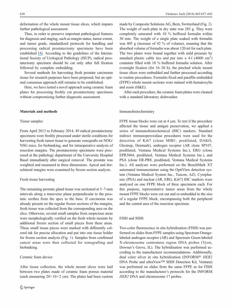
The remaining prostate gland tissue was sectioned at 5–7-mmintervals along a transverse plane perpendicular to the pros-tatic urethra from the apex to the base. If carcinoma wasalready present on the regular frozen sections of the margins,fresh tissue was collected from the corresponding area on theslice. Otherwise, several small samples from suspicious areaswere morphologically verified on the fresh whole mounts byadditional frozen section of small pieces from these areas.These small tissue pieces were marked with differently col-ored ink for precise allocation and put into one tissue holderfor frozen section analysis (Fig. 1). Samples from confirmedcancer areas were then collected for xenografting andbiobanking.
Ceramic foam device
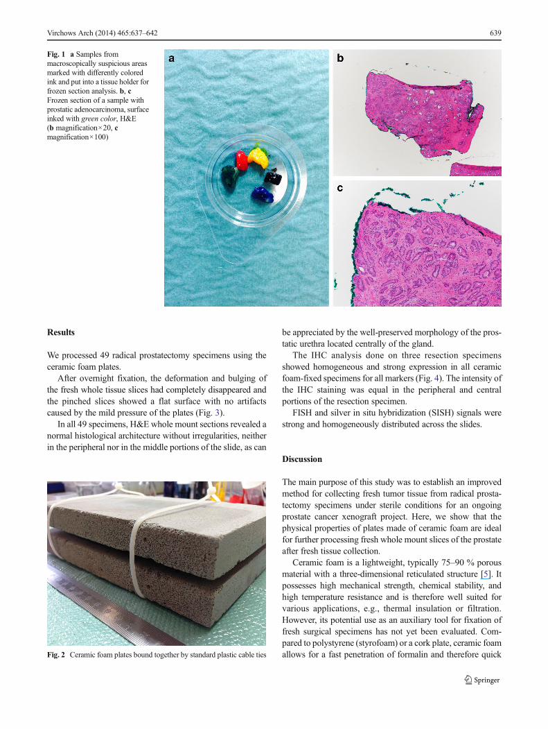
After tissue collection, the whole mount slices were laidbetween two plates made of ceramic foam porous material(each measuring 20×10×2 cm). The plates had been custom
made by Composite Solutions AG, Bern, Switzerland (Fig. 2).The weight of each plate in dry state was 285 g. They werecompletely saturated with 10 % buffered formalin within30 min. The weight of a single plate soaked with formalinwas 405 g (increase of 42 % of volume), meaning that theabsorbed volume of formalin was about 120 ml for each plate.The two plates were bound together with mild pressure bystandard plastic cable ties and put into a 4-l (4000 cm3)container filled with 10 % buffered formalin solution. Afterovernight fixation (for 16–20 h), the pinched whole mounttissue slices were embedded and further processed accordingto routine procedures. Formalin-fixed and paraffin-embedded(FFPE) whole mount sections were stained with hematoxylinand eosin (H&E).
After each procedure, the ceramic foam plates were cleanedwith a standard laboratory dishwasher.
Immunohistochemistry
FFPE tissue blocks were cut at 4 μm. To test if the procedureaffected the tissue and antigen preservation, we applied aseries of immunohistochemical (IHC) markers. Standardindirect immunoperoxidase procedures were used for thedetection of Ki67 (clone MIB1, prediluted, DAKO,Glostrup, Denmark), androgen receptor (AR clone SP107,prediluted, Ventana Medical Systems Inc.), ERG (cloneEPR3864, prediluted, Ventana Medical Systems Inc.), andPSA (clone ER-PR8, prediluted, Ventana Medical SystemsInc.). All analyses were performed on the BenchMark XTautomated immunostainer using the OptiView detection sys-tem (Ventana Medical System Inc., Tuscon, AZ). Cytoplas-mic (PSA) and nuclear (AR, ERG, Ki67) IHC markers wereanalyzed on one FFPE block of three specimens each. Forthis purpose, representative tumor areas from the wholemount FFPE blocks were cut out and re-embedded to the sizeof a regular FFPE block, encompassing both the peripheraland the central area of the resection specimen.
FISH and SISH
Two-color fluorescence in situ hybridization (FISH) was per-formed on slides from FFPE samples using Spectrum Orange-labeled androgen receptor (AR) and Spectrum Green-labeledX-chromosome centromere region DNA probes (Vysis,Downer’s Grove, IL). The hybridization was performed ac-cording to the manufacturer recommendations. Additionally,dual color silver in situ hybridization (INFORM® HER2DNA Probe and ultraView™ SISH Detection Kit, Ventana)was performed on slides from the same FFPE as for FISHaccording to the manufacturer’s protocols for the INFORMHER2 DNA and chromosome 17 probes.
638 Virchows Arch (2014) 465:637–642
Results
We processed 49 radical prostatectomy specimens using theceramic foam plates.
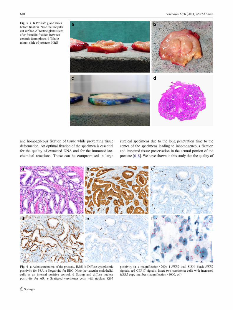
After overnight fixation, the deformation and bulging ofthe fresh whole tissue slices had completely disappeared andthe pinched slices showed a flat surface with no artifactscaused by the mild pressure of the plates (Fig. 3).
In all 49 specimens, H&E whole mount sections revealed anormal histological architecture without irregularities, neitherin the peripheral nor in the middle portions of the slide, as can
be appreciated by the well-preserved morphology of the pros-tatic urethra located centrally of the gland.
The IHC analysis done on three resection specimensshowed homogeneous and strong expression in all ceramicfoam-fixed specimens for all markers (Fig. 4). The intensity ofthe IHC staining was equal in the peripheral and centralportions of the resection specimen.
FISH and silver in situ hybridization (SISH) signals werestrong and homogeneously distributed across the slides.
Discussion
The main purpose of this study was to establish an improvedmethod for collecting fresh tumor tissue from radical prosta-tectomy specimens under sterile conditions for an ongoingprostate cancer xenograft project. Here, we show that thephysical properties of plates made of ceramic foam are idealfor further processing fresh whole mount slices of the prostateafter fresh tissue collection.
Ceramic foam is a lightweight, typically 75–90 % porousmaterial with a three-dimensional reticulated structure [5]. Itpossesses high mechanical strength, chemical stability, andhigh temperature resistance and is therefore well suited forvarious applications, e.g., thermal insulation or filtration.However, its potential use as an auxiliary tool for fixation offresh surgical specimens has not yet been evaluated. Com-pared to polystyrene (styrofoam) or a cork plate, ceramic foamallows for a fast penetration of formalin and therefore quick
Fig. 1 a Samples frommacroscopically suspicious areasmarked with differently coloredink and put into a tissue holder forfrozen section analysis. b, cFrozen section of a sample withprostatic adenocarcinoma, surfaceinked with green color, H&E(b magnification×20, cmagnification×100)
Fig. 2 Ceramic foam plates bound together by standard plastic cable ties
Virchows Arch (2014) 465:637–642 639
and homogeneous fixation of tissue while preventing tissuedeformation. An optimal fixation of the specimen is essentialfor the quality of extracted DNA and for the immunohisto-chemical reactions. These can be compromised in large
surgical specimens due to the long penetration time to thecenter of the specimens leading to inhomogeneous fixationand impaired tissue preservation in the central portion of theprostate [6–8]. We have shown in this study that the quality of
Fig. 3 a, b Prostate gland slicesbefore fixation. Note the irregularcut surface. c Prostate gland slicesafter formalin fixation betweenceramic foam plates. d Wholemount slide of prostate, H&E
Fig. 4 a Adenocarcinoma of the prostate, H&E. b Diffuse cytoplasmicpositivity for PSA. c Negativity for ERG. Note the vascular endothelialcells as an internal positive control. d Strong and diffuse nuclearpositivity for AR. e Scattered carcinoma cells with nuclear Ki67
positivity (a–e magnification×200). f HER2 dual SISH, black HER2signals, red CEP17 signals. Inset: two carcinoma cells with increasedHER2 copy number (magnification×1000, oil)
640 Virchows Arch (2014) 465:637–642
the morphology as well as of the immunohistochemistry andin situ hybridization tested on specimens fixed with the ce-ramic foam method was excellent.
Importantly, this method does not compromise further di-agnostic assessment and the morphology as well as the pros-tatic pseudo-capsule and the resection margins remain intact.
Several methods have previously been proposed to facili-tate tumor tissue collection from fresh radical prostatectomyspecimens [9–15]. Bertilsson et al. described a technique withfreezing of an entire transversal slice of the prostate gland[10]. However, this method delays the process since freshtumor tissue can only be selected after the histological work-up is completed. Furthermore, it does not follow the currentguidelines that recommend a complete embedding of theradical prostatectomy specimen in order not to miss importantprognostic features [4]. Morrison et al. divided the centralportion of every other intervening transection level into quad-rants for fresh tissue procurement while removing the capsulefor clinical examination [12]. The disadvantage of theirmethod was the time required to analyze frozen section ofevery quadrant. Walton et al. proposed to take needlebiopsies from the unfixed surgical specimen, which largelypreserves the integrity of the prostate [14]. However,tumor tissue can easily been missed by such blind sam-pling and the amount of the harvested tissue remainssmall. Schäfer et al. systematically removed and snapfroze inner triangles of the whole transverse sections,leaving the peripheral part of the organ intact. Althoughthis allowed to collect tumor tissue from >80 % of thespecimens without compromising further diagnostic work-up, tumor tissue was randomly captured but not enrichedfor [13]. Warren et al. recently described a method ofpunching multiple (up to 27) cylindrical cores from acomplete transverse slice using a large skin punch forsubsequent snap freezing and storage at −80 °C [15].The holes on the transverse slice were inked and marked on aprostate map. After completion of the histopathology report,the frozen tissue cores were processed for frozen section anal-ysis. This method appears to be complex and time-consuming,and the taken cores with presumable tumor are processed at alater point in time. Notably, all of these previously suggestedprocedures do not address the problem of tissue distortion afterslicing of fresh tissue and are not suitable for immediate freshtissue collection for xenografting.
Conclusions
Our novel method using ceramic foam plates is easily appli-cable and timesaving. It enables retaining the flat shape offresh whole mount sections after collecting tumor tissue forresearch purposes. Therefore, this approach greatly facilitates
collecting of fresh material without compromising standarddiagnostic analyses.
This method could also be applied to other specimens thatare often processed in unfixed state, such as resection speci-mens of breast cancer and soft tissue tumors.
Acknowledgments We thank Sandra Schneider and Rita Epper forexcellent technical support and Manuela Bubendorf for her helpfuladvice.
Funding This study has partly been funded by the Movember Foun-dation and Astellas Pharma AG.
Disclosure The authors declare that they have no conflict of interest.
References
1. Antonov J, Goldstein DR, Oberli A, Baltzer A, Pirotta M,Fleischmann A, Altermatt HJ, Jaggi R (2005) Reliable gene expres-sion measurements from degraded RNA by quantitative real-timePCR depend on short amplicons and a proper normalization. LabInvest: J Tech Methods Pathol 85(8):1040–1050. doi:10.1038/labinvest.3700303
2. Holley T, Lenkiewicz E, Evers L, Tembe W, Ruiz C, Gsponer JR,Rentsch CA, Bubendorf L, Stapleton M, Amorese D, Legendre C,Cunliffe HE, McCullough AE, Pockaj B, Craig D, Carpten J, VonHoff D, Iacobuzio-Donahue C, Barrett MT (2012) Deep clonalprofiling of formalin fixed paraffin embedded clinical samples.PLoS One 7(11):e50586. doi:10.1371/journal.pone.0050586
3. Movember (2013) GAP1 Global Prostate Cancer Biomarker Initiative.http://ch.movember.com/en/report-cards/view/id/637/gap1-global-prostate-cancer-biomarker-initiative [accessed February 8, 2014]
4. Samaratunga H, Montironi R, True L, Epstein JI, Griffiths DF,Humphrey PA, van der Kwast T, Wheeler TM, Srigley JR,Delahunt B, Egevad L, Group IPC (2011) International Society ofUrological Pathology (ISUP) consensus conference on handling andstaging of radical prostatectomy specimens. Working group 1: spec-imen handling. Mod Pathol: Off J U S Can Acad Pathol Inc 24(1):6–15. doi:10.1038/modpathol.2010.178
5. Lautensack C, Giertzsch M, Godehardt M, Schladitz K (2008)Modelling a ceramic foam using locally adaptable morphology. JMicrosc 230(Pt 3):396–404. doi:10.1111/j.1365-2818.2008.01998.x
6. De Marzo AM, Fedor HH, Gage WR, Rubin MA (2002) Inadequateformalin fixation decreases reliability of p27 immunohistochemicalstaining: probing optimal fixation time using high-density tissuemicroarrays. Hum Pathol 33(7):756–760
7. Leong AS, Gilham PN (1989) The effects of progressive formalde-hyde fixation on the preservation of tissue antigens. Pathology 21(4):266–268
8. Varma M, Linden MD, Amin MB (1999) Effect of formalin fixationand epitope retrieval techniques on antibody 34betaE12 immuno-staining of prostatic tissues. Mod Pathol: Off J U S Can Acad PatholInc 12(5):472–478
9. Egevad L (2012) Handling of radical prostatectomy specimens.Histopathology 60(1):118–124. doi:10.1111/j.1365-2559.2011.04002.x
10. Bertilsson H, Angelsen A, Viset T, Skogseth H, Tessem MB,Halgunset J (2011) A new method to provide a fresh frozen prostateslice suitable for gene expression study and MR spectroscopy.Prostate 71(5):461–469. doi:10.1002/pros.21260
Virchows Arch (2014) 465:637–642 641
11. Jhavar SG, Fisher C, Jackson A, Reinsberg SA, Dennis N, FalconerA, Dearnaley D, Edwards SE, Edwards SM, Leach MO, CummingsC, Christmas T, Thompson A, Woodhouse C, Sandhu S, Cooper CS,Eeles RA (2005) Processing of radical prostatectomy specimens forcorrelation of data from histopathological, molecular biological, andradiological studies: a new whole organ technique. J Clin Pathol58(5):504–508. doi:10.1136/jcp.2004.021808
12. Morrison C, Cheney R, Johnson CS, Smith G, Mohler JL (2009)Central quadrant procurement of radical prostatectomy specimens.Prostate 69(7):770–773. doi:10.1002/pros.20925
13. Schafer SC, Pfnur M, Yerly S, Fandel TM, Jichlinski P, Lehr HA(2009) Cryopreservation of prostate cancer tissue during routine
processing of fresh unfixed prostatectomy specimen: demonstrationand validation of a new technique. Prostate 69(2):191–197. doi:10.1002/pros.20868
14. Walton TJ, McCulloch TA, Rees RC, Bishop MC (2005)Obtaining fresh prostate cancer tissue for research: a novelbiopsy needle and sampling technique for radical prostatecto-my specimens. Prostate 64(4):382–386. doi:10.1002/pros.20264
15. Warren AY, Whitaker HC, Haynes B, Sangan T, McDuffus LA, KayJD, Neal DE (2013) Method for sampling tissue for research whichpreserves pathological data in radical prostatectomy. Prostate 73(2):194–202. doi:10.1002/pros.22556
642 Virchows Arch (2014) 465:637–642