Ceramic findings from the archaeological site at Aiano-Torraccia di Chiusi (Siena, Italy): a...
18
ORIGINAL PAPER Ceramic findings from the archaeological site at Aiano-Torraccia di Chiusi (Siena, Italy): a multi-analytical approach E. Cantisani & M. Cavalieri & C. Lofrumento & E. Pecchioni & M. Ricci Received: 20 December 2010 / Accepted: 26 September 2011 # Springer-Verlag 2011 Abstract In 2005, the remains of a Roman villa, dating from the early fourth to the sixth centuries AD, were discovered at the archaeological site of Aiano-Torraccia di Chiusi (Siena, Italy). After being abandoned in the sixth century AD, the complex was occupied by a group of Ostrogothic or Lombardic artisans in the period between the sixth and the seventh centuries AD. Many ceramic remains (coarse pottery and red slip ceramics) from the first to the seventh centuries AD have been discovered on this archaeological site. These findings have been analysed using different analytical techniques (optical microscopy (OM), X-ray diffraction (XRD), X-ray fluorescence (XRF), scanning electron microscope-energy dispersive spectros- copy (SEM-EDS), attenuated total reflection Fourier trans- form infrared (ATR-FTIR), and micro-Raman in order to characterize the ceramic body, the coating, the temper, and to investigate the compositional relationship between the different kinds of ceramics. The use of different techniques on the same samples yielded information at different scales. OM and SEM-EDS yielded interesting information on the coarse pottery: the analyses performed on some minerals and rock fragments suggest that stone tesserae from the Roman villa (in the form of numerous marble fragments) were used in the production of this pottery. Bulk analyses (XRD and XRF) and subsequent micro-analyses (SEM- EDS, ATR-FTIR, and micro-Raman) of the red slip pottery revealed clear chemical, mineralogical and textural differ- ences: some ceramics (the TCC sample group) typically have a Fe-enriched coating while others (the INGR sample group) present a clear difference in grain size but no chemical or mineralogical differences between the ceramic body and the coating. Keywords Aiano-Torraccia di Chiusi (Siena) . Red slip ceramics . Coarse pottery . SEM-EDS . ATR-FTIR . Raman . XRD . XRF . OM Introduction A Belgian-Italian mission of the Université catholique de Louvain has been excavating an archaeological site located in a marginal area of the Ager Volaterranus since 2005. During the last five campaigns, an articulated settlement E. Cantisani (*) CNR-ICVBC Institute for Conservation and Enhancement of Cultural Heritage, Via Madonna del Piano 10, Sesto Fiorentino, Florence, Italy e-mail: [email protected] M. Cavalieri Department of Archaeology and History Art, Institute for Civilizations, Arts and Literature Université catholique de Louvain (UCL) Place B. Pascal, 1 Collège Erasme, P.O. Box L3.03.13, 1348 Louvain-la-Neuve, Belgium e-mail: [email protected] C. Lofrumento Department of Chemistry, University of Florence, Via Della Lastruccia 3, 50019 Sesto Fiorentino, Florence, Italy E. Pecchioni Department of Earth Sciences, University of Florence, Via G. La Pira 4, 50121 Florence, Italy M. Ricci Department of Construction and Restoration, University of Florence, Via Micheli 8, 50121 Florence, Italy Archaeol Anthropol Sci DOI 10.1007/s12520-011-0080-1
Transcript of Ceramic findings from the archaeological site at Aiano-Torraccia di Chiusi (Siena, Italy): a...

ORIGINAL PAPER
Ceramic findings from the archaeological siteat Aiano-Torraccia di Chiusi (Siena, Italy):a multi-analytical approach
E. Cantisani & M. Cavalieri & C. Lofrumento &
E. Pecchioni & M. Ricci
Received: 20 December 2010 /Accepted: 26 September 2011# Springer-Verlag 2011
Abstract In 2005, the remains of a Roman villa, datingfrom the early fourth to the sixth centuries AD, werediscovered at the archaeological site of Aiano-Torraccia diChiusi (Siena, Italy). After being abandoned in the sixthcentury AD, the complex was occupied by a group ofOstrogothic or Lombardic artisans in the period betweenthe sixth and the seventh centuries AD. Many ceramicremains (coarse pottery and red slip ceramics) from the firstto the seventh centuries AD have been discovered on this
archaeological site. These findings have been analysedusing different analytical techniques (optical microscopy(OM), X-ray diffraction (XRD), X-ray fluorescence (XRF),scanning electron microscope-energy dispersive spectros-copy (SEM-EDS), attenuated total reflection Fourier trans-form infrared (ATR-FTIR), and micro-Raman in order tocharacterize the ceramic body, the coating, the temper, andto investigate the compositional relationship between thedifferent kinds of ceramics. The use of different techniqueson the same samples yielded information at different scales.OM and SEM-EDS yielded interesting information on thecoarse pottery: the analyses performed on some mineralsand rock fragments suggest that stone tesserae from theRoman villa (in the form of numerous marble fragments)were used in the production of this pottery. Bulk analyses(XRD and XRF) and subsequent micro-analyses (SEM-EDS, ATR-FTIR, and micro-Raman) of the red slip potteryrevealed clear chemical, mineralogical and textural differ-ences: some ceramics (the TCC sample group) typicallyhave a Fe-enriched coating while others (the INGR samplegroup) present a clear difference in grain size but nochemical or mineralogical differences between the ceramicbody and the coating.
Keywords Aiano-Torraccia di Chiusi (Siena) . Red slipceramics . Coarse pottery . SEM-EDS . ATR-FTIR . Raman .
XRD . XRF. OM
Introduction
A Belgian-Italian mission of the Université catholique deLouvain has been excavating an archaeological site locatedin a marginal area of the Ager Volaterranus since 2005.During the last five campaigns, an articulated settlement
E. Cantisani (*)CNR-ICVBC Institute for Conservationand Enhancement of Cultural Heritage,Via Madonna del Piano 10,Sesto Fiorentino, Florence, Italye-mail: [email protected]
M. CavalieriDepartment of Archaeology and History Art,Institute for Civilizations, Arts and Literature Universitécatholique de Louvain (UCL) Place B. Pascal,1 Collège Erasme, P.O. Box L3.03.13, 1348 Louvain-la-Neuve,Belgiume-mail: [email protected]
C. LofrumentoDepartment of Chemistry, University of Florence,Via Della Lastruccia 3,50019 Sesto Fiorentino, Florence, Italy
E. PecchioniDepartment of Earth Sciences, University of Florence,Via G. La Pira 4,50121 Florence, Italy
M. RicciDepartment of Construction and Restoration,University of Florence,Via Micheli 8,50121 Florence, Italy
Archaeol Anthropol SciDOI 10.1007/s12520-011-0080-1

has been brought to light in this area which is well knownfor significant discoveries that date to Roman times. Thissettlement consists of a villa longinqua with monumentalarchitecture and decorative apparatus that was probablybuilt between the end of the third and the beginning of thefourth century AD. A hexalobate hall, surrounded by amonumental pentalobate ambulatio that was accessedthrough a rectangular vestibule, belongs to this first phase.
The hall, ambulatio and vestibule played an importantrole in connecting different parts of the building: thischaracteristic, highlighted by the coaxial openings at theends of the hall, is found in other contemporary buildings,for example the Cazzanello villa near Tarquinia.
In a later period—dated stratigraphically to the lastquarter of the fourth century AD—the villa was heavilyrestored following a natural traumatic event. In particular,the hexalobate hall was radically transformed, both archi-tectonically and functionally: the floor was loweredconsiderably and three exedras were destroyed and substi-tuted by three rectangular rooms. This new arrangementresulted in an unusual shape: a trilobate hall with atriangular base, very different from the classic trìkonchos.The new floor of the hall was paved with opus signinum:there were geometric decorations in the centre of the halland in the apse in front of the vestibule; the other two apseshad a central ornamental emblema (the best preservedrepresents a flowered goblet placed in a guilloche framedby a denticulate arch) (Cavalieri 2009a).
Even though the hall lost its function as a passagewayduring this phase, becoming a room reserved for otium, theexternal ambulatio retained its function. The structureshowed the first traces of disuse and collapse during thefifth century AD: presumably some parts (like the trilobatehall) were abandoned and progressively hidden by thecollapse of the parietal panels and the roof structure. Otherparts were subjected to the first depredations for recoveringmarbles for use elsewhere.
Only in a later period when the villa, was already inruins, it was used as a quarry for various materials (iron,copper, gold, and vitreous pastes) destined for the produc-tion of small objects such as pins, bracelets and necklacebeads. During this phase, the rectangular rooms, builtoutside the trilobate hall, were used as small workshopsfor the manufacture of pottery (room H) and gold (room L)and bronze (room I) objects while the rooms on thesouthern side accommodated workshops for manufacturingglazes (vestibule, room A) and objects made of iron (roomB) (Cavalieri and Giumlia-Mair 2009) (Fig. 1).
During the second half of the seventh century AD, theexcavated part of the structure was definitively abandoned,although there is evidence that this area was frequentedduring the Late Middle Ages. The presence of peoplepassing not far from the villa was probably due to the
deviation of the Francigena Road (or Via Romea), astestified by Sigeric, Archbishop of Canterbury, whotravelled through this area at the end of the tenth century(Cavalieri 2009b).
The ceramic findings at Aiano-Torraccia di Chiusiinclude coarse pottery, depurated ceramic with a red/brownslip, uncoated depurate, semi-depurate, lamps, amphoraeand dolia. There are also residual elements, mostly externalto the context of the excavation: for example fragments of aKelebe from Volterra dated to the III BC, black gloss potteryfrom the fourth–fifth BC, frustules of protohistoric paste anda very few tiny fragments of Italic Sigillata located in thestratigraphic sequences. These finds have made a detailedinvestigation necessary: firstly to study the phases offrequentation of the area prior to the construction of thevilla and secondly, and more importantly, to study the roleplayed by the villa from the second half of the fifth to thelate seventh centuries when it was abandoned. In particularthis work focuses on the study of a significant number ofceramic findings (dated from the fifth to seventh AD), tofacilitate an understanding of the role the villa played as asite of production activities and how its constituentmaterials (i.e. mosaics and decorative elements) werereutilized. The principal objectives of this paper are tocharacterize the ceramic body and coating of the red slipceramics, the matrix and temper of the coarse pottery inorder to investigate the compositional relationship betweenthese two different kinds of ceramics and to use thisinformation to assess whether both typologies wereproduced locally. This paper also evaluates the extent towhich different techniques can provide information relativeto a given sample at different scales.
Materials and analytical methods
A description of the types of the ceramic findings analysedis reported below (Fig. 2a, b; Table 1), followed by apresentation of the analytical methods.
The samples of these findings consist of coarse uncoatedpottery (AG) and red slip ceramics. The red slip ceramicsamples are subdivided into INGR samples and TCC or“similar to African” samples. This last definition has beenused to identify a few ceramic fragments found in the villathat have a different morphology from the red slip ceramics.It is difficult to assess whether these fragments can beclassified as African red pottery although it is highlyprobable given their tiny size.
Red slip ceramics
The pottery found in the area is a typical representation ofthe production known in Tuscany between the fifth and
Archaeol Anthropol Sci
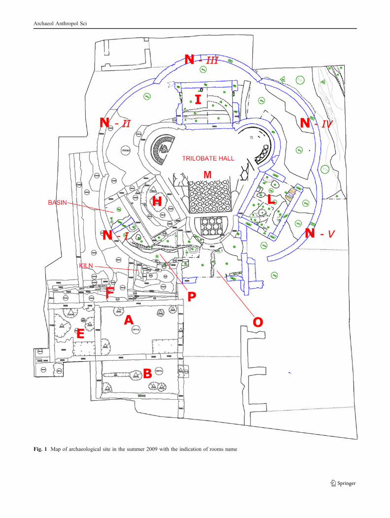
Fig. 1 Map of archaeological site in the summer 2009 with the indication of rooms name
Archaeol Anthropol Sci

Fig. 2 Shape and typologies of red slip ceramics (INGR samples): plates trays (left), closed shapes (right) (a); coarse pottery (AG samples) (b)(drawn by Antonia Fumo and Enrica Boldrini)
Archaeol Anthropol Sci
seventh centuries AD. In fact coarse pottery, red slip pottery,depurated and semi-depurated wares are all present (Fumo2010). Generally, red slip pottery represents a stylisticimitation, often on a regional basis, of Late African RedSlipware, present from as early as the second to the fourthcenturies AD until the seventh century AD. After the seventhcentury AD there was a process of transformation and thetypologies produced in Tuscany gradually acquired specificautonomous characteristics that distinguished them fromthe typologies produced in other parts of Italy during thesame period.
Closed shapes Many shapes have been discovered: jugs(some of them trilobate), mugs, small amphorae, bottles,flasks, and glasses. Despite this, it has been possible todeterminate the chronological period in which these shapeswere produced, from the fifth to the seventh centuries AD,and their distribution in the excavated area.
Plates trays Together with bowls and cups they are themost common red-coated pottery objects found on the siteof the villa. The plate trays have ample diameters, quite lowsides and are footless; they were generally used as dinnerplates. The vases imitate the Hayes 42 and 45 shapes ofLate African Red Slipware (Hayes 1972). Their productioncan be dated to sometime between the fifth and the seventhcenturies AD.
Uncoated ceramics (coarse pottery)
All the typologies are represented in the coarse potteryremains: cooking pots (olle), bowl lids, casseroles, lids,jugs, and small jugs. The cooking pots are of a medium/small size and often have very fine threading on the backand one or more engraved lines for identifying the start ofthe neck. The casserole is a peculiar typology for thischronological range. Cooking pots and casseroles havesmoothed external sides and detached bases with a blade.They have widespread blackening both externally andinternally due to manufacturing and/or contact with fireand coal. The lids are classified in six typologies, based ontheir thickness and height. They were produced on a fastwheel and sometimes smoothed; they rarely show internalor external blackening and their diameters are compatiblewith those of the cooking pots and casseroles.
Some fragments of medium and small-sized jugs havealso been identified. The fragments are of thin smoothedwalls with external and internal blackening, parts of theupper border and parts of mostly small-sized nozzles.The bow lids have a typical crushed-cap shape with asmall groove. They are large and of an appropriatethickness, their surfaces are rough and they were madewith coarse pastes (Cavalieri and Boldrini 2008). Theytoo, can be dated to between the fifth and seventhcenturies AD.
Analytical methods
Diverse analytical techniques were used in order to obtainuseful and representative analyses for each type of ceramicfinding. The techniques chosen were selected because theycould provide the necessary information without requiringredundant and expensive analyses (Maggetti 1982; Maggetti1994; Fabbri 1998; Tite 1999; Turbanti Memmi 2004;
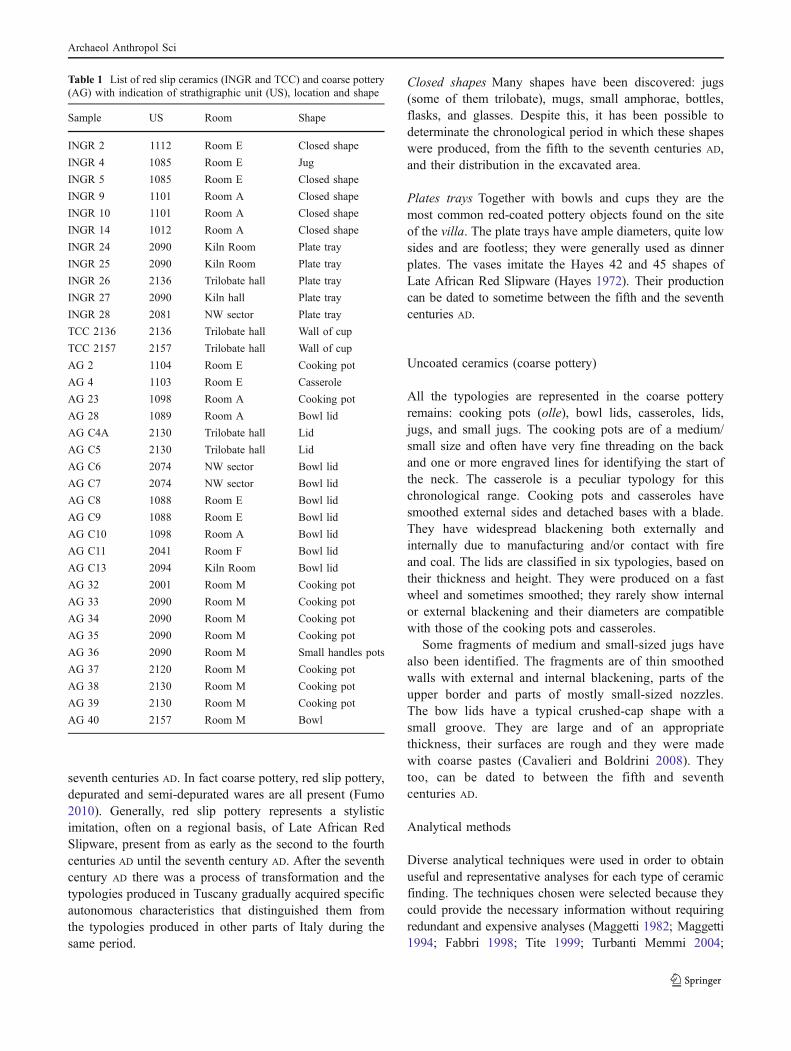
Table 1 List of red slip ceramics (INGR and TCC) and coarse pottery(AG) with indication of strathigraphic unit (US), location and shape
Sample US Room Shape
INGR 2 1112 Room E Closed shape
INGR 4 1085 Room E Jug
INGR 5 1085 Room E Closed shape
INGR 9 1101 Room A Closed shape
INGR 10 1101 Room A Closed shape
INGR 14 1012 Room A Closed shape
INGR 24 2090 Kiln Room Plate tray
INGR 25 2090 Kiln Room Plate tray
INGR 26 2136 Trilobate hall Plate tray
INGR 27 2090 Kiln hall Plate tray
INGR 28 2081 NW sector Plate tray
TCC 2136 2136 Trilobate hall Wall of cup
TCC 2157 2157 Trilobate hall Wall of cup
AG 2 1104 Room E Cooking pot
AG 4 1103 Room E Casserole
AG 23 1098 Room A Cooking pot
AG 28 1089 Room A Bowl lid
AG C4A 2130 Trilobate hall Lid
AG C5 2130 Trilobate hall Lid
AG C6 2074 NW sector Bowl lid
AG C7 2074 NW sector Bowl lid
AG C8 1088 Room E Bowl lid
AG C9 1088 Room E Bowl lid
AG C10 1098 Room A Bowl lid
AG C11 2041 Room F Bowl lid
AG C13 2094 Kiln Room Bowl lid
AG 32 2001 Room M Cooking pot
AG 33 2090 Room M Cooking pot
AG 34 2090 Room M Cooking pot
AG 35 2090 Room M Cooking pot
AG 36 2090 Room M Small handles pots
AG 37 2120 Room M Cooking pot
AG 38 2130 Room M Cooking pot
AG 39 2130 Room M Cooking pot
AG 40 2157 Room M Bowl
Archaeol Anthropol Sci

Cuomo di Caprio 2007; Pecchioni et al. 2007). Most of thedata for the red slip ceramics were obtained through bulkchemical and mineralogical petrographical analyses of theceramic body and coating: X-ray fluorescence (XRF), X-raydiffraction (XRD), optical microscopy (OM). Micro-chemicalanalyses of each individual ceramic body (attenuated totalreflection Fourier transform infrared (ATR-FTIR), and scan-ning electron microscope-energy dispersive spectroscopy(SEM-EDS)) and of coating (ATR-FTIR, SEM-EDS, andmicro-Raman) were performed in order to determine theircomposition.Much of the data for the coarse pottery (uncoatedand with macroscopic added temper) were obtained using OMlinked to SEM-EDS analyses. These techniques yielded dataon the composition and origin of the added temper.
Not every sample was submitted to all the analyses: i.e.the micro-chemical analyses (SEM-EDS), ATR-FTIR andthe micro-Raman analyses were performed only on repre-sentative selected samples, while the petrographical andmineralogical analyses were performed on every sample soas to classify and select the samples that would undergomicro-chemical analyses.
X-ray diffraction Powder XRD was adopted to determine themineralogical composition of the ceramic findings: a PhilipsPW 1050/37 diffractometer was used with a Philips X’PertPRO data acquisition system, operating at 40 kV–20mA, witha Cu anode, a graphite monochromator and with 2°/mingoniometry speed in a scanning range between 5–70°.
X-ray fluorescence XRF on pressed-powder pellets wasadopted to determine the chemical composition; a PhilipsPW 1480 wavelength dispersive spectrometer was used;major elements were determined using a Rh anode andestimated using the method adopted by Franzini et al. 1975.
ATR-FTIR Fourier transform infrared spectroscopy wasperformed on samples of red slip ceramic findings using aSpectrum 100 Perkin Elmer with an attenuated ATR diamondcrystal. The detector was fast recovery deuterated L-alaninetryglycine sulphate, and the spectral range was 4,000–380 cm−1 with a spectral resolution of 4 cm−1.
Electron microscopy The SEM-EDS analyses were per-formed with an electron microscope (Zeiss EVO MA 15),coupled to an analytical system (EDS OXFORD INCA 250).The following standards were used for the semi-quantitativeanalyses: Albite, MgO, Al2O3, SiO2, Wollastonite, MAD-10Feldspar, Ti, and Fe.
Optical microscopy The petrographic description of sampleswas carried out using a polarised light microscope (ZEISSAxioskop; OM) on thin sections (30 μm thickness), observedat different magnifications. The microscope was equipped
with a camera (resolution, 5 megapixels) and dedicated imageanalysis software (AxioVision) for evaluating the texturalparameters.
Micro-Raman A Renishaw single-grating (1,200 grooves/mm) RM2000 micro-Raman spectrometer was used forRaman measurements. Spectra were recorded using the785 nm excitation line of a Renishaw air-cooled diode laser.Laser beam focusing was accomplished through a ×50magnification objective which provided a 1.5-μm laser spotdiameter. Neutral density filters were used to attenuate thelaser power in order to avoid phase transformations. Foreach sample, Raman spectra were taken on an average offive randomly selected points on the coated surface of eachsample of the ceramic findings. Acquisition times rangedfrom 10 to 100 s. Spectra were recorded over the range of200–800 cm−1 at step intervals of 1 cm−1 and with a 50 μmslit aperture. The instrumental broadening, evaluated as theline width (full width at half-maximum) of an emission linefrom a He–Ne lamp, was 3 cm−1. Experimental conditions(i.e. magnification and sample illumination) were keptconstant for all measurements.
Results and discussion
Red slip ceramics
Table 2 reports the mineralogical composition of the redslip ceramic findings obtained by homogenizing the coatingand the ceramic body.
The samples are characterized by the presence of quartz,plagioclases (except TCC 2136 and TCC 2157 whichcontain K-feldspar but no plagioclases) and hematite(except INGR 28). Pyroxenes are only present in the INGR2, INGR 4 and INGR 28 samples; calcite is only present inthe INGR 24, INGR 25, INGR 26, and INGR 28 sampleswith traces in the INGR 14 and INGR 27 samples.Phyllosilicates (illite and muscovite) are present in almostall the samples. There are some differences in themineralogical composition of the INGR and TCC samplesbut the data are insufficient to obtain precise informationabout the manufacturing technology used.
Table 3 reports the bulk chemical analyses performedwith the XRF method using powder obtained by homog-enizing the coating and ceramic body. These data can beconsidered as a mean composition of these ceramics. TheTCC 2136 sample is characterized by higher levels of SiO2
and lower levels of CaO with respect to the INGR samples.These differences can be attributed to the use of differentraw materials for the realization of the TCC and the INGRsamples. In the case of CaO (INGR samples), secondary
Archaeol Anthropol Sci

phenomena associated with the alteration of buried ceramicfindings also need to be investigated.
Mineralogical and chemical analyses, such as XRD andXRF, are very useful for a general characterization of thesamples, but, because they are performed on homogenizedsamples, which contain both coating and body, they do notprovide a complete picture of the method of ceramicproduction and the source of the raw materials. Significantinformation on production technology was obtained bystudying the coating and the body separately: chemicalanalyses (ATR-FTIR which is sensitive to molecularstructure and SEM-EDS for elementary semi-quantitativecharacterization) were performed on the coating and theceramic body. The SEM-EDS data are reported in Table 4.
While the chemical composition of the coating of theINGR 4, INGR 14, and INGR 28 samples is quite similar tothe composition of the ceramic body, the amount of FeOpresent in the coating and the ceramic body in the TCC2136 and TCC 2157 samples (“similar to African”) differsconsiderably; there is more FeO in the coating, suggestingan addition of Fe-rich material so as to obtain a red colour.
The textural information obtained using SEM-EDSreveals that in the INGR 4 and INGR 28 samples (Fig. 3)
the grain size of the skeleton of the particles constituting thecoating is smaller than in the ceramic body. The coating isless porous and has a smaller amount of CaO due to theabsence of secondary CaCO3 which is present in the moreporous ceramic body. The ATR-FTIR spectra of the ceramicbody and the coating in the INGR 24, INGR 25, and INGR27 samples were characterized by the presence of calcite,quartz, and aluminosilicate as the main components. All thesamples contain calcite in different amounts due to eitherthe probable composition of the raw clay and/or tosecondary precipitation phenomena. However, as the mostsignificant absorption peaks of the spectra occur at wavenumbers below 1,300 cm−1, only these portions of thespectra are presented and discussed. The characteristicinfrared frequencies of minerals identified in the ceramicsamples were attributed by correlation data on standardminerals and on the basis of data reported in the literature(Farmer 1974), when it was not possible to have a standardsample of the relevant mineral.
The 1,300–400 cm−1 region of the ATR-FTIR spectra ofthe ceramic bodies of the INGR 24, INGR 25, INGR 27samples, together with their second derivative profile, isgiven in Fig. 4a, b. The second derivative of the IR absorption
Table 3 Chemical analyses(XRF) of red slip ceramics(oxide wt.%)
SiO2 TiO2 Al2O3 Fetot MnO MgO CaO Na2O K2O P2O5 LOI
INGR 5 55.99 0.86 18.00 7.88 0.08 4.12 7.47 0.91 2.64 0.24 1.82
INGR 9 54.56 0.79 16.23 6.80 0.09 3.69 7.82 0.63 2.62 0.27 6.50
INGR 10 60.77 0.81 17.54 7.26 0.07 3.54 4.14 1.01 2.82 0.12 1.91
INGR 14 53.51 1.02 22.37 8.41 0.19 3.36 4.49 0.57 3.33 0.19 2.57
INGR 25 53.90 0.72 15.35 6.74 0.08 3.14 10.69 0.88 2.51 0.36 5.56
INGR 27 55.39 0.81 15.90 7.79 0.06 3.34 7.79 0.81 2.58 0.23 5.28
INGR 28 55.99 0.71 15.73 6.38 0.08 3.10 9.25 0.53 2.32 0.22 7.54
TCC 2136 66.73 0.99 18.45 5.88 0.02 2.24 0.80 0.28 2.54 0.12 1.95
Table 2 Mineralogicalcomposition (XRD) of redslip ceramic findings
Quartz Calcite Plagioclase K-feldspar Hematite Phyllosilicates Pyroxene
INGR 2 X – X X X Traces X
INGR 4 X – X – X X X
INGR 5 X – X – X X –
INGR 9 X – X – X X –
INGR 10 X – X X X X –
INGR 14 X Traces X – X X –
INGR 24 X X X – X Traces –
INGR 25 X X X X X Traces –
INGR 26 X X X X X X –
INGR 27 X Traces X X X Traces –
INGR 28 X X X – – X X
TCC 2136 X – – X X – –
TCC 2157 X – – X X – –
Archaeol Anthropol Sci

spectrum allowed us to determine the mineral phase present inthe pottery specimens. The absorption peaks of quartz weredetected in all the samples: the main SiO stretching band at1,080 cm−1, the shoulder at 1,165 and 512 cm−1 and thedistinctive doublet at 797 and 777 cm−1. Other minerals wereidentified in these samples, in particular, albite (1,096, 989,722, and 425 cm−1) and microcline (1,142, 1,134, 1,053,729, 646, 584, and 464 cm−1) from the feldspar group,muscovite from the phyllosilicate groups (1,062, 1,022, 990,1,062, 1,022, 990, 935, 754, 553, 480, and 412 cm−1) andhematite (537 and 475 cm−1) were inferred from the secondderivative profiles.
In Fig. 5a, b, the absorption and second derivative profilesof IR spectra of the coating of the same samples are reportedso as to highlight the differences in mineralogical composi-
tion between the coating and the ceramic body. The coatingcontains the same constituents as the ceramic body, with asmall difference in the intensity of the hematite absorptionpeak. Minerals of an archaeological artefact can be classifiedas primary minerals (present in the raw materials and nottransformed over a wide temperature range) or firingminerals that were formed during firing (De Benedetto etal. 2002). No firing minerals were observed in the secondderivative profiles of the IR spectra of the ceramicsinvestigated. The absence of any firing mineral in theceramic body clearly indicates that these samples were firedat low temperatures.
Micro-Raman analyses were performed on a limitednumber of samples, but the results encourage furtherresearch into the technical features of the different kinds
Table 4 Chemical analyses (SEM) of the red coatings and ceramic bodies (oxide wt%)
Na2O MgO Al2O3 SiO2 K2O CaO TiO2 FeO
INGR 4 coating 0.9±0.3 3.0±0.4 15.7±0.6 63.4±2.3 2.1±0.5 7.3±0.6 0.9±0.1 6.8±1.1
INGR 4 body 1.2±0.3 2.9±0.4 17.0±1.8 58.9±1.8 2.7±0.6 9.6±0.9 0.9±0.1 6.7±1.0
INGR 14 coating 0.6±0.1 3.0±0.4 22.5±1.7 55.2±2.4 3.2±0.8 5.0±1.2 1.6±0.2 8.8±1.2
INGR 14 body 0.6±0.2 2.8±0.3 21.9±2.0 57.2±1.7 3.8±0.7 5.3±1.3 1.3±0.2 7.1±0.7
INGR 28 coating 0.6±0.1 2.3±0.3 15.8±1.4 65.0±1.3 2.8±0.2 6.9±0.7 0.6±0.3 5.9±1.2
INGR 28 body 0.8±0.1 2.6±0.2 16.3±1.2 58.6±2.7 3.1±0.3 11.3±2.1 0.9±0.2 6.4±1.4
TCC 2136 coating bdl 1.9±0.4 23.1±2.0 57.9±3.5 2.2±0.3 1.4±0.7 1.3±0.2 12.3±1.5
TCC 2136 body 0.4±0.1 1.9±0.2 21.2±2.3 66.5±3.7 3.1±1.0 0.6±0.2 1.1±0.2 5.4±0.2
TCC 2157 coating 0.7±0.0 1.7±0.0 26.3±0.7 56.7±0.2 2.1±0.4 1.0±0.2 1.2±0.1 10.7±1.5
TCC 2157 body 0.64±0.3 1.7±0.2 26.3±1.9 61.1±2.8 2.9±0.1 0.7±0.0 1.3±0.1 5.3±0.5
Fig. 3 SEM back scatteredelectrons image of sampleINGR 28 (the white lineidentifies the transitionbetween ceramic bodyand coating)
Archaeol Anthropol Sci

of artefacts. Two specimens of red slip ceramics (INGR5and INGR27), together with two “similar to African”ceramic findings samples (TCC 2136 and TCC 2157) wereexamined in order to evaluate the potential of micro-Ramanas a complementary technique for studying the differentcomposition of the red coatings.
The usefulness of a micro-Raman investigation of thesepottery wares lies in the capacity of this technique toidentify and distinguish different iron oxides, even if theyare present in small quantities. A valid study of themanufacturing technique is feasible if the behaviour of ironoxide polymorphs is carefully considered. Hematite is themost thermodynamically stable phase under certain firingconditions: at atmospheric pressure, and with temperaturesaround 380°C γ-Fe2O3 particles aggregate, and are trans-formed into α-Fe2O3. In general, no intermediate phases areobserved during the thermal treatment of powders, but ifmicro- and nanoparticles of iron oxides are dispersed in
silica matrices, as in the present case, two processes cantake place: the stabilization of nanometric γ-Fe2O3 particles(Sartoratto et al. 2007, Pérez-Robles et al. 1999), and theformation of ε-Fe2O3 as an intermediate phase (Tronc et al.1998). Previous work (Sartoratto et al. 2007) demonstratedthat a silica matrix behaves like an anti-sintering agent:where the temperature of the maghemite particles does notexceed 900–1,000°C the matrix settles them therebyavoiding their thermal transformation into hematite. Abovethis temperature, mixtures of ε-Fe2O3 and α-Fe2O3 phasesmay form.
Figure 6 shows the spectra acquired on various points onthe INGR 5 coating. The spectra appear similar, exhibitingthe characteristic peaks of hematite (H) at 226, 242, 293,409, 496, and 611 cm−1 (Zoppi et al. 2008). A divergenceof the spectra from the hematite spectra provides informa-tion on the firing temperature, and also on the substitutionof the iron in the hematite structure by different cations.
Fig. 5 Infrared spectra of the coating of INGR 24 (solid line), INGR25 (dashed line), INGR 27 (dotted line) in the region between 1,300and 400 cm−1. a Absorption spectrum; b second derivative profiles. Alalbite, C calcite, H hematite, M microcline, Q quartz, Ms muscovite
Fig. 4 Infrared spectra of the ceramic body of INGR 24 (solid line),INGR 25 (dashed line), INGR 27 (dotted line) in the region 1,300 and400 cm−1. a Absorption spectrum; b second derivative profile. Alalbite, C calcite, H hematite, M microcline, Q quartz, Ms muscovite
Archaeol Anthropol Sci

The 328, 354, 380, 496, and 667 cm−1 Fe–O stretchingmodes represent the main evidence of the presence ofmaghemite (denoted as γ in the spectra) (Chourpa et al.2005, Colomban et al. 2008). Maghemite (γ-Fe2O3) has aninverse spinel structure and can be seen as an iron-deficientform of magnetite. The corresponding Raman bands are notwell defined and their widths and position seem to dependon the sample preparation, because they are directly relatedto the degree of crystallinity of the material.
So the set of the three Raman bands, peaking at 243,304, and 427 cm−1, may be attributed to the Fe–Ostretching modes of the ε-Fe2O3 (denoted as ε in thespectra) phase (Sartoratto et al. 2007), which is another
polymorph of the iron (III) oxide system. However, thepresence of mixed Al-hematite, with an aluminium molarcontent higher than 4%, should not be excluded, due to the304 and 427 cm−1 Raman bands (Zoppi et al. 2008).
Figure 7 is representative of the spectra registered on thered coating of the INGR 27 sample, indicating the presence ofhematite, along with maghemite. In addition to the hematitepeaks, the Raman spectra show a broad band at 748 cm−1,attributable to illitic clay (denoted as I in the spectra).
Some spectra from the coating of samples of the “similarto African” type are displayed in Fig. 8. They indicate thepresence of hematite only: it is possible that Al cationssubstitute some Fe cations (Zoppi et al. 2008).
The majority of the spectra from the analysed samplesexhibit the features of α-quartz (denoted as Q in the spectra)close to 464 cm−1.
Table 4 reports the FeO and SiO2 contents of the INGRand TCC samples: the average Fe/Si ratio in the coating is0.12 in the INGR samples and 0.2 in the TCC samples. Thisindicates a high silica content for both sets of specimens whichsuggests that firing temperatures did not reach 1,000 °C. Thepresence of the maghemite phase, which would havedisappeared at higher firing temperatures, supports this.
Another aspect to be taken into consideration is the Nacontent, which in the INGR samples is quite low. Thisresults in the stabilization of the maghemite phase uponheating to 900°C (Sartoratto et al. 2007). As the spectra ofthe INGR samples show both maghemite and hematitepeaks, it is possible to affirm that the maghemite was notcompletely transformed into hematite which suggests thatthe firing temperatures were probably lower than 900°C(Sartoratto et al. 2007). This assumption is confirmed bythe presence of illite (Leon et al. 2010). If heated to 1,200°C
Fig. 6 Raman spectra of four different points on the coating ofsample INGR 5: H hematite, ε ε-Fe2O3, γ γ-Fe2O3, A Al-for-Fe-substituted hematite, Q quartz
Fig. 8 Raman spectra of four different points on the coating ofsample TCC 2136: H hematite, ε ε-Fe2O3, γ γ-Fe2O3, A Al-for-Fe-substituted hematite, Q quartz
Fig. 7 Raman spectra of four different points on the coating ofsample INGR 27: H hematite, ε ε-Fe2O3, γ γ-Fe2O3, Q quartz, I illite
Archaeol Anthropol Sci

and 1,400°C, the bands relative to the maghemite phasedisappear and there is a blue shift in the hematite peaks. Thissuggests that the bands at 304 and 430 cm−1 are due to thepresence of Al (denoted as A in the spectra) as thesubstituent cation for Fe in the hematite.
It is possible to conclude that the TCC samples were fired attemperatures higher than 900°C: no peaks referring tomaghemite phase were evidenced in their Raman spectra andthis means the maghemite must have been completely trans-formed into hematite. The spectra reported in Fig. 8 show thepeaks corresponding to hematite, in some cases red-shiftedand broadened, with an additional shoulder located near430 cm−1, which could be due to the effect of Al-for-Fereplacement in the hematite crystals (Zoppi et al. 2008).
The petrographical analyses using thin sections (OM)(Table 5) confirm the micro-chemical and mineralogicaldata for the red slip ceramics; the material used for theceramic bodies appears to be depurated, well sorted andwith no temper because of the minimal skeleton (maximumgrain size 100 μm) (Fig. 9a). The grains are made up ofquartz, plagioclase, rare pyroxene, K-feldspar and hematite.A fragment of chamotte is present in the INGR 14 sample.The calcite is clearly due to secondary precipitationphenomena in the INGR 14, INGR 26, INGR 27, andINGR 28 samples. An analysis of the coating shows that itwas obtained using finer but unevenly distributed materialand that it is less porous than the body.
In the TCC 2136 and TCC 2157 samples, the body ischaracterized by an average amount of quartz. This quartz
is uniformly distributed throughout the body and has abimodal grain-size distribution (maximum grain size,250 μm and minimum grain size, 50 μm) (Fig. 9b) and asub-spherical shape. The coating is very fine grained,sometimes it remains unsolvable under polarised microscopeand its thickness is less than 100 μm.
A cross section of the two groups of red slip ceramics isreported in Fig. 10a, b in order to underline their differentaspect and the strong adhesion of the coating to the body.
Uncoated ceramics (coarse pottery)
The mineralogical composition of the coarse pottery,obtained using XRD, is reported in Table 6.
The most important tool for analysing the coarse potterywas OM which provided highly relevant information on itsmineralogical and textural characteristics.
All the samples present quartz and plagioclases, exceptthe AG 4 sample, which featured the presence of calcite.The AG C6 sample has a mineralogical association withquartz, plagioclases, phyllosilicates, and K-feldspar, whilephyllosilicates are absent in the AG 38 sample. Pyroxenesand amphiboles were detected in several samples as werehematite and calcite.
The use of a large quantity of temper consisting ofminerals and rock fragments is evident. The degree ofabundance and the kind of temper used enable the differentsamples to be distinguished according to their function, i.e.the AG 4 sample (casserole) contains only calcitic temper,
Table 5 Petrographical description of red slip ceramics (Qz=quartz, Kf=Kfeldspar, Pl=plagioclase, PX=pyroxene)
Sample Matrix Skeleton Porosity
Name Appearance Amount Grain size Sorting Qz Kf Pl Px Other
INGR 2 Anisotropic Scarce <50 μm Well sorted X – X tr – Scarce oriented
INGR 4 Anisotropic Very scarce <50 μm Well sorted X – X tr Micas Scarce oriented
INGR 5 Anisotropic Scarce <50 μm Well sorted X – X tr – Scarce oriented
INGR 9 Anisotropic Scarce 50–100 μm Well sorted X X X – – Medium oriented
INGR 10 Anisotropic Scarce 50–100 μm Well sorted X X X – – Medium oriented
INGR 14 Anisotropic Scarce <50 μm Well sorted X – X – Chamotte Medium
Secondary calcite Not oriented
INGR 24 Anisotropic Scarce <50 μm Well sorted X – X – – Medium oriented
INGR 25 Anisotropic Scarce <50 μm Well sorted X X X – Fossils Scarce
Not oriented
INGR 26 Anisotropic Scarce 50–100 μm Well sorted X X X – Secondary calcite Medium oriented
INGR 27 Anisotropic Scarce <50 μm Well sorted X X X – Secondary calcite Scarce oriented
INGR 28 Anisotropic Scarce <50 μm Well sorted X – X tr Secondary calcite Scarce oriented
TCC 2136 Anisotropic Medium Max 250 μm Medium sorted X tr – – Scarce
Min 20 μm Not oriented
TCC 2157 Anisotropic Medium Max 250 μm Medium sorted X – – – – Scarce
Min 20 μm Not oriented
Archaeol Anthropol Sci

with a typical bimodal distribution, while the AG C6sample (bowl lid) contains a temper made up of mono andpolycrystalline quartz aggregates as well as tiny amounts ofplagioclase, k-feldspar and chamotte. The AG 38 sample(cooking pot) shows a well-sorted skeleton consisting ofquartz with only two fragments of marble. A largenumber of samples are characterized by large crystals ofplagioclases, clinopyroxenes (augite and diopside), andamphiboles (ferroactinolite and hornblende), and largefragments of marble. Table 7 shows the petrographicaldescription of the samples. Figure 11a–d shows someexamples of the varieties of texture and composition in thesamples examined.
SEM-EDS analyses proved to be very useful forobtaining information about the composition of the singlemineral or rock fragments constituting the temper, foridentifying alteration phenomena, for comparing how thecomposition of the temper varies between the differentsamples and for highlighting the different kinds of temper
in every sample. The temper was characterized byanalysing single minerals and the matrix was characterizedusing areal analyses.
The results of the SEM-EDS analyses showed that thetemper used was constituted by pyroxenes and plagioclasesobtained most probably crushing magmatic rocks and bycalcitic marbles. The analyses of the plagioclases evidenceda prevalently albitic-oligoclasic composition, and pro-nounced alteration phenomena (i.e. calcite and zoisite),while the pyroxenes contained Ca–Fe–Mg. It is importantto stress that the mosaic tesserae of the Roman villa containabundant fragments of white and yellow marbles, veinedmarbles and magmatic rocks (Lapis Laecedaemonius orgreen porphyry and Lapis Porphyrites or red porphyry).These results would seem to suggest that material (i.e.marble) from the destroyed villa was reutilized to makeceramic temper. To provide further evidence of thishypothesis, a thin section of the AG C9 sample(Fig. 12a), with an evident fragment of marble added as
Fig. 9 Photomicrographs of thin section of ceramic body of samplesINGR 28 (a) and TCC 2136 (b) under polarised microscope (n//,magnification, 5×) Fig. 10 Cross sections of INGR (a) and TCC (b) samples
Archaeol Anthropol Sci

temper is compared with three thin sections (Fig. 12b–d) ofwhite marbles with different grain size pertinent to theRoman villa.
SEM-EDS analyses were also used to ascertain thecomposition of the ceramic bodies of the coarse pottery. InTable 8, the main composition of the ceramic bodies isreported as mean measurements from 12 different areas ofeach sample (single area is 20×30 μm), carefully avoidingthe temper.
The ceramic body samples all have a similar chemicalcomposition: only the amount of K2O varies, probablybecause of a variation in the clayey raw material.
Comparing the red slip ceramics and the coarse pottery
The results of the chemical analysis of all the ceramicbodies (the AG, INGR, and TCC samples) carried outusing SEM-EDS, are synthesized in binary (Fig. 13a–d)and in ternary (Fig. 14) plots. The AG areas investigatedwere carefully selected to ensure there was no temper;homogeneous internal areas were used for the INGR andTCC specimens.
A comparison of all the samples demonstrates that theceramic body composition is quite similar for the INGR andAG samples while the TCC samples are clearly different.
From binary diagrams (SiO2/Al2O3, SiO2/MgO,SiO2/K2O, and SiO2/Na2O) (Fig. 13a–d), some of thesedifferences can be clearly observed. The Al2O3–SiO2
diagram shows that the TCC samples have higher values ofAl2O3 than the AG and INGR samples; in the MgO–SiO2
diagram the MgO reading for the TCC samples is below 2%whilst in the other samples it is variable, ranging from above2% up to 7–8%. The K2O–SiO2 diagram shows that thevalues of K2O are very variable and that the variation doesnot depend on the group the specimen comes from; in theNa2O–SiO2 diagram the Na2O values are lower in TCCsamples (less than 1%) and highest in the AG samples (up to5%). In the K2O–SiO2–Al2O3 ternary diagram (Fig. 14),the TCC samples can be distinguished from the AG andINGR samples; the single clustering of the TCC samplesis quite evident.
From this description, it is possible to highlight that threegroups of ceramics can be distinguishable: TCC, INGR andAG samples. The main chemical differences in thecomposition of ceramic body allow to distinguish theTCC group with a lower Na2O, and MgO content and ahigher Al2O3 content. Also, the AG samples have higherlevels of Na2O and MgO and lower levels of K2O than theINGR samples while the Al2O3 and SiO2 content of thesetwo sample groups are quite similar.
Table 6 Mineralogical composition of coarse pottery
Sample Quartz Plagioclase K-feldspar Micas Pyroxene Hematite Calcite Amphibole
AG2 X X – X X X X –
AG4 tr tr – tr – – X –
AG23 X X – X X – X X
AG28 X X – X X X – –
AGC4A X X – – X X X X
AGC5 X X – X X – X X
AGC6 X X X X – – – –
AGC7 X X – X X X X –
AGC8 X X – X X – tr X
AGC9 X X – – X X X X
AGC10 X X – – X – tr tr
AGC13 X X – – X – – X
AG32 X X – X X X X X
AG33 X X – X X X X tr
AG34 X X – X X X X tr
AG35 X X – X X X X X
AG36 X X – X X X tr X
AG37 X X – X X X tr X
AG38 X X – X – – – –
AG39 X X – tr X X – X
AG40 X X – tr X X – tr
Archaeol Anthropol Sci

Table 7 Petrographical description of thin sections of coarse pottery (Qz=quartz, Kf=Kfeldspar, Pl=plagioclase, Px=pyroxene)
Sample Matrix Skeleton Porosity
Name Appearance Amount Grain size Sorting Qz Kf Pl Px Other
AG2 Anisotropic Abundant Max 1 mm Not well sorted X X X Micas Scarce
Min 50 μm Marbles Irregular shapeARF
AG4 Anisotropic Abundant Max 1.5 mm Bimodal tr – – – Calcitic fragments Abundant
Min 100 μm Oriented cracks
AG 23 Anisotropic Abundant Max 1.5 mm Not well sorted X – X X Marbles Scarce
Min 50 μm Amphiboles Irregular shape
AG 28 Anisotropic Abundant Max 2 mm Not well sorted X – X X Chamotte Scarce
Min 50 μm ARF Irregular shape
AGC4A Anisotropic Abundant Max 1.2 mm Not well sorted X – X X Marbles Medium
Min 50 μm Amphiboles Oriented cracks
AGC5 Anisotropic Abundant Max800 μm
Bimodal X – X X Marbles Abundant
Min 50 μm Amphiboles Oriented cracks
AGC6 Anisotropic Scarce Max 1 mm Well sorted X X X – Chamotte Abundant
Min 50 μm Irregular shape
AGC7 Anisotropic Abundant Max 1.5 mm Bimodal X – X X Marbles Abundant
Min 200 μm Oriented cracks
AGC8 Anisotropic Abundant Max 1.5 mm Bimodal X – X X Marbles Abundant
Min 200 μm Amphiboles Irregular shape
AGC9 Anisotropic Abundant Max 1.5 mm Bimodal X – X X Marbles Medium
Min 200 μm Amphiboles microcracks
AGC10 Anisotropic Abundant Max 2 mm Not well sorted X – X – Marbles Abundant
Min 200 μm Amphiboles Irregular shape
AGC11 Anisotropic Abundant Max400 μm
Not well sorted X – X – Marbles Abundant
Min 100 μm Chamotte Irregular shape
AGC13 Anisotropic Medium Max 1 mm Bimodal X – X X Amphiboles Scarce
Min 50 μm Irregular shape
AG 34 Anisotropic Abundant Max 1 mm Medium sorted X – X X Marbles Scarce
Min 50 μm Micas Irregular shape
AG 35 Anisotropic Abundant Max 1.2 mm Medium sorted X – X X Marbles Medium
Min 50 μm Micas Not oriented cracks
AG 36 Anisotropic Abundant Max 1.2 mm Not well sorted X – X X Amphiboles Scarce
Min 50 μm ARF Irregular shape
AG 37 Anisotropic Medium Max 1 mm Not well sorted X X – X Marbles Medium
Min 50 μm Not oriented cracks
AG 38 Anisotropic Medium Max500 μm
Well sorted X – – – 2 fragments of marble Abundant
Min 50 μm Oriented cracks
AG 39 Anisotropic Medium Max 1.4 mm Medium sorted X X – X Amphiboles Medium
Min 50 μm ARF Irregular shape
AG 40 Anisotropic Abundant Max 1.2 mm Not well sorted X – X X Amphiboles Scarce
Min 50 μm Micas Irregular shape
Archaeol Anthropol Sci

Conclusions
A residential site, attributable to the high Roman aristocracy,was occupied by a people of Germanic culture (probablyOstrogoths or Lombards) from the sixth AD. The occupiersused the villa as a quarry for materials required for theirproductive activities (ceramics, metals, and glass). Theanalyses carried out aimed to define the typology, thetechnological level and the provenance of the raw materialused so as to better understand the productive work thatcharacterized the final stage of the villa. This study alsoprovides a comparison with other similar local findings andmay assist in assessing possible trade products.
The discovery of numerous, highly fragmentedceramic findings directed research firstly towards petro-graphic, mineralogical analysis with the objective ofidentifying specific groups, especially for the red slip
ceramics; this research was followed by more preciselytargeted micro-chemical investigation.
The use of different analytical techniques made itpossible to characterize the materials at different scales.The use of bulk chemical or mineralogical techniques alonedoes not adequately characterize the red slip ceramicsbecause the data obtained on the powders show the meancomposition of the samples, with no differentiation betweenbody and coating. Micro-chemical analyses, such as ATR-FTIR, SEM-EDS, and micro-Raman performed on smallfragments of the samples made it possible to differentiatethe composition and texture of the body and the coating.The red slip ceramics are clearly subdivided into twogroups: INGR and TCC. The TCC 2136 and TCC 2157samples that were fired at high temperature, were probablyimported; in fact the temper, observed using OM and SEM-EDS, is made up of well-sorted, rounded quartz fragments
Fig. 11 Photomicrographs of thin sections of the sample AG 4 withevident calcitic added temper with a bimodal grain size distribution andnumerous microcracks (a); thin section of the sample AG C6 with scarcequartz-feldspatic subangular skeleton and chamotte (b); thin section of
the sample AG 38 with quite abundant quartz skeleton and numerouspores (c); thin section of the sample AG 23 with abundant added temperconstituted by quartz, plagioclases, pyroxenes, amphiboles, and rockfragments (d), polarised microscope (n//, magnification 2.5×)
Archaeol Anthropol Sci

(frequently found in African territory), and the coating ischaracterized by a significant Fe enrichment. There is noclear chemical or mineralogical difference between coatingand body in the INGR samples especially as regards theamount of Fe present; the most relevant difference betweenthese samples is their grain size: smaller in the coating thanit is in the body. This coating seems to have been obtainedfrom well-sorted clayey raw material. These INGR ceramicsamples were produced using low firing temperatures.
The coarse pottery was subjected to XRD, OM, andSEM-EDS analyses: these analyses were performed on
both the body and the added temper. Material comingfrom the mosaic tesserae of the Roman villa may havebeen reutilized as temper for these ceramics. This wouldsuggest that the clayey raw materials were probablyextracted from local outcrops: if the temper were obtainedfrom materials coming from the villa, it would be logicalto hypothesize the use of local clay as the raw material. Acomparison of the data relative to the AG and INGRbodies supports this hypothesis: the SiO2 and Al2O3
values are similar whilst the K2O, Na2O, and MgO valuesare heterogeneous. Such results are not inconsistent with
Fig. 12 Photomicrographs of thin section of a calcitic fragment of marble used as temper in the sample AG C9 (a); thin sections of three calcitic whitemarbles pertinent to the Roman villa, with different grain size that could be used in the ceramic as temper after crushing (b–d) (n^, magnification 2.5×)
Table 8 SEM-EDS chemical analyses (oxide wt.%) of bodies of coarse pottery samples (mean measurements from 12 different areas)
Sample Na2O MgO Al2O3 SiO2 K2O CaO TiO2 FeO
AG28 body 2.7±0.8 4.9±1.2 19.3±0.6 60.2±2.0 0.7±0.1 4.4±0.8 0.7±0.2 7.3±0.9
AG 2 body 2.8±1.0 5.3±3.6 18.1±1.0 60.61±5.0 1.6±0.3 3.8±0.8 0.7±0.1 7.1±1.9
Archaeol Anthropol Sci

mineralogically varied compositions within a givenclayey outcrop utilized for ceramic production. Analyses
of different local outcrops of clayey material in the areaare planned.
Fig. 13 a–d Binary diagrams on ceramic bodies of AG, INGR, and TCC samples (SiO2–Al2O3; SiO2–MgO; SiO2–K2O; and SiO2–Na2O)
Fig. 14 Ternary diagram(SiO2–Al2O3–K2O); in thecircle, the samples TCC
Archaeol Anthropol Sci

Acknowledgements The authors would like to thank Daniele DeLuca for the preparation of samples and Giacomo Baldini, EnricaBoldrini, Charles Bossu, Antonia Fumo, and Paola De Idonè forarchaeological suggestions.
References
Cavalieri M (2009a) “Il pavimento in cementizio della villatardoantica di Aiano-Torraccia di Chiusi (Siena). Primi dati sudecorazione musiva, tecnica esecutiva e orizzonte cronologico”.In: Atti del XV Colloquio dell’AISCOM, Aquileia, 7–4 February2009, pp 515–526
Cavalieri M (2009b) “Vivere in Val d’Elsa tra tarda Antichità e altoMedioevo. La villa romana di Aiano-Torraccia di Chiusi (Siena,Italia)”. The Journal of Fasti Online AIAC. Available at www.fastionline.org/docs/FOLDER-it-2009-156.pdf
Cavalieri M, Boldrini E (2008) “La villa tardoantica di Aiano-Torraccia di Chiusi (San Gimignano, Siena-Italia) I materialiceramici: primi dati archeologici ed archeometrici”. In: 3rdinternational conference on late Roman coarse wares, cookingwares and amphorae in the Mediterranean: archaeology andarchaeometry. Comparison between Western and Eastern Medi-terranean, Parma/Pisa, 26–30 March 2008
Cavalieri M, Giumlia-Mair A (2009) Lombardic Glass working inTuscany. Mater Manuf Process 24(9):1023–1032
Chourpa I, Douziech-Eyrolles L, Ngaboni-Okassa L, Fouquenet JF,Cohen-Jonathan S, Soucé M, Marchaisb H, Duboisa P (2005)Molecular composition of iron oxide nanoparticles, precursorsfor magnetic drug targeting, as characterized by confocal Ramanmicrospectroscopy. Analyst 130:1395–1403
Colomban P, Cherifi S, Despert G (2008) Raman identification ofcorrosion products on automotive galvanized steel sheets. JRaman Spectrosc 39(7):881–886
Cuomo di Caprio N (2007) Ceramica in archeologia 2. L’Erma diBretschneider, Rome
De Benedetto GE, Laviano R, Sabbatini L, Zambonin PG (2002)Infrared spectroscopy in the chemical and mineralogical charac-terization of ancient pottery. J Cult Herit 3:177–186
Fabbri B (1998) “The problem of defining the firing temperature ofceramic artefacts”. In: Proceedings of XIII Union Internationaledes Sciences Préhistoriques et Protohistoriques, Forlì (Italy),September 1996, 1:207–214
Farmer VC (ed) (1974) Infrared spectra of minerals. MineralogicalSociety, London
Franzini M, Leoni L, Saitta M (1975) Revisione di una metodologiaanalitica per fluorescenza-X, basata sulla correzione completadegli effetti di matrice. Rendiconti Società Italiana di Mineralogiae Petrologia 31:365–378
Fumo A (2010) “Le ceramiche rivestite di rosso della villa di Aiano-Torraccia di Chiusi (San Gimignano, Siena): uno studio archeolo-gico e archeometrico”. The Journal of Fasti Online AIAC. Availableat http://www.fastionline.org/docs/FOLDER-it-2010-178.pdf
Hayes JW (1972) Late Roman Pottery. British School at Rome,London
Leon Y, Lofrumento C, Zoppi A, Carles R, Castellucci EM, Sciau P(2010) Micro-Raman investigation of terra sigillata slips: acomparative study of central Italian and southern Gaul productions.J Raman Spectros 141(1):1550–1555. doi:10.1002/jrs.2678, Pub-lished online in Wiley Interscience: (www.interscience.wiley.com)
Maggetti M (1982) Phase analysis and its significance for technology andorigin. In: Franklin AD, Olin JS (eds) Archaeological Ceramics.Smithsonian Institution Press, Washington, pp 121–133
Maggetti M (1994) “Mineralogical and petrographical methods for thestudy of ancient pottery” In: Burragato F, Grubessi O, LazzariniL (eds) 1st European workshop on archaeological ceramics,Rome, 10–12 October 1991, pp. 23–35
Pecchioni E, Cantisani E, Pallecchi P, Fratini F, Buccianti A, Pandeli E,Rescic S, Conticelli S (2007) Characterization of the amphorae,stone ballast and stowage materials of the ships from thearchaeological site of Pisa-San Rossore, Italy: inferences on theirprovenance and possible trading routes. Archaeometry 49(1):1–22
Pérez-Robles F, García-Rodríguez FJ, Jiménez-Sandoval S, González-Hernández J (1999) Raman study of copper and iron oxide particlesembebbed in an SiO2 matrix. J Raman Spectrosc 30:1099–1104
Sartoratto PPC, Caiado KL, Pedroza RC, da Silva SW, Morais PC(2007) The thermal stability of maghemite-silica nanocompo-sites: an investigation using X-ray diffraction and Ramanspectroscopy. J Alloys Compd 434:650–654
Tite MS (1999) Pottery production, distribution and consumption. Thecontribution of physical sciences. J Archaeol Method Theory6:181–233
Tronc E, Chanéac C, Jolivet JP (1998) Structural and magneticcharacterization of ε-Fe2O3. J Solid State Chem 139:93–104
Turbanti Memmi I (2004) Pottery production and distribution: thecontribution of mineralogical and petrographical methodologiesin Italy. State of the art and future developments. Periodico diMineralogia 73:239–257
Zoppi A, Lofrumento C, Castellucci EM, Sciau P (2008) Al-for-Fesubstitution in hematite: the effect of low Al concentrations in theRaman spectrum of Fe2O3. J Raman Spectrosc 39:40–46
Archaeol Anthropol Sci