CENTRAL COUNCIL FOR RESEARCH IN UNANI MEDICINE
114
CENTRAL COUNCIL FOR RESEARCH IN UNANI MEDICINE ISSN: 0974-1291 Volume 15 • Number 4 October–December 2020
-
Upload
khangminh22 -
Category
Documents
-
view
0 -
download
0
Transcript of CENTRAL COUNCIL FOR RESEARCH IN UNANI MEDICINE

R.N.I. Registration No. DELENG/2006/18866
CENTRAL COUNCIL FOR RESEARCH IN UNANI MEDICINE
ISSN: 0974-1291
Volume 15 • Number 4 October–December 2020
CENTRAL COUNCIL FOR RESEARCH IN UNANI MEDICINE Ministry of Ayurveda, Yoga & Naturopathy, Unani,
Siddha and Homoeopathy (AYUSH), Government of India61 - 65, Institutional Area, Janakpuri, New Delhi - 110 058
Telephone: +91-11-28521981, 28525982Email: [email protected]
Website: http://ccrum.res.in
This is a peer-reviewed publication and included in the abstracting and indexing of Medicinal and Aromatic Plants Abstracts (MAPA); Biological Abstracts; Chemical Abstracts; Contemporary Researches in Traditional Drugs & Medicinal Plants: Unani Medicine Abstracts, etc.
HIPPOCRATIC JOURNAL OF UNANI MEDICINE

HIPPOCRATICJOURNAL OF
UNANI MEDICINE
Volume 15, Number 4, October - December 2020
Hippocratic J. Unani Med. 15(4): 1 - 105, 2020
CENTRAL COUNCIL FOR RESEARCH IN UNANI MEDICINE Ministry of AYUSH, Government of India
Hippocratic Journal of Unani MedicineEditorial Board
Editor-in-Chief
Prof. Asim Ali KhanDirector General, CCRUM
Editor
Mohammad Niyaz AhmadResearch Officer (Publication), CCRUM
Associate Editors
Dr. Naheed ParveenAssistant Director (Unani), CCRUM
Dr. Ghazala JavedResearch Officer (Unani) Scientist - IV, CCRUM
Advisory Board – InternationalDr. Fabrezio Speziale, Paris, FRANCE Dr. Suraiya H. Hussein, Kuala Lumpur, MALAYSIAMrs. Sadia Rashid, Karachi, PAKISTAN Prof. Ikhlas A. Khan, USADr. Maarten Bode, Amsterdam, THE NETHERLANDS Prof. Abdul Hannan, Karachi, PAKISTANProf. Usmanghani Khan, Karachi, PAKISTAN Prof. Rashid Bhikha, Industria, SOUTH AFRICA
Advisory Board – NationalProf. Allauddin Ahmad, Patna Prof. G.N. Qazi, New DelhiProf. Talat Ahmad, New Delhi Hakim Syed Khaleefathullah, Chennai Prof. Wazahat Husain, Aligarh Dr. Nandini Kumar, New Delhi Prof. K.M.Y. Amin, Aligarh Dr. O.P. Agarawal, New Delhi Dr. A.B. Khan, Aligarh Prof. Y.K. Gupta, New Delhi Dr. Neena Khanna, New Delhi Prof. A. Ray, New Delhi Dr. Mohammad Khalid Siddiqui, Faridabad Prof. S. Shakir Jamil, New Delhi Prof. Ghufran Ahmad, Aligarh Prof. Mansoor Ahmad Siddiqui, Bengaluru Dr. M.A. Waheed, Hyderabad Dr. S.S. Handa, Gurgaon, Haryana Prof. Ram Vishwakarma, Jammu Prof. Irfan Ali Khan, Hyderabad
Editorial OfficeCENTRAL COUNCIL FOR RESEARCH IN UNANI MEDICINE
Ministry of AYUSH, Government of India61 - 65, Institutional Area, Janakpuri, New Delhi - 110 058
Telephone: +91-11-28521981, 28525982Email: [email protected]
Website: www.ccrum.res.in
Annual Subscription: ` 300/- (India) US $ 100/- (Other Countries) Single Issue: ` 150/- (India) US$ 50/- (Other Countries)Payments in respect of subscription may be sent in the form of bank draft drawn in favour of Director General, CCRUM, New Delhi.
Printed and published by Devanand, Assistant Director (Admn.) on behalf of Central Council for Research in Unani MedicineMinistry of AYUSH, Government of India
Printed at Rakmo Press Pvt. Ltd., C-59, Okhla Industrial Area (Phase I), New Delhi - 110020
EditorialThe traditional and herbal systems of medicine have been growing at a tremendous pace and are expected to witness even faster growth and popularity in the near future. According to a report, the global market size of herbal medicine is expected to grow at a CAGR 18.9% reaching to US$ 550 billion by 2030 from an estimated size of US$ 83 billion in 2019. This growth is driven by various factors including rise in the prevalence of liver and heart diseases, increased research and development activities in herbal and traditional systems of medicine, and growing awareness and consciousness among people about the side effects of chemical-based medicines and benefits of natural remedies. The increasing geriatric population, introduction of Current Good Manufacturing Practices (CGMP) by the USFDA for dietary supplements and cost-effectiveness of herbal drugs in comparison to their alternatives are some other factors fueling the growth and expansion of herbal medicine. The COVID-19 pandemic further boosted the popularity of traditional medicine due to the natural measures available for enhancing immunity against respiratory illnesses.
This positive change in the favour of traditional medicine comes with the challenge pertaining to issues of quality, safety and efficacy. The key objectives of WHO Traditional Medicine (TM) Strategy 2014–2023 focus to address this challenge. The goals of the WHO strategy are: ‘(1) harnessing the potential contribution of TM to health, wellness and people-centred health care; and (2) promoting the safe and effective use of TM by regulating, researching and integrating TM products, practitioners and practice into health systems, where appropriate’.
India has already aligned its policy related to traditional medicine with the WHO strategy and set up the National Ayush Mission to support Ayush medical system through cost-effective services, strengthening the educational system, enforcement of quality control of Ayurveda, Siddha, Unani and Homoeopathy drugs and ensuring sustainable availability of raw materials.
On its part, the Central Council for Research in Unani Medicine (CCRUM), through its research programmes, especially clinical research, drug research, literary research, and survey & cultivation of medicinal plants has been contributing significantly in the area of research and development in Unani Medicine and generating scientific data on quality control, safety and efficacy of Unani drugs.
To propagate data of research in Unani Medicine amongst academicians and researchers engaged in the scientific validation of traditional drugs, the CCRUM has been publishing Hippocratic Journal of Unani Medicine (HJUM), a peer-reviewed quarterly journal for over 15 years.
This issue of HJUM is comprised of eight papers. In the first paper entitled ‘Relationship between ‘Afiñ (acrid) taste, phytochemistry and pharmacological actions of drugs of Unani Medicine’, the authors have explored the drugs having ‘Afiñ (acrid) taste in terms of their Af‘äl (pharmacological actions) mentioned in Unani literature and the relationship of this particular taste with the reported pharmacological activities and chemical constituents of the drugs. The second paper based on a survey of 100 Unani physicians presents current perception and practice about the use of Ùabb Muñaffé-i-Khün in cancer management. The third paper presents HPTLC characterization and quality standards of Qurñ Mafäsil Jadéd, a multi-ingredient Unani formulation, effectively used for the management of joint pains of various etiologies. The fourth paper presents outcomes of a study conducted to evaluate the effect of detoxification (‘Amal-i-Tadbér) on the toxicity of Semecarpus anacardium (Balädur) by spectrophotometric estimation of total phenolic content. In the fifth paper, the authors have studied the demographic, epidemiological and clinical characteristics of Kalaf (melasma) and its impact on the quality of life of the participants. The sixth paper evaluates the immuno-modulatory action of Unani treatment against HBV induced compensated cirrhosis of liver through a case series on seven patients. The seventh paper is a case study on the effect of Ùabb Muñaffé-i-Khün, Iöréfal Shähitara and Eczenil ointment in a case of Qübä al-Badan (tinea corporis). The last paper is based on a clinical study conducted to evaluate therapeutic efficacy of Marham Däkhliyün in the management of vaginal candidiasis through a standard controlled single blind clinical trial.
We hope that the contents of this issue would be of great use for the researchers of Unani Medicine and other traditional medical systems. We are thankful to our contributors and learned reviewers for their valuable contributions, time and efforts.
Prof. Asim Ali KhanEditor-in-Chief


Contents
1. Relationship between ‘Afiñ (Acrid) Taste, Phytochemistry and Pharmaco-logical Actions of Drugs of Unani Medicine ..................................................................................................... 1
Khadija Abdul Hafiz, Ghulamuddin Sofi and Ghausia Islam
2. Current Perception and Practice of Using Ùabb Muñaffé-i-Khün in Cancer Management ................ 21
Aliya Khan, Nidhi Sharma, Ghazala Mulla and Ruchika Kaul-Ghanekar
3. HPTLC Characterization and Quality Standards of a Herbal Unani Formulation - Qurñ Mafäñil Jadéd ................................................................................................................................. 33
Zaki Ahmad Siddiqui, Zubaida A. Ansari, Arzeena Jabeen, Mohd Anwar, Mohammad Zakir, Mohammed Abdul Rasheed Naikodi, Nishat Khursheed and Munawwar Husain Kazmi
4. Spectrophotometric Estimation of Total Phenolic Content in Undetoxified and Detoxified Balädur (Semecarpus anacardium) ...................................................................................... 51
Mohd Zakir Siddiqui, Meryam Sardar, Kr. Mohammad Yusuf Amin, Mohd Urooj, Nazish Siddiqui and Sada Akhtar
5. A Clinico-epidemiological Study of Kalaf (Melasma) and its Impact on Quality of Life ................ 61
Ifra Abdul Qaiyyum, Mohammad Nawab and M. H. Kazmi
6. Immuno-modulatory Action of Unani Formulation against HBV Induced Compensated Cirrhosis of Liver .................................................................................................................................. 77
Shabnam Ansari, Mohammad Maaz, Asim Ali Khan, Azhar Jabeen and Shah Alam
7. Effect of Ùabb Muñaffé-i-Khün, Iöréfal Shähitara and Eczenil Ointment in a Case of Qübä al-Badan (Tinea Corporis) ............................................................................................................ 87
Misbahuddin Azhar, Zamir Ahmad and Mustehasan
8. Therapeutic Evaluation of Marham Däkhliyün in Vaginal Candidiasis .............................................. 95
Wasim Ahmad, Farh Naz, Fahmeeda Zeenat and Azhar Hasan

1Hippocratic Journal of Unani Medicine
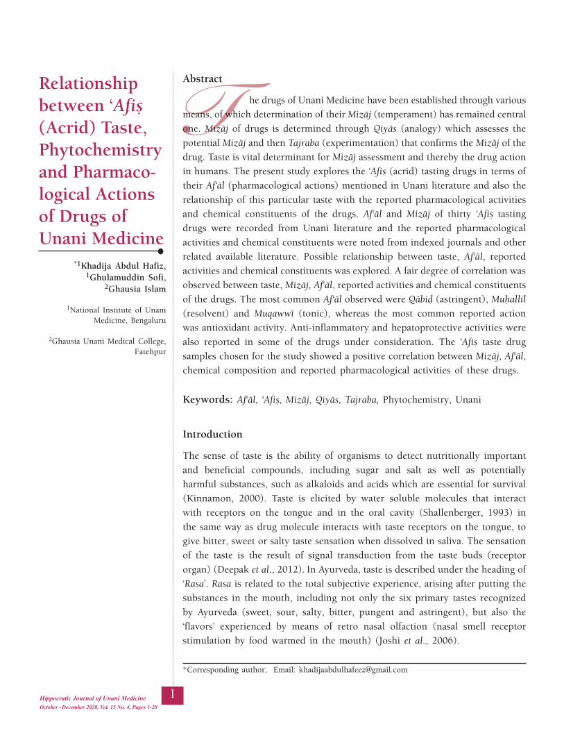
TAbstract
he drugs of Unani Medicine have been established through various means, of which determination of their Mizäj (temperament) has remained central one. Mizäj of drugs is determined through Qiyäs (analogy) which assesses the potential Mizäj and then Tajraba (experimentation) that confirms the Mizäj of the drug. Taste is vital determinant for Mizäj assessment and thereby the drug action in humans. The present study explores the ‘Afiñ (acrid) tasting drugs in terms of their Af‘äl (pharmacological actions) mentioned in Unani literature and also the relationship of this particular taste with the reported pharmacological activities and chemical constituents of the drugs. Af‘äl and Mizäj of thirty ‘Afiñ tasting drugs were recorded from Unani literature and the reported pharmacological activities and chemical constituents were noted from indexed journals and other related available literature. Possible relationship between taste, Af‘äl, reported activities and chemical constituents was explored. A fair degree of correlation was observed between taste, Mizäj, Af‘äl, reported activities and chemical constituents of the drugs. The most common Af‘äl observed were Qäbiò (astringent), Muùallil (resolvent) and Muqawwé (tonic), whereas the most common reported action was antioxidant activity. Anti-inflammatory and hepatoprotective activities were also reported in some of the drugs under consideration. The ‘Afiñ taste drug samples chosen for the study showed a positive correlation between Mizäj, Af‘äl, chemical composition and reported pharmacological activities of these drugs.
Keywords: Af‘äl, ‘Afiñ, Mizäj, Qiyäs, Tajraba, Phytochemistry, Unani
Introduction
The sense of taste is the ability of organisms to detect nutritionally important and beneficial compounds, including sugar and salt as well as potentially harmful substances, such as alkaloids and acids which are essential for survival (Kinnamon, 2000). Taste is elicited by water soluble molecules that interact with receptors on the tongue and in the oral cavity (Shallenberger, 1993) in the same way as drug molecule interacts with taste receptors on the tongue, to give bitter, sweet or salty taste sensation when dissolved in saliva. The sensation of the taste is the result of signal transduction from the taste buds (receptor organ) (Deepak et al., 2012). In Ayurveda, taste is described under the heading of ‘Rasa’. Rasa is related to the total subjective experience, arising after putting the substances in the mouth, including not only the six primary tastes recognized by Ayurveda (sweet, sour, salty, bitter, pungent and astringent), but also the ‘flavors’ experienced by means of retro nasal olfaction (nasal smell receptor stimulation by food warmed in the mouth) (Joshi et al., 2006).
October - December 2020, Vol. 15 No. 4, Pages 1-20
Relationship between ‘Afiñ (Acrid) Taste, Phytochemistry and Pharmaco-logical Actions of Drugs of Unani Medicine
*1Khadija Abdul Hafiz,1Ghulamuddin Sofi,
2Ghausia Islam
1National Institute of Unani Medicine, Bengaluru
2Ghausia Unani Medical College,Fatehpur
*Corresponding author; Email: [email protected]

2Hippocratic Journal of Unani Medicine
The number of basic tastes recognized as primary tastes has varied over the years. Applying the ‘Doctrine of Opposites’, Aristotle considered sweet and bitter tastes to be the example of the doctrine and believed that all other tastes lay between the two extremes. Linnaeus expanded the number of tastes like sweet, sour, sharp, salty, bitter, astringent, viscous, aqueous, and nauseous. Wundt, the founder of experimental psychology, first reduced the number to six (sweet, salt, bitter, sour, metallic and alkaline), then four (sweet, salt, sour and bitter) (Danish, 2016).
Pharmacological basis of drug action revolves around the universal pharmacological principle that similar structures have similar pharmacological activity. If the structure of a substance is known, then its pharmacological behavior can be inferred. Conventional pharmacology uses chemical structure as the basis for pharmacological basis of drug action. According to Charaka, rasa can be different in fresh condition and in dry state of the same substance, and have different pharmacological actions in those conditions, e.g. Piper longum L. is Madhura (sweet) in fresh condition whereas in dry form it is Katu (pungent) and accordingly fresh P. longum is heavier to digest (guru) than dry P. longum which is easy to digest (laghu) (Rath et al., 2014). Beauchamp et al. correlates the pharmacological action of Ibuprofen and Oleocanthal on the basis of their similarities in taste. They point out that both Oleocanthal from olive oil and solution of Ibuprofen, a non-steroidal anti-inflammatory drug, induce similar strong stinging sensations in the throat. Despite not being entirely similar structure, both molecules are anti-inflammatory and have similar profiles, being COX-1 and COX-2 inhibitors (Joshi et al., 2006). Therefore, it is obvious that since rasa indicates the pharmacological behavior of the substance as and when the substance is presented before the user, rasa can be used as a tool to test the substance in use. Modern pharmacology revolves around a central concept that the activity of a chemical is reflected in its chemical structure. Both qualitative and quantitative structure-activity relationship (QSAR) determines the biological activity of the substance and defines those alterations in structure that can change the overall properties of a compound (Rath et al., 2014).
The enormous experiences of Unani physicians are worthy when we look at the literature explaining minute details regarding these determinants of Mizäj. From the present scenario, their experiences will be more useful when they are interpreted by objective measurements (Parveen, 2015). Colour, smell and taste were attempted for objective measurement and it was found that variation in these determinants needs to be further evaluated (Parveen, 2015). The eight tastes defined by Unani Medicine have got unique importance as it reveals distinct Täthér in the body. So attempt was needed to study them separately (Danish, 2016). The present study was carried out in the light of the above discussion and it was decided to explore the relation between taste, Mizäj, Af‘äl
3Hippocratic Journal of Unani Medicine
mentioned by Unani physicians, reported pharmacological activities and the chemical constituents of the drug.
Methodology
The concept of analogy for the determination of the effect of ‘Afiñ taste drug was thoroughly reviewed and the pharmacological action of ‘Afiñ taste drugs on the basis of their phytoconstituents was also explored. Thirty Mufrad (single) drugs of ‘Afiñ taste were selected from different Unani classical books (Khan, 2012; Ibn Sina, 1998; Mohammad, 2002; Kabiruddin, 2007; Ghani, YNM). Phytoconstituents and reputed pharmacological actions of all drugs were searched from authentic sources like ethno-botanical literature, web search engines and indexed journals. For all pharmacological actions mentioned in Unani literature and reported in journals, the chemical constituent of drugs was compared to see the similarity between taste, chemical constituents, and action of the drugs.
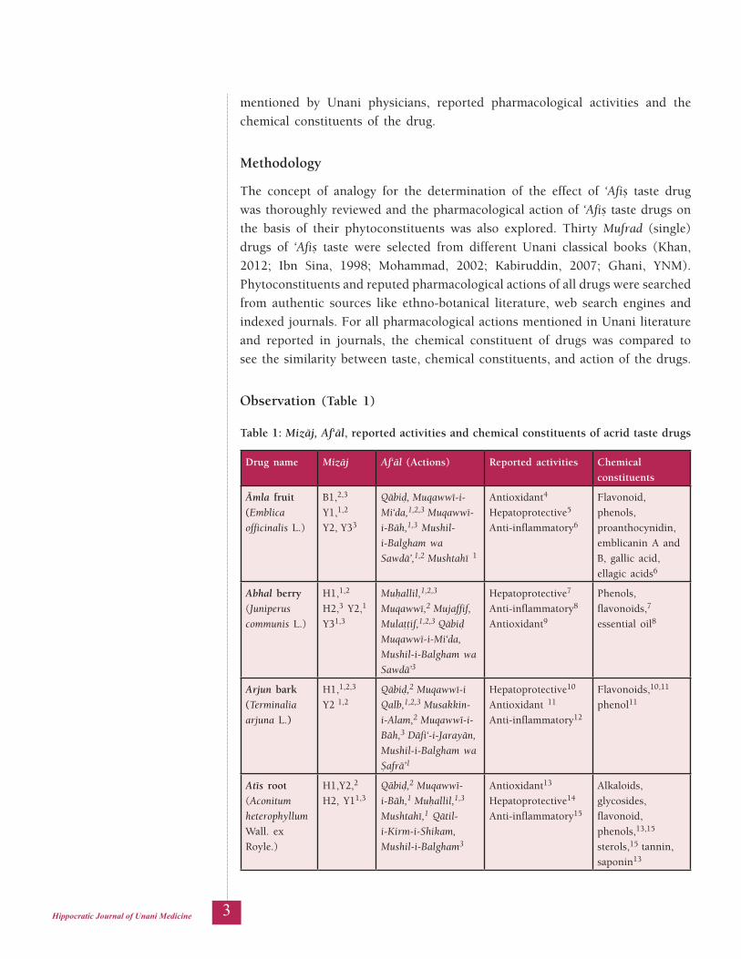
Observation (Table 1)
Table 1: Mizäj, Af‘äl, reported activities and chemical constituents of acrid taste drugs
Drug name Mizäj Af‘äl (Actions) Reported activities Chemical constituents
Ämla fruit(Emblica officinalis L.)
B1,2,3
Y1,1,2
Y2, Y33
Qäbiò, Muqawwé-i-Mi‘da,1,2,3 Muqawwé-i-Bäh,1,3 Mushil-i-Balgham wa Sawdä’,1,2 Mushtahé 1
Antioxidant4
Hepatoprotective5
Anti-inflammatory6
Flavonoid, phenols, proanthocynidin,
emblicanin A and B, gallic acid, ellagic acids6
Abhal berry(Juniperus communis L.)
H1,1,2 H2,3 Y2,1
Y31,3
Muùallil,1,2,3
Muqawwé,2 Mujaffif, Mulaööif,1,2,3 Qäbiò Muqawwé-i-Mi‘da, Mushil-i-Balgham wa Sawdä’3
Hepatoprotective7
Anti-inflammatory8
Antioxidant9
Phenols, flavonoids,7
essential oil8
Arjun bark(Terminalia arjuna L.)
H1,1,2,3
Y2 1,2Qäbiò,2 Muqawwé-i Qalb,1,2,3 Musakkin-i-Alam,2 Muqawwé-i-Bäh,3 Däfi‘-i-Jarayän,
Mushil-i-Balgham wa Ñafrä’1
Hepatoprotective10
Antioxidant 11
Anti-inflammatory12
Flavonoids,10,11
phenol11
Atés root(Aconitum heterophyllum Wall. ex Royle.)
H1,Y2,2
H2, Y11,3Qäbiò,2 Muqawwé-i-Bäh,1 Muùallil,1,3
Mushtahé,1 Qätil-i-Kirm-i-Shikam, Mushil-i-Balgham3
Antioxidant13
Hepatoprotective14
Anti-inflammatory15
Alkaloids, glycosides, flavonoid, phenols,13,15
sterols,15 tannin, saponin13
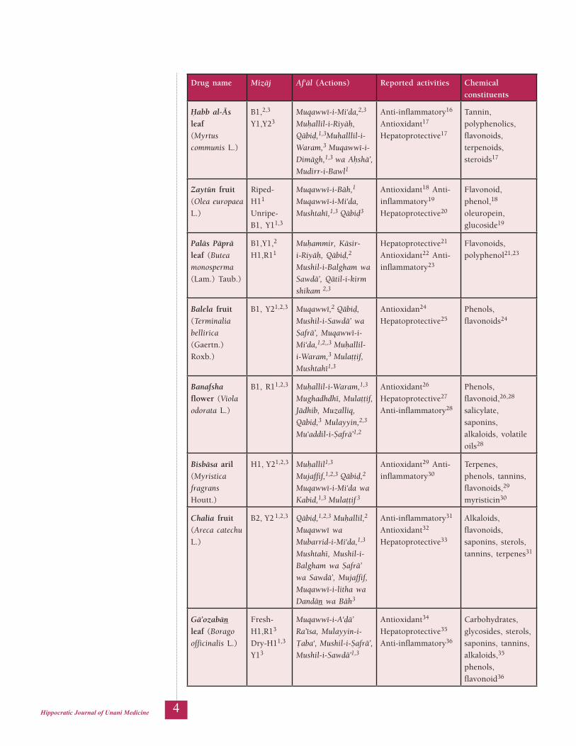
4Hippocratic Journal of Unani Medicine
Drug name Mizäj Af‘äl (Actions) Reported activities Chemical constituents
Ùabb al-Äs leaf(Myrtus communis L.)
B1,2,3
Y1,Y23Muqawwé-i-Mi‘da,2,3 Muùallil-i-Riyäù, Qäbiò,1,3Muùalllil-i-Waram,3 Muqawwé-i-Dimägh,1,3 wa Aùshä’, Mudirr-i-Bawl1
Anti-inflammatory16
Antioxidant17
Hepatoprotective17
Tannin, polyphenolics, flavonoids, terpenoids, steroids17
Zaytün fruit(Olea europaea L.)
Riped- H11 Unripe- B1, Y11,3
Muqawwé-i-Bäh,1 Muqawwé-i-Mi‘da,
Mushtahé,1,3 Qäbiò3
Antioxidant18 Anti-inflammatory19 Hepatoprotective20
Flavonoid, phenol,18
oleuropein, glucoside19
Paläs Päprä leaf (Butea monosperma (Lam.) Taub.)
B1,Y1,2
H1,R11Muùammir, Käsir-i-Riyäù, Qäbiò,2
Mushil-i-Balgham wa Sawdä’, Qätil-i-kirm shikam 2,3
Hepatoprotective21 Antioxidant22 Anti-inflammatory23
Flavonoids, polyphenol21,23
Balela fruit (Terminalia bellirica (Gaertn.) Roxb.)
B1, Y21,2,3 Muqawwé,2 Qäbiò,
Mushil-i-Sawdä’ wa Ñafrä’, Muqawwé-i-Mi‘da,1,2,,3 Muùallil-i-Waram,3 Mulaööif, Mushtahé1,3
Antioxidan24 Hepatoprotective25
Phenols, flavonoids24
Banafsha flower (Viola odorata L.)
B1, R11,2,3 Muùallil-i-Waram,1,3
Mughadhdhé, Mulaööif, Jädhib, Muzalliq, Qäbiò,3 Mulayyin,2,3 Mu‘addil-i-Ñafrä’1,2
Antioxidant26 Hepatoprotective27 Anti-inflammatory28
Phenols, flavonoid,26,28
salicylate, saponins, alkaloids, volatile oils28
Bisbäsa aril (Myristica fragrans Houtt.)
H1, Y21,2,3 Muùallil1,3
Mujaffif,1,2,3 Qäbiò,2
Muqawwé-i-Mi‘da wa Kabid,1,3 Mulaööif 3
Antioxidant29 Anti-inflammatory30
Terpenes, phenols, tannins, flavonoids,29 myristicin30
Chalia fruit (Areca catechu L.)
B2, Y2 1,2,3 Qäbiò,1,2,3 Muùallil,2 Muqawwé wa Mubarrid-i-Mi‘da,1,3
Mushtahé, Mushil-i-Balgham wa Ñafrä’ wa Sawdä’, Mujaffif, Muqawwé-i-litha wa Dandän wa Bäh3
Anti-inflammatory31 Antioxidant32 Hepatoprotective33
Alkaloids, flavonoids, saponins, sterols, tannins, terpenes31
Gä’ozabän leaf (Borago officinalis L.)
Fresh- H1,R13 Dry-H11,3
Y13
Muqawwé-i-A‘òä’ Ra’ésa, Mulayyin-i-Öaba‘, Mushil-i-Ñafrä’, Mushil-i-Sawdä’1,3
Antioxidant34 Hepatoprotective35 Anti-inflammatory36
Carbohydrates, glycosides, sterols, saponins, tannins, alkaloids,35 phenols, flavonoid36

5Hippocratic Journal of Unani Medicine
Drug name Mizäj Af‘äl (Actions) Reported activities Chemical constituents
Gul-i-Surkh (Rosa damascena Mill.)
Fresh- H1, R12 Dried- H1,1,2,3 Y11,3
Mushil, Qäbiò, Mulaööif, Muùallil-i-Waram, Muqawwé-i-Mi‘da wa Bäh1,3
Muqawwé-i-Litha,, Mujaffif1
Anti-inflammatory37 Antioxidant38 Hepatoprotective38
Phenol, alkaloid, flavoniod, terpenoid, saponin, tannin, glycoside, carbohydrates38
Halela fruit (Terminalia chebula Retz.)
B1,Y21,2,3 Muqawwé-i-Mi‘da,
Mushil-i-Ñafrä’ wa Sawdä’,1,2,3 Muqawwé-i-Dandän wa Litha, Mujaffif, Mulayyin1,3 Qäbiò 2,3
Antioxidant39 Hepatoprotective40 Anti-inflammatory39
Flavonoids, phenols, tannins, triterpenoids39
Jämun leaf (Eugenia jambolana Lam.)
B2, Y3,1,3 Muqawwé-i-Mi‘da, 1,2,3 Qäbiò1,3 Häòim,
Muqawwé-i-Litha,1
Muqawwé-i-Bäh1,3
Muùarrik-i-Ishtihä’ 1,2,3
Anti-inflammatory41 Antioxidant42 Hepatoprotective43
Flavonoids, saponins, phenols, steroid, tannin, coumarins42,43
Khär-i-Khasak fruit (Tribulus terrestris L.)
B1,3 Y1,
H12,3Mushtahé, Muùallil-i-Waram, Muqawwé-i-Bäh,1,2,3 Munòij, Mulayyin,1,3 Mudirr-i-Bawl wa Ùayò 1,2,3
Antioxidant44 Hepatoprotective45 Anti-inflammatory46
Flavonoids, phenols44
Majéth root (Rubia cordifolia L.)
H1,2 Y2 2,3 Mufattiù-i-Sudad, Mudirr-i-Bawl, Muqawwé-i-Mi‘da,2,3
Muùallil, Musaffi-i-Khün3
Hepatoprotective47 Antioxidant48 Anti-inflammatory48
Rubiadin,47
terpenoids48
Ma‘én galls (Tamarix gallica L.)
B1,1,2,3 B2,Y2 1,3
Qäbiò,1,2,3 Muqawwé-i-Litha,3 Muùallil-i-Waram-i-Öiùäl,1
Muqawwé-i-Mi‘da,2,3
Mujaffif 1,2
Hepatoprotective49 Antioxidant49 Anti-inflammatory50
Tannins, flavonoids, alkaloid, saponin, Phenols50
Mazu galls (Quercus infectoria Oliv.)
B1,Y2, Y3 1,2,3
Qäbiò, Mujaffif,2,3 Ùäbis,2 Muqawwé-i-Litha, Muùallil-i-Waram,1 Däfi‘-i-Ta‘affun2
Hepatoprotective51 Antioxidant52 Anti-inflammatory53
Carbohydrate, alkaloid, sterol,51 tannins, flavonoids, terpenoids 51,52
Post-i-Anär (Punica granatum L.)
BY 1,2,3 Mujaffif,2 Qäbiò, 1,2
Muùallil, Ùabis2Antioxidant54 Hepatoprotective55 Anti-inflammatory56
Polyphenols, gallic acid, catechin, quercetin, rutin, flavonols, Anthocyanidins55
Pudéna leaf (Mentha arvensis L.)
H1,2
Y2 2,3Muùallil, Mulaööif, 1,2,3 Muqawwé-i-Mi‘da,2 Muqawwé-i-Litha,1 Qäbiò3
Hepatoprotective57 Antioxidant58 Anti-inflammatory59
Alkaloids, flavonoids, polyphenols, tannins, cardiac glycosides, Triterpenoids58

6Hippocratic Journal of Unani Medicine
Drug name Mizäj Af‘äl (Actions) Reported activities Chemical constituents
Pépal leaf (Ficus religiosa L.)
HY,1,2
BY 3Qäbiò,3 Mujaffif,2 Muqawwé-i-Bäh, Muùallil-i-Waram1,2,3
Antioxidant60 Hepatoprotective61 Anti-inflammatory62
Tannins, glycosides, saponins, flavonoids, carbohydrates61
Kä’iphal bark (Myrica nagi L.)
H2, Y2 1,2,3
Muùallil, Qäbiò, Muùallil-i-Waram, 1,2,3 Mushil-i-Sawdä’,3
Muqawwwé-i-Mi‘da wa Bäh,1,3 Mujaffif 2
Antioxidant63 Anti-inflammatory64
Flavonoids, steroids,64
phenols63
Hadjor (Cissus quadrangularis L.)
H,Y 1 Muqawwé, Musakkin-i-Alam, Mushtahé 3
Antioxidant65 Hepatoprotective66 Anti-inflammatory67
Ascorbic acid, carotene, calcium65
Siras (Albizia lebbeck (L.) Benth.)
H2,2
Y2 2,3Muqawwé-i-Litha wa Dandän,1,3 Muùallil, Mujffif, Muñaffi-i-Khün, Muqawwé 2
Antioxidant68 Anti-inflammatory69
Tannins, phenols, alkaloids, steroids, triterpenoids, glycosides, saponins, anthroquinones 68,69
När Mushk (Mesua ferrea L.)
H2, H3, Y3 1,3
Mujaffif,2,3 Muqawwé-i-Mi‘da, Mulaööif,1,2,3
Qäbiò,3 Muùallil-i-Riyäù1,3
Hepatoprotective70 Antioxidant70 Anti-inflammatory71
Phenol70
Baheman (Centaurea behen L.)
H1,2,3
Y12Muùallil-i-Riyäù1,3 Muqawwé-i-Bäh,1,2,3
Qäbiò, Mulaööif 3
Antioxidant72 Hepatoprotective73
Phenols, flavonoid72
Gulnär (Punica granatum L.)
B1,1,2,3
Y2 1,3Qäbiò, Mujaffif,
Ùäbis,1,3 Mundamil-i-Qurüù,3 Muqawwé-i-A‘òä’, Litha wa Dandän 1,3
Antioxidant74 Hepatoprotective75 Anti-inflammatory76
Alkaloids, saponins, tannins, coumarins, terpenoids, steroids, protein, carbohydrates,75
phenols, flavonoids75,76
Babül bark (Acacia nilotica L.)
B1,Y1,2
Y2,3 B21Muqawwé,1,3 Qäbiò,2 Mujaffif,1,2 Muùallil-i-Riyäù,3 Muqawwé-i-Litha wa Dandän1,3
Antioxidant77 Hepatoprotective78 Anti-inflammatory79
Flavonoid78,79
phenols, amino acids,78 alkaloids, glycoside, saponins, tannins steroids79
Käth (Acacia catechu Willd.)
B Y2 2,3 Qäbiò, Muñaffé-i-Khün, Mujaffif, Qätil-i-Kirm Shikam2,3
Antioxidant80 Anti-inflammatory80 Hepatoprotective81
Catechins, epicatechins, flavonoids80
Abbreviation: B=Bärid (cold), H=Ùärr (hot), R=Raöb (moist), Y=Yäbis (dry); 1, 2, 3=first, second, third degree of Mizäj
7Hippocratic Journal of Unani Medicine
Sources: 1(Mohammad, 2002), 2(Kabiruddin, 2007), 3(Ghani, YNM), 4(Liu et al., 2008), 5(Bhuvaneswari et al., 2014), 6(Golechha et al., 2014), 7(Singh et al., 2016), 8(Han et al., 2017), 9(Elmastañ et al., 2006), 10(Chaudhari et al., 2016), 11(Shahriar et al., 2012), 12(Halder et al., 2009), 13(Prasad et al., 2012), 14(Konda et al., 2013), 15(Verma et al., 2010), 16(Rossi et al., 2009), 17(Kumar et al., 2011), 18(Faiza et al., 2011), 19(Sahranavard et al., 2014), 20(Kang et al., 2014), 21(Chavan et al., 2010), 22(Darshan et al., 2012), 23(Gupta et al., 2016), 24(Guleria et al., 2010), 25(Pingale, 2011), 26(Peshin et al., 2017), 27(Elhassaneen et al., 2013), 28(Koochek et al., 2003), 29(Sivaraj et al., 2017), 30(Ozaki et al., 1989), 31(Khan et al., 2011), 32(Phaechamud et al., 2009), 33(Pithayanukul et al., 2009), 34(Segovia et al., 2014), 35(Hamed et al., 2015), 36(Conforti et al., 2008), 37(Valiollah et al,. 2010), 38(Achuthan et al., 2003), 39(Rani et al., 2016), 40(Balakrishna et al., 2017), 41(Kota et al., 2010), 42(Shankar et al., 2012), 43(Kumar et al., 2012), 44(Durgawale et al., 2017), 45(Sugunavarman et al., 2013), 46(Sudheendran et al., 2017), 47(Rao et al., 2006), 48(Charde et al., 2010), 49(Sehrawat et al., 2006), 50(Chaturvedi et al., 2012), 51(Lodhi et al., 2012), 52(Rao et al., 2013), 53(Kaur et al., 2004), 54(Salwe et al., 2015), 55(Khan et al., 2017, 56(Labib et al., 2015), 57(Patil et al., 2012), 58(Ameen et al., 2017), 59(Verma et al., 2003), 60(Al-Ezzy et al., 2017), 61(Selvan et al., 2017), 62(Charde et al., 2010), 63(Rana et al., 2014), 64(Patel et al., 2011), 65(Prabhavathi et al., 2016), 66(Swamy et al., 2010), 67(Panthong et al., 2007), 68(Ariharasiva kumar et al., 2014), 69(Babu et al., 2009), 70(Rajopadhye et al., 2012), 71(Tiwari et al., 2012),72(Chougule et al., 2012),73(Pushplata et al., 2014), 74(Nalini et al., 2015), 75(Kau et al., 2006), 76(Xua et al., 2017), 77(Hegazy et al., 2013), 78(Verma et al., 2014), 79(Safari et al., 2016), 80(Stohs, 2015), 81(Pingale, 2010).
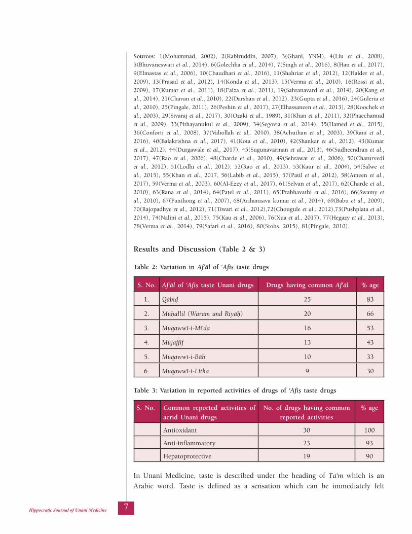
Results and Discussion (Table 2 & 3)
Table 2: Variation in Af‘äl of ‘Afiñ taste drugs
S. No. Af‘äl of ‘Afiñ taste Unani drugs Drugs having common Af‘äl % age
1. Qäbiò 25 83
2. Muùallil (Waram and Riyäù) 20 66
3. Muqawwé-i-Mi‘da 16 53
4. Mujaffif 13 43
5. Muqawwé-i-Bäh 10 33
6. Muqawwé-i-Litha 9 30
Table 3: Variation in reported activities of drugs of ‘Afiñ taste drugs
S. No. Common reported activities of acrid Unani drugs
No. of drugs having common reported activities
% age
Antioxidant 30 100
Anti-inflammatory 23 93
Hepatoprotective 19 90
In Unani Medicine, taste is described under the heading of Öa‘m which is an Arabic word. Taste is defined as a sensation which can be immediately felt
8Hippocratic Journal of Unani Medicine
and described in actual terms or feelings (Khan, 2012). Taste is classified into nine types – Ùulw (Shérén/sweet), Ùirréf (Charparä/pungent), Ùämiò (Khattä/sour), Dasm (Rowghani/fatty), Murr (Kadwä/bitter), ‘Afiñ (Kaséla/acrid), Mäliù (Namkén/salty), Qäbiò (astringent) and Tafih (Phékä/tasteless). These nine types are classified by ancient Unani physicians, while modern Unani physicians classify them into eight types (Danish, 2016; Khan, 2012). They also stated that those substances which bear a certain taste would be dense and earthy, tenuous or moderate in these attributes (Jawhar). In potency (Quwä), it would be hot, cold, or moderate. Now if the dense and earthy substance is hot, it would be bitter; if it is cold, it would be acrid; and if it is moderate, it would be sweet. In case of a substance being tenuous, if it is hot, it would be pungent; if it is cold, it would be sour and if it is moderate, it would be greasy, if the substance being hot, is of moderate density and tenuity it would be salty and if it is cold it would be astringent, if the substance is moderate in coldness and in hotness, according to physicians it would possibly be insipid (Danish, 2016, Khan, 2012).
The interrelated concepts are well-expressed in terms of Mizäj of a drug which is stated in reference to its (Täthér) in the body. To ascertain Mizäj of drugs, Unani Medicine follows the procedure of Qiyäs and Tajraba. Qiyäs predicts the action of a drug through the probable Mizäj whereas Tajraba confirms the same. Qiyäs remains important step in assessing the drug action. The determinants of Mizäj through Qiyäs are organoleptic characters and physical properties of the drug. Among organoleptic characters, taste is considered vital determinant followed by smells and then colours (Khan, 2012; Ibn Sina, 1998). The reason is that it is felt just when it meets the faculty of taste. In case of odour some vapors emanating from the rarefied parts of the drug are felt whereas no vapors arise from the condensed part of that drug. Similarly, a colour which is perceptible may be the colour of the external surface and not of the hidden part of the drug. Sometimes odours indicate taste, such as sweat odour, sour odour, pungent or bitter odour. This shows that the taste is the most precise in giving out the nature of a drug, and then comes odour and colour (Danish, 2016; Khan, 2012).
The observation of the study was utilized to see the correlation of Af‘äl, reported activities and the chemical composition of acrid taste drugs along with their Mizäj. Survey showed that out of 30 acrid drugs, 25 (83%) were ascribed with Qäbiò (astringent) action. In this respect, it was noted that most of the drugs with Muqawwé (tonic) action were specific as Muqawwé-i-Mi‘da (stomach tonic) (53%), Muqawwé-i-Bäh (aphrodisiac) (33%) and Muqawwé-i-litha (30%). It was also noted that 20 drugs (66%) had Muùallil (resolvent) action, while 43% of the drugs were specific as Mujaffif (desiccant) action. The same drugs which have been subjected to screening of the pharmacological activities, the common reported activities that was observed as antioxidant (100%) and 93% were having anti-inflammatory activity while 90% have hepatoprotective action.
9Hippocratic Journal of Unani Medicine
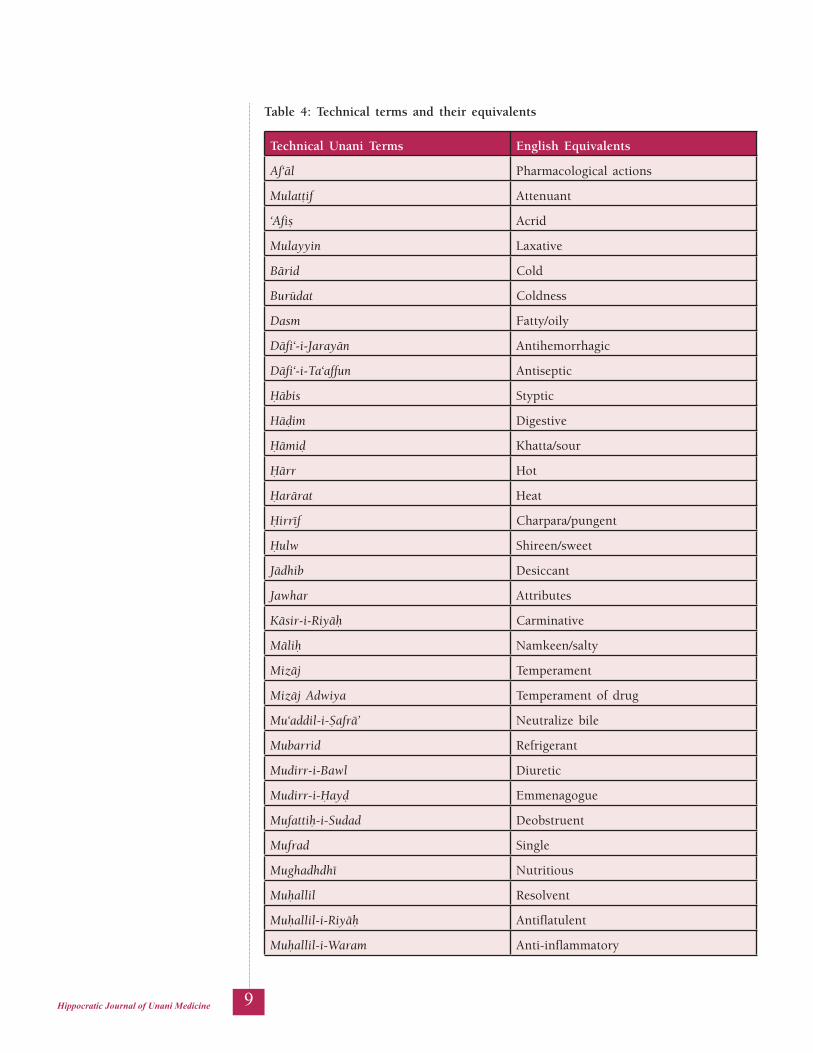
Table 4: Technical terms and their equivalents
Technical Unani Terms English Equivalents
Af‘äl Pharmacological actions
Mulatöif Attenuant
‘Afiñ Acrid
Mulayyin Laxative
Bärid Cold
Burüdat Coldness
Dasm Fatty/oily
Däfi‘-i-Jarayän Antihemorrhagic
Däfi‘-i-Ta‘affun Antiseptic
Ùäbis Styptic
Häòim Digestive
Ùämiò Khatta/sour
Ùärr Hot
Ùarärat Heat
Ùirréf Charpara/pungent
Ùulw Shireen/sweet
Jädhib Desiccant
Jawhar Attributes
Käsir-i-Riyäù Carminative
Mäliù Namkeen/salty
Mizäj Temperament
Mizäj Adwiya Temperament of drug
Mu‘addil-i-Ñafrä’ Neutralize bile
Mubarrid Refrigerant
Mudirr-i-Bawl Diuretic
Mudirr-i-Ùayò Emmenagogue
Mufattiù-i-Sudad Deobstruent
Mufrad Single
Mughadhdhé Nutritious
Muùallil Resolvent
Muùallil-i-Riyäù Antiflatulent
Muùallil-i-Waram Anti-inflammatory
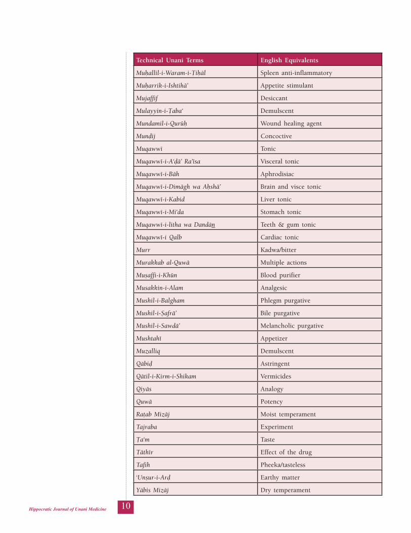
10Hippocratic Journal of Unani Medicine
Technical Unani Terms English Equivalents
Muùallil-i-Waram-i-Öiùäl Spleen anti-inflammatory
Muùarrik-i-Ishtihä’ Appetite stimulant
Mujaffif Desiccant
Mulayyin-i-Öaba‘ Demulscent
Mundamil-i-Qurüù Wound healing agent
Munòij Concoctive
Muqawwé Tonic
Muqawwé-i-A‘òä’ Ra’ésa Visceral tonic
Muqawwé-i-Bäh Aphrodisiac
Muqawwé-i-Dimägh wa Aùshä’ Brain and visce tonic
Muqawwé-i-Kabid Liver tonic
Muqawwé-i-Mi‘da Stomach tonic
Muqawwé-i-litha wa Dandän Teeth & gum tonic
Muqawwé-i Qalb Cardiac tonic
Murr Kadwa/bitter
Murakkab al-Quwä Multiple actions
Muñaffi-i-Khün Blood purifier
Musakkin-i-Alam Analgesic
Mushil-i-Balgham Phlegm purgative
Mushil-i-Ñafrä’ Bile purgative
Mushil-i-Sawdä’ Melancholic purgative
Mushtahé Appetizer
Muzalliq Demulscent
Qäbiò Astringent
Qätil-i-Kirm-i-Shikam Vermicides
Qiyäs Analogy
Quwä Potency
Raöab Mizäj Moist temperament
Tajraba Experiment
Öa‘m Taste
Täthér Effect of the drug
Tafih Pheeka/tasteless
‘Unñur-i-Arò Earthy matter
Yäbis Mizäj Dry temperament

11Hippocratic Journal of Unani Medicine
In spite of chemical composition of these drugs observed to be of highly varied nature, most of the drugs have tannin, flavonoid and phenolic compounds in their chemical composition besides alkaloids and glycosides. Saponins, carbohydrates, triterpenoids and sterols, etc. are other chemical constituents present in these drugs. All the drugs of acrid taste have Yäbis Mizäj with Ùarärat and Burüdat except Gul-i-Banafsha which has Raöb Mizäj with Ùarärat and Burüdat. In Unani literature, it is mentioned that Jawhar of acrid taste substance is dense and earthy, and it is also mentioned that due to the inclination of acrid taste drugs towards ‘Unñur-i-Arò because of these substances acrid taste drugs may have Yäbis Mizäj.
The most common pharmacological action observed was Qäbiò (astringent) action. Astringent drugs create density in the state of parts of an organ and obstruct the channels due to its excessive movements. From the observation table, it was found that most of the drugs have tannin as chemical composition, and tannins are responsible for astringent action because either they bind and precipitate or shrink proteins. Tannin containing drugs have been used traditionally as styptic and internally for the protection of inflamed surfaces of mouth and throat. Actions related to reducing tissue stress load like antioxidant action may be associated with Muqawwé actions as described in Unani Medicine.
The most common reported activity of ‘Afiñ drugs was antioxidant (100%). Out of 30 drugs, 24 drugs have phenols, 26 drugs have flavonoid and 15 drugs have tannins, which are responsible for antioxidant activity (Table 1).
Conclusion
Much information is available regarding reported pharmacological activities of many drugs in reputed journals on internet and many standard books reporting the pharmacological studies done on herbal or traditional drugs and after study it was observed that most of the actions claimed in classical texts were also reported for similar pharmacological activity.
Acknowledgement
Authors are thankful to all the library staff of National Institute of Unani Medicine (NIUM), Bangalore for providing classical literature and other necessary materials related to the subject.
References
1. Achuthan, C. R., Babu, B. H., & Padikkala, J. (2003). Antioxidant and Hepatoprotective effects of Rosa damascene. Pharmaceutical Biology, 41(5), 357–361.

12Hippocratic Journal of Unani Medicine
2. Al-Ezzy, R. M., Saleh, B. H., & A.al anee, R. S. (2017). Assessments of total Flavonoids, Anti-oxidant and Antibacterial activity of Ficus religiosa methanolic extract in vitro. International Journal of Pharmaceutical Sciences Review and Research, 45(2), 6–10.
3. Ameen, N., & Shafi, S. (2017). Evaluation of in vitro anti-oxidant activity of Mentha arvensis L. in memory enhancing study. International Journal of Pharmacognosy and Phytochemical Research, 9(8), 1166–1174.
4. Ariharasiva, K. G., Kavimani, S., Uma, S., & Tamilarasan, M. (2014). Quality evaluation of in vitro antioxidant activity of Albizia lebbeck L. World Journal of Pharmacy and Pharmaceutical Sciences, 3(10), 1267–76.
5. Babu, N. P., Pandi, K. P., & Ignacimuthu, S. (2009). Anti-inflammatory activity of Albizia lebbeck Benth., an ethnomedicinal plant, in acute and chronic animal models of inflammation. Journal of Ethnopharmacology, 125, 356–360.
6. Balakrishna, V., & Lakshmi, T. (2017). Hepatoprotective activity of ethanolic fruit extract of Terminalia chebula on ethanol induced hepatotoxicity in rats. European Journal of Pharmaceutical and Medical Research, 4(4), 533–536.
7. Bhuvaneswari, R., Chidambaranathan, N., & Jegatheesan, K. (2014). Hepatoprotective effect of Embilica officinalis and its silver nanoparticles against CCl4 induced hepatotoxicity in Wistar Albino rats. Digest Journal of Nanomaterials and Biostructures, 9(1), 223–235.
8. Charde, R. M., Charde, M. S., Fulzele, S. V., Satturwar, P. M., & Joshi S. B. (2010). Antioxidant and anti-Inflammatory activity of ethanolic extract of Rubia cordifolia roots. Journal of Pharmacy Research, 3(12), 3070–3071.
9. Charde, R. M., Dhogade, H. J., Charde, M. S., & Kasture, A. V. (2010). Evaluation of antioxidant, wound healing and anti-inflammatory activity of ethanolic extract of leaves of Ficus religeosa. International Journal of Pharmaceutical Sciences and Research, 1(5), 73–82.
10. Chaturvedi, S., Drabu, S., & Sharma, M. (2012). Anti-inflammatory and analgesic activity of Tamarix gallica. International Journal of Pharmacy and Pharmaceutical Sciences, 4(3), 653–658.
11. Chaudhari, G. M., & Mahajan, R. T. (2016). In-vitro hepatoprotective activity of Terminalia arjuna stem bark and its flavonoids against CCl4 induced hepatotoxicity in goat Liver slice culture. Asian Journal of Plant Science and Research, 6(6), 10–17.

13Hippocratic Journal of Unani Medicine
12. Chavan S. D., Ghodake, R. S., Patil, S. B., & Naikwade, N. S. (2010). Hepatoprotective potentials of Butea monosperma (Lam) Taub leaves extract against carbon tetrachloride induced hepatotoxicity by noninvasive method. Der Pharmacia Lettre, 2(5), 62–66.
13. Chougule, P., Pawar, R., Limaye, D., Joshi, Y. M., & Kadam. (2012). In-vitro Antioxidant activity of ethanolic extract of Centaurea behen. Journal of Applied Pharmaceutical Science, 02(04), 106–110.
14. Conforti, F., Sosa, S., Marrelli, M., Menichini, F., Statti, G. A., Uzunov, D., Tubaro, A., Menichini, F., & Loggia, R. D. (2008). In vivo anti-inflammatory and in vitro antioxidant activities of Mediterranean dietary plants. Journal of ethnopharmacology, 116(1), 144–151. https://doi.org/10.1016/j.jep.2007.11.015
15. Danish, M. (2016). Exploration of Murr (bitter) and Hirreef (pungent) Unani drugs in reference to their phytochemistry and pharmacological actions (Thesis). NIUM.
16. Darshan, S., Nitin, M., Kondheru, P., & Bhavesh, L. (2012). Antioxidant activity of Butea monosperma leaf extract. International Journal of Research in Ayurveda and Pharmacy, 3(2), 277–279.
17. Deepak, S., Dinesh, K., Mankaran, S., Gurmeet, S., & Singh, R. M. (2012). Taste masking technologies: A novel approach for the improvement of organoleptic property of pharmaceutical active substance. International Research Journal of Pharmacy, 3(4), 108–116.
18. Durgawale, P. P., & Datkhile, K. D. (2017). Study of polyphenol content and anti-oxidative potential of Tribulus terrestris dry fruit extract. International Journal of Pharmacognosy and Phytochemical Research, 9(5), 716–721.
19. Elhassaneen, Y., Sabry, S., Musalum, T., El-Eskafy, A., & El-Fatah, A. A. (2013). Effect of sweet violet (Viola odorata L.) Blossoms powder on liver and kidney functions as well as serum lipid per oxidation of rats treated with carbon tetrachloride. Journal of American Science, 9(5), 88–95.
20. Elmastañ, D. M., Gülçin, I., Beydemir, S., Küfrevioglu, O. I., & Aboul-Enein, H. Y. (2006). A study on the in vitro antioxidant activity of Juniper (Juniperus communis L.) fruit extracts. Analytical Letters, 39(1), 47–65. http://dx.doi.org/10.1080/ 000327105 00 423385
21. Faiza, I., Wahiba, K., Nassira, G., Chahrazed, B., & Atik, B. F. (2011). Antioxidant potential of olive (Olea europaea L.) from Algeria. Journal of Natural Product and Plant Resources, 1(2), 29–35.
22. Ghani, N. (YNM). Khazainul Advia. Idara Kitab us Shifa, New Delhi.

14Hippocratic Journal of Unani Medicine
23. Golechha, M., Sarangal, V., Ojha, S., Bhatia, J. S., & Arya, D. (2014). Anti-Inflammatory effect of Emblica officinalis in rodent models of acute and chronic inflammation: Involvement of possible mechanisms. International Journal of Inflammation, 1–6. http://dx.doi.org/10.1155/2014/178408
24. Guleria, S., Tiku, A. K., & Rana, S. (2010). Antioxidant activity of acetone extract/ fraction of Terminalia bellirica Roxb. fruit. Indian Journal of Biochemistry and Biophysics, 47(2), 110–116.
25. Gupta, A., & Chaphalkar, S. R. (2016). Anti-inflammatory and anti-microbial activities of aqueous leaves extract of Butea frondosa. Journal of Herbmed Pharmacology, 5(2), 85–88.
26. Halder, S., Bharal, N., Mediratta, P. K., Kaur, I., & Sharma, K. K. (2009). Anti-inflammatory, immunomodulatory and antinociceptive activity of Terminalia arjuna Roxb. bark powder in mice and rats. Indian Journal of Experimental Biology, 47(7), 577–583.
27. Hamed, A. N. E., & Wahid, A. (2015). Hepatoprotective activity of Borago officinalis extract against CCl4-induced hepatotoxicity in rats. Journal of Natural Products, 8, 113–122.
28. Han, X., & Parker, T. (2017). Anti-inflammatory activity of Juniper (Juniperus communis) berry essential oil in human dermal fibroblasts. Cogent Medicine, 4(1), 1–7. http://dx.doi.org/10.1080/2331205X.2017.1306200
29. Hegazy, G. A., Alnoury, A. M., & Gad, H. G. (2013). The role of Acacia arabica extract as an antidiabetic, antihyperlipidemic, and antioxidant in streptozotocin-induced diabetic rats. Saudi Medical Journal, 34(7), 727–733.
30. Joshi, K., Hankey, A., & Patwardhan, B. (2006). Traditional phytochemistry: Identification of drug by ‘taste’. Evidence-Based Complementary and Alternative Medicine, 4(2), 145–148.
31. Kabiruddin, A. H. M. (2007). Makhzan-ul-Mufradat. Idara Kitab-Us-Shifa, New Delhi.
32. Kang, H., & Koppula, S. (2014). Olea europaea Linn. Fruit pulp extract protects against carbon tetrachloride induced hepatic damage in mice. Indian Journal of Pharmaceutical Sciences, 76(4), 274–280.
33. Kaur, G., Jabbar, Z., Athar, M., & Alam, M. S. (2006). Punica granatum (pomegranate) flower extract possesses potent antioxidant activity and abrogates Fe-NTA induced hepatotoxicity in mice. Food and Chemical Toxicology, 44(7), 984-993.
34. Kaur, G., Hamid, H., Ali, A., Alam, M. S., & Athar, M. (2004). Anti-inflammatory evaluation of alcoholic extract of galls of Quercus infectoria. Journal of Ethnopharmacology, 90(2-3), 285–92.
15Hippocratic Journal of Unani Medicine
35. Khan, B. H., Ahmad, F., Ahmad, J., & Yunus, S. M. (2017). Hepatoprotective activity of ethanolic extract of peel of Punica granatum. International Journal of Science and Research, 6(6), 1118–1122.
36. Khan, M. A. (2012). Muheet-i-Azam (Urdu translation, Vol. 1st). Central Council for Research in Unani Medicine, New Delhi.
37. Khan S., Mehmood M.H., Ali A.N.A., Ahmed F.S., Dar A., & Gilani A.H. (2011). Studies on anti-inflammatory and analgesic activities of betel nut in rodents. Journal of Ethnopharmacology, 135: 654-661.
38. Kinnamon S.C. (2000). A plethora of taste receptors. Neuron, 25: 507-510.
39. Konda V.G.R., Eerike M., and Prabhu L. (2013). Evaluation of hepatoprotective activity of ethanolic extract of Aconitum heterophyllum root in Paracetamol induced liver toxicity. Int J Pharm Bio Sci., 4(4): 714–21.
40. Koochek, M. H., Pipelzadeh, M. H., & Mardani, H. (2003). The effectiveness of Viola odorata in the prevention and treatment of formalin-induced lung damage in the rat. Journal of Herbs, Spices & Medicinal Plants, 10(2), 95–103. https://doi.org/ 10.1300/ J044v10n02_11
41. Kota, P. K., Prasad, P. D., Rao, A. N., Reddy, P. D., & Abhinay, G. (2010). Anti-inflammatory activity of Eugenia jambolana in Albino rats. International Journal of Pharma and Bioscience, 1(4), 435–438.
42. Kumar, M. R., Phaneendra, P., Bodhanapu, S., Rahiman, O. M. F., Niyas, K. M., & Tamizmani, T. (2011). Antioxidant and hepatoprotective activity of the aqueous extract of Myrtus communis (Myrtle) Linn. leaves. Pharmacologyonline, 1, 1083–1090.
43. Kumar, Y. V. K., Rao, G. H. S., Santhiswaroop, M., & Chandra, S. D. (2012). Hepatoprotective activity of ethanolic extract of Eugenia jambolana leaf on liver damage caused by paracetamol in rats. International Journal of Research in Pharmaceutical and Nano Sciences, 1(1), 80–86.
44. Labib, R. M., & El-Ahmady, S. H. (2015). Antinociceptive, anti-gastric ulcerogenic and anti-inflammatory activities of standardized Egyptian pomegranate peel extract. Journal of Applied Pharmaceutical Science, 5(1), 048–051.
45. Liu, X., Zhao, M., Wang, J., Yang, B., & Jiang, Y. (2008). Antioxidant activity of methanolic extract of emblica fruit (Phyllanthus emblica L.) from six regions in China. Journal of Food Composition and Analysis, 21(3), 219-228.
46. Lodhi, G., Singh, H. K., Pant, K. K., Rao, C. V., & Hussain, Z. (2012). Hepatoprotective effects of Quercus infectoria gall extract against carbon tetrachloride treated liver injury in rats. International Journal of Applied Research in Natural Products, 5(3), 17–22.

16Hippocratic Journal of Unani Medicine
47. Mohammad, A. H. K. (2002). Bustanul Mufradat. Idara Kitab us Shifa, New Delhi.
48. Nalini, R., & Anuradha, R. (2015). Phytochemical Screening and In-vitro antioxidant activity of ethanolic flower extracts of Punica granatum. International Journal of Pharmaceutical Sciences Review and Research, 30(1), 353–360.
49. Ozaki, Y., Soedigdo, S., Wattimena, Y. R., & Suganda, A. G. (1989). Anti-inflammatory effect of mace, aril of Myristica fragrans Houtt. and its active principles. Japan J. Pharmacol., 49(2), 155–163.
50. Panthong, A., Supraditaporn, W., Kanjanapothi, D., Taesotikul, T., & Reutrakul, V. (2007). Analgesic, anti-inflammatory and venotonic effects of Cissus quadrangularis Linn. Journal of Ethnopharmacology, 110(2), 264–270.
51. Parveen, N. (2015). Development of methods of objective assessment for organoleptic characters involved in determination of mizaj of mufrad drugs (Thesis). NIUM.
52. Patel, T., Dudhpejjya, A., & Sheath, N. (2011). Anti-inflammatory activity of Myrica nagi Linn. bark. Ancient Science of Life, 30(4), 100–103.
53. Patil, K., & Mall, A. (2012). Hepatoprotective activity of Mentha arvensis Linn. leaves against CCl4 induced liver damage in rats. Asian Pacific Journal of Tropical Disease, 2(1), S223–S226.
54. Peshin, T., Azad, C. S, & Kar, H. K. (2017). Antioxidant and free radical scavenging activities of Viola odorata in the search of potential inhibitor of tobacco’s free radicals. Journal of Medicinal Plants Research, 11(27), 433–438. https://doi.org/ 10.5897/ JMPR2017.6421
55. Phaechamud, T., Toprasri, P., & Chinpaisa, C. (2009). Antioxidant activity of Areca catechu extracts in human hepatocarcinoma Hep G2 cell lines. Pharmaceutical Biology, 47(3), 242–247. https://doi.org/10.1080/13880200802434203
56. Pingale, S. S. (2010). Hepatoprotection by Acacia catechu in CCl4 induced liver dysfunction. International Journal of Pharmaceutical Sciences Review and Research, 5(1), 150–154.
57. Pingale, S. S. (2011). Hepatoprotective action of Terminalia bellirica on CCl4 induced hepatic disorders. Der Pharma Chemica, 3(1), 42–48.
58. Pithayanukul, P., Nithitanakool, S., & Bavovada, R. (2009). Hepatoprotective potential of extracts from seeds of Areca catechu and nutgalls of Quercus infectoria. Molecules (Basel, Switzerland), 14(12), 4987–5000. https://doi.org/10.3390/molecules14124987
17Hippocratic Journal of Unani Medicine
59. Prabhavathi, R. M., Prasad, M. P., & Jayaramu, M. (2016). In-vitro antioxidant studies of Cissus quadrangularis (L) extracts. European Journal of Experimental Biology, 6(4), 1–6.
60. Prasad, S. K., Kumar, R., Patel, D. K., Sahu, A. N., & Hemalatha, S. (2012). Physicochemical standardization and evaluation of in-vitro antioxidant activity of Aconitum heterophyllum Wall. Asian Pacific Journal of Tropical Biomedicine, 2(2), S526-S531. https://doi.org/10.1016/S2221-1691(12)60266-4
61. Pushplata, C., Yadunath, J., & Ashish, J. (2014). Protective effect of ethanol extract of Centaurea behen Linn. in carbon tetra chloride-induced hepatitis in rats. International Journal of Pharmacy and Pharmaceutical Sciences, 6(8), 197–200.
62. Rajopadhye, A. A., & Upadhye, A. S. (2012). Hepatoprotective effect of stamen extracts of Mesua ferrea L. against oxidative stress induced by CCl4 in liver slice culture model. Natural Product Sciences, 18(2), 76–82.
63. Rana, R. K., & Patel, R. K. (2014). Antioxidant activity of bark of Myrica nagi. International Journal of Pharmaceutical Sciences Review and Research, 28(1), 99–101.
64. Rani, A. A., Jeeva, S., & Punitha, S. M. J. (2016). Antioxidant, anti-inflammatory, antiproliferative and apoptotic properties of Terminalia chebula (fruit) towards raw 264.7 and oral KB cell lines. International Journal of Applied and Pure Science and Agriculture, 02(05), 90–100.
65. Rao, G. M. M., Rao, C. V., Pushpangadan, P., & Shirwaikar, A. (2006). Hepatoprotective effects of rubiadin, a major constituent of Rubia cordifolia Linn. Journal of Ethno Pharmacology, 103(3), 484–90.
66. Rao, N., Mittal, S., Sudhanshu, & Menghani, E. (2013). In vitro phytochemical screening, antioxidant & antimicrobial activity of the methanolic extract of Quercus infectoria L. International Journal of Pharmacy and Pharmaceutical Sciences, 5(2), 273–277.
67. Rath, S. K., Panja, A. K., Nagar, L., & Shinde, A. (2014). The scientific basis of Rasa (taste) of a substance as a tool to explore its pharmacological behavior. Ancient Science of Life, 33(4), 198–202.
68. Rossi, A., Paola, R. D., Mazzon, E., Genovese, T., Caminiti, R., & Bramanti, P., Pergola, C., Koeberle, A., Werz, O., Sautebin, L., & Cuzzocrea, S. (2009). Myrtucommulone from Myrtus communis exhibits potent anti-Inflammatory effectiveness in vivo. Journal of Pharmacology and Experimental Therapeutics, 329(1), 76–86. https://doi.org/ 10.1124/ jpet.108.143214

18Hippocratic Journal of Unani Medicine
69. Safari, V. Z., Kamau, J. K., Nthiga, P. M., Ngugi, M. P., Orinda, G., & Njagi, E. M. (2016). Antipyretic, anti-inflammatory and antinociceptive activities of aqueous bark extract of Acacia Nilotica (L.) Delile in Albino mice. Journal of Pain Management & Medicine, 2(2), 1–7.
70. Sahranavard, S., Kamalinejad, M., & Faizi, M. (2014). Evaluation of anti-inflammatory and anti-Nociceptive effects of defatted fruit extract of Olea europaea. Iranian Journal of Pharmaceutical Research, 13(Suppl), 119–123.
71. Salwe, K. J., Sachdev, D. O., Bahurupi, Y., & Kumarappan, M. (2015). Evaluation of antidiabetic, hypolipedimic and antioxidant activity of hydroalcoholic extract of leaves and fruit peel of Punica granatum in male Wistar albino rats. Journal of Natural Science, Biology, and Medicine, 6(1), 56–62. https://doi.org/10.4103/0976-9668.149085
72. Segovia, F., Lupo, B., Peiró, S., Gordon, M. H., & Almajano, M. P. (2014). Extraction of Antioxidants from Borage (Borago officinalis L.) leaves-Optimization by response surface method and application in oil-in-water emulsions. Antioxidants (Basel, Switzerland), 3(2), 339–357. https://doi.org/10.3390/antiox3020339
73. Sehrawat, A., & Sultana, S. (2006). Tamarix gallica ameliorates thioacetamide-induced hepatic oxidative stress and hyperproliferative response in Wistar rats. Journal of Enzyme Inhibition and Medicinal Chemistry, 21(2), 215–223. https://doi.org/ 10.1080/ 147563605 00480673
74. Selvan, A., & Chourasia, V. (2017). Hepatoprotective activity of Ficus religiosa leaf extract in rats. Current Research in Pharmaceutical Sciences, 07(02), 64–68.
75. Shahriar, M., Akhter, S., Hossain, M. I., Haque, M. A., & Bhuiyan, M. A. (2012). Evaluation of in vitro antioxidant activity of bark extracts of Terminalia arjuna. Journal of Medicinal Plants Research, 6(39), 5286–5298. https://doi.org/10.5897/JMPR12.580
76. Shallenberger, R. S. (1993). Taste Chemistry (1st ed.). Springer Science Business Media Dordrecht, New York (USA)
77. Shankar, M., & Suthakaran, R. (2012). In vitro antioxidant activities of various extracts of Eugenia jambolana leaves. International Journal of Research in Pharmaceutical and Nano Sciences, 1(2), 317–326.
78. Sina, I. (1998). Al-Qanoon fit-Tib. (2nd vol.). Jamia Hamdard, New Delhi.
79. Singh, H., Prakash, A., Kalia, A. N., & Majeed, A. B. A. (2016). Synergistic hepatoprotective potential of ethanolic extract of Solanum xanthocarpum and Juniperus communis against paracetamol and azithromycin induced liver injury in rats. Journal of Traditional and Complementary Medicine, 6(4), 370–376. https://doi.org/ 10.1016/ j.jtcme.2015.07.005

19Hippocratic Journal of Unani Medicine
80. Sivaraj, S., Patel, V., Mangai, A., Kannayiram, G., & Dasararaju, G. (2017). In-vitro evaluation of antioxidant activity of different extracts of Myristica fragrans Houtt. International Journal of Pharmaceutical Sciences Review and Research, 42(1), 261–264.
81. Stohs, S. J., & Bagchi, D. (2015). Antioxidant, anti-inflammatory, and Chemoprotective properties of Acacia catechu Heart wood extracts. Phytotherapy Research, 29(6), 818–824.
82. Sudheendran, A., & Shajahan, M. A. (2017). In vitro anti-inflammatory and anti-arthritic activity of root and fruit of gokshura (Tribulus terrestris Linn.) International Research Journal of Pharmacy, 8(10), 122–124.
83. Sugunavarman, T., & Jagadeesan, G. (2013). Hepatoprotective activity of Tribulus terrestris on experimental liver damage in mice. International Journal of Modern Research and Reviews, 1(1), 20–23.
84. Swamy, A. H. M. V., Kulkarni, R. V., Thippeswamy, A. H. M., Koti, B. C., & Gore, A. (2010). Evaluation of hepatoprotective activity of Cissus quadrangularis stem extract against isoniazid-induced liver damage in rats. Indian Journal of Pharmacology, 42(6), 397–400.
85. Tiwari, P., & Nandy, S. (2012). Screening of anti-inflammatory activity of Mesua ferrea Linn. flower. International Journal of Biomedical Research, 3(05), 245–252.
86. Valiollah, H., Alireza, G., & Mohammad, H. (2010). Analgesic and anti-inflammatory effect of Rosa damascene hydro alcoholic extract and its essential oil in animal model. Iranian Journal of Pharmaceutical Research, 9(2), 163–163.
87. Verma, P., Singh, S. P., Vind, S. K., Kumar, G. R., & Rao, C. V. (2014). Protective effect of Acacia nilotica (bark) against anti-tubercular drug induced hepatic damage an experimental study. International Journal of Pharmacy and Pharmaceutical Sciences, 6(9), 75–79.
88. Verma, S., Ojha, S., & Raish, M. (2010). Anti-inflammatory activity of Aconitum heterophyllum on cotton pellet induced granuloma in rats. Journal of Medicinal Plants Research, 4(15), 1566–1569. https://doi.org/10.5897/JMPR09.502
89. Verma, S. M., Arora, H., & Dubey, R. (2003). Anti-inflammatory and sedative-hypnotic activity of the methanolic extract of the leaves of Mentha arvensis. Ancient Science of Life, 23(2), 95–99.
90. Xua, J., Zhaoa, Y., & Aisa, H. A. (2017). Anti-inflammatory effect of pomegranate flower in lipopolysaccharide (LPS)-stimulated RAW264.7 macrophages. Pharmaceutical Biology, 55(1), 2095–2101. https://doi.org/10.1080/13880209.2017.1357737

20Hippocratic Journal of Unani Medicine
lkjka'k ;wukuh fpfdRlk dh vkS"kfèk;ksa ds vfQ+l ¼,sfØM½ Lokn]
QkbVksdsfeLVªh vkSj QkekZdksykWftdy fØ;kvksa ds eè; lacaèk*[k+nhtk vCnqy gQ+ht] izks- x+qykeqn~nhu lksQ+h] x+kSfl;k bLyke
Lkkjka'k
;wukuh fpfdRlk dh vkS"kfèk;ka fofHkUu ekè;eksa ls LFkkfir dh xbZa gSa ftuesa buds fet+kt ¼LoHkko½ dk fuèkkZj.k egRoiw.kZ gSA vkS"kfèk;ksa dk fet+kt fd+;kl ¼lekurk½ ds ekè;e ls fuèkkZfjr fd;k tkrk gS tks laHkkfor fet+kt dk vkdyu djrk gS vkSj fQj rtckZ ¼iz;ksx½ vkS"kfèk ds fet+kt dh iqf"V djrk gSA Lokn fet+kt ds ewY;kadu rFkk euq"; esa vkS"kfèk fØ;k ds fy, egRoiw.kZ fuèkkZjd gSA orZeku vè;;u ;wukuh lkfgR; esa of.kZr vQ+vky ¼QkekZdksykWftdy fØ;kvksa½ ds vuqlkj vfQ+l ¼,sfØM½ Lokn okyh vkS"kfèk;ksa dh [kkst djrk gS vkSj fjiksVZ dh xbZ vkS"kèkh; xfrfofèk;ksa vkSj vkS"kfèk;ksa ds jklk;fud ?kVdksa ds lkFk bl fo'ks"k Lokn ds lacaèk dh Hkh [kkst djrk gSA ;wukuh lkfgR; ls rhl vfQ+l ¼,sfØM½ Lokn okyh vkS"kfèk;ksa ds vQ+vky vkSj fet+kt fjdkWMZ fd, x, vkSj fjiksVZ dh xbZ vkS"kèkh; xfrfofèk;ksa vkSj vkS"kfèk;ksa ds jklk;fud ?kVdksa dks vuqØfer if=dkvksa vkSj vU; lacafèkr miyCèk lkfgR; uksV fd;k x;kA Lokn] vQ+vky] fjiksVZ dh xbZ xfrfofèk;ksa vkSj jklk;fud ?kVdksa ds chp laHkkfor lacaèk dk irk yxk;k x;kA vkS"kfèk;ksa ds Lokn] fet+kt] vQ+vky] fjiksVZ dh xbZ xfrfofèk;ksa vkSj jklk;fud ?kVdksa ds chp dkQh gn rd lglacaèk ns[kk x;kA lcls lkèkkj.k ik, x, vQ+vky d+kfct+ ¼,fLVªUtsUV½] eqgfYYky ¼fjt+ksyosUV½] eqD+d+Ooh ¼VkWfUkd½ Fks tcfd lcls lkèkkj.k fjiksVZ dh xbZ fØ;k ,aVhvkWDlhMsaV xfrfofèk FkhA fopkjkèkhu dqN vkS"kfèk;ksa esa ,aVh&bU¶ykesVjh vkSj gsisVksizksVsfDVo xfrfofèk;ka Hkh ikbZ xbZaA vè;;u ds fy, pqus x, vfQ+l Lokn vkS"kfèk uewuksa ls bu ds Lokn] fet+kt] vQ+vky] fjiksVZ dh xbZ xfrfofèk;ksa vkSj jklk;fud ?kVdksa ds chp ,d ldkjkRed lacaèk dk irk pykA
'kCndqath% vQ+vky] vfQ+l] fet+kt] fd+;kl] rtckZ] QkbVksdsfefLVªh] ;wukuh

21Hippocratic Journal of Unani Medicine
BAbstract
ackground: Ùabb Muñaffé-i-Khün (HMK) is a Unani formulation used traditionally as a blood purifier. The individual ingredients of HMK have reported anti-cancer activity, however, its use in the management of cancer has not been yet reported. Thus, a survey was carried out to find out the current perception and practice of Unani doctors towards using HMK in cancer management.
Methodology: After taking approval from Institutional Ethics Committee, a questionnaire was prepared and sent to 100 Unani physicians (Hakims) and their opinion regarding the usage, dosage, safety and tolerability of HMK was documented.
Results: HMK was found to be used in the treatment of blood related disorders by ~66% of Hakims. It has been usually prescribed alone or in combination with other drugs to enhance its effects or to increase its absorption. The average efficacy rate for HMK usage in blood disorders was found to be 8 (scale of 1 to 10). About 39% of Hakims perceived that HMK could be used in the management of various cancers such as breast, prostate, lung, leukemia, oral, renal, bladder and skin.
Conclusion: HMK is largely being used in the treatment of blood related disorders by Hakims in their clinical practice and is perceived to play a significant role in the management of cancers.
Keywords: Ùabb Muñaffé-i-Khün, Survey, Hakims, Perception, Anticancer drug, Practice
Introduction
Cancer is regarded as one of the important causes of mortality and morbidity worldwide (Rafiemanesh et al., 2016). The currently available cancer therapies have serious side effects and thus for more than a decade now, cancer research has shifted its focus towards natural products as well as traditionally used herbal remedies for possible use as adjunct therapies (Savjiyani et al., 2012). The most recent systematic review of cancer patients surveyed globally has shown an increase in the use of Complementary and Alternative Medicine (CAM) (Tangkiatkumjai et al., 2020).
Unani Medicine, one of the branches of Complementary and Alternative Medicine (CAM), is based upon Hippocratic theory of four humors, viz., blood, phlegm,
October - December 2020, Vol. 15 No. 4, Pages 21-31
Current Perception and Practice of Using Ùabb Muñaffé-i-Khün in Cancer Management
1Aliya Khan,1Nidhi Sharma,
2Ghazala Mulla,*1Ruchika Kaul-Ghanekar
1Cancer Research Lab,Interactive Research School for
Health Affairs (IRSHA),Bharati Vidyapeeth, Pune
2Department of Physiology, Z.V.M Unani Medical College and
Hospital, Azam Campus, Pune
*Corresponding author; Email: [email protected]

22Hippocratic Journal of Unani Medicine
yellow bile and black bile (Hongal et al., 2014). Any change in the proportion of these humors, in terms of quality and quantity, modulates the viscosity of the blood. Unani blood purifying drugs with their heat, cold, dry and wet properties maintain the viscosity of the blood. Moreover, these drugs have anti-cancer, anti-oxidant, anti-inflammatory and immunomodulatory activities (Mushir et al., 2019). In Unani text, cancer (Saraöän) has been described as a malignant and melanotic swelling, which spreads rapidly in the body (Naz and Ahmad, 2017). According to Ibn Sina, cancer is a disease caused by accumulation of black bile produced from combustion of yellow bile (Mirra-i-Sawdä’) (Fahad and Shameem, 2018). Various in-vitro and in-vivo studies have reported anticancer activity of Unani medicinal plant extracts such as Nigella sativa, Cuscuta reflexa Roxb. in different human cancer cell lines and animal models (Al-Sheddi et al., 2014; Ali et al., 2020).
Ùabb Muñaffé-i-Khün (HMK), a Unani compound formulation, is used as a blood purifier and for the treatment of boils, scabies, acne, pimples and problems associated with Fasäd i-Dam (imbalance of humours). It is a unique combination of 16 medicinal plants that include Berberis aristata, Cassia absus, Terminalia chebula, Trichoderma glaberrima, Cuminum cyminum, Tephrosia purpurea, Pterocarpus santalinus, Fumaria parviflora, Lawsonia inermis, Piper nigrum, Melia azadirachta, Melia azedarach, Coriandrum sativum, Bauhinia recemosa, Rosa damascene and Genitalia kurroo Royale (Unani Pharmacopoeia of India). In Unani texts, most of the ingredients of HMK have been mentioned to be used in the management of Sawdä’ and anti-cancer activity of most of the ingredients have also been proven scientifically (Kim et al., 2008; Prasanna et al., 2009; Pai et al., 2012; Kapadia et al., 2013; Rajkapoor et al., 2013; Nithya and Sumalatha Jan, 2014; Serasanambati et al., 2015; Santha and Dwidedi, 2015; Nahata et al., 2017; Padmapriya et al., 2017)
Based on the above properties of HMK, a survey was conducted wherein the opinion of Unani physicians (Hakims) regarding the proposed use of HMK as an anti-cancer drug as well as its current practice, safety and tolerability was recorded.
Materials and Methods
Study Design
A mixed-methods study, comprising survey and interview of Hakims from different cities of India, was conducted after taking approval from Institutional Ethics Committee. The survey was conducted at the Unani CME Conference held at Azam Campus, Pune.
23Hippocratic Journal of Unani Medicine
Development of Survey Questionnaire
Literature search using the available databases (Ayush Research Portal, PubMed, Google Scholar) was carried out to collect information and define the theme for developing the questionnaire. A characteristic questionnaire was designed in order to collect the information related to the clinical use of HMK. The questionnaire was directed to 100 Unani physicians (Hakims) through personal communications as well as through emails and phone calls. All the Unani physicians filled it completely to the best of their knowledge and clinical expertise.
Questionnaire Structure
The survey was carried out with a set of questions that included experience in practicing Unani Medicine (in years); frequency and usage of HMK; opinion about the medicine as a blood purifier; opinion about the medicine with anticancer activity and if they had ever prescribed it to cancer patients; whether HMK was prescribed individually or in combination with other Unani formulations; number of tablets and duration prescribed; opinion on mechanism of action on humor and its type; efficacy and safety of the drug; name of the brand commonly used (supplementary information). All the questions were open ended. Questionnaire was given to the Hakims after explaining the study objectives and they were asked to revert with duly filled document.
Statistical Analysis
The survey results and the quantitative data have been reported descriptively. For qualitative variables, frequency with percentage was used to describe the data. Chi-square tests and binary logistic regression were used to compute bivariate odds ratio (OR) to evaluate the preference for using HMK as a blood purifier or as an anticancer drug.
Results
The survey was conducted with a sample size of 100 Unani physicians. The experience of Hakims practicing Unani Medicine varied: 37% with 2-3 years, 20% with 3-5 years, 20% with 5-10 years and 23% with more than 10 years. Survey revealed that 66% Hakims used HMK for the treatment of blood related disorders, specifically skin disorders and remaining 34% used the formulation along with other blood purifiers. The frequency of using HMK in the clinical practice varied among the Hakims: 41% used it frequently, 58% used it sometimes, and only 1% never used the drug. Interestingly, 98% Hakims (p<0.0001, OR- 4.737; 95% CI: 2.607 to 8.609) recognized HMK to be an effective blood purifier. The odds ratio higher than one indicates Hakims preferably consider HMK as an effective blood purifier.

24Hippocratic Journal of Unani Medicine
In the present survey, the physicians had different opinions regarding the mechanism of action of HMK: 45% assumed that HMK cures blood disorders by acting on blood (Dam), 20% assumed that it acts on yellow bile (Ñafrä’), and 35% assumed its action on black bile (Sawdä’) (Fig. 1). This data suggested that since cancer was caused due to combustion of yellow bile and accumulation of black bile, HMK could help in controlling the progression of the disease. Thus, the opinion of Hakims was taken for the use of HMK as an anti-cancer drug. Only 39% Hakims suggested that HMK could be used in the treatment of different types of cancer such as leukemia, skin, renal and bladder.
Ùabb Muñaffé-i-Khün (Ùabb means pill), commonly given in the form of pills, was prescribed by Hakims along with other Unani formulations (mostly blood purifiers) that include Rakt Ñafä, Qurñ al-Kalé, Sharbat-i-‘Unnäb, Ma’jün ‘Ushba, Ma‘jün Muñaffé-i-Khün, ‘Araq Muñaffé-i-Khün, Iöréfal Shähitara, ‘Araq-i-Mundé, Mä’ al-‘Asal. About 28% Hakims prescribed HMK to be given alone for blood purification, 50% prescribed it along with other Unani formulations and 22% believed that it could be given both either alone or in combination with other Unani formulations as per the patients’ requirements.
The dose of HMK suggested by the doctors varied in different patients: 90% prescribed 2 pills (about 0.5 g) BD, and 10% prescribed one pill TDS (Fig. 2). The duration of the dosage prescribed also varied from patient to patient: 33% prescribed for one month, 20% for 2 months and 47% for 3 months (Fig. 2). The Hakims (99%) shared their experience about the beneficial effects of HMK in terms of excellent tolerability in the patients without any side effects. The prominent beneficial effect of the drug was found to be improvement in the skin related problems, with an efficacy of 8 (on the scale of 1 to 10). Only 1% doctors observed that the patients with hyper acidity experienced burning sensation in the chest after consumption of HMK.
Different Hakims prescribed different manufacturing brands of the drug. For example, 33% Hakims prescribed HMK from Hamdard; 8% made the pills
Figure 1: Pie chart representing mechanism of action of HMK as per Unani concept of cancer

25Hippocratic Journal of Unani Medicine
themselves as per the formulary of Unani Medicine; 7% prescribed it from Shama Herbals, 4% from Puritex and 7% from Tayyebi Dawakhana Unani (Indore) Pvt. Ltd., 4% recommended from Hermas Biotech Pvt. Ltd., 1% from Rex Remedies and 1% from Uniherb. Remaining 35% Hakims didn’t comment upon any specific manufacturing company.
Discussion
As per the Unani concept, blood is considered to have different humours (Akhläö) such as Dam (sanguine), Balgham (phlegm), Ñafrä’ (yellow bile) and Sawdä’ (black bile) (Mushir et al., 2019). Each person has a unique and balanced combination of different humours, which may get disturbed under the diseased conditions. According to the Unani principles, cancer is commonly produced from burning of black bile (Iùtiräq-i-Sawdä’) and sometimes from burning of altered yellow bile and impure phlegm (Iùtiräq-i-Ñafrä’-o-Balgham). The causative material of cancer possesses heat, which helps cancer to progress rapidly. Galen had hypothesized in his book (Methods of Treatment) that cancer was a disease associated with black bile and had proposed its removal from the body with administration of appropriate purgative, which can prevent generation and accumulation of black bile in the blood vessels (Emami et al., 2012). Galen had mentioned in his book entitled “Thick Substances with Abnormally High Concentrations” that humour was warm and could lead to ulcerative cancer. The vasculature of cancerous tissue being hyperemic and the blood contents being darker than the other swellings (Emami et al., 2012), cancer could be cured by a drug that would prevent the combustion of yellow bile and phlegm, and thus would purify the blood.
Unani medicines, with blood purifying properties, could expel the excessive humours (either yellow or black bile) and maintain their normal balance
Figure 2: Recommendation of HMK by Hakims - (A) Number of HMK pills prescribed (B) Duration (months) of HMK to be taken
26Hippocratic Journal of Unani Medicine
within the body. In Unani Medicine, cancer can be treated with the medicines having resolvent and repellent properties (Ibn Sina, 2010). Resolvent medicines help to subside the swelling whereas repellant medicines repel the excessive humour (e.g., yellow bile in case of Saraöän in blood) that is balanced by equipoise medicine. HMK is a Unani formulation, used as a blood purifier and for the treatment of boils, scabies, acne, pimples and problems associated with Fasäd al-Dam (imbalance of humours). Components of Ùabb Muñaffé-i-Khün are known to have resolvent, repellant and equipoise properties e.g., Berberis aristata, Coriandrum sativum, Santalum album and Pterocarpus Santalinus have repellent properties. Tephrosia purpurea, Sphaeranthus indicus, Tricholepis angustifolia, Chrozophora plicata, Fumaria officinalis, Lawsonia inermis, Melia azadirachta, Melia azedarach, Hedysarum pseudalhagi have equipoise property, which counterbalances the yellow bile in the blood. On the other hand, Cassia absus has resolvent property (Kabeeruddin et al., 2007).
A survey was conducted to know about the perception and practice of using HMK in cancer management. The survey revealed that being an excellent blood purifier, HMK was frequently recommended for blood related disorders, while some of the Hakims perceived its use for the management of cancer. Most of the ingredients of HMK formulation have been reported to exhibit anti-cancer activity. For instance, B. aristata has been reported to exhibit anticancer activity against human breast cancer cells (MCF-7) (Serasanambati et al., 2015), and Ehrlich ascites mouse model carcinoma (Pai et al., 2012). C. absus has been shown to induce cell cycle arrest and apoptosis in human breast (MCF-7) and larynx cancer (Hep-2) cells (Prasanna et al., 2009). S. indicus has been reported to induce apoptosis in leukemia (HL-60) cells (Nahata et al., 2017) while T. purpurea exhibited anti-cancer activity against hepatocellular carcinoma cells (HCC) (Padmapriya et al., 2017). S. album showed anticancer activity against melanoma, breast and prostate cancer cells and in vivo skin cancer mouse models (Santha & Dwidedi, 2015). P. Santalinus has been shown to inhibit the proliferation of cervical (HeLa) (Kim et al., 2008). L. inermis exhibited anti-cancer activity against skin cancer mouse model (Kapadia et al., 2013). Cytotoxic activity of crude extract of M. azedarach was reported against HT-29, A-549, MCF-7, HepG-2 and MDBK cell lines (Kapadia et al., 2013). C. sativum has been shown to inhibit the growth of HT- 29 cells (Nithya & Jain, 2014). The antitumor activity of ethanol extract of Bauhinia variegata (EBV) was reported against Ehrlich ascites carcinoma (EAC) in Swiss albino mice (Rajkapoor et al., 2013).
The study also revealed that HMK was prescribed either individually or along with other Unani medications. These included Rakt Ñafä, Qurñ al-Kalé, Sharbat-i-‘Unnäb, Ma’jün ‘Ushba, Ma‘jün Muñaffé-i-Khün, ‘Araq Muñaffé-i-Khün, Iöréfal Shähitara, ‘Araq-i-Mundé, Mä’ al-‘Asal having blood purifying and anti-
27Hippocratic Journal of Unani Medicine
inflammatory properties that could enhance curative effect of HMK in blood disorders. The variable dosage and duration pattern of HMK in the patients highlighted the personalized treatment approach followed by the Unani practitioners. Traditional drugs have been generally considered to be safe for human consumption (Mukherjee, 2001), and this was confirmed from our study, wherein HMK was demonstrated to be safe in the patients.
The overall data suggested that HMK, being an excellent blood purifier, was perceived to be used in the management of cancer based upon its property of counterbalancing Sawdä’ and Ñafrä’ found in the cancer.
Conclusion
The survey revealed that HMK was currently being used in the treatment of blood disorders and could balance the body humours and their production, which is important in cancer management. As suggested by the Hakims, HMK cures blood impurities by acting on black or yellow bile, thereby helping in controlling the cancer progression. Thus, future studies are warranted to explore the potential of HMK as an anticancer drug.
Acknowledgment
The authors are thankful to Central Council for Research in Unani Medicine (CCRUM), New Delhi, India for providing financial assistance. The authors are thankful to Interactive Research School for Health Affairs (IRSHA), Bharati Vidyapeeth (Deemed to be) University, Pune for providing support in completion of this work.
Conflict of Interest
The authors declare that they have no conflict of interest.
References
1. Ali, I., Suhail, M., Fazil, M., Ahmad, B., Sayeed, A., Naqshbandi, M. F., & Azam, A. (2020). Anti-cancer and anti-oxidant potencies of Cuscuta reflexa Roxb. plant extracts. American Journal of Advanced Drug Delivery, 8(01), 01–11.
2. Al-Sheddi, E. S., Farshori, N. N., Al-Oqail, M. M., Musarrat, J., Al-Khedhairy, A. A., & Siddiqui, M. A. (2014). Cytotoxicity of Nigella sativa seed oil and extract against human lung cancer cell line. Asian Pacific Journal of Cancer Prevention, 15(2), 983–987.

28Hippocratic Journal of Unani Medicine
3. Emami, S. A., Sahebkar, A., Tayarani-Najaran, N., & Tayarani-Najaran, Z. (2012). Cancer and its treatment in main ancient books of Islamic Iranian traditional medicine (7th to 14th Century AD). Iranian Red Crescent Medical Journal, 14(12), 747.
4. Fahad, T., & Shameem, I. (2018). An evidence-based approach to the management of cervical cancer in Unani system of medicine: A review. Journal of Pharmacognosy and Phytochemistry, 7(2), 2536–2544.
5. Hongal, S., Torwane, N. A., Pankaj, G., Chandrashekhar, B. R., & Gouraha, A. (2014). Role of Unani system of medicine in management of orofacial diseases: A review. Journal of Clinical and Diagnostic Research, 8(10), ZE12–ZE15. https://doi.org/10.7860/ JCDR/ 2014/8335.5018
6. Ibn Sina (2010). Al Qanoon Fil-Tib (4th vol., Urdu translation by Kantoori G. H.). Idarae Kitabul Shifa, New Delhi.
7. Jafari, S., Saeidnia, S., Hajimehdipoor, H., Ardekani, M. R. S., Faramarzi, M. A., Hadjiakhoondi, A., & Khanavi, M. (2013). Cytotoxic evaluation of Melia azedarach in comparison with, Azadirachta indica and its phytochemical investigation. DARU: Journal of Pharmaceutical Sciences, 21(1), 37. https://doi.org/10.1186/2008-2231-21-37
8. Kabeeruddin, H. (2007). Makhzan-Al-Mufradat. Idara Kitabus-Shifa, New Delhi.
9. Kapadia, G. J., Rao, G. S., Sridhar, R., Ichiishi, E., Takasaki, M., Suzuki, N., Konoshima, T., Iida, A., & Tokuda, H. (2013). Chemoprevention of skin cancer: effect of Lawsonia inermis L. (Henna) leaf powder and its pigment artifact, lawsone in the Epstein- Barr virus early antigen activation assay and in two-stage mouse skin carcinogenesis models. Anti-Cancer Agents in Medicinal Chemistry, 13(10), 1500–1507. https://doi.org/10.2174/ 1871 5206113139990096
10. Kwon, H. J., Hong, Y. K., Kim, K. H., Han, C. H., Cho, S. H., Choi, J. S., & Kim, B. W. (2006). Methanolic extract of Pterocarpus santalinus induces apoptosis in HeLa cells. Journal of Ethnopharmacology, 105(1-2), 229–234.
11. M. S. (2012). Anticancer activity of Berberis aristata in Ehrlich ascites carcinoma-bearing mice: A preliminary study. Pharmaceutical Biology, 50(3), 270–277.
12. Mukherjee, P. K. (2001). Evaluation of Indian traditional medicine. Drug Information Journal, 35(2), 623–632.
13. Mushir, A., Jahan, N., & Sofi, G. (2019). Exploration of Unani concept of blood purifying drugs and likely models for its screening. Bangladesh Journal of Medical Science, 18(2), 312–321.

29Hippocratic Journal of Unani Medicine
14. Naaz, F., & Ahmed, S. (2017). Integration of Unani medicine in cancer management. World Journal of Pharmaceutical Research, 6(6), 450–461
15. Nahata, A. (2017). Anticancer agents: a review of relevant information on important herbal drugs. International Journal of Clinical Pharmacology & Toxicology, 6(2), 250–255.
16. Nawab, A., Yunus, M., Mahdi, A. A., & Gupta, S. (2011). Evaluation of anticancer properties of medicinal plants from the Indian sub-continent. Molecular and Cellular Pharmacology, 3(1), 21–29.
17. Nithya, T. G., & Sumalatha, D. (2014). Evaluation of in vitro anti-oxidant and anticancer activity of Coriandrum sativum against human colon cancer HT-29 cell lines. International Journal of Pharmacy and Pharmaceutical Sciences, 6(2), 421–424.
18. Padmapriya, R., Gayathri, L., Ronsard, L., Akbarsha, M. A., & Raveendran, R. (2017). In vitro anti-proliferative effect of tephrosia purpurea on human hepatocellular carcinoma cells. Pharmacognosy Magazine, 13(49), 16–21.
19. Pai, K. S. R., Srilatha, P., Suryakant, K., Setty, M. M., Nayak, P. G., Rao, C. M., & Baliga,
20. Prasanna, R., Harish, C. C., Pichai, R., Sakthisekaran, D., & Gunasekaran, P. (2009). Anticancer effect of Cassia auriculata leaf extract in vitro through cell cycle arrest and induction of apoptosis in human breast and larynx cancer cell lines. Cell Biology International, 33(2), 127–134.
21. Rafiemanesh, H., Mehtarpour, M., Khani, F., Hesami, S. M., Shamlou, R., Towhidi, F., Salehiniya, H., Makhsosi, B. R., & Moini, A. (2016). Epidemiology, incidence and mortality of lung cancer and their relationship with the development index in the world. Journal of Thoracic Disease, 8(6), 1094.
22. Rajkapoor, B., Jayakar, B., & Murugesh, N. (2003). Antitumour activity of Bauhinia variegata against Ehrlich ascites carcinoma induced mice. Pharmaceutical Biology, 41(8), 604–607.
23. Santha, S., & Dwivedi, C. (2015). Anticancer effects of sandalwood (Santalum album). Anticancer Research, 35(6), 3137–3145.
24. Savjiyani, J. V., Dave, H., Trivedi, S., Rachchh, M.A., & Gokani, R. H. (2012). Evaluation of anti-cancer activity of Polyherbal formulation. International Journal of Cancer Research, 8(1), 27–36.
25. Serasanambati, M., Chilakapati, S. R., Manikonda, P. K., & Kanala, J. R. (2015). Anticancer activity of methanolic extract of Berberis aristata in MCF-7 human Breast cancer cell lines. International Journal of Life Sciences Biotechnology and Pharma Research, 4(1), 31– 35.

30Hippocratic Journal of Unani Medicine
26. Tangkiatkumjai, M., Boardman, H., & Walker, D. M. (2020). Potential factors that influence usage of complementary and alternative medicine worldwide: a systematic review. BMC Complementary Medicine and Therapies, 20(1), 1–15
Supplementary Information
Questionnaire used in the survey to understand current perception and practice of using Ùabb Muñaffé-i-Khün in cancer management
Questionnaire
1. Since how many years are you practicing Unani medicines?
a) 2 to 3 years b) 3 to 5 years c) 5 to 10 years d) More than 10 years
2. What do you prescribe for blood purification?
3. How frequently do you prescribe Habbe Musaffi Khoon for blood purification?
a) Very frequently b) Some times c) Never
4. What do you think, is Habbe Musaffi khoon a good medicine (blood purifier)?
a) Yes b) No
5. Have you ever prescribed Habbe Musaffi khoon for cancer patient?
a) Yes b) No
6. What is your opinion, which cancer it can be prescribed for?
7. Do you prescribe Habbe Musaffi Khoon individually or in combination?
8. How many tablets and for how much duration?
9. What is the mechanism, on which humour does it affect?
a) Blood (Dam) b) Phlegm (Balgham) c) Yellow Bile (safra) d) Black Bile (souda)
31Hippocratic Journal of Unani Medicine
lkjka'k dSalj ds mipkj esa gCcs eql¶Qh&,&[k+wu ds mi;ksx dh
orZeku èkkj.kk vkSj vH;klvkfy;k [k+ku] fufèk 'kekZ] x+t+kyk eqYyk] *#fpdk dkSy&?kusdj
lkjka'k
i`"BHkwfe% gCcs eql¶Qh&,&[k+wu ¼,p,eds½ ,d ;wukuh feJ.k gS ftls ikjaifjd :i ls jDr 'kksèkd ds :Ik esa mi;ksx fd;k tkrk gSA ,p,eds ds fo'ks"k ?kVdksa esa dSaljjksèkh xfrfofèk;ka ikbZ xbZa gSa] gkykafd dSalj ds mipkj esa blds mi;ksx dh vHkh rd iqf"V ugha gqbZ gSA bl izdkj dSalj ds mipkj esa ,p,eds dk mi;ksx djus gsrq ;wukuh MkWDVjksa dh orZeku èkkj.kk vkSj vH;kl dk irk yxkus ds fy, ,d losZ{k.k fd;k x;kA
fofèk% laLFkkxr uhfr lfefr ls LohÑfr ysus ds ckn ,d iz'ukoyh rS;kj dj 100 ;wukuh fpfdRldksa ¼gdheksa½ dks Hksth xbZ vkSj ,p,eds ds mi;ksx] [k+qjkd] lqj{kk vkSj lgu'khyrk ls lacafèkr mudh èkkj.kk dk nLrkosthdj.k fd;k x;kA
ifj.kke% jDr lacaèkh fodkjksa ds mipkj esa ,p,eds dk mi;ksx ~66% gdheksa }kjk ik;k x;kA ;g vkerkSj ij vdsys ;k vU; vkS"kfèk;ksa ds lkFk feykdj blds izHkko dks c<+kus ;k blds vo'kks"k.k esa o`f) ds fy, fuèkkZfjr fd;k x;kA jDr fodkjksa esa ,p,eds ds mi;ksx dh vkSlr izHkkodkfjrk nj 8 ¼1 ls 10 ds iSekuk ij½ ikbZ xbZA YkXkHkx 39% gdheksa us ekuk fd ,p,eds dk mi;ksx Lru] izksLVsV] QsQMs+] Y;wdsfe;k] ekSf[kd] xqnsZ] ew=k'k; vkSj Ropk TkSls fofHkUu dSalj mipkj esa fd;k tk ldrk gSA
fu"d"kZ% ,p,eds dk mi;ksx gdheksa }kjk muds uSnkfud vH;kl esa jDr lacaèkh fodkjkas ds mipkj ds fy, fd;k tkrk gS vkSj ekuk tkrk gS fd ;g dSalj ds mipkj esa egRoiw.kZ Hkwfedk fuHkk ldrk gSA
'kCndqath% gCcs eql¶Qh&,&[k+wu] losZ{k.k] gdhe] èkkj.kk] dSaljjksèkh vkS"kfèk] vH;kl

32Hippocratic Journal of Unani Medicine
33Hippocratic Journal of Unani Medicine
QAbstract
urñ Mafäñil Jadéd (QMJ) is a multi-ingredient Unani formulation, effectively used for the management of joint pains of various etiologies. It is in tablet form and has three herbal ingredients – Süranjän Talkh, Zard Chob and Gond Kikar. QMJ is a pharmacopoeial formulation mentioned in Qaräbädén Majédé, a standard pharmacopoeia of Unani Medicine. Quality control of herbal formulation is utmost important to get the consistent and optimal result. Though the standardization of multi-ingredient formulation is not an easy task, simple and sophisticated techniques like TLC, HPTLC can serve the basic aim of quality control. For the purpose of getting authentic and reliable results, QMJ was subjected to HPTLC fingerprinting profile, heavy metal analysis, pesticide residue, microbial load and aflatoxin. HPTLC fingerprinting profile of QMJ was developed and detected by UV λ 366 nm, UV λ 254 nm and visible light (UV λ 580 nm). The heavy metal analysis, pesticide residue, microbial load and aflatoxin were done as per WHO guidelines. The present study laid down the standards for purity and quality of QMJ and can be used as a reference in future to maintain quality and therapeutic authenticity of the formulation. The results obtained in this study may be used as a fingerprint for future reference.
Keywords: Aflatoxin, Heavy metal, HPTLC, Microbial load, Pesticide residue, Qurñ Mafäñil Jadéd, Unani formulation, Quality control
Introduction
Unani Medicine has a long and glorious history of promotion of health as well as prevention and management of diseases through its holistic approach. It has time-tested drugs and therapies for the management of various acute and chronic ailments. It offers a large variety of pharmacological drugs and therapies for the management of various types of joint pains such as osteoarthritis. There is a long list of single and compound formulations meant for the management of joint pains. Qurñ Mafäñil Jadéd (QMJ) is a very well-known anti-arthritic Unani formulation (Anonymous, 1986). It is mentioned in Qaräbädén Majédé, a standard pharmacopoeia of Unani formulations. It is made up of three ingredients, viz., Süranjän Talkh (Colchicum luteum Baker.), Zard Chob (Curcuma longa L.) and Gond Kikar (Acacia nilotica (L.) Willd.) (Table 1, Fig. 1, 2, 3).
It is usually used in the form of tablet; the weight of tablet is 500 mg. The dose of 500 mg tablet is two tablets once a day in the morning. It is recommended for every type of joint pain.
October - December 2020, Vol. 15 No. 4, Pages 33-50
HPTLC Characterization and Quality Standards of a Herbal Unani Formulation - Qurñ Mafäñil Jadéd
1Zaki Ahmad Siddiqui,*2Zubaida A. Ansari,
3Arzeena Jabeen, 4Mohd Anwar,3Mohammad Zakir,
3Mohammed Abdul Rasheed Naikodi,
5Nishat Khursheed,3Munawwar Husain Kazmi
1Department of Biotechnology, Jamia Millia Islamia, New Delhi
2Centre for Interdisciplinary Research in Basic Sciences,
Jamia Millia Islamia, New Delhi
3National Research Institute of Unani Medicine for Skin Disorders,
Hyderabad
4Department of Ilaj-Bit-Tadbeer, Faculty of Unani Medicine,
AMU Aligarh
5SUMER, Jamia Hamdard University, New Delhi
*Corresponding author; Email: [email protected]

34Hippocratic Journal of Unani Medicine
The maintenance of quality of the herbal drugs is of prime importance as the demand of the herbal product is rising exponentially. The WHO has released guidelines for the quality control of herbal drugs and various methods have been described in various guidelines released by it (WHO, 1998a). The pharmacological effect of the drug is based on the secondary metabolites present in the plant parts in the form of alkaloids, glycosides, flavonoids, etc. The estimation of secondary metabolites in multicomponent herbal drug is not an easy task and needs sophisticated test. The adulteration and low quality crude drugs used in the manufacturing of the formulation would leads to substandard drug which might not produce desired therapeutic effects. The other aspect of the quality and safety of herbal drug is the contamination by microbes, fungus, aflatoxins, heavy metals and pesticides. The formulation having any of the contaminants above the permissible limit would be dangerous to the health of the individual (WHO, 1996).
Many herbal medicines can cause a health risk due to the presence of toxic ingredients like heavy metals. Heavy metals are dangerous in the form of their cations and are highly toxic when bonded to the short chains of carbon atoms (Kirmani et al., 2011). Herbs may absorb heavy metals from soil, water or air. Medicinal herbs may be easily contaminated during growing and processing (Khan, 2008; WHO, 1998b). Usually soil is subjected to contamination through atmospheric deposition of heavy metals from point sources such as mining, smelting and different industrial activities (Bin, 2001; Mosihuzzanman, 2008). Some other sources of soil contamination involve use of fertilizers, pesticides, sewage sludge and organic manures (Kirmani et al., 2011). Furthermore, many
Table 1: Composition of Qurñ Mafäñil Jadéd (QMJ)
S. No. Unani name Botanical name Part used Quantity
1. Süranjän Talkh Colchicum luteum Baker. Corm 5 part
2. Zard Chob Curcuma longa L. Rhizome 5 part
3. Gond Kikar Acacia nilotica (L.) Willd. Gum 1 part
Fig. 1: Süranjän Talkh(Colchicum luteum Baker.)
Fig. 2: Zard Chob(Curcuma longa L.)
Fig. 3: Gond Kikar(Acacia nilotica (L.) Willd.)
35Hippocratic Journal of Unani Medicine
dangerous and lethal side effects have been reported, including direct toxic effects, allergic reactions, effects from contaminants, and interactions with drugs and other herbs (Mosihuzzanman, 2008).
The common effects of trace metal toxicity to living organisms include cancer, brain disorder, and gross deformities in development, carcinogenic effects and disruption of biological processes (Zevenhoven, 2001). Mercury, lead and arsenic are carcinogenic and affect the central nervous system while lead and cadmium affect the liver and kidneys (Zevenhoven, 2001; Mustafa, et al., 2004; Khan et al., 2008; Balammal et al., 2012). Atomic absorption spectrophotometry (AAS) is the main method commonly employed to analyze the metals in trace quantities and quantitatively (Sarmani, 1999; WHO, 1998a).
Plant materials may be associated with microbial contamination by bacteria, fungi and viruses. The contamination of the drug by the microbes has adverse effect on the quality of the herbal product. Herbal drugs normally carry bacteria and molds from the soil and unscientific and unhygienic practice of harvesting, collection, drying, storage and transport may add contamination, especially E. coli or Salmonella spp. Therefore, estimation of microbial load becomes an important subject in the quality control of herbal drugs. Laboratory procedures investigating microbial contaminations are laid down in the well-known pharmacopoeias, as well as in the WHO guidelines (WHO, 1998b; WHO 1998a). The fungal contamination should be monitored more carefully because some fungal species produce harmful toxin, especially aflatoxins. Aflatoxins in herbal formulations can be more harmful to health even if they are absorbed in minute amounts. Aflatoxin may be produced from the herbs during storage of the drugs (WHO, 1998a; WHO 1998b). The presence of harmful chemical pesticides should be within permissible limit to avoid harmful effects. The estimation of individual pesticide can be done by GC, MS, or GC/MS (WHO, 1998b; WHO, 2002).
Standardization of herbal drugs is an important aspect for maintaining and assessing the quality and safety of polyherbal formulations as these are combinations of more than one herb to attain the desire therapeutic effect (Sharma et al., 2009). Standardization minimizes batch to batch variation; assures safety, efficacy, quality and acceptability of these herbal medicines (Ahmad et al., 2006).
HPTLC is a sophisticated technique used in pharmaceutical industry for the identification and detection of adulterants, and for quality control of herbs and formulations (Soni, et al., 2010). The benefit of HPTLC is the repeated detection (scanning) of the chromatogram with the same or different conditions. Accordingly, HPTLC can be used for simultaneous assay of several components in a multicomponent formulation (Thoppil et al., 2001). HPTLC data can be
36Hippocratic Journal of Unani Medicine
used as fingerprinting for the quality assessment of formulation in commercial settings. Phyto-constituents in crude drugs or formulation can be checked by standardized HPTLC methods (Dhalwal et al., 2008).
Keeping in the mind the magnitude of the problem, the test drug QMJ was subjected to HPTLC fingerprinting and estimation of aflatoxin, pesticide residues, microbial load and heavy metals.
Material and Methods
Preparation of Qurñ Mafäñil Jadéd: The ingredients of QMJ were procured from the local market of Hyderabad and properly identified on the basis of Unani literature and authenticated by Pharmacy Incharge and confirmed by Incharge, SMP Unit of National Research Institute of Unani Medicine for Skin Disorders (NRIUMSD), Erragadda, Hyderabad. The voucher specimens were preserved under SMPU/CRI-Hyd-14166, SMPU/CRI-Hyd-14167 and SMPU/CRI-Hyd-14168 for Süranjän Talkh (Colchicum luteum Baker.), Zard Chob (Curcuma longa L.) and Gond Kikar (Acacia nilotica (L.) Willd.), respectively in SMP Unit of NRIUMSD for the purpose of record and future reference. Tablets were prepared as per the instruction given in Qaräbädén Majédé and stored in air-tight container for further use. All the ingredients were grounded to fine powder, mixed thoroughly and sieved with a sieve number 80. After that, granules were prepared by mixing of sugar syrup in this powder. Finally, 500 mg tablets were prepared by these granules with the help of automatic tablet making machine (Anonymous, 1986).
HPTLC Analysis of Qurñ Mafäñil Jadéd (QMJ)
DESAGA Sarstedt Gruppe System (Germany) was used for analysis, automatic TLC applicator, UV visible cabinet (λ 254 nm, λ 366nm and λ 580 nm) as imaging system and Proquant 1.6 version software for analysis. Precoated TLC plates 60 F254 was used as stationary phase and development chamber was 20×10 cm. The test sample volume applied was 5 µl, distance of sample spotting from starting was 20 mm, distance of sample from bottom was kept as 10 mm, band length kept as 10 mm, distance between tracks was kept as 20 mm and development distance of TLC plate was 70 mm (Rasheed et al., 2012).
Sample Preparation for PE, CHF and ME Extract
Five grams of powdered sample was mixed with 100 ml of petroleum ether (40-60 0C), 100 ml of chloroform and 100 ml of methanol respectively in separate flasks. The flasks were kept on a laboratory shaker for 6 hours. It was filtered and concentrated to 5 ml and used for TLC. The spot was applied on TLC plate and Chloroform:Methanol:Formic acid (9.6:0.4:0.1, v/v/v) was used as mobile phase for each extract.
37Hippocratic Journal of Unani Medicine
A linear ascending development phase was carried out in a twin trough glass chamber previously saturated with mobile phase vapours for 20 min at room temperature (25±2 °C). After the development, plates were air-dried, scanned by using densitometer of DESAGA Sarstedt Gruppe (Germany) at 254 nm, 366 nm and 580 nm wavelength separately and operated by Pro Quant 1.06 version software for analysis (Rasheed et al., 2012).
Test for Heavy Metals
QMJ was analyzed for the detection of the presence of heavy metals like arsenic (As), mercury (Hg), cadmium (Cd) and lead (Pb), etc. by atomic absorption spectrophotometer method at DSRI, Ghaziabad as per WHO guidelines (WHO 1998b; Bin, 2001).
Test for Pesticidal Residue
The test drug QMJ was assessed for specific pesticide residues like organochlorine compounds, organophosphorus compounds and pyrethroids compounds using GCMS-MS at DSRI, Ghaziabad as per WHO guidelines (WHO 1998b).
Microbial Load Determination
The microbial load was assessed by disc diffusion assay mentioned in WHO guidelines (WHO 1998b).
Test for Aflatoxins
Different type of aflatoxins (e.g. aflatoxins B1, aflatoxins G1, aflatoxin B2 and aflatoxin G2) were determined by using LCMS-MS (WHO 1998b).
Results
Three different extracts of Qurñ Mafäñil Jadéd were subjected to HPTLC analysis, i.e. petroleum ether (PE), chloroform (CHF) and methanol (ME) extracts. The solvent system used was Chloroform: Methanol: Formic acid (9.6:0.4:0.1, v/v/v) and very good separation spots appeared by all three (PE, CHF and ME) extracts (Fig. 4,5,6). TLC plates were scanned under UV λ 254nm, UV λ 366nm and visible light λ 580nm; densitogram was obtained through scanning by HPTLC densitometer (Tables 1,2,3). The corresponding 3D densitogram was obtained for each extract and data is given in Fig. 7,8,9. The corresponding overlay densitogram (Fig. 10,11,12) and vertical representations of densitogram (Fig. 13,14,15) were also obtained for each extracts, i.e., PE, CHF and ME.
The peak obtained for spots appeared in TLC by scanning under densitogram is shown in Table 2,3,4 for each extract (PE, CHF & ME). Maximum number of
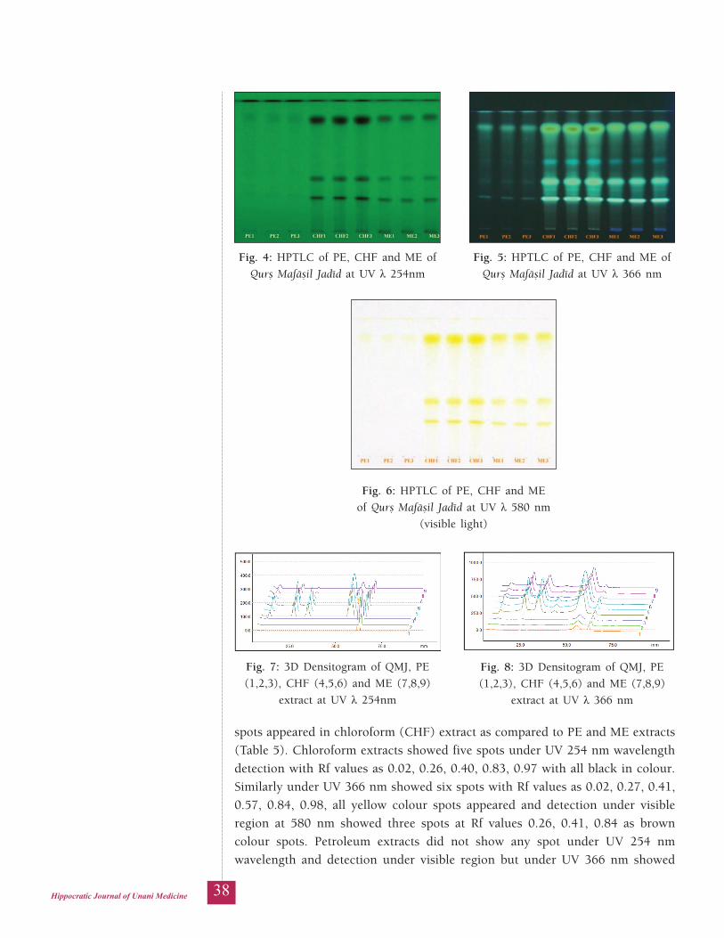
38Hippocratic Journal of Unani Medicine
Fig. 4: HPTLC of PE, CHF and ME of Qurñ Mafäñil Jadéd at UV λ 254nm
Fig. 5: HPTLC of PE, CHF and ME of Qurñ Mafäñil Jadéd at UV λ 366 nm
Fig. 6: HPTLC of PE, CHF and ME of Qurñ Mafäñil Jadéd at UV λ 580 nm
(visible light)
Fig. 7: 3D Densitogram of QMJ, PE (1,2,3), CHF (4,5,6) and ME (7,8,9)
extract at UV λ 254nm
Fig. 8: 3D Densitogram of QMJ, PE (1,2,3), CHF (4,5,6) and ME (7,8,9)
extract at UV λ 366 nm
spots appeared in chloroform (CHF) extract as compared to PE and ME extracts (Table 5). Chloroform extracts showed five spots under UV 254 nm wavelength detection with Rf values as 0.02, 0.26, 0.40, 0.83, 0.97 with all black in colour. Similarly under UV 366 nm showed six spots with Rf values as 0.02, 0.27, 0.41, 0.57, 0.84, 0.98, all yellow colour spots appeared and detection under visible region at 580 nm showed three spots at Rf values 0.26, 0.41, 0.84 as brown colour spots. Petroleum extracts did not show any spot under UV 254 nm wavelength and detection under visible region but under UV 366 nm showed
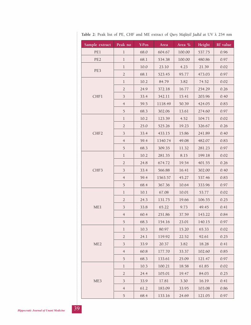
39Hippocratic Journal of Unani Medicine
Table 2: Peak list of PE, CHF and ME extract of Qurñ Mafäñil Jadéd at UV λ 254 nm
Sample extract Peak no Y-Pos Area Area % Height Rf value
PE1 1 68.0 604.67 100.00 537.75 0.96
PE2 1 68.1 534.38 100.00 480.86 0.97
PE31 10.0 23.10 4.23 21.39 0.02
2 68.1 523.45 95.77 473.03 0.97
CHF1
1 10.2 84.79 3.82 74.52 0.02
2 24.9 372.18 16.77 254.29 0.26
3 33.4 342.11 15.41 203.96 0.40
4 59.5 1118.49 50.39 424.05 0.83
5 68.3 302.06 13.61 274.60 0.97
CHF2
1 10.2 123.39 4.52 104.71 0.02
2 25.0 525.26 19.23 326.67 0.26
3 33.4 433.15 15.86 241.89 0.40
4 59.4 1340.74 49.08 482.07 0.83
5 68.3 309.35 11.32 281.23 0.97
CHF3
1 10.2 281.35 8.15 199.18 0.02
2 24.8 674.72 19.54 401.55 0.26
3 33.4 566.88 16.41 302.00 0.40
4 59.4 1563.57 45.27 537.46 0.83
5 68.4 367.36 10.64 333.96 0.97
ME1
1 10.1 67.08 10.01 53.77 0.02
2 24.3 131.75 19.66 106.55 0.25
3 33.8 65.22 9.73 49.45 0.41
4 60.4 251.86 37.59 143.22 0.84
5 68.3 154.16 23.01 140.15 0.97
ME2
1 10.3 80.97 15.20 65.33 0.02
2 24.1 119.92 22.52 92.61 0.25
3 33.9 20.37 3.82 18.28 0.41
4 60.8 177.70 33.37 102.60 0.85
5 68.3 133.61 25.09 121.47 0.97
ME3
1 10.3 100.21 18.58 61.85 0.02
2 24.4 105.01 19.47 84.03 0.25
3 33.9 17.81 3.30 16.19 0.41
4 61.2 183.09 33.95 103.08 0.86
5 68.4 133.16 24.69 121.05 0.97
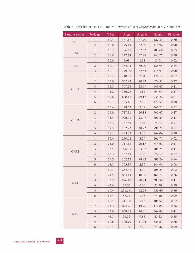
40Hippocratic Journal of Unani Medicine
Table 3: Peak list of PE, CHF and ME extract of Qurñ Mafäñil Jadéd at UV λ 366 nm
Sample extract Peak no Y-Pos Area Area % Height Rf value
PE11 60.4 367.17 67.70 127.33 0.86
2 68.0 175.15 32.30 146.92 0.98
PE21 60.2 296.45 62.52 108.66 0.85
2 68.0 177.74 37.48 143.77 0.98
PE3
1 10.0 7.61 1.38 11.01 0.02
2 60.1 364.42 66.09 123.95 0.85
3 68.1 179.39 32.53 145.91 0.98
CHF1
1 10.4 210.51 2.81 127.12 0.02
2 25.0 532.43 20.43 674.52 0.27
3 33.5 767.73 23.57 643.67 0.41
4 43.2 136.28 1.82 64.84 0.57
5 59.6 688.11 49.17 935.22 0.84
6 68.1 165.16 2.20 125.52 0.98
CHF2
1 10.4 279.62 3.29 166.71 0.02
2 25.0 717.11 20.18 743.67 0.27
3 33.5 996.91 23.47 706.24 0.41
4 43.3 157.49 1.85 75.84 0.57
5 59.5 162.72 48.92 991.24 0.84
6 68.1 195.39 2.30 144.04 0.98
CHF3
1 10.4 279.62 3.29 166.71 0.02
2 25.0 717.11 20.18 743.67 0.27
3 33.5 996.91 23.47 706.24 0.41
4 43.3 157.49 1.85 75.84 0.57
5 59.5 162.72 48.92 991.24 0.84
6 68.1 195.39 2.30 144.04 0.98
ME1
1 10.3 234.41 5.30 106.25 0.02
2 24.5 834.14 18.86 466.77 0.26
3 33.7 926.18 20.94 388.50 0.41
4 43.6 29.05 0.66 21.79 0.58
5 60.4 2312.41 52.28 657.69 0.86
6 68.4 86.74 1.96 74.16 0.99
ME2
1 10.4 211.90 5.13 104.52 0.02
2 24.3 824.26 19.96 447.05 0.26
3 33.8 859.38 20.81 364.83 0.41
4 44.1 36.32 0.88 23.22 0.58
5 60.8 106.79 51.02 625.81 0.86
6 68.4 90.97 2.20 74.96 0.99

41Hippocratic Journal of Unani Medicine
Sample extract Peak no Y-Pos Area Area % Height Rf value
ME3
1 10.6 227.27 5.54 107.12 0.03
2 24.6 819.82 19.97 443.68 0.26
3 34.2 851.39 20.74 358.79 0.42
4 44.5 27.13 0.66 17.52 0.59
5 61.2 086.24 50.83 624.65 0.87
6 68.6 92.49 2.25 74.88 0.99
Table 4: Peak list of PE, CHF and ME extract of Qurñ Mafäñil Jadéd at UV λ 580 nm (visible light)
Sample extract Peak no Y-Pos Area Area % Height Rf value
PE1 - - - - - -
PE2 - - - - - -
PE3 - - - - - -
CHF1
1 24.9 195.92 19.08 130.34 0.27
2 3.3 194.27 18.92 104.18 0.41
3 59.4 636.42 61.99 213.04 0.84
CHF2
1 4.9 237.80 19.24 58.66 0.27
2 33.3 71.25 21.95 34.22 0.41
3 59.4 26.86 58.81 30.57 0.84
CHF3
1 24.8 285.92 19.80 82.57 0.26
2 33.3 322.51 22.34 55.52 0.41
3 59.3 835.41 57.86 40.50 0.84
ME1
1 24.4 98.36 24.30 61.20 0.26
2 33.5 3.43 13.20 27.67 0.41
3 60.3 253.00 62.50 106.45 0.85
ME2
1 4.2 94.21 26.31 51.69 0.25
2 33.7 43.91 12.26 22.83 0.41
3 60.6 19.95 61.43 94.18 0.86
ME3
1 24.5 92.88 25.78 50.12 0.26
2 34.2 39.09 10.85 20.55 0.42
3 61.0 28.31 63.37 93.99 0.87
only one spot with Rf value as 0.85 in yellow colour. Methanol extracts showed five spots under UV 254 nm wavelength detection with Rf values as 0.02, 0.25, 0.41, 0.86, 0.97 with all black in colour. Similarly under UV 366 nm showed six spots with Rf values as 0.03, 0.26, 0.42, 0.59, 0.87, 0.99 with all yellow in colour and detection under visible light region at 580 nm showed three spots at Rf values 0.26, 0.42, 0.87 as represented in Table 5.
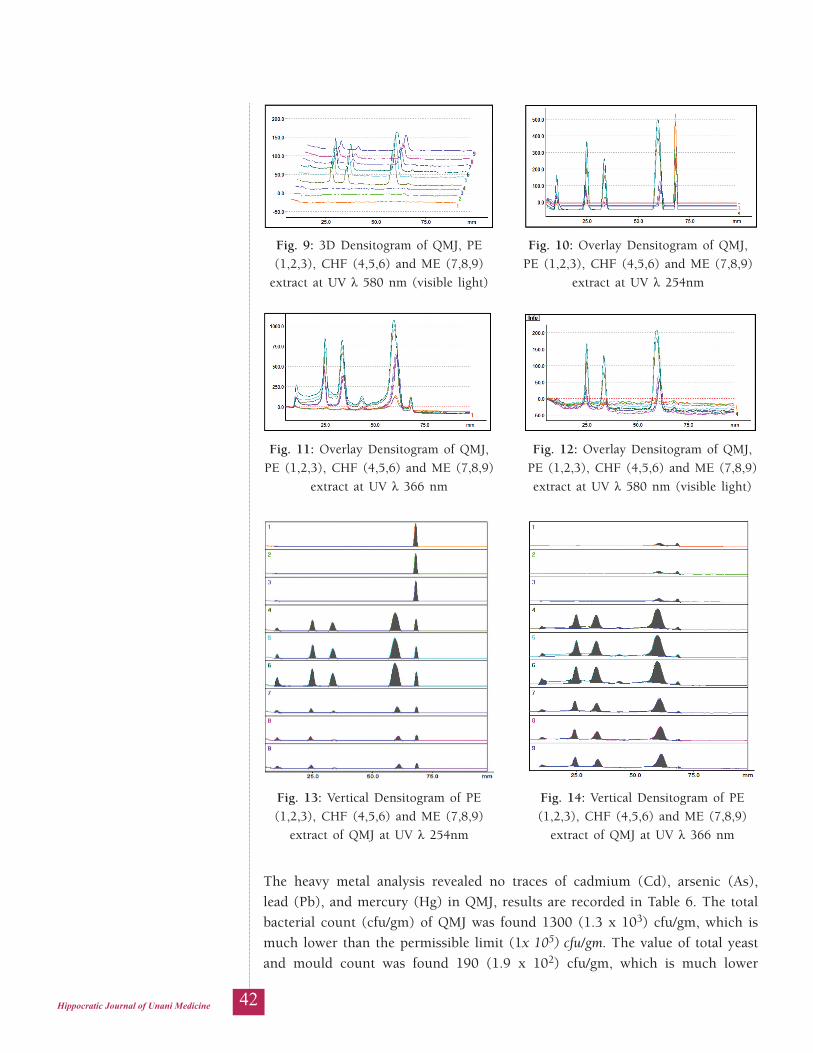
42Hippocratic Journal of Unani Medicine
Fig. 9: 3D Densitogram of QMJ, PE (1,2,3), CHF (4,5,6) and ME (7,8,9)
extract at UV λ 580 nm (visible light)
Fig. 10: Overlay Densitogram of QMJ, PE (1,2,3), CHF (4,5,6) and ME (7,8,9)
extract at UV λ 254nm
Fig. 11: Overlay Densitogram of QMJ, PE (1,2,3), CHF (4,5,6) and ME (7,8,9)
extract at UV λ 366 nm
Fig. 12: Overlay Densitogram of QMJ, PE (1,2,3), CHF (4,5,6) and ME (7,8,9) extract at UV λ 580 nm (visible light)
Fig. 13: Vertical Densitogram of PE (1,2,3), CHF (4,5,6) and ME (7,8,9)
extract of QMJ at UV λ 254nm
Fig. 14: Vertical Densitogram of PE (1,2,3), CHF (4,5,6) and ME (7,8,9)
extract of QMJ at UV λ 366 nm
The heavy metal analysis revealed no traces of cadmium (Cd), arsenic (As), lead (Pb), and mercury (Hg) in QMJ, results are recorded in Table 6. The total bacterial count (cfu/gm) of QMJ was found 1300 (1.3 x 10³) cfu/gm, which is much lower than the permissible limit (1x 10⁵) cfu/gm. The value of total yeast and mould count was found 190 (1.9 x 10²) cfu/gm, which is much lower

43Hippocratic Journal of Unani Medicine
Fig. 15: Vertical Densitogram of PE (1,2,3), CHF (4,5,6) and ME (7,8,9) extract of QMJ
at UV λ 580 nm (visible light)
Table 5: Comparison of PE, CHF and ME extract of Qurñ Mafäñil Jadéd (QMJ)
Solvent System
Chloroform: Methanol: Formic acid (9.6:0.4:0.1, v/v/v)
ExtractsPetroleum ether
extractChloroform extract Methanol extract
Detection system
UV 254 nm
UV 366 nm
under visible light
UV 254 nm
UV 366 nm
under visible light
UV 254 nm
UV 366 nm
under visible light
No. of Spots 1 2 0 5 6 3 5 6 3
Peak 1 - - - 0.02 0.02 - 0.02 0.03 -
Peak 2 - - - 0.26 0.27 0.26 0.25 0.26 0.26
Peak 3 - - - 0.40 0.41 0.41 0.41 0.42 0.42
Peak 4 - - - - 0.57 - - 0.59 -
Peak 5 - 0.85 - 0.83 0.84 0.84 0.86 0.87 0.87
Peak 6 0.97 0.98 - 0.97 0.98 - 0.97 0.99 -
than the permissible limit (1x 10³) cfu/gm. The Escherichia coli, Salmonella, Staphylococcus aureus and Pseudomonas aeruginosa were absent in the test drug QMJ (Table 7). Aflatoxin estimation was done by LCMCMS method for aflatoxins B1, G1, B2 and G2 and result showed no detection of aflatoxins in QMJ (Table 8). The pesticide residue analysis was done for 33 different pesticides

44Hippocratic Journal of Unani Medicine
by GCMSMS and one by UV-Vis spectrophotometer. The result confirms the absence of all pesticide in QMJ (Table 9).
Table 6: Results of heavy metals content in Qurñ Mafäñil Jadéd (QMJ)
Heavy Metals Method Heavy Metals in QMJ
Limit of Quantification
Permissible Limit as per API
Cadmium (Cd) (mg/Kg)
AAS ND 2.5 Not more than 0.3
Arsenic (As) (mg/Kg)
AAS ND 0.5 Not more than3
Lead (Pb) (mg/Kg) AAS ND 1.25 Not more than 10
Mercury (Hg) (mg/Kg)
AAS ND 0.25 Not more than 1
Not Detected = ND
Table 7: Results of microbial load in Qurñ Mafäñil Jadéd (QMJ)
Colony forming units on nutrient agar medium
Result Permissible Limit as per API
Total bacterial count (cfu/gm) 1300 NMT 1x 10⁵ cfu/gm
Total yeast and mould (cfu/gm) 190 NMT 1x 10³ cfu/gm
Escherichia coli (cfu/gm) Absent Absent
Salmonella (cfu/gm) Absent Absent
Staphylococcus aureus (cfu/gm) Absent Absent
Pseudomonas aeruginosa (cfu/gm) Absent Absent
Not more than = NMT; Absent = -Ve
Table 8: Results of aflatoxin in Qurñ Mafäñil Jadéd (QMJ)
Aflatoxin Method Result Limit of Quantification
Permissible Limit as per API
Aflatoxin B1 (mg/Kg)
LCMCMS ND 0.001 NMT 0.5
Aflatoxin G1 (mg/Kg)
LCMCMS ND 0.001 NMT 0.5
Aflatoxin B2 (mg/Kg)
LCMCMS ND 0.001 NMT 0.1
Aflatoxin G2 (mg/Kg)
LCMCMS ND 0.001 NMT 0.1
Not Detected = ND; Not more than = NMT
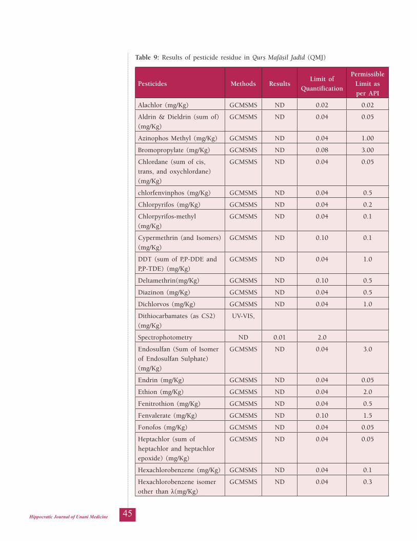
45Hippocratic Journal of Unani Medicine
Table 9: Results of pesticide residue in Qurñ Mafäñil Jadéd (QMJ)
Pesticides Methods ResultsLimit of
Quantification
Permissible Limit as per API
Alachlor (mg/Kg) GCMSMS ND 0.02 0.02
Aldrin & Dieldrin (sum of) (mg/Kg)
GCMSMS ND 0.04 0.05
Azinophos Methyl (mg/Kg) GCMSMS ND 0.04 1.00
Bromopropylate (mg/Kg) GCMSMS ND 0.08 3.00
Chlordane (sum of cis, trans, and oxychlordane) (mg/Kg)
GCMSMS ND 0.04 0.05
chlorfenvinphos (mg/Kg) GCMSMS ND 0.04 0.5
Chlorpyrifos (mg/Kg) GCMSMS ND 0.04 0.2
Chlorpyrifos-methyl (mg/Kg)
GCMSMS ND 0.04 0.1
Cypermethrin (and Isomers) (mg/Kg)
GCMSMS ND 0.10 0.1
DDT (sum of P,P-DDE and P,P-TDE) (mg/Kg)
GCMSMS ND 0.04 1.0
Deltamethrin(mg/Kg) GCMSMS ND 0.10 0.5
Diazinon (mg/Kg) GCMSMS ND 0.04 0.5
Dichlorvos (mg/Kg) GCMSMS ND 0.04 1.0
Dithiocarbamates (as CS2) (mg/Kg)
UV-VIS,
Spectrophotometry ND 0.01 2.0
Endosulfan (Sum of Isomer of Endosulfan Sulphate) (mg/Kg)
GCMSMS ND 0.04 3.0
Endrin (mg/Kg) GCMSMS ND 0.04 0.05
Ethion (mg/Kg) GCMSMS ND 0.04 2.0
Fenitrothion (mg/Kg) GCMSMS ND 0.04 0.5
Fenvalerate (mg/Kg) GCMSMS ND 0.10 1.5
Fonofos (mg/Kg) GCMSMS ND 0.04 0.05
Heptachlor (sum of heptachlor and heptachlor epoxide) (mg/Kg)
GCMSMS ND 0.04 0.05
Hexachlorobenzene (mg/Kg) GCMSMS ND 0.04 0.1
Hexachlorobenzene isomer other than λ(mg/Kg)
GCMSMS ND 0.04 0.3
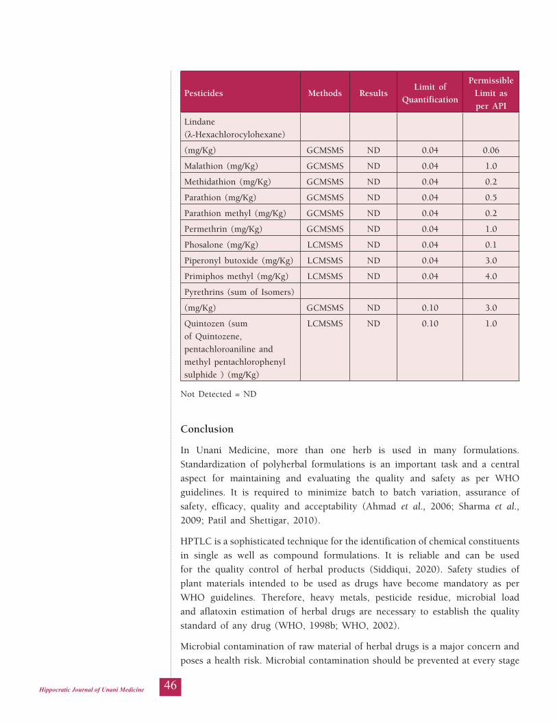
46Hippocratic Journal of Unani Medicine
Pesticides Methods ResultsLimit of
Quantification
Permissible Limit as per API
Lindane (λ-Hexachlorocylohexane)
(mg/Kg) GCMSMS ND 0.04 0.06
Malathion (mg/Kg) GCMSMS ND 0.04 1.0
Methidathion (mg/Kg) GCMSMS ND 0.04 0.2
Parathion (mg/Kg) GCMSMS ND 0.04 0.5
Parathion methyl (mg/Kg) GCMSMS ND 0.04 0.2
Permethrin (mg/Kg) GCMSMS ND 0.04 1.0
Phosalone (mg/Kg) LCMSMS ND 0.04 0.1
Piperonyl butoxide (mg/Kg) LCMSMS ND 0.04 3.0
Primiphos methyl (mg/Kg) LCMSMS ND 0.04 4.0
Pyrethrins (sum of Isomers)
(mg/Kg) GCMSMS ND 0.10 3.0
Quintozen (sum of Quintozene, pentachloroaniline and methyl pentachlorophenyl sulphide ) (mg/Kg)
LCMSMS ND 0.10 1.0
Not Detected = ND
Conclusion
In Unani Medicine, more than one herb is used in many formulations. Standardization of polyherbal formulations is an important task and a central aspect for maintaining and evaluating the quality and safety as per WHO guidelines. It is required to minimize batch to batch variation, assurance of safety, efficacy, quality and acceptability (Ahmad et al., 2006; Sharma et al., 2009; Patil and Shettigar, 2010).
HPTLC is a sophisticated technique for the identification of chemical constituents in single as well as compound formulations. It is reliable and can be used for the quality control of herbal products (Siddiqui, 2020). Safety studies of plant materials intended to be used as drugs have become mandatory as per WHO guidelines. Therefore, heavy metals, pesticide residue, microbial load and aflatoxin estimation of herbal drugs are necessary to establish the quality standard of any drug (WHO, 1998b; WHO, 2002).
Microbial contamination of raw material of herbal drugs is a major concern and poses a health risk. Microbial contamination should be prevented at every stage

47Hippocratic Journal of Unani Medicine
of the drug development, i.e. procurement, transport, storage, preparation and finished product storage. Therefore bacterial and fungal contamination of drugs, especially raw materials, should be prevented (WHO 1998b). Aflatoxins are mycotoxin produced by many species of a fungus Aspergillus, most commonly by Aspergillus flavus and Aspergillus parasiticus. The mycotoxins are considered carcinogenic substance, the adverse effects of aflatoxins in humans ranged from acute hepatic toxicity to chronic disease such as liver cancer (Agag, 2004). Aflatoxins are classified as the most potent natural carcinogens and generally affect the liver. The International Agency for Research on Cancer (IARC) has classified aflatoxin B1 in the group 1 as a human carcinogen and aflatoxins G1, B2 and G2 in the group 2B as possible carcinogens to humans (Meritxell ventura et al., 2004).
Unscientific and haphazard use of synthetic pesticides is increasing day by day and posing health hazards to human beings. Pesticide residues are the remains of pesticides on drug material when it is collected for medicinal use. Pesticide poisoning is taking about 3 lakh deaths worldwide every year. The most lethal pesticides are organophosphates and organochlorines compounds (Goel and Aggarwal, 2007).
The data obtained through HPTLC of Qurñ Mafäñil Jadéd clearly depicted the importance of developing a HPTLC fingerprint pattern for record and future reference. It was found that chloroform extract has shown maximum numbers of spots followed by methanol extract. The sophisticated analytical method of HPTLC is very much valid and has more significance than other conventional techniques. Hence the present study serves as a reference standard for the future studies and also for comparison for quality control study in different batches of the formation. The findings of the present study demonstrated that the values recorded in respect of heavy metals and microbial loads were within the permissible limits while aflatoxin, pesticides and other pathogenic organisms were not found at all. It suggests that the crude drugs used in the preparation of QMJ were genuine and not afflicted with poor soil and environmental conditions and thus qualified the criteria for being used for studies and even for therapeutic use.
References
1. Agag, B. I. (2004). Mycotoxins in foods and feeds 1-aflatoxins. Assiut University Bulletin for Environmental Researches, 7(1), 173–194.
2. Ahmad, I., Aqil, F., & Owais, M. (2006). Quality control, screening, toxicity, and regulation of herbal drugs. In Ahmad, I., Aqil, F., & Owais, M. (Eds.), Modern phytomedicine: Turning medicinal plants into drugs (pp. 25–52). Wiley-VCH GmbH.

48Hippocratic Journal of Unani Medicine
3. Anonymous. (1986). Qaräbädén-i Majédé (9th ed.). All India Unani Tibbi Conference.
4. Balammal, G., Sekar B. M., & Jayachandra R. P. (2012). Analysis of herbal medicines by modern chromatographic techniques. International Journal of Preclinical and Pharmaceutical Research, 3(1), 50–63.
5. Bin, C., Xiaoru, W., & Lee FSC. (2001). Pyrolysis coupled with atomic absorption spectrometry for the determination of mercury in Chinese medicinal materials. Analytica Chimica Acta, 447(1-2), 161–169.
6. Dhalwal, K., Sindhe, V. M., Biradar, Y. S., & Mahadik, K. R. (2008). Simultaneous quantification of bergenin, catechine, and gallic acid from Bergenia ciliate and Bergenia lingulata by useing thin-layer chromatography. Journal of Food Composition and Analysis, 21(6), 496–500.
7. Goel, A., Aggarwal, P. (2007). Pesticide poisoning. The National Medical Journal of India, 20(4), 182–191.
8. Khan, S. A., Khan, Hussain, I., Marwat, K. B., & Akhtar, N. (2008). Profile of heavy metals in selected medicinal plants. Pakistan Journal of Weed Science Research, 14(1-2), 101–110.
9. Kirmani, M. Z., Mohiuddin, S., Naz, F., Naqvi, I. I., & Zahir, E. (2011). Determination of some toxic and essential trace metals in some medicinal and edible plants of Karachi City. Journal of Basic and Applied Sciences, 7(2), 89–95.
10. Meritxell, V., Antonio, G., Jordi, D. F., Francesc, B., Montserrat, A., & Lluis, C. (2004). Determination of aflatoxins B1, G1, B2 and G2 in medicinal herbs by liquid chromatography-tandem mass spectrometry. Journal of Chromatography A, 1048(1), 25–29. https://doi.org/10.1016/j.chroma.2004.07.033
11. Mosihuzzanman, M., & Choudhary, M. I. (2008). Protocols on safety, efficacy, standardization and documentation of herbal medicine. Pure Applied Chemistry, 80(10), 2195–2230.
12. Mustafa, S., Mustafa, T., Narin, I., & Sari, H. (2004). Comparison of Microwave, Dry and wet digestion procedures for the determination of trace metal contents in spice samples produced in Turkey. Journal of Food and Drug Analysis, 12(3), 254–258.
13. Naikodi, M. A. R., Waheed, M. A., Shareef, M. A., Ahmad, M., & Nagaiah, K. (2011). Standardization of the Unani drug –Myristica fragrans Houtt. (Javetri) with modern analytical techniques. Pharm Methods, 2(2), 76–82.
14. Patil, P. S., & Shettigar, R. (2010). An advancement of analytical techniques in Herbal research. Journal of Advanced Scientific Research, 1(1), 08–14.

49Hippocratic Journal of Unani Medicine
15. Rasheed, N. M. A., Nagaiah, K., Mehveen, A., Rehana, A., Waheed, M. A., & Shareef, M. A. (2012). Phytochemical evaluation and quantification of beta-sitosterol in geographical variation of Withania coagulans Dunal by HPTLC analysis. Annals of Phytomedicine, 1(2), 14–22.
16. Sarmani, S. B., Abugassa, I., Hamzah, A., & Yahya, M. D. (1999). Elemental analysis of herbal preparations for traditional medicines by neutron activation analysis with the k0 standardization method. Biological Trace Element Research, 71-72, 365–376. 71-72, 365–376. https://doi.org/10.1007/BF02784224
17. Sharma, A. K., Gaurav, S. S., Balkrishna A. (2009). A rapid and simple scheme for the standardization of polyherbal drugs. International Journal of Green Pharmacy, 3(2), 134–140.
18. Siddiqui, M. Z., Ahmad, G., Amin, K. M. Y., Akhtar, S., & Rehman, A. (2020). HPLC profiling conclusively distinguished two important Unani drugs, namely, Suranjan Shirin (Colchicum autumnale) and Suranjan Talkh (Colchicum luteum). Indian Journal of Traditional Knowledge, 19(1), 170–173.
19. Soni, K., & Naved, T. (2010). HPTLC- Its applications in herbal drug industry. The Pharma Review, 5(2), 112–117.
20. Thoppil, S. O., Cardoza, R. M., & Amin, P. D. (2001). Stability indicating HPTLC determination of rimetazidine as bulk drug and in pharmaceutical formulations, Journal of Pharmaceutical and Biomedical Analysis, 25(1), 15–20. https://doi.org/ 10.1016/s0731-7085(00)00440-4
21. WHO. (1996). Quality assurance of pharmaceuticals: A compendium of guidelines and related materials (2nd vol., 2nd ed.). World Health Organization.
22. WHO. (1998a). Basic tests for drugs, pharmaceutical substances, medicinal plant materials and dosage forms. World Health Organization.
23. WHO. (1998b). Quality control methods for herbal materials. World Health Organization.
24. WHO. (2000). The WHO recommended classification of pesticides by hazard and guidelines to classification 2000–2202 (WHO/PCS/01.5). World Health Organization.
25. Zevenhoven, R., & Kilpinen, P. (2002). Control of pollutions in flue gases and fuel gases (2nd ed.). Espoo/ Turku.
50Hippocratic Journal of Unani Medicine
lkjka'k gcZy ;wukuh feJ.k d+qlZ eQ+kfly tnhn ds ,pihVh,ylh
vfHky{k.k ,oa xq.koRRkk ekudt+dh vgen fln~nhd+h] *t+qcSnk ,- vUlkjh] vt+hZuk tchu] eks- vuoj] eksgEen
t+kfdj] eksgEen vCnqy jk'khn ubZdksMh] fu'kkr [k+q'khZn vkSj equOoj gqlSu dkt+eh
lkjka'k
d+qlZ eQ+kfly tnhn ¼D;w,ets½ ,d cgq&?kVd ;wukuh feJ.k gS tks fofHkUu ,fV;ksykWth ds tksM+ks ds nnZ ds mipkj ds fy, izHkkoh :Ik ls mi;ksx fd;k tkrk gSA ;g xksyh ds :Ik esa gksrk gS vkSj blesa rhu gcZy ?kVd & lwjatku rY[k+] t+nZ pksc vkSj xksan dhdj gksrs gSaA D;w,ets ,d Hks"ktdks'kh; feJ.k gS ftldk mYys[k d+jkcknhu&,ðnh] ;wukuh fpfdRlk dh ,d ekud Hks"ktdks'k] esa fd;k x;k gSA jksx dh fLFkfr esa fu;fer vkSj loksZRre ifj.kke izkIr djus ds fy, gcZy feJ.k dh xq.koRrk fu;a=.k vR;ar egRoiw.kZ gSA gkykafd cgq&?kVd feJ.k dk ekudhdj.k vklku dke ugha gS] Vh,ylh] ,pihVh,ylh tSlh ljy ifj"Ñr rduhdsa xq.koRrk fu;a=.k ds ewy mn~ns'; dh iwfrZ dj ldrh gSaA izkekf.kd vkSj fo'oluh; ifj.kke izkIr djus ds mn~ns'; ls D;w,ets dk ,pihVh,ylh fQaxjfizafVax izksQ+kbYk] Hkkjh èkkrq fo'ys"k.k] dhVuk'kd vo'ks"k] ekbØksfc;y yksM vkSj ,¶ykVkWfDlu ijh{k.k fd;k x;kA D;w,ets dh ,pihVh,ylh fQaxjfizafVax izksQ+kbYk dks ;woh λ 366 ,u,e] ;woh λ 254 ,u,e vkSj n`'; izdk'k ¼;woh λ 580 ,u,e½ }kjk fodkl vkSj irk yxk;k x;kA Hkkjh èkkrq fo'ys"k.k] dhVuk'kd vo'ks"k] ekbØksfc;y yksM vkSj ,¶ykVkWfDlu dk fo'ys"k.k MCY;w,pvks fn'kkfunsZ'kksa ds vuqlkj fd;k x;kA orZeku v?;;u us D;w,ets dh 'kq)rk ,oa xq.koRrk dks fuèkkZfjr fd;k vkSj Hkfo"; esa feJ.k dh xq.koRrk vkSj fpfdRlh; izekf.kdrk dks cuk, j[kus ds fy, ,d lanHkZ ds :Ik esa mi;ksx fd;k tk ldrk gSA bl vè;;u esa izkIr ifj.kkeksa dks Hkfo"; esa lanHkZ ds fy, fQaxjfizaV ds :Ik esa mi;ksx fd;k tk ldrk gSA
'kCndqath% ,¶ykVkWfDlu] Hkkjh èkkrq] ,pihVh,ylh] ekbØksfc;y yksM] dhVuk'kd vo'ks"k] d+qlZ eQ+kfly tnhn] ;wukuh feJ.k] xq.koRrk fu;a=.k
51Hippocratic Journal of Unani Medicine
Abstract
Objective: The objective of the study was to evaluate the effect of detoxification (‘Amal-i-Tadbér) on Semecarpus anacardium (Balädur) toxicity by spectrophotometric estimation of total phenolic content (TPC).
Methods: Qualitative analysis for the presence of phenols was performed. Quantitatively, TPC was determined using the Folin & Ciocalteu reagent, with gallic acid as the standard by the method described by Singleton and Rossi, 1965.
Results: The TPC in aqueous and ethanolic extracts of un-detoxified Balädur was found to be 186 mg/g and 210 mg/g gallic acid equivalent (GAE), respectively. The TPC in aqueous and ethanolic extracts of Balädur after applying hot compression (detoxified Balädur) was found to be 139 mg/g GAE and 183 mg/g GAE. However, application of Patal Jantar revealed the amount of TPC to be 74 mg/g (aqueous extract) and 167 mg/g GAE (ethanolic extract). The TPC in Balädur oil obtained by hot compression method was 172 mg/g GAE.
Conclusion: The study showed reduction in total phenolic content after detoxification which suggested the possible cause of decreased toxicity of Balädur. The TPC of Patal Jantar detoxified Balädur was lesser than detoxified by hot compression. The TPC can be used as an indicator for the standardization of Balädur.
Keywords: Balädur, Semecarpus anacardium, Spectrophotometer, Total phenolic content
Introduction
Balädur is Semecarpus anacardium which belongs to the family Anacardiaceae (Tondon & Sharma, 2011). The trees of Balädur are deciduous and found in the outer Himalayas from Sutlej to Sikkim, common throughout the hotter parts of India as far east as Assam (Anonymous, 1972). Its fruit is mainly used for medicinal purpose which shows resemblance with the heart of birds (Ibn Baitar, 1999). The fruit oil is also used for therapeutic activity. According to Ibn Baitar, ‘Balädur in the Indian language is Anacardia in Greek which means heart shaped’ (Dymock et al., 1972). Belädin, Ùabb al-Fahm, Ùabb al-Qalb, Bhelä, Marking nut, Bhiläwän, Agnékä and Bhilänvanä are some of its vernacular names (Chopra et al., 2006). The fruits of Balädur are about 2.5 cm long oblong shape, smooth and shining, black when ripe seated on a fleshy hypocarp of about 2 cm long, smooth and yellow in colour when ripe (Kirtikar & Basu, 1996). It has characteristic odour and taste is highly acrid and irritating (Tondon & Sharma, 2011). With the consensus of various Unani authors,
October - December 2020, Vol. 15 No. 4, Pages 51-59
Spectrophoto-metric Estimation of Total Phenolic Content in Undetoxified and Detoxified Balädur (Semecarpus anacardium)
1Mohd Zakir Siddiqui,2Meryam Sardar,
3Kr. Mohammad Yusuf Amin,4Mohd Urooj,
3Nazish Siddiqui,3Sada Akhtar
1Department of Biotechnology,Jamia Millia Islamia, New Delhi
2Department of Biosciences,Jamia Millia Islamia, New Delhi
3Department of Ilmul Advia,Ajmal Khan Tibbiya College,
Aligarh Muslim University, Aligarh
4National Research Institute of Unani Medicine for Skin Disorders,
Hyderabad
*Corresponding author; Email: [email protected]

52Hippocratic Journal of Unani Medicine
the temperament of Balädur oil or juice is hot and dry in the fourth degree while the temperament without oil is hot and dry in the third degree (Khan, 2012; Ghani, 2010; Kareem, 1875). It is therapeutically used in the treatment of nervous disorders like paralysis, facial palsy, tremors, numbness, chorea, memory loss, dribbling of urine and nervous debility (Khan, 2012; Kareem, 1875). Anqaroyä Kabér, Anqaroyä Saghér and Ma‘jün-i-Balädur are its important Unani formulations (Anonymous, 2007). The most significant constituents of Balädur oil are phenolic compounds. Vesicant reactions are possibly attributed to phenolic compounds (Raut et al., 2017). Pillay and Siddiqui in 1931 isolated Bhilawanol (46% of the extract) as chief constituent from the juice of the pericarp (Chopra et al., 2006). Its juice on contact with skin causes intense pain and swelling, produces deep bluish coloured vesicles and irritable sore. The mark does not disappear for many months or even for life (Khory & Katrak, 1985). Despite the presence of Bhilawanols as the toxic components, it is still used in the Indian system of medicine (Kumar et al., 2017). In Unani Medicine, it is used after detoxification. There are two methods of its detoxification mentioned in classical Unani literature – Hot compression and Patal Jantar (Baitar, 1999; Khan, 2012). Its use as a therapeutic agent even in the indigenous system of medicine has dwindled to a great extent owing to the fact that the irritation produced by its application cannot be properly controlled (Chopra et al., 2006).
Since the toxicity of Balädur is mainly due to the presence of phenolic compounds, the present study was designed to evaluate the effect of the two methods of detoxification mentioned in Unani literatures, i.e. hot compression and Patal Jantar, on Balädur by spectrophotometric estimation of phenolic contents.
Material and Methods
Procurement and Authentication of Drug
The fruits of Semecarpus anacardium (Balädur) were procured from the Khari Baoli Market, Delhi. The drug was identified by Pharmacognosy Section, Department of Ilmul Advia, Ajmal Khan Tibbiya College, AMU, Aligarh. Further, it was authenticated by National Institute of Science Communication and Information Resources (NISCAIR), New Delhi with Ref. No. NISCAIR/RHMD/Consult/2018//3294-95.
Detoxification of Semecarpus anacardium (Balädur)
But before detoxification the cap of Balädur was removed. Fig. A.1 and A.2 show the images of Balädur with cap and after removal of cap, i.e. uncapped Balädur, respectively.
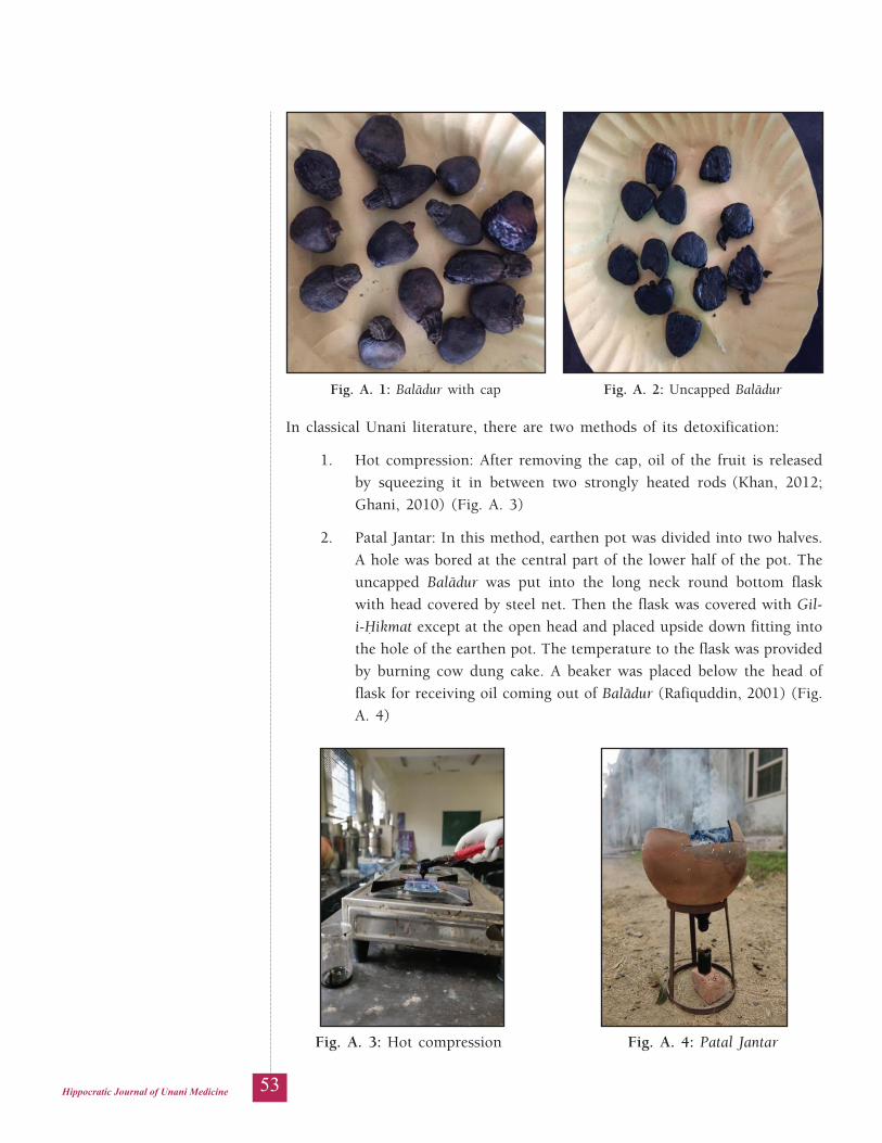
53Hippocratic Journal of Unani Medicine
In classical Unani literature, there are two methods of its detoxification:
1. Hot compression: After removing the cap, oil of the fruit is released by squeezing it in between two strongly heated rods (Khan, 2012; Ghani, 2010) (Fig. A. 3)
2. Patal Jantar: In this method, earthen pot was divided into two halves. A hole was bored at the central part of the lower half of the pot. The uncapped Balädur was put into the long neck round bottom flask with head covered by steel net. Then the flask was covered with Gil-i-Ùikmat except at the open head and placed upside down fitting into the hole of the earthen pot. The temperature to the flask was provided by burning cow dung cake. A beaker was placed below the head of flask for receiving oil coming out of Balädur (Rafiquddin, 2001) (Fig. A. 4)
Fig. A. 1: Balädur with cap Fig. A. 2: Uncapped Balädur
Fig. A. 3: Hot compression Fig. A. 4: Patal Jantar
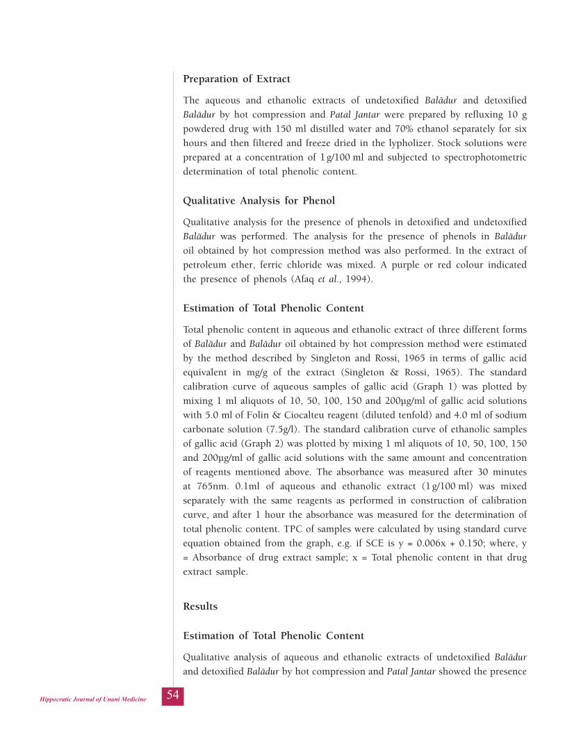
54Hippocratic Journal of Unani Medicine
Preparation of Extract
The aqueous and ethanolic extracts of undetoxified Balädur and detoxified Balädur by hot compression and Patal Jantar were prepared by refluxing 10 g powdered drug with 150 ml distilled water and 70% ethanol separately for six hours and then filtered and freeze dried in the lypholizer. Stock solutions were prepared at a concentration of 1 g/100 ml and subjected to spectrophotometric determination of total phenolic content.
Qualitative Analysis for Phenol
Qualitative analysis for the presence of phenols in detoxified and undetoxified Balädur was performed. The analysis for the presence of phenols in Balädur oil obtained by hot compression method was also performed. In the extract of petroleum ether, ferric chloride was mixed. A purple or red colour indicated the presence of phenols (Afaq et al., 1994).
Estimation of Total Phenolic Content
Total phenolic content in aqueous and ethanolic extract of three different forms of Balädur and Balädur oil obtained by hot compression method were estimated by the method described by Singleton and Rossi, 1965 in terms of gallic acid equivalent in mg/g of the extract (Singleton & Rossi, 1965). The standard calibration curve of aqueous samples of gallic acid (Graph 1) was plotted by mixing 1 ml aliquots of 10, 50, 100, 150 and 200µg/ml of gallic acid solutions with 5.0 ml of Folin & Ciocalteu reagent (diluted tenfold) and 4.0 ml of sodium carbonate solution (7.5g/l). The standard calibration curve of ethanolic samples of gallic acid (Graph 2) was plotted by mixing 1 ml aliquots of 10, 50, 100, 150 and 200µg/ml of gallic acid solutions with the same amount and concentration of reagents mentioned above. The absorbance was measured after 30 minutes at 765nm. 0.1ml of aqueous and ethanolic extract (1 g/100 ml) was mixed separately with the same reagents as performed in construction of calibration curve, and after 1 hour the absorbance was measured for the determination of total phenolic content. TPC of samples were calculated by using standard curve equation obtained from the graph, e.g. if SCE is y = 0.006x + 0.150; where, y = Absorbance of drug extract sample; x = Total phenolic content in that drug extract sample.
Results
Estimation of Total Phenolic Content
Qualitative analysis of aqueous and ethanolic extracts of undetoxified Balädur and detoxified Balädur by hot compression and Patal Jantar showed the presence
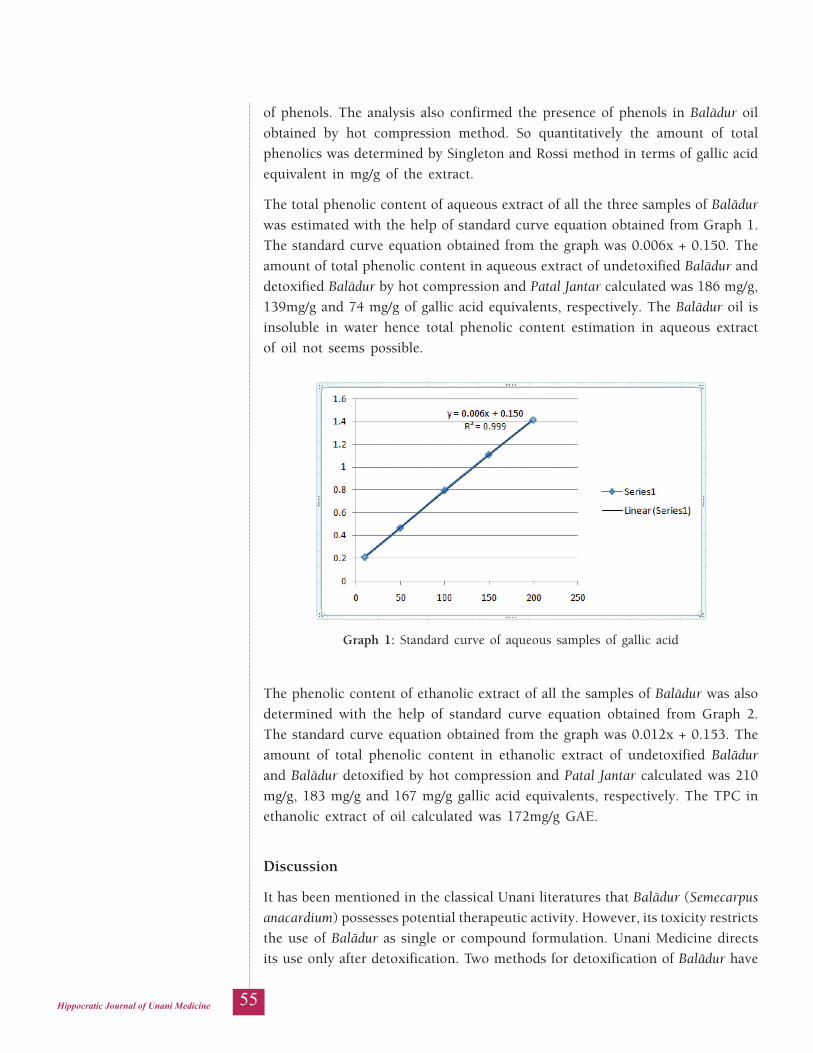
55Hippocratic Journal of Unani Medicine
of phenols. The analysis also confirmed the presence of phenols in Balädur oil obtained by hot compression method. So quantitatively the amount of total phenolics was determined by Singleton and Rossi method in terms of gallic acid equivalent in mg/g of the extract.
The total phenolic content of aqueous extract of all the three samples of Balädur was estimated with the help of standard curve equation obtained from Graph 1. The standard curve equation obtained from the graph was 0.006x + 0.150. The amount of total phenolic content in aqueous extract of undetoxified Balädur and detoxified Balädur by hot compression and Patal Jantar calculated was 186 mg/g, 139mg/g and 74 mg/g of gallic acid equivalents, respectively. The Balädur oil is insoluble in water hence total phenolic content estimation in aqueous extract of oil not seems possible.
Graph 1: Standard curve of aqueous samples of gallic acid
The phenolic content of ethanolic extract of all the samples of Balädur was also determined with the help of standard curve equation obtained from Graph 2. The standard curve equation obtained from the graph was 0.012x + 0.153. The amount of total phenolic content in ethanolic extract of undetoxified Balädur and Balädur detoxified by hot compression and Patal Jantar calculated was 210 mg/g, 183 mg/g and 167 mg/g gallic acid equivalents, respectively. The TPC in ethanolic extract of oil calculated was 172mg/g GAE.
Discussion
It has been mentioned in the classical Unani literatures that Balädur (Semecarpus anacardium) possesses potential therapeutic activity. However, its toxicity restricts the use of Balädur as single or compound formulation. Unani Medicine directs its use only after detoxification. Two methods for detoxification of Balädur have

56Hippocratic Journal of Unani Medicine
been mentioned in Unani literature which are hot compression and Patal Jantar. The toxicity of Balädur is mainly due to the presence of phenolic compounds (Kumar et al., 2017). Therefore, it was imperative to evaluate the effect of detoxification on total phenolic content of Balädur or its toxicity.
Qualitative analysis of aqueous and ethanolic extracts of undetoxified Balädur and detoxified Balädur by hot compression and Patal Jantar showed the presence of phenolic content. The qualitative analysis of Balädur oil obtained by hot compression method also confirmed the presence of phenolic content.
Quantitatively, total phenolic content was determined by Singleton and Rossi method in terms of gallic acid equivalent in mg/g of the extract. The amount of total phenolic content in aqueous extract of undetoxified Balädur and detoxified Balädur by hot compression and Patal Jantar calculated was 186 mg/g, 139 mg/g and 74 mg/g gallic acid equivalents, respectively. Being insoluble nature of oil in water, total phenolic content estimation in aqueous extract of oil was impossible. The amount of total phenolic content in ethanolic extract of undetoxified Balädur and detoxified Balädur by hot compression and Patal Jantar calculated was 210 mg/g, 183 mg/g and 167 mg/g gallic acid equivalents, respectively. The TPC in ethanolic extract of oil calculated was 172 mg/g GAE.
Conclusion
The qualitative analysis confirmed the presence of phenols in undetoxified, detoxified Balädur and the oil obtained by detoxification method. Quantitatively, total phenolic content after detoxification of Balädur was decreased which justified the cause of its decreased toxicity. The TPC in Patal Jantar detoxified Balädur was lesser than Balädur detoxified by hot compression. But the charring of Balädur, increased ash value beyond the permissible limit and lack of standardized procedure are the limitations of detoxification by Patal Jantar.
Graph 2: Standard curve of alcoholic samples of gallic acid
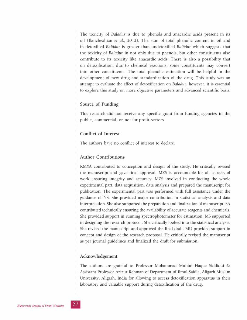
57Hippocratic Journal of Unani Medicine
The toxicity of Balädur is due to phenols and anacardic acids present in its oil (Ilanchezhian et al., 2012). The sum of total phenolic content in oil and in detoxified Balädur is greater than undetoxified Balädur which suggests that the toxicity of Balädur in not only due to phenols, but other constituents also contribute to its toxicity like anacardic acids. There is also a possibility that on detoxification, due to chemical reactions, some constituents may convert into other constituents. The total phenolic estimation will be helpful in the development of new drug and standardization of the drug. This study was an attempt to evaluate the effect of detoxification on Balädur, however, it is essential to explore this study on more objective parameters and advanced scientific basis.
Source of Funding
This research did not receive any specific grant from funding agencies in the public, commercial, or not-for-profit sectors.
Conflict of Interest
The authors have no conflict of interest to declare.
Author Contributions
KMYA contributed to conception and design of the study. He critically revised the manuscript and gave final approval. MZS is accountable for all aspects of work ensuring integrity and accuracy. MZS involved in conducting the whole experimental part, data acquisition, data analysis and prepared the manuscript for publication. The experimental part was performed with full assistance under the guidance of NS. She provided major contribution in statistical analysis and data interpretation. She also supported the preparation and finalization of manuscript. SA contributed technically ensuring the availability of accurate reagents and chemicals. She provided support in running spectrophotometer for estimation. MS supported in designing the research protocol. She critically looked into the statistical analysis. She revised the manuscript and approved the final draft. MU provided support in concept and design of the research proposal. He critically revised the manuscript as per journal guidelines and finalized the draft for submission.
Acknowledgement
The authors are grateful to Professor Mohammad Muhiul Haque Siddiqui & Assistant Professor Azizur Rehman of Department of Ilmul Saidla, Aligarh Muslim University, Aligarh, India for allowing to access detoxification apparatus in their laboratory and valuable support during detoxification of the drug.

58Hippocratic Journal of Unani Medicine
References
1. Afaq, S. H., Tajuddin., & Siddiqui, M. M. H. (1994). Standardization of Herbal drugs. Publication division, Aligarh Muslim University.
2. Anonymous. (1972). The Wealth of India. Publications and Information Directorate, Council of Scientific and Industrial Research.
3. Anonymous. (2007). The Unani Pharmacopoeia of India. Central Council of Research in Unani Medicine.
4. Chopra, R. N., Chopra, I. C., Handa, K. L., & Kapoor, L. D. (2006). Indigenous drugs of India (2nd ed., pp. 407-09). Academic Publishers.
5. Dymock, W., Warden, C. J. H., & Hooper, D. (1972). Pharmacographia Indica. Educational Society Press.
6. Ghani, N. (2010). Khzain-ul-Advia. Central Council for Research in Unani Medicine.
7. Ibn Baitar (1999). Al-Jame al-Mufridat al-Aghzia (Urdu Translation). Central Council of Research in Unani Medicine.
8. Ilanchezhian, R., Joseph, R., & Rabinarayan, A. (2012). Urushiol induced contact dermatitis caused during Shodhna of Bhallataka (Semecarpus anacardium) fruit. Ayu, 33(2), 270–273. https://doi.org/10.4103/0974-8520.105250
9. Kareem, N. (1875). Tarjuma Makhzanul Advia. Matba Naval Kishore.
10. Khan, A. (2012). Muheet-i-Azam (Urdu translation). Central Council of Research in Unani Medicine.
11. Khory, R. N., & Katrak, N. N. (1985). Materia Medica of India and their Therapeutics. Neeraj Publishing House.
12. Kirtikar, K. R., & Basu, B. D. (1996). Indian Medicinal Plants. International Book Distributors.
13. Kumar, M. S., Prashant, T., & Kumar, S. P. (2017). Pharmacology, Phytochemistry and Toxicology of Semecarpus anacardium, International Journal of Pharmaceutical Sciences Review and Research, 42(2), 25–31.
14. Rafiquddin, M. (2001). Minhaj-al-Saidla wal Kimiya. S.H. Offset Press.
15. Raut, A. K. A., Sawant, N. S., Badre, A., Amonkar, A. J., & Vaidya, A. B. D. (2007). Bhallatak (Semecarpus anacardium Linn.) - A review. Indian Journal of Traditional Knowledge, 6(4), 653–59.
16. Singleton, V. L., & Rossi, J. A. (1965). Calorimetry of total Phenolics with Phosphomolybdic-Phosphotungstic Acid Reagents. American Journal of Enology and Viticulture, 16(1), 144–58.
59Hippocratic Journal of Unani Medicine
17. Tondon, N., & Sharma, P. (2011). Quality Standards of Indian Medicinal Plants. Medicinal Plants Unit, Indian Council of Medical Research.
lkjka'k vUkfMVkWfDlQkbM vkSj fMVkWfDlQkbM cyknqj ¼lsehdkiZl
,ukdkfMZ;e½ esa dqy Qsuksfyd lkexzh dk LisDVªksQksVksesfVªd vkdyu
*eks- t+kfdj fln~nhd+h] ej;e ljnkj] dqaoj eksgEen ;qlqQ+ vehu] eks- m:t] ukft+'k fln~nhd+h] lnk v[+rj
Lkkjka'k
mn~ns';% vè;;u dk mn~ns'; dqy Qsuksfyd lkexzh ¼Vhihlh½ ds LisDVªksQksVksesfVªd vkdyu }kjk lsehdkiZl ,ukdkfMZ;e ¼cyknqj½ fo"kkDrrk ij fo"kgj.k ¼vey&,&rnchj½ ds izHkko dk ewY;kadu djuk FkkA
fofèk% QsuksYk dh mifLFkfr dk irk yxkus ds fy, xq.kkRed fo'ys"k.k fd;k x;kA ek=kRed :Ik ls flaxYkVu vkSj jkWlh] 1965 }kjk crkbZ xbZ fofèk }kjk ekud ds :Ik esa xSfyd ,flM ds lkFk Vhihlh dks Qkfyu ,oa fl;ksdkYVw vfHkdeZd dk mi;ksx djds fuèkkZfjr fd;k x;kA
ifj.kkEk% vufMVkWfDlQkbM cyknqj ds tyh; vkSj ,FksukWfyd lRr esa Vhihlh Øe'k% 186 fe-xzk-@xzk- vkSj 210 fe-xzk-@xzk- xSfyd ,flM bD;qosysUV ¼th,bZ½ ik;k x;kA xeZ laihM+u yxkus ds ckn fMVkWfdLQkbM cyknqj ds tyh; vkSj ,FksukWfyd lRr esa Vhihlh 139 fe-xzk-@xzk- th,bZ vkSj 183 fe-xzk-@xzk- th,bZ ik;k x;kA gkykafd] ikrky tarj ds iz;ksx ls irk pyk fd Vhihlh dh ek=k 74 fe-xzk-@xzk- ¼tyh; lRr½ vkSj 167 fe-xzk-@xzk- th,bZ ¼,FksukWfyd lRr½ gSA xeZ laihM+u fofèk }kjk izkIr cyknqj rsy esa Vhihlh 172 fe-xzk-@xzk- th,bZ FkhA
fu"d"kZ% vè;;u esa fo"kgj.k ds ckn dqy Qsuksfyd lkexzh esa deh ns[kh xbZ ftlus cyknqj dh fo"kkDrrk esa deh ds laHkkfor dkj.k dk lq>ko fn;kA iry tarj }kjk fMVkWfDlQkbM cyknqj dh Vhihlh xeZ laihM+u }kjk fMVkWfDlQkbM ls de FkhA Vhihlh dks cyknqj ds ekudhj.k ds fy, ,d ladsrd ds :Ik esa mi;ksx fd;k tk ldrk gSA
'kCndqath% cyknqj] lsehdkiZl ,ukdkfMZ;e] LisDVªksQksVksehVj] dqy Qsuksfyd lkexzh

60Hippocratic Journal of Unani Medicine
61Hippocratic Journal of Unani Medicine
IAbstract
ntroduction: Kalaf (melasma) is an asymptomatic cosmetic disorder having direct negative impact on quality of life (QoL) of the affected individuals. Lack of definitive therapies and higher rate of recurrence are the prominent challenges in the management of melasma.
Objective: To study the demographic, epidemiological and clinical characteristics of Kalaf (melasma) and its impact on QoL of the participants was the main objective of this study.
Materials and Methods: We conducted a cross-sectional study on the participants (n=88) of melasma of any sex aged between 18 and 55 years visiting outpatient department of NRIUMSD, Hyderabad. The data were collected on predesigned format in face-to-face interview and clinical examination, and analyzed retrospectively.
Observations and Results: We observed the average age (39 ± 8.1 years), average age of onset (34 ± 8.1 years), female to male ratio (4:1) and average chronicity of illness (5.2 ± 4.2 years) of the participants with melasma. We found mean MASI (17.84), mean DLQI (20.3) and mean MQOL (51.4) in the participants.
Discussion: This study showed that melasma was common in female in the age group of 30-40 years. The commonest clinical pattern was centro-facial. The probable contributing factors were sun exposure, pregnancy, OCPs and prolonged amenorrhea. The higher mean DLQI and mean MQOL demonstrated the negative impact of melasma on the QoL of the participants independent of severity of the disease.
Conclusion: In conclusion, this study provides the current understanding of the clinico-epidemiological characteristics of melasma. It impacts negatively on QoL of the participants.
Keywords: Dermatology, Hyperpigmentation, MASI, Melasma, Pregnancy, Unani
Introduction
Kalaf (melasma) is an asymptomatic cosmetic disorder commonly encountered in outpatient department of dermatology clinics (Charupalli et al., 2018). It is defined as the hyperpigmentation of sun-exposed areas of the face characterized by symmetrical irregular grey to brown patches (Achar & Rathi, 2011). Its prevalence varies from 0.25% to 4% in South East Asia (Charupalli et al., 2018; Pawar et al., 2015). It is a female predominant disease accounting for 90% of the cases (Achar & Rathi, 2011; Charupalli et al., 2018). It is most commonly
October - December 2020, Vol. 15 No. 4, Pages 61-75
A Clinico-epidemiological Study of Kalaf (Melasma) and its Impact on Quality of Life
1Ifra Abdul Qaiyyum,*1Mohammad Nawab,
1M. H. Kazmi
1National Research Institute of Unani Medicine for Skin Disorders,
Hyderabad
*Corresponding author; Email: [email protected]
62Hippocratic Journal of Unani Medicine
observed in Fitzpatrick skin types IV to VI (Pawaskar et al., 2007; Sarkar et al., 2016; Sardesai et al, 2013). This clinical condition develops slowly and may last for many years.
The exact cause of melasma is still to be determined (Guarneri, 2016; Sonthalia & Sarkar, 2015). But various common contributing and risk factors have been identified such as sun exposure (exposure to UV light) (Jiang et al., 2018), use of oral contraceptive pills (Arora et al., 2017; Pawar et al., 2015), pregnancy (Jiang et al., 2018), stress (Pawar et al., 2015) and exogenous hormones (Pawar et al., 2015). Cosmetics (Handel et al., 2014), genetic predisposition (Arora et al., 2017; Jiang et al., 2018) and phototoxic drugs (Ali et al., 2013) are reported to be linked with the causation of melasma. Unani literature says that dominance of melancholic humour (Khilö Sawdä’), prolonged amenorrhea and liver disorders may cause melasma (Khan, 1906).
A few studies have revealed that melasma has a direct negative impact on quality of life (QoL) and self-esteem of the patients (Jiang et al., 2018; Yalamanchili et al., 2015). The patient may suffer from psychosocial and emotional stress independent of severity of melasma or MASI score (Arora et al., 2017). It directly affects the physical appearance of the patient leading to avoidance of public appearance and social gatherings.
Long duration therapy, lack of definitive therapy (Ravi & Pandya, 2013) and tendency of recurrence (Grimes et al., 2010) are the challenges in the treatment of melasma. Excessive use of chemicals in cosmetics and lifestyle modification may have a link in the development of melasma. Now it becomes essential to have a current and comprehensive understanding and information of the demographic, epidemiological and clinical characteristics and risk factors of melasma. The present study was planned in order to determine the recent trends of clinico-epidemiological characteristics of melasma and its impact on QoL of the patients. The data in respect of clinical patterns, demographic characteristics and contributory factors was generated in this study. The outcome of this study would help in the understanding of the impact of melasma on the QoL of the patients. These data could be utilised to develop strategy for prevention and better management of melasma.
Materials and Methods
A cross-sectional study was conducted on the participants diagnosed with melasma at outdoor patient department (OPD) in National Research Institute of Unani Medicine for Skin Disorders (NRIUMSD), Hyderabad. Melasma was diagnosed clinically on the basis of clinical features such as brown and grey symmetrical macules present on face with serrated or irregular border. Eighty-eight consecutive participants were enrolled into the study during August
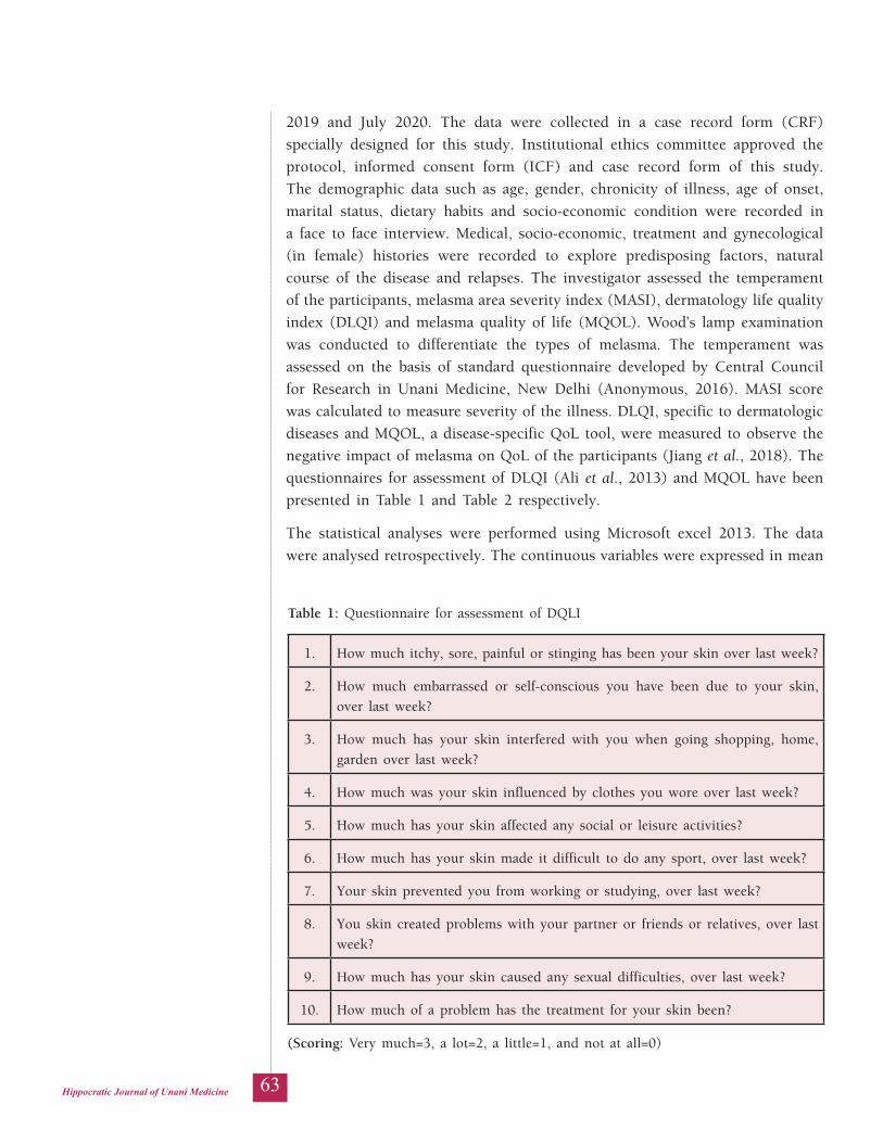
63Hippocratic Journal of Unani Medicine
2019 and July 2020. The data were collected in a case record form (CRF) specially designed for this study. Institutional ethics committee approved the protocol, informed consent form (ICF) and case record form of this study. The demographic data such as age, gender, chronicity of illness, age of onset, marital status, dietary habits and socio-economic condition were recorded in a face to face interview. Medical, socio-economic, treatment and gynecological (in female) histories were recorded to explore predisposing factors, natural course of the disease and relapses. The investigator assessed the temperament of the participants, melasma area severity index (MASI), dermatology life quality index (DLQI) and melasma quality of life (MQOL). Wood’s lamp examination was conducted to differentiate the types of melasma. The temperament was assessed on the basis of standard questionnaire developed by Central Council for Research in Unani Medicine, New Delhi (Anonymous, 2016). MASI score was calculated to measure severity of the illness. DLQI, specific to dermatologic diseases and MQOL, a disease-specific QoL tool, were measured to observe the negative impact of melasma on QoL of the participants (Jiang et al., 2018). The questionnaires for assessment of DLQI (Ali et al., 2013) and MQOL have been presented in Table 1 and Table 2 respectively.
The statistical analyses were performed using Microsoft excel 2013. The data were analysed retrospectively. The continuous variables were expressed in mean
Table 1: Questionnaire for assessment of DQLI
1. How much itchy, sore, painful or stinging has been your skin over last week?
2. How much embarrassed or self-conscious you have been due to your skin, over last week?
3. How much has your skin interfered with you when going shopping, home, garden over last week?
4. How much was your skin influenced by clothes you wore over last week?
5. How much has your skin affected any social or leisure activities?
6. How much has your skin made it difficult to do any sport, over last week?
7. Your skin prevented you from working or studying, over last week?
8. You skin created problems with your partner or friends or relatives, over last week?
9. How much has your skin caused any sexual difficulties, over last week?
10. How much of a problem has the treatment for your skin been?
(Scoring: Very much=3, a lot=2, a little=1, and not at all=0)
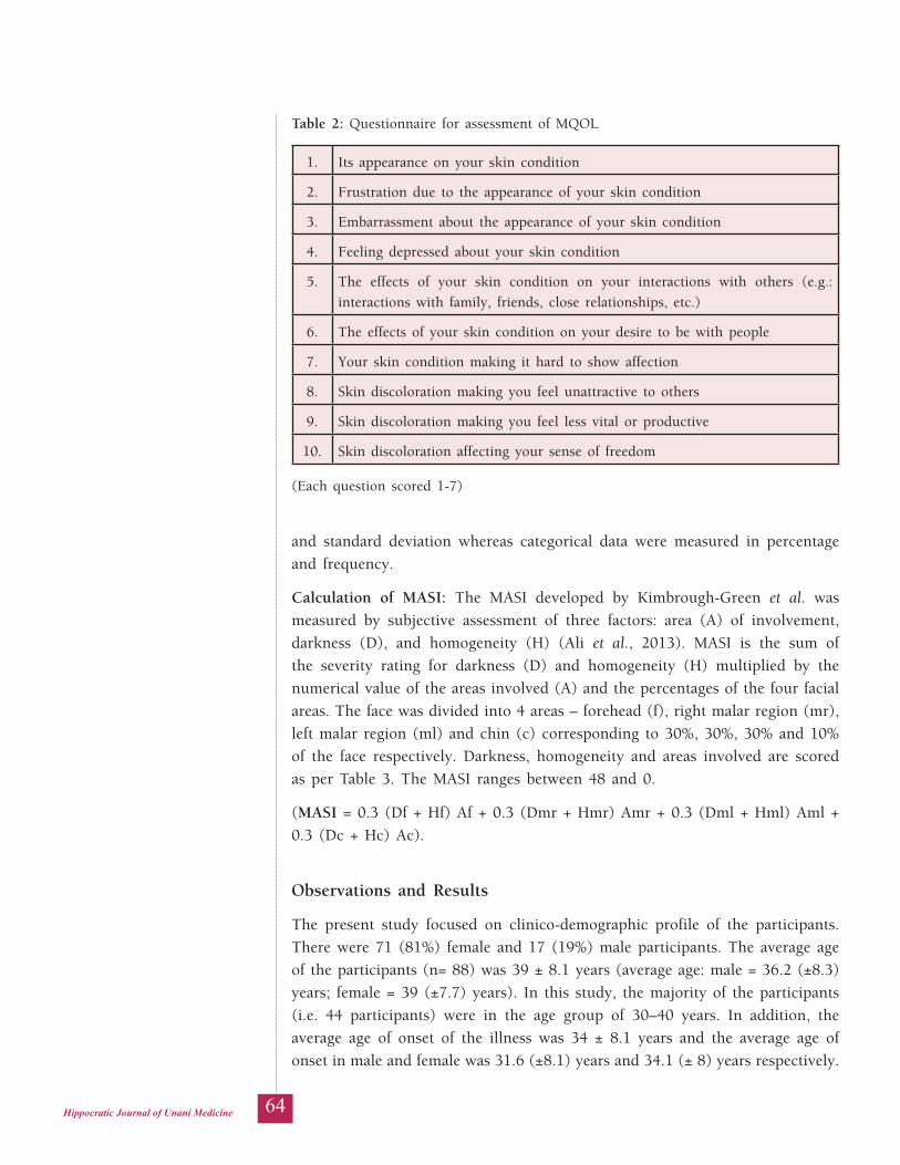
64Hippocratic Journal of Unani Medicine
and standard deviation whereas categorical data were measured in percentage and frequency.
Calculation of MASI: The MASI developed by Kimbrough-Green et al. was measured by subjective assessment of three factors: area (A) of involvement, darkness (D), and homogeneity (H) (Ali et al., 2013). MASI is the sum of the severity rating for darkness (D) and homogeneity (H) multiplied by the numerical value of the areas involved (A) and the percentages of the four facial areas. The face was divided into 4 areas – forehead (f), right malar region (mr), left malar region (ml) and chin (c) corresponding to 30%, 30%, 30% and 10% of the face respectively. Darkness, homogeneity and areas involved are scored as per Table 3. The MASI ranges between 48 and 0.
(MASI = 0.3 (Df + Hf) Af + 0.3 (Dmr + Hmr) Amr + 0.3 (Dml + Hml) Aml + 0.3 (Dc + Hc) Ac).
Observations and Results
The present study focused on clinico-demographic profile of the participants. There were 71 (81%) female and 17 (19%) male participants. The average age of the participants (n= 88) was 39 ± 8.1 years (average age: male = 36.2 (±8.3) years; female = 39 (±7.7) years). In this study, the majority of the participants (i.e. 44 participants) were in the age group of 30–40 years. In addition, the average age of onset of the illness was 34 ± 8.1 years and the average age of onset in male and female was 31.6 (±8.1) years and 34.1 (± 8) years respectively.
Table 2: Questionnaire for assessment of MQOL
1. Its appearance on your skin condition
2. Frustration due to the appearance of your skin condition
3. Embarrassment about the appearance of your skin condition
4. Feeling depressed about your skin condition
5. The effects of your skin condition on your interactions with others (e.g.: interactions with family, friends, close relationships, etc.)
6. The effects of your skin condition on your desire to be with people
7. Your skin condition making it hard to show affection
8. Skin discoloration making you feel unattractive to others
9. Skin discoloration making you feel less vital or productive
10. Skin discoloration affecting your sense of freedom
(Each question scored 1-7)
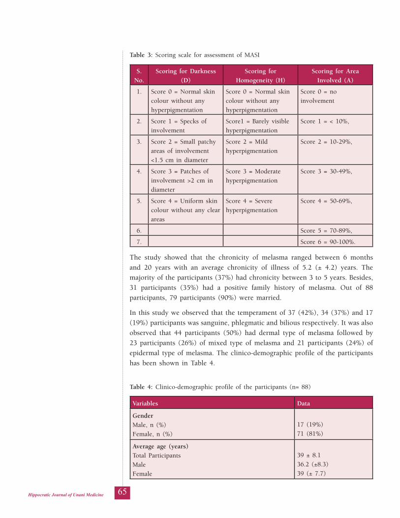
65Hippocratic Journal of Unani Medicine
The study showed that the chronicity of melasma ranged between 6 months and 20 years with an average chronicity of illness of 5.2 (± 4.2) years. The majority of the participants (37%) had chronicity between 3 to 5 years. Besides, 31 participants (35%) had a positive family history of melasma. Out of 88 participants, 79 participants (90%) were married.
In this study we observed that the temperament of 37 (42%), 34 (37%) and 17 (19%) participants was sanguine, phlegmatic and bilious respectively. It was also observed that 44 participants (50%) had dermal type of melasma followed by 23 participants (26%) of mixed type of melasma and 21 participants (24%) of epidermal type of melasma. The clinico-demographic profile of the participants has been shown in Table 4.
Table 3: Scoring scale for assessment of MASI
S. No.
Scoring for Darkness (D)
Scoring for Homogeneity (H)
Scoring for Area Involved (A)
1. Score 0 = Normal skin colour without any hyperpigmentation
Score 0 = Normal skin colour without any hyperpigmentation
Score 0 = no involvement
2. Score 1 = Specks of involvement
Score1 = Barely visible hyperpigmentation
Score 1 = < 10%,
3. Score 2 = Small patchy areas of involvement <1.5 cm in diameter
Score 2 = Mild hyperpigmentation
Score 2 = 10-29%,
4. Score 3 = Patches of involvement >2 cm in diameter
Score 3 = Moderate hyperpigmentation
Score 3 = 30-49%,
5. Score 4 = Uniform skin colour without any clear areas
Score 4 = Severe hyperpigmentation
Score 4 = 50-69%,
6. Score 5 = 70-89%,
7. Score 6 = 90-100%.
Table 4: Clinico-demographic profile of the participants (n= 88)
Variables Data
GenderMale, n (%)Female, n (%)
17 (19%)71 (81%)
Average age (years)Total ParticipantsMaleFemale
39 ± 8.136.2 (±8.3)39 (± 7.7)

66Hippocratic Journal of Unani Medicine
Variables Data
Age groups (years)18-30, n (%)30-40, n (%)40-50, n (%)50-55, n (%)
13 (35%)44 (50%)22 (25%)9 (10%)
Average age of onset (years)Total participantsMaleFemale
34 ± 8.131.6 (±8.1)34.1 (± 8)
Average chronicity of melasma (years)MaleFemale
5.24.55.3
Positive family history of melasma, n (%)MaleFemale
31 (35%)3 (4%)28 (31%)
Marital statusMarried, n (%)Unmarried, n (%)
79 (90%)9
09 (10%)
Mizäj (temperament)Damawé, n (%)Balghamé, n (%)Safrawi, n (%)
37 (42%)34 (37%)17 (19%)
Dietary habitVegetarian, n (%)Non vegetarian, n (%)
33 (37.5%)9
55 (62.5%)
Types of melasmaDermal, n (%)Epidermal, n (%)Mixed, n (%)
44 (50%)21 (24%)23 (26%)
MASI score (Mean ± SD)Total participantsMaleFemale
17.84 ± 9.1318.19± 9.1417.75 ± 9.13
Mean DLQI scoreTotal participantsMaleFemale
20.319.920.4
Mean MQOL scoreTotal participantsMaleFemale
51.449.851.9

67Hippocratic Journal of Unani Medicine
The present study showed that history of sun exposure in 12 participants (14%), OCP in 21 participants (24%), pregnancy in 13 participants (15%), amenorrhea in 12 participants (16%) and liver and endocrinal disorders in 5 participants (6%) were present as probable risk and contributing factors. Besides, 25 participants (28%) had observed menopause. Table 5 displays the participants with their risk factors.
Table 5: Distribution of participants according to risk factors
S.No. Risk factors Number of participants
1. Sun-exposure, n (%) 12 (14%)
2. Oral contraceptive, n (%) 21 (24%)
3. History of pregnancy, n (%) 13 (15%)
4. Amenorrhea 12 (16%)
5. Menopause 25 (28%)
6. Liver and endocrinal disorders 5 (6%)
It was also observed that centro-facial pattern of melasma was present in 41 participants (45%) followed by malar pattern in 36 participants (41%) and mandibular pattern in 11 participants (12.5%). The classification of the participants as per pattern of melasma is displayed in Table 6. Moreover, figures 1 to 3 show the patterns of melasma found in the participants.
Table 6: Distribution of participants according to pattern of melasma
S. No. Types of melasma Number of participants
Female (N=71) Male (N=17)
1. Centro-facial, n (%) 36 (51%) 5 (29%)
2. Malar pattern, n (%) 28 (39%) 8 (47%)
3. Mandibular pattern, n (%) 7 (10%) 4 (23.5%)
Furthermore, we observed that 70 participants had a positive treatment history for melasma as 52 (59%), 11 (12.5%) and 7 (8%) participants had taken allopathic, homeopathic and ayurvedic treatment respectively. In this study, melasma was observed in the participants having III (light brown), IV (medium brown) and V (dark brown) Fitzpatrick types of skin. Out of the 88 participants, 60 (68%) and 21 (24%) had IV and III Fitzpatrick types of skin. Only 7 participants (10%) had V Fitzpatrick type of skin.

68Hippocratic Journal of Unani Medicine
Figure 1: Mandibular pattern of melasma
Figure 2: Malar pattern of melasma
Figure 3: Centro-facial pattern of melasma
69Hippocratic Journal of Unani Medicine
In this study, the average MASI score was 17.84 (ranged between 40.8 and 1.8). The participants had mean DLQI of 20.3 and mean MQOL of 51.4.
Discussion
Melasma is an acquired, asymptomatic and hyper-pigmented lesion on facial skin. It is observed in all ethnic and population groups including Asian (Charupalli et al., 2018; Handel et al., 2014). Its impact on QoL demands urgent remedy and instigate to have current understanding about the disease.
The demographic, epidemiological and clinical profile of melasma was observed in this study. The study showed that the average age of the participants was 39 ± 8.1 years with an average age 39 ± 7.7 years in female and 36.2 ± 8.3 years in male. Acher and Rathi (2011) in their study reported that the average age of patients with melasma was 33.45 years.
In addition, the majority of the participants belonged to 30–40 years of age group. The similar observation was reported in a previously published study (Arora et al., 2017). In another study, 48.33% patients were in the age group of 30–39 years (Kumar & Sharma, 2018).
The present study showed that melasma was predominant in female participants (almost 81%) with male to female ratio of 1:4. Similar finding was observed in several studies (Ali et al., 2013; Arora et al., 2017). Moreover, Sivayathorn et al. reported a female to male ratio of 6:1 in a Malaysian population and 24:1 in Indonesian population (KrupaShankar et al., 2014). Melasma is common in female because of hormonal activities which is considered to be one of the risk factors for the development of melasma.
In our study, the average age of onset of the illness was 31.6 ±8.1 years in males and 34.1 ± 8 years in females. Whereas the mean age of onset was reported 24.78 years in males and 30.80 years in females in a study conducted by Pawar et al. (Pawar et al., 2015). Moreover, Sarkar et al. reported similar mean age of onset in males and females.
In the present study, the average chronicity of melasma was 5.2 (± 4.2) years. In addition, the majority of the participants (i.e. 37%) had chronicity between 3 to 5 years. Similar observation was reported in previous studies (Arora et al., 2017). In a study, it was reported that about 43% of the patients had a disease chronicity of >3 years (KrupaShankar et al., 2014).
Besides, we observed a positive family history of melasma in 31 participants (35%). In another study, a positive family history was observed in 16.67% of patients (Pawar et al., 2015). The study conducted by Achar and Rathi showed a positive family history in 33.33% patients (Achar & Rathi, 2011). In another study, out of 60 participants, a positive family history of melasma in either
70Hippocratic Journal of Unani Medicine
parents or any of the siblings was present in 18 patients (Kumar & Sharma, 2018). In this study, it was found that 79 (90%) participants were married. Marriage has not been associated with melasma in any of the previous studies.
In this study, we observed that the temperament of the participants was sanguine, phlegmatic and bilious except melancholic. Unani literature says that melasma was common in individuals of melancholic temperament (Khan, 1906). Our observation does not correspond with the observation reported in classical literature.
Histologically, melasma is classified on the basis of excessive melanin deposition in the epidermis (epidermal type, 70%), dermal macrophages (dermal type, 10%), or both (mixed type, 20%) (Filoni et al., 2019). The present study demonstrated that the participants had dermal, epidermal and mixed types of melasma. The similar observation was reported in previous studies (Filoni et al., 2019).
In this study we found that sun exposure, OCP, pregnancy, amenorrhea, liver and endocrinal disorders were present as probable risk and contributing factors. It is reported that melasma is common during reproductive period of female. In addition, we found history of taking oral contraceptive pills in the participants. It is reported that female hormones may play a role in the acceleration and aggravation of melasma. The exogenous hormonal therapy (progestogen-estrogen combinations) for ovulation control, dysmenorrhea, infertility, and endometriosis has added another cause for melasma (Hammer, 1968). Development of melasma during third trimester of pregnancy is more likely to be associated with circulating female hormones (Filoni et al., 2019). According to epidemiological data, 14.5% to 56% of pregnant women and 11.3% to 46% of individuals who take oral contraceptives suffer from melasma in different countries (Filoni et al., 2019). In our study, there were 25 participants (28%) in menopause. It has been reported that almost 10% participants start melasma after menopause (Filoni et al., 2019). An association between thyroid hormone and melasma has also been reported in several previous studies (Çakmak et al., 2015).
Melasma is classified into three types: centro-facial, malar, and mandibular patterns (Filoni et al., 2019). In this study we observed that centro-facial was the commonest clinical pattern of melasma followed by malar pattern and mandibular pattern. In a study it was reported that malar pattern melasma was predominant in the southern region of India to a greater extent when compared with the northern region (KrupaShankar et al., 2014).
Furthermore, we observed that a positive treatment history was present in 70 participants. They had taken allopathic, homeopathic and ayurvedic treatment but response of the treatment was poor. This shows that the treatment of melasma is difficult and relapse of melasma is common. In this study, melasma was observed in the participants having III (light brown), IV (medium brown)

71Hippocratic Journal of Unani Medicine
and V (dark brown) Fitzpatrick types of skin. The same observation has been reported in previous studies (Pawaskar et al., 2007).
In this study mean MASI score of the total participants was 17.84 (± 9.13) with a range between 40.8 and 1.8. Arora et al. reported in their study mean MASI score as 4.7 and Sarkar et al. reported it as 20.0 ± 7.5. In another study, MASI score ranged between 4.5 and 38.6 with mean MASI score of 16.94 (Pawar et al., 2015). Another study showed the mean MASI score 12.1±6.5 (median 10.8) (Harumi & Goh, 2016). Our observation is almost similar to the observation reported by Sarkar et al. (2016).
The present study showed that the QoL of the affected individuals was significantly impacted by melasma. We observed significantly higher mean DLQI (20.3) and mean MQOL (51.4) in the participants of this study. QoL includes physical health, interpersonal relationships, and social well-being. A few previous studies also demonstrated that melasma had a negative impact on QoL of the patients (Jiang et al., 2018). Improvement in QoL of the patients is one of the objectives of the proper management of melasma. Moreover, we observed that negative impact on QoL is independent of severity of melasma or MASI score. We classified the participants into mild (a MASI score of 0–16.9), moderate (a MASI score of 17–32.9) and severe (a MASI score of 33–48) cases of melasma and found that mean DLQI and mean MQOL were almost comparable among the three groups. But in a study it was reported that the patients with severe disease had greater impairment of QoL (Ali et al., 2013). In another study, significant positive correlation between MQOL and salary per month was reported (Coban, 2018).
This is a retrospective cross-sectional study. This study had several limitations. The study had small sample size and there was no control group to compare the association of the probable risk factors with the disease. The possibility of bias could not be ruled out in this study. The observations may further be confirmed by another prospective case-control and/or cohort study.
Conclusion
We studied age of onset, gender predilection, risk factors, clinical patterns and impact on QoL with regard to melasma in this study. We need these information for comprehensive understanding of melasma. The literature shows that melasma is a common, acquired, chronic disfiguring and cosmetic skin disorder. Prolonged treatment, lack of definitive therapies and higher rate of relapse are the imminent challenges in its treatment. Our findings may be essential for developing strategy for better prevention and management of melasma.
Moreover, negative impact of melasma on quality of life of the participants has been observed in the study. The patient may suffer from interpersonal,

72Hippocratic Journal of Unani Medicine
psychological, social and emotional stress despite the severity of the disease. The aim of the treatment should include improvement in the QoL of the patient as a strategy for proper management of the disease. The role of stress in inducing melasma has already been established.
This is an observational study. Despite several limitations, it provides current trends of epidemiological and clinical profile of melasma. It may seek attention of the researchers and academia for utilization of the data in the prevention and management of melasma.
Acknowledgment
We are thankful to Director General, Central Council for Research in Unani Medicine, Ministry of AYUSH, Government of India, New Delhi for providing financial support and resource materials for the study.
Conflict of Interest: Authors declare that there is no conflict of interest.
References
1. Achar, A., & Rathi, S. K. (2011). Melasma: A clinico-epidemiological study of 312 cases. Indian Journal of Dermatology, 56(4), 380–382. https://doi.org/10.4103/0019-5154.84722
2. Ali, R., Aman, S., Nadeem, M., & Kazmi, A. H. (2013). Quality of life in patients of melasma. Journal of Pakistan Association of Dermatologists, 23(2), 143–148.
3. Anonymous. (2016). Unani Medicine in India - An overview. Central Council for Research in Unani Medicine. https://ccrum.res.in/writereaddata/UploadFile/Fundamentals%20of%20Unani%20Medicine%20.pdf
4. Arora, P., Meena, N., Sharma, P., & Raihan, M. (2017). Impact of melasma on quality of life in Indian patients. Pigment International, 4(2), 92–97. https://doi.org/10.4103/2349-5847.219683
5. Cakmak, S. K., Özcan, N., Kiliç, A., Koparal, S., Artüz, F., Çakmak, A., & Köse, K. (2015). Etiopathogenetic factors, thyroid functions and thyroid autoimmunity in melasma patients. Postepy Dermatologii i Alergologii, 32(5), 327–330. https://doi.org/10.5114/ pdia.2015.54742
6. Charupalli, K., Rajasekhar, T., & Mukkara, M. (2018). Clinico-epidemiological study of melasma in men. Journal of Clinical and Scientific Research, 7(1), 19–23. https://doi.org/ 10.4103/jcsr.jcsr_6_18
7. Coban, M. (2018). Quality of life of the patients with melisma. Journal of Cosmetology & Trichology, 04(02). https://www.hilarispublisher.com/abstract/quality-of-life-of-the-patients-with-melasma-21725.html

73Hippocratic Journal of Unani Medicine
8. Filoni, A., Mariano, M., & Cameli, N. (2019). Melasma: How hormones can modulate skin pigmentation. Journal of Cosmetic Dermatology,18(2), 458–463. https://doi.org/ 10.1111/jocd.12877
9. Grimes, P. E., Bhawan, J., Guevara, I. L., Colón, L. E., Johnson, L. A., Gottschalk, R. W., & Pandya, A. G. (2010). Continuous therapy followed by a maintenance therapy regimen with a triple combination cream for melasma. Journal of the American Academy of Dermatology, 62(6), 962–967. https://doi.org/10.1016/j.jaad.2009.06.067
10. Guarneri, F. Journal of Pigmentary Disorders. (2016) Journal of Pigmentary Disorders, 2(2), 1–4. https://doi.org/10.4172/2376-0427.S1-003
11. Hammer, C. J. (1968). Melasma induced by oral contraceptive drugs. Northwest Medicine, 67(3), 251–254. https://doi.org/10.1001/jama.199.9.601
12. Handel, A. C., Miot, L. D. B., & Miot, H. A. (2014). Melasma: A clinical and epidemiological review. Anais Brasileiros de Dermatologia, 89(5), 771–782.https:// doi.org/10.1590/abd1806-4841.20143063
13. Harumi, O., & Goh, C. L. (2016). The effect of melasma on the quality of life in a sample of women living in Singapore. Journal of Clinical and Aesthetic Dermatology, 9(1), 21–24.
14. Jiang, J., Akinseye, O., Tovar-Garza, A., & Pandya, A. G. (2018). The effect of melasma on self-esteem: A pilot study. International Journal of Women’s Dermatology, 4(1), 38–42. https://doi.org/10.1016/j.ijwd.2017.11.003
15. Khan, M. A. (1906). Iksir-i-Azam. Matba Nami Munshi Naval Kishor.
16. KrupaShankar, D. S., Somani, V. K., Kohli, M., Sharad, J., Ganjoo, A., Kandhari, S., Mysore, V. R., Aurangabadkar, S., Malakar, S., Vedamurthy, M., Kadhe, G., Motlekar, S., & Ahirrao, P. (2014). A cross-sectional, multicentric clinico-epidemiological study of melasma in India. Dermatology and Therapy, 4(1), 71–81. https://doi.org/10.1007/ s13555-014-0046-1
17. Kumar, A., & Sharma, M. (2018). A clinico-epidemiological study of melasma. International Journal of Research in Dermatology, 4(4):539. https://doi.org/10.18203/ issn.2455-4529.intjresdermatol20184457
18. Pawar, S., khatu, S., & Gokhale, N. (2015). A clinico-epidemiological study of melasma in Pune patients. Journal of Pigmentary Disorders, 2(11), 1–3. https://doi.org/ 10.4172/ 2376-0427.1000219
19. Pawaskar, M. D., Parikh, P., Markowski, T., Mcmichael, A. J., Feldman, S. R., & Balkrishnan, R. (2007). Melasma and its impact on health-related quality of life in Hispanic women. Journal of Dermatological Treatment, 18(1), 5–9. https://doi.org/10.1080/09546630601028778
74Hippocratic Journal of Unani Medicine
20. Rivas, S., & Pandya, A. G. (2013). Treatment of melasma with topical agents, peels and lasers: an evidence-based review. American journal of clinical dermatology, 14(5), 359–376. https://doi.org/10.1007/s40257-013-0038-4
21. Sardesai, V. R., Kolte, J. N., & Srinivas, B. N. (2013). A clinical study of melasma and a comparison of the therapeutic effect of certain currently available topical modalities for its treatment. Indian journal of dermatology, 58(3), 239. https://doi.org/10.4103/0019-5154.110842
22. Sarkar, R., Garg, S., Dominguez, A., Balkrishnan, R., Jain, R. K., & Pandya, A. (2016). Development and validation of a Hindi language health-related quality of life questionnaire for melasma in Indian patients. Indian Journal of Dermatology, Venereology and Leprology, 82(1), 16–22. https://doi.org/10.4103/0378-6323.168937
23. Sonthalia, S., & Sarkar, R. (2015) Etiopathogenesis of melasma. Pigment International, 2(1), 21–27. https://doi.org/10.4103/2349-5847.159389
24. Yalamanchili, R., Shastry, V., & Betkerur, J. (2015). Clinico-epidemiological study and quality of life assessment in melasma. Indian journal of dermatology, 60(5), 519. https://doi.org/10.4103/0019-5154.164415

75Hippocratic Journal of Unani Medicine
lkjka'k dyQ+ ¼esykT+ek½ dk fDyfudks&,fiMsfe;ksykWftdy vè;;u
,oa thou dh xq.koRrk ij bldk izHkkobQ+jk vCnqy d+¸;we] *eksgEen uokc],e-,p- dkt+eh
lkjka'k
ifjp;% dyQ+ ¼esykT+ek½ ,d y{k.kghu dkWLesfVd fodkj gS ftldk izHkkfor O;fDr;ksa ds thou dh xq.koRrk ij udkjkRed izHkko iM+rk gSA lEiw.kZ Fksjsih dh deh vkSj iqujko`fRr dh mPp nj esykT+ek ds mipkj esa izeq[k pqukSfr;ka gSaA
mn~ns';% bl vè;;u dk mn~ns'; dyQ+ ¼esykT+ek½ dh tulkaf[;dh;] ,fiMsfe;ksykWftdy vkSj uSnkfud vfHky{k.kksa dk vè;;u djuk vkSj O;fDr ds thou dh xq.koRrk ij blds izHkko dk voyksdu djuk FkkA
lkexzh vkSj fofèk% geus jk-Ro-jks-;w-fp-v-la-] gSnjkckn ds ckg~; jksxh foHkkx esa vkus okys 18 ls 55 o"kZ dh mez ds fdlh Hkh fyax ds esykT+ek ds 88 jksfx;ksa ij ,d ØkWl lsD'kuy vè;;u fd;kA lEEkq[k b.VjO;w vkSj uSnkfud ijh{k.k esa iwoZ fuèkkZfjr izk:Ik ij MkVk ,df=r fd;k x;k vkSj iwoZO;kih :Ik ls fo'ys"k.k fd;k x;kA
voyksdu vkSj ifj.kke % geus esykT+ek ds jksfx;ksa dh vkSlr vk;q ¼39 ± 8.1 o"kZ½] jksx 'kq#vkr ij vkSlr vk;q ¼34 ± 8.1 o"kZ½] efgyk ls iq:"k vuqikr (4:1) vkSj jksx dh vkSlr nh?kZdkfydrk ¼5.2 ± 4.2 o"kZ½ dk voyksdu fd;kA geus jksfx;ksa esa ehu ,e,,lvkbZ ¼17-84½] ehu Mh,yD;qvkbZ ¼20-3½ vkSj ehu ,eD;wvks,y ¼51-4½ ik;kA
ppkZ% vè;;u ls irk pyk fd 30&40 o"kZ dh vk;q dh efgykvksa esa esykT+ek lkekU; FkkA lcls vke uSnkfud iSVuZ psgjs ij dsafnzr FkkA lw;Z vukoj.k] xHkkZoLFkk] ekSf[kd xHkZfujksèkd xksfy;ksa dk lsou ,oa yacs le; rd jDrlzko laHkkfor ;ksxnku dkjd FksA mPPk ehu Mh,yD;qvkbZ vkSj ehu ,eD;qvks,y us jksx dh xaHkhjrk ds QdZ ds ckotwn jksfx;ksa ds thou dh xq.koRrk ij esykT+ek dk udkjkRed izHkko fn[kk;kA
fu"d"kZ% fu"d"kZ ds rkSj ij ;g vè;;u esykT+ek ds fDyfudks&,fiMsfe;ksykWftdy ds y{k.kksa ds orZeku Kku dh tkudkjh iznku djrk gSA jksfx;ksa ds thou dh xq.koRrk ij bldk udkjkRed izHkko gksrk gSA
'kCndqath% Ropk foKku] vfro.kZdrk] ,e,,lvkbZ] esykT+ek] xHkkZoLFkk] ;wukuh] dyQ+

76Hippocratic Journal of Unani Medicine
77Hippocratic Journal of Unani Medicine
CAbstract
hronic inflammation almost always precedes and accompanies fibrotic changes and the medicines that target the inflammatory cascade of different cytokines such as tumour necrosis factor alpha (TNF-α), interferon-gamma (IFN-γ), different interleukins, etc. typically have immune-modulator and anti-fibrotic activity. Thus, measuring these cytokines serum levels in the cirrhotic individuals and targeting them as a parameter in the improvement of fibrosis could be a strategy in the treatment of HBV induced cirrhosis of liver besides the control of replicating HBV. To evaluate the immuno-modulatory action of Unani treatment, present case series was carried out in 7 patients with HBV induced compensated cirrhosis of liver where patients were treated with a set of Unani treatment for a period of 6–8 months. Estimation of serum levels of IFN-γ and TNF-α was carried out as a prime investigation in addition to evaluation of HBV DNA quantitative, liver function test and fibroscan of liver. Observed significant results substantiate the immuno-modulatory as well as beneficial potential of Unani medicines in the treatment of compensated cirrhosis of liver.
Keywords: Cirrhosis of liver, Unani medicine, IFN-γ, TNF-α, fibrosis
Introduction
Cirrhosis of the liver is the fourth cause of death in adults in Western countries, with complications of portal hypertension being responsible for most casualties. Chronic liver disease (CLD) affects more than 29 million people in Europe and over 300 million people worldwide (Blachier, 2013). The main causes of CLD are alcohol abuse, chronic viral hepatitis (hepatitis B virus and hepatitis C virus), and metabolic factors (non-alcoholic fatty liver disease). Mortality in CLD is primarily due to complications of liver cirrhosis and hepatocellular carcinoma (HCC), which is considerably more prevalent in the patients with cirrhosis (Rosselli, 2013; Annalisa, 2017).
Cirrhosis is defined as the histological development of regenerative nodules surrounded by fibrous bands in response to chronic liver injury that leads to portal hypertension and end stage liver disease (Schuppan, 2008). Cirrhosis is a common pathological consequence of chronic liver disease. Hepatitis B virus (HBV) is one of the etiologies of liver cirrhosis in India. Cirrhosis has two phases, compensated and decompensated (Annalisa, 2017; Schuppan, 2008). Compensated cirrhosis means that the liver is still functioning relatively well. At this stage, the early symptoms may still be vague and some people may be unaware that they have cirrhosis (Annalisa, 2017). Indeed, even after the onset of cirrhosis, the disease can remain asymptomatic, or ‘compensated’, for a long
October - December 2020, Vol. 15 No. 4, Pages 77-85
Immuno-modulatory Action of Unani Formulation against HBV Induced Compensated Cirrhosis of Liver
1Shabnam Ansari,2Mohammad Maaz,
3Asim Ali Khan,2Azhar Jabeen,
*3Shah Alam
1Department of Biotechnology, Faculty of Natural Sciences,
Jamia Millia Islamia, New Delhi
2Department of Moalajat,School of Unani Medical Education
and Research, Jamia Hamdard, New Delhi
3Central Council for Research in Unani Medicine, New Delhi
*Corresponding author; Email: [email protected]

78Hippocratic Journal of Unani Medicine
time. Nevertheless, during this time, portal hypertension progressively develops (non-bleeding esophageal varices) and usually accompanied by a decline in hepatocellular function (D'Amico, 2006).
HBV is a stealth non-cytopathic virus in the liver. The recurrent and chronic hepatitis observed due to HBV infection is rather immune mediated due to activation of T cells and release of cytokines such as tumor necrosis factor alpha (TNF-α), interferon-gamma (IFN-γ), various interleukins, etc. (Attallah et al., 2016). Studies have shown that serum levels of TNF are significantly higher in patients with cirrhosis than in those without cirrhosis, reaching the highest levels in decompensated cirrhosis. It has also been shown that plasma levels of TNF-α correlate with the severity of hepatic encephalopathy (HE) in fulminant hepatic failure (Odeh, 2004). While, IFN-γ is a major factor, reflecting hepatic dysfunction regardless of underlying disease more than liver inflammation parameters. Various studies support the hypothesis that liver damage against HBV infection is due to an immune-mediated destructive mechanism rather than direct cytopathic effect of the virus itself (Attallah et al; 2016).
Thus, the effective inhibition of HBV can partially stop or reverse liver fibrosis in the patients with chronic hepatitis and liver cirrhosis, and the anti-fibrotic strategy focusing on the regulation of hepatic extracellular matrix is still required. Modern anti-fibrotics are still in phase 3 clinical trials while modern antivirals do not fulfil all the clinical needs due to their questionable efficacy, adverse effects and resistance (Siddiqui, 2016; Ansari, 2019).
Unani Medicine has been found to enhance the degradation of collagens in fibrotic liver, regeneration of liver tissue, and has a good action against persistent immune attacks (hepatocyte) in liver of the patients with chronic hepatitis B. However, there are no high quality clinical evidences which can demonstrate if the combination of Unani medicines acting as antiviral and anti-fibrotic therapy can improve the reversion of the cirrhosis due to HBV (Siddiqui, 2016).
The present case series tried to preliminarily substantiate the immunomodulatory action of Unani treatment in improving the serum levels of TNF-α and IFN-γ whose increased levels are strongly linked with advancement in cirrhotic changes.
Material and Methods
Informed consent was taken from the patients. Human data included in this case series was obtained in compliance with the Declaration of Helsinki as revised in 1983.
Intervention
1. ‘Araq-i-Mako (Solanum nigrum) + ‘Araq-i-Käsné (Cichorium intybus) + ‘Araq-i- Biranjäsif (Achillea millefolium) 150 ml, twice daily

79Hippocratic Journal of Unani Medicine
2. Sharbat-i-Jigrén 25 ml, twice daily / Capsule Jigrénä 2 capsules, thrice daily
3. Ùabb-i-Ùiltét 3 pills, twice daily after meals
4. Sharbat-i-Buzüré Mu‘tadil 25 ml, twice daily/ Banädiq al-Buzür 2 tablets, thrice daily
5. Ma‘jün Dabéd al-Ward, 10 g twice daily
6. Decoction of Shähitara (Fumaria officinalis), Sarphokhä (Tephrosa purpurea), Chirä’ita (Swertia chiraita), Gul-i-Mundé (Sphaeranthus indicus), and Ñandal Surkh (Pterocarpus santalinus) each 5 g soaked in 300 ml of water for overnight and boiled for 10 minutes till decoction became half of its original volume, filtered and ingested empty stomach in the morning daily
7. Diet: Avoidance of oily, spicy, fatty, red-meat diet. Advise for intake of soft diet, oatmeal and barley water daily
All the patients in the case series received these drugs for a period of 6–8 months.
Case Presentation
Case 1: A 50-year Indian male was admitted in Majeedia Unani Hospital, Jamia Hamdard, New Delhi on Jan 17, 2015 with chief complains of (1) loss of appetite, (2) mild pain in abdomen, (3) yellow coloration of eye, skin and urine, (4) general weakness, and (5) breathlessness while walking for 3–4 months. He was a diagnosed case of HBV induced compensated cirrhosis of the liver and was taking treatment from a reputed tertiary care liver-specialty hospital in New Delhi for 3 years.
Case 2: A 42-year old Indian male was admitted in Majeedia Unani Hospital, Jamia Hamdard, New Delhi on March 9, 2015 with chief complains of (1) yellow coloration of the eye ×15 days, (2) loss of appetite ×15 days, (3) pain in upper abdomen ×15 days, (4) on and off fever × 2 months, (5) itching all over body × 2 months, and (6) headache × 2 months. He reported positive history of hepatitis B in wife. He was a diagnosed case of hepatitis B for 5 years and was not taking any treatment. He was diagnosed as HBV induced compensated cirrhosis of liver with anaemia, splenomegaly, cholelithiasis and right renal cyst.
Case 3: A 40-year old Indian male was admitted in Majeedia Unani Hospital, Jamia Hamdard, New Delhi on February 13, 2015 with chief complains of (1) jaundice × 8 days, (2) fever × 1 month, (3) pain in abdomen × 2 months, and (4) loss of appetite × 2 months. He had positive history of hepatitis B in siblings. He was diagnosed as HBV induced compensated cirrhosis of liver, gallbladder edema, and periportal lymphadenopathy.
80Hippocratic Journal of Unani Medicine
Case 4: A 35-year old Indian female was admitted in Majeedia Unani Hospital, Jamia Hamdard, New Delhi on March 6, 2014 with chief complains of (1) general weakness, (2) heaviness in the abdomen, (3) easy tiredness, (4) joint pain, and (5) recurrent dark colored urine from 8–10 months. She was diagnosed as a case of HBV induced compensated cirrhosis of liver with gross splenomegaly, esophageal varices grade-II, diabetes mellitus T-2 and dilation of portal vein.
Case 5: A 53-year old Indian female was admitted in Majeedia Unani Hospital, Jamia Hamdard, New Delhi on December 16, 2014 with chief complains of (1) fatigue, (2) jaundice, (3) on and off fever, (4) joint pain, and (5) heaviness in the abdomen for 2 years. She was diagnosed with HBV induced compensated cirrhosis of liver with cholelithiasis.
Case 6: A 40-year old obese Indian female came to OPD of Majeedia Unani Hospital, Jamia Hamdard, New Delhi on October 15, 2015 with chief complains of (1) mild to moderate pain in abdomen, (2) loss of appetite, (3) nausea after taking meals, (4) bloating and tightening of abdomen frequently, (5) general weakness, and 6) negative feeling toward life from 3 years. The patient was taking on and off treatment for HBV induced cirrhosis of livers for 3 years. She was also diagnosed with hypertension, splenomegaly and grade-I esophageal varices.
Case 7: A 47-year old Indian male came to OPD of Majeedia Unani Hospital, Jamia Hamdard, New Delhi on April 15, 2015 with chief complains of (1) general weakness × 6 months, (2) yellow discoloration of urine × 1 year, (3) nausea after taking meals ×1 year, (4) heaviness of abdomen × 1 year, (5) joint pain × 1 year, and (6) headache × 1 year. He was diagnosed with HBV induced cirrhosis of liver with splenomegaly. He took modern antivirals for 2 months and thereafter left taking medicine 3 months ago due to feverish feeling and no symptomatic improvement.
Outcome Measures
Serum TNF-α, IFN-γ, HBV DNA and SGPT, and Fibroscan of the liver was performed at the baseline and end of the treatment. TNF-α and IFN-γ were used as a prime investigation to evaluate the immuno-modulatory action of the Unani treatment. [Normal range of Serum TNF-α (1.7-8.1 pg/mL through chemiluminescence), IFN-γ (<0.1 pg/mL through ELISA), HBV DNA (<20 IU/mL undetectable through real time PCR) and SGPT (<40U/mL through automated biochemistry analyzer), and Fibroscan of liver in NABL laboratories].
Statistical Analysis
Normal-distributed continuous variables were calculated as mean ± standard deviation (SD). They were compared by Wlicoxan signed rank test through

81Hippocratic Journal of Unani Medicine
GraphPad Prism, version 7.00 for Windows created on March 31, 2016. Differences were considered significant when the p value was less than 0.05. Test results were ranked as: ns - Non significant p>0.05, *p < 0.05 significant, **p < 0.01 very significant, ***p < 0.001 extremely significant.
Results
Effect of Unani treatment on Serum TNF-α, IFN-γ, HBV DNA and SGPT, and Fibroscan of liver is demonstrated in Table 1 below:
Table 1: Effect of test drugs on investigations
S. No.
TNF-α (pg/ mL)
IFN-γ (pg/ mL)
Fibroscan (E median)
HBV DNA (IU/ mL)
SGPT (U/ mL)
Base-line
After treat-ment*
Base-line
Aftertreat-ment*
Base-line
Aftertreat-ment*
Base-line
After treat-ment*
Base-line
After treat-
ment**
Case 1 18.1 8.41 46.1 23.4 E (42,0) CAP (188)
E (26,0) CAP (176)
22700 <20 14 49
Case 2 16.22 8.0 42.0 19.6 E (45,0) CAP (148)
E (24,0) CAP (122)
221000 1027 2043 53.81
Case 3 17.3 10.6 39.1 21.2 E (39,0) CAP (171)
E (20,0) CAP (140)
188115 <20 1271 34.1
Case 4 17.7 10.71 46.6 25.1 E (50,0) CAP (166)
E (35,0) CAP (176)
1200 51 204 60
Case 5 14.31 6.36 35.3 20.4 E (21,0) CAP (135)
E (13,0) CAP (134)
91300 <20 105 25.6
Case 6 14.9 7.3 25 20.1 E (21,8) CAP (302)
E (15,0) CAP (305)
506 120 64 25
Case 7 16.7 8.1 25.1 19.0 E (24,6) CAP (205)
E (19,1) CAP (175)
1037 <20 304 45
* p<0.01 very significant; ** p < 0.001 extremely significant.
82Hippocratic Journal of Unani Medicine
Discussion
In chronic HBV infection, repeated immune attacks through T cells trigger production of inflammatory cytokines including IFN-γ, TNF-α, interleukin (IL) 2, etc. which produces repeated inflammation of the hepatocyte. Chronic inflammation in the liver potentiates the formation of the fibrous tissue leading to formation of nodules which consequently advances to compensated or decompensated cirrhosis of the liver and hepatocellular carcinoma, and their complications (Schiff, 2007).
Unani Medicine has immense potential for the treatment of chronic diseases including chronic liver diseases. The diverse actions of a single Unani drug such as antiviral, antifibrotic, immuno-modulatory, anti-oxidant and anti-inflammatory nature made Unani drugs most suitable to be used in the treatment of HBV induced cirrhosis of liver. In this particular case series, we primarily evaluated immunomodulatory action of a set of Unani drugs in reducing the serum levels of IFN-γ and TNF-α which potentiate immune mediated inflammation in the liver. We observed that Unani treatment reduced serum levels of both IFN-γ and TNF-α in the seven patients of HBV induced compensated cirrhosis of liver, very significantly at p<0.01. The observed immuno-modulatory effect of the Unani treatment could be due to immuno-modulatory action of the constituents in the above mentioned Unani treatment, evidenced in various animal models (Nancollas, 1995; Amirghofran, 2000; Kumar, 2003).
Significant reduction (p<0.01) in ‘E’ median in fibroscan was observed in our seven cases. The observed decrease in fibrosis in fibroscan of the liver could be due to antioxidant effect of the constituents of Unani treatment. An antioxidant which may have exerted a preventive effect on hepatocyte injury may also be antifibrogenic directly (Ansari, 2019). Based on this principle, various constituents such as Shähitara (Fumaria officinalis), Sarphokä (Tephrosia purpurea), Chirä’ita (Swertia chiraita), Gul-i-Mundé (Sphaeranthus indicus), Ñandal Surkh (Pterocarpus santalinus), Mako (Solanum nigrum) and Käsné (Cichorium intybus) evidenced as potential antioxidants (inhibiting ROS generation), could be the plausible mechanism of reduction of fibrosis in the fibroscan and liver regenerative effect (Shirwaikar, 2006; Kshirsagar, 2015; Miniaev, 1987; Arulmozhi, 2010; Rafiquzzaman, 2013; Siddiqui, 2015). These drugs have also substantiated their antiviral effect in various in-vitro, in-vivo and clinical studies which could be the possible antiviral effect observed against HBV in our cases (Siddiqui, 2015; Zhou et al., 2015).
Conclusion
The present case series preliminarily substantiates the immuno-modulatory and antifibrotic potential of Unani treatment in HBV induced compensated

83Hippocratic Journal of Unani Medicine
cirrhosis of liver. However, large randomized controlled clinical and rigorous pharmacological studies should be performed to prove their relative efficacy in reversing, controlling and halting advanced outcomes of the disease such as decompensated cirrhosis and hepatocellular carcinoma.
References
1. Blachier, M., Leleu, H., Peck-Radosavljevic, M., Valla, D. C., & Roudot-Thoraval, F. (2013). The burden of liver disease in Europe: a review of available epidemiological data. Journal of Hepatology, 58(3), 593–608. https://doi.org/10.1016/j.jhep.2012. 12.005
2. Rosselli, M., MacNaughtan, J., Jalan, R., & Pinzani, M. (2013). Beyond scoring: A modern interpretation of disease progression in chronic liver disease. Gut, 62(9), 1234–1241. https://doi.org/10.1136/gutjnl-2012-302826
3. Annalisa, B. (2017). Advances and challenges in cirrhosis and portal hypertension. BMC medicine, 15(1), 200. https://doi.org/10.1186/s12916-017-0966-6
4. Schuppan, D., & Afdhal, N. H. (2008). Liver cirrhosis. Lancet (London, England), 371(9615), 838–851. https://doi.org/10.1016/S0140-6736(08)60383-9
5. D'Amico, G., Garcia-Tsao, G., & Pagliaro, L. (2006). Natural history and prognostic indicators of survival in cirrhosis: a systematic review of 118 studies. Journal of Hepatology, 44(1), 217–231. https://doi.org/10.1016/j.jhep.2005.10.013
6. Attallah, A. M., El-Far, M., Zahran, F., Shiha, G. E., Farid, K., & Omran, M. M. (2016). Interferon-gamma is associated with hepatic dysfunction in fibrosis, cirrhosis, and hepatocellular carcinoma. Journal of Immunoassay and Immunochemistry, 6(37), 597–610.
7. Odeh, M., Sabo, E., Srugo, I., & Oliven, A. (2004). Serum levels of tumor necrosis factor-alpha correlate with severity of hepatic encephalopathy due to chronic liver failure. Liver International Journal, 24(2), 110–116. https://doi.org/10.1111/j.1478-3231.2004.0894.x
8. Siddiqui, M. A., & Ansari, S. (2016). Unani treatment decreased fibrosis and improved liver functions in decompensated cirrhosis of liver: a case series. Middle East Journal of Rehabilitation of Health Studies, 3(2), e34195.
9. Ansari, S., Shamsi, Y., & Khan, Q. A. (2019). A review of Artemisia absynthium Linn. (Afsanteen) with special reference of Unani medicine. Journal of Pharmaceutical and Scientific Innovation, 8(1), 11–18.
84Hippocratic Journal of Unani Medicine
10. Schiff, E. R., Sorrell, M. F., & Maddrey, W. C. (2007). Viral hepatitis. Schiff’s Diseases of the Liver (10th ed.). Lippincott Williams and Wilkins.
11. Nancollas, M. P., Peimer, C. A., Wheeler, D. R., & Sherwin, F. S. (1995). Long-term results of carpal tunnel release. Journal of Hand Surgery (Edinburgh, Scotland), 20(4), 470–474. https://doi.org/10.1016/s0266-7681(05)80155-x
12. Amirghofran, Z., Azadbakht, M., & Karimi, M. H. (2000). Evaluation of the immunomodulatory effects of five herbal plants. Journal of Ethnopharmacology, 72(1-2), 167–72.
13. Kumar, I. V., Paul, B. N., Asthana, R., Saxena, A., & Mehrotra, S., & Rajan, G. (2003). Swertia chirayita mediated modulation of interleukin-1 beta, interleukin 6, interleukin-10, interferon gamma, and tumor necrosis factor-alpha in arthritic mice. Immunopharmacology Immunotoxicology, 25(4), 573–83.
14. Shirwaikar, A., Prabhu, K. S., & Punitha, I. S. (2006). In vitro antioxidant studies of Sphaeranthus indicus (Linn). Indian Journal of Experimental Biology, 44(12), 993–996.
15. Kshirsagar, P., Chavan, J., Nimbalkar, M., Yadav, S., Dixit, G., & Gaikwad, N. (2015). Phytochemical composition, antioxidant activity and HPLC profiles of Swertia species from Western Ghats. Natural Product Research, 29(8), 780–784.
16. Miniaev, V. I., & Grabel’nikov, S. A. (1987). The capacity of animals to purposefully alter their respiratory stereotype. Fiziologichnyi Zhurnal, 33(3), 42–47.
17. Arulmozhi, V., Krishnaveni, M., Karthishwaran, K., Dhamodharan, G., & Mirunalini, S. (2010). Antioxidant and anti-hyperlipidemic effect of Solanum nigrum fruit extract on the experimental model against chronic ethanol toxicity. Pharmacognosy Magazine, 6(21), 42–50.
18. Rafiquzzaman, S. M., Kim, E. Y., Kim, Y. R., Nam, T. J., & Kong, I. S. (2013). Antioxidant activity of glycoprotein purified from Undariapinnatifida measured by an in vitro digestion model. International Journal of Biological Macromolecules, 62, 265–272. https://doi.org/10.1016/j.ijbiomac.2013.09.009
19. Siddiqui, M. A., & Ansari, S. (2015). Therapeutic effect of a Unani formulation on HbsAg in chronic Hepatitis B: A case series. Asian Journal of Pharmaceutical and Clinical Research, 8(5), 76–78.
20. Zhou, N. J., Geng, C. A., Huang, X. Y., Ma, Y. B., Zhang, X. M., & Wang, J. L. (2015). Anti-Hepatitis B virus active constituents from Swertia chirayita. Fitoterapia, 100, 27–34. https://doi.org/10.1016/j.fitote.2014.11.011
85Hippocratic Journal of Unani Medicine
lkjka'k ,pchoh iszfjr {kfriwfrZ ;Ñr fljksfll ds fo:) ;wukuh
feJ.k dh bE;wuksekWM~;wysVjh fØ;k'kcue valkjh] eksgEen evkt+] vkfle vyh [k+ku] vt+gj tchu] *'kkg vkye
Lkkjka'k
nh?kZdkfyd bU¶ykes'ku yxHkx ges'kk QkbczksfVd ifjorZu ds igys ls gksrk gS vkSj blds lkFk&lkFk jgrk gSA tks vkS"kfèk;ka fofHkUu lkbVksfdUl tSls V~;wej usØksfll QSDVj vYQk ¼Vh,u,Q& α½] baVjQjkWu&xkek ¼vkbZ,Q,u& γ½] fofHkUu baVjY;wfdUl bR;kfn ds bU¶ykesVjh dSLdsM dks yf{kr djrh gSa muesa vkerkSj ij jksxizfrjksèkd&ekWM~;wysVj vkSj ,aVh&QkbczksfVd xfrfofèk gksrh gSA bl izdkj fljksfll okys O;fDr;ksa esa bu lkbVksfdUl lhje Lrjksa dks ekiuk vkSj mUgsa Qkbczksfll ds lqèkkj esa ,d iSjkehVj ds :Ik esa yf{kr djuk ,pchoh dh izfrÑfr ds fu;a=.k ds vykok ;Ñr ds ,pchoh izsfjr fljksfll ds mipkj esa ,d j.kuhfr gks ldrh gSA orZeku dsl Ük`a[kyk ;wukuh mipkj dh bE;wuks&ekWM~;wysVjh fØ;k dk ewY;kadu djus ds fy, ,pchoh iszfjr {kfriwfrZ ;Ñr fljksfll ds 7 jksfx;ksa esa dh xbZ ftlesa jksfx;ksa dk mipkj 6&8 eghus dh vofèk ds fy, dqN fo'ks"k ;wukuh vkS"kfèk;ksa }kjk fd;k x;kA vkbZ,Q,u&γ vkSj Vh,u,Q&α ds lhje Lrjksa dk vkdyu ,pchoh Mh,u, ek=kRed] yhou QaD'ku VsLV vkSj ;Ñr dk QkbczksLdsu ds ewY;kadu ds vfrfjDr ,d izeq[k tkap ds :Ik esa fd;k x;kA izkIr egRoiw.kZ ifj.kke ,pchoh iszfjr {kfriwfrZ ;Ñr fljksfll ds mipkj esa ;wukuh fpfdRlk dh bE;wuks&ekWM~;wysVjh ds lkFk&lkFk ykHkdkjh {kerk dks fl) djrs gSaA
'kCndqath% ;Ñr fljksfll] ;wukuh fpfdRlk] vkbZ,Q,u&γ] Vh,u,Q&α] Qkbczksfll

86Hippocratic Journal of Unani Medicine
87Hippocratic Journal of Unani Medicine
TAbstract
inea corporis is a superficial infection of skin of trunk, arms and legs of humans commonly caused by three microorganisms, e.g. Trichophyton, Microsporum and Epidermophyton. However, Trichophyton rubrum is the most common cause. According to the philosophy of Unani Medicine, it is an inflammation of skin due to the accumulation of Akhläö Fäsida and Ajsäm Khabétha. Tinea infection is diagnosed on the basis of its clinical appearance and confirmed by microscopic or culture test. In this case study, an 18-year old female student suffering from the disease for last 8 months was given Unani pharmacopoeial formulations Ùabb Muñaffé-i-Khün and Iöréfal Shähitara, and a market product eczenil ointment. These Unani medicines showed remarkable effect in decreasing the inflamed margin within a 21-day treatment. Further control studies may be designed to evaluate the effect of these medicines in the treatment of the disease in a larger sample size.
Keywords: Qübä, Tinea corporis, Ringworm, Unani Medicine
Introduction
A common skin infection caused by fungus is called ringworm (dermatoophytosis). It is called ‘ringworm’ because it causes ring like circular red and itchy rashes on the skin. In Unani Medicine (UM), it is known as Qübä (Havlickova et al., 2008). It is a fungal infection of the skin caused by 40 types of fungi and can affect multiple areas at a time (Parveen et al., 2019). Risk factors include using public showers, contact sports such as wrestling, excessive sweating, contact with animals, obesity, and poor immune function (Havlickova et al., 2008). Generally superficial fungal infections are due to different genera of dermatophytes: Trichophyton, Microsporum and Epidermophyton (Kaur et al., 2019; Jegadeesan et al., 2017). It has been classified according to its disease patterns not due to the agents that cause dermatophytosis/ringworm/Qübä. Fungal infection of the arms, legs and trunk is called Tinea corporis (Parveen et al., 2019). Qübä is a disease condition in which skin becomes rough along with other symptoms including itching, scaling, dryness and sometime fish like scales shed off from the site of lesion (Parveen et al., 2019). Many Unani scholars described this disease condition in their treatises, e.g. Abu al-Hasan Ali bin Sahl Rabban Tabari (770-850 AD) in Firdaus al-Ùikmat (Tabari, 1981), Abu Bakr Muhammad ibn Zakariyya al-Razi (850-923 AD) in 23rd volume of Al-Ùäwé fi’l-Öibb (Razi, 1994)
and Kitäb al-Manñüré fi’l-Öibb (Razi, 1991), Abu al-Hasan Ahmad bin Mohammad Tabari in Al-Mu‘älajät al-Buqräöiya (Tabari, 1997), Abu Mansur al-Hasan ibn Nuù Qumri (d. 980-990) in Kitaäb al-Ghinä wa al-Munä fi ‘Ilm al-Öibb (Qumri,
October - December 2020, Vol. 15 No. 4, Pages 87-94
Effect of Ùabb Muñaffé-i-Khün, Iöréfal Shähitara and Eczenil Ointment in a Case of Qübä al-Badan (Tinea Corporis)
*1Misbahuddin Azhar,2Zamir Ahmad,
3Mustehasan
1Regional Research Institute ofUnani Medicine, Aligarh
2Department of Jild wa Taziniyat,Faculty of Unani Medicine,
AMU, Aligarh
3Central Council for Researchin Unani Medicine,
New Delhi
*Corresponding author; Email: [email protected]

88Hippocratic Journal of Unani Medicine
1930), Ali ibn al-‘Abbas al-Majusi (982–994) in Kitäb Kämil al-Ñinä‘a al-Öibbiyya (Majoosi, 1889), Ibn Sina (980-1037) in Al-Qänün fi’l-Öibb (VIth volume) (Ibn Sina, 1903) and Zayn al-Din Sayyed Isma‘il ibn Husayn Jurjani (1040–1136) in Zakhéra Khawärizm Shähé (Jurjani, 2010). Later on many Mughal and Indian Unani scholars also referred them in their treatises, e.g. Muhammad Akbar Arzani (d. 1722) in Öibb-i-Akbar (Arzani, 1903), Hakim Mohammad Azam Khan (1814-1902) in Iksér-i-A‘zam (Khan, 1873) and Rumüz-i-A‘zam (Khan, 1903), Allama Mohammad Kabiruddin in Sharù-i-Asbäb Urdu Tarjuma-i-Kabér, vol. III (Kabiruddin, 1916).
According to UM, the causes of Qübä are Ùiddat (excessive heat), Qalawiyat (alkalinity) and Sawdäwiyat (coldness & dryness) in the humour of the affected part. In Qübä, either Dam or Sawdä’ becomes Raqéq (liquefied) and slightly basic in nature which increases the penetrating power of humour into the skin. Qübä appears on the skin and causes roughness, desquamation, shedding, itching and sometime hotness at the site (Khan, 1873; Majoosi, 1889; Ibn Sina, 1903; Arzani, 1903; Khan, 1903; Kabeeruddin, 1916; Qumri, 1930; Tabari, 1981; Razi, 1991; Razi, 1994; Tabari, 1997; Jurjani, 2010).
Modern medicine gives it different names according to the site of appearance in the body. Accordingly, contemporary Unani scholars have given different names to the disease considering the site affected, e.g. Qübä at the arm is named as Qübä al-Badan (Tinea corporis) (Ahmad, 2004).
Unani scholars recommended the treatment of Qübä by Fañd (venesection), a component of ‘Iläj bi’l-Tadbér (regimen therapy), Istifrägh-i-Mädda (evacuation of causative matter) and Ghassäl Adwiya (drugs of irrigation) with Muñaffiyät-i-Dam (blood purifiers) for systemic and local application (Khan, 1873; Majoosi, 1889; Ibn Sina, 1903; Arzani, 1903; Khan, 1903; Kabiruddin, 1916; Qumri, 1930; Tabari, 1981; Razi, 1991, 1994; Tabari, 1997; Jurjani, 2010; Ahmad, 2004).
Objective of the study
The objective of this case presentation is to showcase the efficacy of Unani pharmacopoeial formulations Ùabb Muñaffé-i-Khün and Iöréfal Shähitara along with market product Eczenil ointment, a Unani topical medicine, in a case of Qübä (Tinea corporis).
Case presentation
An 18-year old female student presented to the General Outpatient Department at Regional Research Institute of Unani Medicine, Aligarh with complaint of reddish lesions on the left arm with severe itching for the last 8 months. The patient had the history of taking allopathic treatment from private practitioners in her

89Hippocratic Journal of Unani Medicine
locality. The physician prescribed her antifungal, antiallergic systemic medicines and lotion for local application. But she did not get any remarkable relief in the sign and symptoms. She did not have history of diabetes, hypertension and any other systemic illness. No family history of ring worm was found. A photograph of the affected part of the patient was taken before starting the treatment as shown in Figure 1. The patient was investigated for blood sugar fasting, haeamogram, liver function test, kidney function test, lipid profile, urine and stool for routine and microscopic examinations before and after 21 days of treatment.
Figure 1: Before treatment Figure 1a: After treatment of 15 days
Figure 1b: After treatment of 21 days
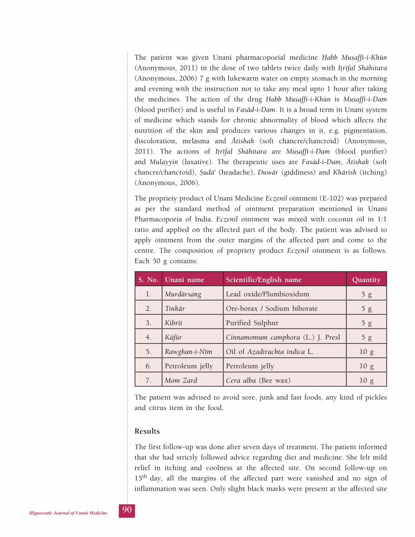
90Hippocratic Journal of Unani Medicine
The patient was given Unani pharmacopoeial medicine Ùabb Muñaffé-i-Khün (Anonymous, 2011) in the dose of two tablets twice daily with Iöréfal Shähitara (Anonymous, 2006) 7 g with lukewarm water on empty stomach in the morning and evening with the instruction not to take any meal upto 1 hour after taking the medicines. The action of the drug Ùabb Muñaffé-i-Khün is Muñaffé-i-Dam (blood purifier) and is useful in Fasäd-i-Dam. It is a broad term in Unani system of medicine which stands for chronic abnormality of blood which affects the nutrition of the skin and produces various changes in it, e.g. pigmentation, discoloration, melasma and Ätishak (soft chancre/chancroid) (Anonymous, 2011). The actions of Iöréfal Shähitara are Muñaffé-i-Dam (blood purifier) and Mulayyin (laxative). The therapeutic uses are Fasäd-i-Dam, Ätishak (soft chancre/chancroid), Ñudä‘ (headache), Duwär (giddiness) and Khärish (itching) (Anonymous, 2006).
The propriety product of Unani Medicine Eczenil ointment (E-102) was prepared as per the standard method of ointment preparation mentioned in Unani Pharmacopoeia of India. Eczenil ointment was mixed with coconut oil in 1:1 ratio and applied on the affected part of the body. The patient was advised to apply ointment from the outer margins of the affected part and come to the centre. The composition of propriety product Eczenil ointment is as follows. Each 50 g contains:
S. No. Unani name Scientific/English name Quantity
1. Murdärsang Lead oxide/Plumbioxidum 5 g
2. Tinkär Ore-borax / Sodium biborate 5 g
3. Kibrét Purified Sulphur 5 g
4. Käfür Cinnamomum camphora (L.) J. Presl 5 g
5. Rawghan-i-Ném Oil of Azadirachta indica L. 10 g
6. Petroleum jelly Petroleum jelly 10 g
7. Mom Zard Cera alba (Bee wax) 10 g
The patient was advised to avoid sore, junk and fast foods, any kind of pickles and citrus item in the food.
Results
The first follow-up was done after seven days of treatment. The patient informed that she had strictly followed advice regarding diet and medicine. She felt mild relief in itching and coolness at the affected site. On second follow-up on 15th day, all the margins of the affected part were vanished and no sign of inflammation was seen. Only slight black marks were present at the affected site

91Hippocratic Journal of Unani Medicine
as shown in Figure 1a. On third follow-up on 21st day, the patient felt better, no itching and burning was felt at the site. The photograph was taken as shown in Figure-1b. The photographs are self-explanatory to showcase the beneficial effect of Unani dugs without any adverse reactions. No significant changes were observed in haemogram, liver function test, kidney function test, lipid profile, urine and stool for routine and microscopic examination as compared to the baseline.
Conclusion
Qübä (Tinea corporis) is a superficial dermatophyte infection characterized by either inflammatory or non-inflammatory lesions on skin regions other than the scalp, groin, palms, and soles. Unani principle-based combination of pharmacopoeial and propriety medicines showed positive effect in the case of Qübä (Tinea corporis). Oral and local use of Unani medicines showed good results without any adverse effect on the body. These results give an idea for effective, cheap and safe management of Tinea corporis. Unani pharmacopoeial medicines effectively counter the disease without any adverse reaction. However, more rigorous and larger studies are needed to confirm the therapeutic efficacy of Ùabb Muñaffé-i-Khün, Iöréfal Shähitara and Eczenil ointment for Tinea corporis.
Conflict of Interest: The authors report no conflicts of interest. There were no funding sources for this study.
Acknowledgement
Authors are thankful to Director General, CCRUM for proving good infrastructure and support.
References
1. Ahmad, Z. (2004). Moalajat-e-Jild, A & U Tibbiya College, Karol Bagh, Delhi.
2. Alam, M. I., Ahsan, S. M., Salam, M., Ahmad, T., Azhar, M. U., & Arfeen, S. (2014). Clinical evaluation of Unani drugs Majoon Suranjan, Safoof Suranjan and Raughan Suranjan in Waja-ul-Mafasil (Rheumatoid arthritis) – A preliminary study. Hippocratic Journal of Unani Medicine, 9(4), 73–84.
3. Alam, M. M. A., Javed, K., & Jafri, M. A. (2005). Effect of Rheum emodi (Revand Hindi) on renal functions in rats. Journal of Ethnopharmacology, 96(1-2), 121–125.
4. Anjum, N., Jamil, S., Hannan, A., Akhtar, J., & Ahmad, B. (2005). Clinical efficacy of Hijamat (cupping) in Waja-ul-Mafasil (Arthritis). Indian Journal of Traditional Knowledge, 4(4), 412–415.

92Hippocratic Journal of Unani Medicine
5. Anonymous. (2006). National formulary of Unani Medicine (vol. I, part I). Department of AYUSH, Ministry of Health and Family Welfare, Government of India, New Delhi.
6. Anonymous. (2011). National formulary of Unani Medicine (vol. I, part III). Department of AYUSH, Ministry of Health and Family Welfare, Government of India, New Delhi.
7. Arzani, A. (1903). Tib-e-Akbar (Urdu translation). Matba Munshi Parag Narayan.
8. Azhar, M. U. (2018). Effect of Herbal Unani Formulation on nephrotic syndrome: a case study. Indian Journal of Traditional Knowledge. 17(4), 807–810.
9. Azhar, M. U., Ahmad, Z., & Mustehasan., (2020c). Effect of Unani medicine in Iltehab-e-Jild Huzaazi (seborrheic dermatitis) of head: a case study. International Journal of Scientific Research in Biological Sciences, 7(2), 41–43.
10. Azhar, M. U., Ansari, R. I., & Ahmad, S. (2019). Clinical effect of Barg-i-Jhao (Tamarix articulate vahl.) in hepatosplenomegaly- a case study. International Journal of AYUSH Case Reports, 3(2), 128–135.
11. Azhar, M. U., Ayub, S., Anjum, N., & Ahmad, S. (2020a). Role of dieto-therapy in weight and dyslipidemia management-a case study. International Journal of Scientific Research in Biological Sciences, 7(1), 17–19.
12. Azhar, M. U., Ayub, S., Anjum, N., & Ahmad, S. (2020b). Effect of Jawarish Bisbasa on dyslipidemia- a case study. International Journal of Scientific Research in Biological Sciences, 7(1), 20–23.
13. Havlickova, B., Czaika, V. A., & Friedrich, M. (2008). Epidemiological trends in skin mycoses world-wide external icon. Mycoses, 51(Suppl 4), 2–15.
14. Ibn Sina. (1903). Al-qanoon fit tib (vol. VI). Matba Munshi Nawal Kishore, Kanpur.
15. Javaid, R., Aslam, M., Javaid, R., Nizami, Q., Javed, K., & Azhar, M. U. (2012). Extract of Ferula foetida regel reverses gentamicin induced nephrotoxicity in rats. Experimental and Clinical Sciences, 11, 760–766.
16. Jegadeesan, M., Kuruvila, S., & Nair, S. (2017). Clinico-etiological study of Tinea corporis: Emergence of Trichophyton mentagrophyes. International Journal of Scientific Study, 5(1), 161–165.
17. Jurjani, I. (2010). Zakheera Khawarazam Shaahi (Urdu translation). Idara-i-Kitab al-Shifa.

93Hippocratic Journal of Unani Medicine
18. Kabeeruddin, M. (1916). Tarjuma Sharah Asbab (vol. III). Daftar al-Maseeh, Karol Bagh, Delhi.
19. Kaur, I., Chaudhary, A., & Singh, H. (2019). Clinico-microbiological aspects of tinea corporis in North India: emergence of Trichophyton tonsurans. International Journal of Research in Dermatology, 5(1), 144–149.
20. Khan, M. A. (1873). Akseer-e-Azam (vol. VI). Matba Nizami, Kanpur.
21. Khan, M. A. (1903). Ramooz-e-Azam (vol. II). Matba Farooqui, Delhi.
22. Majoosi, A. (1889). Kitab Kamilañ-Ñina (vol. IIUrdu translation). Matba Nami Munshi Nawal Kishore, Lucknow.
23. Parveen, N., Mohsin, M., Badrudduja., & Azmat, J. (2019). Dermatophytosis with reference to Unani and Modern Concept: an overview. Journal of Integrated Community Health, 8(1), 1–7.
24. Qumri, H. N. (1930). Ghina Muna’a Tarjuma Minhaj-ul-Ilaj. Dawakhana Madan-ul-Advia, Lucknow.
25. Razi, Z. (1991). Kitab al-Mansuri Fi al-Tibb (Urdu translation). Central Council for Research in Unani Medicine, New Delhi.
26. Razi, Z. (1994). Al-Hawi fit Tib (vol. 23, Urdu translation). Saba Publication, Aligarh.
27. Tabari, A. H. A. M. (1997). Al-Mualajat al-Buqratiya(vol. II, Urdu translation). Central Council for Research in Unani Medicine, New Delhi.
28. Tabari, A. H. A. S. R. (1981).Firdaus-al-Hikmat fit-Tib (Urdu translation). Naved Printing Press, Karachi.
94Hippocratic Journal of Unani Medicine
lkjka'k dwck vy&cnu ¼Vhfu;k d‚ikZsfjl½ ds ,d jksxh esa
gCc&,&eql¶+Q+h&,&[kwu] brjhQy 'kkfgrjk vkSj ,Dt+sfuy ejge dk çHkko
*feLckgqíhu vtgj] t+ehj vgen] eqLrglu
lkjka'k
Vhfu;k d‚ikZsfjl euq";ksa ds Vªad] ckgksa vkSj iSjksa dh Ropk dk lrgh laØe.k gS tks vkerkSj ij rhu lw{ethoksa & Vªk;d‚Q+k;Vu] ekbØksiksje vkSj bihMjeksQ+k;Vu & ds dkj.k gksrk gSA bu esa Vªk;d‚Q+k;Vu :cze lcls vke dkj.k gSA ;wukuh fpfdRlk ds rÙoKku ds vuqlkj ;g v[ykr Qkflnk vkSj vtlke [kchlk ds lap; ds dkj.k Ropk dh lwtu gSA Vhfu;k laØe.k dk funku blds uSnkfud y{k.k ds vkèkkj ij fd;k tkrk gS vkSj lw{e ;k laL—fr ijh{k.k }kjk bl dh iqf"V dh tkrh gSA
bl dsl vè;;u esa 8 eghuksa ls chekjh ls ihfM+r ,d 18 o"kÊ; efgyk Nk= dks ;wukuh Hks"ktdks"kh; feJ.k gCc&,&eql¶+Q+h&,&[kwu] brjhQy 'kkfgrjk vkSj ,d cktkj mRikn ,Dt+sfuy ejge fn;k x;kA bu vkS"kfèk;ksa us 21 fnuksa ds mipkj esa gh lwtu ekftZu dks de djus esa mYys[kuh; çHkko fn[kk;kA bl jksx ds mipkj esa bu vkS"kfèk;ksa ds çHkko dk ewY;kadu djus ds fy, vkxs ,d cM+s vkdkj ds uewus ij fu;a=.k vè;;u fMtkbu fd;k tk ldrk gSA
'kCndqath% dwck] Vhfu;k d‚ikZsfjl] fjaxokeZ] ;wukuh fpfdRlk
95Hippocratic Journal of Unani Medicine
VAbstract
aginal candidiasis is a major problem of growing concern, worldwide. It is so common that 75% women have at least one episode during their lifetime and nearly 45% of women experience two or more. Fortunately, few are plagued with a chronic, recurrent infection. In spite of the prevalence of this disease at mass level, the choice of treatment available in western medicine is comparatively few. Unani Medicine claims to possess a number of effective and safe therapeutic agents that are commonly used in the management of vaginal candidiasis. Marham Däkhliyün, an important patent and proprietary preparation of Oeba India Pharmaceuticals, is one such drug frequently used in candidiasis without any reported serious side effect. In the present study, an attempt has been made to evaluate its efficacy in the management of vaginal candidiasis. A standard controlled single blind clinical trial was conducted. The patients were divided into two groups after confirming the diagnosis by clinical and microbiological examination. The patients in group I, serving as standard control group, were administered Clotrimazole, 100 mg in the form of vaginal pessary daily at bedtime for 7 nights. The test drug was locally applied over vaginal mucosa and fornices in a dose of 5 g at bedtime for 14 days in the patients of group II serving as test group. The patients treated with test drug showed 86% reduction in important clinical features of candidiasis. The findings suggested that the test drug is effective in the treatment of candidiasis.
Keywords: Candidiasis, Moniliasis, Sayalän al-Raùim, Marham Däkhliyün
Introduction
Candidiasis is the most opportunistic mycosis in the world (Panda, 2000). It is a fungal infection of vagina caused by a Gram positive yeast-like fungus Candida albicans. It is present in the vagina in about 20% of women without having any symptom (Dutta, 2003). Vaginal candidiasis is a major problem of growing concern, worldwide. It is so common that 75% women have at least one episode of vaginal candidiasis during their lifetime. Nearly 45% of women experience two or more episodes. Fortunately, few are plagued with a chronic, recurrent infection (Berek, 2007). Since the advent of antibiotics, the incidence of clinical vulvo-vaginal candidiasis has increased. Its incidence is higher in pregnant than in non-pregnant women. The increased incidence in pregnancy is due to increased glycogen content of the vaginal epithelium. For the same reason, it is also common in diabetic patients (Masani, 1982). It thrives on carbohydrate and likes an acidic medium (pH 4.0-5.5). This explains why the patient’s symptoms are temporarily relieved by bathing or douching with one per cent
October - December 2020, Vol. 15 No. 4, Pages 95-105
Therapeutic Evaluation of Marham Däkhliyün in Vaginal Candidiasis
1Wasim Ahmad,2Farh Naz,
*3Fahmeeda Zeenatand
2Azhar Hasan
1Department of Ilmul Advia,Mohammadia Tibbia College,
Malegaon, Nashik, India
2Department of Qabalatwa Amraz-e-Niswan,
Mohammadia Tibbia College, Malegaon, Nashik, India
3Department ofAmraz-e-Niswan wa Atfal,
Faculty of Unani Medicine,AMU, Aligarh, India
*Corresponding author; Email: [email protected]

96Hippocratic Journal of Unani Medicine
sodium bicarbonate solution and during menstruation when the vagina is more alkaline (Kumar & Malhotra, 2008). The microorganism invades on skin and mucus membrane of the organs and survives in the favourable conditions like damp and cold (Berek, 2007). Small numbers of candida commonly occur as normal components of the flora of the human skin, vagina and intestinal tract. It grows excessively and leads to disease only if there are general or local changes that favour the development of the fungus. General factors which favour candidiasis include debility, undernutrition, malignant disease, immunological deficiency state and diabetes. Therapy with antibiotics, steroids and antimitotic agent may play a part. Local factors favouring excessive growth include maceration, moisture and the presence of other eruptions, chiefly eczema of the intertriginous sites (Read et al., 1984). The patient complains of thick curdy vaginal discharge with intense vulvo-vaginal pruritus, vaginal irritation and dysuria (Padubidri & Daftary, 2004).
Unani Medicine postulates that it occurs following the changes in the quality or/and quantity of phlegm (Balgham) in the body especially in pelvic region. Phlegm is described to be synthesized in liver and used by the organs as it is one of the important components of humour necessary to maintain the homeostasis (Zeenat & Hasan, 2016). However in case its quality or quantity is compromised anyhow it becomes a source of diseases and may also serve as a medium for infection (Ahmad et al., 2011). In Unani literature, vaginal candidiasis has been further described to be a type of Sayalän al-Raùim characterized by excessive uterine discharge. It has been described that ‘Ufünat (infection) in the uterus leads to weakening of Quwwat Häòima (digestive faculty) of ‘Urüq-i-Ùayò and predominance of Akhläö Arba‘a are responsible for Sayalän al-Raùim (Ibn Sina, 2007). Majusi (1889) described that Òu‘f-i-Quwwat Jädhiba (weak retensive power), excess of waste in the body and predominance of Akhläö Arba‘a are the causative factor of Sayalän al-Raùim. While some other Unani physicians have described that Sayalän al-Raùim is caused by Òu‘f-i-Quwwat Jädhiba of Raùim along with predominance of Akhläö Arba‘a and accumulation of waste material in the body (Ibn Hubal, 2007; Jurjani, 1903; Kabiruddin, 2003).
Globally, candidiasis is an extremely common infection and is associated with important public health problem but has received only scant attention. In spite of the prevalence of this disease at mass level, the choice of treatment available in western medicine is comparatively few. Even the drugs available for the purpose are not devoid of toxic effects (Rang et al., 2007) and often fail to cure the disease completely. Oral agents are more convenient, less messy, but may have systemic toxicity, e.g., ketoconazole produces hepatotoxicity (Kumar & Malhotra, 2008). Amphotericin B has a wide antifungal activity but it is highly toxic and a variety of reactions may develop after its intravenous use (Satoskar et al., 2001). Clotrimazole is effective against many fungi. Given parenterally,
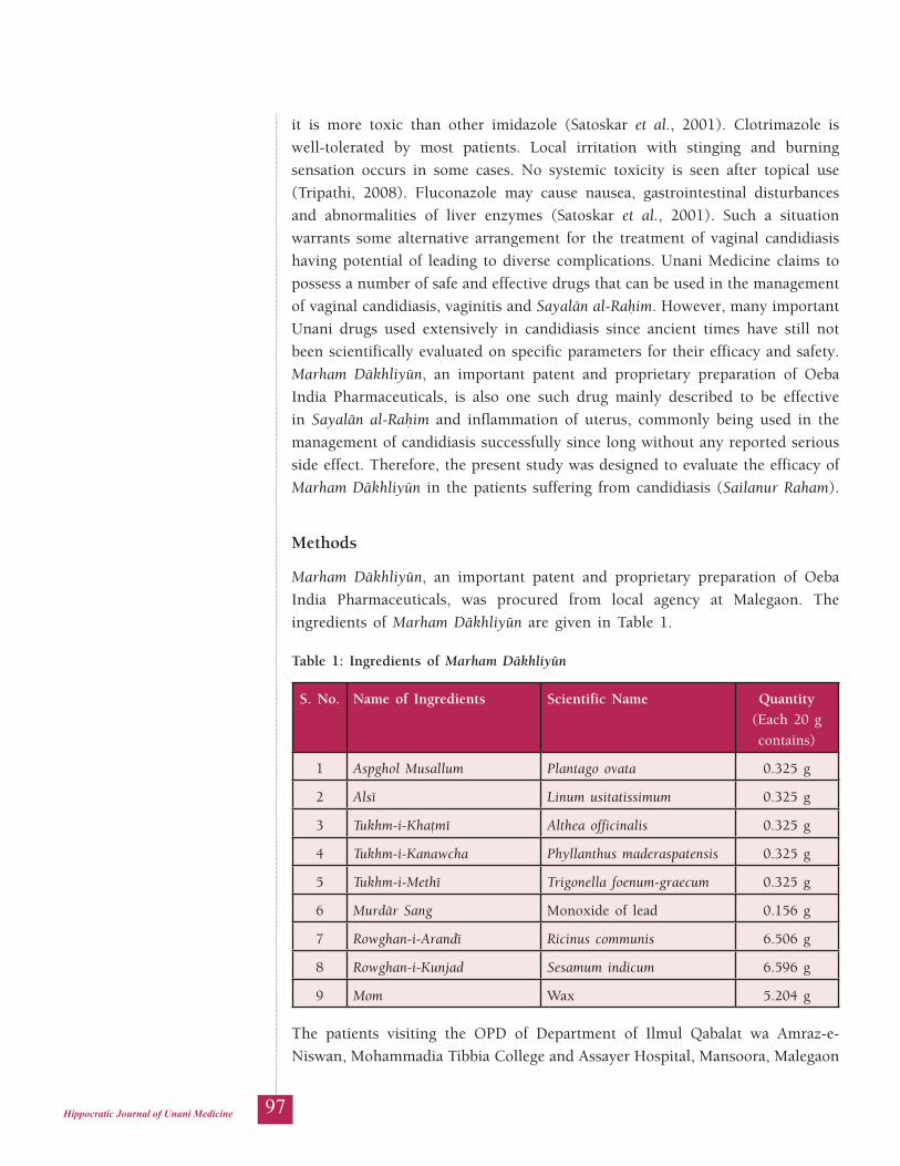
97Hippocratic Journal of Unani Medicine
it is more toxic than other imidazole (Satoskar et al., 2001). Clotrimazole is well-tolerated by most patients. Local irritation with stinging and burning sensation occurs in some cases. No systemic toxicity is seen after topical use (Tripathi, 2008). Fluconazole may cause nausea, gastrointestinal disturbances and abnormalities of liver enzymes (Satoskar et al., 2001). Such a situation warrants some alternative arrangement for the treatment of vaginal candidiasis having potential of leading to diverse complications. Unani Medicine claims to possess a number of safe and effective drugs that can be used in the management of vaginal candidiasis, vaginitis and Sayalän al-Raùim. However, many important Unani drugs used extensively in candidiasis since ancient times have still not been scientifically evaluated on specific parameters for their efficacy and safety. Marham Däkhliyün, an important patent and proprietary preparation of Oeba India Pharmaceuticals, is also one such drug mainly described to be effective in Sayalän al-Raùim and inflammation of uterus, commonly being used in the management of candidiasis successfully since long without any reported serious side effect. Therefore, the present study was designed to evaluate the efficacy of Marham Däkhliyün in the patients suffering from candidiasis (Sailanur Raham).
Methods
Marham Däkhliyün, an important patent and proprietary preparation of Oeba India Pharmaceuticals, was procured from local agency at Malegaon. The ingredients of Marham Däkhliyün are given in Table 1.
Table 1: Ingredients of Marham Däkhliyün
S. No. Name of Ingredients Scientific Name Quantity (Each 20 g contains)
1 Aspghol Musallum Plantago ovata 0.325 g
2 Alsé Linum usitatissimum 0.325 g
3 Tukhm-i-Khaömé Althea officinalis 0.325 g
4 Tukhm-i-Kanawcha Phyllanthus maderaspatensis 0.325 g
5 Tukhm-i-Methé Trigonella foenum-graecum 0.325 g
6 Murdär Sang Monoxide of lead 0.156 g
7 Rowghan-i-Arandé Ricinus communis 6.506 g
8 Rowghan-i-Kunjad Sesamum indicum 6.596 g
9 Mom Wax 5.204 g
The patients visiting the OPD of Department of Ilmul Qabalat wa Amraz-e-Niswan, Mohammadia Tibbia College and Assayer Hospital, Mansoora, Malegaon

98Hippocratic Journal of Unani Medicine
during 2011–13 were screened for the presence of Candida albicans on the basis of clinical signs and symptoms. The diagnosis of screened patients was however confirmed after pathological investigations.
After taking informed consent, sixty diagnosed patients of 18–45 years of age were included in the study and were informed about the disease, examination to be performed and type of treatment. The patients suffering from any other local or systemic diseases were excluded from the study. The permission of Institutional Ethics Committee (IEC) was taken prior to the initiation of the clinical trial. The patients were divided with the help of computer randomized tables/numbers into two groups of 30 patients each (Table 2). The patients in group I, serving as standard control group, were administered Clotrimazole 100 mg in the form of vaginal pessary daily at bedtime for 7 nights to make the study comparable. While the patients in group II, serving as test group, were treated with the test drug Marham Däkhliyün, 5 g per vaginally, once a day at bedtime for two weeks. Abstinence was advised and no concomitant therapy was allowed during the treatment.
Table 2: Treatment Schedule
Group Drug Treatment Dose Duration
Group I Clotrimazole 100 mg x HS 7 days
Group II Marham Däkhliyün 5 g x HS 14 days
The patients were advised for weekly follow-up. They were carefully interviewed at each visit and their statement about the amount, consistency and odour of vaginal discharge, pruritus, burning in vagina and vulva (soreness of vulva and vagina) and lower abdominal pain was recorded. Each patient underwent per vaginal examination in lithotomy position after general and systemic examination. Inflammation and tenderness of vagina was recorded by per speculum examination (Wasim et al., 2016).
Before and after the treatment, specific investigations, such as wet swab test of vaginal discharge, was done to confirm the diagnosis and used as important objective parameters for the assessment. This is the most efficient and cost-effective way to diagnose candidiasis. Wet swab is made quickly and easily. Vaginal discharge was collected by a pipette or swab from the posterior vaginal fornix using Cusco’s speculum in lithotomy position and a small amount of secretion was placed on a clean glass slide. Then a drop of 10% KOH was added to it and examined under a microscope (HPF 45X). KOH destroys white blood cells and bacteria and balances the epithelial cells leaving other than Candida albicans. The presence of candida was recorded in the case record form. On every follow-up, the clinical features were graded on point scales and the changes were recorded in CRF (Wasim et al., 2016).
99Hippocratic Journal of Unani Medicine
Scoring system for overall evaluation of each patient was done. The vaginal discharge was graded (Zeenat & Hasan, 2016) as none (-) for no discharge, mild (slightly) (+) for normal moistness of vagina without staining or moistening the underclothes, moderate (profuse) (++) for undeniably soiled the underclothes that require changing and washing frequently and severe (markedly profuse) (+++) that requires the wearing of some extra absorbent pad. Pruritus (Akhyani et al., 2005) and burning micturition and dysurea were classified as none (-), mild (+), moderate (++) and severe (+++). Lower abdominal pain was assessed by visual analogue scale (Lin et al., 2005) as none (-), mild (+), moderate (++) and severe (+++). Tenderness and congestion of vaginal wall, redness of vaginal mucous membrane, low backache, local soreness and dyspareunia were also graded as none (-), mild (+), moderate (++) and severe (+++). Small number of candida commonly occur as normal components of the flora of the human skin, vagina and intestinal tract (Read et al., 1984). Presence of number of candida was graded as 0–8, 8–15, 15–30 and more than 30 for absent (-), slightly (+), moderate (++) and bulk (+++) respectively (Wasim et al., 2016). The pathological status of each patient was expressed by a scoring system. Scores of 0, 1, 2 and 3 were given for the parameters graded as -, +, ++ and +++. The score for all parameters observed in each patient were then added up. The percentage decrease in scores was determined by comparing the baseline and post treatment. Finally, recorded findings were statistically analyzed using Chi square test to determine the significance.
Results
The test drug was studied in the management of vaginal candidiasis by observing clinical features and laboratory investigations. The findings were tabulated, analyzed and compared with the standard drug (Table 3). Abnormal vaginal discharge was found in 100% of the patients included in each group on the day of registration, while it remained only in 16.66% and 13.33% of the patients and on the basis of percentage, improvement was observed in 83.33% and 86.66% of the cases in group I and II, respectively. Pruritus vulva on day zero was found in 93.33% and 100% of the patients in group I and II, respectively, whereas after treatment it was reduced and found only in 3.57% and 6.66% of the patients and improvement was observed in 96.42% and 93.33% of the cases, respectively. Prior to the treatment local soreness was found in 63.33% and 66.66% of the cases in group I and II, respectively, whereas after treatment it was reduced and found only in 10.52% and 5% of the cases and improvement was observed in 89.47% and 95% of the cases, respectively.
Before the treatment, dyspareunia was found in 60% and 50% of the cases in group I and II, respectively, whereas after treatment it endured only in 5.55% and 6.66% of the cases and improvement was observed in 94.44% and 93.33%
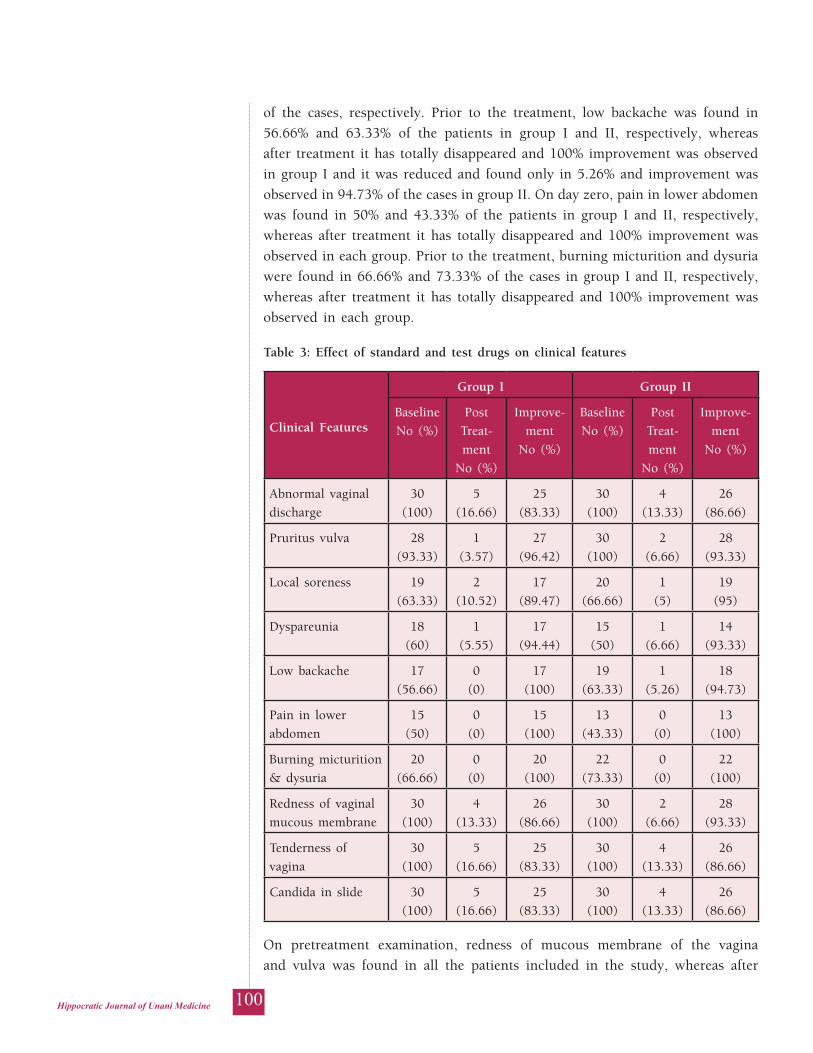
100Hippocratic Journal of Unani Medicine
of the cases, respectively. Prior to the treatment, low backache was found in 56.66% and 63.33% of the patients in group I and II, respectively, whereas after treatment it has totally disappeared and 100% improvement was observed in group I and it was reduced and found only in 5.26% and improvement was observed in 94.73% of the cases in group II. On day zero, pain in lower abdomen was found in 50% and 43.33% of the patients in group I and II, respectively, whereas after treatment it has totally disappeared and 100% improvement was observed in each group. Prior to the treatment, burning micturition and dysuria were found in 66.66% and 73.33% of the cases in group I and II, respectively, whereas after treatment it has totally disappeared and 100% improvement was observed in each group.
Table 3: Effect of standard and test drugs on clinical features
Clinical Features
Group I Group II
Baseline No (%)
PostTreat-ment
No (%)
Improve-ment
No (%)
Baseline No (%)
PostTreat-ment
No (%)
Improve-ment
No (%)
Abnormal vaginal discharge
30(100)
5(16.66)
25(83.33)
30(100)
4(13.33)
26(86.66)
Pruritus vulva 28(93.33)
1(3.57)
27(96.42)
30(100)
2(6.66)
28(93.33)
Local soreness 19(63.33)
2(10.52)
17(89.47)
20(66.66)
1(5)
19(95)
Dyspareunia 18(60)
1(5.55)
17(94.44)
15(50)
1(6.66)
14(93.33)
Low backache 17(56.66)
0(0)
17(100)
19(63.33)
1(5.26)
18(94.73)
Pain in lower abdomen
15(50)
0(0)
15(100)
13(43.33)
0(0)
13(100)
Burning micturition & dysuria
20(66.66)
0(0)
20(100)
22(73.33)
0(0)
22(100)
Redness of vaginal mucous membrane
30(100)
4(13.33)
26(86.66)
30(100)
2(6.66)
28(93.33)
Tenderness of vagina
30(100)
5(16.66)
25(83.33)
30(100)
4(13.33)
26(86.66)
Candida in slide 30(100)
5(16.66)
25(83.33)
30(100)
4(13.33)
26(86.66)
On pretreatment examination, redness of mucous membrane of the vagina and vulva was found in all the patients included in the study, whereas after
101Hippocratic Journal of Unani Medicine
treatment it was found reduced and only 13.33% and 6.66% of patients were found affected. The findings indicated improvement in 86.66% and 93.33% of the cases in respective group. Prior to the treatment, tenderness of vaginal wall on day zero was found in all the patients included in the study, while after treatment it decreased significantly and was found in 16.66% and 13.33% of the cases indicating an improvement in 83.33% and 86.66% of the patients in group I and II, respectively. On pretreatment investigation, candida in slide was found in 100% of the patients in each group, whereas after treatment it decreased significantly and was found only in 16.66% and 13.33% of the patients and an improvement was observed in 83.33% and 86.66% of the cases in respective groups.
Relief in clinical symptoms along with reduction in microbiological count was considered as the criteria of efficacy. The cases having relief from abnormal vaginal discharge along with absence of candida in slide after treatment were rated as cured. While the patients having no relief in abnormal vaginal discharge and candida were found in slide after treatment, were rated as not cured. Complete cure was observed in 83.33% and 86.66% patients in group I and II, respectively, whereas 16.66% and 13.33% cases were not cured, respectively.
Discussion
The findings in respect of different parameters of this study indicated that there was no statistical difference between the two groups suggesting that both the drugs produced almost equal degree of response. The findings of the study suggested that local application of the test drug is equally effective as the local administration of Clotrimazole. Although in Unani Medicine both local and oral treatment is suggested simultaneously to treat the patients of vaginal candidiasis with the aim to improve the quality of phlegm that has undergone through derangement and to improve the local pathology. But only local treatment is also advised commonly to treat such a condition (Zeenat & Hasan, 2016). The present study indicated that only local application is also sufficient to cure the majority of the patients. About 13.33% of the patients were not cured with the test drug probably because few patients may have required the local as well as the systemic intervention as the only local treatment may not be sufficient to negotiate the severity of the disease. The result is in consonance with the Unani principle of treatment that suggests that the disease arises following the disturbance in the quality, quantity or composition of the phlegm (Majoosi, 2005). Candidiasis may develop as sequelae of systemic derangement of phlegm but it may also develop because of its local overthrow that is frequently compounded with other factors that trigger the pathology. The cure rate of only 86% may be because of the demand of the nature of pathological condition for systemic intervention.
102Hippocratic Journal of Unani Medicine
Since the candidiasis is a disease of Khilö Balgham having the symptoms of cold and wet temperament, it is obvious to treat the disease by the drugs that have hot and dry temperament. Therefore, few ingredients having hot and dry temperament (Ghani, 2011; Hakeem, 1999) were included in Marham Däkhliyün.
Most of the ingredients of Marham Däkhliyün have been reported in classical Unani literature as Qäbiò (astringent), Muùallil (resolvent), Muùallil-i-Awräm (antiinflammatory), Mujaffif (dessicative), Mulaööif (demulscent), Munòij (concoctive), Musakkin (analgesic), Rädi‘ (repellent), Murakhkhé (emollient) and effective in phlegmatic derangement (Ghani, 2011; Hakeem, 1999; Khan, 1313H; Nabi, 2007). They have also been reported in ethnopharmacological literature as astringent, demulscent, emollient, useful in backache, inflammation and in relieving the inflammation of mucous membrane of urogenital tract. Pessaries of Trigonella foenum-graecum seeds are therapeutically used in leucorrhoea. Murdär Sang is a powerful local astringent (Anonymous, 1969; Anonymous, 2000; Nadkarni, 1954; Nadkarni, 1954). The anticandidal activity of Linum usitatissimum oil has also been reported in an experimental study (Kaithwas et al., 2011). Due to these medicinal properties, Marham Dakhliyn produces constriction in the vaginal wall, absorbs the vaginal secretions and resolves the discharge and inflammatory condition.
The study clearly showed that the test drug is very effective in candidiasis which is proved by decrease in the amount of abnormal vaginal discharge, pruritus, local soreness, dyspareunia, low backache, pain in lower abdomen, burning micturition and dysuria, redness of mucous membrane of vagina and vulva, tenderness and congestion of vaginal wall, and candida in slide. The efficacy of the test drug may be attributed to its anticandidal activity which was almost equal to that of Clotrimazole. Thus, the study validated the therapeutic application of the test drug Marham Dakhliyn in the management of vaginal candidiasis.
Conclusion
In the light of above findings and discussion, it can be concluded that Unani drug Marham Däkhliyün, a proprietary preparation of Oeba India Pharmaceuticals, possesses significant effect against vaginal candidiasis. Therefore, it may be used in the patients suffering from candidiasis (Sayalän al-Raùim) and associated condition.
Acknowledgements
The authors are extremely thankful to Prof. Ghufran Ahmad, Department of Ilmul Advia, Faculty of Unani Medicine, AMU, Aligarh for valuable guidance and moral encouragement during entire period of the study. We are also very
103Hippocratic Journal of Unani Medicine
grateful to Mr. Arshad Mukhtar, President, Al-Jameatul Mohammadia Al-Khairiah’s Mohammadia Tibbia College, Mansoora, Malegaon, for providing necessary facilities to carry out the research.
References
1. Ahmad, W., Hasan, A., Ansari, A., & Tarannum, T. (2011). Efficacy of a combination of Unani drugs in patients of trichomonal vaginitis. Indian J. Traditional Knowledge, 10(4), 727–730.
2. Akhyani, M., Ganji, M. R., Samadi, N., Khamesan, B., & Daneshpazhooh, M. (2005). Pruritus in haemodialysis patients. BMC Dermatology, https://doi:10.1186/1471-5945-5-7
3. Anonymous. (1969). The Wealth of India, A Dictionary of Indian Raw Materials and Industrial Products (8th vol.). Publication & Information Directorate, CSIR, New Delhi.
4. Anonymous. (2000). PDR for Herbal Medicines. Medicals economics company, Montvale, New Jersey.
5. Berek, J. S. (2007). Berek & Novak’s Gynaecology (14th ed.). Lippincott Williams & Wilkins.
6. Dutta, D.C. (2003). Textbook of Gynaecology (4th ed.). New Central Book Agency, Calcutta.
7. Ghani, N. (2011). Khazainul Advia. Idara Kitabul Shifa, New Delhi.
8. Hakeem, M. A. (1999). Bustanul Mufradat. Zafar Book Depo, Delhi.
9. Ibn Hubal (2007). Kitabul Mukhtarat fit Tib (4th vol.). Urdu translation by CCRUM, New Delhi.
10. Ibn Sina (2007). Al-Qanoon fit Tib (2nd vol.). Urdu translation by Kintoori G.H., Idara Kitabus Shifa, New Delhi.
11. Jurjani, I. (1903). Zakheera Khawarizm Shahi (6th vol.). Urdu translation by Khan, A. H., Matba Munshi Nawal Kishore, Lucknow.
12. Kabiruddin, M. (2003). Al-Akseer (2nd vol.). Aijaz Publishing House, New Delhi.
13. Kaithwas, G., Mukherjee, A., Kumar, P., & Majumdar, D. K. (2011). Linum usitatissimum (linseed/flaxseed) fixed oil: antimicrobial activity and efficacy in bovine mastitis. Inflammopharmacology, 19(1), 45–52.
14. Khan, M. A. (1313H). Muheet-e-Azam (4th vol., 2nd part). Maktaba Nizami, Kanpur.
104Hippocratic Journal of Unani Medicine
15. Khan, M. A. (1313H). Muheet-e-Azam (4th vol., 1st part). Maktaba Nizami, Kanpur.
16. Khan, M. A. (1313H). Muheet-e-Azam (1st vol.). Maktaba Nizami, Kanpur.
17. Kumar, P., & Malhotra, N. (2008). Jeffcoate’s principles of Gynaecology (7th ed.). Jaypee Brothers Medical Publishers, New Delhi.
18. Lin, W. C., Kung, F. T., Chen, T. Y., Tsang, L. L. C., Tsay, W. D., & Cheng, Y. F. (2005). Short term results of transcatheter uterine artery embolization sing Lipiodol and Gelfoam cubes for symptomatic uterine leiomyoma. Chin J Radiol, 30, 17–24.
19. Majoosi, A. I. A. (2005). Kamilus Sana’at al-Tabia (1st vol.). Central Council for Research in Unani Medicine, New Delhi.
20. Majoosi, A. I. A. (1889). Kamilus Sana’at, Urdu translation by Kintoori GH, Matba Munshi Nawal Kishore, Lucknow.
21. Masani, K. M. (1982). A Textbook of Gynaecology (8th ed.). Popoular Prakashan, Bombay.
22. Nabi, M. G. (2007). Makhzan-e-Mufradat-wa-Murakkabat (2nd ed.). Central Council for Research in Unani Medicine, New Delhi.
23. Nadkarni, A. K. (1954). Indian Materia Medica (1st vol., 3rd ed.). Bombay Popular Prakashan, Mumbai.
24. Nadkarni, A. K. (1954). Indian Materia Medica (2nd vol., 3rd ed.). Bombay Popular Prakashan, Mumbai.
25. Padubidri, V. G., & Daftary, S. N. (2004). Hoowkins and Bourne show’s textbook of Gynaecology (14th ed.). Reed Elsevier India Private Limited.
26. Panda, U. N. (2000). Panda’s Textbook of Medicine (1st ed.) CBS Publishers and Distributers, New Delhi.
27. Rang, H. P., Dale, M. M., Ritter, J. M., & Flower, R. J., (2007). Rang and Dale’s Pharmacology (6th ed.). Churchill Livingstone.
28. Read, A. E., Barrit, D. W., & Hewer, R. L. (1984). Modern Medicine (3rd ed.). ELBS/Churchill Livingstone.
29. Satoskar, R. S., Bhandarkar, S. D., & Ainapure, S. S. (2001). Pharmacology & Pharmacotherapeutics(17th ed.). Popular Prakashan, Mumbai.
30. Tripathi, K. D. (2008). Essentials of Medical Pharmacology (6th ed.). Jay Pee Brothers Medical Publishers, New Delhi.
31. Wasim, A., Naz, F., Hasan, A., Zeenat, F., & Parray, S. A. (2016). Clinical evaluation of sailani in vaginal candidiasis. Unani Medicus, 3(1), 50–56.
105Hippocratic Journal of Unani Medicine
32. Zeenat, F., & Hasan, A. (2016). Combating trichomoniasis through a Unani drug ‘Marham Däkhliyün’. Hippocratic Journal of Unani Medicine, 11(1), 23–32.
33. Zeenat, F., & Hasan, A. (2016). Therapeutic evaluation of a polyherbal formulation Leuco-Cure in trichomoniasis. Hippocratic Journal of Unani Medicine, 11(2), 55–64.
lkjka'k Oktkbuy dSafMfM;kfll esa ejge nk[kfy;wu dk fpfdRlh;
ewY;kadu
olhe vgen] Q+jg ukt+] *Qgehnk t+hur] vt+gj glu
lkjka'k
Oktkbuy dSafMfM;kfll fo'o Hkj esa ,d izeq[k fparktud leL;k gSA ;g bruh lkekU; gS fd 75% efgykvksa ds thoudky esa de ls de ,d ckj ?kfVr gksrh gS vkSj yxHkx 45% efgykvksa ds thoudky esa nks ;k nks ls vfèkd ckj ?kfVr gksrh gSA HkkX;o'k muesa ls dqN gh tfVy] vkorZd laØe.k ls xzLr gSaA cM+s iSekus ij bl jksx dh O;kidrk ds ckotwn] ,yksiSfFkd fpfdRlk esa mipkj dk fodYi rqyukRed :Ik ls cgqr de gSA ;wukuh fpfdRlk dbZ izHkkoh ,oa lqjf{kr fpfdRlh; vkS"kfèk;ksa dk nkok djrh gS tks vkerkSj ij Oktkbuy dSafMfM;kfll ds mipkj esa mi;ksx dh tkrh gSaA vksck bafM;k QkekZL;wfVdYl dh ,d egRoiw.kZ isVsaV ,oa LokfeRo vkS"kfèk ejge nk[kfy;wu ,d ,slh vkS"kfèk gS ftldk mi;ksx vDlj dSafMfM;kfll esa fd;k tkrk gS vkSj ftldk dksbZ xaHkhj nq"izHkko ugha ns[kk x;k gSA orZeku vè;;u esa Oktkbuy dSafMfM;kfll ds mipkj esa bldh izHkkodkfjrk dk ewY;kadu djus dk iz;kl fd;k x;k gSA ,d ekud fu;af=r ,dy CykbaM uSnkfud ijh{k.k fd;k x;kA uSnkfud vkSj lw{ethofoKku ijh{k.k }kjk jksxfunku dh iqf"V ds ckn jksfx;ksa dks nks lewgksa esa foHkkftr fd;k x;kA ekud fu;a=.k lewg ds :Ik esa lewg I ds jksfx;ksa dks DyksfVªekt+ksy otkbuy isljh ds :Ik esa 100 fe-xzk- 7 jkrksa rd lksrs le; nh xbZA ijh{k.k lewg ds :Ik esa lewg II ds jksfx;ksa dks ijh{k.k vkS"kfèk otkbuy E;wdkslk vkSj QksfuZDl ij 5 xzke dh ek=k esa 14 fnuksa rd lksrs le; ckgjh :Ik ls yxkbZ xbZA ijh{k.k vkS"kfèk ls mipkfjr jksfx;ksa us dSafMfM;kfll ds egRoiw.kZ uSnkfud y{k.kksa esa 86% deh n'kkZbZA fu"d"kksaZ ls irk pyk fd dSafMfM;kfll ds mipkj esa ijh{k.k vkS"kfèk izHkkoh gSA
'kCndqath% dSafMfM;kfll] eksfufy;kfll] l;yku vy&jfge] ejge nk[kfy;wu

106Hippocratic Journal of Unani Medicine
R.N.I. Registration No. DELENG/2006/18866
CENTRAL COUNCIL FOR RESEARCH IN UNANI MEDICINE
ISSN: 0974-1291
Volume 15 • Number 4 October–December 2020
CENTRAL COUNCIL FOR RESEARCH IN UNANI MEDICINE Ministry of Ayurveda, Yoga & Naturopathy, Unani,
Siddha and Homoeopathy (AYUSH), Government of India61 - 65, Institutional Area, Janakpuri, New Delhi - 110 058
Telephone: +91-11-28521981, 28525982Email: [email protected]
Website: http://ccrum.res.in
This is a peer-reviewed publication and included in the abstracting and indexing of Medicinal and Aromatic Plants Abstracts (MAPA); Biological Abstracts; Chemical Abstracts; Contemporary Researches in Traditional Drugs & Medicinal Plants: Unani Medicine Abstracts, etc.
HIPPOCRATIC JOURNAL OF UNANI MEDICINE