Cell-Specific Production and Antimicrobial Activity of Naphthoquinones in Roots of Lithospermum...
12
Cell-Specific Production and Antimicrobial Activity of Naphthoquinones in Roots of Lithospermum erythrorhizon 1 Lindy A. Brigham 2 *, Paula J. Michaels, and Hector E. Flores Department of Plant Pathology, The Pennsylvania State University, University Park, Pennsylvania 16802–4507 Pigmented naphthoquinone derivatives of shikonin are produced at specific times and in specific cells of Lithospermum erythrorhizon roots. Normal pigment development is limited to root hairs and root border cells in hairy roots grown on “noninducing” medium, whereas induction of additional pigment production by abiotic (CuSO 4 ) or biotic (fungal elicitor) factors increases the amount of total pigment, changes the ratios of derivatives produced, and initiates production of pigment de novo in epidermal cells. When the biological activity of these compounds was tested against soil-borne bacteria and fungi, a wide range of sensitivity was recorded. Acetyl-shikonin and b-hydroxyisovaleryl-shikonin, the two most abundant derivatives in both Agrobacterium rhizogenes-transformed “hairy-root” cultures and greenhouse-grown plant roots, were the most biologically active of the seven compounds tested. Hyphae of the pathogenic fungi Rhizoctonia solani, Pythium aphanidermatum, and Nectria hemato- cocca induced localized pigment production upon contact with the roots. Challenge by R. solani crude elicitor increased shikonin deriv- ative production 30-fold. We have studied the regulation of this suite of related, differentially produced, differentially active compounds to understand their role(s) in plant defense at the cellular level in the rhizosphere. Plants communicate with their environment (the soil, climate conditions, neighboring plants, microorganisms, insects, etc.) by producing a diverse array of chemicals. These secondary metabolites are often characteristic of spe- cific plants and plant families. Many members of the Bor- aginaceae family produce naphthoquinones in their roots. Naphthoquinones are colored substances derived from phenylpropanoid and isoprenoid precursors (Gaisser and Heide, 1996). Plants of the borage family are distributed worldwide and the naphthoquinones from many of these plants have been used in diverse cultures as colorants for cosmetics, fabrics, and foods (Jain and Mathur, 1965; Bal- lantine, 1969; Tabata and Fujita, 1985), and for medicinal applications, including antitumor, antiinflammatory, and antimicrobial agents (Papageorgiou, 1978; Tabata and Fu- jita, 1985). The chemicals involved in the antimicrobial activities studied to date are all derivatives of shikonin and its enantiomer alkannin from the European dye plant Al- kanna tinctoria. Figure 1 illustrates the chemical structure of shikonin, its derivatives, and the proposed biosynthetic pathway. The pattern of shikonin derivatives may differ in any one plant, root culture, or cell-suspension culture (summarized from a literature survey; specific refs. are given in Table I). Because of its value as a pharmaceutical agent, most research to date has focused on understanding the produc- tion and regulation of shikonin with the goal of increasing production of the compound, but the biological signifi- cance of shikonin in the plant has been virtually ignored. Because shikonin and its derivatives have biological activ- ity against microorganisms, it seems likely that these com- pounds may play a role in plant defense in the rhizosphere. Naphthoquinones have been shown to function in allelopa- thy (juglone; Binder et al., 1989), plant-insect interactions (plumbagin; Kubo and Klocke, 1986), and electron trans- port (phylloquinone [vitamin K 1 ]; Hauska, 1988). Many of the products of the phenylpropanoid and isoprenoid path- ways are produced in response to multiple stresses (for review, see Chappell, 1995; Dixon and Paiva, 1995) and frequently lead to the production of antimicrobial com- pounds (Bennett and Wallsgrove, 1994; Rhodes, 1994; Chappell, 1995; Herrmann, 1995). Regulation under nu- merous conditions has been studied using cell-suspension cultures (Table II), but nothing is known about the re- gulation of production of the specific derivatives of shiko- nin or of the regulation in specific cell types within the root. Shikonin accumulates in the cork layer of mature roots (Fujita and Yoshida, 1937) and its formation in sig- nificant quantities gives the roots a characteristic red or purple color. Production and accumulation in new roots has not been systematically studied, nor has the biological activity of these compounds in relation to soil-borne microorganisms. In view of the observations on the antimicrobial proper- ties reported for shikonin and its derivatives, the produc- tion of numerous derivatives of shikonin in the roots of the plant, and the existing information on regulation of the pathways in common with many other plant defense re- sponses, we investigated the regulation and biological ac- tivity of shikonin and its derivatives in hairy-root cultures 1 This research was supported by Department of Energy/Na- tional Science Foundation/U.S. Department of Agriculture Collab- orative Research in Plant Biology award no. BIR-9220330, “Inter- disciplinary Research Training Program in Advanced Root Biology.” 2 Present address: Department of Plant Pathology, The Univer- sity of Arizona, Tucson, AZ 85721– 0036. * Corresponding author; e-mail [email protected]; fax 1–520 – 621–9290. Abbreviations: AS, acetyl-shikonin; DMAS, b,b-dimethylacryl- shikonin; DS, deoxy-shikonin; HIVS b-hydroxyisovaleryl-shiko- nin; IBS, isobutyl-shikonin; IVS, isovaleryl-shikonin; MBS, a-methyl-n-butyl-shikonin. Plant Physiology, February 1999, Vol. 119, pp. 417–428, www.plantphysiol.org © 1999 American Society of Plant Physiologists 417 www.plant.org on June 26, 2014 - Published by www.plantphysiol.org Downloaded from Copyright © 1999 American Society of Plant Biologists. All rights reserved.
-
Upload
go-uni-enugu -
Category
Documents
-
view
1 -
download
0
Transcript of Cell-Specific Production and Antimicrobial Activity of Naphthoquinones in Roots of Lithospermum...

Cell-Specific Production and Antimicrobial Activity ofNaphthoquinones in Roots of Lithospermum erythrorhizon1
Lindy A. Brigham2*, Paula J. Michaels, and Hector E. Flores
Department of Plant Pathology, The Pennsylvania State University, University Park, Pennsylvania 16802–4507
Pigmented naphthoquinone derivatives of shikonin are producedat specific times and in specific cells of Lithospermum erythrorhizonroots. Normal pigment development is limited to root hairs and rootborder cells in hairy roots grown on “noninducing” medium, whereasinduction of additional pigment production by abiotic (CuSO4) orbiotic (fungal elicitor) factors increases the amount of total pigment,changes the ratios of derivatives produced, and initiates productionof pigment de novo in epidermal cells. When the biological activity ofthese compounds was tested against soil-borne bacteria and fungi, awide range of sensitivity was recorded. Acetyl-shikonin andb-hydroxyisovaleryl-shikonin, the two most abundant derivatives inboth Agrobacterium rhizogenes-transformed “hairy-root” culturesand greenhouse-grown plant roots, were the most biologically activeof the seven compounds tested. Hyphae of the pathogenic fungiRhizoctonia solani, Pythium aphanidermatum, and Nectria hemato-cocca induced localized pigment production upon contact with theroots. Challenge by R. solani crude elicitor increased shikonin deriv-ative production 30-fold. We have studied the regulation of this suiteof related, differentially produced, differentially active compounds tounderstand their role(s) in plant defense at the cellular level in therhizosphere.
Plants communicate with their environment (the soil,climate conditions, neighboring plants, microorganisms,insects, etc.) by producing a diverse array of chemicals.These secondary metabolites are often characteristic of spe-cific plants and plant families. Many members of the Bor-aginaceae family produce naphthoquinones in their roots.Naphthoquinones are colored substances derived fromphenylpropanoid and isoprenoid precursors (Gaisser andHeide, 1996). Plants of the borage family are distributedworldwide and the naphthoquinones from many of theseplants have been used in diverse cultures as colorants forcosmetics, fabrics, and foods (Jain and Mathur, 1965; Bal-lantine, 1969; Tabata and Fujita, 1985), and for medicinalapplications, including antitumor, antiinflammatory, andantimicrobial agents (Papageorgiou, 1978; Tabata and Fu-jita, 1985). The chemicals involved in the antimicrobial
activities studied to date are all derivatives of shikonin andits enantiomer alkannin from the European dye plant Al-kanna tinctoria. Figure 1 illustrates the chemical structure ofshikonin, its derivatives, and the proposed biosyntheticpathway. The pattern of shikonin derivatives may differ inany one plant, root culture, or cell-suspension culture(summarized from a literature survey; specific refs. aregiven in Table I).
Because of its value as a pharmaceutical agent, mostresearch to date has focused on understanding the produc-tion and regulation of shikonin with the goal of increasingproduction of the compound, but the biological signifi-cance of shikonin in the plant has been virtually ignored.Because shikonin and its derivatives have biological activ-ity against microorganisms, it seems likely that these com-pounds may play a role in plant defense in the rhizosphere.Naphthoquinones have been shown to function in allelopa-thy (juglone; Binder et al., 1989), plant-insect interactions(plumbagin; Kubo and Klocke, 1986), and electron trans-port (phylloquinone [vitamin K1]; Hauska, 1988). Many ofthe products of the phenylpropanoid and isoprenoid path-ways are produced in response to multiple stresses (forreview, see Chappell, 1995; Dixon and Paiva, 1995) andfrequently lead to the production of antimicrobial com-pounds (Bennett and Wallsgrove, 1994; Rhodes, 1994;Chappell, 1995; Herrmann, 1995). Regulation under nu-merous conditions has been studied using cell-suspensioncultures (Table II), but nothing is known about the re-gulation of production of the specific derivatives of shiko-nin or of the regulation in specific cell types within theroot. Shikonin accumulates in the cork layer of matureroots (Fujita and Yoshida, 1937) and its formation in sig-nificant quantities gives the roots a characteristic red orpurple color. Production and accumulation in new rootshas not been systematically studied, nor has the biologicalactivity of these compounds in relation to soil-bornemicroorganisms.
In view of the observations on the antimicrobial proper-ties reported for shikonin and its derivatives, the produc-tion of numerous derivatives of shikonin in the roots of theplant, and the existing information on regulation of thepathways in common with many other plant defense re-sponses, we investigated the regulation and biological ac-tivity of shikonin and its derivatives in hairy-root cultures
1 This research was supported by Department of Energy/Na-tional Science Foundation/U.S. Department of Agriculture Collab-orative Research in Plant Biology award no. BIR-9220330, “Inter-disciplinary Research Training Program in Advanced RootBiology.”
2 Present address: Department of Plant Pathology, The Univer-sity of Arizona, Tucson, AZ 85721– 0036.
* Corresponding author; e-mail [email protected]; fax1–520 – 621–9290.
Abbreviations: AS, acetyl-shikonin; DMAS, b,b-dimethylacryl-shikonin; DS, deoxy-shikonin; HIVS b-hydroxyisovaleryl-shiko-nin; IBS, isobutyl-shikonin; IVS, isovaleryl-shikonin; MBS,a-methyl-n-butyl-shikonin.
Plant Physiology, February 1999, Vol. 119, pp. 417–428, www.plantphysiol.org © 1999 American Society of Plant Physiologists
417 www.plant.org on June 26, 2014 - Published by www.plantphysiol.orgDownloaded from
Copyright © 1999 American Society of Plant Biologists. All rights reserved.
of Lithospermum erythrorhizon. Because these compoundsare brightly colored, the timing and cellular specificity oftheir production can be followed with simple equipmentsuch as a dissecting microscope. The suite of compoundscan be studied in the context of understanding the multipleactions of plants in fine tuning their response to theirenvironment. The objectives of this study were: (a) to char-acterize the production of naphthoquinone pigments in L.erythrorhizon hairy-root cultures, (b) to analyze the effect(s)of shikonin and its derivatives on soil-borne microorgan-isms, and (c) to determine if the L. erythrorhizon hairy-rootcultures could respond to microorganisms by modifyingthe production of shikonin and its derivatives.
MATERIALS AND METHODS
Plant Material
Seeds of Lithospermum erythrorhizon (Sieb. and Zucc.)were obtained from the Institut fur Pflanzengenetik undKulturpflanzenforschung (Gatersleben, Germany), germi-nated, and grown in sterile potting soil (Pro Mix BX, Pre-mier Horticulture, Red Hill, PA). The plants were main-tained in the greenhouse at 60oC to 80oC with supple-mental lighting from 1000-W high-pressure sodium lampsfor 8 h daily from October through March and in ambientlighting the remainder of the year. Plants were wateredthree times per week. Plants were fertilized twice a
week with Peters 15–16-17 Peat Lite Special (Scotts-SierraHorticultural Products, Marysville, OH) at 200 ppm N.
Hairy-Root Production and Growth Conditions
Seeds of L. erythrorhizon were sterilized with 10% com-mercial bleach for 20 min, rinsed in five changes of sterilewater, and germinated under sterile conditions on solidMurashige and Skoog medium (Murashige and Skoog,1962). Seedlings were grown in GA7 hydroponic culturevessels (Magenta Corp., Chicago, IL), and stems were in-oculated with Agrobacterium rhizogenes strain 15834 (ATCC)to produce hairy roots. Root cultures were obtained from16 independent transformation events. All root culturelines appeared morphologically indistinguishable, and ex-periments described in this paper were performed on asingle root line. Root cultures from the selected line weregrown in the dark at 24°C on M medium (Becard andFortin, 1988) or M-9 (Fujita et al., 1981a) medium. Rootsgrown on solid or liquid M medium did not produceshikonin in sufficient quantities to be observable on visualinspection. M-9 served as an induction medium for shiko-nin because it contains 2.3 times the amount of coppersulfate as the M medium. Liquid root cultures were grownin the dark on a rotary shaker (model G-53, New Bruns-wick Scientific, Edison, NJ) at 90 rpm at 24°C. Solid rootcultures were grown on medium solidified with 0.3%(w/v) Phytagel (Sigma).
Microscopy
Roots and fungal-growth plates were viewed and pho-tographed using a Zeiss STEMI SV8 dissecting microscopeand Kodak 160T slide film. Root hairs were viewed andphotographed on a Zeiss inverted microscope and Kodak160T slide film.
Bacterial and Fungal Inhibition Assays
Bacterial Growth Inhibition
Bacterial cultures were grown overnight at 24°C withshaking in Luria-Bertani broth (Luria and Burrows, 1957)to an optical density of 0.2 at 600 nm. A 100-mL aliquot ofthis liquid culture was spread onto 82-mm plates with solidLuria-Bertani medium. A filter disk saturated with 50 mg ofshikonin or shikonin derivative (dissolved in chloroformand allowed to air dry) was placed on the bacterial lawn.The plates were incubated in the dark at 24°C. The clearzone from the filter disk to the bacteria was measured. Eachexperiment was performed three times for each isolatetested. The following bacterial strains were obtained froma collection of field isolates maintained in the laboratory ofDr. Leland S. Pierson III (Department of Plant Pathology,University of Arizona, Tucson): Bacillus subtilis 613R, Bacil-lus thuringiensis Gnatrol, Clavibacter michigenensis subsp.nebraskensis CN74-1, Agrobacterium radiobacter K84, Agrobac-terium tumefaciens C58, Burkholdaria cepacea Deny, Esche-richia coli ESS, Erwinia amylovora, Erwinia carotovora ATCC15713, Pseudomonas aureofaciens 30-84, Pseudomonas fluore-
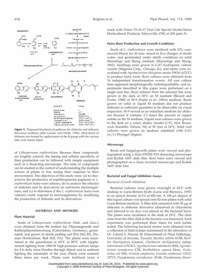
Figure 1. Proposed biochemical pathway for shikonin and shikoninderivative synthesis (after Gaisser and Heide, 1996). Derivatives ofshikonin are formed by replacement of the R-group with the variousfatty acid chains listed.
418 Brigham et al. Plant Physiol. Vol. 119, 1999
www.plant.org on June 26, 2014 - Published by www.plantphysiol.orgDownloaded from Copyright © 1999 American Society of Plant Biologists. All rights reserved.

scens 2-79, Pseudomonas syringae B, Pseudomonas syringae pvphaseolicola, Ralstonia solanacearum, and Serratia marsecens.Erwinia herbicola was isolated from pea seedling roots (L.A.Brigham, unpublished data). A. rhizogenes ATCC 15834 wasmaintained by Paula Michaels at The Pennsylvania StateUniversity (University Park). Xanthomonas campestris pvpelargonii was from the collection of Dr. Gary Moorman(The Pennsylvania State University).
Fungal Growth Inhibition
Fungal isolates were maintained on V-8 medium (200 mLof commercial vegetable juice, 2 g of CaCO3, and 1 g of Glcper liter of water) solidified with 2% agar (Difco Laborato-ries, Detroit, MI) in the dark at 24°C. Inhibition assays wereperformed by dissolving shikonin in solid M-2 medium(modified Martin’s medium: 10 g of Glc, 5 g of peptone, 1 gof KH2PO4 0.5 g of MgSO4, 22 g of agar per liter of double-distilled water) in 35-mm plastic Petri dishes. A 4-mm plugof fungal hyphae was placed on one side of the plate andhyphal length was measured at 24-h intervals. Each isolatewas tested on all concentrations in three separate repli-cates. Fungal isolates Nectria hematococca 34-18 and N. he-
matococca T488 were from the laboratory cultures of Dr.Hans Vanetten (Department of Plant Pathology, Universityof Arizona, Tucson). All of the other fungal isolates werefrom Scott Rasmussen (Department of Plant Pathology,University of Arizona).
Elicitation of Shikonin Derivatives in Liquid Root Cultures
Fungal elicitors were prepared as described previously(Flores et al., 1988). Mycelium from a 2-week-old fungalculture was inoculated into a 2-L Erlenmeyer flask contain-ing 1 L of Schenk and Hildebrandt medium and grown inthe dark on a rotary shaker (model G-53, New BrunswickScientific) at 90 rpm at 25°C for 3 weeks (Schenk andHildebrandt, 1972). Mycelium was filtered off, resus-pended in distilled water, homogenized, centrifuged at18,000 rpm for 30 min, autoclaved at 121°C for 30 min, andstored at 220°C. A 2.5-mL aliquot of this filtrate was usedper 50 mL of root-culture medium. Total shikonin wasquantified by treatment of derivatives with 2.5% KOH for10 min and spectrographic reading at 622 nm (Mizukami etal., 1977).
Table I. Shikonin derivatives detected in plants, cell suspension, and callus culturesX’s indicate that the derivative was present.
Source DS S AS IBS DMAS IVS MBS HIVS
Rootsa X X X XRootsb X X X X XRootsc X X X X X X X XCallus/rootsd X X X X X X XCallus/rootse X X X X X X X XCells/rootsf X X X X X X X XRootsg X X X X X Xa Morimoto et al. (1965) and Morimoto and Hirata (1966). b Tanaka and Odani (1972).
c Kyogoku et al. (1973). d Tabata et al. (1975). e Mizukami et al. (1978). f Fujita et al.(1983). g Tsukada et al. (1983).
Table II. Factors affecting production of shikonin in cell-suspension cultures
Increased Production Decreased Production
Auxina Light, white or bluea
Streptomycin (inhibitor of protein synthesis)b 2,4-Da
Ascorbic acidb p-Coumeric acidb
Suc . 5%b Benzoic acidb
L-Pheb p-Hydroxybenzoic acid (in Linsmaierand Skoog medium)b
Nitrate depletionc Ammoniumc
Copper sulfated Glyd
Endogenous polysaccharidese Lack of acidic polysaccharidesf
Fungal elicitation (Penicillium)g Glnh
Oligosaccharides of cell wall 12- to 22-mersi Mevinolin (inhibitor of 3-hydroxy-3-methylglutaryl-CoA reductase)i
Methyl jasmonatej 2-Aminoindan-2-phosphonic acid (inhib-itor of PAL)j
p-Hydroxybenzoic acid (in M-9 medium)k
a Tabata et al. (1974). b Mizukami et al. (1977, 1978). c Fujita et al. (1981a). d Fujita et al.(1981b). e Fukui, et al. (1990). f Fukui et al. (1983). g Kim and Chang (1990). h Yazaki etal. (1987). i Tani et al. (1992, 1993). j Gaisser and Heide (1996). k Yazaki et al. (1997).
Naphthoquinone Production and Antimicrobial Activity 419
www.plant.org on June 26, 2014 - Published by www.plantphysiol.orgDownloaded from Copyright © 1999 American Society of Plant Biologists. All rights reserved.

Identification and Quantification of Compounds
TLC Analysis
Shikonin derivatives were extracted from the roots andliquid media with chloroform. The chloroform layer wascollected and evaporated to dryness. The residue was re-dissolved in chloroform and spotted onto TLC plates(channeled Kieselgel 60CF254, Merck, Darmstadt, Ger-many) using chloroform as the mobile phase. Standardswere obtained from TCI America (Portland, OR). Specificcompounds were extracted from the plates by scraping offand grinding the silica with a mortar and pestle, anddissolving in chloroform. The identification of the com-pounds was verified by comparison of retention time withknown standards by HPLC.
HPLC Analysis
HPLC was performed on a C18 60-Å 4-mm column (3.9- 3300-mm, Nova-Pak, Waters) fitted to a HPLC device com-prising a system controller (model 600E), a Waters Intelli-gent Sample Processor (model 712), and a photodiode ar-ray detector (model 990, all Waters). The isocratic solventsystem was CH3CN:H2O:CH3COOH:Et3N (630:370:3:3,v/v) Chromatography was performed at a flow rate of 1.2mL min21, a pressure of 30 kg/cm2, and a column temper-ature of 23°C. A520 was monitored, and peaks were com-pared with known standards (TCI America).
RESULTS
Development of Naphthoquinone Pigments in Pot-GrownPlants and in Hairy-Root Cultures
Pigment Development under “Noninducing” Conditions
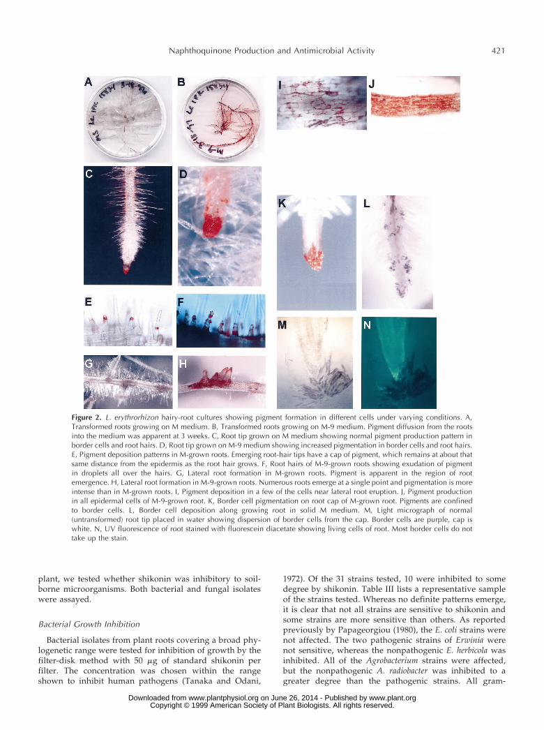
L. erythrorhizon hairy-root cultures grown on solid Mmedium in the dark at 24°C appeared white (Fig. 2A).Roots grew at a rate of 1 to 2 mm/d. Under magnificationpigmentation was apparent in two cell types within theseroots: the root hairs (Fig. 2, C and E) and the root bordercells (Hawes and Brigham, 1992; Brigham et al., 1995;Hawes et al., 1998) (Fig. 2, C and K). Pigment granules inthe root hairs were seen at the apex of the root hairs as theyemerged (Fig. 2E). Pigmentation was not apparent beforethe hair had attained a length of at least 1 mm. As the roothair elongated, the pigment appeared to remain at thesame distance from the epidermal surface as when it wasfirst formed. The pigment was limited to a very specificregion and appeared to be a ring, perhaps because thevacuole occurred at this region of the root hair. In theroot-cap region, pigments were confined to the border cellsalone (Fig. 2, K and M). Removing the border cells from thecap revealed that the pigment was formed in the bordercells subsequent to separation from the cap, because thecap cells themselves were completely devoid of pigment.Border cells were left behind as the root grew through themedium and, in some cases, appeared to outline the root(Fig. 2L). When border cells were visualized under lightmicroscopy in M medium, they appeared deep purple, as
opposed to the white color of the underlying cap cells (Fig.2M). Cells of the cap take up the vital stain fluorescencediacetate, whereas those of the detached border cells do not(Fig. 2N).
Border cells with pigment also displayed less distinctcytoplasmic characteristics and failed to exhibit cytoplas-mic streaming. Plasmolysis was often evident and in thesecases pigmentation was confined to the region of the cyto-plasm and the cell walls appeared clear (data not shown).The pigments appeared red when roots were grown in air,away from contact with the medium (Fig. 2K), and deepblue to purple when the cells of the roots were in contactwith the medium (Fig. 2L). Shikonin derivatives chelateiron in the medium, turning the compounds purple in theprocess (L.A. Brigham, unpublished data). As the rootsaged, pigmentation occurred at the points of lateral rootemergence (Fig. 2G). Higher magnification revealed pig-ment granules that tended to accumulate in the cell walls(Fig. 2I). Shikonin production was also seen in the oldestparts of the root. The exact cellular location of the pigmentwas not obvious from visual inspection, because the olderroot segments also developed a brown coloration, probablyas a result of the production of other phenolic compounds.Comparable patterns of red pigment formation were ob-served in roots grown in pots in the greenhouse. Purplepigmentation was never observed in these roots.
Induction of Shikonin Derivatives
Roots grown on the “inducing” medium (M-9) formedpigments sooner, in greatly increased amounts, and inmore cell types than those grown on the noninducingmedium (M) described above. L. erythrorhizon hairy-rootcultures grown in the dark on M-9 medium at 24°C ap-peared fully pigmented, and the pigments had diffusedinto the medium, giving it a pinkish color (Fig. 2B). Theroot cap appeared to have greatly increased pigmentationand the border cells were not as distinct, probably becauseof increased mucilage production (Fig. 2D). The root hairsappeared to contain more pigment than the noninducedhairs, and the pigment was distributed throughout the cell(Fig. 2F). The amount of pigment production was increasedto the point that it was exuded from the root hair andappeared in droplets on the surface of the hair. Lateral rootformation occurred at shorter intervals than in the controls,and clusters of lateral roots were commonly observedemerging from a single site with greatly increased pigmentproduction (Fig. 2H). In contrast to the noninduced state, inwhich these were the only two cell types that producedpigment, in the induced state pigment was produced in allof the epidermal cells of the root much earlier and beforeother phenolic products had turned the roots brown (Fig.2J). The pigments appeared to accumulate in the cell walland were seen throughout the apoplast.
Effects of Shikonin on Bacterial and Fungal Growth
Because shikonin has been used as an antimicrobialagent in human applications, and because shikonin and itsderivatives are produced exclusively by the root of the
420 Brigham et al. Plant Physiol. Vol. 119, 1999
www.plant.org on June 26, 2014 - Published by www.plantphysiol.orgDownloaded from Copyright © 1999 American Society of Plant Biologists. All rights reserved.
plant, we tested whether shikonin was inhibitory to soil-borne microorganisms. Both bacterial and fungal isolateswere assayed.
Bacterial Growth Inhibition
Bacterial isolates from plant roots covering a broad phy-logenetic range were tested for inhibition of growth by thefilter-disk method with 50 mg of standard shikonin perfilter. The concentration was chosen within the rangeshown to inhibit human pathogens (Tanaka and Odani,
1972). Of the 31 strains tested, 10 were inhibited to somedegree by shikonin. Table III lists a representative sampleof the strains tested. Whereas no definite patterns emerge,it is clear that not all strains are sensitive to shikonin andsome strains are more sensitive than others. As reportedpreviously by Papageorgiou (1980), the E. coli strains werenot affected. The two pathogenic strains of Erwinia werenot sensitive, whereas the nonpathogenic E. herbicola wasinhibited. All of the Agrobacterium strains were affected,but the nonpathogenic A. radiobacter was inhibited to agreater degree than the pathogenic strains. All gram-
Figure 2. L. erythrorhizon hairy-root cultures showing pigment formation in different cells under varying conditions. A,Transformed roots growing on M medium. B, Transformed roots growing on M-9 medium. Pigment diffusion from the rootsinto the medium was apparent at 3 weeks. C, Root tip grown on M medium showing normal pigment production pattern inborder cells and root hairs. D, Root tip grown on M-9 medium showing increased pigmentation in border cells and root hairs.E, Pigment deposition patterns in M-grown roots. Emerging root-hair tips have a cap of pigment, which remains at about thatsame distance from the epidermis as the root hair grows. F, Root hairs of M-9-grown roots showing exudation of pigmentin droplets all over the hairs. G, Lateral root formation in M-grown roots. Pigment is apparent in the region of rootemergence. H, Lateral root formation in M-9-grown roots. Numerous roots emerge at a single point and pigmentation is moreintense than in M-grown roots. I, Pigment deposition in a few of the cells near lateral root eruption. J, Pigment productionin all epidermal cells of M-9-grown root. K, Border cell pigmentation on root cap of M-grown root. Pigments are confinedto border cells. L, Border cell deposition along growing root in solid M medium. M, Light micrograph of normal(untransformed) root tip placed in water showing dispersion of border cells from the cap. Border cells are purple, cap iswhite. N, UV fluorescence of root stained with fluorescein diacetate showing living cells of root. Most border cells do nottake up the stain.
Naphthoquinone Production and Antimicrobial Activity 421
www.plant.org on June 26, 2014 - Published by www.plantphysiol.orgDownloaded from Copyright © 1999 American Society of Plant Biologists. All rights reserved.

positive strains tested were inhibited. In summary, shiko-nin shows selective inhibition to some bacterial strains,including important root pathogens.
Fungal Growth Inhibition and Morphological Changes
Hyphal growth inhibition was tested by a linear growthassay. Twelve fungal isolates were tested and showed awide range of growth responses on medium containing 5,50, 100, and 200 mg/mL shikonin standard. Growth wasmeasured for each isolate until the hyphae of any treatmentreached the edge of the 35-mm plate. The length of time forthe control to reach the edge of the plate varied from 1 d(Pythium aphanidermatum) to 9 d (Colletotrichum destructi-vum). Figure 3 illustrates four different growth patterns.Pythium ultimum showed increasing sensitivity to increas-ing amounts of shikonin (Fig. 3A). P. aphanidermatum wasincreasingly inhibited by increasing concentrations ofshikonin until the highest concentration (200 mg/mL), atwhich point it showed a slight increase in growth over the100 mg/mL concentration (Student’s t test, P 5 0.06; Fig.3B). Nectria hematococca 34-18 showed little inhibition ofhyphal growth even at the highest shikonin concentration(Fig. 3C). Phytophthora parasitica grew faster than the con-trol at 5 and 50 mg/mL (Student’s t test, P 5 0.01; Fig. 3D).Comparisons of growth patterns among all of the fungitested were made by calculation of hyphal growth on eachconcentration of shikonin as a percentage of the growth ofthe control (Table IV).
In addition to the variability of the effect of shikonin onhyphal growth rates, other changes were observed. Somefungal isolates changed the color of the medium containingshikonin from pink to varying shades of purple and blue,
with each isolate producing a slightly different hue (datanot shown). It is known that some fungi are capable ofchanging the pH of their surroundings, but pH measure-ments were not undertaken in this study. The hyphae ofRhizoctonia solani formed fairly uniform sheets over thecontrol plates (Fig. 4A). However, at increasing concentra-tions of shikonin, the hyphal formations exhibited clump-ing and apparent sequestration of pigment within theclumps (Fig. 4, B and C). Aspergillus niger, which was onlymoderately inhibited by shikonin, showed significantchanges in both morphology and pigment formation in itsspores. Spores formed on plates with 5 mg/mL (Fig. 4D)were darker than the controls, and had a dumbbell shapesimilar to the controls, but with increasing amounts ofshikonin, the spores showed less pigmentation. The sporesformed on plates with 200 mg/mL shikonin (Fig. 4E) werebeige and rounded. Glomus intraradices was not tested forgrowth inhibition because it was not known at the time ifthe obligate mycorrhizal fungus would infect L. erythrorhi-zon. However, co-cultivation of hairy roots of carrot, G.intraradices (Chabot et al., 1992), and L. erythrorhizonshowed that this fungus also sequestered the pigments.Red pigment could be seen moving within the hyphae and,over several weeks, the pigments were deposited on theoutside of a small percentage (less than 5%) of the fungalspores (Fig. 4F). Thus, as in the case of bacterial strainstested, fungal isolates exhibited differential sensitivity toshikonin. Several known pathogenic soil-borne fungi suchas Rhizoctonia, Pythium, and Phytophthora were significantlyinhibited by shikonin. In addition, morphological changeswere often observed in hyphal growth patterns and sporeformation.
Table III. Inhibition of bacterial isolates to shikonin, shikonin derivatives, and whole-root chloroform extractionsData are presented as the size of the zone of inhibition. In all cases, SD was less than 0.289.
Bacterial IsolateStandards Root Culture Extractiona
Shikonin DS IBS DMAS IVS MBS AS M M-9 IVS/MBS AS HIVS
mm
B. subtilis 2.0 1.0 0.5 0.0 0.5 1.0 3.5 2.5 4.0 2.0 4.0 4.0B. thuringiensis 2.0 0.5 0.0 0.0 0.1 0.1 2.0 2.5 ndb 0.0 2.0 3.0C. michigenensis subsp. nebraskensis 4.0 4.0 1.0 1.0 0.5 1.5 4.0 4.0 2.0 1.0 3.0 4.0A. radiobacter 3.0 1.0 0.5 0.0 0.0 1.0 3.0 3.0 4.0 1.0 2.0 2.0A. rhizogenes 1.0 0.1 0.1 0.1 0.1 0.5 1.5 1.5 2.0 1.0 1.5 2.0A. tumefaciens 0.5 0.0 0.1 2.0 0.1 0.1 1.5 3.0 3.0 2.0 2.0 3.0B. cepacea 2.0 2.0 2.0 0.0 0.5 0.5 2.0 3.0 4.0 0.5 2.0 3.0E. coli 0.0 0.0 0.0 0.0 0.0 0.0 0.0 0.0 0.0 0.0 0.0 0.0E. amylovora 0.0 0.0 0.0 0.0 0.0 0.0 0.0 0.0 2.0 0.0 0.0 0.0E. carotovora 0.0 0.0 0.0 0.0 0.0 0.0 0.0 0.0 1.0 0.0 0.0 0.0E. herbicola 1.5 0.5 0.1 0.1 0.1 0.5 2.0 2.0 2.0 0.5 2.0 3.0P. aureofaciens 0.0 0.0 0.0 0.0 0.0 0.0 0.0 0.0 nd nd nd ndP. fluorescens 0.0 0.0 0.0 0.0 0.0 0.0 0.0 0.0 nd nd nd ndP. syringae 0.0 0.0 0.0 0.0 0.0 0.0 0.0 0.0 0.0 0.0 0.0 0.0P. syringae pv phaseolicola 0.0 0.0 0.0 0.0 0.0 0.0 0.0 0.0 0.0 0.0 0.0 0.0R. solanacearum 1.0 1.0 0.1 0.1 0.1 0.5 3.0 3.0 2.0 0.5 2.0 2.0S. marsecens 1.0 0.1 0.1 0.0 0.1 0.1 3 2.0 nd nd nd ndX. campestris pv pelargonii 0.0 0.0 0.0 0.0 0.0 0.0 0.0 0.0 0.0 0.0 0.0 0.0
a Chloroform extraction of pigments from roots grown in liquid culture. Separation of components by TLC as described in “Materials andMethods.” b nd, Not determined.
422 Brigham et al. Plant Physiol. Vol. 119, 1999
www.plant.org on June 26, 2014 - Published by www.plantphysiol.orgDownloaded from Copyright © 1999 American Society of Plant Biologists. All rights reserved.

Production of Shikonin Derivatives
The production of different shikonin derivatives in rootssold commercially, in cell-suspension cultures, and in cal-lus cultures has been analyzed by several researchers inChina and Japan (Table I). Different subsets of the knownshikonin derivatives are found in different species of Litho-spermum, in different sources of individual species, and indifferent cell lines. Not all of the known compounds arefound in a single plant or laboratory root culture. However,AS and HIVS were found in all samples in these studies.We wanted to determine if changes occurred in the pro-duction of derivatives under different conditions, includ-ing fungal challenge. All experiments were done on rootcultures that originated from a single transformation eventand were therefore genetically identical. Hairy-root cul-tures have been used as a tissue-culture tool for studies ofsecondary metabolite production in roots and have been
found by numerous studies to “remain more or less stablein their growth and/or secondary metabolite productivitycharacteristics for a number of years” (Hamill and Lidgett,1997). Roots were grown in liquid cultures of M or M-9medium and challenged with R. solani elicitor preparations.Roots grown in M medium remained white as long as themedium was changed frequently. Roots grown in un-changed (depleted) medium eventually accumulatedshikonin derivatives. As shown in Table V, roots grown inM-9 medium formed shikonin derivatives earlier and ingreater quantities than those grown in M medium, and theratios of HIVS to AS varied. HIVS and AS were detected inthe greatest abundance in all of the roots sampled, al-though the relative proportions varied by treatment. IVSand MBS were not readily distinguishable using HPLCunder the conditions we used, but one or both were de-tected in most cultures in the 10% to 15% range. Challengewith R. solani crude elicitor caused a reversal of the ratiosof HIVS and AS compared with treatments on M medium.Shikonin derivatives from plants grown in pots in thegreenhouse were analyzed and found to contain equalamounts (38% each) of HIVS and AS, 14% of IVS/MBS, anda small amount (2%) of DS. Shikonin was not detected inany of the samples analyzed.
Effect of Shikonin Derivatives on Bacterial Growth
Because shikonin itself was shown to have an effect onspecific bacterial and fungal soil isolates, further tests weredone to determine their sensitivity to the shikonin deriva-tives and to the combinations of derivatives produced bythe roots. As has been reported previously (Tanaka andOdani, 1972; Tabata et al., 1975), E. coli was not sensitive toshikonin or any of its derivatives. All organisms that wereinsensitive to shikonin were also insensitive to all of thederivatives tested, including the crude whole-root chloro-form extract (Table III). However, the two Erwinia speciesthat were insensitive to shikonin were insensitive to thederivatives, but were inhibited by an unknown componentof the induced chloroform extract. In most cases, inducedextract (M-9) produced the highest inhibition, followed bynoninduced extracts. AS, HIVS, and shikonin were often asinhibitory as the whole-extract preparations and were themost-active single compounds. IBS and MBS were foundthe least inhibitory in most cases. As in the case of thefungal studies on shikonin, color changes in the bacterialplates were varied (data not shown), but pH was notdetermined. In summary, bacterial isolates showed a widerange of responses to shikonin derivatives singly and incombination. Most isolates were inhibited to the greatestdegree by the two derivatives produced in highest abun-dance by the root cultures.
In Situ Fungal Inhibition and Plant Response to Fungi
To analyze the dynamics of shikonin production in rootcultures on fungi, we placed plugs of fungal hyphae on4-week-old L. erythrorhizon root cultures on solid M andM-9 medium. One-half of them were grown in the light (toinhibit shikonin derivative production) and one-half were
Figure 3. Fungal growth on medium containing no shikonin (control,l), 5 mg/mL shikonin (f), 50 mg/mL shikonin (Œ), 100 mg/mLshikonin (3), or 200 mg/mL shikonin (✳). A, P. ultimum. B, P.aphanidermatum. C, N. hematococca 34-18. D, P. parasitica.
Naphthoquinone Production and Antimicrobial Activity 423
www.plant.org on June 26, 2014 - Published by www.plantphysiol.orgDownloaded from Copyright © 1999 American Society of Plant Biologists. All rights reserved.

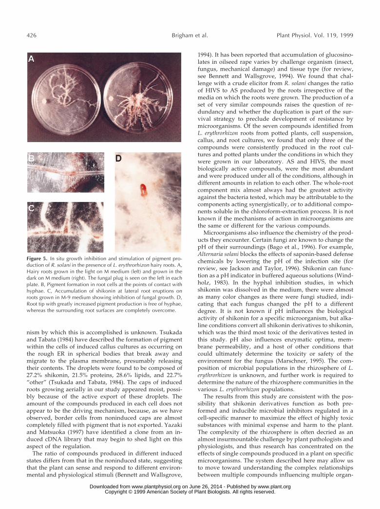
grown in the dark. Four isolates were chosen based ongrowth rates and response to the shikonin concentrationassays (Table VI). The amount of shikonin derivatives thefungi were exposed to on the plates was not quantified.Pigment was apparent upon visual inspection in the rootsgrown on M-9 at this point. P. aphanidermatum was sensi-tive to both the existing shikonin derivatives on the M-9plates and to increased shikonin derivative produced in thedark on both the M and M-9 plates. On the light-grownplates, shikonin production was inhibited and hyphalgrowth was twice that seen on the dark-grown M plates(Table VI; Fig. 5A). R. solani, which is very sensitive to highconcentrations of shikonin (Table VI), was inhibited on theM-9 plates, but not significantly on the M plates, eventhough the fungus did induce pigment formation in theseroots. N. hematococca T488, which is sensitive to shikonin atrelatively low concentrations, showed marked inhibitionon dark-grown plates whether preinduced or not. A. niger,the least-sensitive and slowest-growing isolate, showed nosignificant difference between light- and dark-grownplates, preinduced or not. This was the only one of the fourisolates tested that did not induce localized pigment pro-duction in the roots (Table VI). Figure 5B shows an exam-ple of a region of hyphal contact with the root. The cells inthe region of contact show considerable pigmentation. Ar-eas normally pigmented, such as sites of lateral root emer-gence, had fewer hyphae than regions in between (Fig. 5C).However, all regions where hyphae made contact eventu-ally produced pigment. Whereas most of the roots on theplate were eventually overcome by hyphal growth, the roottips were always devoid of hyphae. These tips, like thosegrown on M-9 medium, were moist and very red, showingincreased pigment production (Fig. 5D). Whereas root epi-dermal cells in direct contact with hyphae showed in-creased pigment production compared with adjacent non-contact cells, the root caps showed increased productionwithout direct hyphal contact.
Pigment production in root cultures was also inducedwith crude preparations of R. solani elicitor. Liquid cultures
of L. erythrorhizon hairy roots were grown for 1 month inliquid M medium. One set of flasks received fresh M me-dium, and the other set received M-9 medium. Root cul-tures were allowed to grow for 1 d, then 2.5 mL (per 50 mLof medium) of fungal elicitor was added to one-half of theM flasks and one-half of the M-9 flasks. Pigment releasedinto the media was evident visually at the end of 2 weeks.The media were collected at 3 weeks and the shikoninderivatives were extracted in chloroform and analyzed byHPLC. The roots treated with a combination of both M-9
Figure 4. Changes in fungal morphology when grown on mediumcontaining shikonin. A, R. solani hyphal growth on control plateshowing an even growth pattern. B, R. solani hyphal growth on 100mg/mL shikonin showing clumping of hyphae. C, R. solani hyphalgrowth on 200 mg/mL shikonin showing sequestration of shikoninpigment in the hyphal clumps. D, Spores of A. niger grown on 5mg/mL shikonin. Spores are dark and dumbbell shaped. E, Spores ofA. niger grown on 200 mg/mL shikonin. Spores are beige and round.F, Spores of G. intraradices grown on M medium in the presence ofL. erythrorhizon hairy roots. Pigment is not apparent in the mediumby visual inspection. Pigment is transported through the hyphae anddeposited on a small fraction of the spores in any one area.
Table IV. Fungal growth on five different concentrations of shikonin (in mg/mL) as a percentage ofgrowth of the control
Data are presented as the percentage of growth compared with the control, and represent theaverages of three separate experiments, each with three replicates. The coefficients of variation (V 5[s 3 100]/X; V, coefficient of variation; s, SD; X, mean) ranged from 2% for the controls to 16% for thetreatments in which growth was most strongly inhibited.
Fungi 0 5 50 100 200
%P. aphanidermatum 100 100 60 16 28P. ultimum 100 88 60 40 16Monosporascus cannonballus 100 83 111 111 94F. oxysporum 100 109 100 87 48F. proliferata 100 100 92 72 52C. destructivum 100 96 96 62 42R. solani 100 100 60 16 0A. niger 100 104 92 83 75N. haematococca 34-18 100 104 100 96 87N. haematococca T488 100 96 85 73 62P. parasitica 100 120 130 70 25P. capsicii 100 104 80 60 28
424 Brigham et al. Plant Physiol. Vol. 119, 1999
www.plant.org on June 26, 2014 - Published by www.plantphysiol.orgDownloaded from Copyright © 1999 American Society of Plant Biologists. All rights reserved.

and fungal elicitor showed the strongest response in totalpigment produced (Table V). The roots treated with fungalelicitor in the inducing medium produced 3 times as muchshikonin as those with fungal elicitor in M medium andalmost 30 times as much as the noninduced roots. Theseresults are consistent with the model that shikonin deriv-atives in the roots function as both preformed and induc-ible antimicrobial compounds.
DISCUSSION
The transformed roots of L. erythrorhizon appear to be auseful model in which to study the production and poten-tial function(s) of a suite of antimicrobial compounds inplant roots (Rhodes et al., 1997). The derivatives of shiko-nin are produced in a highly regulated and visible mannerin specific cell types of the growing root tip. The com-pounds have varying degrees of biological activity againsta wide range of soil-borne microorganisms. Productionof these compounds in changing ratios within the rootvaries with conditions of stress, including challenge bymicroorganisms.
The cellular pattern of shikonin derivative productiondepends on the developmental stage of the root and onenvironmental signals. In relatively “unstressed” root cul-tures pigment production is normally evident only in twocell types, the root hairs and border cells. The identificationof pigments in these cells often requires magnification. Asthe radicle of L. erythrorhizon emerges from the seed, theroot border cells are already pigmented, and as the rootelongates, they remain at intervals along the root as a
potential chemical barrier to microbial infection (Hawesand Brigham, 1992; Hawes et al., 1998). Even under un-stressed conditions, the border cells in the root cultures areintensely pigmented, showing granules distributedthroughout the cell (Tsukada and Tabata, 1984). The un-derlying cap cells are devoid of pigment. From observationin this study, it appears to be a rapid development, becauseno intermediate states of pigment production have beenobserved in border cells. The dynamics of the developmentof these compounds in border cells remains to be studied.The pigment in the root hairs appears to be localized to aregion of the hair between the epidermal surface and themidpoint of the hair. The exact cellular location and themechanism are unknown. The uniform placement of pig-ments in the root hairs at some distance from the rootsurface may also serve as a chemical barrier to microbialinvasion. Preformed inhibitors are often distributed inplants in a tissue-specific manner, often in the outer layersof cells of plant organs (Osbourn, 1996). But, unlike manyof the preformed inhibitors studied to date, the shikoninderivatives, in addition to being formed as part of normaldevelopment, are highly inducible.
In the unstressed roots described above, the pigmentsappeared to remain within the cells in which they wereproduced. In contrast, when the roots were specificallystressed by the addition of copper sulfate, the root hairsmade significantly more pigments and the epidermal cellsproduced amounts sufficient to turn the roots “red.” Inboth these of cell types, the material is clearly extracellular.In root hairs, droplets formed on the surface; in epidermalcells, pigment accumulated in the apoplast. The mecha-
Table V. Relative ratios of shikonin and derivatives produced under different growth andelicitation conditions
Media HIVS AS DS IBS DMAS IVS/MBS Total Shikonina
mg/gb
M 53.91 36.59 1.07 0.68 9.75 12M-9 54.96 20.63 15.18 19M-Rsc 37.21 40.60 124M-9-Rs 23.56 52.45 0.70 1.16 16.33 334Nd 38.87 38.70 2.017 13.90a Total chloform extraction was subjected to 2.5 KOH and A520 was read. b Fresh weight of
roots. c M-Rs, R. solani elicitor. d N, Normal roots from pot-grown plants.
Table VI. Fungal growth on hairy-root culturesData are from a single experiment with two replicates. The whole experiment was performed two
times with similar results.
FungiGrowth
Rate
Hyphal Growth
InductionbM M-9
Dark Light Dark Light
d a mmP. aphanidermatum 1 31 57 33 43 YR. solani 3 55 60 40 35 YN. haematococca 7 9 20 9 27 YA. niger 9 12 10 12 13 Na Days to reach the opposite side of a 35-mm plate. b Induction of shikonin derivatives in the root
(Y, yes; N, no).
Naphthoquinone Production and Antimicrobial Activity 425
www.plant.org on June 26, 2014 - Published by www.plantphysiol.orgDownloaded from Copyright © 1999 American Society of Plant Biologists. All rights reserved.
nism by which this is accomplished is unknown. Tsukadaand Tabata (1984) have described the formation of pigmentwithin the cells of induced callus cultures as occurring onthe rough ER in spherical bodies that break away andmigrate to the plasma membrane, presumably releasingtheir contents. The droplets were found to be composed of27.2% shikonin, 21.5% proteins, 28.6% lipids, and 22.7%“other” (Tsukada and Tabata, 1984). The caps of inducedroots growing aerially in our study appeared moist, possi-bly because of the active export of these droplets. Theamount of the compounds produced in each cell does notappear to be the driving mechanism, because, as we haveobserved, border cells from noninduced caps are almostcompletely filled with pigment that is not exported. Yazakiand Matsuoka (1997) have identified a clone from an in-duced cDNA library that may begin to shed light on thisaspect of the regulation.
The ratio of compounds produced in different inducedstates differs from that in the noninduced state, suggestingthat the plant can sense and respond to different environ-mental and physiological stimuli (Bennett and Wallsgrove,
1994). It has been reported that accumulation of glucosino-lates in oilseed rape varies by challenge organism (insect,fungus, mechanical damage) and tissue type (for review,see Bennett and Wallsgrove, 1994). We found that chal-lenge with a crude elicitor from R. solani changes the ratioof HIVS to AS produced by the roots irrespective of themedia on which the roots were grown. The production of aset of very similar compounds raises the question of re-dundancy and whether the duplication is part of the sur-vival strategy to preclude development of resistance bymicroorganisms. Of the seven compounds identified fromL. erythrorhizon roots from potted plants, cell suspension,callus, and root cultures, we found that only three of thecompounds were consistently produced in the root cul-tures and potted plants under the conditions in which theywere grown in our laboratory. AS and HIVS, the mostbiologically active compounds, were the most abundantand were produced under all of the conditions, although indifferent amounts in relation to each other. The whole-rootcomponent mix almost always had the greatest activityagainst the bacteria tested, which may be attributable to thecomponents acting synergistically, or to additional compo-nents soluble in the chloroform-extraction process. It is notknown if the mechanisms of action in microorganisms arethe same or different for the various compounds.
Microorganisms also influence the chemistry of the prod-ucts they encounter. Certain fungi are known to change thepH of their surroundings (Bago et al., 1996). For example,Alternaria solani blocks the effects of saponin-based defensechemicals by lowering the pH of the infection site (forreview, see Jackson and Taylor, 1996). Shikonin can func-tion as a pH indicator in buffered aqueous solutions (Wind-holz, 1983). In the hyphal inhibition studies, in whichshikonin was dissolved in the medium, there were almostas many color changes as there were fungi studied, indi-cating that each fungus changed the pH to a differentdegree. It is not known if pH influences the biologicalactivity of shikonin for a specific microorganism, but alka-line conditions convert all shikonin derivatives to shikonin,which was the third most toxic of the derivatives tested inthis study. pH also influences enzymatic optima, mem-brane permeability, and a host of other conditions thatcould ultimately determine the toxicity or safety of theenvironment for the fungus (Marschner, 1995). The com-position of microbial populations in the rhizosphere of L.erythrorhizon is unknown, and further work is required todetermine the nature of the rhizosphere communities in thevarious L. erythrorhizon populations.
The results from this study are consistent with the pos-sibility that shikonin derivatives function as both pre-formed and inducible microbial inhibitors regulated in acell-specific manner to maximize the effect of highly toxicsubstances with minimal expense and harm to the plant.The complexity of the rhizosphere is often decried as analmost insurmountable challenge by plant pathologists andphysiologists, and thus research has concentrated on theeffects of single compounds produced in a plant on specificmicroorganisms. The system described here may allow usto move toward understanding the complex relationshipsbetween multiple compounds influencing multiple organ-
Figure 5. In situ growth inhibition and stimulation of pigment pro-duction of R. solani in the presence of L. erythrorhizon hairy roots. A,Hairy roots grown in the light on M medium (left) and grown in thedark on M medium (right). The fungal plug is seen on the left in eachplate. B, Pigment formation in root cells at the points of contact withhyphae. C, Accumulation of shikonin at lateral root eruptions onroots grown in M-9 medium showing inhibition of fungal growth. D,Root tip with greatly increased pigment production is free of hyphae,whereas the surrounding root surfaces are completely overcome.
426 Brigham et al. Plant Physiol. Vol. 119, 1999
www.plant.org on June 26, 2014 - Published by www.plantphysiol.orgDownloaded from Copyright © 1999 American Society of Plant Biologists. All rights reserved.
isms in the rhizosphere. Because the shikonin derivativesare produced from two pathways under intense investiga-tion because of their contribution to antimicrobial prod-ucts, this system may add to our understanding of how theplant regulates these pathways in subtle ways to influencemicrobial population dynamics in the rhizosphere. Studiesare under way to examine the molecular mechanisms ofcell-specific expression, regulation of compound ratios,and microbial sensitivity/tolerance mechanisms.
ACKNOWLEDGMENTS
We thank Drs. Leland S. Pierson and Derek Wood (University ofArizona, Tucson) for providing the bacterial strains used in theseexperiments, Dr. Hans Vanetten and Scott Rasmussen (Universityof Arizona) for providing most of the fungal isolates, Dr. MarthaHawes for providing laboratory space for the experiments per-formed at the University of Arizona, Tom Orum (University ofArizona) for providing assistance in the statistical analyses, andAnthony Omeis (Biology Greenhouse at The Pennsylvania StateUniversity) for propagating the L. erythrorhizon plants and caringfor them in the greenhouse. The carrot hairy-root cultures were agift from Dr. Roger Koide of The Pennsylvania State University.Dr. Gary Moorman graciously provided a critical reading of themanuscript, and Dr. Kazufumi Yazaki (Kyoto University, Japan)provided invaluable background information, discussion, and ad-vice for this research.
Received May 26, 1998; accepted October 22, 1998.
LITERATURE CITED
Bago B, Vierheilig H, Piche Y, Azcon-Aguilar C (1996) Nitratedepletion and pH changes induced by the extraradical myce-lium of the arbuscular mycorrhizal fungus Glomus intraradicesgrown in monoxenic culture. New Phytol 133: 273–280
Ballantine JA (1969) The isolation of two esters of the naphtho-quinone alcohol, shikonin, from the shrub Jatropha glandulifera.Phytochemistry 8: 1587–1590
Becard G, Fortin JA (1988) Early events of vesicular-arbuscularmycorrhiza formation on Ri T-DNA transformed roots. NewPhytol 108: 211–218
Bennett RN, Wallsgrove RM (1994) Secondary metabolites inplant defense mechanisms. New Phytol 127: 617–633
Binder RG, Benson ME, Flath RA (1989) Eight 1,4-naphtho-quinones from Juglans. Phytochemistry 28: 2799–2801
Brigham LA, Woo HH, Hawes MC (1995) Root border cells astools in plant cell biology studies. Methods Cell Biol 49: 377–387
Chabot S, Becard G, Piche Y (1992) Life cycle of Glomus intraradixin root organ culture. Mycologia 84: 315–321
Chappell J (1995) The biochemistry and molecular biology ofisoprenoid metabolism. Plant Physiol 107: 1–6
Dixon RA, Paiva NL (1995) Stress-induced phenylpropanoid me-tabolism. Plant Cell 7: 1085–1097
Flores H, Pickard J, Hoy M (1988) Production of polyacetylenesand thiophenes in heterotrophic and photosynthetic root cul-tures of Asteraceae. In J Lamb, H Breteler, T Arnason, L Hansen,eds, Chemistry and Biology of Naturally-Occurring Acetylenesand Related Compounds (NOARC). Elsevier, Amsterdam, pp233–254
Fujita Y, Hara Y, Ogino T, Suga C (1981a) Production of shikoninderivatives by cell suspension cultures of Lithospermum erythro-rhizon. I. Effects of nitrogen sources on the production of shiko-nin derivatives. Plant Cell Rep 1: 59–60
Fujita Y, Hara Y, Suga C, Morimoto T (1981b) Production ofshikonin derivatives by cell suspension cultures of Lithospermumerythrorhizon. II. A new medium for the production of shikoninderivatives. Plant Cell Rep 1: 61–63
Fujita N, Maeda Y, Suga C, Morimoto T (1983) Production ofshikonin derivatives by cell suspension cultures of Lithospermum
erythrorhizon. III. Comparison of shikonin derivatives of culturedcells and Ko-shikon. Plant Cell Rep 2: 192–193
Fujita N, Yoshida Y (1937) Ueber die Anatomie von Radix Litho-spermi. Yakugaku Zasshi 57: 368–391
Fukui H, Tani M, Tabata M (1990) Induction of shikonin biosyn-thesis by endogenous polysaccharides in Lithospermum erythro-rhizon cell suspension cultures. Plant Cell Rep 9: 73–76
Fukui H, Yoshikawa N, Tabata M (1983) Induction of shikoninformation by agar in Lithospermum cell suspension cultures.Phytochemistry 22: 2451–2453
Gaisser S, Heide L (1996) Inhibition and regulation of shikoninbiosynthesis in suspension cultures of Lithospermum. Phyto-chemistry 41: 1065–1072
Hamill JD, Lidgett AJ (1997) Hairy root cultures: opportunitiesand key protocols for studies in metabolic engineering. In PMDoran, ed, Hairy Roots: Culture and Applications. HarwoodAcademic, Sydney, Australia, pp 1–29
Hauska G (1988) Phylloquinone in photosystem I. Trends BiochemSci 13: 415–416
Hawes MC, Brigham LA (1992) Impact of root border cells onmicrobial populations in the rhizosphere. Adv Plant Pathol 8:119–148
Hawes MC, Brigham LA, Wen F, Woo HH, Zhu Y (1998) Functionof root border cells in plant health: pioneers in the rhizosphere.Annu Rev Phytopathol 36: 311–327
Herrmann KM (1995) The shikimate pathway as an entry to aro-matic secondary metabolism. Plant Physiol 107: 7–12
Jackson AO, Taylor CB (1996) Plant-microbe interactions: life anddeath at the interface. Plant Cell 8: 1651–1668
Jain AC, Mathur SK (1965) A chemical study of the pigment ofArnebia. Bull Natl Inst Sci India 28: 52–56
Kim DJ, Chang HN (1990) Increased shikonin production in Litho-spermum erythrorhizon suspension cultures with in situ extractionand fungal cell treatment (elicitor). Biotech Lett 12: 443–446
Kubo I, Klocke J (1986) Insect ecdysis inhibitors. In MB Green, PAHedin, eds, Natural Resistance of Plants to Pests. AmericanChemical Society, Washington, DC, pp 206–219
Kyogoku K, Terayama H, Tachi Y, Suzuki T, Komatsu M (1973)Constituents of shikon. II. Comparison of contents, constituents,and antibacterial effect of fat soluble fraction between nanshikonand koshikon. Shoyakugaku Zasshi 27: 31–36
Luria SE, Burrows JW (1957) Hybridization between Escherichiacoli and Shigella. J Bacteriol 74: 461–476
Marschner H (1995) Mineral Nutrition in Higher Plants, Ed 2.Academic Press, London, pp 537–595
Mizukami H, Konoshima M, Tabata M (1977) Effect of nutritionfactors on shikonin derivative formation in Lithospermum calluscultures. Phytochemistry 16: 1183–1186
Mizukami H, Konoshima M, Tabata M (1978) Variation in pig-ment production in Lithospermum erythrorhizon callus cultures.Phytochemistry 17: 95–97
Morimoto I, Hirata Y (1966) New naphthoquinone derivativesfrom Lithospermum erythrorhizon. Tetrahedron Lett 31: 3677–3680
Morimoto I, Kishi T, Ikegami S, Hirata Y (1965) Naphthoquinonederivatives from Lithospermum erythrorhizon Siebold et Zucca-rini. Tetrahedron Lett 52: 4737–4739
Murashige T, Skoog F (1962) A revised medium for rapid growthand bioassays with tissue cultures. Physiol Plant 15: 473–497
Osbourn AE (1996) Preformed anti-microbial compounds andplant defense against fungal attack. Plant Cell 8: 1821–1831
Papageorgiou V (1978) Wound healing properties of naphthoqui-none pigments from Alkanna tinctoria. Experientia 34: 1499–1501
Papageorgiou V (1980) Naturally occurring isohexenylnaphthaza-rin pigments: a new class of drugs. Planta Med 38: 193–203
Rhodes MJC (1994) Physiological roles for secondary metabolitesin plants: some progress, many outstanding problems. Plant MolBiol 24: 1–20
Rhodes MJC, Parr AJ, Walton NJ (1997) Studies of secondarymetabolic pathways in transformed roots. In PM Doran, ed,Hairy Roots: Culture and Applications. Harwood Academic,Sydney, Australia, pp 31–41
Schenk RU, Hildebrandt AC (1972) Medium and techniques for
Naphthoquinone Production and Antimicrobial Activity 427
www.plant.org on June 26, 2014 - Published by www.plantphysiol.orgDownloaded from Copyright © 1999 American Society of Plant Biologists. All rights reserved.
induction and growth of monocotyledonous and dicotyledon-ous plant cell cultures. Can J Bot 50: 199–204
Tabata M, Fujita Y (1985) Production of shikonin by plant cellcultures. In M Zaitlin, P Day, A Hollaender, eds, Biotechnologyin Plant Science. Academic Press, San Diego, CA, pp 207–218
Tabata M, Mizukami H, Haoe S, Konoshima M (1975) Anti-microbial activity of Lithospermum erythrorhizon callus cultures.Yakugaku Zasshi 95: 1376–1379
Tabata M, Mizukami H, Hiraoka N, Konoshima M (1974) Pig-ment formation in callus cultures of Lithospermum erythrorhizon.Phytochemistry 13: 927–932
Tanaka Y, Odani T (1972) Pharmacodynamic study on “shiunko.”I. Antibacterial effect of “shiunko.” Yakugaku Zasshi 95: 525–530
Tani M, Fukui H, Shimomura M, Tabata M (1992) Structure ofendogenous oligogalacturonides inducing shikonin biosynthesisin Lithospermum cell cultures. Phytochemistry 31: 2719–2723
Tani M, Takeda K, Yazaki K, Tabata M (1993) Effects of oligoga-lacturonides on biosynthesis of shikonin in Lithospermum cellcultures. Phytochemistry 34: 1285–1290
Tsukada M, Fukui H, Habara C, Tabata M (1983) Comparativestudies on naphthoquinone derivatives in various crude drugsof “zicao” (shikon). Shoyakugaku Zasshi 37: 299–305
Tsukada M, Tabata M (1984) Intracellular localization and secre-tion of naphthoquinone pigments in cell cultures of Lithosper-mum erythrorhizon. Planta Med 51: 338–341
Windholz M, ed (1983) The Merck Index: An Encyclopedia ofChemicals, Drugs, and Biologicals, Ed 9. Merck & Co., Rah-way, NJ
Yazaki K, Matsuoka H (1997) cDNA cloning and functional anal-ysis of LEDI-2, a gene preferentially expressed in the dark inLithospermum erythrorhizon cell suspension cultures (abstract no.1348). Plant Physiol 114: S-261
428 Brigham et al. Plant Physiol. Vol. 119, 1999
www.plant.org on June 26, 2014 - Published by www.plantphysiol.orgDownloaded from Copyright © 1999 American Society of Plant Biologists. All rights reserved.