
Cell penetrating elastin-like polypeptides for therapeutic peptide delivery
21
Cell Penetrating Elastin-like Polypeptides for Therapeutic Peptide Delivery Gene L. Bidwell III 1 and Drazen Raucher 2 1 University of Mississippi Medical Center, Department of Biochemistry, 2500 North State Street, Jackson, MS 39216, Phone: 601-984-1527, Fax: 601-984-1501, [email protected] Abstract Current treatment of solid tumors is limited by side effects that result from the nonspecific delivery of drugs to the tumor site. Alternative targeted therapeutic approaches for localized tumors would significantly reduce systemic toxicity. Peptide therapeutics are a promising new strategy for targeted cancer therapy because of the ease of peptide design and the specificity of peptides for their intracellular molecular targets. However, the utility of peptides is limited by their poor pharmacokinetic parameters and poor tissue and cellular membrane permeability in vivo. This review article summarizes the development of elastin-like polypeptide (ELP) as a potential carrier for thermally targeted delivery of therapeutic peptides (TP), and the use of cell penetrating peptides (CPP) to enhance the intracellular delivery of the ELP-fused TPs. CPP-fused ELPs have been used to deliver a peptide inhibitor of c-Myc function and a peptide mimetic of p21 in several cancer models in vitro, and both polypeptides are currently yielding promising results in in vivo models of breast and brain cancer. Keywords Elastin-like polypeptide; thermal targeting; therapeutic peptide; cell penetrating peptide; c-Myc; p21 1. Introduction Cancer is a complex disease and, as it progresses, it can become aggressive, manifested by the invasion of cells from the primary tumor to the liver, lungs, brain and other organs. Tumor cell metastasis is a major cause of death amongst cancer patients, and in order to prevent this process, it is necessary to effectively treat primary solid tumors. However, current treatment for solid tumors is limited by the fact that only a small percentage of the administered dose of drug reaches the tumor site, while the rest of the drug is distributed throughout the body, which leads to increased toxicity in normal tissues. Tumor tissues differ from normal tissues in anatomical and structural characteristics. They have a heterogeneous distribution of blood vessels and usually lack effective lymphatic drainage. These factors lead to an uneven and slowed blood flow and abnormal fluid © 2010 Elsevier B.V. All rights reserved. 2 corresponding author, University of Mississippi Medical Center, Department of Biochemistry, 2500 North State Street, Jackson, MS 39216, Phone: 601-984-1510, Fax: 601-984-1501, [email protected] . Publisher's Disclaimer: This is a PDF file of an unedited manuscript that has been accepted for publication. As a service to our customers we are providing this early version of the manuscript. The manuscript will undergo copyediting, typesetting, and review of the resulting proof before it is published in its final citable form. Please note that during the production process errors may be discovered which could affect the content, and all legal disclaimers that apply to the journal pertain. NIH Public Access Author Manuscript Adv Drug Deliv Rev. Author manuscript; available in PMC 2011 December 30. Published in final edited form as: Adv Drug Deliv Rev. 2010 December 30; 62(15): 1486–1496. doi:10.1016/j.addr.2010.05.003. NIH-PA Author Manuscript NIH-PA Author Manuscript NIH-PA Author Manuscript
-
Upload
mississippimedical -
Category
Documents
-
view
1 -
download
0
Transcript of Cell penetrating elastin-like polypeptides for therapeutic peptide delivery

Cell Penetrating Elastin-like Polypeptides for TherapeuticPeptide Delivery
Gene L. Bidwell III1 and Drazen Raucher2
1University of Mississippi Medical Center, Department of Biochemistry, 2500 North State Street,Jackson, MS 39216, Phone: 601-984-1527, Fax: 601-984-1501, [email protected]
AbstractCurrent treatment of solid tumors is limited by side effects that result from the nonspecificdelivery of drugs to the tumor site. Alternative targeted therapeutic approaches for localizedtumors would significantly reduce systemic toxicity. Peptide therapeutics are a promising newstrategy for targeted cancer therapy because of the ease of peptide design and the specificity ofpeptides for their intracellular molecular targets. However, the utility of peptides is limited by theirpoor pharmacokinetic parameters and poor tissue and cellular membrane permeability in vivo.This review article summarizes the development of elastin-like polypeptide (ELP) as a potentialcarrier for thermally targeted delivery of therapeutic peptides (TP), and the use of cell penetratingpeptides (CPP) to enhance the intracellular delivery of the ELP-fused TPs. CPP-fused ELPs havebeen used to deliver a peptide inhibitor of c-Myc function and a peptide mimetic of p21 in severalcancer models in vitro, and both polypeptides are currently yielding promising results in in vivomodels of breast and brain cancer.
KeywordsElastin-like polypeptide; thermal targeting; therapeutic peptide; cell penetrating peptide; c-Myc;p21
1. IntroductionCancer is a complex disease and, as it progresses, it can become aggressive, manifested bythe invasion of cells from the primary tumor to the liver, lungs, brain and other organs.Tumor cell metastasis is a major cause of death amongst cancer patients, and in order toprevent this process, it is necessary to effectively treat primary solid tumors. However,current treatment for solid tumors is limited by the fact that only a small percentage of theadministered dose of drug reaches the tumor site, while the rest of the drug is distributedthroughout the body, which leads to increased toxicity in normal tissues.
Tumor tissues differ from normal tissues in anatomical and structural characteristics. Theyhave a heterogeneous distribution of blood vessels and usually lack effective lymphaticdrainage. These factors lead to an uneven and slowed blood flow and abnormal fluid
© 2010 Elsevier B.V. All rights reserved.2corresponding author, University of Mississippi Medical Center, Department of Biochemistry, 2500 North State Street, Jackson, MS39216, Phone: 601-984-1510, Fax: 601-984-1501, [email protected] .Publisher's Disclaimer: This is a PDF file of an unedited manuscript that has been accepted for publication. As a service to ourcustomers we are providing this early version of the manuscript. The manuscript will undergo copyediting, typesetting, and review ofthe resulting proof before it is published in its final citable form. Please note that during the production process errors may bediscovered which could affect the content, and all legal disclaimers that apply to the journal pertain.
NIH Public AccessAuthor ManuscriptAdv Drug Deliv Rev. Author manuscript; available in PMC 2011 December 30.
Published in final edited form as:Adv Drug Deliv Rev. 2010 December 30; 62(15): 1486–1496. doi:10.1016/j.addr.2010.05.003.
NIH
-PA Author Manuscript
NIH
-PA Author Manuscript
NIH
-PA Author Manuscript

dynamics in the tumor tissue. As a result, macromolecules and soluble polymeric carrierspenetrate and accumulate preferentially in tumors relative to normal tissues. Thisphenomenon is called the enhanced permeability and retention (EPR) effect [1-3], and it isthe key to the clinical success of anti-cancer macromolecular carrier systems. With the goalsof increasing specificity and lowering systemic toxicity, many different systems such asmacromolecular prodrugs, liposomes, and micro- and nano-particles have been developed(reviewed in [4-6]) to treat solid tumors. Several natural and synthetic water-solublepolymers, such as N-(2-hydroxypropyl)methacrylamide (HPMA) copolymers, dextrans,poly(ethylene glycol) (PEG), and poly(l-glutamic acid) have been utilized successfully inclinical research or are in human clinical trials (reviewed in [3,7,8]). While clinically usefulanti-tumor activity has been achieved by exploiting the EPR effect and using passivetargeting by macromolecular drug delivery systems, further selectivity is possible by activetargeting.
Bioconjugation of targeting moieties, such as peptide sequences or antibodies which havespecific affinity to cancer cells, to the polymer backbone can further exploit differencesbetween cancer and normal cells through selective receptor-mediated endocytosis. Anotherway of active targeting can be achieved using thermally responsive biopolymers, such aselastin-like polypeptide (ELP), that undergo an inverse phase transition [9-11]. Whenintravenously delivered, these thermally responsive polypeptides are likely to be clearedunder physiological conditions (37 °C). However, they will aggregate and selectivelyaccumulate in tumors where externally induced focused heat (40-42 °C) is applied. Work inthe lab of Chilkoti using human tumors implanted in nude mice has clearly demonstratedthat hyperthermia of the tumor results in increased accumulation of ELP polypeptides[12-14]. This method combines the established advantages of macromolecular carriers withthe additional advantage of active thermal targeting, and it also introduces other synergisticeffects of hyperthermia treatment. Hyperthermia preferentially increases the permeability ofendothelial tumor vasculature to macromolecular drug carriers, which can further enhancethe delivery of drugs to tumors [15-17]. Furthermore, hyperthermia enhances thecytotoxicity of some chemotherapeutic agents [18]. The clinical application of hyperthermiato increase tissue temperatures to 40 - 43 °C has been integrated in multimodal anti-cancerstrategies [19-21], and due to substantial technical improvements, hyperthermia is becomingmore accepted clinically. Selected increase of temperatures in superficial and deep-seatedtumors is accomplished using microwave, radio-frequency, and high-intensity focusedultrasound. Therefore, the effect of hyperthermia combined with the therapeutic effect of apolymeric drug carrier might offer further synergistic advantages in treatment of localizedtumors.
In order to deliver therapeutic cargo to intracellular molecular targets and demonstratetherapeutic efficacy, the ELP carrier must successfully overcome transport barriers to drugdelivery that are posed by unique structural and physiological characteristics of tumors.Despite the beneficial EPR effect, which favors accumulation of macromolecular carriers intumors, there are many factors opposing the delivery of drugs to tumors. A solid tumor doesnot simply exist as a mass of malignant cells, but contains tumor cells, normal cells,extracellular matrix, and tumor blood vessels. Leaky tumor blood vessels are generally somedistance removed from target tumor cells, separated by stroma and other cell types. Tumorand stromal cells produce and assemble the extracellular matrix, which consists of ameshwork of collagens, proteoglycans, and other molecules that together representsignificant barriers to penetration by therapeutics agents [22,23]. Although the lack oflymphatic drainage reduces efflux of the drug away from the tumor, it also reducesredistribution and transport of the drug within the tumor by generating higher pressure in theinterstitium than in the vasculature, which makes macromolecular uptake less efficient [24].Furthermore, once macromolecular carriers reach the cancer cell, there is the additional
Bidwell and Raucher Page 2
Adv Drug Deliv Rev. Author manuscript; available in PMC 2011 December 30.
NIH
-PA Author Manuscript
NIH
-PA Author Manuscript
NIH
-PA Author Manuscript

obstacle of plasma membrane impermeability. The plasma membrane of eukaryotic cells isgenerally impermeable to therapeutic macromolecules such as oligonucleotides and proteinsdue to the large size and inherently poor penetration capabilities of these molecules. Cellpenetrating peptides (CPP) can be used to overcome these transport barriers to drug deliveryand permit noninvasive delivery of polypeptides to their appropriate intracellular moleculartarget.
CPPs are short peptides that can be cationic, amphipathic, or hydrophobic. These peptideshave the ability to efficiently cross cellular plasma membranes and enter the cytoplasm.Furthermore, when CPPs are linked to oligonucleotides, proteins, or nanoparticles, theyfacilitate the transport of these entities across the cell membrane [25-27]. Thus, a number ofinvestigators have assessed the use of CPPs for intracellular delivery of macromolecules(reviewed in [28,29]). CPPs have shown efficacy in vivo for delivery of macromolecules totumors and even across the blood brain barrier [30-34]. As discussed in this review, additionof CPPs to the ELP carrier may not only enhance its uptake into the tumor cells in vitro[35-38], but the CPPs also mediate escape of the polypeptide from the tumor vasculature andentry into the tumor cells.
In addition to physically targeting therapeutic agents to cancer cells, another method ofspecifically inhibiting cancer cell proliferation is to exploit their genetic abnormalities. Insome cancer types, cancer cells have overexpressed and/or permanently active oncogenes,causing hyperactive growth and division and/or protection against apoptosis. Some cancercells have inactivated or missing tumor suppressor genes, resulting in deregulation of theapoptotic pathway and loss of control over the cell cycle and DNA replication. Recentcharacterization of the genetic alterations that occur during carcinogenesis has identifiedmany potential molecular targets for which to develop new therapeutics. One of the majoradvantages of therapeutic peptides is that they are much easier to design using a rationalapproach than small molecule drugs for stimulation or inhibition of a given protein/proteininteraction. These peptides are derived from high-throughput screening or by using NMR orcrystal structures of their molecular target and further optimized by a rational drug designapproach. Such therapeutic peptides can be designed to bind almost any protein of interestwith high affinity and specificity and can interfere with molecular pathways that arederegulated in cancer cells [39,40]. The use of peptides to specifically inhibit aberrantoncogenic or tumor suppressor proteins should be more effective and have fewer side effectsthan current nonspecific cytotoxic drug treatments. However, the clinical efficacy oftherapeutic peptides is limited by pharmacodynamic properties. When applied in vivo,therapeutic peptides are rapidly degraded in circulation, and their relatively large size andoften charged nature makes them impermeable to cancer cell membranes [41,42]. Therefore,in order to advance therapeutic peptides into the clinical setting, a suitable carrier systemthat can overcome these limitations and target the peptide to the tumor site and into thetumor cells is needed.
Attention is being focused on peptide delivery using macromolecular carriers. Micro- andnanospheres are being investigated for their ability to deliver bioactive peptides via the oralroute, stabilizing and delivering them through absorption barriers in the gastrointestinal tract[43]. Liposome - peptide conjugates have been investigated, but the focus of this field is theconjugation of cell penetrating peptides to the surface of liposomes to enhance fusion withthe cell membrane [44]. To overcome limitations of other macromolecular carriers andimprove delivery of peptide therapeutics to solid tumors, our lab is working to develop anELP-based thermally targeted peptide vector. Such a carrier would have all of thecharacteristics and advantages of existing soluble macromolecular carriers, but it would bealso be capable of active targeting by application of local hyperthermia.
Bidwell and Raucher Page 3
Adv Drug Deliv Rev. Author manuscript; available in PMC 2011 December 30.
NIH
-PA Author Manuscript
NIH
-PA Author Manuscript
NIH
-PA Author Manuscript

ELP has been used for thermally targeted delivery of small molecule drugs [37,45-47],plasmid DNA [48], therapeutic peptides [35,36,38,49,50], and proteins [51] in variouscancer models. This review focuses on the use of thermally responsive polypeptide carriersfor hyperthermia targeted delivery of therapeutic peptides. In this article, we present asummary of results from our lab comparing cellular uptake mechanisms and efficiency ofseveral different CPP-fused ELPs and discuss their potential therapeutic utility. We alsodescribe use of ELP to deliver two therapeutic peptides, one that inhibits the function of theoncogenic transcription factor c-Myc [35,36] and one that mimics the Cdk inhibitor p21[38,50].
2. The use of cell penetrating peptides for intracellular delivery of ELPsThe ELP drug carrier used for thermal targeting (ELP1) has a molecular weight of 59.1 kDa[52], and it enters eukaryotic cells at low levels by an endocytic mechanism. In an attempt toimprove the efficiency of cellular internalization of ELP, we modified it at its N-terminuswith several CPPs (Figure 1A) [38]. Originally, we used three CPPs: the penetratin peptidefrom the Drosophila transcription factor Antennapedia [53], The Tat peptide from the HIV-1Tat protein [54], and the MTS (membrane translocating sequence) derived from Kaposifibroblast growth factor (Figure 1B) [55]. In more recent studies, we have also used the BacCPP derived from the bactenecin antimicrobial peptide [56].
2.1. Comparing the efficiency of various CPPs for intracellular delivery of ELPThe ability of each CPP to enhance the cellular uptake of ELP was assessed usingfluorescently labeled CPP-ELP polypeptides for flow cytometry and confocal microscopy.As shown in Figure 2A, each of the three CPPs produced brighter cell staining than theparent ELP polypeptide, and flow cytometry histograms of cell number versus fluorescenceintensity were unimodal, indicating that all cells were bound equally by the CPP-ELPs.When the flow cytometry data was quantified, it was determined that, of the three CPPstested, the penetratin peptide was by far the most efficient. At 30 M, the cellular association/uptake of the polypeptide was increased 1.7 fold for Tat-ELP, 2.6 fold for MTS-ELP, and14.8 fold for Pen-ELP relative to the ELP polypeptide lacking a CPP. The flow cytometryassay used can not directly distinguish polypeptide that has been internalized by the cellfrom polypeptide bound to the cell surface. Therefore, we used the membrane impermeabledye trypan blue to quench the fluorescence of surface bound polypeptide, and calculated thefraction internalized by by dividing the quenched (intracellular) fluorescence by theunquenched (intracellular and extracellular) fluorescence. This calculation allowsdetermination of the percentage of the total amount of polypeptide that is present inside thecell, but it does not give any indication of total polypeptide levels. Performing this assay atvarious time points after cellular exposure to the CPP-ELPs demonstrated that polypeptideinternalization did occur. About 20% of all CPP-ELPs were internalized at the end of a 1 htreatment and, at 24 h after treatment, 60% – 80% of the polypeptides were present insidethe cells (Figure 2C). All CPP-ELPs were internalized at a similar rate which did not differfrom that of the ELP control, indicating that all polypeptides were internalized by a similarmechanism. Internalization and subcellular localization was further confirmed by confocalfluorescence microscopy, which revealed a punctate cytoplasmic distribution for allpolypeptides 24 h after cellular exposure. Previous reports regarding the short CPP peptideshave indicated that this internalization and subcellular distribution can be an artifact of cellfixation [57], but that is not the case with CPP-ELPs, as live cells showed identicalinternalization and localization results [50]. In summary, the data in Figure 2A and Bdemonstrate the cellular levels of polypeptide achieved with the various CPPs, and the datain Figure 2C represents the percentage of that total that is inside the cell at the indicatedtime. Taken together, these data demonstrate that all polypeptides were internalized at a
Bidwell and Raucher Page 4
Adv Drug Deliv Rev. Author manuscript; available in PMC 2011 December 30.
NIH
-PA Author Manuscript
NIH
-PA Author Manuscript
NIH
-PA Author Manuscript

similar rate, which suggests that they are all internalized by a similar mechanism, but muchhigher levels of the CPP-ELPs were delivered into the cell relative to the ELP control.
2.2. The mechanism of CPP-ELP internalizationThe mechanism of CPP internalization has been the subject of much debate. Mechanismsranging from inverted micelles [58] to simple endocytosis [57,59] have been proposed, andit is clear that the mechanism is dependent on the cargo attached to the CPP (reviewed in[29,60]). Given the slow internalization rate observed for all the CPP-ELP polypeptides, wesuspected that a simple endocytosis mechanism was at work to internalize these largepolypeptides. To address this hypothesis, we employed several inhibitors of endocytosis inour flow cytometric cellular uptake assay. Incubation at 4 °C (Figure 3A) or ATP depletion(Figure 3B), both general inhibitors of endocytosis, significantly inhibited the cellularinternalization of all the CPP-ELPs tested. In addition, using hyperosmolar sucrose to blockthe formation of clathrin-coated pits [61] also caused a significant inhibition of polypeptideinternalization (Figure 3C). On the other hand, the use of methyl-β-cyclodextrin to depletethe membrane of cholesterol and block clathrin-independent endocytosis via caveolae [62]had very little effect on CPP-ELP internalization (Figure 3D). We concluded that ELP andCPP-ELP internalization occurs via a caveolae-independent endocytic mechanism.
2.3. The use of CPPs to deliver an ELP-fused TP to tumor cells in vivoPrevious studies have shown that ELP accumulation in tumor vasculature or interstitium canbe increased with focused hyperthermia [12-14]. The next step is to determine if the use ofCPPs allowed entry of the ELP carrier into the tumor cells, a property necessary foreffective drug delivery. To test the ability of the Bac and Tat CPPs to enhance ELP uptakeinto tumor cells in vivo, rats bearing two subcutaneous C6 tumors were intravenouslyinjected with Rhodamine-labeled Bac-ELP1-H1 or Tat-ELP1-H1 (CPP-ELPs with a c-Mycinhibitory peptide cargo, please section 3.3 see below). One tumor was heated above thepolypeptide’s transition temperature for 60 min by illumination with infrared (IR) light, andthe localization of the polypeptide in the tumor tissue was determined by fluorescencemicroscopy. As shown in Figure 4A, Bac-ELP1-H1 is present not only in the tumor bloodvessels, but has also escaped circulation and entered the tumor cells. The polypeptide wasalso able to escape the vasculature and enter the tumor cells when the tumor was heatedabove the Tt. Similarly, Tat-ELP1-H1 was also able to escape the tumor vasculature andenter the tumor cells (Figure 4B). Current work is underway to determine the levels of eachpolypeptide in the tumor both with and without hyperthermia treatment in order to determinethe optimal CPP for tumor delivery in vivo and to demonstrate the ability to thermally targetthe CPP-ELPs to the heated tumor.
3. ELP-based delivery of a c-Myc inhibitory peptidec-Myc is a transcription factor that, when bound to its heterodimerization partner Max,controls the expression of a large number of genes. Overexpression of c-Myc can causeuncontrolled cell proliferation and cancer [63]. A peptide inhibitor of c-Myc function wasfirst discovered by Draeger [64], who screened peptides derived from the helix-loop-helixand leucine zipper domains of c-Myc and Max for their ability to inhibit c-Myc binding toDNA. They found that a peptide from helix 1 (H1) of c-Myc, when two residues weremutated to alanine to increase helicity (H1-S6A, F8A), was able to inhibit the binding ofpurified c-Myc protein to DNA in vitro. Giorello et al. adapted this c-Myc peptide by fusingit to the penetratin peptide and tested its antiproliferative effects in breast cancer cells [65].This cell penetrating version of the H1 peptide was capable of blocking the co-immunoprecipitation of c-Myc and Max and inhibiting proliferation and colony formation ofMCF-7 breast cancer cells grown in culture. However, inhibition of cell proliferation
Bidwell and Raucher Page 5
Adv Drug Deliv Rev. Author manuscript; available in PMC 2011 December 30.
NIH
-PA Author Manuscript
NIH
-PA Author Manuscript
NIH
-PA Author Manuscript

required multiple treatments and was only observed after 11 days of peptide exposure. Toaddress these potency issues, the authors attempted to generate a more stable retro-inversopeptide by synthesizing the peptide in reverse order out of D amino acids, and reported thatthis peptide was a more potent inhibitor of cell proliferation than the L peptide [66], but stillrequired multiple treatments to achieve significant inhibition.
3.1. Pen-ELP-H1In an effort to further improve the stability of the H1 peptide and to adapt it for thermaltargeting, our lab fused the H1-S6A, F8A peptide to the C-terminus of the ELP carrier, andadded the penetratin CPP to the N-terminus [35]. The Pen-ELP-H1 polypeptide was takenup by MCF-7 cells, and the cellular uptake was increased 13-fold when aggregation of thepolypeptide was induced by hyperthermia treatment. This increase is due to the formation ofpolypeptide aggregates under hyperthermia conditions, which are capable of binding to theouter surface of the plasma membrane and being internalized by endocytosis. Pen-ELP-H1localized to the cytoplasm, and a single 1 h exposure to the polypeptide resulted insignificant inhibition of the cell proliferation rate. Furthermore, when the single exposurewas combined with hyperthermia treatment, the antiproliferative effect of Pen-ELP1-H1 wasenhanced 2-fold, while no antiproliferative effect was observed with control polypeptideslacking the Pen CPP or the H1 inhibitory peptide (Figure 5A). Pen-ELP2-H1, a controlpolypeptide that does not aggregate at 42 °C, showed similar inhibition to that seen withPen-ELP1-H1 at 37 °C, demonstrating that the heat enhancement seen with Pen-ELP1-H1was due to its aggregation and resultant enhanced uptake, not to non-specific effects ofhyperthermia. The cytoplasmically localized Pen-ELP-H1 sequestered the endogenous c-Myc protein to the cytoplasm (Figure 5B), resulting in a reduction of c-Myc transcriptionalactivation as assessed by measuring the mRNA levels of c-Myc target genes (Figure 5C).
3.2. Pen-ELP-H1 enhances the potency of topoisomerase II inhibitorsIn addition to its ability to inhibit cell proliferation directly, Pen-ELP-H1 also sensitizedcells to the topoisomerase II inhibitors doxorubicin and etoposide [67]. The IC50 of bothdoxorubicin and etoposide were reduced 1.5 fold by pre-treating MCF-7 breast cancer cellswith the Pen-ELP-H1 polypeptide. These results are promising because, if this effect ispresent in vivo, it could allow administration of the toxic drugs at lower doses, which wouldreduce side effects. The enhancement of potency was specific to the topoisomerase IIinhibiting class of drugs, as no effect was observed on the potency of the topoisomerase Iinhibitor camptothecin or the DNA alkylating agent cisplatin. It is likely that this effect isdue to the ability of Pen-ELP-H1 to reduce expression levels of the enzyme ornithinedecarboxylase (ODC) [35], which catalyzes the first step in polyamine biosynthesis.Lowering polyamine levels is hypothesized to influence topoisomerase II binding andcleavage of DNA by modulating chromatin structure [68-71]. The effect of Pen-ELP-H1 ontopoisomerase II drug potency was not limited to MCF-7 cells, as similar effects were seenin both HeLa cervical carcinoma cells and MES-SA uterine sarcoma cells. These resultsdemonstrate that, in addition to their potential as monotherapy agents, ELP-delivered TPsmay also be useful in combination therapies with either classical chemotherapeutic drugs orwith other TPs.
3.3. Optimizing the antiproliferative effect of the ELP-H1 polypeptide with alternative CPPsAs shown in the model in Figure 6, we concluded that the cytoplasmic Pen-ELP-H1polypeptide bound to nascently translated c-Myc protein, thereby preventing its nuclearlocalization and interaction with Max. Though the penetratin-delivered polypeptide inhibitedcell proliferation with a single treatment, a long incubation period (11 days) was needed toobserve a significant reduction in cell number. Based on the model for c-Myc inhibition, wehypothesized that delivery of the H1 peptide directly into the nucleus could lead to a more
Bidwell and Raucher Page 6
Adv Drug Deliv Rev. Author manuscript; available in PMC 2011 December 30.
NIH
-PA Author Manuscript
NIH
-PA Author Manuscript
NIH
-PA Author Manuscript

efficient, and thus more potent, c-Myc inhibition. With this goal in mind, we synthesizedother CPP-ELP-H1 constructs by utilizing the Tat and Bac CPPs [36]. As shown in Figure7A, of the three CPP-ELP-H1 polypeptides tested, Pen-ELP-H1 was by far the mostefficient for cellular association and uptake, which is consistent with our original resultscollected using the CPP-ELPs without the H1 peptide in HeLa cells. However, in spite of itslower cellular uptake, Bac-ELP-H1 was a far more potent inhibitor of MCF-7 cellproliferation than Pen-ELP-H1 or Tat-ELP-H1 (Figure 7B). When the subcellularlocalization of the CPP-ELP-H1 polypeptides was assessed by confocal fluorescencemicroscopy, we found that Bac-ELP-H1 was able to enter the nucleus of the cells (Figure7C), a trait not seen for any other CPP-ELP-H1 constructs, and the percentage of the cellscontaining nuclear localized polypeptide increased with polypeptide concentration and withheat treatment [36]. As shown in Figure 6, we proposed that the ability of the Bac CPP todeliver a portion of the cellular polypeptide to the nucleus resulted in a more potentinhibition of cell proliferation even though the total intracellular polypeptide levels werelower compared to other CPP-ELP-H1 constructs.
We are currently using Bac-ELP-H1 as a lead compound for in vivo investigation. Inaddition to its antiproliferative activity against breast cancer cells, this polypeptide is apotent inhibitor of ovarian cancer and glioma cell proliferation. Current efforts are testingbiodistribution, tumor uptake, and tumor reduction using Bac-ELP-H1 in mouse breastcancer models and a rat glioma model. If successful, these efforts will demonstrate proof ofprinciple for ELP-based thermally targeted delivery of a TP in vivo.
4. ELP-based delivery of a p21 mimetic peptidep21 is a cyclin dependent kinase inhibitor (CKI) protein than functions to controlprogression through the cell cycle by modulating interactions between cyclins and cyclindependent kinases (Cdk). Also, p21 can interact with the processivity factor of DNApolymerase δ, PCNA. p21 is a p53 controlled gene that is activated during the DNA damageresponse pathway, and loss of p21 activity (either due to p21 mutations or loss of p53function) causes cells to lose the ability to arrest the cell cycle following DNA damage.Much effort has been placed on trying to find peptides which can mimic or restore p21activity for use as anti-cancer agents, and the resulting peptides can be grouped into twoclasses: N-terminal peptides which inhibit Cdk2/cyclin E activation and C-terminal peptideswhich interact with PCNA and inhibit DNA replication (and also inhibit Cdk4/cyclin D1)(reviewed in [40]).
Of the C-terminal peptides, the region between amino acids 139 and 164 has received themost attention. Peptides from the 139-164 region were found to bind directly andspecifically to PCNA [72-74] and were capable of inhibiting the repair of UV-damagedDNA in HeLa cell extracts. A peptide from the same region (141-160) has also been shownto bind to and inhibit Cdk4/cyclin D1 [75]. Peptides from the C-terminal region of p21between amino acids 141 and 160 contain binding motifs for both PCNA (141-152) andCdk4/cyclin D1 (155-160). Therefore, the use of the full length 141-160 peptide as aninhibitor may lead to two separate mechanisms of action. These peptides, when fused to thepenetratin CPP, have shown antiproliferative activity against human keratinocyte-derivedHaCaT cells [75], DLD1 colon cancer cells [76], and CA46 lymphoma cells [77]; and aGFP-fused p21 peptide inhibited proliferation of H1299 non-small cell lung carcinoma cells(p53 deletion), U2OS osteosarcoma cells (p53 wild type), and Saos2 osteocarcinoma cells(p53 deletion) [78].
Bidwell and Raucher Page 7
Adv Drug Deliv Rev. Author manuscript; available in PMC 2011 December 30.
NIH
-PA Author Manuscript
NIH
-PA Author Manuscript
NIH
-PA Author Manuscript

4.1. ELP-fused p21 peptidesIn order to facilitate targeted delivery, we fused the p21 139-164 peptide to the C-terminusof ELP. With the penetratin peptide at the C-terminus, the Pen-ELP-p21 polypeptide wastaken up by HeLa cervical and SKOV-3 ovarian carcinoma cells and localized to thecytoplasm. We demonstrated that the Pen-ELP-p21 polypeptide, but not controlpolypeptides lacking the penetratin CPP or the p21 mimetic peptide, exhibited anantiproliferative effect in both HeLa and SKOV-3 cells (Figure 8A) [38]. Given that p21 is anuclear protein and following the same logic applied to the c-Myc inhibitory peptide, wenext synthesized Bac-ELP-p21 with the goal of enhancing the polypeptide’s nuclear deliveryand potency. When exposed to SKOV-3 cells at 37 °C, Bac-ELP1-p21 had a modestinhibitory effect on cell proliferation. However, treatment of the cells with the polypeptide at42 °C induced aggregation of the polypeptide and lead to binding of the aggregates to theplasma membrane followed by internalization by endocytosis. Under these treatmentconditions, cell proliferation was abolished completely (Figure 8B, top panel). Bac-ELP1-p21 treatment combined with hyperthermia was also very effective for inhibition of MCF-7breast cancer (Figure 8B, middle panel) and Panc-1 pancreatic cancer cell proliferation(Figure 8B, lower panel). As with the Bac-ELP-H1 construct, Bac-ELP-p21 localized to thenucleus in a large percentage of the target cells (Figure 8C), and nuclear localization wasseen when treatment was carried out both above and below the polypeptides transitiontemperature. In order to confirm the mode of action of the ELP-delivered p21 peptide, wetested its ability to inhibit phosphorylation of the tumor suppressor protein Rb. When in thehypophosphorylated state, Rb inhibits progression through the cell cycle, thus functioning asa tumor suppressor. Phosphorylation causes Rb to release its inhibitory binding to thetranscription factor E2F, and cell cycle progression occurs. As shown in Figure 8D, 24 hafter treatment with Bac-ELP1-p21 for 1 h at 42°C, SKOV-3 cells showed a significantdecrease in the pRb levels as compared to untreated or Bac-ELP1 treated cells. The total Rblevel was unchanged, and β-tubulin blotting was used to confirm accurate gel loading. Theseresults suggest that Bac-ELP1-p21 most likely blocks the cell cycle by inhibiting thephosphorylation of the Rb protein.
Bac-ELP-p21 is also being used as a lead molecule for in vivo testing. As shown in Figure 8,Bac-ELP-p21 is a potent inhibitor of SKOV-3, MCF-7, and Panc-1 cell proliferation.Current experiments are utilizing mouse and rat breast cancer models, a mouse ovariancancer model, and mouse pancreatic cancer models to evaluate the delivery and therapeuticefficacy of Bac-ELP-p21.
5. ConclusionThe use of ELP for targeted peptide delivery has several unique features which makes itcomplementary and synergistic with existing targeting modalities. First, it has all advantagesand characteristics of soluble macromolecules which accumulate in tumors due to a passivetargeting EPR effect. Second, ELP based therapeutic peptide carriers are thermallyresponsive and therefore may be additionally actively targeted by application of localhyperthermia. Third, the addition of CPPs to the ELP carrier enhances uptake into the tumorcells in vitro [35-38], and CPPs also mediate the escape of the polypeptide from the tumorvasculature and the entry into the tumor cells. Furthermore, addition of a CPP not onlyincreases cellular uptake of the ELP carrier, but the choice of CPP can also target ELP to thedesired cellular compartment [36]. This allows the attached therapeutic peptide to reach itstarget protein efficiently, resulting in a potent inhibition of cancer cell proliferation. Finally,the use of therapeutic peptides designed to specifically interact with molecular targets whichare aberrantly expressed or mutated only in cancer cells adds a third layer of targeting. As aresult, the function of normal cells will be less affected by treatment, which will furtherreduce systemic side effects compared to conventional non-selective chemotherapeutics. In
Bidwell and Raucher Page 8
Adv Drug Deliv Rev. Author manuscript; available in PMC 2011 December 30.
NIH
-PA Author Manuscript
NIH
-PA Author Manuscript
NIH
-PA Author Manuscript

summary, ELP is an ideal drug carrier because it combines the advantages of active andpassive targeting, is easy to procure in large, pure quantities, and has a modular design thatis easy to modify for attachment of therapeutic agents.
AcknowledgmentsThis work presented here was supported by NIH grant R21 CA113813-01A2, R43 CA135799-01A2, and a WendyWill Case Cancer Foundation grant to DR; and Department of Defense (DOD) Breast Cancer Research Program(BCRP) Era of Hope Postdoctoral Award W81XWH-08-1-0647 to GLB, III. We would also like to thank EmilyThomas for critical reading of the manuscript.
References[1]. Greish K. Enhanced permeability and retention of macromolecular drugs in solid tumors: a royal
gate for targeted anticancer nanomedicines. J Drug Target. 2007; 15(7-8):457–464. [PubMed:17671892]
[2]. Iyer AK, Khaled G, Fang J, Maeda H. Exploiting the enhanced permeability and retention effectfor tumor targeting. Drug Discov Today. 2006; 11(17-18):812–818. [PubMed: 16935749]
[3]. Maeda H, Bharate GY, Daruwalla J. Polymeric drugs for efficient tumor-targeted drug deliverybased on EPR-effect. Eur J Pharm Biopharm. 2009; 71(3):409–419. [PubMed: 19070661]
[4]. Talelli M, Rijcken CJ, van Nostrum CF, Storm G, Hennink WE. Micelles based on HPMAcopolymers. Adv Drug Deliv Rev. 2009
[5]. Matsumura Y. Poly (amino acid) micelle nanocarriers in preclinical and clinical studies. Adv DrugDeliv Rev. 2008; 60(8):899–914. [PubMed: 18406004]
[6]. Vicent MJ, Duncan R. Polymer conjugates: nanosized medicines for treating cancer. TrendsBiotechnol. 2006; 24(1):39–47. [PubMed: 16307811]
[7]. Hu X, Jing X. Biodegradable amphiphilic polymer-drug conjugate micelles. Expert Opin DrugDeliv. 2009; 6(10):1079–1090. [PubMed: 19645633]
[8]. Khare P, Jain A, Gulbake A, Soni V, Jain NK, Jain SK. Bioconjugates: harnessing potential foreffective therapeutics. Crit Rev Ther Drug Carrier Syst. 2009; 26(2):119–155. [PubMed:19673689]
[9]. Urry DW, Luan C-H, Parker TM, Gowda DC, Prasad KU, Reid MC, Safavy A. Temperature ofPolypeptide Inverse Temperature Transition Depends on Mean Residue Hydrophobicity. J. Am.Chem. Soc. 1991; 113:4346–4348.
[10]. Urry DW, Trapane TL, Prasad KU. Phase-structure transitions of the elastin polypentapeptide-water system within the framework of composition-temperature studies. Biopolymers. 1985;24(12):2345–2356. [PubMed: 4092092]
[11]. Urry DW. Free energy transduction in polypeptides and proteins based on inverse temperaturetransitions. Prog Biophys Mol Biol. 1992; 57(1):23–57. [PubMed: 1549698]
[12]. Meyer DE, Kong GA, Dewhirst MW, Zalutsky MR, Chilkoti A. Targeting a GeneticallyEngineered Elastin-like Polypeptide to Solid Tumors by Local Hyperthermia. Cancer Res. 2001;61(4):1548–1554. [PubMed: 11245464]
[13]. Dreher MR, Liu W, Michelich CR, Dewhirst MW, Chilkoti A. Thermal cycling enhances theaccumulation of a temperature-sensitive biopolymer in solid tumors. Cancer Res. 2007; 67(9):4418–4424. [PubMed: 17483356]
[14]. Liu W, Dreher MR, Furgeson DY, Peixoto KV, Yuan H, Zalutsky MR, Chilkoti A. Tumoraccumulation, degradation and pharmacokinetics of elastin-like polypeptides in nude mice. JControl Release. 2006; 116(2):170–178. [PubMed: 16919353]
[15]. Issels RD. Regional hyperthermia combined with systemic chemotherapy of locally advancedsarcomas: preclinical aspects and clinical results. Recent Results Cancer Res. 1995; 138:81–90.[PubMed: 7899701]
[16]. Feyerabend T, Steeves R, Wiedemann GJ, Richter E, Robins HI. Rationale and clinical status oflocal hyperthermia, radiation, and chemotherapy in locally advanced malignancies. AnticancerRes. 1997; 17(4B):2895–2897. [PubMed: 9329557]
Bidwell and Raucher Page 9
Adv Drug Deliv Rev. Author manuscript; available in PMC 2011 December 30.
NIH
-PA Author Manuscript
NIH
-PA Author Manuscript
NIH
-PA Author Manuscript

[17]. van Vulpen M, Raaymakers BW, de Leeuw AA, van de Kamer JB, van Moorselaar RJ,Hobbelink MG, Battermann JJ, Lagendijk JJ. Prostate perfusion in patients with locally advancedprostate carcinoma treated with different hyperthermia techniques. J Urol. 2002; 168(4 Pt 1):1597–1602. [PubMed: 12352464]
[18]. Issels RD. Regional hyperthermia in high-risk soft tissue sarcomas. Curr Opin Oncol. 2008;20(4):438–443. [PubMed: 18525341]
[19]. Falk MH, Issels RD. Hyperthermia in oncology. Int J Hyperthermia. 2001; 17(1):1–18. [PubMed:11212876]
[20]. Dewhirst MW, Prosnitz L, Thrall D, Prescott D, Clegg S, Charles C, MacFall J, Rosner G,Samulski T, Gillette E, LaRue S. Hyperthermic treatment of malignant diseases: current statusand a view toward the future. Semin. Oncol. 1997; 24(6):616–625. [PubMed: 9422258]
[21]. Takahashi I, Emi Y, Hasuda S, Kakeji Y, Maehara Y, Sugimachi K. Clinical application ofhyperthermia combined with anticancer drugs for the treatment of solid tumors. Surgery. 2002;131(1 Suppl):S78–84. [PubMed: 11821791]
[22]. Jain RK. Transport of molecules, particles, and cells in solid tumors. Annu Rev Biomed Eng.1999; 1:241–263. [PubMed: 11701489]
[23]. Jain RK. Delivery of novel therapeutic agents in tumors: physiological barriers and strategies. JNatl Cancer Inst. 1989; 81(8):570–576. [PubMed: 2649688]
[24]. Jain RK. Transport of molecules across tumor vasculature. Cancer Metastasis Rev. 1987; 6(4):559–593. [PubMed: 3327633]
[25]. Nori A, Kopecek J. Intracellular targeting of polymer-bound drugs for cancer chemotherapy. AdvDrug Deliv Rev. 2005; 57(4):609–636. [PubMed: 15722167]
[26]. Snyder EL, Dowdy SF. Cell penetrating peptides in drug delivery. Pharm Res. 2004; 21(3):389–393. [PubMed: 15070086]
[27]. Temsamani J, Vidal P. The use of cell-penetrating peptides for drug delivery. Drug DiscovToday. 2004; 9(23):1012–1019. [PubMed: 15574317]
[28]. Gupta B, Levchenko TS, Torchilin VP. Intracellular delivery of large molecules and smallparticles by cell-penetrating proteins and peptides. Adv Drug Deliv Rev. 2005; 57(4):637–651.[PubMed: 15722168]
[29]. Heitz F, Morris MC, Divita G. Twenty years of cell-penetrating peptides: from molecularmechanisms to therapeutics. Br J Pharmacol. 2009; 157(2):195–206. [PubMed: 19309362]
[30]. Schwarze SR, Ho A, Vocero-Akbani A, Dowdy SF. In vivo protein transduction: delivery of abiologically active protein into the mouse. Science. 1999; 285(5433):1569–1572. [PubMed:10477521]
[31]. Rousselle C, Clair P, Lefauconnier JM, Kaczorek M, Scherrmann JM, Temsamani J. Newadvances in the transport of doxorubicin through the blood-brain barrier by a peptide vector-mediated strategy. Mol Pharmacol. 2000; 57(4):679–686. [PubMed: 10727512]
[32]. Drin G, Rousselle C, Scherrmann JM, Rees AR, Temsamani J. Peptide delivery to the brain viaadsorptive-mediated endocytosis: advances with SynB vectors. AAPS PharmSci. 2002; 4(4):E26.[PubMed: 12645998]
[33]. Adenot M, Merida P, Lahana R. Applications of a blood-brain barrier technology platform topredict CNS penetration of various chemotherapeutic agents. 2. Cationic peptide vectors for braindelivery. Chemotherapy. 2007; 53(1):73–76. [PubMed: 17202815]
[34]. Vives E, Schmidt J, Pelegrin A. Cell-penetrating and cell-targeting peptides in drug delivery.Biochim Biophys Acta. 2008; 1786(2):126–138. [PubMed: 18440319]
[35]. Bidwell GL 3rd, Raucher D. Application of thermally responsive polypeptides directed against c-Myc transcriptional function for cancer therapy. Mol Cancer Ther. 2005; 4(7):1076–1085.[PubMed: 16020665]
[36]. Bidwell GL 3rd, Davis AN, Raucher D. Targeting a c-Myc inhibitory polypeptide to specificintracellular compartments using cell penetrating peptides. J Control Release. 2009; 135(1):2–10.[PubMed: 19095020]
[37]. Bidwell GL 3rd, Fokt I, Priebe W, Raucher D. Development of elastin-like polypeptide forthermally targeted delivery of doxorubicin. Biochem Pharmacol. 2007; 73(5):620–631. [PubMed:17161827]
Bidwell and Raucher Page 10
Adv Drug Deliv Rev. Author manuscript; available in PMC 2011 December 30.
NIH
-PA Author Manuscript
NIH
-PA Author Manuscript
NIH
-PA Author Manuscript

[38]. Massodi I, Bidwell GL 3rd, Raucher D. Evaluation of cell penetrating peptides fused to elastin-like polypeptide for drug delivery. J Control Release. 2005; 108(2-3):396–408. [PubMed:16157413]
[39]. Bidwell GL 3rd, Raucher D. Therapeutic peptides for cancer therapy. Part I - peptide inhibitors ofsignal transduction cascades. Expert Opin Drug Deliv. 2009; 6(10):1033–1047. [PubMed:19637980]
[40]. Raucher D, Moktan S, Massodi I, Bidwell GL 3rd. Therapeutic peptides for cancer therapy. PartII - cell cycle inhibitory peptides and apoptosis-inducing peptides. Expert Opin Drug Deliv.2009; 6(10):1049–1064. [PubMed: 19743895]
[41]. Lipka E, Crison J, Amidon GL. Transmembrane transport of peptide type compounds: prospectsfor oral delivery. J Control Release. 1996; 39(2-3):121–129. [PubMed: 11539926]
[42]. Talmadge JE. Pharmacodynamic aspects of peptide administration biological response modifiers.Adv Drug Deliv Rev. 1998; 33(3):241–252. [PubMed: 10837664]
[43]. Jung T, Kamm W, Breitenbach A, Kaiserling E, Xiao JX, Kissel T. Biodegradable nanoparticlesfor oral delivery of peptides: is there a role for polymers to affect mucosal uptake? Eur J PharmBiopharm. 2000; 50(1):147–160. [PubMed: 10840198]
[44]. Torchilin VP, Levchenko TS. TAT-liposomes: a novel intracellular drug carrier. Curr ProteinPept Sci. 2003; 4(2):133–140. [PubMed: 12678852]
[45]. MacKay JA, Chen M, McDaniel JR, Liu W, Simnick AJ, Chilkoti A. Self-assembling chimericpolypeptide-doxorubicin conjugate nanoparticles that abolish tumours after a single injection.Nat Mater. 2009; 8(12):993–999. [PubMed: 19898461]
[46]. Dreher MR, Raucher D, Balu N, Michael Colvin O, Ludeman SM, Chilkoti A. Evaluation of anelastin-like polypeptide-doxorubicin conjugate for cancer therapy. J Control Release. 2003;91(1-2):31–43. [PubMed: 12932635]
[47]. Bidwell GL 3rd, Davis AN, Fokt I, Priebe W, Raucher D. A thermally targeted elastin-likepolypeptide-doxorubicin conjugate overcomes drug resistance. Invest New Drugs. 2007; 25(4):313–326. [PubMed: 17483874]
[48]. Chen TH, Bae Y, Furgeson DY. Intelligent biosynthetic nanobiomaterials (IBNs) forhyperthermic gene delivery. Pharm Res. 2008; 25(3):683–691. [PubMed: 17762916]
[49]. Massodi I, Thomas E, Raucher D. Application of thermally responsive elastin-like polypeptidefused to a lactoferrin-derived peptide for treatment of pancreatic cancer. Molecules. 2009; 14(6):1999–2015. [PubMed: 19513001]
[50]. Massodi I, Moktan S, Rawat A, Bidwell GL 3rd, Raucher D. Inhibition of ovarian cancer cellproliferation by a cell cycle inhibitory peptide fused to a thermally responsive polypeptidecarrier. Int J Cancer. 2010; 126(2):533–544. [PubMed: 19588502]
[51]. Shamji MF, Chen J, Friedman AH, Richardson WJ, Chilkoti A, Setton LA. Synthesis andcharacterization of a thermally-responsive tumor necrosis factor antagonist. J Control Release.2008; 129(3):179–186. [PubMed: 18547669]
[52]. Meyer DE, Chilkoti A. Genetically encoded synthesis of protein-based polymers with preciselyspecified molecular weight and sequence by recursive directional ligation: examples from theelastin-like polypeptide system. Biomacromolecules. 2002; 3(2):357–367. [PubMed: 11888323]
[53]. Derossi D, Joliot AH, Chassaing G, Prochiantz A. The third helix of the Antennapediahomeodomain translocates through biological membranes. Journal of Biological Chemistry.1994; 269(14):10444–10450. [PubMed: 8144628]
[54]. Vives E, Brodin P, Lebleu B. A truncated HIV-1 Tat protein basic domain rapidly translocatesthrough the plasma membrane and accumulates in the cell nucleus. J Biol Chem. 1997; 272(25):16010–16017. [PubMed: 9188504]
[55]. Lin YZ, Yao SY, Veach RA, Torgerson TR, Hawiger J. Inhibition of nuclear translocation oftranscription factor NF-kappa B by a synthetic peptide containing a cell membrane-permeablemotif and nuclear localization sequence. J Biol Chem. 1995; 270(24):14255–14258. [PubMed:7782278]
[56]. Sadler K, Eom KD, Yang JL, Dimitrova Y, Tam JP. Translocating proline-rich peptides from theantimicrobial peptide bactenecin 7. Biochemistry. 2002; 41(48):14150–14157. [PubMed:12450378]
Bidwell and Raucher Page 11
Adv Drug Deliv Rev. Author manuscript; available in PMC 2011 December 30.
NIH
-PA Author Manuscript
NIH
-PA Author Manuscript
NIH
-PA Author Manuscript

[57]. Richard JP, Melikov K, Vives E, Ramos C, Verbeure B, Gait MJ, Chernomordik LV, Lebleu B.Cell-penetrating peptides. A reevaluation of the mechanism of cellular uptake. J Biol Chem.2003; 278(1):585–590. [PubMed: 12411431]
[58]. Derossi D, Chassaing G, Prochiantz A. Trojan peptides: the penetratin system for intracellulardelivery. Trends Cell Biol. 1998; 8(2):84–87. [PubMed: 9695814]
[59]. Vives E, Richard JP, Rispal C, Lebleu B. TAT peptide internalization: seeking the mechanism ofentry. Curr Protein Pept Sci. 2003; 4(2):125–132. [PubMed: 12678851]
[60]. Edenhofer F. Protein transduction revisited: novel insights into the mechanism underlyingintracellular delivery of proteins. Curr Pharm Des. 2008; 14(34):3628–3636. [PubMed:19075739]
[61]. Heuser JE, Anderson RG. Hypertonic media inhibit receptor-mediated endocytosis by blockingclathrin-coated pit formation. J Cell Biol. 1989; 108(2):389–400. [PubMed: 2563728]
[62]. Thyberg J. Caveolae and cholesterol distribution in vascular smooth muscle cells of differentphenotypes. J Histochem Cytochem. 2002; 50(2):185–195. [PubMed: 11799137]
[63]. Pelengaris S, Khan M, Evan G. c-MYC: more than just a matter of life and death. Nat RevCancer. 2002; 2(10):764–776. [PubMed: 12360279]
[64]. Draeger LJ, Mullen GP. Interaction of the bHLH-zip domain of c-Myc with H1-type peptides.Characterization of helicity in the H1 peptides by NMR. J Biol Chem. 1994; 269(3):1785–1793.[PubMed: 8294427]
[65]. Giorello L, Clerico L, Pescarolo MP, Vikhanskaya F, Salmona M, Colella G, Bruno S, MancusoT, Bagnasco L, Russo P, Parodi S. Inhibition of cancer cell growth and c-Myc transcriptionalactivity by a c-Myc helix 1-type peptide fused to an internalization sequence. Cancer Res. 1998;58(16):3654–3659. [PubMed: 9721875]
[66]. Pescarolo MP, Bagnasco L, Malacarne D, Melchiori A, Valente P, Millo E, Bruno S, Basso S,Parodi S. A retro-inverso peptide homologous to helix 1 of c-Myc is a potent and specificinhibitor of proliferation in different cellular systems. Faseb J. 2001; 15(1):31–33. [PubMed:11099487]
[67]. Bidwell GL 3rd, Raucher D. Enhancing the antiproliferative effect of topoisomerase II inhibitorsusing a polypeptide inhibitor of c-Myc. Biochem Pharmacol. 2006; 71(3):248–256. [PubMed:16316634]
[68]. Bakic M, Chan D, Freireich EJ, Marton LJ, Zwelling LA. Effect of polyamine depletion byalpha-difluoromethylornithine or (2R,5R)-6-heptyne-2,5-diamine on drug-induced topoisomeraseII-mediated DNA cleavage and cytotoxicity in human and murine leukemia cells. Cancer Res.1987; 47(24 Pt 1):6437–6443. [PubMed: 2824033]
[69]. Desiderio MA, Bergamaschi D, Mascellani E, De Feudis P, Erba E, D’Incalci M. Treatment withinhibitors of polyamine biosynthesis, which selectively lower intracellular spermine, does notaffect the activity of alkylating agents but antagonizes the cytotoxicity of DNA topoisomerase IIinhibitors. Br J Cancer. 1997; 75(7):1028–1034. [PubMed: 9083339]
[70]. Feuerstein BG, Pattabiraman N, Marton LJ. Spermine-DNA interactions: a theoretical study.Proc Natl Acad Sci U S A. 1986; 83(16):5948–5952. [PubMed: 3461466]
[71]. Hung DT, Marton LJ, Deen DF, Shafer RH. Depletion of intracellular polyamines may alterDNA conformation in 9L rat brain tumor cells. Science. 1983; 221(4608):368–370. [PubMed:6408733]
[72]. Warbrick E, Lane DP, Glover DM, Cox LS. A small peptide inhibitor of DNA replication definesthe site of interaction between the cyclin-dependent kinase inhibitor p21WAF1 and proliferatingcell nuclear antigen. Curr Biol. 1995; 5(3):275–282. [PubMed: 7780738]
[73]. Chen IT, Akamatsu M, Smith ML, Lung FD, Duba D, Roller PP, Fornace AJ Jr. O’Connor PM.Characterization of p21Cip1/Waf1 peptide domains required for cyclin E/Cdk2 and PCNAinteraction. Oncogene. 1996; 12(3):595–607. [PubMed: 8637717]
[74]. Pan ZQ, Reardon JT, Li L, Flores-Rozas H, Legerski R, Sancar A, Hurwitz J. Inhibition ofnucleotide excision repair by the cyclin-dependent kinase inhibitor p21. J Biol Chem. 1995;270(37):22008–22016. [PubMed: 7665622]
Bidwell and Raucher Page 12
Adv Drug Deliv Rev. Author manuscript; available in PMC 2011 December 30.
NIH
-PA Author Manuscript
NIH
-PA Author Manuscript
NIH
-PA Author Manuscript

[75]. Ball KL, Lain S, Fahraeus R, Smythe C, Lane DP. Cell-cycle arrest and inhibition of Cdk4activity by small peptides based on the carboxy-terminal domain of p21WAF1. Curr Biol. 1997;7(1):71–80. [PubMed: 8999999]
[76]. Cayrol C, Knibiehler M, Ducommun B. p21 binding to PCNA causes G1 and G2 cell cycle arrestin p53-deficient cells. Oncogene. 1998; 16(3):311–320. [PubMed: 9467956]
[77]. Mutoh M, Lung FD, Long YQ, Roller PP, Sikorski RS, O’Connor PM. A p21(Waf1/Cip1)carboxyl-terminal peptide exhibited cyclin-dependent kinase-inhibitory activity andcytotoxicity when introduced into human cells. Cancer Res. 1999; 59(14):3480–3488. [PubMed:10416614]
[78]. Mattock H, Lane DP, Warbrick E. Inhibition of cell proliferation by the PCNA-binding region ofp21 expressed as a GFP miniprotein. Exp Cell Res. 2001; 265(2):234–241. [PubMed: 11302688]
Bidwell and Raucher Page 13
Adv Drug Deliv Rev. Author manuscript; available in PMC 2011 December 30.
NIH
-PA Author Manuscript
NIH
-PA Author Manuscript
NIH
-PA Author Manuscript

Figure 1.Schematic representation of the ELP-based peptide delivery vector. A. The thermallyresponsive ELP polypeptide is fused at its N-terminus to a cell penetrating peptide (CPP) tomediate uptake of the macromolecule across the plasma membrane and dictate intracellularlocalization. At the C-terminus, a therapeutic peptide is added. B. Table of CPPs used todate for intracellular delivery of ELP.
Bidwell and Raucher Page 14
Adv Drug Deliv Rev. Author manuscript; available in PMC 2011 December 30.
NIH
-PA Author Manuscript
NIH
-PA Author Manuscript
NIH
-PA Author Manuscript

Figure 2.A. Flow cytometry histograms showing relative fluorescence of HeLa cells after treatmentwith the indicated CPP-ELP-fluorescein (20μM) for 1 h at 37 °C. The results arerepresentative of a typical experiment. B. Effect of CPP-ELP-fluorescein concentration oncellular uptake as expressed in relative fluorescence units (RFU) normalized to uptake of 5μM ELP. Increasing concentrations of polypeptides were incubated with HeLa cells at 37 °Cfor 1 h. Results are represented as mean ± SEM of three independent experiments. C.Kinetics of internalization of CPP-ELPs. HeLa cells were incubated with 20 μM fluoresceinlabeled proteins for 1h. Cell fluorescence was measured by flow cytometry at 1, 2, 4 and 24h after polypeptide exposure. The percentage internalized was calculated by dividing thetrypan blue quenched (intracellular) fluorescence by the total unquenched fluorescence. Theresults represent the mean ± SEM of three independent experiments. Analysis of variancerevealed that there is a difference in the initial internalization (1 and 2 hrs, p<0.05) betweenMTS-ELP and the other CPP-ELPs, but no CPP-ELPs are significantly different after 4 hand 24h incubation (p>0.17). D. Subcellular localization of CPP-ELPs. HeLa cells weretreated with CPP-ELP-rhodamine (20 μM) for 1 h at 37 °C, and confocal images were taken24 h later. Scale bar = 8 μm. Because of the considerable difference in fluorescence uptakebetween CPP-ELPs, the gain was adjusted individually during each image acquisition.Therefore, the fluorescence intensity of the images does not represent the relative amount ofCPP-ELP in the cell.
Bidwell and Raucher Page 15
Adv Drug Deliv Rev. Author manuscript; available in PMC 2011 December 30.
NIH
-PA Author Manuscript
NIH
-PA Author Manuscript
NIH
-PA Author Manuscript

Figure 3.Effect of inhibitors of endocytosis on CPP-ELP uptake. A. Effect of low temperatureincubation on polypeptide uptake. HeLa cells were incubated with CPP-ELP-fluorescein (20μM) for 1 h at 37 °C and 4 °C. B. Effect of ATP depletion on polypeptide uptake. Theintracellular ATP pool was depleted by pre-incubation with sodium azide and deoxyglucose,followed by treatment of cells with CPP-ELP-fluorescein (20 μM) for 1 h at 37 °C. C. Effectof hyperosmolar sucrose on polypeptide uptake. Cells were incubated with 0.45 M sucrosefor 1 h along with polypeptide treatment (20 μM). D. Effect of methyl-β-cyclodextrin onpolypeptide uptake. Cells were preincubated with methyl-β-cyclodextrin (5 mM) for 30 minbefore polypeptide treatment. In all cases, non-internalized protein was quenched usingtrypan blue prior to flow cytometric analysis, and the cellular uptake is shown as relativefluorescence normalized to uptake of ELP. The results represent the mean ± SEM of threeindependent experiments. A Student’s t-test was used to determine statistical significance (*,p<0.01; +, p<0.05).
Bidwell and Raucher Page 16
Adv Drug Deliv Rev. Author manuscript; available in PMC 2011 December 30.
NIH
-PA Author Manuscript
NIH
-PA Author Manuscript
NIH
-PA Author Manuscript
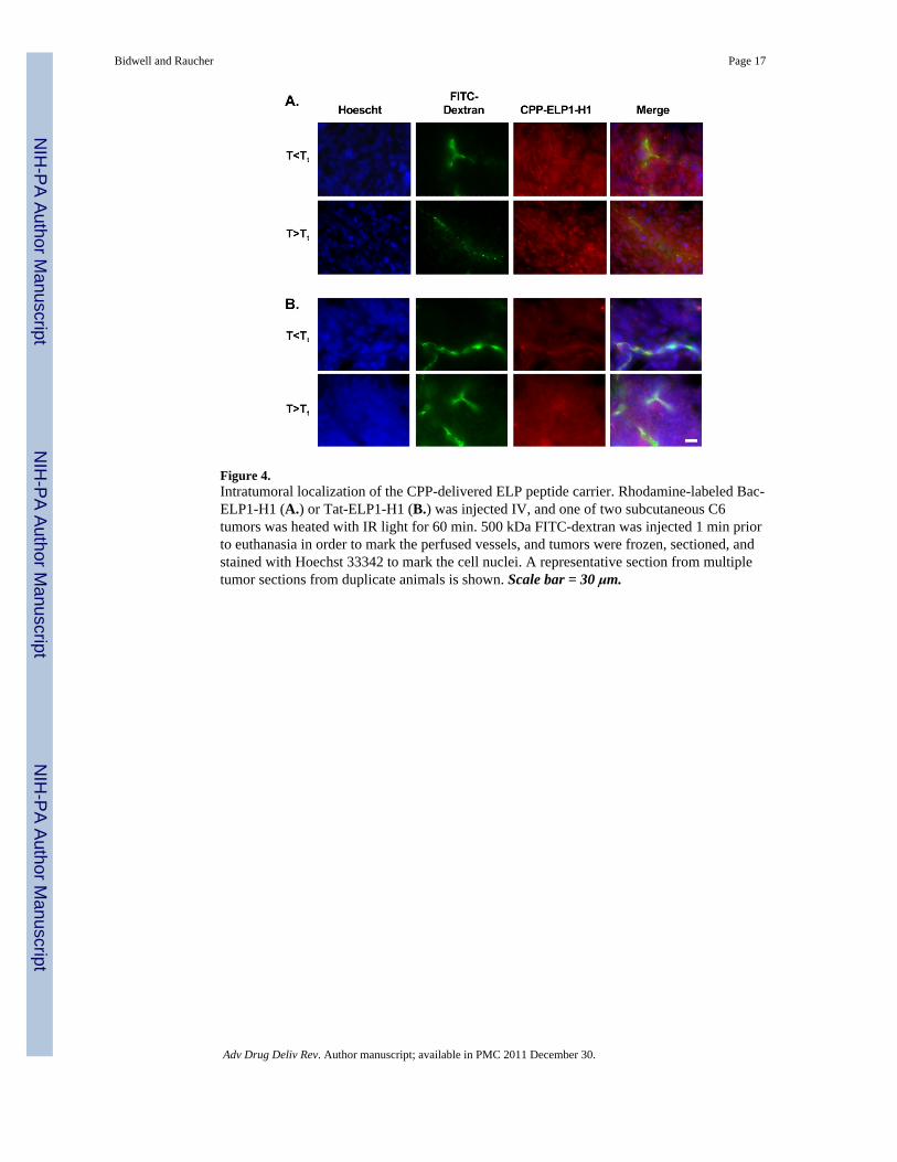
Figure 4.Intratumoral localization of the CPP-delivered ELP peptide carrier. Rhodamine-labeled Bac-ELP1-H1 (A.) or Tat-ELP1-H1 (B.) was injected IV, and one of two subcutaneous C6tumors was heated with IR light for 60 min. 500 kDa FITC-dextran was injected 1 min priorto euthanasia in order to mark the perfused vessels, and tumors were frozen, sectioned, andstained with Hoechst 33342 to mark the cell nuclei. A representative section from multipletumor sections from duplicate animals is shown. Scale bar = 30 μm.
Bidwell and Raucher Page 17
Adv Drug Deliv Rev. Author manuscript; available in PMC 2011 December 30.
NIH
-PA Author Manuscript
NIH
-PA Author Manuscript
NIH
-PA Author Manuscript
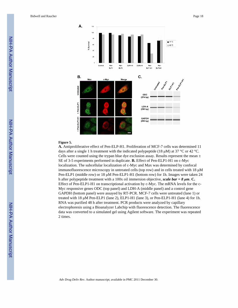
Figure 5.A. Antiproliferative effect of Pen-ELP-H1. Proliferation of MCF-7 cells was determined 11days after a single 1 h treatment with the indicated polypeptide (18 μM) at 37 °C or 42 °C.Cells were counted using the trypan blue dye exclusion assay. Results represent the mean ±SE of 3-5 experiments performed in duplicate. B. Effect of Pen-ELP1-H1 on c-Myclocalization. The subcellular localization of c-Myc and Max was determined by confocalimmunofluorescence microscopy in untreated cells (top row) and in cells treated with 18 μMPen-ELP1 (middle row) or 18 μM Pen-ELP1-H1 (bottom row) for 1h. Images were taken 24h after polypeptide treatment with a 100x oil immersion objective, scale bar = 8 μm. C.Effect of Pen-ELP1-H1 on transcriptional activation by c-Myc. The mRNA levels for the c-Myc responsive genes ODC (top panel) and LDH-A (middle panel) and a control geneGAPDH (bottom panel) were assayed by RT-PCR. MCF-7 cells were untreated (lane 1) ortreated with 18 μM Pen-ELP1 (lane 2), ELP1-H1 (lane 3), or Pen-ELP1-H1 (lane 4) for 1h.RNA was purified 48 h after treatment. PCR products were analyzed by capillaryelectrophoresis using a Bioanalyzer Labchip with fluorescence detection. The fluorescencedata was converted to a simulated gel using Agilent software. The experiment was repeated2 times.
Bidwell and Raucher Page 18
Adv Drug Deliv Rev. Author manuscript; available in PMC 2011 December 30.
NIH
-PA Author Manuscript
NIH
-PA Author Manuscript
NIH
-PA Author Manuscript

Figure 6.Proposed model for c-Myc inhibition by Pen-ELP-H1 and Bac-ELP-H1. Mitogenstimulation induces transcription of mRNA from the c-Myc gene. In the case of Pen-ELP-H1, newly translated c-Myc is bound by the polypeptide in the cytoplasm. Once bound, c-Myc can not be imported into the nucleus and interact with Max. In the case of Bac-ELP-H1, the polypeptide enters the nucleus and interferes directly with the c-Myc and Maxinteraction. Both situations result in the down-regulation of c-Myc-Max responsive genesand lead to inhibition of cell proliferation.
Bidwell and Raucher Page 19
Adv Drug Deliv Rev. Author manuscript; available in PMC 2011 December 30.
NIH
-PA Author Manuscript
NIH
-PA Author Manuscript
NIH
-PA Author Manuscript

Figure 7.Optimization of ELP-H1 delivery with alternative CPPs. A. Cellular uptake of the CPP-ELP-H1 polypeptides. MCF-7 cells were treated for 1 h at 37 or 42 °C with fluoresceinlabeled polypeptides. Levels of each polypeptide were assessed using flow cytometry (n =5,000 cells). Forward and side scatter gating were used to eliminate cell debris from theanalysis, and fluorescence data was normalized to cellular autofluorescence and correctedfor variations in labeling efficiency among the polypeptides. Data represent the average of 3experiments; error bars, SEM.* Difference between 37 and 42 °C levels are statisticallysignificant (ANOVA, p<0.01). † Difference is significant as compared to ELP at 42 °C.(ANOVA, p<0.01). B. MCF-7 proliferation after CPP-ELP-H1 treatment. MCF-7 cells weretreated for 1 h at 37 or 42 °C with various concentrations of Pen-ELP1-H1, Tat-ELPa80-H1,or Bac-ELP1-H1, and the cell viability was determined after 7 days using the MTS assay.The data shown is an overlay of the 42 °C data for each CPP-ELP-H1. C. Subcellularlocalization of Bac-ELP1-H1. MCF-7 cells were treated for 1 h at 42 °C with rhodaminelabeled Bac-ELP1-H1 (30 μM). 48 h after treatment, nuclei were stained with Sytox greenand cells were imaged with a laser scanning confocal microscope. Scale bar = 20 μm.
Bidwell and Raucher Page 20
Adv Drug Deliv Rev. Author manuscript; available in PMC 2011 December 30.
NIH
-PA Author Manuscript
NIH
-PA Author Manuscript
NIH
-PA Author Manuscript

Figure 8.CPP-ELP-p21 polypeptides. A. Inhibition of cell proliferation by Pen-ELP-p21. HeLa andSKOV-3 cells were exposed to the indicated concentration of Pen-ELP2-p21 at 37 °C for 1h, and cell proliferation was determined 72 h later using the MTS assay. B. Inhibition ofproliferation by Bac-ELP-p21. SKOV-3, MCF-7, and Panc-1 cells were exposed to theindicated concentration of Bac-ELP2-p21 at 37 °C or 42 °C for 1 h, and cell proliferationwas determined 6 days later (SKOV-3) or 3 days later (MCF-7 and Panc-1) using the MTSassay. Data represent the mean ± SE of 3 independent experiments. C. Subcellularlocalization of rhodamine labeled Bac-ELP1-p21 in SKOV-3 cells as visualized by confocalmicroscopy. Cells were treated with 20 μM Bac-ELP1-p21 at 37°C or 42°C for 1 h.Confocal images were taken 24 h later. Tubulin was stained as a reference for cellularstructure. Scale bar = 20 μm. The subcellular distribution of Bac-ELP1-p21 was alsoconfirmed in live cells 24 h after a 1 h exposure at 37 or 42 °C (not shown). D. SDS-PAGEanalysis of Rb protein in SKOV-3 cells following treatment with Bac-ELP1-p21. Cells weretreated with the indicated polypeptide (30 μM) at 42°C for 1 h, harvested 24 h later, andlysed. Equal amounts of samples were loaded onto a 12% SDS gel and transferred to a blotwhich was probed with the indicated antibodies.
Bidwell and Raucher Page 21
Adv Drug Deliv Rev. Author manuscript; available in PMC 2011 December 30.
NIH
-PA Author Manuscript
NIH
-PA Author Manuscript
NIH
-PA Author Manuscript


![Interfacial interactions between poly[L-lysine]-based branched polypeptides and phospholipid model membranes](https://static.fdokumen.com/doc/165x107/633df5f7df741406dc0b4c83/interfacial-interactions-between-polyl-lysine-based-branched-polypeptides-and.jpg)