Cell Migration: Integrating Signals from Front to Back
6
20. E. Smirnova, D. Shurland, S. Ryazantsev, A. Van Der Bliek, J. Cell Biol. 143, 351 (1998). 21. A. M. Labrousse, M. D. Zappaterra, D. A. Rube, A. M. van der Bliek, Mol. Cell 4, 815 (1999). 22. H. Sesaki, R. E. Jensen, J. Cell Biol. 147, 699 (1999). 23. S. Arimura, N. Tsutsumi, Proc. Natl. Acad. Sci. U.S.A. 99, 5727 (2002). 24. A. M. van der Bliek, Trends Cell Biol. 9, 96 (1999). 25. J. E. Hinshaw, Annu. Rev. Cell Dev. Biol. 16, 483 (2000). 26. J. E. Hinshaw, S. L. Schmid, Nature 374, 190 (1995). 27. J. E. Hinshaw, Curr. Opin. Struct. Biol. 9, 260 (1999). 28. M. H. Stowell, B. Marks, P. Wigge, H. T. McMahon, Nature Cell Biol. 1, 27 (1999). 29. B. Marks et al., Nature 410, 231 (2001). 30. N. H. Fukushima, E. Brisch, B. R. Keegan, W. Bleazard, J. M. Shaw, Mol. Biol. Cell 12, 2756 (2001). 31. S. Sever, Curr. Opin. Cell Biol. 14, 463 (2002). 32. S. L. Newmyer, A. Christensen, S. Sever, Dev. Cell 4, 929 (2003). 33. H. Gao, D. Kadirjan-Kalbach, J. E. Froehlich, K. W. Oster- young, Proc. Natl. Acad. Sci. U.S.A. 100, 4328 (2003). 34. S. Miyagishima, K. Nishida, T. Kuroiwa, Trends Plant Sci. 8, 432 (2003). 35. S. Miyagishima et al., Plant Cell 13, 2257 (2001). 36. S. Y. Miyagishima et al., Plant Cell 15, 655 (2003). 37. K. Nishida et al., Proc. Natl. Acad. Sci. U.S.A. 100, 2146 (2003). 38. A. Legesse-Miller, R. H. Massol, T. Kirchhausen, Mol. Biol. Cell 14, 1953 (2003). 39. M. Fujiwara, S. Yoshida, Biochem. Biophys. Res. Com- mun. 287, 462 (2001). 40. R. S. McAndrew, J. E. Froehlich, S. Vitha, K. D. Stokes, K. W. Osteryoung, Plant Physiol. 127, 1656 (2001). 41. S. Sever, H. Damke, S. L. Schmid, J. Cell Biol. 150, 1137 (2000). 42. J. Nunnari, unpublished data. 43. A. G. McArthur et al., FEMS Microbiol. Lett. 189, 271 (2000). 44. M. Marti et al., J. Biol. Chem. 278, 24837 (2003). 45. C. A. Vater, C. K. Raymond, K. Ekena, I. Howald-Stevenson, T. H. Stevens, J. Cell Biol. 119, 773 (1992). 46. D. Hoepfner, M. van den Berg, P. Philippsen, H. F. Tabak, E. H. Hettema, J. Cell Biol. 155, 979 (2001). 47. J. Nunnari, H. Gao, K. W. Osteryoung, unpublished data. 48. F. van den Ent, L. Amos, J. Lowe, Curr. Opin. Micro- biol. 4, 634 (2001). 49. K. W. Osteryoung, K. D. Stokes, S. M. Rutherford, A. L. Percival, W. Y. Lee, Plant Cell 10, 1991 (1998). 50. R. Strepp, S. Scholz, S. Kruse, V. Speth, R. Reski, Proc. Natl. Acad. Sci. U.S.A. 95, 4368 (1998). 51. K. W. Osteryoung, R. S. McAndrew, Annu. Rev. Plant Physiol. Plant Mol. Biol. 52, 315 (2001). 52. K. D. Stokes, K. W. Osteryoung, Gene 320, 97 (2003). 53. S. Vitha, R. S. McAndrew, K. W. Osteryoung, J. Cell Biol. 153, 111 (2001). 54. S. Vitha et al., Plant Cell 15, 1918 (2003). 55. R. McAndrew, C. Chi-Ham, J. E. Froehlich, K. W. Osteryoung, unpublished data. 56. A. Gaikwad, V. Babbarwal, V. Pant, S. K. Mukherjee, Mol. Gen. Genet. 263, 213 (2000). 57. M. El-Shami, S. El-Kafafi, D. Falconet, S. Lerbs-Mache, Mol. Genet. Genomics 267, 254 (2002). 58. X. Ma, W. Margolin, J. Bacteriol. 181, 7531 (1999). 59. J. Errington, R. A. Daniel, D. J. Scheffers, Microbiol. Mol. Biol. Rev. 67, 52 (2003). 60. J. Kiessling et al., J. Cell Biol. 151, 945 (2000). 61. K. D. Stokes, R. S. McAndrew, R. Figueroa, S. Vitha, K. W. Osteryoung, Plant Physiol. 124, 1668 (2000). 62. J. L. Marrison et al., Plant J. 18, 651 (1999). 63. S. Walter, J. Buchner, Angew. Chem. Int. Ed. Engl. 41, 1098 (2002). 64. O. A. Koksharova, C. P. Wolk, J. Bacteriol. 184, 5524 (2002). 65. H. Fulgosi, L. Gerdes, S. Westphal, C. Glockmann, J. Soll, Proc. Natl. Acad. Sci. U.S.A. 99, 11501 (2002). 66. J. Maple, N. H. Chua, S. G. Moller, Plant J. 31, 269 (2002). 67. K. S. Colletti et al., Curr. Biol. 10, 507 (2000). 68. R. Itoh, M. Fujiwara, N. Nagata, S. Yoshida, Plant Physiol. 127, 1644 (2001). 69. L. Griparic, A. M. van der Bliek, Traffic 2, 235 (2001). 70. E. Smirnova, L. Griparic, D. L. Shurland, A. M. van der Bliek, Mol. Biol. Cell 12, 2245 (2001). 71. K. R. Pitts, Y. Yoon, E. W. Krueger, M. A. McNiven, Mol. Biol. Cell 10, 4403 (1999). 72. S. Jakobs et al., J. Cell Sci. 116, 2005 (2003). 73. K. L. Cerveny, J. M. McCaffery, R. E. Jensen, Mol. Biol. Cell 12, 309 (2001). 74. Q. Tieu, J. Nunnari, J. Cell Biol. 151, 353 (2000). 75. P. Fekkes, K. A. Shepard, M. P. Yaffe, J. Cell Biol. 151, 333 (2000). 76. A. D. Mozdy, J. M. McCaffery, J. M. Shaw, J. Cell Biol. 151, 367 (2000). 77. Q. Tieu, V. Okreglak, K. Naylor, J. Nunnari, J. Cell Biol. 158, 445 (2002). 78. K. L. Cerveny, R. E. Jensen, Mol. Biol. Cell 14, 4126 (2003). 79. Y. Yoon, E. W. Krueger, B. J. Oswald, M. A. McNiven, Mol. Cell. Biol. 23, 5409 (2003). 80. D. I. James, P. A. Parone, Y. Mattenberger, J.-C. Mar- tinou, J. Biol. Chem. 278, 36373 (2003). 81. H. Berman et al., Nucleic Acids Res. 28, 235 (2000). 82. PDBID: 1IYG, W. Ohashi et al., unpublished data. 83. D. Rube, S. Gandre, A. van der Bliek, 14th Interna- tional C. elegans Conference, 29 June to 3 July 2003, Los Angeles, Abstract 845B. 84. W. B. Huttner, A. Schmidt, Curr. Opin. Neurobiol. 10, 543 (2000). 85. K. Farsad et al., J. Cell Biol. 155, 193 (2001). 86. A. Schmidt et al., Nature 401, 133 (1999). 87. H. Hashimoto, Int. Rev. Cytol. 222, 63 (2003). 88. N. Garrido et al., Mol. Biol. Cell 14, 1583 (2003). 89. W. J. Jeong et al., Plant Physiol. 129, 112 (2002). 90. J. Nunnari et al., Mol. Biol. Cell 8, 1233 (1997). 91. We regret that space constraints have prevented us from citing many relevant papers. We thank S. Merchant and S. Miyagishima for critical reading of the manuscript; P. Beech and A. van der Bliek for sharing relevant findings before publication; S. Vitha, D. Yoder, S. Miyagishima, K. Naylor, A. Stone, C. Song, and H. Gao for providing images for figures; and all the members of our laboratories for invaluable contributions. Supported by NSF grants 0092448 (K.W.O.), 0313520 (K.W.O.), and 0110899 (J.N.); NIH grant R01GM62942A ( J.N.); and the Michigan State University Center for Plant Products and Technologies (K.W.O.). Cell Migration: Integrating Signals from Front to Back Anne J. Ridley, 1 Martin A. Schwartz, 2 Keith Burridge, 5 Richard A. Firtel, 6 Mark H. Ginsberg, 7 Gary Borisy, 8 J. Thomas Parsons, 3 Alan Rick Horwitz 4 Cell migration is a highly integrated multistep process that orchestrates embryonic mor- phogenesis; contributes to tissue repair and regeneration; and drives disease progression in cancer, mental retardation, atherosclerosis, and arthritis. The migrating cell is highly polarized with complex regulatory pathways that spatially and temporally integrate its component processes. This review describes the mechanisms underlying the major steps of migration and the signaling pathways that regulate them, and outlines recent advances investigating the nature of polarity in migrating cells and the pathways that establish it. O ur liaison with cell migration, as hu- mans, begins shortly after concep- tion, accompanies us throughout life, and often contributes to our death. Although migratory phenomena are apparent as early as implantation, cell migration orchestrates morphogenesis throughout embryonic devel- opment (1). During gastrulation, for example, large groups of cells migrate collectively as sheets to form the resulting three-layer em- bryo. Subsequently, cells migrate from vari- ous epithelial layers to target locations, where they then differentiate to form the specialized cells that make up different tissues and or- gans. Analogous migrations occur in the adult. In the renewal of skin and intestine, fresh epithelial cells migrate up from the basal layer and the crypts, respectively. Mi- gration is also a prominent component of tissue repair and immune surveillance, in which leukocytes from the circulation mi- grate into the surrounding tissue to destroy invading microorganisms and infected cells and to clear debris. The importance of cell migration however, goes far beyond humans and extends to plants and even to single- celled organisms (2). Migration contributes to several important pathological processes, including vascular dis- ease, osteoporosis, chronic inflammatory dis- eases such as rheumatoid arthritis and multiple 1 Ludwig Institute for Cancer Research, Royal Free and University College School of Medicine, London W1W 7BS, UK. 2 Departments of Microbiology and Biomed- ical Engineering, Cardiovascular Research Center and Mellon Prostate Cancer Research Institute; 3 Depart- ment of Microbiology; 4 Department of Cell Biology; University of Virginia School of Medicine, Charlottes- ville, VA 22908, USA. 5 Department of Cell and Devel- opmental Biology and Lineberger Comprehensive Cancer Center, University of North Carolina, Chapel Hill, NC 27599, USA. 6 Section of Cell and Develop- mental Biology, Division of Biological Sciences and Center for Molecular Genetics, University of Califor- nia, San Diego, 9500 Gilman Drive, La Jolla, CA 92093– 0634, USA. 7 Department of Cell Biology, The Scripps Research Institute, 10550 North Torrey Pines Road, La Jolla, CA 92037, USA. 8 Department of Cel- lular and Molecular Biology, Northwestern University, School of Medicine, Chicago, IL 60611, USA. R EVIEWS 5 DECEMBER 2003 VOL 302 SCIENCE www.sciencemag.org 1704
-
Upload
independent -
Category
Documents
-
view
1 -
download
0
Transcript of Cell Migration: Integrating Signals from Front to Back
20. E. Smirnova, D. Shurland, S. Ryazantsev, A. Van DerBliek, J. Cell Biol. 143, 351 (1998).
21. A. M. Labrousse, M. D. Zappaterra, D. A. Rube, A. M.van der Bliek, Mol. Cell 4, 815 (1999).
22. H. Sesaki, R. E. Jensen, J. Cell Biol. 147, 699 (1999).23. S. Arimura, N. Tsutsumi, Proc. Natl. Acad. Sci. U.S.A.
99, 5727 (2002).24. A. M. van der Bliek, Trends Cell Biol. 9, 96 (1999).25. J. E. Hinshaw, Annu. Rev. Cell Dev. Biol. 16, 483 (2000).26. J. E. Hinshaw, S. L. Schmid, Nature 374, 190 (1995).27. J. E. Hinshaw, Curr. Opin. Struct. Biol. 9, 260 (1999).28. M. H. Stowell, B. Marks, P. Wigge, H. T. McMahon,
Nature Cell Biol. 1, 27 (1999).29. B. Marks et al., Nature 410, 231 (2001).30. N. H. Fukushima, E. Brisch, B. R. Keegan, W. Bleazard,
J. M. Shaw, Mol. Biol. Cell 12, 2756 (2001).31. S. Sever, Curr. Opin. Cell Biol. 14, 463 (2002).32. S. L. Newmyer, A. Christensen, S. Sever, Dev. Cell 4,
929 (2003).33. H. Gao, D. Kadirjan-Kalbach, J. E. Froehlich, K. W. Oster-
young, Proc. Natl. Acad. Sci. U.S.A. 100, 4328 (2003).34. S. Miyagishima, K. Nishida, T. Kuroiwa, Trends Plant
Sci. 8, 432 (2003).35. S. Miyagishima et al., Plant Cell 13, 2257 (2001).36. S. Y. Miyagishima et al., Plant Cell 15, 655 (2003).37. K. Nishida et al., Proc. Natl. Acad. Sci. U.S.A. 100,
2146 (2003).38. A. Legesse-Miller, R. H. Massol, T. Kirchhausen, Mol.
Biol. Cell 14, 1953 (2003).39. M. Fujiwara, S. Yoshida, Biochem. Biophys. Res. Com-
mun. 287, 462 (2001).40. R. S. McAndrew, J. E. Froehlich, S. Vitha, K. D. Stokes,
K. W. Osteryoung, Plant Physiol. 127, 1656 (2001).41. S. Sever, H. Damke, S. L. Schmid, J. Cell Biol. 150,
1137 (2000).42. J. Nunnari, unpublished data.43. A. G. McArthur et al., FEMS Microbiol. Lett. 189, 271
(2000).44. M. Marti et al., J. Biol. Chem. 278, 24837 (2003).45. C. A. Vater, C. K. Raymond, K. Ekena, I. Howald-Stevenson,
T. H. Stevens, J. Cell Biol. 119, 773 (1992).
46. D. Hoepfner, M. van den Berg, P. Philippsen, H. F.Tabak, E. H. Hettema, J. Cell Biol. 155, 979 (2001).
47. J. Nunnari, H. Gao, K. W. Osteryoung, unpublished data.48. F. van den Ent, L. Amos, J. Lowe, Curr. Opin. Micro-
biol. 4, 634 (2001).49. K. W. Osteryoung, K. D. Stokes, S. M. Rutherford, A. L.
Percival, W. Y. Lee, Plant Cell 10, 1991 (1998).50. R. Strepp, S. Scholz, S. Kruse, V. Speth, R. Reski, Proc.
Natl. Acad. Sci. U.S.A. 95, 4368 (1998).51. K. W. Osteryoung, R. S. McAndrew, Annu. Rev. Plant
Physiol. Plant Mol. Biol. 52, 315 (2001).52. K. D. Stokes, K. W. Osteryoung, Gene 320, 97 (2003).53. S. Vitha, R. S. McAndrew, K. W. Osteryoung, J. Cell
Biol. 153, 111 (2001).54. S. Vitha et al., Plant Cell 15, 1918 (2003).55. R. McAndrew, C. Chi-Ham, J. E. Froehlich, K. W.
Osteryoung, unpublished data.56. A. Gaikwad, V. Babbarwal, V. Pant, S. K. Mukherjee,
Mol. Gen. Genet. 263, 213 (2000).57. M. El-Shami, S. El-Kafafi, D. Falconet, S. Lerbs-Mache,
Mol. Genet. Genomics 267, 254 (2002).58. X. Ma, W. Margolin, J. Bacteriol. 181, 7531 (1999).59. J. Errington, R. A. Daniel, D. J. Scheffers, Microbiol.
Mol. Biol. Rev. 67, 52 (2003).60. J. Kiessling et al., J. Cell Biol. 151, 945 (2000).61. K. D. Stokes, R. S. McAndrew, R. Figueroa, S. Vitha,
K. W. Osteryoung, Plant Physiol. 124, 1668 (2000).62. J. L. Marrison et al., Plant J. 18, 651 (1999).63. S. Walter, J. Buchner, Angew. Chem. Int. Ed. Engl. 41,
1098 (2002).64. O. A. Koksharova, C. P. Wolk, J. Bacteriol. 184, 5524
(2002).65. H. Fulgosi, L. Gerdes, S. Westphal, C. Glockmann, J.
Soll, Proc. Natl. Acad. Sci. U.S.A. 99, 11501 (2002).66. J. Maple, N. H. Chua, S. G. Moller, Plant J. 31, 269 (2002).67. K. S. Colletti et al., Curr. Biol. 10, 507 (2000).68. R. Itoh, M. Fujiwara, N. Nagata, S. Yoshida, Plant
Physiol. 127, 1644 (2001).69. L. Griparic, A. M. van der Bliek, Traffic 2, 235 (2001).70. E. Smirnova, L. Griparic, D. L. Shurland, A. M. van der
Bliek, Mol. Biol. Cell 12, 2245 (2001).
71. K. R. Pitts, Y. Yoon, E. W. Krueger, M. A. McNiven,Mol. Biol. Cell 10, 4403 (1999).
72. S. Jakobs et al., J. Cell Sci. 116, 2005 (2003).73. K. L. Cerveny, J. M. McCaffery, R. E. Jensen, Mol. Biol.
Cell 12, 309 (2001).74. Q. Tieu, J. Nunnari, J. Cell Biol. 151, 353 (2000).75. P. Fekkes, K. A. Shepard, M. P. Yaffe, J. Cell Biol. 151,
333 (2000).76. A. D. Mozdy, J. M. McCaffery, J. M. Shaw, J. Cell Biol.
151, 367 (2000).77. Q. Tieu, V. Okreglak, K. Naylor, J. Nunnari, J. Cell Biol.
158, 445 (2002).78. K. L. Cerveny, R. E. Jensen, Mol. Biol. Cell 14, 4126 (2003).79. Y. Yoon, E. W. Krueger, B. J. Oswald, M. A. McNiven,
Mol. Cell. Biol. 23, 5409 (2003).80. D. I. James, P. A. Parone, Y. Mattenberger, J.-C. Mar-
tinou, J. Biol. Chem. 278, 36373 (2003).81. H. Berman et al., Nucleic Acids Res. 28, 235 (2000).82. PDBID: 1IYG, W. Ohashi et al., unpublished data.83. D. Rube, S. Gandre, A. van der Bliek, 14th Interna-
tional C. elegans Conference, 29 June to 3 July 2003,Los Angeles, Abstract 845B.
84. W. B. Huttner, A. Schmidt, Curr. Opin. Neurobiol. 10,543 (2000).
85. K. Farsad et al., J. Cell Biol. 155, 193 (2001).86. A. Schmidt et al., Nature 401, 133 (1999).87. H. Hashimoto, Int. Rev. Cytol. 222, 63 (2003).88. N. Garrido et al., Mol. Biol. Cell 14, 1583 (2003).89. W. J. Jeong et al., Plant Physiol. 129, 112 (2002).90. J. Nunnari et al., Mol. Biol. Cell 8, 1233 (1997).91. We regret that space constraints have prevented us from
citing many relevant papers. We thank S. Merchant and S.Miyagishima for critical reading of the manuscript; P. Beechand A. van der Bliek for sharing relevant findings beforepublication; S. Vitha, D. Yoder, S. Miyagishima, K. Naylor, A.Stone, C. Song, and H. Gao for providing images for figures;and all the members of our laboratories for invaluablecontributions. Supported by NSF grants 0092448 (K.W.O.),0313520 (K.W.O.), and 0110899 (J.N.); NIH grantR01GM62942A ( J.N.); and the Michigan State UniversityCenter for Plant Products and Technologies (K.W.O.).
Cell Migration: Integrating Signals fromFront to Back
Anne J. Ridley,1 Martin A. Schwartz,2 Keith Burridge,5 Richard A. Firtel,6 Mark H. Ginsberg,7 Gary Borisy,8
J. Thomas Parsons,3 Alan Rick Horwitz4
Cell migration is a highly integrated multistep process that orchestrates embryonic mor-phogenesis; contributes to tissue repair and regeneration; and drives disease progression incancer,mental retardation, atherosclerosis, and arthritis. Themigrating cell is highly polarizedwith complex regulatory pathways that spatially and temporally integrate its componentprocesses. This review describes themechanisms underlying themajor steps ofmigration andthe signaling pathways that regulate them, and outlines recent advances investigating thenature of polarity in migrating cells and the pathways that establish it.
Our liaison with cell migration, as hu-mans, begins shortly after concep-tion, accompanies us throughout life,
and often contributes to our death. Althoughmigratory phenomena are apparent as earlyas implantation, cell migration orchestratesmorphogenesis throughout embryonic devel-opment (1). During gastrulation, for example,large groups of cells migrate collectively assheets to form the resulting three-layer em-bryo. Subsequently, cells migrate from vari-ous epithelial layers to target locations, wherethey then differentiate to form the specialized
cells that make up different tissues and or-gans. Analogous migrations occur in theadult. In the renewal of skin and intestine,fresh epithelial cells migrate up from thebasal layer and the crypts, respectively. Mi-gration is also a prominent component oftissue repair and immune surveillance, inwhich leukocytes from the circulation mi-
grate into the surrounding tissue to destroyinvading microorganisms and infected cellsand to clear debris. The importance of cellmigration however, goes far beyond humansand extends to plants and even to single-celled organisms (2).
Migration contributes to several importantpathological processes, including vascular dis-ease, osteoporosis, chronic inflammatory dis-eases such as rheumatoid arthritis and multiple
1Ludwig Institute for Cancer Research, Royal Free andUniversity College School of Medicine, London W1W7BS, UK. 2Departments of Microbiology and Biomed-ical Engineering, Cardiovascular Research Center and
Mellon Prostate Cancer Research Institute; 3Depart-ment of Microbiology; 4Department of Cell Biology;University of Virginia School of Medicine, Charlottes-ville, VA 22908, USA. 5Department of Cell and Devel-opmental Biology and Lineberger ComprehensiveCancer Center, University of North Carolina, ChapelHill, NC 27599, USA. 6Section of Cell and Develop-mental Biology, Division of Biological Sciences andCenter for Molecular Genetics, University of Califor-nia, San Diego, 9500 Gilman Drive, La Jolla, CA92093–0634, USA. 7Department of Cell Biology, TheScripps Research Institute, 10550 North Torrey PinesRoad, La Jolla, CA 92037, USA. 8Department of Cel-lular and Molecular Biology, Northwestern University,School of Medicine, Chicago, IL 60611, USA.
R E V I E W S
5 DECEMBER 2003 VOL 302 SCIENCE www.sciencemag.org1704

sclerosis, cancer, and mental retardation. Thus,understanding the fundamental mechanisms un-derlying cell migration holds the promise ofeffective therapeutic approaches for treatingdisease, cellular transplantation, and the prepa-ration of artificial tissues.
Over the past few years, immense progresshas been made in understanding cell migration,including the establishment of polar structures,the regulation of the dynamic processes of actinand microtubule polymerization, and the regu-lation of spatial and temporal signal transduc-tion. This review summarizes and highlightssome of these advances in the context of theneed to integrate and coordinate the many cel-lular events that compose migration.
The Migration CycleOur present understanding of cell migration is acomposite derived from studies of different celltypes and environments. In general, cell migra-tion can be usefully conceptualized as a cyclicprocess (3). The initial response of a cell to amigration-promoting agent is to polarize andextend protrusions in the direction of migration.These protrusions can be large, broad lamelli-podia or spike-like filopodia, are usually drivenby actin polymerization, and are stabilized byadhering to the extracellular matrix (ECM) oradjacent cells via transmembrane receptorslinked to the actin cytoskeleton. These adhe-sions serve as traction sites for migration as thecell moves forward over them, and they aredisassembled at the cell rear, allowing it todetach. Interestingly, the movement of cellsheets shows some features of single-cell mi-gration; however, the polarization extendsacross the sheet.
Although many aspects of this picture areshared among different cell types, the detailscan differ greatly. For example, these steps areobserved most distinctly in slow-moving cellssuch as fibroblasts, but are not as obvious infast-moving cells such as neutrophils, whichseem to glide over the substratum. In addition,a cell’s migratory behavior depends on its en-vironment. Somitic cells migrating in vivo, forexample, show large single protrusions andhighly directed migration, in contrast to themultiple small protrusions they display on pla-nar substrates; and cancer cells can modify theirmorphology and nature of migration in re-sponse to environmental changes (4, 5).
The Protrusive MachineryActin filaments are intrinsically polarized withfast-growing “barbed” ends and slow-growing“pointed” ends, and this inherent polarity isused to drive membrane protrusion. However,the organization of filaments depends on thetype of protrusion: In lamellipodia, actin fila-ments form a branching “dendritic” network,whereas in filopodia they are organized intolong parallel bundles (6). Actin polymerizationin lamellipodia is mediated by the Arp2/3 com-
plex, which binds to the sides or tip of a pre-existing actin filament and induces the forma-tion of a new daughter filament that branchesoff the mother filament (6, 7). Activation of theArp2/3 complex is localized by WASP/WAVEfamily members, which are themselves activat-ed at the cell membrane (6) (see below). Push-ing of the membrane, the actual protrusiveevent, is believed to occur not by elonga-tion of the actin filament per se but by an“elastic Brownian ratchet” mechanism, inwhich thermal energy bends the nascentshort filaments, storing elastic energy. Un-bending of an elongated filament againstthe leading edge would then provide thedriving force for protrusion (7).
Several actin-binding proteins regulate therate and organization of actin polymerization inprotrusions by affecting the pool of availablemonomers and free ends (7, 8). For example,profilin prevents self-nucleation by binding toactin monomers and also serves to selectivelytarget monomers to barbed ends. Filament elon-gation is terminated by capping proteins, there-by restricting polymerization to new filamentsclose to the plasma membrane. In addition, dis-
assembly of older filaments, which is needed togenerate actin monomers for polymerization atthe front end, is assisted by proteins of theADF/cofilin family, which sever filaments andpromote actin dissociation from the pointed end.Other proteins play supporting roles in the den-dritic network: Cortactin stabilizes branches,whereas filamin A and �-actinin stabilize theentire network by cross-linking filaments (6).
Filopodial protrusion is thought to occurby a filament treadmilling mechanism, inwhich actin filaments within a bundle elon-gate at their barbed ends and release actinmonomers from their pointed ends (6). Thelong and unbranched filament organization isconsistent with assembly occurring by elon-gation rather than by branched nucleation.Many proteins are enriched at filopodial tips,including Ena/VASP proteins, which bindbarbed ends of actin filaments and antagonizeboth capping and branching, thereby allow-ing continuous elongation of filaments andfascin, which bundles actin filaments andmight thereby generate the stiffness needed toallow efficient pushing of the plasma mem-brane in filopodia (6).
Fig. 1. The polarized cell. (A) PIP3: Leading-edge localization of a green fluorescent protein (GFP)fusion of the PH domain of Akt/PKB in chemotaxing Dictyostelium cells. (Micrograph by R. Meili andR. Firtel.) (B) Phosphorylated �4 integrin: Localization at the leading edge of phosphorylated �4integrin expressed in migrating CHO cells. The localization was assayed by immunostaining with anantibody directed against the phosphorylated �4 integrin cytoplasmic domain. [Micrograph by L. E.Goldfinger and M. H. Ginsberg, reproduced from The Journal of Cell Biology, 2003, Vol. 162, p. 732,by copyright permission of The Rockefeller University Press] (C) PTEN: Localization of a GFP fusionof PTEN in chemotaxing Dictyostelium cells. PTEN is absent from the leading edge but present alongthe lateral sides and posterior of the cell. (Micrograph by R. Meili and R. Firtel.) (D) Activated Rac:Activated Rac localizes preferentially with an effector in the leading edge of migrating 3T3 cells.The interaction of GFP-V12Rac with an effector domain was assayed by fluorescence resonanceenergy transfer (FRET). The enhanced interaction in the leading edge is due to locally regulatedmembrane targeting of the V12Rac. Red and blue represent high and low intensities of FRET (thatis, of interaction), respectively. (Micrograph by M. Del Pozo, W. B. Kiosses, and M. A. Schwartz.)
R E V I E W S
www.sciencemag.org SCIENCE VOL 302 5 DECEMBER 2003 1705

The supramolecular design of lamellipo-dia and filopodia endows them with the ca-pacity to perform distinct functions. Biophys-ical considerations suggest that the dendriticorganization of lamellipodia provides a tightbrush-like structure that is able to push alonga broad length of plasma membrane (7).Through localized activation of the Arp2/3complex, the lamellipodium could be inducedto grow in a particular direction, providingthe basis for directional migration. In con-trast, filopodia, with their parallel bundle or-ganization, are particularly well designed toserve as sensors and to explore the localenvironment, although they are not essentialfor chemotaxis.
Rho family proteins: Central regulatorsof protrusion. Rho family small guano-sine triphosphate (GTP)–binding proteins(GTPases) are pivotal regulators of actin andadhesion organization and control the forma-tion of lamellipodia and filopodia. They areconformationally regulated by the binding ofGTP and GDP: When bound to GTP, they areactive and interact with their downstreamtarget proteins, which include protein ki-nases, lipid-modifying enzymes, and activa-tors of the Arp2/3 complex (9). Rho GTPasesare activated by guanine nucleotide exchangefactors (GEFs) and inactivated by GTPaseactivating proteins (GAPs). Of the RhoGTPases, Rac, Cdc42, and RhoG are requiredfor protrusion of lamellipodia and filopodia.
The major targets for Rac and Cdc42 thatmediate actin polymerization in protrusions arethe WASP/WAVE family of Arp2/3 complexactivators. Rac stimulates lamellipodial exten-sion by activating WAVE proteins (10). Cdc42binds to WASP proteins, and in vitro this stim-ulates the Arp2/3 complex to induce dendriticactin polymerization (6). However, this inter-action may not account for Cdc42’s ability toinduce filopodia, because cells lacking WASPsare still able to form filopodia (11); and, asdescribed above, filopodia contain parallel actinfilaments and not a dendritic network. RhoGdoes not interact directly with WASPs but ap-pears to act upstream of Rac by binding to andactivating a Rac-GEF complex (12).
WAVE/WASP proteins may themselvesregulate the activity of Rac and Cdc42 bybinding to GAPs and GEFs (13–15), andcould thereby generate positive or negativefeedback loops to regulate the extent ofCdc42/Rac-induced actin polymerization.WAVE/WASP proteins can also be regulatedby other stimuli apart from Cdc42 and Rac,including Src family kinases, the adaptor pro-teins Nck and WIP, and phosphoinositides (7,14, 16–19).
Polarizing the Cell: A Keystone ofMigrationFor a cell to migrate, it must be polarized, whichmeans that the molecular processes at the front
and the back of a moving cell are different.Establishing and maintaining cell polarity in re-sponse to extracellular stimuli appear to be me-diated by a set of interlinked positive feedbackloops involving Rho family GTPases, phospho-inositide 3-kinases (PI3Ks), integrins, microtu-bules, and vesicular transport (Figs. 1 and 2).Although the following discussion synthesizesinformation from multiple cell systems, the rel-ative contributions of the various signals dependon the cell type and the specific stimulus.
Cdc42: A master regulator of cell polar-ity. Cdc42 is a master regulator of cell polar-ity in eukaryotic organisms ranging fromyeast to humans. Cdc42 is active toward thefront of migrating cells (20), and both inhi-bition and global activation of Cdc42 candisrupt the directionality of migration (9).One way in which Cdc42 influences polarityis by restricting where lamellipodia form (21)(see below). Cdc42 can also affect polarity bylocalizing the microtubule-organizing center(MTOC) and Golgi apparatus in front of thenucleus, oriented toward the leading edge.Cdc42-induced MTOC orientation may con-tribute to polarized migration by facilitatingmicrotubule growth into the lamella andmicrotubule-mediated delivery of Golgi-derived vesicles to the leading edge, provid-ing membrane and associated proteins neededfor forward protrusion (9, 22). Reorganiza-tion of the MTOC appears to be moreimportant for the migration of slow-movingcells, because in fast-moving cells such asneutrophils and T cells, it is usually locatedbehind the nucleus (23).
The effects of Cdc42 on MTOC positionappear to be exerted mainly through a path-way involving the Cdc42 effector PAR6,which exists in a complex with PAR3 and anatypical protein kinase C (aPKC) (24). Themolecular mechanism by which the PAR6/PAR3/aPKC complex orients the MTOC isincompletely understood, but recent evidencesuggests that it could occur as a result of localcapture of microtubules at the leading edgevia APC, a protein that binds tubulin andlocalizes to the ends of microtubules (9), viaCLIP170 and IQGAP (22) and/or via themicrotubule-based dynein/dynactin motorprotein complex (24).
A downstream target of Cdc42, the kinasePAK1, can itself mediate Cdc42 activationdownstream of heterotrimeric GTP-binding pro-tein (G protein)–coupled receptors, which areactivated by many chemoattractants. These in-teractions define a positive feedback loop be-tween Cdc42 and PAK1, resulting in high Cdc42activity at the leading edge (25). Other feedbackloops involving integrins may also contribute tomaintaining local Cdc42 activation (9, 26).
PI3Ks and PTEN: The gradient amplifiers. Asurprising aspect of chemotaxis is the ability ofcells to respond directionally to very shallowchemoattractant gradients (less than a 10% dif-
ference in the concentration of chemoattractantbetween the front and rear of a cell) (27). Such asmall difference in signaling between the frontand rear needs to be amplified into steeperintracellular signaling gradients in order to gen-erate a cellular response. The phosphoinosi-tides PtdIns(3,4,5)P3 (PIP3) and PtdIns(3,4)P2
[PI(3,4)P2] are key signaling molecules that be-come rapidly and highly polarized in cells thatare exposed to a gradient of chemoattractant(Fig. 2). This amplification process involvesboth localized accumulation and activation ofPI3Ks, which generate PIP3/PI(3,4)P2, and thephosphatase PTEN, which removes them. InDictyostelium, for example, PI3Ks rapidly accu-mulate at the leading edge of cells in response toa chemoattractant, whereas PTEN becomes re-stricted to the sides and the rear (27, 28) (Fig. 1).Cells with altered PI3K or PTEN activity canusually migrate but exhibit a significantly re-duced ability to move directionally up a che-moattractant gradient. Although it is not yet clearwhat regulates the localization of PI3Ks, Cdc42activation is implicated in PTEN exclusion fromprotrusions in leukocytes, and PIP3 appears to berequired for localizing Cdc42 activity (25).These results imply that there is a network ofpositive feedback loops between Cdc42, PI3Kproducts, and PTEN that work together to initi-ate and maintain the polarity of migrating cells,although a Cdc42 paralog has not yet been iden-tified in Dictyostelium.
Localized Rac activation: Initiating andmaintaining protrusion. How do Cdc42 andPI3Ks lead to activation of the actin polymeriza-tion machinery required for active protrusion?The key event appears to be defining where Racis active (Fig. 1). This is probably achieved byactivating or delivering a Rac exchange factorlocally, and indeed several Rac GEFs are acti-vated by PI3K products (29). Once Rac is active,several feedback loops have been identified thathelp maintain directional protrusion. First, Raccan itself stimulate the recruitment and/or acti-vation of PI3Ks at the plasma membrane, whichthen act upstream of Rac by PIP3-sensitive RacGEFs (21, 29). Second, microtubules and Racmay form a positive feedback loop in whichmicrotubule polymerization activates Rac, andRac in turn stabilizes microtubules (22). Third,integrin engagement leads to Rac activation andmembrane targeting (30), and so new adhesionsformed at the leading edge will stimulate Rac,which in turn induces recruitment and clusteringof activated integrins to the edge of lamellipodia(31, 32). PIP3 also contributes to integrin activa-tion (33) and may thereby further enhance thepositive feedback to Rac.
Defining the tail. Is restriction of PIP3, ac-tive Cdc42, and Rac to the front of the cellsufficient to make the back of the cell followthe front? In several cell types, inhibition ofRho leads to the formation of an extended tail,possibly because actomyosin-based contractili-ty in the body of the cell is decreased. Rho may
R E V I E W S
5 DECEMBER 2003 VOL 302 SCIENCE www.sciencemag.org1706
also act in the tail by stabilizing microtubules,which would then promote focal adhesion turn-over (see below) (22, 34). One model for howmigrating cells maintain polarity is based on thefact that Rho and Rac are mutually antagonistic,each suppressing the other’s activity (35). Ac-tive Rac at the leading edge of cells wouldsuppress Rho activity, whereas Rho would bemore active at the sides and rear of the cell andsuppress Rac activity, thereby preventing Rac-
mediated protrusion at sites other than the lead-ing edge (36, 37). However, active Rac hasbeen implicated in detachment at the rear ofmigrating cells (38), and also Rho can lead toRac activation (39).
Integrins and Adhesion in MigrationFor migration to occur, a protrusion mustform and then stabilize by attaching to thesurroundings. Although many different re-
ceptors are involved in the migration ofdifferent cell types, the integrins are a ma-jor family of migration-promoting recep-tors. These receptors act as the “feet” of amigrating cell by supporting adhesion tothe ECM or other cells and by linking viaadapters with actin filaments on the insideof the cell. As described above, integrinsactivate migration-related signaling mole-cules. They are also recipients of “inside-
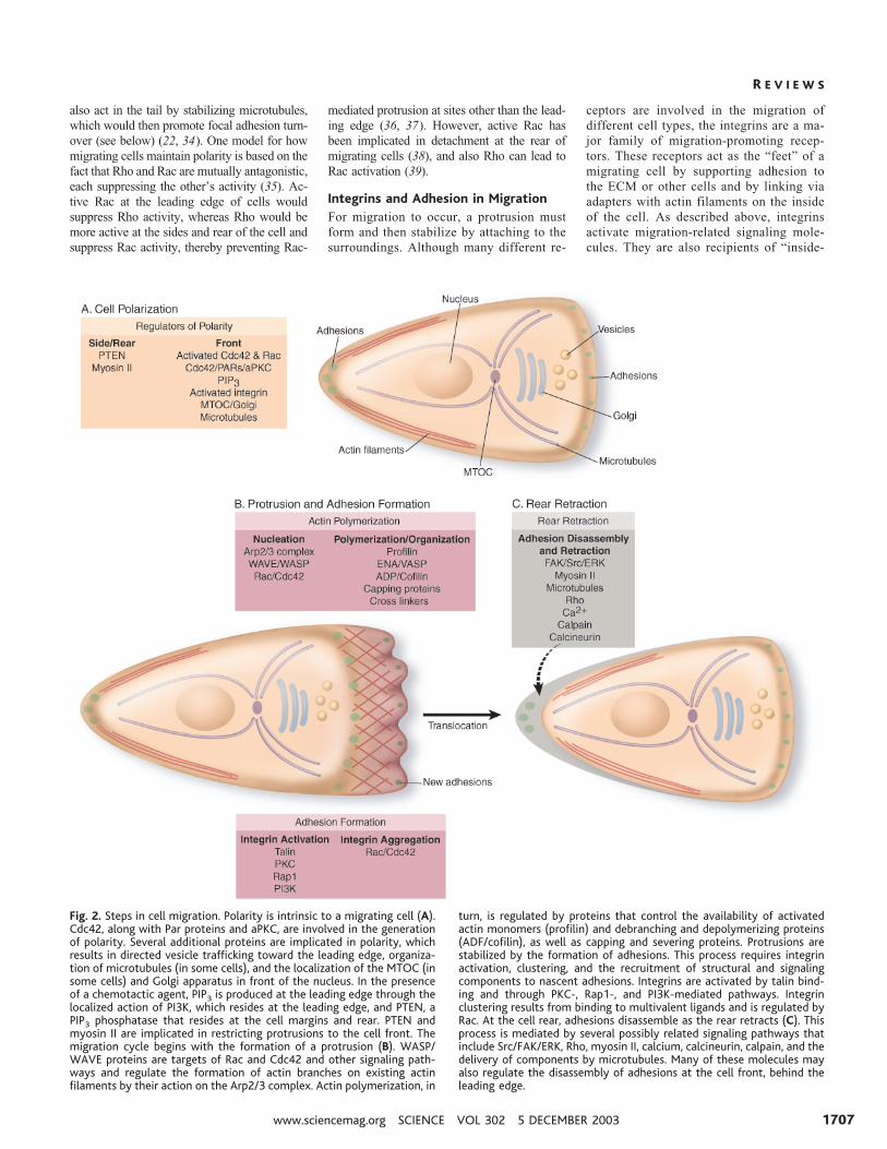
Fig. 2. Steps in cell migration. Polarity is intrinsic to a migrating cell (A).Cdc42, along with Par proteins and aPKC, are involved in the generationof polarity. Several additional proteins are implicated in polarity, whichresults in directed vesicle trafficking toward the leading edge, organiza-tion of microtubules (in some cells), and the localization of the MTOC (insome cells) and Golgi apparatus in front of the nucleus. In the presenceof a chemotactic agent, PIP3 is produced at the leading edge through thelocalized action of PI3K, which resides at the leading edge, and PTEN, aPIP3 phosphatase that resides at the cell margins and rear. PTEN andmyosin II are implicated in restricting protrusions to the cell front. Themigration cycle begins with the formation of a protrusion (B). WASP/WAVE proteins are targets of Rac and Cdc42 and other signaling path-ways and regulate the formation of actin branches on existing actinfilaments by their action on the Arp2/3 complex. Actin polymerization, in
turn, is regulated by proteins that control the availability of activatedactin monomers (profilin) and debranching and depolymerizing proteins(ADF/cofilin), as well as capping and severing proteins. Protrusions arestabilized by the formation of adhesions. This process requires integrinactivation, clustering, and the recruitment of structural and signalingcomponents to nascent adhesions. Integrins are activated by talin bind-ing and through PKC-, Rap1-, and PI3K-mediated pathways. Integrinclustering results from binding to multivalent ligands and is regulated byRac. At the cell rear, adhesions disassemble as the rear retracts (C). Thisprocess is mediated by several possibly related signaling pathways thatinclude Src/FAK/ERK, Rho, myosin II, calcium, calcineurin, calpain, and thedelivery of components by microtubules. Many of these molecules mayalso regulate the disassembly of adhesions at the cell front, behind theleading edge.
R E V I E W S
www.sciencemag.org SCIENCE VOL 302 5 DECEMBER 2003 1707

out signaling”; that is, activation to a high-affinity state by cytoplasmic signals (40).
The integrins are heterodimeric recep-tors consisting of � and � chains with largeligand-binding extracellular domains andshort cytoplasmic domains. The binding ofligands to the extracellular portion of inte-grins leads to conformational changes inthe receptors by changing interactions be-tween the �- and �-chain cytoplasmic do-mains (41) and to integrin clustering. Thiscombination of occupancy and clusteringinitiates intracellular signals such as pro-tein tyrosine phosphorylation, activation ofsmall GTPases, and changes in phospholip-id biosynthesis that regulate the formationand strengthening of adhesion sites, theorganization and dynamics of the cytoskel-eton, and cell polarity during migration(40). Although integrins themselves do nothave any catalytic activity, signals are trans-mitted through direct and indirect interactionswith many partners of integrins.
Activated integrins preferentially local-ize to the leading edge, where new adhe-sions form (31). Integrin affinity is regulat-ed in large part by alterations in the con-formation of the integrin extracellular do-mains that result from interactions at theintegrin cytoplasmic tail (42). Activation ofkey intermediates such as the GTPase Rap1or PKC increase integrin affinity. Con-versely, activation of Raf-1 kinase oftensuppresses integrin activation (43). The cy-toskeletal linker protein talin promotes in-tegrin activation by binding to a subset ofintegrin �-subunit tails and disrupting inte-grin �-�–subunit tail interactions (42, 44).
The signaling potential of integrins canalso be modified by posttranslational mod-ifications of the cytoplasmic domains. Forexample, integrin �4 phosphorylation onserine blocks the binding of paxillin, asignaling adapter protein. In migratingcells, �4 phosphorylation at the leadingedge (Fig. 1) and the consequent release ofbound paxillin are required to maintain sta-ble lamellipodia of cells migrating on li-gands for integrin �4�1 (45).
Formation of adhesions. The mecha-nism by which adhesions assemble in mi-grating cells is a major challenge that isonly beginning to be addressed. Presum-ably it begins with small-scale clusteringdue to the multivalent nature of the ECM towhich the cell is adhering. Some cells,particularly rapidly migrating ones such asleukocytes, have few visible integrin clus-ters, and thus very small submicroscopicadhesions are probably important for theirmigration. In other cells, small adhesionsknown as focal complexes can be observedat the leading edge. Formation of theseadhesions depends on Rac and Cdc42, andthese adhesions stabilize the lamellipodium
by mediating attachment to the ECM,thereby contributing to efficient migration.However, cells with large integrin clusters(“focal adhesions”) are tightly adherent andare typically either nonmigratory or movevery slowly. The assembly of focal adhe-sions involves Rho as well as myosin-in-duced contractility.
During their formation, some proteincomponents enter adhesions with similarkinetics, which suggests that they exist inpreformed cytoplasmic complexes (46).However, other components enter adhe-sions with very distinct kinetics, which isconsistent with a model in which a regula-tory event initiates the serial addition ofdifferent proteins. Paxillin, for example, ispresent in nascent adhesions, whereas �-ac-tinin appears more prominently in “older”adhesions (46).
Tractional forces. By connecting theECM to the intracellular cytoskeleton, in-tegrins serve as both traction sites overwhich the cell moves and as mechanosen-sors, transmitting information about thephysical state of the ECM into the cell andaltering cytoskeletal dynamics (3, 47, 48).Because migrating cells must be able todetach, yet exert traction on the substratum,migration speed is a biphasic function ofthe strength of cell attachment. The latter isdetermined by the density of adhesive li-gands on the substrate, the density of adhe-sion receptors on the cells, and the affinityof the receptors for the adhesive ligands(3). Thus, shifts in any of these parameterscan have a dramatic effect on migration.
The force transmitted to sites of adhe-sion derives from the interaction of myosinII with actin filaments that attach to thesesites. Myosin II activity is regulated bymyosin light-chain (MLC) phosphoryl-ation, which is either directly positivelyregulated by MLC kinase (MLCK) or Rhokinase (ROCK) or negatively regulated byMLC phosphatase, which is itself phospho-rylated and inhibited by ROCK. WhereasMLCK is regulated by intracellular calciumconcentration as well as by phosphoryl-ation by a number of kinases, ROCK isregulated by binding Rho-GTP (49). MLCphosphorylation activates myosin, resultingin increased contractility and transmissionof tension to sites of adhesion.
In migrating cells, the strongest forceshave been reported to be transmitted to thefocal complexes at the leading edge and theretracting regions at the rear (47). In con-trast, in more adhesive cells, force trans-mitted through a focal adhesion to the sub-stratum is proportional to the adhesion’scross-sectional area (50). It is striking thatthe tractional forces measured in manystudies far exceed what should be neededfor cell translocation. One explanation is
that cells in tissue culture may be respond-ing to a “wound” environment, which acti-vates Rho and thus stimulates contractility.Because traction forces are unevenly dis-tributed over migrating cells, integrin sig-naling is a means of reporting these forcedifferences to the cell.
Adhesion disassembly at the front. Ad-hesion disassembly is observed both at theleading edge, where it accompanies the for-mation of new protrusions, and at the cellrear, where it promotes tail retraction. Atthe front of migrating cells, adhesions atthe base of a protrusion disassemble as newadhesions form at the leading edge (46).However, some adhesions persist and ma-ture into larger, more stable structures. Lit-tle is known about adhesion disassemblyversus maturation; however, targeting ofmicrotubules has been implicated as onefactor that promotes adhesion disassembly(34). Both protein kinases and phospha-tases also appear to be central to the regu-lation of adhesion turnover and stability(51). For example, cells lacking the ty-rosine kinases FAK or Src have more andlarger adhesions and migrate poorly (46,52). The interaction of FAK with Src andthe adapter proteins Cas and Crk, which inturn activate Rac-specific GEFs, appears toregulate adhesion turnover. Adhesion turn-over in migrating cells is also regulated bya complex of Rac-associated proteins (53)and by the mitogen-activated protein kinaseERK (54). The emerging evidence favors amodel for adhesion turnover in which acti-vation of the protein tyrosine kinases FAKand Src accompanies the formation of anadhesion signaling complex that in turnmediates the localized activation of Racand ERK. These signals then contribute tothe turnover of adhesions at the leading edge.
Adhesion disassembly and retraction atthe rear. At the rear of migrating cells,adhesions must also disassemble. In fibro-blasts, the rearmost adhesions often tetherthe cell strongly to the substratum, result-ing in a long tail to the site of anchorage.The tension can be sufficient to physicallybreak the linkage between integrin and theactin cytoskeleton, with the result that in-tegrin is left behind while the rest of thecell moves on; a similar behavior has beenobserved in vivo (3). High tension exertedon the rear adhesions contributes to detach-ment (3). Several lines of evidence point toa role for myosin II in this event as well asin the maintenance of polarity. Dictyosteli-um cells deficient in myosin II or its regu-lator PAKa show impaired retraction andthe formation of multiple pseudopodiaalong the sides of the cell (55). A similarphenotype is seen in monocytes or neutrophilsin which myosin II assembly is blocked throughinhibition of Rho or Rho kinase (36, 37). Al-
R E V I E W S
5 DECEMBER 2003 VOL 302 SCIENCE www.sciencemag.org1708

though this retraction contributes to the netmovement of migrating cells, it may also con-tribute to polarity, because the release of adhe-sions at the cell rear is somehow coupled toincreased protrusive activity at the front.
FAK, Src, and the other regulators ofadhesion turnover at the front appear towork at the rear as well. In addition, intra-cellular calcium levels are implicated in thedisassembly of adhesions at the rear. Thetension generated in migrating cells bystrong adhesions in the rear can be suffi-cient to open stretch-activated calciumchannels (56). Potential targets for calciumare the calcium-regulated phosphatase cal-cineurin and the calcium-activated proteasecalpain, which is also activated by ERK andhas the potential to cleave several focal ad-hesion proteins, including integrins, talin,vinculin, and FAK (57, 58).
A Molecular Model for Cell MigrationThe information discussed above can beassembled into an emerging model for howcells polarize and migrate (Fig. 2). Cellsmigrate directionally in response to a vari-ety of cues, including gradients of chemo-kines, growth factors, or ECM molecules.These factors engage cell surface receptors,initiating a cascade of events, including theactivation of G proteins or tyrosine kinases,the stimulation of GEFs for Cdc42, and theactivation of lipid kinases and the subse-quent recruitment of activated Rac. Thelocal activation of Rac and/or Cdc42, inconcert with other regulators such asWASP/WAVE family proteins and theArp2/3 complex, stimulates the formationof a branching actin filament network at theleading edge, which in turn induces a pro-trusion in the direction of migration. Thepolymerization of actin is regulated by pro-teins that cap growing filaments, sever old-er portions of existing filaments, and con-trol the availability of activated actinmonomers. Localized activation of Cdc42and Rac decreases Rho activity and enhanc-es PI3K activity and the production ofPI(3,4)P2/PIP3 at the leading edge. Cdc42also contributes to cell polarization by me-diating reorientation of the MTOC towardthe cell front, leading to growth of micro-tubules and delivery of vesicles into thisregion. Integrins and other adhesion mole-cules are activated by PI3Ks, PKCs, and/orRap via talin, and they stabilize the protru-sion via structural connections to the actinfilaments. They also signal to Rac, whichpromotes recruitment of additional inte-grins and the formation of adhesions. Newadhesions at the leading edge in turn rein-force high Rac, Cdc42, and PI3K activity,whereas the formation of a gradient of Rhoactivity that is low at the leading edge andhigher at the rear and sides further con-
strains Rac activity to the front. Polariza-tion is often accompanied by sensitizationof receptors at the leading edge, thus favor-ing continued movement in the same direc-tion. Adhesions transmit propulsive forcesand serve as traction points over which thecell moves. The migration cycle is complet-ed as adhesions disassemble and the rearretracts. The disassembly of adhesions iscontrolled by pathways that include FAK,ERK, Src, and the protease calpain, as wellas microtubule dynamics. Retraction at therear requires Rho kinase and is a myosin-dependent process. The release of adhe-sions at the rear and front appear to sharesome common mechanisms and are coupledto the formation of protrusions at the front.
Moving Ahead in MigrationResearch into the molecular basis of cellmigration has progressed rapidly over thepast few years. Key regulatory moleculeshave been identified and the mechanisms ofcomponent processes elucidated, providingpotential targets for therapeutic interven-tion in diseases involving cell migration.However, there are still many unresolvedissues regarding how cells establish andmaintain their polarity, how adhesions formand disperse, how cells migrate in vivo, andhow cells recognize their targets. In addi-tion, we know little about how the spatiallysegregated component processes are inte-grated temporally and spatially across thecell. This will require technologies that canrecognize, quantify, and perturb localizedsignals, as well as methods to visualize andcharacterize the dynamics of localizedevents that are below the resolution of thelight microscope. Other challenges includedetermining when and where importantmolecular complexes form and disperse,elucidating the structures of the supramo-lecular complexes that drive migration, ac-cumulating quantitative data on moleculardynamics and concentrations, and develop-ing models of the component processes andtheir integration. Ultimately, we will haveto study these molecular details in cellsmigrating in vivo. Although some of thesequestions and challenges seem daunting atpresent, the future looks bright as imaging,structure, molecular, and transgenic tech-nologies continue to improve.
References and Notes1. S. F. Gilbert, Ed., Developmental Biology (Sinauer,
Sunderland, MA, ed. 7, 2003).2. R. S. Cotran, V. Kumar, T. Collins, S. L. Robbins, B.
Schmitt, Robbins Pathologic Basis of Disease (Saun-ders, Philadelphia, PA, ed. 6, 1999).
3. D. A. Lauffenburger, A. F. Horwitz, Cell 84, 359(1996).
4. B. Knight et al., Curr. Biol. 10, 576 (2000).5. P. Friedl, K. Wolf, Nature Rev. Cancer 3, 362 (2003).6. M. D. Welch, R. D. Mullins, Annu. Rev. Cell. Dev. Biol.18, 247 (2000)
7. T. D. Pollard, G. G. Borisy, Cell 112, 453 (2003).
8. C. G. dos Remedios et al., Physiol. Rev. 83, 433(2003)
9. S. Etienne-Manneville, A. Hall, Nature 420, 629(2002).
10. G. O. Cory, A. J. Ridley, Nature 418, 732 (2002).11. S. B. Snapper et al., Nature Cell Biol. 3, 897 (2001).12. H. Katoh, M. Negishi, Nature 424, 461 (2003).13. S. H. Soderling et al., Cell Biol. 4, 970 (2002).14. G. O. Cory et al., J. Biol. Chem. 277, 45115 (2002).15. N. K. Hussain et al., Nature Cell Biol. 3, 927 (2001).16. S. Suetsugu et al., Dev. Cell 3, 645 (2002).17. N. H. Higgs, T. D. Pollard, Annu. Rev. Biochem. 70,
649 (2001).18. V. Moreau et al., Nature Cell Biol. 2, 441 (2000).19. Y. Sasahara et al., Mol. Cell 10, 1269 (2002).20. R. E. Itoh et al., Mol. Cell. Biol. 22, 6582 (2002).21. S. Srinivasan et al., J. Cell Biol. 160, 375 (2003).22. O. C. Rodriguez et al., Nature Cell Biol. 5, 599 (2003)23. J. M. Serrador, M. Nieto, F. Sanchez-Madrid, Trends
Cell Biol. 9, 228 (1999).24. S. Etienne-Manneville, A. Hall, Curr. Opin. Cell Biol.
15, 67 (2003).25. Z. Li et al., Cell 114, 215 (2003).26. S. Etienne-Manneville, A. Hall, Cell 106, 489 (2001).27. P. Devreotes, C. Janetopoulos, J. Biol. Chem. 278,
20445 (2003).28. S. Merlot, R. A. Firtel, J. Cell Sci. 116, 3471 (2003).29. H. C. Welch, W. J. Coadwell, L. R. Stephens, P. T.
Hawkins, FEBS Lett. 546, 93 (2003).30. M. A. DelPozo et al., EMBO J. 19, 2008 (2000)31. W. B. Kiosses et al., Nature Cell Biol. 3, 316 (2001).32. M. A. Schwartz, S. J. Shattil, Trends Biochem. Sci. 25,
388 (2000).33. S. J. Shattil, Thromb. Haemostasis 74, 149 (1995).34. J. V. Small, I. Kaverina, Curr. Opin. Cell Biol. 15, 40 (2003).35. E. E. Evers et al., Eur. J. Cancer 36, 1269 (2000)36. R. A. Worthylake, K. Burridge, J. Biol. Chem. 278,
13578 (2003).37. J. Xu et al., Cell 114, 201 (2003).38. E. M. Gardiner et al., Curr. Biol. 12, 2029 (2002).39. T. Tsuji et al., J. Cell Biol. 157, 819 (2002).40. B. Geiger, A. Bershadsky, R. Pankov, K. M. Yamada,
Nature Rev. Mol. Cell Biol. 2, 793 (2001).41. J. Emsley, C. G. Knight, R. W. Farndale, M. J. Barnes,
R. C. Liddington, Cell 101, 47 (2000).42. M. Kim, C. V. Carman, T. A. Springer, Science 301,
1720 (2003).43. K. Kinbara, L. E. Goldfinger, M. Hansen, F. L. Chou,
M. H. Ginsberg, Nature Rev. Mol. Cell Biol. 4, 767(2003).
44. S, Tadokoro et al., Science 302, 103 (2003).45. L. E. Goldfinger, J. Han, W. B. Kiosses, A. K. Howe,
M. H. Ginsberg, J. Cell Biol. 162, 731 (2003).46. D. J. Webb, J. T. Parsons, A. F. Horwitz, Nature Cell
Biol. 4, E97 (2002).47. K. A. Beningo, M. Dembo, I. Kaverina, J. V. Small, Y. L.
Wang, J. Cell Biol. 153, 881 (2001).48. C. G. Galbraith, K. M. Yamada, M. P. Sheetz, J. Cell
Biol. 159, 695 (2002).49. K. Riento, A. J. Ridley, Nature Rev. Mol. Cell Biol. 4,
446 (2003).50. N. Q. Balaban et al., Nature Cell Biol. 3, 466 (2001).51. M. Larsen, M. L. Tremblay, K. M. Yamada, Nature Rev.
Mol. Cell Biol. 4, 700 (2003).52. S. K. Alahari, P. J. Reddig, R. L. Juliano, Int. Rev. Cytol.
220, 145 (2002).53. C. E. Turner, K. A. West, M. C. Brown, Curr. Opin. Cell
Biol. 13, 593 (2001)54. A. A. Brahmbhatt, R. L. Klemke, J. Biol. Chem. 278,
13016 (2003).55. C. Y. Chung, G. Potikyan, R. A. Firtel, Mol. Cell 7,
937(2001).56. J. Lee, A. Ishihara, G. Oxford, B. Johnson, K. Jacobson,
Nature 400, 382 (1999).57. B. Hendey, C. B. Klee, F. R. Maxfield, Science 258, 296
(1992).58. A. Glading, D. A. Lauffenburger, A. Wells, Trends Cell
Biol. 12, 46 (2002).59. The authors apologize for omissions in citations and
coverage. Strict space and citation limits prohibitedus from including a complete list of appropriatecitations and some of the interesting recent obser-vations in this field.
R E V I E W S
www.sciencemag.org SCIENCE VOL 302 5 DECEMBER 2003 1709