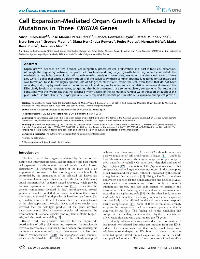
Cell Expansion-Mediated Organ Growth Is Affected by Mutations in Three EXIGUA Genes
13
Cell Expansion-Mediated Organ Growth Is Affected by Mutations in Three EXIGUA Genes Silvia Rubio-Dı´az 1. , Jose ´ Manuel Pe ´ rez-Pe ´ rez 1. , Rebeca Gonza ´ lez-Bayo ´n 1 , Rafael Mun ˜ oz-Viana 1 , Nero Borrega 2 , Gregory Mouille 2 , Diana Herna ´ ndez-Romero 1 , Pedro Robles 1 , Herman Ho ¨ fte 2 ,Marı´a Rosa Ponce 1 , Jose ´ Luis Micol 1 * 1 Instituto de Bioingenierı ´a, Universidad Miguel Herna ´ndez, Campus de Elche, Elche, Alicante, Spain, 2 Institut Jean-Pierre Bourgin, UMR1318 Institut National de Recherche Agronomique/AgroParisTech, INRA Centre de Versailles-Grignon, Versailles, France Abstract Organ growth depends on two distinct, yet integrated, processes: cell proliferation and post-mitotic cell expansion. Although the regulatory networks of plant cell proliferation during organ growth have begun to be unveiled, the mechanisms regulating post-mitotic cell growth remain mostly unknown. Here, we report the characterization of three EXIGUA (EXI) genes that encode different subunits of the cellulose synthase complex specifically required for secondary cell wall formation. Despite this highly specific role of EXI genes, all the cells within the leaf, even those that do not have secondary walls, display small sizes in the exi mutants. In addition, we found a positive correlation between cell size and the DNA ploidy levels in exi mutant leaves, suggesting that both processes share some regulatory components. Our results are consistent with the hypothesis that the collapsed xylem vessels of the exi mutants hamper water transport throughout the plant, which, in turn, limits the turgor pressure levels required for normal post-mitotic cell expansion during leaf growth. Citation: Rubio-Dı ´az S, Pe ´ rez-Pe ´rez JM, Gonza ´lez-Bayo ´n R, Mun ˜ oz-Viana R, Borrega 2 N, et al. (2012) Cell Expansion-Mediated Organ Growth Is Affected by Mutations in Three EXIGUA Genes. PLoS ONE 7(5): e36500. doi:10.1371/journal.pone.0036500 Editor: Miguel A Blazquez, Instituto de Biologı ´a Molecular y Celular de Plantas, Spain Received December 20, 2011; Accepted April 2, 2012; Published May 4, 2012 Copyright: ß 2012 Rubio-Dı ´az et al. This is an open-access article distributed under the terms of the Creative Commons Attribution License, which permits unrestricted use, distribution, and reproduction in any medium, provided the original author and source are credited. Funding: This work was supported by the Ministerio de Ciencia e Innovacio ´ n of Spain [BFU2011-22825 and CSD2007-00057 (TRANSPLANTA) grants, awarded to JLM], the Generalitat Valenciana (PROMETEO/2009/112, to JLM) and the European Commission [LSHG-CT-2006-037704 (AGRON-OMICS), to JLM and HH]. The funders had no role in study design, data collection and analysis, decision to publish, or preparation of the manuscript. Competing Interests: The authors have declared that no competing interests exist. * E-mail: [email protected] . These authors contributed equally to this work. Introduction The final size of plant organs is achieved by the rate of two distinct but integrated processes, cell proliferation and post-mitotic cell expansion, which increase the cell number and cell size, respectively [1]. However, the shape of the plant cell is an important determinant of plant morphogenesis, which is firmly controlled by the organization of the cell wall [2]. Leaves are determinate lateral organs that arise from the flanks of the shoot apical meristem (SAM) as dome-shaped structures, which grow by laminar expansion up to a certain size [3,4]. To identify the genetic components involved in leaf morphogenesis, several genetic screens for non-lethal and visible mutations that disrupt the shape and size of Arabidopsis leaves have been performed [5– 7]. To date, dozens of these leaf mutants have been characterized at the phenotypic and molecular levels, and these studies have revealed that the wild-type products of the genes involved participate in such diverse processes as polar cell expansion, the transduction of hormonal signals, gene regulation, plastid biogen- esis, and chromatin remodeling [8]. Recent work has provided evidence for the organ-wide coordination of cell proliferation and expansion. In Arabidopsis leaves, a decrease in cell number below a certain threshold triggers an increase in mature cell size, a phenomenon that has been termed ‘‘compensation’’ [9,10]. In angustifolia3 (an3) mutants, which are impaired in cell proliferation, the palisade mesophyll cells are larger than normal [11], and AN3 is thought to act as a positive regulator of cell proliferation in leaves [11]. Additional loss-of-function mutants exhibiting a compensation phenotype in their palisade mesophyll cells have been identified and named fugu1 to fugu5 [12]. Examination of the fugu mutants showed that compensated cell enlargement does not occur via the uncoupling of cell division and cell growth, rather, it is sustained by the specific upregulation of cell expansion [12]. Using a Cre/lox recombina- tion system designed for the clonal activation and deletion of AN3, an3-dependent compensation was shown to be a non-cell- autonomous process, and an3 cells seemed to generate and transmit an intercellular signal that enhances post-mitotic cell expansion in neighboring cells [13]. On the other hand, the extra- small sisters (xs) mutants are specifically impaired in cell expansion and are likely to be affected in the cell enlargement response during compensation [14]. Some of these xs mutations strongly suppress the compensated cell enlargement phenotype that is triggered by an3 [14]. This finding led to the conclusion that compensated cell enlargement is mediated by the hyperactivation of cell expansion pathways that require the XS genes. To identify additional factors involved in the coordination of leaf growth, we selected four exigua (exi) mutants from our EMS- induced leaf mutant collection that display small leaves with relatively normal shapes [5]. We found that these exi mutants exhibited specific defects in cell expansion but normal palisade mesophyll cell numbers. The exi mutations were found to affect PLoS ONE | www.plosone.org 1 May 2012 | Volume 7 | Issue 5 | e36500
-
Upload
independent -
Category
Documents
-
view
1 -
download
0
Transcript of Cell Expansion-Mediated Organ Growth Is Affected by Mutations in Three EXIGUA Genes
Cell Expansion-Mediated Organ Growth Is Affected byMutations in Three EXIGUA GenesSilvia Rubio-Dıaz1., Jose Manuel Perez-Perez1., Rebeca Gonzalez-Bayon1, Rafael Munoz-Viana1,
Nero Borrega2, Gregory Mouille2, Diana Hernandez-Romero1, Pedro Robles1, Herman Hofte2, Marıa
Rosa Ponce1, Jose Luis Micol1*
1 Instituto de Bioingenierıa, Universidad Miguel Hernandez, Campus de Elche, Elche, Alicante, Spain, 2 Institut Jean-Pierre Bourgin, UMR1318 Institut National de
Recherche Agronomique/AgroParisTech, INRA Centre de Versailles-Grignon, Versailles, France
Abstract
Organ growth depends on two distinct, yet integrated, processes: cell proliferation and post-mitotic cell expansion.Although the regulatory networks of plant cell proliferation during organ growth have begun to be unveiled, themechanisms regulating post-mitotic cell growth remain mostly unknown. Here, we report the characterization of threeEXIGUA (EXI) genes that encode different subunits of the cellulose synthase complex specifically required for secondary cellwall formation. Despite this highly specific role of EXI genes, all the cells within the leaf, even those that do not havesecondary walls, display small sizes in the exi mutants. In addition, we found a positive correlation between cell size and theDNA ploidy levels in exi mutant leaves, suggesting that both processes share some regulatory components. Our results areconsistent with the hypothesis that the collapsed xylem vessels of the exi mutants hamper water transport throughout theplant, which, in turn, limits the turgor pressure levels required for normal post-mitotic cell expansion during leaf growth.
Citation: Rubio-Dıaz S, Perez-Perez JM, Gonzalez-Bayon R, Munoz-Viana R, Borrega2 N, et al. (2012) Cell Expansion-Mediated Organ Growth Is Affected byMutations in Three EXIGUA Genes. PLoS ONE 7(5): e36500. doi:10.1371/journal.pone.0036500
Editor: Miguel A Blazquez, Instituto de Biologıa Molecular y Celular de Plantas, Spain
Received December 20, 2011; Accepted April 2, 2012; Published May 4, 2012
Copyright: � 2012 Rubio-Dıaz et al. This is an open-access article distributed under the terms of the Creative Commons Attribution License, which permitsunrestricted use, distribution, and reproduction in any medium, provided the original author and source are credited.
Funding: This work was supported by the Ministerio de Ciencia e Innovacion of Spain [BFU2011-22825 and CSD2007-00057 (TRANSPLANTA) grants, awarded toJLM], the Generalitat Valenciana (PROMETEO/2009/112, to JLM) and the European Commission [LSHG-CT-2006-037704 (AGRON-OMICS), to JLM and HH]. Thefunders had no role in study design, data collection and analysis, decision to publish, or preparation of the manuscript.
Competing Interests: The authors have declared that no competing interests exist.
* E-mail: [email protected]
. These authors contributed equally to this work.
Introduction
The final size of plant organs is achieved by the rate of two
distinct but integrated processes, cell proliferation and post-mitotic
cell expansion, which increase the cell number and cell size,
respectively [1]. However, the shape of the plant cell is an
important determinant of plant morphogenesis, which is firmly
controlled by the organization of the cell wall [2]. Leaves are
determinate lateral organs that arise from the flanks of the shoot
apical meristem (SAM) as dome-shaped structures, which grow by
laminar expansion up to a certain size [3,4]. To identify the
genetic components involved in leaf morphogenesis, several
genetic screens for non-lethal and visible mutations that disrupt
the shape and size of Arabidopsis leaves have been performed [5–
7]. To date, dozens of these leaf mutants have been characterized
at the phenotypic and molecular levels, and these studies have
revealed that the wild-type products of the genes involved
participate in such diverse processes as polar cell expansion, the
transduction of hormonal signals, gene regulation, plastid biogen-
esis, and chromatin remodeling [8].
Recent work has provided evidence for the organ-wide
coordination of cell proliferation and expansion. In Arabidopsis
leaves, a decrease in cell number below a certain threshold triggers
an increase in mature cell size, a phenomenon that has been
termed ‘‘compensation’’ [9,10]. In angustifolia3 (an3) mutants,
which are impaired in cell proliferation, the palisade mesophyll
cells are larger than normal [11], and AN3 is thought to act as a
positive regulator of cell proliferation in leaves [11]. Additional
loss-of-function mutants exhibiting a compensation phenotype in
their palisade mesophyll cells have been identified and named
fugu1 to fugu5 [12]. Examination of the fugu mutants showed that
compensated cell enlargement does not occur via the uncoupling
of cell division and cell growth, rather, it is sustained by the specific
upregulation of cell expansion [12]. Using a Cre/lox recombina-
tion system designed for the clonal activation and deletion of AN3,
an3-dependent compensation was shown to be a non-cell-
autonomous process, and an3 cells seemed to generate and
transmit an intercellular signal that enhances post-mitotic cell
expansion in neighboring cells [13]. On the other hand, the extra-
small sisters (xs) mutants are specifically impaired in cell expansion
and are likely to be affected in the cell enlargement response
during compensation [14]. Some of these xs mutations strongly
suppress the compensated cell enlargement phenotype that is
triggered by an3 [14]. This finding led to the conclusion that
compensated cell enlargement is mediated by the hyperactivation
of cell expansion pathways that require the XS genes.
To identify additional factors involved in the coordination of
leaf growth, we selected four exigua (exi) mutants from our EMS-
induced leaf mutant collection that display small leaves with
relatively normal shapes [5]. We found that these exi mutants
exhibited specific defects in cell expansion but normal palisade
mesophyll cell numbers. The exi mutations were found to affect
PLoS ONE | www.plosone.org 1 May 2012 | Volume 7 | Issue 5 | e36500
three genes encoding three subunits of the cellulose synthase
complex that is required for secondary cell wall biosynthesis:
CESA4, CESA7, and CESA8 [15]. We gathered empirical data to
provide a causal explanation that accounts for the small leaf
phenotype of the exi mutants. Our results are consistent with the
hypothesis that internal turgor pressure drives cell expansion
during leaf growth. Based on our own study and those of others,
we hypothesize that a turgor-mediated cell expansion mechanism
might account, at least in part, for leaf growth coordination in
Arabidopsis.
Results
The Exigua Mutants Display Small Leaves Due toDefective Cell Expansion
In a forward genetic screen, we previously identified hundreds
of mutations affecting the size and shape of the vegetative leaves of
Arabidopsis thaliana [5]. Four of the recessive mutations from this
collection, exigua1-1 (exi1-1), exi1-2, exi2 and exi5, caused a
phenotype that is characterized by small, dark-green vegetative
leaves of a relatively normal shape (Figure 1A-E). First- and third-
node leaf area was significantly reduced in the exi mutants in
comparison with their wild-type, Landsberg erecta (Ler) (Figure 1E
and 1G). Other organs in the exi mutants, such as the primary root
(Figure 1H) and the main inflorescence stem (Figure 1I), displayed
smaller sizes than those in Ler. The flowers (Figure 1J) and mature
siliques (Figure 1K) of the exi mutants were also reduced in length
compared with Ler.
To describe the leaf phenotype of the exi mutants at the cellular
level, we drew the cell borders from differential interference
contrast (DIC) micrographs of the adaxial epidermis and the
palisade mesophyll cells (see Materials and Methods). In the leaf
epidermis of first- and third-node leaves, the sizes of the pavement
cells and guard cells are significantly reduced in the exi1-2, exi2 and
exi5 mutants compared with Ler (Figure 2). The number of
epidermal cells per leaf lamina, as estimated from the average
values for adaxial epidermal cell densities and lamina areas (see
Materials and Methods), do not significantly differ between the exi
mutants and Ler (Figure 2E), indicating that the exi mutations do
not alter leaf epidermal cell divisions. We also studied internal leaf
structure in the exi mutants (Figure 3). Transverse sections of the
third-node vegetative leaves of exi1-2, exi2 and exi5 homozygotes
did not reveal significant differences in the interveinal regions
(Figure 3A–D). However, using confocal laser-scanning microsco-
py of native chlorophyll fluorescence and assuming that the
number of chloroplasts per palisade mesophyll cell is similar in all
the genotypes studied (Figure 3E–H), we found that the cell size is
significantly reduced in the exi1-2, exi2 and exi5 mutant leaves in
comparison with those in Ler. In addition, the sizes of the
mesophyll cells, measured using differential intereference contrast
micrographs, were significantly reduced in the first- and third-
node leaves of the exi mutants compared with Ler (P value,0.05)
(Figure 3I). Taken together, our results indicate that the recessive
exi mutations specifically reduce the size of every cell within the
leaf, which in turn generates small leaves with a dark-green color
due to an increased chloroplast density in their mesophyll cells.
The morphological phenotype of different organs in the exi
mutants suggests a positive role for the EXI1, EXI2 and EXI5
genes in the regulation of cell expansion.
To investigate the functional relationship between the EXI
genes, we obtained the exi2 exi5 double mutant. Double mutant
plants were dwarf and had small, dark-green leaves and short
inflorescences (Figure S1), a phenotype that was stronger than
those of their parentals. Based on the severity of this phenotype
[16], we considered the exi2 exi5 double mutant phenotype as
additive.
The Exi Mutations Affect Leaf Ploidy LevelsA positive correlation between cell size and nuclear ploidy levels
has been found in wild-type Arabidopsis leaves [17], and we
questioned to what extent the cell expansion defects observed in
the exi mutant leaves might be caused by altered ploidy levels in
their cells. Cells with higher ploidy levels (.4C) arise by
endoreduplication (i.e., successive DNA replication cycles without
intervening mitoses) [18]. We found that the 8C and 16C nuclei
populations are significantly reduced in the first pair of leaves of
the exi mutant rosettes compared to Ler (Table 1), which is
consistent with a global reduction of the endoreduplication levels
in exi leaves. In addition, a mild but significant increase in the
diploid nuclei population (2C) of the exi leaves compared with that
of Ler was found (Table 1). The 4C population was also more
represented in the exi mutants, compared to Ler, and includes both
mitotic diploid cells in the S/G2 phase of the cell cycle and
tetraploid endoreduplicated cells in G0/G1 that have arisen after
one endocycle. Because we found no evidence for increased cell
division in exi leaves, the enlarged 2C and 4C populations
observed in the exi mutants are likely derived from the small
proportion of cells that enter the endocycle in these mutants. Our
results indicate that cell size and endoreduplication levels are
positively correlated in the exi leaves, suggesting that the function
of the EXI genes in the regulation of cell expansion is coupled with
endoreduplication levels during leaf growth.
The EXI Genes Encode the Three Cellulose SynthaseSubunits
Because none of the exi mutations under study was tagged, we
followed a positional cloning approach for their molecular
identification. Fine mapping of the exi1-1, exi2 and exi5 mutations
defined candidate intervals encompassing 70, 55 and 110 genes,
respectively (Figure 4A). The four exi mutants studied shared a
similar leaf phenotype (see above), and we reasoned that the EXI
genes might be functionally related. Indeed, each of the three
genomic intervals found contained a gene encoding a catalytic
subunit of the cellulose synthase A complex: CESA8 (also named
IRX1; At4g18780) [19], CESA7 (IRX3; At5g17420) [20] and
CESA4 (IRX5; At5g44030) [15] were included within the exi1-1,
exi5 and exi2 candidate intervals, respectively. Interestingly, the
Arabidopsis genome encodes 10 CESA proteins [21], of which
only those produced by the CESA4, CESA7, and CESA8 genes are
integrated into the CESA complex that is responsible for the
biosynthesis of secondary cell walls [22]. The exi1-1 mutant was
found to carry a GRA transition in the acceptor site of the 8th
intron of the CESA8 gene (Figure 4B), thus altering its mRNA
splicing (Figure S2). The exi1-2 mutation (Figure 4B) changes a
conserved glycine residue to glutamic acid in the second
cytoplasmic domain of the CESA8 protein that is required for
substrate binding and catalysis [23]. The exi5 mutation (Figure 4B)
results in the introduction of a stop codon in place of the
tryptophan 954 residue, which causes a premature termination of
translation. The exi2 mutation affects the last exon of the CESA4
gene (Figure 4B) and introduces a premature stop codon that
removes 109 residues from the CESA4 C-terminus.
Homozygotes for a T-DNA insertion in the CESA8 gene were
obtained from the Salk_026812 line [24], which exhibited the
same phenotype already observed in the exi mutants (we will
hereafter refer to this allele as irx1-5; Figure S3). We confirmed the
presence of the T-DNA insertion in the first intron of CESA8 in the
irx1-5 plants (Figure 4B). The F1 progeny of an irx1-5 6 exi1-1
Role of EXIGUA Genes in Leaf Growth Regulation
PLoS ONE | www.plosone.org 2 May 2012 | Volume 7 | Issue 5 | e36500

cross were phenotypically similar to the exi1-1 parental, confirming
that irx1-5 and exi1-1 are allelic. Rosettes of the Salk_029940C line
[24,25], which bear a T-DNA insertion in the first intron of CESA7
(irx3-4 in Figure 4B), also display a small-leaf phenotype (Figure
S3), indicating allelism with our exi5 mutants. Complementation
tests also allowed us to demonstrate allelism between exi2 and the
T-DNA insertion in the third exon of CESA4 (irx5-4 in the
Figure 4B), carried by the Salk_084627 line [24,25], which causes
small, dark-green vegetative leaves (Figure S3).
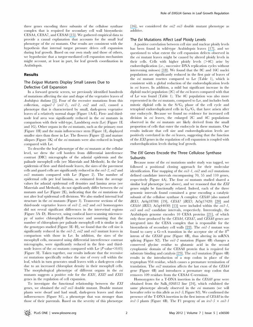
The Exi Mutants Show Altered Vascular TissueMorphology
Mutations in genes encoding CESA complex components cause
an irregular xylem phenotype: xylem vessels are collapsed as a
consequence of reduced cellulose deposition in their cell walls [26].
To analyze whether our exi mutants display similar vascular
defects, we performed scanning electron microscopy on transverse
sections of inflorescence stems (see Materials and Methods). As
expected from the molecular function of the EXI genes in
secondary cell wall biosynthesis, all the exi mutants that we studied
displayed collapsed xylem vessels in the vascular bundles of the
inflorescence stem, whereas their parenchyma cells were of normal
morphology but of smaller sizes than those of Ler (Figure 5A–D).
Based on these observations, we hypothesized that hydraulic
conductivity might be altered in the exi mutants. According to
Poiseuilles law, theoretical hydraulic conductivity of a vessel is
proportional to the fourth power of its radius [27], which implies
that little differences in the vessel radius can lead to great changes
in hydraulic conductivity. We measured the maximal radii from
15 xylem vessels per genotype (Figure 6A), finding that they were
significantly smaller in the exi mutants than in Ler.
The chemical composition of the vascular tissues from the
inflorescence stems was examined using Fourier transform infrared
(FTIR) spectral analysis. A Student’s t-test was performed to
evaluate the differences between the Ler and exi spectra. For
graphical representation, the t-values were plotted against
wavenumbers to determine the significance of the differences at
each wavenumber [28]. Our results suggest that all three mutant
classes had very similar cell wall changes in the vascular tissue of
Figure 1. Morphology of the exi mutants. (A–D) Rosettes of the (A) Landsberg erecta (Ler) wild type and the (B) exi1-2, (C) exi2 and (D) exi5mutants. (E) Series of Ler, exi1-2, exi2 and exi5 leaves. From left to right: two cotyledons and the first to tenth or eleventh leaves. Mutants display small,dark-green leaves of relatively normal shape. (F, G) (F) Rosette mean diameter and (G) lamina area from first-node (white bars) or third-node (greybars) leaves. Asterisks indicate significant differences from the Ler control (P value,0.05; n = 11–20 individuals). (H) Root morphology in Ler (left) andexi5 (right) seedlings grown in near-vertically oriented plates. (I) From left to right, the reproductive morphology of Ler, exi1-2 and exi2 plants grownin pots. (J) Detail of Ler (up) and exi1-2 (down) inflorescences. (K) From left to right, siliques from Ler, exi1-2, exi2 and exi5 plants. Samples were taken(A–H) 21 and (I–K) 38 DAS. All the plants shown were homozygous for the indicated mutations (in italics). Scale bars indicate (A–E, H, J and K) 5 mmand (I) 3 cm.doi:10.1371/journal.pone.0036500.g001
Role of EXIGUA Genes in Leaf Growth Regulation
PLoS ONE | www.plosone.org 3 May 2012 | Volume 7 | Issue 5 | e36500

the stem in the three exi mutants analyzed (Figure 5E–F), including
a reduced cellulose content as suggested by the positive t value at
the wavenumbers 1319, 1157, 1106, 1060, 1037 and 990 cm-1,
correlated with an increase of lignin content as suggested by the
negative t value at the wavenumbers 1708 and, 1515 to 1392 cm-1
[28]. Defective secondary cell wall biosynthesis in the xylem vessels
of exi stems, caused by impaired cellulose biosynthesis, might
increase the sensitivity of the xylem to a high negative water
pressure, which, in turn, would cause the observed collapse of the
xylem.
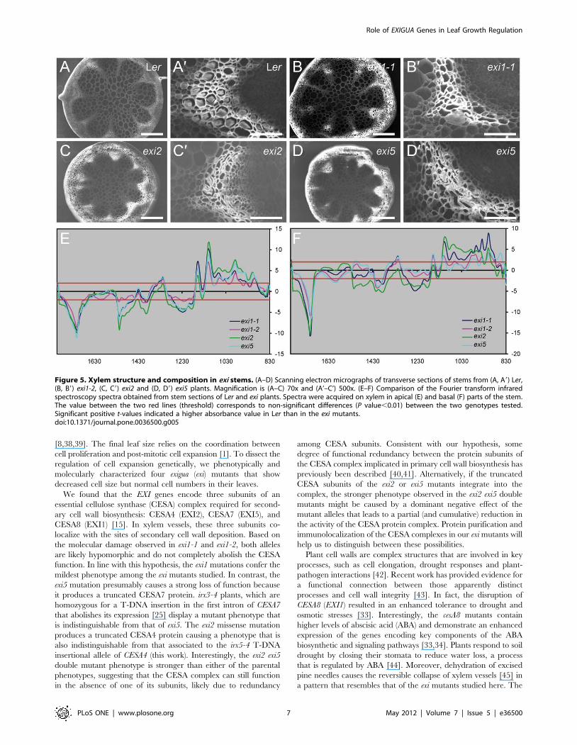
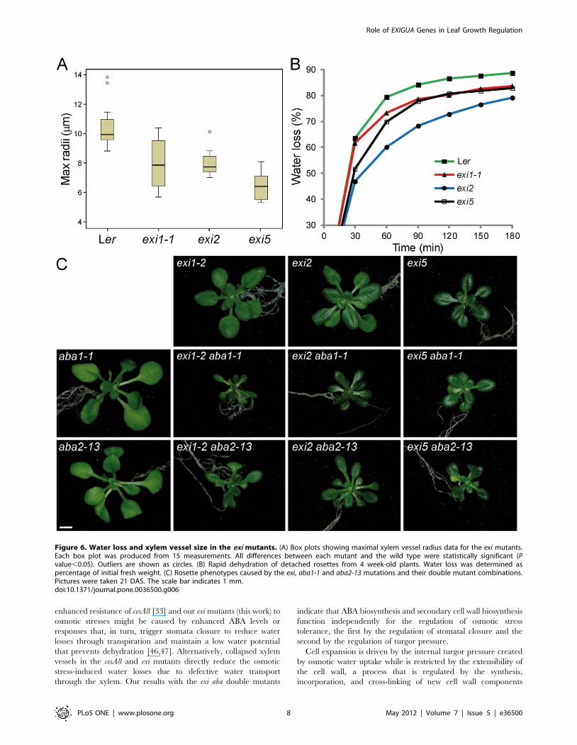
The Exi Mutants Display Increased Resistance to OsmoticStress
A number of physiological processes, such as osmotic stress
responses, are regulated by water availability. Because the exi
mutants show collapsed xylem vessels, water transport might be
obstructed in these mutants and, as a consequence, the exi mutants
might be affected in their osmotic stress responses.
We first tested the responses of exi mutants to rapid
dehydration (see Materials and Methods). Consistent with the
hypothesis that the morphological alterations in xylem vessels
impair water transport in the exi mutants, their dehydration
rates were reduced compared to Ler (Figure 6B). We assayed
the osmotic stress responses by transferring young plants to
media supplemented with different concentrations of the
osmoticum mannitol or NaCl and by measuring the rosette
fresh weight and dry weight after one week of growth on either
of these media (see Materials and Methods). The rosette fresh
weight (Figure 7C) but not the dry weight (Figure 7D) was
significantly decreased in Ler plants grown on mannitol, due to
the severe water loss on the osmoticum-supplemented media.
Similarly, the rosette fresh weight was greatly reduced in the
abscisic acid (ABA)-deficient mutant aba2-13 [29] when grown
on mannitol-supplemented plates, even at low levels (50 mM
mannitol). The exi mutants, however, were more resistant to
mannitol-induced water losses, and their rosette fresh weights
were not significantly affected by increasing the level of
mannitol (Figure 7C). The rosettes of Ler plants grown for
one week on 100 mM mannitol displayed a severe growth
reduction and enhanced senescence in older leaves, whereas the
exi mutant rosettes showed less obvious morphological alter-
ations on high mannitol concentrations (Figure 7A–B). The Ler
rosettes grown on media supplemented with 50 mM NaCl
displayed increased fresh weights after one week due to their
higher water content, compared with the plants grown on the
control media (Figure 7C–D). In contrast, the fresh weights of
the exi rosettes grown for one week on media containing
50 mM NaCl were not significantly different from those of the
Figure 2. Leaf adaxial epidermis in the exi mutants. (A–D) Representative diagrams from the leaf adaxial epidermis of exi mutants. Diagramswere drawn from the differential interference contrast images of cleared third-node leaves collected from (A) Ler, (B) exi1-2, (C) exi2 and (D) exi5plants. (E) Adaxial pavement cell area, cell density and cell number in first-node (white bars) or third-node (grey bars) leaves of the exi mutants. Datawere normalized with respect to the Ler value and are expressed as percentages. Represented values are the mean and the standard deviation,except for the pavement cell area, which did not exhibit a normal distribution and for which the median was used. Asterisks indicate significantdifferences from the Ler control (P value,0.05; n = 11–15). All the data were obtained at 21 DAS from plants grown on half-strength MS agar medium.Scale bars indicate 50 mm.doi:10.1371/journal.pone.0036500.g002
Role of EXIGUA Genes in Leaf Growth Regulation
PLoS ONE | www.plosone.org 4 May 2012 | Volume 7 | Issue 5 | e36500

exi plants grown on the non-supplemented media (Figure 7C–
D), which clearly indicates a defect in water transport in the exi
mutants. Taken together, our results suggest that the increased
resistance to osmotic stress and the low responses of exi mutants
to moderate NaCl levels are caused by defective water flux
regulation due to xylem collapse in the exi mutants.
Figure 3. Internal leaf structure in the exi mutants. (A–D) Transverse sections of a region midway between the midvein and the leaf marginfrom (A) Ler, (B) exi1-2, (C) exi2 and (D) exi5 third-node vegetative leaves. (E–H) Confocal micrographs of palisade mesophyll sections of first-nodeleaves from (E) Ler, (F) exi1-2, (G) exi2 and (H) exi5 mutants. (I) Palisade mesophyll cell size in the subepidermal layer of first-node (white bars) or third-node (grey bars) leaves of Ler and exi mutants. Data were normalized with respect to the Ler value and expressed as percentages (mean and standarddeviation). Asterisks indicate significant differences from the Ler control (P value,0.05; n = 13–16 individuals). All the data were obtained at 21 DASfrom plants grown on half-strength MS agar medium. Scale bars indicate (A–D) 100 mm and (E–H) 50 mm.doi:10.1371/journal.pone.0036500.g003
Table 1. Nuclear DNA ploidy distribution in the first pair of leaves of the exi mutants.
Genotype Ploidy level
2C 4C 8C 16C 32C
Ler 23.462.8 (22.3) 27.561.1 (27.7) 35.061.3 (35.2) 13.362.6 (13.9) 0.860.3 (0.7)
exi1-2 29.261.2 (29.1) 27.361.1 (27.4) 30.8±2.3 (30.4) 11.262.0 (12.4) 1.560.6 (1.6)
exi2 27.661.0 (27.5) 34.561.1 (34.3) 31.9±1.5 (31.9) 5.4±0.8 (5.4) 0.660.1 (0.7)
exi5 32.761.3 (32.7) 34.160.6 (34.1) 28.8±1.2 (28.9) 3.9±0.5 (4.0) 0.560.1 (0.5)
Values are mean6standard deviation. The median is shown in parentheses. Significant differences (P value,0.01, n = 10) from the Ler control are in bold. Values higherthan those of Ler control are in italics. Data analysis was performed as indicated in Materials and Methods. All plants were homozygous for the mutations indicated.doi:10.1371/journal.pone.0036500.t001
Role of EXIGUA Genes in Leaf Growth Regulation
PLoS ONE | www.plosone.org 5 May 2012 | Volume 7 | Issue 5 | e36500

The EXI Genes Act Independently from the ABABiosynthetic Pathway
ABA is involved in many environmental stress responses,
particularly cold, salinity and drought [30]. In response to
drought, an increase in the endogenous ABA levels limits
transpirational water loss through the regulation of stomatal
closure. ABA-deficient mutants, like aba1 and aba2, display a
stunted (wilty) phenotype, even under well-watered conditions, as a
consequence of their inability to retain water because of impaired
stomatal closure [31,32]. Based on the higher ABA levels observed
in the cesA8 mutant, a crosstalk between secondary cell wall
integrity and ABA signaling pathway has been proposed [33,34].
To test whether ABA accumulation in the exi mutants might be, at
least partly, responsible for the Exi phenotype, we generated
double mutant combinations between our exi mutants and either
the aba1-1 or aba2-13 ABA-deficient mutants (Figure 6C). The
aba1-1 and aba2-13 single mutants exhibit a wilty phenotype and
their rosettes are slightly smaller than those of Ler [31,32]. The
rosette phenotypes of the six exi aba double mutant combinations
studied were all similar: they displayed small and darker-green
vegetative leaves as in their exi parental, as well as the wilty
phenotype characteristic of their ABA-deficient parental
(Figure 6C and data not shown). In addition, rosette size was
more reduced in the double mutants than in any of their parentals.
We interpreted the phenotype of the exi aba double mutants as
additive, an observation that also suggests that the phenotype of
the exi mutants is not directly caused by high levels of ABA.
Cell Expansion Defects of the Exi Mutants are Rescued byLiquid Culture
The cellulose biosynthesis activity encoded by the three EXI
genes studied here is restricted to the deposition of secondary walls
of developing xylem [35]. In the exi mutants, however, all the leaf
cells –including those without secondary walls– display small sizes,
despite the fact that most of them lack secondary cell walls. We
then addressed the possible causes of the observed non-cell
autonomy of this phenotype. It has been suggested that water
uptake into the vacuole creates the internal turgor pressure
necessary for the extension of the cell wall through the synthesis of
new wall components [36]. Because cells in the exi leaves are
impaired in water uptake, we reasoned that the internal turgor
pressure required for primary cell wall extension during leaf
growth is limited in the exi mutants. Therefore, the primary cell
wall is not directly affected by the exi mutations, and experimen-
tally increasing the turgor pressure in the exi leaves should rescue
their small-cell phenotype. Alternatively, changes in the secondary
cell wall composition in the exi mutants might be sensed by
younger growing tissues, leading to a complex alteration in the
composition of the primary cell wall in newly formed organs, as
has previously been proposed [37].
To ensure that water uptake was independent from its transport
through the vascular tissue, Ler and the exi mutants were grown for
three weeks in 10 ml of half-strength MS liquid medium (see
Materials and Methods). In this situation, water freely enters
through the stomata into the leaf mesophyll, where the increase in
internal turgor pressure might trigger cell expansion. Although exi
leaves grown in liquid culture remained smaller than those of Ler
(Figure 8I), the size of the palisade mesophyll cells in the Ler and exi
leaves grown in liquid culture did not significantly differ (Figure 8J).
Indeed, the palisade mesophyll cells of the exi mutants grown in
liquid culture were of similar sizes as the Ler mesophyll cells grown
on Petri dishes (Figure 8A–H). Our results confirm the rescue of
primary cell wall expansion in the palisade mesophyll cells of the
exi mutants by experimentally increasing the water uptake in these
cells. Thus, the small cells of the exi mutant leaves are caused by
impaired water transport through the xylem vessels due to their
collapse caused by defective secondary cell wall formation.
Discussion
Forward genetic screening in Arabidopsis is a powerful tool that
has helped to identify several key loci involved in leaf development
Figure 4. Positional cloning of the EXI1, EXI2 and EXI5 genes. (A)Map-based cloning strategy used to identify the EXI genes. Mappingpopulations of 217 F2 plants were derived from an exi1-16Col-0 cross,417 from an exi26Col-0 cross, and 278 from an exi56Col-0 cross. Thecandidate intervals for EXI1, EXI2 and EXI5 were of 231, 233 and 390 kb,respectively, which encompassed 70, 55 and 110 annotated genes. Themolecular markers used for linkage analysis, their map positions and thenumber of informative recombinants identified (in parentheses) areindicated. (B) Structure of the EXI1, EXI2 and EXI5 genes, with indicationof the nucleotide position (relative to the translation start site) andnature of the mutations found; a triangle indicates a T-DNA insertion.Exons are represented as boxes, and introns by lines connecting boxes.The translation start (ATG) and stop (TAA or TGA) codons are alsoindicated.doi:10.1371/journal.pone.0036500.g004
Role of EXIGUA Genes in Leaf Growth Regulation
PLoS ONE | www.plosone.org 6 May 2012 | Volume 7 | Issue 5 | e36500
[8,38,39]. The final leaf size relies on the coordination between
cell proliferation and post-mitotic cell expansion [1]. To dissect the
regulation of cell expansion genetically, we phenotypically and
molecularly characterized four exigua (exi) mutants that show
decreased cell size but normal cell numbers in their leaves.
We found that the EXI genes encode three subunits of an
essential cellulose synthase (CESA) complex required for second-
ary cell wall biosynthesis: CESA4 (EXI2), CESA7 (EXI5), and
CESA8 (EXI1) [15]. In xylem vessels, these three subunits co-
localize with the sites of secondary cell wall deposition. Based on
the molecular damage observed in exi1-1 and exi1-2, both alleles
are likely hypomorphic and do not completely abolish the CESA
function. In line with this hypothesis, the exi1 mutations confer the
mildest phenotype among the exi mutants studied. In contrast, the
exi5 mutation presumably causes a strong loss of function because
it produces a truncated CESA7 protein. irx3-4 plants, which are
homozygous for a T-DNA insertion in the first intron of CESA7
that abolishes its expression [25] display a mutant phenotype that
is indistinguishable from that of exi5. The exi2 missense mutation
produces a truncated CESA4 protein causing a phenotype that is
also indistinguishable from that associated to the irx5-4 T-DNA
insertional allele of CESA4 (this work). Interestingly, the exi2 exi5
double mutant phenotype is stronger than either of the parental
phenotypes, suggesting that the CESA complex can still function
in the absence of one of its subunits, likely due to redundancy
among CESA subunits. Consistent with our hypothesis, some
degree of functional redundancy between the protein subunits of
the CESA complex implicated in primary cell wall biosynthesis has
previously been described [40,41]. Alternatively, if the truncated
CESA subunits of the exi2 or exi5 mutants integrate into the
complex, the stronger phenotype observed in the exi2 exi5 double
mutants might be caused by a dominant negative effect of the
mutant alleles that leads to a partial (and cumulative) reduction in
the activity of the CESA protein complex. Protein purification and
immunolocalization of the CESA complexes in our exi mutants will
help us to distinguish between these possibilities.
Plant cell walls are complex structures that are involved in key
processes, such as cell elongation, drought responses and plant-
pathogen interactions [42]. Recent work has provided evidence for
a functional connection between those apparently distinct
processes and cell wall integrity [43]. In fact, the disruption of
CESA8 (EXI1) resulted in an enhanced tolerance to drought and
osmotic stresses [33]. Interestingly, the cesA8 mutants contain
higher levels of abscisic acid (ABA) and demonstrate an enhanced
expression of the genes encoding key components of the ABA
biosynthetic and signaling pathways [33,34]. Plants respond to soil
drought by closing their stomata to reduce water loss, a process
that is regulated by ABA [44]. Moreover, dehydration of excised
pine needles causes the reversible collapse of xylem vessels [45] in
a pattern that resembles that of the exi mutants studied here. The
Figure 5. Xylem structure and composition in exi stems. (A–D) Scanning electron micrographs of transverse sections of stems from (A, A9) Ler,(B, B9) exi1-2, (C, C9) exi2 and (D, D9) exi5 plants. Magnification is (A–C) 70x and (A’–C’) 500x. (E–F) Comparison of the Fourier transform infraredspectroscopy spectra obtained from stem sections of Ler and exi plants. Spectra were acquired on xylem in apical (E) and basal (F) parts of the stem.The value between the two red lines (threshold) corresponds to non-significant differences (P value,0.01) between the two genotypes tested.Significant positive t-values indicated a higher absorbance value in Ler than in the exi mutants.doi:10.1371/journal.pone.0036500.g005
Role of EXIGUA Genes in Leaf Growth Regulation
PLoS ONE | www.plosone.org 7 May 2012 | Volume 7 | Issue 5 | e36500
enhanced resistance of cesA8 [33] and our exi mutants (this work) to
osmotic stresses might be caused by enhanced ABA levels or
responses that, in turn, trigger stomata closure to reduce water
losses through transpiration and maintain a low water potential
that prevents dehydration [46,47]. Alternatively, collapsed xylem
vessels in the cesA8 and exi mutants directly reduce the osmotic
stress-induced water losses due to defective water transport
through the xylem. Our results with the exi aba double mutants
indicate that ABA biosynthesis and secondary cell wall biosynthesis
function independently for the regulation of osmotic stress
tolerance, the first by the regulation of stomatal closure and the
second by the regulation of turgor pressure.
Cell expansion is driven by the internal turgor pressure created
by osmotic water uptake while is restricted by the extensibility of
the cell wall, a process that is regulated by the synthesis,
incorporation, and cross-linking of new cell wall components
Figure 6. Water loss and xylem vessel size in the exi mutants. (A) Box plots showing maximal xylem vessel radius data for the exi mutants.Each box plot was produced from 15 measurements. All differences between each mutant and the wild type were statistically significant (Pvalue,0.05). Outliers are shown as circles. (B) Rapid dehydration of detached rosettes from 4 week-old plants. Water loss was determined aspercentage of initial fresh weight. (C) Rosette phenotypes caused by the exi, aba1-1 and aba2-13 mutations and their double mutant combinations.Pictures were taken 21 DAS. The scale bar indicates 1 mm.doi:10.1371/journal.pone.0036500.g006
Role of EXIGUA Genes in Leaf Growth Regulation
PLoS ONE | www.plosone.org 8 May 2012 | Volume 7 | Issue 5 | e36500
[36,48]. Inhibition of cellulose biosynthesis, either genetically or
chemically, leads to the activation of an unknown mechanism that
monitors cell wall integrity and prevents further growth [49,50].
The molecular function of the EXI genes provides a causal link for
their mutant phenotype of reduced cellulose biosynthesis in
secondary cell walls (i.e., xylem vessels). However, cells without
secondary walls, such as those in the palisade mesophyll of exi
leaves, also show defective cell expansion in these mutants. One
possibility is that secondary cell wall changes are sensed by the
above-mentioned mechanisms to affect downstream signaling
events, leading to local and systemic alterations in primary cell wall
composition in the exi mutants. Our results, however, are
consistent with the alternative hypothesis that the collapsed xylem
vessels in the exi mutants cause reduced water transport
throughout the plant and compromise the turgor pressure required
during cell expansion. By experimentally increasing the turgor
pressure in the palisade mesophyll cells of exi leaves, we were able
to fully rescue their cell expansion defects, demonstrating that
primary cell wall biosynthesis is not directly affected by the exi
mutations. We postulate that the observed tolerance of the exi
mutants to osmotic stresses is also an indirect consequence of
impaired water transport due to their collapsed xylem vessels.
Moreover, hydraulic conductivity through the vessels is expected
to be compromised in the exi mutants. Consistent with our
hypothesis, mutations in the ESKIMO1 gene that encodes a
member of a family comprising xyloglucan actyltransferases [51],
which is expressed in tissues undergoing secondary cell wall
deposition [52,53], also produce a collapsed xylem phenotype and
a concomitant reduction in the water supply to the vegetative
tissues [52]. Similar to that found in the eskimo1 mutants [52], the
exi mutants exhibit low theoretical hydraulic conductivity and a
likely diminished in planta hydraulic conductance due to restricted
water transport through their collapsed vessels.
Figure 7. Osmotic stress tolerance of the exi mutants. (A–B) Rosette morphology in seedlings grown on media (A) without mannitol or (B)supplemented with 100 mM mannitol (B). (C, D) Effects of different concentrations of mannitol or NaCl on the (C) fresh weight (D) dry weight of 26-day-old rosettes. Plants were grown for 19 days on half-strength MS agar medium and then transferred to half-strength MS agar plates supplementedor not with either mannitol or NaCl at different concentrations. Photographs were taken after one week of treatment, and rosettes were immediatelyweighed (fresh weight) or dried for 16 h at 60uC and then weighted (dry weight). Asterisks indicate significant differences from the control plateswithout mannitol or NaCl (P value,0.01). Each column represents the mean of three measurements (8–10 rosettes each). Scale bars indicate 2 mm.doi:10.1371/journal.pone.0036500.g007
Role of EXIGUA Genes in Leaf Growth Regulation
PLoS ONE | www.plosone.org 9 May 2012 | Volume 7 | Issue 5 | e36500

Strong correlations between the ploidy level and cell size have
been reported in many plant species. Although the transition from
cell proliferation to cell expansion is often accompanied by the
transition from the mitotic cycle to the endocycle [18], some
evidence suggests that endoreduplication and cell expansion could
be uncoupled [54]. A recent example is provided by the
constitutive expression of the basic helix-loop-helix transcription
factor ROOT HAIR DEFECTIVE 6-LIKE 4 (RSL4), which
leads to continuous growth of hair cells without an increase in the
DNA ploidy content [55]. However, a decrease in cell proliferation
in Arabidopsis leaf primordia below some threshold level triggers
enhanced post-mitotic cell expansion [10], such as that caused by
the overexpression of the cyclin-dependent kinase (CDK) inhibitor
gene KIP-RELATED PROTEIN2 (KRP2) [56]. We found that the
exi leaves show a global reduction of endoreduplication levels
(.4C) and a slight increase in the size of their diploid cell
population. Our results indicate that in the exi mutants, as occurs
in wild-type Arabidopsis leaves [17], cell size and ploidy levels are
positively correlated, suggesting crosstalk between the cell wall
integrity and regulation of the endocycle. The WEE1 kinase is a
conserved negative regulator of CDK activity during the G2 phase
of the cell cycle [57], and WEE1 orthologs in yeast have been
linked to cell size control [58]. The misexpression of the WEE1
kinase in tomato resulted in dwarf plants due to a reduction in cell
size that correlated with a decrease in their DNA ploidy levels
[59]. It will be interesting to test whether the activity of some of the
regulators of endoreduplication, such as WEE1, is dependent on
changes in the internal turgor pressure.
The results of our studies with the exi mutants support the
hypothesis that the compensated cell enlargement mechanism
observed in Arabidopsis leaves operates mainly by monitoring the
extent of cell proliferation. We hypothesize that an increase in
turgor pressure precedes the new cell wall biosynthesis that triggers
enhanced cell expansion in post-mitotic cells during compensation.
Figure 8. The cell expansion defects of the exi mutants are rescued by liquid culture. (A–H) Leaf mesophyll of the exi mutants. Diagramswere drawn from differential interference contrast micrographs of cleared first-node leaves collected from (A, E) Ler, (B, F) exi1-2, (C, G) exi2 and (D, H)exi5 plants. (I, J) First-node leaf (I) lamina area and (J) mesophyll cell area in the exi mutants. Data were normalized with respect to the Ler values andare expressed as percentages (mean and standard deviation). Asterisks indicate significant differences from the control seedlings grown on half-strength MS agar medium (P value,0.01). All the data were obtained 21 DAS from seedlings grown on half-strength MS agar plates (A–D, white bars)or in half-strength MS liquid cultures (E–H, grey bars). Scale bars indicate 50 mm.doi:10.1371/journal.pone.0036500.g008
Role of EXIGUA Genes in Leaf Growth Regulation
PLoS ONE | www.plosone.org 10 May 2012 | Volume 7 | Issue 5 | e36500
One candidate to be involved in regulating turgor pressure is
vacuolar H+-ATPase (V-ATPase), which is responsible for proton
translocation from the cytosol. The V-ATPase function is impaired
in the de-etiolated3 (det3) mutant, which displays a severe defect in
normal cell expansion [60]. The light-grown det3 phenotype is due
to hormone-mediated changes in gene expression that, in turn,
prohibit cellulose biosynthesis [61].
Our detailed phenotypic analysis of the leaves of the exi mutant
suggests a mechanism for cell expansion-mediated organ growth
that acts through the regulation of water transport and that
monitors internal turgor pressure. Our exi mutants will serve as
molecular tools to dissect the complex relationship between cell
wall integrity and cell cycle regulation during organ growth.
Materials and Methods
Plant Material and Growth ConditionsArabidopsis thaliana (L.) Heynh. exi1-1, exi1-2, exi2 and exi5
mutants were isolated as described in [5]. Salk_026812 (irx1-5),
Salk_029940 (irx3-4) and SalK_084627 (irx5-4) T-DNA insertion
lines were obtained from the Nottingham Arabidopsis Stock Centre
(NASC; http://arabidopsis.info/) and are described at the
SIGnAL website [24] (http://signal.salk.edu). We confirmed the
T-DNA insertion sites of the mutant lines by PCR amplification
and/or sequencing.
Sterile plant cultures were performed as described in [62], on
150-mm diameter Petri dishes containing 100 ml of half-strength
Murashige and Skoog agar medium supplemented with 1%
sucrose (MS medium). The plants were grown at a density of 50
regularly spaced seedlings per plate. Sterile cultures were
incubated in Conviron TC16 growth chambers (Conviron,
Winnipeg, Canada; http://www.conviron.com/) at 20 6 1uCand 60–70% relative humidity, and with continuous illumination
of 78 mmol m-2 s-1.
Morphological and Ultrastructural AnalysesImages of rosettes and leaves were taken 21 days after
stratification (DAS) with a Panasonic DMC-FX9 digital camera
(2,81662,112 pixels; Osaka, Japan). For quantification of the
cell areas, diagrams of the adaxial epidermis and the underlying
palisade mesophyll from cleared leaves [63] were obtained by
drawing the outlines of the cells on a Wacom Cintiq 18SX
Interactive Pen Display (Wacom Company Ltd., Tokyo, Japan;
http://www.wacom.com/). The cell areas were measured from
these images using the NIS Elements AR 2.30 image analysis
package (Nikon, Tokyo, Japan; http://www.nis-elements.com/
index.html) as described in [64]. Photomicrography and
confocal microscopy of whole leaves and leaf sections were
performed as previously described elsewhere [65,66]. All the
data were statistically analyzed with SPSS (Statistical Package
for the Social Sciences) 15.0 software (SPSS Inc., Chicago, IL,
USA) [67] and graphically represented and digitally processed
with Adobe Photoshop and Illustrator CS3 (Adobe Systems
Incorporated, San Jose, CA, USA; http://www.adobe.com/).
For dehydration assays, the entire root system was excised from
a minimum of four 21 DAS rosettes per genotype and water
loss was measured as previously described [52]. In micrographs
taken from stem transverse sections, we measured the maximal
radii of all vessels in 5 to 10 xylem poles per genotype, using
the ImageJ software (http://rsb.info.nih.gov/ij/). The data from
the 15 larger xylem vessels of each genotype were statistically
analyzed with SPSS.
Ploidy AnalysisFlow-cytometry analysis was performed as previously described
[68]. Briefly, first-node and second-node leaves from 4 different
rosettes were harvested 21 DAS and chopped with a razor blade in
500 ml of cold nuclear isolation buffer [69]. The cell suspension
was then filtered over 30-mm nylon mesh, treated with RNase A
(200 mg ml-1) and stained with propidium iodide (50 mg ml-1) for
10 min. The nuclear DNA content was then analyzed using a
FACS Canto II flow cytometer (BD Biosciences, Franklin Lakes,
NJ, USA; http://www.bdbiosciences.com/), and the data were
processed using FACSDiva 6.1.2 software. A minimum of three
biological replicates, including 10,000 counts each, were studied
per genotype.
Positional Cloning and Sequencing of EXI GenesTo positionally clone the EXI1, EXI2 and EXI5 genes, a low-
resolution map was constructed according to [70,71]; for fine
mapping, SSLP, SNP and InDel markers were designed based on
the polymorphisms between the Ler and Col-0 ecotypes described
in the Arabidopsis Polymorphism Database (http://www.
arabidopsis.org). For sequencing of the alleles, PCR products
spanning the At4g18780, At5g17420 and At5g44030 transcrip-
tional units were obtained using wild-type and mutant genomic
DNA as the templates and the oligonucleotide primers shown in
Table S1. The sequencing reactions were carried out with the ABI
PRISM BigDye Terminator cycle sequencing kit (Life Technol-
ogies Corporation, Carlsbad, CA, USA; http://www.
appliedbiosystems.com) in 5-ml reaction volumes. Sequencing
electrophoreses were performed with an ABI PRISM 3130xl
Genetic Analyzer (Applied Biosystems).
RT-PCR AnalysisTotal RNA was extracted from 3-week-old wild-type and exi1-1
rosettes using TRIzol (Life Technologies Corporation, Carlsbad,
CA, USA; http://www.invitrogen.com/), as instructed by the
manufacturer. A 2-mg aliquot of the mRNA was heated for 5 min
at 65uC and immediately transferred to ice. 500 ng of random
hexanucleotides (Roche Applied Science, Basel, Switzerland;
http://www.roche-applied-science.com/) were added and the
mixture was then incubated at room temperature for 10 min.
First-strand cDNA was synthesized in a 20-ml reaction mixture
containing 0.5 mM of each deoxyribonucleoside triphosphate,
10 mM dithiothreitol, 200 units of SuperScript II enzyme (Life
Technologies Corporation), and 40 units of RNaseOUT. The
primer pairs used (EXI1_3bF and EXI1_3R) are shown in Table
S1.
Scanning Electron MicroscopySegments (1 cm) of Arabidopsis inflorescence stems were
embedded in an 8% agarose solution in sterilized/RNAse-free
water at 50uC. After cooling, the blocks were sectioned using a
vibratome. The 100-mm sections were observed immediately after
sectioning using a Hirox SH-1500 scanning electron microscope
(Hirox Co. Ltd., Tokyo, Japan; http://www.hirox.com/).
Fourier Transform Infrared (FTIR) SpectroscopyAnalyses were carried out on the xylem and parenchyma tissue
from the basal and upper regions of branched stems using 50-mm-
thick vibratome sections of agarose-embedded tissue. For each
genotype, five to six sections from three different plants were
analyzed. The FTIR spectra were collected from a window (50 mm
x 50 mm) targeting the xylem vessels using the IN10 FTIR imaging
system (Thermo Fisher Scientific, Waltham, MA, USA; http://
Role of EXIGUA Genes in Leaf Growth Regulation
PLoS ONE | www.plosone.org 11 May 2012 | Volume 7 | Issue 5 | e36500
www.thermoscientific.com/); normalization of the data and
statistical analysis (Student’s t-test) were performed as described
in [72].
Physiological Analyses and Liquid CultureFor the osmotic stress analyses, sterile cultures were performed as
described above. The plants were grown for 19 days on half-strength
MS agar plates, and a minimum of eight plants per genotype were
transferred to half-strength MS agar plates that had either not been
supplemented or supplemented with mannitol (50 mM or 100 mM)
or NaCl (50 mM). After one week of treatment, photographs were
taken and the rosettes were immediately collected and weighed
(fresh weight), or dried for 16 h at 60uC and then weighed (dry
weight). All the genotypes were sown on triplicate plates.
For the turgor-mediated cell-size rescue of the exi mutants, the
plants were grown for 21 days in 50-ml Falcon conical tubes
containing 10 ml half-strength MS liquid medium at 20 6 1uCand 60–70% relative humidity, with continuous illumination
(78 mmol m-2 s-1) and continuous shaking (100 rpm). The mor-
phometric analyses of the cell area of the adaxial epidermal and
palisade mesophyll cells were performed as described above.
Supporting Information
Figure S1 Additive phenotype of the exi2 exi5 doublemutant. Pictures of (A) exi2 and (B) exi2 exi5 were taken 27 DAS.
Scale bars indicate 3 mm.
(TIF)
Figure S2 Agarose gel electrophoresis of RT-PCR amplification
products obtained from CESA8 transcripts. RNA was extracted
from exi1-1 (lanes 1, 2) and Ler (lanes 3, 4) rosettes collected 21
DAS, reverse transcribed and PCR amplified using the EXI1_3bF
and EXI1_3R primers (Table S1). Lane 5: 1 kb DNA ladder
(Invitrogen).
(TIF)
Figure S3 Mutants carrying T-DNA insertional alleles ofthe EXI genes. Rosettes are shown from (A) Col-0, (B) irx1–5
(Salk_026812), (C) irx5-4 (Salk_084627) and (D) irx3–4
(Salk_029940). Pictures were taken 21 DAS. Scale bars indicate
2 mm.
(TIF)
Table S1 Primers used in this work.
(DOCX)
Acknowledgments
We thank J.M. Serrano, L. Serna and T. Trujillo for technical help.
Author Contributions
Conceived and designed the experiments: JMP-P HH MRP JLM.
Performed the experiments: SR-D JMP-P RG-B RM-V NB GM DH-R.
Analyzed the data: SR-D JMP-P RG-B RM-V GM HH JLM. Contributed
reagents/materials/analysis tools: PR. Wrote the paper: JMP-P JLM HH.
References
1. Bogre L, Magyar Z, Lopez-Juez E (2008) New clues to organ size control in
plants. Genome Biol 9: 226.
2. Chu Z, Chen H, Zhang Y, Zhang Z, Zheng N, et al. (2007) Knockout of the
AtCESA2 gene affects microtubule orientation and causes abnormal cell
expansion in Arabidopsis. Plant Physiology 143: 213–224.
3. Piazza P, Jasinski S, Tsiantis M (2005) Evolution of leaf developmental
mechanisms. New Phytol 167: 693–710.
4. Tsukaya H (2006) Mechanism of leaf-shape determination. Annual Review of
Plant Biology 57: 477–496.
5. Berna G, Robles P, Micol JL (1999) A mutational analysis of leaf morphogenesis
in Arabidopsis thaliana. Genetics 152: 729–742.
6. Serrano-Cartagena J, Robles P, Ponce MR, Micol JL (1999) Genetic analysis of
leaf form mutants from the Arabidopsis Information Service collection. Molecular
and General Genetics 261: 725–739.
7. Horiguchi G, Ferjani A, Fujikura U, Tsukaya H (2006) Coordination of cell
proliferation and cell expansion in the control of leaf size in Arabidopsis thaliana.
Journal of Plant Research 119: 37–42.
8. Perez-Perez JM, Candela H, Quesada V, Robles P, Ponce MR, et al. (2009)
Lessons from a search for leaf mutants in Arabidopsis thaliana. International
Journal of Developmental Biology 53: 1623–1634.
9. Tsukaya H (2002) Interpretation of mutants in leaf morphology: Genetic
evidence for a compensatory system in leaf morphogenesis that provides a new
link between cell and organismal theories. International Review of Cytology 217:
1–39.
10. Tsukaya H (2008) Controlling size in multicellular organs: Focus on the leaf.
PLOS Biology 6: e174.
11. Horiguchi G, Kim G-T, Tsukaya H (2005) The transcription factor AtGRF5 and
the transcription coactivator AN3 regulate cell proliferation in leaf primordia of
Arabidopsis thaliana. Plant Journal 43: 68–78.
12. Ferjani A, Horiguchi G, Yano S, Tsukaya H (2007) Analysis of leaf development
in fugu mutants of Arabidopsis reveals three compensation modes that modulate
cell expansion in determinate organs. Plant Physiology 144: 988–999.
13. Kawade K, Horiguchi G, Tsukaya H (2010) Non-cell-autonomously coordinat-
ed organ size regulation in leaf development. Development 137: 4221–4227.
14. Fujikura U, Horiguchi G, Tsukaya H (2007) Dissection of enhanced cell
expansion processes in leaves triggered by a defect in cell proliferation, with
reference to roles of endoreduplication. Plant and Cell Physiology 48: 278–286.
15. Taylor NG, Howells RM, Huttly AK, Vickers K, Turner SR (2003) Interactions
among three distinct CesA proteins essential for cellulose synthesis. Proc Natl
Acad Sci U S A 100: 1450–1455.
16. Perez-Perez JM, Candela H, Micol JL (2009) Understanding synergy in genetic
interactions. Trends in Genetics 25: 368–376.
17. Melaragno JE, Mehrotra B, Coleman AW (1993) Relationship between
Endopolyploidy and Cell Size in Epidermal Tissue of Arabidopsis. Plant Cell5: 1661–1668.
18. Breuer C, Ishida T, Sugimoto K (2010) Developmental control of endocycles
and cell growth in plants. Curr Opin Plant Biol 13: 654–660.
19. Taylor NG, Laurie S, Turner SR (2000) Multiple cellulose synthase catalytic
subunits are required for cellulose synthesis in Arabidopsis. Plant Cell 12:2529–2540.
20. Taylor NG, Scheible WR, Cutler S, Somerville CR, Turner SR (1999) Theirregular xylem3 locus of Arabidopsis encodes a cellulose synthase required for
secondary cell wall synthesis. Plant Cell 11: 769–780.
21. Richmond TA, Somerville CR (2000) The cellulose synthase superfamily. PlantPhysiol 124: 495–498.
22. Endler A, Persson S (2011) Cellulose synthases and synthesis in Arabidopsis. MolPlant 4: 199–211.
23. Zhong R, Morrison WH, 3rd, Freshour GD, Hahn MG, Ye ZH (2003)
Expression of a mutant form of cellulose synthase AtCesA7 causes dominant
negative effect on cellulose biosynthesis. Plant Physiol 132: 786–795.
24. Alonso JM, Stepanova AN, Leisse TJ, Kim CJ, Chen H, et al. (2003) Genome-wide insertional mutagenesis of Arabidopsis thaliana. Science 301: 653–657.
25. Brown DM, Zeef LA, Ellis J, Goodacre R, Turner SR (2005) Identification of
novel genes in Arabidopsis involved in secondary cell wall formation using
expression profiling and reverse genetics. Plant Cell 17: 2281–2295.
26. Turner SR, Somerville CR (1997) Collapsed xylem phenotype of Arabidopsisidentifies mutants deficient in cellulose deposition in the secondary cell wall.
Plant Cell 9: 689–701.
27. Zimmermann U (1978) Physics of turgor and osmoregulation. Annual Review of
Plant Physiology 29: 121–148.
28. Faix O (1992) Fourier transform infrared spectroscopy. In: Lin SY, Dence, C W,eds. Methods in Lignin Chemistry. Berlin: Springer-Verlag. pp 83–109.
29. Gonzalez-Guzman M, Apostolova N, Belles JM, Barrero JM, Piqueras P, et al.(2002) The short-chain alcohol dehydrogenase ABA2 catalyzes the conversion of
xanthoxin to abscisic aldehyde. Plant Cell 14: 1833–1846.
30. Seki M, Umezawa T, Urano K, Shinozaki K (2007) Regulatory metabolicnetworks in drought stress responses. Curr Opin Plant Biol 10: 296–302.
31. Barrero JM, Piqueras P, Gonzalez-Guzman M, Serrano R, Rodriguez PL, et al.(2005) A mutational analysis of the ABA1 gene of Arabidopsis thaliana highlights
the involvement of ABA in vegetative development. J Exp Bot 56: 2071–2083.
32. Gonzalez-Guzman M, Apostolova N, Belles JM, Barrero JM, Piqueras P, et al.
(2002) The short-chain alcohol dehydrogenase ABA2 catalyzes the conversion ofxanthoxin to abscisic aldehyde. Plant Cell 14: 1833–1846.
33. Chen Z, Hong X, Zhang H, Wang Y, Li X, et al. (2005) Disruption of the
cellulose synthase gene, AtCesA8/IRX1, enhances drought and osmotic stresstolerance in Arabidopsis. Plant J 43: 273–283.
Role of EXIGUA Genes in Leaf Growth Regulation
PLoS ONE | www.plosone.org 12 May 2012 | Volume 7 | Issue 5 | e36500
34. Hernandez-Blanco C, Feng DX, Hu J, Sanchez-Vallet A, Deslandes L, et al.
(2007) Impairment of cellulose synthases required for Arabidopsis secondary cell
wall formation enhances disease resistance. Plant Cell 19: 890–903.
35. Gardiner JC, Taylor NG, Turner SR (2003) Control of cellulose synthase
complex localization in developing xylem. Plant Cell 15: 1740–1748.
36. Cosgrove DJ (2005) Growth of the plant cell wall. Nat Rev Mol Cell Biol 6:
850–861.
37. Bosca S, Barton CJ, Taylor NG, Ryden P, Neumetzler L, et al. (2006)
Interactions between MUR10/CesA7-dependent secondary cellulose biosyn-
thesis and primary cell wall structure. Plant Physiol 142: 1353–1363.
38. Horiguchi G, Fujikura U, Ferjani A, Ishikawa N, Tsukaya H (2006) Large-scale
histological analysis of leaf mutants using two simple leaf observation methods:
identification of novel genetic pathways governing the size and shape of leaves.
Plant Journal 48: 638–644.
39. Micol JL (2009) Leaf development: time to turn over a new leaf? Current
Opinion in Plant Biology 12: 9–16.
40. Persson S, Wei H, Milne J, Page GP, Somerville CR (2005) Identification of
genes required for cellulose synthesis by regression analysis of public microarray
data sets. Proc Natl Acad Sci U S A 102: 8633–8638.
41. Persson S, Paredez A, Carroll A, Palsdottir H, Doblin M, et al. (2007) Genetic
evidence for three unique components in primary cell-wall cellulose synthase
complexes in Arabidopsis. Proc Natl Acad Sci U S A 104: 15566–15571.
42. Carpita NC, McCann M (2000) The cell wall. In: Buchanan B, Gruissem W,
Jones RL, eds. Biochemistry and Molecular Biology of Plants. Rockville, MD:
American Society of Plant Physiologists. pp 52–108.
43. Hamann T, Bennett M, Mansfield J, Somerville C (2009) Identification of cell-
wall stress as a hexose-dependent and osmosensitive regulator of plant responses.
Plant J 57: 1015–1026.
44. Sauter A, Davies WJ, Hartung W (2001) The long-distance abscisic acid signal in
the droughted plant: the fate of the hormone on its way from root to shoot. J Exp
Bot 52: 1991–1997.
45. Cochard H, Froux F, Mayr S, Coutand C (2004) Xylem wall collapse in water-
stressed pine needles. Plant Physiol 134: 401–408.
46. Skirycz A, Memmi S, De Bodt S, Maleux K, Obata T, et al. (2011) A reciprocal15N- labeling proteomic analysis of expanding Arabidopsis leaves subjected to
osmotic stress indicates importance of mitochondria in preserving plastid
functions. Proteome Res 10: 1018–1029.
47. Liang YK, Xie X, Lindsay SE, Wang YB, Masle J, et al. (2010) Cell wall
composition contributes to the control of transpiration efficiency in Arabidopsis
thaliana. Plant J 64: 679–686.
48. Reiter WD, Chapple C, Somerville CR (1997) Mutants of Arabidopsis thaliana
with altered cell wall polysaccharide composition. Plant Journal 12: 335–345.
49. Kohorn BD, Kobayashi M, Johansen S, Riese J, Huang LF, et al. (2006) An
Arabidopsis cell wall-associated kinase required for invertase activity and cell
growth. Plant J 46: 307–316.
50. Hematy K, Sado PE, Van Tuinen A, Rochange S, Desnos T, et al. (2007) A
receptor-like kinase mediates the response of Arabidopsis cells to the inhibition
of cellulose synthesis. Curr Biol 17: 922–931.
51. Gille S, de Souza A, Xiong G, Benz M, Cheng K, et al. (2011) O-Acetylation of
Arabidopsis Hemicellulose Xyloglucan Requires AXY4 or AXY4L, Proteins
with a TBL and DUF231 Domain. Plant Cell 23: 4041–4053.
52. Lefebvre V, Fortabat MN, Ducamp A, North HM, Maia-Grondard A, et al.
(2011) ESKIMO1 disruption in Arabidopsis alters vascular tissue and impairs
water transport. PLoS One 6: e16645.
53. Xin Z, Mandaokar A, Chen J, Last RL, Browse J (2007) Arabidopsis ESK1
encodes a novel regulator of freezing tolerance. Plant J 49: 786–799.54. Sugimoto-Shirasu K, Roberts K (2003) ‘‘Big it up’’: endoreduplication and cell-
size control in plants. Curr Opin Plant Biol 6: 544–553.
55. Yi K, Menand B, Bell E, Dolan L (2010) A basic helix-loop-helix transcriptionfactor controls cell growth and size in root hairs. Nat Genet 42: 264–267.
56. De Veylder L, Beeckman T, Beemster GT, Krols L, Terras F, et al. (2001)Functional analysis of cyclin-dependent kinase inhibitors of Arabidopsis. Plant
Cell 13: 1653–1668.
57. Shimotohno A, Ohno R, Bisova K, Sakaguchi N, Huang J, et al. (2006) Diversephosphoregulatory mechanisms controlling cyclin-dependent kinase-activating
kinases in Arabidopsis. Plant J 47: 701–710.58. Harvey SL, Kellogg DR (2003) Conservation of mechanisms controlling entry
into mitosis: budding yeast wee1 delays entry into mitosis and is required for cellsize control. Curr Biol 13: 264–275.
59. Gonzalez N, Gevaudant F, Hernould M, Chevalier C, Mouras A (2007) The cell
cycle-associated protein kinase WEE1 regulates cell size in relation toendoreduplication in developing tomato fruit. Plant J 51: 642–655.
60. Schumacher K, Vafeados D, McCarthy M, Sze H, Wilkins T, et al. (1999) TheArabidopsis det3 mutant reveals a central role for the vacuolar H(+)-ATPase in
plant growth and development. Genes Dev 13: 3259–3270.
61. Brux A, Liu TY, Krebs M, Stierhof YD, Lohmann JU, et al. (2008) Reduced V-ATPase activity in the trans-Golgi network causes oxylipin-dependent hypocotyl
growth Inhibition in Arabidopsis. Plant Cell 20: 1088–1100.62. Ponce MR, Quesada V, Micol JL (1998) Rapid discrimination of sequences
flanking and within T-DNA insertions in the Arabidopsis genome. Plant Journal14: 497–501.
63. Candela H, Martınez-Laborda A, Micol JL (1999) Venation pattern formation
in Arabidopsis thaliana vegetative leaves. Developmental Biology 205: 205–216.64. Gonzalez-Bayon R, Kinsman EA, Quesada V, Vera A, Robles P, et al. (2006)
Mutations in the RETICULATA gene dramatically alter internal architecture buthave little effect on overall organ shape in Arabidopsis leaves. Journal of
Experimental Botany 57: 3019–3031.
65. Serrano-Cartagena J, Candela H, Robles P, Ponce MR, Perez-Perez JM, et al.(2000) Genetic analysis of incurvata mutants reveals three independent genetic
operations at work in Arabidopsis leaf morphogenesis. Genetics 156: 1363–1377.66. Perez-Perez JM, Ponce MR, Micol JL (2002) The UCU1 Arabidopsis gene
encodes a SHAGGY/GSK3-like kinase required for cell expansion along theproximodistal axis. Developmental Biology 242: 161–173.
67. Horiguchi G, Molla-Morales A, Perez-Perez JM, Kojima K, Robles P, et al.
(2011) Differential contributions of ribosomal protein genes to Arabidopsisthaliana leaf development. Plant J 65: 724–736.
68. Desvoyes B, Ramirez-Parra E, Xie Q, Chua NH, Gutierrez C (2006) Cell type-specific role of the retinoblastoma/E2F pathway during Arabidopsis leaf
development. Plant Physiology 140: 67–80.
69. Galbraith DW, Harkins KR, Knapp S (1991) Systemic endopolyploidy inArabidopsis thaliana. Plant Physiol 96: 985–989.
70. Ponce MR, Robles P, Micol JL (1999) High-throughput genetic mapping inArabidopsis thaliana. Molecular and General Genetics 261: 408–415.
71. Ponce MR, Robles P, Lozano FM, Brotons MA, Micol JL (2006) Low-resolutionmapping of untagged mutations. In: Salinas J, Sanchez-Serrano JJ, eds.
Arabidopsis protocols, 2nd edition: Humana Press. pp 105–113.
72. Mouille G, Robin S, Lecomte M, Pagant S, Hofte H (2003) Classification andidentification of Arabidopsis cell wall mutants using Fourier-Transform
InfraRed (FT-IR) microspectroscopy. Plant J 35: 393–404.
Role of EXIGUA Genes in Leaf Growth Regulation
PLoS ONE | www.plosone.org 13 May 2012 | Volume 7 | Issue 5 | e36500