Histone Methyltransferase DOT1L Drives Recovery of Gene Expression after a Genotoxic Attack

Caught in the act: visualization of an intermediate inthe DNA base-flipping pathway induced by HhaImethyltransferaseJohn R. Horton1, Gary Ratner1,2, Nilesh K. Banavali3, Niu Huang3, Yongseok Choi4,
Martin A. Maier5, Victor E. Marquez4, Alexander D. MacKerell Jr3 and Xiaodong Cheng1,*
1Department of Biochemistry and 2Graduate Program in Biochemistry, Cell, and Development Biology, EmoryUniversity School of Medicine, 1510 Clifton Road, Atlanta, GA 30322, USA, 3Department of Pharmaceutical Sciences,School of Pharmacy, University of Maryland, 20 Penn Street, Baltimore, MD 21201, USA, 4Laboratory of MedicinalChemistry, Center for Cancer Research, NCI-Frederick, 376 Boyles Street, Bldg 376, Rm 104, Frederick,MD 21702-1201, USA and 5Isis Pharmaceuticals, Inc., 2292 Faraday Avenue, Carlsbad, CA 92008, USA
Received May 4, 2004; Revised June 16, 2004; Accepted June 24, 2004 PDB ID 1SKM
ABSTRACT
Rotation of a DNA or RNA nucleotide out of the doublehelix and into a protein pocket (‘base flipping’) is amechanistic feature common to some DNA/RNA-binding proteins. Here, we report the structure ofHhaI methyltransferase in complex with DNA contain-ing a south-constrained abasic carbocyclic sugar atthe target site in the presence of the methyl donorbyproduct AdoHcy. Unexpectedly, the locked southpseudosugar appears to be trapped in the middle ofthe flipping pathway via the DNA major groove, held inplace primarily through Van der Waals contacts with aset of invariant amino acids. Molecular dynamicssimulations indicate that the structural stabilizationobserved with the south-constrained pseudosugarwill not occur with a north-constrained pseudosugar,which explains its lowered binding affinity. Moreover,comparison of structural transitions of the sugarand phosphodiester backbone observed duringcomputational studies of base flipping in theM.HhaI–DNA–AdoHcy ternary complex indicate thatthe south-constrained pseudosugar induces a con-formation on the phosphodiester backbone thatcorresponds to that of a discrete intermediate of thebase-flipping pathway. As previous crystal structuresof M.HhaI ternary complex with DNA displayed theflipped sugar moiety in the antipodal north conforma-tion, we suggest that conversion of the sugar puckerfrom south to north beyond the middle of the path-way is an essential part of the mechanism throughwhich flipping must proceed to reach its finaldestination. We also discuss the possibility of the
south-constrained pseudosugar mimicking a transi-tion state in the phosphodiester and sugar moietiesthat occurs during DNA base flipping in the presenceof M.HhaI.
INTRODUCTION
Many DNA-binding proteins are non-catalytic and exert theireffects by binding at appropriate locations of the double helix.The result of this interaction is, in some cases, alterations in localDNA conformation. Other proteins such as DNA methyltrans-ferases (MTases) recognize specific nucleotide sequences whilesimultaneously bringing the catalytic side chains into proximityof the target nucleotide(s). All catalytically active proteins bal-ance the requirements for recognition against those for catalysis,but for enzymes that act on specific DNA sequences, this bal-ancingact isparticularlydemanding.DNAMTases,whichsharethe core structure of the great majority of S-adenosyl-L-methio-nine (AdoMet)-dependent MTases (1), have had to adapt to acton their giant DNA substrates. Initially, it was difficult to under-stand how DNA MTases acted on a target atom (C5 or N4 ofcytosine or N6 of adenine) that is held at the solvent-accessiblemajor groove surface by base pairing and stacking, and see-mingly inaccessible to the concave active site pocket. Theanswer to this puzzle came when the crystal structure of theHhaI MTase (M.HhaI) complexed with a synthetic DNA duplexwas solved (2). In a process termed ‘base flipping’, the enzymesimplyrotates the targetDNA base�180� (alonganaxisparallelto the DNA major axis) on its flanking phosphodiester bondssuch that the base projects into the catalytic pocket (Figure 1A).This strategy is used by other enzymes, such as those involved inDNA base excision repair, and helps to explain the widespreaduse of base flipping when an enzyme needs access to an indivi-dual base in double-stranded DNA or RNA substrates (3,4).
*To whom correspondence should be addressed. Tel: +1 404 727 8491; Fax: +1 404 727 3746; Email: [email protected]
Present addresses:Nilesh K. Banavali, Department of Biochemistry and Structural Biology, Weill Medical College of Cornell University, 1300 York Avenue, New York, NY 10021,USANiu Huang, Department of Biopharmaceutical Sciences, UCSF, 600, 16th Street, Suite N474E, San Francisco, CA 94143, USA
Nucleic Acids Research, Vol. 32 No. 13 ª Oxford University Press 2004; all rights reserved
Nucleic Acids Research, 2004, Vol. 32, No. 13 3877–3886doi:10.1093/nar/gkh701
Published online July 23, 2004

Fig
ure
1.C
lose
up
vie
ws
of
the
50 -
GC
GC
-30 /
50 -
GX
GC
-30 s
equ
ence
.Th
eto
pp
anel
ssh
ow
the
vie
wfr
om
the
min
or
gro
ove
sid
e,w
hil
eth
em
idd
lep
anel
sv
iew
alo
ng
the
hel
ical
axis
.Th
eO
4*
–O
4*
dis
tan
ces
bet
wee
nW
atso
n–
Cri
ckb
ase-
pai
rp
artn
ers
(A,B
and
D)
and
the
corr
esp
on
din
gO
4*
–C
4*
inte
rstr
and
dis
tan
ce(C
)ar
ela
bel
led.(
A)
The
targ
etX=
Cyt(P
DB
1F
JX),
(B)
X=
abas
icfu
ran
ose
sug
ar(P
DB
9M
HT
),(C
)X=
abas
icso
uth
carb
ocy
clic
sug
ar(P
DB
1S
KM
)an
d(D
)B
-DN
A(P
DB
1C
GC
).(E
–G
)C
hem
ical
repre
sen
tati
on
so
fth
eab
asic
sug
arm
oie
ties
sub
stit
ute
din
the
DN
Ab
ack
bon
e.(H
)T
he
om
itel
ectr
on
den
sity
map
(conto
ure
dat
3.5s
abo
ve
the
mea
n,
wh
ere
the
sou
thsu
gar
and
bo
thth
e50
and
30
ph
osp
hat
esw
ere
om
itte
din
the
stru
ctu
refa
cto
rca
lcu
lati
on
.
3878 Nucleic Acids Research, 2004, Vol. 32, No. 13

Considering its biological importance, it is surprising howmuch remains unknown about the mechanism of base flipping,including how the movement is initiated and advanced. Pre-vious studies with M.HhaI revealed two basic points. First, theenhanced binding affinity of M.HhaI to DNA resulting fromthe presence of an abasic furanose sugar (Figure 1E) at thetarget recognition sequence increases >3-fold when this sugaris replaced by an analogue constrained to the south conforma-tion (Figure 1G), but decreases by a similar amount when thesugar is replaced by an antipodal analogue constrained to thenorth conformation (Figure 1F) (5,6). The rigid bicylo[3.1.0]-hexane scaffold in these structures effectively locks the con-formation of the pseudosugar moiety into either the north(30-endo) and south (20-endo) conformations that normallycharacterize the sugar moieties of standard nucleosides (6).The difference in binding affinity between the constrainedsouth analogue (Figure 1G) and the constrained north analogue(Figure 1F) is >10-fold (7). Second, the crystal structures ofM.HhaI/DNA complexed with either a flipped-out base (cyto-sine, adenine or uracil) or an abasic furanose sugar (PDB9MHT; Figure 1B) display a common sugar-phosphate back-bone conformation with the target sugar moiety in the northconformation (2,8). From these studies, it is clear that thesugar–protein and phosphodiester backbone–protein interac-tions make significant contributions to rotation of the targetbase out of the DNA double helix by base-flipping enzymes.However, it seems contradictory that the north abasic pseudo-sugar decreases the binding affinity whereas the fully flipped-out sugar appears always in the north conformation.
To resolve this apparent discrepancy and to examine theeffect of having a conformationally constrained sugar on the in-teraction between M.HhaI and DNA, we describe here thestructure of M.HhaI in a ternary complex with AdoHcy and a13mer non-palindromic DNA duplex containing a 50-GSGC-30/50-GCGC-30, where ‘S’ is a south bicyclo[3.1.0]hexane, apseudorotationally constrained sugar analogue (6), at the targetposition on one strand, whereas a normal Cyt is at the targetposition on the complementary strand. The ‘target position’ cor-responds to that of the nucleotide that would be flipped out andmethylatedbyM.HhaI. In thepresentstructure,whichwasdeter-mined at a resolution of 2.2 s (PDB code 1SKM), the abasicbicyclo[3.1.0]hexane sugar analogue provides an all carbonscaffold (Figure 1G). Supplementing the crystallographic dataare the results from molecular dynamics (MD) simulationssuggesting that the constrained south pseudosugar induces aconformation on the phosphodiester backbone that mimics themid-point in the trajectory of the base-flipping pathway,possibly representing a transition state in the DNA phosphodi-ester backbone that occurs during flipping.
MATERIALS AND METHODS
Methods of expression and purification were similar to thosepublished earlier (9). Double-stranded DNA oligonucleotideswere prepared by annealing two single-stranded 13mersd(TCCATGCGCTGAC) and d(TGTCAGSGCATGG), whereS is a south bicyclo[3.1.0]hexane, with one 50-thymine over-hang. The synthesis of the constrained sugar-containing strandwas as described previously (6,10). New England Biolabs(Beverly, MA) synthesized the normal strand. Concentrated
M.HhaI (15 ml at �20 mg/ml) was incubated with a 50% molarexcess of AdoHcy at 16�C for 20 min. Oligonucleotide, also in50% molar excess to the protein, was then added into themixture and incubated for 2 h at 4�C. The final protein con-centration was �9 mg/ml for crystallization, carried out usingthe hanging drop method.
A single large crystal in a droplet, under the conditions of25% polyethylene glycol (PEG-6000), 50 mM sodium caco-dylate (pH 6.5) and 75 mM magnesium acetate, was observedafter weeks and kept in the mother liquor for months beforebeing used for X-ray data collection, when the crystal wasflash frozen in a cold nitrogen stream in well solution thatincluded 25% glycerol. Data were collected at beamline X26Cat National Synchrotron Light Source (crystal-to-detector dis-tance was 210 mm, wavelength was 1.10 s, 0.5� rotation and240 s exposure per image, and a total 129 images were used).The crystal was in space group R32 with a unit cell a = b =95.68 s, c = 315.68 s. Data were collected to �2.2 s withan average redundancy of 2.9, <I/s> = 18.4 and overallRsymm = 6.7%. The dataset is better than 95% completedown to 2.7 s; there the data falls off with the last shell(2.25–2.20 s) being only 36.9% complete with a Rsymm of0.179. The structure was solved by molecular replacementusing the REPLACE program (11); refinement proceededusing CNS (12). Final Rfactor and Rfree are 18.3 and 22.1%,respectively, with the final model including 2591 proteinatoms, 499 DNA atoms, 26 AdoHcy atoms and 257 waters.The estimated coordinate error from the Luzzati plot is 0.23 s.
MD simulations used a protocol identical to the publishedDNA–M.HhaI ternary complex simulations (6) using the pro-gram CHARMM (13) with the CHARMM27 all-atom nucleicacid force field (14,15). Starting coordinates for the south andnorth carbocyclic sugar simulations were those of the newreported crystal structure. In the case of the north simulation,the sugar moiety was prepared by applying the crystallogra-phically identified south sugar atoms directly to the northsugar. With the remaining atoms in the system fixed, thenorth sugar atoms were minimized in the presence of massweighted harmonic restraints of 10 kcal/mol/s for 50 steepestdescent steps, followed by removal of the harmonic restraintsand minimization of the sugar for 50 conjugate gradient stepsin the presence of a dihedral harmonic restraint of 1000 kcal/mol/rad on the C40-C30-C20-C10 set at 30�, thereby forcing thesugar to assume the north pucker. It should be noted that thetimescale of the present simulations, 2 ns, is not of adequateduration for larger scale structural changes, including disso-ciation of the north DNA from the protein to occur, although,as is evident, it is adequate to yield interesting observationsallowing for better interpretation of the experimental data onthis system.
Coordinates
Coordinates have been deposited in the Protein Data Bank(accession code 1SKM).
RESULTS
The present study confirms that M.HhaI indeed forms astable ternary complex (DNA–protein–AdoHcy) when thetarget cytosine is replaced by an abasic south-constrained
Nucleic Acids Research, 2004, Vol. 32, No. 13 3879
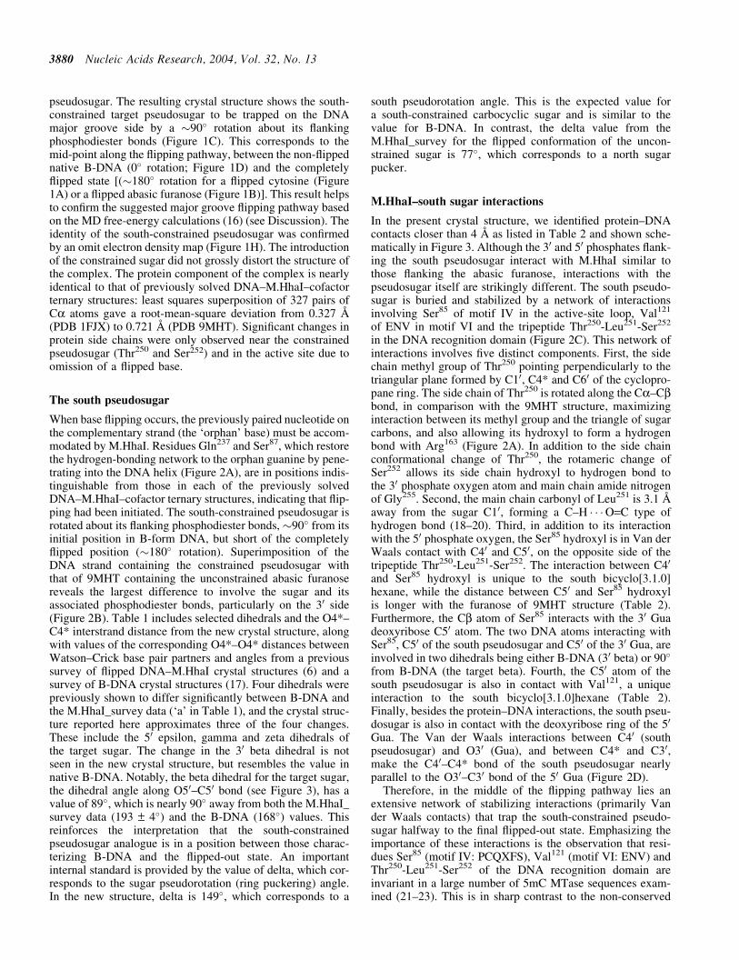
pseudosugar. The resulting crystal structure shows the south-constrained target pseudosugar to be trapped on the DNAmajor groove side by a �90� rotation about its flankingphosphodiester bonds (Figure 1C). This corresponds to themid-point along the flipping pathway, between the non-flippednative B-DNA (0� rotation; Figure 1D) and the completelyflipped state [(�180� rotation for a flipped cytosine (Figure1A) or a flipped abasic furanose (Figure 1B)]. This result helpsto confirm the suggested major groove flipping pathway basedon the MD free-energy calculations (16) (see Discussion). Theidentity of the south-constrained pseudosugar was confirmedby an omit electron density map (Figure 1H). The introductionof the constrained sugar did not grossly distort the structure ofthe complex. The protein component of the complex is nearlyidentical to that of previously solved DNA–M.HhaI–cofactorternary structures: least squares superposition of 327 pairs ofCa atoms gave a root-mean-square deviation from 0.327 s
(PDB 1FJX) to 0.721 s (PDB 9MHT). Significant changes inprotein side chains were only observed near the constrainedpseudosugar (Thr250 and Ser252) and in the active site due toomission of a flipped base.
The south pseudosugar
When base flipping occurs, the previously paired nucleotide onthe complementary strand (the ‘orphan’ base) must be accom-modated by M.HhaI. Residues Gln237 and Ser87, which restorethe hydrogen-bonding network to the orphan guanine by pene-trating into the DNA helix (Figure 2A), are in positions indis-tinguishable from those in each of the previously solvedDNA–M.HhaI–cofactor ternary structures, indicating that flip-ping had been initiated. The south-constrained pseudosugar isrotated about its flanking phosphodiester bonds, �90� from itsinitial position in B-form DNA, but short of the completelyflipped position (�180� rotation). Superimposition of theDNA strand containing the constrained pseudosugar withthat of 9MHT containing the unconstrained abasic furanosereveals the largest difference to involve the sugar and itsassociated phosphodiester bonds, particularly on the 30 side(Figure 2B). Table 1 includes selected dihedrals and the O4*–C4* interstrand distance from the new crystal structure, alongwith values of the corresponding O4*–O4* distances betweenWatson–Crick base pair partners and angles from a previoussurvey of flipped DNA–M.HhaI crystal structures (6) and asurvey of B-DNA crystal structures (17). Four dihedrals werepreviously shown to differ significantly between B-DNA andthe M.HhaI_survey data (‘a’ in Table 1), and the crystal struc-ture reported here approximates three of the four changes.These include the 50 epsilon, gamma and zeta dihedrals ofthe target sugar. The change in the 30 beta dihedral is notseen in the new crystal structure, but resembles the value innative B-DNA. Notably, the beta dihedral for the target sugar,the dihedral angle along O50–C50 bond (see Figure 3), has avalue of 89�, which is nearly 90� away from both the M.HhaI_survey data (193 – 4�) and the B-DNA (168�) values. Thisreinforces the interpretation that the south-constrainedpseudosugar analogue is in a position between those charac-terizing B-DNA and the flipped-out state. An importantinternal standard is provided by the value of delta, which cor-responds to the sugar pseudorotation (ring puckering) angle.In the new structure, delta is 149�, which corresponds to a
south pseudorotation angle. This is the expected value fora south-constrained carbocyclic sugar and is similar to thevalue for B-DNA. In contrast, the delta value from theM.HhaI_survey for the flipped conformation of the uncon-strained sugar is 77�, which corresponds to a north sugarpucker.
M.HhaI–south sugar interactions
In the present crystal structure, we identified protein–DNAcontacts closer than 4 s as listed in Table 2 and shown sche-matically in Figure 3. Although the 30 and 50 phosphates flank-ing the south pseudosugar interact with M.HhaI similar tothose flanking the abasic furanose, interactions with thepseudosugar itself are strikingly different. The south pseudo-sugar is buried and stabilized by a network of interactionsinvolving Ser85 of motif IV in the active-site loop, Val121
of ENV in motif VI and the tripeptide Thr250-Leu251-Ser252
in the DNA recognition domain (Figure 2C). This network ofinteractions involves five distinct components. First, the sidechain methyl group of Thr250 pointing perpendicularly to thetriangular plane formed by C10, C4* and C60 of the cyclopro-pane ring. The side chain of Thr250 is rotated along the Ca–Cbbond, in comparison with the 9MHT structure, maximizinginteraction between its methyl group and the triangle of sugarcarbons, and also allowing its hydroxyl to form a hydrogenbond with Arg163 (Figure 2A). In addition to the side chainconformational change of Thr250, the rotameric change ofSer252 allows its side chain hydroxyl to hydrogen bond tothe 30 phosphate oxygen atom and main chain amide nitrogenof Gly255. Second, the main chain carbonyl of Leu251 is 3.1 s
away from the sugar C10, forming a C–H � � �O=C type ofhydrogen bond (18–20). Third, in addition to its interactionwith the 50 phosphate oxygen, the Ser85 hydroxyl is in Van derWaals contact with C40 and C50, on the opposite side of thetripeptide Thr250-Leu251-Ser252. The interaction between C40
and Ser85 hydroxyl is unique to the south bicyclo[3.1.0]hexane, while the distance between C50 and Ser85 hydroxylis longer with the furanose of 9MHT structure (Table 2).Furthermore, the Cb atom of Ser85 interacts with the 30 Guadeoxyribose C50 atom. The two DNA atoms interacting withSer85, C50 of the south pseudosugar and C50 of the 30 Gua, areinvolved in two dihedrals being either B-DNA (30 beta) or 90�
from B-DNA (the target beta). Fourth, the C50 atom of thesouth pseudosugar is also in contact with Val121, a uniqueinteraction to the south bicyclo[3.1.0]hexane (Table 2).Finally, besides the protein–DNA interactions, the south pseu-dosugar is also in contact with the deoxyribose ring of the 50
Gua. The Van der Waals interactions between C40 (southpseudosugar) and O30 (Gua), and between C4* and C30,make the C40–C4* bond of the south pseudosugar nearlyparallel to the O30–C30 bond of the 50 Gua (Figure 2D).
Therefore, in the middle of the flipping pathway lies anextensive network of stabilizing interactions (primarily Vander Waals contacts) that trap the south-constrained pseudo-sugar halfway to the final flipped-out state. Emphasizing theimportance of these interactions is the observation that resi-dues Ser85 (motif IV: PCQXFS), Val121 (motif VI: ENV) andThr250-Leu251-Ser252 of the DNA recognition domain areinvariant in a large number of 5mC MTase sequences exam-ined (21–23). This is in sharp contrast to the non-conserved
3880 Nucleic Acids Research, 2004, Vol. 32, No. 13

Figure 2. Stereo views of M.HhaI–DNA containing south constrained sugar analogue. Atomic bonds are depicted as green stick for DNA and grey sticks for M.HhaI.Nitrogen, oxygen, sulphur and phosphorus atoms are shown as blue, red, yellow and magenta balls, respectively. Carbon atoms are shown as green (for DNA) andgrey (for protein) balls, respectively. Specific interactions are displayed as dashed single line (hydrogen bonds) and strapped lines (Van der Waals contacts). Theoxygen atoms of water molecules are labelled as w. (A) Detailed structure in the vicinity of the active site and the orphaned guanine. (B) Superimposition of the DNAstrands containing the south sugar from the new structure (coloured) and the M.HhaI–DNA containing an abasic furanose sugar (grey). (C) Detailed interactionsbetween the DNA strand containing the south sugar and M.HhaI. (D) Detailed interactions between the target sugar and 50 Gua. (E) Without changing thephosphodiester bonds, superimposing the chemically modelled north pseudosugar onto the observed south conformation suggests that the specific interactionsbetween the triangle and the side chain methyl group of Thr250 would not exist for the north sugar.
Nucleic Acids Research, 2004, Vol. 32, No. 13 3881

state of Gln237 and Ser87, which penetrate the DNA helix.Consistent with the present observations is a previous muta-genesis study suggesting that Thr250 constrains the conforma-tion of the DNA backbone (24).
The M.HhaI active site
In the absence of a flipped target, the active site of M.HhaIcontains a few water molecules (Figure 2A). For example,water w1 interacts with the side chain of Arg165, and w2interacts with the side chain of Glu119 and the main chaincarbonyl oxygen between Phe79 and Pro80. The water sitew1 corresponds to the position of O4* of the abasic furanosesugar in the 9MHT structure, while the w2 site corresponds tothe exocyclic amino nitrogen N4 of the flipped cytosine (8).Interestingly, Arg165 and Glu119 form an ion pair that waspresent in both the M.HhaI structure without DNA (25) andin the structure with DNA containing only the abasic furanoseat the flipped position (8), but was absent in all other M.HhaI–DNA complexes containing an entire nucleotide (whethercytosine, uracil or adenine) at the active site. Clearly, thepresence of a flipped-out base breaks the salt bridge, resultingin both Glu119 and Arg165 interacting with the base(Glu119 . . .N3, Arg165 . . .O2 and Arg165 . . .O4*). Such struc-tural changes in the active site due to the presence of the basemay contribute to stabilization of the base in the fully flipped-out orientation.
Molecular dynamics simulations
MD simulations were performed on the DNA–M.HhaI–AdoHcy ternary complex starting with the new crystal struc-ture to facilitate an understanding of the interactions betweenthe protein and the constrained pseudosugars and how theycontribute to the enhanced binding of the south-constrainedpseudosugar versus the decreased binding affinity of the northconstrained pseudosugar. In the case of the constrained southpseudosugar (Figure 4A), the probability distribution for the
O4*–C4* distance (peaked at �16.5 s) stays close to thepresent crystallographic value (15.6 s, thick dashed line),being �1 s greater, while in the constrained north simulationthe distances (�18 s) shifts to significantly longer values,�2.5 s longer than the crystallographic value. Interestingly,the increase in the O4*–C4* distance with the north confor-mation yields better agreement with the average value (18.5 s)from the DNA–M.HhaI crystal structures with a completelyflipped conformation (thin line).
Consistent with the extensive contacts between the carbo-cyclic sugar moiety and the protein observed in the crystalstructure is the stability of the O4*–C4* distance and dihedralprobability distributions (see Supplementary Figure 1) in thesouth MD simulation. In contrast, with the north pseudosugarthere are significant deviations from the crystallographicvalues for the O4*–C4* distance and dihedral distributionsare significantly broader than those with the south-constrainedpseudosugar (see Supplementary Figure 1). These resultsindicate that the protein cannot accommodate the north-constrained pseudosugar (e.g. see Figure 2E), contributingto its lower binding affinity. Also, the increase in the O4*–C4* distances in the north simulation is consistent with the
Table 1. Comparison of selected dihedrals (in degrees) and the O4*–C4* (or
O4*–O4*) distance
South (PDB1SKM)
M.HhaI_survey B-DNA_survey
50-DeoxyriboseEpsilona 273.5 264 – 3 187Zeta 292.0 288 – 3 262
Target sugarAlpha 268.7 276 – 5 298Beta 88.5 193 – 4 168
Gammaa 162.5 180 – 4 51Delta 148.6 77 – 3 143
Epsilon 189.8 202 – 6 187Zeta
a134.1 95 – 7 262
30-DeoxyriboseAlpha 300.3 258 – 7 298Betaa 171.6 228 – 6 168
Gamma 57.4 74 – 5 51O40–O40 distance (s) 15.6 18.5 12.0
aIndicates dihedrals that show significant differences between canonical B-DNA (17) and the M.HhaI–DNA survey (6). Boldface type indicates valuesof dihedrals discussed in the text.
Figure 3. Schematical representation of interactions between M.HhaI and thesouth sugar. The distance for specific contact is listed in Table 2. Curved arrowsdenote the dihedrals along the phosphodiester backbone. The letters inparentheses indicate the individual dihedral value is close to the flippedconformation (F), the B-DNA (B) or unique to the south conformation (S).Double dashed arrows indicate Van der Waals contacts and dashed linesindicate hydrogen bonds. Main chain atoms are in grey and side chainatoms in black.
3882 Nucleic Acids Research, 2004, Vol. 32, No. 13

crystallographic observations that north conformations arerequired for binding in the fully flipped state.
DISCUSSION
The new structure of M.HhaI with the constrained southpseudosugar at the target site of the recognition sequenceprovides important clues for DNA–protein interactions andthe mechanism of base flipping. To see the south-constrainedpseudosugar frozen in the major groove side was surprising(Figure 1C) because it was initially thought that flippingoccurred through the minor groove (2). This suppositionwas based on the location of the M.HhaI DNA recognitiondomain, which approaches the DNA from the major grooveside, and thus, sequence-specific interactions in the majorgroove were thought likely to block the major groove-flippingpathway. In addition, DNA-only simulations of abasic systemssupported the minor groove pathway (6). However, the newcrystal structure indicates that base flipping occurs most likelythrough the major groove of the DNA. Such a pathway hasbeen suggested based on free-energy calculations of flipping inDNA complexed to M.HhaI (16).
The presence of the partially flipped pseudosugar in themajor groove of the new crystal structure is consistent withthe large conformational change of the M.HhaI active-site loopupon DNA binding (2). This loop moves towards the DNAfrom the minor groove side, where it would probably interferewith flipping through the minor groove. If flipping occurs viathe minor groove before closing at the active-site loop, solva-tion of the base should occur in a manner similar to DNA inaqueous solution. The energetics of base flipping from DNA inaqueous solution indicate that movement of the target Cyt outof the helix and into an aqueous environment leads to a drasticincrease in the free energy, thus disfavouring flipping (26,27).Therefore, it was hypothesized that the protein must supplyan environment eliminating the unfavourable energeticsassociated with flipping into an aqueous solution (16). Suchan environment may be supplied by the DNA-binding domainvia a major groove-flipping pathway. It was noted earlier thatM.HhaI–DNA interactions involve many main chain atoms ofthe DNA-binding domain, creating an intimate contact surfacein the major groove (2). Of course, we cannot completelyexclude the minor groove pathway, though we consider itunlikely. The south pseudosugar conformation we observe
Table 2. List of contacts between M.HhaI and the target sugar
PDB 1SKM PDB 9MHTSource atoms Target atoms Distance (A) Source atoms Target atoms Distance (A)
South 7 P(50) Ser 85 OG 3.71 Furanose 7 P Ser 85 OG 3.92South 7 O1P(50) Ser 85 C 3.74 Furanose 7 O1P
Ser 85 CA 3.32 Ser 85 CA 3.90Ser 85 CB 3.36 Ser 85 CB 3.59Ser 85 OG 2.69* Ser 85 OG 2.86*Ile 86 N 3.22* Ile 86 N 3.88Ile 86 CG1 3.92 Val 121 CG1 3.83
South 7 O50 Val 121 CG1 3.92South 7 C60 Thr 250 CG2 3.72 Furanose 7 O40 Arg 165 NH1 2.96*
Gua 6 C30 3.54Gua 6 C20 3.75Gua 6 O30 3.47
South 7 C70 Gua 6 C20 3.94Thr 250 CG2 3.88Ser 52 A 3.82Ser 252 CB 3.65Ser 252 OG 3.79
South 7 C20 Leu 251 O 3.72 Furanose 7 C20 Gly 303 O 3.41South 7 C5v Ser 85 OG 3.40 Furanose 7 C50 Ser 85 OG 3.84
Val 121 CG1 3.49South 7 C40 Ser 85 OG 3.37South 7 C10 Thr 250 CG2 3.87 Furanose 7 C10 Cys 81 SG 3.99
Leu 251 C 3.92 Arg 163 NH2 3.82Leu 251 O 3.13* Arg 165 NH1 3.49Ser 252 CA 3.67 Gly 303 O 3.26
South 7 C30 Ser 85 OG 3.99Gua 8 O1P Tyr 254 CD2 3.95 Gua 8 O1P Tyr 254 CD2 3.51Gua 8 O2P Ser 252 OG 2.91* Gua 8 O2P Ser 252 CB 3.94
Gly 255 N 3.65 Gly 255 N 3.96Ala 253 N 3.17* Ala 253 N 3.17*Ala 253 CA 3.81 Ala 253 CA 3.63Ala 253 C 3.73 Ala 253 C 3.57Tyr 254 N 2.76* Tyr 254 N 2.68*Tyr 254 CA 3.54 Tyr 254 CA 3.56Tyr 254 CB 3.60 Tyr 254 CB 3.66Tyr 254 C 3.86Ala 253 CB 3.93Tyr 254 CD2 3.73
Asterisks indicate hydrogen bonding interactions.
Nucleic Acids Research, 2004, Vol. 32, No. 13 3883

could be viewed as having flipped through the minor groove,passed through the active site (because in the absence of theArg165–O4* bonding and other interactions the enzyme wouldnot hold the sugar at the active site) and trapped in theobserved location.
The present observations combined with structural resultsfrom previously published free-energy calculations (16) canshed additional light on the mechanism of flipping. Presentedin Figure 4B is the sugar pseudorotational angle P of the targetCyt sugar as a function of the extent of flipping. As may beseen, the sugar is predominantly sampling south conforma-tions (P = �160�) in the Watson–Crick base-paired state(Center Of Mass COM pseudodihedral angle = �10 or370�, depending on whether flipping begins from the minorgroove or the major groove, respectively). The south confor-mation continues to be sampled as flipping occurs via themajor groove (i.e. COM Pseudodihedral angle decreasingfrom 370�). At �285�, which is 85� from the Watson–Crick base-paired state and hence midway to a fullyflipped-out state, the sugar pucker starts to decrease and con-tinues to decrease as flipping continues to the fully flippedstate at a COM angle of �190� at which the sugar has assumeda north pucker (P �10�), consistent with the crystal structureof the flipped Cyt in the ternary complex (2). Interestingly,COM angle of 285� corresponds to the small free-energy bar-rier that remains to flipping in the presence of the protein [seeFigure 1E of (16)]. Since that barrier occurs approximatelyhalfway between the Watson–Crick and fully flipped statesand the sugar is still assuming a south conformation, consistentwith the new crystal structure, it is suggested that the newcrystal structure corresponds to an intermediate stage of thesugar and phosphodiester backbone moieties halfway alongthe flipping corridor. Consistent with this is the sampling of thetarget beta dihedral during flipping observed in the free-energycalculations (Figure 4C). In the vicinity of the mid-point of theflipping trajectory (COM Pseudodihedral angle = 285�), beta issampling a wide range of values, including the value observedin the present crystal structure (�90�, Table 1), while in boththe Watson–Crick and fully flipped states beta is samplingvalues in the vicinity of 180�, consistent with that observedin B-DNA and in the M.HhaI–DNA complexes (Table 1).Thus, the stronger binding of the south abasic pseudosugarsuggests that this moiety is possibly mimicking a transitionstate (see below) of the phosphodiester backbone that occursduring the base-flipping pathway.
The critical question is: How are the interactions betweenM.HhaI and the DNA phosphodiester backbone facilitatingflipping? Based on surveys of crystal structures of canonicalDNA there is minimal sampling of beta in the region of 90�,suggesting that this conformation is energetically unfavour-able, which is supported by quantum mechanical calculationson model compounds representative of the phosphodiesterbackbone (17). Thus, if flipping requires a transition throughvalues of beta near 90�, as indicated in the free-energy calcu-lations (Figure 4C), and this region is of high energy, then theenzyme can facilitate flipping by favourably binding thisorientation. As shown in Figure 3, this would involve inter-actions between residues Ser85, Val121, Thr250 and Ser252 andthe phosphodiester backbone. Additional interactions betweenthe carbocyclic south pseudosugar and the protein (i.e. thecyclopropane moiety with Thr250 and Leu251) stabilize the
complex so that it does not move beyond the mid-pointstage to the fully flipped state. Such a model is consistentwith respect to interactions of an abasic furanose or northconstrained carbocyclic sugar with M.HhaI. With the abasicfuranose the conformational changes required for flipping canoccur, leading to the fully flipped structure observed in crystalstructures; however, the flexibility of the furanose moietydisallows stabilization of conformations at the mid-point ofthe flipping trajectory, leading to the decreased binding affinity
Figure 4. (A) O4*–C4* distance histograms from the MD simulations with aconstrained carbocyclic south (closed circles) or north sugar (open circles) forthe ternary complex initiated from the (south carbocycle)–DNA–M.HhaI–AdoHcy crystal structure. Included as vertical lines are the O4*–C4*distances from the survey of flipped DNA–M.HhaI crystal structures (6)(thin line), canonical B-DNA (6) (thick line) and the (south carbocycle)–DNA–M.HhaI crystal structure (thick dashed line). Note that C4* is theatom in the south carbocycle that corresponds to the O4* atom in thenormal furanose sugar (see Figure 1E–G). (B) Sampling of the target Cytsugar pucker pseudorotation angle or (C) beta dihedral angle versus thepseudodihedral angle defining the extent of flipping of the target Cyt (16) inthe M.HhaI–DNA–AdoHcy ternary complex (2). Shown are the values of thesugar pseudorotation or beta dihedral angle sampled every 1 ps from eachwindow in the flipping freeenergy calculation reported (16).
3884 Nucleic Acids Research, 2004, Vol. 32, No. 13

as compared to the carbocyclic south pseudosugar. In the caseof the north pseudosugar, it appears that the conformationalchanges required for flipping are disallowed such that favor-able interactions between the enzyme and DNA cannot occurleading to the unfavourable experimental binding affinity (6).Note that in the present MD simulations the movement of thenorth constrained pseudosugar into a conformation beyondthat of the flipped orientation is probably an artefact associatedwith the starting conformation being the partially flipped statefrom the present crystal structure (see Figure 2E).
In summary, based on the present structure it is hypothe-sized that M.HhaI facilitates flipping, in part, by stabilizing anenergetically unfavourable conformation of the phosphodi-ester backbone that may represent a possible transition statein the flipping process. The idea of tight binding of transitionstates by enzymes being responsible for their catalytic cap-abilities was first proposed by Pauling (28). This concept hassubsequently been applied to understand chemical catalysis byenzymes [for a recent review see (29)] as well as beingexploited in the rational design of small molecular weightenzyme inhibitors as structural mimics of transition states(30). However, this concept has been mostly applied to transi-tion states involved in bond making or bond-breaking events,in contrast to the conformational change occurring in the pre-sent case. Similar to catalysis of chemical reactions, facilita-tion of base flipping involves lowering the free-energy barrierto flipping. This represents a change in the conformationaldynamics of a macromolecule (i.e. DNA) responding to anexternal mechanical force imposed by another macromolecule(i.e. the M.HhaI enzyme). Although only a >10-fold increasein binding affinity between oligodeoxynucleotides containingconformationally locked north and south abasic pseudosugarsoccurs (7), versus the 3–4 order of magnitude increase inaffinities generally observed for transition-state mimics ofchemical reactions, it is interesting to speculate that similartransition state principles may be applied to macromolecularconformational changes that go through discrete conforma-tional states.
SUPPLEMENTARY MATERIAL
Supplementary Material is available at NAR Online.
ACKNOWLEDGEMENTS
X.C. is grateful to Dr Richard J. Roberts (New EnglandBiolabs) and Dr Robert M. Blumenthal (Medical College ofOhio) for their continued support and encouragement. Wethank Susan Sunay (Emory University) for help with purifica-tion and crystallization, Annie Heroux (Brookhaven NationalLaboratory) for help with X-ray data collection at beamlineX26C in the facilities of the National Synchrotron LightSource. Work was supported in part by National Institutes ofHealth Grants GM49245 to X.C. and GM51501 to A.D.M.
REFERENCES
1. Schubert,H.L., Blumenthal,R.M. and Cheng,X. (2003) Many paths tomethyltransfer: a chronicle of convergence. Trends Biochem. Sci.,28, 329–335.
2. Klimasauskas,S., Kumar,S., Roberts,R.J. and Cheng,X. (1994) HhaImethyltransferase flips its target base out of the DNA helix. Cell,76, 357–369.
3. Roberts,R.J. and Cheng,X. (1998) Base flipping. Annu. Rev. Biochem.,67, 181–198.
4. Cheng,X. and Roberts,R.J. (2001) AdoMet-dependent methylation,DNA methyltransferases and base flipping. Nucleic Acids Res., 29,3784–3795.
5. Klimasauskas,S. and Roberts,R.J. (1995) M.HhaI binds tightly tosubstrates containing mismatches at the target base. Nucleic Acids Res.,23, 1388–1395.
6. Wang,P., Nicklaus,M.C., Marquez,V.E., Brank,A.S., Christman,J.,Banavali,N.K. and MacKerell,A.D.,Jr (2000) Use ofoligodeoxyribonucleotides with conformationally constrained abasicsugar targets to probe the mechanism of base flipping by HhaI DNA(cytosine C5)-methyltransferase. J. Am. Chem. Soc., 122, 12422–12434.
7. Marquez,V.E., Wang,P., Nicklaus,M.C., Maier,M., Manoharan,M.,Christman,J.K., Banavali,N.K. and Mackerell,A.D.,Jr (2001) Inhibitionof (cytosine C5)-methyltransferase by oligonucleotides containingflexible (cyclopentane) and conformationally constrained(bicyclo[3.1.0]hexane) abasic sites. Nucleos. Nucleot. Nucleic Acids,20, 451–459.
8. O’Gara,M., Horton,J.R., Roberts,R.J. and Cheng,X. (1998) Structures ofHhaI methyltransferase complexed with substrates containingmismatches at the target base. Nature Struct. Biol., 5, 872–877.
9. Kumar,S., Cheng,X., Pflugrath,J.W. and Roberts,R.J. (1992)Purification, crystallization, and preliminary X-ray diffraction analysis ofan M.HhaI–AdoMet complex. Biochemistry, 31, 8648–8653.
10. Maier,M., Choi,Y., Gaus,H., Marquez,V.E. and Manoharan,M. (2004)Synthesis and characterization of oligonucleotides containingconformationally constrained bicyclo[3.1.0]hexane pseudosugaranalogue. Nucleic Acids Res., 32, 3642–3650.
11. Tong,L. (1993) REPLACE, a suite of computer programs for molecular-replacement calculations. J. Appl. Cryst., 26, 748–751.
12. Brunger,A.T., Adams,P.D., Clore,G.M., DeLano,W.L., Gros,P., Grosse-Kunstleve,R.W., Jiang,J.S., Kuszewski,J., Nilges,M., Pannu,N.S. et al.(1998) Crystallography & NMR system: a new software suite formacromolecular structure determination. Acta Crystallogr. D Biol.Crystallogr., 54, 905–921.
13. Brooks,B.R., Bruccoleri,R.E., Olafson,B.D., States,D.J.,Swaminathan,S. and Karplus,M. (1983) CHARMM: a program formacromolecular energy, minimization, and dynamics calculations.J. Comput. Chem., 4, 187–217.
14. Foloppe,N. and MacKerell,A.D.,Jr (2000) All-atom empirical force fieldfor nucleic acids: 1) Parameter optimization based on small moleculeand condensed phase macromolecular target data. J. Comput. Chem.,21, 86–104.
15. MacKerell,A.D.,Jr and Banavali,N.K. (2000) All-atom empirical forcefield for nucleic acids: 2) Application to solution MD simulationsof DNA. J. Comput. Chem., 21, 105–120.
16. Huang,N., Banavali,N.K. and MacKerell,A.D.,Jr. (2003) Protein-facilitated base flipping in DNA by cytosine-5-methyltransferase.Proc. Natl Acad. Sci. USA, 100, 68–73.
17. Foloppe,N. and MacKerell,A.D.,Jr (1999) Contribution of the phos-phodiester backbone and glycosyl linkage intrinsic torsional energetics toDNA structure and dynamics. J. Phys. Chem., B103, 10955–10964.
18. Sutor,D.J. (1962) The C–H � � �O hydrogen bond in crystals. Nature,195, 68–69.
19. Huggins,M.T. and Lightner,D.A. (2001) A C–H � � �O=C hydrogen bond?Intramolecular hydrogen bonding in a novel semirubin. J. Org. Chem.,66, 8402–8410.
20. Matsuura,H., Yoshida,H., Hieda,M., Yamanaka,S.-Y., Harada,T.,Shin-Ya,K. and Ohno,K. (2003) Experimental evidence for intra-molecular blue-shifting C–H � � �O hydrogen bonding by matrix-isolationinfrared spectroscopy. J. Am. Chem. Soc., 125, 13910–13911.
21. Posfai,J., Bhagwat,A.S., Posfai,G. and Roberts,R.J. (1989) Predictivemotifs derived from cytosine methyltransferases. Nucleic Acids Res.,17, 2421–2435.
22. Lauster,R., Trautner,T.A. and Noyer-Weidner,M. (1989) Cytosine-specific type II DNA methyltransferases. A conserved enzyme core withvariable target-recognizing domains. J. Mol. Biol., 206, 305–312.
23. Kumar,S., Cheng,X., Klimasauskas,S., Mi,S., Posfai,J., Roberts,R.J. andWilson,G.G. (1994) The DNA (cytosine-5) methyltransferases.Nucleic Acids Res., 22, 1–10.
Nucleic Acids Research, 2004, Vol. 32, No. 13 3885

24. Vilkaitis,G., Dong,A., Weinhold,E., Cheng,X. and Klimasauskas,S.(2000) Functional roles of the conserved threonine 250 in the targetrecognition domain of HhaI DNA methyltransferase. J. Biol. Chem., 275,38722–38730.
25. Cheng,X., Kumar,S., Posfai,J., Pflugrath,J.W. and Roberts,R.J. (1993)Crystal structure of the HhaI DNA methyltransferase complexed withS-adenosyl-L-methionine. Cell, 74, 299–307.
26. Giudice,E., V�aarnai,P. and Lavery,R. (2001) Energetic andconformational aspects of A:T base pair opening within the DNA doublehelix. Chem. Phys. Chem., 11, 673–677.
27. Banavali,N.K. and MacKerell,A.D.,Jr. (2002) Free energy and structuralpathways of base flipping in a DNA GCGC containing sequence.J. Mol. Biol., 319, 141–160.
28. Pauling,L. (1946) Molecular architecture and biological reactions. Chem.Eng. News, 24, 1375–1377.
29. Garcia-Viloca,M., Gao,J., Karplus,M. and Truhlar,D.G. (2004) Howenzymes work: analysis by modern rate theory and computer simulations.Science, 303, 186–195.
30. Wolfenden,R. (1976) Transition state analog inhibitors and enzymecatalysis. Annu. Rev. Biophys. Bioeng., 5, 271–306.
3886 Nucleic Acids Research, 2004, Vol. 32, No. 13
Copyright © 2022 FDOKUMEN