Catalytic and various biological activities of green silver nanoparticles synthesized from Plumeria...
10
Catalytic and biological activities of green silver nanoparticles synthesized from Plumeria alba (frangipani) flower extract Rani Mata, Jayachandra Reddy Nakkala, Sudha Rani Sadras ⁎ Department of Biochemistry and Molecular Biology, School of Life Sciences, Pondicherry University, Pondicherry, India abstract article info Article history: Received 22 April 2014 Received in revised form 26 November 2014 Accepted 27 February 2015 Available online 28 February 2015 Keywords: Frangipani flower extract Catalytic Biological activities Cytotoxicity Apoptosis Herein, we report the green synthesis of silver nanoparticles using Plumeria alba (frangipani) flower extract (FFE) and their biological applications. The formation of frangipani silver nanoparticles (FSNPs) was confirmed by UV– visible spectroscopy and characterized by DLS particle size analyzer, SEM/EDAX, FTIR, TGA/DSC and XRD. The synthesized spherical FSNPs were found to be 36.19 nm in size as determined by DLS particle size analyzer. EDAX data and XRD pattern of FSNPs confirmed the presence and face-centered cubic (fcc) phase structure of sil- ver. The bioactive groups C-C and C-N present in FFE were involved in the formation of FSNPs as identified by FTIR analysis. FSNPs exhibited powerful catalytic activity by reducing 4-nitrophenol to 4-aminophenol within 8 min and the other organic dyes namely methylene blue and ethidium bromide were moderately degraded. Biological activities of FSNPs are evaluated by means of antioxidant, antibacterial and cytotoxic effect. Antioxidant potential of FSNPs was assessed by various in vitro assays in which they exhibited moderate antioxidant activity. The an- tibacterial effect of FSNPs was tested in two different pathogenic bacterial strains and their bacteriostatic effect was confirmed by growth kinetic study in Escherichia coli. The cytotoxic effect of FSNPs in COLO 205 was analyzed by MTT assay and the IC 50 concentration was found at 5.5 and 4 μg/ml respectively after 24 and 48 h of incubation. Cytotoxic effect of FSNPs in COLO 205 cells was associated with the loss of membrane integrity and chromatin condensation which might have played a crucial role in the induction of apoptosis as evidenced in AO/EB staining. © 2015 Elsevier B.V. All rights reserved. 1. Introduction Nanotechnology has gained intense attention in the recent past due to its wide application in diverse areas like medicine, catalysis, energy and materials [1–4]. Particularly nanoparticles with small size to large surface area have potential medical as well as industrial applications [5]. Researchers make significant efforts towards the synthesis of nano- particles by various means, including physical, chemical and biological methods [6,7]. As chemical synthesis involves the use of chemical reducing agents that may be toxic, such nanoparticles become unsuit- able for biomedical applications. In the last decade, biological methods by using microbes or plant biomasses for the synthesis of nanometals was explored. Biological method has received increasing attention due to the growing need to develop environmentally benign technologies in material synthesis [8]. Green method of synthesizing nanoparticles with plant extracts is advantageous over physical, chemical and micro- bial methods as it is simple, convenient, environment friendly and re- quires less reaction time [9]. Among the noble metals used in green synthesis, silver is widely preferred owing to its antibacterial [10], anti- fungal, larvicidal, antiparasitic [11] and anticancer [12] properties. In green synthesis capping and stabilization of silver nanoparticles are brought about by bioactive compounds like alkaloids, flavonoids and polyphenols present in plant extracts [13]. Reports on the synthesis of green silver nanoparticles using plant extracts such as Camellia sinensis [14], Aloe vera plant extract [15], latex of Jatropha curcas [16] and lemon- grass (Cymbopogon) leaf extract [17] are available. Further, the in vitro antioxidant properties of green silver nanoparticles synthesized from Cassia auriculata flower extract have been reported [18]. Anticancer po- tential silver nanoparticles from Euphorbia chapmaniana on HL-60 has also been reported recently [19]. Silver nanoparticles owing to their cata- lytic property can degrade dyes in industrial effluents and convert ni- trate and nitrite ions in chemical fertilizers to harmless N 2 and thereby find their application in treating environmental pollutants. These nitrate and nitrite ions affect human health by causing methemo- globinemia [20]. The catalytic action of silver nanoparticles can be evaluated by applying many of the chemically synthesized cationic and anionic dyes. Recently, photo catalytic degradation of methyl orange dye by silver nanoparticles synthesized from Ulva lactuca has been reported [21]. In this study, we report the green synthesis of silver nanoparticles using Plumeria alba (frangipani) flower extract (FFE) and their biologi- cal applications. The plant P. alba (frangipani) belonging to the family Apocynaceae and the plant parts like leaves, bark, flowers and latex have been shown to possess various biological activities including Materials Science and Engineering C 51 (2015) 216–225 ⁎ Corresponding author at: Department of Biochemistry and Molecular Biology, Pondicherry University, Pondicherry 605 014, India. E-mail address: [email protected] (S. Rani Sadras). http://dx.doi.org/10.1016/j.msec.2015.02.053 0928-4931/© 2015 Elsevier B.V. All rights reserved. Contents lists available at ScienceDirect Materials Science and Engineering C journal homepage: www.elsevier.com/locate/msec
-
Upload
pondiuni-anthropology -
Category
Documents
-
view
1 -
download
0
Transcript of Catalytic and various biological activities of green silver nanoparticles synthesized from Plumeria...

Materials Science and Engineering C 51 (2015) 216–225
Contents lists available at ScienceDirect
Materials Science and Engineering C
j ourna l homepage: www.e lsev ie r .com/ locate /msec
Catalytic and biological activities of green silver nanoparticlessynthesized from Plumeria alba (frangipani) flower extract
Rani Mata, Jayachandra Reddy Nakkala, Sudha Rani Sadras ⁎Department of Biochemistry and Molecular Biology, School of Life Sciences, Pondicherry University, Pondicherry, India
⁎ Corresponding author at: Department of BiochemPondicherry University, Pondicherry 605 014, India.
E-mail address: [email protected] (S. Rani Sadras).
http://dx.doi.org/10.1016/j.msec.2015.02.0530928-4931/© 2015 Elsevier B.V. All rights reserved.
a b s t r a c t
a r t i c l e i n f oArticle history:Received 22 April 2014Received in revised form 26 November 2014Accepted 27 February 2015Available online 28 February 2015
Keywords:Frangipani flower extractCatalyticBiological activitiesCytotoxicityApoptosis
Herein,we report the green synthesis of silver nanoparticles using Plumeria alba (frangipani) flower extract (FFE)and their biological applications. The formation of frangipani silver nanoparticles (FSNPs) was confirmed by UV–visible spectroscopy and characterized by DLS particle size analyzer, SEM/EDAX, FTIR, TGA/DSC and XRD. Thesynthesized spherical FSNPs were found to be 36.19 nm in size as determined by DLS particle size analyzer.EDAX data and XRD pattern of FSNPs confirmed the presence and face-centered cubic (fcc) phase structure of sil-ver. The bioactive groups C-C and C-Npresent in FFEwere involved in the formation of FSNPs as identifiedby FTIRanalysis. FSNPs exhibited powerful catalytic activity by reducing 4-nitrophenol to 4-aminophenol within 8 minand the other organic dyes namelymethylene blue and ethidium bromide weremoderately degraded. Biologicalactivities of FSNPs are evaluated bymeans of antioxidant, antibacterial and cytotoxic effect. Antioxidant potentialof FSNPs was assessed by various in vitro assays in which they exhibited moderate antioxidant activity. The an-tibacterial effect of FSNPs was tested in two different pathogenic bacterial strains and their bacteriostatic effectwas confirmed by growth kinetic study in Escherichia coli. The cytotoxic effect of FSNPs in COLO 205was analyzedbyMTT assay and the IC50 concentrationwas foundat 5.5 and 4 μg/ml respectively after 24 and 48h of incubation.Cytotoxic effect of FSNPs in COLO 205 cells was associated with the loss of membrane integrity and chromatincondensationwhichmight have played a crucial role in the induction of apoptosis as evidenced inAO/EB staining.
© 2015 Elsevier B.V. All rights reserved.
1. Introduction
Nanotechnology has gained intense attention in the recent past dueto its wide application in diverse areas like medicine, catalysis, energyand materials [1–4]. Particularly nanoparticles with small size to largesurface area have potential medical as well as industrial applications[5]. Researchers make significant efforts towards the synthesis of nano-particles by various means, including physical, chemical and biologicalmethods [6,7]. As chemical synthesis involves the use of chemicalreducing agents that may be toxic, such nanoparticles become unsuit-able for biomedical applications. In the last decade, biological methodsby using microbes or plant biomasses for the synthesis of nanometalswas explored. Biological method has received increasing attention dueto the growing need to develop environmentally benign technologiesin material synthesis [8]. Green method of synthesizing nanoparticleswith plant extracts is advantageous over physical, chemical and micro-bial methods as it is simple, convenient, environment friendly and re-quires less reaction time [9]. Among the noble metals used in greensynthesis, silver is widely preferred owing to its antibacterial [10], anti-fungal, larvicidal, antiparasitic [11] and anticancer [12] properties. In
istry and Molecular Biology,
green synthesis capping and stabilization of silver nanoparticles arebrought about by bioactive compounds like alkaloids, flavonoids andpolyphenols present in plant extracts [13]. Reports on the synthesis ofgreen silver nanoparticles using plant extracts such as Camellia sinensis[14],Aloe veraplant extract [15], latex of Jatropha curcas [16] and lemon-grass (Cymbopogon) leaf extract [17] are available. Further, the in vitroantioxidant properties of green silver nanoparticles synthesized fromCassia auriculata flower extract have been reported [18]. Anticancer po-tential silver nanoparticles from Euphorbia chapmaniana on HL-60 hasalso been reported recently [19]. Silver nanoparticles owing to their cata-lytic property can degrade dyes in industrial effluents and convert ni-trate and nitrite ions in chemical fertilizers to harmless N2 andthereby find their application in treating environmental pollutants.These nitrate and nitrite ions affect human health by causingmethemo-globinemia [20]. The catalytic action of silver nanoparticles can beevaluated by applying many of the chemically synthesized cationicand anionic dyes. Recently, photo catalytic degradation of methylorange dye by silver nanoparticles synthesized from Ulva lactuca hasbeen reported [21].
In this study, we report the green synthesis of silver nanoparticlesusing Plumeria alba (frangipani) flower extract (FFE) and their biologi-cal applications. The plant P. alba (frangipani) belonging to the familyApocynaceae and the plant parts like leaves, bark, flowers and latexhave been shown to possess various biological activities including

217R. Mata et al. / Materials Science and Engineering C 51 (2015) 216–225
antioxidant, antibacterial, antiarthritic activity, antitumor and anti dia-betic activities. The flowers contain several bioactive compounds, in-cluding β-sitosterol, scopoletin, iridoids isoplumericin, plumieride,plumieride coumarate and plumieride coumarate glucoside [22]. Re-cently, the antimicrobial activity of gold nanoparticles synthesizedfrom frangipani flower extract combined with standard antibioticdrugs has been reported [23]. In this study, we focused on the synthesisof frangipani silver nanoparticles (FSNPs) and the evaluationof their po-tential catalytic, antibacterial, antioxidant and anticancer activities invitro. The synthesized FSNPswere characterized byUV–visible spectros-copy, dynamic light scattering (DLS) particle size analyzer, scanningelectron microscope/energy dispersive analysis of X-rays (SEM/EDAX),fourier transform infrared spectroscopy (FTIR), thermo gravimetric an-alyzer/differential scanning calorimeter (TGA/DSC) and X-ray diffrac-tion (XRD).
2. Materials and methods
2.1. Chemicals
The frangipani (P. alba) flowers were collected from PondicherryUniversity, Puducherry, India. All the chemicals used were from Hi-Media and Sigma Aldrich Bangalore, India and were purely analyticalgrade used without further purification. Deionized water was used forall experiments and all assays were performed in triplicates.
2.2. Preparation of flower extract
Fresh frangipani flowers were collected, rinsed with distilled water,shade dried and fine powder was prepared. 1 g of frangipani flowerpowder was mixed with 100 ml of double distilled water in an Erlen-meyer flask and boiled for 10 min. Then the mixture was subjected torepeated filtration with Whatman No. 1 filter paper and the extractwas stored at 4 °C.
2.3. Syntheses of silver nanoparticles
To the 70 ml of silver nitrate (AgNO3)(1 mM) solution 10 ml of FFEwas added and incubated at room temperature. The color change ofthe reactionmixture frompale yellow to yellowish brownwas observedafter 30 min of incubation indicating the formation of FSNPs whichwasfurther confirmed by UV-visible spectrophotometer.
2.4. Characterization
2.4.1. UV–visible spectral analysisThe bio-reduction and formation of FSNPs from FFE was monitored
at regular time intervals using a UV–visible spectrophotometer(Shimadzu 1700). Further, the synthesized FSNPs were purified and col-lected by repeated centrifugation at 18,000 rpm.
2.4.2. DLS particle size analysisThe average size of the synthesized FSNPs was determined by ZETA
Seizers Nanoseries (Malvern Instruments Nano ZS) dynamic lightscattering instrument.
2.4.3. SEM/EDAXA thin film of the sample was prepared by adding a pinch of FSNPs
powder on a copper coated carbon grid and the surface morphology ofFSNPs was analyzed by SEM (Hitachi S-4500). EDAX analysis was per-formed to determine the elemental composition of FSNPs equippedwith SEM machine.
2.4.4. FTIRThe functional groups of phytocompounds present in FFE which are
involved in the formation of FSNPs was identified by FTIR (Thermo
Nicolet Nexus 6700 spectrometer) analysis. FSNPs and FFE powderswere separately mixed with potassium bromide (KBr) to make KBr pel-lets and then subjected to FTIR analysis.
2.4.5. TGA-DSCThermal stability and surface weight loss of FSNPs were analyzed by
SDT Q600 TG and the thermal behavior was analyzed by Q20 DSC.
2.4.6. XRDCrystalline nature of the synthesized FSNPs was analyzed by XRD
analysis. Thin film of FSNPs powder was prepared on a glass slide andrun under monochromatic Cu Kα radiation at 40 kV and 30 mA.
2.5. Catalytic activity
The catalytic activity of FSNPs was analyzed by using three differentdyes namely, methylene blue (MB), 4-nitrophenol (4-NP) and ethidiumbromide (EB) in the presence of NaBH4. In this assay, 2.8 ml of MB(1 mM) was mixed with 0.2 ml of colloidal FSNPs (50 μg/ml) solutionand the total volumewasmade up to 3.5mlwith distilledwater. The ab-sorbance of the reaction mixture was monitored at regular time inter-vals of 30 min [24]. The EB dye degradation assay was also performedin a similar way following the above procedure of MB. On the otherhand reduction of 4-NPwas carried out following the previously report-edmethod [25]. In this analysis, 0.2ml of FSNPs (50 μg/ml)was added to0.2 ml of 4-NP (10−5 M) and 2.5 ml of freshly prepared NaBH4
(0.150 M) and the absorbance of reaction mixture was measured byusing a UV–visible spectrophotometer at different time intervals.
2.6. Antioxidant activities
2.6.1. DPPH (di(phenyl)-(2,4,6-trinitrophenyl)iminoazanium) assayThe free radical quenching activity of FSNPs and FFEwas determined
by DPPH assay [26]. 1 ml of FSNPs and FFE (10-100 µg/ml) were sepa-rately mixed with 3 ml of methanolic DPPH (0.1 mM). The mixturewas incubated at 37 °C for 30 min in the dark and the absorbance wasmeasured by a spectrophotometer at 517 nm. DPPH alone served ascontrol and rutin was used as a standard. The percentage inhibition ofDPPH reduction by FFE and FSNPs was calculated by using the formula:
% of inhibition ¼ Absorbancecontrol−AbsorbancetestAbsorbancecontrol
� 100
2.6.2. Total reductionThe reducing potential of FSNPs and FFE was analyzed accordingly
by the method devised by Yen et al. [27] with simple modificationsusing rutin as a standard. 1 ml of the samples (25-100 µg/ml) wasseparately added to the reaction mixture containing 2.5 ml of PBS(0.2M, pH 6.6) and 2.5 ml of potassium ferricyanide (1%). After incuba-tion at 50 °C for 20 min, 2.5 ml of trichloroacetic acid (TCA, 10%) wasadded to the reaction mixture and then centrifuged at 3000 rpm. Tothe 2.5 ml of supernatant, 0.5 ml of ferric chloride (1%) was added andthe absorbance was measured at 700 nm by using a UV–visiblespectrophotometer.
2.6.3. Superoxide radical scavenging activityThe non-enzymatic phenazine methosulphate (PMS)/nicotinamide
adenine dinucleotide (NADH) system generated super oxide radicals,which reduce nitro blue tetrazolium (NBT) to purple formazan. Thesuperoxide radical scavenging power of FSNPs and FFE was measuredby the standard method of Nishikimi et al. [28] with minor changes.Different concentrations (10–60 μg/ml) of the test samples wereadded to the reaction mixture containing 1 ml of NBT (312 μm in PBSpH (7.4)) and 1 ml of NADH (936 μm in PBS pH (7.4)). Finally 0.1 mlof PMS (126 μm in PBS pH (7.4)) was added to the reaction mixture

Fig. 1. UV–visible spectral analysis of FSNPs at different time intervals.
218 R. Mata et al. / Materials Science and Engineering C 51 (2015) 216–225
and incubated in the dark for 5 min. The absorbance of chromophorewas measured at 560 nm by UV–visible spectrophotometer usingrutin as a reference standard.
2.6.4. Hydrogen peroxide radical scavenging activityThe hydrogen peroxide radical scavenging activity of FSNPs and FFE
was determined following the previously reported method [29]. In thisassay, different concentrations (10–60 μg/ml) of FSNPs and FFE wereseparately mixed with H2O2 (hydrogen peroxide) (40 mM) and the ab-sorbance was measured at 240 nm by a spectrophotometer using sam-ple blank. Rutin was used as a standard compound.
2.6.5. Nitric oxide radical scavenging activityThe nitric oxide radical scavenging potential of FSNPs and FFE was
estimated by the method described by Ebrahimzadeh et al. [30]. Inthis assay, different concentrations (25–150 μg/ml) of FSNPs and FFEwere mixed with sodium nitroprusside (10 mM in PBS) and incubatedfor 150 min at room temperature. After incubation 0.5 ml of Griess re-agent was added and the absorbance of the chromophore formed wasmeasured at 546 nm by a UV–visible spectrophotometer using quercetinas a standard.
2.7. Antibacterial activity
Agar disc diffusion method is a conventional method to checkantibacterial activity. The microorganisms used in this study wereobtained from the Microbial Type Culture Collection (MTCC), Chan-digarh, India. The antibacterial susceptibility of FSNPs was testedagainst Escherichia coli and Bacillus subtilis by modified disc diffu-sion method [31]. The inoculums of bacterial culture was uniformlymixed with nutrient agar and poured in sterile petri plates. 20 μl ofFGNPs (200–400 μg/ml) was loaded on to sterile Whatman No. 1 fil-ter paper discs and placed on pre-inoculated agar plates. Strepto-mycin (400 μg/ml) was used as a standard in this assay. Thediameter of the zone of inhibition was measured by a zone scale(Hi-Media) after 12 h of incubation at 37 °C.
2.7.1. Growth kineticsThe inhibitory effect of FSNPs on the growth kinetics of bacteria was
studied using E. coli culture by constructing the growth curve. Normallybacterial growth curve includes lag phase, log phase, stationary phaseand death phase. In this study E. coliwas cultured in liquidmedia (nutri-ent broth) in the presence of FSNPs at three different concentrations(200, 300 and 400 μg/ml). E. coli cultured in media without FSNPs wasserved as a control. Bacterial growth was measured at 1 h interval at600 nm by a UV–visible spectrophotometer and the growth curve wasplotted.
2.8. Cell culture
The COLO 205 cell line was obtained from NCCS, Pune, India. Thecells were maintained in DMEM (Dulbecco's Modified Eagle Medium)with L-glutamine and 4.5 g/l glucose, 10% fetal bovine serum, penicillinG (100 units/ml) and streptomycin sulfate (0.1 mg/ml) at 5% CO2 and37 °C.
2.8.1. Cytotoxic effectsThe cytotoxic activity of FSNPs and FFE was checked by 3-(4, 5-di-
methylthiazol-2-yl)-2, 5-diphenyltetrazolium bromide (MTT) assay.The COLO 205 (1 × 10⁴ cells/well) cells seeded in 96 well plates weretreated with FSNPs (1-10 μg/ml) as well as FFE (25-250 μg/ml) andthe plates were incubated for 24 and 48 h in a CO2 incubator. Followingincubation 20 μl of MTT (5mg/ml) was added and further incubated for4 h. The purple colored formazan crystals formed were dissolved in150 μl of dimethyl sulfoxide (DMSO) and the resulted chromophorewas measured at 570 nm with reference at 630 nm using a microplate
reader (Bio-RAD 680, USA). The IC50 values for FSNPs and FFE were ob-tained by calculating the percentage of inhibition.
% of inhibition
¼ Mean OD of untreated cells –Mean OD of treated cellsMean OD of untreated cells
� 100
2.8.2. Acridine orange/ethidium bromide (AO/EB) stainingThe AO/EB staining technique has been widely applied to detect
apoptotic cell death. COLO 205 (1×10⁵cells/well) cells seeded in a sixwell plate were treated with an IC50 concentration of FSNPs and incu-bated for 24 h. Following the treatment cells were stained with AO/EB(1 mg/ml) and incubated for 2 min. Followed by the staining cellswere observed under Nikon Eclipse Ti (Japan) fluorescence microscope(20X) for apoptotic changes.
3. Results and discussion
The formation of FSNPs was indicated by the change in color of thesolution to yellowish brown following the addition of FFE to AgNO3 so-lution after 30 min of incubation due to surface plasmon resonance(SPR). The formation of FSNPs was further confirmed by UV–visiblespectroscopy and the results are shown in Fig. 1. The UV-visible spectralanalysis of FSNPs exhibited a SPR peakwith λmax at 445 nm after 30minof incubation at room temperature. The position and shape of the SPRpeak depend on the size, shape and dielectric constant of solutions. Asthe SPR peak position at different time intervals did not show consider-able change, this might be indicative that the nanoparticles formed atdifferent incubation time are similar in shape [32]. Our findings werein accordance with the earlier results of Raman Sukirtha et al. [33]which emphasizing the SPR peak of silver nanoparticles. The broaden-ing of SPR peakwith increasing timemay be attributed to the increasedformation of nanoparticles.
The particle size distribution was analyzed by the DLS particle sizeanalyzer and the average size of the FSNPs formed was found to be36.19 nm as shown in Fig. 2. Data also indicate that the synthesizedFSNPs were between the range of 5-100 nm and it represent the hydro-dynamic diameter of FSNPs in liquid.
The surface morphology of FSNPs was analyzed by SEM and theywere found to be spherical in shape as shown in the SEM image(Fig. 3(A)). This observation is in good agreement with the data fromUV–visible analysis indicating that the FSNPs were in similar shrerical

Fig. 4. FTIR spectrum of FSNPs and FFE.
Fig. 2. Size distribution of FSNPs by DLS particle size analyzer.
219R. Mata et al. / Materials Science and Engineering C 51 (2015) 216–225
shape. EDAX data is helpful as it provides the elemental composition ofthe sample. The qualitative and quantitative elemental analysis can beestimated by measuring the energy and intensity of X-rays. As shownin Fig. 3(B) synthesized FSNPs showed an intense peak for silver andthe other unlabelled peakmight be carbon possibly originated fromFFE.
In the bio-green synthesis of metallic nanoparticles, the activephytochemical groups like amino, carboxylic, sulfhydryls etc. presentin plant extracts play a key role in reducing, capping and subsequentformation of metallic nanoparticles. FTIR spectroscopy measurementsof FFE and FSNPs indicated that the intense vibrational stretches corre-spond to the presence of bioactive or functional groups as shown in(Fig. 4). The FTIR spectrum of FFE exhibited vibrational stretches at3285 cm−1 (O\H stretch of alcohol or phenol), 1690 cm−1 (C_O ofaldehydes), 1392 cm−1 (C\H of alkanes) and 1052 cm−1 (C\N stretchof aliphatic amines). The spectrum of FSNPs showed intensive peaks at1627 cm−1 (C\C stretch aromatic), 1384 cm−1 (C\C bond of aromaticring or amide group-II), 1067 cm−1 (C\N stretch polyphenols) and673 cm−1 (C\H of alkynes). The obtained results indicate that theC\C and C\N vibrational stretches in FSNPs are from polyphenols ofFFE which might have involved in the formation of FSNPs by acting as
Fig. 3. (A) SEM image of FSNPs (arrows indicate the spher
capping and stabilizing agents. Our findings are substantiated by theobservationsmadewith green synthesized silver and gold nanoparticlesfrom tansy fruit which indicated the involvement of phytoconstituentsin the formation of silver nanoparticles [34].
Thermal properties of FSNPs by TGA analysis indicated that theFSNPs began to degrade initially at around 46 °C due to moisture losswith an initial 2% weight loss. Further degradation FSNPs occurred intwo steps with one occurring between 50 and 150 °C and the secondphase of degradation occurred between 200 and 350 °C with a weightloss of 36% and this weight loss may be attributed to the desorption ofplant derived phytocompounds that were involved in the formation ofFSNPs. Further, no degradation was observed between 350 and 800 °Cwhich indicating the stability of metallic silver. The DSC curve inFig. 5(A) shows an endothermic peak at 68 °C and this correlates withthe denaturation enthalpy of FSNPs at 70 °C observed in TGA curve. Sim-ilar findings are reported with green synthesized silver nanoparticlesusing Ricinus communis leaf extract showed the desorption of less stablephytocompounds that are adhered to the silver nanoparticles [35].
The crystallinemetallic nature of synthesized FSNPs was establishedusing XRD analysis. As shown in Fig. 5(B) the FSNPs' XRD pattern indi-cated that a number of Bragg's reflections with 2θ values — 38.2, 44.3and 64.6° sets of lattice planes were observed which may be indexedto (111), (200) and (220) facets of Ag0. These observations are
ical silver nanoparticles) and (B) EDAX data of FSNPs.

Fig. 6. Catalytic activity of FSNPs, (A) (i) 4-nitrophenol + NaBH4 (ii) methylene blue (iii) ethidium bromide without FSNPs; (B) (i) 4-nitrophenol+ NaBH4 + FSNPs (ii) methyleneblue + FSNPs (iii) ethidium bromide + FSNPs, shows degradation.
Fig. 5. (A) TGA and DSC curves of FSNPs and (B) XRD pattern of FSNPs.
220 R. Mata et al. / Materials Science and Engineering C 51 (2015) 216–225

221R. Mata et al. / Materials Science and Engineering C 51 (2015) 216–225
supported by the EDAX data of our studywhich confirm the presence ofelemental silver. These results of XRD were similar to the observationsof Huang et al. which confirm the crystalline nature of silver [36].
Fig. 7. (A) DPPH radical scavenging activity of FSNPs, FFE and rutin. (B) Total reducing power o(D) Hydrogen peroxide radical scavenging activity of FSNPs, FFE and rutin. (E) Nitric oxide radicreplicates (n=3).
The catalytic activity of FSNPs was analyzed by dye degradationmethod using UV–visible spectroscopy. Different dyes were used inthis study to analyze the catalytic activity of FSNPs including MB, 4-NP
f FSNPs, FFE and rutin. (C) Superoxide radical scavenging activity of FSNPs, FFE and rutin.al scavenging activity of FSNPs, FFE and quercetin, (the data representmean± SD of three

Table 1Zone of inhibition (mm) induced by FSNPs at different concentrations in twodifferent bac-terial species.
Zone of inhibition in (mm) by FSNPs
Test sample Conc. (μg/ml) E. coli S. aureus B. cereus B. subtilis
FSNPs 100 – 5 3 –
150 – 8 6 –
200 10 11 8 8250 – 13 11 –
300 13 – – 12400 18 – – 16
Streptomycin 350 28 23 22 20400 28 20
222 R. Mata et al. / Materials Science and Engineering C 51 (2015) 216–225
and EB (Fig. 6(A(i-iii))). Interestingly, among the three dyes used in thisstudy maximum degradation was observed with 4-NP and the reactionwas completed within 8 min as reduction reaction progressed with theyellow colored dye degraded to colorless 4-amino phenol (4-AP) (Fig.6(B(i)). The other two dyes methylene blue and ethidium bromidewere moderately degraded by FSNPs and the reaction was completedin 120 min and 3 h respectively (Fig. 6(B(ii-iii)). These observationsindicate that FSNPs possess good catalytic activity. The probableexplanation for the catalytic activity of FSNPs might be due to silvernanoparticles which facilitate the electron relay from the donor to theacceptor. Owing to their large surface area silver nanoparticles act assubstrate for the electron transfer reaction. Prior to electron transferboth the reactants are absorbed on the surface of the silver nanoparti-cles and subsequently reactant gains electron and gets reduced. Thus,in all catalytic dye degradation reactions FSNPs act as an efficientcatalyst through the electron transfer process. Similar catalytic activityhas been reported recently with gold nanoparticles supported oncarbon tubes [37].
The in vitro antioxidant activity of FSNPs and FFE was assessed byDPPH assay in which the purple colored DPPH turns to yellow upon re-duction by FSNPs and FFE. As shown in Fig. 7(A) the antioxidant activityof both FSNPs and FFE showed an increasing trendwith increasing con-centration. FSNPs induced 50% inhibition of DPPH at a concentration of100 μg/ml, in which FFEwas found to bemore effectivewhich exhibited50% inhibition at a concentration of 30 μg/ml and its activitywas compa-rable to that of standard antioxidant rutin. These activities may be at-tributed to the phytoconstituents present in FFE and that are attachedto FSNPs. Similar antioxidant properties have been reported with goldnanoparticles derived from trolox a vitamin E analogue [38].
The total reducing power of FSNPs and FFE was determined by thestandard ferric ion reducing assay where the Fe3+ ion gets reduced toFe2+ by antioxidants. Results from this study indicated that FSNPs aswell as FFE displayed potent reducing activity with increasing concen-trations and interestingly FSNPs exhibited better reducing activitythan FFE that was comparable with rutin. Similar observations weremadewith Piper longum silver nanoparticles [39], which showed reduc-ing activity at high concentrations between 100 and 600 μg/ml while
Fig. 8. Antibacterial activity of FSNPs against (A) Escherichia coli
FSNPs in this study exhibited good reducing activity at a concentrationrange of 25–100 μg/ml (Fig. 7(B)).
Superoxide radical scavenging activity of FSNPs and FFE was deter-mined by using PMS-NBT reduction system that generated stable super-oxide radicals. The superoxide radical scavenging activity of FSNPs, FFEand rutin is shown in Fig. 7(C), in which they exhibited 41.71, 75.94 and80.49% maximum inhibition at a higher concentration (30 µg/ml) usedin this study. The superoxide radical consumption capacity of FSNPsand FFE increased with increasing concentrations (5–30 μg/ml) andthe superoxide radical quenching activity was more pronounced withFFE than FSNPs which may be attributed to its high content ofphytoconstituents.
Hydrogen peroxide radicals can rapidly cross the cellmembrane andcause toxic effects by interacting directly with thiol groups (−SHgroups) of enzymes and with essential Fe2+ and Cu2+ ions. Thehydroxyl radical scavenging activity of FSNPs and FFE is presented inFig. 7(D)which suggests that FSNPs exhibited better hydrogen peroxideradical scavenging activity than FFE and rutin. An inhibition of 91.80,77.19 and 78.01% was observed at a concentration of 60 μg/mlrespectively for FSNPs, FFE and rutin. Similar hydrogen peroxide radicalscavenging activity was observed with silver nanoparticles fromMorinda pubescens [40].
and (B) Bacillus subtilis, with standard drug streptomycin.

Fig. 9. Growth kinetic study of FSNPs in Escherichia coli.
223R. Mata et al. / Materials Science and Engineering C 51 (2015) 216–225
Nitric oxide is a potent inhibitor of smoothmuscle relaxation, neuro-nal signaling and regulation of cell mediated toxicity. NO• Nitic oxide isless stable at high electronegativity that can easily accept electrons fromFSNPs and FFE and get reduced. Fig. 7(E) illustrates the percentage inhi-bition of nitric oxide radicals by FSNPs, FFE and standard quercetin. Thenitric oxide radical scavenging ability of FSNPs and FFE increased withincreasing concentration. At a concentration of 60 μg/ml FSNPs, FFEand quercetin showed 51.35, 84.04 and 91.73% inhibition respectively.Similar nitric oxide radical scavenging activity has been reported for ce-rium nanoparticles through internal electron transfer [41].
Silver iswell known for its antibacterial activity and has been used toeliminatemicroorganisms inwater and air filtration [42]. The inhibitoryeffect of FSNPs against bacterial growth was evaluated by exposing
Fig. 10. Cytotoxic effects of FSNPs and FFE on COLO 205 for 24 and 48 h b
different concentrations (200–400 μg/ml) of FSNPs to E. coli, andB. subtilis. Results indicated that FSNPs produced an effectiveinhibition zone of 10, 13 and 18 mm against E. coli and 8, 12 and 16mm against B. subtilis respectively for 200, 300 and 400 µg/mlconcentrations after 12 h incubation (Fig. 8(A and B) and Table 1).Where the standard antibiotic streptomycin (400 µg/ml) showed inhi-bition zone of 28 and 20 mm against E. coli and B. subtilis respectivelyafter 12 h of incubation. Thepossiblemechanismof antibacterial activityof FSNPs might be attributed to their interaction with the bacterial cellmembranes andwhichmay resulting in disturbing the respiratory func-tions culminating in bacterial cell death. Our findings were agreed withthe earlier report on antibacterial activity of silver nanoparticles synthe-sized from citrus peel extract [43].
The potential antibacterial effect of FSNPs was confirmed by thegrowth kinetic studies in E. coli using different concentrations (200,300 and 400 μg/ml). As shown in Fig. 9 the FSNPs caused disturbancein the growth pattern of bacteria by interfering in the log phase andcaused a reduction in the number of viable cells. These results confirmthe inhibitory effect of FSNPs in E. coli and the effect was morepronounced with increasing concentrations as compared with control.A similar observation was reported earlier with silver nanoparticles inE. coli and Staphylococcus S. aureus [44].
The FSNPs and FFE were tested for their anticancer effects in COLO205 cells in a dose dependent and time dependent manner as shownin Fig. 10(A and B). Data obtained from the MTT assay indicated thatthe cell viability decreased with increasing concentrations of FSNPswith prolonged incubation time. The IC50 values were found at 5.5 and4 μg/ml respectively after 24 and 48 h of incubation for FSNPs and onthe other hand FFE produced a maximum of 21 and 24% respectivelyafter 24 and 48 h of inhibition at 250 μg/ml. These observations suggestthat FSNPs were more effective on COLO 205 cells even at very lowerconcentration than FFE. Our results corroborate with an earlier reporton the cytotoxic effect of green synthesized silver nanoparticles onHeLa cells [45].
y MTT assay (data represent mean ± SD of three replicates (n=6).
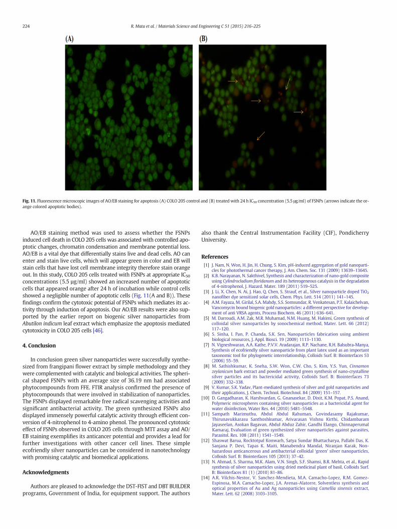
Fig. 11. Fluorescencemicroscopic images of AO/EB staining for apoptosis (A) COLO 205 control and (B) treated with 24 h IC50 concentration (5.5 μg/ml) of FSNPs (arrows indicate the or-ange colored apoptotic bodies).
224 R. Mata et al. / Materials Science and Engineering C 51 (2015) 216–225
AO/EB staining method was used to assess whether the FSNPsinduced cell death in COLO 205 cells was associated with controlled apo-ptotic changes, chromatin condensation and membrane potential loss.AO/EB is a vital dye that differentially stains live and dead cells. AO canenter and stain live cells, which will appear green in color and EB willstain cells that have lost cell membrane integrity therefore stain orangeout. In this study, COLO 205 cells treated with FSNPs at appropriate IC50concentrations (5.5 µg/ml) showed an increased number of apoptoticcells that appeared orange after 24 h of incubation while control cellsshowed a negligible number of apoptotic cells (Fig. 11(A and B)). Thesefindings confirm the cytotoxic potential of FSNPs which mediates its ac-tivity through induction of apoptosis. Our AO/EB results were also sup-ported by the earlier report on biogenic silver nanoparticles fromAbutilon indicum leaf extract which emphasize the apoptosis mediatedcytotoxicity in COLO 205 cells [46].
4. Conclusion
In conclusion green silver nanoparticles were successfully synthe-sized from frangipani flower extract by simple methodology and theywere complemented with catalytic and biological activities. The spheri-cal shaped FSNPs with an average size of 36.19 nm had associatedphytocompounds from FFE. FTIR analysis confirmed the presence ofphytocompounds that were involved in stabilization of nanoparticles.The FSNPs displayed remarkable free radical scavenging activities andsignificant antibacterial activity. The green synthesized FSNPs alsodisplayed immensely powerful catalytic activity through efficient con-version of 4-nitrophenol to 4-amino phenol. The pronounced cytotoxiceffect of FSNPs observed in COLO 205 cells through MTT assay and AO/EB staining exemplifies its anticancer potential and provides a lead forfurther investigations with other cancer cell lines. These simpleecofriendly silver nanoparticles can be considered in nanotechnologywith promising catalytic and biomedical applications.
Acknowledgments
Authors are pleased to acknowledge the DST-FIST and DBT BUILDERprograms, Government of India, for equipment support. The authors
also thank the Central Instrumentation Facility (CIF), PondicherryUniversity.
References[1] J. Nam, N. Won, H. Jin, H. Chung, S. Kim, pH-induced aggregation of gold nanoparti-
cles for photothermal cancer therapy, J. Am. Chem. Soc. 131 (2009) 13639–13645.[2] K.B. Narayanan, N. Sakthivel, Synthesis and characterization of nano-gold composite
using Cylindrocladium floridanum and its heterogeneous catalysis in the degradationof 4-nitrophenol, J. Hazard. Mater. 189 (2011) 519–525.
[3] J. Li, X. Chen, N. Ai, J. Hao, Q. Chen, S. Strauf, et al., Silver nanoparticle doped TiO2
nanofiber dye sensitized solar cells, Chem. Phys. Lett. 514 (2011) 141–145.[4] A.M. Fayaza, M. Girilal, S.A. Mahdy, S.S. Somsundar, R. Venkatesan, P.T. Kalaichelvan,
Vancomycin bound biogenic gold nanoparticles: a different perspective for develop-ment of anti VRSA agents, Process Biochem. 46 (2011) 636–641.
[5] M. Darroudi, A.M. Zak, M.R. Muhamad, N.M. Huang, M. Hakimi, Green synthesis ofcolloidal silver nanoparticles by sonochemical method, Mater. Lett. 66 (2012)117–120.
[6] S. Sinha, I. Pan, P. Chanda, S.K. Sen, Nanoparticles fabrication using ambientbiological resources, J. Appl. Biosci. 19 (2009) 1113–1130.
[7] N. Vigneshwaran, A.A. Kathe, P.V.V. Aradarajan, R.P. Nachane, R.H. Balsubra-Manya,Synthesis of ecofriendly silver nanoparticle from plant latex used as an importanttaxonomic tool for phylogenetic interrelationship, Colloids Surf. B: Biointerfaces 53(2006) 55–59.
[8] M. Sathishkumar, K. Sneha, S.W. Won, C.W. Cho, S. Kim, Y.S. Yun, Cinnamonzeylanicum bark extract and powder mediated green synthesis of nano-crystallinesilver particles and its bactericidal activity, Colloids Surf. B: Biointerfaces 73(2009) 332–338.
[9] V. Kumar, S.K. Yadav, Plant-mediated synthesis of silver and gold nanoparticles andtheir applications, J. Chem. Technol. Biotechnol. 84 (2009) 151–157.
[10] D. Gangadharan, K. Harshvardan, G. Gnanasekar, D. Dixit, K.M. Popat, P.S. Anand,Polymeric microspheres containing silver nanoparticles as a bactericidal agent forwater disinfection, Water Res. 44 (2010) 5481–5548.
[11] Sampath Marimuthu, Abdul Abdul Rahuman, Govindasamy Rajakumar,Thirunavukkarasu Santhoshkumar, Arivarasan Vishnu Kirthi, ChidambaramJayaseelan, Asokan Bagavan, Abdul Abduz Zahir, Gandhi Elango, ChinnaperumalKamaraj, Evaluation of green synthesized silver nanoparticles against parasites,Parasitol. Res. 108 (2011) 1541–1549.
[12] Shaswat Barua, Rocktotpal Konwarh, Satya Sundar Bhattacharya, Pallabi Das, K.Sanjana P. Devi, Tapas K. Maiti, Manabendra Mandal, Niranjan Karak, Non-hazardous anticancerous and antibacterial colloidal ‘green’ silver nanoparticles,Colloids Surf. B: Biointerfaces 105 (2013) 37–42.
[13] N. Ahmad, S. Sharma, M.K. Alam, V.N. Singh, S.F. Shamsi, B.R. Mehta, et al., Rapidsynthesis of silver nanoparticles using dried medicinal plant of basil, Colloids Surf.B: Biointerfaces 81 (1) (2010) 81–86.
[14] A.R. Vilchis-Nestor, V. Sanchez-Mendieta, M.A. Camacho-Lopez, R.M. Gomez-Espinosa, M.A. Camacho-Lopez, J.A. Arenas-Alatorre, Solventless synthesis andoptical properties of Au and Ag nanoparticles using Camellia sinensis extract,Mater. Lett. 62 (2008) 3103–3105.

225R. Mata et al. / Materials Science and Engineering C 51 (2015) 216–225
[15] S.P. Chandran, M. Chaudhary, R. Pasricha, A. Ahmad, M. Sastry, Synthesis of goldnanotriangles and silver nanoparticles using Aloe vera plant extract, Biotechnol.Prog. 22 (2006) 577–583.
[16] H. Bar, D.Kr. Bhui, G.P. Sahoo, P. Sarkar, S.P. De, A. Misra, Green synthesis of silvernanoparticles using latex of Jatropha curcas, Colloids Surf. A Physicochem. Eng.Asp. 339 (2009) 134–139.
[17] S.S. Shankar, A. Rai, A. Ahmad, M. Sastry, Controlling the optical properties of lemon-grass extract synthesized gold nanotriangles and potential application in infrared-absorbing optical coatings, Chem. Mater. 17 (2005) 566–572.
[18] S. Velavan, P. Arivoli, K. Mahadevan, Biological reduction of silver nanoparticlesusing Cassia auriculata flower extract and evaluation of their in vitro antioxidant ac-tivities, Nanosci. Nanotechnol. Int. J. 2 (2012) 30–35.
[19] GhassanMohammad Sulaiman,Wasnaa Hatif Mohammed, Thorria RadamMarzoog,Ahmed Abdul Amir Al-Amiery, Abdul Amir H. Kadhum, Abu Bakar Mohamad,Green synthesis, antimicrobial and cytotoxic effects of silver nanoparticlesusing Eucalyptus chapmaniana leaves extract, Asian Pac. J. Trop. Biomed. 3 (1)(2013) 58–63.
[20] JAMA. H.H. Comly, Supporting palladium metal on gold nanoparticles improves itscatalysis for nitrite reduction, J. Am. Med. Assoc. 257 (1987) 2788–2792.
[21] P. Kumar, M. Govindaraju, S. Senthamilselvi, K. Premkumar, Photocatalytic degrada-tion of methyl orange dye using silver (Ag) nanoparticles synthesized from Ulvalactuca, Colloids Surf. B: Biointerfaces 103 (2013) 658–661.
[22] Edward F. Gilman, Dennis G.Watson, Plumeria albaWhite Frangipani, October 1994.[23] B. Nagaraj, Barasa Malakar, T.K. Divya, N.B. Krishnamurthy, P. Liny, R. Dinesh,
Environmental benign synthesis of gold nanoparticles from the flower extracts ofPlumeria alba Linn. (frangipani) and evaluation of their biological activities, Int. J.Drug Dev. Res. 4 (1) (2012) (ISSN 0975–9344).
[24] T. Jebakumar Immanuel Edison, M.G. Sethuraman, Instant green synthesis ofsilver nanoparticles using Terminalia chebula fruit extract and evaluation of theircatalytic activity on reduction of methylene blue, Process Biochem. 47 (2012)1351–1357.
[25] Rakhi Majumdar, Braja Gopal Bag, Soma Rana, Azadirachta indica (neem) barkextract mediated rapid synthesis of gold nanoparticles and study of its catalyticactivity, J. Res. Chem. Environ. 3 (3) (2013) 144–152.
[26] N. Nenadis, I. Zafiropoulou, M. Tsimidou, Commonly used food antioxidants: acomparative study in dispersed systems, Food Chem. 82 (2003) 403–407.
[27] G.C. Yen, H.Y. Chen, Antioxidant activity of various tea extracts in relation to theirantimutagenicity, J. Agric. Food Chem. 43 (1) (1995) 27–32.
[28] M. Nishikimi, N.A. Rao, K. Yagi, The occurrence of superoxide anion in the reaction ofreduced phenazine methosulfate and molecular oxygen, Biochem. Biophys. Res.Commun. 46 (1992) 849–854.
[29] S.M. Nabavi, M.A. Ebrahimzadeh, S.F. Nabavi, A. Hamidinia, A.R. Bekhradnia,Determination of antioxidant activity, phenol and flavonoids content of Parrotiapersica Mey, Pharmacol. Online 2 (2008) 560–567.
[30] M.A. Ebrahimzadeh, S.F. Nabavi, S.M. Nabavi, Antioxidant activities of methanolextract of Sambucus ebulus L. flower, Pak. J. Biol. Sci. 12 (5) (2009) 447–450.
[31] Q.L. Feng, J. Wu, G.Q. Chen, F.Z. Cui, T.N. Kim, J.O. Kim, A mechanistic study of theantibacterial effect of silver ions on Escherichia coli and Staphylococcus aureus,J. Biomed. Mater. 52 (4) (2000) 662–668.
[32] K.G. Stamplecoskie, J.C. Scaiano, Light emitting diode can control the morphologyand optical properties of silver nanoparticles, J. Am. Chem. Soc. 132 (2010)1825–1827.
[33] Raman Sukirtha, Kandula Manasa Priyanka, Jacob Joe Antony, SoundararajanKamalakkannan, Ramar Thangam, Palani Gunasekaran, Muthukalingan Krishnan,Shanmugam Achiraman, Cytotoxic effect of Green synthesized silver nanoparticlesusing Melia azedarach against in vitro HeLa cell lines and lymphoma mice model,Process Biochem. 47 (2012) 273–279.
[34] S.P. Dubey, M. Lahtinen, M. Sillanpaa, Tansy fruit mediated greener synthesis ofsilver and gold nanoparticles, Process Biochem. 45 (2010) 1065–1071.
[35] Uthirappan Mani, Sujatha Dhanasingh, Rajeswari Arunachalam, Edwin Paul,Ponnusamy Shanmugam, Chellan Rose, Asit Baran Mandal, A simple and greenmethod for synthesis of silver nanoparticles using Ricinus communis leaf extract,Prog. Nanotechnol. Nanomater. 2 (1) (2013) 21–25.
[36] J. Huang, C. Chen, N. He, J. Hong, Y. Lu, L. Qingbiao, et al., Biosynthesis of silver andgold nanoparticles by novel sundried Cinnamomum camphora leaf, Nanotechnology18 (2007) 104–105.
[37] Tarek M. Abdel-Fattah, Alex Wixtrom, Catalytic reduction of 4-nitrophenol usinggold nanoparticles supported on carbon nanotubes, ECS J. Solid State Sci. Technol.3 (4) (2014) M18–M20.
[38] Z. Nie, K.J. Liu, C.J. Zhong, L.F. Wang, Y. Yang, Q. Tian, Y. Liu, Enhanced radical scav-enging activity by antioxidant-functionalized gold nanoparticles: a novel inspirationfor development of new artificial antioxidants, Free Radic. Biol. Med. 43 (2007)1243–1254.
[39] N. Jayachandra Reddy, D. Nagoor Vali, M. Rani, S. Sudha Rani, Evaluation ofantioxidant, antibacterial and cytotoxic effects of green synthesized silver nanopar-ticles by Piper longum fruit, Mater. Sci. Eng. C 34 (2014) 115–122.
[40] L. Inbathamizh, T. Mekalai Ponnu, E. Jancy Mary, In vitro evaluation of antioxidantand anticancer potential of Morinda pubescens synthesized silver nanoparticles, J.Pharm. Res. 6 (2013) 32–38.
[41] A. Chaudhary, Ayurvedic bhasma: nanomedicine of ancient India—its globalcontemporary perspective, J. Biomed. Nanotechnol. 7 (2011) 68–69.
[42] Q. Chen, L. Yue, F. Xie,M. Zhou, Y. Fu, Y. Zhang, et al., J. Phys. Chem. C 112 (2008) 10004.[43] S. Kaviya, J. Santhanalakshmi, B. Viswanathan, J. Muthumary, K. Srinivasan, Biosyn-
thesis of silver nanoparticles using Citrus sinensis peel extract and its antibacterialactivity, Spectrochim. Acta A 79 (2011) 594–598.
[44] Soo-Hwan Kim, Hyeong-Seon Lee, Deok-Seon Ryu, Soo-Jae Choi, Dong-Seok Lee, An-tibacterial activity of silver-nanoparticles against Staphylococcus aureus andEscherichia coli, Korean J. Microbiol. Biotechnol. 39 (1) (2011) 77–85.
[45] M. Jeyaraj, M. Rajesha, R. Arunb, D. MubarakAlic, G. Sathishkumara, G.Sivanandhana, et al., An investigation on the cytotoxicity and caspase-mediated ap-optotic effect of biologically synthesized silver nanoparticles using Podophyllumhexandrum on human cervical carcinoma cells, Colloids Surf. B: Biointerfaces 102(2013) 708–717.
[46] R. Mata, J.R. Nakkala, S.R. Sadras, Biogenic silver nanoparticles from Abutilon indicum:Theirantioxidant, antibacterial and cytotoxic effects in vitro, Colloids Surf B,Biointerfaces, 2015.http://dx.doi.org/10.1016/j.colsurfb.2015.01.052.