Caspofungin Modulates Inflammatory Responses to Aspergillus fumigatus through Stage‐Specific...
17
Caspofungin Modulates Inflammatory Responses to Aspergillus fumigatus through Stage-Specific Effects on Fungal β-Glucan Exposure T. M. Hohl 1 , M. Feldmesser 2 , D. S. Perlin 3 , and E. G. Pamer 1 1Infectious Diseases Service, Memorial Sloan-Kettering Cancer Center and Immunology Program, Sloan- Kettering Institute, New York 2Division of Infectious Diseases, Albert Einstein College of Medicine, Bronx, New York 3Public Health Research Institute, Newark, New Jersey Abstract Echinocandins target fungal β-1,3 glucan synthesis and are used clinically to treat invasive aspergillosis. Although echinocandins do not completely inhibit in vitro growth of Aspergillus fumigatus, they do induce morphological changes in fungal hyphae. Because β-1,3 glucans activate host antifungal pathways via the Dectin-1 receptor, we investigated the effect of echinocandins on inflammatory responses to A. fumigatus. Caspofungin- or micafungin-treated conidia and germlings induced less secretion of tumor necrosis factor (TNF) and CXCL2 by macrophages than did their untreated counterparts. Diminished secretion of TNF and CXCL2 correlated with diminished β- glucan exposure on echinocandin-treated germ tubes. In contrast to treated conidia and germlings, echinocandin-treated hyphae stimulated increased release of TNF and CXCL2 by macrophages and demonstrated intense staining with a β-glucan-specific antibody, particularly at hyphal tips. Our experiments demonstrate that echinocandin-induced morphological changes in A. fumigatus hyphae are accompanied by increased β-glucan exposure, with consequent increases in Dectin-1-mediated inflammatory responses by macrophages. In 2001, the echinocandin caspofungin was licensed in the United States as a second-line agent for the treatment of invasive aspergillosis (IA), which is a common cause of infection- associated morbidity and mortality among immunocompromised patients [1-3]. IA begins with the inhalation of airborne spores (i.e., conidia) that escape weakened pulmonary host defenses and germinate into tissue-invasive hyphae [4,5]. Caspofungin is a noncompetitive inhibitor of β-1,3 glucan synthase, an essential enzyme located on the growing filaments (i.e., hyphae) of Aspergillus fumigatus [6], the most common etiologic agent of IA. Although caspofungin has been found to be effective in animal models of IA [7-10], it does not fully inhibit growth of A. fumigatus, even at high concentrations [11,12]. Echinocandins reduce bulk β-1,3 glucan levels [13] and induce profound morphological changes that include the formation of swollen germ tubes; short, broad-based hyphae with aberrant branching; and distended, balloon-type cells [11]. The minimum drug concentration associated with these changes is referred to as the “minimum effective concentration” (MEC). Reprints or correspondence: Dr. T. M. Hohl, Infectious Diseases Service, MSKCC, 1275 York Ave., Box 9, New York, NY 10021 ([email protected]).. Potential conflicts of interest: none reported. NIH Public Access Author Manuscript J Infect Dis. Author manuscript; available in PMC 2009 July 15. Published in final edited form as: J Infect Dis. 2008 July 15; 198(2): 176–185. doi:10.1086/589304. NIH-PA Author Manuscript NIH-PA Author Manuscript NIH-PA Author Manuscript
-
Upload
independent -
Category
Documents
-
view
0 -
download
0
Transcript of Caspofungin Modulates Inflammatory Responses to Aspergillus fumigatus through Stage‐Specific...

Caspofungin Modulates Inflammatory Responses to Aspergillusfumigatus through Stage-Specific Effects on Fungal β-GlucanExposure
T. M. Hohl1, M. Feldmesser2, D. S. Perlin3, and E. G. Pamer11Infectious Diseases Service, Memorial Sloan-Kettering Cancer Center and Immunology Program, Sloan-Kettering Institute, New York
2Division of Infectious Diseases, Albert Einstein College of Medicine, Bronx, New York
3Public Health Research Institute, Newark, New Jersey
AbstractEchinocandins target fungal β-1,3 glucan synthesis and are used clinically to treat invasiveaspergillosis. Although echinocandins do not completely inhibit in vitro growth of Aspergillusfumigatus, they do induce morphological changes in fungal hyphae. Because β-1,3 glucans activatehost antifungal pathways via the Dectin-1 receptor, we investigated the effect of echinocandins oninflammatory responses to A. fumigatus. Caspofungin- or micafungin-treated conidia and germlingsinduced less secretion of tumor necrosis factor (TNF) and CXCL2 by macrophages than did theiruntreated counterparts. Diminished secretion of TNF and CXCL2 correlated with diminished β-glucan exposure on echinocandin-treated germ tubes. In contrast to treated conidia and germlings,echinocandin-treated hyphae stimulated increased release of TNF and CXCL2 by macrophages anddemonstrated intense staining with a β-glucan-specific antibody, particularly at hyphal tips. Ourexperiments demonstrate that echinocandin-induced morphological changes in A. fumigatus hyphaeare accompanied by increased β-glucan exposure, with consequent increases in Dectin-1-mediatedinflammatory responses by macrophages.
In 2001, the echinocandin caspofungin was licensed in the United States as a second-line agentfor the treatment of invasive aspergillosis (IA), which is a common cause of infection-associated morbidity and mortality among immunocompromised patients [1-3]. IA begins withthe inhalation of airborne spores (i.e., conidia) that escape weakened pulmonary host defensesand germinate into tissue-invasive hyphae [4,5]. Caspofungin is a noncompetitive inhibitor ofβ-1,3 glucan synthase, an essential enzyme located on the growing filaments (i.e., hyphae) ofAspergillus fumigatus [6], the most common etiologic agent of IA.
Although caspofungin has been found to be effective in animal models of IA [7-10], it doesnot fully inhibit growth of A. fumigatus, even at high concentrations [11,12]. Echinocandinsreduce bulk β-1,3 glucan levels [13] and induce profound morphological changes that includethe formation of swollen germ tubes; short, broad-based hyphae with aberrant branching; anddistended, balloon-type cells [11]. The minimum drug concentration associated with thesechanges is referred to as the “minimum effective concentration” (MEC).
Reprints or correspondence: Dr. T. M. Hohl, Infectious Diseases Service, MSKCC, 1275 York Ave., Box 9, New York, NY 10021([email protected])..Potential conflicts of interest: none reported.
NIH Public AccessAuthor ManuscriptJ Infect Dis. Author manuscript; available in PMC 2009 July 15.
Published in final edited form as:J Infect Dis. 2008 July 15; 198(2): 176–185. doi:10.1086/589304.
NIH
-PA Author Manuscript
NIH
-PA Author Manuscript
NIH
-PA Author Manuscript

In contrast, caspofungin induces complete growth inhibition and cell lysis of the dimorphicfungus Candida albicans [12]. At sub-MIC concentrations, caspofungin increases β-glucanexposure on C. albicans, and drug-treated yeast cells trigger enhanced macrophageinflammatory responses [14]. In the case of A. fumigatus, caspofungin appears to inducemembrane depolarization in a subset of cells located at the tips and branch points of the growingmycelium [15], a process that is sufficient to stunt but not arrest filamentous growth.
Not only is β-1,3 glucan an obligate fungal cell wall constituent, it represents a target of theinnate immune system. The mammalian receptor Dectin-1 [16,17] binds to β-1,3-glucan fromPneumocystis carinii [18], C. albicans [19], A. fumigatus [20-22], and Coccidioidesposadasii [23]. It also triggers fungicidal responses that include phagocytosis, release ofinflammatory mediators, and generation of reactive oxygen intermediates [17].
Although echinocandins increase β-glucan exposure on the surface of C. albicans, whether thischange occurs in such clinically important molds as A. fumigatus is not known. Therefore, wetreated A. fumigatus spores with echinocandins in vitro and measured surface β-glucanexposure and macrophage inflammatory responses. TNF and CXCL2, a neutrophilchemoattractant, were chosen as a readout for inflammatory responses, because both haveestablished roles in host defense against A. fumigatus [24-27]. During the early stages ofconidial swelling and germ tube formation, drug-treated cells elicited weaker inflammatoryresponses than did control cells. In contrast, drug-treated hyphae induced strongerinflammatory responses than did control cells. In all cases, the magnitude of the inflammatoryresponse reflected changes in surface β-glucan exposure. Our findings suggest thatechinocandins, by increasing β-glucan exposure on the surface of invading A. fumigatushyphae, may enhance antifungal inflammatory responses to hyphae, independent of directgrowth inhibition.
MATERIALS AND METHODSReagents and antibodies
Chemical and cell culture reagents were purchased from Sigma-Aldrich and Gibco,respectively. Caspofungin (Merck) and micafungin (Astellas) in powder form were obtainedfrom the Memorial Sloan-Kettering Cancer Center pharmacy, dissolved in sterile PBS at 5 mg/mL, and stored at -80°C. The anti-Dectin-1 antibody 2A11 and an IgG isotype control antibodywere purchased from AbD Serotec. Monoclonal antibody (MAb) 744 has been describedelsewhere [20], and an Alexa Fluor 594-linked anti-mouse IgM antibody was obtained fromInvitrogen.
Fungal growth and cultureC. albicans strain SC5314 was grown in yeast extract peptone dextrose overnight at 25°C.Yeast cells were diluted 1:1000 in fresh medium ± 5 ng/mL caspofungin, shaken overnight at25°C, washed 3 times in PBS, killed by UV irradiation from a Stratalinker UV Crosslinker1800 (dose, 4 × 10⁵ mJ/cm2), and added to bone marrow-derived macrophages (BMMϕs) at aratio of 10:1. The MIC of caspofungin for these conditions was 10 ng/mL.
A. fumigatus strains Af293 and ATCC 90906 were cultured as described elsewhere [20]. Forgermination reactions, 5 × 10⁴ conidia were seeded in 96-well culture dishes (Invitrogen) in0.2 mL of RP-10+ (i.e., RPMI 1640, 10% heat-inactivated fetal calf serum [FCS], 5 mmol/LHEPES [pH 7.4], 1.1 mmol/L L-glutamine, 0.5 U/mL penicillin, 0.5 μg/mL gentamicin, 50μg/mL streptomycin, and 50 μmol/L 2-mercaptoethanol) containing caspofungin (0-1000 ng/mL) or micafungin (0-1000 ng/mL) and incubated at 37°C for the times indicated. Culture
Hohl et al. Page 2
J Infect Dis. Author manuscript; available in PMC 2009 July 15.
NIH
-PA Author Manuscript
NIH
-PA Author Manuscript
NIH
-PA Author Manuscript

supernatants were aspirated, and fungal cells were inactivated by UV irradiation, as describedabove.
Cell culture and ELISABMMϕs obtained from 8- to 12-week-old C57BL/6 female mice were prepared as describedelsewhere [20], harvested after 5 days in culture, adjusted to 7.5 × 10⁵ cells/mL in RP-10+, andadded (in a final volume of 0.2 mL) to empty wells or wells containing UV-inactivated fungalcells in 96-well plates.
For experiments with live A. fumigatus conidia, BMMϕs were cultured with 1.5 × 10⁶ conidiain 0.2 mL of RP-10+ with 0-1000 ng/mL caspofungin or micafungin for 18 h at 37°C. Forspecific experiments, 10 μg/mL anti-Dectin-1 or isotype control antibody was added toBMMϕs 10 min before coculture with fungal cells. For experiments with UV-inactivated fungalcells, the BMMϕ coincubation period was 6 h. Culture supernatants were analyzed usingcommercially available ELISA kits for the detection of CXCL2 (R & D Systems) and TNF(BD Biosciences) by use of a VersaMax Microplate Reader (Molecular Devices).
Confocal microscopyA total of 5 × 10⁴ conidia per well was grown on 4-well Lab-Tek glass chamber slides (NalgeNunc International) in RP-10+ with the indicated drug concentration for 8-24 h at 37°C. Pilotexperiments determined that UV inactivation did not alter β-glucan immunoreactivity. Fungalcells were processed for confocal microscopy by use of MAb 744 and Alexa Fluor 594-linkedanti-mouse IgM, as described elsewhere [20]. No immunoreactivity was observed in samplesstained with a control isotype antibody. The samples were scanned in 0.5- to 1.0-μm intervals(in the z-plane), with the use of an upright Leica TCS SP2 AOBS confocal microscopy systemequipped with a 63× Leica HPX PL APO water-immersion objective (numeric aperture 1,2)using the 488-nm and 594-nm laser lines and transmitted light. Composite fluorescence imageswere assembled from individually scanned optical sections obtained from a 30- to 40-μminterval in the z-plane. For all images displayed, the sensitivity of the recording photomultipliertube was set in the linear range and was identical for drug-treated and untreated cells.
For image analysis of drug-treated and untreated hyphae, cellular autofluorescence wasrecorded using the 488-nm laser line to visualize the hyphal area (in cross-section) in opticalsections measuring 237.5 μm × 237.5 μm × 1 μm. Autofluorescence and immunofluorescencerecordings underwent image analysis in MetaMorph software (version 7.1.1.0; MolecularDevices). Threshold settings were applied uniformly for all paired data sets. A low-pass filter(2 × 2 pixels) was applied to autofluorescence stacks to suppress single-pixel noise, and thearea above a set threshold was measured in each optical slice and then summed across all sliceswithin a field of view, to obtain a measure of the total hyphal volume within an image stack.
For image processing of immunofluorescence recordings, no filters were applied, and theintegrated fluorescence intensity in the red channel above a set threshold was measured foreach optical slice and summed across all slices within the same field of view. To associate β-glucan immunoreactivity with fungal biomass for drug-treated and untreated hyphae (after 24h of incubation), the total integrated fluorescence intensity in the red channel was divided bythe summed threshold area delineated by the autofluorescent signal, resulting in a measurementof β-glucan immunoreactivity per hyphal volume. In a single experiment, 4-5 optical sections,each consisting of ∼30-40 optical slices, were evaluated for each condition.
Hohl et al. Page 3
J Infect Dis. Author manuscript; available in PMC 2009 July 15.
NIH
-PA Author Manuscript
NIH
-PA Author Manuscript
NIH
-PA Author Manuscript

RESULTSNo direct elicitation by caspofungin of macrophage TNF production
To determine whether caspofungin directly elicits inflammatory responses, the drug was addedto BMMϕ cultures. No difference in the secretion of TNF or CXCL2 by BMMϕ was observedin drug-treated or control samples (figure 1A) (not shown), even with concurrent addition oflipopolysaccharide (LPS) or killed, swollen A. fumigatus conidia. Thus, caspofungin does nottrigger secretion of TNF or CXCL2 by BMMϕ at rest or in conjunction with inflammatorystimuli. In contrast, C. albicans yeast cells grown in sub-MIC levels of caspofungin elicitedTNF secretion that was ∼4-fold higher than that of control cells (figure 1B), in accordance withresults achieved using a different strain [14].
Caspofungin and the diminishment of inflammatory responses to live A. fumigatus conidiaAt rest, A. fumigatus conidia trigger minimal inflammatory responses [20]. Conidial swellingrepresents the first step of germination and is required for macrophages to initiate inflammatoryand fungicidal responses [28,29].
To examine the effect of caspofungin on inflammatory responses to conidia, BMMϕs andconidia (derived from A. fumigatus strain Af293) were incubated for 18 h in the presence of0-500 ng/mL caspofungin. Caspofungin diminished inflammatory responses in a dose-dependent manner, with a reduction evident at drug concentrations at or above the MEC, whichwas determined to be 63 ng/mL for the experimental conditions (figure 2A). At a drugconcentration of 500 ng/mL, the average percentage (±SD) of TNF and CXCL2 released intothe supernatants by BMMϕs was 49% ± 4% (range, 46%-54%; n = 4 experiments) and 55% ±10% (range, 43%-62%; n = 4 experiments), respectively, compared with the amount releasedfor untreated samples. Similar results were obtained with conidia derived from A. fumigatusstrain ATCC 90906 (not shown).
To determine whether exposure of conidia to caspofungin diminishes Dectin-1 signaling bymacrophages, the effect of antibody-mediated receptor blockade on inflammatory responsesto drug-treated and untreated conidia was examined. MAb 2A11 binds to the extracellularportion of Dectin-1 and blocks signal transduction through its intracellular immunoregulatorytyrosine-based activation motif-like motif [30]. Macrophages isolated from Dectin-1-/- micedo not bind MAb 2A11 [31], indicating that the antibody is specific for Dectin-1 and does notbind to other C-type lectins (e.g., dendritic cell-specific intercellular adhesion molecule 3-grabbing nonintegrin or Dectin-2, receptors with different carbohydrate-binding propertiesimplicated in the recognition of fungal cells) [32-34].
In agreement with previous studies [20-22,31], antibody-blocking experiments demonstratedthat Dectin-1 is largely responsible for macrophage production of TNF and CXCL2 in responseto conidia (figure 2B). The reduced macrophage inflammatory response observed in associationwith drug-treated conidia reflects the diminished Dectin-1-associated signaling triggered bythese cells.
The conidia-macrophage coincubation assay described above measures inflammatoryresponses to the earliest stage of conidial germination. In caspofungin-treated and drug-freesamples, >99% of conidia associated with macrophages at the conclusion of the incubationperiod did not form germ tubes (figure 2C). Rare hyphae that escaped macrophage control wereobserved in all samples (<1% of cells), consistent with the inability of caspofungin to preventgermination (figure 2C, white arrows). In sum, these results suggest that caspofungin reducesthe inflammatory responses of BMMϕs to live conidia.
Hohl et al. Page 4
J Infect Dis. Author manuscript; available in PMC 2009 July 15.
NIH
-PA Author Manuscript
NIH
-PA Author Manuscript
NIH
-PA Author Manuscript

Caspofungin and diminishment of inflammatory responses to A. fumigatus germlingsTo examine the effect of caspofungin on inflammatory responses triggered at later stages ofgermination, 5 × 10⁴ conidia per well were incubated in the presence or absence of drug for 8h, to complete the swelling reaction and form short germ tubes. BMMϕs were then added toUV-inactivated fungal cells for 6 h. Similar results were observed with A. fumigatus strainAf293 (figure 3) and ATCC 90906 (not shown).
The amounts of TNF and CXCL2 released in BMMϕ culture supernatants decreased in a dose-dependent manner if the caspofungin concentration exceeded the MEC (figure 3A). At a drugconcentration of 500 ng/mL, the average percentage (±SD) of TNF and CXCL2 released byBMMϕs was 51% ± 7% (range, 43%-64%; n = 7 experiments) and 61% ± 8% (range, 53%-74%;n = 7 experiments) respectively, compared with the amount released from untreated samples.Inflammatory responses of BMMϕs were 90%-95% attributable to Dectin-1 signaling, anddrug treatment reduced Dectin-1-associated production of TNF and CXCL2 (figure 3B).Germlings grown in micafungin elicited a similar decrease in TNF and CXCL2 secretion byBMMϕs (figure 3C), indicating a general effect of echinocandin drugs.
Caspofungin and enhancement of inflammatory responses to A. fumigatus hyphaeTo examine inflammatory responses to hyphae, 5 × 10⁴ conidia were grown for 24 h in mediumcontaining 0-500 ng/mL caspofungin, and they were then inactivated by UV exposure. Despitea marked reduction in fungal growth in drug-treated wells (see below), BMMϕ inflammatoryresponses were enhanced in a dose-dependent manner (figure 4A). Fungal hyphae grown in500 ng/mL caspofungin induced an average (±SD) of 4.11-fold (±2.39-fold) more TNF (range,1.90- to 7.84-fold; n = 8 experiments) and 2.90-fold (±1.40-fold) more CXCL2 (range, 1.53-to 5.41-fold; n = 8 experiments) than did hyphae grown in drug-free medium. Enhancedinflammatory responses to drug-treated hyphae were observed with A. fumigatus strain ATCC90906 as well (not shown).
The increase in secretion of TNF and CXCL2 by BMMϕsin caspofungin-treated samples wasattributable to increased Dectin-1-associated signaling (figure 4B). Enhanced BMMϕinflammatory responses were observed with micafungin-treated hyphae as well (figure 4C).This effect was seen in association with different fungal growth media, both containing serum(RP-10+) and lacking serum (RPMI 1640, 0.165 mol/L 3-(N-morpholino) propanesulfonic acid[pH 7.0]), and with conidial inocula that ranged from 5 × 10³ to 5 × 10⁴ cells (data not shown).
Hyphae may elaborate factors that reduce the inflammatory responses of BMMϕs in proportionto mycelial growth. Because growth is markedly reduced in echinocandin-treated samples (seebelow), excluding this explanation for the observed difference in inflammatory response isimportant. To examine whether macrophage responsiveness diminished when cells werestimulated with control hyphae, rather than with drug-treated hyphae, LPS was added to bothexperimental conditions. Macrophage inflammatory responses were nearly identical in bothcases (figure 4D). This finding supports the notion that low-level inflammatory responses tonon-drug-treated hyphae and enhanced inflammatory responses to drug-treated hyphae are notassociated with changes in macrophage responsiveness to other inflammatory stimuli.
To examine inflammatory responses to A. fumigatus during the transition from germlings tohyphae, fungal cells were grown in drug-free medium or in medium containing caspofungin,and, at defined time points between 8 h and 24 h, they were inactivated by UV exposure.Inflammatory responses decreased as fungal cells completed the transition from germlings tohyphae in drug-free medium, despite the increase in fungal mass that occurred during hyphalgrowth (figure 4E). In contrast, fungal cells exposed to caspofungin induced a large increasein the secretion of TNF and CXCL2 by macrophages after 12-18 h of growth in medium
Hohl et al. Page 5
J Infect Dis. Author manuscript; available in PMC 2009 July 15.
NIH
-PA Author Manuscript
NIH
-PA Author Manuscript
NIH
-PA Author Manuscript
containing drugs (figure 4E). Drug-treated hyphae were more inflammatory than drug-treatedgermlings. In all cases examined, inflammatory responses to drug-treated and control cellswere highly associated with Dectin-1 signaling.
Caspofungin and alteration of β-glucan immunoreactivity on germ tubes and hyphaeFungal cells were examined by confocal microscopy to examine drug-dependent changes insurface β-glucan exposure during germ tube formation and hyphal growth. A monoclonalantibody that recognizes β-1,3-glucan was used for experiments involvingimmunofluorescence [20]. Untreated germlings demonstrated strong β-glucanimmunoreactivity on the remnant of the swollen conidium and diffuse staining along theprotruding germ tube (figure 5A). In contrast, their drug-treated counterparts lacked β-glucanimmunoreactivity on the protruding germ tube.
As filaments increased in length (during 10-12 h of incubation in RP-10+), they remainedimmunoreactive along the entire structure (figure 5B and C). In drug-treated samples, β-glucanimmunoreactivity became apparent on the tips of germ tubes after 10 h of growth (figure 5B),with marked enhancement at distal segments noted after 2 additional hours of growth (figure5C). At this later point in time, the growth inhibitory effects of caspofungin were apparent.
At 15 h of growth, drug-free samples contained weakly immunoreactive and nonreactivehyphae (figure 5D). The morphologically distinct hyphae observed in drug-treated samplesdisplayed intense β-glucan immunoreactivity concentrated at peripheral segments (figure5D). Drug-treated hyphae displayed persistent β-glucan immunoreactivity after 18 h of growthin caspofungin, predominantly at distal segments but extending to more central areas as well(figure 5E). In contrast, control samples were markedly less immunoreactive than drug-treatedcells (at the same time point) and less mature hyphae (figure 5D). At the final time pointexamined (at 24 h), drug-treated hyphae displayed far greater β-glucan immunoreactivity thandid untreated hyphae (figure 6). This finding was noted with different conidial inocula (5 ×10³-5 × 10⁴ conidia), fungal growth media, and echinocandin drugs (caspofungin andmicafungin) (figure 6) (not shown).
To examine differences in β-glucan immunoreactivity between drug-treated and untreatedhyphae, the integrated fluorescence intensity of β-glucan immunoreactivity was determinedfor all hyphal filaments within a field of view consisting of ∼30 - 40 optical slices (see Materialsand Methods for detail). To relate the β-glucan fluorescence intensity to the hyphal mass withineach field of view, fungal cell autofluorescence was recorded to visualize the hyphal cross-sectional area in each optical slice (and field of view). In 2 experiments, drug-treated hyphaedisplayed >10-fold greater β-glucan immunoreactivity than did untreated hyphae, normalizedfor hyphal mass (table 1).
DISCUSSIONIn the present study, we found that echinocandins shape inflammatory responses to A.fumigatus through modulation of surface β-glucan content. Surprisingly, this process occursin a stage-specific manner: drug-treated germlings display diminished surface β-glucanimmunoreactivity and elicit reduced inflammatory responses, whereas drug-treated hyphaeexhibit a dramatic increase in surface β-glucan immunoreactivity and trigger enhancedinflammatory responses. The latter result is consistent with the finding that macrophagescooperate with caspofungin to inhibit hyphal growth in an additive manner [35]. The resultsof the present study support echinocandins increasing hyphal susceptibility to Dectin-1-mediated responses by direct effects on the fungal cell wall. Drug-dependent changes in β-glucan exposure and macrophage inflammatory responses occur at drug levels that are relevantfor therapy [36].
Hohl et al. Page 6
J Infect Dis. Author manuscript; available in PMC 2009 July 15.
NIH
-PA Author Manuscript
NIH
-PA Author Manuscript
NIH
-PA Author Manuscript

A second finding is that A. fumigatus, under drug-free conditions, limits β-glucan exposureduring hyphal growth, given the low-level Dectin-1-dependent inflammatory response tohyphae, compared with the response to germlings. C. albicans conceals β-glucan during yeastcell and pseudohyphal growth beneath a mannoprotein layer [19]. Shielding β-glucan orlimiting surface exposure may provide a growth or survival advantage within the diverseecological niches inhabited by these organisms [14,37]. Caspofungin disrupts the normal cellwall architecture of both organisms and exerts similar proinflammatory effects on C.albicans yeast cells [14] and A. fumigatus hyphae. However, in contrast to their effect onresponses to C. albicans, echinocandin drugs can decrease inflammatory responses to specificgrowth stages of A. fumigatus.
The A. fumigatus ΔpksP mutant defective in pigment biosynthesis forms conidia with highlevels of surface β-glucan [38]. ΔpksP conidia display heightened in vitro susceptibility tophagocyte defense mechanisms [39] and reduced virulence in animal models of IA [40].Although this mutant strain likely harbors other changes that influence disease outcome, it isstriking that enhanced β-glucan exposure correlates with decreased virulence.
A. fumigatus spores contain minimal amounts of β-glucan on their surface [20], and β-glucansbecome exposed during swelling and germ tube formation. When caspofungin exposure occurs,a reduction in de novo synthesis may account for the reduced β-glucan surface display on theinitial germ tube segment. Alternatively, the initial step of germ tube formation could depend,in part, on preformed stores. Either way, caspofungin appears to prevent the immunoreactivepolymer from reaching the fungal surface at the earliest stage of filamentous growth, given thenear absence of immunoreactive β-glucan on short drug-treated filaments. Compensatorychanges in cell wall constituents or assembly may be associated with the reduced inflammatoryresponses observed with swollen conidia and germ tubes, at the eventual cost of dysmorphicand stunted growth.
Continued inhibition of glucan synthase leads to a paradoxical increase in surface β-glucan atthe ends of growing filaments. Increased surface accumulation of β-glucan may result fromsecondary changes in the activities of enzymes involved in polymer biosynthesis and turnover(e.g., β-1,3-glucanosyltransferases [41] and β-1,3-glucanases [42-44]). Because Dectin-1 bindsexclusively to β-1,3-linked glucose oligomers [45], echinocandin-induced changes in otherpolysaccharide constituents of the cell wall are unlikely to contribute directly to the Dectin-1-dependent inflammatory responses measured in these studies.
The antifungal properties of echinocandin drugs may not be adequately described by theireffects on growth [37,46]. Compounds with direct immune stimulatory properties or those thatenhance the immune recognition of target microbes have an increasing role in the treatment ofinfectious diseases. For example, the Toll-like receptor (TLR) agonist imiquimod is effectiveagainst lesions associated with human papillomavirus [47]. A variety of compounds orconstituents of antifungal agents have immune modulatory properties that may enhance fungalclearance. Amphotericin B directly stimulates TLR-dependent inflammatory cytokine releasein a fungus-independent manner [48]. Recently, empty liposomes were found to have beneficialimmune stimulatory effects in a murine model of IA [49]. In contrast, echinocandin drugs areunlikely to cause general activation of inflammatory responses. Their immunomodulatoryproperties depend on the presence of fungal cells, providing a mechanism by which cell-mediated and humoral responses [50] are focused to sites of hyphal growth. Recognition thatcaspofungin modulates inflammatory responses to specific growth forms of A. fumigatus haspotential implications for prophylactic and therapeutic strategies. Because host inflammatoryresponses make a critical contribution to the clearance of invasive aspergillosis, echinocandindrugs may be most effective for hyphal lesions and at the time of host myeloid cell recovery.
Hohl et al. Page 7
J Infect Dis. Author manuscript; available in PMC 2009 July 15.
NIH
-PA Author Manuscript
NIH
-PA Author Manuscript
NIH
-PA Author Manuscript

AcknowledgmentsCandida albicans strain SC5314 was a gift from Brad Spellberg (Harbor-University of California Los Angeles MedicalCenter). We thank Ewa Menet for outstanding technical assistance and Yevgeniy Romin and Katia Manova-Todorovafor assistance with confocal microscopy and image analysis.
Financial support: T.M.H. is a Biomedical Fellow of the Charles H. Revson Foundation; National Institutes of Health(training grant T32 [to T.M.H.], grants R01 AI059663 and R21 AI065745 [to M.F.], and grant R01 AI67359 [toE.G.P.]).
References1. Deresinski SC, Stevens DA. Caspofungin. Clin Infect Dis 2003;36:1445–57. [PubMed: 12766841]2. Hope WW, Shoham S, Walsh TJ. The pharmacology and clinical use of caspofungin. Expert Opin
Drug Metab Toxicol 2007;3:263–74. [PubMed: 17428155]3. Lin SJ, Schranz J, Teutsch SM. Aspergillosis case-fatality rate: systematic review of the literature.
Clin Infect Dis 2001;32:358–66. [PubMed: 11170942]4. Latgé JP. Aspergillus fumigatus and aspergillosis. Clin Microbiol Rev 1999;12:310–50. [PubMed:
10194462]5. Barnes PD, Marr KA. Aspergillosis: spectrum of disease, diagnosis, and treatment. Infect Dis Clin
North Am 2006;20:545–61. [PubMed: 16984868]6. Beauvais A, Drake R, Ng K, Diaquin M, Latgé JP. Characterization of the 1,3-β-glucan synthase of
Aspergillus fumigatus. J Gen Microbiol 1993;139:3071–8. [PubMed: 8126434]7. Abruzzo GK, Flattery AM, Gill CJ, et al. Evaluation of the echinocandin antifungal MK-0991
(L-743,872): efficacies in mouse models of disseminated aspergillosis, candidiasis, andcryptococcosis. Antimicrob Agents Chemother 1997;41:2333–8. [PubMed: 9371329]
8. Abruzzo GK, Gill CJ, Flattery AM, et al. Efficacy of the echinocandin caspofungin againstdisseminated aspergillosis and candidiasis in cyclophosphamide-induced immunosuppressed mice.Antimicrob Agents Chemother 2000;44:2310–8. [PubMed: 10952573]
9. Petraitiene R, Petraitis V, Groll AH, et al. Antifungal efficacy of caspofungin (MK-0991) inexperimental pulmonary aspergillosis in persistently neutropenic rabbits: pharmacokinetics, drugdisposition, and relationship to galactomannan antigenemia. Antimicrob Agents Chemother2002;46:12–23. [PubMed: 11751105]
10. Wiederhold NP, Kontoyiannis DP, Chi J, Prince RA, Tam VH, Lewis RE. Pharmacodynamics ofcaspofungin in a murine model of invasive pulmonary aspergillosis: evidence of concentration-dependent activity. J Infect Dis 2004;190:1464–71. [PubMed: 15378439]
11. Kurtz MB, Heath IB, Marrinan J, Dreikorn S, Onishi J, Douglas C. Morphological effects oflipopeptides against Aspergillus fumigatus correlate with activities against (1,3)-β-d-glucan synthase.Antimicrob Agents Chemother 1994;38:1480–9. [PubMed: 7979276]
12. Bartizal K, Gill CJ, Abruzzo GK, et al. In vitro preclinical evaluation studies with the echinocandinantifungal MK-0991 (L-743,872). Antimicrob Agents Chemother 1997;41:2326–32. [PubMed:9371328]
13. Kahn JN, Hsu MJ, Racine F, Giacobbe R, Motyl M. Caspofungin susceptibility in Aspergillus andnon-Aspergillus molds: inhibition of glucan synthase and reduction of β-d-1,3 glucan levels in culture.Antimicrob Agents Chemother 2006;50:2214–6. [PubMed: 16723587]
14. Wheeler RT, Fink GR. A drug-sensitive genetic network masks fungi from the immune system. PLoSPathog 2006;2:e35. [PubMed: 16652171]
15. Bowman JC, Hicks PS, Kurtz MB, et al. The antifungal echinocandin caspofungin acetate killsgrowing cells of Aspergillus fumigatus in vitro. Antimicrob Agents Chemother 2002;46:3001–12.[PubMed: 12183260]
16. Brown GD, Gordon S. Immune recognition: a new receptor for β-glucans. Nature 2001;413:36–7.[PubMed: 11544516]
17. Brown GD. Dectin-1: a signalling non-TLR pattern-recognition receptor. Nat Rev Immunol2006;6:33–43. [PubMed: 16341139]
Hohl et al. Page 8
J Infect Dis. Author manuscript; available in PMC 2009 July 15.
NIH
-PA Author Manuscript
NIH
-PA Author Manuscript
NIH
-PA Author Manuscript
18. Steele C, Marrero L, Swain S, et al. Alveolar macrophage-mediated killing of Pneumocystis cariniif. sp. muris involves molecular recognition by the Dectin-1 β-glucan receptor. J Exp Med2003;198:1677–88. [PubMed: 14657220]
19. Gantner BN, Simmons RM, Underhill DM. Dectin-1 mediates macrophage recognition of Candidaalbicans yeast but not filaments. EMBO J 2005;24:1277–86. [PubMed: 15729357]
20. Hohl TM, Van Epps HL, Rivera A, et al. Aspergillus fumigatus triggers inflammatory responses bystage-specific β-glucan display. PLoS Pathog 2005;1:e30. [PubMed: 16304610]
21. Steele C, Rapaka RR, Metz A, et al. The β-glucan receptor dectin-1 recognizes specific morphologiesof Aspergillus fumigatus. PLoS Pathog 2005;1:e42. [PubMed: 16344862]
22. Gersuk GM, Underhill DM, Zhu L, Marr KA. Dectin-1 and TLRs permit macrophages to distinguishbetween different Aspergillus fumigatus cellular states. J Immunol 2006;176:3717–24. [PubMed:16517740]
23. Viriyakosol S, Fierer J, Brown GD, Kirkland TN. Innate immunity to the pathogenic fungusCoccidioides posadasii is dependent on Toll-like receptor 2 and Dectin-1. Infect Immun2005;73:1553–60. [PubMed: 15731053]
24. Mehrad B, Strieter RM, Standiford TJ. Role of TNF-α in pulmonary host defense in murine invasiveaspergillosis. J Immunol 1999;162:1633–40. [PubMed: 9973423]
25. Warris A, Bjorneklett A, Gaustad P. Invasive pulmonary aspergillosis associated with infliximabtherapy. N Engl J Med 2001;344:1099–100. [PubMed: 11291675]
26. Mehrad B, Strieter RM, Moore TA, Tsai WC, Lira SA, Standiford TJ. CXC chemokine receptor-2ligands are necessary components of neutrophil-mediated host defense in invasive pulmonaryaspergillosis. J Immunol 1999;163:6086–94. [PubMed: 10570298]
27. Schuh JM, Blease K, Hogaboam CM. CXCR2 is necessary for the development and persistence ofchronic fungal asthma in mice. J Immunol 2002;168:1447–56. [PubMed: 11801688]
28. Ibrahim-Granet O, Philippe B, Boleti H, et al. Phagocytosis and intracellular fate of Aspergillusfumigatus conidia in alveolar macrophages. Infect Immun 2003;71:891–903. [PubMed: 12540571]
29. Philippe B, Ibrahim-Granet O, Prévost MC, et al. Killing of Aspergillus fumigatus by alveolarmacrophages is mediated by reactive oxidant intermediates. Infect Immun 2003;71:3034–42.[PubMed: 12761080]
30. Brown GD, Taylor PR, Reid DM, et al. Dectin-1 is a major β-glucan receptor on macrophages. J ExpMed 2002;196:407–12. [PubMed: 12163569]
31. Taylor PR, Tsoni SV, Willment JA, et al. Dectin-1 is required for β-glucan recognition and controlof fungal infection. Nat Immunol 2007;8:31–8. [PubMed: 17159984]
32. Serrano-Gómez D, Domínguez-Soto A, Ancochea J, Jimenez-Heffernan JA, Leal JA, Corbi AL.Dendritic cell-specific intercellular adhesion molecule 3-grabbing nonintegrin mediates binding andinternalization of Aspergillus fumigatus conidia by dendritic cells and macrophages. J Immunol2004;173:5635–43. [PubMed: 15494514]
33. McGreal EP, Rosas M, Brown GD, et al. The carbohydrate-recognition domain of Dectin-2 is a C-type lectin with specificity for high mannose. Glycobiology 2006;16:422–30. [PubMed: 16423983]
34. Sato K, Yang XL, Yudate T, et al. Dectin-2 is a pattern recognition receptor for fungi that coupleswith the Fc receptor γ chain to induce innate immune responses. J Biol Chem 2006;281:38854–66.[PubMed: 17050534]
35. Chiller T, Farrokhshad K, Brummer E, Stevens DA. The interaction of human monocytes, monocyte-derived macrophages, and polymorphonuclear neutrophils with caspofungin (MK-0991), anechinocandin, for antifungal activity against Aspergillus fumigatus. Diagn Microbiol Infect Dis2001;39:99–103. [PubMed: 11248522]
36. Stone JA, Holland SD, Wickersham PJ, et al. Single- and multiple-dose pharmacokinetics ofcaspofungin in healthy men. Antimicrob Agents Chemother 2002;46:739–45. [PubMed: 11850256]
37. Hohl TM, Pamer EG. Cracking the fungal armor. Nat Med 2006;12:730–2. [PubMed: 16829914]38. Luther K, Torosantucci A, Brakhage AA, Heesemann J, Ebel F. Phagocytosis of Aspergillus
fumigatus conidia by murine macrophages involves recognition by the dectin-1 β-glucan receptorand Toll-like receptor 2. Cell Microbiol 2007;9:368–81. [PubMed: 16953804]
Hohl et al. Page 9
J Infect Dis. Author manuscript; available in PMC 2009 July 15.
NIH
-PA Author Manuscript
NIH
-PA Author Manuscript
NIH
-PA Author Manuscript

39. Jahn B, Langfelder K, Schneider U, Schindel C, Brakhage AA. PKSP-dependent reduction ofphagolysosome fusion and intracellular kill of Aspergillus fumigatus conidia by human monocyte-derived macrophages. Cell Microbiol 2002;4:793–803. [PubMed: 12464010]
40. Langfelder K, Jahn B, Gehringer H, Schmidt A, Wanner G, Brakhage AA. Identification of apolyketide synthase gene (pksP) of Aspergillus fumigatus involved in conidial pigment biosynthesisand virulence. Med Microbiol Immunol 1998;187:79–89. [PubMed: 9832321]
41. Mouyna I, Morelle W, Vai M, et al. Deletion of GEL2 encoding for a β(1-3)glucanosyltransferaseaffects morphogenesis and virulence in Aspergillus fumigatus. Mol Microbiol 2005;56:1675–88.[PubMed: 15916615]
42. Fontaine T, Hartland RP, Beauvais A, Diaquin M, Latge JP. Purification and characterization of anendo-1,3-β-glucanase from Aspergillus fumigatus. Eur J Biochem 1997;243:315–21. [PubMed:9030754]
43. Fontaine T, Hartland RP, Diaquin M, Simenel C, Latgé JP. Differential patterns of activity displayedby two exo-β-1,3-glucanases associated with the Aspergillus fumigatus cell wall. J Bacteriol1997;179:3154–63. [PubMed: 9150209]
44. Mouyna I, Sarfati J, Recco P, Fontaine T, Henrissatz B, Latge JP. Molecular characterization of acell wall-associated β(1-3)endoglucanase of Aspergillus fumigatus. Med Mycol 2002;40:455–64.[PubMed: 12462524]
45. Palma AS, Feizi T, Zhang Y, et al. Ligands for the β-glucan receptor, Dectin-1, assigned using“designer” microarrays of oligosaccharide probes (neoglycolipids) generated from glucanpolysaccharides. J Biol Chem 2006;281:5771–9. [PubMed: 16371356]
46. Douglas CM. Understanding the microbiology of the Aspergillus cell wall and the efficacy ofcaspofungin. Med Mycol 2006;44:S95–9.
47. Garland SM. Imiquimod. Curr Opin Infect Dis 2003;16:85–9. [PubMed: 12734440]48. Sau K, Mambula SS, Latz E, Henneke P, Golenbock DT, Levitz SM. The antifungal drug amphotericin
B promotes inflammatory cytokine release by a Toll-like receptor- and CD14-dependent mechanism.J Biol Chem 2003;278:37561–8. [PubMed: 12860979]
49. Lewis RE, Chamilos G, Prince RA, Kontoyiannis DP. Pretreatment with empty liposomes attenuatesthe immunopathology of invasive pulmonary aspergillosis in corticosteroid-immunosuppressedmice. Antimicrob Agents Chemother 2007;51:1078–81. [PubMed: 17194825]
50. Torosantucci A, Bromuro C, Chiani P, et al. A novel glyco-conjugate vaccine against fungalpathogens. J Exp Med 2005;202:597–606. [PubMed: 16147975]
Hohl et al. Page 10
J Infect Dis. Author manuscript; available in PMC 2009 July 15.
NIH
-PA Author Manuscript
NIH
-PA Author Manuscript
NIH
-PA Author Manuscript

Figure 1.Caspofungin and a lack of direct inducement of inflammatory responses. A, Secretion of tumornecrosis factor (TNF) by 105 bone marrow-derived macrophages (BMMϕs) cultured inRP-10+ (see Materials and Methods for details) with 0 or 500 ng/mL caspofungin. Samplescontained 100 ng/mL lipopolysaccharide (LPS) or 5 × 10⁵ heat-killed, swollen Aspergillusfumigatus conidia, as indicated. B, Secretion of TNF by 105 BMMϕs incubated with 106 UV-inactivated Candida albicans yeast cells grown in yeast extract peptone dextrose containing 0or 5 ng/mL caspofungin. A and B, Bar graphs denote the average TNF concentration (+SD) in3 wells per condition. One of 3 representative experiments is shown. *P < .05, by 2-tailed ttest, compared with control condition (i.e., no drug treatment).
Hohl et al. Page 11
J Infect Dis. Author manuscript; available in PMC 2009 July 15.
NIH
-PA Author Manuscript
NIH
-PA Author Manuscript
NIH
-PA Author Manuscript
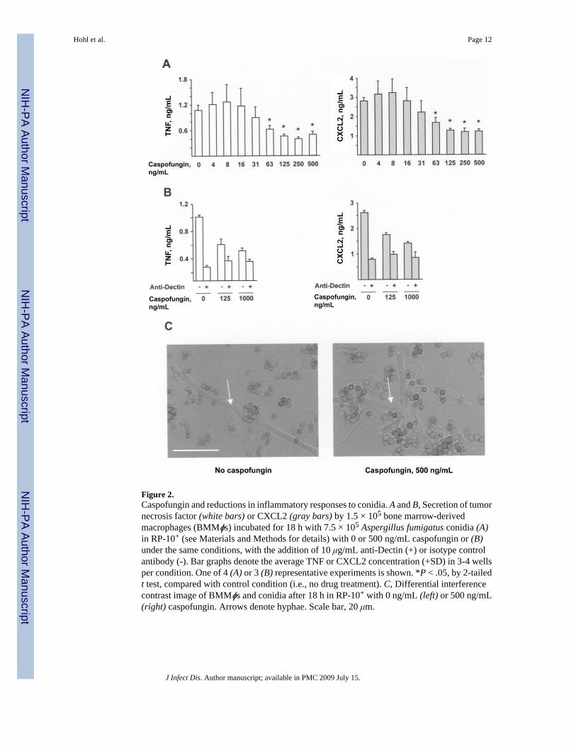
Figure 2.Caspofungin and reductions in inflammatory responses to conidia. A and B, Secretion of tumornecrosis factor (white bars) or CXCL2 (gray bars) by 1.5 × 10⁵ bone marrow-derivedmacrophages (BMMϕs) incubated for 18 h with 7.5 × 10⁵ Aspergillus fumigatus conidia (A)in RP-10+ (see Materials and Methods for details) with 0 or 500 ng/mL caspofungin or (B)under the same conditions, with the addition of 10 μg/mL anti-Dectin (+) or isotype controlantibody (-). Bar graphs denote the average TNF or CXCL2 concentration (+SD) in 3-4 wellsper condition. One of 4 (A) or 3 (B) representative experiments is shown. *P < .05, by 2-tailedt test, compared with control condition (i.e., no drug treatment). C, Differential interferencecontrast image of BMMϕs and conidia after 18 h in RP-10+ with 0 ng/mL (left) or 500 ng/mL(right) caspofungin. Arrows denote hyphae. Scale bar, 20 μm.
Hohl et al. Page 12
J Infect Dis. Author manuscript; available in PMC 2009 July 15.
NIH
-PA Author Manuscript
NIH
-PA Author Manuscript
NIH
-PA Author Manuscript

Figure 3.Caspofungin and decreases in inflammatory responses to germlings. A-C, Secretion of tumornecrosis factor (TNF) or CXCL2 by 1.5 × 105 bone marrow-derived macrophages (BMMϕs)incubated with Aspergillus fumigatus germlings for 6 h. A total of 5 × 10⁴ conidia were grownin RP-10+ (see Materials and Methods for details) for 8 h at 37°C with 0-500 ng/mLcaspofungin (A, B) or micafungin (C) before UV inactivation and the addition of BMMϕs.B, Cocultures contained 10 μg/mL anti-Dectin (+) or 10 μg/mL isotype control antibody (-).Bar graphs denote the average TNF or CXCL2 concentration (+SD) in 3-4 wells per condition.One of 7 (A) or 3 (B, C) representative experiments is shown. *P < .05, by 2-tailed t test,compared with control condition (i.e., no drug treatment).
Hohl et al. Page 13
J Infect Dis. Author manuscript; available in PMC 2009 July 15.
NIH
-PA Author Manuscript
NIH
-PA Author Manuscript
NIH
-PA Author Manuscript

Figure 4.Caspofungin and enhancement of inflammatory responses to hyphae. A-D, Secretion of tumornecrosis factor (TNF) or CXCL2 by 1.5 × 105 bone marrow-derived macrophages (BMMϕs)incubated for 6 h with Aspergillus fumigatus hyphae. A total of 5 × 10⁴ conidia were grownin RP-10+ (see Materials and Methods for details) for 24 h at 37°C with 0-500 ng/mLcaspofungin (A, B, D) or micafungin (C) before UV inactivation and the addition of BMMϕs.B and D, Cocultures contained 10 μg/mL anti-Dectin (+), 10 μg/mL isotype control antibody(-), or 100 ng/mL lipopolysaccharide, as indicated. E, TNF secretion by BMMϕs stimulatedwith UV-inactivated fungal cells grown for the indicated period in RP-10+ (empty circles) orin RP-10+ with 500 ng/mL caspofungin (filled circles). Coincubations were performed in thepresence of anti-Dectin (red lines) or control isotype antibody (black lines), as described forpanel B. A-E, Bar graphs denote the average TNF or CXCL2 concentration (+SD) in 3-6 wellsper condition. One of either 8 (A) or 3 (B-E) representative experiments is shown. *P < .05,by 2-tailed t test, compared with control condition (i.e., no drug treatment).
Hohl et al. Page 14
J Infect Dis. Author manuscript; available in PMC 2009 July 15.
NIH
-PA Author Manuscript
NIH
-PA Author Manuscript
NIH
-PA Author Manuscript

Figure 5.Caspofungin and modulation of β-glucan immunoreactivity in a stage-specific manner. A-E,A total of 5 × 10⁴ conidia were grown in RP-10+ (see Materials and Methods for details) withoutdrug (left) or with 500 ng/mL caspofungin (right) for 8-18 h, as indicated; stained with an anti-β-glucan antibody (monoclonal antibody 744); and examined by confocal microscopy.Representative epifluorescence and differential interference contrast (DIC) images are shown.Fluorescence images are a composite of scanned images obtained at 0.5-μm (A-B) or 1.0-μm(C-E) intervals. Scale bar, 20 μm.
Hohl et al. Page 15
J Infect Dis. Author manuscript; available in PMC 2009 July 15.
NIH
-PA Author Manuscript
NIH
-PA Author Manuscript
NIH
-PA Author Manuscript

Figure 6.Echinocandins and increases in β-glucan immunoreactivity on hyphae. A total of 5 × 10³
conidia were grown in RP-10+ (see Materials and Methods for details) for 24 h with 500 ng/mL caspofungin (left) or 500 ng/mL micafungin (middle) or without drug (right).Representative fields are shown with composite fluorescence images overlaid on differentialinterference contrast images. Scale bar, 20 μm.
Hohl et al. Page 16
J Infect Dis. Author manuscript; available in PMC 2009 July 15.
NIH
-PA Author Manuscript
NIH
-PA Author Manuscript
NIH
-PA Author Manuscript

NIH
-PA Author Manuscript
NIH
-PA Author Manuscript
NIH
-PA Author Manuscript
Hohl et al. Page 17
Table 1Quantitative analysis of β-glucan immunoreactivity for caspofungin-treated and untreated Aspergillus fumigatushyphae
Integrated fluorescence intensity/fungal mass,a by hyphae groupExperiment Caspofungin treated Untreated
1 21.41b ± 8.34 1.83 ± 0.732 43.73b ± 6.96 2.96 ± 4.67
NOTE. Data are the average ratio (±SD) of β-glucan immunofluorescence intensity normalized to hyphal mass, as calculated from 4-5 fields of view percondition.
aArbitrary units.
bP < .02, by 2-tailed t test, compared with control condition (untreated hyphae).
J Infect Dis. Author manuscript; available in PMC 2009 July 15.