Experimental study of coil delivery wire insertion force in ...
Howard et al. BMC Genetics 2013, 14:28http://www.biomedcentral.com/1471-2156/14/28
RESEARCH ARTICLE Open Access
Carriers of a novel frame-shift insertion inWNT16a possess elevated pancreatic expressionof TCF7L2Eric W Howard1, Latonya F Been2, Megan Lerner3, Daniel Brackett4, Stan Lightfoot3, Elizabeth C Bullen1
and Dharambir K Sanghera2*
Abstract
Background: The discovery of TCF7L2 as a global type 2 diabetes (T2D) gene has sparked investigations to explorethe clinical utility of its variants for guiding the development of new diagnostic and therapeutic strategies. However,interpreting the resulting associations into function still remains unclear. Canonical Wnt signaling regulates β-cateninand its binding with TCF7L2, which in turn is critical for the production of glucagon-like peptide-1 (GLP-1). This studyexamines the role of a novel frame-shift insertion discovered in a conserved region of WNT16a, and it is proposedthat this mutation affects T2D susceptibility in conjunction with gene variants in TCF7L2.
Results: Our results predicted that the insertion would convert the upstream open reading frame in the Wnt16amRNA to an alternative, in-frame translation initiation site, resulting in the prevention of nonsense-mediated decay,leading to a consequent stabilization of the mutated WNT16a message. To examine the role of Wnt16a in the Wntsignaling pathway, DNA and serum samples from 2,034 individuals (48% with T2D) from the Sikh Diabetes Study wereused in this investigation. Prevalence of Wnt16a insertion did not differ among T2D cases (33%) and controls (32%).However, there was a 3.2 fold increase in Wnt16a mRNA levels in pancreatic tissues from the insertion carriers and asignificant increase (70%, p < 0.0001) in luciferase activity in the constructs carrying the insertion. The expression ofTCF7L2 mRNA in pancreas was also elevated (~23-fold) among the insertion carriers (p=0.003).
Conclusions: Our results suggest synergistic effects of WNT16a insertion and the at-risk ‘T’ allele of TCF7L2 (rs7903146)for elevating the expression of TCF7L2 in human pancreas which may affect the regulation of downstream target genesinvolved in the development of T2D through Wnt/β-catenin/TCF7L2 signaling pathway. However, further studies wouldbe needed to mechanistically link the two definitively.
Keywords: β-cat /TCF7L2 signaling, Wnt16a, Exome sequencing, Insertion polymorphism, TCF7L2 gene variants, Geneexpression, Pancreatic β-cells, Type 2 diabetes
BackgroundTranscription factor 7-like 2 (TCF7L2) has been stronglylinked to type 2 diabetes (T2D) susceptibility, with anelevated genetic predisposition accounting for 20% ofT2D cases [1]. The association of common intronic vari-ants in the TCF7L2 gene with the increased susceptibilityfor T2D has been extensively documented in major ethnicgroups of the world by several different investigators [2].
* Correspondence: [email protected] of Pediatrics, College of Medicine, University of OklahomaHealth Sciences Center, 940 Stanton L. Young Blvd., Rm 317 BMSB, OklahomaCity, OK 73104, USAFull list of author information is available at the end of the article
© 2013 Howard et al.; licensee BioMed CentraCommons Attribution License (http://creativecreproduction in any medium, provided the or
Meta-analysis of the published studies estimated the oddsratio (OR) of 1.46 (p=5.4x10-140) [3]. TCF7L2 polymor-phisms were also significantly linked to diabetes risk inour own studies in Asian Indian Sikhs [4,5]. Indeed, ourrecent Sikh genome-wide association study (GWAS) andmeta-analyses in Sikhs (n=7,329/3,354 cases) and SouthAsians (n=47,303/19, 482 cases) showed a robust associ-ation of TCF7L2 (rs7903146), with OR 1.5 (p=7.8x10⁻¹⁹)and OR 1.13 (p=6.1x10⁻²⁵) in Sikhs and South Asians,respectively [6]. However, despite extensive replication, nostudy has unequivocally demonstrated the underlyingmolecular mechanism of this association. Little is known
l Ltd. This is an Open Access article distributed under the terms of the Creativeommons.org/licenses/by/2.0), which permits unrestricted use, distribution, andiginal work is properly cited.

Howard et al. BMC Genetics 2013, 14:28 Page 2 of 11http://www.biomedcentral.com/1471-2156/14/28
about the clinical role of TCF7L2 in T2D beyond progres-sion from impaired glucose tolerance to diabetes [7].Various in vitro and in vivo studies have shown that
several components of the Wnt pathway are involved inβ-cell proliferation [8], insulin secretion and cholesterolmetabolism [9], and production of glucogon-like peptide-1 (GLP-1) [10]. Wnts are secreted glycoproteins with awell-established role in the early stages of developmentthrough adulthood [11]. Wnts bind to frizzled and LRPreceptors, which, in turn, inactivate the degradation com-plex consisting of AXIN, DVL, and GSK3B (Figure 1).This prevents the phosphorylation of β-catenin by GSK3B,and leads to its binding to the nuclear transcriptionfactors, TCF7, LEF1, TCF7L1 and TCF7L2, leading to theactivation of more than 60 different genes involved ingrowth regulation and differentiation, as well as GLP-1expression [12]. Since Wnt signaling has a role in regulat-ing and stabilizing β-catenin and its binding with TCF7L2,we hypothesized that any alternation in the canonicalWnt pathway would have profound consequences in insu-lin secretion and the generation of new β-cells, particu-larly given that Wnt signaling is required for normaldevelopment of the pancreas and islets during embryonicgrowth [13].The present investigation is a follow-up study to
explore the role of a novel, four-nucleotide (CCCA)insertion polymorphism we discovered in the mostconserved region of WNT16a in US American Sikhs.
Figure 1 Wnt signaling pathway in diabetes mellitus.
The objectives of this investigation are: 1) to study thepotential role of this WNT16a insertion in T2D in ourdiabetic sample of Punjabi Sikhs, 2) to quantify and com-pare gene expression of WNT16a and TCF7L2 betweencarriers and non-carriers of the CCCA insertion withinthe WNT16a gene using mRNA samples from 27 frozenhuman pancreatic tissues, 3) to investigate the functionalimpact of this insertion on protein levels and messagetranslation using a luciferase reporter vector containingthe wild-type and mutant WNT16a 5′untranslated re-gions (UTR) transfected into cultured cells, and 4) to per-form immunohistochemistry to examine the expression ofWNT16a in human pancreas among insertion carriers vs.non-carriers.
MethodsStudy participantsThe DNA samples of 2,034 (52% male) individuals fromour ongoing Sikh Diabetes Study (SDS) were used [14].Of these, ~48% were ascribed as having T2D based onestablished guidelines of the American Diabetes Associ-ation, as described [15]. A medical record indicatingeither (1) a fasting blood glucose (FBG) ≥126 mg/dL(≥7.0 mmol/L) after a minimum 12 h fast or (2) a 2 hpost-glucose level (2 h oral glucose tolerance test[OGTT]) ≥ 200 mg/dL (≥11.1 mmol/L) on more thanone occasion, combined with symptoms of diabetes,confirmed the diagnosis. Impaired fasting glucose (IFG)is defined as a fasting blood glucose level ≥100 mg/dL(5.6 mmol/L) but ≤126 mg/dL (7.0 mmol/L), as describedpreviously [16]. Common characteristics observed in dia-betics include excessive thirst, hunger, polyuria, blurry vi-sion, common skin and urinary tract infections, nocturia,loss of bladder control, and fatigue. Impaired glucosetolerance (IGT) is defined as a 2 h OGTT >140 mg/dL(7.8 mmol/L) but <200 mg/dL (11.1 mmol/L). Subjectswith IFG or IGT were considered pre-diabetics and wereexcluded from the analysis. The 2 h OGTTs wereperformed following the criteria of the World HealthOrganizations (WHO) (75 g oral load of glucose). Bodymass index (BMI) was calculated as (weight (kg)/height(meter) [2]. Homeostasis Model Assessment (HOMA) forinsulin resistance (HOMA-IR) was calculated as fastingglucose X fasting insulin/22.5, as described [17].The normoglycemic subjects were recruited from the
same Punjabi Sikh community and geographic location asthe T2D patients [14]. The majority of the subjects wererecruited from the state of Punjab in North India andPunjabi Sikhs living in the US. Individuals of South, East,and Central Indian origin were excluded, as were individualswith type-1 diabetes, a family member with type 1 diabetes,rare forms of T2D called maturity-onset diabetes of young(MODYs), or secondary diabetes (e.g., hemochromatosis,pancreatitis). Demographic and clinical characteristics of the
Howard et al. BMC Genetics 2013, 14:28 Page 3 of 11http://www.biomedcentral.com/1471-2156/14/28
SDS subjects are summarized in Table 1. All bloodsamples were obtained at the baseline visit and all partici-pants provided a written informed consent for these inves-tigations. All SDS protocols and consent documents werereviewed and approved by the University of OklahomaInstitutional Review Board and the Human Subject Protec-tion Committees at the participating hospitals and insti-tutes in India.
Metabolic estimationsInsulin was measured by radio-immuno assay (DiagnosticProducts, Cypress, USA). Serum lipids (total cholesterol,low density lipoprotein cholesterol [LDL-C], high-density lipoprotein [HDL-C], very low-density lipopro-tein cholesterol [VLDL-C], and triglycerides [TG]) weremeasured by using standard enzymatic methods (Roche,Basel, Switzerland), as described [16,18]. C-peptide, TNFα,and MCP-1 measures were simultaneously quantifiedusing Millipore’s Magnetic MILLIPLEX Human Metabolicpanel (St. Charles, MO) and analyzed on a Bio-plex 200multiplex system (Bio-Rad Hercules, CA), as describedpreviously [19].
Whole-genome exome sequencingWe performed genome-wide exome sequencing on twoPunjabi Sikh subjects: a 64-year-old healthy normogly-cemic male, and a 67-year-old diabetic female, using anIllumina GAIIx and “SureSelect Human All Exon Kit” byAgilent Technologies and “Paired-End SequencingLibrary Prep by Illumina” (Version 1.0.1). The sequencescontaining 75x reads were filtered against public data-bases of genetic variants. The present investigation isfocused on exploring the role of a frame-shift insertion(CCCA) discovered in a conserved region of humanWNT16a gene (Additional file 1: Figure S1).
GenotypingGenotyping of the insertion polymorphisms was performedby polymerase chain reaction (PCR) and a gel-based assay.Forward primer Wnt16a-F (5') [TACCACTCTCCTCCCTCC] and reverse primer Wnt16a-R (3') [CCCTGATCAAATCCCCAAAT] were used to amplify the region containingthe identified insertion; PCR amplification generated a458 bp product in the sample containing no insertion. PCRconditions included an initial denaturation for 5 min. at95°C, followed by 36 cycles (30 sec. 95°C, 45 sec. 53.7°C,30 sec. 72°C), and a 10 min. extension at 72°C. Positive andnegative controls were included for every PCR. 15μL of thePCR product was then separated on a 2.5% nusieve/agarose gel (3:1) for 2.5 hours at 140 volts to determine thegenotype of participants as insertion (462 bp), non-insertion carriers 458 bp, and heterozygotes containinginsertion/normal sequence of 462/458 bp (Additional file 1:Figure S2). To confirm the presence of the WNT16a
insertion scored on the gel-based assay, approximately30 samples were sequenced using an ABI 3730 capillarysequencer (Applied Biosystems Inc. Foster City, CA) andwere analyzed using Mutation Surveyor DNA variant ana-lysis software (v4.0.6.)(SoftGenetics, State College, PA).Genotyping of rs7903146, located in intron 3 of theTCF7L2 gene, was performed with a TaqMan genotypingassay (Applied Biosystems, Foster City, CA), using a 7900genetic analyzer, as described previously [4].
Quantitative gene expression studies on WNT16aGene expression studies for Wnt16a were performed using27 human pancreatic tissue specimens (13 diabetic and 14non-diabetics) collected from the Department of Surgeryat the University of Oklahoma Health Sciences Center.Total RNA was extracted from frozen tissues (stored inliquid nitrogen) using Ambion’s mirVana RNA kits (GrandIsland, NY), followed by RT-PCR using Bio-Rad’s iScriptRT-PCR kit (Hercules, CA), according to the manufac-turers’ instructions. Real Time PCR was then performedusing an ABI 7900HT genetic analyzer in conjunction withQiagen’s QuantiTect primer assay (Chatworth, CA) andBio-Rad’s iTaq SYBR Green Supermix with ROX (Hercules,CA). Results were then analyzed on ABI’s RQ Manager(v.1.2.1) software. Beta-actin was used as a normalizingcontrol.
Transient DNA transfection and dual-luciferase assayThe 5′ UTRs of the wild-type and mutant Wnt16a mes-sage were incorporated into oligonucleotide primers asdepicted in Additional file 1: Figure S3. Note that eachof the 5′ primers incorporated a Sac I site for insertioninto pCI-GFP, followed by the sequence of the Wnt16a5′ UTR, then a region homologous to firefly luciferase.The pCI-GFP vector was developed by inserting eGFPinto the parent vector, pCI-Neo (Promega, Madison,WI), and allowed us to monitor transfection efficiency.The 3′ primer was homologous to a site in the pGL3vector past a unique Xba I site in the vector. After PCRamplification using pGL3 as a template, the amplimerswere digested with Sac I and Xba I, and then ligated intopCI-GFP. For transfection into cultured cells, each con-struct (0.125 μg per culture well) was added to 1 μl Plus re-agent and 15 μl Opti-MEM (Life Technologies, Carlsbad,CA), along with 0.125 μg per well of an empty pGL3-Basicvector (which served as carrier DNA) and 0.01 μg perwell pGL4.74 (a Renilla luciferase construct used fornormalization) for a total of 0.26 μg DNA. This wasadded to 0.5 μl Lipofectamine reagent in an additional15 μl of Opti-MEM and used to transfect HEK-293cells (74,000 cells per well) in a 48-well plate. After48 hours in medium plus 10% calf serum, cells werewashed in PBS, and lysed for luciferase activity. Lysateswere diluted until the luciferase values fell within a

Howard et al. BMC Genetics 2013, 14:28 Page 4 of 11http://www.biomedcentral.com/1471-2156/14/28
linear response range. Both firefly and Renilla luciferasevalues were measured using a dual luciferase detectionkit (Promega, Madison, WI).
ImmunohistochemistryFormalin-fixed paraffin-embedded pancreatic tissues werecut at a thickness of 4 μm, mounted on SuperfrostPlus®slides (Statlab Medical Products, Lewisville, TX), and subse-quently deparaffinized, rehydrated, and washed in Tris Buff-ered Immunohistochemistry wash buffer + Tween 20(TBST, catalog# 935B, Cell Marque, Rocklin, CA). Antigenretrieval was accomplished by placing slides in 10 mMcitrate buffer, pH 6.0 (cat. #S2389,Target Retrieval Solution,DAKO, Carpentaria, CA), in a steamer for 20 minutes,followed by 20 minutes cooling in deionized water at roomtemperature. According to the manufacturer’s directions,sections were treated with a background blocker (cat.#927B, Cell Marque, Rocklin, CA) and a peroxidaseblocking reagent (cat. #925B, Cell Marque, Rocklin, CA) toinhibit endogenous peroxidase activity, followed by three,five-minute washes each in deionized water. Rabbit anti-Wnt antibody was prepared in antibody diluent (cat.#936B, Cell Marque, Rocklin, CA) and added to slides at2 μg/ml (1:500 dilution, cat. #LS-A9630, MBL InternationalCorporation, Woburn, MA). Antigen retrieval was accom-plished according to the manufacturer’s recommendationfor the Wnt16 antibody (LSBio, Woburn, MA). Followingincubation for 1 hr at room temperature, the sectionswere processed for immunohistochemistry using theHiDef detection HRP Mouse/Rabbit polymer system(cat. #954D, Cell Marque, Rocklin, CA). Sections werewashed three times for five minutes each in tris buff-ered immunohistochemistry wash buffer + Tween 20(TBST), incubated with the amplifier, washed threetimes for five minutes each in TBST, and incubate with la-beled polymer. Following a final wash in TBST, slides wereincubated with 3′3′diaminobenzidine tetrahydrochloride(DAB) (cat. #957D, DAB substrate Kit, Cell Marque, Rock-lin, CA). Counterstaining was performed with Immuno*
Master Hematoxylin (American Master*Tech Scientific,Inc., Lodi, CA). Controls were incubated with rabbit IgGisotype at 2 μg/ml (rabbit [DA1E] mAB IgGXP® isotypecontrol, cat. #3900, Cell Signaling Technologies Danvers,MA). A total of seven tissues (1 T2D and 6 controls) withWnt16a genotypes were used for immunohistochemistry.Slides were scored based on intensity (0- no, 1- weak,2- moderate and 3- strong), and the area of stain (0 for0%, 1-<10%, 2-between 10-15%, and 3- between 51-81%).The consolidated scores (ranging from 0–7) were derivedfrom the sum of scores of intensity and area, negative be-ing in the range of 0–2, weakly positive-3, moderatelypositive ranging from 4–5, and highly positive rangingfrom 6–7.
Statistical analysisAssociation analysisData quality for SNP genotyping was checked byestablishing reproducibility of control samples. Departurefrom Hardy-Weinberg equilibrium in controls was checkedusing Pearson’s Chi-square, as reported previously [5].Descriptive statistical analyses were performed with SPSSStatistics Software (v 15.0). The chi-square test for categor-ical variables and t-test for continuous variables were usedto test differences where appropriate. While multivariatelogistic-regression was used to assess the association of theinsertion with T2D and obesity, multivariate linear-regression was used for each quantitative trait after adjust-ment for relevant covariates (age, sex, diabetes status, BMI,and medication), assuming an additive model. Skewed vari-ables were detected by Shapiro-Wilk’s test for continuoustraits. Subsequently, TG, total cholesterol, LDL-C, VLDL-C,FBG, C-peptide, MCP-1, and HOMA-IR were normalizedby log-transformation before statistical comparisons, and allp-values were derived from analyses of transformed data.The summary statistics (β, S.E., and p-values) were used toassess SNP-phenotype association. Gene expression analyseswere performed using Applied Biosystems’ RQ Manager(v.1.2), which uses the comparative CT method for relativequantification. We determined the ΔCT value by (TargetAverage CT-Endogenous Control Average CT), then calcu-lated the ΔΔCT to determine the fold-difference in geneexpression by ΔCT Target - ΔCT Calibrator. For the amountof target determination, the data were normalized to the en-dogenous control and relative to the calibrator by using2-ΔΔCT as described [20] . For reporter assays, the resultsare presented as the mean ± average deviation from themean for the number of observation, as indicated. Statis-tical significance of differences between groups was esti-mated using a two-tailed t test.
ResultsWhole-exome sequencingAs summarized in Additional file 2: Table S1, a total of20,306 mutations were found in the control and 21,258 inthe diabetic subjects. Among these, 4,673 and 4,842 novelSNPs were uniquely present in control and T2D cases, re-spectively. To identify the functional significance of thevariants identified, we performed initial comparative gen-omic screening on the mutations found in some selectedloci using UCSC’s Vista Genome Browser. From these re-sults, several candidate genes involved in insulin secretion,β-cell proliferation, or related pathways were identified(data not shown). Interestingly, novel substitution inWNT16a, which showed a 4-base-pair frame-shift inser-tion near two known SNPs, was in an evolutionarily con-served region (as shown in Additional file 1: Figure S3)and was predicted to be disruptive.

Table 1 Clinical characteristics of study subjects stratified by Wnt16a insertion carriers versus non-carriers
Trait Non carrier Carrier P value
Number 1377 657 -
% Males 52 51 -
Age (yrs) 53.4 ± 12.9 51.7 ± 12.1 0.004
Obesity
BMI (kg/m2) 26.8 ± 4.9 26.8 ± 5.1 0.930
Weight (kg) 69.8 ± 14.0 70.0 ± 14.3 0.811
Waist (cm) 93.6 ± 12.2 93.4 ± 12.0 0.704
WHR 0.95 ± 0.08 0.95 ± 0.08 0.242
Metabolic
Fasting Blood Glucose mg/dL 120.7 ± 45.4 121.5 ± 45.4 0.736
Insulin (μIU/mL) 6.9 (6.6-7.3) 7.5 (7.0-8.1) 0.081
HOMA-IR 2.0 (1.9-2.2) 2.2 (2.1-2.4) 0.059
C-peptide pg/mL 519.5 (473.7-569.7) 602.8 (525.6-691.4) 0.078
Inflammation
TNFα pg/mL 7.9 (7.4-8.5) 9.2 (8.2-10.2) 0.029
MCP1 pg/mL 315.5 (296.7-335.6) 326.5 (297.9-357.8) 0.541
Lipid
Triglyceride mg/dL 149.0 ± 82.3 151.5 ± 85.9 0.542
Total Cholesterol mg/dL 173.8 ± 52.9 179.2 ± 49.9 0.038
HDL-C mg/dL 37.2 ± 14.7 37.9 ± 14.2 0.292
LDL-C mg/dL 102.1 ± 40.0 104.9 ± 38.3 0.143
WHR-Waist to hip ratio, LDL-C -low density lipoprotein cholesterol, HDL-C -high density lipoprotein cholesterol, BMI- body mass index, HOMA-IR -homeostasismodel assessment for insulin resistance.
Howard et al. BMC Genetics 2013, 14:28 Page 5 of 11http://www.biomedcentral.com/1471-2156/14/28
Association studiesA genetic screening of 2,034 SDS individuals (977 T2Dcases and 1,057 controls T2D cases and 1,057 controls)showed that 33% of T2D cases and 32% non-diabeticcontrols were carriers of a CCCA insertion; the numberof carriers of this insertion did not differ significantly
Figure 2 Distributions of serum levels (mean ±SD) of inflammatory cycarriers in SDS subjects. Serum levels of TNFα were significantly higher (pwas seen with MCP-1. The statistical analysis was performed in combined sof age, BMI, gender, and T2D status.
among cases versus controls (p=0.08). Multiple regres-sion analysis, performed in diabetics and non-diabeticcontrols separately, did not reveal any association of theWnt16a insertion with obesity (BMI, waist-to-hip ratio[WHR]) (Table 1). However, the insertion carriersshowed moderately higher mean (±SD) levels of total
tokines (TNFα and MCP-I) among Wnt16a insertion carriers non-=0.008) in insertion carriers, while a similar but non-significant trendamples (T2D and controls) after adjusting for the confounding effects
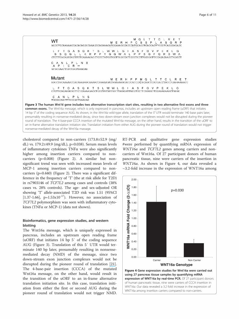
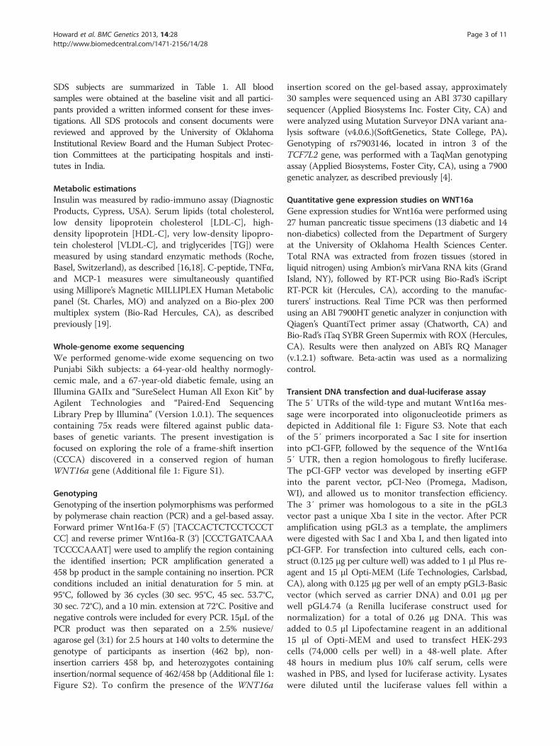
Figure 3 The human Wnt16 gene includes two alternative transcription start sites, resulting in two alternative first exons and threecommon exons. The Wnt16a message, which is only expressed in pancreas, includes an upstream open reading frame (uORF) that initiates14 bp 5′ of the coding sequence AUG. As shown, in the Wnt16a wild-type allele, translation of the 5′ UTR would terminate 140 base pairs later,presumably resulting in nonsense-mediated decay, since two down-stream exon junction complexes would not be disrupted during the pioneerround of translation. The 4 base-pair CCCA insertion of the mutated Wnt16a message, on the other hand, results in the transition of the uORF toan in-frame alternative translation initiation site. Translation initiation from either AUG during the pioneer round of translation would not triggernonsense-mediated decay of the Wnt16a massage.
1.40
1.60
1.80
2.00
ang
e (2
-ΔΔC
t)
p=0.030
Howard et al. BMC Genetics 2013, 14:28 Page 6 of 11http://www.biomedcentral.com/1471-2156/14/28
cholesterol compared to non-carriers (173.8±52.9 (mg/dL) vs. 179.2±49.9 (mg/dL), p=0.038). Serum mean levelsof inflammatory cytokines TNFα were also significantlyhigher among insertion carriers compared to non-carriers (p=0.008) (Figure 2). A similar but non-significant trend was seen with increased mean levels ofMCP-1 among insertion carriers compared to non-carriers (p=0.440) (Figure 2). There was a significant dif-ference in the frequency of ‘T’ (the at risk allele for T2D)in rs7903146 of TCF7L2 among cases and controls (38%cases vs. 28% controls). The age- and sex-adjusted ORshowing ‘T’ allele-associated T2D risk was 1.51 (95%CI[1.37-1.66], p=1.53x10⁻¹⁷). However, no association ofTCF7L2 polymorphism was seen with inflammatory cyto-kines (TNFα or MCP-1) (data not shown).
0.00
0.20
0.40
0.60
0.80
1.00
1.20
Carrier Non Carrier
WN
T16
a m
RN
A F
old
Ch
WNT16a Genotype
Figure 4 Gene expression studies for Wnt16a were carried outusing 27 pancreas tissue samples by quantifying mRNAexpression of WNT16a by real-time PCR. Of 27 participant donorsof human pancreatic tissue, nine were carriers of CCCA insertion inWNT16a. Our data revealed a 3.2 fold increase in the expression ofWNT16a among insertion carriers compared to non-carriers.
Bioinformatics, gene expression studies, and westernblottingThe Wnt16a message, which is uniquely expressed inpancreas, includes an upstream open reading frame(uORF) that initiates 14 bp 5′ of the coding sequenceAUG (Figure 3). Translation of this 5′ UTR would ter-minate 140 bp later, presumably resulting in nonsense-mediated decay (NMD) of the message, since twodown-stream exon junction complexes would not bedisrupted during the pioneer round of translation [21].The 4-base-pair insertion (CCCA) of the mutatedWnt16a message, on the other hand, would result inthe transition of the uORF to an in-frame alternativetranslation initiation site. In this case, translation initi-ation from either the first or second AUG during thepioneer round of translation would not trigger NMD.
RT-PCR and qualitative gene expression studies#were performed by quantifying mRNA expression ofWNT16a and TCF7L2 genes among carriers and non-carriers of Wnt16a. Of 27 participant donors of humanpancreatic tissue, nine were carriers of the insertion inWNT16a. As shown in Figure 4, our data revealed a~3.2-fold increase in the expression of WNT16a among
0.0
6.0
12.0
18.0
24.0
30.0
36.0
Carriers Non Carriers
TC
F7L
2 m
RN
A F
old
Ch
ang
e (2
-ΔΔC
t)
Wnt16a Genotype
p=0.003
0.0
5.0
10.0
15.0
20.0
25.0
30.0
35.0
40.0
45.0
50.0
NG Controls T2D Patients
TC
F7L
2 m
RN
A F
old
Ch
ang
e (
2-ΔΔ
Ct)
WNT16a Insertion Carriers
Carriers Onlyp=0.155
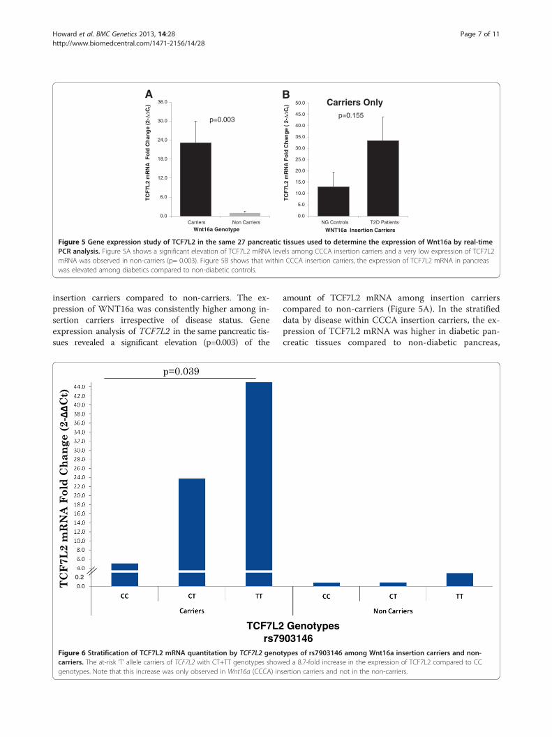
A B
Figure 5 Gene expression study of TCF7L2 in the same 27 pancreatic tissues used to determine the expression of Wnt16a by real-timePCR analysis. Figure 5A shows a significant elevation of TCF7L2 mRNA levels among CCCA insertion carriers and a very low expression of TCF7L2mRNA was observed in non-carriers (p= 0.003). Figure 5B shows that within CCCA insertion carriers, the expression of TCF7L2 mRNA in pancreaswas elevated among diabetics compared to non-diabetic controls.
Howard et al. BMC Genetics 2013, 14:28 Page 7 of 11http://www.biomedcentral.com/1471-2156/14/28
insertion carriers compared to non-carriers. The ex-pression of WNT16a was consistently higher among in-sertion carriers irrespective of disease status. Geneexpression analysis of TCF7L2 in the same pancreatic tis-sues revealed a significant elevation (p=0.003) of the
0.2
TCF7L2rs79
Figure 6 Stratification of TCF7L2 mRNA quantitation by TCF7L2 genocarriers. The at-risk ‘T’ allele carriers of TCF7L2 with CT+TT genotypes showgenotypes. Note that this increase was only observed in Wnt16a (CCCA) in
amount of TCF7L2 mRNA among insertion carrierscompared to non-carriers (Figure 5A). In the stratifieddata by disease within CCCA insertion carriers, the ex-pression of TCF7L2 mRNA was higher in diabetic pan-creatic tissues compared to non-diabetic pancreas,
Genotypes03146
types of rs7903146 among Wnt16a insertion carriers and non-ed a 8.7-fold increase in the expression of TCF7L2 compared to CCsertion carriers and not in the non-carriers.

Figure 7 Reporter constructs assembled with the mutated formof the Wnt16a 5′ UTR are more efficiently translated than thewild-type form. The wild-type and mutant Wnt16a 5′ UTRsequences were inserted adjacent to a luciferase cDNA, and theresulting plasmids were used to transfect HEK-293 cells. 48 hoursafter transfection, cells were lysed, and firefly luciferase (FFL) andRenilla luciferase (RL) levels were measured. Our reporter constructsusing the wild-type and the mutant (insertion) sequence of Wnt16ashowed significantly increased (70%, p<0.0001) levels of luciferaseprotein in the constructs carrying the mutant sequence.
Howard et al. BMC Genetics 2013, 14:28 Page 8 of 11http://www.biomedcentral.com/1471-2156/14/28
however, this increase was not statistically significant(p=0.155). (Figure 5B). Further stratification of quanti-tative mRNA expression among the at-risk ‘T’ allelecarriers of TCF7L2 SNP (rs7903146) revealed that theCT+TT genotypes showed an 8.7-fold increase in theexpression of TCF7L2 compared to CC genotypes inWnt16 a insertion carriers, while the non-insertion car-riers showed the same allelic trend at a significantly re-duced magnitude (Figure 6).
Luciferase reporter assayIn order to further evaluate the influence of the CCCAinsertion on translation of the Wnt16a message, we
assembled reporter constructs driven by the cytomegalo-virus (CMV) promoter that included the wild-type andthe mutant sequence of the Wnt16a 5′ UTRs. The longuORF was mimicked in our luciferase construct by thepresence of a translation stop site in-frame with the up-stream AUG. If the first AUG in the message was usedto initiate translation, then no luciferase protein shouldhave been produced. Indeed, when we inserted the add-itional four nucleotides to replicate what occurs in themutant situation, we noted a significantly increased level(~70%) of luciferase expression (p=0.0001) (Figure 7).This suggests that the upstream AUG can act as an effi-cient translation initiation site. In the wild-type gene,this would reduce expression of the full-length proteinby preventing initiation at the second AUG. In the pres-ence of the CCCA insertion, both AUGs are in the samereading frame, so full-length protein would be producedregardless of which AUG was used to initiate translation.
ImmunohistochemistryImmunoperoxidase staining of paraffin-embedded pancre-atic tissues of normoglycemic controls and diabetic caseswere scored for the intensity of antibody as described inmethods. As shown in Figure 8, the tissues with insertioncarriers revealed a higher expression of Wnt16a showinghigh intensity staining among insertion carriers versesnegative staining in non-carriers. The scoring intensitywas indifferent among diabetics and non-diabetics.
DiscussionThe key effector pathway of Wnt signaling (β-cat/TCF7L2) has been recently implicated in metabolichomoeostasis, diabetes, obesity, osteoporosis, cardiovas-cular disease, and cancer [9,22-24]. The discovery ofTCF7L2 as a T2D susceptibility gene in different ethnicpopulations through genome-wide studies has triggerednumerous investigations to explore the clinical utility ofidentifying TCF7L2 genetic variations, and whether theidentified SNPs can be used as markers for tailoring cus-tomized therapeutics. However, the underlying molecularmechanism by which TCF7L2 variants influence T2D re-mains unclear. While a number of recent studies havesuggested the essential involvement of β-cat/TCF7L2 inthe Wnt signaling pathway for pancreatic developmentand function [25,26], the role of β-cat in pancreatic β celldevelopment remains unclear and controversial [13,27].Mice lacking β-cat developed pancreatitis prenatally; how-ever, they later recovered from pancreatitis and regeneratednormal pancreas and duodenal villi from wild-type cellsthat escaped earlier β-cat deletion. These observations sug-gested that mouse embryos were capable of overcomingsubstantial β-cat reduction through complicated compen-satory mechanisms [13]. Other studies have shown that theover-expression of β-cat at different development stages
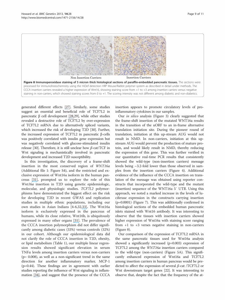
Figure 8 Immunoperoxidase staining of 5 micron thick histological sections of paraffin-embedded pancreatic tissues. The sections wereprocessed for immunohistochemistry using the HiDef detection HRP Mouse/Rabbit polymer system as described in detail under methods. TheCCCA insertion carriers revealed a higher expression of Wnt16, showing staining score from +1 to +3 among insertion carriers versus negativestaining in non-carriers, which showed staining scores from 0 to +1. The scoring intensity was not different among diabetic and non-diabetics.
Howard et al. BMC Genetics 2013, 14:28 Page 9 of 11http://www.biomedcentral.com/1471-2156/14/28
generated different effects [27]. Similarly, some studiessuggest an essential and beneficial role of TCF7L2 inpancreatic β cell development [28,29], while other studiesrevealed a destructive role of TCF7L2 by over-expressionof TCF7L2 mRNA due to alternatively spliced variants,which increased the risk of developing T2D [30]. Further,the increased expression of TCF7L2 in pancreatic β-cellswas positively correlated with insulin gene expression butwas negatively correlated with glucose-stimulated insulinrelease [30]. Therefore, it is still unclear how β-cat/TCF inWnt signaling is mechanistically involved in pancreaticdevelopment and increased T2D susceptibility.In this investigation, the discovery of a frame-shift
insertion in the most conserved region of WNT16a(Additional file 1: Figure S4), and the restricted and ex-clusive expression of Wnt16a isoform in the human pan-creas [31], prompted us to explore the role of thisWnt16a insertion in T2D using genetic epidemiologic,molecular, and physiologic studies. TCF7L2 polymor-phisms have demonstrated the biggest effect on the riskfor developing T2D in recent GWAS and replicationstudies in multiple ethnic populations, including ourown studies in Asian Indians [4-6,32,33]. The Wnt16aisoform is exclusively expressed in the pancreas ofhumans, while its close relative, Wnt16b, is ubiquitouslyexpressed in many other organs [31]. The prevalence ofthe CCCA insertion polymorphism did not differ signifi-cantly among diabetic cases (33%) versus controls (32%)in our cohort. Although our epidemiological data didnot clarify the role of CCCA insertion in T2D, obesity,or lipid metabolism (Table 1), our multiple linear regres-sion results showed significant elevation in serumTNFα levels among insertion carriers versus non-carriers(p= 0.008), as well as a non-significant trend in the samedirection for another inflammatory marker, MCP-1(p=0.44). These findings are in agreement with earlierstudies reporting the influence of Wnt signaling in inflam-mation [34], and suggest that the presence of the CCCA
insertion appears to promote circulatory levels of pro-inflammatory cytokines in our samples.Our in silico analysis (Figure 3) clearly suggested that
the frame-shift insertion of the mutated WNT16a resultsin the transition of the uORF to an in-frame alternativetranslation initiation site. During the pioneer round oftranslation, initiation at this up-stream AUG would notresult in NMD. In non-carriers, initiation at this up-stream AUG would prevent the production of mature pro-tein, and would likely result in NMD, thereby reducingthe expression of this gene. This was further verified inour quantitative real-time PCR results that consistentlyshowed the wild-type (non-insertion carriers) messagelevels being ~3.2-fold lower than those observed in sam-ples from the insertion carriers (Figure 4). Additionalevidence of the influence of the CCCA insertion on trans-lation of the message was obtained using reporter con-structs that incorporated the wild-type and the mutant(insertion) sequence of the WNT16a 5′ UTR. Using thisapproach, we noted a marked increase in the levels of lu-ciferase expression in the constructs carrying insertion(p=0.0001) (Figure 7). This was additionally confirmed inhistological sections of the embedded human pancreaticislets stained with Wnt16 antibody. It was interesting toobserve that the tissues with insertion carriers showedhigher expression of Wnt16a with staining score rangingfrom +1 to +3 verses negative staining in non-carriers(Figure 8).Our comparison of the expression of TCF7L2 mRNA in
the same pancreatic tissues used for Wnt16a analysisshowed a significantly increased (p=0.003) expression ofTCF7L2 among the WNT16a insertion carriers comparedto the wild-type (non-carriers) (Figure 5A). This signifi-cantly enhanced expression of Wnt16a and TCF7L2among insertion carriers in human pancreas would be pre-dicted to affect the expression of several β-cat /TCF7L2 orWnt downstream target genes [22]. It was interesting toobserve that, despite the fact that the frequency of the at-
Howard et al. BMC Genetics 2013, 14:28 Page 10 of 11http://www.biomedcentral.com/1471-2156/14/28
risk ‘T’ allele in rs7903146 of TCF7L2 did not differ amongWNT16a insertion and non-carriers (0.34 insertion carriersvs. 0.33 non-carriers), TCF7L2 mRNA levels were signifi-cantly elevated (~23 folds) among WNT16a insertioncarriers vs. non-carriers (Figure 5A). Additionally, the at-risk ‘T’ allele carriers of TCF7L2 (rs7903146) also showedsignificantly increased expression of TCF7L2 mRNA inpancreas compared to CT and CC carriers (Figure 6). Thisis consistent with enhanced Wnt signaling, something wewould predict given the impact of the Wnt16a insertionmutation identified here.TCF7L2 has been shown to be abundantly expressed
in GLP-1-producing intestinal epithelial cells [35]. It hasalso been shown to be expressed in pancreas and to me-diate pancreatic β cell proliferation and survival [28,36].However, in other studies, TCF7L2 was shown to bepresent at low levels or not expressed at all in pancreas[29,35,37]. We have identified a significant elevation ofTCF7L2 mRNA in pancreas, especially among theCCCA insertion carriers, which appears to increase dia-betes risk by increasing the expression of TCF7L2among ‘T’ risk allele carriers of rs7903146 of TCF7L2.These results suggest a synergistic effect of Wnt16ainsertion and the at-risk ‘T ’allele of TCF7L2 incompounding the risk of T2D, likely through elevatedβ-cat/TCF7L2 activity and the expression of downstreamWnt targets. Higher expression of TCF7L2 among ‘T’ al-lele carriers was evident in pancreatic tissues of diabeticpatients compared to non-diabetic controls. These re-sults are in agreement with earlier findings by Lysenkoet al. [30], where carriers of ‘T’ allele in rs7903146 ofTCF7L2 exhibited five-fold increases in TCF7L2 mRNAlevels in pancreatic islets of diabetic patients, andshowed an associated impairment of insulin secretion.Previous findings by others have shown that, while ele-vated mRNA expression of TCF7L2 was linked with ‘T’risk allele of rs7903146, even though no apparent in-crease in TCF7L2 protein amount was observed [38,39].In spite of this, the same groups demonstrated that thehigher mRNA expression of TCF7L2 variants resulted inthe down-regulation of GLP-1-induced insulin secretion,and increased the risk of T2D through Wnt signaling[38,40]. Since GLP-1 receptors are primarily located inpancreas and Wnt16a is exclusively expressed in pan-creas, it is quite conceivable common insertion poly-morphism in WNT16a may affect GLP-1 receptoractivity by modulating TCF7L2 expression, thus influ-ence GLP-1-induced insulin secretion. Since Wntsignaling is known to stabilize the binding of β-cateninwith TCF7L2, which is critical for expression of manyother genes involved in β-cell development, any alter-ation in the canonical Wnt pathway should have pro-found consequences in insulin secretion and thegeneration of new β-cells, as this pathway is required to
be tightly regulated. It will be also of interest to deter-mine if WNT16a can modulate GLP-1 receptor expres-sion independent of TCF7L2.
ConclusionsTo our knowledge, ours is the first study reporting therole of WNT16a in β-cat/TCF7L2 signaling and the riskof developing T2D, which appears to be mediated throughthe increased expression of TCF7L2 in pancreas, a path-way critical for the regulation of several dozen down-stream genes involved in glucose metabolism, apoptosis,skeletal muscle function, and atherosclerosis. Therefore, adetailed examination of Wnt16a and its potential role ingenetic predisposition to T2D through Wnt signaling, andcross-talk between other signaling pathways, may helpidentify therapeutic targets for the treatment of T2D.
Additional files
Additional file 1: Figure S1. Exome sequencing reveals the presence of4 base pair insertion (CCCA) between A and T of ATG start codon in Wnt16awhich was confirmed by targeted sequencing of 30 DNA samples using anABI 3730 sequencer (Applied Biosystemes Inc. Foster City, USA) and wereanalyzed using Mutation Surveyor (v4.0.6.). Figure S2. Nusieve-Agarose (3:1)gel showing wild-type (458 bp), heterozygous insertion (458/462 bp) andhomozygous insertion (462 bp) bands in WNt16a gene. Figure S3.Oligonucleotides used to amplify Wnt-16a luciferase reporter constructs.Figure S4. Comparative genomic analysis showing evolution of translationinitiation sites in Wnt16a. Arrow indicates the position of insertion in theevolutionarily conserved region at the start codon.
Additional file 2: Table S1. Genome-wide Exome Sequencing in AsianSikhs.
Competing interestsWe declare that there is no conflict of interests that could be perceived asprejudicing the impartiality of the research reported.
Author contributionsConceived and designed the experiments: DKS; Provided pancreatic tissuesand immunohistochemistry: DB, ML, SL; Western blotting and luciferasestudies: EWH, ECB; Genotyping, gene expression and analysis: LFB;Contributed reagents/materials/analysis tools: DKS and EWH; Wrote thepaper: DKS and EWH; Guarantors: DKS, EWH. All authors read and approvedthe final manuscript.
AcknowledgementsThis work was partly supported by R01DK082766 funded by the NationalInstitute of Diabetes and Digestive and Kidney Diseases and NOT-HG-11-009funded by National Genome Research Institute, USA. We thank the SDSparticipants and research staff who made the study possible. Dr. Sangheraand Dr. Howard are the guarantors of this work, had full access to all thedata, and take full responsibility for the integrity of the data and theaccuracy of data analysis.
Author details1Department of Cell Biology, College of Medicine, University of OklahomaHealth Sciences Center, Oklahoma City, OK, USA. 2Department of Pediatrics,College of Medicine, University of Oklahoma Health Sciences Center, 940Stanton L. Young Blvd., Rm 317 BMSB, Oklahoma City, OK 73104, USA.3Department of Surgery, College of Medicine, University of Oklahoma HealthSciences Center, Oklahoma City, OK, USA. 4Veteran Affairs, VA Medical Center,Oklahoma City, OK, USA.
Howard et al. BMC Genetics 2013, 14:28 Page 11 of 11http://www.biomedcentral.com/1471-2156/14/28
Received: 21 September 2012 Accepted: 4 April 2013Published: 23 April 2013
References1. Grant SF, et al: Variant of transcription factor 7-like 2 (TCF7L2) gene
confers risk of type 2 diabetes. Nat Genet 2006, 38:320–3.2. Ip W, Chiang YT, Jin T: The involvement of the wnt signaling pathway
and TCF7L2 in diabetes mellitus: The current understanding, dispute,and perspective. Cell Biosci 2012, 2:28.
3. Cauchi S, et al: TCF7L2 is reproducibly associated with type 2 diabetes invarious ethnic groups: a global meta-analysis. J Mol Med (Berl) 2007,85:777–82.
4. Sanghera DK, et al: TCF7L2 polymorphisms are associated with type 2diabetes in Khatri Sikhs from North India: genetic variation affects lipidlevels. Ann Hum Genet 2008, 72:499–509.
5. Sanghera DK, et al: Impact of nine common type 2 diabetes riskpolymorphisms in Asian Indian Sikhs: PPARG2 (Pro12Ala), IGF2BP2, TCF7L2and FTO variants confer a significant risk. BMC Med Genet 2008, 9:59.
6. Saxena RS SD, Been LF, Gravito ML, Braun T, Bjonnes A, Young R, Ho W,Rasheed A, Frossard P, Xueling S, Hasnali N, Venkatesan R, Chidambaram M,Liju S, Rees S, Peng-Keat Ng D, Wong TY, Yamauchi T, Hara K, Tanaka Y,Hirose H, McCarthy M, Morris A, Basit A, Barnett A, Katulanda P, Matthews D,Mohan V, Wander GS, Singh JR, Mehra N, Ralhan S, Kamboh MI, et al:Genome-wide association study identifies novel loci contributing to type2 diabetes in individuals of Punjabi origin from Southeast Asia.Diabetes 2013. doi:10.2337/db12-1077.
7. Florez JC, et al: TCF7L2 polymorphisms and progression to diabetes inthe Diabetes Prevention Program. N Engl J Med 2006, 355:241–50.
8. Polakis P: Wnt signaling and cancer. Genes Dev 2000, 14:1837–51.9. Fujino T, et al: Low-density lipoprotein receptor-related protein 5 (LRP5)
is essential for normal cholesterol metabolism and glucose-inducedinsulin secretion. Proc Natl Acad Sci USA 2003, 100:229–34.
10. Doble BW, Woodgett JR: GSK-3: tricks of the trade for a multi-taskingkinase. J Cell Sci 2003, 116:1175–86.
11. Moon RT, Kohn AD, De Ferrari GV, Kaykas A: WNT and beta-cateninsignalling: diseases and therapies. Nat Rev Genet 2004, 5:691–701.
12. Gordon MD, Nusse R:Wnt signaling: multiple pathways, multiple receptors,and multiple transcription factors. J Biol Chem 2006, 281:22429–33.
13. Papadopoulou S, Edlund H: Attenuated Wnt signaling perturbs pancreaticgrowth but not pancreatic function. Diabetes 2005, 54:2844–51.
14. Sanghera DK, et al: The Khatri Sikh Diabetes Study (SDS): study design,methodology, sample collection, and initial results. Hum Biol 2006, 78:43–63.
15. American Diabetes Association: Diagnosis and classification of diabetesmellitus. Diabetes Care 2004, 27(Suppl 1):S5–S10.
16. Sanghera DK, et al: Testing the association of novel meta-analysis-deriveddiabetes risk genes with type II diabetes and related metabolic traits inAsian Indian Sikhs. J Hum Genet 2009, 54:162–8.
17. Matthews DR, et al: Homeostasis model assessment: insulin resistanceand beta-cell function from fasting plasma glucose and insulinconcentrations in man. Diabetologia 1985, 28:412–9.
18. Sanghera DK, et al: Genome-wide linkage scan to identify loci associatedwith type 2 diabetes and blood lipid phenotypes in the Sikh DiabetesStudy. PLoS One 2011, 6:e21188 doi:10.1371/journal.pone.0021188.
19. Braun TR,BL, Blackett PR, Sanghera DK: Vitamin D deficiency and cardio-metabolic risk in a north indian community with highly prevalent type 2diabetes. J Diabetes Metab 2012, 3:213.
20. Livak KJ, Schmittgen TD: Analysis of relative gene expression data usingreal-time quantitative PCR and the 2(−Delta Delta C(T)) Method.Methods 2001, 25:402–8.
21. Maquat LE, Hwang J, Sato H, Tang Y: CBP80-promoted mRNPrearrangements during the pioneer round of translation, nonsense-mediated mRNA decay, and thereafter. Cold Spring Harb Symp Quant Biol2010, 75:127–34.
22. Jin T, Liu L: The Wnt signaling pathway effector TCF7L2 and type 2diabetes mellitus. Mol Endocrinol 2008, 22:2383–92.
23. Rachner TD, Khosla S, Hofbauer LC: Osteoporosis: now and the future.Lancet 2011, 377:1276–87.
24. Manolagas SC, Almeida M: Gone with the Wnts: beta-catenin, T-cell factor,forkhead box O, and oxidative stress in age-dependent diseases ofbone, lipid, and glucose metabolism. Mol Endocrinol 2007, 21:2605–14.
25. Lim HW, et al: Identification of differentially expressed mRNA duringpancreas regeneration of rat by mRNA differential display.Biochem Biophys Res Commun 2002, 299:806–12.
26. Rulifson IC, et al: Wnt signaling regulates pancreatic beta cellproliferation. Proc Natl Acad Sci USA 2007, 104:6247–52.
27. Heiser PW, Lau J, Taketo MM, Herrera PL, Hebrok M: Stabilization of beta-catenin impacts pancreas growth. Development 2006, 133:2023–32.
28. Shu L, et al: Transcription factor 7-like 2 regulates beta-cell survival andfunction in human pancreatic islets. Diabetes 2008, 57:645–53.
29. Korinek V, et al: Depletion of epithelial stem-cell compartments in thesmall intestine of mice lacking Tcf-4. Nat Genet 1998, 19:379–83.
30. Lyssenko V, et al: Mechanisms by which common variants in the TCF7L2gene increase risk of type 2 diabetes. J Clin Invest 2007, 117:2155–63.
31. Fear MW, Kelsell DP, Spurr NK, Barnes MR: Wnt-16a, a novel Wnt-16isoform, which shows differential expression in adult human tissues.Biochem Biophys Res Commun 2000, 278:814–20.
32. Kooner JS, et al: Genome-wide association study in individuals of SouthAsian ancestry identifies six new type 2 diabetes susceptibility loci. NatGenet, 43:984–9.
33. Mao H, Li Q, Gao S: Meta-analysis of the relationship between commontype 2 diabetes risk gene variants with gestational diabetes mellitus.PLoS One 2012, 7:e45882.
34. Gustafson B, Smith U: Cytokines promote Wnt signaling andinflammation and impair the normal differentiation and lipidaccumulation in 3 T3-L1 preadipocytes. J Biol Chem 2006, 281:9507–16.
35. Yi F, Brubaker PL, Jin T: TCF-4 mediates cell type-specific regulation ofproglucagon gene expression by beta-catenin and glycogen synthasekinase-3beta. J Biol Chem 2005, 280:1457–64.
36. Liu Z, Habener JF: Glucagon-like peptide-1 activation of TCF7L2-dependent Wnt signaling enhances pancreatic beta cell proliferation.J Biol Chem 2008, 283:8723–35.
37. Barker N, et al: Identification of stem cells in small intestine and colon bymarker gene Lgr5. Nature 2007, 449:1003–7.
38. Schafer SA, et al: Impaired glucagon-like peptide-1-induced insulinsecretion in carriers of transcription factor 7-like 2 (TCF7L2) genepolymorphisms. Diabetologia 2007, 50:2443–50.
39. Shu L, et al: Decreased TCF7L2 protein levels in type 2 diabetes mellituscorrelate with downregulation of GIP- and GLP-1 receptors and impairedbeta-cell function. Hum Mol Genet 2009, 18:2388–99.
40. Schafer SA, Machicao F, Fritsche A, Haring HU, Kantartzis K: New type 2diabetes risk genes provide new insights in insulin secretionmechanisms. Diabetes Res Clin Pract 2011, 93(Suppl 1):S9–24.
doi:10.1186/1471-2156-14-28Cite this article as: Howard et al.: Carriers of a novel frame-shiftinsertion in WNT16a possess elevated pancreatic expression of TCF7L2.BMC Genetics 2013 14:28.
Submit your next manuscript to BioMed Centraland take full advantage of:
• Convenient online submission
• Thorough peer review
• No space constraints or color figure charges
• Immediate publication on acceptance
• Inclusion in PubMed, CAS, Scopus and Google Scholar
• Research which is freely available for redistribution
Submit your manuscript at www.biomedcentral.com/submit
Copyright © 2022 FDOKUMEN