Multistate Systems with Graduate Failure and Equal Transition Intensities
Upload
khangminh22Category
view
2download
0
fphys-12-774681 November 23, 2021 Time: 16:12 # 1
ORIGINAL RESEARCHpublished: 29 November 2021
doi: 10.3389/fphys.2021.774681
Edited by:Elisabetta Salvioni,
Monzino Cardiology Center, ScientificInstitute for Research, Hospitalization
and Healthcare (IRCCS), Italy
Reviewed by:Giulio Calcagni,
Bambino Gesù Children Hospital,Scientific Institute for Research,Hospitalization and Healthcare
(IRCCS), ItalyMarcella Rocchetti,
University of Milano-Bicocca, Italy
*Correspondence:Guoxin Ni
Specialty section:This article was submitted to
Exercise Physiology,a section of the journalFrontiers in Physiology
Received: 12 September 2021Accepted: 09 November 2021Published: 29 November 2021
Citation:Yan Z, Zeng N, Li J, Liao T and
Ni G (2021) Cardiac Effectsof Treadmill Running at Different
Intensities in a Rat Model.Front. Physiol. 12:774681.
doi: 10.3389/fphys.2021.774681
Cardiac Effects of Treadmill Runningat Different Intensities in a Rat ModelZhipeng Yan1, Ni Zeng2, Jieting Li3, Tao Liao1 and Guoxin Ni1*
1 Department of Rehabilitation Medicine, First Affiliated Hospital, Fujian Medical University, Fuzhou, China, 2 Departmentof Rehabilitation Medicine, The Affiliated Hospital of Guizhou Medical University, Guizhou, China, 3 Department ofRehabilitation Medicine, Fuzhou Second Affiliated Hospital, Xiamen University, Fuzhou, China
Purpose: In this study, we investigated the effect of treadmill exercise training on cardiachypertrophy, collagen deposition, echo parameters and serum levels of cardiac troponinI (cTnI) in rats, and how they differ with various exercise intensities, hence exploringpotential signal transduction.
Methods: Male Sprague-Dawley rats were randomly divided into sedentary (SED),low-intensity running (LIR), medium-intensity running (MIR), and high-intensity running(HIR) groups. Each exercise group had 3 subgroups that were sacrificed for cardiactissue analyses at 1, 4, and 8 weeks, respectively, and all rats participated in adaily 1 h treadmill routine 5 days per week. Echocardiographic measurements wereperformed 24 h after the last exercise session. Additionally, myocardium samples andblood were collected for histological and biochemical examinations. Changes in theextracellular signal-regulated kinases 1/2 (ERK1/2) signal pathway were detected byWestern blotting.
Results: After a week of running, ventricular myocyte size and the phosphorylation ofERK1/2 increased in the HIR group, while left ventricular (LV) diastolic diameter valuesand LV relative wall thickness increased in the LIR and MIR groups. In addition, weobserved heart enlargement, cTnI decrease, and ERK1/2 signal activation in each of theexercise groups after 4 weeks of running. However, the HIR group displayed substantialrupture and increased fibrosis in myocardial tissue. In addition, compared with the LIRand MIR groups, 8 weeks of HIR resulted in structural damage, fiber deposition, andincreased cTnI. However, there was no difference in the activation of ERK1/2 signalingbetween the exercise and SED groups.
Conclusion: The effect of running on cardiac hypertrophy was intensity dependent.In contrast to LIR and MIR, the cardiac hypertrophy induced by 8 weeks of HIR wascharacterized by potential cardiomyocyte injury, which increased the risk of pathologicaldevelopment. Furthermore, the ERK signaling pathway was mainly involved in thecompensatory hypertrophy process of the myocardium in the early stage of exercise andwas positively correlated with exercise load. However, long-term exercise may attenuateERK signaling activation.
Keywords: exercise, treadmill running, cardiac structure and function, cardiac hypertrophy, ERK signalingpathway
Frontiers in Physiology | www.frontiersin.org 1 November 2021 | Volume 12 | Article 774681

fphys-12-774681 November 23, 2021 Time: 16:12 # 2
Yan et al. Cardiac Effects of Treadmill Running
INTRODUCTION
Exercise and physical activity are effective ways to reduce therisk of cardiovascular diseases, such as heart attack and stroke,and can provide valuable benefits beyond those of medications.Improved cardiac performance, along with cardiac hypertrophy,is a major feature of endurance exercise, leading to a constellationof adaptations that affect the structure, electrical conduction, andfunction of the heart and contribute to appropriate increases incardiac output (Khoury et al., 2019; Moreira et al., 2020). Studiesin animal models of exercise-induced cardiac hypertrophy (herein response to treadmill exercise, voluntary wheel running, andswim training) have shown preserved or enhanced contractilefunction and relative cardiac hypertrophy (Konhilas et al., 2004;Radovits et al., 2013; Mi et al., 2019). The important differencesthat exist between these experimental methods may affect theinterpretation of the results. There is a plethora of studiesdemonstrating that treadmill running is a preferred option incomparative studies of the effects of exercise training becauseof its ability to precisely control exercise intensity and volume(Kemi et al., 2004; Wang et al., 2010; Tang et al., 2011).
Physiological cardiac hypertrophy induced by enduranceexercise is related to an increase in cardiac mass andindividual cardiomyocyte growth in both length and width,with no interstitial or replacement fibrosis or cell damage;this is considered reversible and does not develop into heartfailure (Ellison et al., 2012). In contrast, although pathologicalhypertrophy is initially induced as a compensatory response tothe growth of the ventricle, this kind of hypertrophy progressesto ventricular chamber dilatation, with wall thinning throughthe loss of myocytes and contractile dysfunction, resulting inadverse cardiovascular events (Nakamura and Sadoshima, 2018).The impact of endurance exercise on heart health depends on thecombination of intensity, time duration, frequency, and exercisetype (Davos, 2019). However, less is known regarding cardiacadaptations to the different intensities of treadmill running inrats. Most studies have shown that low- and moderate-intensityexercise attenuates abnormal cardiac remodeling and myocardialdysfunction and improves functional capacity (Mi et al., 2019;Pagan et al., 2019). Additionally, clinical studies also support thisrecommendation by identifying the benefits that can be derivedfrom low- and moderate-intensity exercise (Wasfy and Baggish,2016). Exercise performed at a high intensity appears to conveygreater cardioprotective benefits than exercise of a moderateintensity, which may be because of the increase in aerobic fitness(Swain and Franklin, 2006). However, recent data show that high-intensity exercise has an adverse effect on the heart, along withimpairment of cardiomyocyte Ca2+ handling, mitochondrialrespiration, and the activation of proapoptotic and profibroticactivity (Olah et al., 2015; Ljones et al., 2017). Given that intensitydependence for cardiac function remains controversial, it isimperative to better understand how different treadmill runningintensities alter the phenotype of the murine heart.
Exploring the molecular mechanisms of cardiac hypertrophyis helpful to better understand the adaptability of the heart.Among the numerous signaling factors, the extracellular signal-regulated kinases 1/2 (ERK1/2) pathway has been suggested to
promote cardiac hypertrophy (Yan et al., 2021). For instance,transgenic mice overexpressing an activated dual specificitymitogen-activated protein kinase kinase 1 (MEK1) mutant underthe transcriptional control of the cardiac-specific α-myosin heavychain promoter were found to induce cardiac hypertrophyin vivo, which constitutively activated ERK1/2 in the heart(Bueno et al., 2000). Furthermore, another study demonstratedthat the ERK1/2 signaling pathway uniquely regulates thebalance between eccentric and concentric growth of the heart(Kehat et al., 2011). Even though many studies conducted onpressure overload or mutagenesis have examined the role ofthe MEK1/2-ERK1/2 pathway, the regulatory mechanisms forexercise-induced cardiac hypertrophy remain unclear.
The present study was designed to develop a rat model ofthree treadmill running programs to test whether different typesof intensity training can induce cardiac structural and functionalchanges. Our hypothesis was that different exercise loads wouldresult in intensity-dependent myocardial hypertrophy, whichmight be associated with different functional consequences inthe left ventricle (LV) in rats. Therefore, we aimed to provide astructural and functional characterization of the LV in exercise-induced hypertrophy in rats. Additionally, we investigatedmolecular alterations in exercise-induced cardiac hypertrophy.
MATERIALS AND METHODS
Experimental Animals and ExerciseProtocolsA total of 72 male Sprague-Dawley rats of 8 weeks of age and200–220 g in weight were randomized into four groups of the samesize: (1) sedentary control (SED, n = 18), (2) low-intensityrunning (LIR, n = 18), (3) medium-intensity running (MIR,n = 18), and (4) high-intensity running (HIR, n = 18). Eachexercise group was divided into three subgroups which weresacrificed for evaluation of cardiac tissues at 1, 4, and 8 weeks,respectively, following the end of the last training (n= 6 for eachtime subgroup). The rats were housed in cages under controlledtemperature (22 ± 1◦C) and, humidity (50%) conditions with a12/12 h light/dark cycle and were allowed ad libitum access tostandard rodent chow and water. This study was approved by theAnimal Ethics Committee of Fujian Medical University.
All animals were first acclimatized to run on a treadmill ata speed of 10 m/min for 30 min/day for 1 week. Subsequently,animals in the LIR, MIR, and HIR groups exercised regularlyaccording to the previously described running protocols for 1,4, and 8 weeks (Ni et al., 2013). Training speed and inclinationvaried as follows: LIR, 15.2 m/min with 0◦ incline for 60 min,5 days/week; MIR, 19.3 m/min with 5◦ incline for 60 min,5 days/week; and HIR, 26.8 m/min with 10◦ incline for 60 min,5 days/week. The exercise-trained rats were encouraged to run bymild electrical stimulation. Meanwhile, the rats in the SED groupmaintained sedentary lifestyles.
EchocardiographyTransthoracic echocardiography was performed for all rats 24 hafter the last bout of exercise, and heart function was assessed
Frontiers in Physiology | www.frontiersin.org 2 November 2021 | Volume 12 | Article 774681

fphys-12-774681 November 23, 2021 Time: 16:12 # 3
Yan et al. Cardiac Effects of Treadmill Running
by echocardiography (Vivid E9, General Electric Company,CT, United States). The rats were anesthetized using 10%chloral hydrate (3 mL/kg intraperitoneally). The followingstructural variables were measured according to M-modetracings: LV end-diastolic dimension (LVEDD), LV end-systolicdimension (LVESD), LV end-diastolic volume (LVEDV), LV end-systolic volume (LVESV), LV posterior wall thickness (LVPWT),interventricular septum thickness (IVST), ejection fraction (EF),fractional shortening (FS), stroke volume (SV), heart rate (HR),and cardiac output (CO).
Biochemical MeasurementsAfter completion of the transthoracic echocardiogram, theabdominal cavity was quickly opened, and blood samples werecollected from the ventral aorta. The blood samples were keptat room temperature for 1 h, centrifuged at 300 × g for15 min, and stored at −80◦C until analysis. Cardiac troponinT (cTnI) was measured by enzyme-linked immunosorbentassay (ELISA) according to the manufacturer’s instructions(Elabscience Biotechnology Co., Ltd., Wuhan, China).
Histological EvaluationAfter completing echocardiography, the whole hearts wereperfused with 250–300 mL normal saline, rapidly excised, andweighed on an electronic balance to calculate the heart mass(HM) to body mass (BM) ratio (HM/BM). Subsequently, thehearts were either fixed in 4% paraformaldehyde for histologicalanalysis or frozen in liquid nitrogen for protein analysis. After24 h fixation in paraformaldehyde solution, the atria wereremoved, and the ventricles were dehydrated with ethanol,embedded in paraffin, and cut transversely into 5 µm sections.Hematoxylin-eosin (HE) staining and wheat germ agglutininwere performed on the heart sections to measure myocyte cross-sectional area. Picrosirius red (PSR) staining was performed todefine the average volume of collagen deposition in the heart. Thecaptured images were analyzed using Image-Pro Plus 6.0 (MediaCybernetics, Inc., Rockville, MD, United States).
Western Blot AnalysisProteins were extracted from snap-frozen heart tissueusing a RIPA buffer (P0013B; Beyotime Biotechnology).A bicinchoninic acid (BCA) protein analysis kit (P0010S;Beyotime Biotechnology) was used to quantify the amount ofprotein, and then the protein concentrations were normalizedbefore all Western blot experiments. Equal amounts of proteinwere separated by 10% sodium dodecyl sulfate polyacrylamidegel electrophoresis (SDS-PAGE) and transferred onto anitrocellulose membrane. Subsequently, these membranes wereblocked with non-fat milk for 1 h at room temperature, followedby overnight incubation at 4◦C with primary antibodies againstMEK (ab178876, 1:20,000; Abcam), ERK (ab36991, 1:2,000;Abcam), p-ERK (Thr202/Tyr204) (#4376, 1:1,000; CST), andGAPDH (60004-1-Ig, 1:2,000; Proteintech). The membraneswere then incubated with horseradish peroxidase-conjugatedsecondary antibodies (SA00001-1, 1:20,000; Proteintech) for1 h at room temperature. The membranes were developed withchemiluminescence reagents (P0019; Beyotime Biotechnology),
the chemiluminescence signals were analyzed using anAzure Biosystems C600 imager (Azure Biosystems Inc.,CA, United States).
Statistical AnalysisAll statistical analyses were carried out using SPSS 23.0software (IBM Corp., Armonk, NY, United States). Data areexpressed as mean ± standard deviation. Differences betweenmultiple groups were tested by one-way analysis of variance(ANOVA). Post hoc LSD or Kruskal–Wallis H tests were used formultiple comparisons. A p-value of under 0.05 was consideredstatistically significant.
RESULTS
Body Weight and Heart WeightWe expected different exercise intensities to be associated withcardiac hypertrophy. At the gross level, the hearts of the exercisedrats were remarkably larger than those of the SED animals andpresented with typical hypertrophic changes after 4 and 8 weeksof exercise (Figure 1A). The data in Table 1 show that there wasa significant decrease in body weight in the MIR and HIR groupsafter 1, 4, and 8 weeks of exercise and a lower body weight in theLIR group compared with the SED group after 8 weeks of exercise(p < 0.05). A significant difference in heart weight was identifiedin the MIR and HIR groups at 8 weeks of exercise (p < 0.05).When comparing the HM/BM ratios, although 4 and 8 weeksof different treadmill running intensities resulted in a significantincrease (p< 0.05), no significant differences were found betweenthe SED group and other exercise groups after 1 week, which wasconsistent with the gross morphologic examination. At the tissuelevel, we found that the HIR group displayed significantly greaterventricular myocyte size compared with the SED group, and thisalso held true between the HIR group and other exercise groupsafter 1 week of exercise (Figures 1B,C). When compared withthe ventricular myocytes of the SED group, the cross-sectionalarea of the ventricular myocytes of the rats subjected to differenttreadmill running intensities for 4 and 8 weeks was significantlylarger (p < 0.01).
Morphometry and HistologyMyocardial structural changes and fibrosis are hallmarks ofcardiac hypertrophic remodeling. Compared with the myocardialstructure of the SED group, the cardiomyocytes in the HIRgroup began to occasionally and slightly rupture after 4 weeksof exercise, while those after 8 weeks of exercise were irregularin shape and disorganized in arrangement (Figure 2A). Similarto the changes in myocardial structure, collagen deposition inthe myocardium was also detected in the HIR group following 4and 8 weeks of exercise, showing a significant increase comparedwith the SED group and other exercise groups (p < 0.01;Figures 2B,C). However, the fibrosis in the ventricular myocytesdecreased significantly in the MIR group after 8 weeks (p< 0.05).After 1 week, all trained rats showed no changes in myocardialstructure and no collagen deposition in the myocardium.
Frontiers in Physiology | www.frontiersin.org 3 November 2021 | Volume 12 | Article 774681

fphys-12-774681 November 23, 2021 Time: 16:12 # 4
Yan et al. Cardiac Effects of Treadmill Running
FIGURE 1 | SD rats were subjected to different treadmill running intensities for 1, 4, and 8 weeks. (A–C) Hypertrophic changes of the hearts were observed by grossmorphologic examination (A) and WGA staining (B,C). Scale bar: 5 mm (A), 50 µm (B). Data are presented as the means ± SEM. *P < 0.05, **P < 0.01.
Morphological and FunctionalParametersTo determine whether cardiac hypertrophy caused by exercises ofvarying intensities differed in terms of morphology and function,we analyzed the rats’ heart using echocardiography (Figure 2D).The data in Table 2 show that LVEDD, LVESD, LVEDV, andLVESV were significantly increased in the LIR and MIR groupscompared with the SED group after 1 week, but there wereno significant changes in the left ventricle internal diameter orvolumes in the HIR group. However, the resting cardiac functionin rats as assessed by FS and EF was significantly lower in the LIRand MIR groups. After 4 weeks of exercise, the data in Table 3revealed that MIR decreased LVEDV and increased LVESD andLVESV (all p < 0.05). Moreover, the decrease in LVEDV and HRresulted in lower CO in the HIR group compared with the LIR
and MIR groups. The data in Table 4 showed that 8-week low-to-medium-intensity running resulted in a significant increase inLVEDD compared with the SED group (p < 0.05). Additionally,the HR in the LIR group was significantly higher than that of theSED group, resulting in a significant increase in CO (p < 0.05).
Serum Cardiac Troponin I LevelsFigure 3A shows the levels of the serum cTnI of the differenttreadmill running intensity groups. Compared with the SEDgroup, no significant difference was found in cTnI content inthe exercise groups with varying intensities following 1 week ofexercise. However, after 4 weeks of exercise, the levels of cTnI inthe LIR, MIR, and HIR groups were significantly lower than thoseof the SED group. After 8 weeks of exercise, the levels of cTnI inthe LIR and MIR groups were significantly lower than those of the
Frontiers in Physiology | www.frontiersin.org 4 November 2021 | Volume 12 | Article 774681

fphys-12-774681 November 23, 2021 Time: 16:12 # 5
Yan et al. Cardiac Effects of Treadmill Running
TABLE 1 | Average body weight and heart weight along with heart weight to body weight ratio for the different groups studied (n = 6 in each case).
SED LIR MIR HIR
1 week
BW (g) 326.5 ± 15 323.3 ± 11.9 304.5 ± 15.3a 294.8 ± 20.8bd
HW (mg) 1039 ± 46 1055.2 ± 100.3 986 ± 82.8 982.4 ± 82.5
HW/BW (mg/g) 3.2 ± 0.2 3.3 ± 0.2 3.2 ± 0.3 3.3 ± 0.1
4 weeks
BW (g) 435 ± 25.5 405.5 ± 25.6 384.2 ± 36.2b 368.3 ± 24.9b
HW (mg) 1126.5 ± 127.1 1233.8 ± 85.1 1247.2 ± 134.4 1281.5 ± 130.7
HW/BW (mg/g) 2.6 ± 0.1 3.1 ± 0.3b 3.3 ± 0.3b 3.5 ± 0.2bc
8 weeks
BW (g) 479.8 ± 27.4 448.2 ± 29.8a 419.2 ± 15.4b 407.3 ± 17.2bc
HW (mg) 1192.8 ± 87.8 1192.8 ± 87.8 1351.2 ± 106.7a 1398.7 ± 121.5b
HW/BW (mg/g) 2.5 ± 0.1 2.9 ± 0.1b 3.2 ± 0.2bc 3.4 ± 0.2bd
BW body weight, HW heart weight, HW/BW heart weight/body weight ratio.Data are presented as means ± SEM.aP < 0.05 vs. SED.bP < 0.01 vs. SED.cP < 0.05 vs. LIR.dP < 0.01 vs. LIR. (at the same age).
SED group, whereas there was no significant difference betweenthe HIR and SED groups.
Protein AnalysisWe assessed MEK-ERK1/2 signaling to investigate themechanisms underlying the differences in exercise-inducedhypertrophy. As shown in Figures 3B,C, after 1 week of exercise,the phosphorylation of ERK1/2 was significantly increased inthe HIR group compared with the SED group (p < 0.01), aswell as the other exercise groups (p < 0.05), whereas therewas no difference between the LIR, MIR, and SED groups.After 4 weeks of exercise, there was a significant increase in thephosphorylation of ERK1/2 in the LIR, MIR, and HIR groupscompared with the SED group (p < 0.01). However, after 8 weeksof exercise, there were no longer any significant differencesbetween the groups.
DISCUSSION
In the present study, we provided an in vivo structural andfunctional comparison of exercise-induced hypertrophy in a ratmodel, depicting that changes in cardiac response differ basedon exercise intensity and potential signal transduction. Here, ourfindings indicate that running leads to cardiac hypertrophy inan intensity-dependent manner. In contrast to LIR and MIR,8 weeks of HIR-induced cardiac hypertrophy was characterizedby potential cardiomyocyte injury, which increased the riskof pathological development. Furthermore, the ERK signalingpathway was mainly involved in the compensatory hypertrophicprocess of the myocardium in the early stage of exercise andwas positively correlated with exercise load. However, long-termexercise may attenuate ERK signaling activation.
In the process of long-term adaptation to regular exercise,myocardial hypertrophy leads to an increased blood pumpingability to meet the increased metabolic needs of the whole
body (Ellison et al., 2012). Cardiac hypertrophy is characterizedby myocyte hypertrophy, structural rearrangement, and theaccumulation of cardiac collagen (Krzesiak et al., 2017).Following exercise training in humans and animal models,heart mass typically increases (Wang et al., 2010; Davos, 2019).Macroscopically, we found that the increases in the size of thehearts in the running groups were more obvious after 4 and8 weeks. Moreover, the HM/BM ratio, which is widely used toassess cardiac hypertrophy, has shown similar results. Althoughthe ratio of heart mass to tibia length (HM/TL) would seemto be more reliable, data from other studies (Shioi et al., 2003;Tang et al., 2017) have indicated that there were no significantdifferences between HM/BM and HM/TL. Microscopically, ourstudy showed that 1 week of HIR induced cardiac hypertrophy,while myocyte hypertrophy mirrored the macroscopic situationonly after 4 and 8 weeks of running. Indeed, there is evidencethat high-intensity treadmill models more easily alter cardiacphenotypes in rodents (Kemi et al., 2005; Liao et al., 2015).Furthermore, these cardiac dimensions are consistent withprevious experimental results (Tang et al., 2011), which havesuggested that myocardial hypertrophy occurs in tandem withan increase in exercise intensity and extension of exercise time.To induce a hypertrophic heart, a longer period of exercisemay be required.
In terms of structural arrangement and cardiac collagen,we found that low-to-medium-intensity running led to propercell morphological characteristics and decreased fibrosis, whichlikely contributed to overall myocardial tissue integrity andhomeostasis for cardiac beneficial effects (Libonati et al., 2011;Pagan et al., 2019). HIR, however, may have a different impacton the heart. Although few studies have compared the effectsof different exercise intensities on histological changes in rathearts, Liao et al. (2015) have shown that a wide range of largelyprimary myocellular defects and myocardium ischemia wereobserved after high-intensity exercise. This is consistent withour finding of some counterproductive cardiac consequences
Frontiers in Physiology | www.frontiersin.org 5 November 2021 | Volume 12 | Article 774681

fphys-12-774681 November 23, 2021 Time: 16:12 # 6
Yan et al. Cardiac Effects of Treadmill Running
FIGURE 2 | SD rats were subjected to different treadmill running intensities for 1, 4, and 8 weeks. (A–C) Histological changes of the hearts were observed by HEstaining (A), PSR staining (B,C). Scale bar: 50 µm (A,B). (D) Typical M-mode images of echocardiogram in the left ventricle. Data are presented as themeans ± SEM. *P < 0.05, **P < 0.01.
induced by exercise. We also noted that collagen deposition inthe myocardium also increased significantly. An elegant studydemonstrated increased cardiac fibrosis after long-term intensivetreadmill running in a rat model and represented potentiallyadverse cardiac remodeling (Benito et al., 2011). Data fromreviews also suggest the development of ventricular fibrosis asassessed by magnetic resonance imaging (MRI) in enduranceathletes (Malek and Bucciarelli-Ducci, 2020). As a result of the
accumulation of these minor injuries, exercise may produceunfavorable results.
The mechanisms by which high-intensity exercise promotescardiac pathologies are unknown. It is possible that long-term cardiac overload plays a role by promoting physiologicalremodeling in the early phases, but the heart, as the circulationcenter of energy and nutrition, is very sensitive to ischemia andhypoxia, which may eventually become maladaptive in the long
Frontiers in Physiology | www.frontiersin.org 6 November 2021 | Volume 12 | Article 774681

fphys-12-774681 November 23, 2021 Time: 16:12 # 7
Yan et al. Cardiac Effects of Treadmill Running
TABLE 2 | Echocardiographic analysis of SD rats subjected to different treadmill running intensities for 1 week.
Criteria SED LIR MIR HIR
LVEDD (mm) 6.493 ± 0.979 7.495 ± 0.48a 7.628 ± 0.677a 6.8 ± 0.456
LVESD (mm) 3.07 ± 0.814 4.46 ± 0.576b 4.285 ± 0.701b 3.602 ± 0.538c
LVEDV (lL) 0.657 ± 0.222 0.942 ± 0.168a 0.998 ± 0.219b 0.72 ± 0.142d
LVESV (lL) 0.085 ± 0.062 0.225 ± 0.079b 0.207 ± 0.089a 0.124 ± 0.054c
SV (lL) 0.57 ± 0.186 0.717 ± 0.129 0.82 ± 0.206a 0.626 ± 0.117
IVST (mm) 1.567 ± 0.121 1.533 ± 0.216 1.417 ± 0.147 1.48 ± 0.311
LVPWT (mm) 2 ± 0.237 1.767 ± 0.137a 1.75 ± 0.164a 1.88 ± 0.217
EF (%) 86.923 ± 4.504 76.211 ± 5.784b 79.323 ± 6.731a 82.571 ± 6.55
FS (%) 51.733 ± 5.072 38.446 ± 7.29b 43.678 ± 7.186a 46.719 ± 6.314
HR(bpm) 440.167 ± 31.141 413.5 ± 45.465 405.167 ± 23.609 427.4 ± 25.706
CO 251.675 ± 83.315 276.854 ± 53.919 354.382 ± 30.831a 254.583 ± 87.635d
Data are presented as the means ± SEM.aP < 0.05 vs. SED.bP < 0.01 vs. SED.cP < 0.05 vs. LIR.dP < 0.05 vs. MIR.
TABLE 3 | Echocardiographic analysis of SD rats subjected to different treadmill running intensities after 4 weeks.
Criteria SED LIR MIR HIR
LVEDD (mm) 6.697 ± 0.847 6.952 ± 0.407 7.552 ± 0.329 6.528 ± 0.712
LVESD (mm) 4.015 ± 0.827 3.614 ± 0.539 4.458 ± 0.508b 3.883 ± 0.553
LVEDV (lL) 0.707 ± 0.25 0.766 ± 0.12 0.96 ± 0.113a 0.653 ± 0.19
LVESV (lL) 0.175 ± 0.096 0.124 ± 0.047 0.218 ± 0.062b 0.153 ± 0.066
SV (lL) 0.532 ± 0.158 0.64 ± 0.9 0.742 ± 0.07 0.5 ± 0.158
IVST (mm) 1.582 ± 0.116 1.512 ± 0.232 1.567 ± 0.186 1.4 ± 0.237
LVPWT (mm) 1.903 ± 0.152 1.86 ± 0.163 1.933 ± 0.121 1.783 ± 0.232
EF(%) 78.347 ± 4.861 83.461 ± 5.951 76.589 ± 6.245 75.89 ± 7.862
FS(%) 42.306 ± 4.308 47.809 ± 6.952 40.696 ± 5.733 40.209 ± 6.849
HR(bpm) 383 ± 39.542 411.2 ± 42.222 360.833 ± 56.926 341 ± 71.828b
CO 205.042 ± 70.87 263.764 ± 46.099 266.84 ± 44.624 166.91 ± 54.841bc
Data are presented as the means ± SEM.aP < 0.05 vs. SED.bP < 0.05 vs. LIR.cP < 0.01 vs. MIR.
TABLE 4 | Echocardiographic analysis of SD rats subjected to different treadmill running intensities after 8 weeks.
Criteria SED LIR MIR HIR
LVEDD (mm) 6.458 ± 0.761 7.117 ± 0.624a 7.202 ± 0.635a 7.065 ± 0.618
LVESD (mm) 3.197 ± 0.849 3.72 ± 0.952 4.128 ± 0.724 3.993 ± 0.722
LVEDV (lL) 0.64 ± 0.187 0.828 ± 0.203 0.85 ± 0.205 0.805 ± 0.201
LVESV (lL) 0.097 ± 0.079 0.15 ± 0.118 0.186 ± 0.088 0.17 ± 0.086
SV (lL) 0.498 ± 0.14 0.68 ± 0.121a 0.664 ± 0.144 0.635 ± 0.127
IVST (mm) 1.508 ± 0.165 1.643 ± 0.074 1.512 ± 0.223 1.637 ± 0.181
LVPWT (mm) 2.01 ± 0.222 1.998 ± 0.085 1.856 ± 0.134 1.995 ± 0.157
EF (%) 85.355 ± 7.926 82.997 ± 8.031 79.036 ± 6.454 79.405 ± 6.301
FS (%) 50.387 ± 8.344 47.633 ± 8.177 43.156 ± 6.244 43.2 ± 5.843
HR (bpm) 365.833 ± 61.029 421.5 ± 43.284a 410.4 ± 28.676 400 ± 23.401
CO 203.713 ± 69.378 288.195 ± 67.943a 272.106 ± 63.302 253.157 ± 46.626
Data are presented as the means ± SEM.aP < 0.05 vs. SED.
Frontiers in Physiology | www.frontiersin.org 7 November 2021 | Volume 12 | Article 774681

fphys-12-774681 November 23, 2021 Time: 16:12 # 8
Yan et al. Cardiac Effects of Treadmill Running
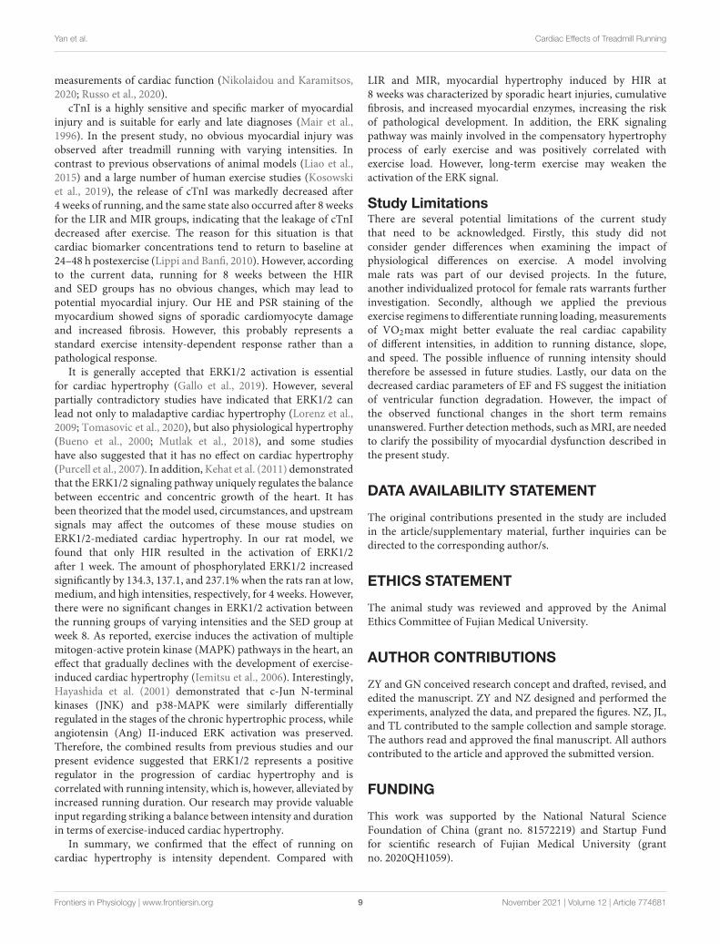
FIGURE 3 | Effects of different treadmill running intensities after 1, 4, and 8 weeks on serum cTnI levels (A) and ERK phosphorylation (B,C) in rats. Data arepresented as the means ± SEM. *P < 0.05, **P < 0.01.
term. A recent study supports the idea that balanced intenseexercise training induces hypertrophy that is not associated withpathological remodeling (Olah et al., 2021).
Transthoracic echocardiography is accurate and widely usedin the field to assess cardiac structure and function. The presentstudy found that low-to-medium intensity running induced asignificant increase in absolute LV diastolic diameter values anda decrease in LV relative wall thickness after 1 week. Theseresults indicate a shift to eccentric LV remodeling. Accordingto Morganroth’s dichotomous concept, regular aerobic sports,such as running, are accompanied by cardiac chamber dilation,which is referred to as eccentric hypertrophy (Morganrothet al., 1975; Ellison et al., 2012). However, in contrast, otherstudies have demonstrated that short-duration exercise appearsto have little in the way of a negative impact on ventricularfunction (Oxborough et al., 2010). Our study also showedthat FS and EF significantly decreased in the LIR and MIR
groups, but not in the HIR group, in the short term. Thisdisparity might be a consequence of myocyte hypertrophy in theHIR group. In short-term low-to-moderate-intensity training,cardiomyocyte contractility and maximal oxygen consumption(VO2max) were like those of the control group (Kemi et al.,2004). Meanwhile, heart growth is related to an increasein chamber dilation. In order to maintain the normal ratioof the number of cardiomyocytes to blood capillaries, theremay be a temporary decline in systolic function. Moreover,the impact of prolonged running on EF and FS was notsignificantly altered between the exercise groups and theSED group after weeks 4 and 8. The observed unchangedcardiac function is in line with recent findings on rat andathlete hearts (Oosthuyse et al., 2012; Asif et al., 2018).Although transthoracic echocardiography is accurate and widelyused to evaluate cardiac structure and function in this field,MRI technology provides more accurate and reproducible
Frontiers in Physiology | www.frontiersin.org 8 November 2021 | Volume 12 | Article 774681
fphys-12-774681 November 23, 2021 Time: 16:12 # 9
Yan et al. Cardiac Effects of Treadmill Running
measurements of cardiac function (Nikolaidou and Karamitsos,2020; Russo et al., 2020).
cTnI is a highly sensitive and specific marker of myocardialinjury and is suitable for early and late diagnoses (Mair et al.,1996). In the present study, no obvious myocardial injury wasobserved after treadmill running with varying intensities. Incontrast to previous observations of animal models (Liao et al.,2015) and a large number of human exercise studies (Kosowskiet al., 2019), the release of cTnI was markedly decreased after4 weeks of running, and the same state also occurred after 8 weeksfor the LIR and MIR groups, indicating that the leakage of cTnIdecreased after exercise. The reason for this situation is thatcardiac biomarker concentrations tend to return to baseline at24–48 h postexercise (Lippi and Banfi, 2010). However, accordingto the current data, running for 8 weeks between the HIRand SED groups has no obvious changes, which may lead topotential myocardial injury. Our HE and PSR staining of themyocardium showed signs of sporadic cardiomyocyte damageand increased fibrosis. However, this probably represents astandard exercise intensity-dependent response rather than apathological response.
It is generally accepted that ERK1/2 activation is essentialfor cardiac hypertrophy (Gallo et al., 2019). However, severalpartially contradictory studies have indicated that ERK1/2 canlead not only to maladaptive cardiac hypertrophy (Lorenz et al.,2009; Tomasovic et al., 2020), but also physiological hypertrophy(Bueno et al., 2000; Mutlak et al., 2018), and some studieshave also suggested that it has no effect on cardiac hypertrophy(Purcell et al., 2007). In addition, Kehat et al. (2011) demonstratedthat the ERK1/2 signaling pathway uniquely regulates the balancebetween eccentric and concentric growth of the heart. It hasbeen theorized that the model used, circumstances, and upstreamsignals may affect the outcomes of these mouse studies onERK1/2-mediated cardiac hypertrophy. In our rat model, wefound that only HIR resulted in the activation of ERK1/2after 1 week. The amount of phosphorylated ERK1/2 increasedsignificantly by 134.3, 137.1, and 237.1% when the rats ran at low,medium, and high intensities, respectively, for 4 weeks. However,there were no significant changes in ERK1/2 activation betweenthe running groups of varying intensities and the SED group atweek 8. As reported, exercise induces the activation of multiplemitogen-active protein kinase (MAPK) pathways in the heart, aneffect that gradually declines with the development of exercise-induced cardiac hypertrophy (Iemitsu et al., 2006). Interestingly,Hayashida et al. (2001) demonstrated that c-Jun N-terminalkinases (JNK) and p38-MAPK were similarly differentiallyregulated in the stages of the chronic hypertrophic process, whileangiotensin (Ang) II-induced ERK activation was preserved.Therefore, the combined results from previous studies and ourpresent evidence suggested that ERK1/2 represents a positiveregulator in the progression of cardiac hypertrophy and iscorrelated with running intensity, which is, however, alleviated byincreased running duration. Our research may provide valuableinput regarding striking a balance between intensity and durationin terms of exercise-induced cardiac hypertrophy.
In summary, we confirmed that the effect of running oncardiac hypertrophy is intensity dependent. Compared with
LIR and MIR, myocardial hypertrophy induced by HIR at8 weeks was characterized by sporadic heart injuries, cumulativefibrosis, and increased myocardial enzymes, increasing the riskof pathological development. In addition, the ERK signalingpathway was mainly involved in the compensatory hypertrophyprocess of early exercise and was positively correlated withexercise load. However, long-term exercise may weaken theactivation of the ERK signal.
Study LimitationsThere are several potential limitations of the current studythat need to be acknowledged. Firstly, this study did notconsider gender differences when examining the impact ofphysiological differences on exercise. A model involvingmale rats was part of our devised projects. In the future,another individualized protocol for female rats warrants furtherinvestigation. Secondly, although we applied the previousexercise regimens to differentiate running loading, measurementsof VO2max might better evaluate the real cardiac capabilityof different intensities, in addition to running distance, slope,and speed. The possible influence of running intensity shouldtherefore be assessed in future studies. Lastly, our data on thedecreased cardiac parameters of EF and FS suggest the initiationof ventricular function degradation. However, the impact ofthe observed functional changes in the short term remainsunanswered. Further detection methods, such as MRI, are neededto clarify the possibility of myocardial dysfunction described inthe present study.
DATA AVAILABILITY STATEMENT
The original contributions presented in the study are includedin the article/supplementary material, further inquiries can bedirected to the corresponding author/s.
ETHICS STATEMENT
The animal study was reviewed and approved by the AnimalEthics Committee of Fujian Medical University.
AUTHOR CONTRIBUTIONS
ZY and GN conceived research concept and drafted, revised, andedited the manuscript. ZY and NZ designed and performed theexperiments, analyzed the data, and prepared the figures. NZ, JL,and TL contributed to the sample collection and sample storage.The authors read and approved the final manuscript. All authorscontributed to the article and approved the submitted version.
FUNDING
This work was supported by the National Natural ScienceFoundation of China (grant no. 81572219) and Startup Fundfor scientific research of Fujian Medical University (grantno. 2020QH1059).
Frontiers in Physiology | www.frontiersin.org 9 November 2021 | Volume 12 | Article 774681

fphys-12-774681 November 23, 2021 Time: 16:12 # 10
Yan et al. Cardiac Effects of Treadmill Running
REFERENCESAsif, Y., Wlodek, M. E., Black, M. J., Russell, A. P., Soeding, P. F., and Wadley, G. D.
(2018). Sustained cardiac programming by short-term juvenile exercise trainingin male rats. J. Physiol. 596, 163–180. doi: 10.1113/JP275339
Benito, B., Gay-Jordi, G., Serrano-Mollar, A., Guasch, E., Shi, Y., Tardif, J. C.,et al. (2011). Cardiac arrhythmogenic remodeling in a rat model of long-termintensive exercise training. Circulation 123, 13–22.
Bueno, O. F., De Windt, L. J., Tymitz, K. M., Witt, S. A., Kimball, T. R., Klevitsky,R., et al. (2000). The MEK1-ERK1/2 signaling pathway promotes compensatedcardiac hypertrophy in transgenic mice. EMBO J. 19, 6341–6350. doi: 10.1093/emboj/19.23.6341
Davos, C. H. (2019). Do we have to reconsider the guidelines for exercise intensitydetermination in cardiovascular rehabilitation? Eur. J. Prev. Cardiol. 26, 1918–1920.
Ellison, G. M., Waring, C. D., Vicinanza, C., and Torella, D. (2012). Physiologicalcardiac remodelling in response to endurance exercise training: cellular andmolecular mechanisms. Heart 98, 5–10. doi: 10.1136/heartjnl-2011-300639
Gallo, S., Vitacolonna, A., Bonzano, A., Comoglio, P., and Crepaldi, T. (2019).ERK: a key player in the pathophysiology of cardiac hypertrophy. Int. J. Mol.Sci. 20:2164. doi: 10.3390/ijms20092164
Hayashida, W., Kihara, Y., Yasaka, A., Inagaki, K., Iwanaga, Y., and Sasayama,S. (2001). Stage-specific differential activation of mitogen-activated proteinkinases in hypertrophied and failing rat hearts. J. Mol. Cell Cardiol. 33, 733–744.doi: 10.1006/jmcc.2001.1341
Iemitsu, M., Maeda, S., Jesmin, S., Otsuki, T., Kasuya, Y., and Miyauchi, T. (2006).Activation pattern of MAPK signaling in the hearts of trained and untrainedrats following a single bout of exercise. J. Appl. Physiol. 101, 151–163. doi:10.1152/japplphysiol.00392.2005
Kehat, I., Davis, J., Tiburcy, M., Accornero, F., Saba-El-Leil, M. K., Maillet, M.,et al. (2011). Extracellular signal-regulated kinases 1 and 2 regulate the balancebetween eccentric and concentric cardiac growth. Circ. Res. 108, 176–183. doi:10.1161/CIRCRESAHA.110.231514
Kemi, O. J., Haram, P. M., Loennechen, J. P., Osnes, J. B., Skomedal, T.,Wisloff, U., et al. (2005). Moderate vs. high exercise intensity: differentialeffects on aerobic fitness, cardiomyocyte contractility, and endothelialfunction. Cardiovasc. Res. 67, 161–172. doi: 10.1016/j.cardiores.2005.03.010
Kemi, O. J., Haram, P. M., Wisløff, U., and Ellingsen, Ø (2004). Aerobic fitness isassociated with cardiomyocyte contractile capacity and endothelial function inexercise training and detraining. Circulation 109, 2897–2904. doi: 10.1161/01.CIR.0000129308.04757.72
Khoury, S. R., Evans, N. S., and Ratchford, E. V. (2019). Exercise as medicine. Vasc.Med. 24, 371–374.
Konhilas, J. P., Maass, A. H., Luckey, S. W., Stauffer, B. L., Olson, E. N., andLeinwand, L. A. (2004). Sex modifies exercise and cardiac adaptation in mice.Am. J. Physiol. Heart Circ. Physiol. 287, H2768–H2776.
Kosowski, M., Mlynarska, K., Chmura, J., Kustrzycka-Kratochwil, D., Sukiennik-Kujawa, M., Todd, J. A., et al. (2019). Cardiovascular stress biomarkerassessment of middle-aged non-athlete marathon runners. Eur. J. Prev. Cardiol.26, 318–327. doi: 10.1177/2047487318819198
Krzesiak, A., Delpech, N., Sebille, S., Cognard, C., and Chatelier, A. (2017).Structural, contractile and electrophysiological adaptations of cardiomyocytesto chronic exercise. Adv. Exp. Med. Biol. 999, 75–90. doi: 10.1007/978-981-10-4307-9_5
Liao, J., Li, Y., Zeng, F., and Wu, Y. (2015). Regulation of mTOR pathway inexercise-induced cardiac hypertrophy. Int. J. Sports Med. 36, 343–350.
Libonati, J. R., Sabri, A., Xiao, C., Macdonnell, S. M., and Renna, B. F. (2011).Exercise training improves systolic function in hypertensive myocardium.J. Appl. Physiol. 111, 1637–1643.
Lippi, G., and Banfi, G. (2010). Exercise-related increase of cardiactroponin release in sports: an apparent paradox finally elucidated? Clin.Chim. Acta Int. J. Clin. Chem. 411, 610–611. doi: 10.1016/j.cca.2010.01.009
Ljones, K., Ness, H. O., Solvang-Garten, K., Gaustad, S. E., and Hoydal, M. A.(2017). Acute exhaustive aerobic exercise training impair cardiomyocytefunction and calcium handling in Sprague-Dawley rats. PLoS One 12:e0173449.doi: 10.1371/journal.pone.0173449
Lorenz, K., Schmitt, J. P., Schmitteckert, E. M., and Lohse, M. J. (2009). A newtype of ERK1/2 autophosphorylation causes cardiac hypertrophy. Nat. Med. 15,75–83. doi: 10.1038/nm.1893
Mair, J., Genser, N., Morandell, D., Maier, J., Mair, P., Lechleitner, P., et al. (1996).Cardiac troponin I in the diagnosis of myocardial injury and infarction. Clin.Chim. Acta Int. J. Clin. Chem. 245, 19–38.
Malek, L. A., and Bucciarelli-Ducci, C. (2020). Myocardial fibrosis inathletes-Current perspective. Clin. Cardiol. 43, 882–888. doi: 10.1002/clc.23360
Mi, C., Qin, X., Hou, Z., and Gao, F. (2019). Moderate-intensity exercise allowsenhanced protection against oxidative stress-induced cardiac dysfunction inspontaneously hypertensive rats. Braz. J. Med. Biol. Res. 52:e8009. doi: 10.1590/1414-431X20198009
Moreira, J. B. N., Wohlwend, M., and Wisløff, U. (2020). Exercise and cardiachealth: physiological and molecular insights. Nat. Metab. 2, 829–839.
Morganroth, J., Maron, B. J., Henry, W. L., and Epstein, S. E. (1975). Comparativeleft ventricular dimensions in trained athletes. Ann. Intern. Med. 82,521–524.
Mutlak, M., Schlesinger-Laufer, M., Haas, T., Shofti, R., Ballan, N., Lewis, Y. E.,et al. (2018). Extracellular signal-regulated kinase (ERK) activation preservescardiac function in pressure overload induced hypertrophy. Int. J. Cardiol. 270,204–213.
Nakamura, M., and Sadoshima, J. (2018). Mechanisms of physiological andpathological cardiac hypertrophy. Nat. Rev. Cardiol. 15, 387–407.
Ni, G. X., Liu, S. Y., Lei, L., Li, Z., Zhou, Y. Z., and Zhan, L. Q.(2013). Intensity-dependent effect of treadmill running on knee articularcartilage in a rat model. BioMed Res. Int. 2013:172392. doi: 10.1155/2013/172392
Nikolaidou, C., and Karamitsos, T. (2020). Should everyone have an MRI in heartfailure? Cardiovasc. Diagn. Ther. 10, 549–553.
Olah, A., Barta, B. A., Sayour, A. A., Ruppert, M., Virag-Tulassay, E., Novak, J.,et al. (2021). Balanced intense exercise training induces atrial oxidative stresscounterbalanced by the antioxidant system and atrial hypertrophy that is notassociated with pathological remodeling or arrhythmogenicity. Antioxidants10:452.
Olah, A., Nemeth, B. T., Matyas, C., Horvath, E. M., Hidi, L., Birtalan, E., et al.(2015). Cardiac effects of acute exhaustive exercise in a rat model. Int. J. Cardiol.182, 258–266. doi: 10.1016/j.ijcard.2014.12.045
Oosthuyse, T., Avidon, I., Likuwa, I., and Woodiwiss, A. J. (2012). Progression ofchanges in left ventricular function during four days of simulated multi-stagecycling. Eur. J. Appl. Physiol. 112, 2243–2255. doi: 10.1007/s00421-011-2201-z
Oxborough, D., Birch, K., Shave, R., and George, K. (2010). “Exercise-inducedcardiac fatigue”–a review of the echocardiographic literature. Echocardiography27, 1130–1140. doi: 10.1111/j.1540-8175.2010.01251.x
Pagan, L. U., Damatto, R. L., Gomes, M. J., Lima, A. R. R., Cezar, M. D. M.,Damatto, F. C., et al. (2019). Low-intensity aerobic exercise improves cardiacremodelling of adult spontaneously hypertensive rats. J. Cell Mol. Med. 23,6504–6507. doi: 10.1111/jcmm.14530
Purcell, N. H., Wilkins, B. J., York, A., Saba-El-Leil, M. K., Meloche, S., Robbins,J., et al. (2007). Genetic inhibition of cardiac ERK1/2 promotes stress-inducedapoptosis and heart failure but has no effect on hypertrophy in vivo. Proc. Natl.Acad. Sci. U.S.A. 104, 14074–14079. doi: 10.1073/pnas.0610906104
Radovits, T., Olah, A., Lux, A., Nemeth, B. T., Hidi, L., Birtalan, E., et al. (2013). Ratmodel of exercise-induced cardiac hypertrophy: hemodynamic characterizationusing left ventricular pressure-volume analysis. Am. J. Physiol. Heart Circ.Physiol. 305, H124–H134.
Russo, V., Lovato, L., and Ligabue, G. (2020). Cardiac MRI: technical basis. Radiol.Med. 125, 1040–1055.
Shioi, T., McMullen, J. R., Tarnavski, O., Converso, K., Sherwood, M. C., Manning,W. J., et al. (2003). Rapamycin attenuates load-induced cardiac hypertrophy inmice. Circulation 107, 1664–1670.
Swain, D. P., and Franklin, B. A. (2006). Comparison of cardioprotective benefitsof vigorous versus moderate intensity aerobic exercise. Am. J. Cardiol. 97,141–147. doi: 10.1016/j.amjcard.2005.07.130
Tang, X., Chen, X. F., Wang, N. Y., Wang, X. M., Liang, S. T., Zheng, W., et al.(2017). SIRT2 acts as a cardioprotective deacetylase in pathological cardiachypertrophy. Circulation 136, 2051–2067. doi: 10.1161/CIRCULATIONAHA.117.028728
Frontiers in Physiology | www.frontiersin.org 10 November 2021 | Volume 12 | Article 774681

fphys-12-774681 November 23, 2021 Time: 16:12 # 11
Yan et al. Cardiac Effects of Treadmill Running
Tang, X. Y., Hong, H. S., Chen, L. L., Lin, X. H., Lin, J. H., and Lin, Z. (2011).Effects of exercise of different intensities on the angiogenesis, infarct healing,and function of the left ventricle in postmyocardial infarction rats. Coron ArteryDis. 22, 497–506. doi: 10.1097/MCA.0b013e32834993d9
Tomasovic, A., Brand, T., Schanbacher, C., Kramer, S., Hümmert, M. W., Godoy,P., et al. (2020). Interference with ERK-dimerization at the nucleocytosolicinterface targets pathological ERK1/2 signaling without cardiotoxic side-effects.Nat. Commun. 11:1733. doi: 10.1038/s41467-020-15505-4
Wang, Y., Wisloff, U., and Kemi, O. J. (2010). Animal models in the study ofexercise-induced cardiac hypertrophy. Physiol. Res. 59, 633–644. doi: 10.33549/physiolres.931928
Wasfy, M. M., and Baggish, A. L. (2016). Exercise dose in clinical practice.Circulation 133, 2297–2313.
Yan, Z. P., Li, J. T., Zeng, N., and Ni, G. X. (2021). Role of extracellular signal-regulated kinase 1/2 signaling underlying cardiac hypertrophy. Cardiol. J. 28,473–482.
Conflict of Interest: The authors declare that the research was conducted in theabsence of any commercial or financial relationships that could be construed as apotential conflict of interest.
Publisher’s Note: All claims expressed in this article are solely those of the authorsand do not necessarily represent those of their affiliated organizations, or those ofthe publisher, the editors and the reviewers. Any product that may be evaluated inthis article, or claim that may be made by its manufacturer, is not guaranteed orendorsed by the publisher.
Copyright © 2021 Yan, Zeng, Li, Liao and Ni. This is an open-access articledistributed under the terms of the Creative Commons Attribution License (CC BY).The use, distribution or reproduction in other forums is permitted, provided theoriginal author(s) and the copyright owner(s) are credited and that the originalpublication in this journal is cited, in accordance with accepted academic practice. Nouse, distribution or reproduction is permitted which does not comply with these terms.
Frontiers in Physiology | www.frontiersin.org 11 November 2021 | Volume 12 | Article 774681
Copyright © 2022 FDOKUMEN