Call for Editorial Board Members
232
IJAA / Volume 6 Number 4 (Part - II) / July - August 2019 Call for Editorial Board Members As you are well aware that we are a medical and health sciences publishers; publishing peer-reviewed journals and books since 2004. We are always looking for dedicated editorial board members for our journals. If you completed your master degree and must have at least five years experience in teaching and having good publication records in journals and books. If you are interested to be an editorial board member of the journal; please provide your complete resume and affiliation through e-mail (i.e. info@ rfppl.co.in) or visit our website (i.e.www.rfppl.co.in) to register yourself online. Call for Publication of Conference Papers/Abstracts We publish pre-conference or post-conference papers and abstracts in our journals, and deliver hard copy and giving online access in a timely fashion to the authors. For more information, please contact: For more information, please contact: A Lal Publication–in-charge Red Flower Publication Pvt. Ltd. 48/41-42, DSIDC, Pocket-II Mayur Vihar Phase-I Delhi – 110 091 (India) Phone: 91-11-22754205, 45796900 E-mail: [email protected]
-
Upload
khangminh22 -
Category
Documents
-
view
3 -
download
0
Transcript of Call for Editorial Board Members
IJAA�/�Volume�6�Number�4�(Part�-�II)�/�July�-�August�2019
Call�for�Editorial�Board�Members
As�you�are�well�aware�that�we�are�a�medical�and�health�sciences�publishers;�publishing�peer-reviewed�journals�and�books�since�2004.�
We�are� always� looking� for�dedicated�editorial� board�members� for�our�journals.� If� you� completed�your�master�degree�and�must�have�at� least�five�years�experience�in�teaching�and�having�good�publication�records�in�journals�and�books.�
If�you�are�interested�to�be�an�editorial�board�member�of�the�journal;�please�provide�your�complete�resume�and�affiliation�through�e-mail�(i.e.�[email protected])�or�visit�our�website�(i.e.www.rfppl.co.in)�to�register�yourself�online.
Call�for�Publication�of�Conference�Papers/Abstracts
We�publish�pre-conference�or�post-conference�papers� and� abstracts� in�our�journals,�and�deliver�hard�copy�and�giving�online�access�in�a�timely�fashion�to�the�authors.
For�more�information,�please�contact:
For�more�information,�please�contact:A�Lal
Publication–in-chargeRed�Flower�Publication�Pvt.�Ltd.48/41-42,�DSIDC,�Pocket-II
Mayur�Vihar�Phase-IDelhi�–�110�091�(India)
Phone:�91-11-22754205,�45796900E-mail:�[email protected]
IJAA�/�Volume�6�Number�4�(Part�-�II)�/�July�-�August�2019
Free�Announcements�of�your�Conferences/Workshops/CMEs
This�privilege�to�all�Indian�and�other�countries�conferences�organizing�committee�members�to�publish�free�announcements�of�your�conferences/workshops.� If� you� are� interested,� please� send� your� matter� in� word�formats�and�images�or�pictures�in�JPG/JPEG/Tiff�formats�through�e-mail�attachments�to�[email protected].
Terms�&�Conditions�to�publish�free�announcements:1.� Only�conference�organizers�are�eligible�up�to�one�full�black�and�
white�page,�but�not�applicable�for�the�front,�inside�front,�inside�back�and�back�cover,�however,�these�pages�are�paid.
2.� Only�five�pages�in�every�issue�are�available�for�free�announcements�for�different�conferences.
3.� This�announcement�will� come�in� the�next�coming�issue�and�no�priority�will�be�given.
4.� All�legal�disputes�subject�to�Delhi�jurisdiction�only�
5.� The�executive�committee�of�the�Red�Flower�Publication�reserve�the� right� to� cancel,� revise� or�modify� terms� and� conditions� any�time�without�notice.
For�more�information,�please�contact:A�Lal
Publication–in-chargeRed�Flower�Publication�Pvt.�Ltd.
48/41-42,�DSIDC,�Pocket-IIMayur�Vihar�Phase-I
Delhi�–�110�091�(India)Phone:�91-11-22754205,�45796900
E-mail:�[email protected]

IJAA�/�Volume�6�Number�4�(Part�-�II)�/�July�-�August�2019
Win�Free�Institutional�Subscription!
Simply�fill�out�this�form�and�return�scanned�copy�through�e-mail�or�by�post�to�us.
Name�of�the�Institution__________________________________________________
Name�of�the�Principal/Chairman_________________________________________
Management�(Trust/Society/Govt./Company)_____________________________
Address�1_______________________
Address�2_______________________
Address�3_______________________
City_________________
Country_____________
PIN�Code____________
Mobile______________
Email_______________
We�are�regular�subscriber�of�Red�Flower�Publication�journals.
�ear�of�first�subscription______________
List�of�ordered�journals�(if�you�subscriberd�more�then�5�titles,�please�attach�separate�sheet)
Ordered�through
Name�of�the�Vendor Subscription��ear �irect/subs��r
Name�of�the��ournal�for��hich�you��ish�to�be�free��inner
Terms�&�Conditions�to�win�free�institutional�subscription
1.� �Only�institutions�can�participate�in�this�scheme2.� In�group�institutions�only�one�institution�would�be�winner3.�� Only�five�institutions�will�be�winner�for�each�journal4.� An�institution�will�be�winner�only�for�one�journal5.� The�free�subscription�will�be�valid�for�one�year�only�(i.e.�1,�Jan�-�31,�Dec)6.� This�free�subscription�is�not�renewable,�however�can�be�renewed�with�payment7.� Any�institution�can�again�participate�after�five�years8.� All�legal�disputes�subject�to�Delhi�jurisdiction�only9.� This� scheme�will�be�available� to�participate� throughout�year,�but�draw�will�be�held� in� last�week�of�
August�every�year10.� The�executive�committee�of�the�Red�Flower�Publication�reserve�the�right�to�cancel,�revise�or�modify�
terms�and�conditions�any�time�without�notice.I�confirm�and�certify�that�the�above�information�is�true�and�correct�to�the�best�of�my�knowledge�and�belief.
Place:� � � � � � � � � � Signature�with�Seal�Date:
IJAA�/�Volume�6�Number�4�(Part�-�II)�/�July�-�August�2019
Revised�Rates�for�2020�(Institutional)
Title�of�the��ournal Fre�uencyIndia�IN��Print�Only
India�IN��Online�Only
Outside�India��S��Print�Only
Outside�India��S��Online�Only
Community�and�Public��ealth�Nursing 3 6000 5500 469 430Indian�Journal�of�Agriculture��usiness 2 6000 5500 469 430Indian�Journal�of�Anatomy 4 9000 8500 703 664Indian�Journal�of�Ancient�Medicine�and��oga 4 8500 8000 664 625Indian�Journal�of�Anesthesia�and�Analgesia 6� 8000 7500 625 586Indian�Journal�of��iology 2 6000 5500 469 430Indian�Journal�of�Cancer�Education�and�Research 2 9500 9000 742 703Indian�Journal�of�Communicable�Diseases 2 9000 8500 703 664Indian�Journal�of�Dental�Education 4 6000 5500 469 430Indian�Journal�of�Diabetes�and�Endocrinology 2 8500 8000 664 625Indian�Journal�of�Emergency�Medicine 4 13000 12500 1016 977Indian�Journal�of�Forensic�Medicine�and�Pathology 4 16500 16000 1289 1250Indian�Journal�of�Forensic�Odontology 2 6000 5500 469 430Indian�Journal�of�Genetics�and�Molecular�Research 2 7500 7000 586 547Indian�Journal�of�Law�and��uman��ehavior 3 6500 6000 508 469Indian�Journal�of�Legal�Medicine 2 9000 8500 703 664Indian�Journal�of�Library�and�Information�Science 3 10000 9500 781 742Indian�Journal�of�Maternal-Fetal���Neonatal�Medicine 2 10000 9500 781 742Indian�Journal�of�Medical�and��ealth�Sciences 2 7500 7000 586 547Indian�Journal�of�Obstetrics�and�Gynecology 4 10000 9500 781 742Indian�Journal�of�Pathology:�Research�and�Practice 6 12500 12000 977 938Indian�Journal�of�Plant�and�Soil 2 7000 6500 547 508Indian�Journal�of�Preventive�Medicine 2 7500 7000 586 547Indian�Journal�of�Research�in�Anthropology 2 13000 12500 1016 977Indian�Journal�of�Surgical�Nursing 3 6000 5500 469 430Indian�Journal�of�Trauma�and�Emergency�Pediatrics 4 10000 9500 781 742Indian�Journal�of�Waste�Management 2 10000 9500 781 742International�Journal�of�Food,�Nutrition���Dietetics 3 6000 5500 469 430International�Journal�of�Forensic�Science 2 10500 10000 820 781International�Journal�of�Neurology�and�Neurosurgery 4 11000 10500 859 820International�Journal�of�Pediatric�Nursing 3 6000 5500 469 430International�Journal�of�Political�Science 2 6500 6000 508 469International�Journal�of�Practical�Nursing 3 6000 5500 469 430International�Physiology 3 8000 7500 625 586Journal�of�Animal�Feed�Science�and�Technology 2 8300 7800 648 609Journal�of�Cardiovascular�Medicine�and�Surgery 4 10500 10000 820 781Journal�of�Emergency�and�Trauma�Nursing 2 6000 5500 469 430Journal�of�Food�Additives�and�Contaminants 2 6000 5500 430 391Journal�of�Food�Technology�and�Engineering 2 5500 5000 430 391Journal�of�Forensic�Chemistry�and�Toxicology 2 10000 9500 781 742Journal�of�Global�Medical�Education�and�Research 2 6400 5900 500 461Journal�of�Global�Public��ealth 2 12500 12000 977 938Journal�of�Microbiology�and�Related�Research 2 9000 8500 703 664Journal�of�Nurse�Midwifery�and�Maternal��ealth 3 6000 5500 469 430Journal�of�Orthopedic�Education 3 6000 5500 469 430Journal�of�Pharmaceutical�and�Medicinal�Chemistry 2 17000 16500 1328 1289Journal�of�Plastic�Surgery�and�Transplantation 2 26900 26400 2102 2063Journal�of�Psychiatric�Nursing 3 6000 5500 469 430Journal�of�Radiology 2 8500 8000 664 625Journal�of�Social�Welfare�and�Management 4 8000 7500 625 586New�Indian�Journal�of�Surgery 6 8500 7500 664 625Ophthalmology�and�Allied�Sciences 3 6500 6000 508 469Pediatric�Education�and�Research 4 8000 7500 625 586Physiotherapy�and�Occupational�Therapy�Journal 4 9500 9000 742 703RFP�Gastroenterology�International 2 6500 6000 508 469RFP�Indian�Journal�of��ospital�Infection 2 13000 12500 1016 977RFP�Indian�Journal�of�Medical�Psychiatry 2 8500 8000 664 625RFP�Journal�of��iochemistry�and��iophysics 2 7500 7000 586 547RFP�Journal�of�Dermatology�(Formerly�Dermatology�International) 2 6000 5500 469 430RFP�Journal�of�ENT�and�Allied�Sciences�(Formerly�Otolaryngology�International) 2 6000 5500 469 430RFP�Journal�of�Gerontology�and�Geriatric�Nursing 2 6000 5500 469 430RFP�Journal�of��ospital�Administration 2 7500 7000 586 547�rology,�Nephrology�and�Andrology�International 2 8000 7500 625 586
Terms�of�Supply:
1.� Agency�discount�12.5�.�Issues�will�be�sent�directly�to� the�end�user,�otherwise�foreign�rates�will�be�charged.2.� All� back� volumes�of�all� journals� are�available� at� current� rates.3.� All� journals�are�available�free�online�with�print�order�within�the�subscription�period.4.� All� legal� disputes� subject� to� Delhi� jurisdiction.5.� Cancellations�are� not� accepted� orders� once� processed.
6.� Demand� draft/che�ue�should� be� issued� in� favour�of���ed�Flo�er�Publication� P�t�� �td��� payable�at��elhi�
7.� Full� pre-payment� is� re�uired.� It� can� be�done� through� online�(http://rfppl.co.in/subscribe.php�mid�7).
8.� No�claims�will�be�entertained�if�not�reported�within�6�months�of�the�publishing�date.9.� Orders�and�payments�are�to� be�sent� to�our�of��ce� address�as�given�below.10.�Postage��� �andling�is� included� in� the� subscription� rates.11.�Subscription�period�is�accepted�on� calendar�year�basis� (i.e.� Jan�to�Dec).��owever�orders�may�be�placed�any�time�throughout� the� year.
Order� m
Red�Flower�Publication� Pvt.�Ltd.,�48/41-42,�DSIDC,�Pocket-II,�Mayur�Vihar�Phase-I,�Delhi�-�110�091�(India)
Mobile:�8130750089,�Phone:�91-11-45796900,�22754205,�22756995,�E-mail:�[email protected],�Website:�www.rfppl.co.in
IJAA�/�Volume�6�Number�4�(Part�-�II)�/�July�-�August�2019
Indian��ournal�of�Anesthesia�and�Analgesia
Editor�in�Chief
�����Mubarak
Professor����ead,�Dept.�of�Anaesthesiology
Govt.�Medical�College,��ozhikode�673008,��erala
Associate�Editors
�alit��upta��SM�-Curie�Cancer�Center,�Delhi
Mridu�Paban�Nath��Gauhati�Medical�College,�Guwahati
Sandeep�Sahu��Sanjay�Gandhi�Postgradaute�Institute�of�Medical�
Sciences,�Lucknow
National�Editorial�Board
�aura��S��TomarAll�India�Institute�of�Medical�Sciences,�New�Delhi
Man�ula�Sudeep�Sarkar�Seth�G�S�MC�and���E�M��ospital,�Mumbai
Mukesh�Som�anshiGovt.�Medical�College���AG��ospitals,��ota
Naresh��anpatrao�Tirpude�Govt.�Medical�College,�Nagpur
Palla�i�Ahlu�alia�Teerthanker�Mahaveer�Medical�College���
Research�Centre,�Moradabad,��ttar�Pradesh
Pramod��umarPD��Medical�College,�Rajkot
Saramma�P�AbrahamMOSC�Medical�College,��olencherry
International�Editorial�Board
Amar�eet����Patil��Manchester��niversity��ospitals�N�S�Foundation�Trust,�Manchester,��nited��ingdom
Managing�EditorA�Lal
Publication�EditorManoj��umar�Singh
Aims�and�Scope
The� Indian� Journal� of� Anesthesia� and� Analgesia� (IJAA)� is� of��cial� peer-reviewed� scienti��c� journal� addresses� all�aspects�of�anesthesia�practice,�including�anesthetic�administration,�pharmacokinetics,�preoperative�and�postoperative�considerations,�coexisting�disease�and�other�complicating�factors,�cost�issues,�and�similar�concerns�anesthesiologists�contend�with�daily.�The�Journal�seeks�a�balance�between�outstanding�basic�scienti��c�reports�and�de��nitive�clinical�and�management�investigations.�The�Journal�welcomes�manuscripts�re��ecting�rigorous�analysis,�even�if�unusual�in�style�and�focus.
Readership:�Anesthesiologists,�Critical�Care�Physicians�and�Surgeons.
Of��ce�of�Publication:��ed�Flo�er�Publication�P�t���td���48/41-42,�DSIDC,�Pocket-IIMayur�Vihar�Phase-I,�Delhi�–�110�091(India),�Phone:�91-11-22754205,�45796900,�Fax:�91-11-22754205
E-mail:�[email protected],�Website:�www.rfppl.co.in
Satish�����eshpande�Government�Medical�College,�Latur
S�arnalingam�Thanga�eluTagore�Medical�College����ospital,�Chennai
S�ati�BishtVydehi�Institute�of�Medical�Sciences,�And�
Research�Centre,��angalore
�ma��ariharanDr�Ram�Manohar�Lohia��ospital���PGIMER,�
New�Delhi�
Vikas�ChauhanAll�India�Institute�of�Medical�Sciences,�New�Delhi
IJAA�/�Volume�6�Number�4�(Part�-�II)�/�July�-�August�2019
1262 Indian�Journal�of�Anesthesia�and�Analgesia
Copyright�Information Subscription�Information
Ad�ertisement
�isclaimer
For�Authors
As� soon� as� article� is� accepted� for� publication,�authors� will� be� re�uested� to� assign� copyright� of�the� article� (or� to� grant� exclusive� publication� and�dissemination� rights)� to� the� publisher� (respective�the� owner� if� other� than� Red� Flower� Publication�Pvt.� Ltd.).� This� will� ensure� the� widest� possible�protection�and�dissemination�of�information�under�copyright�laws.
More� information� about� copyright� regulations�for�this�journal�is�available�at�www.rfppl.co.in
For�Readers
All�the�advice�and�information�in�this�journal�are�believed� to�be� true�and�accurate�at� the�date�of� its�publication.�Neither� the�editors,�nor�the�publisher�can� accept� any� legal� responsibility� for� any� errors�or�omissions�that�may�have�been�made.�The�author�is� totaly� resposible� for� any� error� or� omission� in�the� article.� The� publisher� makes� no� warranty,�express� or� implied,� with� respect� to� the� material�contained��herein.
All� articles� published� in� this� journal� are�protected� by� copyright,� which� covers� the�exclusive� rights� to� reproduced� and� distribute� the�article� (e.g.� as�offprints),� as�well� as� all� translation�rights.�No� material� published� in� this� journal�may�be� reproduced� photographically� or� stored� on�micro��lm,� in� electronic�databases�on�video� disks,�etc,� without� obtaining� written� permission� from�the� publisher� (respective� the� copyright� owner�if� other� than� Red� Flower� Publication� Pvt.� Ltd.).�The� � use� of� general� descriptive� names,� trade�names,� trademarks,� etc.,� in� this� publication,� even�if� not� speci��cally� identi��ed,� does� not� imply� that�these�names�are�not�protected�by�the�relevant�laws�and��regulations.
���ed�Flo�er�Publication�P�t���td������
�ournal�Website
Red�Flower�Publication�Pvt.�Ltd.�has�partnered�for� permission� to� reuse� our� content� please� locate�the�material�that�you�with�to�use�on�link�rfppl.co.in�and�click�on�permission�link�and�enter�the� title�of�the�publication�that�you�wish�to�use.�For�assistance�in�placing�a�permission�re�uest.
http://rfppl.co.in/about_journal.php�jid�24
The�Indian�Journal�of�Anesthesia�and�Analgesia�is�published�four�times�a�year.
Volume� 6� (6� issues)� will� be� published� in� 2019�pISSN:�2349-8471,�eISSN:�2455-6238�
For� information� on� subscription� rates� please�contact
Subscription�and�Marketing�ManagerRed�Flower�Publication�Pvt.�Ltd.48/41-42,�DSIDC,�Pocket-IIMayur�Vihar�Phase-IDelhi�-�110�091(India)Phone:�91-11-45796900,�22754205,�[email protected]
The�journal�is�distributed�free�of�cost�to�members�of� editorial� board.� Institutional� subscription� rates�(India)�INR�7500�and�(other�countries)��SD586.
All� correspondence� regarding� individual� and�society� subscriptions,� subscription� rates� for� 2019�and� advertisements� should� be� addressed� to:�[email protected].
E-mail:�[email protected].
Red� Flower� Publication� Pvt.� Ltd.� publishes�advertisement� in� this� journal� reliance� upon� the�responsibility� of� the� advertiser� to� comply� with�all� legal� re�uirements� relating� to� the� marketing�and� sale� of� products� or� services� advertised.� Red�Flower�Publication�Pvt.�Ltd.�and�the�editors�are�not�responsible� for� claims�make� in� the� advertisement�published� in� the� journal.� The� appearance� of�advertisements� in� Red� Flower� Publication� Pvt.�Ltd.�publications�does�not�constitute�endorsement,�implied�or� intended,�of� the�product�advertised�or�the�claims�make�for�it�by�the�advertiser.
The� views� expressed� in� this�publication�do�not�necessarily�re��ect�those�of�Red�Flower�Publication�Pvt.�Ltd.
Red�Flower�Publication�Pvt.�Ltd.�do�not�endorse�the��uality�or�the�value�of�the�advertised/sponsored�products�described�therein.
IJAA�/�Volume�6�Number�4�(Part�-�II)�/�July�-�August�2019
IN�IAN��O��NA��OF�ANEST�ESIA�AN��ANA��ESIA
�uly���August������Volume���Number����Part���II�
�Original�Research�Articles
Contents
Study�on�Comparing�the�Postoperati�e�Analgesic�Efficacy�of��ltrasound��uided�Tans�erse�� Abdominis�Plane�Block��ith�������Bupi�acaine�and���������opi�acaine�in�� �aparoscopic�Surgeries� ����
� P��alyan�Chakravarthy,�Sireesha�Ejjapureddi,��emnath��abu��otla,�Jaya�Chandra,���Ramya,�M�Ramya
A�Comparati�e�Study�of�Caudal�Analgesia��ith�Bupi�acaine�Alone�and�Bupi�acaine�� �ith�Butorphanol�in�Pediatric�Surgeries� ����� Shaik�Salman,�C�Geetha
Proseal��aryngeal�Mask�Air�ay��/s�Endotracheal�Intubation�for��ynaecological�� �aparoscopic�Surgeries� ����� Amruta�Changdeo�Patil,�Sonal�S��hatavkar,�Rosly�R�Jacob,�Chaitanya��dayan�Gaidhani
Effects�of�Clonidine�on�Spinal�Anesthesia��ith��yperbaric�Bupi�acaine� ����� Ananda��hat,�Parimala��,��alajibabu�PR
A�Comparati�e�Study�of�Oral��abapentin�and�Oral�Clonidine�as�Preempti�e�� Analgesia�under�Spinal�Anesthesia�for�Abdomino�Pel�ic�Surgeries� ����� Ankita��ajare,�Irfan�A�Waris
�ltrasound��uided�Combined�Superficial�Cer�ical�Ple�us�Block�Interscalene�Block�for�� Anesthesia�in�Cla�icular�Fractures��A��etrospecti�e�Obser�ational�Cohort�Study� ����� S.�Arun,�Deepan�C,�Jomy�Thomas
Prospecti�e�Study�to�E�aluate�the��ole�of�Vasopressin�in��ypernatremia�Treatment�in�� Brain��ead�Patients� ����� G��alaji,���Jeyarani
Pre�ention�of��ypotension�Follo�ing�Subarachnoid�Block��Efficacy�of�Preloading��ith�� �ydro�yethyl�Starch�Versus��inger�s��actate�Solution� ����� �alajibabu�PR,�Ananda��hat
A�Comparati�e�Study�of�Analgesia��ith��opi�acaine�and��e�medetomidine��s�� �opi�acaine�and�Fentanyl�in�Epidural�Anesthesia�in��o�er��imb�Surgeries� ����� �urra�Ramesh��umar,�Appa�Rao�Mekala
Pain�on�Propofol�In�ection:�Comparati�e�Study�of�Pre�Treatment��ith�Intra�enous�� �ignocaine��Ondansetron�and�Fentanyl�for�the�Pre�ention�of�Pain� ����� Chetan�Arun�Patil,�Neha�P��amble,�Manoj��umar�N�Gajbhare
�e�medetomidine�is�a�Better�Ad�u�ant�than�Clonidine���ith��opi�acaine�in�� Supracla�icular�Brachial�Ple�us�Block� ����� Gunaseelan�Sivasamy,�Priyanka�Selvam,�Thirunavukkarasu�M,�Raghuraman�MS
A�Prospecti�e��andomised�Controlled�Study�of�Pre�Empti�e�Oral�Flupirtine�on�� Postoperati�e�Analgesia�in�Patients��ndergoing�Abdominal�Surgeries�� �nder��eneral�Anesthesia� ����� Ilango�Ganesan,�C�Manikandan
A�Comparison�of��e�medetomidine��ith�Thiopentone�Sodium�Versus�Esmolol��ith�� Thiopentone�Sodium�to�Attenuate�the��emodynamic�Stress��esponses�after�� Electrocon�ulsi�e�Therapy� ����� ���rishna�Chaitanya,�A�Prashanth,�Shaik��ala,��anth�Pavan��umar,���Suresh��abu
Comparison�of��o���ose�Fentanyl��ith��o���ose��e�amethasone�as�an�Ad�u�ant�� to������Bupi�acaine�in�Supracla�icular�Block��ia�Multipoint�In�ection�� Techni�ue�under�Sonographic��uidance� ����� �unaal��umar�Sharma,�Rajesh��umar�Verma,�Surinder�Singh,�Avinash�Goyal
A�Study�of�Comparison�of�Intubating�Conditions�and��aemodynamic�Effects�after�the�� Administration�of�Succinylcholine�and��ocuronium�Bromide� ����� Lovina�Neil,��arshil�Shah
IJAA�/�Volume�6�Number�4�(Part�-�II)�/�July�-�August�2019
1264 Indian�Journal�of�Anesthesia�and�Analgesia
Comparison�of�Bilateral�Superficial�Cer�ical�Ple�us�Block�and�Incision��ine�Infiltration�� for�Postoperati�e�Analgesia�for�Thyroid�Surgeries��nder��eneral�Anesthesia� ����
� Nagaraj�AV,�Pradeep�A�Dongare
Crystalloid�Preload�Versus�Crystalloid�Co�load��uring�Electi�e�Caesarean�Section�� �nder�Spinal�Anesthesia� ����
� Naveed�Abrar,�Ahmedi�Fathima,�Waseem�Anjum
Intraarticular�O�one�Therapy�for��nee�Osteoarthritis:�A�Single�Centre�E�perience� ����� Neha�Goyal
Comparison�Bet�een�Intra�enous�Fentanyl�and��e�medetomidine�to��ecrease�� Se�oflurane���Induced�Agitation�in�Paediatric�Patients��ndergoing��o�er�� Abdominal�Surgery:�A�Prospecti�e��andomi�ed�Obser�ational�Study� ����
� Puneeth�J,�Mahantesh�S�Mudakanagoudar
A�Comparison�Bet�een�Airtra��Optical��aryngoscope�and�Con�entional�Macintosh�� �aryngoscope�for�Intubation�in�Adult�Surgical�Patients��A�Prospecti�e��andomi�ed�� Controlled�Study� ����
� S�Selvamani,�P�Anand
Comparison�of�Oral�Pregabalin�Versus�Bolus��ose�of�Intra�enous��e�medetomidine�� in�Attenuating�the��emodynamic�responses��uring��aparoscopic�Cholecystectomy:�� A�Prospecti�e��andomi�ed�double�Blind�Study� ����
� Seema�Partani,�lalita�Jeenger,�karishma�Johari,�Sarita�Prasad,�Priyam�Sharma,�Sunanda�Gupta
Study�of�the�Effect�of��ifferent�Temperatures�on��uality�of�Subarachnoid�Blockade�� using�������yperbaric�Bupi�acaine�for�Infraumbilical�Surgeries� ����
� Shobha�Dhayalan,�Asha�Narayan�Gowda,�Raghavendra�Rao
Comparati�e�E�aluation�of�the��ole�of�������yperbaric�Bupi�acaine��ith�and�� �ithout�Clonidine�under�Spinal�Anesthesia� ����
� Ashwini�T,�Subash�S,�Ananda��hat
Comparison�of��e�obupi�acaine��ith�or��ithout�Epinephrine�for��umbar�Spine�Surgery� ����� Vijay��umar,�Anil�Ratnawat
Comparison�Bet�een�Interscalene�Block�using�������opi�acaine��ith��o��dose�� �e�medetomidine�and�using�������opi�acaine�Alone�in��pper�Arm�Surgeries:�� An�Obser�ational�Study� ����
� Jayanta�Chakraborty,�Soumen�Mandal
Comparison�of�Preempti�e�Intraperitoneal�Instillation�and�Nebulisation�of������� �opi�acaine�in��aparoscopic�Cholecystectomy�for�Post�Operati�e�Pain��elief� ����
� Jyoti�Pathania,�Meenal�Aggarwal,�Anupam�Sharma,�Ajay�Sood,�Girish�Sharma,��unty�Sirkek
�eal�time��ltrasound��uided�Catheteri�ation�of�the�Internal��ugular�Vein:�A�Prospecti�e�� Comparison��ith�the��andmark�Techni�ue� ����
� Shahbaz�Alam,�Pallavi�Ahluwalia
A�Comparati�e�Study�of�Intrathecal��e�obupi�acaine�and��e�obupi�acaine��ith�� Mida�olam�in��o�er�Abdominal�and��o�er�limb�Surgeries� ����
� Siddesh�N��adur,�Ravi�kumar�M,�Abhishek�M�Patil
Attenuation�of��emodynamic��esponse�to��aryngoscopy�and�Endotracheal�� Intubation�by�using�Oral�I�abradine� ����
� Vikas��edi,�Gaurav�Chopra,�Ashutosh�Singh,�Madhuri�Sharma
Comparison�of��aryngeal�Mask�Air�ay�and�Endotracheal�Intubation�in�Paediatric�� Patients:�A�Comparati�e�Study� ����
� Viral�Prakashkumar�Patel,�Rekha��ayer
Pre�entable�Anesthesia�Mishaps:�An�O�er�ie�� ����� Renuka�R,�Anshumali
�uidelines�for�Authors� ����
IJAA�/�Volume�6�Number�4�(Part�-�II)�/�July�-�August�2019
1265
�Original�Research�ArticleIndian�Journal�of�Anesthesia�and�Analgesia�
2019;�6(4)�(Part�-�II):�1265-1274DOI:�http://dx.doi.org/10.21088/ijaa.2349.8471.6419.29
Study�on�Comparing�the�Postoperati�e�Analgesic�Efficacy�of��ltrasound��uided�Tans�erse�Abdominis�Plane�Block��ith�������Bupi�acaine�and���������opi�acaine�in��aparoscopic�Surgeries
P� �alyan� Chakra�arthy��� Sireesha� E��apureddi��� �emnath� Babu� �otla��� �aya� Chandra������amya���M��amya�
1,3Associate�Professor,� 2Assistant�Professor,� 4,5Senior�Resident,� 6Post�Graduate,�Department�of�Anesthesia,�Great�Eastern�Medical�School����ospital,�Ragolu,�Srikakulam,�Andhra�Pradesh�532484,�India.
Corresponding�Author:�Sireesha� E��apureddi,�Assistant� Professor,� Department� of� Anesthesia,� Great� Eastern� Medical� School� ���ospital,�Ragolu,�Srikakulam,�Andhra�Pradesh�532484,�India.
E�mail:�[email protected]
�ecei�ed�on�15.04.2019,�Accepted�on�14.05.2019
Abstract
Introduction:� Improvements� in� surgical� training,� as� well� as� developments� of� instruments,� imaging,� and�surgical�techni�ues,�have�made�laparoscopic�surgery�safe�and�feasible�across�different�medical�fields.�It�has�its�own�advantages�as� it� is�minimally� invasive�with� less�postoperative�pain,�more� rapid� recovery,� shorter�hospital�stay�and�earlier�return�to�normal�activity.�Aims:�The�objective�of�the�present�study�is�to�compare�the�efficacy�of�a�single�shot�ultrasound�guided�Transversus�Abdominis�plane�block�with�0.375��Ropivacaine�and�0.25��bupivacaine�in�providing�post�operative�analgesia�upto�24�hours�for�laparoscopic�surgeries.�Materials�and�Methods:�It�is�a�prospective,�randomized,�double�blinded�study�in�60�adult�patients�including�both�males�and� females�belonging� to�American�Society�of�Anesthesiologist� (ASA)� I��� II�were� included� in� the� current�study.�Results:��upivacaine�and�ropivacaine�provided�e�ually�effective�analgesia�with�TAP�block�till�24�hours�after�the�block.�There�is�also�no�significant�difference�in�hemodynamics�and�sedation�scores�in�between�the�groups.�In�both�the�groups�the�mean,�duration�of�time�taken�for�the�pain�score�to�be��4�(moderate�pain)�by�numerical�rating�scale�was�around�15�hours�after�the�block.�Only�seven�patients�(23.3�)�in�bupivacaine�group�and�nine�patients�(30.0�)�in�ropivacaine�group�received�the�rescue�analgesic�once.�Regarding�the�duration�of�analgesia�both�the�drugs�provided�e�ually�effective�analgesia�till�the�end�of�observation�period�i.e.,�24�hours�post�operatively.�Conclusion:��upivacaine�and�Ropivacaine�in�laparoscopic�surgeries�showed�that�both�(0.25�)bupivacaine�and�(0.375�)�ropivacaine�provided�e�ually�effective�postoperative�analgesia,�better�pain�scores�and�re�uired�less�doses�of�rescue�analgesics�in�the�first�24�hours�duration�after�the�block.
�ey�ords:�Postoperative�Analgesic;�Tansverse�Abdominis�Plane;��upivacaine;�Ropivacaine.
�o��to�cite�this�article:
P��alyan� Chakravarthy,� Sireesha� Ejjapureddi,� �emnath� �abu� �otla� et� al.� Study� on� Comparing� the� Postoperative� Analgesic�Efficacy�of��ltrasound�Guided�Tansverse�Abdominis�Plane��lock�with�0.25���upivacaine�and�0.375��Ropivacaine�in�Laparoscopic�Surgeries.�Indian�J�Anesth�Analg.�2019;6(4):1265-1274.
Introduction
Laparoscopic� surgery� has� existed� since� the�
development� of� diagnostic� laparoscopy� and� it�
has� since� become� a� fre�uently� applied� techni�ue�
for�a�wide���eld�of� indications.�The�procedure�has�
become�the�gold�standard�for�many�organ�systems.�
Signi��cant� improvements� in� surgical� training,� as�
IJAA�/�Volume�6�Number�4�(Part�-�II)�/�July�-�August�2019
1266 Indian�Journal�of�Anesthesia�and�Analgesia
well� as� developments� of� instruments,� imaging,�and� surgical� techni�ues,� have� made� laparoscopic�surgery� safe� and� feasible� across�different�medical���elds.�It�has�its�own�advantages�as�it�is�minimally�invasive�with�less�post-operative�pain,�more�rapid�recovery,� shorter� hospital� stay� and� earlier� return�to�normal�activity.��owever,�patients�undergoing�laparoscopic� abdominal� surgery� experience�moderate� or� even� severe� pain� in� the� early� post-operative�period.�This�pain�is�caused�by�a�number�of� mechanisms,� including� incision� the� anterior�abdominal� wall,� pneumo� peritoneum� causing�stretching�of�anterior�abdominal�wall.
The�most� traditional� approach� to�postoperative�pain� relief� is� multimodal� using� nonsteroidal�anti-in��ammatory� drugs� (NSAIDs)� and� opioids.�Nonetheless,� there� are� severe� restrictions� on�the� availability� of� opioids� and� other� essential�medications� which� are� used� to� reduce� nausea,�vomiting,� constipation,� urinary� retention,�respiratory� depression� and� sedation,� used� for�the� management� of� pain.2� Therefore,� the� use� of�non-opioid� analgesic� techni�ues� can� lead� to� an�improved��uality�of�recovery�for�surgical�patients.�Poorly� controlled� acute� pain� after� abdominal�surgery� is� associated� with� a� variety� of� unwanted�postoperative� conse�uences,� including� patient�suffering,� distress,� respiratory� complications,�delirium,�myocardial�ischaemia,�prolonged�hospital�stay�and�an�increased�likelihood�of�chronic�pain.�A�number�of�modalities�have�been�tried�over�the�years�to�reduce�the�postoperative�pain�after�laparoscopic�cholecystectomy,�including�systemic�analgesia�with�non-� steroidal� anti-in��ammatory� drugs� (NSAIDs)�and�opioids,�port-site�local�anesthetic�in��ltration�,�intravenous� patient� controlled� analgesia,� patient�controlled� thoracic� epidural� analgesia.� Trans�versus� abdominis� plane� block,� intraperitoneal�lavage�of�local�anesthetic�agents�and�low�pressure�pneumoperitoneum�.
Introduced� 10� years� ago� in� Ireland,� where�there� was� a� lack� of� facilities� and� staff� for�acute� postoperative� pain� treatment,� it� became�increasingly� popular� worldwide� because� of�its� relative� simplicity� and� ef��cacy.� TAP� block�signi��cantly� reduces� pain� associated� with� lower�abdominal�surgery,�regardless�of�whether�it�is�used�as�the�primary�anaesthetic�or�for�pain�control�after�general� or� spinal� anesthesia.� With� the� advent� of��ltrasound�imaging�and�the�promise�of�improved�localization� and� ef��cacy,� TAP� blocks� have� once�again� been� brought� to� the� forefront� and� have�gained�importance�as�an�analgesic�modality.�In�the�past�few�years,�there�have�been�increasing�numbers�of�reports�describing�the�use�of�TAP�blocks�for�pain�
relief� for� adult� and�paediatric� abdominal� surgical�procedures.� Furthermore,� the� extent� of� morbidity�arising�from�complications�remains�unknown.�Any�new�intervention�should�include�an�assessment�of�the�degree�of�patient�satisfaction�with�tolerability�of�the�procedure.�TAP�blocks�are�believed�to�provide�improved� postoperative� analgesia� and� reduced�re�uirements� for� medications� for� pain� relief� and�a� systematic� review� on� this� topic� is� timely.� With�most�of�the�studies�concentrating�on�the�analgesic�modality� as� such,� we� decided� to� compare� the�analgesic� ef��cacy� of� two� local� anesthetics� viz.��upivacaine�and�Ropivacaine�for�lower�abdominal�laparoscopic�surgeries.
Materials�and�Methods
The� study� was� approved� by� the� hospital� ethics�committee� of� hospital� and� informed� consent� was�obtained�from�the�study�groups.
It� is�a�prospective,� randomized,�double�blinded�study.
A�total�of�60�adult�patients�including�both�males�and� females� belonging� to� American� Society� of�Anesthesiologist�(ASA)�I���II�were�included�in�the�current�study.
Inclusion� Criteria:� ASA� physical� status� I/II�patients�in�Laparoscopic�surgeries�as�Laparoscopic�appendicectomy,� laparoscopic� myomectomy,�Laparoscopic�assisted�vaginal�hysterectomy.
Laparoscopic�sterilization,�Laparoscopic�ovarian�cystectomy,� Laparoscopic� sleeve� gastrectomy,��araitric� surgeries.� Diagnostic� laparoscopy� and��ysterolaparoscopy.
Exclusion� Criteria:� �nown� hypersensitivity� for�study�drugs,�Surgeries�where�epidural�analgesia�is�used�for�postoperative�pain�relief.
Patients� were� allocated� randomly� to� the� two�groups,� Group-1� (bupivacaine)� and� Group-2�(ropivacaine)�using�a�computer�generated�random�numbers� table� when� they� were� received� in� the�preoperative�area.�An�anesthesiologist�not�involved�in�the�study�prepared�the�syringes�containing�either�bupivacaine�or�ropivacaine.
Group-l� (n�30):� received� 20� ml� Inj.�upivacaine�0.25��on�each�side�of�the�abdomen.
Group-2� (n�30):� received�20�ml� Inj.Ropivacaine�0.375��on�each�side�of�the�abdomen.
After� randomization,� Group� l� received� 20� ml�0.25�� bupivacaine� (10� ml� of� 0.5�� bupivacaine��110�ml�of�sterile�water)�and�Group�2�received�20�ml�
IJAA�/�Volume�6�Number�4�(Part�-�II)�/�July�-�August�2019
1267
0.375��ropivacaine� (10�ml�of�0.75%� ropivacaine���l0�ml�of�sterile�water)�on�each�side.
The� following� monitoring� methods� were� used�are�Six�channel�ECG�connected-�Leads�II���V5�were�monitored,�Non� invasive�blood�pressure�monitor,�Pulse�oximetry�and�base-line�heart�rate,�mean�blood�pressures,�and�oxygen�saturation�were�recorded.
Techni�ue�of�Tranversus�abdominis�plane�(tap)�block.
All� surgeones� were� performed� under� general�anesthesia,�endotracheal�intubation�and�controlled�ventilation.� Anesthesia� was� induced� with� inj.�Propofol�2�mg/kg�iv,�Inj.Fentanyl�1-2�mcg/kg,�lnj.�Midazolam� l� mg� iv,� endotracheal� intubation� was�facilitated�with� lnj.Vecuronium�0.1�mg/kg� iv�and�maintained�with�oxygen�and�nitrous�oxide�(50:50)�sevo��urane� one� MAC� and� intermittent� doses� of�vecuronium.�lnj.ketorolac�30�mg�IM�is�given�twice�daily���rst�dose�being�30�mins�prior�to�performing�the�block.
�� At� the� end� of� the� surgical� procedure�and� before� extubation� the� TAP� block� is�performed�under�ultrasound�guidance.�The�anaesthesiologist�performing� the� block�was�blinded�from�the�local�anaesthetic�drug�that�was�being�used.�After�skin�preparation�with�the� antiseptic,� the� �SG� probe� (Sonosite,��othell,�WA)�transducer�with�a�fre�uency�of�5-10�m�z�is�covered�with�a�sterile�sleeve.
The� transducer� probe� placed� in� the� anterior�axillary� line�between�the� iliac�crest�below�and�the�costal�margin�above,� the� following� structures� can�be�seen�from�super��cial�to�deep-skin,�subcutaneous�tissue,� the� external� obli�ue� muscle,� internal�obli�ue�muscle,�the�transverses�abdominis�muscle,�peritoneum�and�bowel�loops.�Once�the�transversus�abdominis�plane�is�identi��ed�between�the�internal�obli�ue� and� transversus� muscles;� a� 23G� spinal�needle� is� inserted,� the� needle� tip� was� visualised�
using�the�ultrasound�probe�and�20�ml�of�the�study�drug� is� injected�after�negative�aspiration�of�blood�while� looking� for� the� local� spread� of� the� drug� in�the�plane�between�internal�obli�ue�and�transversus�muscle�using��SG.��lock�is�repeated�on�the�opposite�side.� After� the� block,� neuromuscular� blockade� is�ade�uately�reversed�with�lnj.Neostigmine�and�Inj.�glycopyrolate� and� the� patient� is� extubated� and�shifted�to�post�anesthesia�care�unit�(PAC�).
Parameters� observed.� The� heart� rate� mean�arterial�pressure�and�oxygen�saturation,�pain�score�using� numerical� rating� scale� and� sedation� score�using� Ramsay� sedation� score� were� monitored� at�every���fteen�minute�interval�for�the���rst�hour�and�at�the�end�of�second�hour�in�the�post�anesthesia�care�unit.
For�the���rst�24�hour�period�in�the�ward�the�pain�scores�and�sedation�scores�were�noted�at�4th�hour,�8th�hour,�12th�and�24th�hour.�The�time�of�Re�uirement�of�rescue�analgesia�and�the�number�of�rescue�analgesic�doses�were�also�recorded.
Pain� score� is� assessed� using� numerical� rating�scale�(Fig.�1).
The� Ramsay� sedation� score� is� used� to� monitor�sedation
Ramsay�Sedation�Score3
1���anxious�and�agitated
2���cooperative�and�tran�uil
3���drowsy�but�responsive�to�command
4���asleep�but�responsive�to�a�glabellar�tap
5� �� asleep� with� a� sluggish� response� to� tactile�Stimulation
6���asleep�and�no�response
Excessive�sedation�was�de��ned�as�a�score�greater�than�4/6.�When�the�Pain�score��4�as�per�numerical�rating�scale�rescue�analgesic�Inj.�Tramadol�50�mg�iv�was�given�to�a�maximum�of�three�doses�in�24�hours.�
Pain�score�0-10�numerical�rating
0-10�numerical�rating�scale
0
No�pain
1 2 3 4 5
Moderate
Pain
6 7 8 9 10
Worst
Possible�Pain
Fig���:�Numerical�rating�scale
Study�on�Comparing�the�Postoperative�Analgesic�Efficacy�of��ltrasound�Guided�Tansverse�Abdominis�Plane��lock�with�0.25���upivacaine�and�0.375��Ropivacaine�in�Laparoscopic�Surgeries

IJAA�/�Volume�6�Number�4�(Part�-�II)�/�July�-�August�2019
1268 Indian�Journal�of�Anesthesia�and�Analgesia
Statistical�Analysis
was� done� using�� the� social� 1.5�
�Data�were� as�mean�or� and� P value�
less� than� 05� was� � The�were� using� chi�
s�uare� test.�The� were�using� test.�The�
pain� scores�and� scores�were�using� Mann� test� and� chi� s�uare�
� The� of� was�test.� The�
total� of� used� was�using�chi�s�uare�
Fig���:�Comparison�of�mean�heart�rate�among�both�the�groups�at�various�intervals
�esults
All� the� sixty� patients� included� in� the� study� were�randomly�divided�into�30�patients�in�each�group.
Table��:�Demographic�data�in�study
Varibles��roup����n�����
Mean��S���roup����n�����
Mean��S��p��alue
Age�(years) 42.4�(8.1) 41.8�(8.2) 0.93
Weight�(kgs)(kg/s�metre)
59.27�(11.16) 60.73�(13.54) 0.64
Male 3�(10.0�) 8�(26.7�) 0.09
Female 27�(90�) 22�(73.3�)
82
80
78
76
74
72
70
68
�aseline At�15�minutes At�30�minutes At�45�minutes At�1�hour At�2�hour
Group�1
Group�2
82
81
80
79
78
77
76
75
74
73
72
Group�1
Group�2
�aseline At�15�minutes At�30�minutes At�45�minutes At�1�hour At�2�hour
Fig���:�Comparison�of�Mean�arterial�pressure�among�both�the�groups�at�various�intervals
IJAA�/�Volume�6�Number�4�(Part�-�II)�/�July�-�August�2019
1269
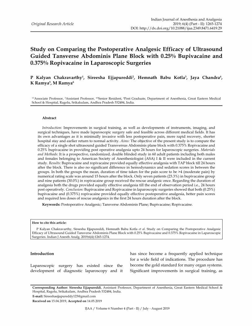
Fig���:�Comparison�of�Mean�Percentage�oxygen�saturation�among�both�the�groups�at�various�intervals
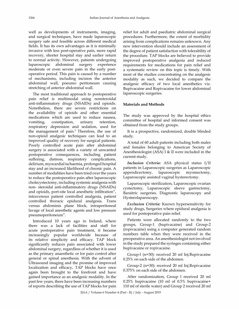
Fig���:�Pain�score�and�sedation�score
Group�1
Group�2
99.9
99.8
99.7
99.6
99.5
99.4
99.3
99.2
99.1
99
98.9�aseline At�15�minutes At�30�minutes At�45�minutes At�1�hour At�2�hour
There� were� no� signi��cant� differences� between�the� two� groups� with� respect� to� age,� weight� and�gender�(Table�1).
There�was�no�signi��cant�differences�in�observed�physiological� variables� of� heart� rate,� mean� blood�pressure� and� percentage� oxygen� saturation� in�between� the� groups� at� various� time� intervals�(Fig.�2).
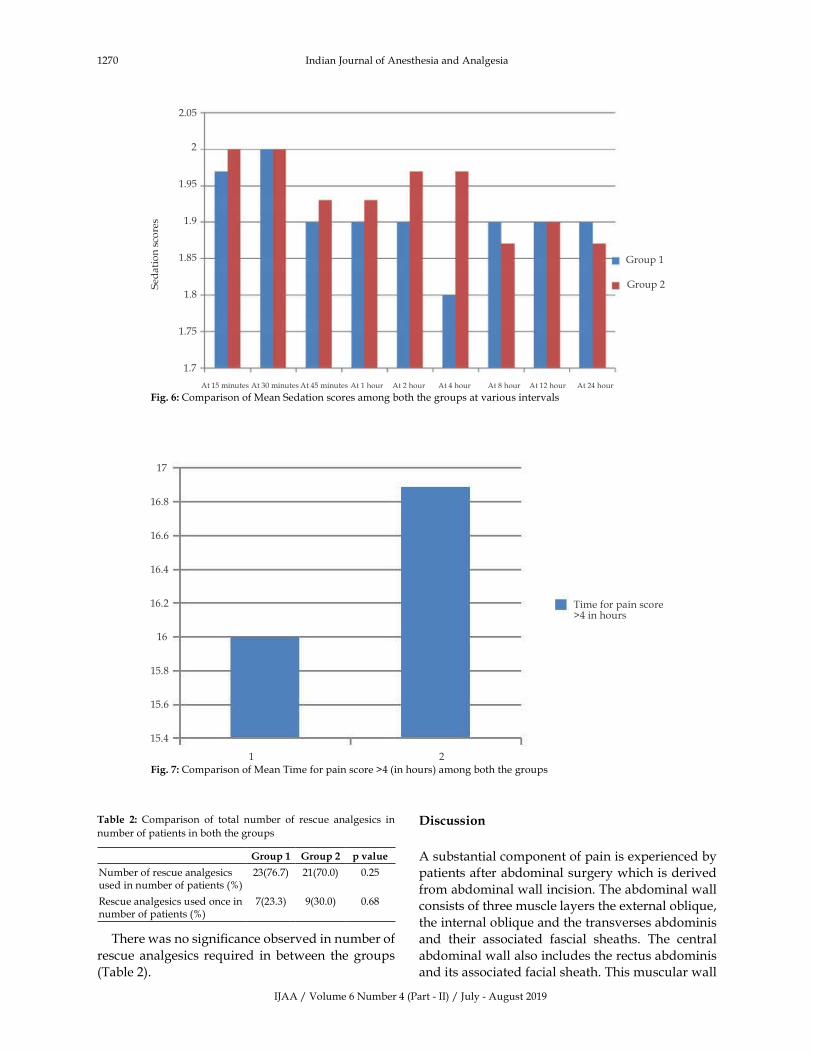
There� was� no� signi��cance� observed� in� pain�scores� or� sedation� scores� monitored� at� different�time�intervals�in�between�the�groups�(Fig.�5).
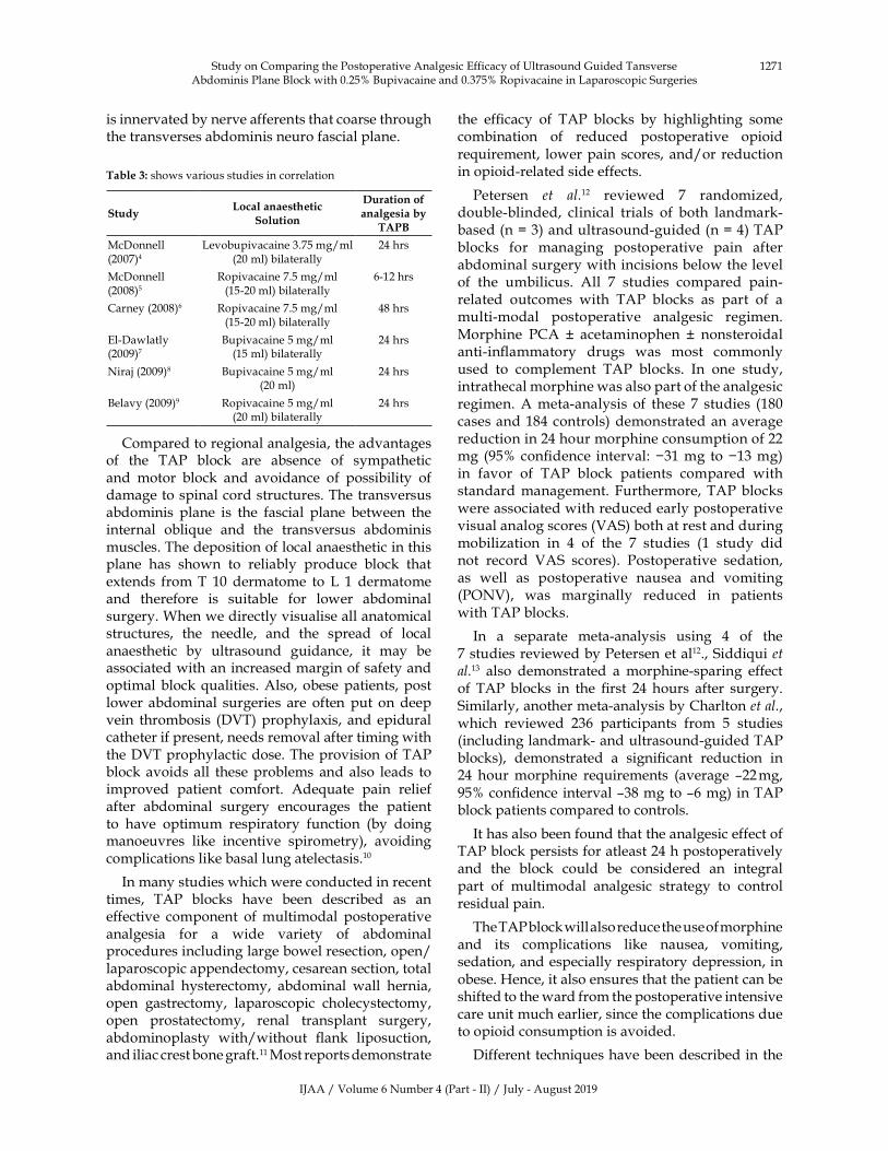
There� was� no� signi��cance� observed� in� total�duration�of�analgesia�time�for�pain�score��4�(Fig.�7).
Study�on�Comparing�the�Postoperative�Analgesic�Efficacy�of��ltrasound�Guided�Tansverse�Abdominis�Plane��lock�with�0.25���upivacaine�and�0.375��Ropivacaine�in�Laparoscopic�Surgeries
IJAA�/�Volume�6�Number�4�(Part�-�II)�/�July�-�August�2019
1270 Indian�Journal�of�Anesthesia�and�Analgesia
Group�1
Group�2
2.05
2
1.95
1.9
1.85
1.8
1.75
1.7
At�15�minutes�At�30�minutes�At�45�minutes�At�1�hour� At�2�hour� At�4�hour� At�8�hour� At�12�hour� At�24�hour�
Fig���:�Comparison�of�Mean�Sedation�scores�among�both�the�groups�at�various�intervals
Time�for�pain�score�4�in�hours
1 2
17
16.8
16.6
16.4
16.2
16
15.8
15.6
15.4
Fig���:�Comparison�of�Mean�Time�for�pain�score��4�(in�hours)�among�both�the�groups
Table� �:� Comparison� of� total� number� of� rescue� analgesics� in�
number�of�patients�in�both�the�groups
�roup�� �roup�� p��alue
Number�of�rescue�analgesics�used�in�number�of�patients�(�)
23(76.7) 21(70.0) 0.25
Rescue�analgesics�used�once�in�number�of�patients�(�)
7(23.3) 9(30.0) 0.68
There�was�no�signi��cance�observed�in�number�of�
rescue� analgesics� re�uired� in� between� the� groups�
(Table�2).
�iscussion
A�substantial�component�of�pain�is�experienced�by�
patients�after�abdominal�surgery�which�is�derived�
from�abdominal�wall�incision.�The�abdominal�wall�
consists�of�three�muscle�layers�the�external�obli�ue,�
the�internal�obli�ue�and�the�transverses�abdominis�
and� their� associated� fascial� sheaths.� The� central�
abdominal�wall�also�includes�the�rectus�abdominis�
and�its�associated�facial�sheath.�This�muscular�wall�
IJAA�/�Volume�6�Number�4�(Part�-�II)�/�July�-�August�2019
1271
is�innervated�by�nerve�afferents�that�coarse�through�the�transverses�abdominis�neuro�fascial�plane.
Table��:�shows�various�studies�in�correlation�
Study�ocal�anaesthetic
Solution
�uration�ofanalgesia�by�
TAPB
McDonnell�(2007)4
Levobupivacaine�3.75�mg/ml(20�ml)�bilaterally
24�hrs
McDonnell�(2008)5
Ropivacaine�7.5�mg/ml(15-20�ml)�bilaterally
6-12�hrs
Carney�(2008)6 Ropivacaine�7.5�mg/ml(15-20�ml)�bilaterally
48�hrs
El-Dawlatly�(2009)7
�upivacaine�5�mg/ml(15�ml)�bilaterally
24�hrs
Niraj�(2009)8 �upivacaine�5�mg/ml(20�ml)
24�hrs
�elavy�(2009)9 Ropivacaine�5�mg/ml(20�ml)�bilaterally
24�hrs
Compared�to�regional�analgesia,�the�advantages�of� the� TAP� block� are� absence� of� sympathetic�and� motor� block� and� avoidance� of� possibility� of�damage�to�spinal�cord�structures.�The�transversus�abdominis� plane� is� the� fascial� plane� between� the�internal� obli�ue� and� the� transversus� abdominis�muscles.�The�deposition�of�local�anaesthetic�in�this�plane� has� shown� to� reliably� produce� block� that�extends� from� T� 10� dermatome� to� L� 1� dermatome�and� therefore� is� suitable� for� lower� abdominal�surgery.�When�we�directly�visualise�all�anatomical�structures,� the� needle,� and� the� spread� of� local�anaesthetic� by� ultrasound� guidance,� it� may� be�associated�with�an�increased�margin�of�safety�and�optimal�block��ualities.�Also,�obese�patients,�post�lower� abdominal� surgeries� are� often� put� on�deep�vein� thrombosis� (DVT)�prophylaxis,� and� epidural�catheter�if�present,�needs�removal�after�timing�with�the�DVT�prophylactic�dose.�The�provision�of�TAP�block�avoids�all� these�problems� and� also� leads� to�improved� patient� comfort.� Ade�uate� pain� relief�after� abdominal� surgery� encourages� the� patient�to� have� optimum� respiratory� function� (by� doing�manoeuvres� like� incentive� spirometry),� avoiding�complications�like�basal�lung�atelectasis.10
In�many�studies�which�were�conducted�in�recent�times,� TAP� blocks� have� been� described� as� an�effective� component� of� multimodal� postoperative�analgesia� for� a� wide� variety� of� abdominal�procedures�including�large�bowel�resection,�open/laparoscopic�appendectomy,�cesarean�section,�total�abdominal� hysterectomy,� abdominal� wall� hernia,�open� gastrectomy,� laparoscopic� cholecystectomy,�open� prostatectomy,� renal� transplant� surgery,�abdominoplasty� with/without� ��ank� liposuction,�and�iliac�crest�bone�graft.11�Most�reports�demonstrate�
the� ef��cacy� of� TAP� blocks� by� highlighting� some�combination� of� reduced� postoperative� opioid�re�uirement,� lower�pain� scores,� and/or� reduction�in�opioid-related�side�effects.
Petersen� et� al.12� reviewed� 7� randomized,�double-blinded,� clinical� trials� of� both� landmark-based�(n���3)�and�ultrasound-guided�(n���4)�TAP�blocks� for� managing� postoperative� pain� after�abdominal�surgery�with�incisions�below�the�level�of� the� umbilicus.� All� 7� studies� compared� pain-related� outcomes� with� TAP� blocks� as� part� of� a�multi-modal� postoperative� analgesic� regimen.�Morphine� PCA� �� acetaminophen� �� nonsteroidal�anti-in��ammatory� drugs� was� most� commonly�used� to� complement� TAP� blocks.� In� one� study,�intrathecal�morphine�was�also�part�of�the�analgesic�regimen.�A�meta-analysis� of� these� 7� studies� (180�cases�and�184�controls)�demonstrated�an�average�reduction�in�24�hour�morphine�consumption�of�22�mg� (95��con��dence� interval:��31�mg� to��13�mg)�in� favor� of� TAP� block� patients� compared� with�standard�management.� Furthermore,�TAP�blocks�were�associated�with�reduced�early�postoperative�visual�analog�scores�(VAS)�both�at�rest�and�during�mobilization� in� 4� of� the� 7� studies� (1� study� did�not� record� VAS� scores).� Postoperative� sedation,�as� well� as� postoperative� nausea� and� vomiting�(PONV),� was� marginally� reduced� in� patients�with�TAP�blocks.
In� a� separate� meta-analysis� using� 4� of� the�7�studies�reviewed�by�Petersen�et�al12.,�Siddi�ui�et�al.13� also� demonstrated� a� morphine-sparing� effect�of� TAP� blocks� in� the� ��rst� 24� hours� after� surgery.�Similarly,�another�meta-analysis�by�Charlton�et�al.,�which� reviewed� 236� participants� from� 5� studies�(including�landmark-�and�ultrasound-guided�TAP�blocks),� demonstrated� a� signi��cant� reduction� in�24� hour� morphine� re�uirements� (average� –22�mg,�95��con��dence�interval�–38�mg�to�–6�mg)�in�TAP�block�patients�compared�to�controls.
It�has�also�been�found�that�the�analgesic�effect�of�TAP�block�persists�for�atleast�24�h�postoperatively�and� the� block� could� be� considered� an� integral�part� of� multimodal� analgesic� strategy� to� control�residual�pain.
The�TAP�block�will�also�reduce�the�use�of�morphine�and� its� complications� like� nausea,� vomiting,�sedation,�and�especially�respiratory�depression,�in�obese.��ence,�it�also�ensures�that�the�patient�can�be�shifted�to�the�ward�from�the�postoperative�intensive�care�unit�much�earlier,�since�the�complications�due�to�opioid�consumption�is�avoided.
Different�techni�ues�have�been�described�in�the�
Study�on�Comparing�the�Postoperative�Analgesic�Efficacy�of��ltrasound�Guided�Tansverse�Abdominis�Plane��lock�with�0.25���upivacaine�and�0.375��Ropivacaine�in�Laparoscopic�Surgeries
IJAA�/�Volume�6�Number�4�(Part�-�II)�/�July�-�August�2019
1272 Indian�Journal�of�Anesthesia�and�Analgesia
performance�of�transverses�abdominis�plane�(TAP)�block.� In� current� study� we� used� the� ultrasound�guided� approach,� the� needle� was� inserted� in� the�lumbar� triangle� of� Petit� to� reach� the� transverses�abdominis�plane.�Although�we�did�not�encounter�any� complications� during� TAP� blocks� procedure,�the� true� incidence� of� complications� such� as�systemic� toxicity,� vascular� or�visceral� injury�were�still� unknown.� It� is� conceivable� that� the� needle�visualization� might� reduce� the� incidence� or�potential�for�complications,�but�studies�con��rming�this�statement�is�lacking.
In� current� study� we� compared� the� analgesic�ef��cacy�of��upivacaine�0.25�� (total�dose�100�mg)�with�Ropivacaine�0.375��(total�dose�of�150�mg)�in�TAP� block� for� patients� undergoing� laparoscopic�surgeries.� In� our� study� both.� �upivacaine� and�ropivacaine� provided� e�ually� effective� analgesia�with�TAP�block�till�24�hours�after�the�block.�There�is�also�no�signi��cant�difference�in�hemodynamics�and�sedation�scores�in�between�the�groups.�In�both�the�groups�the�mean,�duration�of�time�taken�for�the�pain�score�to�be��4�(moderate�pain)�by�numerical�rating�scale� was� around� 15� hours� after� the� block.� Only�seven� patients� (23.3�)� in� bupivacaine� group� and�nine�patients�(30.0�)�in�ropivacaine�group�received�the�rescue�analgesic�once.�Regarding�the�duration�of� analgesia� both� the� drugs� provided� e�ually�effective�analgesia�till�the�end�of�observation�period�i.e�24�hours�post�operatively.
In�a� study�by�McDonnell�Gerald�et� al.�compared�ropivacaine� 0.� 75�� with� saline� in� TAP� block� for�caesarean�delivery�they�found�that�patients�who�have�undergone�TAP�block�with�ropivacaine�had�reduced�8�hour�morphine�re�uirement�and�a� longer� time�to���rst� patient� controlled�analgesia-morphine� re�uest.�This�study�supports�the�current�study�since�analgesia�from�TAP�block�is�superior�to�saline�group.4
Ra��� described� the� use� of� 20� mL� of� �a� local�anaesthetic� agent�� for� each� side� re�uiring�analgesia.14�Subse�uently,�McDonnell�et�al.�reported�the�use�of�20�mL�of�0.5��lidocaine�for�each�side�in�healthy�volunteers.4
Over� the� past� 3� year,� a� series� of� studies� have�highlighted� the� value� of� ef��cacy� of� various� local�anaesthetic�agents�in�Transversus�Abdominis�Plane�(TAP)� �lock,� after� the� initial� description� of� the�techni�ue�by�Ra��.14�Transversus�Abdominis�Plane��lock�as�described�by�Ra���involves�identifying�the�neurovascular�plane�of�the�abdominal�musculature�and�injecting�a� local�anaesthetic�agent� therein.��e�performed� abdominal� ��eld� block� via� the� lumbar�triangle�without�any�untoward�se�uelae.
With�the�techni�ue�of�ultrasound�guided�nerve�blockade� gaining� popularity,� this� techni�ue�was� also� applied� to� injection� of� bupivacaine� and�ropivacaine� in� the� TAP� block.� �owever� injection�via� Petit�s� triangle� using� double� POP� techni�ue�resulted�in�reliable�deposition�into�the�transversus�abdominis�plane.�Moreover� it�may�not� always�be�possible� to� use� ultrasound� guided� techni�ues� for�administering�TAP��lock�where�such�facilities�are�not�available.
The� landmark-based� techni�ue� for� the� TAP�block,� have� been� performed� without� dif��culty� in�the� children.15�Alternative�approaches� to� the�TAP�block�using�ultrasound�guidance�have�recently�been�described� in� a� case� series� of� children� undergoing�inguinal� hernia� repair.11� The� optimal� approach�remains�to�be�demonstrated.�There�are�now�a�variety�of�techni�ues�for�the�TAP�block�and�the�analgesic�merit�of�each�is�being�elucidated�in�ongoing�studies.�Although� it� is� possible� to� ultrasonically� visualize�the� 3� muscle� layers� of� the� abdominal� wall,� there�is�variation�in�these�muscle�layers�that�can�restrict�the�use�of�ultrasound�over�the�triangle�of�Petit.15�As�a�result,� the�needle�insertion�point�as�described�in�the�ultrasound�studies,�which�is�dependent�on�the�ade�uate�identi��cation�of�the�3�muscle�layers,�can�vary.�In�the�current�study,��SG�guided�TAP��lock�is� performed� following� the� induction� of� general�anesthesia.
This�will�alter�the�location�of�the�injectate�as�will�the� angle� of� the� needle� insertion� to� skin,� which�contrasts� to� the� landmark� approach�s� description.�Although� there� is� an� ever-increasing� access� to�ultrasound,� it� is� far� from� universal� and� there� is�a� continuing� interest� in� landmark� techni�ues.16�
Moreover�ultrasound�machine�may�not�be�available�at�all�places�especially�in�peripheral�health�centers�where� the� blind� techni�ue� alone� is� the� option� for�giving� TAP�.� 100�� success� rate� with� TAP� block�have�been�obtained�using� landmark�techni�ue�for�posterior�approach�of�block.14�To�our�knowledge�till�now�no�study�has�been�performed�to�compare�the�ef��cacy� of� landmark�versus�ultrasound� techni�ue�for�posterior�approach�of�TAP�block.
TAP� injection� of� local� anaesthetic� injection�cephalad� to� the� iliac� crest� likely� involves� T10–L1�nerve�roots�and�implies�that�the�techni�ue�may�be�limited�to�use�in�lower�abdominal�surgery.17
Sinha� A� et� al.� conducted� a� study� to� evaluate�ef��cacy� of� Ropivacaine� with� dexmeditomidine�versus� ropivacaine� with� plain� saline� which�concluded� that� addition� of� dexmeditomidine�to� local� anaesthetics� for� performing� TAP� �lock�has� provided� ade�uate� and� longer� duration� of�

IJAA�/�Volume�6�Number�4�(Part�-�II)�/�July�-�August�2019
1273
analgesia�and�supported�for�early�mobilization�by�providing� continuous� analgesia� post� operatively�and�also�supports�the�current�study.
Maitreyi� Gajanan� Mankikar,� Shalini� Pravin�Sardesai,� Poonam� Sachin� Ghodki� et� al.� evaluated�Sixty�patients�undergoing�caesarean�section�under�spinal�anesthesia�who�were�randomised�to�undergo�TAP�block�with�ropivacaine�(n���30)�versus�control�group� (n���30)�with�normal� saline,� in� addition� to�standard� analgesia�with� intravenous� paracetamol�and� tramadol.� This� study� concluded� that� Mean�re�uirement� of� tramadol� in� the� ��rst� 24� h� was�reduced� in��S� guided� TAP� block� after� caesarean�section�which�supports�the�current�study.18
In� addition,� a� growing� number� of� reports�suggest� that� TAP� blocks� may� also� be� a� safe�alternative� to� neuraxial� blockade� in� patients�who� are� anti-coagulated,� coagulopathic,� or� in�patients�who�would�not�tolerate�the�hemodynamic�se�uelae�often�associated�with�profound�neuraxial�sympathectomy.
The�TAP�block�is�an�effective�and�safe�adjunct�to�multimodal�postoperative� analgesia� for� abdominal�surgery.� Multiple� studies� have� demonstrated� its�superiority� over� standard� medical� therapy� for�postoperative� pain� control.� Limited� data� also�suggest� that� in� select� patient� populations,� TAP�blocks/catheters�may�provide�comparable�analgesia�as�well� as� patient� satisfaction� to� epidural� therapy.��owever,� the�data� is� less� encouraging� for�patients�who�receive�intrathecal�morphine�during�C-section,�where�the�addition�of�TAP�blocks�does�not�appear�to�improve�postoperative�pain�control.�Nonetheless,�it�may�be�a�good�alternative�strategy�for�patients�who�are�highly�sensitive�to�opioids.��ence,�current�study�was� conducted� using� plain� local� anaesthetics� for�performing�TAP��lock�for�various�surgeries.
D.� �elavy,� P.J.� Cowlishaw,� M.� �owes� et� al.�evaluated� the� analgesic� ef��cacy� of� TAP� block�in� patients� undergoing� caesarean� delivery� and�concluded�that�the��SG�guided�TAP�block�reduces�morphine� re�uirements� after� caesarean� delivery�when�used�as�a�component�of�a�multimodal�analgesic�regimen�which�supports�the�current�study.9
The� bene��t� of� ade�uate� postoperative�analgesia� are� clear� and� include� a� reduction� in�the� postoperative� stress� response,� reduction� in�postoperative� morbidity,� and� in� certain� types� of�surgery,�improved�surgical�outcome.�Effective�pain�control�also�facilitates�rehabilitation�and�accelerates�recovery� from� surgery.�Other� bene��ts� of� effective�regional�analgesic�techni�ues�include�reduced�pain�intensity,�decreased� incidence�of�side� effects� from�analgesics�and�improved�patient�comfort.
In� our� experience,� the� TAP� block� has� a� fast�learning� curve� and� re�uires� short� performance�time�especially�by�an�experienced�anesthesiologist.��owever� it� is� possible� that�different� providers� in�different�clinical�circumstances�may���nd�obstacles�to� the� routine� implementation� of� a� TAP� block� as�part� of� a� multimodal� pain� strategy� to� improve�postoperative��uality�of�recovery.
Conclusion
In� our� experience,� the� TAP� block� has� a� fast�learning� curve� and� re�uires� short� performance�time�especially�by�an�experienced�anesthesiologist.��owever�it�is�possible�that�different�s�providers�in�different�clinical�circumstances�may���nd�obstacles�to� the� routine� implementation� of� a� TAP� block� as�part� of� a� multimodal� pain� strategy� to� improve�postoperative��uality�of�recovery.
Our� current� study� which� is� a� prospective�randomized�double� blinded� study� comparing� the�postoperative� analgesic� ef��cacy� of� ultrasound�guided� transverses� abdominis� plane� block� with�bupivacaine� and� ropivacaine� in� laparoscopic�surgeries� showed� that� both� (0.25�)� bupivacaine�and� (0.375�)� ropivacaine� provided� e�ually�effective�postoperative�analgesia,�better�pain�scores�and�re�uired�less�doses�of�rescue�analgesics�in�the���rst�24�hours�duration�after�the�block.
�eferences
1.� Reynolds� W� Jr.� The� first� laparoscopic�cholecystectomy.�JSLS.�2001�Jan-Mar;5(1):89–94.
2.� Petersen�PL,�Stjernholm�P,��ristiansen�V�,� et� al.�The� beneficial� effect� of� transverses� abdominis�plane� block� after� laparoscopic� cholecystectomy�in�day-case�surgery:�A�randomized�clinical� trial.�Anesthesia�Analgesia�2012;115:527–33.
3.� Ramsay�M,�Savege�T,�Simpson��,�et�al.�Controlled�sedation�with�Alphaxalone-�Alphadolone.��ritish�Medical�Journal,�1974,656–9.
–
5.� McDonnell� JG,� Curley� G,� Carney� J,� et� al.� The�analgesic� efficacy� of� transversus� abdominis�plane� block� after� cesarean� delivery:� a�randomized�controlled� trial.�Anesth�Analg.�2008�Jan;106(1):186–91.
6.� Carney� J,� Finnerty� O,� Rauf� J,� et� al.� Ipsilateral�Transversus� Abdominis� Plane� �lock� Provides�
Study�on�Comparing�the�Postoperative�Analgesic�Efficacy�of��ltrasound�Guided�Tansverse�Abdominis�Plane��lock�with�0.25���upivacaine�and�0.375��Ropivacaine�in�Laparoscopic�Surgeries
IJAA�/�Volume�6�Number�4�(Part�-�II)�/�July�-�August�2019
1274 Indian�Journal�of�Anesthesia�and�Analgesia
Effective� Analgesia� After� Appendectomy� in�Children:�A�Randomized�Controlled�Trial.�Anesth�Analg.�2010�Oct;111(4):998–1003.
7.� El-Dawlatly� A,� Turkistani� SC,� �ettner� A� et� al.��ltrasound-guided� transversus� abdominis�plane�block:�description�of�a�new�techni�ue�and�comparison�with�conventional�systemic�analgesia�during� laparoscopic� cholecystectomy.� �ritish�Journal�of�Anesthesia,�2009;102,6,1:763–767.
8.� Niraj� G,� Searle� A,� Mathews� M,� et� al.� Analgesic�efficacy� of� ultrasound-guided� transversus�abdominis� plane� block� in� patients� undergoing�open� appendicectomy.� �r� J� Anaesth.� 2009�Oct;103(4):601–5.
9.� �elavy� D,� Cowlishaw� PJ,� �owes� M,� et� al.��ltrasound-guided�transversus�abdominis�plane�block�for�analgesia�after�Caesarean�delivery,��JA:��ritish�Journal�of�Anesthesia,�2009;103(5):726–730.
10.� Chaudhuri� S� and� Goyal� SS.� �ltrasound-guided� transversus� abdominis� plane� block:�A� technically� easier� analgesic� option� in� obese�compared� to� epidural.� Anesth� Essays� Res.� 2012�Jul-Dec;6(2):226–228.
11.� Carney� J,� Finnerty� O,� Rauf� J,� et� al.� Ipsilateral�Transversus� Abdominis� Plane� �lock� Provides�Effective� Analgesia� After� Appendectomy� in�Children:�A�Randomized�Controlled�Trial.�Anesth�Analg�October�2010;111(4):998–1003.
12.� Petersen� PL,� Mathiesen� O,� Torup� �,� et� al.�The� transversus� abdominis� plane� block:� a�
valuable� option� for� postoperative� analgesia�� A�
topical� review.� Acta� Anaesthesiol� Scand.� 2010�
May;54(5):529–35.
Fujiwara� Shibata� et� al
guided�
plane� � ish� of�
2010;105:853–6.
–
15.� Loukas� M,� Tubbs� RS,� El-Sedfy� A,� et� al.� The�
clinical� anatomy� of� the� triangle� of� Petit.� �ernia�
2007;11:441–4.
16.� �erai�S,�Saxena��N,�Anand�R,�et�al.�Comparative�
evaluation�of�transversus�abdominis�plane�block�
with� transcutaneous� electrical� nerve� stimulation�
for� postoperative� analgesia� following� lower�
segment�caesarean�section.�J�Obstet�Anaesth�Crit�
Care.�2011;1:30–4.
17.� Tran� TMN,� Ivanusic� JJ,� �ebbard� P,� et� al.�
Determination� of� spread� of� injectate� after�
ultrasound-guided� transversus� abdominis� plane�
block:� a� cadaveric� study.��r� J�Anaesth� Jan� 2009;�
102(1):123–127.
18.� Mankikar� MG,� Sardesai� SP,� Ghodki� PS.�
�ltrasound-guided�transversus�abdominis�plane�
block� for� post-operative� analgesia� in� patients�
undergoing� caesarean� section.� Indian� J� Anaesth�
2016;60:253–7.
��Red�Flower�Publication�Pvt.�Ltd.�
�Original�Research�ArticleIndian�Journal�of�Anesthesia�and�Analgesia�
2019;�6(4)�(Part�-�II):�1275-1281DOI:�http://dx.doi.org/10.21088/ijaa.2349.8471.6419.30
A�Comparati�e�Study�of�Caudal�Analgesia��ith�Bupi�acaine�Alone�and�Bupi�acaine��ith�Butorphanol�in�Pediatric�Surgeries
Shaik�Salman���C��eetha�
1Registar,� Aware� Global� �ospital,� �yderabad,� Telangana� 500035,� India.� 2Associate� Professor,� Department� of� Anaesthesiology,�Gandhi�Medical�College,�Secunderabad,�Telangana�500003,�India.
Corresponding� Author:� C� �eetha,� Assistant� Professor,� Associate� Professor,� Department� of� Anaesthesiology,� Gandhi� Medical�College,�Secunderabad,�Telangana�500003,�India.
E�mail:�[email protected]
�ecei�ed�on�23.05.2019,�Accepted�on�11.07.2019
Abstract
Introduction:�The�assessment�of�pain�in�small�children�is�often�difficult� to� interpret�as�the�most�common�sign�of�pain�is�crying�which�is�also�seen�in�a�myriad�of�non�pain�full�conditions.�Epidural�space�in�children�favours�rapid�longitudinal�spread�of�drugs�and�makes�it�effective�in�treating�postoperative�pain.�Aim:�The�aim�of�this�study�was�to�evaluate�the�efficacy�of�caudal�bupivacaine�alone�or� in�combination�with�butorphanol�for�postoperative�analgesia�in�children�undergoing�infra-umbilical�surgeries.�Materials�and�Methods:�A�Simple�Randamoized�which�includes�50�patients�posted�for�urogenital�operations�such�as�herniotomy,�orchidopexy,�urethroplasty� and� CTEV� Correction� divided� into� two� groups� of� 25� each.� Group� �� received� 0.25�� plain�bupivacaine�and�Group����received�0.25���upivacaine�with��utorphanol�adjuvant.�The�effect�of� recovery�from� caudal� blockade� and� duration� of� analgesia� was� compared� and� contrasted.� Results:� There� were� no�significant�changes�in�heart�rate,�blood�pressure�and�oxygen�saturation�between�two�groups.�Postoperative�pain�score�was�comparable�in�two�groups�in�first�eight�hours,�but�it�is�significantly�less�in�bupivacaine�with�butorphanol�group�which�is�statistically�significant.�There�is�a�significant�difference�between�the�groups�in�the�mean�duration�of�analgesia�with�Group����having�a�much�longer�duration�compared�to�Group��.�3�patient�in�Group���and�5�patient�in�Group����had�nausea�in�postoperative�period,�which�is�statistically�insignificant�(p�0.05).�No�episodes�of�any�other�clinically�significant�postoperative�complications�were�recorded.�Conclusion:��utorphanol�is�considered�to�be�a�safe�and�effective�adjuvant�to��upivacaine�for�caudal�analgesia�in�children�undergoing�surgery�below�umbilicus.
�ey�ords:��upivacaine;��utorphanol;�Pediatric�Surgeries
�o��to�cite�this�article:
Shaik�Salman,�C�Geetha.�A�Comparative�Study�of�Caudal�Analgesia�with��upivacaine�Alone�and��upivacaine�with��utorphanol�in�Pediatric�Surgeries.�Indian�J�Anesth�Analg.�2019;6(4):1275-1281.
Introduction
Pain,� as� de��ned� by� international� association�for� study� of� pain,� is� an� unpleasant� sensory� and�emotional� experience� associated� with� actual� or�potential� tissue� damage� or� described� in� terms� of�
such� damage.� The�mechanism� of�pain� perception�in�pediatric1�population�is�different�and�is�complex�and� is� not� often� ade�uately� understood� rather�than� emphasizing� on� the� clinical� evaluation�alone;� biopsychosocial� perspective� needs� to� be�looked�deeply�while�managing�pain�in�this�special�

IJAA�/�Volume�6�Number�4�(Part�-�II)�/�July�-�August�2019
1276 Indian�Journal�of�Anesthesia�and�Analgesia
population.� The� assessment� of� pain� in� small�children� is� often�dif��cult� to� interpret� as� the�most�common�sign�of�pain�is�crying�which�is�also�seen�in�a�myriad�of�non�painful�conditions.�There�have�been�recent�developments�in�the�pediatric�post�operative�pain� management� with� emphasis� of� ade�uate�treatment�of�pain�early�to�prevent�morbidity�in�this�patient�population.�These�developments�are�highly�important�in�developing�nations�where�the�progress�of�anesthesia�specialty�has�been�non�uniform�and�at�a�varied�pace.
The�various�methods2,3�of�providing�pain�relief�have� some� side� effects� which� prohibit� their� use�in� children� for� eg.� narcotics,� because� of� their�respiratory� depression,� the� other� analgesics�which�cannot�be�given�for�some�time�after�general�anesthesia� due� to� the� fear� of� vomiting� and� a�spiration,� the� fear� of� the� needles� in� the� case� of�parentally� administered� analgesics.� The� regional�anesthetic� techni�ues� signi��cantly� decrease�post� operative� pain� and� systematic� analgesic�re�uirements.� Caudal� route� was� chosen� for�this� study� as� it� is� one� of� the� simplest� and� safest�techni�ues� in� pediatric� anesthesia� with� a� high�success� rate.� Epidural� space� in� children� favours�rapid� longitudinal� spread�of�drugs� and�makes� it�effective�in�treating�postoperative�pain.
Caudal� block� is� usually� done� after� the�introduction� of� general� anesthesia� an� disused� as�an� adjunct� to� intraoperative� anesthesia� as�well� as�postoperative� analgesia� in� children� undergoing�surgical�procedures�below�the�level�of�the�umbilicus.�Caudal�analgesia�can�reduce�the�amount�of�inhaled�and� IV� anesthetic� administration,� attenuates� the�stress� response� to� surgery� facilitates� a� rapid,�smooth� recovery,� and� provides� good� immediate�post� operative� analgesia1,3.� In� order� to� decrease�peri-operative� analgesic� re�uirements� after� single�shot� caudal� epidural� blockade,� various� additives,�such�as�morphine,�fentanyl,�clonidine�and�ketamine�with�local�anesthetics�have�been�investigated.
The�aim�of�this�study�was�to�evaluate�the�ef��cacy�of�caudal�bupivacaine�alone�or�in�combination�with�butorphanol�for�postoperative�analgesia�in�children�undergoing�infra-umbilical�surgeries.
Materials�and�Methods
The� present� study� is� Simple� Randamoized�study� at� Osmania� general� hospital� and� Niloufer�hospital,� between� August� 2016-September� 2017,�who� underwent� lower� abdominal� and� lower�limb� surgeries� after� obtaining� institutional� ethical�committee�and�parental�written�informed�consent.
Inclusion�Criteria:�Age�groups�1-10�years,�ASA�grade�I�and�II,�Cases�scheduled�for�operations�such�as� urethroplasty,� herniotomy,� orchidopexy� and�CTEV�correction.
Exclusion� criteria:� �/o� of� central� nervous�disease,�sacral�abnormalities,��/o�of�drug�allergy,��/o�of�bleeding�disorder�AND�skin�infection�at�the�site�of�block.
Patients�were� allocated� by� randomly� in� to� two�groups�of�25�patients�each.
Group���receive�0.25��plain��upivacaine�1�ml/kg�for�caudal�block.
Group����25�mcg/kg�added�to�0.25��bupivacaine�1�ml/kg.
In� all� children,� age,� body� weight,� and� baseline�vital� parameters�were� recorded.��istory� regarding�previous�anesthesia,�surgery,�any�signi��cant�medical�illness,� medications� and� allergy� was� recorded.�Complete�physical�examination,�airway�assessment�and�local�examination�of�lower�back�were�done.
�emoglobin�percentage,�bleeding�time,�clotting�time,�blood�sugar,�urea,�serum�creatinine�and�urine�analysis,��SG.�Patients�were�fasted�for�4�hours�and�pre� medicated� with� oral� Midazolam� 0.5� mg/kg�30�minutes�before�surgery.�After�applying�standard�monitors,� an� intravenous� cannula� was� secured�and� Isolyte-p� solution� was� infused� to� provide���uid� during� surgery.� Injection� Glycopyrrolate�0.01� mg/kg� was� administered� intravenously� as�premedicant.�General�anesthesia�was�induced�with�Thiopentone�sodium�5�mg/kg,�2��sevo��urane�and�Nitrous�oxide�in�oxygen�via�mask.
Endotracheal� intubation� was� facilitated� by�administering� injection� vecuronium� bromide�0.1� mg/kg� intravenously.� After� securing�Endotracheal� tube,� patients� were� placed� in� left�lateral�position.
Procedure
After� placing� lateral� position,� skin� of� the� back�over�the�sacrum�was�scrub�using�povidone�iodine�solution,��nder�aseptic�precautions,�a�short�beveled�22� G� needle� was� introduced� proper� position�of� needle� con��rmed� by� the� pop� sensed� during�penetration�of�sacro-coccygeal�membrane�of�caudal�epidural� space,� which� was� followed� by� whoosh�test�done�using�0.5�ml�of�air�after�needle�insertion�negative� aspiration� of� blood� and� cerebrospinal���uid,� then� 1� ml/kg� of� local� anaesthetic� agent�0.25�� bupivacaine� given� to� Group� �� and� 0.25���upivacaine� with� �utorphanol� adjuvant� to�Group����was�administered�slowly.
IJAA�/�Volume�6�Number�4�(Part�-�II)�/�July�-�August�2019
1277
After�deposition�of� the�drug� in� epidural� space,�patients� were� placed� in� supine� position� and�anesthesia�was�maintained�by�1��sevo��urane,�50��of�Nitrous�oxide�plus�50��oxygen�and�top�up�doses�of�vecuronium�bromide�(1/5th�of�the�loading�dose�of�0.1�mg/kg).
�R� and� blood� pressure� were� recorded� just�before� and� after� surgical� incision� and� then� every�5� min� interval� till� the� end� of� surgery,� residual�neuromuscular�blockade�was�reversed�and�patients�were�transferred�to�the�post�operative�ward.
�sing�the�paediatric�observations�FLACC�(face,�legs,� activity,� cry,� consolability)� pain� scale� with�its� 0-10� score� range,� each� patients� pain� intensity�was�assessed�at�the�end�of�surgery�and�then�every�30� min� interval� until� the� patient� became� ��t� to�discharge�from�postoperative�ward.
If� the� FLACC� pain� scale�was� 4� or�more,� rectal�Paracetomol�20�mg/kg�was�administered.
Observations� were� continued� for� 24� hours.Complications� such� as� postoperative� nausea� and�vomiting�(PONV),�respiratory�depression,�urinary�retention,� hypotension� and� bradycardia� were�also� noted.� Respiratory� depression� was� de��ned�as� a� decrease� in� SpO
2� of� less� than� 95�� re�uiring�
supplementary�oxygen.��ypotension�was�de��ned�as� fall� of� 20�� mean� arterial� pressure� from� base�line.� �radycardia� was� de��ned� as� �R� below�80�beats/min�for�age�1�year�and�60�beats/min�for�ages�above�1�year.�The�parameters�were�compared�in�two�groups�and�results�subjected�to�appropriate�statistical� analysis� are� �emodynamic� parameters�and��uality�of�postoperative�analgesia�effect.
Stastistical�Analysis
All�recorded�data�were�entered�using�MS�Excel�software� and� analysed� using� spss� 16� version�software� for� determining� statistical� signi��cance.�Numerical�variables�were�presented�as�mean�and�standard� deviation� (SD)� and� categorical� variables�were�presented�as�fre�uency�(�).
Student�s� t� test� was� used� for� between-group�comparisons�between�categorical�variables.
A� p� value� of� �0.05� was� taken� to� be� signi��cant�and� a� p� value� of� �0.001� was� considered� highly�signi��cant.
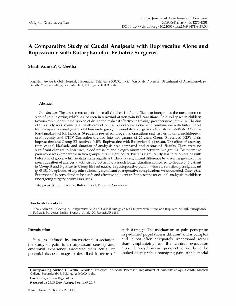
�esults
This� study� includes� 50� patients� posted� for�urogenital� operations� such� as� herniotomy,�orchidopexy,� urethroplasty� and�CTEV�Correction�divided� into� two� groups� of� 25� each.� Group� ��received� 0.25�� plain� bupivacaine� and� Group� ���received� 0.25�� �upivacaine� with� �utorphanol�adjuvant.� The� effect� of� recovery� from� caudal�blockade�and�duration�of�analgesia�was�compared�and�contrasted.
Table��:�Patient�characteristics�and�clinical�parameters
Patient�details �roup�B� �roup�BB
Age�(in�years)� 3.72� 3.70�
Weight�(in��g)� 12� 12�
Gender�M:F�Ratio� 23:02� 23:02�
Duration�of�Anesthsisa�(in�min)� 35� 31�
�aseline��eart�Rate�(�eats�per�min)� 106.8� 104�
�aseline�map� 72.9� 75�
Fig���:�Nature�of�Operations
A�Comparative�Study�of�Caudal�Analgesia�with��upivacaine�Alone�and��upivacaine�with��utorphanol�in�Pediatric�Surgeries
IJAA�/�Volume�6�Number�4�(Part�-�II)�/�July�-�August�2019
1278 Indian�Journal�of�Anesthesia�and�Analgesia
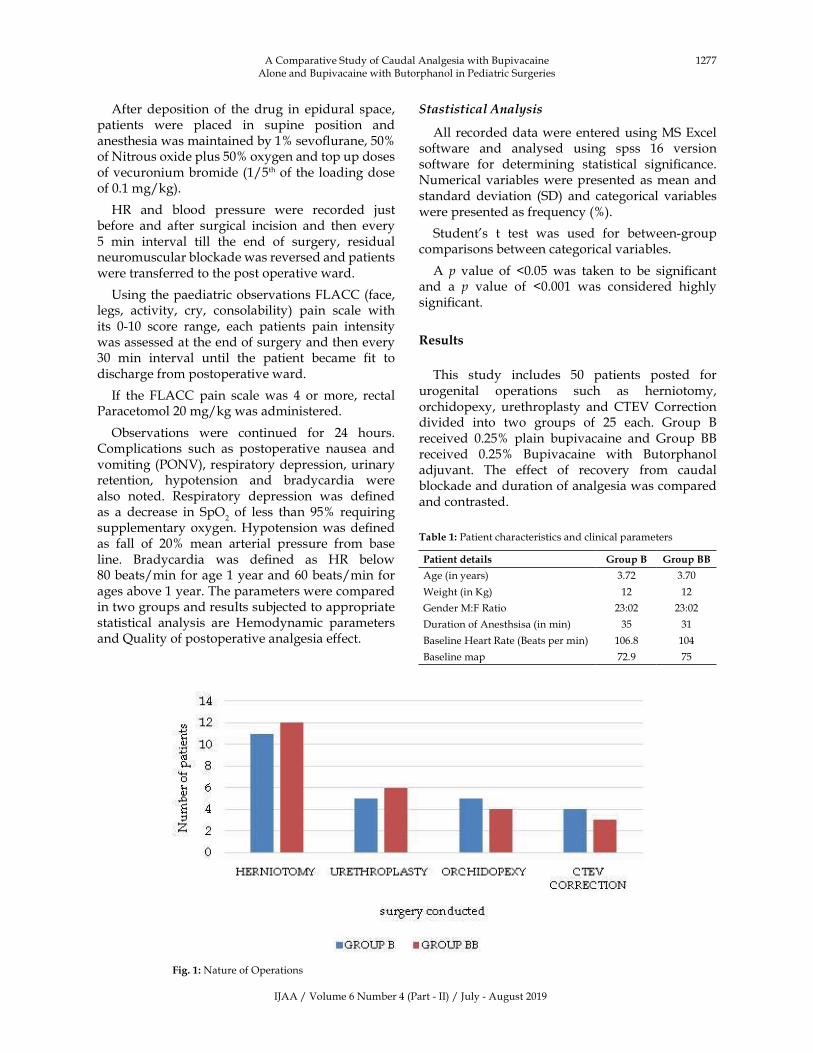
Table��:��aemodynamic�changes�during�surgery
�roup�B �roup�BBStandard�de�iation�
for�B�group�Standard�de�iation�
for�BB�group� ���alue�
��� MAP� ��� MAP� ��� MAP� ��� MAP� ��� MAP�
�ase�line� 104� 75� 106.80� 72.90� 8.3� 4.6� 8.1� 5.7� �0.10� �0.10�
After�incision�5�min� 91� 70� 97.7� 68.9� 7.7� 4.3� 8.3� 4.6� �0.05� �0.10�
10�min� 91.3� 70� 93.5� 68.1� 6.9� 4.9� 6.8� 5.1� �0.06� �0.07�
20�min� 89� 71� 91.2� 68.2� 6.48� 5.3� 6.01� 5.5� �0.07� �0.10�
30�min� 88� 71� 89.5� 68.2� 5.5� 5.8� 5.45� 5.7� �0.10� �0.10�
60�min� 85� 70� 86.5� 70� 5.3� 4.4� 5.25� 4.7� �0.06� �0.07�
90�min� 82� 72� 85� 68.1� 5.1� 5.9� 5.1� 5.2� �0.05� �0.10�
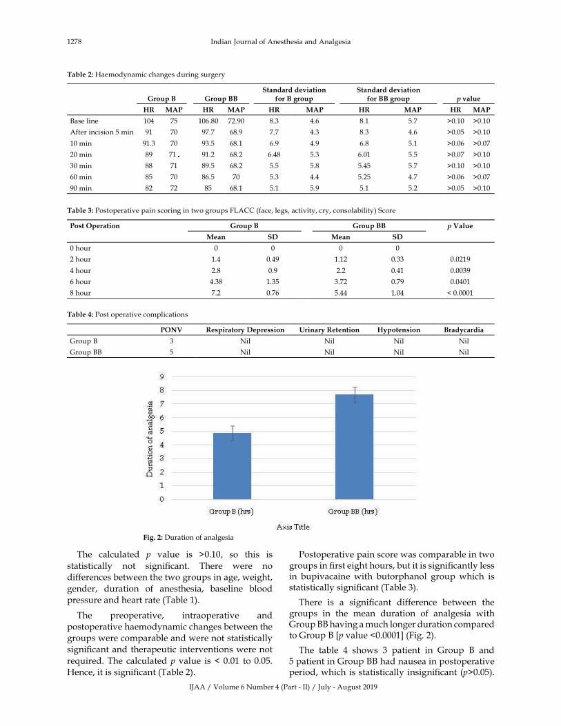
Table��:�Postoperative�pain�scoring�in�two�groups�FLACC�(face,�legs,�activity,�cry,�consolability)�Score
Post�Operation �roup�B� �roup�BB ��Value�
Mean� S�� Mean� S��
0�hour� 0� 0� 0� 0�
2�hour 1.4� 0.49� 1.12� 0.33� 0.0219�
4�hour 2.8� 0.9� 2.2� 0.41� 0.0039�
6�hour 4.38� 1.35� 3.72� 0.79� 0.0401�
8�hour 7.2� 0.76� 5.44� 1.04� ��0.0001�
Table��:�Post�operative�complications
PONV� �espiratory��epression� �rinary��etention� �ypotension� Bradycardia
Group��� 3� Nil� Nil� Nil� Nil�
Group���� 5� Nil� Nil� Nil� Nil�
Fig���:�Duration�of�analgesia
The� calculated� p� value� is� �0.10,� so� this� is�
statistically� not� signi��cant.� There� were� no�
differences�between�the�two�groups�in�age,�weight,�
gender,� duration� of� anesthesia,� baseline� blood�
pressure�and�heart�rate�(Table�1).
The� preoperative,� intraoperative� and�
postoperative�haemodynamic�changes�between�the�
groups�were�comparable�and�were�not�statistically�
signi��cant�and�therapeutic� interventions�were�not�
re�uired.�The� calculated�p� value� is� �� 0.01� to� 0.05.�
�ence,�it�is�signi��cant�(Table�2).
Postoperative�pain�score�was�comparable�in�two�groups�in���rst�eight�hours,�but�it�is�signi��cantly�less�in� bupivacaine� with� butorphanol� group�which� is�statistically�signi��cant�(Table�3).
There� is� a� signi��cant� difference� between� the�groups� in� the� mean� duration� of� analgesia� with�Group����having�a�much�longer�duration�compared�to�Group����p�value��0.0001��(Fig.�2).
The� table� 4� shows� 3� patient� in� Group� �� and�5�patient�in�Group����had�nausea�in�postoperative�period,�which� is�statistically�insigni��cant� (p�0.05).�
IJAA�/�Volume�6�Number�4�(Part�-�II)�/�July�-�August�2019
1279
No� episodes� of� any� other� clinically� signi��cant�postoperative�complications�were�recorded.
�iscussion
Over�the�recent�years,�the�concept�of�providing�ade�uate� postoperative� analgesia� in� pediatric�patients� is� well� established,� however,� various�methods�showed�side-effects�limiting�their�use�such�as� respiratory�depression�with� IV�opioids.�With�a�high�success�rate,�caudal�analgesia�was�proved� to�be�a�simple�and�effective�techni�ue�in�children.
Caudal� epidural� analgesia� is� one� of� the� most�popular�and�commonly�performed�regional�blocks�in� pediatric� anesthesia.� It� is� a� reliable� and� safe�techni�ue�that�can�be�used�with�general�anesthesia�for� intra� and� postoperative� analgesia� in� patients�undergoing� abdominal� and� lower� limbsurgeries.The� main� disadvantage� of� caudal� anesthesia� is�the�short�duration�of�action�after�a�single�injection�of� local� anesthetic� solution.� Speci��c� character� of�caudal�block�in�pediatric�age�group.
Increased� ��uidity� of� epidural� fat� Increased�diffusion� of� local� anesthetic� up� to� 6-7� �ear� of�age.Excellent� blockade� after� caudal� anesthesia�can� be� achieved� up� to� 6-7� �ear� of� age.� The�volume� prescription� scheme� of� Armit� age� that�was� published� many� years� ago� still� remains�the� most� dependable,� asfollows:� 0.5� mL/kg:�All� sacral� dermatomes� are� blocked.� The� upper�limit� of� anesthesia� is� at� least�Midthoracic.�When�1.25� mL/kg� is� injected,� excessive� rostral� spread�(above�T4)�canoccuritis�therefore�preferable�not�to�administer�more�than�1�mL/kg�of�local�anesthetic.�In� present� study� both� patients� receive� 0.25��bupivacaine� 1� ml/kg� as� per� armitage� formula.�An� ideal� combination� of� local� anesthetic� and�adjuvant�should�provide�ade�uate�intraoperative�anesthesia,�good�extended�postoperative�analgesia�without� prolonging� the� motor� blockade� or�producing� adverse� hemodynamic� or� respiratory�conse�uences.
Different� additives� have� been� used� in� order�to� improve� the� duration� of� action� as� well� as� the��uality� of� analgesia� of� the� local� anesthetic� used�in� the� single� shot� caudal� block� techni�ue� such� as�opioids,� epinephrine,� clonidine,� ketamine� and�neostigmine.� The� aim� of� this� randomized� control�study�was�to�compare�the�duration�of�postoperative�analgesia,�sedation,�as�well�as�the�incidence�of�any�side�effect�of�caudally�administered�butorphanol�to�bupivacaine�in�pediatric�patients�undergoing�lower�abdominal�and�lower�limb�surgeries.
There�has�been�a�study�by�Lawhorn�CD,�Stoner�JM,�Schmitz�ML,��rown�RE�Jr,�Stewart�FW,�Volpe�P,� Shirey� R4� in� the� literature� of� butorphanol�use� for� caudal� anesthesia/analgesia� in� pediatric�population� undergoing� genitourinary� procedure.�It�was�found�that�re�uirement�of�rescue�analgesia�in�post� anesthesia� care�unit� and� total� numbers� of�morphine� doses� administered� were� signi��cantly�less� in� patients� in� whom� butorphanol� 30� �g/� kg�was� added� to� bupivacaine� in� caudal� epidural�analgesia.�Our�study�s���ndings�are�consistent�with�their���ndings�but�the�differences�from�the�present�study� were:� they� had� used� 0.25�� bupivacaine�with� 1:200,000� epinephrine� and� caudal� epidural�analgesia�along�with�general�anesthesia.
In� another� study,� by� Ohta� �,� �atsuno� M,��awana�S,�Namiki�A5�butorphanol�has� also�been�used� in� patients� of� cerebral� palsy� undergoing�elective�orthopedic�operations�and�it�was�found�to�be�safe�and�useful�for�postoperative�pain�control�in�children.
One� of� the� interesting� ��ndings� of� the� present�study�is�the�paucity�of�side�effects�associated�with�caudal�butorphanol�as�mentioned�in�the�literature.�Its�high�lipid�solubility�and�high�af��nity�for�opioid�receptors� are� additional� factors� that� contribute� to�the�paucity�of�side�effects�with�its�use.
�igh� lipid� solubility� increases� diffusion� in�the� spinal� cord� and� limits� the� amount� of� drugs�remaining� in� the� CSF,� capable� of� reaching� the�brainstem� where� side� effects� are� detected.� In� a�recent� trial� it� has� been� demonstrated� that� there�were� less� chances� of� complication� or� side� effects�with� caudal� analgesia� as� compared� to� parenteral�use� of� analgesics� or� penile� block� in� patients� for�circumcision.
For�the�pediatric�caudal�epidural�analgesia,�other�opioids� like� morphine,� buprenorphine,� fentanyl�and�tramadol�have�been�used.�In�a�study�by�Gaitini�LA,�Somri�M,�Vaida�SJ,��anovski��,�Mogilner�G�et�al.�added�fentanyl�to�bupivacaine�in�caudal�epidural�block�to�observe�changes�in�plasma�catecholamine�levels�in�postoperative�period.�Addition�of�fentanyl�citrate� to� bupivacaine� in� caudal� epidural� block� in�children� did� not� in��uence� the� stress� response� to�surgery,�nor�did�it�improve�the�analgesic�intensity�of�the�caudal�block�as�described�in�literature.6
Drug�like�clonidine�has�also�been�used�in�a�study�by�Lee�JJ,�Rubin�AP�but�its�use�in�caudal�analgesia�resulted� in�signi��cant�prolongation�of�duration�of�postoperative� analgesia,�with� the� side� effects� like�bradycardia�and�urinary�retention.7�Veena�Chatrath,�Sarabjit� �aur.� compared� ef��cacy� of� caudally�
A�Comparative�Study�of�Caudal�Analgesia�with��upivacaine�Alone�and��upivacaine�with��utorphanol�in�Pediatric�Surgeries
IJAA�/�Volume�6�Number�4�(Part�-�II)�/�July�-�August�2019
1280 Indian�Journal�of�Anesthesia�and�Analgesia
administered� clonidine� with� that� of� butorphanol,�the� mean� duration� of� analgesia� was� statistically�longer� (p� value��� 0.01)� in� the� group��utorphanol�(822.0���217.41�min)�than�Clonidine�group�(745.4���216.69�min).�The�total�number�of��rescue��analgesic�doses�re�uired�in�the���rst�24�hrs�was�lesser�in�group���(0.80���0.41)�and�C�(0.96���0.45).8
Caudal�epidural�use�of�morphine,�buprenorphine�or� butorphanol� in� a� study� by� Ohta� �,� �atsuno�M,� �awana� S,� Namiki� A.� did� not� increase� the�fre�uency�of�side�effects�such�as�nausea,�vomiting�etc.,�and�need�for�rescue�analgesia�was�also�less�in�these�patients�within�24�hours�after�operation.5
The�use�of�tramadol�in�combination�with�0.25��bupivacaine� resulted� in� a� signi��cant� increase� in�the�analgesia�time�in�study�by�Senel�AC,�Akyol�A,�Dohman�D,� Solak�M.� In� a� study� by� R.� Pauranik,�P.� Gupta.,� added� ketamine� 5� mg/kg� caudally�and� compared� it� with� butorphanol� 20� �g/kg.�The� study� concluded� the� butorphanol� provided�superior�analgesia�for�a�longer�duration�than�with�ketamine.10
In� our� study,� a� total� of� 50� patients� in� the� age�group�of�2-8�years�were�divided�randomly� in�two�groups�(n�25).�There�were�no�differences�between�two� groups� with� regard� to� demographic� pro��le.Mean�age�in�group���(receiving�bupivacaine�alone)�was� 3.72� and� in� group���� (receiving� bupivacaine�plus� butorphanol)� was� 3.70.� Sex� ratio� was� also�comparable,� in� both� group� �� and� group� ���M:F�23:02.
During� the� study� in� both� groups� heart� rate,�blood�pressure� (S�P,�D�P,�MAP)�were�measured.�The�preoperative,�intraoperative�and�postoperative�haemodynamic�changes�between�the�groups�were�comparable� and� were� not� statistically� signi��cant�(p�value��0.05)�and�therapeutic�interventions�were�not�re�uired.
The� post� operative� pain� score�was� comparable�in�two�groups�at�0,2,4,6,8�hrs,�rescue�analgesia�was�given�with�FLACC�score�more�than�4.�The�score�in�butorphanol� group� was� signi��cantly� lower� with�mean�(5.44���1.04)�at�8�hrs�while�group���has�mean�(7.2���0.76)�with�p�value�0.0001.
In� the� present� study� the� duration� of� analgesia�is� signi��cantly� longer� in� group� ��.� The� mean�duration� of� analgesia� of� group� �� was� (4.875� ��0.54�hrs)�and�that�of�group����was�(7.7���0.55�hrs)�with�p�value��0.0001�which�is�highly�signi��cant.�In�a� study� by�Vinita� Singh,�Ashish��anaujia� and�G.�P.� Singh.� Compared� ef��cacy� of� bupivacaine� plus�butorphanol� with� plain� bupivacaine� and� plain�butorphanol,� in� this� study� Sixty� ASA� physical�
status�I�and�II�patients�of�either�sex�aged�1–10�year�were�randomized�to�one�of�three�groups.�Group�L�received� 1�ml/kg� of� 0.25�� bupivacaine;� Group���received�1�ml/kg�of�25��g/kg�butorphanol�diluted�in�normal�saline;�and�Group�L��received�1�ml/kg�of�25��g/kg�butorphanol�in�combination�with�0.25��bupivacaine,� in� caudal� epidural� anesthesia.��emodynamic� variables� (�R� and� MAP)� and�respiratory� rate� were� monitored� in� all� patients.�Sedation�score,�pain�score�and�re�uirement�of�rescue�analgesia� were� recorded� at� preset� time� intervals�along� with� postoperative� complications.4� There�was� no� difference� among� the� groups� regarding�sedation� scores,� re�uirement� of� rescue� analgesia�and�postoperative�complications.�Mean�duration�of�analgesia�was�maximum�in�group��L�(14.5���3.5�hr,�p�0.001),�than�in�group�L�(8.8���4.8�hr)�and�group���(6.8���2.9�hr).�Conclusion�of�this�study�is�comparable�with�our�study,�addition�of�25��g/kg�butorphanol�to�bupivacaine�resulted�in�superior�analgesia�with�a�longer�period�compared�with�caudal�bupivacaine�and� butorphanol� alone,� without� an� increase� of�side�effects.
�undan�Gosavi,�Nilam�Virkar� et� al.� Compared�caudal��utarphanol�and�Clonidine�as�an�Adjuvant�to� �upivacaine.5� This� study� compared� duration�of� analgesia� and� side� effects� of� butorphanol� and�clonidine� in� caudal� block� along� with� general�anesthesia� in� this� double� blind,� randomised,�controlled� prospective� study.� 60� ASA� grade� I�children�of�20�to�10�years�posted�for�infraumbilical�abdominal� or� genitor� urinary� surgeries� were�randomly� divided� in� three� groups� to� receive�bupivacaine� 0.25�� 1� ml/kg� with� either� normal�saline�1�ml�(group��,�control)�or�with�butorphanol�25� mcg/kg� (group� ��)� or� clonidine� 1� mcg/kg�(group��C)�in�1�ml�by�caudal�route.�Intraoperative�and�postoperative�haemodynamic�and�respiratory�parameters� were� recorded.� �uality� of� analgesia�was�assessed�using�modi��ed�objective�pain�score.�Results� were� analysed� using� Student�s� t� test� and�chi-� s�uare� test.� �oth� ��� and� �C� group� showed�better� haemodynamic� pro��le� than� group� �.�bradycardia�and�hypotension�were�at�minimum�at�this�dose�of�clonidine.�Clonidine�group�had�duration�of� analgesia� (460.6� �� 45.86� mins)� signi��cantly�longer� than� ��� (378.8� �� 13.31�mins)� and��� group�but� also� had� higher� sedation� score� in� immediate�postoperative�period�and�can�be�a�good�option�in�paediatric�neuraxial�blockade.�Though�we�did�not�compare�with� clonidine� our� results� are� similar� to�group��,�group����where�duration�of�analgesia� is�signi��cantly�longer�in�group�with�butorphanol.
Complications�such�as�postoperative�nausea�and�

IJAA�/�Volume�6�Number�4�(Part�-�II)�/�July�-�August�2019
1281
vomiting�(PONV),�respiratory�depression,�urinary�retention,� hypotension� and� bradycardia� were�also� noted.� Respiratory� depression� was� de��ned�as� a� decrease� in� SpO
2� of� less� than� 95�� re�uiring�
supplementary�oxygen.��ypotension�was�de��ned�as�fall�of�20��mean�arterial�pressure�from�base�line.��radycardia�was�de��ned�as��R�below�80�beats/min�for� age� 1� year� and� 60� beats/min� for� ages� above�1�year.�In�present�study�3�patient�in�Group���and�5�patients�in�Group����had�nausea�in�postoperative�period,� which� was� statistically� in� signi��cant� (p��� 0.05).� There� has� been� no� other� side� effects� like�respiratory� depression,� hypotension,� bradycardia�or� urinary� retention.� Although� the� children� in�butorphanol�group�has�higher�sedation� than�with�plain�bupivacaine�group.
Conclusion
Caudal� �utorphanol� 25� �g/kg,� combined�with� 0.25�� �upivacaine� 1� ml/kg,� provides�longer� duration� of� postoperative� analgesia,�less� re�uirement� of� rescue� analgesia,� stable�intraoperative� vitals,� in� signi��cant� changes�in� hemodynamics� intraoperatively� and� fewer�postoperative� side� effects.� �ence� Children�remained� calm,� �uiet� and�minimally� sedated� but�easily�arousal.�Thus,��utorphanol�is�considered�to�be�a�safe�and�effective�adjuvant�to��upivacaine�for�caudal� analgesia� in� children� undergoing� surgery�below�umbilicus.
�eferences
1.� Lloyd-Thomas� AR.� Pain� management� in�paediatric�patients.��r�J�Anaesth�1990;64:85–104.
2.� �halil� SN,� �anna� E,� Farag� A,� et� al.� Presurgical�caudal� block� attenuates� stress� response� in�children.� Middle� East� J� Anesthesiol� 2005;18:391–400.
3.� Gehdoo�RP.�Post�Operative�Pain�Management�in�Paediatric�Patients.�Ind�J�Anaesth�2004;48:406–411.
4.� Lawhorn�CD,�Stoner�JM,�Schimtz�ML,�et�al.�Caudal�epidural� butorphanol� plus� bupivacaine� versus�bupivacaine�in�pediatric�outpatient�genitourinary�procedures.�J�Clin�Anesth�1997;9:103–8.
5.� Ohta��,��atsuno�M,��awana�S.�Epidural�opioids�for�postoperative�pain�control�in�pediatric�patients�with�cerebral�palsy�Masui�1993;42(5):664–668.
6.� Gaitini� LA,� Somri� M,� Vaida� SJ,� et� al.� Does� the�addition� offentanyl� to� bupivacaine� in� caudal�epidural�block�have�an�effect�on�the�plasma�level�of� catecholamines� in� children�.� Anesth� Analg�2000;90(5):102–133.
7.� Lee� JJ,�Rubin�AP.�Comparison� of� a� bupivacaine�clonidine� mixture� with� plain� bupivacaine� for�caudal�analgesia� in�children.��r� J�Anaesth.�1994;�72:258–62.
8.� Veena�Chatrath,�Sarabjit��aur�et�al.�comparative�evaluation�of�efficacy�of�caudal�bupivacaine�alone�or�in�combination�with�butorphanol�or�clonidine�for�postoperative�analgesia�in�children.�Journal�of�Evolution� of�Medical� and�Dental� Sciences.� 2012�Oct;1(4):496–503.
9.� Senel� AC,� Akyol� A,� Dohman� D,� et� al.�Caudal� bupivacaine-tramadol� combination�for� postoperative� analgesia� in� pediatric�herniorrhaphy.� Acta� Anaesthesiol� Scand.� 2001�Jul;45(6):786–9.
10.� Pauranik�R,�Gupta�P.�Caudal�additives�in�children:�a� comparative� evaluation� of� �upivacaine� with�ketamine� vs� bupivacaine� with� butorphanol.�Pediatric� Anesthesia� and� Critical� Care� Journal.�2014;2(2):82–89.
11.� Singh�V,� �anaujia� A� and� Singh� GP.�Efficacy� of�Caudal��utorphanol.�Indian�Journal�of�Pediatrics.�2006�Feb;73:147–150.
12.� �undan� G,� Nilam� V� et� al.� Comparison� of��utarphanol� and� Clonidine� as� an� Adjuvant� to��upivacaine� in� Caudal� Anesthesia.� �ombay��ospital�Journal.�2011;53(3):610–617.
A�Comparative�Study�of�Caudal�Analgesia�with��upivacaine�Alone�and��upivacaine�with��utorphanol�in�Pediatric�Surgeries
IJAA�/�Volume�6�Number�4�(Part�-�II)�/�July�-�August�2019
1282 Indian�Journal�of�Anesthesia�and�Analgesia
Indian��ournal�of�Anesthesia�and�Analgesia
�ibrary��ecommendation�Form
If�you�would� like� to�recommend�this� journal� to�your� library,� simply�complete� the�form�given�below�and�return�it�to�us.�Please�type�or�print�the�information�clearly.�We�will�forward�a�sample�copy�to�your�library,�along�with�this�recommendation�card.
Please�send�a�sample�copy�to:
Name�of�Librarian
Name�of�Library�
Address�of�Library
�ecommended�by:
�our�Name/�Title
Department
Address
�ear��ibrarian�
I�would�like�to�recommend�that�your�library�subscribe�to�the�Indian�Journal�of�Anesthesia�and�Analgesia.�I�believe�the�major�future�uses�of�the�journal�for�your�library�would�provide:
1.� �seful�information�for�members�of�my�specialty.
2.� An�excellent�research�aid.
3.� An�invaluable�student�resource.
I�ha�e�a�personal�subscription�and�understand�and�appreciate�the��alue�an�institutional�subscription��ould�mean�to�our�staff�
Should�the�journal�you�re�reading�right�now�be�a�part�of�your��niversity�or�institution�s�library��To�have�a�free�sample�sent�to�your�librarian,�simply�fill�out�and�mail�this�today�
Stock�Manager�Red�Flower�Publication�Pvt.�Ltd.48/41-42,�DSIDC,�Pocket-IIMayur�Vihar�Phase-IDelhi�-�110�091(India)Phone:�Phone:�91-11-45796900,�22754205,�22756995,�Cell:��91-9821671871E-mail:�[email protected]
��Red�Flower�Publication�Pvt.�Ltd.�
�Original�Research�ArticleIndian�Journal�of�Anesthesia�and�Analgesia�
2019;�6(4)�(Part�-�II):�1283-1292DOI:�http://dx.doi.org/10.21088/ijaa.2349.8471.6419.31
Proseal� �aryngeal� Mask� Air�ay� �/s� Endotracheal� Intubation� for��ynaecological��aparoscopic�Surgeries
Amruta�Changdeo�Patil���Sonal�S��hata�kar����osly����acob���Chaitanya��dayan��aidhani�
1,3,4Resident,�2Professor,�Dept�of�Anesthesia,�Dr�D��Patil�Medical�College,�Sant�Tukaram�Nagar,�Pimpri�Colony,�Pimpri-Chinchwad,�Maharashtra�411018,�India.
Corresponding�Author:�Sonal�S��hat�akar,� Professor,�Dept�of�Anesthesia,�Dr�D��Patil�Medical�College,� Sant�Tukaram�Nagar,�Pimpri�Colony,�Pimpri-Chinchwad,�Maharashtra�411018,�India.
E�mail:�[email protected]
�ecei�ed�on�18.04.2019,�Accepted�on�27.06.2019
Abstract
Background:�Proseal�LMA�(PLMA),�is�the�latest�entrant�in�the�family�of�LMA�designed�for�positive�pressure�ventilation� and�protection� against� aspiration.� These� features� of� PLMA� is� especially� useful� in� laparoscopy�surgeries,�which� is�widely�preferred� these�days� and� can�act� as� an�alternative� to� Endotracheal� tube� (ETT).�Rigid�laryngoscopy�during�endotracheal�intubation�(gold�standard�for�safe�glottic�seal)�causes�haemodynamic�responses�which� adds� to� the� stress� of� pneumoperitoneum� in� laparoscopic� surgeries,� unlike� Proseal� LMA�which�is�a�supraglottic�device�and�less�invasive.�Aim:�To�compare�the�efficacy�and�safety�of�Proseal�Laryngeal�Mask�Airway�with�Portex� endotracheal� intubation� in�gynaecological� laparoscopic�surgeries�under�general�anesthesia.� Setting� and� �esign:� A� prospective,� randomised� study� was� conducted� in� 60� females,� ASA-I� ��ASA-II�patients�undergoing�laparoscopic�gynaecological�surgery�under�general�anesthesia.�Ethical�clearance�and�written�consent�was�obtained�before�the�study.�Patients�were�randomly�divided�into�two�groups-PLMA�(P)�and�Endotracheal�tube�(E)�depending�on�the�device�used�to�secure�airway.�Ease,�attempt�of�of�insertion,�haemodynamic�parameters�and�postoperative�complications�were�studied.�Results:�Insertion�rate�was�100��in�both�the�groups.�Vital�parameters�like�heart�rate,�systolic�bp,�diastolic�bp�and�mean�arterial�pressure�were�relatively� lower�with�Proseal�LMA�at�1�min,�3�min�and�5�min�and�after�removal�as�compared�to�ETT.�The�difference�was� statistically� significant.�There�was� no� significant�difference� in�End� tidal�CO
2,� SpO
2,� during�
baseline,�insertion�and�removal�of�device,�before�and�after�pneumoperitoneum,�also�in�airway�pressure�during�insertion,� before� and� after� pneumoperitoneum.� Perioperative� complication�was� higher� with� endotracheal�tube.�Conclusion:�The�Proseal�LMA�offers�a�safe�and�effective�alternative�for�airway�management�in�patients�undergoing�gynaecological�laparoscopic�procedures�under�general�anesthesia.
�ey�ords:�Proseal�Laryngeal�Mask�Airway;�Endotracheal�Intubation;�Laparoscopic�Surgeries,�
�o��to�cite�this�article:
Amruta�Changdeo�Patil,�Sonal�S��hatavkar,�Rosly�R�Jacob�et�al.�Proseal�Laryngeal�Mask�Airway�v/s�Endotracheal�Intubation�for�Gynaecological�Laparoscopic�Surgeries.�Indian�J�Anesth�Analg.�2019;6(4):1283-1292.
Introduction
Conventional�open�surgeries�are�now�progressing�to� keyhole� laparoscopic� surgeries.� The� wide� use�of� laparoscopy� has� revolutionised� open� surgical�procedure.�laparoscopy�has�increased�success�rates�
with�decreased�morbidity�in�surgical�patients.�Few�
disadvantages� due� to� pneumoperitoneum�mainly�
concerns� anesthesia� techni�ues.� Thus,� different�anaesthetic� techni�ues� are� being� practised� by�
modifying� use� of� LMA.� LMA� has� challenged� the�
standard�ETT�used�during�general�anesthesia.1
IJAA�/�Volume�6�Number�4�(Part�-�II)�/�July�-�August�2019
1284 Indian�Journal�of�Anesthesia�and�Analgesia
Till�date,�the�cuffed�tracheal�tube�was�considered�as� the� gold� standard� for� providing� a� safe� glottic�seal,�especially� for� laparoscopic�procedures�under�general� anesthesia.� The� disadvantages� of� tracheal�intubation,�which�involves�rigid�laryngoscopy,�are�in�terms�of�concomitant�haemodynamic�responses�and� damage� to� the� oropharyngeal� structures� at�insertion.�Postoperative�sore�throat�is�also�a�serious�concern.� This� precludes� the� global� utility� of� the�tracheal�tube�and�re�uires�a�better�alternative.2
The�Proseal�LMA�was�introduced�by�Dr.�Archie��rain�in�2000.3�The�Proseal�Laryngeal�Mask�Airway�(PLMA),�the�latest�entrant�in�the�family�of�LMA,�is�a�useful�tool�in�airway�management.4
Proseal� LMA� which� is� a� supraglottic� device�and� less� invasive� is� considered� to� cause� lesser�haemodynamic� response.5� The� Proseal� laryngeal�mask� airway� (PLMA)� has� a� dorsal� cuff� which�pushes� the�mask� anterior� to�provide� a� better� seal�around�the�glottic�aperture�and�permits�high�airway�pressures�without� leak.�The�drain� tube�parallel� to�the�ventilation�tube�permits�drainage�of�passively�regurgitated� gastric� ��uid.2� The� built� in� bite� block�reduces� the� chances� of� damage� to� the� device� by�inadvertent�biting�by�the�patient.1
In� laparoscopic� surgeries,� increased� abdominal�pressure� from� pneumoperitoneum� re�uires�higher� airway� pressures� for� ade�uate� pulmonary�ventilation,� for� which� the� PLMA� has� proved� to�be� ade�uate� in� previous� studies.6� PLMAs� have�been�the�subject�of�study�by�various�authors�over�many�years.
From� 1981,� Dr.� Archie� �rain�s� ��rst� prototype�LMA� to� the� year� 2000,� a� variety� of� special� LMA�were� released� with� PLMA� being� the� latest.�Since� then� many� studies,� like� in� 2002,� J.� Roger�Maltby,� Michael� T.� �eriault,� Neil� C.� Watson� et�al.� conducted,� �The� LMA-ProSeal� is� an� effective�alternative� to� tracheal� intubation� for� laparoscopic�cholecystectomy�,� however� further� studies� were�re�uired.� In� 2005,�Cook,� Lee� and�Nolan�analyzed�and� summarized� the� published� literature� relating�to� the� PLMA.� They� found� that� compared� to� the�classic�LMA,�PLMA�insertion�takes�a� few�seconds�longer,� but� overall� success� is� e�uivalent.� In� 2010,�Lalwani�J,�Dubey��P,�Sahu��S,�Shah�PJ�conducted�a� study� Proseal� Laryngeal� Mask� Airway:� An�alternative�to�endotracheal�intubation�in�paediatric�patients� for� short�surgical�procedures�and�proved�that� PLMA� is� a� safe� and� effective� alternative� to�endotracheal�intubation.
�ence,�we�made�an�earnest�attempt�to�compare�this�device�with�the�standard�endotracheal�tube�in�gynaecological� laparoscopic�surgeries� for� the�ease�
of� insertion,� haemodynamic� changes� occurring�during� insertion,� before� pneumoperitoneum,�after� pneumoperitoneum� and� after� removal�of� the� device.� We� also� compared� the� airway�pressure� on� insertion,� on� pneumoperitoneum,�10� minutes� after� pneumoperitoneum,� after�release� of� pneumoperitoneum� and� perioperative�complications�in�both�the�groups.
Materials�and�Methods
After� obtaining� Institutional� Ethics� Committee�clearance,� the� study�was� carried� out� in� 60� female�patients� belonging� to� ASA� (American� Society� of�Anaesthesiologists)� grade� I� and� II,� aged� between�18�to�45�years,�scheduled�for�elective�gynaecological�laparoscopic� surgeries.� The� design� of� study� was�a� Prospective,� randomized� control,� comparative�study� extended� over� a� period� of� one� year� in�our�institute.
We� conducted� the� study� on� 60� patients,� after�dividing�30�patients�in�each�group�for�better�validity�of� results.� �eeping� the� signi��cance� level� of� 5�,�power�of�study�at�80��and�based�on�the�study�by�Lim���et�al.,9�the�sample�size�was�calculated�using�Winpepi� Statistical� package.� Randomization� was�done�using�a�computer�generated�random�number�table.�After�obtaining� informed�written�consent�in�their�own�understandable�language,�patients�were�randomly�assigned�to�one�of�the�two�groups:
�roup� ���� (Proseal� laryngeal� mask� airway� was�used)�-�30
�roup�����(Endotracheal�tube�was�used)�-�30
�Patients�belonging�to�ASA�I�or�II,�between�the�age�of�18-45,�mouth�opening��2.5�cms�with�availability�of� informed� consent� were� taken� and� patients�with�ASA� III�or�more,�other� comorbidities,��RTI,�increased� risk� of� aspiration� (pregnancy,� hiatus�hernia,� re��ux� disorders),� �MI� �30� kg/m2,� posted�for�emergency�surgeries�were�excluded.
Pre�o�erative�Evaluation
Patients� were� assessed� for� routine� investigations.�The� procedure� was� explained� to� the� patient� and�written�informed�consent�was�taken.
All� patients� were� kept� fasting� for� 8� hours.�In� the� preoperative� room,� the� patient�s� baseline�pulse,� blood� pressure� and� heart� rate� were� taken.�In�the�operating�room,�all�monitors�were�attached�-� pulse� oximeter,� ECG� and� non-invasive� blood�pressure�cuff�and�a�wide�bore�20�G�intravenous�line�established.�The�patients�were�pre-medicated�with�
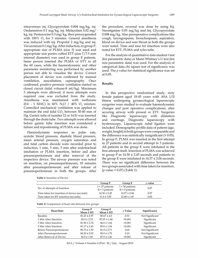
IJAA�/�Volume�6�Number�4�(Part�-�II)�/�July�-�August�2019
1285
intravenous� inj.� Glycopyrrolate� 0.004� mg/kg,� inj.�Ondansetron�0.1�mg/kg,�inj.�Midazolam�0.02�mg/kg,�inj.�Pentazocine�0.3�mg/kg,�then�preoxygenated�with� 100�� O
2� for� 3� minutes.� General� anesthesia�
was� induced�with� inj.� Propofol� 2�mg/kg� and� inj.�Vecuronium�0.1�mg/kg.�After�induction,�in�group�P,�appropriate� size� of� PLMA� (size� 3)� was� used� and�appropriate�size�portex�cuffed�ETT�(size�7/7.5�mm�internal� diameter)� was� used� in� group� E� patients.�Same� person� inserted� the� PLMA� or� ETT� in� all�the� 60� cases,� while� the� haemodynamic� and� other�parameter�monitoring�was� performed� by� another�person� not� able� to� visualise� the� device.� Correct�placement� of� device� was� con��rmed� by� manual�ventilation,� auscultation,� capnography.� Once�con��rmed,�positive�pressure�ventilation�started�on�closed� circuit� (tidal� volume-8� ml/kg).� Maximum�3� attempts� were� allowed,� if� more� attempts� were�re�uired� case� was� excluded� from� the� study.�Anesthesia� was� maintained� with� iso��urane�(0.6� -� 1� MAC)� in� 60�� N
2O� /� 40�� O
2� mixture.�
Controlled�mechanical� ventilation�was� applied� to�maintain� the� end� tidal�CO
2�between�30-40�mm�of�
�g.�Gastric�tube�of�number�12�or�14�Fr�was�inserted�through�the�drain�tube.�Two�attempts�were�allowed�before� gastric� tube� insertion� was� considered� a�failure�and�repositioning�of�PLMA�was�done.
�aemodynamic� responses� as� pulse� rate,�systolic� blood� pressure,� diastolic� blood� pressure,�mean� arterial� pressure,� oxygen� saturation� and�end� tidal� carbon� dioxide� were� recorded� prior� to�induction,� 1�min,� 3�min,� 5�min�after� endotracheal�intubation� or� PLMA� insertion,� before� and� after�pneumoperitoneum� and� after� removal� of� the�respective�device.�The�airway�pressure�was�noted�on� insertion,� on� pneumoperitoneum,� 10� minutes�after� pneumoperitoneum� and� after� release� of�pneumoperitoneum� in� both� the� groups.� After�
the� procedure,� reversal� was� done� by� using� Inj.�Neostigmine� 0.05� mg/kg� and� Inj.� Glycopyrrolate�0.008�mg/kg.�Also�perioperative�complications�like�cough,� laryngospasm,� bronchospasm,� aspiration,�blood�on�device�and�sore�throat�in�both�the�groups�were�noted.�Time�and�ease�for�insertion�were�also�noted�for�ETT,�PLMA�and�ryles�tube.
For�the�analysis�of��uantitative�data�student�t�test�(for�parametric�data)�or�Mann�Whitney�s���test�(for�non�parametric�data)�was�used.�For�the�analysis�of�categorical�data�chi�s�uare�test�of�signi��cance�was�used.�The�p�value�for�statistical�signi��cance�was�set�at�0.05.
�esults
In� this� prospective� randomised� study,� sixty�female� patient� aged� 18-45� years� with� ASA� I/II���tness� undergoing� gynaecological� laparoscopic�surgeries�were�studied�to�evaluate�haemodynamic�changes� and� post� operative� complications,� after�securing� airway� with� proseal� or� ETT.� Surgeries�like� Diagnostic� laparoscopy� with� dilatation�and� curettage,� Diagnostic� laparoscopy� with�hysteroscopy,� Laparoscopic� tubal� ligation� were�included.�Demographic�pro��le�data�of�patient�(age,�weight,�height)�in�both�groups�were�comparable�and�the�difference�was�statistically�insigni��cant�(��0.05).�In�group�P,�PLMA�was�inserted�in�the���rst�attempt�in�27�patients�and�in�second�attempt�in�3�patients.�All�patients� in� the�group�E�were� intubated� in� the���rst�attempt�itself.�Insertion�of�PLMA�was�achieved�in�group�P�in�14.30���2.45�seconds�and�patients�in�the�group�E�were�intubated�in�14.57���2.04�seconds.�There� was� no� signi��cant� difference� between� the�two�groups�associated�with�time�taken�for�insertion.�(p�value���0.07)�(Table�1).
Table��:�Insertion�of�Device
�roup�P �roup�E p��alue
No.�of�attempts�of�insertionI���27�patientsII���3�patients
I���30�patientsII���0�patients
0.07
Time�taken�for�insertion�of�device�(seconds) 14.30���2.45 14.57���2.04 0.07
Time�taken�for�RT�insertion�(seconds) 11.0���3.05 12.40���1.47 0.02
Table��:�Comparison�of�heart�rate�between�two�groups
�eart��ate�roup�P
Mean���S��roup�E
Mean���S�p�Value Significance
�aseline 81.23���2.97 80.67���4.0 0.53 Not�Significant
1�Min�After�Insertion 83.0���2.72 87.07���1.68 �0.001 Significant
3�Min�After�Insertion 81.90���2.74 86.0���1.64 �0.001 Significant
5�Min�After�Insertion 81.17���2.45 85.0���1.64 �0.001 Significant
�efore�Pneumoperitoneum 80.73���1.92 81.0���2.71 0.66 Not�Significant
After�Pneumoperitoneum 84.20���2.05 85.0���1.70 0.10 Not�Significant
After�Removal�of�Device 82.0���1.68 87.0���1.46 �0.001 Significant
Proseal�Laryngeal�Mask�Airway�v/s�Endotracheal�Intubation�for�Gynaecological�Laparoscopic�Surgeries
IJAA�/�Volume�6�Number�4�(Part�-�II)�/�July�-�August�2019
1286 Indian�Journal�of�Anesthesia�and�Analgesia
Co��arison�of��ital�Para�eters
Relatively�lower�values�were�observed�in�group�P�than�group�E�at�1�min�(p��0.001),�3�mins�(p��0.001),�5� mins� (p� �0.001)� and� after� removal� of� device�(p� �0.001),� which� were� found� to� be� statistically�signi��cant�(Table�2).
Relatively� lower� values� were� observed� in�group�P� than� group� E� at� 1�min� (p� value� �0.001),�3�mins�(p�value��0.001),�5�mins�(p�value��0.001)�and�after�removal�of�device�(p�value��0.001)�and�were�found�to�be�statistically�signi��cant.�The�difference�between� the� groups� at� baseline� (p� �� 0.42),� before�pneumoperitoneum� (p� value� �� 0.38)� and� after�pneumoperitoneum�(p�value��0.07)�were�not�found�to�be�statistically�signi��cant�(Table�3).
Relatively�lower�values�were�observed�in�group�P�than�group�E�at�1�min�(p��0.001),�3�mins�(p��0.001),�5� mins� (p� �0.001)� and� after� removal� of� device�(p� �0.001)� which� were� statistically� signi��cant.�
The� differences� between� the� groups� at� baseline�(p���0.81),�before�pneumoperitoneum�(p���0.25)�and�after�pneumoperitoneum�(p���0.20)�were�found�to�be�statistically�not�signi��cant�(Table�4).
Relatively� lower� values� were� observed� in�group�P� than�group�E�at�1�min�(p��0.001),�3�mins�(p��0.001),� 5�mins� (p��0.001)�and�after� removal�of�device� (p� �0.001).� The� differences� between� two�groups� were� found� to� be� statistically� signi��cant.�The� difference� between� the� groups� at� baseline�(p���0.33),�before�pneumoperitoneum�(p���0.17)�and�after�pneumoperitoneum�(p���0.56)�were�not�found�to�be�statistically�signi��cant�(Table�5�and�Graph�1).
Co��arison�of�EtC�2��et�een�t�o��rou�s
There�was�no�signi��cant�difference�in�EtCO2�in�both�
the�groups�at�baseline� (p���0.66)� 1�min� (p���0.63),�3�mins� (p���0.10),�5�mins� (p���0.77)�after� insertion�of� device,� before� pneumoperitoneum� (p� �� 0.07)�
Table��:�Comparison�of�systolic�blood�pressure�between�two�groups
Systolic�Blood�Pressure�roup�P
Mean���S��roup�E
Mean���S���Value Significance
�aseline 123.97���1.65 123.60���1.86 0.42 Not�Significant
1�Min�After�Insertion 128.07���1.72 133.03���1.29 �0.001 Significant
3�Min�After�Insertion 127.0���1.25 132.0���1.53 �0.001 Significant
5�Min�After�Insertion 125.03���1.79 129.93���1.43 �0.001 Significant
�efore�Pneumoperitoneum 123.87���1.59 124.37���2.65 0.38 Not�Significant
After�Pneumoperitoneum 128.73���1.61 129.4���3.75 0.07 Not�Significant
After�Removal�of�Device 125.0���1.64 130.0���1.64 �0.001 Significant
Table��:�Comparison�of�diastolic�blood�pressure�between�two�groups
�iastolic�Blood�Pressure�roup�P
Mean���S��roup�E
Mean���S� ��Value Significance
�aseline 84.0���1.64 83.87���2.54 0.81 Not�Significant
1�Min�After�Insertion 86.13���1.57 90.97���1.52 �0.001 Significant
3�Min�After�Insertion 85.0���1.25 90.0���1.64 �0.001 Significant�
5�Min�After�Insertion 84.0���1.33 88.0���1.64 �0.001 Significant�
�efore�Pneumoperitoneum 83.90���1.24 84.8���3.42 0.25 Not�Significant
After�Pneumoperitoneum 87.0���1.53 87.87���4.56 0.20 Not�Significant
After�Removal�of�Device 85.0���1.53 90.07���1.48 �0.001 Significant
Table��:�Comparison�of�Airway�Pressure�between�two�groups
Air�ay�Pressure�roup�P
Mean���S��roup�E
Mean���S���Value Significance
On�Insertion 21.60���1.99 22.27���1.63 0.16 Not�Significant
On�Pneumoperitoneum 25.27���1.99 24.60���1.49 0.14 Not�Significant
10�Min�After�Pneumoperitoneum 26.73���2.13 25.80���1.51 0.06 Not�Significant
On�Release�of�Pneumoperitoneum 21.67���1.49 22.80���1.51 0.07 Not�Significant
IJAA�/�Volume�6�Number�4�(Part�-�II)�/�July�-�August�2019
1287
and� after� pneumoperitoneum� (p��� 0.40)� and� after�
removal�of�the�airway�device�(p���0.06)�(Graph�2).
Co��arison�of�S��2��et�een�t�o��rou�s
There�was�no�signi��cant�difference�in�SpO2�in�both�
the�groups�at�baseline� (p���0.99)� 1�min� (p���0.73),�
3�mins� (p���0.17),�5�mins� (p���0.18)�after� insertion�
of� device,� before� pneumoperitoneum� (p� �� 0.09)�
and�after�pneumoperitoneum.� (p���0.40)�and�after�
removal�of�the�airway�device�(p���0.58)�(Graph�3).
There�was�no�signi��cant�difference�in�the�mean�
airway�pressure�observed�between�both�the�groups�
at� insertion� (p� �� 0.16),� on� pneumoperitoneum�
(p���0.14),�10�min�after�pneumoperitoneum�(p���0.06)�
and� on� release� of� pneumoperitoneum� (p� �� 0.07)�
(Table�6�and�Graph�4).
Incidence� of� �erio�erative� co��lications� in� �oth�
�rou�s
The� difference� in� the� perioperative� complications�
observed�between�both�the�groups�was�statistically�
signi��cant,�with�cough�(p���0.04).�blood�on�device�
(p����0.03)�and�sore�throat�(p���0.04)�lesser�in�P�group.�
Pie-Charts� Showing� Incidence� of� Perioperative�
Complications�(Chart�1)
Table��:�Incidence�of�perioperative�complications�in�both�groups
Complications�roup�P �roup�E
p�ValueNumber � Number �
Cough 2 6.6 5 16.6 0.04
Laryngospasm 0 0 0 0
�ronchospasm 0 0 0 0
�lood�on�device 1 3.3 3 10 0.03
Aspiration 0 0 0 0
Sore�Throat 2 6.6 6 20 0.04
Comparison�of�mean�arterial�pressure
120
100
80
60
40
20
0
Group�P Group�E
1�min�after�insertion
3�min�after�insertion
5�min�after�insertion
�efore�Pneumo�
Peritoneum
After�Pneumo�
Peritoneum
After�removal
�aseline
�raph��:�Showing�comparison�of�mean�arterial�pressure�in�both�the�groups
Proseal�Laryngeal�Mask�Airway�v/s�Endotracheal�Intubation�for�Gynaecological�Laparoscopic�Surgeries

IJAA�/�Volume�6�Number�4�(Part�-�II)�/�July�-�August�2019
1288 Indian�Journal�of�Anesthesia�and�Analgesia
Comparison�of�EtCO2
40
35
30
25
20
15
10
5
0
Group�P Group�E
1�min�after�
insertion
3�min�after�
insertion
5�min�after�
insertion
�efore�
Pneumo�Peritoneum
After�
Pneumo�Peritoneum
After�
removal
�aseline
�raph��:�Showing�comparison�of�EtCO2�in�both�the�groups
Comparison�of�SpO2
100
99
98
97
96
95
1�min�after�
insertion
3�min�after�
insertion
5�min�after�
insertion
�efore�
Pneumo�
Peritoneum
After�
Pneumo�
Peritoneum
After�
removal
�aseline
Group�P Group�E
�raph��:�Showing�comparison�of�SpO2�in�both�the�groups
Comparison�of�airway�pressure
30
25
20
15
10
5
0
On�insertion On
Pneumo�
Peritoneum
10�min�afterPneumo�
Peritoneum
On�release�ofPneumo�
Peritoneum
�raph��:�Showing�comparison�of�airway�pressure�in�both�the�groups
IJAA�/�Volume�6�Number�4�(Part�-�II)�/�July�-�August�2019
1289
�iscussion
Laparoscopic� surgeries� are� associated� with�
pneumoperitoneum� leading� to� increase� in�
intra-abdominal� pressure� and� intra-thoracic�pressure,� thus� ETT� is� considered� as� the� gold�
standard.11�Endotracheal�tube�insertion�is�associated�
with�haemodynamic�changes�due�to�laryngoscopy12�
and� postoperative� disadvantages� like� sore� throat,�
cough�and�hoarseness.13
Reid� and� �race� were� the� ��rst� to� describe�the� haemodynamic� changes� in� response� to�
laryngoscopy� and� intubation.14� On� direct�
laryngoscopy,�in�less�than�5�seconds�these�changes�
begin,�peak�at�1-2�minutes�and�return�towards�the�
baseline�in�5�minutes.15
The�Proseal�LMA�is�a�new�entrant�in�the�family�
of�LMA�with�added�features�over�the�classic�LMA.�
Reduced�risk�of�gastric�insuf��ations,�regurgitation�
and� aspiration� are� associated� with� usage� of�
PLMA.�Maltby� et� al.� postulated� that� laparoscopic�
surgery�was� an� important� test� for� evaluating� the�effectiveness� of� SADs� use� in� positive� pressure�
ventilation.8
After� approval� from� the��ospital� research� and�
ethics� committee,� a� prospective,� randomized,�
comparative� study� was� conducted� on� 60� adult�
female� patients� undergoing� gynaecological�laparoscopic�surgeries�under�general�anesthesia.
After� applying� the� inclusion� and� exclusion�criteria,� the� patients� were� randomly� divided� into�two�groups�of�30�each�using�a�computer�generated�random�number�table.
Our�aim�was�to�compare�the�ef��cacy�and�safety�of� Proseal� laryngeal� mask� airway� with� portex�endotracheal� intubation� in� sixty� adult� female�patients� undergoing� gynaecological� laparoscopic�surgeries�under�general�anesthesia.
Ease�of�Insertion�of��evice
The���rst�attempt�success�rate�in�group�P�was�90��and�100��at�the�second�attempt,�while�all�patients�in�the�group�E�were�intubated�in�the���rst�attempt�itself.�The�difference�between�the�two�groups�was�statistically�not�signi��cant.�(p�value���0.07).
In� a� study� conducted� by� M� Misra� et� al.,� The�Pro-seal�LMA�and�tracheal�tube:�A�comparison�of�events� at� insertion� of� the� airway� device,� showed�100��success�at� insertion� in�both� the�groups�with�success� rate� at� ��rst� attempt� 88�� for� PLMA� and�100�� for� ETT.� For� PLMA,� ��rst/� second/� third�attempt�observed�in�44/�4/�1�patients�respectively.7
�i�e��a�en�for�Insertion�of��evice
The�mean� time� taken� for� insertion� of� device�was�14.30� �� 2.45� seconds� in� group� P� and� 14.57� �� 2.04�seconds� in� group� E.� The� difference� between�the� two� groups� was� not� statistically� signi��cant�(p�value���0.07).
The� ��ndings� observed� in� our� study� were�concurrent� with� the� study� done� by� Avhad� V,�Comparison� of� safety� and� ef��cacy� of� Proseal�laryngeal�mask�airway�v/s�endotracheal�intubation�for� gynaecological� diagnostic� laparoscopy,� in�2017.� The� mean� time� taken� for� insertion� was�18.2���5�seconds�for�PLMA�and�25.6���8.1�seconds�in�ETT,�which�was�statistically�signi��cant.�Successful�PLMA� insertion� at� ��rst� attempt� in� 35� patients�and� second� attempt� in� 5� patients,� while� for� ETT,�33�patients�in���rst�and�7�patients�in�second�attempt�and�the�difference�was�observed�to�be�insigni��cant.16
Co��arison�of��ital�Para�eters
Comparison�of�heart�rate�between�the�two�groups
Values�observed�in�group�P�were�relatively� lower�than�group�E�at�1�min�(p��0.001),�3�mins�(p��0.001),�5� mins� (p� �0.001)� and� after� removal� of� device�(p� �0.001),� which� were� found� to� be� statistically�
Pie�chart�showing�perioperativecomplications�in�Group�E
Cough �lood�on�device
Sore�throat No�side�effects
�����
�����
�����
�����
Proseal�Laryngeal�Mask�Airway�v/s�Endotracheal�Intubation�for�Gynaecological�Laparoscopic�Surgeries
Chart��:�Showing�Incidence�of�Perioperative�Complications�
IJAA�/�Volume�6�Number�4�(Part�-�II)�/�July�-�August�2019
1290 Indian�Journal�of�Anesthesia�and�Analgesia
signi��cant.
The�differences�observed�between�two�groups�at�baseline�(p���0.53)�before�peritoneum�(p���0.66)�and�after�pneumoperitoneum�(p���0.10)�were�not�found�to�be�statistically�signi��cant.
In�2005,�P�Shroff,�S��amath�in�their�randomized�comparative� study� between� the� proseal� laryngeal�mask� airway� and� the� endotracheal� tube� for�laparoscopic� surgery� observed� mean� heart�rate� values� with� PLMA� as� 98� �� 22� per� min,�104���16�per�min,�98���17�per�min,�98���17�per�min,�and�92���13�per�min�and�with�ETT�as�99���10�per�min,�102���11�per�min,�99���13�per�min,�109���13�per�min�and�103���7�per�min�at�pre�induction,�after�induction,�before� and� after� pneumoperitoneum� and�postoperatively� respectively.� The� difference�observed�between�the�two�groups�as�regards�to�the�mean�heart�rate�was�statistically�signi��cant.1
Co��arison�of�Systolic��lood�Pressure:
Values�observed�in�group�P�were�relatively� lower�than� group� E� at� 1� min� (p� value� �� 0.42),� 3� mins�(p�value��0.001),�5�mins�(p�value��0.001)�and�after�removal�of�device�(p�value��0.001)�were�found�to�be�statistically�signi��cant.
The� differences� observed� between� the� groups�at� baseline� (p� �� 0.42),� before� pneumoperitoneum�(p� value� �� 0.38)� and� after� pneumoperitoneum�(p� value� �0.07)� were� not� found� to� be� statistically�signi��cant.
�alpana� Shah,� in� 2017,� studied� the� Proseal�laryngeal�mask�airway�as�an�alternative�to�standard�endotracheal�tube�in�securing�upper�airway�in�the�patients�undergoing�beating-heart�coronary�artery�bypass�grafting.� In� this� study,� it�was� seen� that�all�the� hemodynamic� parameters� in� PLMA� group�were�better�than�in�the�ETT�group�and�this���nding�was�statistically�signi��cant�(p���0.05).10
Co��arison�of��iastolic��lood�Pressure
Values�observed�in�group�P�were�relatively� lower�than�group�E�at�1�min�(p��0.001),�3�mins�(p��0.001),�5� mins� (p� �0.001)� and� after� removal� of� device�(p��0.001)�which�were�statistically�signi��cant.
The� difference� between� the� groups� at� baseline�(p���0.81),�before�pneumoperitoneum�(p���0.25)�and�after�pneumoperitoneum�(p���0.20)�were�not�found�to�be�statistically�signi��cant.
Sharma��,�Sahai�C�et�al.,�conducted�a�study�of�100�consecutive� cases� of� laparoscopic� surgery.� Their�results�were�pre-induction�D�P�77.82���12.27�mm�
of��g�reached�to�79.21���16.35�mm�of��g�at�1�min�and�82.01���16.45�mm�of��g�at�5�mins�after�insertion�of� PLMA� (p-value� 0.053),� which� was� statistically�signi��cant.� They� concluded� that� there� were�minimum�haemodynamic�responses�to�insertion�of�Proseal�Laryngeal�Mask�Airway.6
Co��arison�of��ean�Arterial�Pressure
Values�observed�in�group�P�were�relatively�lower�than�group�E�at�1�min�(p��0.001),�3�mins�(p��0.001),�5� mins� (p� �0.001)� and� after� removal� of� device�(p��0.001).�The�difference�between�two�groups�was�found�to�be�statistically�signi��cant.
The� difference� between� the� groups� at� baseline�(p���0.33),�before�pneumoperitoneum�(p���0.17)�and�after�pneumoperitoneum�(p���0.56)�were�not�found�to�be�statistically�signi��cant.
Saraswat� N,� �umar� A� et� al.� compared� Proseal�LMA�and�Endotracheal�tube�in�patients�undergoing�laparoscopic� surgeries� under� general� anesthesia.�They�concluded�statistically�signi��cant�increase�in�heart�rate�and�mean�blood�pressure�was�observed�10�seconds�after�intubation�and�persisted�till�3�mins�after� intubation� and� also� during� extubation� in�the� ETT� group.� �owever� statistically� signi��cant�increase�in�PLMA�group�was�seen�only�10�seconds�after�insertion.2
Co��arison�of�End��idal�Car�on��ioxide
There� was� no� signi��cant� difference� in� EtCO2
in� both� the� groups� at� baseline� (p� �� 0.66),� 1� min�(p���0.63),�3�mins�(p���0.10),�5�mins�(p���0.77)�after�insertion� of� device,� before� (p� �� 0.07)� and� after�pneumoperitoneum�(p���0.40)�and�after�removal�of�the�airway�device�(p���0.06).
Co��arison�of�S��2
There� was� no� signi��cant� difference� in� SpO2�
in� both� the� groups� at� baseline� (p� �� 0.99)� 1� min�(p���0.73),�3�mins�(p���0.17),�5�mins�(p���0.18)�after�insertion� of� device,� before� (p� �� 0.09)� and� after�pneumoperitoneum�(p���0.40)�and�after�removal�of�the�airway�device�(p���0.58).
In� 2003,� Maltby� JR� et� al.� conducted� a� study,�LMA� –� Classic� and� LMA� –� Proseal� are� effective�alternatives� to� endotracheal� intubation� for�gynaecologic� laparoscopy.� They� observed� the�differences� between� LMA-C/PLMA� and� ETT�groups� for� SpO
2,� SpO
2� and� PETCO
2� were� not�
statistically�signi��cant�before�or�during�peritoneal�insuf��ations.8
IJAA�/�Volume�6�Number�4�(Part�-�II)�/�July�-�August�2019
1291
Air�ay��ressure
There� was� no� signi��cant� difference� in� the� mean�airway�pressure�observed�between�both�the�groups�at� insertion� (p� �� 0.16),� on� pneumoperitoneum�(p���0.14),�10�min�after�pneumoperitoneum�(p���0.06)�and�on�release�of�pneumoperitoneum�(p���0.07).
In� 2012,� �andan� G� et� al.� studied� comparison�of� haemodynamic� and� metabolic� stress� response�caused�by�endotracheal� tube�and�Proseal�LMA�in�laparoscopic� cholecystectomy.� They� concluded�that�although�the�peak�airway�pressures�increased�after� carboperitoneum� in� both� the� groups,� it� did�not�disrupt�ventilation�and�the�difference�was�not�statistically�signi��cant.5
Perio�erative�co��lications
The�difference�in�the�perioperative�complications�observed�between�both�the�groups�was�statistically�signi��cant,� cough� (p� �� 0.04),� blood� on� device�(p� �� 0.03)� and� sore� throat� (p� �� 0.04)� with� values�higher�in�E�group.
In� 2003,� Maltby� JR� et� al.� observed� a� ten-fold�difference� in� fre�uency� of� coughing� at� removal�of� the�ETT�87��vs�LMAC/PLMA�8�,�which�was�statistically�signi��cant�(����0.001).�Sore�throat�24�hr�postoperatively�was�more�common�with�ETT�than�LMA-C/PLMA�(28��vs�17�;�p���0.05).�No�patient�reported�numbness�of�the�tongue�or�other�morbidity�attributable�to�the�airway�devices.8
Conclusion
Proseal�LMA�was�found�to�be�better�than�ETT�in�the�following�ways:
1.� Lesser� haemodynamic� response� on�insertion�and�removal.
2.� Reduced� postoperative� complications� like�cough,�blood�on�device�and�sore�thoat.
The� ease� and� time� taken� for� insertion� of�Proseal�Laryngeal�Mask�Airway� is� comparable� to�endotracheal�tube.�It�provides�similar�ef��ciency�like�ETT�for�controlled�positive�pressure�ventilation.
Thus� the� Proseal� LMA� offers� a� safe� and�effective� airway� management� alternative� in�patients� undergoing� gynaecological� laparoscopic�procedures� under� general� anesthesia� with�controlled� ventilation� with� the� added� advantage�of� minimal� haemodynamic� response� and�postoperative�complications.
�eferences
1.� P� Shroff,� S� �amath.� Randomized� comparative�study�between�the�proseal�laryngeal�mask�airway�and� the� endotracheal� tube� for� laparoscopic�surgery.� The� Internet� Journal� of�Anesthesiology�2005;11:1.
2.� Saraswat� N,� �umar� A,� Mishra� A,� et� al.�Comparison� of� Proseal� LMA� and� Endotracheal�tube� in� patients� undergoing� laparoscopic�surgeries� under� general� anesthesia.� Indian�Journal�of�Anesthesia.�2011;55(2):129–34.
3.� Sharma��,� Sood� J,��umara�VP.��ses�of� LMA� in�present�day� anesthesia.� J�Anaes�Clin� Pharmacol�2007;23:5–15.
4.� �rain�AIJ,�Verghese�C,�Strube�PJ.�The�Laryngeal�Mask�Airway�Proseal-A�laryngeal�mask�with�an�oesophageal� vent.� �ritish� Journal� of�Anesthesia.�2000;84(5):650–654.
5.� �andan�G,�Turkay�C,��alil��,�et�al.�Comparison�of�haemodynamic�and�metabolic�stress�response�caused�by�endotracheal�tube�and�Proseal�LMA�in�laparoscopic�cholecystectomy.�J�Res�Med�Sci.�2012�Feb;17(2):148–153.
6.� Sharma��,�Sahai�C,��hattacharya�A,�et�al.�ProSeal�laryngeal�mask�airway:�A�study�of�100�consecutive�cases� of� laparoscopic� surgery.� Indian� J� Anaesth�2003;47:467–72.
7.� Misra� MN,� Ramamurthy� �.� The� Proseal� LMA�and�the�tracheal� tube:�a�comparison�of�events�at�insertion�of�the�airway�device.�Internet�Journal�of�Anaesthesiology.�2008;16:2.
8.� Maltby� JR,� �eriault� MT,� Watson� NC,� et� al.LMA�–�Classic� and�LMA�–�Proseal� are� effective�alternatives� to� endotracheal� intubation� for�gynecologic� laparoscopy.� Canadian� Journal� of�Anesthesia.�2003;50(1):71–7.
9.� Lim� �,� Goel� S,� �rimacombe� JR.� The� Proseal�Laryngeal�Mask�Airway�is�an�effective�alternative�to� laryngoscope� guided� tracheal� intubation� for�gynaecological�laparoscopy.�Anesthesia�Intensive�care.�2007;35(1):52–6.
10.� Shah:�Proseal�Laryngeal�Mask�Airway:�alternative�to� standard� endotracheal� tube� in� patients�undergoing� beating-heart� CA�G.� Annals� of�Cardiac�Anesthesia.�2017�Jan-Mar;20(1):61–66.
11.� Lind�JF,�Warrian�WG,�Wankling�WJ.�Responses�of�the�gastroesophageal�junctional�zone�to�increases�in�abdominal�pressure.�Can�J�Surg.�1966;9:32–8.
12.� �on� Sebastian,� Anand� T.� Talikoti,� Dinesh��rishnamurthy.� Attenuation� of� haemodynamic�responses� to� laryngoscopy� and� endotracheal�intubation� with� intravenous� dexmedetomidine:�A� comparison� between� two� doses.� Indian�J.�Anesthesia.�2017;61(1):48–54.
13.� Divatia� JV,� Dr.� �howmick� �.� Complications�
Proseal�Laryngeal�Mask�Airway�v/s�Endotracheal�Intubation�for�Gynaecological�Laparoscopic�Surgeries
IJAA�/�Volume�6�Number�4�(Part�-�II)�/�July�-�August�2019
1292 Indian�Journal�of�Anesthesia�and�Analgesia
of� endotracheal� intubation� and� other� airway�management� procedures.� Indian� J.� Anesthesia.�2005;49(4):308–318.
14.� Reid� LC,� �race� DE.� Irritation� of� respiratory�tract� and� its� reflex� effect�on� heart� surgery.� Surg�Gynaecol�obste.�1940;70:157–62.
15.� �enderson� J.� In:� Airway� Management� in� the�adult.�Miller�s�Anesthesia�7th�ed.�Miller�RD,�editor�Philadelphia:� Churchill� Livingstone;� 2010.pp�1573–610.
16.� Avhad�V,�Oak�S,�Shetty�A.�Comparison�of�safety�and� efficacy� of� Proseal� Laryngeal�Mask�Airway�vs� Endotracheal� Intubation� for� Gynecological�Diagnostic� Laparoscopy.� Res� Inno� in� Anesth.�2017;2(1):9–13.
17.� Singh�S,�Singh�AP.�A�Comparative�Evaluation�of�the�Laryngeal�Mask�Airway�Proseal�and�Tracheal�Intubation� for� Laparoscopic� Cholecystectomy.�IJIMS.�2014;1(10):119–125.
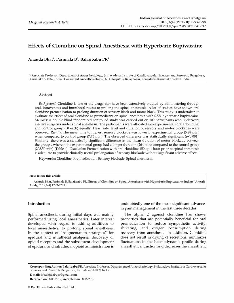
��Red�Flower�Publication�Pvt.�Ltd.�
�Original�Research�ArticleIndian�Journal�of�Anesthesia�and�Analgesia�
2019;�6(4)�(Part�-�II):�1293-1298DOI:�http://dx.doi.org/10.21088/ijaa.2349.8471.6419.32
Effects�of�Clonidine�on�Spinal�Anesthesia��ith��yperbaric�Bupi�acaine
Ananda�Bhat���Parimala�B���Bala�ibabu�P��
1,3Associate�Professor,�Department�of�Anaesthesiology,�Sri�Jayadeva�Institute�of�Cardiovascular�Sciences�and�Research,��engaluru,��arnataka�560069,�India.�2Consultant�Anaesthesiologist,�N���ospitals,�Rajajinagar,��engaluru,��arnataka�560010,�India.
Corresponding�Author:�Bala�ibabu�P�,�Associate�Professor,�Department�of�Anaesthesiology,�Sri�Jayadeva�Institute�of�Cardiovascular�Sciences�and�Research,��engaluru,��arnataka�560069,�India.
E�mail:�[email protected]
�ecei�ed�on�08.05.2019,�Accepted�on�08.06.2019
Abstract
Background:�Clonidine� is�one�of� the�drugs� that�have�been�extensively�studied�by�administering� through�oral,� intravenous�and�intrathecal� routes� to�prolong�the�spinal�anesthesia.�A�lot�of�studies�have�shown�oral�clonidine�premedication�to�prolong�duration�of�sensory�block�and�motor�block.�This�study�is�undertaken�to�evaluate�the�effect�of�oral�clonidine�as�premedicant�on�spinal�anesthesia�with�0.5��hyperbaric�bupivacaine.�Methods:�A�double�blind�randomized�controlled�study�was�carried�out�on�100�participants�who�underwent�elective�surgeries�under�spinal�anesthesia.�The�participants�were�allocated�into�experimental�(oral�Clonidine)�and� control�group� (50� each)� e�ually.��eart� rate,� level� and�duration�of� sensory� and�motor�blockades�were�observed.�Results:�The�mean�time�to�highest�sensory�blockade�was�lower� in�experimental�group�(5.28�min)�when�compared�to�control�group�(7.76�min).�The�observed�difference�was�statistically�significant�(p�0.001).�Similarly,� there�was� a� statistically� significant�difference� in� the�mean�duration�of�motor� blockade� between�the�groups,�wherein�the�experimental�group�had�a�longer�duration�(264�min)�compared�to�the�control�group�(208.50�min)�(Table�4).�Conclusion:�Premedication�with�oral�clonidine�150�g,�1�hour�prior�to�spinal�anesthesia�is�ade�uate�to�provide�clinically�useful�prolongation�of�sensory�blockade�without�significant�adverse�effects.
�ey�ords:�Clonidine;�Pre-medication;�Sensory�blockade;�Spinal�anesthesia.
�o��to�cite�this�article:
Ananda��hat,�Parimala��,��alajibabu�PR.�Effects�of�Clonidine�on�Spinal�Anesthesia�with��yperbaric��upivacaine.�Indian�J�Anesth�Analg.�2019;6(4):1293-1298.
Introduction
Spinal� anesthesia� during� initial� days� was� mainly�performed� using� local� anaesthetics.� Later� interest�developed� with� regard� to� adding� additives� to�local� anaesthetics,� to� prolong� spinal� anesthesia.�In� the� context� of� �Augmentation� strategies�� for�epidural� and� intrathecal� analgesia,� discovery� of�opioid�receptors�and�the�subse�uent�development�of�epidural�and�intrathecal�opioid�administration�is�
undoubtedly�one�of�the�most�signi��cant�advances�in�pain�management�in�the�last�three�decades.1
The� alpha� 2� agonist� clonidine� has� shown�properties� that� are� potentially� bene��cial� for� oral�premedication� to� reduce� sympathetic� activity,�shivering,� and� oxygen� consumption� during�recovery� from� anesthesia.� In� addition,� Clonidine�does�not� result� in�drying�of�secretions;�minimizes���uctuations� in� the� haemodynamic� pro��le� during�anaesthetic�induction�and�decreases�the�anaesthetic�
IJAA�/�Volume�6�Number�4�(Part�-�II)�/�July�-�August�2019
1294 Indian�Journal�of�Anesthesia�and�Analgesia
re�uirements� of� both� opioid� and� volatile�anaesthetics.� Clonidine� provides� signi��cant�bene��ts�for�preoperative�anxiety�and�analgesia.2
Clonidine� is� one� of� the� drugs� that� have� been�extensively�studied�by�administering�through�oral,�intravenous� and� intrathecal� routes� to� prolong� the�duration� of� action� of� spinal� anesthesia.��owever,�it� is� of� interest� to� ��nd� the� effectiveness� of� oral�clonidine�given�as�a�premedicant�on�the�duration�of�bupivacaine�spinal�anesthesia.�A�lot�of�studies�have�shown� oral� clonidine� premedication� to� prolongduration�of�sensory�block�and�motor�block.3-5�This�study�was�undertaken�to�evaluate�the�effect�of�oral�clonidine� as� a� premedicant,� on� spinal� anesthesia�with�0.5��hyperbaric�bupivacaine.
���ectives
This� study�was� carried� out� to� assess� following�effects�of�oral�clonidine�(150��g),�as�a�premedicant�on�hyperbaric�bupivacaine�(0.5�)�spinal�anesthesia.
1.� Onset�and�duration�of�sensory�and�motor�blockade.
2.� �aemodynamic�effects.
Materials�and�Methods
Study�desi�n�and��artici�ants
A�double�blind�randomized�controlled�study�was�carried�out�in� the�Department�of�Anaesthesiology�of� a� tertiary� teaching� institution� for� a� period� of�one�year.�All�the�patients�who�underwent�elective�surgeries�under�spinal�anesthesia�during�the�study�period� were� selected� for� the� study.� A� total� of�100�patients�participated�in�the�study.
Inclusion�criteria
1.� Age�between�20-60�years
2.� ASA�I���II�patients
3.� Elective� lower� abdominal� and� lower� limb�surgeries�performed�in�supine�position
4.� Weight:�40�–�60�kg
5.� �eight:�Around�150�cm
Exclusion�criteria
1.� ASA�III���IV�patients
2.� �ypertensive� patients� on� beta� blockers,�calcium�channel�blockers
3.� Patients�on�digoxin
4.� Patients�who�re�uire�opioids,� intravenous�or� inhalational� anaesthetics� as�supplementation�for�regional�anesthesia
5.� Patients� who� had� inade�uate� block�re�uiring�general�anesthesia.
Rando�i�ation�and�sa��lin�
The�participants�were�allocated� into� experimental�and� control� group� e�ually.� Randomization� was�carried�out�based�on�computer�generated�random�numbers.�Each�group�consisted�of�50�participants.
Group� A� (Control):� Placebo� and� 3� ml� (15� mg)�hyperbaric�bupivacaine�(0.5�)�spinal�anesthesia.
Group��� (Experiment):� Oral� clonidine� (150� �g)�as� premedicant� and� 3� ml� (15� mg)� hyperbaric�bupivacaine�(0.5�)�spinal�anesthesia.
Ethical�a��roval�and�infor�ed�consent
Approval� was� obtained� from� the� Institutional�Ethics� Committee� prior� to� the� commencement� of�the�study.�Each�participant�was�explained�in�detail�about�the�study�and�informed�consent�was�obtained�prior�to�the�data�collection.
Procedure
The�participants�were�pre-medicated�the�previous�night� with� T.� Alprazolam� 0.5� mg� �S� and� Inj.�Ranitidine�50�mg�intravenous�and�Inj.�Ondansetron�4�mg�was�administered�intravenously�30�minutes�before� shifting� to� operation� theatre.� �aseline�parameters,�namely�heart�rate,�non�invasive�blood�pressure� (NI�P)� and� SpO
2� were� recorded� before�
pre-medication.�Oral�Clonidine�(150��g)�with�sips�of�water�was�administered�to�the�participants�in�the�experimental�group,�60�minutes�before�anesthesia.�Preloading� was� done� with� 10� –� 15� ml/kg� of�Ringer�s� Lactate� solution� over� thirty� minutes�followed� by� infusion� of� 8� –� 10� ml/kg/hour�intravenously� as� maintenance� ��uid.� �R� and� �P�recorded� just� before� spinal� anesthesia� was� taken�as� �0�� min� reading.� �nder� aseptic� precautions,�lumbar�puncture�was�done� in� the� lateral�position�at�L2-L3�space,�using�26�G��uincke�s�spinal�needle.�3�ml�of�0.5��hyperbaric�bupivacaine�was�injected�at�the�rate�of�1ml�in�3�sec�with�table���at�and�patient�was�immediately�turned�supine�and�supplemental�oxygen�started.
�ata�collection
�aemod�namic� monitoring:�Continuous�monitoring�of� ECG,� �R,� SpO
2,� NI�P� was� carried� out.�
��potension�was�de��ned�as�decrease�in�S�P�of���30��from�the�baseline�value�and�was�treated�with�bolus�intravenous� ��uid� and� intravenous� bolus� dose� of�
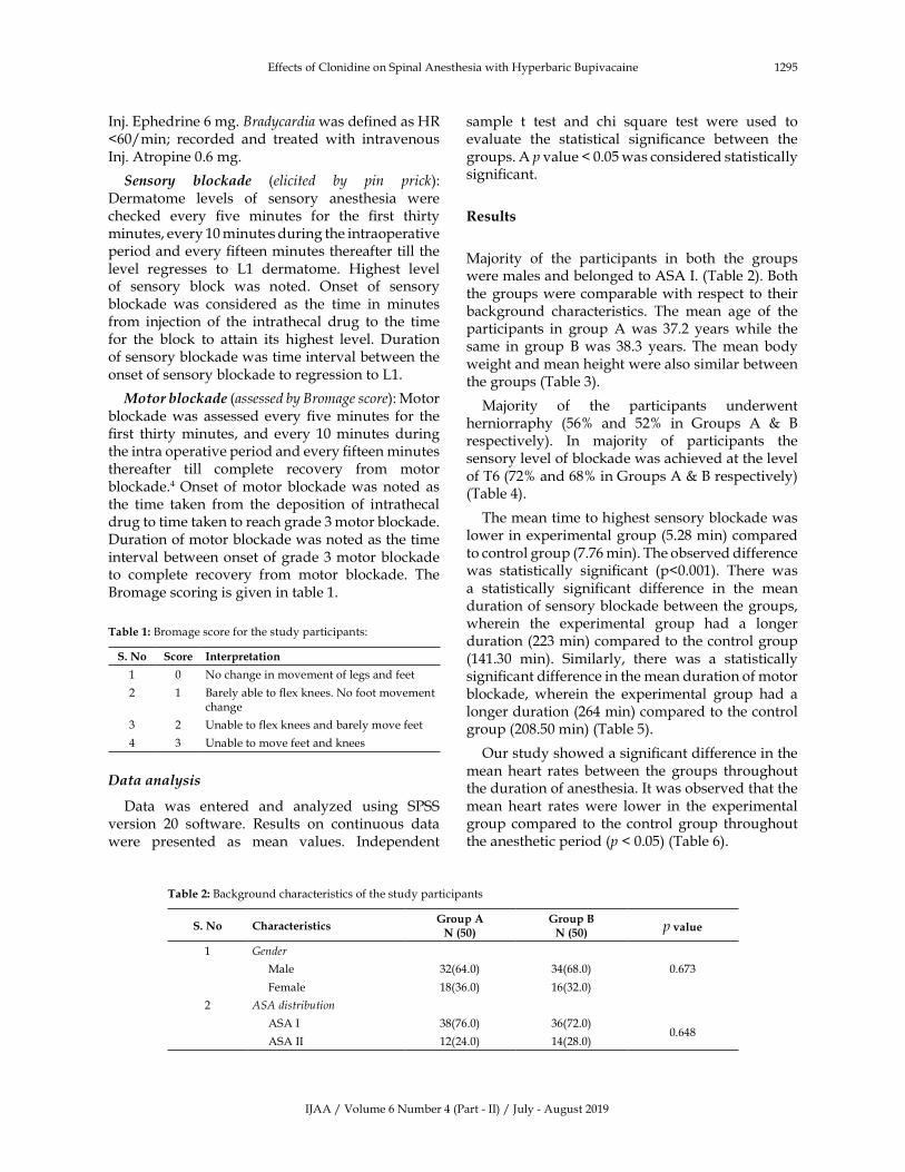
IJAA�/�Volume�6�Number�4�(Part�-�II)�/�July�-�August�2019
1295
Inj.�Ephedrine�6�mg.�Brad�cardia�was�de��ned�as��R��60/min;� recorded� and� treated� with� intravenous�Inj.�Atropine�0.6�mg.
Sensory� �loc�ade� (elicited� b�� pin� prick):�Dermatome� levels� of� sensory� anesthesia� were�checked� every� ��ve� minutes� for� the� ��rst� thirty�minutes,�every�10�minutes�during�the�intraoperative�period�and�every���fteen�minutes�thereafter�till�the�level� regresses� to� L1� dermatome.� �ighest� level�of� sensory� block� was� noted.� Onset� of� sensory�blockade� was� considered� as� the� time� in� minutes�from� injection� of� the� intrathecal� drug� to� the� time�for� the� block� to� attain� its� highest� level.� Duration�of�sensory�blockade�was�time�interval�between�the�onset�of�sensory�blockade�to�regression�to�L1.
�otor��loc�ade�(assessed�b��Bromage�score):�Motor�blockade�was� assessed� every���ve�minutes� for� the���rst� thirty�minutes,� and� every� 10�minutes� during�the�intra�operative�period�and�every���fteen�minutes�thereafter� till� complete� recovery� from� motor�blockade.4�Onset�of�motor� blockade�was�noted� as�the� time� taken� from� the� deposition� of� intrathecal�drug�to�time�taken�to�reach�grade�3�motor�blockade.�Duration�of�motor�blockade�was�noted�as�the�time�interval�between�onset�of�grade�3�motor�blockade�to� complete� recovery� from� motor� blockade.� The��romage�scoring�is�given�in�table�1.
Table��:��romage�score�for�the�study�participants:
S��No Score Interpretation
1 0 No�change�in�movement�of�legs�and�feet
2 1 �arely�able�to�flex�knees.�No�foot�movement�change
3 2 �nable�to�flex�knees�and�barely�move�feet
4 3 �nable�to�move�feet�and�knees
�ata�analysis
Data� was� entered� and� analyzed� using� SPSS�version� 20� software.� Results� on� continuous� data�were� presented� as� mean� values.� Independent�
sample� t� test� and� chi� s�uare� test� were� used� to�evaluate� the� statistical� signi��cance� between� the�groups.�A�p�value���0.05�was�considered�statistically�signi��cant.
�esults
Majority� of� the� participants� in� both� the� groups�were�males�and�belonged�to�ASA�I.�(Table�2).��oth�the�groups�were�comparable�with�respect� to� their�background� characteristics.� The� mean� age� of� the�participants� in� group�A�was� 37.2� years�while� the�same� in� group� ��was� 38.3� years.� The�mean� body�weight�and�mean�height�were�also�similar�between�the�groups�(Table�3).
Majority� of� the� participants� underwent�herniorraphy� (56�� and� 52�� in� Groups� A� �� ��respectively).� In� majority� of� participants� the�sensory�level�of�blockade�was�achieved�at�the�level�of�T6�(72��and�68��in�Groups�A�����respectively)�(Table�4).
The�mean�time�to�highest�sensory�blockade�was�lower�in�experimental�group�(5.28�min)�compared�to�control�group�(7.76�min).�The�observed�difference�was� statistically� signi��cant� (p�0.001).� There� was�a� statistically� signi��cant� difference� in� the� mean�duration�of�sensory�blockade�between�the�groups,�wherein� the� experimental� group� had� a� longer�duration�(223�min)�compared�to�the�control�group�(141.30� min).� Similarly,� there� was� a� statistically�signi��cant�difference�in�the�mean�duration�of�motor�blockade,� wherein� the� experimental� group� had� a�longer�duration�(264�min)�compared�to�the�control�group�(208.50�min)�(Table�5).
Our�study�showed�a�signi��cant�difference�in�the�mean�heart� rates� between� the� groups� throughout�the�duration�of�anesthesia.�It�was�observed�that�the�mean� heart� rates�were� lower� in� the� experimental�group�compared� to� the� control� group� throughout�the�anesthetic�period�(p���0.05)�(Table�6).
Table��:��ackground�characteristics�of�the�study�participants
S��No Characteristics�roup�AN�����
�roup�BN�����
p��alue
1 �ender
0.673Male 32(64.0) 34(68.0)
Female 18(36.0) 16(32.0)
2 ASA�distribution
ASA�I 38(76.0) 36(72.0)0.648
ASA�II 12(24.0) 14(28.0)
Effects�of�Clonidine�on�Spinal�Anesthesia�with��yperbaric��upivacaine
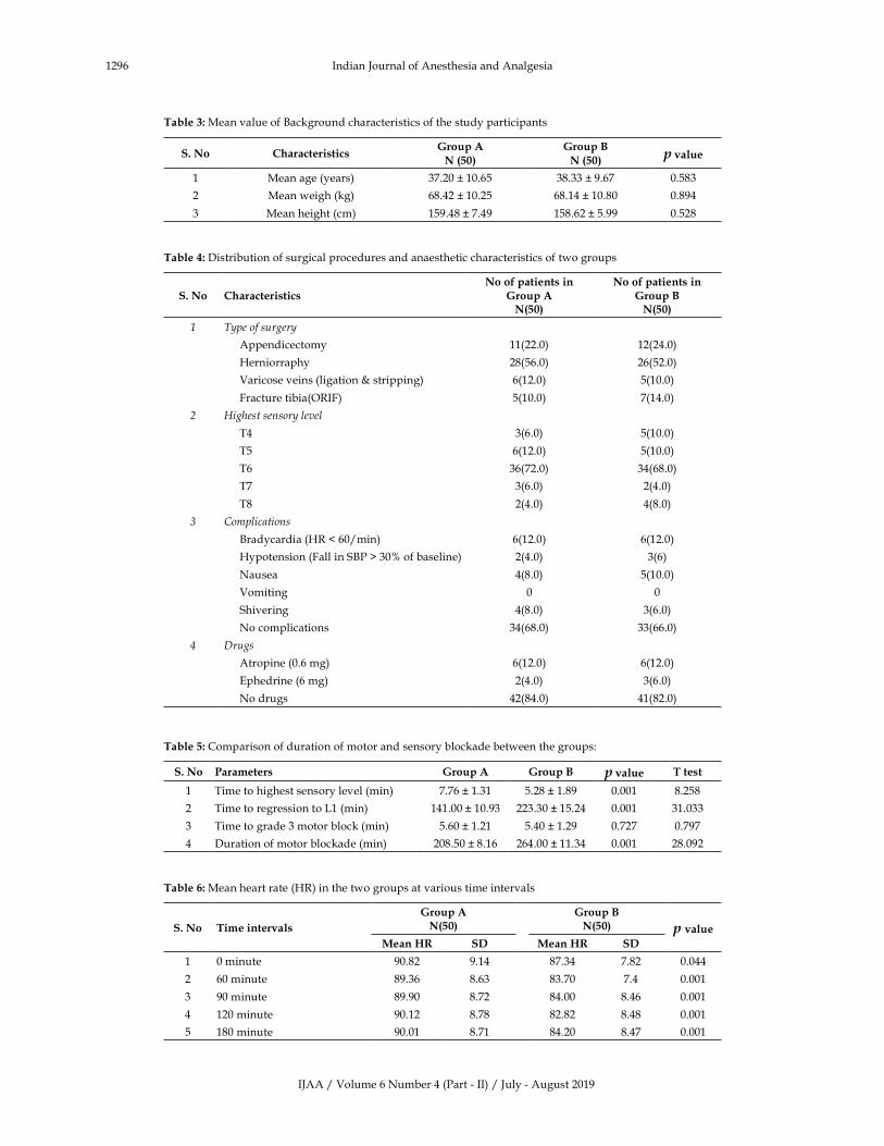
IJAA�/�Volume�6�Number�4�(Part�-�II)�/�July�-�August�2019
1296 Indian�Journal�of�Anesthesia�and�Analgesia
Table��:�Mean�value�of��ackground�characteristics�of�the�study�participants
S��No Characteristics�roup�AN�����
�roup�BN�����
���alue
1 Mean�age�(years) 37.20���10.65 38.33���9.67 0.583
2 Mean�weigh�(kg) 68.42���10.25 68.14���10.80 0.894
3 Mean�height�(cm) 159.48���7.49 158.62���5.99 0.528
Table��:�Distribution�of�surgical�procedures�and�anaesthetic�characteristics�of�two�groups
S��No CharacteristicsNo�of�patients�in
�roup�AN����
No�of�patients�in�roup�BN����
� T�pe�of�surger�
Appendicectomy 11(22.0) 12(24.0)
�erniorraphy 28(56.0) 26(52.0)
Varicose�veins�(ligation���stripping)� 6(12.0) 5(10.0)
Fracture�tibia(ORIF)� 5(10.0) 7(14.0)
� �ighest�sensor��le�el
T4 3(6.0) 5(10.0)
T5 6(12.0) 5(10.0)
T6 36(72.0) 34(68.0)
T7 3(6.0) 2(4.0)
T8 2(4.0) 4(8.0)
� Complications
�radycardia�(�R���60/min) 6(12.0) 6(12.0)
�ypotension�(Fall�in�S�P���30��of�baseline) 2(4.0) 3(6)
Nausea 4(8.0) 5(10.0)
Vomiting 0 0
Shivering 4(8.0) 3(6.0)
No�complications 34(68.0) 33(66.0)
� �rugs
Atropine�(0.6�mg)� 6(12.0) 6(12.0)
Ephedrine�(6�mg)� 2(4.0) 3(6.0)
No�drugs 42(84.0) 41(82.0)
Table��:�Comparison�of�duration�of�motor�and�sensory�blockade�between�the�groups:
S��No Parameters �roup�A �roup�B ���alue T�test
1 Time�to�highest�sensory�level�(min) 7.76���1.31 5.28���1.89 0.001 8.258
2 Time�to�regression�to�L1�(min)� 141.00���10.93 223.30���15.24 0.001 31.033
3 Time�to�grade�3�motor�block�(min) 5.60���1.21 5.40���1.29 0.727 0.797
4 Duration�of�motor�blockade�(min) 208.50���8.16 264.00���11.34 0.001 28.092
Table��:�Mean�heart�rate�(�R)�in�the�two�groups�at�various�time�intervals
S��No Time�inter�als
�roup�AN����
�roup�BN���� ���alue
Mean��� S� Mean��� S�
1 0�minute 90.82 9.14 87.34 7.82 0.044
2 60�minute 89.36 8.63 83.70 7.4 0.001
3 90�minute 89.90 8.72 84.00 8.46 0.001
4 120�minute 90.12 8.78 82.82 8.48 0.001
5 180�minute 90.01 8.71 84.20 8.47 0.001
IJAA�/�Volume�6�Number�4�(Part�-�II)�/�July�-�August�2019
1297
�iscussion
The� �-2� agonist� Clonidine� has� shown� properties�that� have� potential� applications� as� an� oral�premedication.� A� lot� of� studies� have� shown� that�premedication� with� Oral� Clonidine� prolongs�the� duration� of� sensory� and� motor� blockade.�In� our� study,� oral� clonidine� dose� of� 150� �g� was�administered� to� the� experimental� group� patients.�The�mean� time� taken� to� achieve� the�highest� level�of� sensory� blockade� was� lower� in� the� Clonidine�group� when� compared� to� the� controls� (p�0.001).��owever,� for� the� existing� dosage,� our� study�could� not� demonstrate� signi��cant� changes� in� the�hemodynamic� parameters� between� the� Clonidine�and�control�groups.
Manish3� in� his� study� found� the� time� to� highest�sensory�level�in�the�placebo�group�was�7.3���0.9�min�and�in�patients�pre�medicated�with�clonidine�it�was�4.2���0.5�min.�This�study�showed�that�the�clonidine�group� of� patients� had� signi��cantly� faster� onset� of�sensory�blockade4.��arbhej4�found�that�time�to�highest�sensory� level� was� lower� in� the� Clonidine� group,�similar�to�our�study.�Similar�results�were�obtained�in�studies�done�by��oichi�Ota5�and�Singh��6.
Our�study�demonstrated�a�very�strong�statistically�signi��cant� difference� between� the� two� groups� as�suggested�by�the��-value�of��0.001�indicating�that�duration� of� sensory� blockade� was� signi��cantly�prolonged�in�the�clonidie�group�of�patients3,4,7,8.
Our�study�compared�the�total�duration�of�motor�blockade� between� the� two� groups� of� patients.�Patients� in� control� group� had� motor� block� for�duration�of�208���8.16�min�as�compared�to�patients�in�clonidine�group�who�had�motor�block�for�a�longer�duration�of�264���11.34�min.�On�statistical�analysis�there� was� a� very� strong� statistically� signi��cant�difference�between�the�two�groups�as�suggested�by�the�p-value�of���0.0013,4,6,8.�
In�our�study,�the�heart�rate�of�patients�in�the�two�groups�at�various�time�intervals�was�compared.�The�mean�heart�rate�in�the�clonidine�group�of�patients�was� signi��cantly� less� at� time� interval�of� 0,� 60,� 90,�120�and�180�minutes�compared�to�the�control�group�patients.�The�signi��cant�decrease�in�heart�rate�in�the�patients�premedicated�with�clonidine�could�be�due�to� the� centrally� mediated� effect� of� clonidine� and�inhibition�of�noradrenaline�release�from�peripheral�pre-junctional�nerve�endings3,4,9.
Conclusion
The�results�of�our�study�correlate�with�most�of�the�
earlier�studies.�Premedication�with�oral�clonidine�150� �g,� 1� hour� prior� to� spinal� anesthesia� is�ade�uate�to�provide�clinically�useful�prolongation�of� sensory� blockade� without� signi��cant� adverse�effects.� �owever� prolonged�motor� blockade� can�be� uncomfortable� for� patients.� The� mechanism�whereby� oral� clonidine� may� affect� spinal�anesthesia�is�unclear.�Oral�clonidine�may�exert�its�effects�within�the�CNS,�at�peripheral�nerve�roots,�or� by� potentiation� of� effects� of� local� anesthetics.��ecause�we�did�not�explore�mechanisms�of�action,�our�study�did�not�determine�whether�effects�of�oral�clonidine�on�spinal�anesthesia�occurred�within�the�CNS� or� within� peripheral� nerves.� Thus,� further�studies�are�recommended�to�determine�exact�sites�of� action� of� oral� clonidine� on� spinal� anesthesia.�Further� studies� with� reference� to� postoperative�analgesic� re�uirements� and� sedation� score�are�recommended.
Conflict�of�interest:�Nil
Fundin�:�Nil
Ethical�a��roval:�Obtained�
�eferences
1.� Ashok��umar� Saxena� and� Shiva� �umar� Arava.�Current� concepts� in� neuraxial� administration�of� opioids� and� non-opioids:� an� overview�and� future� perspectives.� Indian� J� Anesthesia.�2004;48(1):13–24.
2.� �ayashi� �,� Maze� M.� Alpha-2� adrenoceptor�agonists� and� anesthesia.� �r� J� Anaesth.� 1993;71:�108–18.
3.� Manish� �ohli,� �havana� Raval,� Indu� A� Chadha.�Effects�of�oral�clonidine�and� intrathecal� fentanyl�on� intrathecal� 0.5���upivacaine.� J�Anaesth�Clin�Pharmacol.�2008;24(1):35–38.
4.� �arbhej� Singh,� Jin� Liu,� George� �.� Gaines� et� al.�Effects�of�oral�clonidine�and� intrathecal� fentanyl�on�tetracaine�spinal�block.�Anesth�Analg.�1994;79:�1113–6.
5.� �oichi�Ota,�Akiyoshi�Namiki,��oshihito��jike�et�al.�Prolongation�of�tetracaine�spinal�anesthesia�by�oral�clonidine.�Anesth�Analg.�1992;75:262–4.
6.� Singh� �,� Gaines� G,� Liu� J,� et� al.� Effects� of� oral�clonidine�premedication�on�spinal� subarachnoid�block�(abstract).�Anethesiology.�1993;79:A802.
7.� �oichi� Ota,� Akiyoshi� Namiki,� �iroshi� Iwasaki�et�al.� Dosing� interval� for� prolongation� of�tetracaine� spinal� anesthesia� by� oral� clonidine� in�humans.�Anesth�Analg.�1994;79:1117–20.
Effects�of�Clonidine�on�Spinal�Anesthesia�with��yperbaric��upivacaine
IJAA�/�Volume�6�Number�4�(Part�-�II)�/�July�-�August�2019
1298 Indian�Journal�of�Anesthesia�and�Analgesia
8.� Liu� S,� Chiuu�AA,�Neal� JM� et� al.� Oral� clonidine�prolongs� lidocaine� spinal� anesthesia� in� human�volunteers.�Anesthesiology.�1995;82:1353–9.
9.� �oichi�O,�Akiyoshi�N,��iroshi�I�et�al.�Dose-related�prolongation� of� tetracaine� spinal� anesthesia�by� oral� clonidine� in� humans.� Anesth� Analg.�1994;79:1121–25.
��Red�Flower�Publication�Pvt.�Ltd.�
�Original�Research�ArticleIndian�Journal�of�Anesthesia�and�Analgesia�
2019;�6(4)�(Part�-�II):�1299-1304DOI:�http://dx.doi.org/10.21088/ijaa.2349.8471.6419.33
A�Comparati�e�Study�of�Oral��abapentin�and�Oral�Clonidine�as�Preempti�e�Analgesia�under�Spinal�Anesthesia�for�Abdomino�Pel�ic�Surgeries
Ankita��a�are���Irfan�A�Waris�
1PG� Resident,� 2Assistant� Professor,� Department� of� Anesthesia,� �haja� �anda� Nawaz� Institute� of� Medical� Sciences,� �alaburagi,��arnataka�585104,�India.
Corresponding�Author:�Ankita��a�are,�PG�Resident,�Department�of�Anesthesia,��haja��anda�Nawaz�Institute�of�Medical�Sciences,��alaburagi,��arnataka�585104,�India.
E�mail:�[email protected]
�ecei�ed�on�15.04.2019,�Accepted�on�19.06.2019
Abstract
Aim:�To�assess�the�effect�of�oral�Gabapentin�and�oral�Clonidine�used�as�Preemptive�analgesia�to�attenuate�postoperative� pain� in� patients� undergoing� elective� Abdomino-Pelvic� surgeries� under� Spinal� Anesthesia.�Ob�ecti�e:�To�assess�postoperative�analgesic�benefit,� their�postoperative�efficacy�with�respect� to�duration�of�analgesia�and�total�postoperative�re�uirement�and�side�effects�if�any�of�both�the�groups.�Material�and�Method:60�patients�of�either�sex�of�age�between�18-65�years,�ASA�grade�I���II,�patient�admitted�to��haja�banda�nawaz�teaching� and� general� hospital� for� elective� abdominal� surgeries� under� spinal� anesthesia�were� included� in�the�study.�The�patients�were�randomly�allocated�into�two�groups�of�30�each,�group�G�received�Gabapentin�300�mg� tablet�orally�90�min�before�surgery,�group�C�received�clonidine�100��g� tablet�orally�90�min�before�surgery.�Duration�of�postoperative�analgesia,�Degree�of�postoperative�pain� (VAS�score)�and�added�rescue�analgesia� re�uired� in� 24� hrs� were� recorded� postoperatively.� Result:� Analysis� revealed� the� postoperative�analgesic�efficacy�of�oral�Gabapentin�showed�better�pain�tolerance�compared�to�that�of�oral�Clonidine.�The�Ramsay� sedation� score� showed� a� significant� sedative� effect� by�Gabapentin� than� in� Clonidine� at� 90�mins,�haemodynamic�parameters� changes� suggested�Gabapentin� to�be�haemodynamically� stable� than� clonidine,�Morphine�consumption�in�24�hrs�was�significantly�high�in�Clonidine�group�with�increased�incidence�of�nausea�and�vomiting.�Conclusion:�Oral�Gabapentin� 300�mg�given� before� 90�minutes� as� preemptive� analgesia�was�more�effective�in�reducing�postoperative�pain�and�morphine�consumption,�also�providing�better�anxiolysis�in�patients�undergoing�abdomino-pelvic�Surgeries�under�spinal�anesthesia�compared�to�Oral�Clonidine�100��g.
�ey�ords:�Analgesia;�Clonidine;�Gabapentin.
�o��to�cite�this�article:
Ankita��ajare,� Irfan�A�Waris.�A�Comparative�Study�of�Oral�Gabapentin�and�oral�Clonidine�as�Preemptive�Analgesia�under�Spinal�Anesthesia�for�Abdomino-Pelvic�Surgeries.�Indian�J�Anesth�Analg.�2019;6(4):1299-1304.
Introduction
Anesthesia� as� a� subject� by� itself� originated� in� an�endeavor�to�offer�pain�relief� to� the�patient�during�surgical� procedures.� �ut� acute� pain� following�surgery�has�been�managed�inade�uately�because�of�wide�variety�of�myths�and�fears.�The�incidence�of�
post�operative�pain�has�been�found�to�be�between�25�-76�.1
Preemptive� analgesia� is� an� antinociceptive�treatment� that� prevents� establishment� of� altered�processing� of� afferent� input,� which� ampli��es�postoperative� pain.� The� concept� of� preemptive�analgesia�was�formulated�by�Crile�at�the�beginning�
IJAA�/�Volume�6�Number�4�(Part�-�II)�/�July�-�August�2019
1300 Indian�Journal�of�Anesthesia�and�Analgesia
of� the� previous� century� on� the� basis� of� clinical�observations.
Three� different� de��nitions� have� been� used� as�the� basis� for� the� recent� clinical� trials.� Preemptive�analgesia�has�been�de��ned�as�treatment�that:
1.� Starts�before�surgery;
2.� Prevents� the� establishment� of� central�sensitization� caused� by� incisional� injury�(covers�only�the�period�of�surgery);�
3.� Prevents� the� establishment� of� central�sensitization� caused� by� incisional� and�in��ammatory� injuries� (covers� the� period�of� surgery� and� the� initial� postoperative�period).2
Owing�to�this��protective��effect�on�the�nociceptive�system,�pre-emptive�analgesia�has�the�potential�to�be�more�effective�than�a�similar�analgesic�treatment�initiated� after� surgery.� Gabapentin,� a� structural�analog�of�gamma�aminobutyric�acid,�is�used�as�an�anticonvulsant�drug�since�1993.�Their�main�site�of�action� is� �
2-�� ligand� that�has� analgesic,� anxiolytic�
and� sleep-modulating� activities� Pre-treatment�with� gabapentin� can� block� the� development� of�hyperalgesia.� Studies� have� demonstrated� that�mechanical� hyperalgesia� surrounding� the� wound�in�postoperative�patients�and�experimentally,�heat-induced,�secondary�hyperalgesia�share�a�common�mechanism�and�that�central�neuronal�sensitization�contributes� to� post-operative� pain.� Gabapentin�has� a� selective� effect� on� the� nociceptive� process�involving�central�sensitization.3,4
Clonidine,�a�centrally�acting��2�agoinst�is�a�potent�
antihypertensive�drug�has�shown�properties�that�are�potentially� bene��cial� for� premedication� to� reduce�sympathetic� activity,� to� minimize� ��uctuations�in� the� hemodynamic� pro��le� during� anesthetic�induction� and� to� decrease� anesthetic� re�uirement�for�both�opioid�and�volatile�anesthetics.�Clonidine�provides� signi��cant� bene��ts� for� preoperative�anxiety� and� analgesia.� Clonidine� has� non-opiate�antinociceptive�properties,�which�might�be�used�as�an� alternative� to� postoperative� analgesia� without�opioid-induced�side�effects.5,6
�ence� the�present� study�will� be� done� to� study��� compare� effects� of� Oral� Gabapentine� �� Oral�Clonidine�on�Post-operative�analgesia�in�abdomino-pelvic� surgeries� when� used� along� with� central�neuraxial�blockade�as�a�Preemptive�analgesia.
Materials�and�Methods
A�prospective�randomized�double�blind�study�was�conducted� in� �haja� �anda� Nawaz� Teaching� and�
general� hospital� �alaburagi� for� the� collection� of�data.�60�patients�of�ASA�grade�I�and�II�between�age�group�18-65�years�scheduled�for�elective�abdomino-pelvic�Surgeries,�with�estimated�duration�of�surgery�90-120�minutes,� to�be�performed�under�SA��were�enrolled�in�the�study�after�obtaining�clearance�from�the� institutional� ethical� committee.� After� taking�a� detailed� history,� thorough� general� physical�examination,� and� all� pertinent� investigation�were�carried�out� to� exclude�any�systemic�disease.�Exclusion� criteria� included:� 1)� Patient� refusal,� 2)�history� of� �ncontrolled� �ypertension,� Diabetes,�and� Liver� disease� �� Peripheral� vascular� disease�3)� Pregnant� �� Lactating� patients,� 4)� Patients� on�Antihypertensive� drugs,� Sedatives,� �ypnotics,�Antidepressants,� Corticosteroids,� 5)� Patients�with�Chronic�pain�syndrome���patients�who�have�taken�NSAID� in� last� 48�hrs,� 6)� Patients�having� absolute�contraindication� for� spinal� anesthesia� 7)� Patients�already�taking�oral�Gabapentin,�oral�Clonidine.
Consent�was�taken,�the�procedure�was�explained�to�each�patient.�Absolute�fasting�of�at�least�8�hours�was� advised,� without� (prior)� administering� any�premedication.��isual� Analo�� Score� and�Ra�say�Sedation� score,� was� explained� to� the� patients.�Patient�s�basal�pulse�rate�and�basal�blood�pressure�was� recorded.�A�peripheral� intravenous� line�with�20�gauge�cannula�was�secured�in�one�of�the�upper�limbs.� Patients�were� preloaded�with� 10�ml/kg� of�Ringer� lactate� 30� minutes� prior� to� the� scheduled�time�of�surgery�and�all��aemodynamic�parameters�were�recorded.�Patients�were�randomly�divided�in�two�groups�by�a�staff�nurse�who�was�not�involved�in�the�study.
�roup� �:� recieved� Tablet� gabapentin� 300� mg�orally�90�mins�preoperatively.
�roup�C:�recieved�Tablet�Clonidine�100��g�orally�90�mins�preoperatively.
�emodynamic�parameters�were�noted.��pon�the�arrival� in�Operation� room,� �aseline�Non-Invasive��lood� Pressure� (NI�P),� Electrocardiogram� (ECG),�Pulse� Rate� (PR)� and� Oxygen� Saturation� (SpO
2�)�
was� noted���monitored,� thereafter��nder� aseptic�precautions� Lumbar� puncture� was� performed�with� 25� gauge� �uincke�s� spinal� needle� using� a�midline� approach�with� the� patients� in� the� left� or�right�lateral�decubitus�position�at�lumbar�3-4�inter�space�and�when�a� free���ow�of�clear�cerebrospinal���uid� is� obtained,� the� local� anaesthetic� agent�that� is� 3.5� ml� of� 0.5�� hyperbaric� �upivacaine�administered.� Immediately� after� the� injection� the�needle�withdrawn,�the�patient�turned�supine,�onset�of� sensory� block� upto� T6� dermatome� assessed�bilaterally�by�loss�of�pinprick�sensation�with�a�short�
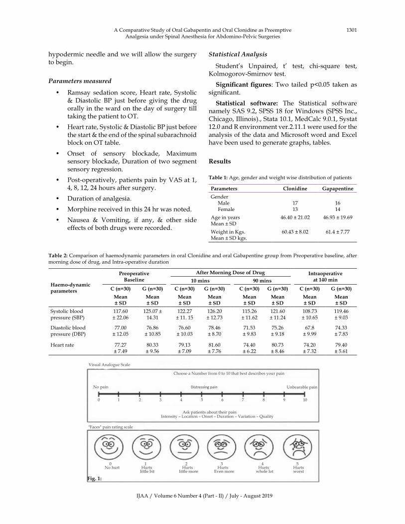
IJAA�/�Volume�6�Number�4�(Part�-�II)�/�July�-�August�2019
1301
hypodermic�needle�and�we�will�allow�the�surgery�to�begin.
Para�eters��easured
�� Ramsay� sedation� score,��eart� rate,� Systolic��� Diastolic� �P� just� before� giving� the� drug�orally�in�the�ward�on�the�day�of�surgery�till�taking�the�patient�to�OT.
�� �eart�rate,�Systolic���Diastolic��P�just�before�the�start���the�end�of�the�spinal�subarachnoid�block�on�OT�table.
�� Onset� of� sensory� blockade,� Maximum�sensory�blockade,�Duration�of�two�segment�sensory�regression.
�� Post-operatively,�patients�pain�by�VAS�at�1,�4,�8,�12,�24�hours�after�surgery.
�� Duration�of�analgesia.
�� Morphine�received�in�this�24�hr�was�noted.
�� Nausea� �� Vomiting,� if� any,� �� other� side�effects�of�both�drugs�were�recorded.
Visual�Analogue�Scale
Choose�a�Number�from�0�to�10�that�best�describes�your�pain
No�pain D��������������� �nbearable�pain
0 1 2 3 4 5 6 7 8 9 10
Ask�patients�about�their�pain�Intensity�–�Location�–�Onset�–�Duration�–�Variation�–��uality
�Faces��pain�rating�scale
0�No�hurt
1�urts�little�bit
2�urts�
little�more
3�urts�
Even�more
4�urts�whole�lot
5�urts�worst
Statistical�Analysis
Student�s� �npaired,� t�� test,� chi-s�uare� test,��olmogorov-Smirnov�test.
Signi��cant���gures:� Two� tailed� p�0.05� taken� as�signi��cant.
Statistical� soft�are:� The� Statistical� software�namely�SAS�9.2,�SPSS�18�for�Windows�(SPSS�Inc.,�Chicago,�Illinois).,�Stata�10.1,�MedCalc�9.0.1,�Systat�12.0�and�R�environment�ver.2.11.1�were�used�for�the�analysis�of�the�data�and�Microsoft�word�and�Excel�have�been�used�to�generate�graphs,�tables.
�esults
Table��:�Age,�gender�and�weight�wise�distribution�of�patients
Parameters Clonidine �apapentine
Gender�Male�Female
1713
1614
Age�in�years�Mean���SD�
46.40���21.02 46.93���19.69
Weight�in��gs.Mean���SD�kgs.
60.43���8.02 61.4���7.77
A�Comparative�Study�of�Oral�Gabapentin�and�Oral�Clonidine�as�Preemptive�Analgesia�under�Spinal�Anesthesia�for�Abdomino-Pelvic�Surgeries
�aemo�dynamic�parameters
Preoperati�e�Baseline
After�Morning��ose�of��rug Intraoperati�e�at�����min���mins ���mins
C��n���� ���n���� C��n���� ���n���� C��n���� ���n���� C��n���� ���n����
Mean���S�
Mean���S�
Mean���S�
Mean���S�
Mean���S�
Mean��S�
Mean��S�
Mean��S�
Systolic�blood�pressure�(S�P)
117.60��22.06
125.07���14.31
122.27��11.�15
126.20��12.73
115.26��11.62
121.60��11.24
108.73��10.65
119.46��9.03
Diastolic�blood�pressure�(D�P)
77.00��12.05
76.86��10.85
76.60��10.03
78.46��8.70
71.53��9.83
75.26��9.18
67.8��9.99
74.33��7.83
�eart�rate 77.27��7.49
80.33��9.56
79.13��7.09
81.60��7.76
74.40��6.22
80.73��8.46
74.20��7.32
79.40��5.61
Table��:�Comparison�of�haemodynamic�parameters�in�oral�Clonidine�and�oral�Gabapentine�group�from�Preoperative�baseline,�after�morning�dose�of�drug,�and�Intra-operative�duration
Fig���:
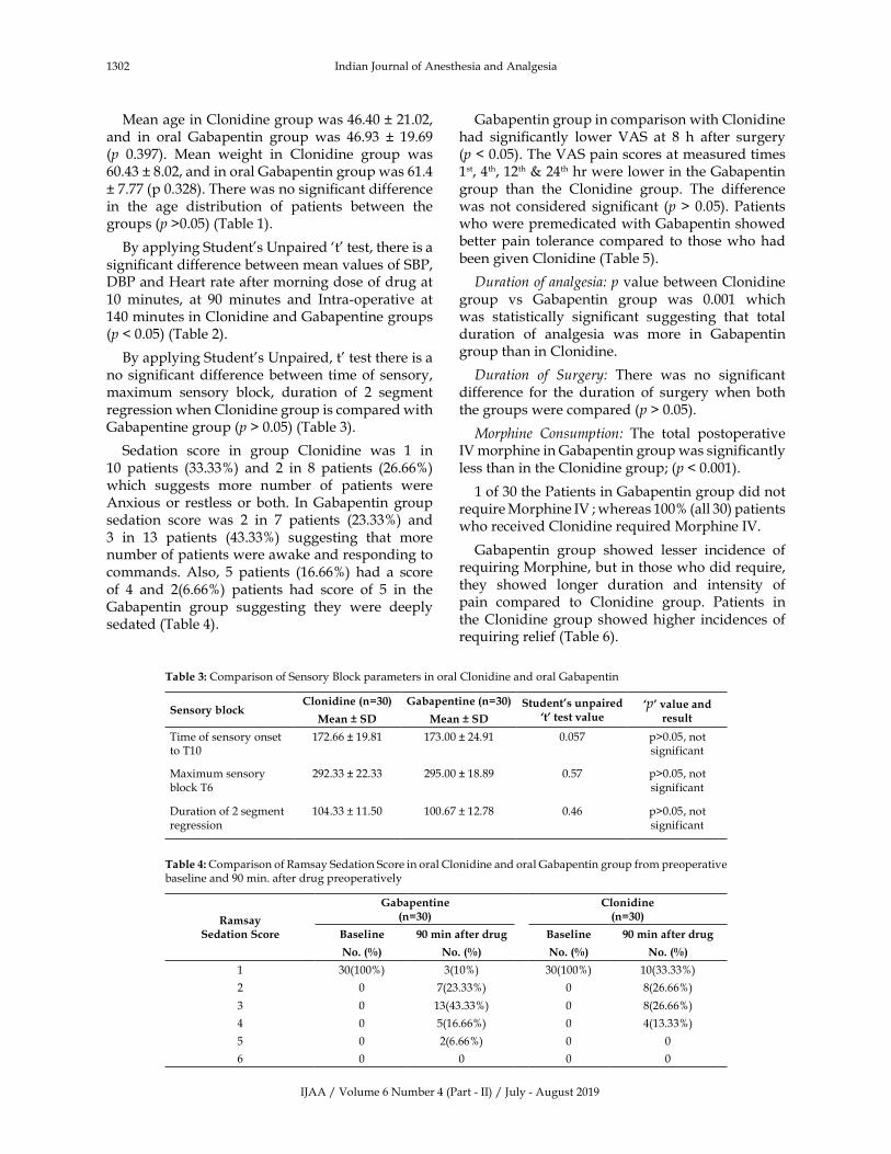
IJAA�/�Volume�6�Number�4�(Part�-�II)�/�July�-�August�2019
1302 Indian�Journal�of�Anesthesia�and�Analgesia
Mean�age�in�Clonidine�group�was�46.40���21.02,�and� in� oral� Gabapentin� group� was� 46.93� �� 19.69�(p� 0.397).� Mean� weight� in� Clonidine� group� was�60.43���8.02,�and�in�oral�Gabapentin�group�was�61.4���7.77�(p�0.328).�There�was�no�signi��cant�difference�in� the� age� distribution� of� patients� between� the�groups�(p��0.05)�(Table�1).
�y�applying�Student�s��npaired��t��test,�there�is�a�signi��cant�difference�between�mean�values�of�S�P,�D�P�and��eart�rate�after�morning�dose�of�drug�at�10� minutes,� at� 90� minutes� and� Intra-operative� at�140�minutes�in�Clonidine�and�Gabapentine�groups�(p���0.05)�(Table�2).
�y�applying�Student�s��npaired,�t��test�there�is�a�no�signi��cant�difference�between�time�of�sensory,�maximum� sensory� block,� duration� of� 2� segment�regression�when�Clonidine�group�is�compared�with�Gabapentine�group�(p���0.05)�(Table�3).
Sedation� score� in� group� Clonidine� was� 1� in�10� patients� (33.33�)� and� 2� in� 8� patients� (26.66�)�which� suggests� more� number� of� patients� were�Anxious�or� restless� or�both.� In�Gabapentin� group�sedation� score� was� 2� in� 7� patients� (23.33�)� and�3� in� 13� patients� (43.33�)� suggesting� that� more�number�of�patients�were�awake�and�responding�to�commands.� Also,� 5� patients� (16.66�)� had� a� score�of� 4� and� 2(6.66�)� patients� had� score� of� 5� in� the�Gabapentin� group� suggesting� they� were� deeply�sedated�(Table�4).
Gabapentin�group�in�comparison�with�Clonidine�had� signi��cantly� lower� VAS� at� 8� h� after� surgery�(p���0.05).�The�VAS�pain�scores�at�measured�times�1st,�4th,�12th���24th�hr�were�lower�in�the�Gabapentin�group� than� the� Clonidine� group.� The� difference�was�not�considered� signi��cant� (p���0.05).�Patients�who�were�premedicated�with�Gabapentin�showed�better�pain� tolerance� compared� to� those�who�had�been�given�Clonidine�(Table�5).
�uration�of�analgesia:�p�value�between�Clonidine�group� vs� Gabapentin� group� was� 0.001� which�was� statistically� signi��cant� suggesting� that� total�duration� of� analgesia� was� more� in� Gabapentin�group�than�in�Clonidine.
�uration� of� Surger�:� There� was� no� signi��cant�difference� for� the� duration� of� surgery�when� both�the�groups�were�compared�(p���0.05).
Morphine� Consumption:� The� total� postoperative�IV�morphine�in�Gabapentin�group�was�signi��cantly�less�than�in�the�Clonidine�group;�(p���0.001).
1�of�30�the�Patients�in�Gabapentin�group�did�not�re�uire�Morphine�IV�;�whereas�100��(all�30)�patients�who�received�Clonidine�re�uired�Morphine�IV.
Gabapentin� group� showed� lesser� incidence� of�re�uiring�Morphine,�but�in�those�who�did�re�uire,�they� showed� longer� duration� and� intensity� of�pain� compared� to� Clonidine� group.� Patients� in�the�Clonidine�group�showed�higher� incidences�of�re�uiring�relief�(Table�6).
Table��:�Comparison�of�Sensory��lock�parameters�in�oral�Clonidine�and�oral�Gabapentin
Sensory�blockClonidine��n���� �abapentine��n���� Student�s�unpaired�
�t��test��alue�p���alue�and�
resultMean���S� Mean���S�
Time�of�sensory�onset�to�T10
172.66���19.81 173.00���24.91 0.057 p�0.05,�not�significant
Maximum�sensory�block�T6
292.33���22.33 295.00���18.89 0.57 p�0.05,�not�significant
Duration�of�2�segment�regression�
104.33���11.50 100.67���12.78 0.46 p�0.05,�not�significant
Table��:�Comparison�of�Ramsay�Sedation�Score�in�oral�Clonidine�and�oral�Gabapentin�group�from�preoperative�baseline�and�90�min.�after�drug�preoperatively
�amsay�Sedation�Score
�abapentine�n����
Clonidine�n����
Baseline� ���min�after�drug Baseline ���min�after�drug
No����� No����� No����� No�����
1 30(100�) 3(10�) 30(100�) 10(33.33�)
2 0 7(23.33�) 0 8(26.66�)
3 0 13(43.33�) 0 8(26.66�)
4 0 5(16.66�) 0 4(13.33�)
5 0 2(6.66�) 0 0
6 0 0 0 0
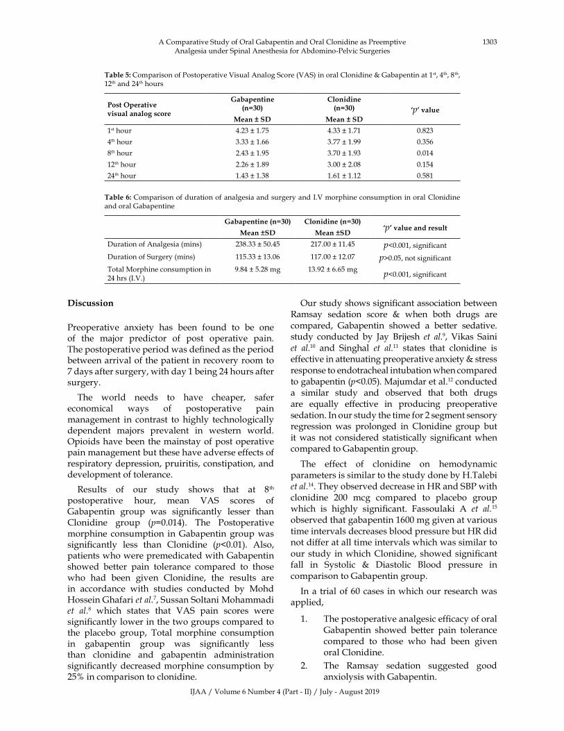
IJAA�/�Volume�6�Number�4�(Part�-�II)�/�July�-�August�2019
1303
�iscussion
Preoperative� anxiety� has� been� found� to� be� one�of� the� major� predictor� of� post� operative� pain.�The�postoperative�period�was�de��ned�as�the�period�between�arrival�of�the�patient�in�recovery�room�to�7�days�after�surgery,�with�day�1�being�24�hours�after�surgery.
The� world� needs� to� have� cheaper,� safer�economical� ways� of� postoperative� pain�management� in� contrast� to� highly� technologically�dependent� majors� prevalent� in� western� world.�Opioids�have�been�the�mainstay�of�post�operative�pain�management�but�these�have�adverse�effects�of�respiratory�depression,�pruiritis,�constipation,�and�development�of�tolerance.
Results� of� our� study� shows� that� at� 8th�postoperative� hour,� mean� VAS� scores� of�Gabapentin� group� was� signi��cantly� lesser� than�Clonidine� group� (p�0.014).� The� Postoperative�morphine� consumption� in�Gabapentin�group�was�signi��cantly� less� than� Clonidine� (p�0.01).� Also,�patients�who�were�premedicated�with�Gabapentin�showed� better� pain� tolerance� compared� to� those�who� had� been� given� Clonidine,� the� results� are�in� accordance� with� studies� conducted� by� Mohd��ossein�Ghafari�et�al.7,�Sussan�Soltani�Mohammadi�et� al.8� which� states� that� VAS� pain� scores� were�signi��cantly�lower�in�the�two�groups�compared�to�the� placebo� group,� Total� morphine� consumption�in� gabapentin� group� was� signi��cantly� less�than� clonidine� and� gabapentin� administration�signi��cantly�decreased�morphine�consumption�by�25��in�comparison�to�clonidine.
Our�study�shows�signi��cant�association�between�Ramsay� sedation� score� �� when� both� drugs� are�compared,� Gabapentin� showed� a� better� sedative.�study� conducted� by� Jay� �rijesh� et� al.9,� Vikas� Saini�et� al.10� and� Singhal� et� al.11� states� that� clonidine� is�effective�in�attenuating�preoperative�anxiety���stress�response�to�endotracheal�intubation�when�compared�to�gabapentin�(p�0.05).�Majumdar�et�al.12�conducted�a� similar� study� and� observed� that� both� drugs�are� e�ually� effective� in� producing� preoperative�sedation.�In�our�study�the�time�for�2�segment�sensory�regression� was� prolonged� in� Clonidine� group� but�it�was�not� considered� statistically� signi��cant�when�compared�to�Gabapentin�group.
The� effect� of� clonidine� on� hemodynamic�parameters�is�similar�to�the�study�done�by��.Talebi�et�al.14.�They�observed�decrease�in��R�and�S�P�with�clonidine� 200� mcg� compared� to� placebo� group�which� is� highly� signi��cant.� Fassoulaki� A� et� al.15
observed�that�gabapentin�1600�mg�given�at�various�time�intervals�decreases�blood�pressure�but��R�did�not�differ�at�all�time�intervals�which�was�similar�to�our� study� in�which�Clonidine,� showed�signi��cant�fall� in� Systolic� �� Diastolic� �lood� pressure� in�comparison�to�Gabapentin�group.
In�a� trial�of�60�cases� in�which�our� research�was�applied,
1.� The�postoperative�analgesic�ef��cacy�of�oral�Gabapentin� showed� better� pain� tolerance�compared� to� those� who� had� been� given�oral�Clonidine.
2.� The� Ramsay� sedation� suggested� good�anxiolysis�with�Gabapentin.
A�Comparative�Study�of�Oral�Gabapentin�and�Oral�Clonidine�as�Preemptive�Analgesia�under�Spinal�Anesthesia�for�Abdomino-Pelvic�Surgeries
Table��:�Comparison�of�Postoperative�Visual�Analog�Score�(VAS)�in�oral�Clonidine���Gabapentin�at�1st,�4th,�8th,�12th�and�24th�hours
Post�Operati�e��isual�analog�score
�abapentine�n����
Clonidine�n����� �p���alue
Mean���S� Mean���S�
1st�hour 4.23���1.75 4.33���1.71 0.823
4th�hour 3.33���1.66 3.77���1.99 0.356
8th�hour 2.43���1.95 3.70���1.93 0.014
12th�hour 2.26���1.89 3.00���2.08 0.154
24th�hour 1.43���1.38 1.61���1.12 0.581
Table��:�Comparison�of�duration�of�analgesia�and�surgery�and�I.V�morphine�consumption�in�oral�Clonidine�and�oral�Gabapentine
�abapentine��n���� Clonidine��n�����p���alue�and�result
Mean��S� Mean��S�
Duration�of�Analgesia�(mins) 238.33���50.45 217.00���11.45 p�0.001,�significant
Duration�of�Surgery�(mins) 115.33���13.06 117.00���12.07 p�0.05,�not�significantTotal�Morphine�consumption�in�24�hrs�(I.V.)
9.84���5.28�mg 13.92���6.65�mgp�0.001,�significant
IJAA�/�Volume�6�Number�4�(Part�-�II)�/�July�-�August�2019
1304 Indian�Journal�of�Anesthesia�and�Analgesia
3.� As� haemodynamic� parameters� changes�in� Gabapentin� group� were� statistically�insigni��cant,� Gabapentin� is� considered�haemodynamically�stable.
4.� Morphine� consumption� in� 24� hrs� was�signi��cantly� high� in� Clonidine� group�compared�to�Gabapentin.
5.� There�were� no� signi��cant� change� in� time�of�sensory�onset,�maximum�sensory�block�and� duration� of� 2� segment� regression�when�Clonidine�was�compared�with�drug�Gabapentin.
6.� In� comparison� of� side� effects,� there� was�increased�incidence�of�nausea�and�vomiting�in�Clonidine�group.
Conclusion
We� conclude� that,� Oral� Gabapentin� 300� mg�given� before� 90� minutes� as� preemptive� analgesia�was�more�effective�in�reducing�postoperative�pain�and�morphine�consumption,�also�providing�better�anxiolysis�in�patients�undergoing�abdomino-pelvic�Surgeries� under� spinal� anesthesia� compared� to�Oral�Clonidine�100��g.
Conflict�of�Interest:��one
�eferences
1.� Turan� A,� �aya� G,� �aramanlioglu� �,� Pamukcu��,� Apfel� CC.� Effect� of� oral� gabapentin� on�postoperative� epidural� analgesia.� �r� J� Anaesth�2006�Feb;96(2):242–6.
2.� �issin.� Preemptive� Analgesia.� Anesthesiology�2000;93:1138–43.
3.� Dirks�J,��ilsted��L,�Moiniche�S,�et�al.�Mechanism�of� post� operative� pain:� Clinical� indication� for�a� contribution� of� central� neural� sensitization.�Anaesthesiology�2002;97:1591–6.
4.� Mao�J,�Chen�LL.�gabapentin�in�pain�management.�Anesth�Analg.�2000;91:680–7.
5.� �idalgo�MP,� Auzani� JA,� Rumpel� LC,� et� al.� The�
clinical� effect� of� small� oral� Clonidine� doses� on�
perioperative� outcomes� in� patients� undergoing�
abdominal� hysterectomy.� Anaesth� Analg� 2005�
Mar;100(3):795–802.
6.� Tryba�M,�Gehling�M.�Clonidine-a�potent�analgesic�
adjuvant.� Curr� Opin� Anaesthesiol.� 2002� Oct;�
15(5):511–7.
7.� Ghafari� Md.� �,� Akrami� M.� Preoperative�
Gabapentin�or�Clonidine�decreases�postoperative�pain�and�morphine�consumption�after�abdominal�
hysterectomy.� Research� Journal� of� �iological�
Sciences.�2009;4(4):458–63.
8.� Mohammadi� SS,� Seyedi� M.� Comparing� oral�gabapentin�versus�clonidine�as�premedication�on�
early� postoperative� pain,� nausea� and� vomiting�
following� general� anesthesia.� Saudi� J� Anaesth.�
2009;3:25–8.
9.� �rijesh� J,� Shukla� �,� Singh� S.P.� Comparing� the�
Effects� of� Oral� Gabapentin� and� Clonidine� on�Preoperative� Anxiolysis� and� Attenuation� of�
Stress� Response� to� Endotracheal� Intubation.�
International� Journal� of� Science� and� Research�
(IJSR).�2015�Aug;4(8):1735–40.
10.� Saini� V,� Lakesh� �� Anand.� Oral� Clonidine�
premedication�in�patients�undergoing�intraocular�
surgery� under� local� anesthesia.� J� Anaesth� Clin�Pharmacol.�2009;25(3):311–315.
11.� Singhal� S�,� �aur� �,� Arora� P.� Oral� clonidine�
versus�gabapentin�as�premedicant�for�obtunding�
hemodynamic� response� to� laryngoscopy� and�
tracheal� intubation.� Saudi� J� Anaesth.� 2014�
Apr-Jun;8(2):172–177.
12.� Majumdar� S,� Das� A,� Das� �,� et� al.� Comparative�
evaluation� of� oral� gabapentin� versus� clonidine�
as� premedication� on� preoperative� sedation� and�
laryngoscopic�stress�response�attenuation�for�the�patients�undergoing�general�anesthesia.�Perspect�
Clin�Res.�2015�Oct-Dec;6(4):211–216.
13.� Talebi� �,� Nourozi� A,� Fateh� S,� et� al.� Effects� of�
oral� clonidine� premedication� on� haemodynamic�
response�to�laryngoscopy�and�tracheal�intubation.�
Pak�J��iol�Sci.�2010;13:1146–50.
14.� Fassoulaki� A,� Melemeni� A,� Paraskeva� A,� et� al.
Gabapentin� attenuates� the� pressor� response� to�
direct�laryngoscopy�and�tracheal�intubation.��r�J�
Anaesth.�2006;96:769–73.
��Red�Flower�Publication�Pvt.�Ltd.�
�Original�Research�ArticleIndian�Journal�of�Anesthesia�and�Analgesia�
2019;6(4)�(Part�-�II):�1305-1309DOI:�http://dx.doi.org/10.21088/ijaa.2349.8471.6419.34
�ltrasound� �uided� Combined� Superficial� Cer�ical� Ple�us� Block�Interscalene�Block�for�Anesthesia�in�Cla�icular�Fractures:�A��etrospecti�e�Obser�ational�Cohort�Study
S�Arun����eepan�C����omy�Thomas�
1Assistant� Professor� 2,3Junior� Residents,� Department� of� Anaesthesiology,� Dr.� Somervell� Memorial� CSI� Medical� College,��arakonam,�Trivandrum,��erala�695504,�India.�
Corresponding�Author:�S��Arun,�Assistant�Professor,�Department�of�Anaesthesiology,�Dr.�Somervell�Memorial�CSI�Medical�College,��arakonam,�Trivandrum,��erala�695504,�India.
E�mail:�[email protected]
�ecei�ed�on�27.05.2019,�Accepted�on�11.07.2019
Abstract
Background:�Even� though�various�peripheral�nerve�blocks�are�used� for� analgesia� in� clavicular� fractures.�The�use�of�ultrasound�guided�combined�superficial� cervical�plexus�block�and� interscalene�blocks�as�a� sole�anaesthetic� techni�ue� for� surgical� fixation� of� clavicular� fractures� are� not� well,� established.�Materials� and�Methods:�A� retrospective� chart� review� of� patient� undergoing� clavicular� fracture� surgeries�was� performed.�Patients� received� combined� superficial� cervical� plexus� block� and� interscalene� block�were� included� in� the�study.��lock�success�and�complication�rate�were�evaluated.�Results:�Of�the�20�patients�underwent�clavicular�fracture� surgery,� 12� of� them� received� ultrasound� guided� combined� superficial� cervical� plexus� block� and�interscalene�block.��lock�success�rate�was�found�to�be�100�.�Time�for�rescue�analgesia�found�to�be�6.66���0.84�hours.�And�there�were�no�occurence�of�any�Complications.�Conclusion:�We�concluded�that�ultrasound�guided�combined�superficial�cervical�plexus�block�and�interscalene�block�can�be�used�as�a�sole�anaesthetic�techni�ue�in�clavicular�fracture�repair.
�ey�ords:�Interscalene��lock;�Clavicular�Fractures;��rachial�plexus
�o��to�cite�this�article:
S� Arun,� Deepan� C,� Jomy� Thomas.� �ltrasound� Guided� Combined� Superficial� Cervical� Plexus� �lock-Interscalene� �lock� for�Anesthesia�in�Clavicular�Fractures:�A�Retrospective�Observational�Cohort�Study.�Indian�J�Anesth�Analg.�2019;6(4):1305-.
Introduction
Clavicle� fractures� comprised� up� to� 4�� of� all�fractures� among� all� age� groups1.� Isolated� clavicle�fractures� comprising� 44.1�� of� all� fractures� in�shoulder�gridle2.
Surgical� treatment� of� clavicular� fractures�had� show� signi��cant� advantages� like� increased�postoperative� strength3,� �uicker� time� to� return�
to� normal� activities� and� low� re-fracture� rates4,5,6.Traditionally�the�clavicular�fracture�surgeries�were�performed� under� general� anesthesia.� Analgesia�for� clavicular� fractures� can� be� challenging� for�anaesthesiologist�secondary�to�complex�and�varied�innervation.� Literatures� describing� hetrogenous�innervation� of� clavicle� and� overlying� skin.� The�sensory� innervation� of� clavicle� have� contribution�from�both�cervical�and�brachial�plexus7.
IJAA�/�Volume�6�Number�4�(Part�-�II)�/�July�-�August�2019
1306 Indian�Journal�of�Anesthesia�and�Analgesia
The� contemporary� literature� surrounding�the� optimal� regional� anaesthetic� techni�ue� for�clavicular� surgery� which� can� provide� superior�postoperative�analgesia�and�minimizing�symptoms�intra-operatively� is� lacking.� The� various� nerve�blocks� that� are� used� to� anaesthetize� the� clavicle�super��cial� cervical� plexus� block,� interscalene�brachial� plexus� block,� supraclavicular� block,� and�a�combination�of�both�interscalene�and�super��cial�cervical�plexus�block8.
Many�factors�play�arole�in�selecting�the�optimal�regional�anaesthetic�methods�like�fracture�location�and� complexity,� incision� over� mixed� innervation�and�surgical�positioning�and�manipulations.
Eventhough� interscalene� blocks� were� used�for� shoulder� surgeries,� the� dual� innervation�of� the� clavicular� area� suggest� that� a� single�interscalene� block� maynot� be� suf��cient� for� the�surgical�pain9.
With�the�advent�of�point�of�care�ultrasonography�reports�of�successful�usage�of�combined�peripheral�nerve� blocks� are� emerging.� �ltrasound� guided�techni�ues� increased� the�success� rates�and�reduce�the� complications� associated�with� the� procedures�as�well�as�drugs.�The�reduction�of�local�anaesthetics�with� ultrasound� guided� techni�ues� had� helped�us� to� perform� multiple� blocks� without� the� risk�for�toxicity.
�erring�AA�et�al.10�and�vandepitte�et�al.11�reported�a�successful�use�of�super��cial�cervical�plexus�block�and�interscalene�block�for�clavicular�surgery.�Gray�et�al.12�stated�that�clavicular�surgery�also�a�indication�for�super��cial�cervical�plexus�block.
The� objective� of� our� retrospective� cohort� study�is� to� demonstrate� the� effectiveness� of� combined�super��cial� cervical� plexus� block� and� interscalene�block� as� a� sole� anaesthetic� that� can� be� used� for�patients�undergoing�clavicular�fractures�repair.
Materials�and�Methods
After� obtaining� approval� from� the� hospital�institutional� review� board,� a� retrospective� chart�review� of� patients� undergoing� shoulder� surgery�over� a� period� of� 3� years� starting� from� July� 2015�was�performed.�The�anaesthetic�records�including�preoperative� evaluation,� intraoperative� records�and� post� anesthesia� records� were� obtained� from�the�medical�records�department.�The�postoperative�records�including�hospital�course,�physician�notes�also� obtained,� patient� demographics,� surgical�procedure,� intraoperative� anaesthetics� and� PAC��course�were�recorded.
Patients� with� clavicular� fractures� repair� under�interscalene� and� super��cial� cervical� plexus� block�were�recorded.�Among�those�the� local�anaesthetic�used,�concentration�of�the�drug,�volume�used�were�noted.��lock�success�and�acute�complications�if�any�also�noted�from�the�charts.
Standard�contraindications�to�placement�of�nerve�blocks�included�patient�refusal,�coagulopathy�and�drug�allergy�and�infection�of�intended�site.
In� accordance� with� the� hospital� protocol�appropriate�informed�consent�were�obtained�from�the�patients.
�lock� success� is� de��ned� as� one� which� did� not�necessitate� conversion� to� general� anesthesia.�Rescue� analgesia� time� noted� from� the� records.��emodynamic�parameters�were�also�noted.
Patients� who� were� converted� to� general�anesthesia� (block� failure)� and� those�patients�were�block�duration�are�not�recorded�are�excluded�from�the�study.
Anaesthetic��ethods
�ltrasound� guided� combined� interscalene� and�super��cial� cervical� plexus� block� were� performed�under� ultrasound� guidance� (LOGI�� GE� �ealth�Care),�12M�z�linear�transducer�was�used.
The� transducer� positioned� in� transverse� plane�and�the�carotid�artery�was�identi��ed�at�the�level�of�cricoid.�Then�the�transducer�was�moved�laterally�to�identify�the�anterior�and�middle�scalene�muscles�and�we�can�able�to�found�the�brachial�plexus�in�between.�A�Lateral� to�medial,� in-plane�needle� insertion�was�done.�Once�inside�the�interscalene�groove�and�after�careful�aspiration�and�needle�tip�con��rmation�15�ml�of�0.25��ropivacaine�was�injected.
After� the�performance�of� the� interscalene�block�the� needle�was� withdrawn� and� redirected� to� the�cervical� plexus.� The� hyperechoic� fascia� of� the�sternocleidomastoid� muscle� on� its� posterolateral�border�was�identi��ed�and�the�cervical�plexus�will�be�identi��ed�as�small�hypoechoic�nodules�super��cial�to� pre� vertebral� facia� overlies� the� interscalene�groove.�The�needle�tip�was�adjusted�to�inject�10�ml�of�0.25��ropivacaine�adjacent�to�the�plexus.
Statistical�analysis
Descriptive� statistics� of� the� study� are� calculated�and� data� emerged� were� analysed� using� SPSS�statistics� software.� Continious� �uantitative� data�were� expressed� as� numbers,� mean� and� standard�deviation� and� �ualitative� data�were� expressed� as�numbers�and�percentage.
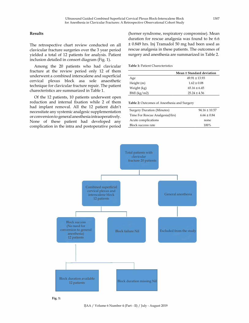
IJAA�/�Volume�6�Number�4�(Part�-�II)�/�July�-�August�2019
1307
�esults
The� retrospective� chart� review� conducted� on� all�clavicular�fracture�surgeries�over�the�3�year�period�yielded� a� total� of� 12�patients� for� analysis.� Patient�inclusion�detailed�in�consort�diagram�(Fig.�1).
Among� the� 20� patients� who� had� clavicular�fracture� at� the� review� period� only� 12� of� them�underwent�a�combined�interscalene�and�super��cial�cervical� plexus� block� asa� sole� anaesthetic�techni�ue�for�clavicular�fracture�repair.�The�patient�charecteristics�are�summarized�in�Table�1.�
Of� the�12�patients,�10�patients�underwent�open�reduction� and� internal� ��xation� while� 2� of� them�had� implant� removal.� All� the� 12� patient� didn�t�necessitate�any�systemic�analgesic�supplementation�or�conversion�to�general�anesthesia�intraoperatively.�None� of� these� patient� had� developed� any�complication�in�the�intra�and�postoperative�period�
(horner�syndrome,�respiratory�compromise).�Mean�
duration� for�rescue�analgesia�was�found� to�be�6.6�
��0.849�hrs.�Inj�Tramadol�50�mg�had�been�used�as�
rescue�analgesia�in�these�patients.�The�outcomes�of�
surgery�and�anesthesia�are�summarized�in�Table�2.
Table��:�Patient�Charecteristics
Mean���Standard�de�iation
Age 49.91���13.93
�eight�(m) 1.62���0.08
Weight�(kg) 65.16���6.45
�MI�(kg/m2) 25.24���4.56
Table��:�Outcomes�of�Anesthesia�and�Surgery
Surgery�Duration�(Minutes) 94.16���10.57
Time�For�Rescue�Analgesia(�rs) 6.66���0.84
Acute�complications� none
�lock�success�rate 100�
Total�patients�with�clavicular
�fracture�20�patients
General�anesthesia
�lock�failure�Nil Excluded�from�the�study
�lock�duration�available�
12�patients �lock�duration�missing�Nil
�lock�success�(No�need�for�
conversion�to�general�anesthesia)�12�patients
Combined� �superficialcervical�plexus�and�interscalene�block�
12�patients�
�ltrasound�Guided�Combined�Superficial�Cervical�Plexus��lock-Interscalene��lock�for�Anesthesia�in�Clavicular�Fractures:�A�Retrospective�Observational�Cohort�Study
Fig���:
IJAA�/�Volume�6�Number�4�(Part�-�II)�/�July�-�August�2019
1308 Indian�Journal�of�Anesthesia�and�Analgesia
�iscussion
The� combined� interscalene� and� super��cial� cervical�plexus� block� has� been� shown� to� be� promising�anaesthetic�techni�ue�for�clavicular�fracture�surgeries.
Since� clavicle� received� innervation� both� from�cervical� plexus� as� well� as� from� brachial� plexus�the�combined�interscalene�and�super��cial� cervical�plexus�block�may�be�the�best�option�for�the�surgical���xation�of�clavicle�fractures.
�erring�et�al.10�reported�the�the�ultrasound�guided�super��cial�cervical�plexus�block� for�analgesia�and�anesthesia�in�emergency�settings.
�shema� et� al.13� reported� the� successful� use� of�supraclavicular�along�with�C5,�C6�nerve�blocks�for�clavicular� surgery.� This� supports� the� innervation�of� clavicular� surface� by� supraclavicular� nerve� a�branch�of�super��cial�cervical�plexus.
Vandepitte� et� al.11� also� reported� successful�combined� interscalene� and� super��cial� cervical�plexus�block�usage� in� surgical� repair�of�clavicular�fracture�in�a�pregnant�women.
�alban� O� et� al.14� in� their� retrospective� study�found�that�ultrasound�guided�combined�super��cial�cervical� plexus� and� interscalene� blocks� had� been�successfully�used�as�a�sole�anesthesia�for�clavicular�fracture� patients�without� any� complications.� This�was�further�con��rmed�by�seni�pot�sangbam�et�al.15�
in�their�prospective�observational�study.
Contractor�et�al.16�reported�horner�syndrome�and�hoarsness� of� voice� in� some� patients� and� also� the�usage�dexmedtomidine�1�mcg/kg.
�ut�in�our�study�we�didn�t�found�any�side�effects.�And� in� our� study� we� didn�t� use� any� additional�sedatives�and�analgesics�other�than�the�nerve�block.�The�duration�of� rescue�analgesia�was� found� to�be�6�hrs,�which�is�comparable�to�other�studies.
Reverdy� et� al.17� reported� a� high� patient�satisfaction�rate�in�patient�who�underwent�surgery�for� clavicular� fracture�under�combined�super��cial�cervical�plexus�block�interscalene�block.
The� use� of� ultrasonography� in� the� study� had�further� reduced� the� complication� associated�with�these�nerve�blocks.
�i�itations
This�study�is�limited�by�the�small�number�of�cases�as�hospital�admissions�for�clavicular�surgeries�are�usually�low.�A�randomized�controlled�trial�of�larger�sample�size�comparing�the�two�groups�will�have�a�stronger�implication�on�the�outcome�of�the�study.
Conclusion
�ased� on� our� experience� we� suggest� that� the�combined� super��cial� cervical� plexus� block� and�interscalene�block�can�be�used�as�a�sole�anaesthetic�method�for�patients�with�clavicular�fracture�repair.�A�further�randomized�control�studies�are�re�uired�to�determine�which�constitute�the�better�method�for�clavicular�fracture�repairs.
Ac�no�led�e�ent
I�would� like� to�thank�my�wife�Dr.�Aparna�for�her�assistance� with� the� study.� We� didn�t� have� any���nancial� support� or� sponsorship� and� there� is� no�con��ict�of�interest.
Conflict�of�interest:�NIL
�eferences
1.� Nord�vist� A,� and� Petersson� C.� The� incidence�of� fractures� of� the� clavicle.� Clin.� Orthop.�1994;300:127–132.
2.� Postacchini� FS.� Gumina� P.� De� Santis,� et� al.Epidemiology� of� Clavicle� Fractures.� J.� Shoulder�Elbow�Surg.�2002;11:452–56.
3.� Witzel� �.� �Intramedullary� osteosynthesis� in�fractures� of� the� mid-third� of� the� clavicle� in�sports� traumatology�.� �� Orthop�nfall.� 2007;�145(5):639–42.
4.� Jubel�A,�Andemahr� J,� �ergmann��,� et� al� Elastic�stable� intramedullary� nailing� of� midclavicular�fractures� in� athletes� �ritish� Journal� of� Sports�Medicine.�2003;37:480–84.
5.� Meier� C,� Grueninger� P,� Platz� A.� Elastic� stable�intramedullary�nailing�for�midclavicular�fractures�in�athletes:�indications,�technical�pitfalls�and�early�results.�ActaOrthop��elg.�2006;72:269–75.
6.� Morgan�RJ,��ankston�LS,��oenig�MP,���Connor�PM.� Evolving� Management� of� Middle-Third�Clavicle� Fractures� in� the� National� Football�League.� The� American� Journal� of� Sports�Medicine.� 2010;38(10):2092–96.� https://doi.org/10.1177/0363546510372795.
7.� Dejerine�J.�Syndromes�sensitifs.�In:�Dejerine�J,�ed.�Semeiologie�des�Affections�du�SystemeNerveux.�Paris,�France:�Masson;�1914.
8.� Tran� D�,� Tiyaprasertkul� W,� and� Gonzalez� AP.�Analgesia� for� clavicular� fracture� and� surgery:� a�call� for� evidence.� Regional�Anesthesia� and� Pain�Medicine,�2013;38(6):539–43.
9.� Jonathan�P.��line,��ltrasound-Guided�Placement�of� Combined� Superficial� Cervical� Plexus� and�Selective� C5� Nerve� Root� Catheters:� A� Novel�
IJAA�/�Volume�6�Number�4�(Part�-�II)�/�July�-�August�2019
1309
Approach� to� Treating� Distal� Clavicle� Surgical�Pain.�AANA�Journal.�2013�Feb;81(1):19–22.
10.� �erring� AA,� Stone� M�,� Frenkel� O,� et� al.� The�ultrasound-guided� superficial� cervical� plexus�block�for�anesthesia�and�analgesia� in�emergency�care�settings.�Am�J�Emerg�Med.�2012;30:263–67.
11.� Vandepitte� C,� Latmore� M,� O�Murchu� E� et� al.�Combined� interscalene� and� superficial� cervical�plexus� blocks� for� surgical� repair� of� a� clavicular�fracture� in� a� 15� week� pregnant� woman.�International�journal�of�obstetric�anesthesia.�2014�May;23(2)194–5.
12.� Gray� AT.� Superficial� cervical� plexus� block.�In:� Gray� AT,� ed.� Atlas� of� �ltrasound-Guided�Regional� Anesthesia.� 2nd� ed.� Philadelphia,� PA:�Saunders;�2012.
13.� �eshima��,�Otake��.�Successful�clavicle�fracture�surgery�performed�under�selective�supraclavicular�nerve�block�using�the�new�subclavian�approach.�JA�Clinical�reports.�2016;2:34.
14.� �alaban�O,�D�lgero�lu�TC,�Ayd�n�T.��ltrasound-Guided� Combined� Interscalene-Cervical� Plexus��lock� for� Surgical� Anesthesia� in� Clavicular�Fractures:� A� Retrospective� Observational�Study.� Anesthesiology� Research� and� Practice.�2018;Article�ID�7842128,�6�page.
15.� Seni� P,� Jonan� P�.� Efficacy� of� Combined�Interscalene��lock�and�Superficial�Cervical�Plexus��lock�for�Surgeries�of�the�Clavicle:�A�Prospective�Observational� Study.� Journal� of� Clinical� and�Diagnostic�Research.�2019�Feb;13(2):�C05–�C08.
16.� Contractor���,�Shah�VA,�Gajjar�VA.��ltrasound�guided� superficial� cervical� plexus� and�interscalene� brachial� plexus� block� for� clavicular�surgery.� Anaesth� Pain� and� Intensive� Care.�2016;20(4):447–450.
17.� Fabien� Reverdy;� Combined� interscalene-superficial� cervical� plexus� block� for� clavicle�surgery:� an� easy� techni�ue� to� avoid� general�anesthesia,��JA:��ritish�Journal�of�Anesthesia.�2015�Dec�22;Volume�115,�Issue�e�Letters�Supplement.
�ltrasound�Guided�Combined�Superficial�Cervical�Plexus��lock-Interscalene��lock�for�Anesthesia�in�Clavicular�Fractures:�A�Retrospective�Observational�Cohort�Study
IJAA�/�Volume�6�Number�4�(Part�-�II)�/�July�-�August�2019
1310 Indian�Journal�of�Anesthesia�and�Analgesia
�ed�Flo�er�Publication��P���td��
�resents�its�Book��ublications�for�sale
��� Practical�Emergency�Trauma�To�icology�Cases�Workbook�� in�Simulation�Training�������� b���i�ekanshu��erma��Shi��Rattan��ochar�&��e�endra�Richhari�a� IN����/�S���
��� MC�s�in�Minimal�Access�&�Bariatric�Surgery�������� b��Anshuman��aushal�&��hru���undra� IN����/�S���
��� Biostatistics�Methods�for�Medical��esearch��������� b��San�ee��Sarmukaddam� � IN����/�S���
��� MC�s�in�Medical�Physiology��������b��Bharati�Mehta�&� Bharti�Bhandari�Rathore� � IN����/�S���
��� Synopsis�of�Anesthesia��������b���alit��upta�&�� Bha�na��upta� � IN�����/�S���
��� Shipping�Economics��������b���.�Amutha���h.�.� IN����/�S���
��� Breast�Cancer:�Biology��Pre�ention�and�Treatment��������
� b��Rana��.�Singh���h.�.�&�A.�Ramesh�Rao���h.�.�� IN����/�S����
��� Child�Intelligence��������b��Ra�esh�Shukla��M�.� IN����/�S���
��� Pediatric�Companion��������b��Ra�esh�Shukla��M�.� IN����/�S���
Order�from
�ed�Flo�er�Publication�P�t���td�
48/41-42,�DSIDC,�Pocket-II
Mayur�Vihar�Phase-I
Delhi�-�110�091(India)
Mobile:�8130750089,�Phone:�91-11-45796900,�22754205,�22756995
E-mail:�[email protected]
Special�Note!
Please�note�that�our�all�Customers,�Advertisers,�Authors,�Editorial��oard�Members�and�Editor-in-Chief�are�advised�to�pay�any�type�of�charges�against�Article�Processing,�Editorial��oard�Membership�Fees,�Postage����andling�Charges�of�author�copy,�Purchase�of�Subscription,�Single�issue�Purchase�and�Advertisement�in�any�Journal�directly�to�Red�Flower�Publication�Pvt.�Ltd.
Nobody� is� authorized� to� collect� the� payment� on� behalf� of� Red� Flower� Publication�Pvt.� Ltd.� and�company�is�not�responsible�of�respective�services�ordered�for.

��Red�Flower�Publication�Pvt.�Ltd.�
�Original�Research�ArticleIndian�Journal�of�Anesthesia�and�Analgesia�
2019;6(4)�(Part�-�II):�1311-1118DOI:�http://dx.doi.org/10.21088/ijaa.2349.8471.6419.35
Prospecti�e�Study�to�E�aluate�the��ole�of�Vasopressin�in��ypernatremia�Treatment�in�Brain��ead�Patients
��Bala�i���B��eyarani�
1Assistant�Professor,�2Associate�Professor,�Department�of�Anesthesia,�Thanjavur�Medical�College,�Thanjavur,�Tamil�Nadu�613004,�India.
Corresponding�Author:���Bala�i,�Assistant�Professor,�Department� of�Anesthesia,� Thanjavur�Medical� College,� Thanjavur,�Tamil�Nadu�613004,�India.
E�mail:�[email protected]
�ecei�ed�on�07.05.2019,�Accepted�on�08.06.2019
Abstract
�rain�dead�patients�are�potential�organ�donors�but�the�pathological�changes�associated�with�braindeath�can�affect�graft�survival.�Among�various�effects�of�braindeath�endocrine�and�autonomic�changes�are�noteworthy.�Central� diabetes� insipidus� characterized�by� reduced� level� of� anti-diuretic� hormone� in� brain�dead�patients�can�result�in�hypernatremia�which�inturn�may�affect�the�survival�of�the�transplanted�liver�graft.�Managing�hypernatremia�with�exogenous�vasopressin�replacement�improves�the�liver�graft�function.Vasopressin�also�maintains�haemodynamic�stability�and�reduce�excessive�free�water�loss�as�urine.�It� is�given�as�intravenous�infusion�at�0.01-0.04��/min�or�maximum�2.4�/hour.
�ey�ords:� �raindeath;� Endocrine� dysfunction;� Anti-diuretic� hormone� (vasopressin);� Central� diabetes�insipidus;��ypernatremia;�Liver�transplantation;�Liver�graft�survival;�Vasopressin�infusion.
�o��to�cite�this�article:
G��alaji,���Jeyarani.�Prospective�Study�to�Evaluate�the�Role�of�Vasopressin�in��ypernatremia�Treatment�in��rain�Dead�Patients.�Indian�J�Anesth�Analg.�2019;6(4):1311-1118.
Introduction
Traumatic� brain� injury� or� other� physiological�change� to� the� brain� (e.g.� cerebral� hemorrhage,�CVA)�may�result�in�irreversible�loss�of�brain�stem�function,�leading�to�brain�death.�These�patients�can�donate�viable�organs�to�those�who�in�need�of�them.
�rainstem�dysfunction�causes�many� changes� in�normal� physiology.� These� alterations� in� the� cell�homeostasis�may� adversely� affect� the� viability� of�donated�organs�which�can�affect�the�graft�function.��nderstanding� the� brain� death� pathophysiology�
helps�us�managing�the�adverse�se�uele�reasonably,�which� inturn� improves� the� viability� of� the�transplanted�organs.
Of�the�major�changes�autonomic�and�endocrine�malfunctions� are� signi��cant.� Central� Diabetes�insipidus� as� a� result� of� failure� of� posterior�pituitary�to�secrete�ade�uate�anti-diuretic�hormone�(vasopressin)� is� a� common�endocrine�dysfunction�seen�in�the�brain-dead�patients.
Diabetes�insipidus�is�characterized�by�high�serum�osmolarity� and� hypematremia.� �ypernatremia� is�serum�sodium�level���l45�mE�/L3.
IJAA�/�Volume�6�Number�4�(Part�-�II)�/�July�-�August�2019
1312 Indian�Journal�of�Anesthesia�and�Analgesia
Organs� usually� donated� are� cornea,� liver,�kidneys,� heart/heart� valves.� Organs� sensitive� to�
hypematremia�are�kidney,�liver,�heart.�Donor�liver�grafts�with�hypernatremia�are�prone�to�rejection�on�transplantation.� Adverse� ouycome� is� more� likely�when� the� sodium� level� is�more� than� 155�mE�/L,�in�particular�when� the� hypernatremia� duration� is�longer�before�organ�harvesting.3
Various� studies� have� highlighted� the� effect� of�high� serum� sodium� level� on� liver� graft� survival.�So� administering� exogenous� vasopressin� as�intravenous� infusion� to� replace� its� de��ciency�to� treat� hypernatremia� can� improve� the� of� the�transplanted�organs� especially� the� liver.�Which� is�the�aim�of�our�study.
In� this� study� we� have� attempted� to� treat�hypematremia� using� vasopressin� in� clinically�brain-dead�patients�who�were�waiting� for�apnoea�test�and�brain�death�certi��cation,�by�assessing� the�serum�sodium� levels�before�and�after� starting� the�vasopressin� therapy.� Vasopressin� also� improves�the�blood�pressure,�as�hypotension�is�a�known�and�fre�uent� complication� of� brain� stem� injury� and�brain�death.
Ai��of�the�Study
To�evaluate�the�role�of�vasopressin�in�the�treatment�of� hyperatremia� in� clinically� brain-dead� patients�using� intravenous� vasopressin� infusion� at� a� dose�of� 0.01-0.04� �/min� or� maximum� 2.4� �/hr� in�40�clinically�brain-dead�patients.
Materials�and�Methods
It� is� a� prospective� study� of� vasopressin� in� the�management� of� hypermatrmia� in� clinically�brain-dead� patients,� conducted� in� Government�General� �ospital,� Chennai,� during� the� period�2010–2011.
Study��esi�n
Prospective,� interventional.� After� obtaining�institutional� ethical� committee� clearance,� 40� clini-
cally�brain-dead�patients�with�hypermatremia�were�selected�using�following�criteria:
Inclusion�Criteria
�� Clinically� brain-dead� patients� (ASA� PS� 6),�diagnosed�by�neurosurgeon�or�neurophysian�using� brainstem� re��ex� tests,� waiting� for�apnoea�test�and�brain�death�certi��cation.
�� Traumatic�injury�
�� Serum�Na��level���145�me�/L
�� �rine�output���4�ml/kg/hr
Exclusion�Criteria
�� �rain-dead� patients� with� serum� Na�� �� 1�46�me�/L�
�� �rine�output���1�.5-2m�1/kg/hr
�� Patients�with�known�renal�pathology
�� Allergy�to�vasopressin�group�of�drugs
�utco�e��easures
�� Serum� Na�� level� (by� venous� or� arterial�sample)
�� �rine�output
�� �lood�pressure
�� Pulse�rate
�� Serum�potassium
�� Serum�creatinine
�onitorin��Interval
�lood�pressure-�1st�hour���every�15�mins,�2nd���3rd�hour���every�30�mins,�subse�uent�hours���hourly�monitoring�(if�stable�with�supports)�for�6�hours.
All� other� parameters� were� monitored� second�hourly.
�aterials
1.� l8G�ven��on
2.� �eparin
3.� A�G�analysis�source
4.� Intra�venous���uids�(5�D,�RL,�1/2NS)
5.� Monitors-Monitors:� ECG,� Pulse� oximetry,�Capnography,�NI�P
6.� Vasopressin�injection
Study��ethod
After� receiving� information� about� a� clinically�braindead� patient� information� from� any� ward�or� IC�,� patients� were� examined� and� clinical�brain� death� certi��cation� was� con��rmed� twice� at�4-6� hours� interval.� Investigations� were� evaluated�to� rule� out� other� possibilities� of� unconciousness�like�intoxication.
After� verifying� the� inclusion� criteria� and�con��rming�the�diagnosis�of�hypermatremia,�written�
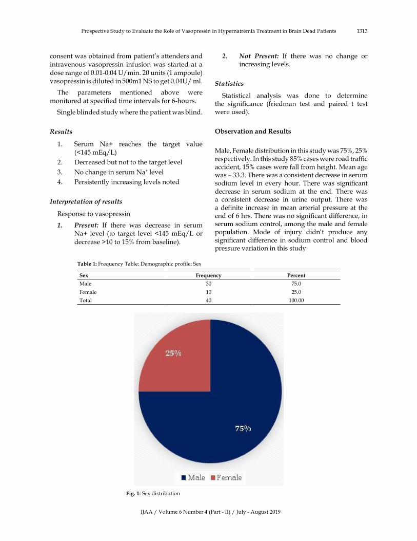
IJAA�/�Volume�6�Number�4�(Part�-�II)�/�July�-�August�2019
1313
consent�was�obtained�from�patient�s�attenders�and�intravenous�vasopressin� infusion�was� started� at� a�dose�range�of�0.01-0.04��/min.�20�units�(1�ampoule)�vasopressin�is�diluted�in�500m1�NS�to�get�0.04�/�ml.
The� parameters� mentioned� above� were�monitored�at�speci��ed�time�intervals�for�6-hours.
Single�blinded�study�where�the�patient�was�blind.
Results
1.� Serum� Na�� reaches� the� target� value�(�145�mE�/L)
2.� Decreased�but�not�to�the�target�level
3.� No�change�in�serum�Na��level
4.� Persistently�increasing�levels�noted
Inter�retation�of�results
Response�to�vasopressin�
��� Present:� If� there� was� decrease� in� serum�Na�� level� (to� target� level� �145�mE�/L�or�
decrease��10�to�15��from�baseline).
2�� �ot� Present:� If� there� was� no� change� or�increasing�levels.
Statistics
Statistical� analysis� was� done� to� determine�the� signi��cance� (friedman� test� and� paired� t� test�were�used).
Obser�ation�and��esults
Male,�Female�distribution�in�this�study�was�75�,�25��respectively.�In�this�study�85��cases�were�road�traf��c�accident,�15��cases�were�fall�from�height.�Mean�age�was�–�33.3.�There�was�a�consistent�decrease�in�serum�sodium� level� in� every� hour.� There�was� signi��cant�decrease� in� serum� sodium� at� the� end.� There� was�a� consistent� decrease� in� urine� output.� There� was�a�de��nite� increase� in�mean�arterial� pressure�at� the�end�of�6�hrs.�There�was�no�signi��cant�difference,�in�serum�sodium�control,�among�the�male�and�female�population.� Mode� of� injury� didn�t� produce� any�signi��cant� difference� in� sodium� control� and� blood�pressure�variation�in�this�study.
Prospective�Study�to�Evaluate�the�Role�of�Vasopressin�in��ypernatremia�Treatment�in��rain�Dead�Patients
Table��:�Fre�uency�Table:�Demographic�profile:�Sex
Se� Fre�uency Percent
Male 30 75.0
Female 10 25.0
Total 40 100.00
Fig���:�Sex�distribution
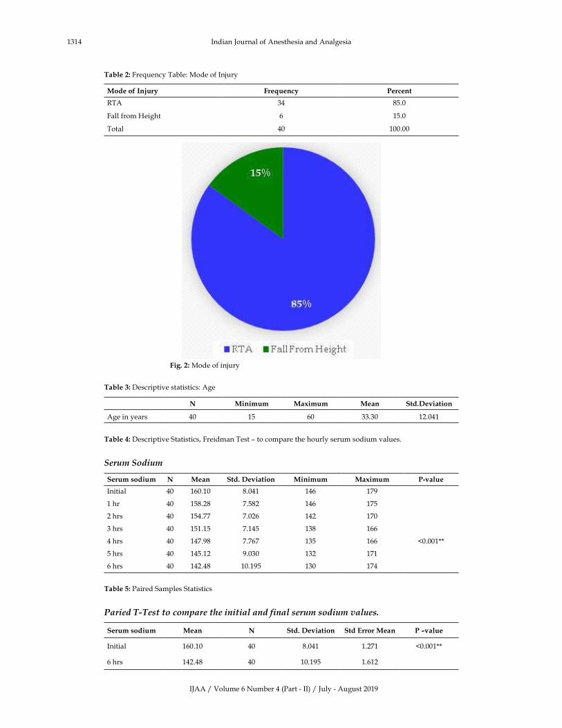
IJAA�/�Volume�6�Number�4�(Part�-�II)�/�July�-�August�2019
1314 Indian�Journal�of�Anesthesia�and�Analgesia
Table��:�Fre�uency�Table:�Mode�of�Injury
Mode�of�In�ury Fre�uency Percent
RTA 34 85.0
Fall�from��eight 6 15.0
Total 40 100.00
� � Fig���:�Mode�of�injury
Table��:�Descriptive�statistics:�Age
N Minimum Ma�imum Mean Std��e�iation
Age�in�years 40 15 60 33.30 12.041
Table��:�Descriptive�Statistics,�Freidman�Test�–�to�compare�the�hourly�serum�sodium�values.
Seru��Sodiu�
Serum�sodium N Mean Std���e�iation Minimum Ma�imum P��alue
Initial 40 160.10 8.041 146 179
1�hr 40 158.28 7.582 146 175
2�hrs 40 154.77 7.026 142 170
3�hrs 40 151.15 7.145 138 166
4�hrs 40 147.98 7.767 135 166 �0.001��
5�hrs 40 145.12 9.030 132 171
6�hrs 40 142.48 10.195 130 174
Table��:�Paired�Samples�Statistics
Paried����est�to�co��are�the�initial�and�final�seru��sodiu��values�
Serum�sodium Mean N Std���e�iation Std�Error�Mean P���alue
Initial 160.10 40 8.041 1.271 �0.001��
6�hrs 142.48 40 10.195 1.612
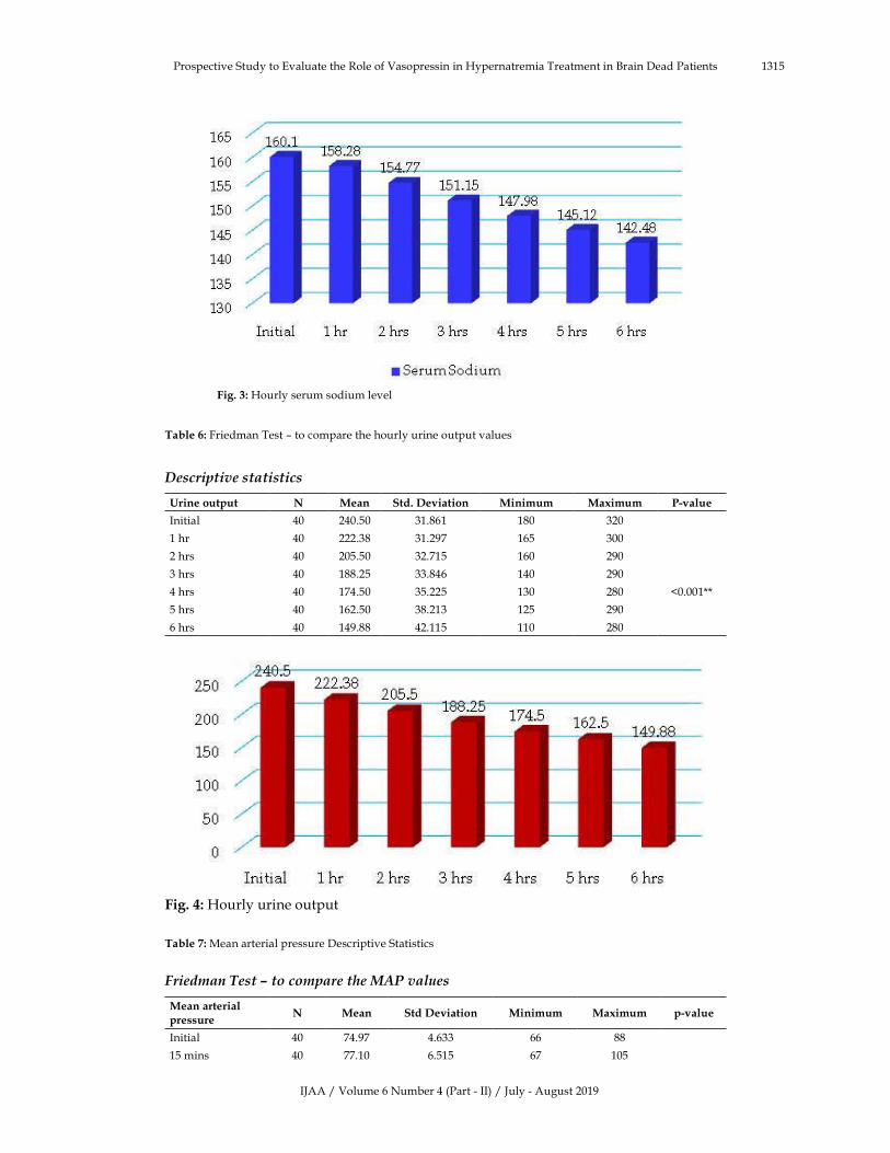
IJAA�/�Volume�6�Number�4�(Part�-�II)�/�July�-�August�2019
1315Prospective�Study�to�Evaluate�the�Role�of�Vasopressin�in��ypernatremia�Treatment�in��rain�Dead�Patients
� Fig���:��ourly�serum�sodium�level
Table��:�Friedman�Test�–�to�compare�the�hourly�urine�output�values
�escri�tive�statistics
�rine�output N Mean Std���e�iation Minimum Ma�imum P��alue
Initial 40 240.50 31.861 180 320
1�hr 40 222.38 31.297 165 300
2�hrs 40 205.50 32.715 160 290
3�hrs 40 188.25 33.846 140 290
4�hrs 40 174.50 35.225 130 280 �0.001��
5�hrs 40 162.50 38.213 125 290
6�hrs 40 149.88 42.115 110 280
Fig���:��ourly�urine�output
Table��:�Mean�arterial�pressure�Descriptive�Statistics
Fried�an��est���to�co��are�the��AP�values
Mean�arterial�pressure
N Mean Std��e�iation Minimum Ma�imum p��alue
Initial 40 74.97 4.633 66 88
15�mins 40 77.10 6.515 67 105
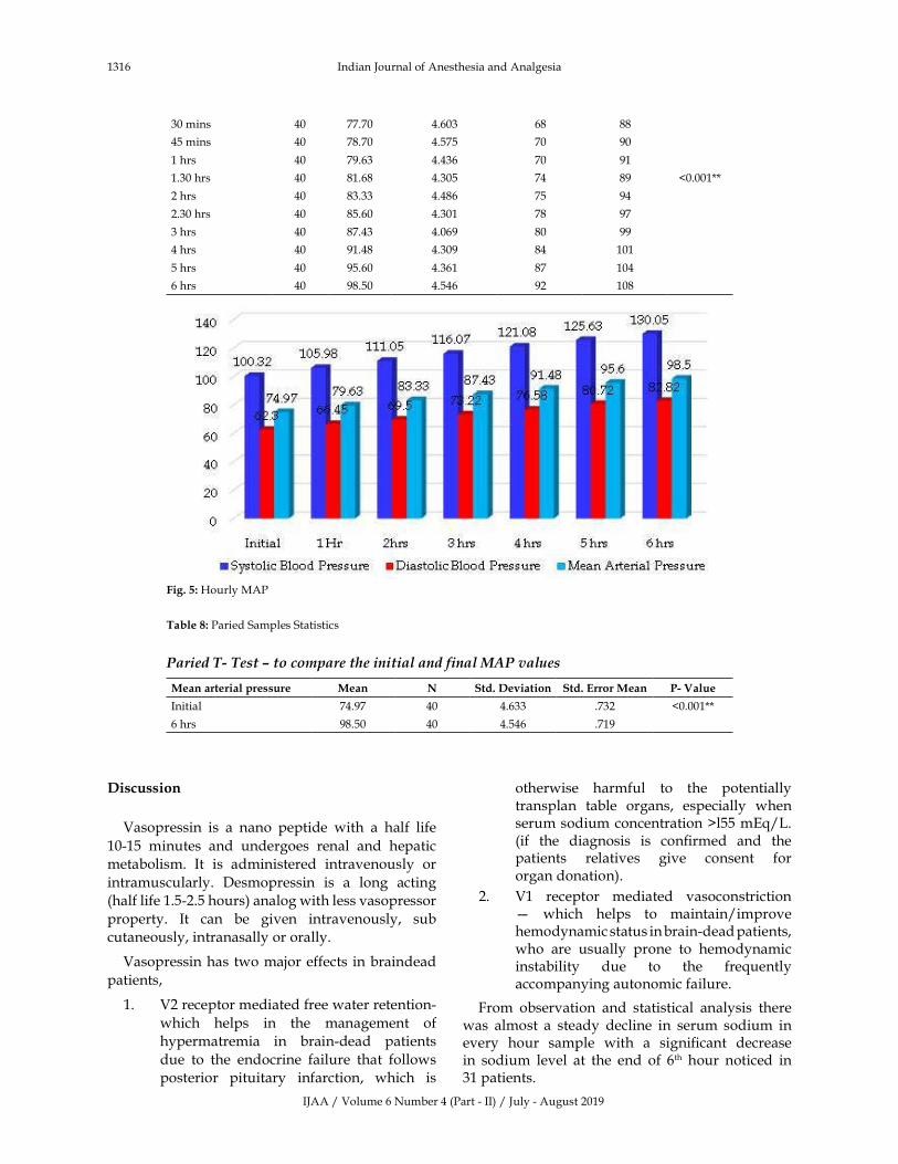
IJAA�/�Volume�6�Number�4�(Part�-�II)�/�July�-�August�2019
1316 Indian�Journal�of�Anesthesia�and�Analgesia
30�mins 40 77.70 4.603 68 88
45�mins 40 78.70 4.575 70 90
1�hrs 40 79.63 4.436 70 91
1.30�hrs 40 81.68 4.305 74 89 �0.001��
2�hrs 40 83.33 4.486 75 94
2.30�hrs 40 85.60 4.301 78 97
3�hrs 40 87.43 4.069 80 99
4�hrs 40 91.48 4.309 84 101
5�hrs 40 95.60 4.361 87 104
6�hrs 40 98.50 4.546 92 108
Fig���:��ourly�MAP
Table��:�Paried�Samples�Statistics
Paried�����est���to�co��are�the�initial�and�final��AP�values
Mean�arterial�pressure Mean N Std���e�iation Std��Error�Mean P��Value
Initial 74.97 40 4.633 .732 �0.001��
6�hrs 98.50 40 4.546 .719
�iscussion
Vasopressin� is� a� nano� peptide� with� a� half� life�
10-15� minutes� and� undergoes� renal� and� hepatic�
metabolism.� It� is� administered� intravenously� or�
intramuscularly.� Desmopressin� is� a� long� acting�
(half�life�1.5-2.5�hours)�analog�with�less�vasopressor�
property.� It� can� be� given� intravenously,� sub�
cutaneously,�intranasally�or�orally.
Vasopressin�has� two�major�effects� in�braindead�
patients,
1.� V2�receptor�mediated�free�water�retention-
which� helps� in� the� management� of�
hypermatremia� in� brain-dead� patients�
due� to� the� endocrine� failure� that� follows�
posterior� pituitary� infarction,� which� is�
otherwise� harmful� to� the� potentially�transplan� table� organs,� especially� when�serum�sodium�concentration��l55�mE�/L.�(if� the� diagnosis� is� con��rmed� and� the�patients� relatives� give� consent� for�organ�donation).
2.� V1� receptor� mediated� vasoconstriction��� which� helps� to� maintain/improve�hemodynamic�status�in�brain-dead�patients,�who� are� usually� prone� to� hemodynamic�instability� due� to� the� fre�uently�accompanying�autonomic�failure.
From� observation� and� statistical� analysis� there�was� almost� a� steady� decline� in� serum� sodium� in�every� hour� sample� with� a� signi��cant� decrease�in� sodium� level� at� the� end� of� 6th� hour� noticed� in�31�patients.
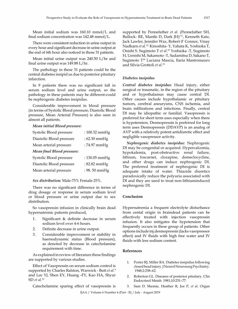
IJAA�/�Volume�6�Number�4�(Part�-�II)�/�July�-�August�2019
1317
Mean� initial� sodium� was� 160.10� mmol/L� and���nal�sodium�concentration�was�142.48�mmol/L.
There�were�consistent�reduction�in�urine�output�in�every�hour�and�signi��cant�decrease�in�urine�output�at�the�end�of�6th�hour�also�noticed�in�those�31�patients.
Mean� initial�urine� output�was� 240.50� L/hr�and���nal�urine�output�was�149.88�L/hr.
The�pathology�in�these�31�patients�could�be�the�central�diabetes�insipid�us�due�to�posterior�pituitary�infarction.
In� 9� patients� there� was� no� signi��cant� fall� in�serum� sodium� level� and� urine� output,� so� the�pathology�in�these�patients�may�be�different-could�be�nephrogenic�diabetes�insipidus.
Considerable� improvement� in� blood� pressure�(in�terms�of�Systolic��lood�pressure,�Diastolic��lood�pressure,� Mean� Arterial� Pressure)� is� also� seen� in�almost�all�patients.
�ean�initial��lood��ressure:
Systolic��lood�pressure�� :�100.32�mm�g�
Diastolic��lood�pressure�� :�62.30�mm�g�
Mean�arterial�pressure�� :�74.97�mm�g�
�ean���nal��lood��ressure:
Systolic��lood�pressure�� :�130.05�mm�g�
Diastolic��lood�pressure�� :�82.82�mm�g�
Mean�arterial�pressure�� :�98.�50�mm�g�
Sex�distri�ution:�Male-75��Female-25�.
There�was�no� signi��cant� difference� in� terms� of�drug� dosage� or� response� in� serum� sodium� level�or� blood� pressure� or� urine� output� due� to� sex�distribution.
So�vasopressin� infusion� in�clinically�brain�dead�hypernatremic�patients�produced,
1.� Signi��cant� �� de��nite� decrease� in� serum�sodium�level�over�4-6�hours.
2.� De��nite�decrease�in�urine�output.
3.� Considerable� improvement� or� stability� in�haemodynamic� status� (�lood� pressure),�as� denoted� by� decrease� in� catecholamine�re�uirement�with�time.
As�explained�in�review�of�literature�these���ndings�are�supported�by�various�studies.
Effect�of�Vasopressin�on�serum�sodium�control�is�supported�by�Charles�Ralston,�Warwick�-��utt�et�al.9�and�Lee��J,�Shen�E�,��uang� -F�,��ao��A,�Shyur�SD�et�al.10
Catecholamine� sparing� effect� of� vasopressin� is�
supported� by� Pennefather� et� al.� (Pennefather� S�,��ullock-�RE,�Mantle�D,�Dark�J�)�11,��enneth��atz,�Jack�Lawler,�Jennifer�Wax,�Robert�0��Connor,�Vinay�Nadkarn�et�al.12,��inoshita-��,��ahata��,��oshioka�T,�Onishi�S.�Sugimoto�T�et�al.15��oshioka�-T,�Sugimoto��,��enishi�M,�Sakamoto�-T,�Sadamitsu�D,�Sakano�T,�Sugimoto�T18�Luciana�Mascia,� Ilaria�Mastromauro�and�Silvia�Grottoli�et�al.19
�ia�etes�insi�idus
Central� dia�etes� insi�idus:� �ead� injury,� either�surgical�or�traumatic,�in�the�region�of�the�pituitary�and� or� hypothalamus� may� cause� central� DI.�Other� causes� include� hypothalamic� or� pituitary�tumors,� cerebral� aneurysms,� CNS� ischemia,� and�brain� in��ltrations� and� infections.� Finally,� central�DI� may� be� idiopathic� or� familial.� Vasopressin� is�preferred�for�short�term�uses�especially�when�there�is�hypotension.�Desmopressin�is�preferred�for�long�term�uses�Desmopressin�(DDAVP)�is�an�analog�of�AVP�with�a�relatively�potent�antidiuretic�effect�and�negligible�vasopressor�activity.
�e�hro�enic� dia�etes� insi�idus:� Nephrogenic�DI�may�be�congenital�or�ac�uired.��ypercalcemia,�hypokalemia,� post-obstructive� renal� failure,�lithium,� foscarnet,� clozapine,� demeclocycline,�and� other� drugs� can� induce� nephrogenic� DI.�The� preferred� treatment� of� nephrogenic� DI� is�ade�uate� intake� of� water.� Thiazide� diuretics�paradoxically�reduce�the�polyuria�associated�with�DI�and�they�are�used�to�treat�non-lithiuminduced�nephrogenic�DI.
Conclusion
�ypernatremia� a� fre�uent� electrolyte� disturbance�from� cental� origin� in� braindead� patients� can� be�effectively� treated� with� injection� vasopressin�infusion.� It� also� mitigates� the� hypotension� that�fre�uently�occurs�in�these�group�of�patients.�Other�options�include�inj.desmopressin�(lacks�vasopressor�effect)� and� IV� ��uids�with� high� free�water� and� IV���uids�with�less�sodium�content.
�eferences
1.� Porter�RJ,�Miller�RA.�Diabetes�insipidus�following�closed�head�injury.�J�Neurol�Neurosurg�Psychiatry.�1948;2:258–62.
2.� Roberton�GL.�Diseases�of�posterior�pituitary.�Clin�Endocrinol�Metab.�1981;10:251–77.
3.� Sam� D.� Shemie,� �eather� R,� Joe� P,� et� al.� Organ�
Prospective�Study�to�Evaluate�the�Role�of�Vasopressin�in��ypernatremia�Treatment�in��rain�Dead�Patients

IJAA�/�Volume�6�Number�4�(Part�-�II)�/�July�-�August�2019
1318 Indian�Journal�of�Anesthesia�and�Analgesia
donor�treatment�in�Canada-�recommendations�of�the� forum� on�medical�management� to� optimize�donor�organ�potential.�CMAJ.�2006�Mar�14;174(6).
4.� Pauline�M.�Todd,�Rebecca�N.�Jerome,�and�Adrian�A.� Jar�uin-Valdivia.� Organ� preservation� in� a�brain� dead� patient:� information� support� for�neurocritical� care� protocol� development.J� Med�Libr�Assoc.�2007�Jul;95(3):238–245.
5.� Arita� �,� �ozumi� T,� Oki� S.� et� al.� The� functions�of� the� hypothalamo� pituitary� axis� in� braindead�patients.�Acta-�Neurochir�(Wien).�1993;123:64–75.
6.� Salim�A,�Martin�M,��rown�C,�et�al.�The�effect�of�a� protocol� of� aggressive� donor� management:�implications� for� the� national� organ� donor�shortage.�J�Trauma.�2006;61:429-35.
7.� Richardson� DW,� Robinson� AG.� Desmopressin.�Ann�Intern�Med�1985;103(2):228–39.
8.� Debelak� L,� Pollak� R,� Reckard� C.� Arginine�vasopressin� versus� desmopressin� in� the�management� of� diabetes� insipidus� in� the� brain�dead�donor.�Transplant�proc.�1990;22:35�1.
9.� Charles�-�Ralston,�Warwick�-��utt�et�al.�Continuous�vasopressin� replacement� in� Diabetes� insipidus:�Arch�Dis�-Child.�1990;65:896–7.
10.� Lee� �J,� Shen� E�,� �uang� F�,� et� al.� Continuous�infusion� of� vasopressin� in� comatose� children�with� neurogenic� diabetes� insipidus.� J.� Pediatr�Endocrinol�Metab.�1995�Oct-Dec;8(4):257–62.
11.� Penne� father� S�,� �ullock� RE,� Mantle� D,� et� al.��se�of�low�dose�arginine�vasopressin�to�support�braindead� organ� donors.� Transplantation�1995;59:(1)�58–62.
12.� �atz� �,� LawlerJ,� Wax� J,� O�Connor� R,� et� al.�Vasopressin�pressor�effects�in�critically�ill�children�during� evaluation� for� braindeath� and� organ�recovery.�Resuscitation2000;47:33–40.
13.� Figueras� J,� �us�uets� J,� Grande� L,� et� al.� The�deleterious�effects�of�donor�high�plasma�sodium�and�extended�preservation�in�liver�transplantation.�Transplantation.�1996;61:410–413.
14.� Totsuka� E,� Dodson.� F,� �rakami� Al,� et� al.�Influence� of� high� donor� plasma� sodium� levels�on� early� postoperative� graft� function� in� human�liver� transplantation:� Effects� of� correction� of�
donor� hypernatremia.� Liver� Transplant� Surg.1999;5:421–8.
15.� �inoshita��,��ahata��,��oshioka�T,�et�al.�Long�term�renal�preservation�after� brain�death,�maintained�with� vasopressin� and� epinephrine:� Transpi� mt.�1990�May;3(1):15–8.
16.� Jennifer� A.� Frontera,� Thomas� �aib� et� al.� �ow�I� Manage� the� Adult� Potential� Organ� Donor:�Donation� After� Neurological� Death� (Part� 1)-�Neurocrit�Care.�2010;12:103–10.
17.� Totsuka�E,�Fung��,��akamada��,�et�al.�Analysis�of�Clinical�variables�of�donors�and�recipients�with�respect� to� short� term� graft� outcome,� in� human�liver�transplantation:�Transplant�Proc.�2004�Oct;3�6(8):22�15–8.
18.� �oshioka� T,� Sugimoto� �,� �enishi� M,� et� al.�Prolonged� hemodynamic� maintenance,� by� the�combined� administration� of� Vasopressin� and�Epinephrine� in� brain� death-� a� clinical� study.�Neurosurgery.�1986�May;18(5):565–7.
19.� Luciana� Mascia,� Ilaria� Mastromauro� and� Silvia�Grottoli� Management� of� Neuroendocrine�Instability,� Endocrinology� and� Metabolism:�Edited��y:��yuzi��amoi,� IS�N�978-953-307-367–�2011.
20.� �erri� M.� Robertson,� and� D.� Ryan� Cook.Perioperative� management� of� the� multiorgan�donor.�Anesth�Analg.�1990;70:546–56.
21.� Shah�V,��hosale�G.�Organ�donor�problems,� and�their�management.� Indian� J�Crit�Care�Med�2006�Jan-Mar;10(1):29–34.
22.� �ohenegger� M,� Vermes� M,� Mauritz� W,� et� al.�Austria-� Serum� vasopressin� (AVP)� levels� in�polyuric� brain-dead� organ� donors:� Eur� Arch�Psychiatry�Neurol�Sci.�1990;239(4):267–9.
23.� �artshorn� J,� �artshom� E.� Vasopressin� in�treatment�of�diabeteinsipidus.�J�Neurosurg�Rurs.1988;20:58–9.
24.� �owlett�TA,��eogh�AM,�Perry�L,� et� al.�Anterior�and� posterior� pituitary� functions� in� brain� dead�donors.�Transplantation.�1989;47:828–34.
25.� Gramm� �J,� Meinhold� �,� �ickel� �,� et� al.� Acute�endocrine� dysfunction� after� brain� death.�Transplantation.�1992;54:851–7.
��Red�Flower�Publication�Pvt.�Ltd.�
�Original�Research�ArticleIndian�Journal�of�Anesthesia�and�Analgesia�
2019;6(4)�(Part�-�II):�1319-1324DOI:�http://dx.doi.org/10.21088/ijaa.2349.8471.6419.36
Pre�ention� of� �ypotension� Follo�ing� Subarachnoid� Block�� Efficacy� of�Preloading��ith��ydro�yethyl�Starch�Versus��inger�s��actate�Solution
Bala�ibabu�P����Ananda�Bhat�
1,2Associate�Professor,�Department�of�Anaesthesiology,�Sri�Jayadeva�Institute�of�Cardiovascular�Sciences�and�Research,��engaluru,��arnataka�560069,�India.�
Corresponding�Author:�Ananda�Bhat,�Associate�Professor,�Department�of�Anaesthesiology,�Sri�Jayadeva�Institute�of�Cardiovascular�Sciences�and�Research,��engaluru,��arnataka�560069,�India.�
E�mail:�[email protected]
�ecei�ed�on�08.05.2019,�Accepted�on�08.06.2019
Abstract
Background:� Arterial� hypotension� is� a� potential� hazard� associated� with� spinal� anesthesia.� Routinely,�crystalloids� and� colloids� are� used� for� managing� hypotension.� While� crystalloids� easily� move� out� of� the�intravascular� space,� colloids� are� confined� to� the� intravascular� space,� thereby� minimizing� the� risk� of�hypotension.�This�study�was�carried�out�to�compare�the�efficacy�of�10���ydroxyethyl�starch�with�Ringer�s�lactate� solution.�Methods:� This� double� blind� randomized� controlled� trial� was� carried� out� on� 100� patients�who�underwent� spinal� anesthesia�during� the� study�period.� The� study� participants�were� randomized� into�experimental� group� (6���ydroxyethyl� starch,� 10�ml/�g)� and� control� group� (Ringer�s� lactate,� 20�ml/�g).��lood�pressure�and�heart�rate�were�recorded�periodically�up�to�60�minutes.�Sensory� level�of�blockade�was�checked�after�5�minutes.�Results:�It�was�observed�that�as�the�surgery�prolonged,�the�incidence�of�hypotension�increased�among�the�controls�compared� to� the�experimental�group.�While� the� initial� incidence�was�higher�among�the�experimental�group�(24�),�the�incidence�was�greater�for�the�controls�beyond�25�minutes�(42�).�The�difference�in�the�incidence�was�statistically�significant�(p�0.05).�Conclusion:��ydroxyethyl�starch�is�superior�to�Ringer�s�lactate�solution�in�prevention�of��ypotension�following�spinal�anesthesia.�Incidence�of�hypotension�is� reduced�but�not� completely� eliminated.��ydroxyethyl� starch� also�has� several� other� advantages� such� as�prophylaxis�against�venous�thrombosis�and�decreased�allergic�potential.
�ey�ords:��ydroxy�ethyl�starch;��ypotension;�Ringer�lactate;�Spinal�anesthesia.
�o��to�cite�this�article:
�alajibabu�PR,�Ananda��hat.�Prevention�of��ypotension�Following�Subarachnoid��lock;�Efficacy�of�Preloading�with��ydroxyethyl�
Starch�Versus�Ringer�s�Lactate�Solution.�Indian�J�Anesth�Analg.�2019;6(4):1319-1324.
Introduction
Spinal� anesthesia� is� one� of� the�widely�practiced�
anaesthetic� techni�ues� over� years� for� lower�
abdominal� and� lower� limb� surgeries.� If� spinal�
anesthesia�is�applied�with�proper�skills�and�care,�
it�is�a�safe�procedure�which�provides�satisfactory�outcomes.� It� is� a� preferred� method� of� choice�among� the� patients� and� surgeons� considering�the� benefits� and� comforts� achieved� on-table.�Spinal�anesthesia�for�routine�surgical�procedures�assumes� special� importance� in� developing�countries� because� of� economic� reasons,� lack�
IJAA�/�Volume�6�Number�4�(Part�-�II)�/�July�-�August�2019
1320 Indian�Journal�of�Anesthesia�and�Analgesia
of� availability� of� sophisticated� anaesthetic�apparatus� and� compressed� gases� in� the�remote�areas.
The� potential� hazard� associated� with� spinal�anesthesia� is�arterial�hypotension.�This�can�cause�signi��cant� morbidity� and� mortality.� Methods�used� to� reduce� the� incidence� and� severity� of�spinal�hypotension�includes�head�down�position,�prophylactic� vasopressors,� leg� elevation� and�strapping,�use�of� in��atable�boots�and�preloading�the�patients�with� intravenous���uids.�Crystalloids�solutions� being� of� lower� molecular� weight�will� enter� the� interstitial� space� due� to� lack� of�intrinsic� colloid� osmotic� pressure� resulting� in�pulmonary� edema� which� interferes� with� tissue�oxygen� exchange.� Colloids� are� higher�molecular�substances� than� crystalloids� and� are� similar� to�plasma� in� oncotic� pressure� and� remain� con��ned�to� intravascular� space� with� little� expansion� of�interstitial�space.
It� is� indeed� a� challenge� to� manage� arterial�hypotension� on� table.� Although� several� studies�have�been�carried�out�to�evaluate�the�effectiveness�of� spinal� anesthesia,� very� few� studies� have�been�carried�out� to� address� the� adverse� effect,�mainly�hypotension.� An� analysis� into� the� therapeutics�involved� in� the� management� of� arterial�hypotension�will�go�a�long�way�in�minimizing�the�morbidity� and� mortality� associated� with� spinal�anesthesia.
���ectives
1.� To� compare� the� preloading� ef��cacy�of� Ringer�s� lactate,� 20� ml/kg� and��ydroxyethyl� starch� 6�,� 10� ml/kg� body�weight,� for� prevention� of� �ypotension�following�subarachnoid��lock.
2.� To� evaluate� the� adverse� outcomes� of��ydroxyethyl� starch� among� the� study�participants.
Methodology
Study�settin��and��artici�ants
This�double�blind�randomized�controlled�trial�was�carried� out� in� the� Department� of� Anesthesiology�of� a� tertiary� teaching� institution� for� a� period� of�10�months.� All� the� patients� who�were� scheduled�for�surgeries�involving�spinal�anesthesia�during�the�study�period�were�selected�for�the�study.��ased�on�the�selection�criteria,�a�total�of�100�participants�were�included�in�the�study.
Inclusion�criteria
�� Patient�belonging�to�ASA�Grade�I���II.
�� �ody�Weight�40�to�80�kilograms.
�� �Age�20�to�60�years�of�both�the�sexes.
�� Patients� undergoing� various� elective�and� emergency� operations� under� spinal�anesthesia� in� whom�minimal� blood� loss� is�anticipated.
Exclusion�criteria
�� Any� patients� in� whom� spinal� anesthesia� is�contraindicated.
�� Patients�with�cardio�vascular,�respiratory���central�nervous�system�disorders.
Rando�i�ation�and��rou�in�
The� study� participants� were� grouped� into�experimental�and�control�groups.�The�experimental�group� consisted� of� 50� participants� who� received�6�� �ydroxyethyl� starch,� 10� ml/�g.� The� control�group� consisted� of� 50� participants� who� received�Ringer�lactate,�20�ml/�g.�All�the�participants�were�randomly�allocated�into�the�groups�using�computer�generated�random�numbers.
Ethical�a��roval�and�infor�ed�consent
Approval� was� obtained� from� the� Institutional�Ethics� Committee� prior� to� the� commencement� of�the�study.�Each�participant�was�explained�in�detail�about�the�study�and�informed�consent�was�obtained�prior�to�the�commencement�of�data�collection.
Procedure
Following�routine�investigations�and�pre-anaesthetic�evaluation,� all� participants� were� pre-medicated�with�injection�Midazolam�0.02�mg/kg�and�injection�Atropine� 0.6� mg,� through� intra� venous� route,�30�minutes�before�surgery.
�asal� parameters� like� heart� rate,� Oxygen�saturation�and�blood�pressure�were�monitored.�A�peripheral� intravenous� line� with� 18� or� 20� gauge�canula� was� secured� in� one� of� the� upper� limbs.�Volume�infusion�was�determined�in�accordance�to�the�body�weight.�In�both�the�groups,�the�solutions�were�infused�over�a�period�of�20�minutes.�After�pre-loading�all�patients�received�ringer�lactate�infusion�for���uid�maintenance.
�nder�aseptic�precautions�lumbar�puncture�was�performed�with�23�gauge�spinal�needle� (�uinke�s�
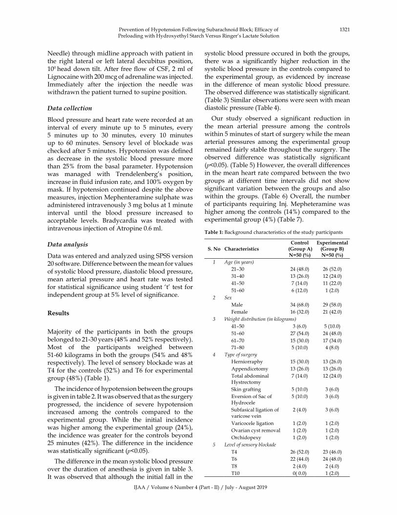
IJAA�/�Volume�6�Number�4�(Part�-�II)�/�July�-�August�2019
1321
Needle)�through�midline�approach�with�patient�in�the� right� lateral� or� left� lateral� decubitus� position,�100�head�down�tilt.�After�free���ow�of�CSF,�2�ml�of�Lignocaine�with�200�mcg�of�adrenaline�was�injected.�Immediately� after� the� injection� the� needle� was�withdrawn�the�patient�turned�to�supine�position.
�ata�collection
�lood�pressure�and�heart�rate�were�recorded�at�an�interval� of� every� minute� up� to� 5� minutes,� every�5� minutes� up� to� 30� minutes,� every� 10� minutes�up� to� 60�minutes.� Sensory� level� of� blockade� was�checked�after�5�minutes.��ypotension�was�de��ned�as� decrease� in� the� systolic� blood� pressure� more�than� 25�� from� the� basal� parameter.��ypotension�was� managed� with� Trendelenberg�s� position,�increase�in���uid�infusion�rate,�and�100��oxygen�by�mask.� If�hypotension�continued�despite�the�above�measures,�injection�Mephenteramine�sulphate�was�administered�intravenously�3�mg�bolus�at�1�minute�interval� until� the� blood� pressure� increased� to�acceptable� levels.� �radycardia� was� treated� with�intravenous�injection�of�Atropine�0.6�ml.
�ata�analysis
Data�was�entered�and�analyzed�using�SPSS�version�20�software.�Difference�between�the�mean�for�values�of�systolic�blood�pressure,�diastolic�blood�pressure,�mean� arterial� pressure� and� heart� rate� was� tested�for�statistical�signi��cance�using�student��t�� test�for�independent�group�at�5��level�of�signi��cance.
�esults
Majority� of� the� participants� in� both� the� groups�belonged�to�21-30�years�(48��and�52��respectively).�Most� of� the� participants� weighed� between�51-60�kilograms�in�both�the�groups�(54��and�48��respectively).�The�level�of�sensory�blockade�was�at�T4� for�the�controls� (52�)�and�T6� for�experimental�group�(48�)�(Table�1).
The�incidence�of�hypotension�between�the�groups�is�given�in�table�2.�It�was�observed�that�as�the�surgery�progressed,� the� incidence� of� severe� hypotension�increased� among� the� controls� compared� to� the�experimental� group.� While� the� initial� incidence�was�higher�among� the�experimental�group� (24�),�the� incidence�was�greater� for� the�controls�beyond�25�minutes� (42�).�The�difference� in� the� incidence�was�statistically�signi��cant�(p�0.05).
The�difference�in�the�mean�systolic�blood�pressure�over� the�duration�of�anesthesia� is�given� in� table�3.�It�was�observed� that�although� the�initial� fall� in� the�
systolic�blood�pressure�occured�in�both�the�groups,�there� was� a� signi��cantly� higher� reduction� in� the�systolic�blood�pressure�in�the�controls�compared�to�the� experimental� group,� as� evidenced� by� increase�in� the� difference� of� mean� systolic� blood� pressure.�The�observed�difference�was�statistically�signi��cant.�(Table�3)�Similar�observations�were�seen�with�mean�diastolic�pressure�(Table�4).
Our� study� observed� a� signi��cant� reduction� in�the� mean� arterial� pressure� among� the� controls�within�5�minutes�of�start�of�surgery�while�the�mean�arterial� pressures� among� the� experimental� group�remained�fairly�stable�throughout�the�surgery.�The�observed� difference� was� statistically� signi��cant�(p�0.05).�(Table�5)��owever,�the�overall�differences�in�the�mean�heart�rate�compared�between�the�two�groups� at� different� time� intervals� did� not� show�signi��cant� variation�between� the�groups�and�also�within� the� groups.� (Table� 6)�Overall,� the� number�of� participants� re�uiring� Inj.�Mepheteramine�was�higher�among�the�controls� (14�)�compared�to� the�experimental�group�(4�)�(Table�7).
Table��:��ackground�characteristics�of�the�study�participants
S��No CharacteristicsControl
���roup�A��N�������
E�perimental��roup�B��N�������
� Age��in��ears�
21–30 24�(48.0) 26�(52.0)
31–40 13�(26.0) 12�(24.0)
41–50 7�(14.0) 11�(22.0)
51–60 6�(12.0) 1�(2.0)
� Se�
Male 34�(68.0) 29�(58.0)
Female 16�(32.0) 21�(42.0)
� �eight�distribution��in�kilograms�
41–50 3�(6.0) 5�(10.0)
51–60 27�(54.0) 24�(48.0)
61–70 15�(30.0) 17�(34.0)
71–80 5�(10.0) 4�(8.0)
� T�pe�of�surger�
�erniorraphy 15�(30.0) 13�(26.0)
Appendicetomy 13�(26.0) 13�(26.0)
Total�abdominal��ystrectomy
7�(14.0) 12�(24.0)
Skin�grafting 5�(10.0) 3�(6.0)
Eversion�of�Sac�of��ydrocele
5�(10.0) 3�(6.0)
Subfasical�ligation�of�varicose�vein
2�(4.0) 3�(6.0)
Varicocele�ligation 1�(2.0) 1�(2.0)
Ovarian�cyst�removal 1�(2.0) 1�(2.0)
Orchidopexy 1�(2.0) 1�(2.0)
� �e�el�of�sensor��blockade
T4 26�(52.0)� 23�(46.0)
T6 22�(44.0) 24�(48.0)
T8 2�(4.0) 2�(4.0)
T10 0(�0.0) 1�(2.0)
Prevention�of��ypotension�Following�Subarachnoid��lock;�Efficacy�of�Preloading�with��ydroxyethyl�Starch�Versus�Ringer�s�Lactate�Solution
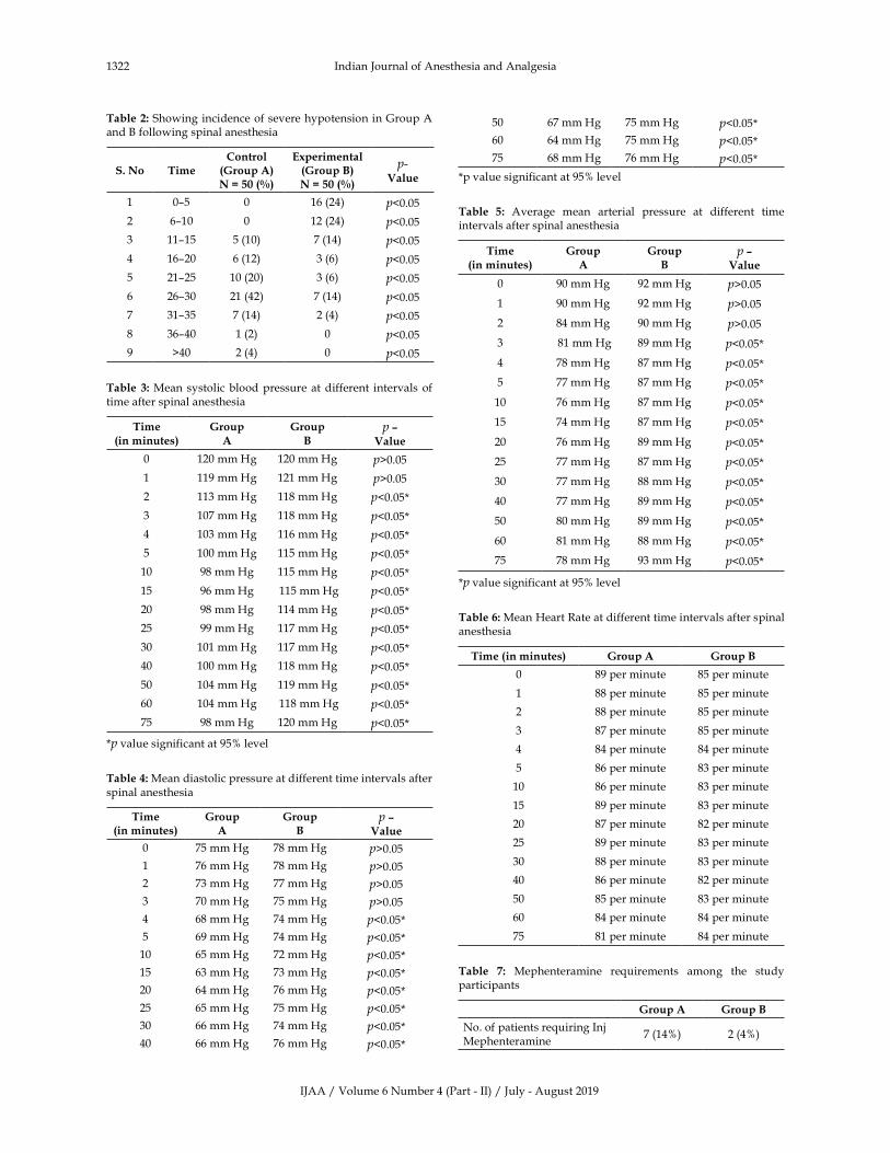
IJAA�/�Volume�6�Number�4�(Part�-�II)�/�July�-�August�2019
1322 Indian�Journal�of�Anesthesia�and�Analgesia
Table��:�Showing�incidence�of�severe�hypotension�in�Group�A�and���following�spinal�anesthesia
S��No TimeControl�
��roup�A�N���������
E�perimental��roup�B��N���������
p��Value
1 0–5 0 16�(24) p�0.05
2 6–10 0 12�(24) p�0.05
3 11–15 5�(10) 7�(14) p�0.05
4 16–20 6�(12) 3�(6) p�0.05
5 21–25 10�(20) 3�(6) p�0.05
6 26–30 21�(42) 7�(14) p�0.05
7 31–35 7�(14) 2�(4) p�0.05
8 36–40 1�(2) 0 p�0.05
9 �40 2�(4) 0 p�0.05
Table� �:�Mean� systolic�blood�pressure�at�different� intervals� of�time�after�spinal�anesthesia
Time�in�minutes�
�roup�A
�roup�B
p���Value
0 120�mm��g 120�mm��g p�0.05
1 119�mm��g 121�mm��g p�0.05
2 113�mm��g 118�mm��g p�0.05�
3 107�mm��g 118�mm��g p�0.05�
4 103�mm��g 116�mm��g p�0.05�
5 100�mm��g 115�mm��g p�0.05�
10 98�mm��g 115�mm��g p�0.05�
15 96�mm��g �115�mm��g p�0.05�
20 98�mm��g 114�mm��g p�0.05�
25 99�mm��g 117�mm��g p�0.05�
30 101�mm��g 117�mm��g p�0.05�
40 100�mm��g 118�mm��g p�0.05�
50 104�mm��g 119�mm��g p�0.05�
60 104�mm��g �118�mm��g p�0.05�
75 98�mm��g 120�mm��g p�0.05�
�p�value�significant�at�95��level
Table��:�Mean�diastolic�pressure�at�different�time�intervals�after�spinal�anesthesia
Time�in�minutes�
�roup�A
�roup�B
p���Value
0 75�mm��g 78�mm��g p�0.05
1 76�mm��g 78�mm��g p�0.05
2 73�mm��g 77�mm��g p�0.05
3 70�mm��g 75�mm��g p�0.05
4 68�mm��g 74�mm��g p�0.05�
5 69�mm��g 74�mm��g p�0.05�
10 65�mm��g 72�mm��g p�0.05�
15 63�mm��g 73�mm��g p�0.05�
20 64�mm��g 76�mm��g p�0.05�
25 65�mm��g 75�mm��g p�0.05�
30 66�mm��g 74�mm��g p�0.05�
40 66�mm��g 76�mm��g p�0.05�
50 67�mm��g 75�mm��g p�0.05�
60 64�mm��g 75�mm��g p�0.05�
75 68�mm��g 76�mm��g p�0.05�
�p�value�significant�at�95��level
Table� �:� Average� mean� arterial� pressure� at� different� time�intervals�after�spinal�anesthesia
Time�in�minutes�
�roup�A
�roup�B
p���Value
0 90�mm��g 92�mm��g p�0.05
1 90�mm��g 92�mm��g p�0.05
2 84�mm��g 90�mm��g p�0.05
3 �81�mm��g 89�mm��g p�0.05�
4 78�mm��g 87�mm��g p�0.05�
5 77�mm��g 87�mm��g p�0.05�
10 76�mm��g 87�mm��g p�0.05�
15 74�mm��g 87�mm��g p�0.05�
20 76�mm��g 89�mm��g p�0.05�
25 77�mm��g 87�mm��g p�0.05�
30 77�mm��g 88�mm��g p�0.05�
40 77�mm��g 89�mm��g p�0.05�
50 80�mm��g 89�mm��g p�0.05�
60 81�mm��g 88�mm��g p�0.05�
75 78�mm��g 93�mm��g p�0.05�
�p�value�significant�at�95��level
Table��:�Mean��eart�Rate�at�different�time�intervals�after�spinal�anesthesia
Time��in�minutes� �roup�A �roup�B
0 89�per�minute 85�per�minute
1 88�per�minute 85�per�minute
2 88�per�minute 85�per�minute
3 87�per�minute 85�per�minute
4 84�per�minute 84�per�minute
5 86�per�minute 83�per�minute
10 86�per�minute 83�per�minute
15 89�per�minute 83�per�minute
20 87�per�minute 82�per�minute
25 89�per�minute 83�per�minute
30 88�per�minute 83�per�minute
40 86�per�minute 82�per�minute
50 85�per�minute 83�per�minute
60 84�per�minute 84�per�minute
75 81�per�minute 84�per�minute
Table� �:� Mephenteramine� re�uirements� among� the� study�participants
�roup�A �roup�B
No.�of�patients�re�uiring�Inj�Mephenteramine
7�(14�) 2�(4�)
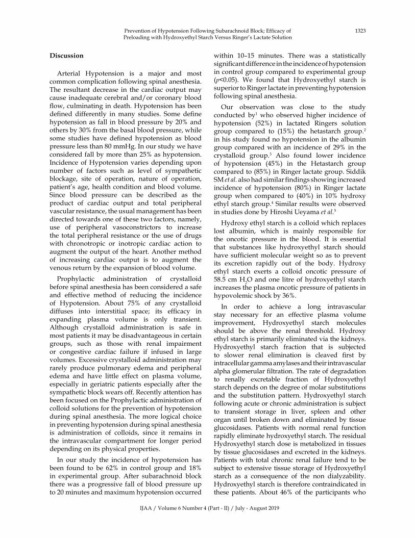
IJAA�/�Volume�6�Number�4�(Part�-�II)�/�July�-�August�2019
1323
�iscussion
Arterial� �ypotension� is� a� major� and� most�common�complication�following�spinal�anesthesia.�The� resultant� decrease� in� the� cardiac� output�may�cause� inade�uate� cerebral� and/or� coronary�blood���ow,�culminating�in�death.��ypotension�has�been�de��ned� differently� in�many� studies.� Some� de��ne�hypotension�as� fall� in�blood�pressure�by�20��and�others�by�30��from�the�basal�blood�pressure,�while�some� studies� have� de��ned� hypotension� as� blood�pressure�less�than�80�mm�g.�In�our�study�we�have�considered�fall�by�more�than�25��as�hypotension.�Incidence� of��ypotension�varies�depending�upon�number� of� factors� such� as� level� of� sympathetic�blockage,� site� of� operation,� nature� of� operation,�patient�s�age,�health�condition�and�blood�volume.�Since� blood� pressure� can� be� described� as� the�product� of� cardiac� output� and� total� peripheral�vascular�resistance,�the�usual�management�has�been�directed�towards�one�of�these�two�factors,�namely,�use� of� peripheral� vasoconstrictors� to� increase�the� total�peripheral� resistance�or� the�use�of�drugs�with� chronotropic� or� inotropic� cardiac� action� to�augment�the�output�of�the�heart.�Another�method�of� increasing� cardiac� output� is� to� augment� the�venous�return�by�the�expansion�of�blood�volume.
Prophylactic� administration� of� crystalloid�before�spinal�anesthesia�has�been�considered�a�safe�and� effective� method� of� reducing� the� incidence�of� �ypotension.� About� 75�� of� any� crystalloid�diffuses� into� interstitial� space;� its� ef��cacy� in�expanding� plasma� volume� is� only� transient.�Although� crystalloid� administration� is� safe� in�most�patients�it�may�be�disadvantageous�in�certain�groups,� such� as� those� with� renal� impairment�or� congestive� cardiac� failure� if� infused� in� large�volumes.�Excessive�crystalloid�administration�may�rarely� produce� pulmonary� edema� and� peripheral�edema� and� have� little� effect� on� plasma� volume,�especially� in� geriatric� patients� especially� after� the�sympathetic�block�wears�off.�Recently�attention�has�been�focused�on�the�Prophylactic�administration�of�colloid�solutions�for�the�prevention�of�hypotension�during� spinal� anesthesia.� The�more� logical� choice�in�preventing�hypotension�during�spinal�anesthesia�is� administration� of� colloids,� since� it� remains� in�the� intravascular� compartment� for� longer� period�depending�on�its�physical�properties.
In� our� study� the� incidence� of� hypotension� has�been� found� to� be� 62�� in� control� group� and� 18��in� experimental� group.� After� subarachnoid� block�there�was�a�progressive� fall� of�blood�pressure�up�to�20�minutes�and�maximum�hypotension�occurred�
within� 10–15� minutes.� There� was� a� statistically�signi��cant�difference�in�the�incidence�of�hypotension�in�control�group�compared�to�experimental�group�(p�0.05).� We� found� that� �ydroxyethyl� starch� is�superior�to�Ringer�lactate�in�preventing�hypotension�following�spinal�anesthesia.
Our� observation� was� close� to� the� study�conducted� by1�who� observed� higher� incidence� of�hypotension� (52�)� in� lactated� Ringers� solution�group� compared� to� (15�)� the� hetastarch� group.2�in�his�study�found�no�hypotension�in�the�albumin�group� compared�with� an� incidence�of� 29�� in� the�crystalloid� group.3� Also� found� lower� incidence�of� hypotension� (45�)� in� the� �etastarch� group�compared�to�(85�)�in�Ringer�lactate�group.�Siddik�SM�et�al.�also�had�similar���ndings�showing�increased�incidence� of� hypotension� (80�)� in� Ringer� lactate�group� when� compared� to� (40�)� in� 10�� hydroxy�ethyl�starch�group.4�Similar�results�were�observed�in�studies�done�by��iroshi��eyama�et�al.5
�ydroxy�ethyl�starch�is�a�colloid�which�replaces�lost� albumin,� which� is� mainly� responsible� for�the� oncotic� pressure� in� the� blood.� It� is� essential�that� substances� like� hydroxyethyl� starch� should�have� suf��cient�molecular�weight� so� as� to�prevent�its� excretion� rapidly� out� of� the� body.� �ydroxy�ethyl� starch� exerts� a� colloid� oncotic� pressure� of�58.5� cm��
2O�and�one� litre�of�hydroxyethyl� starch�
increases�the�plasma�oncotic�pressure�of�patients�in�hypovolemic�shock�by�36�.
In� order� to� achieve� a� long� intravascular�stay� necessary� for� an� effective� plasma� volume�improvement,� �ydroxyethyl� starch� molecules�should� be� above� the� renal� threshold.� �ydroxy�ethyl�starch�is�primarily�eliminated�via�the�kidneys.��ydroxyethyl� starch� fraction� that� is� subjected�to� slower� renal� elimination� is� cleaved� ��rst� by�intracellular�gamma�amylases�and�their�intravascular�alpha�glomerular���ltration.�The�rate�of�degradation�to� renally� excretable� fraction� of� �ydroxyethyl�starch�depends�on�the�degree�of�molar�substitutions�and� the� substitution� pattern.� �ydroxyethyl� starch�following�acute�or�chronic�administration�is�subject�to� transient� storage� in� liver,� spleen� and� other�organ�until�broken�down�and�eliminated�by� tissue�glucosidases.� Patients� with� normal� renal� function�rapidly�eliminate�hydroxyethyl�starch.�The�residual��ydroxyethyl�starch�dose�is�metabolized�in�tissues�by�tissue�glucosidases�and�excreted�in�the�kidneys.�Patients�with� total� chronic� renal� failure� tend� to�be�subject�to�extensive�tissue�storage�of��ydroxyethyl�starch� as� a� conse�uence� of� the� non� dialyzability.��ydroxyethyl�starch�is�therefore�contraindicated�in�these�patients.�About� 46��of� the�participants�who�
Prevention�of��ypotension�Following�Subarachnoid��lock;�Efficacy�of�Preloading�with��ydroxyethyl�Starch�Versus�Ringer�s�Lactate�Solution
IJAA�/�Volume�6�Number�4�(Part�-�II)�/�July�-�August�2019
1324 Indian�Journal�of�Anesthesia�and�Analgesia
were� administered� �ydroxyethyl� starch� excreted�it� in� the� urine� within� two� days� and� 64�� within�eight�days.
The�more� stable�hemodynamic� status� observed�after� colloid�administration�probably� relates� to� its�remaining�in�the�intravascular�compartment�longer�than� crystalloids.� Extra� vascular� redistribution�of� crystalloids� may� be� so� rapid� that� it� may� be�impossible�to�infuse�them�fast�enough�to�maintain�intravascular� volume.6� reported� that� colloid�osmotic�pressure�decrease�by�only�1.7�mm��g�after�preloading� with� 3�� dextran� 70� before� epidural�anesthesia� for� caesarean� section,� compared� with�a� 5.6� mm� �g� decrease� after� preloading� with�Ringer�lactate.
In� our� study� we� did� not� come� across� any�allergic� reactions� to� the� �ydroxyethyl� starch.� In�our� study� we� found� that� hydroxyethyl� starch�is� superior� to� Ringer�s� lactate� in� prevention� of��ypotension�following�spinal�anesthesia.�Incidence�of� hypotension� is� reduced� but� not� completely�eliminated.� �ydroxyethyl� starch� also� has� several�other� advantages� such� as� prophylaxis� against�venous�thrombosis�and�decreased�allergic�potential�in�comparison�to�gelatin.
Conclusion
In�our�study�we�have�found�that��ydroxyethyl�starch� is� more� effective,� than� lactated� Ringer�s�solution� in� preventing� hypotension� in� patients�undergoing� surgeries� under� spinal� anesthesia.��owever,� the� incidence� of� hypotension� was�only� reduced� but� not� completely� eliminated� in�this� study.� The� dose� response� relationship� in�
eliminating�the� incidence�of�hypotension�needs�to�be�further�explored.
Conflict�of�interest:�Nil
�unding:�Nil
�thical�appro�al:�Obtained
�eferences
1.� Sharma�S�,�Gajraj�NM.�Prevention�of�hypotension�during� spinal� anesthesia:� A� comparison� of�intravascular�administration�of��etastarch�versus�Lactated� ringer�s� solution� Anesthesia� analgesia�1997;84:111–114.
2.� Mathru� M,� Rao� TI�.� Intravenous� albumin�administration� for� prevention� of� spinal�hypotension�during�caesarean�section.�Anesthesia�and�Analgesia.�1980;59:655–658.
3.� Riley� ET,� Cohen� SE.� Prevention� of� hypotension�after� spinal� anesthesia� for� caesarean� section:� 6��hetastarch� versus� lactated� Ringer�s� solution.�Anesthesia�Analgesia.�1995;81:838–842.
4.� Siddik� SM,� Aouad� MT,� �ai� GE,� et� al.��ydroxyethylstarch�10��is� superior� to�Ringer�s�solution�for�preloading�before�spinal�anesthesia�for� Cesarean� section� Canadian� Journal� of�Anesthesia.�2000;47(7):616–621.
5.� �iroshi��,��an-ling��e.�Effects�of�crystolloid�and�colloid�preload�on�blood�volume�in�the�parturient�undergoig�spinal�anesthesia�for�elective�caesarean�section�Anaesthesiology.�1999;91:1571–76.
6.� Wennberg�E,�Frid�I.�Comparison�of�ringers�lactate�with� 3�� Dextran� 70� for� volume� loading� before�extradural� caesarean� section� �ritish� Journal� of�Anesthesia.�1990;65:654–60.
��Red�Flower�Publication�Pvt.�Ltd.�
�Original�Research�ArticleIndian�Journal�of�Anesthesia�and�Analgesia�
2019;6(4)�(Part�-�II):�1325-1332DOI:�http://dx.doi.org/10.21088/ijaa.2349.8471.6419.37
A� Comparati�e� Study� of� Analgesia� �ith� �opi�acaine� and� �e�mede�tomidine��s��opi�acaine�and�Fentanyl� in�Epidural�Anesthesia�in��o�er��imb�Surgeries
Burra��amesh��umar���Appa��ao�Mekala�
1Post�Graduate,� 2Assistant�Professor,�Department�of�Anaesthesiology,�Gandhi�Medical�College,�Secunderabad,�Telangana�500003,�India
Corresponding� Author:� Appa� �ao� Mekala,� Assistant� Professor,� Department� of� Anaesthesiology,� Gandhi� Medical� College,�Secunderabad,�Telangana�500003,�India.
E�mail:�[email protected]
�ecei�ed�on�22.03.2019,�Accepted�on�14.05.2019
Abstract
Background:� The� popular� techni�ues� of� regional� anesthesia� used� for� surgeries� of� lower� limbs� are�Subarachnoid�block�is�better� term�anesthesia�and�epidural�anesthesia.�Limitations�of� intrathecal�anesthesia�are� short� duration� of� analgesia,� onset� of� sympathetic� blockade� rapidly� and� brief� postoperative� analgesia�duration.�Aim:�To�evaluate�the�synergistic�effect�of�addition�of�dexmedetomidine�to�ropivacaine�0.75��and�fentanyl�to�ropivacaine�.75��in�epidural�anesthesia�for�surgeries�of�lower�limbs.�Materials�and�Methods:�The�study�conducted�in�Gandhi�hospital�during�period�between�December�2016�to�November�2017.�Institutional�ethical�committee�clearance�as�well�as�informed�consent�from�all�patients�was�obtained�from�all�patients.�One�hundred�patients,�who�had�various�elective�lower�limb�surgical�procedures�belonging�to�ASA�class�I�and�II�were�included�in�the�study.�Group�RD�(n���50)–15�ml�of�0.75��ropivacaine���1.0��g/kg�of�dexmedetomidine,�Group�RF�(n�50)�15�ml�of�0.75��ropivacaine�and�Fentanyl�1��g/kg.�Results:�The�mean�time�of�onset�of�sensory�blockade� in�group�RD�is�5.26���1.49�mins�and�in�RF�10.04���2.5�mins.�There� is�highly�statistical�significant�difference�between�the�groups�(p���0.000).�The�mean�time�taken�for�the�onset�of�motor�blockade�is�11.22���2.61�mins� in�group�RD�and�15.36���3.28�mins�in�group�RF�There� is�statistical�significant�difference�between�the�groups�(p���0.000).�There�is�no�statistically�significant�difference�in�the�mean�heart�rate,�mean�systolic�blood�pressure,�diastolic�blood�pressure�and�mean�arterial�pressure�between�groups�at�various�intervals.��radycardia�and�dry�mouth�seen�only�in�the�RD�group�none�was�in�RF�group.��ypotension,�nausea�and�vomiting,�tremors�observed�in�both�groups�on�comparision�were�stastically�insignificant.�Conclusion:�Dexmedetomidine�can�be�used�as�a�more�potent�and�safer�alternative�to�Fentanyl�in�epidural�anesthesia�as�a�adjuvant�to�ropivacaine.
�ey�ords:�Dexmedetomidine;�Ropivacaine;�Fentanyl;�Epidural�Anaesthesia
�o��to�cite�this�article:
�urra� Ramesh� �umar,� Appa� Rao�Mekala.�A� Comparative� Study� of� Analgesia� with� Ropivacaine� and� Dexmedetomidine� vs�Ropivacaine�and�Fentanyl�in�Epidural�Anesthesia�in�Lower�Limb�Surgeries.�Indian�J�Anesth�Analg.�2019;6(4):1325-.
Introduction
The�most�popular� regional�anesthesia� techni�ues�
used� for� lower� limb� surgeries� are� intrathecal�
anesthesia� and� epidural� anesthesia.� Intrathecal�
anesthesia�also�called�as�sub�arachnoid�block�has�a�
fewer�constraints�like�duration�of�anesthesia�being�shorter,� extension� of� anesthesia� cannot� be� made�for� prolonged� surgeries,� onset� of� sympathetic�blockade� being� rapid,� shorter� post� operative�analgesia�duration�and�troublesome�complication�of� postdural� puncture� headache� (PDP�).� �ence�the�most�preferred�anaesthetic�techni�ue�for�lower�
IJAA�/�Volume�6�Number�4�(Part�-�II)�/�July�-�August�2019
1326 Indian�Journal�of�Anesthesia�and�Analgesia
limb�surgeries� these�days� is�epidural�anesthesia.1�
As� it�provides�surgical�anesthesia�effectively�and�can�meet�the�duration�of�surgical�needs�extensively,�provides�prolonged�post�operative�analgesia,� the�incidence�of�hemodynamic�changes�is�reduced�as�a� result� of� sympathetic� blockade� as� it� produces�segmental� anesthesia� unlike� subarachnoid� block�anesthesia�and�there�is�no�PDP��incidence�as�the�dura�is�not�pierced�are�the�advantages�of�epidural�anesthesia.�For�epidural�anesthesia,�different�local�anesthesia�are�used1.�The�disadvantage�of�lidocaine�is�its�duration�of�action�being�intermediate�and�the�disadvantage�of�bupivacaine� though� long�acting,�is� increased� fatal� cardiac� toxicity� incidence� after�accidental� intravascular� injection,�because�of� low�dosage� for� cardiovascular� collapse� and� central�nervous�system�toxicity�(cc/cns)2.��ecause�of�this�reason,�an�increase�in�search�for�alternative�drugs�with�desirable�blocking�properties�of�bupivacaine�but� with� a� greater� margin� of� safety.� The� newer�long� acting� amide� local� anaesthetics� were�Ropivacaine� and� levobupivacaine� which� have� a�wide�margin� of� safety� compared� to� bupivacaine,�with� all� advantages.2� Since,� Ropivacaine� has� all�the�advantages�of�bupivacaine�with�lower�cardiac�toxicity,3� it�may� be� an� ideal� local� anaesthetic� for�epidural� anesthesia.� Ropivacaine� was� found�to� be� an� effective� local� anaesthetic� for� epidural�anesthesia� in� many� studies.4,5,6,7� �ence� in� our�study� ropivacaine� was� selected� as� the� study�drug.� Fentanyl� is� a� highly� selective� �� receptor�agonist,� which� is� mainly� responsible� for� its�analgesic� properties.� It� acts� by� increasing� intra-cellular� calcium� concentration� which� in� turn�increases� ��� conductance� and� hyperpolarization�of� cell� membranes.� This� decreased� membrane�conductance�decreases�both�pre�and�postsynaptic�responses.� Analgesia� is� produced� principally�through� interaction� with� �� receptors� at� supra�spinal� sites.� Fentanyl� also� binds� to� �� receptors�causing�spinal�analgesia,�sedation�and�anesthesia.��ence� in� this� study,� 0.75�� ropivacaine� with�dexmedetomidine� and� 0.75�� ropivacaine� with�fentanyl�were�compared�as�epidural�anesthesia�in�lower�limb�surgeries.
Materials�and�Methods
A� prospective,� randomized,� double� blind,� case�control,�observational,�interventional�comparative�study� is� designed� during� period� between�December�2016�to�November�2017�in�50�patients.�The� study� was� undertaken� after� obtaining�institutional� ethical� committee� clearance� as�well�as� informed� consent� from� all� patients.� Patients�
who� met� all� inclusion� criteria� were� randomly�selected.� No� distinction� is� made� between�males�and� females.� Informed� written� consent� was�obtained�from�all�the�patients.��sing�a�computer�generated� random� number� table,� randomization�was� done.�One� hundred� patients,� scheduled� for�various� elective� lower� limb� surgical� procedures�belonging� to� ASA� class� I� and� II� were� included�in� the�study.�Group�RD�(n���50)-�15�ml�of�0.75��ropivacaine���1.0��g/kg�of�dexmedetomidine�(inj.Dextomid-1�ml���100�mcg,�1�ml)�Group�RF�(n���50)�15� ml� of� 0.75�� ropivacaine� (Ropivacaine� 0.75��preservative�free�–�ROPIN�0.75��20�ml�ampoules,�Neon� laboratories,� India)� fentanyl� 1� �g/kg� inj�Fentanyl�-1�ml���50�mcg,�2�ml.
Inclusion� criteria:� patients� who� were� adult�patients� who� were� aged� between� 18� to� 66� years�of�both�sex,�patients�belonging�to�ASA�class�I�and�II,�weight�which�is�greater�than�50�kgs�and�height�should�be�between�150�to�180�cms.
Exclusion�criteria:�patients�who�refused�regional�anesthesia,� who� were� pregnant,� lactating,� posted�for�emergency�surgeries,� obese�patients�with��MI�greater�than�30,�patients�having�intracranial�pressure�raised,�severe�hypoyolemia,�bleeding�coagulopathy,�local�infection,�uncontrolled�hypertension,�diabetes�mellitus,� neurological� disorder,� deformities� of�spine,�cardiac�and�hepatic�disease,�allergy�to�local�anaesthetics�and�dexmedetomidine.
A� routine� pre-anaesthetic� examination� was�conducted�on�the�evening�before�surgery�assessing�history�and�general�condition�of�the�patient,�airway�assessment� by� Mallampatti� grading,� nutritional�status,�height�and�weight�of�the�patient,�a�detailed�examination� of� the� Cardiovascular� system,�Respiratory� system� and� Central� nervous� system�and�examination�of�the�spine.�At�bed�time�on�the�previous� night,� the� patients� were� premedicated�with�tablet�alprazolam�0.5�mg�and�tablet�ranitidine�150� mg� orally� before� surgery.� On� the� previous�night,� they�were� kept�nil� orally� 10�pm�onwards.�Patient�s� basal� pulse� rate� and� blood� pressure�were� recorded� on� the� day� of� surgery.� In� one� of�the� upper� limbs,� a� peripheral� intravenous� line�with�18�gauge�cannula�after� local�anesthesia�was�secured.� Recordings� of� heart� rate,� non-invasive�measurement� of� systolic� blood� pressure� (S�P),�diastolic� blood� pressure� (D�P),� mean� arterial�pressure� (MAP),� continuous� electrocardiogram�(ECG)�monitoring� and� oxygen� saturation� (SpO
2)�
was�done�in�all�the�patients�who�were�preloaded�with�500�ml�of�Ringer�lactate�30�minutes�prior�to�the�epidural�procedure.�Multi�parameter�monitor�was� connected.� Patients� were� in� sitting� position�

IJAA�/�Volume�6�Number�4�(Part�-�II)�/�July�-�August�2019
1327
for�epidural�anesthesia�after�that�draped�with�the�sterile� clothes.� �nder� aseptic� precautions,� after�in��ltrating�skin�with�the�2��xylocaine�waited�for�2�minutes.� �y� loss� of� resistance� techni�ue� to� air�using�18G�Tuohy�needle�via�the�midline�approach�at� either� L2-3� or� L3-4� inter� spinous� space,� next�epidural�space�was�identi��ed.�An�epidural�catheter�was���xed�and�threaded�at�3�cms�inside�the�epidural�space.�After�aspiration,�a� test�dose�of�3�ml�of�2��lignocaine�with�1:200000�adrenaline�was� injected�through� the� catheter.� Study� drug� prepared� by�another�colleague�anaesthetist�who�was�unaware�Of� the� study� according� to� the� randomized� study�numberd� enerated� against� the� patient.� Drug�15� ml� which� was� a� mixture� of� the� ropivacaine�0.75��and�added�study�drugs.�Dexmedetomidine�or� fentanyl.� Dexmedetomidine� which� comes� in�ampoules� 1�ml� contains� 100� �g/ml� taken� in� the�insulin�syringe�0.1�ml�contains�10��g.�According�to�the�body�weight�dexmedetomidine�solution�added�to� the� 0.75�� ropivacaine� solution.� In� our� study�we�are�taking�as�1.0��g/kg�of�dexmedetomidine.�Fentanyl�which�comes�in�2ml�and�10�ml�ampoules�in�which�1�ml�contains�50��g/ml.�Drug�was�taken�in� 2�ml� syringe� in�which� 1ml� contain� 50� �g/ml.�insulin�syringe�also�used�where�drug�re�uirement�was� more� than� one� ml� (for� exact� calculation� of�the�drug�according�to�body�weight)�added�to�the�0.75��ropivacaine�according�to� the�body�weight.�In� our� study�we� are� taking�1� �g/kg� of� fentanyl.��sing� a� short� bevel� 22� gauge� needle,� sensory�blockade�was�assessed�and�on�either�side,� it�was�tested�in�the�mid�clavicular�line�on�the�chest,�trunk�and� lower� limbs.��sing�modi��ed��romage�scale,�motor�blockade� in� the� lower� limbs�was�assessed.�Grade� 0� means� no� motor� block,� grade� 1� means�inability�to�raise�extended�leg,�able�to�move�knees�and�feet,�grade�2�means�inability�to�raise�extended�leg�and�move�knee,�able�to�move�feet�and�grade�3�means� complete�motor�block�of� the� lower� limbs.�Ramsay� Sedation� scale� scoring� was� 1� if� alert�and� wide� awake� (S1),� 2� if� arousable� to� verbal�command� (S2),� 3� if� arousable�with� gentle� tactile�stimulation� (S3),� 4� if� arousable� with� vigorous�shaking�(S4)�and�5�if�unarousable�(S5).�Till�the�end�of�1�hour,�measurements�of�blood�pressure,�heart�rate,� and� oxygen� saturation� was� ecorded� every�5�minutes�and�then�every�15�minutes�till� the�end�of� surgery.� �sing� SPSS� version� 20.0,� statistical�analysis�was�done.��y�calculating�mean,�standard�deviation,� range,� descriptive� statistics�was�done.��sing�unpaired�t-�test�two�way�repeated�measure�ANOVA� and� chis�uare,� the� inferential� statistics�(test�of�signi��cance)�was�done.
�esults
Table��:�Demographics.
Age��istribution
Age�in�years �roup��� �roup��F
15–25 8 10
26–35 11 8
36–45 11 10
46–55 12 11
56–65 8 11
Total� 50 50
Minimum�age�in�years 18 20
Maximum�age�in�years 56 60
Se���istribution
Se��roup��� �roup��F
Number���� Number����
Male 31�(62�) 36�(72�)
Female 19�(38�) 14�(28�)
Total 50�(100�) 50�(100�)
Weight�(�gs) Group�RD Group�RF
P�Value-0.27 56.10���6.11 58.64���5.17
�eight�(cms) Group�RD Group�RF
Mean�height�in�cms 169.03 170
Minimum�height�in�cms 152 150
Maximum�height�in�cms 180 180
Table� 1� shows� that� the� minimum� age� in�groups� RD� and� RF� were� 20� and� 18� years�respectively.� The� maximum� age� in� both� groups�RD�and�RF�was�65�years�respectively.��etween�the�
Group�RD�and�Group�RF,�there�was�no�statistical�signi��cant� difference� in� the� age� of� patients.�
�oth� groups� were� similar� with� respect� to� age�distribution� (p�0.05).� �etween� the� groups,� there�
is� no� statistical� signi��cant� difference� in� the� sex�distribution� of� the� patients.� In� both� the� groups�there�is�a�predominance�of�male�patients.�The�mean�body�weight� in� group�RD� is� 56.10��� 6.11�kg�and�group�RF�is�58.64���5.17�kg�There�is�no�statistical�signi��cant� difference� in� the� body� weight� of�
patients�between�the�groups�(p���0.27).�The�mean�height�in�group�RD�is�169.03�cm�and�RF�is�170�cm.�
There�is�no�statistical�signi��cant�difference�in�the�height�between�the�groups.
Table� �:� Type� of� surgical� procedure,� mean� time� for� onset� of�
sensory� and� motor� block� (mins),� maximum� level� of� sensory�blockade�attained,�motor�blockade�grade.
Type�of�surgery�roup��� �roup��F
Number���� Number����
�oth�bones�leg 12�(24�) 13�(26�)
Femur 23�(46�) 25�(46�)
�nee�surgery 15�(30�) 12�(24�)
A�Comparative�Study�of�Analgesia�with�Ropivacaine�and�Dexmedetomidine�vs�Ropivacaine�and�Fentanyl�in�Epidural�Anesthesia�in�Lower�Limb�Surgeries
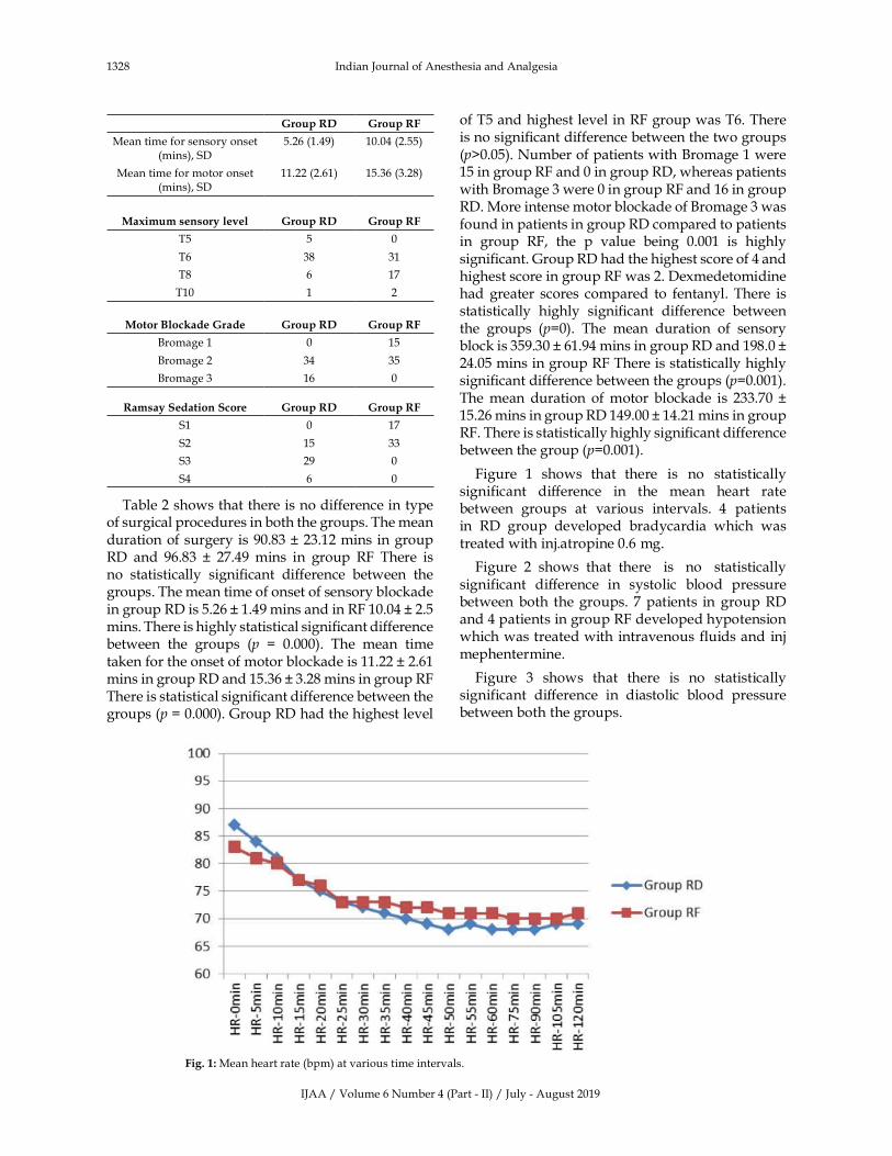
IJAA�/�Volume�6�Number�4�(Part�-�II)�/�July�-�August�2019
1328 Indian�Journal�of�Anesthesia�and�Analgesia
�roup��� �roup��F
Mean�time�for�sensory�onset�(mins),�SD
5.26�(1.49) 10.04�(2.55)
Mean�time�for�motor�onset�(mins),�SD
11.22�(2.61) 15.36�(3.28)
Ma�imum�sensory�le�el �roup��� �roup��F
T5 5 0
T6 38 31
T8 6 17
T10 1 2
Motor�Blockade��rade �roup��� �roup��F
�romage�1 0 15
�romage�2 34 35
�romage�3 16 0
�amsay�Sedation�Score �roup��� �roup��F
S1 0 17
S2 15 33
S3 29 0
S4 6 0
Table�2�shows�that�there�is�no�difference�in�type�of�surgical�procedures�in�both�the�groups.�The�mean�duration�of�surgery�is�90.83���23.12�mins� in�group�RD� and� 96.83� �� 27.49�mins� in� group� RF� There� is�no� statistically� signi��cant� difference� between� the�groups.�The�mean�time�of�onset�of�sensory�blockade�in�group�RD�is�5.26���1.49�mins�and�in�RF�10.04���2.5�mins.�There�is�highly�statistical�signi��cant�difference�between� the� groups� (p� �� 0.000).� The� mean� time�taken�for�the�onset�of�motor�blockade�is�11.22���2.61�mins�in�group�RD�and�15.36���3.28�mins�in�group�RF�There�is�statistical�signi��cant�difference�between�the�groups�(p���0.000).�Group�RD�had�the�highest�level�
of�T5�and�highest�level�in�RF�group�was�T6.�There�is�no�signi��cant�difference�between�the�two�groups�(p�0.05).�Number�of�patients�with��romage�1�were�15�in�group�RF�and�0�in�group�RD,�whereas�patients�with��romage�3�were�0�in�group�RF�and�16�in�group�RD.�More�intense�motor�blockade�of��romage�3�was�found�in�patients�in�group�RD�compared�to�patients�in� group� RF,� the� p� value� being� 0.001� is� highly�signi��cant.�Group�RD�had�the�highest�score�of�4�and�highest�score�in�group�RF�was�2.�Dexmedetomidine�had� greater� scores� compared� to� fentanyl.� There� is�statistically� highly� signi��cant� difference� between�the� groups� (p�0).� The� mean� duration� of� sensory�block�is�359.30���61.94�mins�in�group�RD�and�198.0���24.05�mins�in�group�RF�There�is�statistically�highly�signi��cant�difference�between�the�groups�(p�0.001).�The�mean� duration�of�motor� blockade� is� 233.70� ��15.26�mins�in�group�RD�149.00���14.21�mins�in�group�RF.�There�is�statistically�highly�signi��cant�difference�between�the�group�(p�0.001).
Figure� 1� shows� that� there� is� no� statistically�significant� difference� in� the� mean� heart� rate�between� groups� at� various� intervals.� 4� patients�in� RD� group� developed� bradycardia� which� was�treated�with�inj.atropine�0.6�mg.
Figure� 2� shows� that� there� is� no� statistically�significant� difference� in� systolic� blood� pressure�between� both�the�groups.� 7�patients� in�group�RD�and�4�patients�in�group�RF�developed�hypotension�which�was�treated�with�intravenous� fluids�and� inj�mephentermine.
Figure� 3� shows� that� there� is� no� statistically�significant� difference� in� diastolic� blood� pressure�between�both�the�groups.
Fig���:�Mean�heart�rate�(bpm)�at�various�time�intervals.
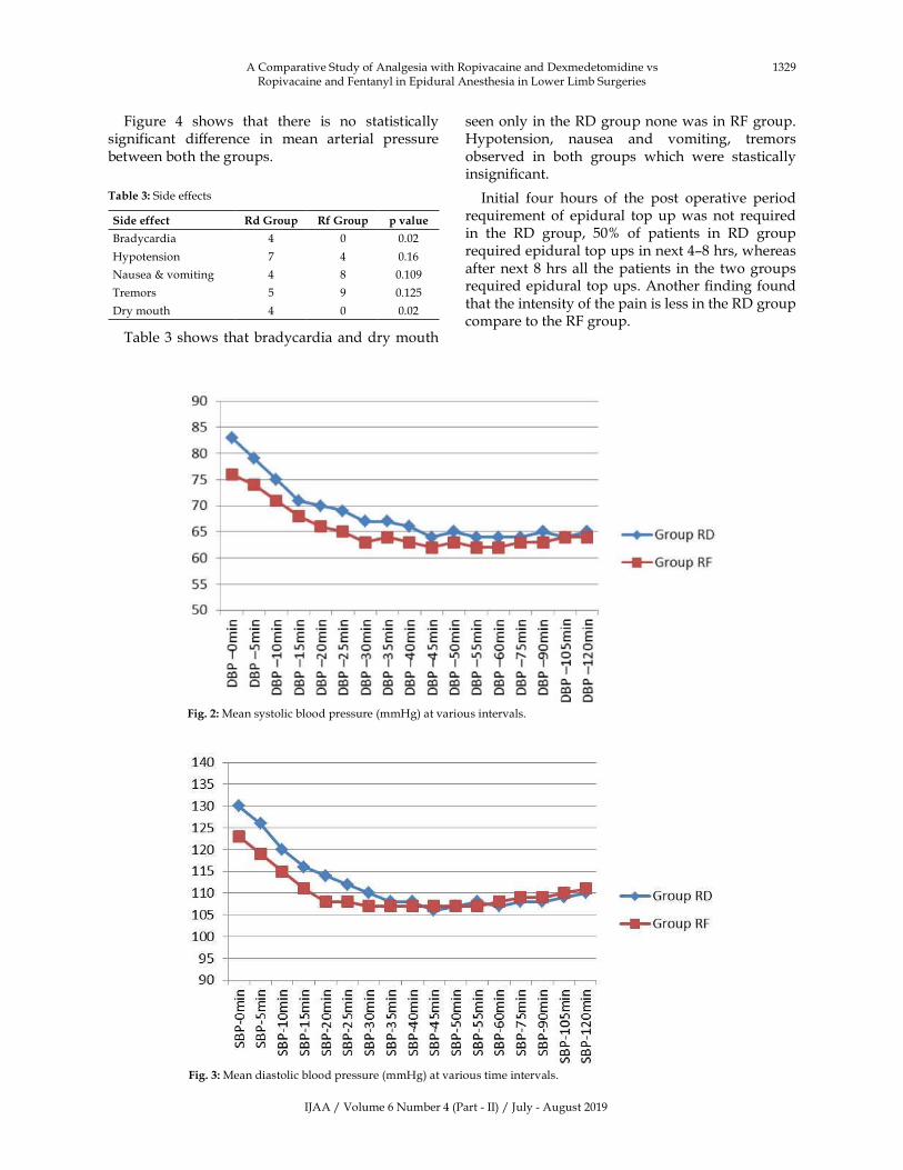
IJAA�/�Volume�6�Number�4�(Part�-�II)�/�July�-�August�2019
1329
Figure� 4� shows� that� there� is� no� statistically�significant� difference� in� mean� arterial� pressure�between�both�the�groups.
Table��:�Side�effects
Side�effect �d��roup �f��roup p��alue
�radycardia 4 0 0.02
�ypotension 7 4 0.16
Nausea���vomiting 4 8 0.109
Tremors 5 9 0.125
Dry�mouth 4 0 0.02
Table�3�shows�that�bradycardia�and�dry�mouth�
seen�only�in�the�RD�group�none�was�in�RF�group.��ypotension,� nausea� and� vomiting,� tremors�observed� in� both� groups� which� were� stastically�insigni��cant.
Initial� four� hours� of� the� post� operative� period�re�uirement� of� epidural� top� up�was� not� re�uired�in� the� RD� group,� 50�� of� patients� in� RD� group�re�uired�epidural�top�ups�in�next�4–8�hrs,�whereas�after�next� 8�hrs�all� the�patients� in� the� two�groups�re�uired�epidural�top�ups.�Another���nding�found�that�the�intensity�of�the�pain�is�less�in�the�RD�group�compare�to�the�RF�group.
Fig���:�Mean�systolic�blood�pressure�(mm�g)�at�various�intervals.
Fig���:�Mean�diastolic�blood�pressure�(mm�g)�at�various�time�intervals.
A�Comparative�Study�of�Analgesia�with�Ropivacaine�and�Dexmedetomidine�vs�Ropivacaine�and�Fentanyl�in�Epidural�Anesthesia�in�Lower�Limb�Surgeries
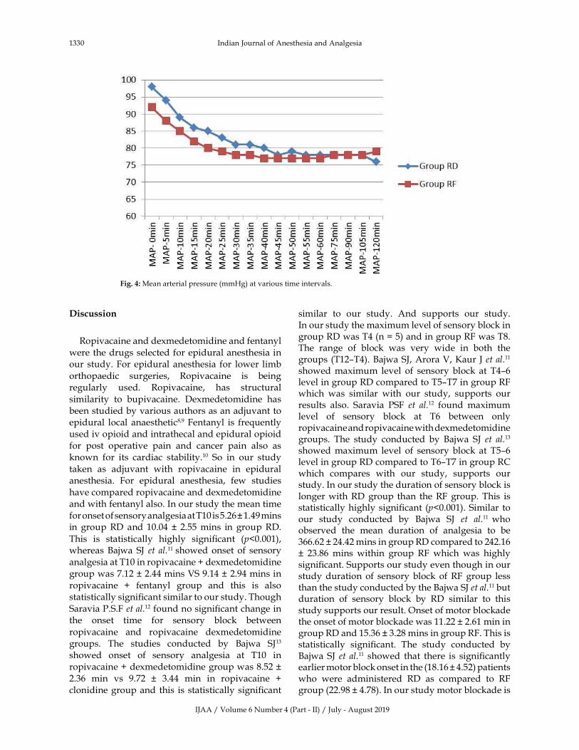
IJAA�/�Volume�6�Number�4�(Part�-�II)�/�July�-�August�2019
1330 Indian�Journal�of�Anesthesia�and�Analgesia
Fig���:�Mean�arterial�pressure�(mm�g)�at�various�time�intervals.
�iscussion
Ropivacaine�and�dexmedetomidine�and�fentanyl�
were�the�drugs�selected�for�epidural�anesthesia�in�our� study.�For�epidural�anesthesia� for� lower� limb�
orthopaedic� surgeries,� Ropivacaine� is� being�regularly� used.� Ropivacaine,� has� structural�
similarity� to� bupivacaine.� Dexmedetomidine� has�been�studied�by�various�authors�as�an�adjuvant�to�epidural� local� anaesthetic8,9�Fentanyl� is� fre�uently�used�iv�opioid�and�intrathecal�and�epidural�opioid�
for� post� operative� pain� and� cancer� pain� also� as�known� for� its� cardiac� stability.10� So� in� our� study�
taken� as� adjuvant� with� ropivacaine� in� epidural�anesthesia.� For� epidural� anesthesia,� few� studies�
have�compared�ropivacaine�and�dexmedetomidine�and�with�fentanyl�also.�In�our�study�the�mean�time�
for�onset�of�sensory�analgesia�at�T10�is�5.26���1.49�mins�in� group�RD� and�10.04� �� 2.55�mins� in� group�RD.�
This� is� statistically� highly� signi��cant� (p�0.001),�whereas��ajwa� SJ� et� al.11� showed�onset� of� sensory�
analgesia�at�T10�in�ropivacaine���dexmedetomidine�group�was�7.12���2.44�mins�VS�9.14���2.94�mins�in�
ropivacaine� �� fentanyl� group� and� this� is� also�statistically�signi��cant�similar�to�our�study.�Though�
Saravia�P.S.F�et�al.12�found�no�signi��cant�change�in�the� onset� time� for� sensory� block� between�
ropivacaine� and� ropivacaine� dexmedetomidine�groups.� The� studies� conducted� by� �ajwa� SJ13�
showed� onset� of� sensory� analgesia� at� T10� in�ropivacaine���dexmedetomidine�group�was�8.52���
2.36� min� vs� 9.72� �� 3.44� min� in� ropivacaine� ��clonidine�group�and� this� is� statistically�signi��cant�
similar� to� our� study.� And� supports� our� study.�In�our�study�the�maximum�level�of�sensory�block�in�group�RD�was�T4�(n���5)�and�in�group�RF�was�T8.�The� range� of� block� was� very� wide� in� both� the�groups�(T12–T4).��ajwa�SJ,�Arora�V,��aur�J�et�al.11
showed�maximum� level� of� sensory� block� at� T4–6�level�in�group�RD�compared�to�T5–T7�in�group�RF�which� was� similar� with� our� study,� supports� our�results� also.� Saravia� PSF� et� al.12� found� maximum�level� of� sensory� block� at� T6� between� only�ropivacaine�and�ropivacaine�with�dexmedetomidine�groups.� The� study� conducted� by� �ajwa� SJ� et� al.13�
showed�maximum� level� of� sensory� block� at� T5–6�level�in�group�RD�compared�to�T6–T7�in�group�RC�which� compares� with� our� study,� supports� our�study.�In�our�study�the�duration�of�sensory�block�is�longer�with�RD�group� than� the�RF�group.�This� is�statistically�highly�signi��cant� (p�0.001).�Similar� to�our� study� conducted� by� �ajwa� SJ� et� al.11� who�observed� the� mean� duration� of� analgesia� to� be�366.62���24.42�mins�in�group�RD�compared�to�242.16��� 23.86�mins� within� group� RF� which� was� highly�signi��cant.�Supports�our�study�even�though�in�our�study�duration� of� sensory�block� of�RF� group� less�than�the�study�conducted�by�the��ajwa�SJ�et�al.11�but�duration� of� sensory� block� by� RD� similar� to� this�study�supports�our�result.�Onset�of�motor�blockade�the�onset�of�motor�blockade�was�11.22���2.61�min�in�group�RD�and�15.36���3.28�mins�in�group�RF.�This�is�statistically� signi��cant.� The� study� conducted� by��ajwa� SJ� et� al.11� showed� that� there� is� signi��cantly�earlier�motor�block�onset�in�the�(18.16���4.52)�patients�who� were� administered� RD� as� compared� to� RF�group�(22.98���4.78).�In�our�study�motor�blockade�is�
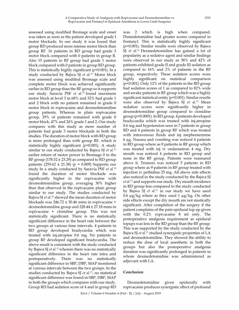
IJAA�/�Volume�6�Number�4�(Part�-�II)�/�July�-�August�2019
1331
assessed�using�modi��ed��romage�scale�and�onset�was�taken�as�soon�as�the�patient�developed�grade�I�motor� blockade.� In� our� study� it� was� found� that�group�RD�produced�more�intense�motor�block�than�group� RF.� 16� patients� in� RD� group� had� grade� 3�motor�block�compared�with�0�patients�in�group�R.�Also� 15� patients� in� RF� group� had� grade� 1�motor�block�compared�with�0�patients�in�group�RD�group.�This�is�statistically�highly�signi��cant�(p�0.001).�In�a�study� conducted� by� �ajwa� SJ� et� al.11�Motor� block�was� assessed� using� modi��ed� �romage� scale� and�complete� motor� block� was� achieved� signi��cantly�earlier�in�RD�group�than�the�RF�group�so�it�supports�our� study.� Saravia� PSF� et� al.15� found� maximum�motor�block�at�level�3�in�68��and�32��had�grade�1�and� 2� block�with�no� patient� remained� in� grade� 0�motor�block�in�ropivacaine�and�dexmedetomidine�group� patients.� Whereas� in� plain� ropivacaine�group,� 29�� of� patients� remained� with� grade� 0�motor�block,�47��and�24��grade�1�and�2.�Our�study�compares� with� this� study� as� more� number� of�patients� had� grade� 3�motor� blockade� in� both� the�studies.�The�duration�of�motor�block�with�RD�group�is�more� prolonged� than�with� group� RF,�which� is�statistically� highly� signi��cant� (p�0.001).� A� study�similar�to�our�study�conducted�by��ajwa�SJ�et�al.11�
earlier�return�of�motor�power�to��romage�0�in�the�RF�group�(178.52���23.29)�as�compared�to�RD�group�patients� (259.62� �� 21.38)� (p� �� 0.009)� Supports� our�study�In�a�study�conducted�by�Saravia�PSF�et�al.15�
found� the� duration� of� motor� blockade� was�signi��cantly� higher� in� the� ropivacaine� with�dexmedetomidine� group,� averaging� 30�� higher�than�that�observed�in�the�ropivacaine�plain�group�similar� to� our� study.� The� studies� conducted� by��ajwa�SJ�et�al.16�showed�the�mean�duration�of�motor�blockade�was�246.72���30.46�mins�in�ropivacaine���dexmedetomidine�group�and�228.44���27.18�mins�in�ropivacaine� �� clonidine� group.� This� was� not�statistically� signi��cant.� There� is� no� statistically�signi��cant�difference�in�the�heart�rate�between�the�two�groups�at�various�time�intervals.�4�patients�in�RD� group� developed� bradycardia� which� was�treated� with� inj.atropine� 0.6� mg.� No� patients� in�group� RF� developed� signi��cant� bradycardia.� The�above�result�is�consistent�with�the�study�conducted�by��ajwa�SJ�et�al.11�wherein�there�was�no�statistically�signi��cant� difference� in� the� heart� rate� intra� and�postoperatively.� There� was� no� statistically�signi��cant�difference�in�S�P,�D�P,�MAP�monitored�at�various�intervals�between�the�two�groups.�In�the�studies�conducted�by��ajwa�SJ�et�al.11,�no�statistical�signi��cant�difference�was�found�in�S�P,�D�P,�MAP�in�both�the�groups�which�compares�with�our�study.�Group�RD�had�sedation�score�of�4�and�in�group�RD�
was� 2� which� is� high� when� compared.�Dxmedetomidine�had�greater� scores� compared� to�Fentanyl.� This� is� statistically� highly� signi��cant�(p�0.001).�Similar� results�were�observed�by��ajwa�SJ� et� al.11� Dexmedetomidine� has� gained� a� lot� of�popularity�as�a�sedative�agent�and�similar���ndings�were� observed� in� our� study� as� 38�� and� 42�� of�patients�exhibited�grade�II�and�grade�III�sedation�as�compared� to� 16�� and� 2�� of� patients� in� the� RF�group,� respectively.� These� sedation� scores� were�highly� signi��cant� on� statistical� comparison�(p�0.001).�Only�12��of�the�patients�in�the�RD�group�had�sedation�scores�of�1�as�compared�to�82��wide�and�awake�patients�in�RF�group�which�was�a�highly�signi��cant�statistical�entity�(p�0.001).�Similar�results�were� also� observed� by� �ajwa� SJ� et� al.11� Mean�sedation� scores� were� signi��cantly� higher� in�dexmedetomidine� group� compared� to� clonidine�group�(p�0.0001).�In�RD�group,�4�patients�developed�bradycardia� which� was� treated� with� inj.atropine�0.6�mg�and�hypotension�seen�in�7�patients�in�group�RD�and�4�patients�in�group�RF�which�was�treated�with� intravenous� ��uids� and� inj� mephentermine�6��g.�Nausea�and�vomiting�was�noticed�4�patients�in�RD�group�where�as�8�patients�in�RF�group�which�was� treated� with� inj� iv� ondansetron� 4� mg.� Dry�mouth� was� noticed� 4� patients� in� RD� group� and�none� in� the� RF� group.� Patients� were� reassured�above� it.� Tremors� was� noticed� 5� patients� in� RD�group�where�as�9�patients�in�RF�group�treated�with�injection�iv�pethidine�25�mg.�All�above�side�effects�also�noticed�in�the�study�conducted�by�the��ajwa�SJ�et�al.11�and�supports�our�study.�Dry�mouth�incidence�in�RD�group�less�compared�to�the�study�conducted�by� �ajwa� SJ� et� al.11� in� our� study� we� have� used�0.6��g/kg�where� as� they�used�1��g/kg.�On�other�side�effects�except�the�dry�mouth�are�not�stastically�signi��cant.�After� completion� of� the� surgery� if� the�patient�complains�of�the�pain�epidural�top�up�given�with� the� 0.2�� ropivacaine� 8� ml� only.� The�postoperative� analgesia� re�uirement� as� epidural�topups�was�less�in�the�RD�group�than�the�RF�group.�This�was�supported�by�the�study�conducted�by�the��ajwa�SJ�et�al.11�studied�synergistic�properties�of�LA�and�dexmedetomidine.�They�showed�the�ability�to�reduce� the� dose� of� local� anesthetic� in� both� the�groups� but� also� the� postoperative� analgesia�duration�was�signi��cantly�prolonged�in�patients�in�whom� dexmedetomidine� was� administered� as�adjuvant�with�LA.
Conclusion
Dexmedetomidine� given� epidurally� with�ropivacaine�produces�synergistic�effect�of�profound�
A�Comparative�Study�of�Analgesia�with�Ropivacaine�and�Dexmedetomidine�vs�Ropivacaine�and�Fentanyl�in�Epidural�Anesthesia�in�Lower�Limb�Surgeries
IJAA�/�Volume�6�Number�4�(Part�-�II)�/�July�-�August�2019
1332 Indian�Journal�of�Anesthesia�and�Analgesia
and� motor� blockade� and� sensory� blockade�duration� being� prolonged.� There� is� relatively�
less� incidence� of� complications� and� side� effects�when� dexmedetomidine� used� as� adjunct� in� the�epidural� anesthesia.� �ence� its� concluded� that�dexmedetomidine�can�be�used�as�a�more�potent�and�safer�alternative�to�Fentanyl�in�epidural�anesthesia�as�a�adjuvant�to�ropivacaine.
�eferences
1.� Crawford� JS.� Experiences� with� epidural� blood�patch.�Anesthesia.�1980;35:513–5.
2.� Miller,� Ronald� D.� Miller�s� Anesthesia.� 7th� ed.�Philadelphia:� Elsevier� Churchill� Livingstone;�
2009:2.
3.� Casati�A,�Putzu�M.��upivacaine,�levobupivacaine�and�Ropivacaine:�are�they�clinically�different���est�Practice�and�Research.�Clinical�Anaesthesiology.2005;19:247–68.
4.� Arthur�GR,�Feldman��S,�Norway�S�,�et�al.�Acute�IV� toxicity� of� LEA� 103,� a� new� local� anesthetic,�compared� to� lidocaine� and� bupivacaine� in� the�
awake�dog.�anesthesiology�1986;65:A182.
5.� �atz� JA,� �ridenbaugh� PO,� �narr� DC,� et� al.�Pharmacodynamics� and� pharmacokinetics� of�epidural� ropivacaine� in� humans.� Anesth�Analg.�1990�Jan;70(1):16-21.
6.� Concepcion�M,�Arthur�GR,�Steele�SM,�et�al.�A�new�local�anesthetic,�ropivacaine.�Its�epidural�effects�in�humans.�Anesthesia�and�analgesia.�1990;70:80–5.
7.� �atz� JA,��narr�DC,� �ridenbaugh� PO.�A� double�blind�comparison�of�0.5���upivacaine�and�0.75��
Ropivacaine�administered�epidurally�in�humans.�Regional�anesthesia.�1990;15:250–2.
8.� �rown� DL,� Carpenter� RL,� Thompson� GE.�Comparison� of� 0.5�� Ropivacaine� and� 0.5���upivacaine� for� epidural� anesthesia� in�patients� undergoing� lower� extremity� surgery.�anesthesiology.�1990;72:633–6.
9.� Vieira�AM,�Schnaider�T��et�al.�Epidural�clonidine�or� dexmedetomidine� for� post-cholecystectomy�analgesia�and�sedation.�Rev��ras�Anestesiol.�2004�Aug;(54(4)):�p.�473–8.
10.� Schnaider� T�,� Vieira� AM� et� al.� Intraoperative�analgesic�effect�of�epidural�ketamine,�clonidine�or�dexmedetomidine� for�upper�abdominal� surgery.�Rev��ras�Anestesiol.�2005�Oct;(55(5)):�p.�525–31.
11.� Ahmadinejad� M,� Vatankhah� M,� Shokoohi� M,�Nouroozi�M,�Ahmadipour�M.�Comparison�of�the�effect�of�intravenous�and�epidural�administration�of� fentanyl� on� pain� severity� and� hemodynamic�status� in� patients� with� abdominal� and� thoracic�trauma.� �ahedan� J� Res� Med� Sci.� 2013;(�JRMS)�15(2):14–18.
12.� �ajwa� SJ,� Arora� V,� �aur� J� et� al.� Comparative�evaluation� of� dexmedetomidine� and� fentanyl�for�epidural�analgesia�in�lower�limb�orthopaedic�surgeries.� Saudi� Journal� of� Anesthesia.� 2011�Oct-Dec;(5(4)):365–70.
13.� Saravia�P.S.F,� Sabbag�AT�et� al.�Synergistic� effect�between� dexmedetomidine� and� ropivacaine�0.75�� in� epidural� anesthesia.� Rev� Assoc� Med��ras.�2008�mae-april;(54(2)):110–5.
14.� �ajwa�SJ,��ajwa�S�,��aur�J�et�al.�Dexmedetomidine�and� clonidine� in� epidural� anesthesia:A�comparative� evaluation.� Indian� Journal� of�Anesthesia.�2011�Mar-Apr;55(2):116.
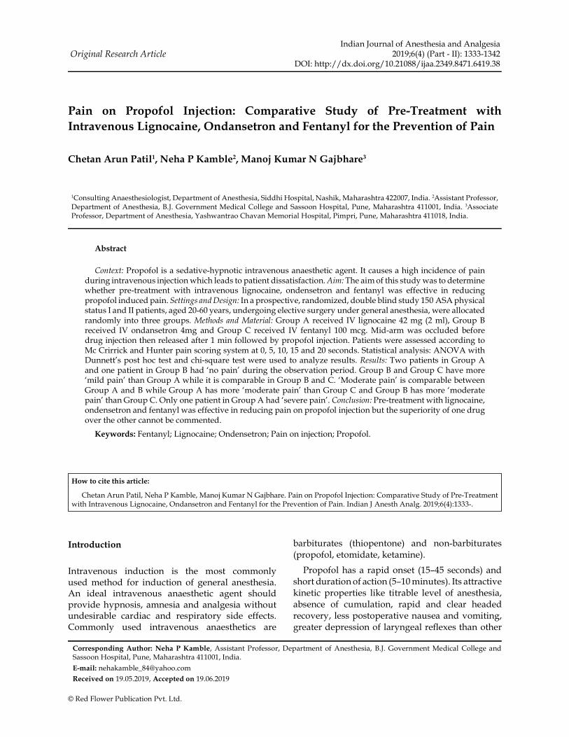
��Red�Flower�Publication�Pvt.�Ltd.�
�Original�Research�ArticleIndian�Journal�of�Anesthesia�and�Analgesia�
2019;6(4)�(Part�-�II):�1333-1342DOI:�http://dx.doi.org/10.21088/ijaa.2349.8471.6419.38
Pain� on� Propofol� In�ection:� Comparati�e� Study� of� Pre�Treatment� �ith�
Intra�enous��ignocaine��Ondansetron�and�Fentanyl�for�the�Pre�ention�of�Pain
Chetan�Arun�Patil���Neha�P��amble���Mano���umar�N��a�bhare�
1Consulting�Anaesthesiologist,�Department�of�Anesthesia,�Siddhi��ospital,�Nashik,�Maharashtra�422007,�India.�2Assistant�Professor,�Department�of�Anesthesia,��.J.�Government�Medical�College�and�Sassoon��ospital,�Pune,�Maharashtra�411001,� India.� 3Associate�Professor,�Department�of�Anesthesia,��ashwantrao�Chavan�Memorial��ospital,�Pimpri,�Pune,�Maharashtra�411018,�India.
Corresponding�Author:�Neha�P��amble,�Assistant�Professor,�Department� of�Anesthesia,��.J.�Government�Medical�College� and�Sassoon��ospital,�Pune,�Maharashtra�411001,�India.
E�mail:�[email protected]
�ecei�ed�on�19.05.2019,�Accepted�on�19.06.2019
Abstract
Conte�t:�Propofol�is�a�sedative-hypnotic�intravenous�anaesthetic�agent.�It�causes�a�high�incidence�of�pain�during�intravenous�injection�which�leads�to�patient�dissatisfaction.�Aim:�The�aim�of�this�study�was�to�determine�whether� pre-treatment� with� intravenous� lignocaine,� ondensetron� and� fentanyl� was� effective� in� reducing�propofol�induced�pain.�Settings�and��esign:�In�a�prospective,�randomized,�double�blind�study�150�ASA�physical�status�I�and�II�patients,�aged�20-60�years,�undergoing�elective�surgery�under�general�anesthesia,�were�allocated�randomly� into� three�groups.�Methods�and�Material:�Group�A�received�IV� lignocaine�42�mg�(2�ml),�Group���received� IV� ondansetron�4mg� and�Group�C� received� IV� fentanyl� 100�mcg.�Mid-arm�was�occluded� before�drug�injection�then�released�after�1�min�followed�by�propofol�injection.�Patients�were�assessed�according�to�Mc�Crirrick�and��unter�pain�scoring�system�at�0,�5,�10,�15�and�20�seconds.�Statistical�analysis:�ANOVA�with�Dunnett�s�post�hoc�test�and�chi-s�uare�test�were�used�to�analyze�results.�Results:�Two�patients�in�Group�A�and�one�patient�in�Group���had��no�pain��during�the�observation�period.�Group���and�Group�C�have�more��mild�pain��than�Group�A�while�it�is�comparable�in�Group���and�C.��Moderate�pain��is�comparable�between�Group�A�and���while�Group�A�has�more��moderate�pain��than�Group�C�and�Group���has�more��moderate�pain��than�Group�C.�Only�one�patient�in�Group�A�had��severe�pain�.�Conclusion:�Pre-treatment�with�lignocaine,�ondensetron�and�fentanyl�was�effective�in�reducing�pain�on�propofol�injection�but�the�superiority�of�one�drug�over�the�other�cannot�be�commented.
�ey�ords:�Fentanyl;�Lignocaine;�Ondensetron;�Pain�on�injection;�Propofol.
�o��to�cite�this�article:
Chetan�Arun�Patil,�Neha�P��amble,�Manoj��umar�N�Gajbhare.�Pain�on�Propofol�Injection:�Comparative�Study�of�Pre-Treatment�with�Intravenous�Lignocaine,�Ondansetron�and�Fentanyl�for�the�Prevention�of�Pain.�Indian�J�Anesth�Analg.�2019;6(4):1333-.
Introduction
Intravenous� induction� is� the� most� commonly�used�method� for� induction� of� general� anesthesia.An� ideal� intravenous� anaesthetic� agent� should�provide�hypnosis,� amnesia� and�analgesia�without�undesirable� cardiac� and� respiratory� side� effects.�Commonly� used� intravenous� anaesthetics� are�
barbiturates� (thiopentone)� and� non-barbiturates�(propofol,�etomidate,�ketamine).
Propofol�has�a� rapid�onset� (15–45�seconds)�and�short�duration�of�action�(5–10�minutes).�Its�attractive�kinetic� properties� like� titrable� level� of� anesthesia,�absence� of� cumulation,� rapid� and� clear� headed�recovery,� less�postoperative�nausea�and�vomiting,�greater�depression�of�laryngeal�re��exes�than�other�
IJAA�/�Volume�6�Number�4�(Part�-�II)�/�July�-�August�2019
1334 Indian�Journal�of�Anesthesia�and�Analgesia
commonly� used� anaesthetic� agents1� and�minimal�side�effects�makes�it�an�ideal�agent�for�induction�of�anesthesia.2
Propofol� is� known� to� cause� severe,� sharp,�stinging� or� burning� pain� on� injection,� especially�when� given� in� small� veins� on� dorsum� of� hand.�This� is� clinically� unacceptable� as� it� can� cause�agitation� and� interfere�with� smooth� induction�of�anesthesia.�The�reported�incidence�varies�between�28��and�90��in�adults�when�a�vein�on�dorsum�of�hand� is�used.3� It� can�be� immediate�or�delayed� in�nature.�The�immediate�pain�probably�results�from�direct�irritant�effect�whereas�delayed�pain�results�from� indirect� effect� via� the� kinin� cascade� which�occurs�10–20�seconds�later.4�Nafamostat�mesilate,�a�kallikrein�inhibitor,�is�effective�in�decreasing�pain�on�injection�of�propofol.�Its�effectiveness�has�been�attributed� to� the� activation� of� kallikrein-� kinin�system� as� the� propofol� emulsion� contacts� free�nerve�endings� in� the�vein.�Factor��II� is�activated�which� converts�prekallikrein� to�kallikrein,�which�then� cleaves� high-molecular� weight� kininogen�to� release� bradykinin.� �radykinin� causes�vasodilatation�and�increased�permeability�of�vein,�increasing� contact� between� a�ueous� phase� of�propofol�and�free�nerve�endings�of�vein.�This�will�manifest�as�pain.5,6
Drugs� like� lignocaine,� opioids� like� fentanyl,�morphine� and�butarphanol,�magnesium� sulphate,�paracetamol,� ephedrine,� metoclopramide� and�many� more� have� been� tried� to� reduce� this� pain.�Of� these� the� most� commonly� used� methods� are�pre-treatment�with� IV� (intravenous)� lignocaine�or�IV�fentanyl.
Lignocaine�reduces�pain�due�its�local�anaesthetic�action,7� fentanyl,� has� some�peripherally�mediated�analgesic� action�within� the� clinical�dosage� range,8�ondansetron9�due� to� its�multifaceted�action�as�Na�channel� blocker,� a� 5�T
3� receptor� antagonist� and�
�-opioid� agonist,� can� be� used� to� alleviate� pain�produced� by� propofol.� �owever� the� ef��cacy� of�ondansetron�has�never�been�compared�with�drugs�like�lignocaine�and�fentanyl.
Our�aim�is�to�compare�the�ef��cacy�of�lignocaine,�ondansetron� and� fentanyl� in� reducing� pain� on�injection� of� propofol,� to� detect� an� agent� with�minimal�side�effects�and�the�hemodynamic�changes�due�to�these�drugs.
Materials�and�Methods
After�approval�by� the��ospital�Ethics�Committee,�we� studied� 150� patients� of� either� sex,� aged�
20-60� years,� scheduled� for� elective� surgery� under�general�anesthesia.
All� patients� who� belong� to� the� ASA� physical�status� class� I� and� II� were� enrolled� for� this�prospective� randomized,� double-blind� study.�Patients� were� not� included� if� they� refused� to�participate�in�the�study,�a�history�of�allergy�to�the�study� medications,� hemodynamically� unstable�patients,� presence� of� infection� on� the� dorsum� of�hand,� dif��cult� IV� cannulation.� Also� excluded�were�patients� of�ASA�physical� status� III� and� IV,�pregnant� and� diabetic� patients,� patients� with�pre-existing� cardiac� conduction� defects,� patients�receiving�analgesics�and�patients�with�dif��culty�in�communication.
In� a� pilot� study� on� 20� patients� we� found� that�all� patients� complained� of� pain� during� propofol�injection.� The� incidence� of� moderate� to� severe�pain� was� highest� at� 15� seconds.� So� the� number�needed� to� bring� down� the� incidence� of� pain� by�50�,�keeping�the�type�I�error�at�5��and�power�of�study�at�95��was�44.�We�used�a�web�based�sample�size�calculator.�To�round� it�off�we�have� taken� the�number�of�patients�in�each�group�as�50.
Randomization� list� was� generated� by� random�number� function� using� the� Microsoft� Excel� 2003�spreadsheet,� resulting� in� a� list� of� 150� assigned� to�participants� receiving� the� drugs.� Randomization�was� conducted� using� se�uentially� numbered,�opa�ue�and�sealed�envelope.�Patients�were�divided�in�three�groups,�50�patients�in�each�group.
Group�A:�IV�Lignocaine�42�mg�(2�ml)�diluted�to�5�cc.
Group��:�IV�Ondansetron�4mg�diluted�to�5cc.
Group�C:�IV�Fentanyl�100�mcg�diluted�to�5cc.
The�study�drug�was�prepared�in�identical�looking�(5ml)� coded� syringes� by� an� anaesthesiologist� not�involved�in�the�study.�The�drug�administrator�and�the�person�making�the�observations�were�blind�to�the�study�drug.
A� complete� pre-anaesthetic� evaluation� was�done.� After� explaining� the� anaesthetic� procedure�to�the�patients,�written�informed�consent�was�taken�to� include� them� in� the� study.� They� received� the�study�medication�as�per�randomization.�During�the�preoperative� rounds� all� patients� were� explained�about� the� pain� scoring� method� advocated� by�Mc�Crirrick�and��unter�.
All� patients� were� kept� fasting� 6� hours� before�surgery.� They� were� advised� to� take� tablet�alprazolam� 0.5� mg� and� tablet� ranitidine� 150� mg�night� before� surgery.� In� the� operating� room,�
IJAA�/�Volume�6�Number�4�(Part�-�II)�/�July�-�August�2019
1335
non-invasive� blood� pressure,� electrocardiogram�and�pulse�oximeter�were�attached�and�baseline�vital�parameters� were� noted.� Intravenous� access� was�established�with�an�18-G�cannula�on�the�dorsum�of�the�non-dominant�hand.�No�analgesic�or�sedative�drugs� were� given� before� induction.� Venous�occlusion�of�the�arm�proximal�to�the�puncture�site�was�maintained�with�a�blood�pressure�cuff�in��ated�to�40�mm�g.�The�study�drugs�were�injected�over�30� seconds� and� venous� occlusion� was� released�after�1�min�of�completion�of�study�drug�injection.�Propofol� (2� mg/kg)� was� administered� through�the� intravenous� cannula.� During� the� ��rst� 25��of� the� calculated� propofol� dose,� the� patient� was�assessed�according�to�four-�point�pain�score�at�0,�5,�10,�15�and�20�seconds.�Following�which,�general�anesthesia� was� continued� with� the� remainder�of� the� calculated� dose� of� propofol.� Vecuronium�0.1� mg/kg� intravenous� was� used� to� facilitate�tracheal� intubation.� Anesthesia� was� maintained�with� iso��urane� 1�� and� 60�� nitrous� oxide� with�oxygen�on�controlled�ventilation�with�intermittent�bolus�of�vecuronium.
Intraoperative� analgesia� was� maintained�with� incremental� doses� of� intravenous� fentanyl�50�mcg.�At�the�end�of�surgery,�inhalational�agents�were�discontinued� and� neuromuscular� blockade�was� reversed�with� neostigmine� 0.5�mg/kg�with�glycopyrrolate� 0.1� mg/kg� intravenously.� The�participants�were�extubated�and�transferred�to�the�recovery� area� after� con��rmation� of� satisfactory�recovery� criteria.� All� observations� were�recorded� by� an� anaesthesiologist� not� involved�in�the�study.
��c�Crirric��and��unter�scale�of��ain�assess�ent�on�in�ection��ith��ro�ofol
Pain�score �egree�of�pain �esponse�
0 None Negative� response� to��uestioning
1 Mild Pain� reported� in� response� to��uestioning� only,�without� any�behavioral�signs
2 Moderate Pain� reported� in� response� to��uestioning� and� accompanied�by� a� behavioral� sign� or� pain�reported� spontaneously�without��uestioning
3 Severe Strong� vocal� response� or�response�accompanied�by�facial�grimacing,� arm�withdrawal� or�tears
The�following�observations�were�made:
Age,�sex,�ASA�grade,�body�mass�index�(�MI).
Pain� scores� at� 0,� 5,� 10,� 15� and� 20� seconds� after�
propofol�injection.
�eart�rate�and�oxygen�saturation�was�monitored�continuously,�recorded�at�baseline,�0,�5,�10,�15�and�20�seconds�after�propofol�injection.
Mean� arterial� blood� pressure� at� baseline,� after�20�seconds,�5,�10,�15�and�20�minutes.
Any�adverse�effects.
Statistics�Analysis
Data� represented� as�mean� �� SD� (n� �� 50).� All� the�results�were�analyzed�statistically�by�one-way�and�two-way� ANOVA� with� Dunnett�s� post� hoc� test�and�Chi� s�uare� test�by�using�Graphpad�Prism�7.0�(GraphPad�Software,� Inc.,�CA,��SA).����0.05�was�considered�as�signi��cant.
�esults
All� the� three� groups� were� comparable� (p� �� 0.05)�with�respect�to�demographic�parameters�and�ASA�Physical�Status�class�as�shown�(Fig.�1�and�2).
There�was�no� statistically� signi��cant� difference�in�the�mean�pulse�rate�recorded�at�baseline�and�at�0� seconds� but� a� statistically� signi��cant� difference�was�noted�at�5,�10,�15�and�20�seconds�when�compared�with�baseline�pulse�rate�(p���0.05)� (Table�1,�Fig.�3)�There�was�a�statistically�signi��cant�difference�in�the�mean�blood�pressure�recorded�at�baseline�and�that�recorded�after�20�seconds,�5,�10,�15�and�20�minutes�(p���0.05)�(Table�2,�Fig.�4).
No� signi��cant� changes�were� noted� in� the� SpO2
between�the�studied�groups�(p���0.05)�(Fig.�5).
The�incidence�of�pain�when�compared�amongst�the� groups�was� statistically� insigni��cant� (Table� 3,�Fig.�6).�Chi�–�s�uare�test���2�–�Test��was�used�for�the�comparison� of� incidence� of� pain.� Comparison� of�Group�A�with�Group���and�Group�C�gives�p�value�as�0.0956�and�0.514�respectively,�while�comparison�of�Group���with�Group�C�gives�p�value�as�0.1333�which�is�statistically�insigni��cant.
After�administration�of�drug,�48�(96�)�patients�in�Group�A,�49�(98�)�patients�in�Group���and�all�patients��50�(100�)��in�Group�C�experienced�pain�(p�0.7675)�(Table�4).�Pain�score�was�compared�in�all�the�groups�at�0,�5,�10,�15�and�20�seconds�(Table�5,�Fig.�7).
The� mean� pain� score� (MPS)� in� Group� A,� ��and� C� was� 1.46� �� 0.61,� 1.42� �� 0.53,� 1.48� �� 0.50�respectively�(Fig.�8).�Chi�–�s�uare�test�was�used�for�the�comparison�of�pain�scores�in�the�study�groups�(Fig.�9).�Comparison�of��MPS�0�(no�pain)�in�Group�A�with�Group���and�C�gives���value�as�2.037�and�
Pain�on�Propofol�Injection:�Comparative�Study�of�Pre-Treatment�with�Intravenous�Lignocaine,�Ondansetron�and�Fentanyl�for�the�Prevention�of�Pain

IJAA�/�Volume�6�Number�4�(Part�-�II)�/�July�-�August�2019
1336 Indian�Journal�of�Anesthesia�and�Analgesia
0.555� respectively,� while� comparison� of� Group���with�Group�C� gives� p� value�as� 0.0964�which� is�statistically�insigni��cant.
Comparison�of� �MPS� 1�(mild� pain)� in�Group�A�with�Group���and�C�gives�p�value�as�0.036�and�0.015respectively�which�is�statistically�signi��cant�while
comparison�of�Group���with�Group�C�gives�p�value�as�0.534�which�is�statistically�insigni��cant.
The� �MPS�2�� (moderate�pain)�in�Group�A�when�compared�with�Group���(p���0.074)�was�statistically�insigni��cant� and� that� with� Group� C� (p� �� 0.0005)�was�signi��cant�while�comparison�of�Group���with�
Age�(�ear)
Weight�(kg)�eight�(meter)
�MI
A � C
80
70
60
50
40
30
20
10
0
Fig���:�Graphical�representation�of�average�age,�weight,�height�and��MI�of�the�patients�in�different�drug�treated�group��Mean���SD�(n�50)�.�Column�width.
Male Female I II
Sex�(�) ASA(�)
A
�
C
35
30
25
20
15
10
5
0
Fig���:�Graphical�representation�of�Gender�and�ASA�stage�wise�distribution�of�patients�in�different�drug� treated� group� �Data� represented� as� n� (�).� Statistical� analysis� for� the� Gender� of� patients�
�2�0.1642,�p���0.9212.Statistical�analysis�for�ASA�Physical�Status��2�3.589,�p���0.1662.�Chi�s�uare�test�was�performed�for�the�statistical�analysis�.�Column�width.
Table��:�Effect�of�different�drug�treatment�on�the�pulse�rate�at�different�time�interval�after�the�administration�of�drug
�roupPulse�rate��Beats/min�
Baseline ��Sec ��Sec ���Sec ���Sec ���Sec
A 89.3���14.3 89.7���14.5 94.2���14.8 98.7���14.8�� 102.6���15.7�� 106.5���16.4��
� 88.3���9 88.1���9.2 94.1���9.5� 101.2���9.8�� 106.7���9.61�� 112.2���10.6��
C 87.5���9.1 87���9.2 90.9���9.5 102.7���9.5�� 112.4���9.2�� 123.63���8.7��
Mean���SD�(n�50)������0.01�compared�to�baseline
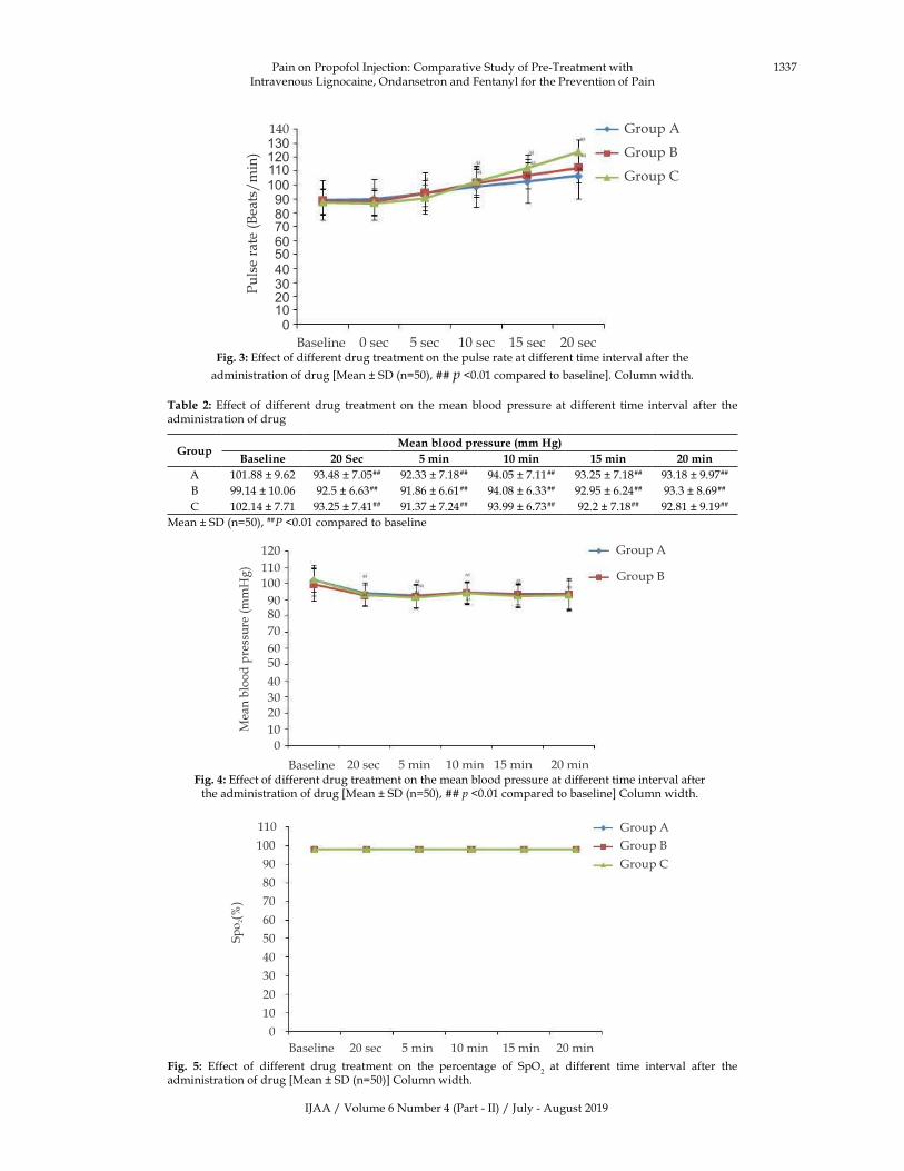
IJAA�/�Volume�6�Number�4�(Part�-�II)�/�July�-�August�2019
1337
Group�A
Group��
Group�C
�aseline 0�sec 5�sec 10�sec 15�sec 20�sec
140�������������������������������
Fig���:�Effect�of�different�drug�treatment�on�the�pulse�rate�at�different�time�interval�after�the�
administration�of�drug��Mean���SD�(n�50),����p��0.01�compared�to�baseline�.�Column�width.
Table� �:� Effect� of� different� drug� treatment� on� the�mean� blood� pressure� at� different� time� interval� after� the�administration�of�drug
�roupMean�blood�pressure��mm��g�
Baseline ���Sec ��min ���min ���min ���min
A 101.88���9.62 93.48���7.05�� 92.33���7.18�� 94.05���7.11�� 93.25���7.18�� 93.18���9.97��
� 99.14���10.06 92.5���6.63�� 91.86���6.61�� 94.08���6.33�� 92.95���6.24�� 93.3���8.69��
C 102.14���7.71 93.25���7.41�� 91.37���7.24�� 93.99���6.73�� 92.2���7.18�� 92.81���9.19��
Mean���SD�(n�50),������0.01�compared�to�baseline
Fig���:�Effect�of�different�drug�treatment�on�the�mean�blood�pressure�at�different�time�interval�after�the�administration�of�drug��Mean���SD�(n�50),����p��0.01�compared�to�baseline��Column�width.
Fig�� �:� Effect� of� different� drug� treatment� on� the� percentage� of� SpO2� at� different� time� interval� after� the�
administration�of�drug��Mean���SD�(n�50)��Column�width.
Pain�on�Propofol�Injection:�Comparative�Study�of�Pre-Treatment�with�Intravenous�Lignocaine,�Ondansetron�and�Fentanyl�for�the�Prevention�of�Pain
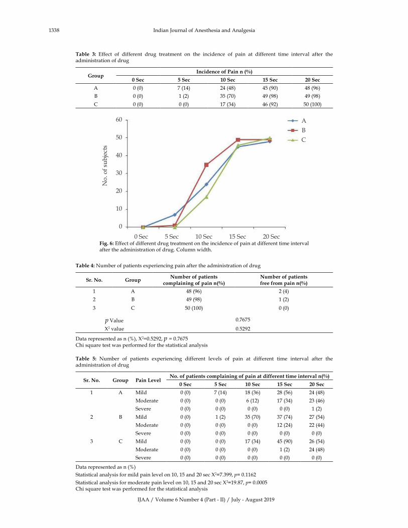
IJAA�/�Volume�6�Number�4�(Part�-�II)�/�July�-�August�2019
1338 Indian�Journal�of�Anesthesia�and�Analgesia
Table� �:� Effect� of� different� drug� treatment� on� the� incidence� of� pain� at� different� time� interval� after� the�administration�of�drug
�roupIncidence�of�Pain�n����
��Sec ��Sec ���Sec ���Sec ���Sec
A� 0�(0) 7�(14) 24�(48) 45�(90) 48�(96)
�� 0�(0) 1�(2) 35�(70) 49�(98) 49�(98)
C� 0�(0) 0�(0) 17�(34) 46�(92) 50�(100)
A
�
C
0�Sec 5�Sec 10�Sec 15�Sec 20�Sec
60
50
40
30
20
10
0
Fig���:�Effect�of�different�drug�treatment�on�the�incidence�of�pain�at�different�time�interval�after�the�administration�of�drug.�Column�width.
Table��:�Number�of�patients�experiencing�pain�after�the�administration�of�drug
Sr��No� �roupNumber�of�patients�
complaining�of�pain�n���Number�of�patients�free�from�pain�n���
1 A 48�(96) 2�(4)
2 � 49�(98) 1�(2)
3 C 50�(100) 0�(0)
p�Value 0.7675
�2�value 0.5292
Data�represented�as�n�(�),��2�0.5292,�p���0.7675Chi�s�uare�test�was�performed�for�the�statistical�analysis
Table� �:� Number� of� patients� experiencing� different� levels� of� pain� at� different� time� interval� after� the�administration�of�drug
Sr��No� �roup Pain��e�el�No��of�patients�complaining�of�pain�at�different�time�inter�al�n���
��Sec ��Sec ���Sec ���Sec ���Sec
1 A Mild 0�(0) 7�(14) 18�(36) 28�(56) 24�(48)
Moderate 0�(0) 0�(0) 6�(12) 17�(34) 23�(46)
Severe� 0�(0) 0�(0) 0�(0) 0�(0) 1�(2)
2 � Mild 0�(0) 1�(2) 35�(70) 37�(74) 27�(54)
Moderate 0�(0) 0�(0) 0�(0) 12�(24) 22�(44)
Severe� 0�(0) 0�(0) 0�(0) 0�(0) 0�(0)
3 C Mild 0�(0) 0�(0) 17�(34) 45�(90) 26�(54)
Moderate 0�(0) 0�(0) 0�(0) 1�(2) 24�(48)
Severe� 0�(0) 0�(0) 0�(0) 0�(0) 0�(0)
Data�represented�as�n�(�)�
Statistical�analysis�for�mild�pain�level�on�10,�15�and�20�sec��2�7.399,�p��0.1162
Statistical�analysis�for�moderate�pain�level�on�10,�15�and�20�sec��2�19.87,�p��0.0005Chi�s�uare�test�was�performed�for�the�statistical�analysis

IJAA�/�Volume�6�Number�4�(Part�-�II)�/�July�-�August�2019
1339
Group� C� (p� �� 0.010)� was� statistically� signi��cant.�Only�one�patient�(2.08�)�had��MPS�3��(severe�pain)�in�Group�A�which�is�statistically�insigni��cant.
There�were� few� adverse� events� observed� in� all�the�groups�during�the�study�period��Table�7�.
Pain�on�Propofol�Injection:�Comparative�Study�of�Pre-Treatment�with�Intravenous�Lignocaine,�Ondansetron�and�Fentanyl�for�the�Prevention�of�Pain
0�Sec
5�Sec
10�Sec
15�Sec
20�Sec
50
45
40
35
30
25
20
15
10
5
0
A � C
Fig�� �:� Number� of� patients� experiencing� different� level� of� pain� at� different� time� interval� after� the�administration�of�drug.�Column�width.
A � C
2.5
2
1.5
1
0.5
0
GroupFig���:�Effect�of�different�drug�treatment�on�the�level�of�mean�pain�score��Mean���SD�(n�50)�.�Column�width.
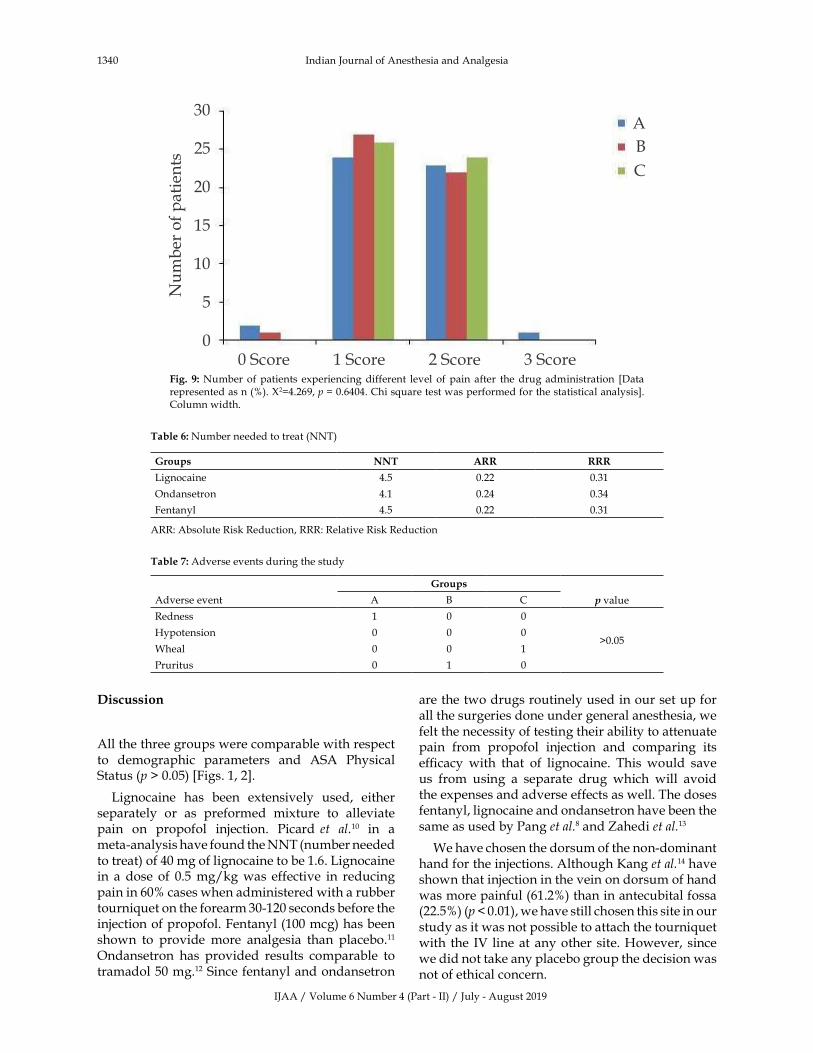
IJAA�/�Volume�6�Number�4�(Part�-�II)�/�July�-�August�2019
1340 Indian�Journal�of�Anesthesia�and�Analgesia
�iscussion
All�the�three�groups�were�comparable�with�respect�to� demographic� parameters� and� ASA� Physical�Status�(p���0.05)��Figs.�1,�2�.
Lignocaine� has� been� extensively� used,� either�separately� or� as� preformed� mixture� to� alleviate�pain� on� propofol� injection.� Picard� et� al.10� in� a�meta-analysis�have�found�the�NNT�(number�needed�to�treat)�of�40�mg�of�lignocaine�to�be�1.6.�Lignocaine�in� a� dose� of� 0.5�mg/kg�was� effective� in� reducing�pain�in�60��cases�when�administered�with�a�rubber�tourni�uet�on�the�forearm�30-120�seconds�before�the�injection�of�propofol.�Fentanyl�(100�mcg)�has�been�shown� to� provide�more� analgesia� than� placebo.11�Ondansetron� has� provided� results� comparable� to�tramadol�50�mg.12�Since�fentanyl�and�ondansetron�
are�the�two�drugs�routinely�used�in�our�set�up�for�all�the�surgeries�done�under�general�anesthesia,�we�felt�the�necessity�of�testing�their�ability�to�attenuate�pain� from� propofol� injection� and� comparing� its�ef��cacy� with� that� of� lignocaine.� This� would� save�us� from� using� a� separate� drug� which� will� avoid�the�expenses�and�adverse�effects�as�well.�The�doses�fentanyl,�lignocaine�and�ondansetron�have�been�the�same�as�used�by�Pang�et�al.8�and��ahedi�et�al.13
We�have�chosen�the�dorsum�of�the�non-dominant�hand�for�the�injections.�Although��ang�et�al.14�have�shown�that�injection�in�the�vein�on�dorsum�of�hand�was�more�painful�(61.2�)�than�in�antecubital�fossa�(22.5�)�(p���0.01),�we�have�still�chosen�this�site�in�our�study�as�it�was�not�possible�to�attach�the�tourni�uet�with� the� IV� line�at�any�other� site.��owever,� since�we�did�not�take�any�placebo�group�the�decision�was�not�of�ethical�concern.
A
�
C
0�Score 1�Score 2�Score 3�Score
30
25
20
15
10
5
0
Fig�� �:�Number�of�patients� experiencing�different� level� of�pain� after� the�drug�administration� �Data�represented�as�n�(�).��2�4.269,�p���0.6404.�Chi�s�uare�test�was�performed�for�the�statistical�analysis�.�Column�width.
Table��:�Number�needed�to�treat�(NNT)
�roups NNT A�� ���
Lignocaine 4.5 0.22 0.31
Ondansetron 4.1 0.24 0.34
Fentanyl 4.5 0.22 0.31
ARR:�Absolute�Risk�Reduction,�RRR:�Relative�Risk�Reduction
Table��:�Adverse�events�during�the�study
�roups
Adverse�event A � C ��value
Redness 1 0 0
�0.05�ypotension 0 0 0
Wheal 0 0 1
Pruritus 0 1 0
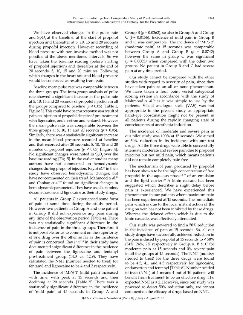
IJAA�/�Volume�6�Number�4�(Part�-�II)�/�July�-�August�2019
1341
We� have� observed� changes� in� the� pulse� rate�and� SpO
2�at� the� baseline,� at� the� start� of� propofol�
injection�and�thereafter�at�5,�10,�15�and�20�seconds�during� propofol� injection.� �owever� recording� of�blood�pressure�with�non-invasive�method�was�not�possible� at� the� above�mentioned� intervals.� So�we�have� taken� the� baseline� reading� (before� starting�of�propofol� injection)�and� thereafter� at� the�end�of�20� seconds,� 5,� 10,� 15� and� 20� minutes.� Following�which�changes�in�the�heart�rate�and�blood�pressure�would�be�construed�as�resulting�from�pain.
�aseline�mean�pulse�rate�was�comparable�between�the� three�groups.�The� intra-group�analysis�of�pulse�rate� showed�a�signi��cant� increase� in� the�pulse� rate�at�5,�10,�15�and�20�seconds�of�propofol�injection�in�all�the�groups�compared�to�baseline�(p���0.05)��Table�1,�Figure�3�.�This�could�have�been�a�response�as�a�result�of�pain�on�injection�of�propofol�despite�of�pre-treatment�with�lignocaine,�ondansetron�and�fentanyl.��owever�the� mean� pulse� rate� was� comparable� between� the�three�groups�at�5,�10,�15�and�20�seconds� (p���0.05).�Similarly,�there�was�a�statistically�signi��cant�increase�in� the� mean� blood� pressure� recorded� at� baseline�and�that�recorded�after�20�seconds,�5,�10,�15�and�20�minutes� of� propofol� injection� (p� �� 0.05)� �Figure� 4�.�No�signi��cant�changes�were�noted� in�S
PO
2�over�the�
baseline�reading��Fig.�5�.�In�the�earlier�studies�many�
authors� have� not� commented� on� hemodynamic�changes�during�propofol�injection.�Ray�et�al.15�in�their�study� have� observed� hemodynamic� changes,� but�have�not�commented�on�their�trend.�Mahmood�et�al.16�and�Canbay� et� al.17� found�no�signi��cant� changes� in�hemodynamic�parameters.�They�have�used�ketamine,�dexamethasone�and�lignocaine�as�their�study�drugs.
All�patients�in�Group�C�experienced�some�form�of� pain� at� some� time� during� the� study� period.��owever�two�patients�in�Group�A�and�one�patient�in� Group� �� did� not� experience� any� pain� during�any�time�of�the�observation�period�(Table�4).�There�was� no� statistically� signi��cant� difference� in� the�incidence�of�pain�in�the�three�groups.�Therefore�it�is�not�possible�for�us�to�comment�on�the�superiority�of�one�drug�over�the�other�as� far�as� the� incidence�of�pain�is�concerned.�Ray�et�al.15�in�their�study�have�documented�a�signi��cant�difference�in�the�incidence�of� pain� between� the� lignocaine� and� fentanyl�pre-treatment� group� (14.3� vs.� 42.9).� They� have�calculated� the�NNT� (number� needed� to� treat)� for�fentanyl�and�lignocaine�to�be�4�and�2�respectively.
The� incidence�of� �MPS� 1�� (mild�pain)� increased�with� time,� with� peak� at� 15� seconds� and� then�declining� at� 20� seconds.� �Table� 5�� There� was� a�statistically� signi��cant� difference� in� the� incidence�of� �mild� pain�� at� 15� seconds� in� Group� A� and�
Group���(p���0.0362),�so�also�in�Group�A�and�Group�C� (���0.0156).� Incidence� of�mild�pain� in�Group���and�C�was� comparable.� The� incidence�of� �MPS� 2��(moderate� pain)� at� 15� seconds� was� comparable�between� Group� A� and� Group� �� (p� �� 0.0742)�however� the� same� in� group� C� was� signi��cant�(p� �� 0.0005��when� compared� with� the� other� two�groups.�No�patient� in�Group���and�C�had� severe�pain�at�any�time�period.
Our� study� cannot� be� compared�with� the� other�studies�with�regard� to�severity�of�pain,�since�they�have� taken� pain� as� an� all� or� none� phenomenon.�We� have� taken� a� four� point� verbal� categorical�scoring� system� in� accordance� with� the� study� of�Mahmood� et� al.16�as� it� was� simple� to� use� by� the�patients.� Visual� analogue� scale� (VAS)� was� not�appropriate� to� the� present� study� as� appropriate�hand-eye� coordination� might� not� be� present� in�all� patients� during� the� rapidly� changing� state� of�consciousness�of�anesthesia�induction.
The� incidence� of� moderate� and� severe� pain� in�our�pilot�study�was�100��at�15�seconds.�We�aimed�at� 50�� reduction� in� its� incidence� with� our� test�drugs.�All�the�three�drugs�were�able�to�successfully�attenuate�moderate�and�severe�pain�due�to�propofol�injection�but�not�mild�pain,�which�means�patients�did�not�remain�completely�pain�free.
The� mechanism� of� pain� produced� by� propofol�has�been�shown�to�be�the�high�concentration�of�free�propofol� in� the� a�ueous� phase18,19� of� an� emulsion�and� the� lipid� carrier.20� A� kinin� cascade� has� been�suggested� which� describes� a� slight� delay� before�pain� is� experienced.� We� have� experienced� this�phenomenon�in�our�patients�where�maximum�pain�has�been�experienced�at�15�seconds.�The�immediate�pain�which�is�due�to�the�local�irritant�action�of�the�drug�on�vein�has�not�been�inhibited�by�these�drugs.�Whereas� the� delayed� effect,� which� is� due� to� the�kinin�cascade,�was�effectively�attenuated.
Our�study�was�powered�to���nd�a�50��reduction�in� the� incidence� of� pain� at� 15� seconds.� So,� all� our�study�drugs�have�successfully�achieved�reduction�in�the�pain�induced�by�propofol�at�15�seconds�to���50��(34�,�24�,�2��respectively� in�Group�A,�����C� for�moderate� pain� at� 15� seconds� and� 0�� severe� pain�in�all�the�groups�at�15�seconds).�The�NNT�(number�needed� to� treat)� for� the� three� drugs� were� found�to� be� 4.5,� 4.1� and� 4.5� respectively� for� lignocaine,�ondansetron�and�fentanyl��Table�6�.�Number�needed�to�treat�(NNT)�of�4�means�4�out�of�10�patients�will�bene��t�from�treatment�to�be�an�effective�drug.�The�expected�NNT�is���2.��owever,�since�our�study�was�powered� to� detect� 50�� reduction� only,�we� cannot�comment�on�the�ef��cacy�of�drugs�based�on�NNT.
Pain�on�Propofol�Injection:�Comparative�Study�of�Pre-Treatment�with�Intravenous�Lignocaine,�Ondansetron�and�Fentanyl�for�the�Prevention�of�Pain
IJAA�/�Volume�6�Number�4�(Part�-�II)�/�July�-�August�2019
1342 Indian�Journal�of�Anesthesia�and�Analgesia
One�patient� in� the�Group�A�had� redness�at� the�site� of� drug� injection,� one� patient� had� pruritus�in�Group���and�one�had�a�wheal�in� the�Group�C.�Overall�these�were�very�mild�reactions�and�did�not�need�any�form�of�intervention.
Conclusion
We� conclude� that� pre-treatment�with� the� three�drugs� Lignocaine� 42� mg,� Ondansetron� 4� mg�and� Fentanyl� 100� mcg� effectively� decreased� the�incidence�of�pain�on�propofol�injection�at�15�seconds.��owever�the�superiority�of�one�drug�over�the�other�cannot� be� commented.� All� the� three� drugs� were�comparable�with�respect�to�hemodynamic�changes�and�adverse�effects.
�e�� message:� Propofol,� an� ideal� intravenous�anaesthetic� agent� causes� pain� on� injection� which�interferes� with� smooth� induction� of� anesthesia.�Fentanyl� and� Ondensetron� were� compared� with�Lignocaine� to� test� their� ability� to� attenuate� pain�from� propofol� injection.� All� the� three� drugs�effectively� reduced�pain�on�propofol� injection�but�none�was�found�superior�over�other.
�eferences
1.� Mc�Leskey�C��et�al.�Adverse�events�in�multicenter�phase� IV� study� of� propofol:� evaluation� by�anaesthesiologist� and� post� anesthesia� care� unit�nurses.�Anesth�analg.�1993;73(4):3–9.
2.� Mcleod.,�N.�oheimer.�Propofol� infusion�as�main�agent�for�day�care�surgery.�Post�graduate�Medical�journal.�1985;61�suppl�3:105–107.
3.� �ynynen� M,� �orttila� �,� Tammisto� T.� Pain� on�IV� injection� of� propofol� (ICI� 35868)� in� emulsion�formulation:� short� communication.� Acta�anesthesiologica�Scandinavica.�1985;29:651–2.
4.� �lement� W,� Arndt� JO.� Pain� on� injection� of�propofol,�the�effects�of�concentration�and�diluent.�r�J�Anaesth.�1991;67:281–84.
5.� Iwama��,�Nakane�M,�Ohmori�S�et�al.�Nafamostat�mesilate� a� kallikrein� inhibitor� prevents� pain�on� injection� with� propofol.� �r� J� Anesth.� 1998;�81:963–964.
6.� Nakane� M,� Iwama� �.� A� potential� mechanism�of� propofol� induced� pain� on� injection� based� on�studies� using� nafamostat� mesilate.� �r� J� Anesth.�1999;83(3):397–404.
7.� Mangar� D,� �olak� EJ.� Tourni�uet� at� 50� mm��g�followed� by� intravenous� lidocaine� diminishes�
hand� pain� associated� with� propofol� injection.�Anesth�Analg.�1992;74:250–2.
8.� Pang�WW,�Mok�MS,��uang�S,�et�al.�The�analgesic�effect� of� fentanyl,� morphine,� meperidine� and�lidocaine� in� the�peripheral� veins:� a� comparative�study.�Anesth�Analg.�1998;86:382–6.
9.� Ambesh� SP,�Dubey� P�,� Sinha� P�.�Ondansetron�pre-treatment� to� alleviate� pain� on� propofol�injection:� a� randomized,� controlled,� double-blinded�study.�Anesth�Analg.�1999;89(1):197–9.
10.� Picard� P,� Tramer� MR.� Prevention� of� pain� on�injection�with�propofol:�a��uantitative�systematic�review.�Anesth�Analg.�2000;90:963–9.
11.� Ray� S,� Pal� R,� Pal� S,� et� al.� Preclusion� of� pain� on�injection� with� propofol� :� Evaluating� the� effects�of� lignocaine� or� fentanyl� pre-treatment.� Anesth�Essays�Res.�2011;5:33–8.
12.� Memis�D,�Turan�A�et�al.�The�prevention�of�propofol�injection� pain� by� tramadol� or� ondansetron,�European� Journal� of� anaesthesiology.� 2002;�19(1):47–51.�Cambridge��niversity�Press.
13.� �ahedi��,�Maleki�A�and�Rostami�G.�Ondansetron�Pre-treatment� Reduces� Pain� on� Injection� of�Propofol.�Acta�Medica�Iranica.�2012;50(4):239–43.
14.� �ye–joo� �ang,� Mi-� �oung� �won� et� al.� Clinical�factors�affecting�the�pain�on�injection�of�propofol�Clinical� Research� Article,� �orean� J� Anesthesiol.�2010�Mar;58(3):239–243.
15.� Ray�S,�Pal�R,�Pal�S,��irtania�J,�Sarbapalli�D,�Sarkar��,� et� al.� Preclusion� of� pain� on� injection� with�propofol� :�Evaluating�the�effects�of� lignocaine�or�fentanyl�pre-treatment.�Anesth�Essays�Res.�2011;�5:33–8.
16.� Mahmood� I,� �asmine�M.� Prevention� of� pain� on�propofol� injection;� A� comparative,� randomized,�double�blind�study�between�lignocaine,�ketamine,�dexamethasone�and�placebo.�Professional�Med�J.�Sep�2010;17(3):405–410.
17.� O�Canbay,�N�Celebi�et�al.�Efficacy�of�intravenous�acetaminophen� and� lidocaine� on� propofol�injection�pain.��r�J�Anaesth.�2008;100(1):95–98.
18.� Schaub� E,� �ern� C� Landau� R.� Pain� on� injection:�A� double� blind� comparison� of� propofol� with�lidocaine� pre� treatment� versus� propofol�formulated� with� long� and� medium� chain�triglycerides.�Anesth�Analg.�2004;99:1699–702.
19.� �abl� J,� Doenicke� A,� Monch� V.� New� propofol�LCT/MCT� fat� emulsion� as� solvent:� approach� to�reducing�pain�on�injection�of�propofol.�Eur��osp�Pharm.�1995;1:15–21.
20.� Doenicke�AW,� Roizen�MF,� Rau� J,� O�Connor�M,��ugler� J,��lotz��,��abl� J.� Pharmacokinetics� and�pharmacodynamics�of�propofol�in�a�new�solvent.�Anesth�Analg.�1997;85(6):1399–403.
��Red�Flower�Publication�Pvt.�Ltd.�
�Original�Research�ArticleIndian�Journal�of�Anesthesia�and�Analgesia�
2019;6(4)�(Part�-�II):�1343-1346DOI:�http://dx.doi.org/10.21088/ijaa.2349.8471.6419.39
�e�medetomidine� is� a� Better�Ad�u�ant� than�Clonidine���ith��opi�acaine� in�
Supracla�icular�Brachial�Ple�us�Block
�unaseelan�Si�asamy���Priyanka�Sel�am���Thiruna�ukkarasu�M����aghuraman�MS�
1Associate� Professor,� 2Post� Graduate,� 4Professor,� Department� of� Anaesthesiology� and� Critical� Care,� 3Assistant� Professor� Cum�Statistician,� Department� of� Community� Medicine,� Sri� Venkateshwaraa� Medical� College� �ospital� �� Research� Centre,� Ariyur,�Puducherry�605102,�India.
Corresponding�Author:�Priyanka�Sel�am,�Post�Graduate,�Department�of�Anesthesia�and�Critical�Care,�Sri�Venkateshwaraa�Medical�College��ospital�and�Research�Centre,�Ariyur,�Puducherry�605102,�India.
E�mail:�[email protected]
�ecei�ed�on�24.04.2019,�Accepted�on�22.05.2019
Abstract
Regional� anesthesia� has� some� advantages� over� general� anesthesia� such� as� it� can� be� used� in� outpatient�anesthesia,�for�patients�with�full�stomach,�for�diabetic�patients,�associated�cardiac,�pulmonary,�hepatic�or�renal�damage�and�poly-trauma.�Alpha-2�agonists�added�to�local�anaesthetic�drugs�increases�the�duration�of�painless�period�during�and�after�surgery.��ere�we�elicit�the�clinical�Performance�of�this�two�drugs�dexmedetomidine�and�clonidine�as�an�additive�agent�to�local�anaesthetics�like�ropivacaine� in�blocking�the�brachial�plexus�by�supraclavicular�method.�The�clinical�parameters�we�study�here�are�onset,�duration�of�Duration�of�blockade�(both�S�and�M)�and�analgesia�time�and�hemodynamic�stability.�Material�and�Methods:�Prospective�study�was�done�on�patients�undergoing�upper�arm�surgeries�under�brachial�block�were�split�into�two�e�uivalent�groups.�D-group�received�0.375��ropivacaine�(30�ml)�plus�one�mcg/kg�dexmedetomidine,�C-�group�received�0.375��ropivacaine� (30�ml)�plus�one�mcg/kg�clonidine.�Results:�Statistical� analysis� shows�significant�difference� in�onset�of�sensory�(S)�and�motor�(M)�blockade,�highly�significance�in�duration�of�sensory�and�motor�blockade�between�D� and� C� groups� (p�0.0001).� �igh� Statistical� significance�was� seen� analgesia� duration� (p�0.0001)�and�number�of�rescue�analgesics�used�(p�0.0001)�among�D�and�C�groups.�Conclusion:�Dexmedetomidine�has�more�additive�benefits�than�clonidine�when�combined�with�ropivacaine�by�making�the�onset�of�clinical�effect�earlier,�prolonging�the�blockade�extent�(both�S�and�M)�and�painless�period�during�post�surgery�with�fewer�re�uirements�of�rescue�analgesics�after�arm,�forearm�and�hand�surgeries.
�ey�ords:�Ropivacaine;�Clonidine;�Dexmedetomidine;�Sensory�blockade� (S);�Motor�blockade(M);�Local�anaesthetic.
�o��to�cite�this�article:
Gunaseelan�Sivasamy,�Priyanka�Selvam,�Thirunavukkarasu�M�et�al.�Dexmedetomidine�is�a��etter�Adjuvant�than�Clonidine,�with�Ropivacaine�in�Supraclavicular��rachial�Plexus��lock.�Indian�J�Anesth�Analg.�2019;6(4):1343-1346.
Introduction
Surgerie�s�done�for�arm,� forearm�and�hand�under�peripheral� nerve� block� provides� excellent� intra-operative�analgesia�and�prolongs�the�post-operative�analgesia�duration�with� least� complications1.�Pain�transferred�by�nerve���bers�is�the�basis�for�regional�
block�working�mechanism�and�the�transfer�of�pain�
can� be� interrupted� along� their� pathway.� Other�important�advantages�are�postoperative�analgesia,�
early� ambulation,� no� airway� manipulation,� early�feed� intake� by� oral� route� and� lesser� incidence�
of� postoperative� respiratory,� gastric,� intestinal�and� thrombo-embolic� complications.2� Regional�
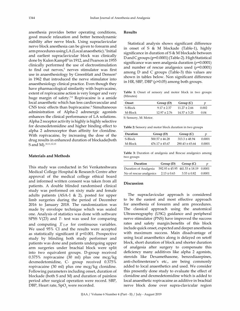
IJAA�/�Volume�6�Number�4�(Part�-�II)�/�July�-�August�2019
1344 Indian�Journal�of�Anesthesia�and�Analgesia
anesthesia� provides� better� operating� conditions,�good�muscle� relaxation� and� better� hemodynamic�stability� after� nerve� block.� �sing� supraclavicular�nerve�block�anesthesia�can�be�given�to�forearm�and�arm�procedures�using�LA�(Local�anaesthetic).3�Initial�and� earliest� supraclavicular� block� was� clinically�done�by��ulen��ampff4�in�1912,�and�Pearson�in�1955�clinically� performed� the� use� of� electrostimulation�to� ��nd� out� nerves,5� nerves� stimulator� was� ��rst�use�in�anaesthesiology�by�Greenblatt�and�Denson6�in� 1962� that� introduced� the� nerve� stimulator� into�anaesthesiology�clinical�practice.�Even�though�they�have�pharmacological�similarity�with�bupivacaine,�extent�of�ropivacaine�action�is�very�longer�and�very�huge� margin� of� safety.7-8� Ropivacaine� is� a� amide�local�anaesthetic�which�has�less�cardiovascular�and�CNS�toxic�effects�than�bupivacaine.9�Simultaneous�administration� of� Alpha-2� adrenergic� agonists�enhances�the�clinical�performance�of�LA�solutions.�Alpha�2�receptor�activity�is�highly�is�highly�selective�for�dexmedetomidine�and�higher�binding�effect�to�alpha� 2� adrenoceptor� than� af��nity� for� clonidine.�With� ropivacaine,� by� increasing� the� dose� of� the�drug�results�in�enhanced�duration�of�blockade(both�S�and�M).10,11,12,13
Materials�and�Methods
This� study�was� conducted� in� Sri�Venkateshwara�Medical�College��ospital���Research�Centre�after�approval� of� the� medical� college� ethical� board�and�informed�written�consent�was�taken�from�all�patients.� A� double� blinded� randomised� clinical�study� was� performed� on� sixty� male� and� female�adults� patients� (ASA-1� �� 2),� posted� for� upper�limb� surgeries� during� the� period� of� December�2016� to� January� 2018.� The� randomization� was�made� by� envelope� techni�ue� which� was� sealed�one.�Analysis�of�statistics�was�done�with�software�SPSS� V(23)� and� T�� test� was� used� for� comparing�
and� computing� � for� continuous� variables.�We� used� 95�� CI� and� the� results� were� accepted�as� statistically� signi��cant� if� p�0.001.� Prospective�study� by� blinding� both� study� performer� and�patients�was�done�and�patients�undergoing�upper�arm� surgeries� under� brachial� block� were� split�into� two� e�uivalent� groups.� D-group� received�0.375�� ropivacaine� (30� ml)� plus� one� mcg/kg�dexmedetomidine,� C-� group� received� 0.375��ropivacaine� (30� ml)� plus� one� mcg/kg� clonidine.�Following�parameters�including�onset,�duration�of�blockade�(both�S�and�M)�and�duration�of�painless�period�after�surgical�operation�were� record.�S�P,�D�P,��eart�rate,�SpO
2�were�recorded.
�esults
Statistical� analysis� shows� signi��cant� difference�in� onset� of� S� �� M� blockade� (Table-1),� highly�signi��cance�in�duration�of�S���M�blockade�between�D�and�C�groups�(p�0.0001)�(Table-2).��igh�Statistical�signi��cance�was�seen�analgesia�duration�(p�0.0001)�and� number� of� rescue� analgesics� used� (p�0.0001)�among�D� and� C� groups� (Table-3)� this� values� are�shown� in� tables�below.�Non�signi��cant�difference�in��R,�S�P,�D�P�(p�0.05)�among�both�groups.
Table� �:� Onset� of� sensory� and� motor� block� in� two� groups�
(Minutes)
Onset �roup���� �roup��C� p
S-�lock 9.17���2.37 11.27���2.66 0.002
M-�lock 12.97���2.76 14.57���3.25 0.04
S:�Sensory,�M:�Motor.
Table��:�Sensory�and�motor�block�duration�in�two�groups�
�uration �roup���� �roup��C� �p
S-�lock 500.57���46.28 313.3���48.94 0.0001
M-�lock 476.17���45.67 290.43���65.64 0.0001
Table� �:� Duration� of� analgesia� and� Rescue� analgesics� among�two�groups
�uration �roup���� �roup��C� p
Duration�of�Analgesia 592.93���45.95 441.53���18.19 0.0001
No�of�rescue�analgesics 2.13���0.63 3.53���0.82 0.0001
�iscussion
The� supraclavicular� approach� is� considered�to� be� the� easiest� and� most� effective� approach�for� anesthesia� of� forearm� and� arm� procedures.�The� classical� approach� using� the� anatomical��ltrasonography� (�SG)� guidance� and� peripheral�nerve�stimulator�(PNS)�have�improved�the�success�rates� and� safety� margin.bene��ts� of� this� block�include��uick�onset,�expected�and�deeper�anesthesia�with� maximum� success.� Main� disadvantage� of�using�local�anaesthetics�along�is�delayed�on�setoff�block,�short�duration�of�block�and�shorter�duration�of� analgesia� after� surgery� to� compensate� this�de��ciency� many� additives� like� alpha� 2� agonists,�steroids� like� Dexamethasone,� benzodiazepines,�anti-cholinesterase�s� etc.,� are� being� commonly�added�to�local�anaesthetics�and�used.�We�consider�this�presently�done�study�to�evaluate� the�effect�of�clonidine�and�dexmedetomidine�which�is�added�to�local�anaesthetic�ropivacaine�as�additive�in�brachial�nerve� block� done� over� supra-clavicular� region�
IJAA�/�Volume�6�Number�4�(Part�-�II)�/�July�-�August�2019
1345
mainly�in�the�limb�operations�of�arm�and�forearm.�Duration� of� S� and� M� blockade� was� signi��cantly�extended� in� study�done� by�Don� Sebastian� et� al14,�Vania�k�et�al.15,��amlesh�k�et�al.16,�Saritha�S�swami�et�al.17,�Chaudhary�et�al.18�(p�0.01,�p�0.0001,�p�0.001,�p�0.001,�p�0.001)�with�high�statistical�signi��cance.�Duration�of�painless�period�was�extended� in�Don�Sebastian�et�al.14,��amlesh�k�et�al.16,�Saritha�S�swami�et�al.17,�and�statistically�signi��cant.
In� our� current� study� onset� of� S� �� M� block�(p�0.002),� (p�0.04)�were�slower�in�clonidine�group�of�patients�and�faster�in�group�of�patients�receiving�dexmedetomidine.� Duration� of� sensory� block�(p�0.0001),�motor� block� (p�0.0001)�were� longer� in�dexmedetomidine� group� and� highly� signi��cant�statistically.� Duration� of� analgesia� (p�0.0001)� is�longer�and�number�of�rescue�analgesics�(p�0.0001)�used� were� lesser� in� dexmedetomidine� group�and� statistically� highly� signi��cant� in� comparison�with� clonidine� group.� �ence� the� result� of� this�present�study�has�correlated�with�Sarita�S�Swami17,�Chaudhary�et�al.18,Vania�k�et�al.15,�and�Don�sebastin14
study�by� extending� the�duration�of� S���M� block,�duration�of�painless�period�and�decreased�need�for�extra�dose�of�analgesics.
Dexmedetomidine�action� is�mainly� through� the�alpha�2�receptor�which�is�adrenergic�in�nature�the�selectivity�to�this�receptor�is�very�high�and�acts�as�agonist.�The�activity�at�alpha�1�recepyor�and�alpha�2� receptor� is� 1:220� times� for� clonidine� and� 1:1620�times� for� dexmedetomidine.� Following� are� the�mechanisms�of� action�at� alpha�2� receptor� level� as�agonist�(1)�analgesia�is�provided�mostly�by�central�action� (2)� by� acting� over� the� peripheral� nerve�directly�(3)vaso-constriction�mediated�by�receptors�(4)� knocking� off� the� in��ammatory� mechanism.�Extended� duration� of� action� is� due� to� the� peri-neural�administration�of�alpha�2�receptor�agonist�as�additive�to�local�anaesthetics.19
Nerve� hyperpolarisation� is� the� mechanism�for� extending� the� painless� period.� it� is� brought�about� by� blocking� the� cation� Ih� current� in� the�nerve.� This� blocking� also� results� in� prolonged�action�over�C���bers� (pain)� than� in�A�alpha���bers�(motor).� Action� potential� is� inhibited� at� higher�level� by� dexmedetomidine� than� clonidine.� Loss�of� pain� and� sedation� mechanism� by� blocking�the� release� of� pain� producing� substance� P� at� the�level� of� dorsal� root� and�by� stimulating� the� alpha�2� receptors� belonging� to� the� adrenergic� group� at�L.� coerleus.�Other�mode� is� by� blocking� the� noci-ceptive� neurotransmission� of� the� nor-adrenergic�system� at� the� descending� pathway.� Clonidine� by�producing� alpha� one� mediated� vaso-constriction�
and� decreases� the� uptake� of� clonidine-local�anesthesia�mixture�and�reduces�the�uptake�into�the�circulatory�system,whereas�dexmedetomidine�has�limited�action�on�alpha�one�receptors.19�
Experimental� studies� done� on� rats� especially�by� brumett� and� his� team�have� proved� that� alpha�2� agonists� in� large� doses� have� extended� the� time�of�nerve�blocks�done�on�sciatic�nerve�when�added�to� local� anaesthetics.� On� examination� done� on�histopathology� of� sciatic�nerve,� it� has�been� found�that� myelin� sheath� of� the� nerve� and� axon� were�not� affected� by� the� perineural� alpha� 2� agonists�mainly� dexmedetomidine.� There� was� increase� in�cytokines�numbers�at�the�site�of�nerve�damage.this�is� the�main�reason� for� the�use�of�alpha�2�agonists�as� additives� to� local� anaesthetics� and� a� concrete�evidence� for� the�neuro�protective�effect�produced�by� dexmedetomidine.� �igh� safety� margin� of�this� drugmakes� it� abetter� choice� for� intrathecal�use.� In� addition� it� produces� glomerular� ��ltration�enhancement,� enhancement� of� threshold� for�seizures,�reduces�salivary�secretion�and�IOP�(intra�ocular�pressure).19
Conclusion
Dexmedetomidine� has� more� additive� bene��ts�than� clonidine� when� combined� with� ropivacaine�by� making� the� onset� of� clinical� effect� earlier,�prolonging� the� blockade� extent� (both� S� and� M)�and� painless� period� during� post� surgery� with�fewer�re�uirements�of�rescue�analgesics�after�arm,�forearm�and�hand�surgeries.�
�eferences
1.� Damien��,�Murhy,�Collin�JL,�et�al.�Novel�analgesic�adjuvants� for� brachial� plexus� block:� A� systemic�review.�Anesth�Analg.�2000;90:1122–8.
2.� Rathod� �,� Parikh� �,� �padhayaya� RM.�Comparative� study� of� 0.375�� �upivacaine� and�0.375��Ropivacaine� in�brachial�plexus�block�via�supraclavicular� approach.� International� Journal�of��iomedical�Research.�2015;6(02):77–82.
3.� Singh� S,�Aggarwal�A.�A� randomized� controlled�double-blinded�prospective�study�of� the�efficacy�of� clonidine� added� to� bupivacaine� as� compared�with� bupivacaine� alone� used� in� supraclavicular�brachial� plexus� block� for� upper� limb� surgeries.�Indian�J�Anaesth.�2010;54:552–7.
4.� Singh� G,� Saleem� M�.� Comparison� between�Conventional�Techni�ue�and��ltrasound�Guided�Supraclavicular� �rachial� Plexus� �lock� in� �pper�Limb�Surgeries.�Int�J�Sci�Stud.�2014;2(8):169–176.
Dexmedetomidine�is�a��etter�Adjuvant�than�Clonidine,�with�Ropivacaine�in�Supraclavicular��rachial�Plexus��lock
IJAA�/�Volume�6�Number�4�(Part�-�II)�/�July�-�August�2019
1346 Indian�Journal�of�Anesthesia�and�Analgesia
5.� �atz� J.� �se� of� nerve� stimulators� for� peripheral�nerve�blocks.�In:��enzon��T,�Raja�SN,��orsook�D,�et�al.,�eds.�Essentials�of�pain�medicine�and�regional�anesthesia.� New� �ork:� Churchill� Livingstone,�1999:395–8.
6.� Greenblatt� GM,� Denson� GS.� Needle� nerve�stimulator-locator:� nerve� blocks� with� a� new�instrument� for� locating� nerves.� Anesth� Analg�1962;41:599–602.
7.� �lein� SM,� Greengrass� RA,� Steele� SM,� et� al.� A�comparison�of��.���bupi�acaine���.���ropivacaine,�and� 0.75�� ropivacaine� for� interscalene� brachial�plexus�block.�Anesth�Analg�1998;87:13169.
8.� Scott�D�,�Lee�A,�Fagan�D,�et�al.�Acute�toxicity�of�ropivacaine� compared�with� that�of� bupivacaine.�Anesth�Analg�1989;69:5639.
9.� �dita�N,�Singh�MM,�Gupta�S,�et�al.�Dose�dependant�effect�of�intrathecal�dexmedetomidine�on�isobaric�ropivacaine� in� spinal� anesthesia� for� abdominal�hysterectomy:�Effect�on�block�characteristics�and�hemodynamics.�J�Anaes�Clin�Pharm.�2015;31:72–9.
10.� Abdallah� FW,� �rull� R.� Facilitatory� effects� of�perineural� dexmedetomidine� on� neuraxial� and�peripheral�nerve�block:�A�systematic�review�and�metaanalysis.��r�J�Anaesth.�2013;110:91525.
11.� Swami�SS,��eniya�VM,�Ladi�SD,�et�al.�Comparison�of� dexmedetomidine� and� clonidine� (�
2� agonist�
drugs)� as� an� adjuvant� to� local� anesthesia�in� supraclavicular� brachial� plexus� block:� A�randomised� doubleblind� prospective� study.�Indian�J�Anaesth.�2012;56:2439.
12.� Chakraborty� S,� Chakrabarti� J,� Mandal�MC,� et� al.� Effect� of� clonidine� as� adjuvant� in�bupivacaineinduced� supraclavicular� brachial�
plexus� block:� A� randomized� controlled� trial.�Indian�J�Pharmacol.�2010;42:747.
13.� Esmaoglu� A,� �egenoglu� F,� Akin� A,� et� al.�Dexmedetomidine� added� to� levobupivacaine�prolongs� axillary� brachial� plexus� block.� Anesth�Analg.�2010�Dec;111(6):1548–51.
14.� Don� S.,� Ravi�M,�Dinesh��,� et� al.Comparision� of�Dexmedetomidine�and�clonidine�as�Adjuvant� to�Ropivacaine� in� Supraclavicular� �rachial� Plexus�Nerve��locks.� J� of�Dental� and�Medical� Sciences.�2015;14(3):91–7.
15.� �anvee�V,�Patel��,�Doshi�M,� et� al.�Comparative�Study�of�Clonidine�And�Dexmedetomidine�as�an�adjuvant�with�Inj.�Ropivacaine�in�Supraclavicular��rachial� Plexus� �lock� for� upper� limb� surgery.� J�Res�Med�Den�Sci.�2015;3(2):127–30.
16.� �amlesh���Shekhawat,�Jain�N,�et�al.�Comparison�of�Clonidine�and�Dexmedetomidine�as�an�Adjuvant�to� 0.375�� of� Ropivacaine� in� Supraclavicular��rachial� Plexus� �lock:� A� Prospective� Study.JMSCR.�2018;6(4):1190–97.
17.� Swami� SS,� �eniya� VM,� Ladi� SD,� Comparison�of� dexmedetomidine� and� clonidine� (�
2� agonist�
drugs)� as� an� adjuvant� to� local� anesthesia�in� supraclavicular� brachial� plexus� block:� A�randomised� doubleblind� prospective� study.�Indian�J�Anaesth.�2012;56:2439.
18.� Chaudhary� ��� et� al.� Comparison� of� effects� of�dexmedetomidine� and� clonidine� as� adjuvant�to� bupivacaine� 0.25�� in� ultrasound� guided�supraclavicular� brachial� plexus� block.� Int� J� Res�Med�Sci.�2017;5(10):4512–4518.
19.� Sharma� S,� Jain� P.� Dexmedetomidine� and�Anesthesia.� Indian� Journal� of� Clinical� Practice.�2013;24(3):223–25.
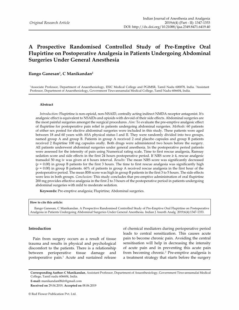
��Red�Flower�Publication�Pvt.�Ltd.�
�Original�Research�ArticleIndian�Journal�of�Anesthesia�and�Analgesia�
2019;6(4)�(Part�-�II):�1347-1353DOI:�http://dx.doi.org/10.21088/ijaa.2349.8471.6419.40
A� Prospecti�e� �andomised� Controlled� Study� of� Pre�Empti�e� Oral�Flupirtine�on�Postoperati�e�Analgesia�in�Patients��ndergoing�Abdominal�Surgeries��nder��eneral�Anesthesia
Ilango��anesan���C�Manikandan�
1Associate�Professor,�Department�of�Anaesthesiology,�ESIC�Medical�College�and�PGIMSR,�Tamil�Nadu�600078,� India.� 2Assistant�Professor,�Department�of�Anaesthesiology,�Government�Tiruvannamalai�Medical�College,�Tamil�Nadu�606604,�India.
Corresponding�Author:�C�Manikandan,�Assistant�Professor,�Department�of�Anaesthesiology,�Government�Tiruvannamalai�Medical�College,�Tamil�nadu�606604,�India.
E�mail:�[email protected]
�ecei�ed�on�29.04.2019,�Accepted�on�08.06.2019
Abstract
Introduction:�Flupirtine�is�non-opioid,�non-NSAID,�centrally�acting�indirect�NMDA�receptor�antagonist.�It�s�analgesic�effect�is�e�uivalent�to�NSAIDs�and�opioids�with�devoid�of�their�side�effects.�Abdominal�surgeries�are�the�most�painful�surgeries�amongst�the�surgical�procedures.�Aim:�To�evaluate�the�pre-emptive�analgesic�effect�of� flupirtine�for�postoperative�pain�relief� in�patients�undergoing�abdominal�surgeries.�Methods:�60�patients�of�either�sex�posted�for�elective�abdominal�surgeries�were�included�in�this�study.�These�patients�were�aged�between�18�and�60�years�with�ASA�physical�status�I�and�II.�They�were�randomly�divided�into�two�groups,�named�group�A�and�group��.� Patients� in�group�A� received�2�oral�placebo� capsules� and�group���patients�received�2� flupirtine�100�mg�capsules�orally.��oth�drugs�were�administered�two�hours�before� the� surgery.�All�patients�underwent�abdominal�surgeries�under�general�anesthesia.�In�the�postoperative�period�patients�were�assessed�for�the�intensity�of�pain�using�Numerical�rating�scale,�Time�to�first�rescue�analgesia,�Ramsay�sedation�score�and�side�effects�in�the�first�24�hours�postoperative�period.�If�NRS�score���4,�rescue�analgesic�tramadol�50�mg�iv�was�given�at�6�hours�interval.�Results:�The�mean�NRS�score�was�significantly�decreased�(p���0.00)�in�group���patients�for�the�first�3�hours.�The�time�to�first�rescue�analgesia�was�significantly�high�(p���0.00)�in�group���patients.�60��of�patients�in�group�A�received�rescue�analgesia�in�the�first�hour�of�the�postoperative�period.�The�mean�RSS�score�was�high�in�group���patients�in�the�first�3�to�5�hours.�The�side�effects�were�less�in�both�groups.�Conclusion:�This�study�concludes�that�pre-emptive�administration�of�oral�flupirtine�200�mg�provides�effective�analgesia�in�the�first�2�to�3�hours�of�the�postoperative�period�in�patients�undergoing�abdominal�surgeries�with�mild�to�moderate�sedation.
�ey�ords:�Pre-emptive�analgesia;�Flupirtine;�Abdominal�surgeries.
�o��to�cite�this�article:
Ilango�Ganesan,�C�Manikandan.�A�Prospective�Randomised�Controlled�Study�of�Pre-Emptive�Oral�Flupirtine�on�Postoperative�Analgesia�in�Patients��ndergoing�Abdominal�Surgeries��nder�General�Anesthesia.�Indian�J�Anesth�Analg.�2019;6(4):1347-1353.
Introduction
Pain� from� surgery� occurs� as� a� result� of� tissue�trauma� and� results� in� physical� and� psychological�discomfort� to� the�patients.� There� is� a� relationship�between� perioperative� tissue� damage� and�postoperative� pain.1� Acute� and� sustained� release�
of�chemical�mediators�during�perioperative�period�
leads� to� central� sensitization.� This� causes� acute�pain�to�become�chronic�pain.�Avoiding�the�central�
sensitisation�will� help� in� decreasing� the� intensity�of� acute� pain� and� in� preventing� this� acute� pain�
from� becoming� chronic.2� Pre-emptive� analgesia� is�a� treatment�strategy� that� starts�before� the�surgery�
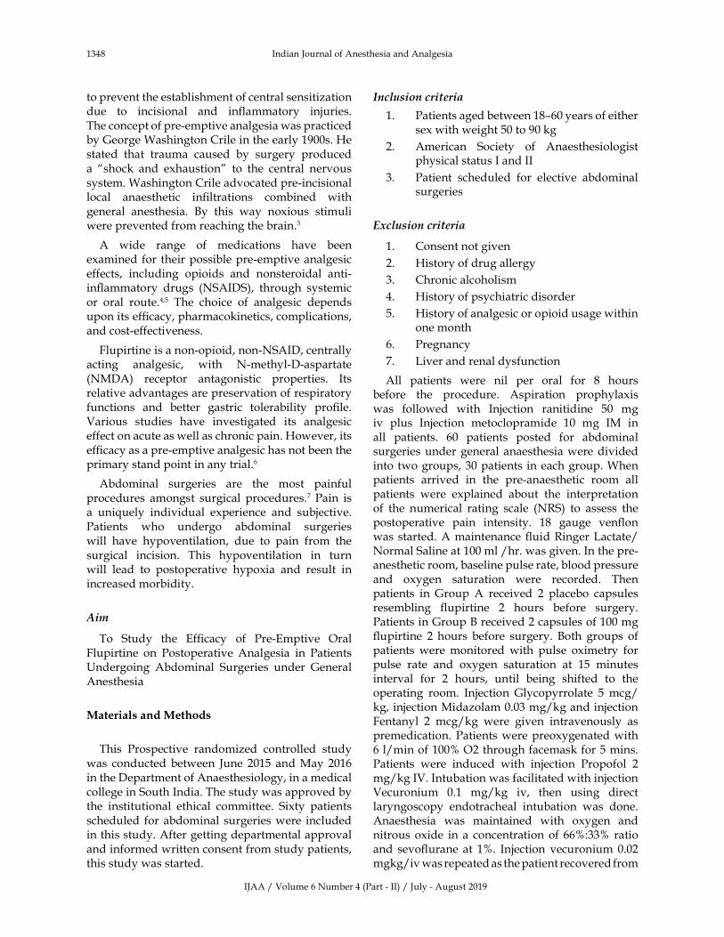
IJAA�/�Volume�6�Number�4�(Part�-�II)�/�July�-�August�2019
1348 Indian�Journal�of�Anesthesia�and�Analgesia
to�prevent�the�establishment�of�central�sensitization�due� to� incisional� and� in��ammatory� injuries.�The�concept�of�pre-emptive�analgesia�was�practiced�by�George�Washington�Crile�in�the�early�1900s.��e�stated� that� trauma� caused� by� surgery� produced�a� �shock� and� exhaustion�� to� the� central� nervous�system.�Washington�Crile�advocated�pre-incisional�local� anaesthetic� in��ltrations� combined� with�general� anesthesia.� �y� this� way� noxious� stimuli�were�prevented�from�reaching�the�brain.3�
A� wide� range� of� medications� have� been�examined�for� their�possible�pre-emptive�analgesic�effects,� including� opioids� and� nonsteroidal� anti-in��ammatory� drugs� (NSAIDS),� through� systemic�or� oral� route.4,5� The� choice� of� analgesic� depends�upon�its�ef��cacy,�pharmacokinetics,�complications,�and�cost-effectiveness.
Flupirtine�is�a�non-opioid,�non-NSAID,�centrally�acting� analgesic,� with� N-methyl-D-aspartate�(NMDA)� receptor� antagonistic� properties.� Its�relative�advantages�are�preservation�of�respiratory�functions� and� better� gastric� tolerability� pro��le.�Various� studies� have� investigated� its� analgesic�effect�on�acute�as�well�as�chronic�pain.��owever,�its�ef��cacy�as�a�pre-emptive�analgesic�has�not�been�the�primary�stand�point�in�any�trial.6
Abdominal� surgeries� are� the� most� painful�procedures� amongst� surgical� procedures.7� Pain� is�a� uni�uely� individual� experience� and� subjective.�Patients� who� undergo� abdominal� surgeries�will� have� hypoventilation,� due� to� pain� from� the�surgical� incision.� This� hypoventilation� in� turn�will� lead� to� postoperative� hypoxia� and� result� in�increased�morbidity.
Ai�
To� Study� the� Ef��cacy� of� Pre-Emptive� Oral�Flupirtine� on� Postoperative�Analgesia� in� Patients��ndergoing� Abdominal� Surgeries� under� General�Anesthesia
Materials�and�Methods
This� Prospective� randomized� controlled� study�was� conducted� between� June� 2015� and�May� 2016�in�the�Department�of�Anaesthesiology,�in�a�medical�college�in�South�India.�The�study�was�approved�by�the� institutional� ethical� committee.� Sixty� patients�scheduled� for� abdominal� surgeries�were� included�in�this�study.�After�getting�departmental�approval�and�informed�written�consent�from�study�patients,�this�study�was�started.
Inclusion�criteria
1.� Patients�aged�between�18–60�years�of�either�sex�with�weight�50�to�90�kg
2.� American� Society� of� Anaesthesiologist�physical�status�I�and�II
3.� Patient� scheduled� for� elective� abdominal�surgeries
Exclusion�criteria
1.� Consent�not�given
2.� �istory�of�drug�allergy
3.� Chronic�alcoholism
4.� �istory�of�psychiatric�disorder
5.� �istory�of�analgesic�or�opioid�usage�within�one�month
6.� Pregnancy
7.� Liver�and�renal�dysfunction
All� patients� were� nil� per� oral� for� 8� hours�before� the� procedure.� Aspiration� prophylaxis�was� followed� with� Injection� ranitidine� 50� mg�iv� plus� Injection� metoclopramide� 10� mg� IM� in�all� patients.� 60� patients� posted� for� abdominal�surgeries�under�general�anaesthesia�were�divided�into�two�groups,�30�patients� in�each�group.�When�patients� arrived� in� the� pre-anaesthetic� room� all�patients� were� explained� about� the� interpretation�of� the� numerical� rating� scale� (NRS)� to� assess� the�postoperative� pain� intensity.� 18� gauge� ven��on�was� started.�A�maintenance���uid�Ringer�Lactate/Normal�Saline�at�100�ml�/hr.�was�given.�In�the�pre-anesthetic�room,�baseline�pulse�rate,�blood�pressure�and� oxygen� saturation� were� recorded.� Then�patients� in� Group� A� received� 2� placebo� capsules�resembling� ��upirtine� 2� hours� before� surgery.�Patients�in�Group���received�2�capsules�of�100�mg���upirtine� 2� hours� before� surgery.� �oth� groups� of�patients�were�monitored�with� pulse� oximetry� for�pulse� rate� and� oxygen� saturation� at� 15� minutes�interval� for� 2� hours,� until� being� shifted� to� the�operating� room.� Injection� Glycopyrrolate� 5� mcg/kg,�injection�Midazolam�0.03�mg/kg�and�injection�Fentanyl� 2� mcg/kg� were� given� intravenously� as�premedication.�Patients�were�preoxygenated�with�6�l/min�of�100��O2�through�facemask�for�5�mins.�Patients� were� induced� with� injection� Propofol� 2�mg/kg�IV.�Intubation�was�facilitated�with�injection�Vecuronium� 0.1� mg/kg� iv,� then� using� direct�laryngoscopy� endotracheal� intubation� was� done.�Anaesthesia� was� maintained� with� oxygen� and�nitrous�oxide� in� a� concentration� of� 66�:33�� ratio�and� sevo��urane�at� 1�.� Injection�vecuronium� 0.02�mgkg/iv�was�repeated�as�the�patient�recovered�from�
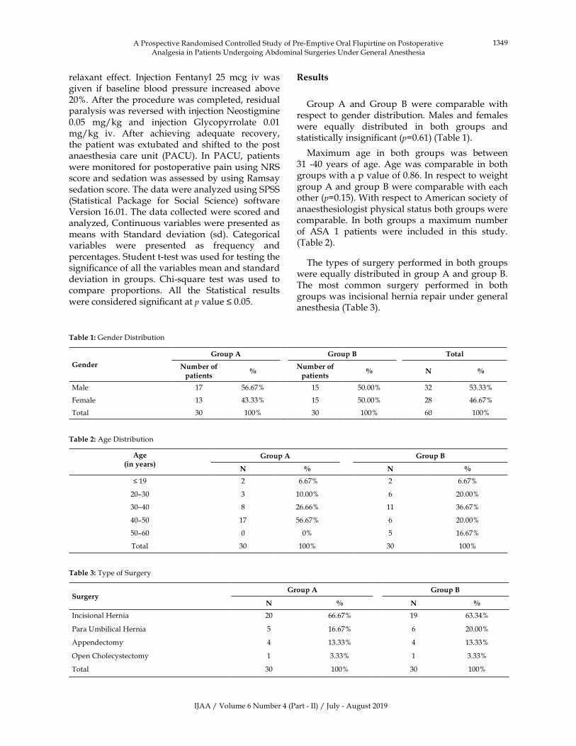
IJAA�/�Volume�6�Number�4�(Part�-�II)�/�July�-�August�2019
1349
relaxant� effect.� Injection� Fentanyl� 25� mcg� iv� was�given� if� baseline� blood� pressure� increased� above�20�.�After�the�procedure�was�completed,�residual�paralysis�was�reversed�with�injection�Neostigmine�0.05� mg/kg� and� injection� Glycopyrrolate� 0.01�mg/kg� iv.� After� achieving� ade�uate� recovery,�the�patient�was� extubated�and� shifted� to� the�post�anaesthesia� care� unit� (PAC�).� In� PAC�,� patients�were�monitored�for�postoperative�pain�using�NRS�score�and�sedation�was�assessed�by�using�Ramsay�sedation�score.�The�data�were�analyzed�using�SPSS�(Statistical� Package� for� Social� Science)� software�Version�16.01.�The�data�collected�were�scored�and�analyzed,�Continuous�variables�were�presented�as�means� with� Standard� deviation� (sd).� Categorical�variables� were� presented� as� fre�uency� and�percentages.�Student�t-test�was�used�for�testing�the�signi��cance�of�all�the�variables�mean�and�standard�deviation� in� groups.� Chi-s�uare� test� was� used� to�compare� proportions.� All� the� Statistical� results�were�considered�signi��cant�at�p�value���0.05.�
Table��:�Gender�Distribution
�ender
�roup�A �roup�B Total
Number�of�patients
�Number�of�patients
� N �
Male 17 56.67� 15 50.00� 32 53.33�
Female 13 43.33� 15 50.00� 28 46.67�
Total 30 100� 30 100� 60 100�
Table��:�Age�Distribution
Age�in�years�
�roup�A �roup�B
N � N �
��19 2 �6.67� 2 6.67�
20–30 3 10.00� 6 20.00�
30–40 8 26.66� 11 36.67�
40–50 17 56.67� 6 20.00�
50–60 0 0� 5 16.67�
Total 30 100� 30 100�
Table��:�Type�of�Surgery
Surgery�roup�A �roup�B
N � N �
Incisional��ernia 20 66.67� 19 63.34�
Para��mbilical��ernia 5 16.67� 6 20.00�
Appendectomy 4 13.33� 4 13.33�
Open�Cholecystectomy 1 3.33� 1 3.33�
Total 30 100� 30 100�
�esults
Group� A� and� Group� �� were� comparable� with�respect� to�gender�distribution.�Males� and� females�were� e�ually� distributed� in� both� groups� and�statistically�insigni��cant�(p�0.61)�(Table�1).
Maximum� age� in� both� groups� was� between�31� -40� years� of� age.�Age�was� comparable� in� both�groups�with�a�p�value�of�0.86.�In�respect�to�weight�group�A�and�group���were�comparable�with�each�other�(p�0.15).�With�respect�to�American�society�of�anaesthesiologist�physical�status�both�groups�were�comparable.� In� both� groups� a�maximum� number�of� ASA� 1� patients� were� included� in� this� study.(Table�2).
The� types�of�surgery�performed�in�both�groups�were�e�ually�distributed�in�group�A�and�group��.�The� most� common� surgery� performed� in� both�groups�was�incisional�hernia�repair�under�general�anesthesia�(Table�3).
A�Prospective�Randomised�Controlled�Study�of�Pre-Emptive�Oral�Flupirtine�on�Postoperative�Analgesia�in�Patients��ndergoing�Abdominal�Surgeries��nder�General�Anesthesia
IJAA�/�Volume�6�Number�4�(Part�-�II)�/�July�-�August�2019
1350 Indian�Journal�of�Anesthesia�and�Analgesia
Pulse� rate� and� oxygen� saturation� were� not�signi��cantly� changed� after� giving� the� study�drugs.� The� mean� value� of� pulse� rate� after� drug�administration�was�graphed�in�a�line�diagram.�No�signi��cant� change� in� the�pulse� rate�was�observed�after�drug�administration.�There�were�no�signi��cant�changes� in�pulse�rate,�systolic�blood�pressure�and�diastolic� blood� pressures� in� group� �� during� the�surgery�and�postoperative�period.�(Fig.�1).
�oth�groups�were�comparable�in�the�duration�of�surgery.�It�was�statistically�not�signi��cant�(p�0.71).�The� mean� duration� of� surgery� in� group� A� and�group� �� was� 123.67� minutes� and� 121.17� minutes�respectively�(Fig.�2).
The� mean� numerical� rating� scale� scores� were�initially� lower� in�Group���patients� than�Group�A�for� ��rst� 3� hours.� Of� these,� the� ��rst� 2� hours�of� postoperative� analgesia� were� statistically�
signi��cant.�The�mean�value�of�NRS�score�in�group���was� initially� less� than� three� for� the� ��rst� 3� hours.��igher�values�were�noted�between�the�4-6th�hour�of�the�postoperative�period�(Fig.�3).
Sixty� percent� of� the� patients� in� group� A�asked� rescue� analgesia� in� the� ��rst� hour� of� the�postoperative�period.��ut�only�3.3��of�patients� in�group� �� asked� rescue� analgesia� in� the� ��rst� hour.�81�� (13�� in� the� 2nd� hour,� 46�� in� a� 3rd� hour)� of�patients�in�group���re�uired�rescue�analgesia�after�3rd�hour�of�the�postoperative�period.�This�difference�was�signi��cant�(Fig.�4).
There� was� a� statistically� signi��cant� change�noted� in�Ramsay�sedation�score�(RSS)� for� the���rst�5�hours�of�the�postoperative�period.�The�patients�in�the� ��upirtine� group� exhibited�more� sedation,� but�remained�responsive�to�commands�(Fig.�5).
Fig���:�Pulse�Rate�after�Drug�(Placebo/Flupirtine)�Administration
Fig���:�Duration�of�Surgery�(in�Minutes)
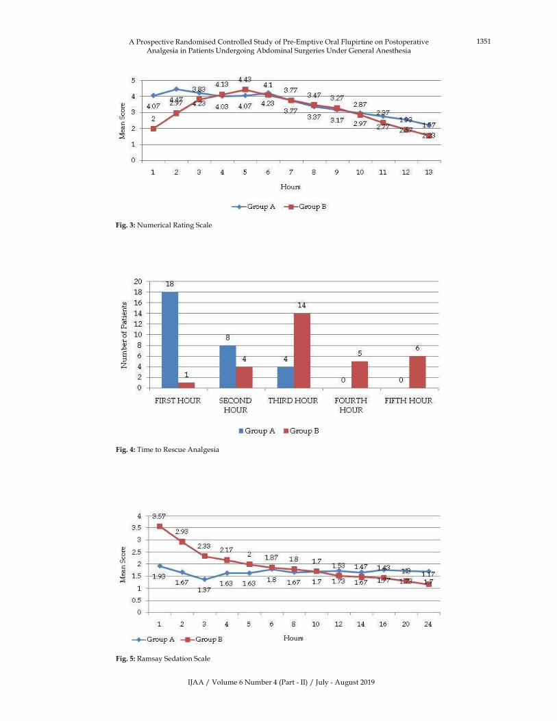
IJAA�/�Volume�6�Number�4�(Part�-�II)�/�July�-�August�2019
1351
Fig���:�Numerical�Rating�Scale
Fig���:�Time�to�Rescue�Analgesia
Fig���:�Ramsay�Sedation�Scale
A�Prospective�Randomised�Controlled�Study�of�Pre-Emptive�Oral�Flupirtine�on�Postoperative�Analgesia�in�Patients��ndergoing�Abdominal�Surgeries��nder�General�Anesthesia
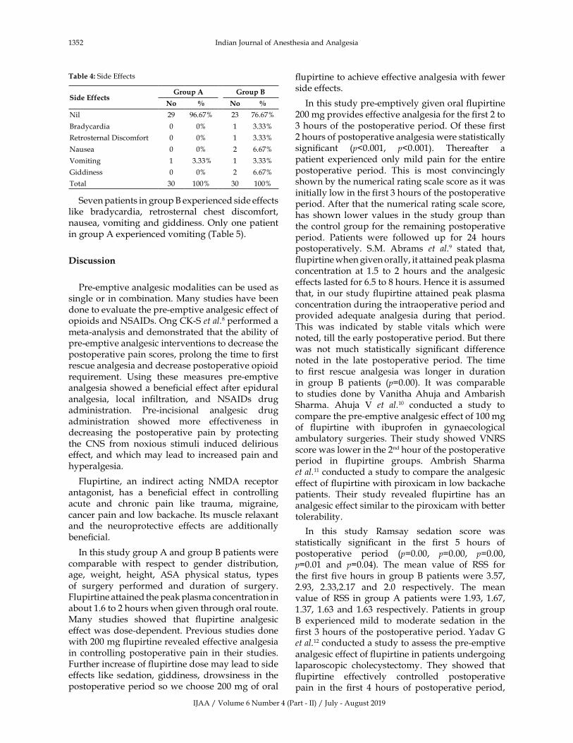
IJAA�/�Volume�6�Number�4�(Part�-�II)�/�July�-�August�2019
1352 Indian�Journal�of�Anesthesia�and�Analgesia
Table��:�Side�Effects
Side�Effects�roup�A �roup�B
No � No �
Nil 29 96.67� 23 76.67�
�radycardia 0 0� 1 3.33�
Retrosternal�Discomfort� 0 0� 1 3.33�
Nausea� 0 0� 2 6.67�
Vomiting� 1 �3.33� 1 3.33�
Giddiness� 0 0� 2 6.67�
Total 30 100� 30 100�
Seven�patients�in�group���experienced�side�effects�like� bradycardia,� retrosternal� chest� discomfort,�nausea,�vomiting�and�giddiness.�Only�one�patient�in�group�A�experienced�vomiting�(Table�5).
�iscussion
Pre-emptive�analgesic�modalities�can�be�used�as�single�or� in�combination.�Many�studies�have�been�done�to�evaluate�the�pre-emptive�analgesic�effect�of�opioids�and�NSAIDs.�Ong�C�-S�et�al.8�performed�a�meta-analysis�and�demonstrated�that�the�ability�of�pre-emptive�analgesic�interventions�to�decrease�the�postoperative�pain�scores,�prolong�the�time�to���rst�rescue�analgesia�and�decrease�postoperative�opioid�re�uirement.� �sing� these� measures� pre-emptive�analgesia�showed�a�bene��cial�effect�after�epidural�analgesia,� local� in��ltration,� and� NSAIDs� drug�administration.� Pre-incisional� analgesic� drug�administration� showed� more� effectiveness� in�decreasing� the� postoperative� pain� by� protecting�the� CNS� from� noxious� stimuli� induced� delirious�effect,�and�which�may� lead�to� increased�pain�and�hyperalgesia.
Flupirtine,� an� indirect� acting� NMDA� receptor�antagonist,� has� a� bene��cial� effect� in� controlling�acute� and� chronic� pain� like� trauma,� migraine,�cancer�pain�and�low�backache.�Its�muscle�relaxant�and� the� neuroprotective� effects� are� additionally�bene��cial.�
In�this�study�group�A�and�group���patients�were�comparable� with� respect� to� gender� distribution,�age,� weight,� height,� ASA� physical� status,� types�of� surgery� performed� and� duration� of� surgery.�Flupirtine�attained�the�peak�plasma�concentration�in�about�1.6�to�2�hours�when�given�through�oral�route.�Many� studies� showed� that� ��upirtine� analgesic�effect�was�dose-dependent.�Previous�studies�done�with�200�mg���upirtine�revealed�effective�analgesia�in� controlling� postoperative� pain� in� their� studies.�Further�increase�of���upirtine�dose�may�lead�to�side�effects� like� sedation,� giddiness,� drowsiness� in� the�postoperative�period�so�we�choose�200�mg�of�oral�
��upirtine�to�achieve�effective�analgesia�with�fewer�side�effects.
In�this�study�pre-emptively�given�oral���upirtine�200�mg�provides�effective�analgesia�for�the���rst�2�to�3�hours�of� the�postoperative�period.�Of� these���rst�2�hours�of�postoperative�analgesia�were�statistically�signi��cant� (p�0.001,� p�0.001).� Thereafter� a�patient� experienced� only�mild� pain� for� the� entire�postoperative� period.� This� is� most� convincingly�shown�by�the�numerical�rating�scale�score�as�it�was�initially�low�in�the���rst�3�hours�of�the�postoperative�period.�After�that�the�numerical�rating�scale�score,�has� shown� lower� values� in� the� study� group� than�the�control�group�for�the�remaining�postoperative�period.� Patients� were� followed� up� for� 24� hours�postoperatively.� S.M.� Abrams� et� al.9� stated� that,���upirtine�when�given�orally,�it�attained�peak�plasma�concentration� at� 1.5� to� 2� hours� and� the� analgesic�effects�lasted�for�6.5�to�8�hours.��ence�it�is�assumed�that,� in�our�study���upirtine�attained�peak�plasma�concentration�during�the�intraoperative�period�and�provided� ade�uate� analgesia� during� that� period.�This� was� indicated� by� stable� vitals� which� were�noted,�till�the�early�postoperative�period.��ut�there�was� not� much� statistically� signi��cant� difference�noted� in� the� late� postoperative� period.� The� time�to� ��rst� rescue� analgesia� was� longer� in� duration�in� group� �� patients� (p�0.00).� It� was� comparable�to� studies� done� by� Vanitha�Ahuja� and�Ambarish�Sharma.� Ahuja� V� et� al.10� conducted� a� study� to�compare�the�pre-emptive�analgesic�effect�of�100�mg�of� ��upirtine� with� ibuprofen� in� gynaecological�ambulatory� surgeries.� Their� study� showed�VNRS�score�was�lower�in�the�2nd�hour�of�the�postoperative�period� in� ��upirtine� groups.� Ambrish� Sharma�et�al.11�conducted�a�study�to�compare�the�analgesic�effect�of���upirtine�with�piroxicam�in�low�backache�patients.� Their� study� revealed� ��upirtine� has� an�analgesic�effect�similar�to�the�piroxicam�with�better�tolerability.
In� this� study� Ramsay� sedation� score� was�statistically� signi��cant� in� the� ��rst� 5� hours� of�postoperative� period� (p�0.00,� p�0.00,� p�0.00,�p�0.01� and� p�0.04).� The� mean� value� of� RSS� for�the���rst���ve�hours� in�group���patients�were�3.57,�2.93,� 2.33,2.17� and� 2.0� respectively.� The� mean�value�of�RSS� in� group�A�patients�were�1.93,� 1.67,�1.37,� 1.63� and� 1.63� respectively.� Patients� in� group��� experienced� mild� to� moderate� sedation� in� the���rst�3�hours�of�the�postoperative�period.��adav�G�et�al.12�conducted�a�study�to�assess�the�pre-emptive�analgesic�effect�of���upirtine�in�patients�undergoing�laparoscopic� cholecystectomy.� They� showed� that���upirtine� effectively� controlled� postoperative�pain� in� the� ��rst� 4� hours� of� postoperative� period,�
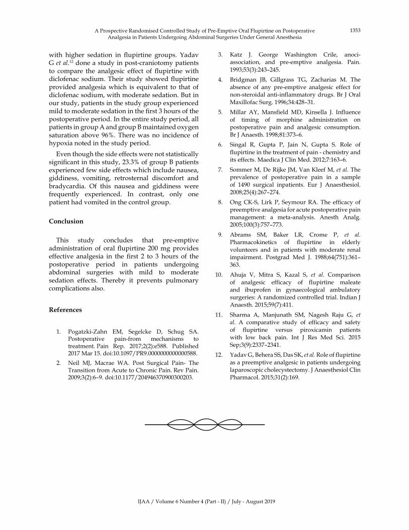
IJAA�/�Volume�6�Number�4�(Part�-�II)�/�July�-�August�2019
1353
with� higher� sedation� in� ��upirtine� groups.� �adav�G�et�al.12�done�a�study�in�post-craniotomy�patients�to� compare� the� analgesic� effect� of� ��upirtine�with�diclofenac� sodium.�Their� study� showed���upirtine�provided� analgesia�which� is� e�uivalent� to� that� of�diclofenac�sodium,�with�moderate�sedation.��ut�in�our�study,�patients�in�the�study�group�experienced�mild�to�moderate�sedation�in�the���rst�3�hours�of�the�postoperative�period.�In�the�entire�study�period,�all�patients�in�group�A�and�group���maintained�oxygen�saturation� above� 96�.� There�was� no� incidence� of�hypoxia�noted�in�the�study�period.
Even�though�the�side�effects�were�not�statistically�signi��cant�in�this�study,�23.3��of�group���patients�experienced�few�side�effects�which�include�nausea,�giddiness,� vomiting,� retrosternal� discomfort� and�bradycardia.� Of� this� nausea� and� giddiness� were�fre�uently� experienced.� In� contrast,� only� one�patient�had�vomited�in�the�control�group.
Conclusion
This� study� concludes� that� pre-emptive�administration� of� oral� ��upirtine� 200�mg�provides�effective� analgesia� in� the� ��rst� 2� to� 3� hours� of� the�postoperative� period� in� patients� undergoing�abdominal� surgeries� with� mild� to� moderate�sedation� effects.� Thereby� it� prevents� pulmonary�complications�also.
�eferences
1.� Pogatzki-�ahn� EM,� Segelcke� D,� Schug� SA.�Postoperative� pain-from� mechanisms� to�treatment.�Pain� Rep.� 2017;2(2):e588.� Published�2017�Mar�15.�doi:10.1097/PR9.0000000000000588.
2.� Neil� MJ,� Macrae� WA.� Post� Surgical� Pain-� The�Transition�from�Acute�to�Chronic�Pain.�Rev�Pain.�2009;3(2):6–9.�doi:10.1177/204946370900300203.
3.� �atz� J.� George� Washington� Crile,� anoci-
association,� and� pre-emptive� analgesia.� Pain.�
1993;53(3):243–245.
4.� �ridgman� J�,� Gillgrass� TG,� �acharias� M.� The�
absence� of� any� pre-emptive� analgesic� effect� for�
non-steroidal�anti-inflammatory�drugs.��r�J�Oral�
Maxillofac�Surg.�1996;34:428–31.
5.� Millar� A�,� Mansfield� MD,� �insella� J.� Influence�
of� timing� of� morphine� administration� on�
postoperative� pain� and� analgesic� consumption.�
�r�J�Anaesth.�1998;81:373–6.
6.� Singal� R,� Gupta� P,� Jain� N,� Gupta� S.� Role� of�
flupirtine�in�the�treatment�of�pain�-�chemistry�and�
its�effects.�Maedica�J�Clin�Med.�2012;7:163–6.
7.� Sommer�M,�De�Rijke�JM,�Van��leef�M,�et�al.�The�
prevalence� of� postoperative� pain� in� a� sample�
of� 1490� surgical� inpatients.� Eur� J� Anaesthesiol.�
2008;25(4):267–274.
8.� Ong�C�-S,� Lirk�P,�Seymour�RA.�The� efficacy�of�
preemptive�analgesia�for�acute�postoperative�pain�
management:� a� meta-analysis.� Anesth� Analg.�
2005;100(3):757–773.
9.� Abrams� SM,� �aker� LR,� Crome� P,� et� al.�
Pharmacokinetics� of� flupirtine� in� elderly�
volunteers� and� in� patients� with�moderate� renal�
impairment.� Postgrad� Med� J.� 1988;64(751):361–
363.
10.� Ahuja� V,� Mitra� S,� �azal� S,� et� al.� Comparison�
of� analgesic� efficacy� of� flupirtine� maleate�
and� ibuprofen� in� gynaecological� ambulatory�
surgeries:�A�randomized�controlled�trial.�Indian�J�
Anaesth.�2015;59(7):411.
11.� Sharma� A,� Manjunath� SM,� Nagesh� Raju� G,� et�
al.� A� comparative� study� of� efficacy� and� safety�
of� flupirtine� versus� piroxicamin� patients�
with� low� back� pain.� Int� J� Res� Med� Sci.� 2015�
Sep;3(9):2337–2341.
12.� �adav�G,��ehera�SS,�Das�S�,�et�al.�Role�of�flupirtine�
as�a�preemptive�analgesic�in�patients�undergoing�
laparoscopic�cholecystectomy.�J�Anaesthesiol�Clin�
Pharmacol.�2015;31(2):169.
A�Prospective�Randomised�Controlled�Study�of�Pre-Emptive�Oral�Flupirtine�on�Postoperative�Analgesia�in�Patients��ndergoing�Abdominal�Surgeries��nder�General�Anesthesia

IJAA�/�Volume�6�Number�4�(Part�-�II)�/�July�-�August�2019
1354 Indian�Journal�of�Anesthesia�and�Analgesia
Red�Flower�Publication�Pvt.�Ltd.
CAPT�RE��O�R�MAR�ET
For�advertisin��in�this��ournal
Please�contact:
International�print�and�online�display�ad�ertising�salesAd�ertisement�Manager
Phone:�91-11-22756995,�22754205,�45796900,�Cell:��91-9821671871E-mail:�[email protected]
�ecruitment�and�Classified�Ad�ertisingAd�ertisement�Manager
Phone:�91-11-22756995,�22754205,�45796900,�Cell:��91-9821671871E-mail:�[email protected]
��Red�Flower�Publication�Pvt.�Ltd.�
�Original�Research�ArticleIndian�Journal�of�Anesthesia�and�Analgesia�
2019;6(4)�(Part�-�II):�1355-1360DOI:�http://dx.doi.org/10.21088/ijaa.2349.8471.6419.41
A�Comparison� of��e�medetomidine��ith� Thiopentone� Sodium�Versus�Esmolol��ith�Thiopentone�Sodium�to�Attenuate�the��emodynamic�Stress��esponses�after�Electrocon�ulsi�e�Therapy
B��rishna�Chaitanya���A�Prashanth���Shaik��ala����anth�Pa�an��umar�����Suresh�Babu�
1Assistant�Professor,�Dept.�of�Anesthesia�2Assistant�Professor,�Dept.�of�Psychiatry,�3Postgraduate,�Dept.�of�Anesthesia,�4Postgraduate,�Dept.�of�Anesthesia,� 5Assistant�Professor,�Dept.�of�Preventive�Social�Medicine,�Maharajah�s�Institute�of�Medical�Sciences�(MIMS),�Vizianagaram,�Andhra�Pradesh�535217,�India.
Corresponding�Author:�A�Prasanth,�Assistant�Professor,�Dept.�of�Psychiatry,�Maharajah�s� Institute�of�Medical�Sciences� (MIMS),�Vizianagaram,�Andhra�Pradesh�535217,�India.E�mail:��[email protected]
�ecei�ed�on�07.05.2019,�Accepted�on�08.06.2019
Abstract
Modified� electroconvulsive� therapy� (ECT)� under� anesthesia� is� an� important� in� the� treatment� of� severe,�persistent�depression;�bipolar�disorder�and�schizophrenia;�especially�resistant�cases.��owever,�it�is�commonly�associated� with� acute� hyper� dynamic� responses.�Aims:� To� compare� the� effects� of� dexmedetomidine� and�esmolol� on� patients�� haemodynamics,� motor� seizure� duration,� and� recovery� times� following� ECT.� Stud���esign:�Randomised�Prospective�Double��linded�Study.�Materials�and�Methods:�90�cases�aged�between�18�to�50�years�belonging�to�ASA�grade�I�and�II�were�randomly�divided�into�three�groups�with�30�each.�Group�A�received�normal�saline�(placebo),�Group���received�dexmedetomidine�1��g/kg,�and�Group�C�received�esmolol�1�mg/kg;�before� induction�with� thiopentone�sodium�2�mg/kg�and�muscle� relaxation�with�succinylcholine�0.75�mg/kg.��emodynamic�parameters�were�recorded�at�different�time�intervals.�The�seizure�duration�using�arm�isolation�method�and�recovery�times�using�post-anesthesia�discharge�scoring�system�(PADSS)�were�noted.Anal�sis:�Data�analysis�was�done�using�SPSS�(Statistical�product�and�service�solutions)�software�trial�version�21�for�windows.�Results�were�expressed�as�mean��SD,�proportions�and�percentages.�One�way�ANOVA�test�was�used�to�assess�the�significant�differences�between�groups.�Results:�PostECT�rise�in�hemodynamic�parameters�was�significantly�less�in�dexmedetomidine�group�as�compared�to�esmolol�and�control�group�at�2,�4,�6,�and�8�min�using�ANOVA�test.�There�was�no�significant�difference�in�seizure�duration,�emergence,�and�recovery�among�the�three�groups.�Conclusions:��oth�drugs�reduce�the�hyperdynamic�response�to�ECT�without�affecting�the�seizure�duration,�but�dexmedetomidine�has�more� favourable� response� in�view�of�stable�vitals,� smooth�emergence�and�no�adverse�effect�on�recovery�duration.
�ey�ords:�Dexmedetomidine;�Esmolol;�Modified�electroconvulsive�therapy.
�o��to�cite�this�article:
���rishna�Chaitanya,�A�Prashanth,�Shaik��ala.�A�Comparison�of�Dexmedetomidine�with�Thiopentone�Sodium�Versus�Esmolol�with� Thiopentone� Sodium� to� Attenuate� the� �emodynamic� Stress� Responses� after� Electroconvulsive� Therapy.� Indian� J� Anesth�Analg.�2019;6(4):1355-1360.
Introduction
Electroconvulsive� therapy� (ECT)� is� a�well� known�
treatment�for�severe�depression�in�patients�who�are�
resistant� to� pharmacotherapy.� Nowadays,� almost�
all�the�ECT�procedures�are�performed�under�general�
anesthesia;�also�known�as�modi��ed�ECT.��owever,�it�is�commonly�associated�with�acute�hyper�dynamic�responses,� including� initial� parasympathetic�response� followed� by� transient� hypertension� and�tachycardia� due� to� catecholamines� release� in� the�body��During�the�sympathetic�stimulation��systolic�blood�pressure�may�increase�by�30�–40��and�heart�
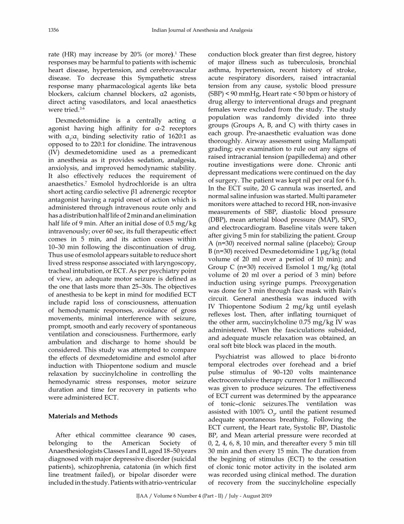
IJAA�/�Volume�6�Number�4�(Part�-�II)�/�July�-�August�2019
1356 Indian�Journal�of�Anesthesia�and�Analgesia
rate� (�R)�may� increase� by� 20�� (or�more�.1�These�responses�may�be�harmful�to�patients�with�ischemic�heart� disease,� hypertension,� and� cerebrovascular�disease.� To� decrease� this� Sympathetic� stress�response� many� pharmacological� agents� like� beta�blockers,� calcium� channel� blockers,� �2� agonists,�direct� acting� vasodilators,� and� local� anaesthetics�were�tried.2-6
Dexmedetomidine� is� a� centrally� acting� ��agonist� having� high� af��nity� for� �-2� receptors�with� �
2:�
1� binding� selectivity� ratio� of� 1620:1� as�
opposed�to�to�220:1�for�clonidine.�The�intravenous�(IV)� dexmedetomidine� used� as� a� premedicant�in� anesthesia� as� it� provides� sedation,� analgesia,�anxiolysis,� and� improved� hemodynamic� stability.�It� also� effectively� reduces� the� re�uirement� of�anaesthetics.7� Esmolol� hydrochloride� is� an� ultra�short�acting�cardio�selective��1�adrenergic�receptor�antagonist�having�a�rapid�onset�of�action�which�is�administered� through�intravenous�route�only�and�has�a�distribution�half�life�of�2�min�and�an�elimination�half�life�of�9�min.�After�an�initial�dose�of�0.5�mg/kg�intravenously;�over�60�sec,�its�full�therapeutic�effect�comes� in� 5� min,� and� its� action� ceases� within�10–30�min� following� the� discontinuation� of� drug.�Thus�use�of�esmolol�appears�suitable�to�reduce�short�lived�stress�response�associated�with�laryngoscopy,�tracheal�intubation,�or�ECT.�As�per�psychiatry�point�of� view,� an� ade�uate�motor� seizure� is� de��ned� as�the�one�that�lasts�more�than�25–30s.�The�objectives�of�anesthesia�to�be�kept�in�mind�for�modi��ed�ECT�include� rapid� loss� of� consciousness,� attenuation�of� hemodynamic� responses,� avoidance� of� gross�movements,� minimal� interference� with� seizure,�prompt,�smooth�and�early�recovery�of�spontaneous�ventilation�and� consciousness.� Furthermore,� early�ambulation� and� discharge� to� home� should� be�considered.�This�study�was�attempted�to�compare�the� effects� of�dexmedetomidine�and�esmolol�after�induction� with� Thiopentone� sodium� and� muscle�relaxation� by� succinylcholine� in� controlling� the�hemodynamic� stress� responses,� motor� seizure�duration� and� time� for� recovery� in� patients� who�were�administered�ECT.
Materials�and�Methods
After� ethical� committee� clearance� 90� cases,�belonging� to� the� American� Society� of�Anaesthesiologists�Classes�I�and�II,�aged�18–50�years�diagnosed�with�major�depressive�disorder�(suicidal�patients),� schizophrenia,� catatonia� (in� which� ��rst�line� treatment� failed),� or� bipolar� disorder� were�included�in�the�study.�Patients�with�atrio-ventricular�
conduction�block�greater�than���rst�degree,�history�of� major� illness� such� as� tuberculosis,� bronchial�asthma,� hypertension,� recent� history� of� stroke,�acute� respiratory� disorders,� raised� intracranial�tension� from� any� cause,� systolic� blood� pressure�(S�P)���90�mm�g,��eart�rate���50�bpm�or�history�of�drug�allergy�to�interventional�drugs�and�pregnant�females�were�excluded� from�the�study.�The�study�population� was� randomly� divided� into� three�groups� (Groups�A,� �,� and�C)�with� thirty� cases� in�each� group.� Pre-anaesthetic� evaluation� was� done�thoroughly.�Airway�assessment�using�Mallampati�grading;�eye�examination� to� rule�out�any� signs�of�raised�intracranial�tension�(papilledema)�and�other�routine� investigations� were� done.� Chronic� anti�depressant�medications�were�continued�on�the�day�of�surgery.�The�patient�was�kept�nil�per�oral�for�6�h.�In� the�ECT�suite,� 20�G�cannula�was� inserted,� and�normal�saline�infusion�was�started.�Multi�parameter�monitors�were�attached�to�record��R,�non-invasive�measurements� of� S�P,� diastolic� blood� pressure�(D�P),�mean� arterial�blood�pressure� (MAP),�SPO
2�
and�electrocardiogram.��aseline�vitals�were� taken�after�giving�5�min�for�stabilizing�the�patient.�Group�A� (n�30)� received�normal� saline� (placebo);�Group���(n�30)�received�Dexmedetomidine�1��g/kg�(total�volume� of� 20� ml� over� a� period� of� 10� min);� and�Group�C� (n�30)� received� Esmolol� 1�mg/kg� (total�volume� of� 20� ml� over� a� period� of� 3� min)� before�induction� using� syringe� pumps.� Preoxygenation�was�done�for�3�min�through�face�mask�with��ain�s�circuit.� General� anesthesia� was� induced� with�IV� Thiopentone� Sodium� 2� mg/kg� until� eyelash�re��exes� lost�� Then,� after� in��ating� tourni�uet� of�the�other�arm,�succinylcholine�0.75�mg/kg�IV�was�administered.� When� the� fasciculations� subsided,�and�ade�uate�muscle� relaxation�was� obtained,� an�oral�soft�bite�block�was�placed�in�the�mouth.
Psychiatrist� was� allowed� to� place� bi-fronto�temporal�� electrodes� over� forehead� and� a� brief�pulse� stimulus� of� 90–120� volts� maintenance�electroconvulsive�therapy�current�for�1�millisecond�was� given� to� produce� seizures.� The� effectiveness�of�ECT�current�was�determined�by�the�appearance�of� tonic–clonic� seizures.The� ventilation� was�assisted�with� 100��O
2,� until� the� patient� resumed�
ade�uate� spontaneous� breathing.� Following� the�ECT�current,� the��eart� rate,�Systolic��P,�Diastolic��P,� and�Mean� arterial� pressure�were� recorded� at�0,�2,�4,�6,�8,�10�min,�and�thereafter�every�5�min�till�30�min�and�then�every�15�min.�The�duration�from�the� begining� of� stimulus� (ECT)� to� the� cessation�of� clonic� tonic� motor� activity� in� the� isolated� arm�was�recorded�using�clinical�method.�The�duration�of� recovery� from� the� succinylcholine� especially�
IJAA�/�Volume�6�Number�4�(Part�-�II)�/�July�-�August�2019
1357
spontaneous�breathing�was�recorded.�Patients�were�assessed�for�side�effects�such�as�nausea,�vomiting,�hypotension/hypertension,�respiratory�depression�after� the� electrical� stimulus� and� were� discharged�from�the�post-anesthetic�care�unit�to�the�psychiatry�department�according�to�postanesthesia�discharge�scoring�system�(PADSS)�criteria.
Analysis
Data� analysis� was� done� using� SPSS� (Statistical�product� and� service� solutions)� software� trial�version� 21� for� windows.� Results� were� expressed�as� mean� �� standard� deviation,� proportions� and�percentages.� One� way� ANOVA� test� was� used� to�assess� the� signi��cant� differences� between� groups.�For�all�statistical�analysis�p��0.05�was�considered�as�statistically�signi��cant.
�esults
There�was�no�signi��cant�difference�in�the�baseline�variables�like�heart�rate�and�mean�arterial�pressure�among� the� three� groups� (p�0.05).� (Table� 2)� There�was�also�a�signi��cant�increase�in�the�heart�rate�and�MAP�while� immediately�after�giving�ECT�and�up�to�2nd�and�4th�minute�in�the�groups�A�and�C�(p�0.05)�unlike�Group���which�showed�no�signi��cant�change�in� the� �R� and� MAP� throughout� the� observation�period�(p�0.05)�(Tables�3�and�5).
Post��oc�tests�showed�that�there�was�a�signi��cant�difference� (p�0.05)� in� the� heart� rate� and� MAP�between�Group�A�and���and�Group�A�and�C�during�infusion�of� the�drug�and�at�2,�4,�6�and�8�minutes.�A� comparison� between� Group� �� and� group� C�revealed� signi��cant� difference� regarding� �R� and�
Table��:�Shopwing�the�Demographic�data
�roup�A��control��Mean���S��
�roupB��de�medetomidine��Mean���S��
�roup�C��esmolol�Mean���S��
Age�(years) 33.4���9.05� 34���8.21 34.2���8.41
Sex�ratio�(male:female) 20:10 21:9 19:11
Weight�(kg) 56.12���14� 55.2���5.12 56.5���7.16
SD-�standard�de�iation
Table��:�Showing�The�intergroup�comparison�of�baseline�vitals
Baseline��roup�A��control��
Mean�S���roup�B��de�medet�omidine��Mean�S��
�roup�C��esmolol��Mean�S��
�R�(/min)� 76.5���4.52� 77.03���3.84� 76.2���4.10
S�P�(mm�g)� 119.93���3.77� 121.0���3.29� 120.26���3.53
D�P(mm�g) 76.4�3.51 76.93���3.08� 77.26���2.75
MAP(mm�g) 90.86���3.06� 91.6���2.44 91.53���2.36
SpO2(�) 98.93���0.77� 98.86���0.80� 98.86���0.76
�R�–��eart�rate;�S�P�–�Systolic�blood�pressure;�MAP�–�Mean�arterial�pressure;�D�P�–�Diastolic�blood�pressure;�SpO
2�–�Oxygen�saturation
Table��:�showing�the�Post�electroconvulsive�therapy�changes�in�heart�rate�in�Group�A,��,�and�C�at�different�time�interval
Time�min��roup�A�beats/min�
�roup�B�beats/min�
�roup�C�beats/min�
Infusion 79.60 83.43 80.53
Induction 76.93 72.17 78.43
0 108.97 76.03 92.83
2 106.03 81.17 94.53
4 98.80 80.63 86.87
6 89.23 81.43 82.87
8 80.10 81.80 81.33
10 80.03 81.80 81.33
15 80.10 81.80 81.33
20 80.03 81.80 81.33
25 80.10 81.80 81.33
30 80.03 81.80 81.33
�R�of�three�groups�with�time.
A�Comparison�of�Dexmedetomidine�with�Thiopentone�Sodium�Versus�Esmolol�with�Thiopentone�Sodium�to�Attenuate�the��emodynamic�Stress�Responses�after�Electroconvulsive�Therapy

IJAA�/�Volume�6�Number�4�(Part�-�II)�/�July�-�August�2019
1358 Indian�Journal�of�Anesthesia�and�Analgesia
Table��:�Showing�the�comparison�of��R�among�the�Groups.
Time�roup�A��ersus�
�roup�B��P��alue��ith�significance��
�roup�A��ersus��roup�C��P��alue��ith�
significance��
�roup�B��ersus��roup�C��P��alue��ith�
significance��
�aseline 0.63�(NS) 0.30�(NS) 0.63�(NS)
After�study�drug�infusion �0.001�(�S)� �0.001�(�S) 0.06�(NS)
After�induction 0.08�(NS)� 0.92�(NS) 0.18�(NS)
After�ECT
0�min 0.001�(�S) 0.280�(NS) 0.000�(�S)
2�min 0.000�(�S) 0.000�(�S) 0.000�(�S)
4�min 0.000�(�S) 0.000�(�S) 0.000�(�S)
6�min 0.000�(�S) 0.000�(�S) 0.000�(�S)
8�min 0.000�(�S) 0.000�(�S) 0.349�(NS)
10�min 0.224�(NS) 1.388�(NS) 0.738�(NS)
15�min 1.383�(NS) 1.383�(NS) 1.383�(NS)
20�min 1.383�(NS) 1.383�(NS) 1.383�(NS)
25�min 1.383�(NS) 1.383�(NS) 1.383�(NS)
30�min 1.383�(NS) 1.383�(NS) 1.383�(NS)
(S�–�Significant�(p�0.05);�NS�–�Nonsignificant�(p�0.05);��S�–��ighly�significant�(p�0.001).�ECT�–�Electroconvulsive�therapy)
Table� �:� Showing� Postelectroconvulsive� therapy� changes� in�MAP� in� Group� A,� �,� and� C� at� different� time�interval�MAP�3�groups
Time�min��roup�A�mm�of��g�
�roup�B�mm�of��g�
�roup�C�mm�of��g�
infusion 93.87���3.27 93.77���6.50 92.73���5.90
induction 89.40���3.28 89.83���6.40 90.53���5.87
Ect 119.40���3.50 92.97���8.33 104.70���6.59
2 117.27���3.62 94.70���7.78 102.73���6.48
4 109.57���3.29 92.43���7.23 97.00���6.22
6 99.47���3.41 91.97���6.59 93.20���6.42
8 93.60���3.21 93.50���5.69 92.93���6.11
10 93.60���3.21 93.17���6.06 92.93���6.11
15 93.60���3.21 93.17���6.06 92.93���6.11
20 93.60���3.21 93.17���6.06 92.93���6.11
25 93.60���3.21 93.17���6.06 92.93���6.11
30 93.60���3.21 93.17���6.06 92.93���6.11
Table��:�Showing�comparison�of�MAP�among�the�3�groups
Time�roup�A��ersus��roup�B��P��alue��ith�significance��
�roup�A��ersus��roup�C��P��alue��ith�
significance��
�roup�B��ersus��roup�C��P��alue��ith�
significance��
After�study�drug�infusion �0.001�(�S)� �0.001�(�S) 0.06�(NS)
After�induction 0.08�(NS)� 0.92�(NS) 0.18�(NS)
After�ECT
0�min 0.756 0.417 0.616
2�min 0.000 0.000 0.000
4�min 0.000 0.000 0.000
6�min 0.000 0.000 0.003
8�min 0.000 0.000 0.402
10�min 0.940 0.619 0.672
15�min 0.753 0.628 0.865
20�min 0.753 0.628 0.865
25�min 0.753 0.628 0.865
30�min 0.753 0.628 0.865
(S�–�Significant�(p�0.05);�NS�–�Nonsignificant�(p�0.05);��S�–��ighly�significant�(��0.001).�ECT�–�Electroconvulsive�therapy)�
IJAA�/�Volume�6�Number�4�(Part�-�II)�/�July�-�August�2019
1359
MAP� at� 2,� 4� and� 6� minutes� only.� There� was� no�signi��cant�difference�between�among�the�3�groups�after�8�minutes.
�iscussion
ECT� induces� generalized� tonic–clonic� epileptic�seizure.�The�patients�undergoing�for�ECT�might�be�on�a�number�of�anti�psychotic�medications�which�can�cause�exaggerated�cardiovascular�responses.�In�our�study,� postECT� hyper� dynamic� responses� were�signi��cantly� less� in� the� dexmedetomidine� group�at�0,�2,�4,�6,�and�8�min�as�compared�with�Group�A�and�Group�C.� similar�observations�were�noted�by�Shams� and� ElMasry7� and� �egec� et� al.8.� Although,�Fu�and�White9�have�found� that�dexmedetomidine�extended�the�seizure�activity�duration�during�ECT,�Shams� and� ElMasry,7� Mizrak� et� al.,10� Cohen� and�Stewart,11� and� Dodawad12� have� found� that� there�was� no� signi��cant� differences� in� the� duration� of�seizure� activity� when� using� dexmedetomidine�with�the�control�population,�Similar���ndings�were�noted� in�our�study.�Saito� et�al.,13��owie�et�al.14�and�Weinger� et�al.15�found�no�signi��cant�differences� in�the�duration�of�seizures,�when�comparing�esmolol�versus�control�group�before�ECT�which�was�similar�in�our�group.�Motor�seizure�duration�and�recovery�from� anesthesia� were� similar� in� all� the� groups.�In� our� study�we�observed� that� dexmedetomidine�reduced� emergence� agitation� following� recovery�from�anesthesia� for� ECT,� in� contrast� the� recovery�was�smooth.
The� duration� of� Motor� seizure� activity� was�not� effected� and� recovery� was� not� delayed.�There� were� no� side� effects� such� as� headache,�respiratory� depression,� hypoxemia,� bradycardia,�hypotension,� jaw� pain,� and� muscle� spasms.�Esmolol� has� a� very� fast� onset� of� action� (2� min)�while�dexmedetomidine�has�a�little�delayed�onset�of�action�and�has�to�be�given�by�infusion.�To�make�double�blinding�possible,�we�have�given�esmolol�also�in�same�fashion�as�dexmedetomidine,�but�this�may�reduce�the�ef��cacy�of�esmolol�as�it�is�rapidly�eliminated�from�the�body.�To�avoid�this�bias�and�rather� than� making� esmolol� less� effective,� we�have� changed� the� anaesthesiologist,� after� giving�esmolol� in� bolus� form� and� dexmedetomidine� in�an� infusion,�which� is� a�preferred� techni�ue.16�As�ECT�procedures�are�performed� fre�uently� in� the�outpatient� setting,� the� anaesthetic� agents� used�for� these�procedures�should�have�rapid� recovery�pro��les.� In� the� our� study,� none� of� these� two�drugs� (esmolol� and� dexmedetomidine)� prolong�the�recovery�times.�These�drugs�may�be�superior�
to� other� drugs� for� ECT� because� of� their� short�hal��ife� and� wide� therapeutic� indices.� �owever,�the� implications�of� these���ndings� re�uire� further�investigation.
Conclusion
�oth�esmolol�and�dexmedetomidine�attenuate� the�hyper�dynamic�response�to�ECT�without�affecting�the� seizure�duration,� but�dexmedetomidine� has� a�more�favourable�response�in�view�of�stable�vitals,�smooth� emergence,� and� no� adverse� effect� on�recovery�duration.
�i�itations
The� monitoring� of� seizure� duration� by�observing� tonic–clonic� activity� and� not� using�electroencephalogram� (EEG)� was� a� limitation� of�our� study� because� EEG� seizure� duration� activity�may�be�longer�than�motor�seizure�activity.
�inancial�Support�and�Sponsorship:�Nil.
Conflicts�of�Interest:�There�are�no�conflicts�of�interest.
�eferences
1.� Ding��,�White�PF.�Anesthesia�for�electroconvulsive�therapy.�Anesth�Analg.�2002;94:135164.
2.� Pal� R,� Pal� VS.� Anesthesia� for� electroconvulsive�therapy:�A�noble�approach.�Int�J�Med�Res��ealth�Sci.�2015;4:8905.
3.� Locala� JA,� Irefin� SA,� Malone� D,� Cywinski�J�,� Samuel� SW,� Naugle� R.� The� comparative�hemodynamic� effects� of� methohexital� and�remifentanil�in�electroconvulsive�therapy.�J�ECT.
4.� 2005;21:125.
5.� �umar�A,�Sharma�D�,�Mani�R.�A�comparison�of�propofol� and� thiopentone� for� electroconvulsive�therapy.� J� Anaesthesiol� Clin� Pharmacol.�2012;28:3537.
6.� Van�den��roek�WW,�Groenland�T�,�Mulder�PG,�et�al.��etablockers�and�electroconvulsive�therapy:�A�review.�Tijdschr�Psychiatr�2008;50:20515.
7.� �oere� E,� �irkenh�ger� T�,� Groenland� T�,� et� al.�etablocking� agents� during� electroconvulsive�therapy:�A�review.��r�J�Anaesth.�2014;113:4351.
8.� Shams� T,� ElMasry� R.� �etofol� dexmedetomidine�combination�in�ECT:�A�punch�for�depression�and�agitation.�Indian�J�Anaesth.�2014;58:27580.
9.� �egec� �,� Toprak� �I,� Demirbilek� S,� et� al.Dexmedetomidine� blunts� acute� hyperdynamic�
A�Comparison�of�Dexmedetomidine�with�Thiopentone�Sodium�Versus�Esmolol�with�Thiopentone�Sodium�to�Attenuate�the��emodynamic�Stress�Responses�after�Electroconvulsive�Therapy
IJAA�/�Volume�6�Number�4�(Part�-�II)�/�July�-�August�2019
1360 Indian�Journal�of�Anesthesia�and�Analgesia
responses� to� electroconvulsive� therapy� without�altering� seizure� duration.� Acta� Anaesthesiol�Scand.�2008;52:3026.
10.� Fu� W,� White� PF.� Dexmedetomidine� failed� to�block� the� acute� hyperdynamic� response� to�electroconvulsive� therapy.� Anesthesiology.�1999;90:4224.
11.� Mizrak�A,��oruk�S,�Ganidagli�S,�et�al.�Premedication�with�dexmedetomidine�and�midazolam�attenuates�agitation�after�electroconvulsive�therapy.�J�Anesth.2009;23:610.
12.� Cohen� M�,� Stewart� JT.� Treatment� of�postelectroconvulsive� therapy� agitation� with�dexmedetomidine.�J�ECT.�2013;29:e234.
13.� Dodawad� R,� Sumalatha� G�,� Sandeep� P,� et� al.�Dexmedetomidine�at�a�dose�of�0.6microgram/kg�in� attenuation� of� hemodynamic� stress� response�of� electroconvulsive� therapy.� J� Evid� �ased�Med��ealth.�2015;2:47718.
14.� Saito� S,� Nishihara� F,� Akihiro� T,� et� al.� Landiolol�and�esmolol�prevent�tachycardia�without�altering�cerebral�bloodflow.�Can�J�Anaesth.�2005;52:102734.
15.� �owie� M�,� �iestand� DC,� �vara� DA,� �im� P�,�McSweeney�TD,�Coffman� JA.�Defining� the�dose�range� for� esmolol� used� in� electroconvulsive�therapy�hemodynamic�attenuation.�Anesth�Analg.1992;75:80510.
16.� Weinger� M�,� Partridge� �L,� �auger� R,� et�al.� Prevention� of� the� cardiovascular� and�neuroendocrine� response� to� electroconvulsive�therapy:�I.�Effectiveness�of�pretreatment�regimens�on�hemodynamics.�Anesth�Analg.�1991;73:55662.
17.� Moshiri� E,� Modir� �,� �agheri� N,� et� al.�Premedication� effect� of� dexmedetomidine� and�alfentanil�on�seizure�time,�recovery�duration,�and�hemodynamic� responses� in� electroconvulsive�therapy.�Ann�Card�Anaesth.�2016;19:2638.
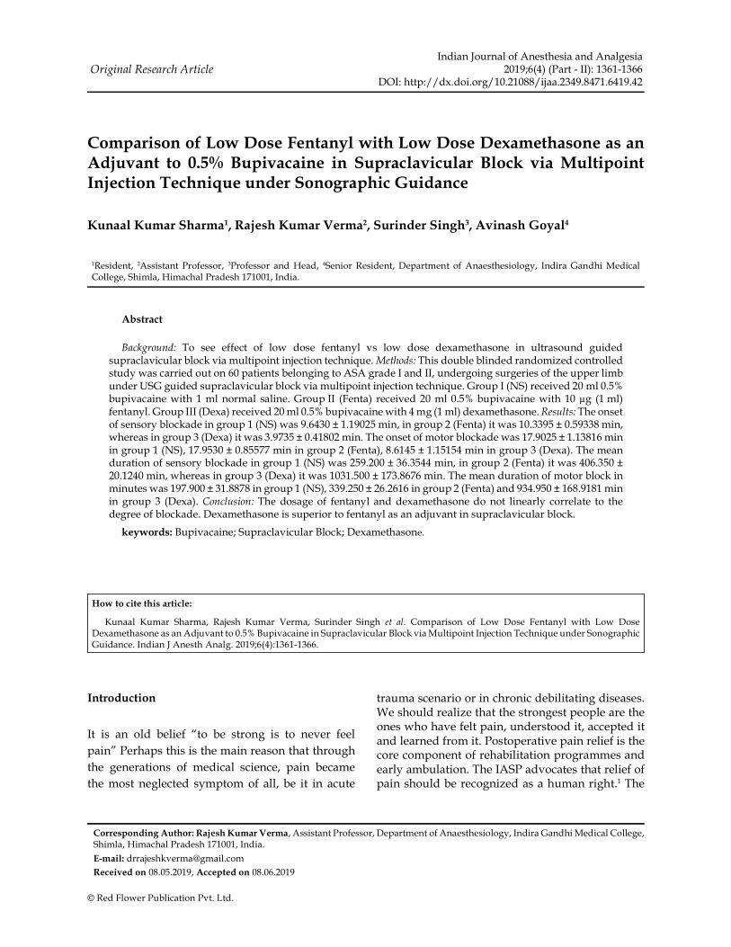
��Red�Flower�Publication�Pvt.�Ltd.�
�Original�Research�ArticleIndian�Journal�of�Anesthesia�and�Analgesia�
2019;6(4)�(Part�-�II):�1361-1366DOI:�http://dx.doi.org/10.21088/ijaa.2349.8471.6419.42
Comparison�of��o���ose�Fentanyl��ith��o���ose��e�amethasone�as�an�Ad�u�ant� to� ����� Bupi�acaine� in� Supracla�icular� Block� �ia�Multipoint�In�ection�Techni�ue�under�Sonographic��uidance
�unaal��umar�Sharma����a�esh��umar�Verma���Surinder�Singh���A�inash��oyal�
1Resident,� 2Assistant� Professor,� 3Professor� and��ead,� 4Senior�Resident,�Department� of�Anaesthesiology,� Indira�Gandhi�Medical�College,�Shimla,��imachal�Pradesh�171001,�India.
Corresponding�Author:��a�esh��umar�Verma,�Assistant�Professor,�Department�of�Anaesthesiology,�Indira�Gandhi�Medical�College,�Shimla,��imachal�Pradesh�171001,�India.
E�mail:�[email protected]
�ecei�ed�on�08.05.2019,�Accepted�on�08.06.2019
Abstract
Background:� To� see� effect� of� low� dose� fentanyl� vs� low� dose� dexamethasone� in� ultrasound� guided�supraclavicular�block�via�multipoint�injection�techni�ue.�Methods:�This�double�blinded�randomized�controlled�study�was�carried�out�on�60�patients�belonging�to�ASA�grade�I�and�II,�undergoing�surgeries�of�the�upper�limb�under��SG�guided�supraclavicular�block�via�multipoint�injection�techni�ue.�Group�I�(NS)�received�20�ml�0.5��bupivacaine�with�1�ml�normal� saline.�Group�II� (Fenta)� received�20�ml�0.5��bupivacaine�with�10��g� (1�ml)�fentanyl.�Group�III�(Dexa)�received�20�ml�0.5��bupivacaine�with�4�mg�(1�ml)�dexamethasone.�Results:�The�onset�of�sensory�blockade�in�group�1�(NS)�was�9.6430���1.19025�min,�in�group�2�(Fenta)�it�was�10.3395���0.59338�min,�whereas�in�group�3�(Dexa)�it�was�3.9735���0.41802�min.�The�onset�of�motor�blockade�was�17.9025���1.13816�min�in�group�1�(NS),�17.9530���0.85577�min�in�group�2�(Fenta),�8.6145���1.15154�min�in�group�3�(Dexa).�The�mean�duration�of�sensory�blockade�in�group�1�(NS)�was�259.200���36.3544�min,�in�group�2�(Fenta)�it�was�406.350���20.1240�min,�whereas�in�group�3�(Dexa)�it�was�1031.500���173.8676�min.�The�mean�duration�of�motor�block�in�minutes�was�197.900���31.8878�in�group�1�(NS),�339.250���26.2616�in�group�2�(Fenta)�and�934.950���168.9181�min�in�group� 3� (Dexa).�Conclusion:�The�dosage�of� fentanyl� and�dexamethasone�do�not� linearly� correlate� to� the�degree�of�blockade.�Dexamethasone�is�superior�to�fentanyl�as�an�adjuvant�in�supraclavicular�block.
key�ords:��upivacaine;�Supraclavicular��lock;�Dexamethasone.
�o��to�cite�this�article:
�unaal� �umar� Sharma,� Rajesh� �umar� Verma,� Surinder� Singh� et� al.� Comparison� of� Low� Dose� Fentanyl� with� Low� Dose�Dexamethasone�as�an�Adjuvant�to�0.5���upivacaine�in�Supraclavicular��lock�via�Multipoint�Injection�Techni�ue�under�Sonographic�Guidance.�Indian�J�Anesth�Analg.�2019;6(4):1361-1366.
Introduction
It� is� an� old� belief� �to� be� strong� is� to� never� feel�
pain��Perhaps�this�is�the�main�reason�that�through�
the� generations� of� medical� science,� pain� became�
the�most� neglected� symptom�of� all,� be� it� in� acute�
trauma�scenario�or�in�chronic�debilitating�diseases.�We�should�realize�that�the�strongest�people�are�the�ones�who�have�felt�pain,�understood�it,�accepted�it�and�learned�from�it.�Postoperative�pain�relief�is�the�core�component�of�rehabilitation�programmes�and�early�ambulation.�The�IASP�advocates�that�relief�of�pain�should�be�recognized�as�a�human�right.1�The�
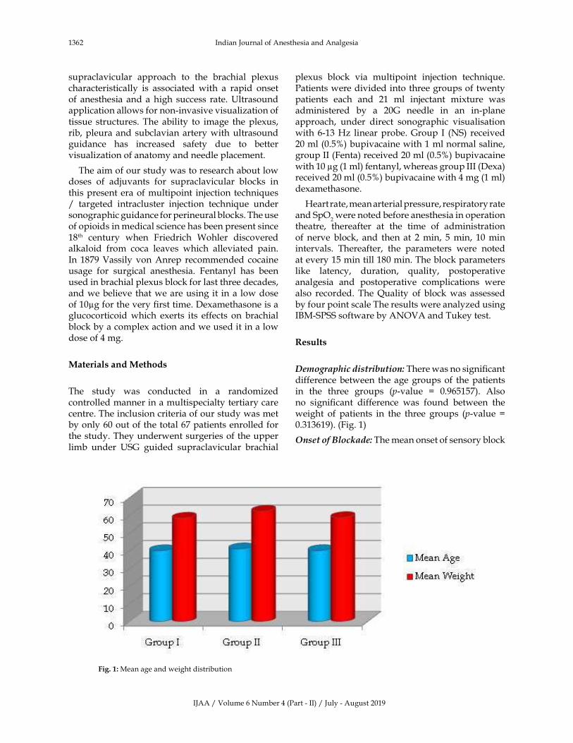
IJAA�/�Volume�6�Number�4�(Part�-�II)�/�July�-�August�2019
1362 Indian�Journal�of�Anesthesia�and�Analgesia
supraclavicular� approach� to� the� brachial� plexus�characteristically� is� associated�with� a� rapid� onset�of� anesthesia� and�a�high� success� rate.��ltrasound�application�allows�for�non-invasive�visualization�of�tissue� structures.�The� ability� to� image� the�plexus,�rib,�pleura�and�subclavian�artery�with�ultrasound�guidance� has� increased� safety� due� to� better�visualization�of�anatomy�and�needle�placement.
The�aim�of�our�study�was�to�research�about�low�doses� of� adjuvants� for� supraclavicular� blocks� in�this�present�era�of�multipoint� injection�techni�ues�/� targeted� intracluster� injection� techni�ue� under�sonographic�guidance�for�perineural�blocks.�The�use�of�opioids�in�medical�science�has�been�present�since�18th� century� when� Friedrich� Wohler� discovered�alkaloid� from� coca� leaves� which� alleviated� pain.�In�1879�Vassily�von�Anrep� recommended�cocaine�usage� for� surgical� anesthesia.� Fentanyl� has� been�used�in�brachial�plexus�block�for�last�three�decades,�and�we�believe�that�we�are�using�it� in�a� low�dose�of�10�g�for�the�very���rst�time.�Dexamethasone�is�a�glucocorticoid�which� exerts� its� effects� on�brachial�block�by�a�complex�action�and�we�used�it�in�a�low�dose�of�4�mg.
Materials�and�Methods
The� study� was� conducted� in� a� randomized�controlled�manner�in�a�multispecialty�tertiary�care�centre.�The�inclusion�criteria�of�our�study�was�met�by�only�60�out�of�the�total�67�patients�enrolled�for�the�study.�They�underwent�surgeries�of�the�upper�limb� under� �SG� guided� supraclavicular� brachial�
plexus� block� via� multipoint� injection� techni�ue.�Patients�were�divided�into�three�groups�of�twenty�patients� each� and� 21� ml� injectant� mixture� was�administered� by� a� 20G� needle� in� an� in-plane�approach,� under� direct� sonographic� visualisation�with�6-13��z� linear�probe.�Group� I� (NS)� received�20�ml�(0.5�)�bupivacaine�with�1�ml�normal�saline,�group�II�(Fenta)�received�20�ml�(0.5�)�bupivacaine�with�10��g�(1�ml)�fentanyl,�whereas�group�III�(Dexa)�received�20�ml�(0.5�)�bupivacaine�with�4�mg�(1�ml)�dexamethasone.
�eart�rate,�mean�arterial�pressure,�respiratory�rate�and�SpO
2�were�noted�before�anesthesia�in�operation�
theatre,� thereafter� at� the� time� of� administration�of�nerve� block,� and� then� at� 2�min,� 5�min,� 10�min�intervals.� Thereafter,� the� parameters� were� noted�at�every�15�min�till�180�min.�The�block�parameters�like� latency,� duration,� �uality,� postoperative�analgesia� and� postoperative� complications� were�also� recorded.� The��uality� of� block�was� assessed�by�four�point�scale�The�results�were�analyzed�using�I�M-SPSS�software�by�ANOVA�and�Tukey�test.
�esults
�e�o�ra�hic�distri�ution:�There�was�no�signi��cant�difference�between� the�age�groups�of� the�patients�in� the� three� groups� (p-value� �� 0.965157).� Also�no� signi��cant� difference� was� found� between� the�weight� of� patients� in� the� three� groups� (p-value� ��0.313619).�(Fig.�1)�
�nset�of��loc�ade:�The�mean�onset�of�sensory�block�
Fig���:�Mean�age�and�weight�distribution
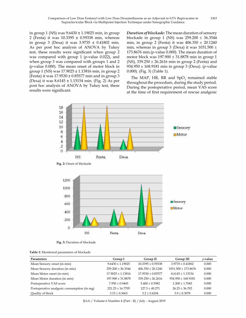
IJAA�/�Volume�6�Number�4�(Part�-�II)�/�July�-�August�2019
1363
in�group�1�(NS)�was�9.6430���1.19025�min,�in�group�2� (Fenta)� it� was� 10.3395� �� 0.59338� min,� whereas�in� group� 3� (Dexa)� it� was� 3.9735� �� 0.41802� min.�As� per� post� hoc� analysis� of� ANOVA� by� Tukey�test,� these� results� were� signi��cant� when� group� 2�was� compared�with� group� 1� (p-value� 0.022),� and�when�group�3�was�compared�with�groups�1�and�2�(p-value�0.000).�The�mean�onset�of�motor�block� in�group�1�(NS)�was�17.9025���1.13816�min,�in�group�2�(Fenta)�it�was�17.9530���0.85577�min�and�in�group�3�(Dexa)�it�was�8.6145���1.15154�min.�(Fig.�2)�As�per�post�hoc�analysis�of�ANOVA�by�Tukey�test,� these�results�were�signi��cant.
�uration�of��loc�ade:�The�mean�duration�of�sensory�blockade� in� group� 1� (NS)� was� 259.200� �� 36.3544�min,� in� group� 2� (Fenta)� it� was� 406.350� �� 20.1240�min,�whereas� in�group�3�(Dexa)� it�was�1031.500���173.8676�min.(p-value�0.000).�The�mean�duration�of�motor�block�was�197.900���31.8878�min�in�group�1�(NS),�339.250���26.2616�min�in�group�2�(Fenta)�and�934.950���168.9181�min�in�group�3�(Dexa).�(p-value�0.000).�(Fig.�3)�(Table�1).
The� MAP,� �R,� RR� and� SpO2� remained� stable�
throughout�the�procedure,�during�the�study�period.�During�the�postoperative�period,�mean�VAS�score�at�the�time�of���rst�re�uirement�of�rescue�analgesic�
Fig���:�Onset�of�blockade
Table��:�Monitored�parameters�of�blockade�
Parameters �roup�I �roup�II �roup�III p��alue
Mean�Sensory�onset�(in�min) 9.6430���1.19025 10.3395���0.59338 3.9735���0.41802 0.000
Mean�Sensory�duration�(in�min) 259.200���36.3544 406.350���20.1240 1031.500���173.8676 0.000
Mean�Motor�onset�(in�min) 17.9025���1.13816 17.9530���0.85577 8.6145���1.15154 0.000
Mean�Motor�duration�(in�min) 197.900���31.8878 339.250���26.2616 934.950���168.9181 0.000
Postoperative�VAS�score 7.950���0.9445 5.400���0.5982 1.200���1.7045 0.000
Postoperative�analgesic�consumption�(in�mg) 221.25���16.7705 127.5���49.271 26.25���36.702 0.000
�uality�of�block 3.15���0.3663 3.2���0.4104 3.9���0.3078 0.000
Fig���:�Duration�of�blockade
Comparison�of�Low�Dose�Fentanyl�with�Low�Dose�Dexamethasone�as�an�Adjuvant�to�0.5���upivacaine�in�Supraclavicular��lock�via�Multipoint�Injection�Techni�ue�under�Sonographic�Guidance

IJAA�/�Volume�6�Number�4�(Part�-�II)�/�July�-�August�2019
1364 Indian�Journal�of�Anesthesia�and�Analgesia
(75�mg�diclofenac),�was�least� in�group� III� (1.200��1.7045�)�and�maximum�in�group�I�(7.950���0.9445).�(Fig.� 4).� These� results� were� highly� statistically�signi��cant� (p� �� 0.000).� The� rescue� analgesia� was�given� as� 75� mg� diclofenac� intramuscularly.� The�mean�postoperative�rescue�analgesic�consumption�(diclofenac)�in�group�I�was�221.25���16.7705�mg,�in�group�II�it�was�127.5���49.271�mg,�whereas�in�group�III�it�was�just�26.25���36.702�mg�(p���0.000).�(Fig.�5).�The�mean��uality�of�block�was�in�group�III�was�3.9���0.30,�in�group�II�it�was�3.2���0.41�and�in�group�I�it�was�3.15���0.166.
�iscussion
Supraclavicular�block�for�upper�extremity�surgery�anaesthetizes� the� brachial� plexus� at� its� divisions,�
where� it� is� in� its�most� compact� form.�Therefore� it�
provides� a� complete� and� reliable� block� for� upper�extremity�surgery.
Attardi� et� al.2� showed� that� dexamethasone�s�mechanism�of�action�in�perineural�blockade�results�
from� decreased� nociceptive� C-��bre� activity� via�a� direct� effect� on� glucocorticoid� receptors� and�inhibitory� effect� on� potassium� channels.� It� is� also�suggested�that�by�local�vasoconstrictive�effect�there�occurs�a�reduction�of�local�anaesthetic�absorption,�which� leads� to� �uicker� onset� and� prolongation�of� blockade� as� demonstrated� by� Shishido� et� al.3.�Also,� Stan� et� al.4� showed� that� glucocorticoids�can� prolong� analgesia� period� by� suppressing�the� synthesis� of� in��ammatory� mediators.�
Neurotoxicity� to� perineural� corticosteroids� is�related�mainly� to� the�preservative�benzyl�alcohol,�
Fig���:�Postoperative�VAS�at�the�time�of�first�re�uirement�of�rescue�analgesic
Fig���:�Postoperative�rescue�analgesic�consumption
IJAA�/�Volume�6�Number�4�(Part�-�II)�/�July�-�August�2019
1365
vehicle� polyethylene� glycol� and� the� presence� of�insoluble�steroid�particulate�matter�in�the�injectate.�Dexamethasone� is� non-particulate� and� we� used�a� preservative-free� formulation.� In� addition,� Ma�et� al.5� have� demonstrated� neuroprotective� effects�of� perineurally� administered� dexamethasone� in�murine�studies.�Perineural�corticosteroid�injections�are�widely�used�throughout�the�world.�The�time�of�onset�of�blockade�with�dexamethasone�in�our�study�group� III� (sensory� onset� 3.973��� 0.418�min,�motor�onset�8.614���1.151�min�)�was�faster�than�that�seen�by�Parveen�et�al.6� (sensory�onset�28.20���3.02�min,�motor�onset�38.70���4.25�min)�and�Alarasan� et�al.7�
(sensory�onset� 10.36���1.99�min,�motor�onset�12���1.64� min).� Although,� Parveen� et� al.� and� Alarasn�et�al.�used�a�higher�dose�of�dexamethasone�(8�mg)�in� comparison� to� our�dose� of� 4�mg,�we�were� still�able�to�get�faster�onset�of�block�in�our�study�group.
We� attribute� this� to� the� use� of� multipoint�injection� techni�ue� which� resembles� the� targeted�intracluster�injection�techni�ue�of�Techasuk�et�al.,�in�which�we�injected�11�ml�of�the�injectant�mixture�at�the�eight�ball�corner�pocket,� followed�by� injecting�5�ml� at� the�middle� trunks� and� 5�ml� at� the� upper�trunks.�Techasuk�et�al.8�have�observed�better�results�with� targeted� intracluster� injection� techni�ue�in� comparison� to� double� injection� techni�ue� in�supraclavicular�block.
We� have� used� 4� mg� dexamethasone� and� the�duration� of� sensory� and� motor� block� in� our�study�is�17.192���2.897�hrs�and�15.582���2.815�hrs.�This� is� comparable� to� the� study� by� El-�aradey�et� al.9�which� however� used� a� higher� dose� (8�mg)�of� dexamethasone.� We� believe� that� dose� of�dexamethasone�has� little� to�do�with�prolongation�of�block�duration�and�analgesia.�This�is�evident�in�the� study�by�Liu� et� al.10,� in�which� the� addition� of�1�mg,�2�mg�and�4�mg�dexamethasone� to� the� local�anaesthetic� mixture� signi��cantly� prolonged� the�analgesia� duration� to� 22.3� hours,� 23.3� hours� and�21.2�hours� in�each�of� the�respective�groups�which�had�received��SG�guided�supraclavicular�block.
Fentanyl� is� a� proven� adjuvant� for� perineural�blockade,�but� its�usage�in� low�doses�has�not�been�widely�studied.�We�believe�that�this�is�for�the���rst�time�that�fentanyl�is�being�used�in�such�a�low�dose�of�10��g� in�20�ml�bupivacaine� (0.5�).�And�on� the�top� of� that,� signi��cance� of� prolongation� of� block�even�at�this�low�dosage�of�fentanyl�emphasizes�the�reason�for�need�of�more�such�studies�with�low�dose�of�fentanyl.
The� duration� of� sensory� and� motor� block� in�group� II� (fentanyl)�was� signi��cantly�prolonged� in�comparison�to�the�control�group�I�(NS).�Rajkhowa�
et�al.11�used�higher�dose�(50�g)�of�fentanyl�as�adjuvant�in�supraclavicular�block�and�got�sensory�and�motor�block�of�7.75���0.47�hrs�and�6.56���0.43�hrs�duration,�while�we�used�10��g�and�obtained�comparable�result�of�sensory�and�motor�block�of�6.77���0.335�hrs�and�5.65���0.437�hrs.�We�believe�that�as�per�the�the�study�by� Gissen� et� al.12,� the� local� anaesthetic� properties�of� fentanyl� were� responsible� for� this� effect.� Also�Stein�et� al.13,�have�shown�that� the�opioid�receptors�on�the�peripheral�nerve�terminals�get�up-regulated�due� to� in��ammation� at� the� fractured� site,� thereby�cytokines�activate�the�endogenous�opioid�peptides�and�cause�local�analgesia�at�the�site�of�in��ammation.�The�opioids�inhibit�neuronal���ring�and�transmitter�release,� and� also� inhibit� the� release� of� substance-�P.�We�believe� that� this�mechanism�also�came� into�play� while� we� used� the� low� dose� of� fentanyl� for�supraclavicular�block�in�our�study.
The� intergroup� comparison� between� the�dexamethasone�group�and�fentanyl�group�showed�that�dexamethasone�is�superior�to�fentanyl�in�terms�of�faster�onset�of�blockade,�prolonging�the�duration�of� block,� �uality� of� the� block� and� postoperative�analgesia.�This�deduction�is�analogous�to�the�study�by��aghoobi�et�al.14�who�although�had�used�higher�doses� of� fentanyl� (100� �g)� and� dexamethasone�(8�mg)� alongwith� larger� injectant� volume� in� their�study�on�axillary�block.
Conclusion
The� use� of� low� dose� dexamethasone� (4mg)�prolonged� the� duration� of� block� to� a� far� greater�extent� (sensory� 17.192��� 2.897�hrs,�motor� 15.582���2.815�hrs)�than�the�use�of�low�dose�fentanyl�(10��g),�(sensory�6.772���0.335�hrs,�motor�5.654���0.437�hrs).�The� postoperative� VAS� score� at� the� time� of� ��rst�re�uirement�of� rescue�analgesia�was� lowest� in� the�dexamethasone� study� group.� Likewise,� the� mean�postoperative� rescue� analgesic� re�uirement� of�diclofenac�in�dexamethasone�group�was�least�among�the�three�study�groups.�Also,�the��uality�of�block�was�most�superior�in�the�dexamethasone�group.�So�we�advocate� its� use� over� fentanyl� in� supraclavicular�block.� Administration� of� supraclavicular� block�under� sonographic� guidance� resulted� in� stable�haemodynamics� throughout� the� study� period� in�all�the�three�groups,�and�no�patient�developed�any�adverse�effects�related�to�supraclavicular�block.
�i�itations:�The�sample�size�of�our�study�groups�was� small.� We� also� did� not� measure� the� serum�concentration� and� CSF� levels� of� the� adjuvants�used�in�our�study�in�order�to��uantify�the�systemic�absorption�of�these�drugs�(if�any).
Comparison�of�Low�Dose�Fentanyl�with�Low�Dose�Dexamethasone�as�an�Adjuvant�to�0.5���upivacaine�in�Supraclavicular��lock�via�Multipoint�Injection�Techni�ue�under�Sonographic�Guidance
IJAA�/�Volume�6�Number�4�(Part�-�II)�/�July�-�August�2019
1366 Indian�Journal�of�Anesthesia�and�Analgesia
�eferences
1.� International� association� for� the� study� of� pain:�Pain�Definitions.�The�need�of� a� taxonomy.�Pain.�1979;6(3):247.
2.� Attardi��,�Takimoto��,�Gealy�R,�et�al.�Glucocorticoid�induced� upregulation� of� a� pituitary� ��� channel�mRNA�in�vitro�and�in�vivo.�Receptors�Channels.�1993;1:287–293.
3.� Shishido� �,� �ikuchi� S,� �eckman� �,� et� al.�Dexamethasone� decreases� blood� flow� in�normal� nerves� and� dorsal� root� ganglia.� Spine�2002;27:581–586.
4.� Stan� T,� Goodman� E,� Cardida� �,� et� al.� Adding�methylprednisolone�to�local�anaesthetic�increases�the�duration�of�axillary�block.�Reg�Anaesth�Pain�Med.�2004;29:380–381.
5.� Ma� R,� Wang� �,� Lu� C,� et� al.� Dexamethasone�attenuated� bupivacaine-induced� neuron� injury�in�vitro�through�a�threonine-serine�protein�kinase��-dependent� mechanism.� Neuroscience.� 2010�May;167(2):329–342.
6.� Parveen� S,� Jan� M,� Taj� A,� et� al.� Effect� of�dexamethasone�as�an�adjuvant�with�bupivacaine�in�ultrasound�guided�single�shot�supraclavicular�brachial� plexus� block� in� upper� extremity�surgeries-�a�prospective�randomized�study.� Int�J�Res�Med�Sci.�2017�May;5(5):2139–2143.
7.� Alarasan� A�,� Agrawal� J,� Choudhary� �,� et�al.� Effect� of� dexamethasone� in� low� volume�supraclavicular�brachial�plexus�block:�A�double-
blinded� randomized� clinical� study.� Journal� of�Anaesthesiology� Clinical� Pharmacology.� 2016�Apr;32(2):45.
8.� Techasuk� W,� Gonz�lez� AP,� �ernucci� F,� et� al.�A� randomized� comparison� between� double-injection� and� targeted� intracluster-injection�ultrasound-guided� supraclavicular� brachial�plexus�block.�Anesth�Analg.�2014;118:1363–1369.
9.� El-�aradey�GF,�Elshmaa�NS.�The�efficacy�of�adding�dexamethasone,� midazolam,� or� epinephrine� to�0.5�� bupivacaine� in� supraclavicular� brachial�plexus� block.� Saudi� Journal� of�Anesthesia.� 2014�Nov;8(1):S78–S83.
10.� Liu�J,�Richman��A,�Grodofsky�SR,et�al.�Is�there�a�dose�response�of�dexamethasone�as�adjuvant�for�supraclavicular� brachial� plexus� nerve� block�� A�prospective� randomized� double-blinded� clinical�study.�J�Clin�Anesth.�2015;27:237–42.
11.� Rajkhowa�T,�Das�N,�Parua�S,�et�al.�Fentanyl�as�an�adjuvant�for�brachial�plexus�block:�a�randomized�comparative� study.� Int� J� Clin� Trials.� 2016�May;3(2):64–67.
12.� Gissen�AJ,� Gugino� LD,� Datta� S,� et� al.� Effects� of�fentanyl�and�sufentanil�on�peripheral�mammalian�nerves.�Anesth�Analg.�1987;66(12):1272–1276.
13.� Stein�C,�Lang�LJ.�Peripheral�mechanisms�of�opioid�analgesia.�Curr�Opin�Pharmacol.�2009;19:3–8.
14.� �aghoobi� S,� Seddighi� M,� �azdi� �,� et� al.�Comparison� of� postoperative� analgesic� effect� of�dexamethasone� and� fentanyl� added� to� lidocaine�through� axillary� block� in� forearm� fracture.� Pain�Research�and�Treatment�2013;2013:1–6.

��Red�Flower�Publication�Pvt.�Ltd.�
�Original�Research�ArticleIndian�Journal�of�Anesthesia�and�Analgesia�
2019;6(4)�(Part�-�II):�1367-1372DOI:�http://dx.doi.org/10.21088/ijaa.2349.8471.6419.43
A� Study� of� Comparison� of� Intubating� Conditions� and� �aemodynamic�Effects� after� the� Administration� of� Succinylcholine� and� �ocuronium�Bromide
�o�ina�Neil����arshil�Shah�
1Senior�Resident,�2Junior�Resident,�Department�of�Anesthesia,�Sterling��ospital,�Ahmedabad,�Gujarat�380052,�India.
Corresponding�Author:��o�ina�Neil,�Senior�Resident,�Department�of�Anesthesia,�Sterling��ospital,�Ahmedabad,�Gujarat,�380052,�India.
E�mail:�[email protected]
�ecei�ed�on�07.05.2019,�Accepted�on�08.06.2019
Abstract
Background�and�Aim:�Though�rocuronium�has�a�rapid�onset�of�neuromuscular�blockade�like�succinylcholine�without�the�latter�s�adverse�effects,�its�use�is�limited�due�to�its�prolonged�action.�Present�study�was�performed�with�an�aim�to�compare�the�outcome�of�using�Rocuronium�and�Succinylcholie�as�muscle�relaxant�(MR).�Material�and�Methods:�A�single-center,�prospective-randomized,�blinded�study�of�60�patients,�divided�into�2�groups�and�intubation�conditions�were�evaluated.�Results:�The�intubating�conditions�were�scored�as�excellent�in�29�and�26,�and�Good�intubating�conditions�were�observed�in�1�and�4�Patients�of�group�I�and�2�respectively.�Conclusion:�We�conclude�that�rocuronium,�an�innovative�nondepolarizing�MR�with�a�succinct�onset�of�action�and�transitional�duration,�but�lack�of�the�unpleasant�reactions�connected�with�succinylcholine�may�be�a�appropriate�alternative�to�succinylcholine�for�tracheal�intubation.
�ey�ords:�Muscle�relaxant;�Randomized�trial;�Rocuronium;�Succinylcholine.
�o��to�cite�this�article:
Lovina�Neil,��arshil�Shah.�A�Study�of�Comparison�of�Intubating�Conditions�and��aemodynamic�effects�after�the�Administration�of�Succinylcholine�and�Rocuronium��romide.�Indian�J�Anesth�Analg.�2019;6(4):1367-1372.
Introduction
From�the�ancient�time�man�has�always�been�in�the��uest�for�perfection.�Anesthesia�has�been�receptive�to�new� ideas���new�discoveries� for�better�patient�care.� Introduction� of� newer� drugs� �� techni�ues�have�further�helped�in�improvement�in�anesthesia�practice.� At� present� endotracheal� intubation� is�an� essential� element� of� organization� of� general�anesthesia� throughout� surgical� procedures� to�maintain� airway,� to� allow� IPPV� and� to� prevent�aspiration.�Dangerous�period� for�aspiration� is� the�
time� interval� between� suppression� of� protective�re��exes��� development� of� satisfactory� intubating�conditions.��ence� this� time� interval� should� be� as�short�as�possible.1-5
Succinylcholine� introduced�by�the�Daniel��ovet�et�al.�in�1949,�revolutionized�anaesthetic�practice�by�providing�intense�neuromuscular�blockage�of�very��uick�start���very�small�period�of�action,�and�yet�relaxant�of�alternative� for�habitual� intubation�and��uick� succession� initiation� of� anesthesia.5�Adding�up�to�fasciculation�it�had�numerous�adverse�effects�as�well.�Therefore�the�search�is�on�for�agent,�which�
IJAA�/�Volume�6�Number�4�(Part�-�II)�/�July�-�August�2019
1368 Indian�Journal�of�Anesthesia�and�Analgesia
has��uick�onset�of� action,��uick� revival,�and�non-cumulative�effect,�without�cvs�side�effects,�with�high�potency,� pharmacologically� inactive� metabolites,�and�reversibility�with�cholinesterase�inhibitors.6-8
Recently� developed� neuromuscular� blocking�drugs�are�of� intermediate�duration���to�a�major�extent� free� of� side� effects.� �owever� even� after�intubating� doses� onset� of� action� is� relatively�slow� as� compared� to� succinylcholine.� Among�the� muscle� relaxants� in� current� utilize,� the�transitional� acting� non-depolarizing� muscle�relaxants-�vecuronium���atracurium�besylate�are�good-looking�alternatives.9-15��owever�,neither�of�them�has� been� demonstrated� to�have� signi��cant�shorter�onset� time�as� re�uired�for��uick�tracheal�intubation� as� compared� to� succinylcholine.� The�use�of�high�initial�bolus�doses�of�either�atracuruim�or� vecuronium� condensed� the� beginning� time�however� at� the� cost� of� an� extended� period� of�action� which� may� be� undesirable� in� certain�situations.� Rocuronium� bromide� is� a� novel�aminosteriod� neuro-muscular� blocking� agent�linked� to�vecuronium�bromide�but�has�a�greater�lipophilicity,�lesser�potency�and�a�very�fast�onset�of� action.� Good� to� excellent� tracheal� intubating�condition� have� been� reported� with� in� 60-90�seconds�after�a�dose�in�a�range�of�0.6-0.9�mg/kg.16-21�Several� clinical� studies� conducted� by� various�workers� have� con��rmed� the� brief� onset� time�of� rocuronium� bromide.� In� most� studies� the�timing�of�tracheal�intubation�was�determined�by�neuromuscular�monitoring�or�the�intubation�was�done�at�a�prearranged�moment�in�time�following�organization� of� neuromuscular� blocking� drugs.�In� clinical� practice� however� neuromuscular�monitoring� and� accurate� timing� are� rarely�used,� and� many� anaesthesiologists� commence�laryngoscopy�based�on�clinical�assessment.
The�aim�of�the�current�research�is�to�examine�and�evaluate� the� intubating� conditions,� onset� of� time,�time� of� action� �� haemodynamic� effects� in� ASA�Gr� I/II� receiving� succinylcholine� �� rocuronium�bromide�for��uick�tracheal�intubations.
Materials�and�Methods
Sixty�patients�of�ASA�grade�I�and�II,�aged�between�20�to�60�years,�go�through�a�variety�of�surgical�events�were� elected� for� the� research.� Ethical� approval�was� taken� from� the� institute� ethical� committee�and� written� informed� consent� taken� from� the� all�participants.�Exclusion�criteria�were:�Patients�with�systemic�Disease�and�those�who�were�not�willing�to�participate�in�the�study.
60� Patients�were� separated� in� 2� groups.�Group�I� Patients� (n�30)� received� Inj.Succinylcholine�chloride� 1.5� mg/kg� IV,� Group� II� Patients� (n�30)�received� Inj.Rocuronium� �romide� 0.9� mg/kg� IV�For�Tracheal�Intubation.
On� entrance� to� the� operating� room,� vital�parameters�were�evaluated�and�iv�admittance�was�establish.� Surface� electrodes� of� neuromuscular�monitor� were� applied� to� forearm� at� wrist� to�stimulate�ulnar�nerve.�Patients�were�Premedicated�with� Inj.� Glycopyrolate� 4� �g/kg� IV� and� Inj.�Fentanyl� 2� �g/kg� IV,� 15� min.� before� induction.�After�Pre-oxygenation�for�3�minutes�anesthesia�was�induced�with�Inj.�Thiopentone�5-7�mg/kg�IV�till�the�loss�of�eye�lashes�re��ex.
Prior� to� delivery� of� muscle� relaxant� the�supramaximal� stimulus� was� determined� with�the� help� of� the� peripheral� nerve� stimulation� by�observing� contraction� of� adductor� pollicis� by�visual,� tactile�assessment.�After� induction,�muscle�relaxant�according�to�the�group�given�in�running�IV�line.�After�giving�muscle�relaxant�the�single�twitch�stimulus�given�all�10�seconds,�the�time�interval�from�end� of� injection� of� muscle� relaxant� to� maximum�suppression�of� control� twitch�height� (we� called� it�onset�time)�was�noted.
An� experienced� anaesthesiologist,� unaware�about�the�muscle�relaxant,�performed�endotracheal�intubation.� Intubation� attempt� was� tried� at�60� seconds� following� deposition� of� the� muscle�relaxant.� Intubating� conditions� were� assessed�as� excellent,� good,� fair� or� poor� based� on� jaw�relaxation,� location� and� faction� of� vocal� cords�and� diaphragmatic� response� to� intubation� using�Copenhagen� consensus� conference� rating� scale.�If� the� intubation� conditions� assessed,� not� found�satisfactory,�the�intubation�stopped�and�subse�uent�attempts� were� made� at� 30� seconds� interval� until�intubation�was�achieved�with�acceptable�intubating�conditions.� Time� duration� of� intubation� after�muscle�relaxant�noted�at�60�sec,�90�sec,�120�seconds.
Table� �:� Copenhagen� Consensus� Conference� Rating�Scale� Intubating� conditions� clinically� acceptable� clinically�unacceptable
Variables E�cellent �ood Poor
1.�Laryngoscopy Easy Fair Difficult
2.�Vocal�Cords
�� Position Abducted Intermediate Closed
�� Movements None Moving Closing
3.�Reaction�to�Intubations
�� Movements�of�Limb None Slight Vigorous
�� Coughing None Diaphragm Sustained�(�10s)
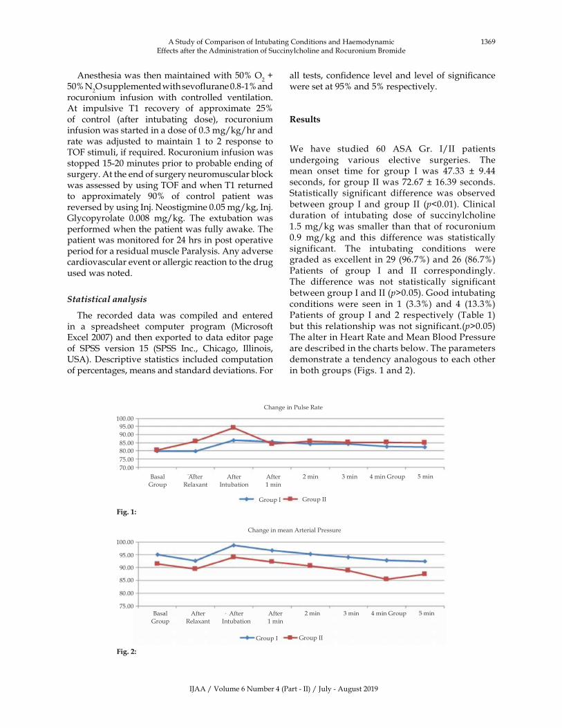
IJAA�/�Volume�6�Number�4�(Part�-�II)�/�July�-�August�2019
1369
Anesthesia�was�then�maintained�with�50��O2���
50��N2O�supplemented�with�sevo��urane�0.8-1��and�
rocuronium� infusion� with� controlled� ventilation.�At� impulsive� T1� recovery� of� approximate� 25��of� control� (after� intubating� dose),� rocuronium�infusion�was�started�in�a�dose�of�0.3�mg/kg/hr�and�rate� was� adjusted� to� maintain� 1� to� 2� response� to�TOF�stimuli,�if�re�uired.�Rocuronium�infusion�was�stopped�15-20�minutes�prior�to�probable�ending�of�surgery.�At�the�end�of�surgery�neuromuscular�block�was�assessed�by�using�TOF�and�when�T1�returned�to� approximately� 90�� of� control� patient� was�reversed�by�using�Inj.�Neostigmine�0.05�mg/kg,�Inj.�Glycopyrolate� 0.008� mg/kg.� The� extubation� was�performed�when�the�patient�was�fully�awake.�The�patient�was�monitored�for�24�hrs�in�post�operative�period�for�a�residual�muscle�Paralysis.�Any�adverse�cardiovascular�event�or�allergic�reaction�to�the�drug�used�was�noted.
Statistical�analysis
The� recorded� data� was� compiled� and� entered�in� a� spreadsheet� computer� program� (Microsoft�Excel�2007)�and�then�exported�to�data�editor�page�of� SPSS� version� 15� (SPSS� Inc.,� Chicago,� Illinois,��SA).�Descriptive�statistics� included� computation�of�percentages,�means�and�standard�deviations.�For�
all� tests,� con��dence� level�and� level�of�signi��cance�were�set�at�95��and�5��respectively.
�esults
We� have� studied� 60� ASA� Gr.� I/II� patients�undergoing� various� elective� surgeries.� The�mean� onset� time� for� group� I� was� 47.33� �� 9.44�seconds,� for�group�II�was�72.67���16.39�seconds.�Statistically� significant� difference� was� observed�between� group� I� and�group� II� (p�0.01).� Clinical�duration� of� intubating� dose� of� succinylcholine�1.5�mg/kg�was�smaller� than� that�of� rocuronium�0.9� mg/kg� and� this� difference� was� statistically�significant.� The� intubating� conditions� were�graded�as�excellent�in�29�(96.7�)�and�26�(86.7�)�Patients� of� group� I� and� II� correspondingly.�The� difference� was� not� statistically� significant�between�group�I�and�II�(p�0.05).�Good�intubating�conditions�were� seen� in� 1� (3.3�)� and� 4� (13.3�)�Patients� of� group� I� and� 2� respectively� (Table� 1)�but�this�relationship�was�not�significant.(p�0.05)�The�alter�in��eart�Rate�and�Mean��lood�Pressure�are�described�in�the�charts�below.�The�parameters�demonstrate�a�tendency�analogous�to�each�other�in�both�groups�(Figs.�1�and�2).
Change�in�Pulse�Rate
100.00
95.00
90.00
85.00
80.00
75.00
70.00
�asal�
Group
After
Relaxant
After
Intubation
After
1�min
2�min 3�min 4�min�Group 5�min
Group�I Group�II
Change�in�mean�Arterial�Pressure
100.00
95.00
90.00
85.00
80.00
75.00�asal�
Group
After
Relaxant
After
Intubation
After
1�min
2�min 3�min 4�min�Group 5�min
Group�I Group�II
A�Study�of�Comparison�of�Intubating�Conditions�and��aemodynamic�Effects�after�the�Administration�of�Succinylcholine�and�Rocuronium��romide
Fig���:
Fig���:
IJAA�/�Volume�6�Number�4�(Part�-�II)�/�July�-�August�2019
1370 Indian�Journal�of�Anesthesia�and�Analgesia
�iscussion
Traditionally� succinylcholine� has� been� the�neuromuscular� blocking� drug� of� preference� for�schedule� intubations� and� �uick� string� induction.�The�smaller�onset�of�action�has�made�it�a�preferred,�except�the�utilize�of�succinylcholine�can�though�be�connected�with�many�adverse�effects�as�bradycardia,�dysrhythmia,� hyperkalemia,� rise� in� intra� ocular�pressure,� increase� intra� cranial� pressure,� increase�intra�gastric�pressure,�post�op�myalgia.�Therefore,�a�non-depolarising�neuromuscular�blocker�with�a��uick�onset�of�action,�comprising�a�lesser�period�of�action�is�desirable.22,23
Preliminary� studies� in� animals� demonstrated�that�rocuronium,�being�a�small�ef��cacy�compound�was� connected� with� a� �uick� onset� of� outcome�contrast� to�other�compounds�such�as�pancronium�and�vecuronium.24-27
Preliminary� trials� in� animal� established�rocuronium� to� be� 10-20�� as� powerful� as�vecuronium� and� ED� doses� were� set� up� to� be�from�0.26�mg/kg� to� 0.30�mg/kg.� Intubating�dose�of� rocuronium� utilized� in� the� present� study� are�0.6� mg/kg� and� 0.9�mg/kg.� �se� of� superior� dose�of� rocuronium� to� advance� intubating� conditions�through� rapid� series� intubation� and� to� engrave�short� the� onset� time� underneath� 60� seconds� has�been� advised� by� a� variety� of� workers� but� doses�superior� then� 0.6� mg/kg� would� be� connected�with�a�extended�duration�of�action�which�may�be�unsuitable�in�numerous�situations.28
In� the� majority� studies,� a� suitable� timing� of�tracheal�intubation�has�been�resolute�by�3�ways-
1.� Clinical�Judgement.
2.� Neuromuscular� monitoring� either� by�Twitch�suppression�or�TOF�ratio.
3.� Predetermined�time�after�the�administration�of� neuromuscular� blocking� Agent� eg.�60�secs,�90�secs,�120�secs�etc.
Method� utilizing� judgment� alone� is� relatively�insensitive.� Onset� time� varies� with� diverse� nerve�stimulation� rates�utilized.�Cooper� et� al.24� establish�onset�time�for�rocuronium�0.6�mg/kg�as�90�seconds�by� 0.1��z� stimulation� and� 58� seconds� using� TOF�stimulation.� On� the� other� hand,� a� ��xed� time� for�tracheal�intubation�can�be�utilized.�In�current�study�we�have�utilized�all�the�3�parameters.29-30
Clinical� criteria� like� jaw� relaxation,� vocal�cord� movement� were� evaluated� according� to�Copenhagen� consensus� conference� rating� scale.�Onset�time�and�duration�of�action�were�measured�using� neuromuscular� monitoring.� Patients� were�
intubated�at�predetermined�intervals.�At�the�time�of�intubation�clinical�conditions�were�noted.�Land�and�Stovner25�were�most�likely�the�primary�to�bring�in�a�rating�scale�as�a�tool�for�the�evaluation�of�intubating�conditions� in� which� the� three� main� criteria:� Jaw�relaxation,�vocal�cords�(position�and�motility)�and�reaction� to� intubation� were� rated� by� descriptive�scores�such�as�excellent,�satisfactory�and�fair.
Findings�of�current�research,�concerning�intubating�conditions�show�excellent,�good�and�poor�conditions�accomplished�after�the�administration�of�rocuronium�0.9�mg/kg,�or�succinylcholine�1.5�mg/kg�subse�uent�routine� induction� for� voluntary� operations.�Present� research� data� demonstrated� that� there� is�not� an� considerable� dissimilarity� in� the� intubating�conditions� after� the� administration� of� rocuronium�0.9�mg/kg,� or� succinylcholine� 1.5�mg/kg.� Parallel���ndings� were� establish� in� the� studies� done� by,�Cooper�et�al.9,�Fredrick�et�al.14,��hou�et�al.13,�Wierda�et�al.15�and�Weiss�J��et�al.
In�a�study�carry�out�by�Mc�Court���C�et�al.5�the�intubating�conditions�after�0.9�mg/kg�rocuronium�come� out� to� be� nearly� indistinguishable� to� those�observed�after�1.0�mg/kg�succinylcholine� i.e.�96��v/s�97��clinically�acceptable�intubating�conditions.�Sparr19� and� Crul16� et� al.,� examined� rocuronium�s�ef��cay� in� emergency� intubating� conditions�utilizing� it� ��rmly� as� for� the� situation� for� rapid�se�uence�induction�in�unintended�but�still�optional�cases.� In� those� studies,� the� fre�uency�distribution�of� �excellent�,� �good�,� or� clinically� acceptable�intubating�conditions,�60�seconds�after�0.6�mg/kg�or�0.9�mg/kg�rocuronium�were�compared�with�those�observed� after� 1.0� mg/kg� succinylcholine.� The���ndings� specify� that� intubating� conditions� were�more� encouraging� at� 60� sec� after� administration�of�rocuronium�in�the�dose�of�0.9�mg/kg�compared�to�dose�of�0.6�mg/kg�in�unpremedicated�patients.�Analogous� outcome� were� observed� in� further�studies�of��hardwaj�N.�et�al.22�In�a�study�by�cooper�et� al.9,� they� found� the�onset� time�of�88.90� seconds�and�60.40� seconds� for�rocuronium�0.6�mg/kg�and�succinylcholine� 1.0�mg/kg� respectively.� The� time�to� accomplish� utmost� block� of� 72� seconds� with�rocuronium� 0.9� mg/kg� was� signi��cantly� longer�than�a�time�of�about�47�seconds�with�succinylcholine�1.5�mg/kg�in�the�current�study�and�is�in�agreement�with�the�result�of�other�studies.31-32
There� were� no� signi��cant� changes� in� �R� and�MAP� after� the� administration� of� muscle� relaxant�in�each�of�group�in�this�study.�The�tiny�rise�in��R�and�diminish� in�MAP�after� induction�and�muscle�relaxant�may�be�owing�to�cardiovascular�effects�of�induction�dose�of�thiopentone.
IJAA�/�Volume�6�Number�4�(Part�-�II)�/�July�-�August�2019
1371
Conclusion
We� conclude� that� rocuronium,� a� newer�nondepolarizing� muscle� relaxant� with� a� concise�onset� of� action� and� transitional� duration,� but�devoid�of�the�unfavorable�reactions�connected�with�succinylcholine�may�be�a�appropriate�substitute�to�succinylcholine�for�tracheal�intubation.�Rocuronium�in�a�dose�of�0.9�mg/kg�may�be�precious�option�to�succinylcholine� for� �uick� tracheal� intubation� in�emergency�situations.
�eferences
1.� Textbook�of�Anesthesia�by�Ronald�D.�Miller.�2014
2.� Tryba� M,� �orn� A,� Thole� �,� �enz� M.� Rapid�se�uence�orotracheal�intubation�with�rocuronium:�a� randomized� double-blind� comparison� with�suxamethonium� a� preliminary� communication.�Eur�J�Anaesth.�1994;11(9):44–48.
3.� Dubois� M�,� Lapeyre� G,� Lea� D,� et� al.�Pharmacodynamic� effects� of� three� doses� of�Org�9426�used�for�endotracheal�intubation�in�humans.�J�Clin�Anesth.�1992;4:472–479.
4.� �evan� DR,� Fiset� P,� �lendran� P.� Textbook�of� Anesthesia� by� Ronald� D.� Miller.�Pharmacodynamic�behavior�of�rocuronium�in�the�elderly.�Can�J�Anaesth.�1993;40(2):127–132.
5.� Mc� Court� �C,� Salmela� L,� Mirakhur� R�� et� al.�Camparision�of�rocuronium�and�suxamethonium�for� use� during� rapid� se�uence� induction� of�anaesthisia:�Anesthesia.�1998;53;867–71.
6.� Mirakhur� R�,� R� Cooper,� Clarke� SJ.� Onset� and�intubating� conditions� of� rocuronium� bromide�compared� to� those�of� suxamethonium.�Eur� J.�A.�1994;11(9):41–43.
7.� Jerrold� �� Levy� et� al.� Determination� of�haemodynamic�and�histamine�role�of�rocuronium�(ORG�9426)�when�administered�in�increased�doses�under� N
2O/O
2-� Sufentanil� anesthesia.� Anaesth�
Analog.�1994;70:318–21.
8.� Nitschmann�P�et�al.�Comparison�of�haemodynamic�effect� of� rocuronium� bromide� with� those� of�vecuronium� in� patients� undergoing� CA�G�surgery.�Eur�J�Anaesth.�1994;11:113–115.
9.� Cooper� R,� Mirakhur� R�,� Clarke� RSJ.� et� al.�Comparison� of� intubating� condition� after�administration� of� ORG� 9426� (Rocuronium)� and�suxamethonium.��JA.�1992;69:269–273.
10.� Tom� �,� Caldwell� J.� Rapid� tracheal� intubation�with� large� dose� rocuronium.� Anaesth.� Analg.�20000;90:175–179.
11.� Meistelman�C,�Plaud��,�Donati�F.�A�comparison�of�neuromuscular�blocking�effect�of� rocuronium�bromide� at� the� adductor� pollicis� and� laryngeal�adductor�muscles.�Eur�J.A.�1994;11(9):33–36.
12.� �ooij�L�DJ,�Curl�JF.�A�comparison�of�vecuronium�with� the� hypothetical� ideal� neuromuscular�blocking� drug.� In:� Ed.� S� Agoston� et� al.� Clinical�experience� with� norcuron.� Excerpta� Medica�Amsterdam.�1983;3–8.
13.� TJ� �hou� et� al.� Onset� offset� characteristics� and�intubating� conditions� of� rapacuronium:� A�comparison�with�rocuronium.�r�J�Anaesth.�2000;�85(2):246–50.
14.� Puhringer� F�,� �huenl-�rady� �S,� �oller�J,� Mitterschiffthaler� G.� Evaluation� of� the�endotracheal�intubating�conditions�of�rocuronium�(Org� 9426)� and� succinylcholine� in� outpatient�surgery.�Anesth�Analg.�1992;75:37–40.
15.� JM���Wierda� et� al.� Time� course� of� action� and�intubating� conditions� following,� vecuronium,�rocuronium� and� mivacurium.� Anesthesia.� 1995;�50:393–396.
16.� Crul� JF,� Vanbelleghem� V,� �uyse� L� et� al.�Rocuronium�with�alfantenil�and�propofol�allows�intubation�with�in�45�seconds.�Eur�J�Anaesth�1995;�12(1):111–12.
17.� Toni�Magorian�et�al.�Comparison�of�Rocuronium,�Succinylcholine� and� vecuronium� for� rapid�se�uence�induction�of�anesthesia�in�adult�patients.�Anaesthesiology.�1993;79:913–918.
18.� Neeraja��harti�et�al.�Rocuronium,� time�course�of�action�and�intubating�conditions�–�A�comparison�with� atracurium� and� vecuronium.� Indian� J�Anaesth.�2001;45(5):363–369.
19.� Sparr���J,�Luger�T�J,��eidegger�T�et�al.�Camparison�of� intubating� conditions� after� rocuronium�and� suxamthonium� following� rapid� se�uence�induction�with�thiopentone�in�elective�case.�Acta�Anaesthesiol�Scand.�1996;40(4);425–30.
20.� Foldes�FF,�Nagashima��,�Nguyen��D,�et�al.�The�neuromuscular� effects� of� Org� 9426� in� patients�receiving� balanced� anesthesia.� Anaesthesiology�1991;75:191–196.
21.� Magorian� T,� Wood� P,� Coldwell� JE� et� al.�Pharmacokinetics� onset,� duration� of� action� of�rocuronium� in� humans;� normal� v/s� hepatic�dysfunction.�Anaesthesiology.�1991;75:1069.
22.� �hardwaj�N;��ala�I;�Arya�V�;�Chari�P.�Comparison�of� intubating� conditions� after� rocuronium� and�succinylcholine.�Journal�of�anaesthesiology.�1999�Jul;15(3):285–9.
23.� Weiss� J�,�Gratz� I,�Goldberg�ME,� et� al.�Double� –blind�comparison�of�two�doses�of�rocuronium�and�succinylcholine� for� rapid� se�uence� intubation.� J�Clin�Anaesth.�1997�Aug;9(5):379–82.
24.� Cooper� RA,� Mirakur� R�,� Maddlneni� VR.�Neuromuscular� effects� of� rocuronium� bromide�during� fentanyl� and� halothane� anesthesia.�Anesthesia.�1993;48:103–105.
25.� Land� I,� Stonner� J.� Experimental� and� clinical�experiences�with�a�neuromuscular� relaxant�R05-
A�Study�of�Comparison�of�Intubating�Conditions�and��aemodynamic�Effects�after�the�Administration�of�Succinylcholine�and�Rocuronium��romide
IJAA�/�Volume�6�Number�4�(Part�-�II)�/�July�-�August�2019
1372 Indian�Journal�of�Anesthesia�and�Analgesia
3816,� diallylnor� –� toxiferine.� Acta� Anaesthesiol�Scand.�1962;6:85–97.
26.� Levy�J�,�Davis�G,�Duggan�J,�et�al.�Determination�of� the�haemodynamics� and�histamine� release� of�rocuronium� when� administered� in� increased�doses� under� N
2O/O
2,� sufentanil� anesthesia.�
Anesth�Analg.�1994;78:318–321.
27.� Olkkola� �T,� Tammisto� T.� �uantifying� the�interaction� of� rocuronium� with� etomidate,�fentanyl,� midazolam,� propofol,� thiopental� and�isoflurane,� using� closed-loop� feedback� control�ofv�rocuronium�infusion.�Anaesth�Analg.�1994;78:�691–696.
28.� Wierda� JM��,� �ennis� PJ,� De� Wit� APM,� et�al.� Pharmacodynamic� and� pharmacokinetic�studies� with� Org� 9426.� In� eds� �owman� et� al.�Neuromuscular�blocking�agents:�past�present�and�future.�Interface�London.�1990.pp.182–93.
29.� �ooij� L�DJ,� �nape� �TA.� The� neuromuscular�blocking�effect�of�Org�9426.�Anesthesia.�1991;46:�341–343.
30.� Dubois� M�,� Dawn� EL,� �ideshwar� �,� et� al.�Pharmacodunamics� of� rocuronium� with� and�without�prior�administration�of�succinylcholine.�J�Clin�Anesth.�1995;7:44–48.
31.� Tang� J,� Joshi� GP.� Comparision� of� rocuronium�and� mivacurium� to� succinylcholine� during�outpatient� laproscopic� surgery.� Anesth� Analg�1996;82:994–998.
32.� �arve�M,� Sharma� R.� Comparision� of� intubating�conditions� and� time� course� of� action� of�rocuronium� bromide� and� succinylcholine� in�paediatric�patients.� Indian� J.�Anaesth.�2002;46(6)�:465–468.
��Red�Flower�Publication�Pvt.�Ltd.�
�Original�Research�ArticleIndian�Journal�of�Anesthesia�and�Analgesia�
2019;6(4)�(Part�-�II):�1373-1376DOI:�http://dx.doi.org/10.21088/ijaa.2349.8471.6419.44
Comparison� of�Bilateral� Superficial�Cer�ical� Ple�us�Block� and� Incision��ine�Infiltration�for�Postoperati�e�Analgesia�for�Thyroid�Surgeries��nder��eneral�Anesthesia
Nagara��AV���Pradeep�A��ongare�
1,2Assistant�Professor,�Department�of�Anesthesiology,�ESIC�PGIMSR�Medical�College,��engaluru,��arnataka�560010,�India.
Corresponding�Author:�Pradeep�A�Dongare,�Assistant�Professor,�Department�of�Anesthesiology,�ESIC�PGIMSR�Medical�College,��engaluru,��arnataka�560010,�India.
E�mail:�[email protected]�
�ecei�ed�on�19.04.2019,�Accepted�on�22.05.2019
Abstract
Introduction:�Thyroid�surgeries�are�usually�performed�under�general�anesthesia�as�it�involves�manipulation�of� the�trachea.Studies�comparing�bilateral� superficial�cervical�plexus�block�and�incision� line� infiltration�for�postoperative�analgesia� in�thyroid�surgeries�are�sparse.��ence,�we�decided�to�evaluate�the�effect�of��SCP��and�incision�line�infiltration�of�local�anaesthetic�on�post-operative�VAS�and�analgesic�re�uirements.�Methods:After�obtaining�ethical�clearance�from�the�institutional�ethical�committee,�70�patients�were�randomly�assigned�to�2�groups�of�35�patients�each.�Patient�s�in�group�were�administered��SCP��with�20�ml�of�0.125��bupivacaine�and�10�ml�of�normal�saline�along�the�line�of�incision.�Patient�s�in�group�I�were�administered��SCP��with�20�ml�of�normal�saline�and�10�ml�of�0.25��bupivacaine�along�the�line�of�incision.�Intra-operatively�re�uirement�for�analgesics�were�recorded.�Results�on�continuous�measurements�are�presented�on�Mean���SD�(Min-Max)�and�results�on�categorical�measurements�are�presented�in�Number�(�).�Significance�was�assessed�at�5��level�of�significance.�Student�t�test�(two�tailed,�independent)�was�used�to�find�the�significance�of�study�parameters�on�continuous�scale�between�two�groups�(Inter�group�analysis)�on�metric�parameters.�Results:�Mean�VAS�scores�obtained�postoperatively�to�assess�postoperative�pain�were�slightly�lower�statistically�significant�in�the�2nd,�12th
and�24th�hour.�Re�uirement�of�inj�paracetamol�ande�inj�tramadol�was�not�significantly�different�between�the�two�groups.�Vocal�cord�movement�and�the�incidence�of�sore�throat�was�comparable�between�the�two�groups.�None�of�the�patients�in�both�the�two�groups�had�any�episode�of�nausea�or�vomiting.�Conclusion:�We�found�that�both��SCP��and�incision�line�infiltration�are�effective�methods�for�providing�analgesia�for�thyroid�surgeries.�There�is�no�difference�between�the�re�uirement�of�analgesics�or�postoperative�pain�scores�over�a�24�hr�period.�
�ey�ords:�Analgesia;�Cervical�plexus�block;�Infiltration;�Thyroidectomy
�o��to�cite�this�article:
Nagaraj�AV,�Pradeep�A�Dongare.�Comparison�of��ilateral�Superficial�Cervical�Plexus��lock�and�Incision�Line�Infiltration�for�Postoperative�Analgesia�for�Thyroid�Surgeries��nder�General�Anesthesia.�Indian�J�Anesth�Analg.�2019;6(4):1373-1376.
Introduction
Thyroid� surgeries� are� usually� performed� under�general� anesthesia� as� it� involves� manipulation�of� the� trachea.� Local� anesthesia� and� regional�
anesthesia�alone�for�thyroidectomy�have�been�used�
previously1,2.� Postoperative� analgesia� for� thyroid�
surgeries� has� conventionally� been� intravenous�
opioids�and�acetaminophen.�Opioids�like�fentanyl,�
morphine� provide� very� good� intra-operative� and�

IJAA�/�Volume�6�Number�4�(Part�-�II)�/�July�-�August�2019
1374 Indian�Journal�of�Anesthesia�and�Analgesia
postoperative� analgesia,� but� are� associated� with�side�effects�such�as�respiratory�depression,�pruritus,�constipation,�urinary�retention���nausea�vomiting3.
Acetaminophen� provides� good� analgesia,� but�Sormer�JM�et�al.4�have�found�that�70��of�the�patients�had�an�VAS�score�of�more�than�40�mm�when�it�was�used�as�sole�analgesic.�Gozal�et�al.�in�a�study�found�that�the�mean�pain�scores�post�thyroidectomy�was�69�mm� and� 90�� of� the� patients� re�uired� opioids�postoperatively5.
�ilateral� super��cial� cervical� plexus� block� has�been� used� for� regional� anesthesia� �� analgesia.Andrieu� et� al.� conducted� a� study� to� assess� the�analgesic� ef��cacy� of� bilateral� super��cial� cervical�plexus� block� (�SCP�)� under� general� anesthesia��� found� that� �SCP�� improved� intraoperative�analgesia� �� reduced� analgesic� re�uirements3
Wound� in��ltration� after� thyroid� surgeries� have�also� been� found� to� provide� ade�uate� analgesia1-4.�agul�A� et� al.� conducted� a� study� on� pre� incision�in��ltration�of� local� anaesthetic� on�post-op�pain���concluded�that�it�provided�easy���better�analgesic�control6.� Studies� comparing� bilateral� super��cial�cervical� plexus� block� and� incision� line� in��ltration�for�postoperative�analgesia�in�thyroid�surgeries�are�sparse.� �ence,� we� decided� to� evaluate� the� effect�of� �SCP�� and� incision� line� in��ltration� of� local�anaesthetic� on� postoperative� VAS� and� analgesic�re�uirements.
Materials�and�Methods
After� obtaining� ethical� clearance� from� the�institutional� ethical� committee,� 70� patients� were�randomly�assigned�to�2�groups�of�35�patients�each.�It� was� a� prospective� randomized� double� blinded�study.�The�sample�size�of�35�patients�in�each�group�was�calculated�from�a�study�conducted�by�Aysenur�et� al.8,� comparing� �SCP�� and�Wound� in��ltration.�In�their�study�they�found�that�the�total�re�uirement�of�postoperative�morphine�to�be�14.3���4.32.��ased�on�this�we�calculated�a�sample�size�of�31�considering�a�reduction�of�20��in�the�re�uirement�of�morphine.�We� included�35�patients�in�our�study�to�allow�for�dropouts.�ASA�1���2�patients,�undergoing�thyroid�surgeries�were�included�in�the�study.�Patients�with�known� allergy,� sensitivity� or� contraindication� to�opioids,�local�anaesthetics�or�any�NSAID,�renal�or�liver� failure,�history�of�asthma,�history�of� clotting�disorder,� retrosternal� goiter� and� previous� history�of� dif��cult� intubations� were� excluded� from� the�study.� After� obtaining� an� informed� consent,� they�were� premedicated�with� tablet� ranitidine� 150�mg�and� tablet� alprazolam� 0.5� mg� the� previous� night�
�� were� asked� not� to� consume� solid� food� after�12.00� AM� on� the� day� of� surgery.� Patients� were�shifted�into�the�operation�theatre�and�a�large�bore�intravenous� cannula� (18G)� were� secured.Pulse�oximeter,� non� invasive� blood� pressure,� ECG,� end�tidal� carbon-dioxide,� bispectral� index� monitors�were�connected.�Patients�were�pre-oxygenated�with�100��O
2�for�3�mins.�Inj.�glycopyrrolate�0.2�mg�IV,�inj.�
midazolam�1�mg�IV���inj.�fentanyl�2�mcg/kg�body�weight�were� administered.� Patient�swere� induced�with� Inj.� Propofol� 2�mg/kg�body�weight� and� the�airway� secured� with� Inj.� vecuronium� 0.1� mg/kg�body�weight.�Patient�s�in�group�were�administered��SCP�� with� 20� ml� of� 0.125�� bupivacaine� and�10ml� of� normal� saline� along� the� line� of� incision.�Patient�s� in� group� I� were� administered� �SCP��using�the�landmark�techni�ue�with�20�ml�of�normal�saline� and� 10�ml� of� 0.25�� bupivacaine� along� the�line� of� incision.� Intra-operatively� re�uirement� for�analgesics� were� recorded.� SpO
2,� blood� pressure,�
ECG,�EtCO2,��IS�was�recorded�every�5�mins.�Intra-
operatively�patients�were�maintained�with�oxygen,�nitrous� oxide,� sevo��urane� �� titrated� vecuronium�1�mg�intermittent�boluses�to�the��IS�of�40-60.�After�completion� of� surgery� neuromuscular� blockade�were�reversed�with�Inj.�neostigmine�2.5�mg�and�Inj.�glycopyrrolate�0.4�mg�IV.
After�ade�uate�clinical�parameters�are�achieved,�patient�swere�extubated.
Postoperatively,�patient�swere�monitored�for�1hr�in� the� recovery��� then�shifted� to� the�ward.� If� the�patient� complained� of� pain� inj.� tramadol� 50� mg�IV� bolus� were� given� slowly.� SpO
2,� NI�P,� VAS�
scores�were� assessed� hourly� for� the� 1st� 2� hours���subse�uently� every�5� hours� for� the�next�10�hours��� 6th� hourly� for� the� next� 12� hours.� The� time� to�demand� of� 1st� analgesic� (Inj.� tramadol� 50� mg)�were� noted.� Total� dose� of� tramadol� administered�during�the�1st�24�hours�were�noted.�Adverse�effects�such� asbradycardia,� hypotension/hypertension,�sedation,� nausea/vomiting,� desaturation,�interscalene� block,� stellate� ganglion� block� were�noted�down.�The�results�of�the�above�observations�were� subjected� to� statistical� analysis.� Descriptive�and�inferential�statistical�analysis�was�carried�out.�Results�on�continuous�measurements�are�presented�on�Mean���SD�(Min-Max)�and�results�on�categorical�measurements� are� presented� in� Number� (�).�Signi��cance�was�assessed�at�5��level�of�signi��cance.�Student� t� test� (two� tailed,� independent)�was�used�to� ��nd� the� signi��cance� of� study� parameters� on�continuous�scale�between�two�groups�(Inter�group�analysis)� on� metric� parameters.� Leven�s� test� for�homogeneity� of� variance� has� been� performed� to�assess� the� homogeneity� of� variance.� Chi-s�uare/�
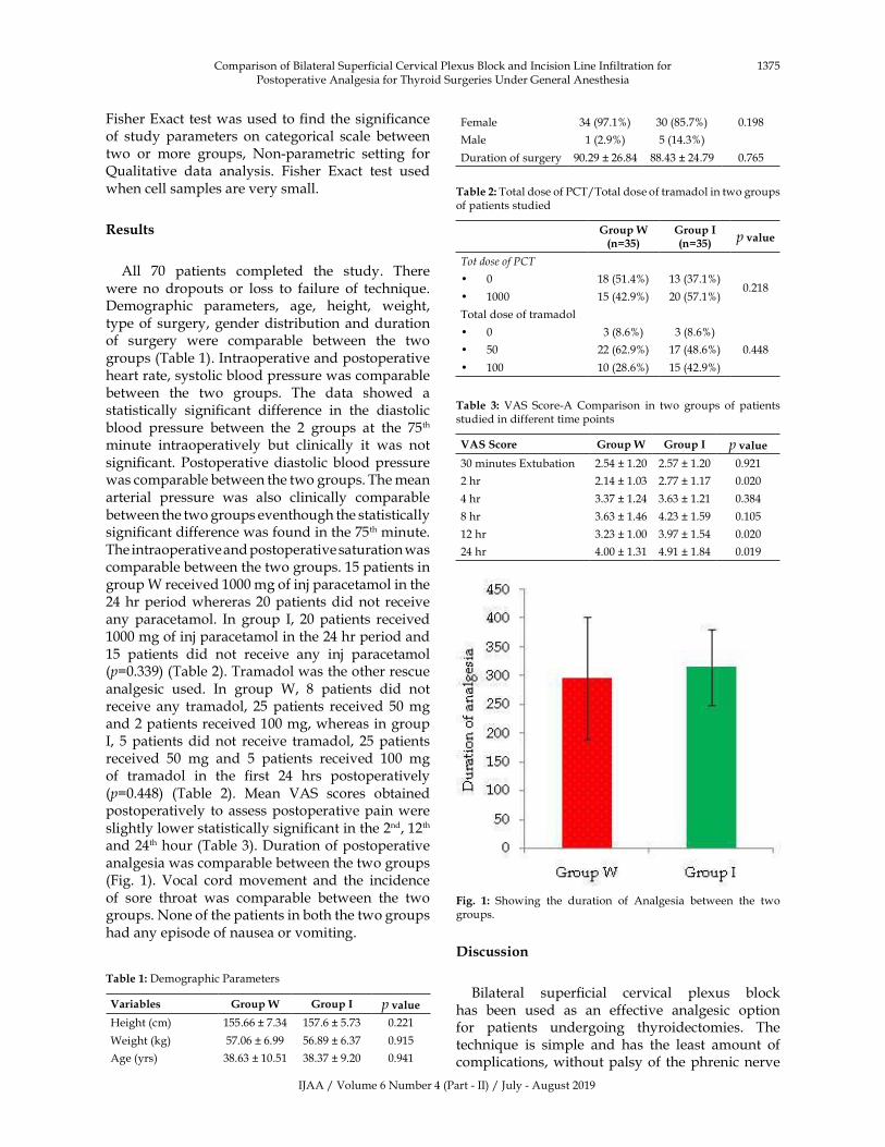
IJAA�/�Volume�6�Number�4�(Part�-�II)�/�July�-�August�2019
1375
Fisher�Exact�test�was�used�to���nd�the�signi��cance�of� study� parameters� on� categorical� scale� between�two� or� more� groups,� Non-parametric� setting� for��ualitative� data� analysis.� Fisher� Exact� test� used�when�cell�samples�are�very�small.
�esults
All� 70� patients� completed� the� study.� There�were� no� dropouts� or� loss� to� failure� of� techni�ue.�Demographic� parameters,� age,� height,� weight,�type�of� surgery,�gender�distribution�and�duration�of� surgery� were� comparable� between� the� two�groups�(Table�1).�Intraoperative�and�postoperative�heart�rate,�systolic�blood�pressure�was�comparable�between� the� two� groups.� The� data� showed� a�statistically� signi��cant� difference� in� the� diastolic�blood� pressure� between� the� 2� groups� at� the� 75th�minute� intraoperatively� but� clinically� it� was� not�signi��cant.� Postoperative�diastolic�blood�pressure�was�comparable�between�the�two�groups.�The�mean�arterial� pressure� was� also� clinically� comparable�between�the�two�groups�eventhough�the�statistically�signi��cant�difference�was�found�in�the�75th�minute.�The�intraoperative�and�postoperative�saturation�was�comparable�between�the�two�groups.�15�patients�in�group�W�received�1000�mg�of�inj�paracetamol�in�the�24�hr�period�whereras�20�patients�did�not� receive�any� paracetamol.� In� group� I,� 20� patients� received�1000�mg�of�inj�paracetamol�in�the�24�hr�period�and�15� patients� did� not� receive� any� inj� paracetamol�(p�0.339)�(Table�2).�Tramadol�was�the�other�rescue�analgesic� used.� In� group� W,� 8� patients� did� not�receive� any� tramadol,� 25� patients� received� 50�mg�and�2�patients�received�100�mg,�whereas�in�group�I,� 5�patients�did�not� receive� tramadol,� 25�patients�received� 50� mg� and� 5� patients� received� 100� mg�of� tramadol� in� the� ��rst� 24� hrs� postoperatively�(p�0.448)� (Table� 2).� Mean� VAS� scores� obtained�postoperatively� to� assess�postoperative�pain�were�slightly�lower�statistically�signi��cant�in�the�2nd,�12th
and�24th�hour�(Table�3).�Duration�of�postoperative�analgesia�was�comparable�between�the�two�groups�(Fig.� 1).� Vocal� cord�movement� and� the� incidence�of� sore� throat� was� comparable� between� the� two�groups.�None�of�the�patients�in�both�the�two�groups�had�any�episode�of�nausea�or�vomiting.
Table��:�Demographic�Parameters
Variables �roup�W �roup�I p��alue�eight�(cm) 155.66���7.34 157.6���5.73 0.221
Weight�(kg) 57.06���6.99 56.89���6.37 0.915
Age�(yrs) 38.63���10.51 38.37���9.20 0.941
Female 34�(97.1�) 30�(85.7�) 0.198
Male 1�(2.9�) 5�(14.3�)
Duration�of�surgery 90.29���26.84 88.43���24.79 0.765
Table��:�Total�dose�of�PCT/Total�dose�of�tramadol�in�two�groups�of�patients�studied
�roup�W�n����
�roup�I�n����
p��alue
Tot�dose�of��CT
�� 0 18�(51.4�) 13�(37.1�)0.218
�� 1000 15�(42.9�) 20�(57.1�)
Total�dose�of�tramadol
�� 0 3�(8.6�) 3�(8.6�)
0.448�� 50 22�(62.9�) 17�(48.6�)
�� 100 10�(28.6�) 15�(42.9�)
Table� �:� VAS� Score-A�Comparison� in� two� groups� of� patients�studied�in�different�time�points
VAS�Score �roup�W �roup�I p��alue
30�minutes�Extubation 2.54���1.20 2.57���1.20 0.921
2�hr 2.14���1.03 2.77���1.17 0.020
4�hr 3.37���1.24 3.63���1.21 0.384
8�hr 3.63���1.46 4.23���1.59 0.105
12�hr 3.23���1.00 3.97���1.54 0.020
24�hr 4.00���1.31 4.91���1.84 0.019
Fig�� �:� Showing� the� duration� of� Analgesia� between� the� two�groups.
�iscussion
�ilateral� super��cial� cervical� plexus� block�has� been� used� as� an� effective� analgesic� option�for� patients� undergoing� thyroidectomies.� The�techni�ue� is� simple� and� has� the� least� amount� of�complications,�without�palsy�of� the�phrenic�nerve�
Comparison�of��ilateral�Superficial�Cervical�Plexus��lock�and�Incision�Line�Infiltration�for�Postoperative�Analgesia�for�Thyroid�Surgeries��nder�General�Anesthesia
IJAA�/�Volume�6�Number�4�(Part�-�II)�/�July�-�August�2019
1376 Indian�Journal�of�Anesthesia�and�Analgesia
in�comparison�to�deep�cervical�plexus�block.�There�have� been� various� studies� which� have� compared�the� use� of� �SCP�� and� other� analgesics� and� have�found� the� reduced� re�uirement� of� opioids.� The�post-thyroidectomy�pain�seems�to�last�for�a�period�of� 24� hours� after� which� duration� most� patients�are�able�to�tolerate�the�pain.�In�a�study�conducted�by�Gozal� et� al.5� the�mean� pain� score�was� 6.9� and�there� was� re�uirement� of� morphine� in� the� ��rst�24�hrs.� Traditionally�post-thyroidectomy�pain�has�been� treated� with� opioids� or� non� steroidal� anti�in��ammatory�drugs.�Supplementation�with�wound�in��ltration�or��SCP��has�been�found�to�reduce�the�re�uirement� of� opioids� substantially.� It� has� been�hypothesised� that� this� is� mainly� because� of� the�large�super��cial�component�of�the�surgery.�In�our�study�we� have� tried� to� compare� the� effectiveness�of�incision�line�in��ltration�and��SCP��as�analgesic�modalities.� Since� both� these� techni�ues� involve�super��cial� in��ltration� of� the� local� anaesthetic� we�have�assumed�that�there�is�not�much�of�a�technical�bias.�Also,�we�have�used�the�same�cumulative�dose�of�local�anaesthetic�in�our�study.�Shih�et�al.�in�their�study�found�that�the�median�time�to���rst�analgesic�re�uirement�was�360.8�minutes,�but�they�had�used�0.5��bupivacaine� for� administration� of� the� block.�Their� study� was� under� powered� to� detect� any�difference�3�in�the�incidence�of�PONV�and�did�not�show� any� increase� in� intraoperative� anaesthetic�re�uirements.� In�our�study�we�have�administered�only�a�single�dose�of�opioid�analgesic�at�the�time�of�induction�of�anesthesia.
Conclusion
A� study� conducted� by� Aysenur� et� al.8� showed�that� the� duration� of� analgesia� was� longer� in� the�wound� in��ltration� group� in� comparison� to� the�group� which� received� �SCP�� but� we� found� no�such� difference.� They� even� found� a� statistically�signi��cant� difference� in� the� re�uirement� of�morphine�unlike�our�study.�They�added�adrenaline�1:2,00,000�in�order�to�prolong�the�duration�of�action�of� 0.25�� bupivacaine� whereas� we� administered�only�0.25��bupivacaine.
Eti�et�al.9�have�found� that� there� is�no�signi��cant�difference� in� the� duration� of� analgesia� or� opioid�re�uirement�with�the�two�techni�ues.
In� conclusion,� we� found� that� both� �SCP�� and�incision� line� in��ltration� are� effective�methods� for�
providing�analgesia�for�thyroid�surgeries.�There�is�
no�difference�between�the�re�uirement�of�analgesics�
or�postoperative�pain�scores�over�a�24�hr�period.
�eferences
1.� Porter� S,� Schwartz� A,� DeMaria,� et� al.� Thyroid,�
Parathyroid� and� Parotid� Surgery.� In:� Levine�
AI,� Govindraj� S,� DeMaria,� Jr� S,� editors.�
Anasethesiology� and� Otolaryngology.� Springer.�
2013.
2.� Mukhopadhyay� S,� Niyogi� M,� Dutta� M,� et� al.�
�ilateral� superficial� cervical� plexus� block� with�
or� without� low-dose� intravenous� ketamine�
analgesia:� effective,� simple,� safe,� and� cheap�
alternative� to� conventional� general� anesthesia�
for� selected� neck� surgeries.� Local� and� regional�
anesthesia.�2012;5:1–7.
3.� Andrieu�G,�Amrouni��,�Robin�E�et�al.�Analgesic�
efficacy� of� bilateral� superficial� cervical� plexus�
block�administered�before�thyroid�surgery�under�
general�anesthesia.��r�J�Anaesth.�2007;99:561–66.
4.� Sonner� JM,� �ynson� JM,� Clark� O,� et� al.� Nausea�
and�vomiting�following�thyroid�and�parathyroid�
surgery.�J�Clin�Anesth.�1997;9:398–402.
5.� Gozal��,�Shapira�SC,�Gozal�D,�et�al.��upivacaine�
wound� infiltration� in� thyroid� surgery� reduces�
postoperative� pain� and� opioid� demand.� Acta�
Anaesthesiol�Scand.�1994;38:813–5.
6.� �agul� A,� Taha� R,� Metcalfe� MS,� et� al.� Pre-
incision� infiltration� of� local� anesthetic� reduces�
postoperative�pain�with�no�effects�on�bruising�and�
wound� cosmesis� after� thyroid� surgery.� Thyroid.�
2005�Nov;15:1245–8.
7.� Shih� ML,� Duh� ��,� �sieh� C�,� et� al.� �ilateral�
superficial� cervical� plexus� block� combined�with�
general� anesthesia� administered� in� thyroid�
operations.�World�J�Surg.�2010�Oct;34:2338–43.
8.� Aysenur� D,� Ali� A,� Mine� C,� et� al.� Which�
method� is� the�most� effective� for� reducing� post-
thyroidectomy� pain:bilateral� superficial� cervical�
block� or� wound� infiltration�� A� prospective�
randomized,� double-blind� study.� Acta� Medica�
Mediterranea�2014;30:�1159.
9.� Eti��,�Irmak�P,�Gulluoglu��M,�et�al.�Does��ilateral�
Superficial� Cervical� Plexus� �lock� Decrease�
Analgesic� Re�uirement� After� Thyroid� Surgery��
Anesth�Analg.�2006;102:1174–6.
��Red�Flower�Publication�Pvt.�Ltd.�
�Original�Research�ArticleIndian�Journal�of�Anesthesia�and�Analgesia�
2019;6(4)�(Part�-�II):�1377-1382DOI:�http://dx.doi.org/10.21088/ijaa.2349.8471.6419.45
Crystalloid�Preload�Versus�Crystalloid�Co�load��uring�Electi�e�Caesarean�Section��nder�Spinal�Anesthesia
Na�eed�Abrar���Ahmedi�Fathima���Waseem�An�um�
1,2Assistant�Professor,�Department�of�Anaesthesiology,�3Assistant�Professor,�Department�of�Community�Medicine,�Sri�Devaraj��rs�Medical�College�(SD�MC),�Sri�Devaraj��rs�Academy�of��igher�Education�and�Research,�Tamaka��olar,��arnataka�536101,�India.
Corresponding�Author:�Ahmedi�Fathima,�Assistant�Professor,�Department�of�Anaesthesiology,�Sri�Devaraj��rs�Medical�College�(SD�MC),�Sri�Devaraj��rs�Academy�of��igher�Education�and�Research,�Tamaka��olar,��arnataka�536101,�India.E�mail:�[email protected]
�ecei�ed�on�23.04.2019,�Accepted�on�08.06.2019
Abstract
Background�and�Aim:�Spinal�anesthesia�is�a�preferred�techni�ue�of�anesthesia�for�pregnant�women�undergoing�caesarean� section.� The�major�disadvantage�with� this� techni�ue� is�maternal�hypotension�which� carries� the�greatest�risk�to�mother�and�foetus.�This�study�was�designed�to�analyse�various�advantages�and�disadvantages�associated�with�crystalloid�preloading�and�co-loading�during�spinal�anesthesia.�Methods:��undred�parturients�aged�20�to�40�years�of�physical�status�ASA�I�and�II�undergoing�elective�caesarean�section�under�SA��were�divided�into�two�groups�with�50�patients�in�each�group.�In�Crystalloid�Preload�group,�15�ml/kg�of�Ringer�lactate�(RL)�was�preloaded�20�minutes�before�spinal�anesthesia.�In�Co-load�group,�15�ml/kg�of�RL�was�co-loaded�20�minutes� just� after� lumbar�puncture.�Vital�parameters�were�noted�before� and�after�giving� spinal�anesthesia.�Results:�The�occurence�of�hypotension�(S�P���100�mm�g)�in�Group�P�is�30��and�20��in�Group�C�which�is�statistically� insignificant� (p���0.35).�The� incidence�of�nausea� is�8����6��in�group�P���group�C�respectively�(p���0.69).�The�incidence�of�vomiting�is�2��in�group�P���4��in�group�C�(p���0.55).�Mephentermine�was�used�in�19�patients�in�Group�P���13�patients�in�Group�C�which�is�statistically�insignificant.�Conclusion:�oth�preloading�and�co-loading�with�RL�have�similar�effect�on�occurence�of�hypotension�in�pregnant�women�receiving�spinal�anesthesia.�Precious�time�need�not�be�wasted�in�preloading.�Periodic�measurement�of��P�in�parturients�for�early�detection�of�hypotension�and�administration�of�vasopressors�for�maintaining��P�close�to�baseline�can�ensure�better�outcome.
�ey�ords:�Subarachnoid�block;��ypotension;�Crystalloid�preloading;�Crystalloid�co-loading.
�o��to�cite�this�article:
Naveed�Abrar,�Ahmedi�Fathima,�Waseem�Anjum.�Crystalloid�Preload�Versus�Crystalloid�Co-load�During�Elective�Caesarean�Section��nder�Spinal�Anesthesia.�Indian�J�Anesth�Analg.�2019;6(4):1377-82.
Introduction
Spinal�anesthesia�has�been�extensively�used�for�caesarean�section�because�of�greater�maternal�safety�and� fetal� bene��ts.1� �ut� the� major� disadvantage�
with� this� techni�ue� is�hypotension�which� is�more�
common� and� profound� in� pregnant� women� due�
to� various� causes.� This� hypotension� can� cause�
nausea,� vomiting,� cardiovascular� collapse� in� the�
mother,�along�with�fetal�hypoxia�and�acidosis�due�
IJAA�/�Volume�6�Number�4�(Part�-�II)�/�July�-�August�2019
1378 Indian�Journal�of�Anesthesia�and�Analgesia
to�placental�hypoperfusion2.�Therefore,�Prevention�of�hypotension� is� necessary� for�better� outcome�of�both�mother�and�the�foetus.
Fluid� loading�before� spinal�anesthesia�which� is�referred�to�as�preload�is�followed�for�prevention�of�hypotension.�It�is�a�common�conventional�practice�in� anesthesia.� �ut,� preloading� of� crystalloid� is�rapidly� redistributed,� and� may� induce� atrial�natriuretic�peptide�secretion,�resulting�in�peripheral�vasodilatation� followed� by� an� increased� rate� of�excretion�of�the�preloaded���uid.2
Routinely� used� methods� to� prevent� or� treat�maternal� hypotension� include� preloading� with���uids�(colloid�or�crystalloid),�wedge�placement� to�prevent�aortocaval�compression�and�administration�of�vasopressor�drugs.
A�more�logical�approach�might�be�to�administer���uid� at� the� time� that� the� local� anaesthetic� block�starts� to� act.� This� might� increase� intravascular�volume� expansion� during� vasodilatation� from�the� sympathetic� blockade� and� decrease� ��uid�redistribution�and�excretion.2
Fluid� administered� before� induction� of� spinal�anesthesia� is� referred� to� as� �preloading�� and���uid� infused� at� the� time� of� induction� is� referred�to� as� �co-loading�.� Various� studies� suggest���uid� infusion� to� be� more� effective� if� delayed�until� induction� of� spinal� anesthesia� and� rapidly�administered� subse�uently.� Three� possible� ��uid�combinations� have� been� compared� in� various�studies:� crystalloid� versus� colloid� preloading,�crystalloid� preloading� versus� crystalloid�co-loading,�and�colloid�preloading�versus�colloid�co-loading.
The� present� study� was� planned� to� analyse�various� advantages� and�disadvantages� associated�with�crystalloid�preloading�and�co-loading�during�spinal�anesthesia�and�the�possible�relative�bene��ts�of�each�of�these�methods.
���ective�of�the�study
1.� To� assess� and� compare� the� safety� and�ef��cacy� of� crystalloid� preload� and�crystalloid� co-load� for� prevention� of�maternal� hypotension� in� parturients�undergoing� elective� caesarean� section�under�spinal�anesthesia.
2.� To� assess� various� haemodynamic�parameters� like� heart� rate,� systolic� �P,�diastolic��P���mean�arterial�pressure.
3.� To� assess� side-effects� like� nausea� and�vomiting� in� patients� with� crystalloid�preload�and�co-load.
Materials�and�Methods
Study�Partici�ants�and�Recruit�ent
We� conducted� a� prospective� double� blinded�randomized� controlled� study�over� a� 2� year�period�between�December�2014�to�December�2016.��undred�patients� aged� 20-40� years� belonging� to�ASA� I� and�II� undergoing� elective� caesarean� section� were�randomly�distributed�for�the�study�into�two�groups.
Allocation�of��rou�s
�ased� on� the� study� done� by� Manu� �ose� et� al.5�
considering�signi��cant��ypotension�in�Group�1�and�Group�2�with�odds�ratio�of�3.25,�alpha�error�5��and�power�of�80��the�sample�size�was�calculated�using�OpenEpi� software�version�2.3.1�which� came� to�be�49�in�each�group�which�is�rounded�to�50�per�group.
�roup� �� (preload� group)� -� 15� ml/kg� of�Ringer� lactate� was� preloaded� 20� minutes� before�commencement�of�spinal�anesthesia.
�roup� C� (co-load� group)� -� 15� ml/kg� of� Ringer�lactate� was� co-loaded� in� 20� minutes� just� after�lumbar�puncture.
The� following�patients�were�excluded� from� the�study
1.� Emergency�surgeries.
2.� ASA�grade�III�and�IV.
3.� Severe�anaemia,�coagulation�abnormalities�and�bleeding�disorders.
4.� Morbid�obese�patients.
5.� Patients�with�multiple�pregnancies.
6.� Patients�with�other�co-morbid�conditions.
7.� Patients�with�raised�ICP.
8.� Patients�with�previous�history�of�surgeries�on�the�spine.
9.� Patients�with� spinal�deformities� and�with�history�of�backache.
10.� Patients� with� active� skin� lesions� over�lumbosacral�region.
After�a�detailed�pre-anaesthetic�examination�and�obtaining� informed� consent,� all� the� patients�were�cannulated�with�18G�IV�cannula�and�premedicated�with� Inj�Ranitidine� 50�mg��� Inj.�Metoclopramide�10� mg� IV� one� hour� before� surgery.� In� operation�theatre,�Patient�was�made�to�lie�on�operating�table�with� left� lateral� tilt.�Monitors� like�pulse�oximetry,�non-invasive�blood�pressure���electrocardiography�were�connected.��ase�line�heart�rate,�systolic�blood�pressure,�diastolic�blood�pressure�and�mean�arterial�pressure�values�were�recorded.
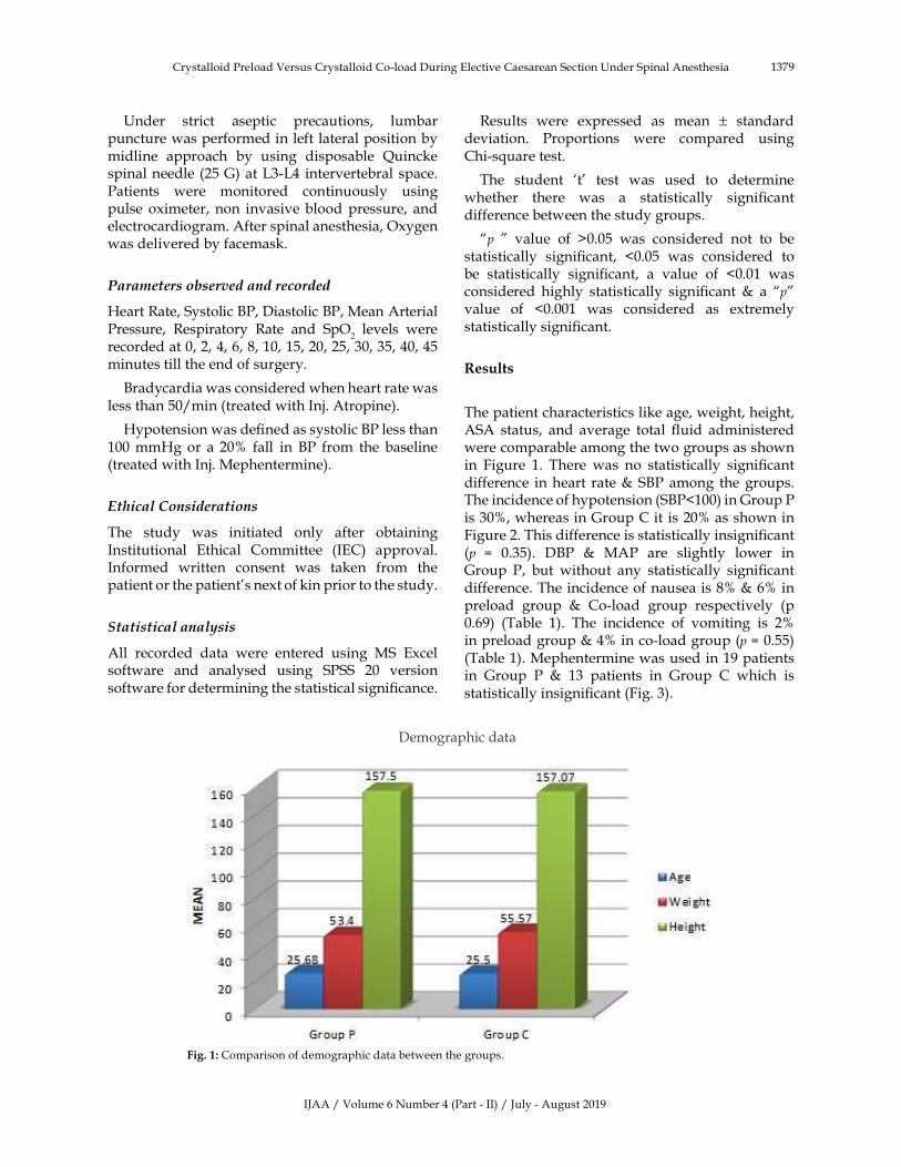
IJAA�/�Volume�6�Number�4�(Part�-�II)�/�July�-�August�2019
1379
�nder� strict� aseptic� precautions,� lumbar�puncture�was�performed�in�left�lateral�position�by�midline� approach� by� using� disposable� �uincke�spinal�needle�(25�G)�at�L3-L4� intervertebral�space.�Patients� were� monitored� continuously� using�pulse� oximeter,� non� invasive�blood�pressure,� and�electrocardiogram.�After�spinal�anesthesia,�Oxygen�was�delivered�by�facemask.
Para�eters�o�served�and�recorded
�eart�Rate,�Systolic��P,�Diastolic��P,�Mean�Arterial�Pressure,� Respiratory� Rate� and� SpO
2� levels� were�
recorded�at�0,�2,�4,�6,�8,�10,�15,�20,�25,�30,�35,�40,�45�minutes�till�the�end�of�surgery.
�radycardia�was�considered�when�heart�rate�was�less�than�50/min�(treated�with�Inj.�Atropine).
�ypotension�was�de��ned�as�systolic��P�less�than�100�mm�g�or� a� 20�� fall� in� �P� from� the� baseline�(treated�with�Inj.�Mephentermine).
Ethical�Considerations
The� study� was� initiated� only� after� obtaining�Institutional� Ethical� Committee� (IEC)� approval.�Informed� written� consent� was� taken� from� the�patient�or�the�patient�s�next�of�kin�prior�to�the�study.
Statistical�analysis
All� recorded� data� were� entered� using� MS� Excel�software� and� analysed� using� SPSS� 20� version�software�for�determining�the�statistical�signi��cance.�
Results� were� expressed� as� mean� �� standard�deviation.� Proportions� were� compared� using�Chi-s�uare�test.
The� student� �t�� test� was� used� to� determine�whether� there� was� a� statistically� signi��cant�difference�between�the�study�groups.
�p� �� value� of� �0.05� was� considered� not� to� be�statistically� signi��cant,� �0.05� was� considered� to�be� statistically� signi��cant,� a� value� of� �0.01� was�considered� highly� statistically� signi��cant� �� a� �p�value� of� �0.001� was� considered� as� extremely�statistically�signi��cant.
�esults
The�patient�characteristics�like�age,�weight,�height,�ASA� status,� and� average� total� ��uid� administered�were�comparable�among�the�two�groups�as�shown�in� Figure� 1.� There� was� no� statistically� signi��cant�difference� in�heart� rate���S�P�among� the�groups.�The�incidence�of�hypotension�(S�P�100)�in�Group�P�is�30�,�whereas�in�Group�C�it� is�20��as�shown�in�Figure�2.�This�difference�is�statistically�insigni��cant�(p� �� 0.35).� D�P� �� MAP� are� slightly� lower� in�Group� P,� but� without� any� statistically� signi��cant�difference.�The�incidence�of�nausea�is�8����6��in�preload� group� �� Co-load� group� respectively� (p�0.69)� (Table� 1).� The� incidence� of� vomiting� is� 2��in�preload�group���4��in�co-load�group�(p���0.55)�(Table�1).�Mephentermine�was�used�in�19�patients�in� Group� P� �� 13� patients� in� Group� C� which� is�statistically�insigni��cant�(Fig.�3).
Demographic�data
Fig���:�Comparison�of�demographic�data�between�the�groups.
Crystalloid�Preload�Versus�Crystalloid�Co-load�During�Elective�Caesarean�Section��nder�Spinal�Anesthesia

IJAA�/�Volume�6�Number�4�(Part�-�II)�/�July�-�August�2019
1380 Indian�Journal�of�Anesthesia�and�Analgesia
S�P�100�mm�g
Fig���:�Number�of�patients�with�S�P�100�mm�g�in�the�groups.
Fig���:�Comparison�of�usage�of�Mephentermine�in�both�the�groups.
Table��:�Incidence�of�Nausea�and�Vomiting.
�roupNA�SEA Vomitting
Total�es No �es No
Preload 4�(8�) 46�(92�) 01�(2�) 49�(98�) 50
Coload 03�(6�) 47�(93�) 02�(4�) 48�(96�) 50
Total 7 93 03 97 100
�iscussion
Spinal� anesthesia� is� a� standard� techni�ue� for�pregnant� women� undergoing� elective� caesarean�section� but� hypotension� remains� the� main�complication�with�this�techni�ue.�This�hypotension�has� harmful� effects� on� both� mother� and� foetus,�leading� to� maternal� nausea,� vomiting� and�fetal�hypoxia.
�ence� various� measures� have� been� utilized� to�decrease� the� incidence� of� hypotension� following�spinal� anesthesia.� One� such� measure� is� ��uid�preloading,� but� it� is� associated� with� several�drawbacks.� An� alternate� measure� is� co-loading�during� spinal� anesthesia.� Although,� experience�with�this�approach�is�limited.
Studies� done� by� Parmar� et� al.� found� that�Co-loading� with� 20� ml/kg� of� ringer� lactate� is� as�
IJAA�/�Volume�6�Number�4�(Part�-�II)�/�July�-�August�2019
1381
effective� as� preloading� with� same� volume� over�20�minutes�in�lower�limb�surgeries�and�believes�that�it�is�not�re�uired�to�spend�time�to�deliver�preload�and�delay�surgery�for�the�prevention�of�SA�induced�hypotension.1
Dyer�et�al.�conducted�a�study�and�concluded�that�an� e�uivalent� volume� of� crystalloid� administered�rapidly,�immediately�after�the�performance�of�spinal�anesthesia�for�elective�caesarean�section,�is�associated�with� a� lower� pre-delivery� re�uirement� for� the�vasopressor�ephedrine�than�a�traditional�preload2.
�han�et�al.�conducted�a�study�and�concluded�that�there�is�signi��cantly�lower�incidence�of�post-spinal�hypotension� found� in�co-load�group�than�preload�group�and�parturient�in�the�co-load�group�re�uired�signi��cantly� less� vasopressor� doses� than� the�pre-load�group.3
Aparna�Williams� et� al.� conducted� a� study� and�concluded�that�both�preloading�and�co-loading�with�15ml/kg�of�RL�solution�are�ineffective�in�prevention�of�spinal�induced�maternal�hypotension.4
�ose� et� al.� conducted� a� study� concluding� that�co-loading�with�15�ml/kg�of�Ringer�lactate�solution�is�as�effective�as�preloading�with�same�volume�over�20�minutes� before� subarachnoid� block� to� prevent�hypotension�and�bradycardia.5
Singh� et� al.� conducted�a� study�where�he� found�that���uid�preloading�had�no�effect�on�the�incidence�of� hypotension� and� bradycardia� following� spinal�anesthesia.6
In� a� study� conducted� by� Tamilselvan� et� al.� he�found� that� despite� increase� in� cardiac� output�following� ��uid� preload,� particularly� with� �ES�1.0� L,� hypotension� still� occurred.� �is� data�suggested� that� increase� in� CO� after� ��uid� preload�cannot�compensate�for�reductions�in�arterial�blood�pressure�following�spinal�anesthesia.7
Rout� et� al.� conducted� a� study� and� found� that�hypotension� associated�with� spinal� anesthesia� for�caesarean� section� cannot� be� eliminated� by� ��uid�preloading�in�the�supine�wedged�patient.8
Gunusen� et� al.� conducted� a� study� where� he�found� that� the� fre�uency� of� moderate� or� severe�hypotension� was� lower� in� the� ephedrine� group�than� in� the� crystalloid� or� colloid� preload� group.�The�incidence�of�nausea�was�signi��cantly�different�between� the� crystalloid� preload� and� ephedrine�group.� �mbilical� blood� gas� analysis� and� Apgar�scores�were�similar�in�all�groups.�The�combination�of� an� ephedrine� infusion� at� 1.25� mg/min� with�a� crystalloid� co-load� was� more� effective� than���uid�preloading�with� crystalloid� or� colloid� in� the�
prevention�of�moderate�and�severe�hypotension.9
Ngan�ee�et�al.�conducted�a�study�where�he�found�that� the� combination� of� high� dose� phenylephrine�infusion� and� rapid� crystalloid� co-loading� is�effective�for�preventing�hypotension�during�spinal�anesthesia�for�caesarean�delivery.10
From�our�study�it�can�be�concluded�that�preloading���co-loading�have�similar�effects�on�the�incidence�of�hypotension�following�spinal�anesthesia�in�caesarean�section.�Therefore,�it�is�preferable�to�avoid�the�time�delay� which� occurs� in� preloading� the� parturient.�Co-loading�or�Preloading�may�not�be�very�effective�in� preventing� hypotension,� it� is� sensible� to� use�vasopressors�alongside�to�prevent�hypotension���its�adverse�effects.
Conclusion
From�our�study�it�can�be�inferred�that:
1.� �oth� preloading� and� co-loading� with�15� ml/kg� of� RL� solution,� have� similar�effect� on� the� occurence� of� hypotension� in�the� obstetric� population� receiving� spinal�anesthesia.
2.� Time� delay� due� to� preloading� can� be�avoided� as� preloading� alone� is� not� very�effective� for� the� prevention� of� maternal�hypotension.
3.� Periodic� measurement� of� the� blood�pressure�in�the�patients�(at�1�min�intervals)�for� early� detection� of� hypotension� and�administration� of� vasopressors� for�maintaining� the� maternal� blood� pressure�close� to� the� baseline� can� ensure� better�outcome.
Con��ict�of�Interest:�None
Financial�Su��ort�and�S�onsorshi�:�Nil
Ac�no�led�e�ent:�Nil
�eferences
1.� Dyer�RA,�Farina��,� Joubert� IA,� et� al.�Crystalloid�preload� versus� rapid� crystalloid� administration�after� induction�of� spinal� anesthesia� (co-load)� for�elective�caesarean�section.�Anaesth�Intensive�Care.2004;32:351–7.
2.� Parmar�S�,�Sheikh�ANA,�Shalu�PS.�A�comparative�study� of� preloading� versus� co-loading� of�crystalloid� to� prevent� spinal� anesthesia� induced�hypotension.�Journal�of�Evolution�of�Medical�and�Dental�Sciences.�2012;1(5):746.
Crystalloid�Preload�Versus�Crystalloid�Co-load�During�Elective�Caesarean�Section��nder�Spinal�Anesthesia
IJAA�/�Volume�6�Number�4�(Part�-�II)�/�July�-�August�2019
1382 Indian�Journal�of�Anesthesia�and�Analgesia
3.� �han� M,� Wa�ar-ul-Nisai,� Faroo�i� A,� et� al.�Crystalloid� Co-Load:� A� better� option� than�Crystalloid�Pre-Load�for�Prevention�of�Post�spinal��ypotension� in� Elective� Caesarean� Section.� The�Internet�Journal�of�Anaesthesiology.�2013;32:1.
4.� Williams�A,�Jacob�J,�Verghese�M,�et�al.�Crystalloid�preload�versus�crystalloid�co-load�for�parturients�undergoing� caesarean� section� under� spinal�anesthesia.�J�Obstet�Anaesth�Crit�Care.�2012;2:10–5.
5.� �ose�M,��ini�G,�and��rishna��M�et�al.�Comparison�of� crystalloid� preloading� versus� crystalloid� co-loading�to�prevent�hypotension�and�bradycardia�following� spinal� anesthesia.� J�Anaesth� Clin�Pharmacol.�2008;24(1):53–56.�
6.� Singh� J,� Ranjit� S,� Shrestha� S,� et� al.� Effect� of�preloading� on� haemodynamics� of� the� patient�undergoing� surgery� under� spinal� anesthesia.��athmandu� �niversity� Medical� Journal.�2010;8(30):216–221.
7.� Tamilselvan�P,�Fernando�R,��ray�J,�et�al.�The�effects�of� crystalloid� and� colloid� preload� on� cardiac�
output� in� the� parturient� undergoing� planned�Caesarean� Delivery� under� Spinal� Anesthesia:� a�Randomized� trial.�Anesth�Analg.� 2009;109:1916–21.
8.� Rout�CC,�Rocke�DA,�Levin�J,�et�al.�A�Re-evaluation�of�the�role�of�crystalloid�preload�in�the�prevention�of�hypotension�associated�with�spinal�anesthesia�for� elective� caesarean� section.� �r.� J.� Anaesth�1993;79:262–9.
9.� Gunusen� I,��araman�S,�Ertugrul�V,� et� al.�Effects�of�fluid�preload�(crystalloid�or�colloid)�compared�with�crystalloid�co-load�plus�ephedrine� infusion�on� hypotension� and� neonatal� outcome� during�spinal�anesthesia�for�caesarean�delivery.�Anaesth�Intensive�Care.�2010;38:647–53.
10.� Ngan�ee� WD,� �haw� �S,� Ng� FF.� Prevention�of� hypotension� during� spinal� anesthesia� for�caesarean� delivery:� an� effective� techni�ue�using� combination� phenylephrine� infusion�and� crystalloid� co-hydration.� Anaesthesiology.2005;103:744–50.
��Red�Flower�Publication�Pvt.�Ltd.�
�Original�Research�ArticleIndian�Journal�of�Anesthesia�and�Analgesia�
2019;6(4)�(Part�-�II):�1383-1386DOI:�http://dx.doi.org/10.21088/ijaa.2349.8471.6419.46
Intraarticular� O�one� Therapy� for� �nee� Osteoarthritis:� A� Single� Centre�E�perience
Neha��oyal
Associate� Professor,� Department� of� Anaesthesiology,� American� International� Institute� of� Medical� Sciences,� �edwas,� �daipur,�Rajasthan�313001,�India.
Corresponding� Author:�Neha��oyal,� Associate� Professor,� Department� of� Anaesthesiology,� American� International� Institute� of�Medical�Sciences,��edwas,��daipur,�Rajasthan�313001,�India.
E�mail:�[email protected]
�ecei�ed�on�06.05.2019,�Accepted�on�22.05.2019
Abstract
Introduction:�Osteoarthritis�of�the�knee�joint�is�a�widely�prevalent�problem�and�leads�to�decrease�in�physical�function� through�pain� and� reduced� range� of�motion.� Since� there� is� no� cure� for� the�disease,� the�main� aim�of�treatment�if�to�reduce�pain�and�preserve�function.�While�early�osteoarthritis�is�treated�with�exercise�and�lifestyle�modifications�only,�measures�like�intra-articular�ozone�administration�and�platelet�rich�plasma�therapy�reduce�pain,�restore�function�and�enable�patients�to�exercise�more�effectively.�We�studied�the�effectiveness�of�ozone�administration�in�patients�with�knee�osteoarthritis.�Materials�and�Methods:�Thirty�patients�with��ellgren-Lawrence�grade�2-3�osteoarthritis�of�both�knees�were�recruited�and�administered�intra-articular�ozone.�Pain�and�patient�global�assessment�scores�were�recorded�on�the�visual�analogue�scale�at�baseline�and�at�6�months.Results:�All�patients�experienced�improvement�with�an�improvement�of�mean�pain�VAS�from�6.9�at�baseline�to�2.9�at�6�months.�Patient�global�assessment�also�improved�from�5.1�to�1.3.�Conclusion:�Intra-articular�ozone�administration�is�effective�in�reducing�pain�in�patients�with�osteoarthritis�of�the�knees.
�ey�ords:��nee;�Osteoarthritis;�Ozone;�Pain.
�o��to�cite�this�article:
Neha� Goyal.� Intraarticular� Ozone� Therapy� for� �nee� Osteoarthritis:� A� Single� Centre� Experience.� Indian� J� Anesth� Analg.�2019;6(4):1383-1386.
Introduction
Osteoarthritis� (OA)� of� the� knee� is� a� commonly�
prevalent� condition� that� causes� both� pain� and�functional� limitation.� Long� known� to� be� caused�
by� mechanical� stress� only,� chemical� factors� like�oxidative� stress� have� come� to� be� recognised�
as� contributing� factors� in� the� causation� of� OA.�Female�gender,�advancing�age�and�obesity�are�the�
fre�uently�encountered�associates�of�OA�of�knees.1�
The� diagnosis� of� OA� remains� largely� clinical.2,3�The�common�complaints�by� the�patients�are�pain,�stiffness,� swelling� and� dif��culty� in� using� stairs�and� getting� up� from� sitting� position.� Tenderness�and� joint� crepitus�may� be� appreciated� on� clinical�examination.� Signi��cant� swelling�may� be� seen� in�wet� OA� where� there� is� effusion� of� the� joint� and�limb� deformity� may� be� seen� in� advanced� cases.�A� thorough� history� and� clinical� examination�are� usually� suf��cient� to� ascertain� the� diagnosis.�A�further�roentgenogram�helps�to�stage�the�OA�of�
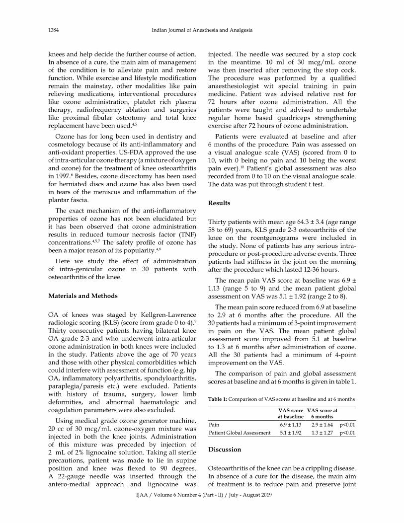
IJAA�/�Volume�6�Number�4�(Part�-�II)�/�July�-�August�2019
1384 Indian�Journal�of�Anesthesia�and�Analgesia
knees�and�help�decide�the�further�course�of�action.�In�absence�of�a�cure,�the�main�aim�of�management�of� the� condition� is� to� alleviate� pain� and� restore�function.�While�exercise�and�lifestyle�modi��cation�remain� the� mainstay,� other� modalities� like� pain�relieving� medications,� interventional� procedures�like� ozone� administration,� platelet� rich� plasma�therapy,� radiofre�uency� ablation� and� surgeries�like� proximal� ��bular� osteotomy� and� total� knee�replacement�have�been�used.4,5
Ozone�has� for� long�been�used� in�dentistry� and�cosmetology�because�of� its�anti-in��ammatory�and�anti-oxidant�properties.��S-FDA�approved�the�use�of�intra-articular�ozone�therapy�(a�mixture�of�oxygen�and�ozone)�for�the�treatment�of�knee�osteoarthritis�in�1997.6��esides,�ozone�discectomy�has�been�used�for�herniated�discs� and�ozone�has�also�been�used�in� tears� of� the�meniscus� and� in��ammation� of� the�plantar�fascia.
The� exact�mechanism� of� the� anti-in��ammatory�properties� of� ozone� has� not� been� elucidated� but�it� has� been� observed� that� ozone� administration�results� in� reduced� tumour� necrosis� factor� (TNF)�concentrations.4,5,7� The� safety� pro��le� of� ozone� has�been�a�major�reason�of�its�popularity.4,8
�ere� we� study� the� effect� of� administration�of� intra-genicular� ozone� in� 30� patients� with�osteoarthritis�of�the�knee.
Materials�and�Methods
OA� of� knees� was� staged� by� �ellgren-Lawrence�radiologic�scoring�(�LS)�(score�from�grade�0�to�4).9�Thirty� consecutive� patients� having� bilateral� knee�OA� grade� 2-3� and�who�underwent� intra-articular�ozone�administration�in�both�knees�were�included�in� the� study.� Patients� above� the� age� of� 70� years�and�those�with�other�physical�comorbidities�which�could�interfere�with�assessment�of�function�(e.g.�hip�OA,�in��ammatory�polyarthritis,�spondyloarthritis,�paraplegia/paresis� etc.)� were� excluded.� Patients�with� history� of� trauma,� surgery,� lower� limb�deformities,� and� abnormal� haematologic� and�coagulation�parameters�were�also�excluded.
�sing�medical�grade�ozone�generator�machine,�20� cc� of� 30� mcg/mL� ozone-oxygen�mixture�was�injected� in� both� the� knee� joints.� Administration�of� this� mixture� was� preceded� by� injection� of�2��mL�of�2��lignocaine�solution.�Taking�all�sterile�precautions,� patient� was� made� to� lie� in� supine�position� and� knee� was� ��exed� to� 90� degrees.�A� 22-gauge� needle� was� inserted� through� the�antero-medial� approach� and� lignocaine� was�
injected.� The� needle�was� secured� by� a� stop� cock�in� the� meantime.� 10� ml� of� 30� mcg/mL� ozone�was� then� inserted� after� removing� the� stop� cock.�The� procedure� was� performed� by� a� �uali��ed�anaesthesiologist� wit� special� training� in� pain�medicine.� Patient� was� advised� relative� rest� for�72� hours� after� ozone� administration.� All� the�patients� were� taught� and� advised� to� undertake�regular� home� based� �uadriceps� strengthening�exercise�after�72�hours�of�ozone�administration.
Patients� were� evaluated� at� baseline� and� after�6�months�of� the�procedure.�Pain�was�assessed�on�a� visual� analogue� scale� (VAS)� (scored� from� 0� to�10,�with� 0� being� no� pain� and� 10� being� the�worst�pain� ever).10� Patient�s� global� assessment�was� also�recorded�from�0�to�10�on�the�visual�analogue�scale.�The�data�was�put�through�student�t�test.
�esults
Thirty�patients�with�mean�age�64.3���3.4�(age�range�58�to�69)�years,��LS�grade�2-3�osteoarthritis�of�the�knee� on� the� roentgenograms� were� included� in�the�study.�None�of�patients�has�any�serious� intra-procedure�or�post-procedure�adverse�events.�Three�patients� had� stiffness� in� the� joint� on� the�morning�after�the�procedure�which�lasted�12-36�hours.
The�mean�pain�VAS�score�at�baseline�was�6.9���1.13� (range� 5� to� 9)� and� the� mean� patient� global�assessment�on�VAS�was�5.1���1.92�(range�2�to�8).
The�mean�pain�score�reduced�from�6.9�at�baseline�to� 2.9� at� 6� months� after� the� procedure.� All� the�30�patients�had�a�minimum�of�3-point�improvement�in� pain� on� the� VAS.� The� mean� patient� global�assessment� score� improved� from� 5.1� at� baseline�to� 1.3� at� 6�months� after� administration� of� ozone.�All� the� 30� patients� had� a� minimum� of� 4-point�improvement�on�the�VAS.
The� comparison�of� pain� and� global� assessment�scores�at�baseline�and�at�6�months�is�given�in�table�1.
Table��:�Comparison�of�VAS�scores�at�baseline�and�at�6�months
VAS�score�at�baseline
VAS�score�at���months
Pain 6.9���1.13 2.9���1.64 p�0.01
Patient�Global�Assessment 5.1���1.92 1.3���1.27 p�0.01
�iscussion
Osteoarthritis�of�the�knee�can�be�a�crippling�disease.�In�absence�of�a�cure�for� the�disease,� the�main�aim�of� treatment� is� to� reduce� pain� and� preserve� joint�
IJAA�/�Volume�6�Number�4�(Part�-�II)�/�July�-�August�2019
1385
function.� Ozone� injection� into� the� joint� has� been�used� for� the� treatment� of� knee� OA.� Ozone� leads�to�production�of�reactive�oxygen�species�and�lipid�oxidation�products.8,11�It�thus�reduces�in��ammation�and� improves� function� through� repair� and�vascularization.� There� is� downregulation� of�proteolytic� enzymes� and� proin��ammatory.�Articular� cartilage� and� matrix� may� be� formed�through�secondary�proliferation�of���broblasts�and�chondrocytes.4
The� primary� objective� of� this� study� was�to� assess� the� efficacy� of� intra-articular� ozone�administration� in� alleviating� pain� in� patients�with�osteoarthritis�of� the�knee.�Multiple� studies�have�documented� the� beneficial� effects� of� intra-articular� ozone� administration� in� patients� with�osteoarthritis� of� the� knees� over� a� short� term.12�This� study� assesses� the� effect� of� this� procedure�on�knee�pain�over�an�intermediate�period.�While�most� of� the� studies� included� administration� of�ozone� unilaterally,� we� have� included� patients�who� underwent� bilateral� intra-articular� ozone�administration.
The� intermediate� to� longer� term� bene��ts� of�ozone� administration� may� be� derived� from� post�ozone�administration�downregulation�of�cytokines�which�are� responsible� for� sustained� in��ammation�and�progression�of� structural�damage�as�noted� in�certain�studies.4
One� study� demonstrated� intra-articular� ozone�to� be� signi��cantly� superior� to� intra-articular�methylprednisolone� in� patients� with� knee�OA.11� Jesus� et� al.� and� �ashemi� et� al.� compared�intra-articular� ozone� with� sterile� air� and� found�ozone� to� be� effective� at� 4� months.13,14� Ozone�was�also� found� to� be� effective� in� post-arthroscopic�surgery�patients�of�knee�OA.15
In� our� study,� patients� had� signi��cant�improvement� at� 6� months� after� intra-articular�ozone� administration.� Regular� �uadriceps�strengthening� exercise� is� a� contributory� factor�for� maintaining� function� and� is� likely� to� have�accounted� for� better� patient� global� assessment�scores�at�6�months.
Conclusion
Intra-articular�ozone�administration�is�an�effective�tool� for� reducing� pain� and� improving� function�in� patients� with� knee� osteoarthritis.� Regular��uadriceps� strengthening� exercise� after� the�procedure�can�prolong�the�effect�of�ozone.
�eferences
1.� Richmond� J,� �unter� D,� Irrgang� J,� et� al.American� Academy� of� Orthopaedic� Surgeons�clinical� practice� guideline� on� the� treatment� of�osteoarthritis�(OA)�of�the�knee.�J��one�Joint�Surg�Am.�2010;92:990–3.
2.� �usija� L,� �ridgett� L,� Williams� SRM,� et� al.�Osteoarthritis.� �est� Pract� Res� Clin� Rheumatol.�2010;24:757–68.
3.� Tehrani-�anihashemi� A,� Davatchi� F,� Jamshidi�A-R,� et� al.� Prevalence� of� osteoarthritis� in� rural�areas�of�Iran:�a�W�O-ILAR�COPCORD�study.�Int�J�Rheum�Dis.�2014;17:384–8.
4.� �occi� VA.� Scientific� and� medical� aspects� of�ozone� therapy.� State� of� the� art.� Arch�Med� Res.�2006;37:425–35.
5.� Manoto� SL,� Maepa� MJ,� Motaung� S�.� Medical�ozone�therapy�as�a�potential� treatment�modality�for�regeneration�of�damaged�articular�cartilage�in�osteoarthritis.�Saudi�J��iol�Sci.�2018;25:672–9.
6.� Adams�ME,�Atkinson�M�,�Lussier�AJ,�et�al.�The�role� of�viscosupplementation�with�hylan�G-F�20�(Synvisc)�in�the�treatment�of�osteoarthritis�of�the�knee:� a� Canadian� multicenter� trial� comparing�hylan� G-F� 20� alone,� hylan� G-F� 20� with� non-steroidal� anti-inflammatory� drugs� (NSAIDs)�and� NSAIDs� alone.� Osteoarthritis� Cartilage.�1995;3:213–25.
7.� Giombini� A,� Menotti� F,� Di� Cesare� A,� et� al.�Comparison� between� intrarticular� injection�of� hyaluronic� acid,� oxygen� ozone,� and� the�combination� of� both� in� the� treatment� of� knee�osteoarthrosis.� J� �iolRegul�omeost� Agents.�2016;30:621–5.
8.� Alberto� Alexandre� E�,� Andrea� Alexandre� EI.�Disc� �erniation� and� �nee� Arthritis� as� Chronic�Oxidative� Stress�Diseases:� The�Therapeutic�Role�of�Oxygen�Ozone�Therapy.� J�Arthritis��Internet�.�2015��cited�2019�May�6�;04.�Available�from:�http://www.omicsgroup.org/journals/disc-herniation-and-knee-arthritis-as-chronic-oxidative-stress-diseases-the-therapeutic-role-of-oxygen-ozone-therapy-2167-7921-1000161.php�aid�58544.
9.� �ellgren� J�,� Lawrence� JS.� Radiological�assessment� of� osteo-arthrosis.� Ann� Rheum� Dis.�1957;16:494–502.
10.� Nadrian��,�Moghimi�N,�Nadrian�E,�et�al.�Validity�and�reliability�of�the�Persian�versions�of�WOMAC�Osteoarthritis�Index�and�Le�uesneAlgofunctional�Index.�Clin�Rheumatol.�2012;31:1097–102.
11.� Mishra� S�,� Pramanik� R,� Das� P,� et� al.� Role� of�intra-articular�ozone�in�osteo-arthritis�of�knee�for�functional�and�symptomatic�improvement�2011;5.
12.� Al-Jaziri� AA,� Mahmoodi� SM.� Painkilling� effect�of� ozone-oxygen� injection� on� spine� and� joint�osteoarthritis.�Saudi�Med�J.�2008;29:553–7.
Intraarticular�Ozone�Therapy�for��nee�Osteoarthritis:�A�Single�Centre�Experience
IJAA�/�Volume�6�Number�4�(Part�-�II)�/�July�-�August�2019
1386 Indian�Journal�of�Anesthesia�and�Analgesia
13.� Lopes� de� Jesus� CC,� Dos� Santos� FC,� de� Jesus�LMO�,�et�al.�Trevisani�VFM.�Comparison�between�intra-articular�ozone�and�placebo�in�the�treatment�of� knee� osteoarthritis:� A� randomized,� double-blinded,� placebo-controlled� study.� PloS� One.�2017;12:e0179185.
14.� �ashemi�M,�Jalili�P,�Mennati�S,�et�al.�The�Effects�of�Prolotherapy�With� �ypertonic� Dextrose� Versus�Prolozone� (Intraarticular� Ozone)� in� Patients�
With��nee�Osteoarthritis.�Anesthesiol�Pain�Med��Internet�.� 2015� �cited� 2019� May� 6�;5.� Available�from:� https://www.ncbi.nlm.nih.gov/pmc/articles/PMC4644302/.
15.� Wang� �,�Wang� G,� Liu� C,� et� al.� Effectiveness� of�intra-articular� ozone� injections� on� outcomes� of�post-arthroscopic�surgery�for�knee�osteoarthritis.�Exp�Ther�Med.�2018;15:5323–9.
��Red�Flower�Publication�Pvt.�Ltd.�
�Original�Research�ArticleIndian�Journal�of�Anesthesia�and�Analgesia�
2019;6(4)�(Part�-�II):�1387-1397DOI:�http://dx.doi.org/10.21088/ijaa.2349.8471.6419.47
Comparison� Bet�een� Intra�enous� Fentanyl� and� �e�medetomidine�to� �ecrease� Se�oflurane� �� Induced� Agitation� in� Paediatric� Patients��ndergoing� �o�er� Abdominal� Surgery:� A� Prospecti�e� �andomi�ed�Obser�ational�Study
Puneeth�����Mahantesh�S�Mudakanagoudar�
1Senior�Resident,�Dept.� of�Anesthesiology� and�Critical�Care,��empegowda� Institute� of�Medical� Sciences,� �engaluru,��arnataka�560004,� India.� 2Associate�Professor,�Dept.�of�Anaesthesiology,�SDM�College�of�Medical�Sciences�and��ospital,�Sattur,�Dharwad,��arnataka�580009,�India.
Corresponding�Author:�Mahantesh�S�Mudakanagoudar,�Associate�Professor,�Dept.�of�Anaesthesiology,�SDM�College�of�Medical�Sciences�and��ospital,�Sattur,�Dharwad,��arnataka�580009,�India.
E�mail:�[email protected]
�ecei�ed�on�24.05.2019,�Accepted�on�11.07.2019
Abstract
Introduction:�Sevoflurane�is�widely�used�in�paediatric�anesthesia.�It�has�a�rapid�induction�and�rapid�recovery�profile.�It�is�pleasant,�non-pungent�and�non�irritant�to�the�respiratory�airways.�Emergence�agitation�(EA)�in�children�early�after�sevoflurane�anesthesia� is�a�common�postoperative�problem,�with�incidence�up�to�80�.�Fentanyl�and��
2-agonists�like�Dexmedetomidine�have�been�shown�to�be�effective�measures�in�decreasing�the�
incidence�of�EA.�Aims�and�Ob�ecti�es:�To�compare�the�incidence�of�intravenous�dexmedetomidine�and�fentanyl�to�decrease�the�post�operative�agitation�after�sevoflurane�anesthesia�in�paediatric�patients�undergoing�lower�abdominal� surgery.�Material� and�methods:�Sixty�ASA�physical� status� I� and� II� children�aged�2–9�years�were�included� in� this� study.� After� inhalation� induction�with� sevoflurane,� patients�were� randomly� assigned� to�receive�either�Saline�(group�N,�n�20),�fentanyl�1�mic/kg�IV�(group�F,�n�20)�or�dexmedetomidine�0.3�mic/kg�IV�(group�D,�n�20)�10�minutes�before�discontinuation�of�anesthesia.�Results:�The�incidence�of�agitation�was�significantly� higher� in� group� N� compared� with� other� two� groups,� the� incidence� of� agitation� was� 60��in�Group�N,�45�� in�Group�F�and�20��in�Group�D.�There�was�no�significant�difference�(p���0.05)�between�the� three�groups� in�modified�Aldrete�recovery�scores,�but�emergence�time�was�more� in�dexmedetomidine�when�compared�with�other�groups�which�was�statistically� significant.�Conclusion:�Fentanyl�1�mic/kg� iv�or�dexmedetomidine�0.3�mic/kg�iv�that�is�administered�10�minutes�before�the�termination�of�anesthesia�reduces�the�postoperative�agitation�in�children.
�ey�ords:�Agitation;�Dexmedetomidine;�Fentanyl;�Sevoflurane.
�o��to�cite�this�article:
Puneeth� J,� Mahantesh� S�Mudakanagoudar.� Comparison� �etween� Intravenous� Fentanyl� and� Dexmedetomidine� to� Decrease�Sevoflurane� –� Induced� Agitation� in� Paediatric� Patients� �ndergoing� Lower� Abdominal� Surgery:� A� Prospective� Randomized�Observational�Study.�Indian�J�Anesth�Analg.�2019;6(4):1387-1397.
Introduction
Sevo��urane� is� an� inhalational� agent� of� choice� in�paediatric�anesthesia�practice�since�many�years.�The�relative� lack� of� airway� irritation,� non-pungency,�low� blood� gas� partition� coef��cient� with� rapid�induction� and� rapid� recovery� as� well� as� limited�
cardio-respiratory� depression� made� it� a� suitable�inhalational�agent�in�children.
Emergence� agitation� (EA)� following� general�anesthesia�in�children�is�an�evolving�problem�since�sevo��urane� has� become� a� popular� anesthetic� for�pediatric� anesthesia.1� Although� EA� is� self-limited�and�occurs�within�the���rst�30-minutes�of�recovery�
IJAA�/�Volume�6�Number�4�(Part�-�II)�/�July�-�August�2019
1388 Indian�Journal�of�Anesthesia�and�Analgesia
in�Post�Anesthesia�Care��nit�(PAC�),�but�it�can�last�up�to�2�days�and�end�up�in�causing�physical�injuries�to�the�child�and�mental�trauma�to�parents.2
Apart� from� sevo��urane� several� other� factors�can� lead� to� EA� such� as� rapid� awakening� in� an�unfamiliar� environment,� painful� events� like�surgical� wounds,� agitation� on� induction,� airway�obstructions,� environmental� disturbances,� the�duration�of�anesthesia,�hyperthermia,�hypothermia,�type�and�site�of�operation,�premedication.2�Among�these� postoperative� pain� and� inhalation� induced�agitation�are�commonly�seen�in�our�practice.�Various�medications�and�procedures�like�caudal�block�will�combat�postoperative�pain�induced�agitation.
�owever,� the� exact� etiology� of� agitation� after�Sevo��urane� anesthesia� is� still� not� known.� There�being�a�possibility�that�it�exerts�an�irritant�effect�on�the�central�nervous�system.3
Various� medications,� including� opioids,�sedatives�and�alpha-2�agonists�have�been�tried�with�various�success.
Fentanyl�is�a�potent�opioid�receptor�agonist�with�sedative�and�analgesic�effects.�It�is�routinely�used�in�the�practice�of�pediatric�perioperative�medicine.�Some�clinical� trials�have�shown�that� fentanyl�can�prevent�EA�under�sevo��urane�anesthesia�in�children.4
Dexmedetomidine� is� the� latest� addition� to� the�Group�of��
2-adrenergic�receptor�agonist�approved�
by�FDA�in�1999�for�use�in�humans�for�analgesia�and�sedation.�It�is�a�highly�selective�adrenergic�receptor�agonist,� exhibits� sympatholytic,� sedative,� and�analgesic�effects.
Dexmedetomidine� has� several� advantages�over� traditional� medications� such� as� narcotics�and� benzodiazepines.� Owing� to� its� sedative� and�analgesic� action,�no� respiratory�depression,� better�haemodynamic�stability,�minimal�or�no�signi��cant�adverse�effect�pro��le�has�lead�to�its�use�in�paediatric�anesthesia.�It�can�be�used�as�premedication,�sedative�for� invasive� procedures,� anti-shivering� agent� and�reduction�of�emergence�agitation.
We�decided�to�undertake�this�study�to�compare�the� effect� of� the� selective� �
2-adrenergic� receptor�
agonist�Dexmedetomidine�with�Fentanyl�to�reduce�emergence�agitation�and�also�observe�the�recovery�characteristics� after� Sevo��urane� anesthesia� in�children� aged� 3-10� years� undergoing� lower�abdominal�surgeries
Materials�and�Methods
Clearance�for�this�prospective�randomized�double�blind� study� was� obtained� from� the� institutional�
ethical� committee.� After� thorough� preoperative�evaluation,�written�and�informed�consent�from�the�patient�s�parent/guardian�was�taken.�Nil�by�mouth�status�of� the�child�was�con��rmed�and�Midazolam�0.5�mg/kg�orally�as�a�premedication�was�given�half�an�hour�before�surgery.
The�patients�were�randomly�allocated�into�either�of�the�three�groups:-
1.� Group�N:� to� receive� normal� saline� 10�ml�i.v.�10�minutes�before�the�end�of�anesthesia,�(n�20)
2.� Group� F:� to� receive� Fentanyl�1�mcg/kg�diluted�to�10�ml�of�normal�saline�i.v.�10�minutes�before�the�end�of�anesthesia,�(n�20)
3.� Group� D:� to� receive� Dexmedetomidine�0.3�mcg/kg�diluted�to�10�ml�of�normal�saline�i.v.�10�minutes�before�the�end�of�anesthesia,�(n�20)�by�using�envelope�method.
In� the� operation� theatre,� standard� monitoring�with� electrocardiogram,�noninvasive� oscillometric�blood� pressure� (NI�P)� and� pulse� oximetry� was�initiated� and� baseline� values� were� recorded� and�continuous�monitoring�was�done�during�the�whole�study.� Patients� were� induced� with� inhalation�induction�via�transparent�face�mask�after�saturating�the�breathing�system�(Jackson-Rees�modi��cation�of�Ayer�s� t-piece)� with� a�mixture� of� sevo��urane� 8��with�N
2O� 60�� in�O
2.� After� loss� of� consciousness,�
intravenous�line�was� inserted�and�when�ade�uate�depth�of�anesthesia�was�reached,�a�Proseal�laryngeal�mask� airway� (PLMA)� of� appropriate� size� for� the�age�and�weight�of�the�child�was�placed�and�patient�was� allowed� to� breath� spontaneously.�Ventilation�was�assisted�if�the�patient�becomes�apneic�or�if�the�end-tidal�carbon�dioxide�increased�to��55�mm�g.
After� PLMA� insertion� and� before� the� start� of�surgery,� Caudal� block� with� 1.0� ml/kg� of� 0.25��bupivacaine�was�performed�in�all�patients.�Failure�of� caudal� block� was� de��ned� as� increase� in� heart�rate� and� or� mean� arterial� blood� pressure� (MAP)��� 10�� than� pre-incisional� value� at� the� start� of�surgery.�Sevo��urane�was�reduced�to�2��with�60��N20� in� O
2.� The� anesthetic� agents� were� delivered�
in� a� concentration� that� maintained� a� stable�heart� rate,� blood� pressure� and� respiratory� rate�(base�line���20�).
Ten� minutes� before� the� end� of� anesthesia,�Group� N:� received� Normal� saline� 10� ml� i.v.,�Group� F:� received� Fentanyl� 1� mcg/kg� diluted�in� 10�ml� of� normal� saline� i.v.,� Group�D:� received�Dexmedetomidine�0.3�mcg/kg�diluted�in�10�ml�of�normal�saline�i.v.,�slowly�over�ten�minutes.
IJAA�/�Volume�6�Number�4�(Part�-�II)�/�July�-�August�2019
1389
At� the� end� of� the� procedure� anesthetic� gases�were� discontinued� and� maintained� on� O
2� 100��
�� 5L/min.� PLMA� was� removed� when� patient�showed� ade�uate� recovery� from�anesthesia.� Then�the�patient�was� transferred� to� the�post� anesthesia�care� unit� (PAC�)� for� monitoring� of� vital� signs�and�scoring�of�various�scales�used�in�our�study�at�regular�intervals.
The� following� parameters� were� recorded:�duration� of� anesthesia� (time� from� the� start� of�induction� till� discontinuation� of� anesthetics)� in�min.,� duration� of� surgery� (from� skin� incision� to���nal�skin�sutures)�in�min.,�time�of�emergence�(time�from� the� discontinuation� of� anesthesia� till� ��rst�response/eye� opening� to� command/stimuli)� in�min.,� Emergence� scoring�was� done� using�watcha�scale� scoring� (Table� 1).� Paediatric� Anesthesia�Emergence�Delirium�Score�(PAEDS)�and�Objective�pain� scale� (Table� 2��� 3)�was�used� to�differentiate�delirium�from�pain�behavior�and�vital�parameters�like�heart�rate,�mean�arterial�pressure,� respiratory�rate,� end� tidal� carbon� dioxide,� oxygen� saturation�and�MAC�of�sevo��urane�were�monitored.
In�PAC��parents/gaurdian�were�allowed� to�be�at�the�child�s�bed�side�immediately�upon�admission�along� with� one� trained� nurse� and� the� junior�resident,�to�stay�with�the�patient�until�discharge�to�the�ward�and�recorded�each�score�of�different�scales�at�regular�intervals.
Patients�were�kept� in�PAC��until�they�attained�an�Aldrete�score�of�9�(Table�4)�or�more�and�free�from�vomiting.�The�time�to�meet�these�criteria�was�also�
recorded�as�recovery�time�in�minutes.�Patients�who�had�vomiting�were�given�ondansetron�0.1�mg/kg�i.v.� Patients� who� had� pain� were� given�morphine�0.1�mg/kg.�i.v.
Statistical�Analysis
All�the�data�obtained�were�presented�in�mean���SD�form� and� analysis� of� their� signi��cance� was� done�by� using� the� p� values� obtained� through� Student�t-� test.� The� ANOVA� test� (analysis� of� variance)�was�used� for� continuous�variables�and�chi-s�uare�test� for� discrete� variables.� The� software� used� for�statistics�is�SPSS�20.00.�p���0.05�was�considered�to�be�statistically�signi��cant.
�esults
Demographic�data�(gender,�age,�weight)�and�type�of� surgery� were� comparable� in� all� the� groups�Tables� 5-7.� Duration� of� Anesthesia� and� duration�of� surgery� were� also� comparable� in� all� the� three�groups� Table� 8.� The� duration� of� emergence� was�more�in�Dexmedetomidine�group�when�compared�with� other� two� groups� Figure� 1.� There� was� no�signi��cant�difference�in�heart�rate�and�mean�arterial�pressure�in�between�each�groups.��ut�there�was�a�decrease� in� heart� rate� and�mean� arterial� pressure�in� Dexmedetomidine� group� when� compared�with�other�two�groups�after�administration�of� the�study� drug� but� was� not� signi��cant� statistically�Figures�2�and�3.
Comparison��etween�Intravenous�Fentanyl�and�Dexmedetomidine�to�Decrease�Sevoflurane�–�Induced�Agitation�in�Paediatric�Patients��ndergoing�Lower�Abdominal�Surgery:�A�Prospective�Randomized�Observational�Study
Table��:�The�Watcha�scale�scoring.
Beha�iour Score
Asleep 0
Calm 1
Crying,�but�can�be�consoled 2
Crying,�but�cannot�be�consoled 3
Agitated�and�thrashing�around 4
Scores�of�zero,�one�and�two�were�considered�as�absence�of�EA,�and�scores�of�three�and�four�as�presence�of�EA.
Table��:�The�Paediatric�Anesthesia�Emergence�Delirium�(PAED)�Scale�scoring.
Beha�iour Not�at�all �ust�a�little �uite�a�bit Very�much E�tremely
Makes�eye�contact�with�caregiver 4 3 2 1 0
Actions�are�purposeful 4 3 2 1 0
Aware�of�surroundings 4 3 2 1 0
Restless 0 1 2 3 4
Inconsolable 0 1 2 3 4
The�scores�are�summed�and�the�total�correlates�positively�with�the�degree�of�ED.�

IJAA�/�Volume�6�Number�4�(Part�-�II)�/�July�-�August�2019
1390 Indian�Journal�of�Anesthesia�and�Analgesia
Figure� 4� shows� the� number� and� percentage� of�patients�in�whom�agitation�was�present�according�to�Watcha�Scale.�The�incidence�of�agitation�was�60��in�Group�N,�45��in�Group�F�and�20��in�Group�D�on�arrival�to�PAC��which�was�signi��cant.The�number�of�patients�who�had�agitation�was�comparable�by�the�end�of�20�minutes�in�all�the�three�groups.
According� to�the�PAED�scale�scoring,� the�scores�were�higher�in�Group�N�when�compared�with�Group�F,�and�group�F�scores�was�higher�when�compared�with�Group�D� on� arrival� and� gradually�decreased�over�time�Table�9.��ence�in�our�study�we�found�that�
the�degree�of�agitation�was� less� in�Group�D�when�compared�with�other�two�groups�and�that�in�Group�F�was�less�when�compared�with�Group�N.
According� to� the� Objective� Pain� Scale� scoring�Table�10,�the�scores�were�higher�in�Group�N�when�compared�with�Group�F,�and�Group�F�scores�was�higher�when� compared� with� Group� D� on� arrival�and� gradually� decreased� over� time.� This� showed�that� the�groups�which�were�having�higher�degree�of�agitation�also�had�a�higher�Objective�Pain�Scale�score.The�difference�in�duration�of�recovery�time�in�three�groups�was�not�signi��cant�Table�11.
Table��:�Objective�Pain�scale�scoring.
Parameter Finding Points�
Systolic�blood�pressure Increase���20��of�preoperative�blood�pressure 0
Increase�20-30��of�preoperative�blood�pressure� 1
Increase���30��of�preoperative�blood�pressure� 2
Crying Not�crying 0
Responds�to�age�appropriate�nurturing�(tender�loving�care)� 1
Does�not�respond�to�nurturing 2
Movements No�movements�relaxed 0
Restless�moving�about�in�bed�constantly 1
Thrashing�(moving�wildly)�or�rigid�(stiff) 2
Agitation Asleep�or�calm 0
Can�be�comforted�to�lessent�the�agitation�(mild)� 1
Cannot�be�comforted�(hysterical) 2
Complains�of�pain� Asleep�or�states�no�pain 0
Cannot�localize�pain 1
Localizes�pain 2
Total�score���S�M�(points�for�all�scorable�parameters)�
Inter�retation
��Minimum�Score:�0�
��Maximum�Score:�10�
��Maximum�Score�if�too�young�to�complain�of�pain�8.
��The�higher�the�score�the�greater�the�degree�of�pain.
Table��:�Aldrete�Recovery�Scoring.
Criteria Points
Color
Pink�Pale�or�duskyCyanotic
2�10
Respiration
Can�breathe�deeply�and�coughShallow�but�ade�uate�exchangeApnea�or�obstruction
210
IJAA�/�Volume�6�Number�4�(Part�-�II)�/�July�-�August�2019
1391Comparison��etween�Intravenous�Fentanyl�and�Dexmedetomidine�to�Decrease�Sevoflurane�–�Induced�Agitation�in�Paediatric�Patients��ndergoing�Lower�Abdominal�Surgery:�A�Prospective�Randomized�Observational�Study
Circulation
�P��/-�20��of�normal�P��/-�20-50��of�normal�P���50��of�normal
210
Consciousness
Awake,�alert�and�orientedArousable�but�readily�drifts�back�to�sleepNo�response
210
Acti�it�
Moves�all�extremitiesMoves�two�extremitiesNo�movement
210
Patients�were�kept�in�PAC��until�they�attained�an�Aldrete�score�of���9.
Table� �:�Distribution� of�male� and� female� in� three� study� groups� (Dexmedetomidine,� Fentanyl� and�Normal�saline).
Se� �e�medetomidine � Fentanyl � Normal�saline � Total
Male 19 95.00 19 95.00 18 90.00 56
Female 1 5.00 1 5.00 2 10.00 4
Total 20 100.00 20 100.00 20 100.00 60
The�difference�in�distribution�of�male�and�female�samples�in�three�groups�was�not�significant�(Chi-s�uare�0.5357)�((p��value�0.765017).
Table��:�Comparison�of�three�study�groups�(Dexmedetomidine,�Fentanyl�and�Normal�saline)�with�respect�to�mean�age�and�weight�by�one�way�ANOVA.
�roupsAge Weight �ender
Mean S� Mean S�
Dexmedetomidine 4.10 2.59 13.35 6.49
Fentanyl 3.95 2.33 13.40 5.54
Normal�saline 4.00 2.18 13.75 5.10
F-value 0.0207 0.0288
P-value 0.9795 0.9716
The�difference�in�distribution�of�samples�by�age�and�weight�in�three�groups�was�not�significant�(P�value�0.9795�and�0.9716�respectively).
Table��:�Comparison�of�three�study�groups�(Dexmedetomidine,�Fentanyl�and�Normal�saline)�with�respect�to�type�of�surgery.
Types�of�surgery �e�medetomidine � Fentanyl � Normal�saline � Total
Circumcision 10 50.00 12 60.00 11 55.00 33
�erniotomy 8 40.00 6 30.00 8 40.00 22
Orchidopexy 2 10.00 2 10.00 1 5.00 5
Total 20 100.00 20 100.00 20 100.00 60
Chi-s�uare�0.4820,�p�0.9740
The� difference� in� distribution� of� samples� by� types� of� surgery� in� three� groups� was� not� significant�(Chi-s�uare�0.4820)�(p�value�0.9740).
IJAA�/�Volume�6�Number�4�(Part�-�II)�/�July�-�August�2019
1392 Indian�Journal�of�Anesthesia�and�Analgesia
Table��:�Comparison�of�three�study�groups�(Dexmedetomidine,�Fentanyl�and�Normal�saline)�with�respect�to�mean�Anesthesia�and�Surgery�duration�by�one�way�ANOVA.
��roupsAnesthesia�duration Surgery�duration
Mean S� Mean S�
Dexmedetomidine 56.90 8.87 31.25 8.72
Fentanyl 54.90 8.73 30.50 8.72
Normal�saline 54.55 9.20 31.00 8.97
F-value 0.4027 0.0376
P-value 0.6704 0.9631
The�difference�in�duration�of�anesthesia�and�surgery�in�three�groups�was�not�signi��cant�(p�value�0.6704�and�0.9631�respectively).
�p�0.05�indicates�significant�at�5��level.
Table��:�Comparison�of�three�study�groups�(Dexmedetomidine,�Fentanyl�and�Normal�saline)�with�PAED�scale�scores�at�different�intervals�by�one�way�ANOVA�test.
Time�points Summary �e�medetomidine Fentanyl Normal�saline Total F��alue p��alue
On�arrival Mean 9.60 12.35 13.80� 11.92 4.4245 0.0164�
� SD 3.98 4.86� 4.72 4.79
15�min Mean 8.00 9.70 10.40 9.37 4.2668 0.0188�
� SD 2.45 2.81 2.74 2.82
30�min Mean 3.90 4.15 4.35 4.13 0.9839 0.3801
SD 0.79 1.09 1.14 1.02
45�min Mean 3.00 3.00 3.00 3.00 0.0000 1.0000
� SD 0.00 0.00 0.00 0.00
60�min Mean 2.20 2.45 2.60 2.42 3.5946 0.0339�
� SD 0.41 0.51 0.50 0.50
�(p���0.05�indicates�significant�at�5��level.
Table���:�Comparison�of�three�study�groups�(Dexmedetomidine,�Fentanyl�and�Normal�saline)�with�OP�scale�scores�at�different�intervals�by�one�way�ANOVA�test.
Time�points Summary �e�medetomidine Fentanyl Normal�saline Total F��alue p��alueOn�arrival Mean 2.75� 4.00 4.50 3.75 5.9091 0.0047�
� SD 1.86 1.69� 1.40 1.79
15�min Mean 2.65� 3.75 4.15 3.52 5.5090 0.0065�
� SD 1.73 1.48 1.18 1.59
30�min Mean 0.15 0.35 0.45 0.32 2.2075 0.1193
SD 0.37 0.49 0.51 0.47
45�min Mean 0.00 0.00 0.00 0.00 0.0000 1.0000
� SD 0.00 0.00 0.00 0.00
60�min Mean 0.00 0.00 0.00 0.00 0.0000 1.0000
� SD 0.00 0.00 0.00 0.00
�p���0.05�indicates�significant�at�5��level.
Table� ��:� Comparison� of� three� study� groups� (Dexmedetomidine,� Fentanyl� and�Normal� saline)�with�mean�recovery�time�
�roups Mean S�
Dexmedetomidine 57.0 10.8
Fentanyl 61.0 11.7
Normal�saline 66.3 14.1
F-value 1.8561
p-value 0.1681

IJAA�/�Volume�6�Number�4�(Part�-�II)�/�July�-�August�2019
1393
Fig���:�Comparison�of�three�study�groups�(Dexmedetomidine,�Fentanyl�and�Normal�saline)�with�respect�to�mean�emergency�time.
Fig���:�Comparison�of�three�study�groups�(Dexmedetomidine,�Fentanyl�and�Normal�saline)�with�respect�to�heart�rate�at�different�intervals.
Comparison��etween�Intravenous�Fentanyl�and�Dexmedetomidine�to�Decrease�Sevoflurane�–�Induced�Agitation�in�Paediatric�Patients��ndergoing�Lower�Abdominal�Surgery:�A�Prospective�Randomized�Observational�Study
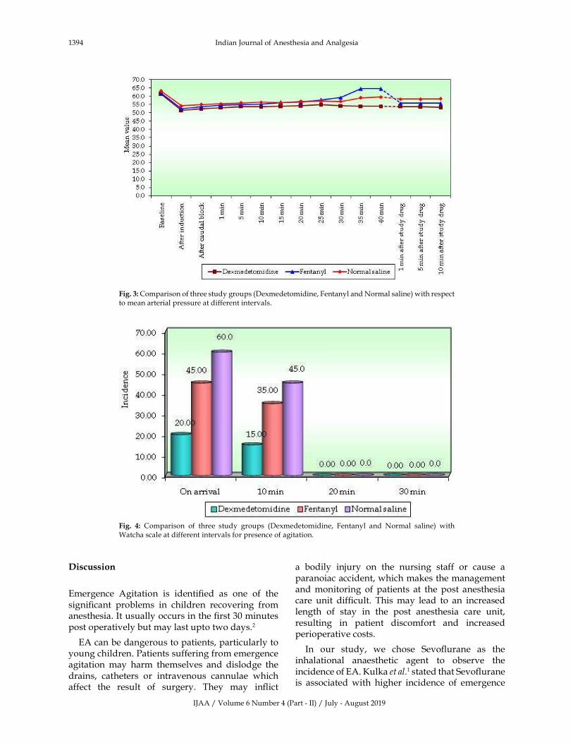
IJAA�/�Volume�6�Number�4�(Part�-�II)�/�July�-�August�2019
1394 Indian�Journal�of�Anesthesia�and�Analgesia
Fig���:�Comparison�of�three�study�groups�(Dexmedetomidine,�Fentanyl�and�Normal�saline)�with�respect�to�mean�arterial�pressure�at�different�intervals.
Fig�� �:� Comparison� of� three� study� groups� (Dexmedetomidine,� Fentanyl� and� Normal� saline)� with�Watcha�scale�at�different�intervals�for�presence�of�agitation.
�iscussion
Emergence� Agitation� is� identi��ed� as� one� of� the�signi��cant� problems� in� children� recovering� from�anesthesia.�It�usually�occurs�in�the���rst�30�minutes�post�operatively�but�may�last�upto�two�days.2
EA�can�be�dangerous�to�patients,�particularly�to�young�children.�Patients�suffering�from�emergence�agitation� may� harm� themselves� and� dislodge� the�drains,� catheters� or� intravenous� cannulae� which�affect� the� result� of� surgery.� They� may� in��ict�
a� bodily� injury� on� the� nursing� staff� or� cause� a�paranoiac�accident,�which�makes�the�management�and�monitoring� of� patients� at� the�post� anesthesia�care� unit� dif��cult.� This�may� lead� to� an� increased�length� of� stay� in� the� post� anesthesia� care� unit,�resulting� in� patient� discomfort� and� increased�perioperative�costs.
In� our� study,� we� chose� Sevo��urane� as� the�inhalational� anaesthetic� agent� to� observe� the�incidence�of�EA.��ulka�et�al.1�stated�that�Sevo��urane�is� associated�with� higher� incidence� of� emergence�
IJAA�/�Volume�6�Number�4�(Part�-�II)�/�July�-�August�2019
1395
agitation� in� upto� 80�� in� children.� Singh� et� al.5
found� a� higher� incidence� of� emergence� agitation�associated� with� Sevo��urane� (40�)� than� with�Des��urane�(26�)�and�Iso��urane�(18�)�in�children.��Two�uni�ue�intrinsic�characteristics�of�Sevo��urane�were�speculated�by��olzki�et�al.5�that�might�account�for�the�development�of�emergence�agitation.
First,� this� anaesthetic� agent� exerts� an� irritating�side�effect�on�the�central�nervous�system.�Second,�epileptiform� activity� has� been� reported� during�the� use� of� Sevo��urane� anesthesia.� Agitation�may�be� related� to� the� similar� CNS� effects� of� these�anesthetics,� which� may� affect� brain� activity� by�interfering� with� the� balance� between� neuronal�synaptic�inhibition�and�excitation�in�the�CNS.
Davis� et� al.6� suggested� that� inade�uate� pain�relief� may� be� the� cause� of� agitation,� particularly�after� short� surgical� procedures� for� which� peak�effects� of� analgesics� may� be� delayed� until� the�child� is� completely� awake.� Postoperative� pain�has�been� the�next� probable� factor�when�assessing�a� child�s� behaviour� upon� emergence� because� of�the� overlapping� clinical� picture� with� emergence�agitation.� The� children� in� all� the� three� groups� in�our�study�were�given�Caudal�block�with�1.0�ml/kg�of� 0.25�� bupivacaine� after� PLMA� insertion� and�before� the� start� of� surgery� to� exclude�pain� as� the�confounding�factor.
Intense� preoperative� anxiety,� both� in� children�and� their� parents� has� also� been� associated� with�an� increased� likelihood� of� restless� recovery� and�agitation� from� anesthesia.� The� children� in� all� the�three� groups� in� our� study� were� premedicated�with� midazolam� 0.5� mg/kg� orally� half� an� hour�before� surgery.� �ain� et� al.7� determined� the�relationship� between� preoperative� anxiety,� ED,�and� postoperative� maladaptive� behaviours.� �e�found�a�higher�incidence�in�children�who�were�not�premeditated�and�underwent�surgery�and�general�anesthesia�using�Sevo��urane.
�owever,� the�aetiology�of�Sevo��urane� induced�EA�is�still�unknown�and�a�clear-cut�strategy�for�its�prevention�has�not�been�developed.
The� decision� of� whether� to� treat� emergence�agitation� with� additional� pharmacological�medication�depends�upon�the�severity�and�duration�of�symptoms.
Various� pharmacological� agents� are� used� to�combat�sevo��urane�induced�agitation�in�Paediatric�age� group.� �se� of� opioids� like� Fentanyl,� which�serve� as� sedatives� and� analgesics�have�been� tried�and� used� widely� for� control� of� agitation.8,9,10� �se�of� analgesics� like� �etorolac,� paracetamol� have�
been� used� to� decrease� emergence� agitation.11��se�of� alpha� 2� adrenoreceptor� agonists� like�Clonidine�and�Dexmedetomidine�have�been�recommended�to�elimininate�pain�as�a�potential�source�of�agitation.12,13�
�se�of�benzodiazepines�like�Midazolam,�owing�to�their� sedative� property� have� been� suggested� to�calm�the�post�operative�agitation.14
Among� them� opioids� like� Fentanyl� and� alpha�2� adrenoreceptor� agonist� like� Dexmedetomidine�have� sedative� and� analgesic� effects� and� both� are�known� to� reduce� emergence� agitation.� Thus� we�selected�these�two�drugs�in�our�study.
Matin� et� al.15� suggested� that� Fentanyl�premedicated� children� experienced� less� anxiety�and�strain�during�surgery�and�had�better�recovery.�Manaa� et� al.9� and� Cravero� et� al.8� in� their� studies�observed� a� signi��cant� reduction� in� emergence�agitation� in� children� receiving� Fentanyl.� In� our�study,�we�chose�Inj.�Fentanyl�in�the�dose�of�1�mic/kg�i.v.�which�was� in�consensus�with�the�dose�chosen�by�Manaa�et�al.9,�Cravero�et�al.8.
�ecause� of� its� sedative� and� analgesic� effects,��2�adrenoreceptor� agonists� like� Clonidine� and�
Dexmedetomidine� have� been� used� safely� and�effectively� for� the� management� of� Sevo��urane�agitation� in� paediatric� patients.� Cravero� et� al.8
and� �ulka� et� al.1� found� a� reduction� of� agitation�with� Clonidine� 2� mcg/kg� i.v.� in� children�undergoing�surgery�under�Sevo��urane�anesthesia.�Dexmedetomidine� a� recent� addition� to� the� �
2�
adrenoreceptor�agonist�group�has�also�been�studied�extensively�in�the�reduction�of�emergence�agitation.
Dexmedetomidine� owing� to� its� sedative� and�analgesic� action,�no� respiratory�depression,� better�haemodynamic�stability,�minimal�or�no�signi��cant�adverse�effect�pro��le�has�lead�to�its�use�in�paediatric�anesthesia.� It� can� be� used� as� a� premedication,�sedative� for� invasive� procedures,� shivering� and�reduction�of�emergence�agitation.
Isik� et� al.16� and� Guler� et� al.17� observed� that�Dexmedetomidine�0.5�mic/kg�or�1�mic/kg�reduced�post� Sevo��urane� agitation� signi��cantly� but� it�also� prolonged� to� early� phase� of� post� anesthesia�recovery.
�ackache� et� al.13� used� Dexmedetomidine� i.v.�after� induction� of� anesthesia� in� the� dose� of�0.15� mic/kg� and� 0.3� mic/kg.� �e� observed� that�incidence�of�postoperative�agitation�was�37��in�the�control�group,�17��in�children�receiving�0.15�mic/kg�and� 10�� in� children� receiving� 0.3�mic/kg.� In� our�study,�we�used�Inj.�Dexmedetomidine�0.3�mic/kg�i.v.;�this�was�in�accordance�with�the�dose�chosen�by�Manaa�et�al.9,�Masami�et�al.12.
Comparison��etween�Intravenous�Fentanyl�and�Dexmedetomidine�to�Decrease�Sevoflurane�–�Induced�Agitation�in�Paediatric�Patients��ndergoing�Lower�Abdominal�Surgery:�A�Prospective�Randomized�Observational�Study
IJAA�/�Volume�6�Number�4�(Part�-�II)�/�July�-�August�2019
1396 Indian�Journal�of�Anesthesia�and�Analgesia
We�have�given�the�study�drug�10�minutes�before�the�end�of�anesthesia,�our�study�was�in�consensus�with�the�study�conducted�by�Manaa�et�al.9.
Thus,� we� decided� to� conduct� our� study� with�Dexmedetomidine� 0.3� mcg/kg� and� comparing� it�with�Fentanyl�1�mcg/kg�on�reduction�of�emergence�agitation� after� Sevo��urane� anesthesia� in� children�aged� 2-9� years� of� ASA� physical� status� I� or� II�undergoing�elective�lower�abdominal�surgery.
Post� operative� scales�used� for� agitation� scoring�and�pain�scoring�were�the�Watcha�scale18,19�and�The�Paediatric�Anesthesia�Emergence�Delirium�(PAED)�Scale20.�The�PAED�scale�is�validated�but�is�dif��cult�to� use� in� the� routine� clinical� practice.It� is� better�to� use� a� simple� scale� like� watcha� scale� to� detect�delirium�and�then�use�the�PAED�scale�to�measure�its�degree21.
Agitation�due�to�pain�is�a�signi��cant�confounding�factor� for� the� evaluation� of� the� presence� or�measurement�of�the�degree�of�ED.
The� patient� can� be� agitated� even� if� they� have�pain,�hence�we�used�objective�pain�scale,�so�that�we�can�differentiate�if�the�patient�was�agitated�due�to�pain�or�due�to�general�anesthesia�with�sevo��urane.�The�total�score�in�objective�pain�scale�is�10,��ut�since�the�patient�in�our�study�was�in�the�age�group�of�2�to�9�years,�not�all�the�patient�were�able�to�complaint�of�pain.��ence,�that�parameter�was�removed�from�the�objective�pain�scale�for�all�the�patients�and�then�the�maximum� score� had� become� 8.�Among� other�parameters� like� systolic� blood� pressure,� crying,�movements�and�agitation�except�for�systolic�blood�pressure�other�parameters�were�overlapping�to�an�extent�with�the�parameters�used�in�the�PAED�scale.��ence� in� our� study�we� observed� that� the� groups�which� were� having� higher� degree� of� agitation�according�to�PAED�scale�also�had�a�higher�objective�pain�scale�score.
In� our� study� we� observed� that� the� patients� in�the�Group�N�showed�higher�scores�of�agitation�in�comparison�to�Group�F,�and�Group�F�showed�higher�scores�of�agitation�in�comparison�to�Group�D.
The� difference� between�Group�D� and�Group� F�was�signi��cant,�suggesting�that�Dexmedetomidine�is� more� effective� in� reduction� of� the� emergence�agitation� after� Sevo��urane� anesthesia� compared�to�Fentanyl.
We�also�observed�that�the�agitation�scores�were�higher� in� the� ��rst� ten� minutes� post� operatively�and� by� the� end� of� 30�minutes,� patients� in� all� the�three� groups� were� comparable� with� regard� to�agitation�scorings.�This�was�in�consensus�with�the�observation�made�by�Vlajkovic�et�al.14.
Erdil� et� al.10� observed� that� Dexmedetomidine�proved� to� be� better� than� Fentanyl� in� reducing�the� agitation;� however� the� difference� was� not�signi��cant.
In� contrast� to� Manaa� et� al.9� who� proved� that�Fentanyl�was�slightly�better�than�Dexmedetomidine.�In�our�study�60��of�the�children�in�Group�N�showed�presence� of� agitation� as� against� 45�� in� Group� F�and�20��in�Group�D�on�arrival� in�post�anesthesia�care�unit.
Aldrete� Recovery� Score� was� used� to� assess�recovery� of� patient� in� PAC�� and� those� patients�who�attained�an�aldrete�recovery�score�of���9�were�dischared�from�the�PAC��to�the�ward�and�the�time�taken�to�achieve�this�score�was�noted22.
None�of�the�patients�showed�any�adverse�effects�or�intraoperative�event�of�signi��cance�or�post�operative�complications� like�nausea/vomiting,�hypotension,�bradycardia,�hypoxemia,�hypoventilation,�apnoea,�and�respiratory�depression.
Conclusion
The� dose� of� fontanel� 1� mcg/kg� i.v.� or�dexmedetomidine� 0.3� mcg/kg� i.v.� that� is�administered�10�minutes�before�the�discontinuation�of�anesthesia�reduced�the�postoperative�agitation�in�children.
Dexmedetomidine� was� more� effective� when�compared� with� fentanyl� in� attenuation� of�sevo��urane-induced�agitation�in�paediatric�patients�undergoing�lower�abdominal�surgery.
�eferences
1.� Emergence�agitation�in�paediatric�patients.�Masui2007�May;56(5):554–9.
.�2 Masoumeh� �S,� Fatemeh� F.� Postanesthetic�Emergence� Agitation� in� Pediatric� Patients�under� General� Anesthesia.� Iran� J� Pediatr.� 2014�Apr;24(2):184–190.
3.� Singh� R,� �harbanda� M,� et� al.� Comparative�evaluation� of� incidence� of� emergence� agitation�and�postoperative� recovery� profile� in� paediatric�patients� after� isoflurane,� sevoflurane� and�desflurane� anesthesia.� Indian� J�Anaesth.�2012;56(2):156–161
4.� Shi�F,��iao���et�al.�Effects�of�Fentanyl�on�Emergence�Agitation� in� Children� under� Sevoflurane�Anesthesia:� Meta-Analysis� of� Randomized�Controlled�Trials.�PLoS�One.�2015�Aug�14;10(8).
5.� �olzki� J� and� �retz� FJ.� Changing� aspects� of�sevoflurane� in� paediatric� anesthesia:� 1975–99.�Pediatr�Anesth.�1999;9:283–86.
IJAA�/�Volume�6�Number�4�(Part�-�II)�/�July�-�August�2019
1397
6.� Davis�PJ,�Greenberg�JA,�Gendelman�M,�Recovery�characteristics� of� sevoflurane� and� halothane� in�preschool-aged� children� undergoing� bilateral�myringotomy� and� pressure� e�ualization� tube�insertion.�Anesth�Analg.�1999;88:34–8.
7.� �ain��,�Caldwell-Andrews�AA,�Weinberg�ME,�et�al.� Sevoflurane� versus� �alothane:� postoperative�maladaptive�behavioral�changes.�Anesthesiology.2005;102:720–26.
8.� Cravero�JP,��each�M,�Thyr��,�et�al.�The�effect�of�small�dose�fentanyl�on�the�emergence�characteristics�of�paediatric� patients� after� sevoflurane� anesthesia�without�surgery.�Anesth�Analg.�2003;97:364–7.
9.� Manaa� EM,� Abdelhaleem� AA,� Mohamed� EA.�Fentanyl� versus� dexmedetomidine� effect� on�agitation� after� sevoflurane� anesthesia.� Saudi� J�Anaesth.�2007;1:57–61.
10.� Erdil�F,�Demirbilek�S,��egec���et�al.�The�effects�of�Dexmedetomidine� and� Fentanyl� on� emergence�characteristics� after� adenoidectomy� in� children.�Anaesth�Intensive�Care.�2009�Jul;37(4):571–6.
11.� Johannesson� GP,� Floren� M,� Lindahl� SG.�Sevoflurane� for� ENT� –� surgery� in� children� :� a�comparison� with� halothane.� Acta� Anesthesiol�Scand.�1995;39:546–50.
12.� Masami�S,�Gotaro�S,�Misako�T�et�al.�Effect�of�single-dose� Dexmedetomidine� on� emergence� agitation�and�recovery�profiles�after�Sevoflurane�anesthesia�in�pediatric�ambulatory�surgery.�Japanese�journal�of�anesthesia.�2010;26:675–682.
13.� Ibacache� ME,� Mu�oz� �R,� �randes� V.� Single�does� Dexmedetomidine� reduces� agitation� after�Sevoflurane�anesthesia�in�children.�Anesth�Analg�2004;98:60–3.
14.� Vlajkovic�GP,�Sindjelic�RP.� Emergence�Delirium�in� Children:� Many� �uestions,� Few� Answers.�
Anesth�Analg.�2007�Jan;104(1):84–91.
15.� Matin,�Abdul�M,�et�al.�Fentanyl�as�a�premedication�
for� anesthesia� in� paediatric� surgery.�Journal�
of� Chittagong� Medical� College� Teachers��
Association.�2008;19(1):23–27.
16.� Isik� �,� Arslan� M,� Tunga� AD,� et� al.�
Dexmedetomidine�decreases�emergence�agitation�
in�pediatric�patients�after�Sevoflurane�anesthesia�
without�surgery.�Pediatr�Anesth.�2006;16:748–53.
17.� Guler� G,� Akin� A,� Tosun� �,� et� al.� Single� dose�
Dexmedetomidine� reduces� agitation� and�
provides� smooth� extubation� after� pediatric�
adenotonsillectomy.� Pediatr� Anesth.�
2005;15:762–66.
18.� �im� N�,� �im� S�,� �oon� �J,� et� al.� Effect� of�
Dexmedetomidine� on� Sevoflurane� Re�uirments�
and�Emergence�Agitation�in�Children��ndergoing�
Ambulatory� Surgery.� �onsei� Med� J.� 2014;55(1):�
209–15.
19.� Watcha� MF,� Ramirez-Ruiz� M,� White� PF,� et�
al.� Perioperative� effects� of� oral� ketorolac� and�
acetaminophen� in� children� undergoing� bilateral�
myringotomy.�Can�J�Anaesth.�1992;39:649–54.
20.� �ajwa� SA,�Costi�D,�Cyna�AM.�A� comparison� of�
emergence� delirium� scales� following� general�
anesthesia� in� children.�Pediatr�Anaesth.�2010;20:�
704–11.
21.� Pieters� �J,� Penn�E,�Nicklaus� P.� et� al.� Emergence�
delirium� and� postoperative� pain� in� children�
undergoing� adenotonsillectomy:� a� comparison�
of� propofol� vs� sevoflurane� anesthesia.� Paediatr�
Anaesth.�2010;20:944–50.
22.� Aldrete�JA,��roulik�D.�A�post-anesthetic�recovery�
score.�Anesth�Analg.�1970;49:924–34.
Comparison��etween�Intravenous�Fentanyl�and�Dexmedetomidine�to�Decrease�Sevoflurane�–�Induced�Agitation�in�Paediatric�Patients��ndergoing�Lower�Abdominal�Surgery:�A�Prospective�Randomized�Observational�Study
IJAA�/�Volume�6�Number�4�(Part�-�II)�/�July�-�August�2019
1398 Indian�Journal�of�Anesthesia�and�Analgesia
RED�ART.NET(A�product�of�RF�Library�Services�(P)�Limited)
(Publications�available�for�purchase:�Journals,��ooks,�Articles�and�Single�issues)(Date�range:�1967�to�till�date)
The�Red��art�is�an�e-commerce�and�is�a�product�of�RF�Library�Services�(P)�Ltd.�It�covers�a�broad� range�of� journals,��ooks,�Articles,� Single� issues� (print���Online-PDF)� in�English�and��indi�languages.�All�these�publications�are�in�stock�for�immediate�shipping�and�online�access�in�case�of�online.
Benefits�of�shopping�online�are�better�than�con�entional��ay�of�buying�1.� Convenience.�2.� �etter�prices.�3.� More�variety.�4.� Fewer�expenses.5.� No�crowds.�6.� Less�compulsive�shopping.�7.� �uying�old�or�unused�items�at�lower�prices.�8.� Discreet�purchases�are�easier.�
�RL:�www.redkart.net
��Red�Flower�Publication�Pvt.�Ltd.�
�Original�Research�ArticleIndian�Journal�of�Anesthesia�and�Analgesia�
2019;6(4)�(Part�-�II):�1399-1411DOI:�http://dx.doi.org/10.21088/ijaa.2349.8471.6419.48
A�Comparison�Bet�een�Airtra��Optical��aryngoscope�and�Con�entional�Macintosh� �aryngoscope� for� Intubation� in� Adult� Surgical� Patients:�A�Prospecti�e��andomi�ed�Controlled�Study
S�Sel�amani���P�Anand�
1Professor,�2Junior�Resident,�Department�of�Anesthesia,�Sree��alaji�Medical�College�and��ospital,�Chrompet,�Chennai,�Tamil�Nadu�600044,�India.
Corresponding�Author:�P�Anand,�Junior�Resident,�Department�of�Anesthesia,�Sree��alaji�Medical�College�and��ospital,�Chrompet,�Chennai,�Tamil�Nadu�600044,�India.
E�mail:�[email protected]
�ecei�ed�on�24.01.2019,�Accepted�on�11.07.2019
Abstract
Tracheal� intubation� using� a� laryngoscope� is� considered� as� a� gold� standard� 1� of� airway� management�during�administration�of�general�anesthesia�and�also�in�critical�care�settings�because�of�its�several�advantages�including�2,3�-�Isolation�of�respiratory�tract�from�Gastro�intestinal�system�and�hence�minimal�risk�of�aspiration.�Allows�delivery�of�oxygen�and�anaesthetic�gases�via�positive�pressure�ventilation�without�inflation�of�stomach.�Access�to�tracheobronchial�tree�for�pulmonary�lavage�and�drug�administration�(e.g.�inhaled�bronchodilators).Improved�access�to�head�and�neck�surgeries.�Airway�management�is�important�for�anesthesia�because�adverse�respiratory� events� are� responsible� for� 75�� of� ASA� closed� claims.� Of� these� failed� ventilation� is� the�main�culprit�(38�),�followed�by�faulty�placement�of�endotracheal�tube�in�esophagus�(17�)�and�difficult�intubation�(18�).�Approximately�600�patients�die�each�year�in�the�developed�world�from�complications�due�to�airway�management� and� also� in� the� underdeveloped�world� is� much� grimmer.4,5,6�Conte�t:�We� compared� tracheal�intubation� using� airtra�� optical� laryngoscope�with� macintosh� laryngoscope� in� adult� patients� undergoing�elective�procedures�under�general� anesthesia�Aims:� This� study� is� to� compare� the� intubating� conditions� in�adult�surgical�patients�using�airtra��optical�laryngoscope�with�macintosh�laryngoscope�with�respect�to�ease�of�intubation,�time�taken�for�intubation,�airway�trauma�and�hemodynamic�response�to�laryngoscopy.�Settings�and��esign:�Prospective�randomised�interventional�study.�Materials�and�Methods:�This�study�was�done�in�Sree��alaji�Medical�College�and��ospital,�Chennai�at�Department�of�Anaesthesiology�and�Critical�Care�from�August�2016�to�February�2018.�It�was�a�Single�centre,�prospective,�randomized,�parallel�group,�open�label,�interventional�controlled�study.�After�obtaining�institutional�ethical�committee�approval.�60�patients�(sample�size)�who�were�posted�for�elective�surgery�re�uiring�general�anesthesia�(Recruitment)�with�satisfying�inclusion�criteria�were�enrolled�in�the�study�after�obtaining�informed�consent�from�the�patients�and�relatives.�2�groups�by�random�number�allotted�by�computer�based�randomization�Statistical�Anal�sis:�Descriptive�and�inferential�statistical�methods�were�used.�Results:�Mean�duration�of�intubation�with�the�Airtra��group�was�15.93�secs�whereas�in�the�Macintosh�group�it�was�found�to�be�38.70�secs.�It�was�computed�using�Levene�s�T�test�and�was�found�to�be�statistically�significant.�In�the�Macintosh�group,�4�patients�had�an�Total�IDS�of�5�or�greater,�indicating�moderate�to�severe�intubation�difficulty,�whereas�no�patient�in�the�Airtra��group�had�an�Total�IDS�of�more�than�3�and�was�found�to�be�statistically�significant.�Conclusion:�Our�study�concludes�that�endotracheal�intubation�is�easier�with�Airtra��compared�to�Macintosh�laryngoscope�as�it�provides�good�glottis�view.�In�addition�to�that�Airtra��have�less�intubation�duration,�less�hemodynamic�response�for�intubation�and�less�Airway�trauma�compared�to�Macintosh.�
�ey�ords:�Macintosh;�Airtra�;�Laryngoscope;�Intubation;�Airway.
�o��to�cite�this�article:
S�Selvamani,�P�Anand.�A�Comparison��etween�Airtra��Optical�Laryngoscope�and�Conventional�Macintosh�Laryngoscope�for�Intubation�in�Adult�Surgical�Patients:�A�Prospective�Randomized�Controlled�Study.�Indian�J�Anesth�Analg.�2019;6(4):1399-1411.

IJAA�/�Volume�6�Number�4�(Part�-�II)�/�July�-�August�2019
1400 Indian�Journal�of�Anesthesia�and�Analgesia
Introduction�
Airway� management� has� been� the� domain� of�dominance�for�anesthesiologists�over�ages.
Laryngoscopy� and� intubation� are� the�fundamental� skills� that� every� clinician� aspires� to�learn� and� practice.� The� most� fre�uent� end� point�for�almost�all�the�studies�was�the�ease�of�intubation�along� with� ability� to� minimize� the� sympathetic�response� to� tracheal� instrumentation.�Airtra�� is� a�recently�developed� laryngoscope�which� facilitates�easy� visualisation� of� glottis� through� matrix� of�se�uentially�arranged�lenses�and�mirror.
Materials�and�Methods
This� study� was� done� in� Sree� �alaji� Medical�College� �� �ospital,� Chennai� at� Department� of�Anaesthesiolgy�and�Critical�Care�from�August�2016�to�February�2018.
It�was�a�Single�centre,�prospective,�randomized,�parallel�group,�open�label,�interventional�controlled�study.
After� obtaining� institutional� ethical� committee�approval.� 60� patients� (sample� size)� are� posted�for� elective� surgery� re�uiring� general� anesthesia�(Recruitment)� with� satisfying� inclusion� criteria�were�enrolled�in�the�study�after�obtaining�informed�consent�from�the�patients�and�relatives.
Randomization:� 2� groups� by� random� number�allotted�by�computer�based�randomization.
Allocation�and�inter�ention:�2�groups
�roup�A:�30�Patients�–airtra��optical�laryngoscope
�roup� B:� 30� Patients� –conventional� macintosh�laryngoscope
Inclusion�Criteria
�� ASA�1���2�patients
�� Age�18-65�years,�both�sexes
�� Elective�surgical�cases�re�uiring�GA
�� MPC�1,�2,���3�patients
Exclusion�Criteria
�� Severe� CVS,� RS,� hepatic,� renal� disease�patients
�� Any�valvular,�conduction�abnormality,�I�D,��ypertensive�patients
�� Patients� on� antihypertensive� drugs� or� beta�blockers
�� Anticipated�dif��cult�airway�patients
�� �MI�more�than�40
�� Patient�refusal
�aterial
�� Airtra��optical�laryngoscope�adult�size
�� Macintosh�laryngoscope�3���4�size�blade
Air�ay�Assess�ent��������0
Previous� surgery� and� anesthesia� records,� �/O�snoring,� �/O� voice� change,� �/O� previous�surgery,� �urns,� Trauma,� Tumour� in� and� around�the� oral� cavity,� neck� or� cervical� spine� were�asked� in� the� history.� �/O� systemic� illness� like��ypertension,� Diabetes,� Ankylosing� spondylitis,�Rheumatoid� arthritis� were� asked� and� recorded.�General� examination� included� examination�for� facial� anomalies,� Anomalies� of� the� mouth,�Temperomandibular� joint� pathology,� and� tongue,�pathology�of�palate�and�pathology�of�nose.�Weight�in� kilograms� and� �eight� in� centimeters� were�recorded� and� �ody� Mass� Index� was� calculated.�Individual�airway�indices�were�measured.
Sa�son� and� �oun�� �odification� of� �alla��atti��radin�:�
The�patient�kept� in� sitting�position�with�maximal�mouth� opening� with� protruding� tongue,� without�phonation� and� the� observer�s� eye� in� level� with�patient�s�mouth�and�the�degree�to�which�the�faucial�pillars,� uvula,� soft� palate,� and� hard� palate� were�visible�were�recorded�and�classi��ed�as�follows:
�rade� I:� Faucial� pillars,� uvula,� soft� palate� and�hard�palate�visible
�rade�II:��vula,�soft�palate�and�hard�palate�visible
�rade�III:��ase�of�uvula�or�none,�soft�palate�and�hard�palate�visible
�rade�I�:�Only�hard�palate�visible.
�hyro�ental�distance
Distance� between� the� thyroid� notch� and� mental�symphysis� when� the� neck� is� fully� extended� and�mouth�closed�(�6.5�cm�or���6.5�cm)
�ther��ays�of�air�ay�assess�ent
A�O� �oint� mo�ement:� Patient� asked� to� look� at� the�ceiling�without�raising�the�eyebrow�and�the�range�of�movements�were�measured
IJAA�/�Volume�6�Number�4�(Part�-�II)�/�July�-�August�2019
1401
�eck� ��e�ion:� Patient� was� asked� to� touch� the�manubrium� sternum� with� chin� and� the� range� of�movements�measured.
TM��function:�The�patient�was�asked�to�open�the�mouth� wide� open� and� the� inter� incisor� distance�measured.� Examiner�s� index���nger�was�placed� in�front� of� the� tragus� and� thumb� over� the� mastoid�process� and� the� patient� was� asked� to� open� the�mouth� and� sliding� movement� of� the� mandibular�condyle�was�assessed.
�pper� lip�bite�test:�The�patient�was�asked�to�bite�the�upper�lip�with�the�lower�incisor�and�graded�as�follows:
Class� 1:� Lower� incisor� can� bite� the� upper� lip�above�the�vermilion�line
Class� 2:� Lower� incisor� can� bite� the� upper� lip�below�the�vermilion�line
Class�3:�Lower�incisor�cannot�bite�the�upper�lip
Sterno� mental� distance:� Distance� between� the�sternal�notch�and�mental�symphysis�when�the�neck�was�fully�extended�and�mouth�closed.
�eck�circumference:�Measured�in�cm�at�the�level�of�thyroid�notch.
��amination� of� dentition:� Abnormalities� like�cracking,�buck�tooth,�loose,�arti��cial�and�absence�of�incisors�were�examined�and�recorded.
Procedure
After� assessment� patient� shifted� to� operating�room.� i.v� line� started� and� SpO
2,� ECG,� NI�P� and�
ETCO2�(After�intubation)�moniters�connected.
�remedication:� 0.2�mg�glycopyrrolate,� 2�mcg/kg�fentanyl�iv�route�10�mins�before�Induction.
�reo��genation:�with� 100��02� for� 3mins� at� tidal�volume�respiration
Base� line:� SpO2� �R,� Systolic� �P,� Diastolic� �P,�
MAP�was�noted
Induction:�2.5�mg�/kg�propofol
Rela�ant�for�intubation:�0.5�mg/kg�induction�dose�of� atracurium� 0.1� mg/kg� maintenance� dose� of�atracurium
Intubation:� airtra�/macintosh� laryngoscopy�according�to�the�group
Monitoring:�SPO2,��R,�Systolic��P,�Diastolic��P,�MAP�at�3rd,�5th,�10th�minute.
�utco�e��easures
�rimar��measures
�� Ease�of�intubation�assessed�by�IDS�score
Secondar��measures
�� �aemodynamic�response
�� Airway�trauma
�� Intubation�time
Intu�ation��ifficulty�Score:�������2
Intubation� dif��culty� score� was� used� to� evaluate�intubating� performance� of� laryngoscopy.� IDS�scoring� was� developed� by� adnet� et� al.� in� 1997.�IDS� score� is� a� blend� of� objective� and� subjective�criteria� that� permit� a� �uantitative� and� �ualitative�approach� to� the� progressive� nature� of� the�dif��culty� in� intubation.� It� appears� to� be� the� best�indicator�till�date.
7�variables�are�used.
���-�No�of�supplementary�attempts.�An�attempt�is�de��ned�as�one�advancement�of�tracheal�tube�in�the�direction�of�the�glottis�during�direct�laryngoscopy.�(for�Attempt�1/2/3/4,�N1�Score�is�0/1/2/3)
��� -� No� of� supplementary� operators� directly�operating� (not� assisting)� (for� operators� 1/2/3/4,�N2�Score�is�0/1/2/3)
��� -� No:� of� alternative� techni�ues� used.� (each�additional� techni�ues� like�oral� intubation� to�blind�nasotracheal� intubation,� curved� blade� to� straight�blade�etc�N3�Score�is�1�or�more)
���-�Cormack�Lehane�grade�minus�one.�(for�CLG�1/2/3/4,�N5�Score�is�0/1/2/3)
���-�Subjectively�increased�lifting�force�re�uired�during� laryngoscopy.� (for� normal� N� 5�0,� for�increased�N�5�1)
��� -�Need� for� external� laryngeal�manipulation�(for�not�re�uired�N6�0,�for�re�uired�N6�1)
��� -� Position� of� vocal� cords.(N7� Score� 0� for�abduction,�1�for�adduction)
Total�IDS�Score���sum�of�scores�(N1�to�N7)
I�S�Score��e�ree�of�difficulty
0�-�Ease
1�to�5�-�Slight�dif��culty
��5�-�Moderate�to�major�dif��culty
In��nitive�-�Impossible�intubation
In�this�scoring�the�value�of�IDS�is��0��in�full�visual�view�of�glottic�opening�with�vocal�cords�are�seen�to�be�nicely�abducted.�Every�variation�from�this�de��ned��ideal��intubation�increases�the�scoring�that�indicate�increasing�dif��culty�of�intubation.�The�total�IDS�score�being�the�sum�of�all�variation�from�the�de��nition.
A�Comparison��etween�Airtra��Optical�Laryngoscope�and�Conventional�Macintosh�Laryngoscope�for�Intubation�in�Adult�Surgical�Patients,�A�Prospective�Randomized�Controlled�Study

IJAA�/�Volume�6�Number�4�(Part�-�II)�/�July�-�August�2019
1402 Indian�Journal�of�Anesthesia�and�Analgesia
Cor�ac��and��ehane��radin��Syste�:����
Entire�vocal�cord�visualized�-�Grade�I
Posterior�part�of�vocal�cords�seen�-�Grade�IIa
Arytenoids�only�seen�-�Grade�IIb
Epiglottis�only�seen�(liftable)�-�Grade�IIIa
Tip�of�epiglottis�only�seen�(adherent)�-�Grade�IIIb
No�glottis�structure�seen�-�Grade�IV
Apart� from� Cormack-Lehane� and� Intubation�Dif��culty� Score,� the� following� factors� were� also�noted.
��Intubation� time:� It�measured�from�entry�of� the�device� into� the� oral� cavity� until� con��rmation� of�proper�placement�of�tracheal�tube.
���eart�rate��s�stolic�B���diastolic�B���mean�arterial�pressure� and�S�O
��were�measured�at� 3rd,� 5th���10th
minute�from�pre�induction.
�� Airwa�� trauma:� All� complications� will� be�recorded,� with� special� attention� to� common�complications�such�as�upper�airway,�dental�trauma�and� blood� soiling� of� airtra�� or� macintosh� blade�after� intubation.� If� intubation� with� Airtra�� failed�and� saturation� maintained,�Macintosh� blade� was�used�for�intubation�and�if�the�saturation�decreased,�mask� ventilation� with� 100�� oxygen� followed� by�intubation�with�Macintosh�laryngoscope.
Fig���:�Comparison�of�age�in�both�groups
�esults
All�data�were�collected�and�tabulated.
�Table��:�Age
Age�in�years�
�roup�A � �roup�B � ���alue
��20 2 07 1 03
0.092
21–30 8 27 9 30
31–40 7 23 8 27
41–50 5 17 4 13
51–65 8 27 8 27
Total 30 100 30 100
Table��:�Gender
�ender �roup�A � �roup�B � ���alue
Male 18 60 16 53
0.524Female 12 40 14 47
Total 30 100 30 100
Table��:��MI�Distribution
BMI�roup�A �roup�B
���ValueNo � No �
��18.5 4 13 3 10
0.00118.5�–�24.9 10 33 11 37
25�–�29.9 12 40 11 37
��30 4 13 5 17
Total 30 100 30 100
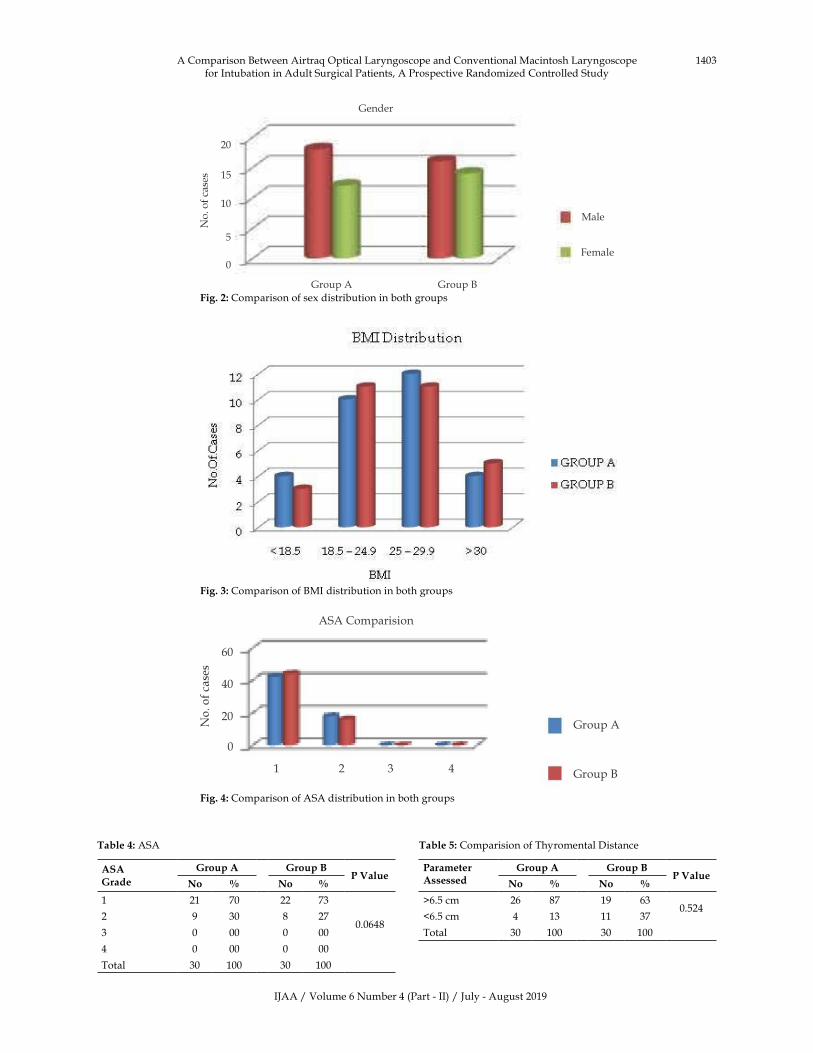
IJAA�/�Volume�6�Number�4�(Part�-�II)�/�July�-�August�2019
1403
Fig���:�Comparison�of�sex�distribution�in�both�groups
Fig���:�Comparison�of��MI�distribution�in�both�groups
Table��:�ASA
ASA�rade
�roup�A �roup�BP�Value
No � No �
1 21 70 22 73
0.06482 9 30 8 27
3 0 00 0 00
4 0 00 0 00
Total 30 100 30 100
Table��:�Comparision�of�Thyromental�Distance
ParameterAssessed
�roup�A �roup�BP�Value
No � No �
�6.5�cm 26 87 19 630.524
�6.5�cm 4 13 11 37
Total 30 100 30 100
Male
Female
Gender
20
15
10
5
0
Group�A Group��
ASA Comparision�
Group�A
Group��
60
40
20
0
1 2 3 4
Fig���:�Comparison�of�ASA�distribution�in�both�groups
A�Comparison��etween�Airtra��Optical�Laryngoscope�and�Conventional�Macintosh�Laryngoscope�for�Intubation�in�Adult�Surgical�Patients,�A�Prospective�Randomized�Controlled�Study
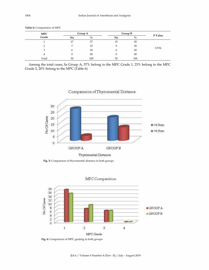
IJAA�/�Volume�6�Number�4�(Part�-�II)�/�July�-�August�2019
1404 Indian�Journal�of�Anesthesia�and�Analgesia
Table��:�Comparision�of�MPC
MPC�rade
�roup�A �roup�BP�Value
No � No �
1 17 57 15 50
0.5342 7 23 9 30
3 6 20 6 20
4 0 00 0 00
Total 30 100 30 100
Among�the�total�cases,�In�Group�A,�57��belong�to�the�MPC�Grade�1,�23��belong�to�the�MPC�Grade�2,�20��belong�to�the�MPC�(Table�6)�
Fig���:�Comparison�of�thyromental�distance�in�both�groups
Fig���:�Comparison�of�MPC�grading�in�both�groups
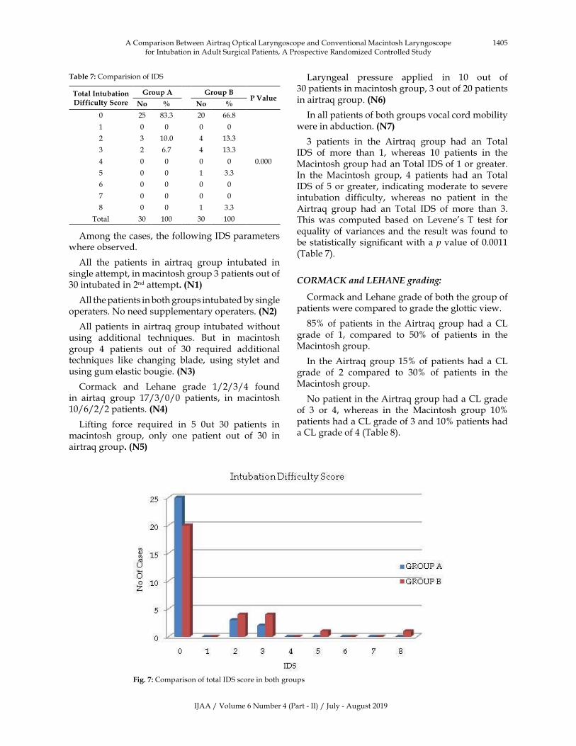
IJAA�/�Volume�6�Number�4�(Part�-�II)�/�July�-�August�2019
1405
Table��:�Comparision�of�IDS
Total�Intubation��ifficulty�Score
�roup�A �roup�BP�Value
No � No �
0 25 83.3 20 66.8
0.000
1 0 0 0 0
2 3 10.0 4 13.3
3 2 6.7 4 13.3
4 0 0 0 0
5 0 0 1 3.3
6 0 0 0 0
7 0 0 0 0
8 0 0 1 3.3
Total 30 100 30 100
Among�the�cases,�the�following�IDS�parameters�where�observed.
All� the� patients� in� airtra�� group� intubated� in�single�attempt,�in�macintosh�group�3�patients�out�of�30�intubated�in�2nd�attempt���N��
All�the�patients�in�both�groups�intubated�by�single�operaters.�No�need�supplementary�operaters.��N��
All�patients� in�airtra��group� intubated�without�using� additional� techni�ues.� �ut� in� macintosh�group� 4� patients� out� of� 30� re�uired� additional�techni�ues� like� changing� blade,� using� stylet� and�using�gum�elastic�bougie.��N��
Cormack� and� Lehane� grade� 1/2/3/4� found�in� airta�� group� 17/3/0/0� patients,� in� macintosh�10/6/2/2�patients.��N��
Lifting� force� re�uired� in� 5� 0ut� 30� patients� in�macintosh� group,� only� one� patient� out� of� 30� in�airtra��group���N��
Laryngeal� pressure� applied� in� 10� out� of�30�patients�in�macintosh�group,�3�out�of�20�patients�in�airtra��group.��N��
�In�all�patients�of�both�groups�vocal�cord�mobility�were�in�abduction.��N��
3� patients� in� the� Airtra�� group� had� an� Total�IDS� of� more� than� 1,� whereas� 10� patients� in� the�Macintosh�group�had�an�Total�IDS�of�1�or�greater.�In� the� Macintosh� group,� 4� patients� had� an� Total�IDS�of�5�or�greater,� indicating�moderate� to� severe�intubation� dif��culty,� whereas� no� patient� in� the�Airtra�� group� had� an� Total� IDS� of� more� than� 3.�This� was� computed� based� on� Levene�s� T� test� for�e�uality�of�variances� and� the� result�was� found� to�be�statistically�signi��cant�with�a�p�value�of�0.0011�(Table�7).
C�R�AC��and��E�A�E��radin�:
Cormack�and�Lehane�grade�of�both�the�group�of�patients�were�compared�to�grade�the�glottic�view.
85��of� patients� in� the�Airtra��group�had� a�CL�grade� of� 1,� compared� to� 50�� of� patients� in� the�Macintosh�group.
In� the�Airtra��group� 15�� of�patients� had� a�CL�grade� of� 2� compared� to� 30�� of� patients� in� the�Macintosh�group.
No�patient�in�the�Airtra��group�had�a�CL�grade�of� 3� or� 4,� whereas� in� the� Macintosh� group� 10��patients�had�a�CL�grade�of�3�and�10��patients�had�a�CL�grade�of�4�(Table�8).
Fig���:�Comparison�of�total�IDS�score�in�both�groups
A�Comparison��etween�Airtra��Optical�Laryngoscope�and�Conventional�Macintosh�Laryngoscope�for�Intubation�in�Adult�Surgical�Patients,�A�Prospective�Randomized�Controlled�Study
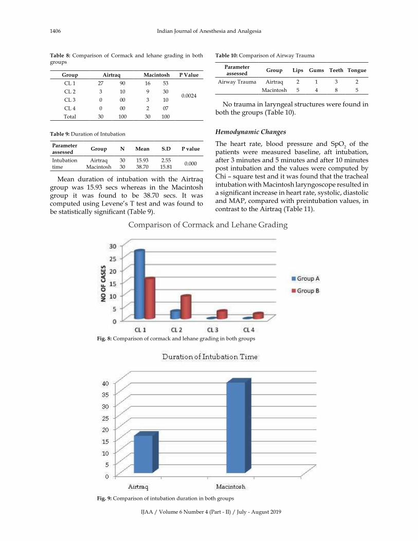
IJAA�/�Volume�6�Number�4�(Part�-�II)�/�July�-�August�2019
1406 Indian�Journal�of�Anesthesia�and�Analgesia
Table� �:�Comparison� of�Cormack�and� lehane� grading� in� both�groups
�roup Airtra� Macintosh P�Value
CL�1 27 90 16 53
0.0024CL�2 3 10 9 30
CL�3 0 00 3 10
CL�4 0 00 2 07
Total 30 100 30 100
Table��:�Duration�of�Intubation
Parameterassessed
�roup N Mean S�� P��alue
Intubation�time
Airtra�Macintosh
3030
15.9338.70
2.5515.81
0.000
Mean� duration� of� intubation� with� the� Airtra��group� was� 15.93� secs� whereas� in� the� Macintosh�group� it� was� found� to� be� 38.70� secs.� It� was�computed�using�Levene�s�T�test�and�was�found�to�be�statistically�signi��cant�(Table�9).
Table���:�Comparison�of�Airway�Trauma
Parameterassessed
�roup �ips �ums Teeth Tongue
Airway�Trauma Airtra� 2 1 3 2
Macintosh 5 4 8 5
No�trauma�in�laryngeal�structures�were�found�in�both�the�groups�(Table�10).
�e�odyna�ic�Chan�es
The� heart� rate,� blood� pressure� and� SpO2� of� the�
patients� were� measured� baseline,� aft� intubation,�after�3�minutes�and�5�minutes�and�after�10�minutes�post�intubation�and�the�values�were�computed�by�Chi�–�s�uare�test�and�it�was�found�that�the�tracheal�intubation�with�Macintosh�laryngoscope�resulted�in�a�signi��cant�increase�in�heart�rate,�systolic,�diastolic�and�MAP,�compared�with�preintubation�values,�in�contrast�to�the�Airtra��(Table�11).
Comparison�of�Cormack�and�Lehane�Grading
Fig���:�Comparison�of�cormack�and�lehane�grading�in�both�groups
Fig���:�Comparison�of�intubation�duration�in�both�groups
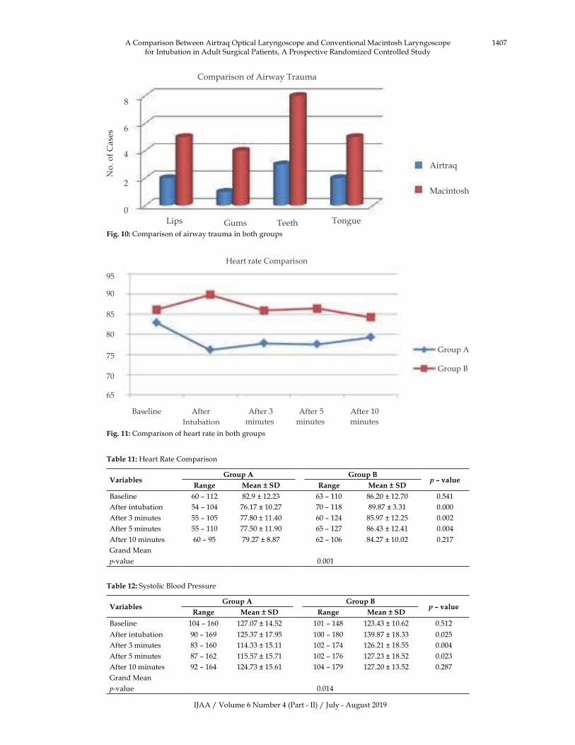
IJAA�/�Volume�6�Number�4�(Part�-�II)�/�July�-�August�2019
1407
Comparison�of�Airway�Trauma
8
6
4
2
0
Airtra�
Macintosh
Lips Gums Teeth Tongue
Fig����:�Comparison�of�airway�trauma�in�both�groups
�eart�rate�Comparison
Group�A
Group��
95
90
85
80
75
70
65
�aseline After
Intubation
After�3
minutes
After�5
minutes
After�10
minutes
Fig����:�Comparison�of�heart�rate�in�both�groups
Table���:��eart�Rate�Comparison
Variables�roup�A �roup�B
�����alue�ange Mean���S� �ange Mean���S�
�aseline 60�–�112 82.9���12.23 63�–�110 86.20���12.70 0.541
After�intubation 54�–�104 76.17���10.27 70�–�118 89.87���3.31 0.000
After�3�minutes 55�–�105 77.80���11.40 60�–�124 85.97���12.25 0.002
After�5�minutes 55�–�110 77.50���11.90 65�–�127 86.43���12.41 0.004
After�10�minutes 60�–�95 79.27���8.87 62�–�106 84.27���10.02 0.217
Grand�Mean
p-value 0.001
Table���:�Systolic��lood�Pressure�
Variables�roup�A �roup�B
�����alue�ange Mean���S� �ange Mean���S�
�aseline 104�–�160 127.07���14.52 101�–�148 123.43���10.62 0.512
After�intubation 90�–�169 125.37���17.95 100�–�180 139.87���18.33 0.025
After�3�minutes 83�–�160 114.33���15.11 102�–�174 126.21���18.55 0.004
After�5�minutes 87�–�162 115.57���15.71 102�–�176 127.23���18.52 0.023
After�10�minutes 92�–�164 124.73���15.61 104�–�179 127.20���13.52 0.287
Grand�Mean
p-value 0.014
A�Comparison��etween�Airtra��Optical�Laryngoscope�and�Conventional�Macintosh�Laryngoscope�for�Intubation�in�Adult�Surgical�Patients,�A�Prospective�Randomized�Controlled�Study
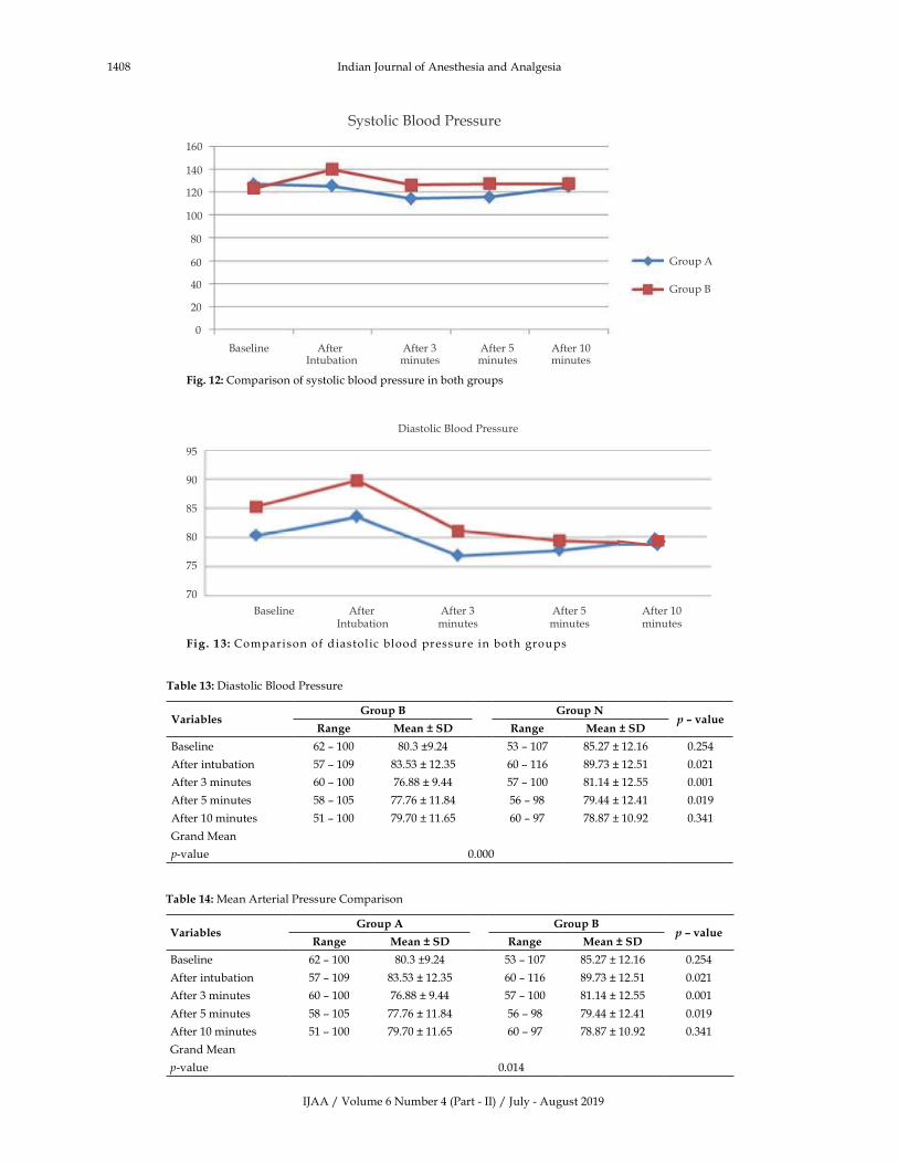
IJAA�/�Volume�6�Number�4�(Part�-�II)�/�July�-�August�2019
1408 Indian�Journal�of�Anesthesia�and�Analgesia
Group�A
Group��
160
140
120
100
80
60
40
20
0
�aseline AfterIntubation
After�3minutes
After�5minutes
After�10minutes
Fig����:�Comparison�of�systolic�blood�pressure�in�both�groups
95
90
85
80
75
70
�aseline AfterIntubation
After�3minutes
After�5minutes
After�10minutes
D � Piastolic� lood� ressure
Fig����: �Comparison�of�diastolic �blood�pressure�in�both�groups
Table���:�Diastolic��lood�Pressure
Variables�roup�B �roup�N
�����alue�ange Mean���S� �ange Mean���S�
�aseline 62�–�100 80.3��9.24 53�–�107 85.27���12.16 0.254
After�intubation 57�–�109 83.53���12.35 60�–�116 89.73���12.51 0.021
After�3�minutes 60�–�100 76.88���9.44 57�–�100 81.14���12.55 0.001
After�5�minutes 58�–�105 77.76���11.84 56�–�98 79.44���12.41 0.019
After�10�minutes 51�–�100 79.70���11.65 60�–�97 78.87���10.92 0.341
Grand�Mean
p-value 0.000
Table���:�Mean�Arterial�Pressure�Comparison
Variables�roup�A �roup�B
�����alue�ange Mean���S� �ange Mean���S�
�aseline 62�–�100 80.3��9.24 53�–�107 85.27���12.16 0.254
After�intubation 57�–�109 83.53���12.35 60�–�116 89.73���12.51 0.021
After�3�minutes 60�–�100 76.88���9.44 57�–�100 81.14���12.55 0.001
After�5�minutes 58�–�105 77.76���11.84 56�–�98 79.44���12.41 0.019
After�10�minutes 51�–�100 79.70���11.65 60�–�97 78.87���10.92 0.341
Grand�Mean
p-value 0.014

IJAA�/�Volume�6�Number�4�(Part�-�II)�/�July�-�August�2019
1409
M �ean�arterial�pressure Comparison
Fig����:�Comparison�of�mean�arterial�pressure�in�both�groups
Oxygen�saturationComparison
Fig����:�Comparison�of�oxygen�saturation�in�both�groups
Table���:�Oxygen�Saturation�Comparison
Variables�roup�A �roup�B
�����alue�ange Mean���S� �ange Mean���S�
�aseline 97�–�100 98.77���0.94 81�–�100 98.3���3.38 0.458
After�intubation 98�–�100 99.90���0.55 100�–�100 100���0 0.524
After�3�minutes 97�–�100 99.87���0.55 100�–�100 100���0 0.264
After�5�minutes 97�–�100 99.82���0.55 100�–�100 100���0 0.365
After�10�minutes 97�–�100 99.90���0.41 100�–�100 100���0 0.527
p-value 0.0681
�owever,� SpO2� changes� in� the� pre� and� post�
intubation� periods� in� both� the� groups� was� not�statistically�signi��cant.
�iscussion
Expert�airway�management�is�an�essential�skill�for�anesthesiologist.�Dif��cult� endotracheal� intubation�is� mostly� caused� by� dif��cult� direct� laryngoscopy�with�impaired�view�of�vocal�cords.�Despite�all�the�information� currently� available,� no� single� factor�reliably� predict� these� dif��culties.� �nfortunately�
many� dif��cult� intubations� are� not� be� recognized�until� after� induction� of� anesthesia.� �nexpected�dif��cult� intubation� lead� to� critical� situation,�especially� who� are� dif��cult� to� ventilate� by�mask,�who�are�at�risk�for�gastric�regurgitation�and�patients�with�limited�cardiopulmonary�reserves.
When� a� person� in� supine� position� and�head� in�the� neutral� position,� the� laryngeal� axis� is� almost�horizontal.� The� pharyngeal� axis� is� 30-450� from�the� horizontal� axis� and� the� oral� axis� almost�perpendicular�to�the�laryngeal�axis.�For�a�successful�direct�laryngoscopy�for�the�exposure�of�the�glottis�
A�Comparison��etween�Airtra��Optical�Laryngoscope�and�Conventional�Macintosh�Laryngoscope�for�Intubation�in�Adult�Surgical�Patients,�A�Prospective�Randomized�Controlled�Study
IJAA�/�Volume�6�Number�4�(Part�-�II)�/�July�-�August�2019
1410 Indian�Journal�of�Anesthesia�and�Analgesia
opening,� the� oral,� pharyngeal� and� laryngeal� axes�alignment�is�re�uired.�Elevation�of�the�head�about�10cm� with� pads� below� the� occiput� aligns� the�laryngaeal�and�pharyngeal�axes��
Conventional� macintosh� laryngoscopy� fails� to�get�desired�laryngeal�view�in�patients�with�dif��cult�airway� like� short� neck,� anteriorly� placed� larynx,�small�jaw,�MPC�3���MPC�4�patients,�cervical�spine�immobilization�needed�patients�etc.��ut�reports�said�that�airtra��have�shown�improvement�in�laryngeal�view�and�ease�of�intubation�in�normal�and�dif��cult�airway�patients��
The� advantages� of� airtra�� optical� laryngoscope�from�the�available�literatures�are15
1.� Airtra��does�not�need�alignment�of�the�axes�to� improve� intubating� condition� because�the�axis�of�airtra��is�curved�and�the�image�is�transmitted�through�lenses�and�mirrors.
2.� Airta�� is� useful� in� patients� with�altered� airway� and� magill�s� position�contraindicated�patients.
3.� The� displayed� anatomy� is� magni��ed� in�proximal�view��nder.
4.� The� anatomical� structure� and� anomalies�are�easily�viewed�with�help�of�airtra�.
5.� Airtra��associated�with�less�hemodynamic�changes� due� to� less� manipulation� of� the�airway�and�only�clockwise�or�anticlockwise�movement� and� upwards� or� downwards�movement� was� re�uired,� not� the� lifting�movement�as�in�macintosh�laryngoscope.
6.� Airtra�� signi��cantly� reduces� the� duration�of�intubation.�
7.� A� clip� on� wireless� video� system� is� also�available� in� airtra��which� allows�viewing�on�an�external� screen.� It� is� also�useful� for�teaching�purposes.
8.� Shortens� the� endotracheal� intubation�learning�curve�in�novice�personnel.
9.� Channel�loading�type�of�video�laryngoscope�example� Airtra�� optimal� laryngoscope�provides� perfect� visualization� of� larynx�yet� may� result� in� failure� of� endotracheal�intubation17.
10.� When�insuf��cient�visualization�occurs� the�airtra�� may� be� used� to� open� an� airway�path.� Insert�Fibreoptic�bronchoscope�with�an� endotracheal� tube� either� in� channel� or�ext�to�the�device�and�direct� it�towards�the�epiglottis.
11.� We� too� have� employed� this� combined�techni�ue� in� both� adult� and� paediatric�patients�in�dif��cult�airway.16,17
12.� Many� compared� the� degree� of� cervical�spine� movement� in� laryngoscope�performed� using� airtra�� and�conventional� macintosh� laryngoscope.�Although� significant� movement� of�cervical� spine� from� baseline� was� noted�during� all� procedure� (p�0.05),� cervical�spine� extension� with� airtra�� was� 29��less� than� measured� during� macintosh�laryngoscopy�between�occiput�and�c4�and�44�� less� at� the� c3/c4� motion� segment.�Anterior� deviation� of� the� vertebral�bodies�from�baseline�were�32�,�35�,�38��and� 40�� less� at� the� atlass,� c2,� c3� and�c4�vertebrae� respectively� during� airtra��laryngoscopy�(p�0.01).
13.� Some� study� also� demonstrated�that� laryngoscopy� using� the� airtra��laryngoscope� involves� less� movement�of� cervical� spine� compared� to�conventional� procedure� using�macintosh�laryngoscope.��
14.� Videolaryngoscope� are� new� intubating�device� which� provide� an� indirect� view�of� the� upper� airway� in� dif��cult� airway�management,� they� improves� Cormack-lehane� grade� and� achieve� the� same� or� a�higher�intubation�success�rate� in� less� time�compared�with�direct�laryngoscope.���
It�was� generally� easy� to� insert� the� airtra�� in� to�the�oral�cavity,�to�obtain�a�full�view�of�the�laryngeal�aperture�and�to�intubate�the�endotracheal�tube�into�the�trachea�without�major�complication.�In�airtra��the�endotracheal�tube�can�be�attached�to�the�side�of�the�blade�and�the�tip�of�the�ET�tube�is�visible�on�the�proximal�view��nder.�Once�laryngeal�aperture�was�positioned�in�the�centre�of�the�proximal�view��nder,�it�was�easy�to�introduce�the�ET�tube�into�the�trachea.
Eventhough� we� have� a� good� view� of� glottis�there�was�dif��culty�in�negotiating�the�ET�tube�into�the� trachea,� that� result� in� prolonged� intubation.�The� back� and� up� maneuver� or� clockwise� or�anticlockwise�movement�of�airtra��was�needed�to�introduce�the�ET�tube�into�the�trachea.
Summary
In� our� study� Airtra�� laryngoscopy� had� less�intubation� dif��culty� score� than� macintosh�laryngoscopy.� Airtra�� had� less� Cormack� and�Lehane� grading,� less� intubation� duration,� less�airway�trauma�and�less�hemodynamic�response�for�intubation�than�Macintosh.
IJAA�/�Volume�6�Number�4�(Part�-�II)�/�July�-�August�2019
1411
Conclusion
Our�study�concludes� that�endotracheal� intubation�is� easier� with� Airtra�� compared� to� Macintosh�laryngoscope� as� it� provide� good� glottis� view.�In� addition� to� that� Airtra�� have� less� intubation�duration,� less� hemodynamic� response� for�intubation� and� less� Airway� trauma� compared� to�Macintosh.� Airtra�� laryngoscope� signi��cantly�improve�the�view�of�glottic�opening�and�facilitates�fast,� easy� and� reliable� intubation.� Airtra�� reduce�the�need�of�more�sophisticated�and�complex�airway�instrument� like� ��exible� ��breoptic� bronchoscope�to� a� particular� extent.� It� can� also� be� useful� in�routine� anesthesia� management,� in� critical� care,�anticipated,� unanticipated� airway� situations.� Due�to�less�hemodynamic�response�for�laryngoscopy�for�airtra��may�have�advantage�in�clinical�situation�like�coronary�artery�disease�or�cardiac�arrhythmias�and�neuro�surgery�patients.
Con��ict�of�Interest:�None
�eferences
1.� Caplan�RA,� Posner��L,�Ward�RJ,� et� al.�Adverse�respiratory�events�in�anesthesia:�A�closed�claims�analysis.�Anaesthesiology.�1990;72:828–33.
2.� Cheney� FW.� The� American� Society� of�Anesthesiologists� Closed� Claims� Project:� what�have� we� learned,� how� has� it� affected� practice,�and� how� will� it� affect� practice� in� the� future��Anesthesiology.�1999;91:552–6.
3.� Peterson� GN,� Domino� ��,� Caplan� RA,� et� al.�Management� of� the� difficult� airway:� a� closed�claims�analysis.�Anesthesiology�2005;103:33–9.
4.� Mort� TC.� Emergency� tracheal� intubation.�complications� associated� with� repeated�laryngoscopic�attempts.�Anaesth�Analg.�2004;99:�607–13.
5.� Mort� TC.� Esophageal� intubation� with� indirect�clinical� tests� during� emergency� tracheal�intubation:a� report� on� patient� morbidity.� J�Clini�Anesth.�2005;17:255–62.
6.� Tse�JC,�Rimm�E�,��ussain�A.�Predicting�difficult�endo-� tracheal� intubation� in� surgical� patients�scheduled� for� general� anesthesia.� a� prospective�blind�study.�AnaesthAnalg.�1995;81:254–58.�
7.� �allapragada� SV,� Parasa� M,� and� Shaik� MS.�Comparison�of�tracheal�intubation�by�postgraduate�residents� of� anesthesiology� using� airtra�� and�macintosh�laryngoscopes:�an�observational�study.�2016�May-Aug;10(2):233–237.
8.� Liu� L� et� al.� Tracheal� intubation� of� a� difficult�airway� using� Airway� Scope,� Airtra�,� and�Macintosh�laryngoscope:�A�comparative�manikin�study�of�inexperienced�personnel.�Anesth�Analg.�2010�Apr;�110:1049.
9.� M� Aroof� M,� �han� RM.� Modified� mallampatti�techni�ue� correlates� better� with� Cormack� and�Lehane� grades� of� laryngoscopic� view.� Anesth�Analg.�2002;95:957–9.
10.� �amamoto� �,� Tsubokawa� T,� Shibata� �,�et� al.� Predicting� difficult� intubation� with�indirect� laryngoscopy.� Anesthesiology.� 1997�Feb;86(2):316-21.
11.� Adnet� F,� �orron� SW,� Racine� S�,� et� al.� The�intubation� difficulty� scale� (IDS):� Proposal� and�evaluation� of� a� new� score� characterizing� the�complexity�of�endotracheal�intubation.
12.� Richard� Levitan,� E.� Andrew� Ochroch.� Airway�management�and�Direct�Laryngoscopy:�A�Review�and��pdate.�Crit�Care.�2006;16:373–88.
13.� L�D� �oh,� CF� �ong,� Ip-�am� PC.� The� modified�Cormack-Lehane� score� for� the� grading� of� direct�laryngoscopy:�evaluation�in�the�asian�population.�Anaesth�Intensive�Care.�2002;30:48–51.
14.� Maharaj� C�,� �uckley� E,� �arte� ��,� et� al.�Endotracheal�intubation�in�patients�with�cervical�spine�immobilization.�A�comparison�of�Macintosh�and�Airtra��laryngoscopes.�Anesthesiology.�2007;�107:53–59.
15.� �irabayashi���and�Seo�N.�Airtra�� laryngoscope�has� an� advantage� over�Macintosh� laryngoscope�for� Nasotracheal� intubation� by� novice�laryngoscopists.�J�Anesth.�2009;23:172–3.
16.� Ward� S,� �oheimer� N,� Wealtherley� �C,� et�al.� Pharmacokinetics� of� atracurium� and� its�metabolites�in�patients�with�normal�renal�function�and� in� patients� in� renal� failure.�r� J� Anaesth.�1987;59:697–706.
17.� �irbagyashi� �,.� Fujith� A,� Seo� N,� et� al.�Cervical�spine� movement� during� laryngoscopy� using�the� airway� scope� compared� with� macintosh�laryngoscope.�Anesthesia�2007;b2,1050�by
18.� Cormack� RS,� Lehane� J.� Difficult� tracheal�intubation�in�obstetrics.�Anaesthesiology.�1984;39:�1105–11.
A�Comparison��etween�Airtra��Optical�Laryngoscope�and�Conventional�Macintosh�Laryngoscope�for�Intubation�in�Adult�Surgical�Patients,�A�Prospective�Randomized�Controlled�Study
IJAA�/�Volume�6�Number�4�(Part�-�II)�/�July�-�August�2019
1412 Indian�Journal�of�Anesthesia�and�Analgesia
Instructions�to�Authors
Submission�to�the�journal�must�comply�with�the�Guidelines�for�Authors.Non-compliant�submission�will�be�returned�to�the�author�for�correction.
To�access�the�online�submission�system�and�for�the�most�up-to-date�version�of�the�Guide�for�Authors�please�visit:
http://www.rfppl.co.in
Technical�problems�or�general��uestions�on�publishing�with�IJAA�are�supported�by�Red�Flower�Publication�Pvt.�Ltd.�s�Author�Support�team�(http://rfppl.co.in/article_
submission_system.php�mid�5�)
Alternatively,�please�contact�the�Journal�s�Editorial�Office�for�further�assistance.
Editorial�Manager
Red�Flower�Publication�Pvt.�Ltd.
48/41-42,�DSIDC,�Pocket-II
Mayur�Vihar�Phase-I
Delhi�-�110�091(India)
Mobile:�9821671871,�Phone:�91-11-22754205,�45796900,�22756995
E-mail:�[email protected]
��Red�Flower�Publication�Pvt.�Ltd.�
�Original�Research�ArticleIndian�Journal�of�Anesthesia�and�Analgesia�
2019;6(4)�(Part�-�II):�1413-1421DOI:�http://dx.doi.org/10.21088/ijaa.2349.8471.6419.49
Comparison� of� Oral� Pregabalin� Versus� Bolus� �ose� of� Intra�enous��e�medetomidine� in� Attenuating� the� �emodynamic� responses� �uring�
�aparoscopic�Cholecystectomy:�A�Prospecti�e��andomi�ed�double�Blind�Study
Seema�Partani����alita��eenger����arishma��ohari���Sarita�Prasad���Priyam�Sharma���Sunanda��upta�
1Professor,� 2Assitant�Professor,� 3Resident,� 4Resident,� 5Resident,� 6Professor,�Department�of�Anesthesia,�Geetanjali�Medical�College�and��ospital,��daipur,�Rajasthan�313001,�India.
Corresponding�Author:� lalita� �eenger,�Assitant� Professor,� Department� of�Anesthesia,� Geetanjali�Medical�College� and��ospital,��daipur,�Rajasthan�313001,�India.E�mail:�[email protected]
�ecei�ed�on�23.05.2019,�Accepted�on�11.07.2019
Abstract
Background�and�Aims:�Laparoscopic�cholecystectomy�is�being�preferred�surgery� for�gall�bladder�diseases�under� general� anesthesia� in� the� present� era.� This� study�was� designed� to� compare� oral� pregabalin� versus�intravenous�dexmedetomidine�as�premedication�in�attenuating�the�hemodynamic�responses�in�laparoscopic�cholecystectomy.�Material� and�Method:�This� prospective� randomized�double�blind�study�was� conducted� in�90�patients�(ASA)�grade�I�or�II,�divided�into�two�groups�of�45�each.�Group�P-�received�oral�pregabalin�150�mg�1�hr�prior�to�surgery�and�intravenous�normal�saline�(0.9�).�Group�D-�received�oral�placebo�tab�1�hr�prior�to�surgery�and�IV�dexmedetomidine�1��g/kg�with�normal�saline.�Demographic�data�and�haemodynamic�parameter�like�heart�rate,�systolic,�diastolic�and�mean�blood�pressure�along�with�oxygen�saturation�and�end�tidal�CO
2�were�
noted.�Assessment�of�pain�by�visual�analogue�pain�score�(VAS)�and�sedation�by�Ramsay�Sedation�Scale�(RSS)�was�done.�The�time�to�first�rescue�analgesic�and�total�dose�of�analgesics�in�24�hrs�were�noted.�Statistical�analysis�was�done�using�SPSS�software�(version�17,�SPSS,�Chicago,�IL).�Result:�In�group�P�significant�haemodynamic�response�was�observed�at�laryngoscopy,�after�intubation�and�during�pneumoperitonium�while�in�group�D�it�was�significantly�attenuated�(p�0.05).�In�group�D�VAS�score�was�lower�and�RSS�score�was�more�as�compare�to�group�P�which�was�statistically�significant�(p��0.05).�The�time�for�first�res�ue�analgesic�was�earlier�in�group�P�(37.5���9.30)�than�group�D�(58.06���11.62)�(p�0.001).�Conclusion:�Dexmedetomidine�was�found�to�be�more�effective�than�pregabalin�in�maintaining�hemodynamic�responses�along�with�better�postoperative�analgesia�and�more�sedation�than�pregabalin�group.
�ey�ords:�Pregabalin;�Dexedetomidine;�Laproscopic�cholicystectomy;�Pneumoperitoneum.
�o��to�cite�this�article:
Seema� Partani,� Lalita� Jeenger,� �arishma� Johari� et� al.� Comparison� of� Oral� Pregabalin� Versus� �olus� Dose� of� Intravenous�Dexmedetomidine�in�Attenuating�the��emodynamic�responses�During�Laparoscopic�Cholecystectomy:�A�Prospective�Randomized�double��lind�Study.�Indian�J�Anesth�Analg.�2019;6(4):�1413-1421.
Introduction
Laparoscopic�abdominal�surgery�is�being�preferred�in� present� era� as� it� has� many� advantages� over�open� surgery� like� less� postoperative� pain,� shorter�hospitalstay,�early�mobilization,�faster�recovery�and�bettercosmetic� results1.� Laryngoscopy,� intubation�
and�pneumoperitoneum�during�general�anesthesia�are�severe�noxious�stimuli�that�can�produce�an�intense�sympathetic� stimulation� leading� to� increased� level�of� serum� catecholamines� and� vasopressin� which�further� leads� to� adverse� hemodynamic� response�like� tachycardia,� hypertension,� arrthymias� etc2.�Inaddition�toincreased�intraabdominal�pressure�with�
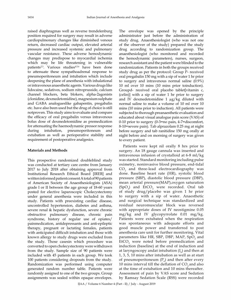
IJAA�/�Volume�6�Number�4�(Part�-�II)�/�July�-�August�2019
1414 Indian�Journal�of�Anesthesia�and�Analgesia
raised�diaphragmas�well�as� reverse� trendelenburg�position�re�uired�for�surgery�may�result�in�adverse�cardiopulmonary� changes� like� diminished� venous�return,� decreased� cardiac� output,� elevated� arterial�pressure� and� increased� systemic� and� pulmonary�vascular� resistance.� These� adverse� hemodynamic�changes� may� predispose� to� myocardial� ischemia�which� may� be� life� threatening� in� vulnerable�patients2,3.� Various� studies4,5,6� have� been� done�to� attenuate� these� sympathoadrenal� response� to�pneumoperitoneum�and� intubation�which� include�deepening�the�plane�of�anesthesia�with�inhalational�or�intravenous�anaesthetic�agents.�Various�drugs�like�lidocaine,�sedatives,�sodium�nitroprusside,�calcium�channel� blockers,� beta� blokers,� alpha-2agonists�(clonidine,�dexmedetomidine),�magnesium�sulphate�and� GA�A� analogueslike� gabapentin,� pregabalin�etc.�have�also�been�used�but�the�drug�of�choice�is�still�notproven.�This�study�aims�to�evaluate�and�compare�the� ef��cacy� of� oral� pregabalin� versus� intravenous�bolus�dose� of� dexmedetomidine� as�premedication�for�attenuating�the�haemodynamic�pressor�response�during� intubation,� pneumoperitoneum� and�extubation� as� well� as� perioperative� stability� and�re�uirement�of�postoperative�analgesics.
Materials�and�Methods�
This� prospective� randomized� doubleblind� study�was�conducted�at�tertiary�care�centre�from�January�2017� to� July� 2018� after� obtaining� approval� from�Institutional� Research� Ethical� �oard� �IRE��� and�written�informed�patient�consent�A�total�of�90�patients�of� American� Society� of� Anaesthesiologists� (ASA)�grade�I�or�II�between�the�age�group�of�18-60�years�posted� for� elective� laparoscopic� Cholecystectomy�under� general� anesthesia� were� included� in� this�study.� Patients� with� preexisting� cardiac� disease,�uncontrolled� hypertension,� diabetes� and� asthma,�severe�renal���hepatic�dysfunction,�severe�chronic�obstructive� pulmonary� disease,� chronic� pain�syndrome,� history� of� regular� use� of� opiates/painmedication,� antidepressants� and� anti-epileptic�therapy,� pregnant� or� lactating� females,� patients�with�anticipated�dif��cult�intubation�and�those�with�known�allergy�to�study�drugs�were�excluded�from�the� study.� Those� casesin� which� procedure� was�converted�to�open�cholecystectomy�were�withdrawn�from� the� study.� Sample� size� of� 90� patients� were�included�with� 45�patients� in� each�group.�We�took�100�patients� considering�dropouts� from�the�study.�Randomization� was� performed� using� computer�generated� random� number� table.� Patients� were�randomly�assigned�to�one�of�the�two�groups.�Group�assignments�was� sealed�within�opa�ue�envelopes.�
The� envelope� was� opened� by� the� principle�administrator� just� before� the� administration� of�study� drug.� Anaesthesiologist� (who� was� not� one�of� the� observer� of� the� study)� prepared� the� study�drug� according� to� randomization� group.� The�anaesthesiologist� (who� monitored� and� recorded�the� hemodynamic� parameters),� nurses,� surgeon,�research�assistant�and�the�patient�were�blinded�to�the�randomization.�Patients�in�both�the�groups�received�study�drug�as�per� the�protocol:�Group�P-�received�oral�pregabalin�150�mg�with�a�sip�of�water�1�hr�prior�to� surgery� and� intravenous� normal� saline� (0.9�)�10� ml� over� 10� mins� (10� mins� prior� toinduction).�Groupd-� received� oral� placebo� tablet�vitamin� c,�(celin)�� with� a� sip� of� water� 1� hr� prior� to� surgery�and� IV� dexmedetomidine� 1� �g/kg� diluted� with�normal� saline� to�make�a�volume�of� 10�ml� over� 10�mins�(10�mins�prior�to�induction).�All�patients�were�subjected�to�thorough�preanaesthetic�evaluation�and�educated�about�visual�analogue�pain�score�(VAS)�of�0-10�prior�to�surgery�(0-3�no�pain,�4-7�discomfort,�8-10�severe�pain).�Tab�alprazolam�0.25�mg�at�night�before�surgery�and�tab�ranitidine�150�mg�orally�at�night�before�and�on�morning�of�surgery�was�given�to�every�patient.
Patients� were� kept� nil� orally� 8� hrs� prior� to�surgery.�An�18�gauge�cannula�was�inserted�and�intravenous�infusion�of�crystalloid�at�6-8�ml/kg�was�started.�Standard�monitoring�including�pulse�oximetry,�noninvasive�blood�pressure,�end-tidal�CO
2� and� three-lead� electrocardiography� was�
done.� �aseline� heart� rate� (�R),� systolic� blood�pressure� (S�P),� diastolic� blood� pressure� (D�P),�mean� arterial� pressure(MAP),oxygen� saturation�(SpO
2)� and� EtCO
2� were� recorded.� Oral� tab�
of� study� drug/placebo� was� given� 1� hr� prior�to� surgery� with� a� sip� of� water.� Anaesthetic�and� surgical� techni�ue� was� standardized� and�residual� neuromuscular� block� was� reversed�with� appropriate� doses� of� IV� neostigmine� 0.05�mg/kg� and� IV� glycopyrrolate� 0.01� mg/kg.�Patients� were� extubated� when� the� respiration�was� spontaneous� with� ade�uate� efforts� and�good� muscle� power� and� transferred� to� post�anesthesia�care�unit�for�further�monitoring.�Vital�parameters� like��R,�S�P,�D�P,�MAP,�SpO
2�and�
EtCO2� were� noted� before� premedication� and�
induction�(baseline)�at�the�end�of�induction�and�at�laryngoscopy�andat�intubation�(I
0)�and�then�at�
1,�3,�5,�10�mins�after�intubation�as�well�as�at�start�of�pneumoperitoneum�(P
0)�and�then�after�every�
10�mins�interval�till�the�deflation�of�CO2�and�also�
at�the�time�of�extubation�and�10�mins�thereafter.�Assessment�of�pain�by�VAS� score�and�Sedation�by�Ramsay� Sedation� Scale� (RSS)�were� recorded�
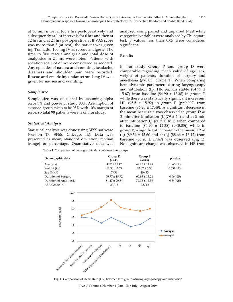
IJAA�/�Volume�6�Number�4�(Part�-�II)�/�July�-�August�2019
1415
at�30�min� interval� for�2�hrs�postoperatively�and�subse�uently�at�1�hr�intervals�for�6�hrs�and�then�at�12�hrs�and�at�24�hrs�postoperatively.�If�VAS�score�was�more�than�3�(at�rest),�the�patient�was�given�inj.�Tramadol�100�mg�IV�as�rescue�analgesic.�The�time� to� first� rescue� analgesic� and� total� dose� of�analgesics� in� 24� hrs� were� noted.� Patients� with�sedation�scale�of��3�were�considered�as�sedated.�Any�episodes�of�nausea�and�vomiting,�headache,�dizziness� and� shoulder� pain� were� recorded.�Rescue�anti-emetic�inj.�ondansetron�4�mg�IV�was�given�for�nausea�and�vomiting.
Sa��le�si�e
Sample� size� was� calculated� by� assuming� alpha�error�5��and�power�of�study�80�.�Assumption�of�exposed�group�taken�to�be�95��with�10��margin�of�error,�so�total�90�patients�were�taken�for�study.
Statistical�Analysis
Statistical�analysis�was�done�using�SPSS�software�(version� 17,� SPSS,� Chicago,� IL).� Data� was�presented� as� mean,� standard� deviation,� median�(range)� or� percentage.� �uantitative� data� was�
analyzed�using�paired�and�unpaired� t-test�while�categorical�variables�were�analyzed�by�Chi-s�uare�test.� p� values� less� than� 0.05� were� considered�signi��cant.
�esults
In� our� study� Group� P� and� group� D� were�comparable� regarding� mean� value� of� age,� sex,�weight� of� patients,� duration� of� surgery� and�anesthesia� (p�0.05)� (Table� 1).� When� comparing�hemodynamic� parameters� during� laryngoscopy�and� intubation� (I
0),� �R� remain� stable� (84.77� ��
15.67)� from� baseline� (84.90� �� 12.38)� in� group� D�while�there�was�statistically�signi��cant�increasein��R� (95.5� �� 15.92)� in� group� P� (p�0.002)� from�baseline�(86.20���17.49).�A�signi��cant�decrease�in�the�mean�heart� rate�was�observed�in�group�D�at�3�min� after� intubation� (I
3)(79� �� 14)� and�at� 5�min�
after� intubation(I5)� (80.5� �� 18.1)�when� compared�
to� baseline� (84.90� �� 12.38)� (p�0.05))� while� in�group�P,�a�signi��cant�increase�in�the�mean��R�at�(I
3)� (89.59���15.60�and�at� (I
5)� (88.66���16.12)� from�
baseline� (86.20� �� 17.49)� was� observed� (Fig� 1).�No�signi��cant� change�was�observed� in��R�from�
Comparison�of�Oral�Pregabalin�Versus��olus�Dose�of�Intravenous�Dexmedetomidine�in�Attenuating�the��emodynamic�responses�During�Laparoscopic�Cholecystectomy:�A�Prospective�Randomized�double��lind�Study
Table��:�Comparison�of�demographic�data�between�two�groups
�emographic�data��roup���n����
�roup�P�n����
p��alue
Age�(yrs) 42.7���11.47� 42.27���11.29� 0.846(NS)
Weight�(kg) 61.38���7.33� 62.87���5.50� 0.651(NS)
Sex�(M/F) 7/38� 10/35� -
Duration�of�Surgery 59.77���18.92 65.95���13.21 0.06(NS)
Duration�of�Anesthesia� 81.47���20.84� 79.13���15.59� 0.54(NS)
ASA�Grade�I/II 27/18 �33/12� �-
Fig���:�Comparison�of��eart�Rate�(�R)�between�two�groups�duringlaryngoscopy�and�intubation
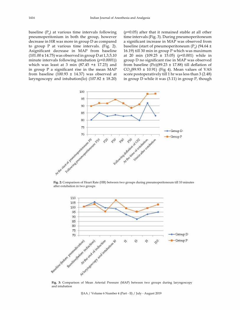
IJAA�/�Volume�6�Number�4�(Part�-�II)�/�July�-�August�2019
1416 Indian�Journal�of�Anesthesia�and�Analgesia
Fig���:�Comparison�of��eart�Rate�(�R)�between�two�groups�during�pneumoperitoneum�till�10�minutes�after�extubation�in�two�groups
baseline� (P0)� at� various� time� intervals� following�
pneumoperitonium� in� both� the� group,� however�decrease�in��R�was�more�in�group�D�as�compared�to� group� P� at� various� time� intervals.� (Fig.� 2).�Asigni��cant� decrease� in� MAP� from� baseline�(101.00���14.75)�was�observed�in�group�D�at�1,3,5,10�minute�intervals�following�intubation�(p�0.0001))which� was� least� at� 3� min� (87.45� ��� 17.23)� and�in� group� P� a� signi��cant� rise� in� the� mean� MAP�from� baseline� (100.93� �� 14.37)� was� observed� at�laryngoscopy�and� intubation(Io)� (107.82���18.20)�
(p�0.05)� after� that� it� remained� stable� at�all� other�time�intervals�(Fig.�3).�During�pneumoperitoneum�a�signi��cant�increase�in�MAP�was�observed�from�baseline�(start�of�pneumoperitoneum�(P
0)�(94.64���
16.19)�till�30�min�in�group�P�which�was�maximum�at� 20� min� (109.25� �� 15.05)� (p�0.001)� while� in�group�D�no�signi��cant�rise�in�MAP�was�observed�from�baseline� (Po)(89.23� ��17.88)� till�de��ation� of�CO
2(89.93� �� 10.91)� (Fig� 4).� Mean� values� of� VAS�
score�postoperatively�till�1�hr�was�less�than�3�(2.48)�in�group�D�while�it�was�(3.11)�in�group�P,�though�
Fig�� �:� Comparison� of� Mean� Arterial� Pressure� (MAP)� between� two� groups� during� laryngoscopy�and�intubation
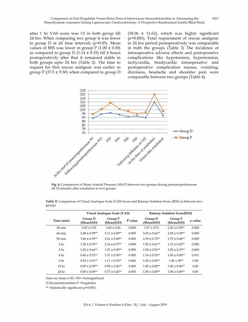
IJAA�/�Volume�6�Number�4�(Part�-�II)�/�July�-�August�2019
1417Comparison�of�Oral�Pregabalin�Versus��olus�Dose�of�Intravenous�Dexmedetomidine�in�Attenuating�the��emodynamic�responses�During�Laparoscopic�Cholecystectomy:�A�Prospective�Randomized�double��lind�Study
after� 1� hr� VAS� score� was� �3� in� both� group� till�24�hrs.�When�comparing�two�group�it�was�lower�in� group� D� at� all� time� intervals� (p�0.05).� Mean�values�of�RSS�was�lower�in�group�P�(1.00���0.00)�as�compared�to�group�D�(1.14���0.35)�till�4�hours�postoperatively� after� that� it� remained� stable� in�both� groups� upto� 24� hrs� (Table� 2).� The� time� to�re�uest� for� ��rst� rescue� analgesic� was� earlier� in�group�P�(37.5���9.30)�when�compared�to�group�D�
(58.06� �� 11.62),� which� was� highly� signi��cant�(p�0.001).� Total� re�uirement� of� rescue� analgesic�in�24�hrs�period�postoperatively�was�comparable�in� both� the� groups� (Table� 3)� The� incidence� of�intraoperative� adverse� effects� and� postoperative�complications� like� hypotension,� hypertension,�tachycardia,� bradycardia� intraoperative� and�postoperative� complication� nausea,� vomiting,�dizziness,� headache� and� shoulder� pain� were�comparable�between�two�groups�(Table�4).
Fig���:�Comparison�of�Mean�Arterial�Pressure�(MAP)�between�two�groups�during�pneumoperitoneum�till�10�minutes�after�extubation�in�two�groups
Table��:�Comparison�of�Visual�Analogue�Scale�(VAS)�Score�and�Ramsay�Sedation�Score�(RSS)�in�between�two�groups
Visual�Analogue�Scale��VAS� �amsay�Sedation�Score��SS�
Time��min��roup��
�Mean�S���roup�P
�Mean�S��P��alue
�roup���Mean�S��
�roup�P�Mean�S��
���alue
30�min 3.05���0.53 3.68���0.56 0.000 3.57���0.73 2.45���0.50�� 0.000
60�min 2.48���0.59�� 3.11���0.49�� 0.000 3.02���0.66�� 2.09���0.29�� 0.000
90�min 1.86���0.55�� 2.61���0.49�� 0.000 2.39���0.72�� 1.75���0.44�� 0.000
2�hr 1.50���0.55�� 2.16���0.37�� 0.000 1.95���0.61�� 1.23���0.42�� 0.000
3�hr 1.02���0.66�� 1.91���0.29�� 0.000 1.50���0.51�� 1.02���0.15�� 0.000
4�hr 0.66���0.53�� 1.57���0.50�� 0.000 1.14���0.35�� 1.00���0.00�� 0.011
6�hr 0.52���0.51�� 1.11���0.32�� 0.000 1.00���0.00�� 1.00���00�� 0.00
12�hr 0.09���0.29�� 0.98���0.26�� 0.000 1.00���0.00�� 1.00���0.00�� 0.00
24�hr 0.00���0.00�� 0.77���0.42�� 0.000 1.00���0.00�� 1.00���0.00�� 0.00
Data�are�mean���SD,�NS��Nonsignificant
D-Dexmedetomidine�P-�Pregabalin
��-�Statistically�significant�(p�0.001).

IJAA�/�Volume�6�Number�4�(Part�-�II)�/�July�-�August�2019
1418 Indian�Journal�of�Anesthesia�and�Analgesia
�iscussion�
Laparoscopic� cholecystectomy� is� considered� gold�standard�and�one�of�the�preferred�surgery�for�gall�bladder� diseases� under� general� anesthesia� in� the�present� era� due� to� its� well� known� advantages.��owever,�like�any�other�surgery�it�is�also�associated�with� sympathoadrenal� response� occurring� due�to� direct� laryngoscopy,� tracheal� intubation,�extubation�and�pneumoperitoneum�which�evokes�hemodynamic� instabilities.�Various�methods�have�been�used�to�attenuate�stress�response.�This�study�was� designed� to� compare� oral� pregabalin� versus�bolus� dose� of� intravenous� dexmedetomidine� as�premedication� in� attenuating� the� hemodynamic�responses� to� laryngoscopy,� intubation� and� also�during� pneumoperitoneum� in� laparoscopic�cholecystectomy.� The� incidence� of� bradycardia�was� 11.11�� in� group� D�while� 2.22�� in� group� P,�this� difference� may� be� due� to� highly� selective��2� agonist� action� of� dexmedetomidine� resulting�in� sympatholysis.� The� incidence� of� tachycardia�was�6.66�� in�group�D�while� it�was�20�� in�group�P� which� re��ects� better� hemodynamic� stability� of�dexmedetomidine� than� pregabalin.� A� signi��cant�reduction� in� �R� following� loading� dose� of�Dexmedetomidine�(6�mcg/kg)�after�intubation�and�
after� 20� min� of� pneumoperitoneum� as� compared�to� saline�group�was� observed� in� a� study�done�by�Vora��.S�et�al.13.�Rastogi���et�al.10�studied�the�effect�of� two� different� doses� of� oral� pregabalin� (75� mg�and� 150� mg)� as� premedication� and� observed� an�increase�in��R�in�group�P
150�from�baseline�(80.65���
3.84)�after�1min�of�laryngoscopy�(107���2.41)�which�was� similar� to�our�study.�Gupta���et�al.9� reported�that� after� premedication� with� oral� pregabalin�(150� mg)� and� placebo,� there� was� a� signi��cant�increase�in��R�in�both�groups�but�the�increase�was�less�in�pregabalin�group.�We�recorded�a�decrease�in�MAP�(98.61���19.21)�from�baseline�(101���14.75)�in�group�D�at�the�time�of�laryngoscopy�and�intubation,�while� in� group�P� there�was� a� signi��cant� increase�in� MAP� (107.82� �� 18.20)� from� baseline� (100.93� ��14.37).�Maximum�decrease� in�MAP�was� found� in�group�D,� at� 3�min�after� intubation� (87.45��� 17.23)�although� there�was�no�signi��cant�change� in�MAP�from� baseline� was� found� in� group� P� following�intubation.�When�comparing�both� the�groups,� fall�in�MAP� was� signi��cantly� more� in� group� D� than�in� group� P� (p�0.05).� This� may� be� attributed� to�sympatholytic�action�of�dexmedetomidine�on��2A�receptors� located� in� brainstem� vasomotor� centre�and� difference� in� the� route� of� administration� of�study�drugs.�Similar�to�our�study�Rastogi���et�al.10�
Table��:�Data�of�patients�re�uiring�rescue�analgesics�in�both�groups
�roup����n�����mean���S��
�roup�P��n�����mean���S��
���alue
Time�of�1st�rescue�Analgesic 58.06���11.62 37.5���9.30 0.0001
Total�no�of�patients�re�uiring�rescue�analgesics
1st
dose2nd
Dose3rd
dose1st
dose2nd
Dose3rd
dose–
28 13 1 23 15 3
Total�dose�of�analgesic�in�24�hrs 134.09���52.57 147.72���62.83 0.26
Data�are�mean�SD,�NS��NonsignificantD-Dexmedetomidine�P-�Pregabalin��-�Statistically�significant�(p�0.001)�
Table��:�Comparison�of�intraoperative�adverse�effect�and�postoperative�complications�between�two�groups
Variable �roup����n���� �roup�P��n���� ���alue
�ypotension 3 6.66� 8 17.77� 0.10
�ypertension 2 4.44� 5 11.11� 0.23
Tachycardia 3 6.66� 9 20� 0.06
�radycardia 5 11.11� 1 2.22� 0.09
Nausea 1 2.22� 3 6.66� 0.29
Vomiting 1 2.22� 2 4.44� 0.55
Dizziness 2 4.44� 6 13.33� 0.13
�eadache 1 2.22� 4 8.88� 0.16
Shoulder�pain 1 2.22� 2 4.44� 0.55
Data�are�mean�SD,�NS��Nonsignificant
D-Dexmedetomidine�P-�Pregabalin
��-�Statistically�significant�(p�0.001).
IJAA�/�Volume�6�Number�4�(Part�-�II)�/�July�-�August�2019
1419
in� compared� the� effect� of� two� different� doses� of�oral�pregabalin�(75�mg�and�150�mg)�and�observed�a� decrease� in� MAP� (87.06� �� 3.90)� from� baseline�(93.15���2.59)�after� induction� in�patients� receiving�pregabalin�(150�mg).�Meena�R�et�al.14�in�2016�studied�the� effect� of� oral� diazepam� �10� mg� (�S)� �� 5� mg�(1�hr�before� surgery)��with� two�different�doses�of�oral�pregabalin��75�mg�(�S)���150�mg,�300�mg�(1�hr�before� surgery)�� and� reported� signi��cant� increase�in�MAP�(106.44���6.24)�from�baseline�(91.14���4.16)�with�diazepam�while�increase�in�MAP�was�less�in�pregabalin� group�P
150�(98.43� �� 7.78)� from�baseline�
(91.22���6.90)�which�was�similar�to�our�study.
CO2� insuf��ation� along� with� trendelenburg�
position�re�uired�in� laparoscopic�surgeries�causes�signi��cant� release� of� catecholamines,� cortisol,�renin� and� vasopressin� leading� to� increase� in�systemic� vascular� resistance� and� pulmonary�vascular� resistance� and� tachycardia7.� In� our�study� an� increase� in� MAP� from� baseline� during�pneumoperitoneum�in�bothgroups�were�observed,�however� this� increase� was� more� in� pregabalin�group� when� compared� to� dexmedetomidine�groupthat� shows�dexmedetomidine� is� better� than�pregabalinin�attenuating�the�stress�response�during�pneumoperitionium,�Vora��S�et�al.13�also�reported�that� dexmedetomidine� is� better� than� pregabalin�in� attenuating� the� stress� response� during�pneumoperitonium.� Manne� GR� et� al.15� reported�signi��cant�decrease�in�MAP�after�starting�the�infusion�(dexmedetomidine� 1� �g/kg)� and� there� was� no�signi��cant�rise�in�MAP�during�pneumoperitonium�till�release�of�CO
2�(p�0.001).�Gupta���et�al.9�studied�
the� effect� of� clonidine� (200� �g)� and� pregabalin�(150�mg)�as�oral�premedication�during�laparoscopic�cholecystectomy�and�observed�that�haemodynamic�responses�were�attenuated�by�both�drugs�and�were�maintained� throughout� intraoperative� period,�however� clonidine� was� superior� to� pregabalin�for� attenuation� of� haemodynamic� responses� to�laryngoscopy� and� laparoscopy.�Analgesic� ef��cacy�of� dexmedetomidine� and� pregabalin� in� different�doses� has� been� studied� in� various� studies� with�different�results.�Dexmedetomidine�had�a�moderate�analgesic�effect�with� sedation�due� to� its�action�on�postsynaptic� alpha-2� adrenergic� receptor,� located�in�locus�coeruleus�and�receptors�in�the�dorsal�horn�of�spinal�cord20,21�The�analgesic�effect�of�pregabalin�is� due� to� binding� of� pregabalin� at� �-2-delta� site�with�conse�uent�reduction�in�release�of�excitatory�neurotransmitter� like� norepinephrine,� glutamate,�substance� P.8,22-24� In� our� study,� group� P� patients�experienced�more�postoperative�pain�as�compared�to� group�D� (p�0.05).� The� time� to� ��rst� re�uest� for�rescue�analgesic�was�earlier�(37.5���9.30)�in�group�P�
than� group� D� (58.06� �� 11.62)(p�0.001).� The� total�dose�of�analgesic�in�24�hrs�was�higher� in�group�P�(147.72� �� 52.57)� as� compared� to� group� D� (134.09��� 62.83).� Though� this� difference� was� statistically�insigni��cant� (p�0.26)� but� we� observed� that�patients� in� group�D�were� pain� free� in� immediate�postoperative� period� and� more� comfortable� in�24�hrs.�Pathak�AS�et�al.7�compared�the�two�different�doses� of� dexmedetomidine� (1� mcg/kg� and�0.7�mcg/kg)�given�preoperatively�as�bolus�in�patients�undergoing�laparoscopic�surgery�and�found�better�postoperative�analgesia�with�lower�VAS�scores�and�delayed� time� for���rst� rescue� analgesia� in�patients�receiving� dexmedetomidine� 1� �g/kg.� Esmat� IM�et� al.12� observed� a� signi��cant� reduction� in� VAS�Score� in� patients� receiving� two�different� doses� of�pregabalin�(150�mg,�300�mg)�1�hr�prior�to�surgery.�Pain� scores� were� lower� in� pregabalin� 300� mg.�Sundar�AS�et�al.25�found�no�difference�in�VAS�Score�and� total� fentanyl� re�uirement� postoperatively�among� group� P� (PG
150mg)� and� group�C� (NS)� at� 6,�
12�or�24�hrs�after� coronary�artery�bypass�grafting�(CA�G)�surgery.�Dexmedetomidine�and�pregabalin�also� have� anxiolytic� and� sedative� properties.�Dexmedetomidine�provides�sedation�by�stimulation�of� �2A� and� �2C� receptors� which� are� located� in�locus� ceruleus� in� the� spinal� cord.Theanxiolytic�property� of� pregabalin� is� by� decreasing� the�synthesis� of�neurotransmitter� glutamate� to� act� on�central�nervous�system.�In�our�study� the�sedation�score�was� higher� in� group�D� than� group� P� at� all�time� intervals� (p��� 0.05)� but� none� of� the� patients�in� bothgroups� had� sedation� score� more� than� �4.�Dexmedetomidine�in�a�dose�of�1�mcg/kg�has�been�shown�to�cause�increased�sedation�levels�and�need�for� oxygen� supplementation� by� few� authors26,27.�Sebastian���et�al.17�found�higher�sedation�scores�in�Dexmedetomidine� groups� (dex� 0.5,� 0.75� �g/kg)�than� normal� saline� (p� �� 0.05),� Similarly� Manne.�GR�et�al.15�also�observed� increased� sedation� levels�in� patients� receiving� low� dose� dexmedetomidine�infusion�(0.2��g/kg/h,�0.4��g/kg/h).�In�contrast�to�our�study�Parveen�S�et�al.19� found�higher�sedation�score�in�pregabalin�group�(2.73���0.55)�as�compared�to�oral�clonidine�(2.20���0.41),�this�can�be�attributed�to�difference� in� the� route�of�administration�as�we�administered� alpha-2� agonist� dexmedetomidine�IV.� Anand� L�� et� al.18� showed� that� sedation�score� measured� by� somnolence� sedation� scale�wascomparable�between�the�control�and�pregabalin�group�(150�mg)�(p���0.05).�No�patient�had�sedation�score�of�3�and�all�patients�were�free�of�sedation�at�6� hrs� which� is� similar� to� our� study.� The� adverse�effects�were�found�to�be�statistically�insigni��cant�in�our�study�(p���0.05).
Comparison�of�Oral�Pregabalin�Versus��olus�Dose�of�Intravenous�Dexmedetomidine�in�Attenuating�the��emodynamic�responses�During�Laparoscopic�Cholecystectomy:�A�Prospective�Randomized�double��lind�Study
IJAA�/�Volume�6�Number�4�(Part�-�II)�/�July�-�August�2019
1420 Indian�Journal�of�Anesthesia�and�Analgesia
In� our� study,� the� incidence� of� postoperative�nausea� and� vomiting� (PONV)� was� found�higher� in� Pregabalin� group� (6.66�)� than� the�dexmedetomidine� group� (2.22�),� 2� patients� from�group� D� (4.44�)� while� 6� patients� from� group� P�(13.33�)� experienced� postoperative� dizziness,�1�patient�from�group�D�while�4�patients�in�group�P�experienced�headache�and�shoulder�pain.�All�these�complications�were�statistically�insigni��cant�in�our�study� (p� �� 0.05).� Esmat� IM� et� al.12� observed� that�postoperative�vomiting�was�statistically�signi��cant�in�patients�receiving�pregabalin�300�mg�(p���0.01).�Gupta�P�et�al.16�in�2017�and�Meena�R�et�al.14�observed�that� only� one� patient� in� group� P� (150� mg)� and�2�patients� in�group�P� (300�mg)� suffered�dizziness�but�it�was�statistically�insigni��cant�(p���0.05).
Limitation�of�our�study�was� that�we�could�not�measure� stress� mediators� such� as� endogenous�catecholamines� or� cortisol� and�dial� concentration�of� sevo��urane.� We� did� not� use� �IS� monitoring�and� invasive� blood� pressure� monitoring.�A�control�group�was�not�included�in�the�study�for�comparison.
Conclusion
This� studyconcluded� that� both� dexmedetomidine�(1� �g/kg� IV� bolus)� and� pregabalin� (150� mg�orally)� is� effective� in� attenuating� haemodynamic�stress� response� during� laryngoscopy,� intubation�and� pneumoperitoneum� when� given� as�premedication� for� laparoscopic� cholecystectomy.Dexmedetomidine� was� found� to� be� superior� in�ameliorating� the� haemodynamic� responses� to�laryngoscopy� and� laparoscopy� along� with� better�postoperative� analgesia� without� any� signi��cant�adverse�effects.
�eferences
1.� �isgaard� T,� �ehlet� �,� Rosenberg� J.� Pain� and�convalescence�after�laparoscopic�cholecystectomy.�Eur�J�Surg.�2001;167:84–96.
2.� Joris� JL,� Noirot� DP,� Legrand� MJ,� et� al.��emodynamic� changes� during� laparoscopic�cholecystectomy.�AnaesthAnalg�1993;76:1067–71.
3.� Wahba� RW,� �ei� �ue� F,� �leiman� SJ.�Cardiopulmonary� function� and� laparoscopic�cholecystectomy.�Can�J�Anaesth.�1995;42(1):51–63.
4.� Raichurkar� A,� Dinesh� �,� Ravi� M,� et� al.� A�comparative�study�of�oral�pregablin�and�clonidine�for� attenuation� of� hemodynamic� responses� to�laryngoscopy�and�tracheal� intubation.�Journal�of�Clinical�and��iomedical�Sciences.�2015;5:25–9.
5.� Salman� E,� Celik� C,� Candar� S.� premedication�with� single� dose� pregabalin� 150� mg� attenuates�hemodynamic� response� to� laryngoscopy� and�endotracheal�intubation.�2012;1:297.�doi:�10.4172/scientificreports�297.
6.� �handari�G,�Mitra�S,�Shahi��N,�et�al.�Pre-emptive�use� of� oral� pregablin� attenuates� the� pressor�response� of� laryngoscopy� and� endotracheal�intubation:� A� double� blind� randomized� placebo�controlled�study.�Annals�of�International�Medical�and�Dental�Research.�2016;�2:110–4.
7.� Pathak� AS,� Paranjpe� JS,� �ulkarni� R�.�Comparison�of�two�doses�of�dexmedetomidine�on�haemodynamic� stability� in� patients� undergoing�laparoscopic� surgeries.� Journal� of� �rishna�Institute� of� Medical� Sciences� �niversity.� 2016;5:�35–44.
8.� Agrawal�A,�Gautam�S,�Gupta�D,�et�al.�Evaluation�of� a� single� preoperative� dose� of� pregabalin�for� attenuation� of� postoperative� pain� after�laparoscopic� cholecystectomy,� �ritish� Journal� of�Anesthesia.�2008;101(5):700–4.
9.� Gupta��,�Sharma�D,�Gupta�P�.�Oral�premedication�with� pregabalin� or� clonidine� for� hemodynamic�stability� during� laryngoscopy� and� laparoscopic�cholecystectomy:� A� comparative� evaluation.�Saudi�J�Anaesth.�20111;5(2):179–84.
10.� Rastogi� �,� Gupta� �,� Gupta� P�,� et� al.� Oral�pregabalin� premedication� for� attenuation� of�haemodynamic� pressor� response� of� airway�instrumentation� during� general� anesthesia:� A�dose�response�study.�Indian�Journal�of�Anesthesia.2012;56(1):49–54.
11.� Saraf�R,�Jha�M,��umar�S,�et�al.�Dexmedetomidine,�the� ideal� drug� for� attenuating� the� pressor�response.� Pediatric� Anesthesia� and� Critical� care�Journal.�2013;1:78–86.
12.� Esmat� IM,� �anan� MF.� Comparative� study�between�paracetamol�and�two�different�doses�of�pregabalin�on�postoperative�pain�in�laparoscopic�cholecystectomy.� Saudi� Journal� of� Anesthesia.�2015;9(4):376–80.
13.� Vora��S,��aranda��,�Shah�VR,�et�al.�The�effects�of�dexmedetomidine�on�attenuation�of�hemodynamic�changes�and�there�effects�as�adjuvant�in�anesthesia�during� laparoscopic� surgeries.� Saudi� Journal� of�Anesthesia.�2015;9(4):386–92.
14.� Meena� R,� Meena� �,� Prakash� S.� Study� of�attenuation� of� cardiovascular� response� during�laryngoscopy� and� intubation� using� two�different�dose�of�pregabalin�as�premedication� in�controlled�hypertensive�patients-A�RCT.� Journal�of� Anesthesia� and� Clinical� Research.� 2016;7(2):�607–13.
15.� Manne� GR,� �padhyay� MR,� Swadia� VN.� Effects�of� low� dose� dexmedetomidine� infusion� on�haemodynamic� stress� response,� sedation� and�postoperative�analgesia�re�uirements�in�patients�
IJAA�/�Volume�6�Number�4�(Part�-�II)�/�July�-�August�2019
1421
undergoing�laparoscopic�cholecystectomy.�Indian�Journal�of�Anesthesia.�2014;58:726–31.
16.� Gupta� P,� Saxena� A,� Chaudhary� L.� Effect� of�pregabalin� premedication� on� re�uirement� of�anaesthetic� and� analgesic� drugs� in� laparoscopic�cholecystectomy:�Randomized�comparison�of�two�doses.�2016;11(2):330–33.
17.� Sebastian� �,� Tallikoti� AT,� �rishnamurthy� D.�Attenuation� of� hemodynamic� responses� to�laryngoscopy� and� endotracheal� intubation� with�intravenous� dexmedetomidine:� a� comparison�between�two�doses.�2017;61(1):58–63.
18.� Anand�L�,�Sandhu�M,�Singh�J,�et�al.�Evaluation�of�analgesic�efficacy�of�pregabalin�for�postoperative�pain� relief� after� laparoscopic� cholecystectomy:� a�double�blind�study.�Anesthesia�Pain�and�Intensive�care.�2017;21(2):174–80.
19.� Parveen�S,�Negi�DS,��umar�R,�et�al.�Oral�clonidine�vs� oral� pregabalin� premedication� to� attenuate�pressor�response�to�direct�laryngoscopy�in�patients�undergoing� laparoscopic� cholecystectomy:� A�randomized� double� blind� study.� Journal� of�clinical�and�diagnostic�research.�2016;10(9):21–25.
20.� De� �ock� M,� Crochet� �,� Morimont� C,� et� al.�Intravenous� or� epidural� clonidine� for� intra-and�postoperative� analgesia.� Anaesthesiology.� 1993;�79:525–31.��Pub�Med�
21.� Correa-Sales� C,� Rabin� �C,� Maze� M.� A� hypnotic�response� to� dexmedetomidine,� an� alpha� 2�
agonist,� is� mediated� to� locus� coeruleus� in� rats.�Anesthesiology.�1992;76:948–52.
22.� Dahl� J�,� Mathieson� O,� Moiniche� S.� Protective�premedication:� an� option� with� gabapentin� and�related� drugs.� A� review� of� gabapentin� and�pregabalin�in�the�treatment�of�postoperative�pain.�Acta�Anaesthesiol�Scand.�2004;48:1130–6.
23.� Rowbotham� DJ.� Gabapentin:� A� new� drug� for�postoperative�pain.��r�JAneath.�2006;96:152–5.
24.� Turan�A,��aya�G,��aramanlioglu��,� et� al.�Effect�of� oral� gabapentin� on� postoperative� epidural�analgesia.��r�J�Anaesth.�2006;96:242–6.
25.� Sundar� AS,� �odali� R�,� Sulaiman� S,� et� al.� the�effects� of� preemptive� pregabalin� on� attenuation�of�stress�response�to�endotracheal�intubation�and�opioid-sparing�effect� in�patients�undergoing�off-pump�coronary�artery�bypass�grafting.�Annals�of�cardiac�anesthesia.�2012;15(1):18–25.
26.� Sulhyan� SR,� Vagarali� AT,� Patil� SS,� et� al.� A�comparative� clinical� study� of� dexmedetomidine�versus� placebo� to� attenuate� hemodynamic�response� to� endotracheal� lintubation� in� patients�undergoing� off� pump� coronary� arterial� bypass�grafting.�J�Sci�Soc.�2014;41:151–5.
27.� �elleville� JP,� Ward� DS,� �loor� �C,� et� al.� Effects�of� intravenous� dexmedetomidine� in� humans.�I.� Sedation,� ventilation,� and� metabolic� rate.�Anaesthesiology.�1992;77:1125–33.
Comparison�of�Oral�Pregabalin�Versus��olus�Dose�of�Intravenous�Dexmedetomidine�in�Attenuating�the��emodynamic�responses�During�Laparoscopic�Cholecystectomy:�A�Prospective�Randomized�double��lind�Study
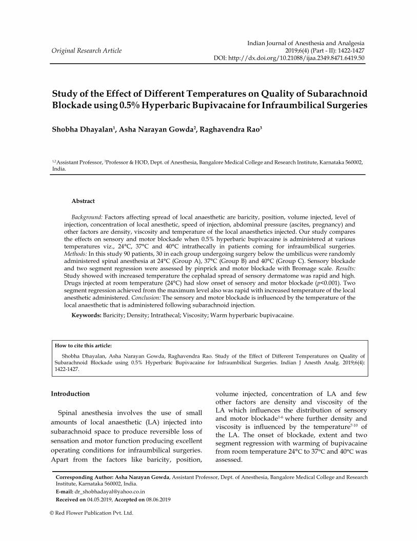
��Red�Flower�Publication�Pvt.�Ltd.�
�Original�Research�ArticleIndian�Journal�of�Anesthesia�and�Analgesia�
2019;6(4)�(Part�-�II):�1422-1427DOI:�http://dx.doi.org/10.21088/ijaa.2349.8471.6419.50
Study�of�the�Effect�of��ifferent�Temperatures�on��uality�of�Subarachnoid�Blockade�using�������yperbaric�Bupi�acaine�for�Infraumbilical�Surgeries
Shobha��hayalan���Asha�Narayan��o�da����agha�endra��ao�
1,2Assistant�Professor,�3Professor����OD,�Dept.�of�Anesthesia,��angalore�Medical�College�and�Research�Institute,��arnataka�560002,�India.
Corresponding�Author:�Asha�Narayan��o�da,�Assistant�Professor,�Dept.�of�Anesthesia,��angalore�Medical�College�and�Research�Institute,��arnataka�560002,�India.
E�mail:�[email protected]
�ecei�ed�on�04.05.2019,�Accepted�on�08.06.2019
Abstract
Background:�Factors�affecting�spread�of� local�anaesthetic�are�baricity,�position,�volume� injected,� level�of�injection,�concentration�of�local�anesthetic,�speed�of�injection,�abdominal�pressure�(ascites,�pregnancy)�and�other�factors�are�density,�viscosity�and�temperature�of�the�local�anaesthetics�injected.�Our�study�compares�the� effects� on� sensory�and�motor�blockade�when�0.5��hyperbaric�bupivacaine� is� administered�at�various�temperatures� viz.,� 24�C,� 37�C� and� 40�C� intrathecally� in� patients� coming� for� infraumbilical� surgeries.�Methods:�In�this�study�90�patients,�30�in�each�group�undergoing�surgery�below�the�umbilicus�were�randomly�administered�spinal�anesthesia�at�24�C�(Group�A),�37�C�(Group��)�and�40�C�(Group�C).�Sensory�blockade�and� two� segment� regression�were� assessed�by�pinprick� and�motor�blockade�with��romage� scale.�Results:�Study�showed�with�increased�temperature�the�cephalad�spread�of�sensory�dermatome�was�rapid�and�high.�Drugs�injected�at�room�temperature�(24�C)�had�slow�onset�of�sensory�and�motor�blockade�(p�0.001).�Two�segment�regression�achieved�from�the�maximum�level�also�was�rapid�with�increased�temperature�of�the�local�anesthetic�administered.�Conclusion:�The�sensory�and�motor�blockade�is�influenced�by�the�temperature�of�the�local�anaesthetic�that�is�administered�following�subarachnoid�injection.
�ey�ords:��aricity;�Density;�Intrathecal;�Viscosity;�Warm�hyperbaric�bupivacaine.
�o��to�cite�this�article:
Shobha� Dhayalan,� Asha� Narayan� Gowda,� Raghavendra� Rao.� Study� of� the� Effect� of� Different� Temperatures� on� �uality� of�Subarachnoid� �lockade� using� 0.5�� �yperbaric� �upivacaine� for� Infraumbilical� Surgeries.� Indian� J� Anesth� Analg.� 2019;6(4):�1422-1427.
Introduction
Spinal� anesthesia� involves� the� use� of� small�
amounts� of� local� anaesthetic� (LA)� injected� into�
subarachnoid� space� to�produce� reversible� loss� of�
sensation�and�motor�function�producing�excellent�
operating� conditions� for� infraumbilical� surgeries.
Apart� from� the� factors� like� baricity,� position,�
volume� injected,� concentration� of� LA� and� few�other� factors� are� density� and� viscosity� of� the�LA� which� in��uences� the� distribution� of� sensory�and� motor� blockade1-6�where� further� density� and�viscosity� is� in��uenced� by� the� temperature7-10� of�the� LA.� The� onset� of� blockade,� extent� and� two�segment�regression�with�warming�of�bupivacaine�from�room�temperature�24�C�to�37�C�and�40�C�was�assessed.
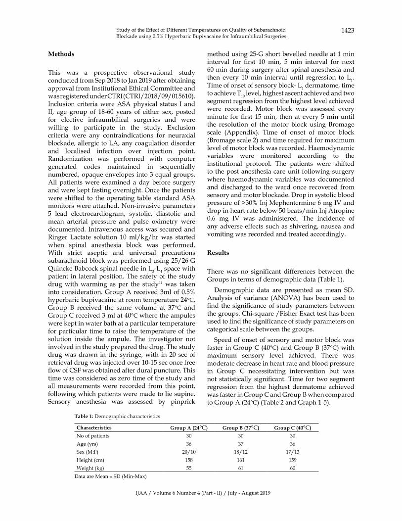
IJAA�/�Volume�6�Number�4�(Part�-�II)�/�July�-�August�2019
1423
Methods
This� was� a� prospective� observational� study�conducted�from�Sep�2018�to�Jan�2019�after�obtaining�approval�from�Institutional�Ethical�Committee�and�was�registered�under�CTRI�(CTRI/2018/09/015610).�Inclusion� criteria�were� ASA�physical� status� I� and�II,� age� group� of� 18-60� years� of� either� sex,� posted�for� elective� infraumbilical� surgeries� and� were�willing� to� participate� in� the� study.� Exclusion�criteria� were� any� contraindications� for� neuraxial�blockade,�allergic�to�LA,�any�coagulation�disorder�and� localised� infection� over� injection� point.Randomization� was� performed� with� computer�generated� codes� maintained� in� se�uentially�numbered,�opa�ue�envelopes�into�3�e�ual�groups.�All� patients� were� examined� a� day� before� surgery�and�were�kept�fasting�overnight.�Once�the�patients�were�shifted� to� the�operating� table� standard�ASA�monitors�were�attached.�Non-invasive�parameters�5� lead� electrocardiogram,� systolic,� diastolic� and�mean� arterial� pressure� and� pulse� oximetry� were�documented.� Intravenous�access�was�secured�and�Ringer� Lactate� solution� 10� ml/kg/hr� was� started�when� spinal� anesthesia� block� was� performed.�With� strict� aseptic� and� universal� precautions�subarachnoid�block�was�performed�using�25/26�G��uincke��abcock�spinal�needle�in�L
3-L
4�space�with�
patient� in� lateral�position.�The�safety�of� the�study�drug� with� warming� as� per� the� study11� was� taken�into�consideration.�Group�A�received�3ml�of�0.5��hyperbaric�bupivacaine�at�room�temperature�24�C,�Group� �� received� the� same� volume� at� 37�C� and�Group�C�received�3�ml�at�40�C�where�the�ampules�were�kept�in�water�bath�at�a�particular�temperature�for�particular� time� to� raise� the� temperature�of� the�solution� inside� the� ampule.� The� investigator� not�involved�in�the�study�prepared�the�drug.�The�study�drug�was�drawn� in� the�syringe,�with� in� 20� sec� of�retrieval�drug�was�injected�over�10-15�sec�once�free���ow�of�CSF�was�obtained�after�dural�puncture.�This�time�was�considered�as�zero�time�of�the�study�and�all� measurements� were� recorded� from� this� point,�following�which�patients�were�made�to�lie�supine.�Sensory� anesthesia� was� assessed� by� pinprick�
method�using�25-G�short�bevelled�needle�at�1�min�interval� for� ��rst� 10� min,� 5� min� interval� for� next�60�min�during�surgery�after�spinal�anesthesia�and�then� every� 10� min� interval� until� regression� to� L
1.�
Time�of�onset�of�sensory�block-�L1�dermatome,�time�
to�achieve�T10�level,�highest�ascent�achieved�and�two�
segment�regression�from�the�highest�level�achieved�were� recorded.� Motor� block� was� assessed� every�minute� for���rst� 15�min,� then�at� every�5�min�until�the� resolution� of� the� motor� block� using� �romage�scale� (Appendix).� Time� of� onset� of� motor� block�(�romage�scale�2)�and�time�re�uired�for�maximum�level�of�motor�block�was�recorded.��aemodynamic�variables� were� monitored� according� to� the�institutional� protocol.� The� patients� were� shifted�to� the�post�anesthesia�care�unit� following�surgery�where� haemodynamic� variables� was� documented�and� discharged� to� the� ward� once� recovered� from�sensory�and�motor�blockade.�Drop�in�systolic�blood�pressure�of��30��Inj�Mephentermine�6�mg�IV�and�drop�in�heart�rate�below�50�beats/min�Inj�Atropine�0.6� mg� IV� was� administered.� The� incidence� of�any�adverse�effects� such�as� shivering,�nausea�and�vomiting�was�recorded�and�treated�accordingly.
�esults
There� was� no� signi��cant� differences� between� the�Groups�in�terms�of�demographic�data�(Table�1).
Demographic� data� are� presented� as� mean� SD.�Analysis� of� variance� (ANOVA)� has� been� used� to���nd�the�signi��cance�of�study�parameters�between�the�groups.�Chi-s�uare�/Fisher�Exact�test�has�been�used�to���nd�the�signi��cance�of�study�parameters�on�categorical�scale�between�the�groups.
Speed�of�onset�of�sensory�and�motor�block�was�faster�in�Group�C�(40�C)�and�Group���(37�C)�with�maximum� sensory� level� achieved.� There� was�moderate�decrease�in�heart�rate�and�blood�pressure�in� Group� C� necessitating� intervention� but� was�not� statistically� signi��cant.� Time� for� two� segment�regression� from� the� highest� dermatome� achieved�was�faster�in�Group�C�and�Group���when�compared�to�Group�A�(24�C)�(Table�2�and�Graph�1-5).
Table��:�Demographic�characteristics�
Characteristics �roup�A�����C� �roup�B�����C� �roup�C�����C�
No�of�patients 30 30 30
Age�(yrs) 36 37 36
Sex�(M:F) 20/10 18/12 17/13
�eight�(cm) 158 161 159
Weight�(kg) 55 61 60
Data�are�Mean���SD�(Min-Max)
Study�of�the�Effect�of�Different�Temperatures�on��uality�of�Subarachnoid��lockade�using�0.5���yperbaric��upivacaine�for�Infraumbilical�Surgeries
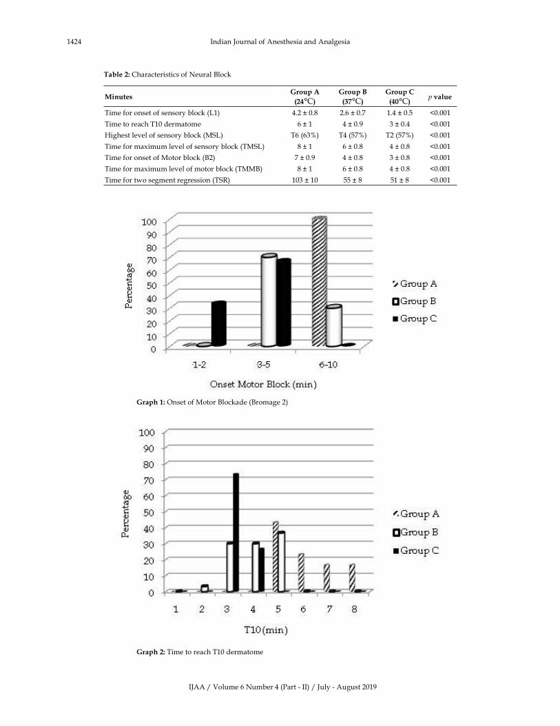
IJAA�/�Volume�6�Number�4�(Part�-�II)�/�July�-�August�2019
1424 Indian�Journal�of�Anesthesia�and�Analgesia
Table��:�Characteristics�of�Neural��lock
Minutes�roup�A�
����C�
�roup�B�
����C�
�roup�C�
����C�p��alue
Time�for�onset�of�sensory�block�(L1) 4.2���0.8 2.6���0.7 1.4���0.5 �0.001
Time�to�reach�T10�dermatome 6���1 4���0.9 3���0.4 �0.001
�ighest�level�of�sensory�block�(MSL) T6�(63�) T4�(57�) T2�(57�) �0.001
Time�for�maximum�level�of�sensory�block�(TMSL)� 8���1 6���0.8 4���0.8 �0.001
Time�for�onset�of�Motor�block�(�2) 7���0.9 4���0.8 3���0.8 �0.001
Time�for�maximum�level�of�motor�block�(TMM�) 8���1 6���0.8 4���0.8 �0.001
Time�for�two�segment�regression�(TSR) 103���10 55���8 51���8 �0.001
� �raph��:�Onset�of�Motor��lockade�(�romage�2)
� �raph��:�Time�to�reach�T10�dermatome
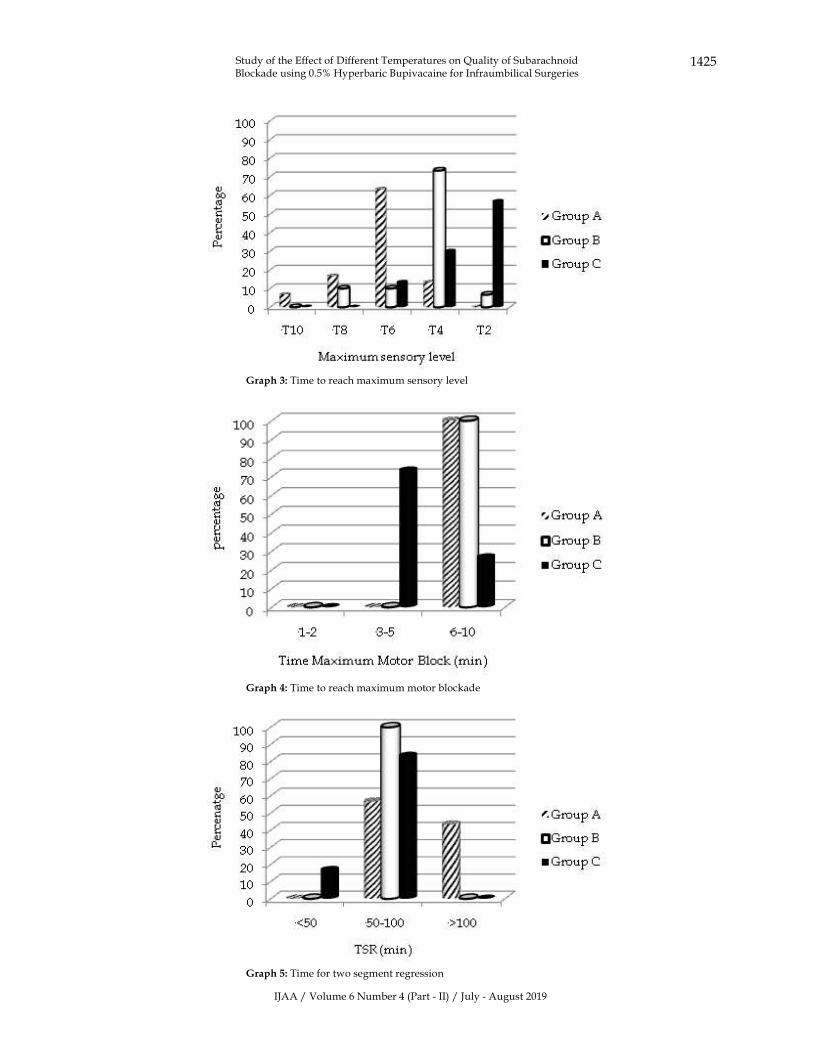
IJAA�/�Volume�6�Number�4�(Part�-�II)�/�July�-�August�2019
1425
� � �raph��:�Time�to�reach�maximum�sensory�level
� � �raph��:�Time�to�reach�maximum�motor�blockade
� � �raph��:�Time�for�two�segment�regression
Study�of�the�Effect�of�Different�Temperatures�on��uality�of�Subarachnoid��lockade�using�0.5���yperbaric��upivacaine�for�Infraumbilical�Surgeries
IJAA�/�Volume�6�Number�4�(Part�-�II)�/�July�-�August�2019
1426 Indian�Journal�of�Anesthesia�and�Analgesia
�iscussion
Large�comparative�studies�and�multiple�prospective�randomized� control� trials� have� reported� with�increase� in� temperature� of� bupivacaine� there�was�faster�onset�of�sensory�blockade.�In�addition�our� study� demonstrated� when� 0.5�� hyperbaric�bupivacaine� was� administered� intrathecally�at� 24�C� (room� temperature)� had� slow� onset� of�sensory� blockade.� At� 37�C� and� 40�C� when� 0.5��hyperbaric� bupivacaine� was� administered� there�was� rapid�onset�of� sensory�and�motor�blockade.�There�was�higher�level�of�sensory�block�achieved.�Regression� of� two� segment� from� highest�dermatome� achieved� also� was� faster� with� 37�C
and�40�C�compared�to�24�C.
Miriam� E� Tucker12� had� failed� spinal� anesthesia�in�a�group�of�14�cases�who�underwent�C-sections�which�was�believed�to�be�the�result�of�exposure�of�the�anaesthetic�to�lower�temperatures.�Due�to�which�there�will�be� signi��cant� clinical� conse�uences� like�pain� during� surgery,� repeat� subarachanoid� block�or� conversion� to� general� anesthesia.� The� other�contributing�factors�for�failed�neuraxial�anesthesia�like� technical� failure� has� to� be� identi��ed� and�managed�accordingly.
The� density� of� LA� is� key� determinant� of� LA�distribution�with�in�the�subarachnoid�space13.�CSF�density� varies� between� 1.00028� to� 1.00100� g/ml14.�For�every�increase�in�temperature�by�1�C�between�23�C� and� 37�C� the� density� of� all� plain� solutions�fall� by� 0.0003� mg/ml.� Viscosity� and� density� of�the� LA� reduces� with� warming15� and� affects� the�distribution�of�spinal�anesthesia.�The�time�of�onset�of�LA�is�related�to�the�pka�values�which�is�between�7.8�and�9.1�when�there�will�be�a�higher�percentage�of� non-ionized� free� base� which� is� close� to� the�physiologic�p�16.�So�a�more�rapid�onset�and�higher�level�of�sensory�block�is�achieved17,18.
Aria� et� al.19� Showed� increase� in� the� fraction�of� unionized� drug� due� to� decrease� in� pka� of� LA�solution� when� the� temperature� of� the� LA� was�increased� which� was� demonstrated� by� increased�uptake�of�LA�by�mammalian�nerve�with�increased�temperature.
Data� from� �iguchietal20� proved� strong�relationship�between�CSF�density�and�highest�level�of�sensory�block�achieved.
Depending� on� thermodynamics,� temperature�serves�to�gauge�the�intensity�of�the�thermal�energy�that�is�an�actual�energy�of�motion�(kinetic�energy)�of�the�individual�mobile�particle�matter.��igher�level�
of�sensory�block�achieved�could�be�due�to�increased�temperature�with�which�molecular� kinetic� energy�is� increased� and� number� of� individual� mobile�particles�increases.21
�i�itations
Even� though� the� waterbath� was� within� the�operating� room� the� temperature� of� the� ampule�will�drop�once� it� is� retrieved� from� the�waterbath,�so�it�was�impossible�to�administer�the�drug�exactly�at� 40�C� or� 37�C.� The� temperature� of� the� injectate�was�not�directly�measured.� In�our�research�centre�thermostatically� controlled� waterbath/incubator�was� not� available.� So� precise� temperature� was�not� maintained.� Waterbath� which� was� used� in�our� setup� was� relatively� economical� which� can�be�used� in� low�setup�hospitals�but�precautions�of�contamination� has� to� be� taken.With� precautions,�prewarming� of� LA� solutions� is� inherently� safe.��sing� thermostatically� controlled� waterbath� or�intravenous�solution�warmer�LA�ampules�may�be�warmed�upto�43�C.�Ensure�that�overheating�is�not�allowed� to� occur10.� Dry� heating� may� prevent� the�risk� of� undetected� contamination� by� water� from�heating�in�a�water�bath.
Conclusions
With� increase� in� temperature� of� hyperbaric�bupivacaine�from�24�C�to�37�C�and�40�C�for�spinal�anesthesia�there�is�higher�ascend�of�sensory�block,�rapid� onset� and� faster� regression� of� sensory� and�motor�block.�Warmed�bupivacaine�can�be�preferred�in� ambulatory� surgery� and� in� setups� with� high�turnover� rate� of� surgeries.� With� smaller� dose� we�can�achieve�a�high�level�of�blockade�so�can�reduce�the� dosage�of� the�LA� without�much� of� change� in�haemodynamics� which� must� be� con��rmed� by�further�studies.
Source�of�su��ort:�Nil
Presentation�at�a��eetin�:�Nil
Con��ict�of�interest:�None
A��endix:��romage�scale
�romage�1-�Free�movement�of�legs�and�feet.
�romage� 2� -� Just� able� to� ��ex� knee� with� free�movement�of�the�feet.
�romage�3�-��nable�to���ex�knees,�but�with� free�movement�of�the�feet.
�romage�4�-��nable�to�move�the�legs�or�feet.
IJAA�/�Volume�6�Number�4�(Part�-�II)�/�July�-�August�2019
1427
�eferences
1.� Chambers�WA,�Edstorm���,�Scott�D�.�Effect�of�baricity� in� spinal� anesthesia� with� bupivacaine.��r�J�Anaesth.�1981;53:279–82.
2.� Jankowska�A,�Veillette��.�Comparison�of�different�blocks� during� spinal� anesthesia� using� isobaric�versus� 2�� hyperbaric� lidocaine.� Can� J� Anaesth.�2000;47:137–42.
3.� McLeod� GA.� Density� of� spinal� anesthesia�solutions� of� bupivacaine,� levobupivacaine� and�ropivacaine� with� and� without� dextrose.� �r� J�Anaesth.�2004;92:547–51.
4.� Parlow�JL,�Money�P,�Chang�PS,�et�al.�The�addition�of� opioids� alters� the� density� and� spread� of�intrathecal� local� anesthetics:� an� in� vitro� study.�Can�J�Anaesth.�1999;46:66–70.
5.� Sanderson� P,� Read� J,� Littlewood� DG,� et�al.� Interaction� between� baricity� (glucose�concentration)� and� other� factors� that� influence�the� intrathecal� dissemination� of� the� drug.� �r� J�Anaesth.�1994;73:744–6.
6.� Lui�AC,�Polis�T�,�Cicutti�NJ.�Cerebrospinal�fluid�density� and� spinal� anesthesia� solutions� in� body�temperature� surgical� patients.� Can� J� Anaesth.�1998;45:297–303.
7.� Stienstra� R,� Gielen� M,� Van� Poorten� F.� Spinal�anesthesia� with� simple� bupivacaine.� 0.5��regression� of� sensory� and� motor� block� with�different�temperatures�of�the�anesthetic�solution.�Anesth�Analg.�1989;69:593–7.
8.� Callesen�T,� Jarnvig�I,�Thage��,�et�al.�Influence�of�bupivacaine�temperature�on�the�dissemination�of�spinal�analgesia.�Anesthesia.�1991;46:17–9.
9.� Stienstra� R,� Gielen� M,� �roon� JW,� van� Poorten�F.� The� influence� of� temperature� and� injection�rate� on� the� distribution�of� a� solution� containing�bupivacaine�and�methylene�blue�in�a�spinal�canal�model.�Reg�Anesth.�1990;15:6–11.
10.� Stienstra� R,� Van� Poorten� JF.� The� bupivacaine�temperature� at� 0.5�� affects� the� sensory� level� of�spinal�anesthesia.�Anesth�Analg.�1988;67:272-6.
11.� �oung-Chang� PA,� WAsa� �,� Eri� T,� et� al.� The�influence�of�hyperbaric�bupivacaine�temperature�on�the�spread�of�spinal�anesthesia.�Anesth�Analg.�2006;102:272–5.
12.� Miriam�E.�Tucker.�Spinal�anesthesia�failed�due�to�cold� temperature.�MDedge� ObGyn�News�1� July�2010.
13.� Na� ��,� �opacz� DJ.� Chloroprocaine� spinal�solutions:�density�at�37�degrees�C�and�p��titration.�Analg.�Anesthesia.�2004Jan;98(1):70–4.
14.� Richarson�MG,�Wissler�RN.�Lumbar�cerebrospinal�fluid� density� in� pregnant� and� non-pregnant�women.�Anesthesiology.�1996;85:326–30.
15.� Aria� �C,� �eda� W,� Takimoto� E.� The� influence�of� hyperbaric� bupivacaine� temperature� on� the�spread� of� spinal� anesthesia.� Analg.� Anesthesia.�2006�Jan;102(1):272–5.
16.� Leykin� �,� Nespolo� R,� Foltran� F.� Postoperative�anesthesia� and� analgesia� after� intra-articular�injection�of�heated�levobupivacaine�compared�to�room� temperature:� a� randomized� double-blind�study.�Arthroscopy.�2009�Sep;25(9):1019–24.
17.� Ririe� DG,� Walker� FO,� James� RL,� �utterworth� J.�Effect�of�alkalizing�lidocaine�on�the�median�nerve�block.� �ritish� Journal� of� Anesthesia.� 2000;84(2):�63–8.
18.� Sinnott� CJ,� Garfield� JM,� Thalhammer� JG,� et� al.�The�addition�of�sodium�bicarbonate� to� lidocaine�decreases�the�duration�of�peripheral�nerve�block�in�the�rat.�Anesthesiology.�2000;93(4):1045–52.
19.� Snchez�V,�Arthur�R,�Strichartz�GR.�Fundamental�properties� of� local� anesthetics.� I.� The� ionization�dependence� of� lidocaine� and� octanol:� partition�of�the�buffer�in�the�solvent�and�the�temperature.�Anesth�Analg.�1987;66:159–65.
20.� �iguchi��,��irata�J,�Adachi��,�et�al.�Influence�of�lumbosacral�cerebrospinal�fluid�density,�velocity�and�volume�on�the�extent�and�duration�of�simple�spinal�anesthesia�of�bupivacaine.�Anesthesiology.2004;100:106–14.
21.� Stoner� CD.� Consultations� on� the� nature� of� free�energy�and� entropy�with� respect� to�biochemical�thermodynamics.�Entropy.�2000;2:106–41.
Study�of�the�Effect�of�Different�Temperatures�on��uality�of�Subarachnoid��lockade�using�0.5���yperbaric��upivacaine�for�Infraumbilical�Surgeries
��Red�Flower�Publication�Pvt.�Ltd.�
�Original�Research�ArticleIndian�Journal�of�Anesthesia�and�Analgesia�
2019;6(4)�(Part�-�II):�1428-1433DOI:�http://dx.doi.org/10.21088/ijaa.2349.8471.6419.51
Comparati�e�E�aluation�of�the��ole�of�������yperbaric�Bupi�acaine��ith�and��ithout�Clonidine�under�Spinal�Anesthesia
Ash�ini�T���Subash�S���Ananda�Bhat�
1,3Associate�Professor,�Department�of�Anesthesiology,�Sri�Jayadeva�Institute�of�Cardiovascular�Sciences�and�Research,��engaluru,��arnataka� 560069,� India.� 2Consultant,� Cardiac� Anesthesiologist,� Department� of� Anesthesia,� �erala� Institute� of� Medical� science�Trivandrum,��erala�695029,�India.
Corresponding�Author:�Ananda�Bhat,�Associate�Professor,�Department�of�Anesthesiology,�Sri�Jayadeva�Institute�of�Cardiovascular�Sciences�and�Research,��engaluru,��arnataka�560069,�India.
E�mail:�[email protected]
�ecei�ed�on�15.05.2019,�Accepted�on�19.06.2019
Abstract
Background:�Among� the�various�modalities�of� regional�anesthesia,� spinal� anesthesia� is�widely�practiced.�Clonidine�is�of�interest�because�it�preserves�cardiovascular�reflexes,�provides�sedation,�greater�intra�operative�haemodynamic�stability�and�a�reduction�in�anaesthetic�and�postoperative�analgesics�re�uirements�and�also�has� a�marked�opioid� sparing�effect.�This� study�was� carried�out� to� compare� the�hemodynamic�parameters�between�0.5��hyperbaric��upivacaine�with�and�without�Clonidine�at�various�doses.�Methods:�This�double�blind�randomized�controlled�trial�was�carried�out�in�a�total�of�60�patients�of�age�group�20�to�60�years�of�ASA�grade�I�and�II�undergoing�gynaecological�and�other�lower�abdominal�surgery�under�spinal�anesthesia.�The�control�group�consisted�of�15�mg�0.5��hyperbaric�bupivacaine�while�the�experimental�group�was�further�classified�into� three� groups� based�on� the�additional� dosage�of�Clonidine.� Presence� of�hypotension� and� bradycardia�was�documented.�Results:��radycardia�was�present�in�73.3��of�the�participants�in�group�IV�while�it�was�nil�in�Group�I�(controls).��ypotension�was�present�in�73.3��of�the�participants�in�group�IV�while�it�was�absent�in�group�I�participants.�There�was�a�statistically�significant�difference�in�the�hypotension�between�group�I���III,�I���IV�and�II���IV�(p�0.05).�Conclusion:�our�study�demonstrates�that�intrathecal�clonidine�at�the�usual�dose�of�1�g/kg�is�associated�with�bradycardia�and�relative�hypotension.�Therefore,�0.75��g/�g�of�clonidine�is�the�preferred�dose�for�addition�to�0.5��hyperbaric�bupivacaine�in�patients�undergoing�gynaecological�and�lower�abdominal�surgeries.
�ey�ords:��radycardia;�Clonidine;��yperbaric�bupivacaine;��ypotension;�Spinal�anesthesia.
�o��to�cite�this�article:
Ashwini�T,�Subash�S.�Ananda��hat.�Comparative�Evaluation�of� the�Role�of� 0.5���yperbaric��upivacaine�with� and�without�Clonidine�under�Spinal�Anesthesia.�Indian�J�Anesth�Analg.�2019;6(4):1428-1433.
Introduction
Regional� anesthesia� has� been� advantageous� to�
patients,� surgeons� and� anaesthesiologists� alike�
for� many� years� now� due� to� the� simplicity� of� the�
method,�preservation�of�consciousness,�avoidance�of� airway� instrumentation� and� rapid� recovery�
with� signi��cant� postoperative� analgesia.1� Among�the� various� modalities� of� regional� anesthesia,�spinal� anesthesia� is� widely� practised� because� of�its� technical� simplicity� and� a� high� success� rate.�It� is� a� central� neuraxial� block� in� which�drugs� are�injected� into� the� subarachnoid� space� to� produce�sympathetic,� sensory� and� motor� block.� It� has� the�advantage�of�minimal�systemic�absorption�of�drugs�
IJAA�/�Volume�6�Number�4�(Part�-�II)�/�July�-�August�2019
1429
as� compared� to� the� drugs� injected� intravenously�resulting� in� less� physiological� disturbances� and�negligible�systemic�side�effects.
Although� Lidocaine� (Lignocaine),� a� synthetic�local� anaesthetic�had�been�widely�used�due� to� its�safety�and�popularity,�recent�studies�have�examined�its� effects� on� transient� neurological� symptoms�following�its�use�for�subarachnoid�block.2��owever,��upivacaine�has�increased�potency�over�lignocaine�and� is� commonly� used� as� local� anaesthetic� for�spinal�anesthesia�although�its�duration�of�action�is�shorter.2
A�number�of�drugs�e.g.�opioids,�benzodiazepines,�neostigmine� and� ketamine� have� been� used�intrathecally�as� an�adjuvant� to� local� anaesthetics.3
The� purpose� of� combining� adjuvants� with� local�anaesthetics� is� to� lower� dose� of� each� agent,�improve�analgesic�ef��cacy�and�reduce�the�severity�of� side� effects.� Most� commonly� used� intrathecal�adjuvants� are� opioids.� They� improve� the� �uality�of� intraoperative� analgesia� and� prolong� the�postoperative� analgesia�without� signi��cant� motor�or�autonomic�blockade.��owever,�side�effects�such�as�pruritus,�nausea�and�vomiting,�urinary�retention�and�delayed�respiratory�depression�have�prompted�further� research� towards� non-opioid� analgesics�with�less�serious�side�effects.
Clonidine�is�known�to�increase�both�sensory�and�motor� blockade� of� local� anaesthetics.4� Intrathecal�clonidine� has� been� used� as� an� adjuvant� to� local�anaesthetics�in�various�surgical�procedures�without�any� clinically� signi��cant� side� effects.5� Clonidine�is� of� interest� because� it� preserves� cardiovascular�re��exes,�provides�sedation,�greater�intra�operative�haemodynamic� stability� and� a� reduction� in�anaesthetic� and� postoperative� analgesics�re�uirements�and�also�has�a�marked�opioid�sparing�effect.�Moreover,�intrathecal�clonidine�also�prolongs�the� duration� of� hyperbaric� bupivacaine� spinal�block.6,7�Previous�studies�have�described�the�use�of�clonidine�in�a�wide�range�(15��g–150��g).��owever,�the� dose� response� relationship� of� using� clonidine�as�an�adjuvant�is�less�explored.�An�insight�into�the�role�of�Clonidine�as�adjuvant�will�help�in�achieving�effective�regional�anesthesia�with�minimal�adverse�effects.
The�present�study�is�to�assess�the�haemodynamic�response� and� post� operative� analgesia� provided�by� low� dose� (0.5,� 0.75,� 1.0� �g/kg� body� weight)�intrathecal�clonidine�admixed�with�0.5��hyperbaric�bupivacaine� as� compared� to� 0.5�� hyperbaric�bupivacaine� alone� in� patients� undergoing�gynaecological� and� other� lower� abdominal�surgeries.
���ectives
This� study� was� carried� out� to� compare� the�hemodynamic�parameters�between�0.5��hyperbaric��upivacaine�with�and�without�Clonidine�at�various�doses.
Materials�and�Methods
Study�settin��and��artici�ants
This�double�blind�randomized�controlled�trial�was�carried�out�in� the�Department�of�Anaesthesiology�of� a� tertiary� teaching� institution� for� a� period� of�four� months.� A� total� of� 60� patients� of� age� group�20� to� 60� yrs� of� ASA� grade� I� and� II� undergoing�gynaecological�and�other�lower�abdominal�surgery�under�spinal�anesthesia�were�selected�for�the�study.
Exclusion�criteria
�� Patients�with�spine�abnormalities�
�� Presence�of�skin�infection�or�local�cellulitis.
�� Presence� of� systemic� disorders� including�cardiovascular,� neurological,� hematological�and�coagulation�disorders.
Rando�i�ation
The�participants�were�randomized�into�
Group�I�(control�group)�(15�mg�of�0.5��hyperbaric�bupivacaine)�
Group�II�(Study�Group):�15�mg�of�0.5��hyperbaric�bupivacaine���clonidine�(0.5��g/kg)
Group� III� (Study� Group):� 15� mg� of� 0.5��hyperbaric�bupivacaine���Clonidine�(0.75��g/kg)
Group� IV� (Study� Group):� 15� mg� of� 0.5��hyperbaric�bupivacaine���Clonidine�(1��g/kg)
Randomization�was�carried�out�using�computer�generated�random�numbers.
Sa��le�si�e�and�sa��lin��techni�ue
All� the� patients� who� underwent� elective� surgical�procedure� under� spinal� anesthesia� and� were�selected�through�the�selection�criteria�participated�in� the� study.� A� total� of� 60� patients� were� selected�and�were�randomized�into�four�treatment�groups,�with�15�participants�in�each�group.
Ethical�a��roval�and�Infor�ed�consent
Approval� was� obtained� from� the� Institutional�Ethics� Committee� prior� to� the� commencement� of�
Comparative�Evaluation�of�the�Role�of�0.5���yperbaric��upivacaine�with�and�without�Clonidine�under�Spinal�Anesthesia
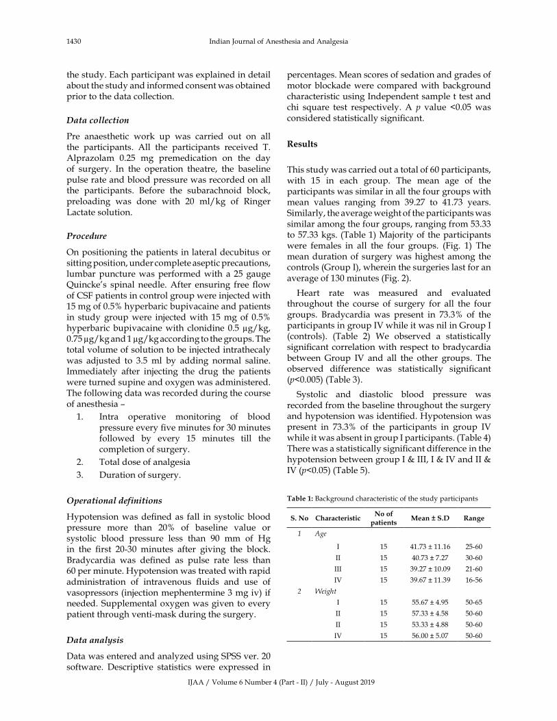
IJAA�/�Volume�6�Number�4�(Part�-�II)�/�July�-�August�2019
1430 Indian�Journal�of�Anesthesia�and�Analgesia
the�study.�Each�participant�was�explained�in�detail�about�the�study�and�informed�consent�was�obtained�prior�to�the�data�collection.
�ata�collection
Pre� anaesthetic� work� up� was� carried� out� on� all�the� participants.� All� the� participants� received� T.�Alprazolam� 0.25� mg� premedication� on� the� day�of� surgery.� In� the� operation� theatre,� the� baseline�pulse�rate�and�blood�pressure�was�recorded�on�all�the� participants.� �efore� the� subarachnoid� block,�preloading� was� done� with� 20� ml/kg� of� Ringer�Lactate�solution.
Procedure�
On�positioning�the�patients� in�lateral�decubitus�or�sitting�position,�under�complete�aseptic�precautions,�lumbar�puncture�was�performed�with� a� 25�gauge��uincke�s� spinal� needle.�After� ensuring� free� ��ow�of�CSF�patients�in�control�group�were�injected�with�15�mg�of�0.5��hyperbaric�bupivacaine�and�patients�in� study�group�were� injected�with� 15�mg�of�0.5��hyperbaric�bupivacaine�with� clonidine�0.5��g/kg,�0.75��g/kg�and�1��g/kg�according�to�the�groups.�The�total�volume�of�solution�to�be�injected�intrathecaly�was� adjusted� to� 3.5� ml� by� adding� normal� saline.�Immediately� after� injecting� the� drug� the� patients�were�turned�supine�and�oxygen�was�administered.�The�following�data�was�recorded�during�the�course�of�anesthesia�–�
1.� Intra� operative� monitoring� of� blood�pressure�every���ve�minutes�for�30�minutes�followed� by� every� 15� minutes� till� the�completion�of�surgery.
2.� Total�dose�of�analgesia
3.� Duration�of�surgery.
��erational�definitions
�ypotension�was�de��ned�as� fall� in�systolic�blood�pressure� more� than� 20�� of� baseline� value� or�systolic� blood� pressure� less� than� 90� mm� of� �g�in� the� ��rst� 20-30� minutes� after� giving� the� block.��radycardia� was� de��ned� as� pulse� rate� less� than�60�per�minute.��ypotension�was�treated�with�rapid�administration� of� intravenous� ��uids� and� use� of�vasopressors�(injection�mephentermine�3�mg�iv)�if�needed.�Supplemental�oxygen�was�given�to�every�patient�through�venti-mask�during�the�surgery.
�ata�analysis
Data�was�entered�and�analyzed�using�SPSS�ver.�20�software.� Descriptive� statistics� were� expressed� in�
percentages.�Mean�scores�of�sedation�and�grades�of�motor�blockade�were� compared�with� background�characteristic�using�Independent�sample�t�test�and�chi� s�uare� test� respectively.� A� p� value� �0.05� was�considered�statistically�signi��cant.
�esults
This�study�was�carried�out�a�total�of�60�participants,�with� 15� in� each� group.� The� mean� age� of� the�participants�was�similar�in�all�the�four�groups�with�mean� values� ranging� from� 39.27� to� 41.73� years.�Similarly,�the�average�weight�of�the�participants�was�similar�among�the�four�groups,�ranging�from�53.33�to�57.33�kgs.�(Table�1)�Majority�of�the�participants�were� females� in� all� the� four� groups.� (Fig.� 1)� The�mean�duration�of� surgery�was�highest�among� the�controls�(Group�I),�wherein�the�surgeries�last�for�an�average�of�130�minutes�(Fig.�2).
�eart� rate� was� measured� and� evaluated�throughout� the� course� of� surgery� for� all� the� four�groups.� �radycardia� was� present� in� 73.3�� of� the�participants�in�group�IV�while�it�was�nil�in�Group�I�(controls).� (Table� 2)� We� observed� a� statistically�signi��cant� correlation�with�respect� to�bradycardia�between� Group� IV� and� all� the� other� groups.� The�observed� difference� was� statistically� signi��cant�(p�0.005)�(Table�3).
Systolic� and� diastolic� blood� pressure� was�recorded�from�the�baseline�throughout�the�surgery�and�hypotension�was�identi��ed.��ypotension�was�present� in� 73.3�� of� the� participants� in� group� IV�while�it�was�absent�in�group�I�participants.�(Table�4)�There�was�a�statistically�signi��cant�difference�in�the�hypotension�between�group�I���III,�I���IV�and�II���IV�(p�0.05)�(Table�5).
Table��:��ackground�characteristic�of�the�study�participants
S��No CharacteristicNo�of�
patientsMean���S�� �ange
� Age
I 15 41.73���11.16 25-60
II 15 40.73���7.27 30-60
III 15 39.27���10.09 21-60
IV 15 39.67���11.39 16-56
� �eight
I 15 55.67���4.95 50-65
II 15 57.33���4.58 50-60
II 15 53.33���4.88 50-60
IV 15 56.00���5.07 50-60
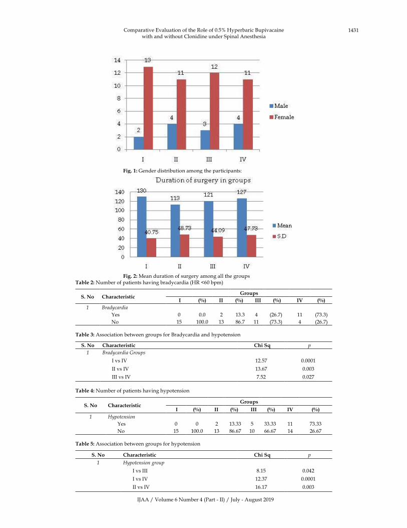
IJAA�/�Volume�6�Number�4�(Part�-�II)�/�July�-�August�2019
1431
� � Fig���:�Gender�distribution�among�the�participants:
� � Fig���:�Mean�duration�of�surgery�among�all�the�groupsTable��:�Number�of�patients�having�bradycardia�(�R��60�bpm)
S��No Characteristic�roups
�I ��� �II ��� �III ��� IV ���
� Brad�cardia
�es 0 0.0 2 13.3 4 (26.7) 11 (73.3)
No 15 100.0 13 86.7 11 (73.3) 4 (26.7)
Table��:�Association�between�groups�for��radycardia�and�hypotension�
S��No Characteristic Chi�S� p
� Brad�cardia��roups
I�vs�IV 12.57 0.0001
II�vs�IV 13.67 0.003
III�vs�IV 7.52 0.027
Table��:�Number�of�patients�having�hypotension
S��No Characteristic�roups
�I ��� �II ��� �III ��� IV ���
� ��potension
�es 0 0 2 13.33 5 33.33 11 73.33
No 15 100.0 13 86.67 10 66.67 14 26.67
Table��:�Association�between�groups�for�hypotension
S��No Characteristic Chi�S� p
� ��potension�group
I�vs�III 8.15 0.042
I�vs�IV 12.37 0.0001
II�vs�IV 16.17 0.003
Comparative�Evaluation�of�the�Role�of�0.5���yperbaric��upivacaine�with�and�without�Clonidine�under�Spinal�Anesthesia
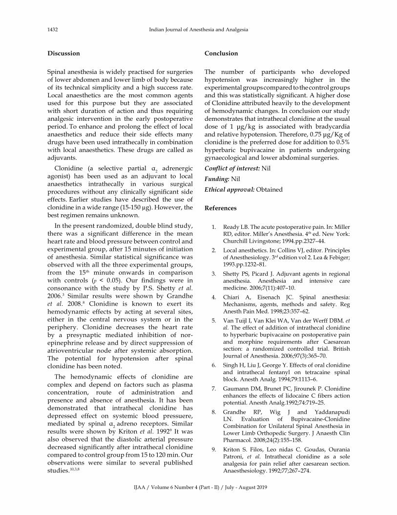
IJAA�/�Volume�6�Number�4�(Part�-�II)�/�July�-�August�2019
1432 Indian�Journal�of�Anesthesia�and�Analgesia
�iscussion
Spinal�anesthesia�is�widely�practised�for�surgeries�of�lower�abdomen�and�lower�limb�of�body�because�of� its� technical� simplicity�and�a�high�success�rate.�Local� anaesthetics� are� the� most� common� agents�used� for� this� purpose� but� they� are� associated�with� short� duration� of� action� and� thus� re�uiring�analgesic� intervention� in� the� early� postoperative�period.�To�enhance�and�prolong�the�effect�of� local�anaesthetics� and� reduce� their� side� effects� many�drugs�have�been�used�intrathecally�in�combination�with� local� anaesthetics.� These� drugs� are� called� as�adjuvants.
Clonidine� (a� selective� partial� �2� adrenergic�
agonist)� has� been� used� as� an� adjuvant� to� local�anaesthetics� intrathecally� in� various� surgical�procedures� without� any� clinically� signi��cant� side�effects.� Earlier� studies� have� described� the� use� of�clonidine�in�a�wide�range�(15-150��g).��owever,�the�best�regimen�remains�unknown.
In�the�present�randomized,�double�blind�study,�there� was� a� signi��cant� difference� in� the� mean�heart�rate�and�blood�pressure�between�control�and�experimental�group,�after�15�minutes�of�initiation�of� anesthesia.� Similar� statistical� signi��cance�was�observed�with�all�the�three�experimental�groups,�from� the� 15th� minute� onwards� in� comparison�with� controls� (p� �� 0.05).� Our� ��ndings� were� in�consonance� with� the� study� by� P.S.� Shetty� et� al.2006.3� Similar� results� were� shown� by� Grandhe�et� al.� 2008.8� Clonidine� is� known� to� exert� its�hemodynamic� effects� by� acting� at� several� sites,�either� in� the� central� nervous� system� or� in� the�periphery.� Clonidine� decreases� the� heart� rate�by� a� presynaptic� mediated� inhibition� of� nor-epinephrine�release�and�by�direct�suppression�of�atrioventricular� node� after� systemic� absorption.�The� potential� for� hypotension� after� spinal�clonidine�has�been�noted.
The� hemodynamic� effects� of� clonidine� are�complex� and� depend� on� factors� such� as� plasma�concentration,� route� of� administration� and�presence� and� absence� of� anesthesia.� It� has� been�demonstrated� that� intrathecal� clonidine� has�depressed� effect� on� systemic� blood� pressuere,�mediated� by� spinal� �
2�adreno� receptors.� Similar�
results� were� shown� by� �riton� et� al.� 19929� It� was�also� observed� that� the� diastolic� arterial� pressure�decreased�signi��cantly�after� intrathecal�clonidine�compared�to�control�group�from�15�to�120�min.�Our�observations� were� similar� to� several� published�studies.10,3,8
Conclusion
The� number� of� participants� who� developed�hypotension� was� increasingly� higher� in� the�experimental�groups�compared�to�the�control�groups�and�this�was�statistically�signi��cant.�A�higher�dose�of�Clonidine�attributed�heavily�to�the�development�of�hemodynamic�changes.�In�conclusion�our�study�demonstrates�that�intrathecal�clonidine�at�the�usual�dose� of� 1� �g/kg� is� associated� with� bradycardia�and�relative�hypotension.�Therefore,�0.75��g/�g�of�clonidine�is�the�preferred�dose�for�addition�to�0.5��hyperbaric� bupivacaine� in� patients� undergoing�gynaecological�and�lower�abdominal�surgeries.
Con��ict�of�interest:�Nil
Fundin�:�Nil
Ethical�a��roval:�Obtained�
�eferences
1.� Ready�L�.�The�acute�postoperative�pain.�In:�Miller�RD,�editor.�Miller�s�Anesthesia.�4th�ed.�New��ork:�Churchill�Livingstone;�1994.pp.2327–44.
2.� Local�anesthetics.�In:�Collins�VJ,�editor.�Principles�of�Anesthesiology.�3rd�edition�vol�2.�Lea���Febiger;�1993.pp.1232–81.
3.� Shetty�PS,�Picard� J.�Adjuvant� agents� in� regional�anesthesia.� Anesthesia� and� intensive� care�medicine.�2006;7(11):407–10.
4.� Chiari� A,� Eisenach� JC.� Spinal� anesthesia:�Mechanisms,� agents,� methods� and� safety.� Reg�Anesth�Pain�Med.�1998;23:357–62.
5.� Van�Tuijl�I,�Van��lei�WA,�Van�der�Werff�D�M.�et�al.�The�effect�of�addition�of� intrathecal�clonidine�to�hyperbaric�bupivacaine�on�postoperative�pain�and� morphine� re�uirements� after� Caesarean�section:� a� randomized� controlled� trial.� �ritish�Journal�of�Anesthesia.�2006;97(3):365–70.
6.� Singh��,�Liu�J,�George��.�Effects�of�oral�clonidine�and� intrathecal� fentanyl� on� tetracaine� spinal�block.�Anesth�Analg.�1994;79:1113–6.
7.� Gaumann�DM,��runet�PC,�Jirounek�P.�Clonidine�enhances� the� effects� of� lidocaine� C� fibers� action�potential.�Anesth�Analg.1992;74:719–25.
8.� Grandhe� RP,� Wig� J� and� �addanapudi�LN.� Evaluation� of� �upivacaine-Clonidine�Combination� for��nilateral�Spinal�Anesthesia� in�Lower�Limb�Orthopedic�Surgery.�J�Anaesth�Clin�Pharmacol.�2008;24(2):155–158.
9.� �riton� S.� Filos,� Leo� nidas� C.� Goudas,� Ourania�Patroni,� et� al.� Intrathecal� clonidine� as� a� sole�analgesia� for� pain� relief� after� caesarean� section.�Anaesthesiology.�1992;77;267–274.
IJAA�/�Volume�6�Number�4�(Part�-�II)�/�July�-�August�2019
1433
10.� Dobrydnjov�I,�Axelsson��,�Thorn�SE,�et�al.�Clonidine�Combined�with�Small-Dose��upivacaine�During�Spinal� Anesthesia� for� Inguinal� �erniorrhaphy:�
A� Randomized� Double-�linded� Study.� Anesth�Analg.�2003;96:1496–503.
Comparative�Evaluation�of�the�Role�of�0.5���yperbaric��upivacaine�with�and�without�Clonidine�under�Spinal�Anesthesia
��Red�Flower�Publication�Pvt.�Ltd.�
�Original�Research�ArticleIndian�Journal�of�Anesthesia�and�Analgesia�
2019;6(4)�(Part�-�II):�1434-1437DOI:�http://dx.doi.org/10.21088/ijaa.2349.8471.6419.52
Comparison�of��e�obupi�acaine��ith�or��ithout�Epinephrine�for��umbar�Spine�Surgery
Vi�ay��umar���Anil��atna�at�
1,2Assistant�Professor,�Department�of�Anesthesia,�Pacific�Medical�College�and��ospital��daipur,�Rajasthan�313001,�India.
Corresponding� Author:� Anil� �atna�at,� Assistant� Professor,� Department� of� Anesthesia,� Pacific� Medical� College� and� �ospital��daipur,�Rajasthan�313001,�India.
E�mail:�[email protected]
�ecei�ed�on�22.05.2019,�Accepted�on�19.06.2019
Abstract
Introduction:�The�aim�was�to�establish�if�a�decrease�in�the�amount�of�epinephrine�from�1:200,000�to�1:400,000�added�to�epidural�levobupivacaine�produces�a�comparable�decrease�in�local�anaesthetic�assimilation�from�the�epidural�space�while�holding�the�similar�clinical�effectiveness�and�acceptability�in�patients�undergoing�elective�lumbar�spine�surgery.�Materials�and�Methods:�A�total�of�120�patients�with�ASA�physical�status�1�to�3�and�aged�18�–�85�years,�who�were�schedule�to�undergo�elective�lumbar�spine�surgery,�were�enrolled�for�the�study.�Total�dose�of�75�mg�was�administered.�The�end�of�injection�of�study�drug�was�termed��Time�0��for�the�purposes�of�subse�uent�patient�assessment.�Intraoperative�sedation�was�offered�with�added�IV�midazolam�and�propofol�as�essential�at�the�judgment�of�the�anaesthesiologist.�Results:�Levobupivacaine�0.5��produces�comparatively�small�motor�blockade.�In�fact,�in�53��of�all�patients�studied,�no�motor�block�of�the�lower�extremities�could�be�demonstrated.�even�though�the�addition�of�either�1:200,000�or�1:400,00�epinephrine�tended�to�increase�the�degree�of�motor�blockade,�it�was�not�statistical�significance.�in�addition,�in�those�patients�who�did�build�up�some�degree�of�motor�blockade,�its�period�was�not�diverse�among�both�the�groups.�Conclusion:�Present�Study�reveals� that� 0.5�� levobupivacaine,�with�or�without� epinephrine,� is� a�appropriate� anaesthetic� for�utilize� in�lumbar�spine�surgery.�The�addition�of�epinephrine�be�likely�to�increase�the�duration�of�blockade,�diminish�the� ensuing� local� anaesthetic� concentration,� and� advance� intraoperative� anaesthetic� �uality,� even� though�statistical�significance�was�not�there�for�any�T10�was�achieved�within�15�minutes�of�administering�the�epidural�injection�in�all�patient�groups.
�ey�ords:�Levobupivacaine;�Epinephrine;�Lumbar�spine�surgery.
�o��to�cite�this�article:
Vijay��umar,�Anil�Ratnawat.�Comparison�of�Levobupivacaine�with�or�without�Epinephrine�for�Lumbar�Spine�Surgery.�Indian�J�Anesth�Analg.�2019;6(4):�1434-1437.
Introduction
Spinal� anesthesia� is� the� largely� used� techni�ue�
for� infraumbilical� surgeries� since� its� unmatchable�
dependability,�cost�effectiveness,�effectual�analgesia,�
muscle� relaxation� and� long-lasting� postoperative�analgesia.� Recent� advances� in� anesthesia� have�
authorized�added�surgeries�to�be�carry�out�on�day�case�basis.1�The�chattels�of�an�anaesthetic�agent�used�for�day�case�surgeries� in� spinal�anesthesia�should�have� reduced� incidence� of� anesthesia� related�dif��culties,� should� offer� suf��cient� postoperative�analgesia� and� permit� early� patient� discharge.�Levobupivacaine,� a� pure� S(�)� enantiomer� of�bupivacaine�is�a�long�acting�amide�local�anaesthetic�
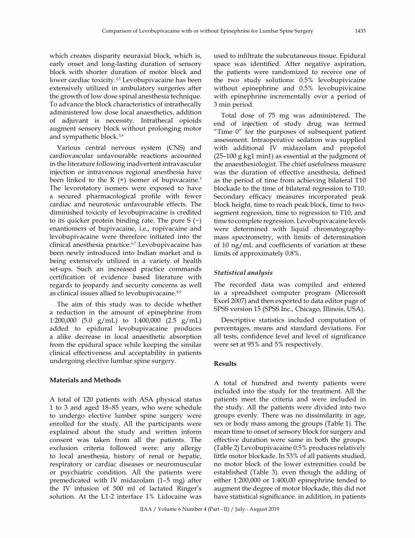
IJAA�/�Volume�6�Number�4�(Part�-�II)�/�July�-�August�2019
1435
which� creates� disparity�neuraxial�block,�which� is,�early� onset� and� long-lasting� duration� of� sensory�block� with� shorter� duration� of� motor� block� and�lower�cardiac�toxicity.2,3�Levobupivacaine�has�been�extensively� utilized� in� ambulatory� surgeries� after�the�growth�of�low�dose�spinal�anesthesia�techni�ue.�To�advance�the�block�characteristics�of�intrathecally�administered�low�dose�local�anaesthetics,�addition�of� adjuvant� is� necessity.� Intrathecal� opioids�augment�sensory�block�without�prolonging�motor�and�sympathetic�block.3,4
Various� central� nervous� system� (CNS)� and�cardiovascular� unfavourable� reactions� accounted�in�the�literature�following�inadvertent�intravascular�injection� or� intravenous� regional� anesthesia� have�been� linked� to� the� R� (�)� isomer� of� bupivacaine.5�The� levorotatory� isomers� were� exposed� to� have�a� secured� pharmacological� pro��le� with� fewer�cardiac� and� neurotoxic� unfavourable� effects.� The�diminished�toxicity�of�levobupivacaine�is�credited�to� its��uicker�protein�binding�rate.�The�pure�S�(�)�enantiomers� of� bupivacaine,� i.e.,� ropivacaine� and�levobupivacaine� were� therefore� initiated� into� the�clinical�anesthesia�practice.6,7�Levobupivacaine�has�been�newly� introduced� into� Indian�market� and� is�being� extensively� utilized� in� a� variety� of� health�set-ups.� Such� an� increased� practice� commands�certi��cation� of� evidence� based� literature� with�regards� to� jeopardy�and�security�concerns�as�well�as�clinical�issues�allied�to�levobupivacaine.8,9
The� aim� of� this� study� was� to� decide� whether�a� reduction� in� the� amount� of� epinephrine� from�1:200,000� (5.0� g/mL)� to� 1:400,000� (2.5� g/mL)�added� to� epidural� levobupivacaine� produces�a� alike� decrease� in� local� anaesthetic� absorption�from�the�epidural�space�while�keeping�the�similar�clinical� effectiveness� and� acceptability� in� patients�undergoing�elective�lumbar�spine�surgery.
Materials�and�Methods
A� total� of� 120� patients� with� ASA� physical� status�1� to� 3� and� aged� 18–85� years,� who� were� schedule�to� undergo� elective� lumber� spine� surgery� were�enrolled� for� the� study.� All� the� participants� were�explained� about� the� study� and� written� inform�consent� was� taken� from� all� the� patients.� The�exclusion� criteria� followed� were:� any� allergy�to� local� anesthesia,� history� of� renal� or� hepatic,�respiratory� or� cardiac� diseases� or� neuromuscular�or� psychiatric� condition.� All� the� patients� were�premedicated� with� IV� midazolam� (1–5� mg)� after�the� IV� infusion� of� 500� ml� of� lactated� Ringer�s�solution.� At� the� L1-2� interface� 1�� Lidocaine� was�
used�to�in��ltrate�the�subcutaneous�tissue.�Epidural�space� was� identi��ed.� After� negative� aspiration,�the� patients� were� randomized� to� receive� one� of�the� two� study� solutions:� 0.5�� levobupivicaine�without� epinephrine� and� 0.5�� levobupivicaine�with� epinephrine� incrementally� over� a� period� of�3�min�period.
Total� dose� of� 75� mg� was� administered.� The�end� of� injection� of� study� drug� was� termed��Time� 0�� for� the� purposes� of� subse�uent� patient�assessment.� Intraoperative� sedation� was� supplied�with� additional� IV� midazolam� and� propofol�(25–100�g�kg1�min1)�as�essential�at�the�judgment�of�the�anaesthesiologist.�The�chief�usefulness�measure�was� the� duration� of� effective� anesthesia,� de��ned�as� the�period�of� time�from�achieving�bilateral�T10�blockade�to�the�time�of�bilateral�regression�to�T10.�Secondary� ef��cacy� measures� incorporated� peak�block�height,�time�to�reach�peak�block,�time�to�two-segment�regression,�time�to�regression�to�T10,�and�time�to�complete�regression.�Levobupivacaine�levels�were� determined� with� li�uid� chromatography-mass� spectrometry,� with� limits� of� determination�of�10�ng/mL�and�coef��cients�of�variation�at� these�limits�of�approximately�0.8�.
Statistical�analysis
The� recorded� data� was� compiled� and� entered�in� a� spreadsheet� computer� program� (Microsoft�Excel�2007)�and�then�exported�to�data�editor�page�of�SPSS�version�15�(SPSS�Inc.,�Chicago,�Illinois,��SA).
Descriptive� statistics� included� computation� of�percentages,� means� and� standard� deviations.� For�all� tests,� con��dence� level�and� level�of�signi��cance�were�set�at�95��and�5��respectively.
�esults
A� total� of� hundred� and� twenty� patients� were�included� into� the� study� for� the� treatment.�All� the�patients� meet� the� criteria� and� were� included� in�the� study.�All� the�patients�were�divided� into� two�groups�evenly.�There�was�no�dissimilarity� in�age,�sex�or�body�mass�among�the�groups�(Table�1).�The�mean�time�to�onset�of�sensory�block�for�surgery�and�effective� duration� were� same� in� both� the� groups.�(Table�2)�Levobupivacaine�0.5��produces�relatively�little�motor�blockade.�In�53��of�all�patients�studied,�no�motor� block� of� the� lower� extremities� could� be�established� (Table� 3).� even� though� the� adding� of�either�1:200,000�or�1:400,00� epinephrine� tended� to�augment�the�degree�of�motor�blockade,�this�did�not�have�statistical�signi��cance.�in�addition,�in�patients�
Comparison�of�Levobupivacaine�with�or�without�Epinephrine�for�Lumbar�Spine�Surgery
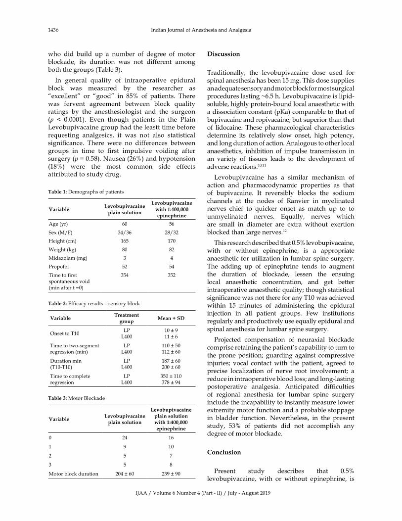
IJAA�/�Volume�6�Number�4�(Part�-�II)�/�July�-�August�2019
1436 Indian�Journal�of�Anesthesia�and�Analgesia
who� did� build� up� a� number� of� degree� of� motor�blockade,� its� duration� was� not� different� among�both�the�groups�(Table�3).
In� general� �uality� of� intraoperative� epidural�block� was� measured� by� the� researcher� as��excellent�� or� �good�� in� 85�� of� patients.� There�was� fervent� agreement� between� block� �uality�ratings� by� the� anesthesiologist� and� the� surgeon�(p� �� 0.0001).� Even� though� patients� in� the� Plain�Levobupivacaine�group�had�the�leastt�time�before�re�uesting� analgesics,� it� was� not� also� statistical�signi��cance.� There� were� no� differences� between�groups� in� time� to� ��rst� impulsive� voiding� after�surgery�(p���0.58).�Nausea�(26�)�and�hypotension�(18�)� were� the� most� common� side� effects�attributed�to�study�drug.�
Table��:�Demographs�of�patients
Variable�e�obupi�acaine�plain�solution
�e�obupi�acaine�ith��:��������epinephrine
Age�(yr) 60 56
Sex�(M/F) 34/36 28/32
�eight�(cm) 165 170
Weight�(kg) 80 82
Midazolam�(mg) 3 4
Propofol 52 54
Time�to�first�spontaneous�void�(min�after�t��0)
354 352
Table��:�Efficacy�results�–�sensory�block
VariableTreatment�
groupMean���S�
Onset�to�T10LP
L40010���911���6
Time�to�two-segment�regression�(min)
LPL400
110���50112���60
Duration�min�(T10-T10)
LPL400
187���60200���60
Time�to�complete�regression
LPL400
350���110378���94
Table��:�Motor��lockade
Variable�e�obupi�acaine�plain�solution
�e�obupi�acaine�plain�solution��ith��:��������epinephrine
0 24 16
1 9 10
2 5 7
3 5 8
Motor�block�duration 204���60 239���90
�iscussion
Traditionally,� the� levobupivacaine� dose� used� for�spinal�anesthesia�has�been�15�mg.�This�dose�supplies�an�ade�uate�sensory�and�motor�block�for�most�surgical�procedures�lasting��6.5�h.�Levobupivacaine�is�lipid-soluble,�highly�protein-bound�local�anaesthetic�with�a�dissociation�constant�(p�a)�comparable�to�that�of�bupivacaine�and�ropivacaine,�but�superior�than�that�of� lidocaine.� These� pharmacological� characteristics�determine� its� relatively� slow� onset,� high� potency,�and�long�duration�of�action.�Analogous�to�other�local�anaesthetics,� inhibition� of� impulse� transmission� in�an� variety� of� tissues� leads� to� the� development� of�adverse�reactions.10,11
Levobupivacaine� has� a� similar� mechanism� of�action� and� pharmacodynamic� properties� as� that�of� bupivacaine.� It� reversibly� blocks� the� sodium�channels� at� the� nodes� of� Ranvier� in� myelinated�nerves� chief� to� �uicker� onset� as� match� up� to� to�unmyelinated� nerves.� E�ually,� nerves� which�are� small� in� diameter� are� extra� without� exertion�blocked�than�large�nerves.12
This�research�described�that�0.5��levobupivacaine,�with� or� without� epinephrine,� is� a� appropriate�anaesthetic�for�utilization�in� lumbar�spine�surgery.�The� adding� up� of� epinephrine� tends� to� augment�the� duration� of� blockade,� lessen� the� ensuing�local� anaesthetic� concentration,� and� get� better�intraoperative�anaesthetic��uality;�though�statistical�signi��cance�was�not�there�for�any�T10�was�achieved�within� 15� minutes� of� administering� the� epidural�injection� in� all� patient� groups.� Few� institutions�regularly�and�productively�use�e�ually�epidural�and�spinal�anesthesia�for�lumbar�spine�surgery.
Projected� compensation� of� neuraxial� blockade�comprise�retaining�the�patient�s�capability�to�turn�to�the�prone�position;� guarding� against� compressive�injuries;� vocal� contact� with� the� patient,� agreed� to�precise� localization� of� nerve� root� involvement;� a�reduce�in�intraoperative�blood�loss;�and�long-lasting�postoperative� analgesia.� Anticipated� dif��culties�of� regional� anesthesia� for� lumbar� spine� surgery�include�the�incapability�to�instantly�measure�lower�extremity�motor�function�and�a�probable�stoppage�in� bladder� function.� Nevertheless,� in� the� present�study,� 53�� of� patients� did� not� accomplish� any�degree�of�motor�blockade.
Conclusion
Present� study� describes� that� 0.5��levobupivacaine,� with� or� without� epinephrine,� is�
IJAA�/�Volume�6�Number�4�(Part�-�II)�/�July�-�August�2019
1437
a�appropriate�anaesthetic�for�utilization�in�lumbar�spine�surgery.�The�adding�up�of�epinephrine�tends�to� augment� the� duration� of� blockade,� reduce� the�resultant� local� anaesthetic� concentration,� and�recover� intraoperative� anesthetic� �uality;� though�statistical�signi��cance�was�not�achived�for�any�T10�was� achieved�within� 15�minutes� of� administering�the�epidural�injection�in�all�patient�groups.
�eferences
1.� Attri� JP,� �aur� G,� �aur� S,� et� al.� Comparison� of�levobupivacaine� and� levobupivacaine� with�fentanyl�in� infraumbilical�surgeries�under�spinal�anesthesia.� Anesthesia,� essays� and� researches.�2015;9:178.
2.� Gerges� FJ,� �anazi� GE,� Jabbour-khoury� SI.�Anesthesia� for� laparoscopy:�a� review.�Journal�of�clinical�anesthesia.�2006;18:67–78.
3.� Rawal�N.�Analgesia�for�day-case�surgery.��ritish�journal�of�anesthesia.�2001;87:73–87.
4.� �anazi�G,�Aouad�M,�Jabbour-�houry�S,�et�al.�Effect�of�low-dose�dexmedetomidine�or�clonidine�on�the�characteristics� of� bupivacaine� spinal� block.�Acta�Anaesthesiologica�Scandinavica.�2006;50:222–7.
5.� Groban� L.� Central� nervous� system� and� cardiac�effects� from� long-acting� amide� local� anesthetic�toxicity� in� the� intact� animal� model.� Regional�anesthesia�and�pain�medicine.�2003;28:3–11.
6.� Leone� S,� Di� Cianni� S,� Casati� A,� et� al.�Pharmacology,� toxicology,� and� clinical� use� of�new� long� acting� localanesthetics,� ropivacaine�and� levobupivacaine.� Acta� �io� Medica� Atenei�Parmensis.�2008;79:92–105.
7.� Gunter� J�.� �enefit� and� risks� of� local� anesthetics�in� infants� and� children.� Pediatric� Drugs.� 2002;�4:649–72.
8.� Peduto� VA,� �aroncini� S,� Montanini� SA� et� al.�A� prospective,� randomized,� double-blind�comparison� of� epidural� levobupivacaine� 0.5��with� epidural� ropivacaine� 0.75�� for� lower�limb� procedures.� Eur� J� Anaesthesiol.� 2003�Dec;20(12):979-83.
9.� �ajwa� SJS,� �aur� J.� Clinical� profile� of�levobupivacaine� in� regional� anesthesia:� A�systematic� review.� Journal� of� anaesthesiology,�clinical�pharmacology.�2013;29:530.
10.� Catterall�WA,�Mackie�.�Local�anesthetics.�Goodman���Gilman�s�the�pharmacological�basis�of�therapeutics,�12th�ed�McGraw-�ill,�Medical�Publishing�Division,�New��ork,�N�.�2011.pp.565–82.
11.� Day�T�,�Skarda�RT/�The�pharmacology�of� local�anesthetics.�Veterinary�Clinics�of�North�America:�E�uine�Practice.�1991;7:489–500.
12.� Nau� C,� Vogel� W,� �empelmann� G,� et� al.�Stereoselectivity� of� �upivacaine� in� Local�Anesthetic–sensitive� Ion� Channels� of� Peripheral�Nerve.� Anesthesiology:� The� Journal� of� the�American� Society� of� Anesthesiologists.� 1999;�91:786–.
Comparison�of�Levobupivacaine�with�or�without�Epinephrine�for�Lumbar�Spine�Surgery
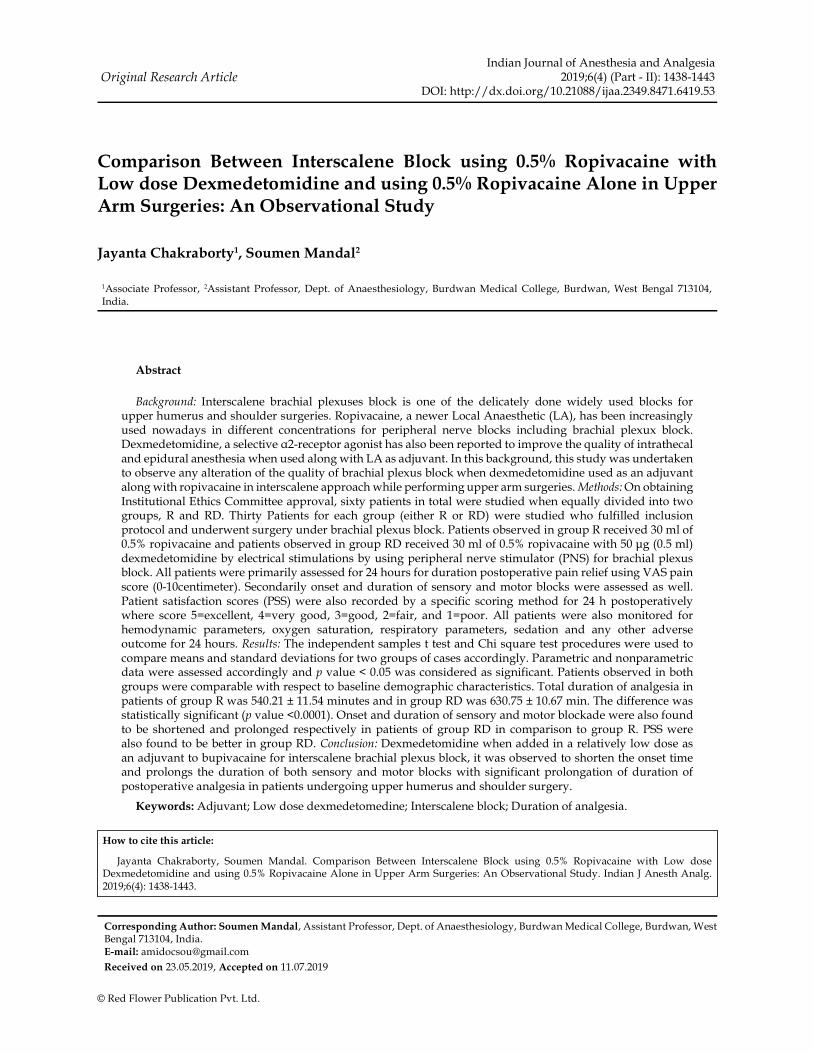
��Red�Flower�Publication�Pvt.�Ltd.�
�Original�Research�ArticleIndian�Journal�of�Anesthesia�and�Analgesia�
2019;6(4)�(Part�-�II):�1438-1443DOI:�http://dx.doi.org/10.21088/ijaa.2349.8471.6419.53
Comparison� Bet�een� Interscalene� Block� using� ����� �opi�acaine� �ith��o��dose��e�medetomidine�and�using�������opi�acaine�Alone�in��pper�Arm�Surgeries:�An�Obser�ational�Study
�ayanta�Chakraborty���Soumen�Mandal�
1Associate� Professor,� 2Assistant�Professor,� Dept.� of�Anaesthesiology,� �urdwan�Medical� College,� �urdwan,�West� �engal� 713104,�India.
Corresponding�Author:�Soumen�Mandal,�Assistant�Professor,�Dept.�of�Anaesthesiology,��urdwan�Medical�College,��urdwan,�West��engal�713104,�India.E�mail:�[email protected]
�ecei�ed�on�23.05.2019,�Accepted�on�11.07.2019
Abstract
Background:� Interscalene� brachial� plexuses� block� is� one� of� the� delicately� done� widely� used� blocks� for�upper�humerus�and�shoulder�surgeries.�Ropivacaine,�a�newer�Local�Anaesthetic�(LA),�has�been�increasingly�used� nowadays� in� different� concentrations� for� peripheral� nerve� blocks� including� brachial� plexux� block.�Dexmedetomidine,�a�selective��2-receptor�agonist�has�also�been�reported�to�improve�the��uality�of�intrathecal�and�epidural�anesthesia�when�used�along�with�LA�as�adjuvant.�In�this�background,�this�study�was�undertaken�to�observe�any�alteration�of�the��uality�of�brachial�plexus�block�when�dexmedetomidine�used�as�an�adjuvant�along�with�ropivacaine�in�interscalene�approach�while�performing�upper�arm�surgeries.�Methods:�On�obtaining�Institutional�Ethics�Committee�approval,�sixty�patients�in�total�were�studied�when�e�ually�divided�into�two�groups,� R� and� RD.� Thirty� Patients� for� each� group� (either� R� or� RD)� were� studied� who� fulfilled� inclusion�protocol�and�underwent�surgery�under�brachial�plexus�block.�Patients�observed�in�group�R�received�30�ml�of�0.5��ropivacaine�and�patients�observed�in�group�RD�received�30�ml�of�0.5��ropivacaine�with�50��g�(0.5�ml)�dexmedetomidine�by�electrical�stimulations�by�using�peripheral�nerve�stimulator�(PNS)�for�brachial�plexus�block.�All�patients�were�primarily�assessed�for�24�hours�for�duration�postoperative�pain�relief�using�VAS�pain�score� (0-10centimeter).�Secondarily�onset�and�duration�of�sensory�and�motor�blocks�were�assessed�as�well.�Patient� satisfaction� scores� (PSS)�were�also� recorded�by� a� specific� scoring�method� for� 24�h�postoperatively�where� score� 5�excellent,� 4�very� good,� 3�good,� 2�fair,� and� 1�poor.� All� patients� were� also� monitored� for�hemodynamic� parameters,� oxygen� saturation,� respiratory� parameters,� sedation� and� any� other� adverse�outcome�for�24�hours.�Results:�The�independent�samples�t�test�and�Chi�s�uare�test�procedures�were�used�to�compare�means�and�standard�deviations�for�two�groups�of�cases�accordingly.�Parametric�and�nonparametric�data�were�assessed�accordingly�and�p�value���0.05�was�considered�as�significant.�Patients�observed�in�both�groups�were�comparable�with�respect�to�baseline�demographic�characteristics.�Total�duration�of�analgesia�in�patients�of�group�R�was�540.21���11.54�minutes�and�in�group�RD�was�630.75���10.67�min.�The�difference�was�statistically�significant�(p�value��0.0001).�Onset�and�duration�of�sensory�and�motor�blockade�were�also�found�to�be� shortened�and�prolonged�respectively� in�patients�of�group�RD� in� comparison� to�group�R.�PSS�were�also�found�to�be�better�in�group�RD.�Conclusion:�Dexmedetomidine�when�added�in�a�relatively�low�dose�as�an�adjuvant�to�bupivacaine�for�interscalene�brachial�plexus�block,�it�was�observed�to�shorten�the�onset�time�and�prolongs� the�duration� of� both� sensory� and�motor� blocks�with� significant�prolongation� of�duration�of�postoperative�analgesia�in�patients�undergoing�upper�humerus�and�shoulder�surgery.
�ey�ords:�Adjuvant;�Low�dose�dexmedetomedine;�Interscalene�block;�Duration�of�analgesia.
�o��to�cite�this�article:
Jayanta� Chakraborty,� Soumen� Mandal.� Comparison� �etween� Interscalene� �lock� using� 0.5�� Ropivacaine� with� Low� dose�Dexmedetomidine�and�using�0.5��Ropivacaine�Alone�in��pper�Arm�Surgeries:�An�Observational�Study.�Indian�J�Anesth�Analg.�2019;6(4):�1438-1443.
IJAA�/�Volume�6�Number�4�(Part�-�II)�/�July�-�August�2019
1439
Introduction
Regional�anesthesia�is�a�recommended�techni�ue�for� upper� and� lower� limb� surgeries� with� better�postoperative� pro��le.1,2� Interscalene� approach�brachial�plexuses�block� is� one�of� the�most�widely�used�blocks�for�upper�end�of�humerus�and�shoulder�surgeries.
Research� have� been� continuing� in� the� recent�years�for�an�ideal�local�anaesthetic�drug�that�should�possess�a�fast�sensory�onset,�differential�offset,�with�an� earlier� offset� of� motor� than� sensory� blockade,�enabling� early� ambulation� with� prolonged�analgesia.�Ropivacaine,�a�newer�Local�Anaesthetic�(LA),� has� been� increasingly� used� nowadays� in�different� concentration� for� peripheral� nerve�blocks.� It� has� lesser� cardiac� toxicity� and� higher�safety� margin� when� compared� to� bupivacaine.3,4�It� produces� differential� neural� blockade� with� less�motor�block,�hence�well�tolerated�for�postoperative�analgesia� and� reduced� cardiovascular� and�neurological�toxicity.5,6��owever,�it�has�been�found�that�onset�time�for�ropivacaine�used�in�major�nerve�or� plexus� block� is� typically� 20-30� mins� with� an�average�duration�of�block�is�360-720�mins.�M�
Many� drugs� such� as� clonidine8� and� fentanyl�9� have� been� used� successfully� as� adjuvant� with�different� LAs� for� the� early� onset� of� the� block�and� for� prolonging� the� duration� of� the� block.�Dexmedetomidine,� an� �2-receptor� agonist,� with��2/�1�selectivity�8�times�more�than�that�of�clonidine�has� also� been� reported� to� improve� the� �uality� of�intrathecal� and� epidural� anesthesia10,11�when�used�along�with�LA�as�adjuvant.��se�of�dexmedetomidine�as�an�adjuvant�to�0.5��ropivacaine�in�interscalene�approach�brachial�plexus�block�had�been�provided�with�relatively�lesser�amount�of�concrete�data�so�far.�Conse�uently�a�suitable�dose�of�dexmedetomidine�for� brachial� plexus� blocks� had� still� not� been�established� convincingly.� �ence� in� this� study� an�optimal�dose�of�50��g�dexmedetomidine�had�been�chosen�based�on�few�of�the�earlier�studies.12,13,14,15
This� observational� study� was� primarily� aimed�to� know� whether� 50� �g� dexmedetomidine� as�an� adjuvant� to� 0.5�� ropivacaine� in� interscalene�approach� brachial� plexus� block� using� peripheral�nerve�stimulator�can�alter�the��uality�of�the�block.
���ectives
�rimar�� outcome:� To� compare� total� duration�of� analgesia� following� successful� interscalene�approach� brachial� plexus� block� between�two�groups.
Secondar�� outcome:� To� compare� onset� and�duration� of� motor� and� sensory� anesthesia� along�with�patient� satisfaction�between�two�groups�and�also�to�compare�any�adverse�outcome�like�sedation,�bradycardia,� hypotension� and� voice� change� etc.�between�these�two�groups.
Stud�� design:� Longitudinal� comparative�observational�study.
Stud��period:�October�2017�to�September�2018
At� orthopaedic� Operation� Theatre,� �urdwan�Medical�College,��urdwan.
Materials�and�Methods
On� approval� from� the� Institutional� Ethics�Committee,�sixty�patients�were�observed�following�interscalene�brachial�plexus�block�when�they�were�e�ually� divided� into� two� groups,� R� and� RD� for�study� convenience.� Preoperatively� patients� were�counseled�and� familiarized�with� the�use�of�visual�analog�scale�(VAS)�pain�score�for�the�assessment�of�perioperative�pain.�All�the�patients�participating�in�the�study�were�explained�clearly�about�the�purpose�and�nature�of�the�study�in�their�own�understandable�language�and�written�informed�consent�was�taken.
Inclusion�criteria
�� American�society�of�Anesthesiologists�(ASA)�physical�status�I�and�II.
�� Aged�between�18�and�60�years.
�� Scheduled� for� upper� limb� surgery� mainly�upper� end� of� humerus� and� shoulder�surgeries.
Exclusion�criteria
�� Patients�on�beta�blockers.
�� Pregnant.
�� With�coagulopathy.
�� Morbid�obesity.
�� Severe�cardio-pulmonary�disease.
�� Neurological�de��cits�in�the�operative�arm.
Pre� anaesthetic� assessment� was� done� for� every�patient�following�which�patients�in�Group�R�received�30� ml� of� 0.5�� ropivacaine� and� patients� in� Group�RD�received�30�ml�of�0.5��ropivacaine�with�50��g�(0.5�ml)�dexmedetomidine�by�electrical�stimulation�with�PNS.�Same�anaesthesiologists�prepared�study�drug� solutions� and� administered� the� block� and�were�designed�to�observe�the��uality�and�duration�
Comparison��etween�Interscalene��lock�using�0.5��Ropivacaine�with�Low�dose�Dexmedetomidine�and�using�0.5��Ropivacaine�Alone�in��pper�Arm�Surgeries:�An�Observational�Study
IJAA�/�Volume�6�Number�4�(Part�-�II)�/�July�-�August�2019
1440 Indian�Journal�of�Anesthesia�and�Analgesia
of�the�block�and�the�hemodynamics�and�respiration�postoperatively� for� 24� hours� for� each� patient� of�both�the�groups.�These�anaesthesiologists�collected�and� analyzed� the�data� and�were� also� involved� in�management�of�the�patients�perioperatively.
Each� patient� observed� in� the� study� were�premedicated� with� tablet� alprazolam� 0.5� mg� and�tablet�ranitidine�150�mg�orally�at�bedtime�the�and�kept�fasting�for�10�hours�overnight.
In� the� following� morning� on� arrival� of� each�patient�for�surgery,�18�gauge�intravenous�cannula�was�inserted�with�infusion�of�Ringer�s�lactate.�Each�patient� was� thoroughly� monitored� for� heart� rate�(�R),�noninvasive�measurements�of�systolic�blood�pressure� (S�P),� diastolic� �P� (D�P),� mean� arterial�pressure� (MAP)�and� continuous�ECG,� respiratory�rate� (RR),� and� oxygen� saturation� (SpO
2).� The�
baseline�S�P,�D�P,�MAP,�and��R�were�recorded.
After� positioning� of� each� patient� on� operating�table� with� the� head� turned� to� opposite� side,�interscalene� groove� was� identi��ed� by� rolling� the���nger� posterior� to� sternocleidomastoid� muscle�between� the� bellies� of� the� anterior� and� middle�scalene� muscle� at� the� level� of� cricoid� cartilage.�Skin�over�the�insertion�site�was�in��ltrated�with�2��lignocaine.
Interscalene�block�was�performed�in�all�patients�with� the� peripheral� nerve� stimulator� (Stimuplex,����raun)�connected�to�5�cm,�22�gauge,�short�bevel�insulated�stimulating�needle�by�modi��ed�Winnies�approach.�The�intensity�of�stimulating�current�was�initially�set�to�deliver�1�mA�with�impulse�duration�of� 0.1� ms.� Thereafter,� current� was� gradually�decreased�to�0.5�mA.�The�localization�of�the�plexus�was�considered�optimal�when�an�output�current�of��0.5�mA�caused�the�contraction�of�pectoralis�muscle,�deltoid,� biceps� or� triceps.� After� eliciting� motor�response� of� any� of� these� muscles,� 30� ml� of� 0.5��ropivacaine� alone� was� given� in� patients� of� group�R�and�and�30�ml�of�0.5��ropivacaine�with�50�mcg�(0.5�ml)�of�dexmedetomidine�in�patients�group�RD�as� per� study� protocol� in� increments� of� 5� ml� after���xing�the�stimulating�needle�aspirating�in�between�to�avoid�inadvertent�intravascular�injection.
�emodynamic� parameters� such� as� �R,� S�P,�D�P� as� well� as� SpO
2,� and� RR� were� monitored� at�
every�20�min�interval�till�1�hour�of�LA�injection�and�then�every�30�min�till�2�hour�and�thereafter�every�hour� till� the� end� of� surgery� and� postoperatively�one�hourly�till���rst�24�hour.�Adverse�events�such�as�hypotension�(20��decrease�in�relation�to�the�baseline�value),� bradycardia� (�R� �� 45� bpm),� hypoxemia�(SpO
2���90�),�perioperative�nausea�and�vomiting,�
and�development�of�change�of�voice�(if�any)�were�
recorded.� Clinically� relevant� bradycardia� (heart�rate� �� 45� bpm)� spells� were� treated� with� atropine�(0.6�mg� IV).� Sedation�was� evaluated�by�using� the��niversity�of�Michigan�Sedation�Scale�(�MSS)12�of�0�to�4��0���awake�and�alert;�1���minimally�sedated/sleepy,�appropriate�response�to�conversion�and/or�sound;�2���moderately�sedated,�somnolent/sleepy,�easily� aroused� with� tactile� stimulation� and/or�simple�verbal�command;�3���deeply�sedated/deep�sleep,� aroused� only� with� signi��cant� stimulation�and�4���could�not�be�aroused�.
Patient�s�perception�of�pain�was�assessed�using�VAS�(0–10).
�lock�was�considered�inade�uate�when�sensory�anesthesia� was� not� achieved� within� 30� min�following�which�general�anesthesia�was�considered�and�patient�was�not�included�in�the�study.
The�following�parameters�were�assessed.
Onset�of�sensor��block
Sensory�block�was�assessed�by�loss�of�sensation�to� pinprick� over� the� C5T1� dermatomes� using� a�3points�scale�which�is�as�follows:
0�sharp�pain,� 1�dull�pain� (analgesia),� 2�no�pain�(anesthesia).
Sensory� onset� time� was� de��ned� as� the� time�interval� between� the� end� of� LA� administration�and�establishment�of�score�2�on�3�point�scale�on�all�nerve�territories.
Onset�of�motor�block
Motor�block�was�assessed�using��romage�scale.
0�Normal�motor�functions�with�full���exion�and�extension�of�the�elbow,�wrist,�and���ngers
1� Decreased� motor� strength� with� the� ability� to�move���ngers�only
2�Complete�motor�blockade�with�the�inability�to�move���ngers.
Motor�block�onset�time�was�de��ned�as�the�time�interval�between�the�end�of�LA�administration�and�complete�motor�block�(score�2).
�uration�of�sensor��block
Duration� of� sensory� block� was� de��ned� as� the�time�interval�between�the�end�of�LA�administration�and�the�complete�resolution�of�anesthesia�(score�0�on�a�3�point�scale)�on�all�nerves.
�uration�of�motor�block
Duration�of�motor�block�was�de��ned�as�the�time�interval�between�the�end�of�LA�administration�and�the�recovery�of�complete�motor�function�(Score�0�on��romage�scale).
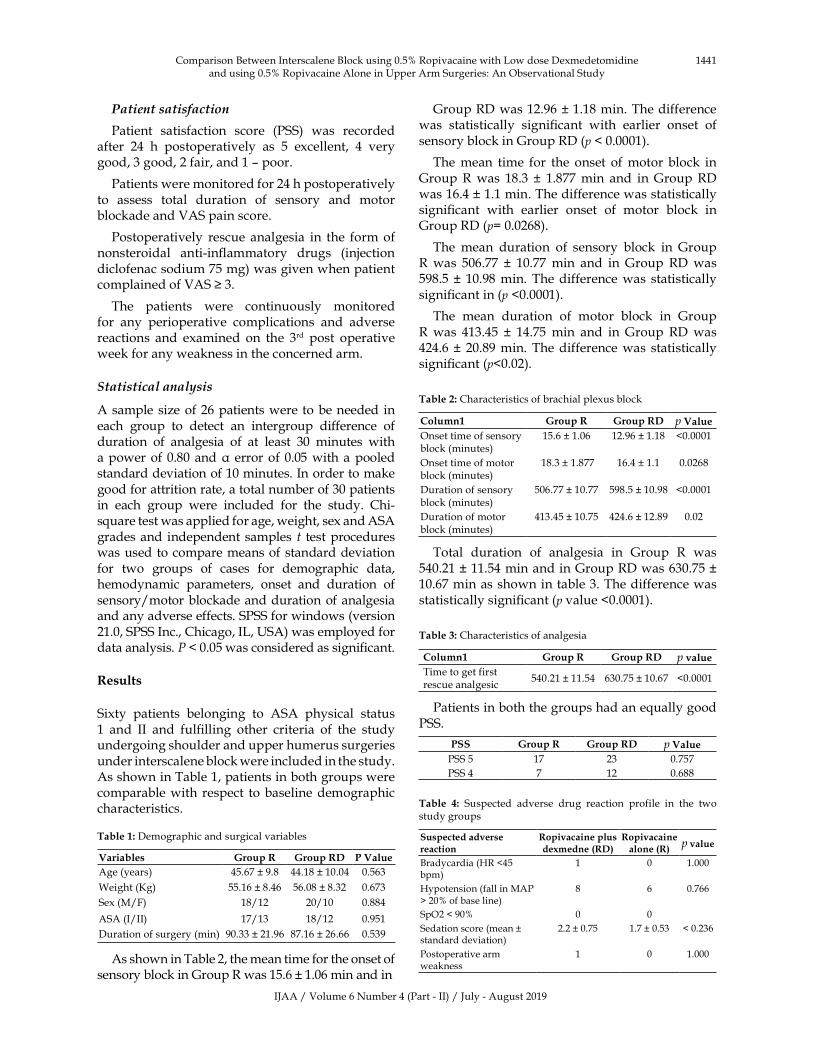
IJAA�/�Volume�6�Number�4�(Part�-�II)�/�July�-�August�2019
1441
Patient�satisfaction
Patient� satisfaction� score� (PSS)� was� recorded�after� 24� h� postoperatively� as� 5� excellent,� 4� very�good,�3�good,�2�fair,�and�1�–�poor.
Patients�were�monitored�for�24�h�postoperatively�to� assess� total� duration� of� sensory� and� motor�blockade�and�VAS�pain�score.
Postoperatively� rescue�analgesia� in� the� form�of�nonsteroidal� anti-in��ammatory� drugs� (injection�diclofenac�sodium�75�mg)�was�given�when�patient�complained�of�VAS���3.
The� patients� were� continuously� monitored�for� any� perioperative� complications� and� adverse�reactions� and� examined� on� the� 3rd� post� operative�week�for�any�weakness�in�the�concerned�arm.
Statistical�analysis
A�sample� size�of�26�patients�were�to�be�needed� in�each� group� to� detect� an� intergroup� difference� of�duration� of� analgesia� of� at� least� 30� minutes� with�a�power� of�0.80� and���error�of� 0.05�with�a�pooled�standard�deviation�of�10�minutes.�In�order�to�make�good�for�attrition�rate,�a�total�number�of�30�patients�in� each� group� were� included� for� the� study.� Chi-s�uare�test�was�applied�for�age,�weight,�sex�and�ASA�grades�and� independent� samples� t� test�procedures�was�used� to�compare�means�of�standard�deviation�for� two� groups� of� cases� for� demographic� data,�hemodynamic� parameters,� onset� and� duration� of�sensory/motor�blockade�and�duration�of�analgesia�and�any�adverse�effects.�SPSS�for�windows�(version�21.0,�SPSS�Inc.,�Chicago,�IL,��SA)�was�employed�for�data�analysis.�����0.05�was�considered�as�signi��cant.
�esults
Sixty� patients� belonging� to� ASA� physical� status�1� and� II� and� ful��lling� other� criteria� of� the� study�undergoing�shoulder�and�upper�humerus�surgeries�under�interscalene�block�were�included�in�the�study.�As�shown�in�Table�1,�patients�in�both�groups�were�comparable�with� respect� to�baseline�demographic�characteristics.
Table��:�Demographic�and�surgical�variables
Variables �roup�� �roup��� P�Value
Age�(years) 45.67���9.8� 44.18���10.04� 0.563
Weight�(�g)� 55.16���8.46� 56.08���8.32� 0.673
Sex�(M/F)� 18/12 20/10 0.884
ASA�(I/II)� 17/13 18/12 0.951
Duration�of�surgery�(min) 90.33���21.96� 87.16���26.66� 0.539
As�shown�in�Table�2,�the�mean�time�for�the�onset�of�sensory�block�in�Group�R�was�15.6���1.06�min�and�in
Group�RD�was�12.96���1.18�min.�The�difference�was� statistically� signi��cant� with� earlier� onset� of�sensory�block�in�Group�RD�(p���0.0001).
The� mean� time� for� the� onset� of� motor�block� in�Group�R� was� 18.3� �� 1.877� min� and� in�Group� RD�was�16.4���1.1�min.�The�difference�was�statistically�signi��cant� with� earlier� onset� of� motor� block� in�Group�RD�(p��0.0268).
The� mean� duration� of� sensory� block� in� Group�R� was� 506.77� �� 10.77� min� and� in� Group� RD� was�598.5� �� 10.98�min.� The�difference�was� statistically�signi��cant�in�(p��0.0001).
The� mean� duration� of� motor� block� in� Group�R� was� 413.45� �� 14.75� min� and� in� Group� RD� was�424.6� �� 20.89�min.� The�difference�was� statistically�signi��cant�(p�0.02).
Table��:�Characteristics�of�brachial�plexus�block
Column� �roup�� �roup��� p�Value
Onset�time�of�sensory�block�(minutes)�
15.6���1.06� 12.96���1.18 �0.0001
Onset�time�of�motor�block�(minutes)
�18.3���1.877� 16.4���1.1� 0.0268
Duration�of�sensory�block�(minutes)�
506.77���10.77 598.5���10.98 �0.0001
Duration�of�motor�block�(minutes)
413.45���10.75 424.6���12.89� 0.02
Total� duration� of� analgesia� in� Group� R� was�540.21���11.54�min�and�in�Group�RD�was�630.75���10.67�min�as�shown�in�table�3.�The�difference�was�statistically�signi��cant�(p�value��0.0001).
Table��:�Characteristics�of�analgesia
Column� �roup�� �roup��� p��alueTime�to�get�first�rescue�analgesic
540.21���11.54 630.75���10.67 �0.0001
Patients�in�both�the�groups�had�an�e�ually�good�PSS.
PSS �roup�� �roup��� p�Value
PSS�5 17 23 0.757
PSS�4 7 12 0.688�
Table� �:� Suspected� adverse� drug� reaction� profile� in� the� two�study�groups
Suspected�ad�erse�reaction
�opi�acaine�plusde�medne�����
�opi�acaine�alone����
p��alue
�radycardia�(�R��45�bpm)
1 0 1.000
�ypotension�(fall�in�MAP���20��of�base�line)
8 6 0.766
SpO2���90� 0 0
Sedation�score�(mean���standard�deviation)
2.2���0.75 1.7���0.53 ��0.236
Postoperative�arm�weakness
1 0 1.000
Comparison��etween�Interscalene��lock�using�0.5��Ropivacaine�with�Low�dose�Dexmedetomidine�and�using�0.5��Ropivacaine�Alone�in��pper�Arm�Surgeries:�An�Observational�Study
IJAA�/�Volume�6�Number�4�(Part�-�II)�/�July�-�August�2019
1442 Indian�Journal�of�Anesthesia�and�Analgesia
�iscussion
Dexmedetomidine�so�far�had�been�shown�promising�outcome�in�intravenous�conscious�sedation�in�IC��patients.� Different� studies� done� in� different� set�ups� also� had� shown� that� dexmedetomidine�s� use�could� prolong� analgesia� when� used� with� local�anaesthetics� for� neuraxial� blocks� by� virtue� of� its�effects�on�spinal��
2�receptors.
Drugs�such�as�clonidine�and� fentanyl�had�been�in� use� as� successful� adjuvant� with� bupivacaine�and/or� ropivacaine� for� the� early� onset� as� well� as�for� prolonging� the� duration� of� brachial� block.�Dexmedetomidine� for� being� a� relatively� more�selective��
2�agonist�with��
2�adrenoreceptors�located�
in�the�CNS�and�spinal�cord�level�had�been�found�to�be�responsible�for�the�sympatholysis,�sedation,�and�antinociception� mediated� by� G-protein� inhibition�of� L-type� calcium� channels� thereby� producing� a�complex� pattern� of� analgesia� by� both� spinal� and�supraspinal� mechanism.� Dexmedetomidine� had�also� been� shown� to� exert� an� anti� hyperalgesic�action� in� neuropathic� pain� states� involving� the�peripheral� nervous� system� and� so� had� been�increasingly�found�to�be�used�as�an�adjuvant�with�various� local� anaesthetics� in� peripheral� nerve�blocks� to� decrease� the� time�of� onset� and� increase�the� duration� of� analgesia.� Although� clonidine�added� to� bupivacaine� prolonged� the� duration� of�anesthesia�and�analgesia� in�brachial�plexus�block,�but�was� found� to�be�associated�with�bradycardia,�hypotension,� and� respiratory� depression� and�over� the� past� few� years� dexmedetomidine� had�gradually� replaced� clonidine� as� an� adjuvant� of�choice.�Ropivacaine,�a�newer�LA�with�less�cardiac�and�neural� toxicity�than�bupivacaine,�had�been�in�use�currently�as�an�ideal�agent�for�neural�blockade.�owever�it�was�found�that�ropivacaine�could�be�less�effective�at�times�and�larger�volumes�of�drug�might�be�re�uired�for�getting�ade�uate�block.�Some�recent�randomized,� double-blind� trials� had� shown� that�dexmedetomidine�1�microgram/�g�as�an�adjuvant�to�0.5��levobupivacaine�for�axillary�brachial�plexus�blockade� shortened� block� onset� time,� prolonged�duration� of� motor� and� sensory� effects,� extended�postoperative� analgesia� and� decrease� in� total�analgesic�use.15�Ropivacaine�being�a�newer�LA�had�been� in� use� in� various� concentrations� of� 0.25�,�0.5�,�and�0.75��for�nerve�blocks�producing�greater�sensory� and� motor� differential� blockade� than�bupivacaine,� in� a� dose� dependent� manner,� with�higher�concentrations�(1�)�causing�greater�degree�of�motor�blockade�than�lower�concentration�(0.5��and�0.75�).4�In�the�present�study�0.5��ropivacaine�had�been�chosen�with�an�idea�to�achieve�maximum�
possible�sensory�blockade�with�minimum�effect�on�motor�movements�and�to�minimise�unnecessary�side�effects�of�inadvertent�high�block.�Dexmedetomidine�dose�had�been�chosen�to�be�50�microgram�to�avoid�untoward� bradycardia,� hypotension� and� sedation�yet�to�achieve�ade�uate�adjuvant�effect�for�a�desired��uality�of�block�during�surgery�and�post�operative�period�as�well.� Some� of� the�previous� studies�had�reported� that� 50� microgram� dexmedetomedine�could�produce�signi��cant�prolongation�of�analgesia�when�added� to� ropivacaine� in� brachial�blocks.8,� 10,�
15� It� was� clearly� found� in� the� present� study� that�there�was�no�signi��cant�difference�between�the�two�groups�R�and�RD�as� far�as� the�demographic�data�are�concerned�while�it�was�also�observed�that�mean�total�duration�of�analgesia�in�patients�of�group�RD�(630.75� �� 10.67� minutes)� was� signi��cantly� longer�than�the�patients�of�group�R�(540.21���11.54�minutes)�till�the�receipt�of���rst�dose�of�rescue�analgesic�which�was�very�much�in�corroboration�with�another�study�of�Agarwal�S,�Aggarwal�R,�Gupta�P�et�al.�where�they�had�shown�the�adjuvant�effect�of�dexmedetomedine�with�bupivacaine�in�brachial�plexus�block.16�Present�study� also� had� shown� to� improve� the� onset� of�motor�and�sensory�block� in�patients�of�group�RD�due�the�adjuvant�effect�of�dexmedetomedine�which�was�similar� to�many�other�studies.�Total�duration�of�sensory�and�motor�block�were�also�found�to�be�signi��cantly�prolonged�in�patients�of�group�RD�for�the� use� of� dexmedetomedine� and� these� ��ndings�could� well� be� compared� to� other� recent� works�done� with� dexmedetomidine� in� brachial� block.�One�of�the�possible�causes�of�prolongation�of�effect�of� local� anaesthetic� could� be� the� vasoconstriction�caused� by� dexmedetomedine� at� the� injection� site�by�its�alpha�2�adrenoceptor�agonist�effect�resulting�delayed� absorption� of� local� anaesthetic� (AM�AbdElmaksoud� and� his� colleagues).8� Moreover� it�was� also� found� that� that� patient� satisfaction� was�also� better� in� patients� of� group� RD� which� was�assessed� by� a� speci��ed� protocol� based� scoring�system� and� though� not� signi��cant� as� such� but�found�to�be�almost�comparable�to�a�previous�study�that� showed� similar� kind� of� patient� satisfaction�pattern� for� patients� receiving� dexmedetomedine�as� an� adjuvant.17� Many� other� studies� had� also�emphasized�on�the�role�of�dexmedetomedne�for�its�adjuvant� effect� on� pain� relief� thereby� prolonging�the� effect� of� brachial� block� in� post� operative�period� and� reducing� the� re�uirement� of� opioid�rescue� analgesics� especially.� Although� this� study�was�not�designed�to�comment�on�this�perspective�but�as�per� the�observations� it�was��uite�clear� that�diclofenac� sodium,� the� rescue� analgesic� used� in�this�study�was�needed�after�a�longer�period�of�post�
IJAA�/�Volume�6�Number�4�(Part�-�II)�/�July�-�August�2019
1443
operative� time� in� patients� of� group� RD� receiving�dexmedetomedine�as� an� adjuvant�with� respect� to�the�patients�of�group�R�without�dexmedetomedine.�
There� were� limitations� in� this� study� of� which�non�availability�of�ultrasography�machine�guided�interscalene�brachial�plexus�block�was�the�foremost�one� which� was� more� of� a� technical� issue� in� this�medical� college.� This� medical� college� being� a�peripheral� college� certain� logistic� shortcomings�such�as�lack�of�proper�post�operative�care�facilities�made�the�postoperative�assessment�of�pain�of�each�patient� a� little� more� dif��cult� and� unassuming� on�the�basis�of�VAS�scoring�for�24�hours�post�operative�period� which� could� have� been� better� otherwise.�Variations� in� observations� by� anaesthesiologists�could� be� another� method� related� problem� which�could�not�be�addressed�properly.
Conclusion
This� observation� based� comparable� study� had�substantial� data� to� conclude� that� low� dose�dexmedetomedine�when�added�as�adjuvant�to�0.5��ropivacaine� in� interscalene� brachial� plexus� block�produced� signi��cant� prolongation� of� duration�of� analgesia� in� the� operated� arm� without� much�clinically�concerning�side�effects.
�eferences
1.� Mc�enzie� PJ,� Loach� A�.� Local� anesthesia� for�orthopaedic� surgery.� �r� J� Anaesth.� 1986;58:779–89.��PubMed:�3524641�
2.� Jacob�A�,�Walsh�MT,�Dilger�JA.�Role�of�regional�anesthesia� in� the� ambulatory� environment.�Anesthesiol� Clin.� 2010;28:251–66.� �PubMed:�20488393�
3.� Casati�A,�Fanelli�G,�Albertin�A,�et�al.�Interscalene�brachial� plexus� anesthesia� with� either� 0.5��ropivacaine� or� 0.5�� bupivacaine.� Minerva�Anestesiol.�2000;66:39–44.�PubMed:�10736981�
4.� �lein� SM,� Greengrass� RA,� Steele� SM,� et� al.A� comparison� of� 0.5�� bupivacaine,� 0.5��ropivacaine,� and� 0.75�� ropivacaine� for�interscalene�brachial�plexus�block.�Anesth�Analg�1998;87:1316–9.��PubMed:�9842819�
5.� �uthiala�G,�Chaudhary�G.�Ropivacaine:�A�review�of� its� pharmacology� and� clinical� use.� Indian� J�Anaesth.� 2011;55:104–10.� �PMCID:� PMC3106379���PubMed:�21712863�
6.� McClellan��J,�Faulds�D.�Ropivacaine:�An�update�
of� its� use� in� regional� anesthesia.� Drugs.� 2000�Nov;60(5):1065-93.��PubMed:�11129123�
7.� Local�Anesthetics:�Miller�s�Anesthesia;�8th�edition,�Elsevier� Saunders� Philadelphia,� PA� 19103-2899.�2015�Chapter�36:�1028–54.
8.� El�ennawy� AM,� AbdElwahab� AM,�AbdElmaksoud�AM,�et�al.�Addition�of� clonidine�or� dexmedetomidine� to� bupivacaine� prolongs�caudal� analgesia� in� children.� �r� J� Anaesth.�2009;103:268–74.��PubMed:�19541679�
9.� Sindjelic� RP,� Vlajkovic� GP,� Davidovic� L�,� et�al.� The� addition� of� fentanyl� to� local� anesthetics�affects�the��uality�and�duration�of�cervical�plexus�block:� A� randomized,� controlled� trial.� Anesth�Analg.�2010;111:234–7.��PubMed:�20519423�
10.� �anazi�GE,�Aouad�MT,�Jabbour-�houry�SI,�et�al.�Effect�of�low-dose�dexmedetomidine�or�clonidine�on�the�characteristics�of�bupivacaine�spinal�block.�Acta�Anaesthesiol�Scand.�2006;50:222–7.
11.� Congedo�E,�Sgreccia�M,�De�Cosmo�G.�New�drugs�for� epidural� analgesia.�Curr�Drug�Targets.� 2009;�10:696–706.
12.� Swami� SS,� �eniya� VM,� Ladi� SD.� Comparison�of� dexmedetomidine� and� clonidine� (�
2� agonist�
drugs)� as� an� adjuvant� to� local� anesthesia�in� supraclavicular� brachial� plexus� block:� A�randomized� doubleblind� prospective� study.�Indian� J� Anaesth.� 2012;56:243–9.� �PMCID:�PMC3425283���PubMed:�22923822�
13.� �oshitomi� T,� �ohjitani� A,� Maeda� S,� et� al.�Dexmedetomidine� enhances� the� local� anesthetic�action� of� lidocaine� via� an� �2A� adrenoceptor.�Anesth� Analg.� 2008;107:96–101.�PubMed:�18635472�
14.� Saadawy�I,��oker�A,�Elshahawy�MA,�et�al.�Effect�of� dexmedetomidine� on� the� characteristics� of�bupivacaine�in�a�caudal�block�in�pediatrics.�Acta�Anaesthesiol� Scand.� 2009;53:251–6.� �PubMed:�19076110�
15.� Esmaoglu� A,� �egenoglu� F,� Akin� A,� et� al.�Dexmedetomidine� added� to� levobupivacaine�prolongs� axillary� brachial� plexus� block.� Anesth�Analg.�2010;111:1548–51.��PubMed:�20889939�
16.� Agarwal� S,� Aggarwal� R,� Gupta� P.�Dexmedetomidine� prolongs� the� effect� of�bupivacaine� in� supraclavicular� brachial� plexus�block.� J� Anaesthesiol� Clin� Pharmacol.� 2014� Jan-Mar;�30(1):�36–40.
17.� �aur� �,� Singh� G,� Rani� S� et� al.� Effect� of�dexmedetomidine� as� an� adjuvant� to�levobupivacaine� in� supraclavicular� brachial�plexus� block:� A� randomized� double-blind�prospective�study.�J�Anaesthesiol�Clin�Pharmacol.�2015�Jul-Sep;31(3):333–8.
Comparison��etween�Interscalene��lock�using�0.5��Ropivacaine�with�Low�dose�Dexmedetomidine�and�using�0.5��Ropivacaine�Alone�in��pper�Arm�Surgeries:�An�Observational�Study
��Red�Flower�Publication�Pvt.�Ltd.�
�Original�Research�ArticleIndian�Journal�of�Anesthesia�and�Analgesia�
2019;6(4)�(Part�-�II):�1444-1448DOI:�http://dx.doi.org/10.21088/ijaa.2349.8471.6419.54
Comparison�of�Preempti�e�Intraperitoneal�Instillation�and�Nebulisation�of�������opi�acaine�in��aparoscopic�Cholecystectomy�for�Post�operati�e�Pain��elief
�yoti�Pathania���Meenal�Aggar�al���Anupam�Sharma���A�ay�Sood����irish�Sharma���Bunty�Sirkek�
1,4Professor,� Department� of� Anaesthesiology,� Indira� Gandhi� Medical� College� and� �ospital,� Shimla,� �imachal� Pradesh� 171001,�India.� 2Junior� Intensivist,� Saifee��ospital,� Mumbai,� Maharashtra� 400004,� India.� 3,6Assistant�Professor,� 5Professor,� Department� of�Anaesthesiology,� Dr��ashwant� Singh� Parmar� Government� Medical� College� and� �ospital,� Nahan,� Sirmour,� �imachal� Pradesh,�173001,�India.
Corresponding� Author:� Anupam� Sharma,� Assistant� Professor,� Department� of� Anaesthesiology,� Dr� �ashwant� Singh� Parmar�Government�Medical�College�and��ospital,�Nahan,�Sirmour,��imachal�Pradesh,�173001,�India.
E�mail:�[email protected]
�ecei�ed�on�23.05.2019,�Accepted�on�27.06.2019
Abstract
Ob�ecti�e:� The� etiology� of� postoperative� pain� in� patients� undergoing� laproscopic� cholecystectomy� is�multifactorial�consisting�of�incisional�and�visceral�pain�from�the�operation�itself�and�parietal�pain�from�trauma�and�irritation�to�the�peritoneum�and�diaphragm.��esign:�This�randamozied�single�blind�study�was�conducted�on� 60� patients� of� either� sex� undergoing� laproscopic� cholecystectomy� under� general� anesthesia.� Group� A��n�30��received�15�ml�of�0.5��ropivacaine�by�instillation�and�group����n�30��received�15�ml�of�0.5��ropivacane�intraperitoneally�by�piston�type�of�nebulization�10�minutes�before�surgery�and�both�the�groups�got�5�ml��0.5���ropivacaine� at� the� trocar� site� at� the� end�of� surgery.�Results:�Patients� in�both� the�groups�were� comparable�with�respect�to�age,�sex�and�weight��p�0.05�.�VAS�score�for�incisional�pain�was�significantly�lower�in�Group�A�at� 1hour�and�8�hrs�postoperatively��p�0.02�and�p�0.04��respectively.�At�all�other� intervals� the� incisional�pain,�Visceral�pain�and�shoulder� tip�pain�was�comparable� amongst� the� two�groups� �p�0.05�.�Time� to� first�analgesia�was�longer�in�group�A��2.29�hrs��as�compared�to�Group����1.66�hrs��but�was�stastically�insignifiacant��p�0.5�.�Mean� total� number� of� analgesic�used�was� 1.4� in� both� the� groups� �p�1�.�Conclusion:� Intraoperative�subdiaphgramatic� and� intraperitoneal� instillation� of� ropivacaine� 0.5�� is� beneficial� and� better� modality� of�pain�relief�because�of�ease�of�techni�ue�and�better�VAS�scores�as�compared�to�intrperitoneal�nebulization�of�ropivacaine.
�ey�ords:� Intraperitoneal� nebulization;� Instillation;� Ropivacaine;� Laproscopic� cholecystectomy;�Preemptive.
�o��to�cite�this�article:
Jyoti�Pathania,�Meenal�Aggarwal,�Anupam�Sharma�et�al.�Comparison�of�Preemptive�Intraperitoneal�Instillation�and�Nebulisation�of�0.5��Ropivacaine�in�Laparoscopic�Cholecystectomy�for�Post-operative�Pain�Relief.�Indian�J�Anesth�Analg.�2019;6(4):�1444-1448.
Introduction
Pain� after� laparoscopic� surgery� is� the� result�of� surgical� manipulations� and� intraperitoneal�insuf��ation� of� carbon� dioxide� causing� peritoneal�
stretching,� diaphragmatic� irritation,� changes� in�
intra-abdominal� p�,� moreover� retention� of� the�
insuf��ated�gas�in�the�abdominal�cavity�after�surgery�are� all� attributed� for� post� operative� pain.1� The�
ef��cacy�of�intraperitoneal�use�of�local�anaesthetics�for� postoperative� pain� relief� in� laparoscopic�
IJAA�/�Volume�6�Number�4�(Part�-�II)�/�July�-�August�2019
1445
procedures� is� still� debated.� Some� studies� suggest�that�local�anaesthetic�in��ltration�does�attenuate�post�operative�pain�after�laparoscopic�cholecystectomy2.�Some� suggest� that� it� does� not� improve� patient�outcome� in� terms� of� post� operative� pain� after�laparoscopic�surgery.3
Intraperitoneal� aerosolization� of� local�anaesthetics�has�proven�ef��cient�in�spreading�drugs�homogenously� throughout� the�peritoneum4,5.�This�techni�ue� combines� the� effect� of�gas� conditioning�and� the� analgesic� bene��ts� of� local� anaesthetic�instillation4.� Till� now� there� have� been� only� a� few�trials�comparing�the�analgesic�effects�achieved�with�intra-peritoneal� nebulisation�with� intra-peritoneal�instillation�of�local�anaesthetic�agents�in�laproscopic�cholecystectomy.� Optimal� management� of�postoperative�pain�is�important�as�this�procedure�is�now�carried�out�in�an�ambulatory�setting.��eeping�the� above� factors� in�mind,� the� present� study�was�conducted�in�our�institution.
Material�and�Methods
After�approval�of�Institutional�Research�and�Ethical�Committee�a�prospective�randomized�single�blind��observer�was�blind�to�study��was�carried�out�in�60�ASA�I�and�ASA�II�patients�posted� for� laproscopic�cholecystectomy�under�general�anesthesia.�Patients�were� randomly� allocated� into� into� two� groups� of�30�each.
Group�A:�Subdiaphragmatic� and� Sub� hepatic�Instillation�of�Ropivacaine
Group��:�Intraperitoneal� Nebulisation� of�Ropivacaine� using� piston� type� air�nebulizer�
In�both�the�groups,�15ml�of�0.5��ropivacaine�was�given� intraperitoneally� (by� instillation� in�group�A�and�by�nebulisation�in�group��)�immediately�after�the�insertion�of�trocars�and�5ml�of�0.5��ropivacaine�was�in��ltrated�into�the�port�sites�at�the�end�of�the�surgery.
Exclusion� Criteria:� Patients� with� severe�chronic� obstructive� airway� disease�� Patients� with�coronary�artery�disease���istory�of�allergy�to�Local�Anaesthetic� agents�� Conversion� of� Laparoscopic�Cholecystectomy� to� Open� Cholecystectomy�� Intra�Operative� intra� abdominal� drain� insertion�� Age���20�years�and���60�years.
Anesthesia��echni�ue:�Patients�were�kept�nil�per�orally�for�at�least�6�hrs.�In�the�operation�theatre,�all�patients�were�connected�to�monitors�and�baseline�vital�data�(mean�arterial�pressure,�heart�rate,�pulse�oximetry� and� respiratory� rate)� were� recorded.�
Intravenous�line�was�secured�and�intravenous���uid�was� started� in� the� contralateral� arm.� The�patient�
was�given�Injection�glycopyrolate�0.01�mg/kg�i.v.,�injection�butorphanol�0.02�mg/kg�i.v.�as�analgesic�after� preoxygenation� with� 100�� Oxygen� for�3� minutes� followed� by� induction� with� injection�thiopentone� 5�mg/kg� intravenously� followed� by�injection�succinyl�choline�2�mg/kg�intravenously.�Airway� was� secured� with� appropriate� size�endotracheal�tube�and�anesthesia�was�maintained�with�N
2O� (66�),�O
2� (33�)� and� iso��urane� (0-1�).�
Non� depolarizing� muscle� relaxant� atracurium�
(0.5�mg/kg)�was�used.�After�insertion�of�the�trocar,�in� Group� A� sub� diaphragmatic� and� sub� hepatic�instillation�of�15�ml�of�0.5��ropivacaine�was�done�and�surgery�was�started�10�min�after�the�instillation�of�the�drug.�In�group��,�nebulisation�with�15�ml�of�0.5��ropivacaine�was�done�intraperitoneally.�The�
piston� type�air�nebulizer�was�kept� at� a�height�of�6�ft�to�minimize�contaminated�air.�Intraperitoneal�pressure�was�maintained� between� 12-15�mm��g�and�gas�was�vented� if�pressure� increases.�At� the�end�of�surgery,�the�port�sites�were�in��ltrated�with�5�ml�of�0.5��ropivacaine� in�both�the�groups�and�iso��urane� was� stopped,� injection� ondansetron�0.1�mg/kg�was�given�and�muscle� relaxation�was�reversed� with� injection� neostigmine� 0.05� mg/kg�i.v.� and� injection� glycopyrrolate� 0.01� mg/kg� i.v.�
Patient� was� extubated� and� 100�� O2� was� given�
via� the�mask� for� 5�minutes� in� operation� theatre.�Patients� were� monitored� intra-operatively� for��eart� rate�Mean� blood� pressure�Arterial� oxygen�saturation�� These� parameters� were� noted� at�time� of� instillation� or� nebulisation� (taken� as� 0�minutes),�then�at�3�minutes,�5�minutes,�10�minutes�and� thereafter� every� 10�minutes�until� the� end�of�surgery.� Patients� were� analyzed� postoperatively�for�following�parameters:
1.� Incisional�Pain�(At�0,�1,�2,�3,�4,�5,�6,�8,�16�and�24�hrs)
2.� Abdominal�(Visceral)�Pain�(At�0,�1,�2,�3,�4,�5,�6,�8,�16�and�24�hrs)
3.� Shoulder�Pain�(At�0,�1,�2,�3,�4,�5,�6,�8,�16�and�24�hrs)
4.� Duration�between� time�of� extubation� and���rst�dose�of�analgesic�(diclofenac)
5.� Analgesic�re�uirement�for�24�hrs
The� Intensity� of� pain� was� assessed� on� Visual�Analogue�Scale�(VAS).�Analgesic�re�uirement�was�
assessed� in� terms� of� administration� of�number� of�injections�of�diclofenac�sodium��75�mg��on�demand.�Analgesic�was�given�if�patient�has�pain�e�uivalent�to�VAS�of�4�or�more.
Comparison�of�Preemptive�Intraperitoneal�Instillation�and�Nebulisation�of�0.5��Ropivacaine�in�Laparoscopic�Cholecystectomy�for�Post-operative�Pain�Relief
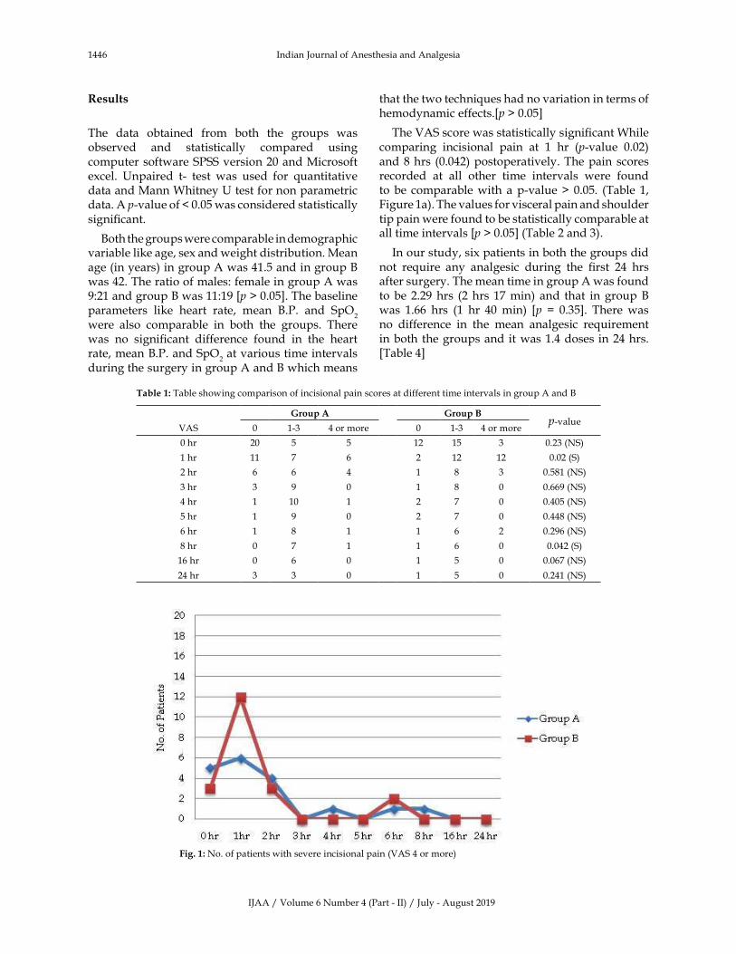
IJAA�/�Volume�6�Number�4�(Part�-�II)�/�July�-�August�2019
1446 Indian�Journal�of�Anesthesia�and�Analgesia
�esults
The� data� obtained� from� both� the� groups� was�observed� and� statistically� compared� using�computer�software�SPSS�version�20�and�Microsoft�excel.� �npaired� t-� test� was� used� for� �uantitative�data�and�Mann�Whitney���test�for�non�parametric�data.�A�p-value�of���0.05�was�considered�statistically�signi��cant.
�oth�the�groups�were�comparable�in�demographic�variable�like�age,�sex�and�weight�distribution.�Mean�age�(in�years)�in�group�A�was�41.5�and�in�group���was�42.�The�ratio�of�males:�female�in�group�A�was�9:21�and�group���was�11:19��p���0.05�.�The�baseline�parameters� like� heart� rate,� mean� �.P.� and� SpO
2
were� also� comparable� in� both� the� groups.� There�was� no� signi��cant� difference� found� in� the� heart�rate,�mean��.P.�and�SpO
2�at�various�time�intervals�
during�the�surgery�in�group�A�and���which�means�
that�the�two�techni�ues�had�no�variation�in�terms�of�hemodynamic�effects.�p���0.05�
The�VAS�score�was�statistically�signi��cant�While�comparing� incisional� pain� at� 1� hr� (p-value� 0.02)�and�8�hrs� (0.042)�postoperatively.�The�pain� scores�recorded� at� all� other� time� intervals� were� found�to� be� comparable�with� a�p-value���0.05.� (Table� 1,�Figure�1a).�The�values�for�visceral�pain�and�shoulder�tip�pain�were�found�to�be�statistically�comparable�at�all�time�intervals��p���0.05��(Table�2�and�3).
In�our�study,�six�patients�in�both�the�groups�did�not� re�uire� any� analgesic� during� the� ��rst� 24� hrs�after�surgery.�The�mean�time�in�group�A�was�found�to� be� 2.29�hrs� (2�hrs� 17�min)� and� that� in� group���was� 1.66� hrs� (1� hr� 40�min)� �p� �� 0.35�.� There�was�no� difference� in� the� mean� analgesic� re�uirement�in�both�the�groups�and�it�was�1.4�doses�in�24�hrs.��Table�4�
Table��:�Table�showing�comparison�of�incisional�pain�scores�at�different�time�intervals�in�group�A�and��
�roup�A �roup�Bp-value
VAS 0 1-3 4�or�more 0 1-3 4�or�more
0�hr 20 5 5 12 15 3 0.23�(NS)
1�hr 11 7 6 2 12 12 0.02�(S)
2�hr 6 6 4 1 8 3 0.581�(NS)
3�hr 3 9 0 1 8 0 0.669�(NS)
4�hr 1 10 1 2 7 0 0.405�(NS)
5�hr 1 9 0 2 7 0 0.448�(NS)
6�hr 1 8 1 1 6 2 0.296�(NS)
8�hr 0 7 1 1 6 0 0.042�(S)
16�hr 0 6 0 1 5 0 0.067�(NS)
24�hr 3 3 0 1 5 0 0.241�(NS)
�� Fig���:�No.�of�patients�with�severe�incisional�pain�(VAS�4�or�more)

IJAA�/�Volume�6�Number�4�(Part�-�II)�/�July�-�August�2019
1447Comparison�of�Preemptive�Intraperitoneal�Instillation�and�Nebulisation�of�0.5��Ropivacaine�in�Laparoscopic�Cholecystectomy�for�Post-operative�Pain�Relief
Table��:�Table�showing�comparison�of�visceral�pain�scores�at�different�time�intervals�in�group�A�and���
�roup�A �roup�Bp��alue
VAS � ��� ��or�more � ��� ��or�more
0�hr 20 5 5 14 3 3 0.39�(NS)
1�hr 9 8 7 4 9 13 0.098�(NS)
2�hr 6 6 4 2 7 13 0.755�(NS)
3�hr 2 10 0 1 8 0 0.846�(NS)
4�hr 0 10 2 1 8 0 0.422�(NS)
5�hr 1 9 0 1 8 0 0.243�(NS)
6�hr 1 8 1 0 7 2 0.447�(NS)
8�hr 0 7 1 1 5 1 0.281�(NS)
16�hr 0 6 0 1 5 0 0.093�(NS)
24�hr 3 3 0 1 5 0 0.394�(NS)
Table��:�Table�showing�comparison�of�shoulder�tip�pain�scores�at�different�time�intervals�in�group�A�and���
�roup�A �roup�Bp-value
VAS 0 1-3 4�or�more 0 1-3 4�or�more
0�hr 29 1 0 30 0 0 0.317�(NS)
1�hr 23 1 0 25 0 1 0.977�(NS)
2�hr 16 0 0 12 0 0 1�(NS)
3�hr 12 0 0 8 1 0 0.248�(NS)
4�hr 12 0 0 8 1 0 0.248�(NS)
5�hr 10 0 0 8 1 0 0.292�(NS)
6�hr 9 0 0 9 0 0 1�(NS)
8�hr 8 0 0 7 0 0 1�(NS)
16�hr 6 0 0 6 0 0 1�(NS)
24�hr 6 0 0 6 0 0 1�(NS)
Table��:�Time�of�first�analgesic�dose�and�total�analgesic�re�uirement�in�the�post-operative�period
No��of�doses � � � �Mean�doses�in�
���hrsMean�time��hrs��to��st�
analgesic�
Group�A 6 9 12 3 1.4 2.29�
Group�� 6 8 14 2 1.4 1.66�
�p�0.35
�iscussion
Laparoscopic� surgeries� have� become� increasingly�popular� mainly� due� to� the� lower� perioperative�morbidity,� less� postoperative� pain,� reduced�postoperative�infections,�less�scar�deformations�and�shorter�length�of�stay�in�the�hospital�as�compared�to�open�surgery.�Although�local�anaesthetic�instillation�intraperitoneally� and� at� the� site� of� incisions� has�been� shown� to� reduce� postoperative� pain� and�analgesic� re�uirement,� but� it� is� not� enough� to�eliminate�visceral�and�shoulder�pain.�One�possible�reason�could�be�the�non-uniform�distribution�of�the�local�anaesthetic�in�the�peritoneal�cavity.�This�led�to�the�use�of�nebulisation�as�a�new�modality�to�deliver�drugs� intraperitoneally.� It� has� been� reported� to�provide�a�homogenous�spread�of�drug�allowing�a�better�distribution�throughout�the�peritoneum.6
In� our� study,� the� duration� of� analgesic� effect�of� 100� mg� ropivacaine� when� instilled� into� the�
peritoneal� cavity�was� found� to� be� 2� hr� 17�min� as�compared�to�Labaille�T�et�al.7,�who�found�the�duration�
of�analgesia�to�be�1�hr�10�min�while�using�100�mg�of�ropivacaine.�This�could�be�attributed�to�the�fact�
that�the�incision�sites�were�not�in��ltrated�with�local�anaesthetic�and�moreover,�the�rescue�analgesic�was�
administered�to�the�patients�at�a�VAS�score�of���3�by� them�we�prescribed�rescue�analgesia�at�a�VAS�
score�of���4.�Another�reason�for�a�shorter�pain�free�period�in�their�study�could�be�a�longer�duration�of�
surgery,�mean�duration�being� 125�min�but� in�our�study�20-50�min�in�most�of�the�cases.�They�also�used�
300� mg� of� ropivacaine� in� one� of� the� groups� and�found� that�a�higher�dose�did�not� improve�clinical�
effectiveness� but� lead� to� excessively� large�plasma�concentrations�of�the�drug.
IJAA�/�Volume�6�Number�4�(Part�-�II)�/�July�-�August�2019
1448 Indian�Journal�of�Anesthesia�and�Analgesia
In�our�study,� the�analgesic�effect�of� ropivacaine,�either�instilled�or�nebulized�into�the�peritoneal�cavity,�were�almost�comparable.��owever,�at�1�hr�and�8�hrs�postoperatively� instillation� proved� to� be� better�than� nebulization� in� terms� of� a� lower� VAS� score.�This� is� in� accordance�with� the� results�of� the� study�conducted�by��uccerio�M6�et�al.�who�also�concluded�that�abdominal�pain�was�comparable�in�instillation�and�nebulization�group.��ut�they�found�a�signi��cant�difference�in�analgesic�ef��cacy�of�the�two�techni�ues�in� terms� of� shoulder� pain�with� nebulization� being�more� effective� than� instillation.� 25� patients� of�nebulization�group�as�compared�to�no�patient�in�the�instillation� group� had� shoulder� pain.� The� possible�reason�for�this�reason�could�be�the�use�of�Aeroneb�Pro�system,�which�is�a�microvibration�based�nebulization�device,� by� �uccerio�M6�et� al.� for� nebulization.� This�device�generates�aerosol�particles�with�mass�median�diameter���5�microns,�thus�ensuring�an�ef��cient�drug�delivery.�On�the�other�hand,�in�our�study,�we�used�a� custom�made�nebulization�device�using�a�piston�nebulizer� which� generates� comparatively� larger�size�aerosol�particles.�Thus�the�complete�delivery�of�drug�into�the�peritoneal�cavity�could�not�be�assured.�Another�difference�was�the�use�of�60�mg�ropivacaine�in�their�nebulization�group�in�comparison�to�100�mg�used�in�our�study.�This�could�be�a�probable�reason�for�a�higher� incidence�of�postoperative�nausea�and�vomiting� seen� in� the� nebulization� group� of� their�study� (22�)�as� compared� to�ours� (3�).�This� study�was�in�accordance�with�ours�as�they�too�did�not�use�a�control�group.�This�was�because�previous�studies�have�shown�controversial�bene��ts�of�intraperitoneal�instillation.
In� a� study� conducted� by� �issgard� T8� et� al.,�both� intraabdominal� and� shoulder� pain� after�laparoscopic� cholecystectomy� decreased�signi��cantly� in� the� ropivacaine� instillation� group�for�2�hrs�postoperatively.�From�3�hr�onwards,�both�shoulder� pain� and� intraabdominal� pain� began� to�increase.� �owever,� this� was� not� the� case� in� our�study.�The�difference�could�possibly�be�attributed�to� the� lower� concentration� of� ropivacaine� used�which� was� 0.2�� in� their� study� as� compared� to�0.5�� ropivacaine� used� in� our� study.� Moreover,�in� our� cases,� the�drug�was� preferentially� sprayed�on�the�right�hemidiaphragm�but�they�instilled�the�drug� on� both� sides� of� the� diaphragm.� Although�they� used� a� higher� total� dose� of� ropivacaine,� i.e.�286�mg,�most� of� it�was�given� at� the� incision� sites�(210� mg)� and� a� comparatively� less� dose� was�given� intraabdominally.� This� well� explains� why�incisional� pain� was� well� controlled� even� beyond�3� hrs�whereas� intraabdominal� and� shoulder� pain�increased�at�3�hrs�postoperatively.
In�another�study�conducted�by�Goldstein�A2�et�al.,�who� used� 150� mg� of� ropivacaine� instillation,� the�mean�number�of�rescue�analgesic�doses�(morphine�in�this�case)�was�0.35,�whereas�in�our�study�the�mean�number� of� rescue� analgesic�doses� (diclofenac)�was�1.4.�This�difference�could�be�because�of�a�higher�dose�of� ropivacaine�used� in� their� study� as� compared� to�100�mg�in�ours.��owever,�the�rescue�analgesia�usage�was�comparable�to�ours�i.e.�mean�doses�amounting�to�1.5�in�their�study�as�compared�to�1.4�in�ours.
The� incidence� of� postoperative� nausea� and�vomiting� in� the� study�conducted�by�Goldstein�A2
et� al.� in� the� ropivacaine� group�was� (15�)� slightly�higher� than� our� study� (10�).� �ecause� of� study�conducted� in� gynecological� procedures� with� a�more�extensive�surgical�manipulation�and�a�longer�duration� of� surgery� as� compared� to� laparoscopic�cholecystectomy.� Moreover,� tubal� manipulation,�which� was� re�uired� in� a� good� number� of� cases,�is� known� to� cause� more� nausea� and� vomiting.�In� accordance� with� our� study,� this� study� also�reported� no� hemodynamic� adverse� effects� of�ropivacaine�in�the�patients.
In� the� study� conducted� by� Callesen� T9� et� al.,�who� used� instillation� of� a� total� dose� of� 285� mg�ropivacaine� intraperitoneally,� the� incidence� of�postoperative�nausea�and�vomiting�was�13��which�is� comparable� to� that� in� our� study� i.e.� 10�� in� the�instillation�group.�The�mean�duration�of�analgesia�in�this�study�was�around�4�hrs,�whereas�it�was�2�hrs�17�minutes� in� the� instillation�group� in� our� study.�The� difference� could� be� because� of� the� use� of� a�signi��cantly�higher�dose�of�ropivacaine�i.e.�285�mg�as�compared�to�100�mg�in�our�study.
Conclusion
Our� study� concluded� that� intraperitoneal�administration� of� ropivacaine� is� bene��cial�in� controlling� postoperative� pain� in� patients�undergoing� laparoscopic� cholecystectomy,� with�instillation�being� a� better�mode� of� administration�of� the� drug� as� compared� to� nebulisation.� In� our�study,� the� local� anesthetic� has� been� administered�before� the� surgical� dissection� thus,� its� role� is�in� �pre-emptive� analgesia�� which� refers� that�previously�administered�medications�modulate�the�arousal� of� nociceptive� action� in� the�postoperative�period.� Moreover,� ropivacaine� does� not� seem� to�have�any�adverse�effects�on�the�hemodynamics�of�the�patient�and�as�it�provides�analgesia�for�2-3�hrs�only� as� far� as� visceral� pain� is� concerned� thus,� its�combination�with� other� drugs� should� be� tried� to�prolong�the�duration�of�analgesia.
IJAA�/�Volume�6�Number�4�(Part�-�II)�/�July�-�August�2019
1449Comparison�of�Preemptive�Intraperitoneal�Instillation�and�Nebulisation�of�0.5��Ropivacaine�in�Laparoscopic�Cholecystectomy�for�Post-operative�Pain�Relief
�i�itation�of�the�study
1.� The�number�of�patient�taken�was�small�as�
only�60�patients�were�enrolled�in�the�study.
2.� Piston� type� of� air� nebulization� was� used�
instead�of�aeroneb�pro�technology�as�it�was�
not�available�in�our�institution.
Con��ict�of�interest:�This�study�did�not�receive�any�
grant�or�help�from�any�pharmaceutical�company�at�
any�level.
�eferences
1.� Mouton�WG,��essell�JR,�Otten��T,�et�al.�Pain�after�
laparoscopy.�Surg�Endosc.�1999;13:�445–8.
2.� Goldstein� A,� Grimault� P,� �eni�ue� A,� et� al.�Preventing�postoperative�pain�by�local�anaesthetic�
instillation�after�laparoscopic�gynecologic�surgery:�
A�placebo-controlled�comparison�of�bupivacaine�
and�ropivacaine.�Anesth�Analg.�2000;91:403–7.
3.� �aufman� �,� �irsch� I,� Ostrovsky� L,� et� al.� Pain�
relief�by�continuous� intraperitoneal�nebulization�
of� ropivacaine� during� gynecologic� laparoscopic�
surgery-� a� randomized� controlled� study� and�review� of� literature.� J� Minim� Invasive� Gynecol.�
2008;15(5):554–8.
4.� Ingelmo� PM,� Somaini� M,� �ucciero� M,� et� al.�Nebulisation�of�local�anaesthetics�in�laparoscopic�surgery:�A�new�tool� for�postoperative�analgesia.�European� Journal� of� Pain� Supplements�2010;4:235–8.
5.� Greib� N,� Schlotterbeck� �,� Dow� WA,� et� al.� An�evaluation�of�gas�humidifying�devices�as�a�means�of�intraperitoneal�local�anaesthetic�administration�for� laparoscopic� surgery.� Anesth� Analg.�2008;107:549–51.
6.� �uccerio� M,� Ingelmo� PM,� Fumagalli� R,� et� al.�Intraperitoneal�ropivacaine�nebulization�for�pain�management�after�laparoscopic�cholecystectomy:�a� comparison� with� intraperitoneal� instillation.�Anesth�Analg.�2011;113:1266–71.
7.� Labaille�T,�Mazoit�J�,�Pa�ueron��,�et�al.�the�clinical�efficacy�and�pharmacokinetics�of� intraperitoneal�ropivacaine� for� laparoscopic� cholecystectomy.�Anesth�Analg.�2002;94:100–5.
8.� �isgaard� T,� �larskov� �,� �ristiansen� V�,� et� al.�Multi-regional� local� anaesthetic� infiltration�during� laparoscopic�cholecystectomy� in�patients�receiving�prophylactic�multi-modal� analgesia:�A�randomized,� double-blinded,� placebo-controlled�study.�Anesth�Analg.�1999;89:1017–24.
9.� Callesen�T,��jort�D,�Mogensen�T,�et�al.�Combined�field�block�and�i.p.�instillation�of�ropivacaine�for�pain�management�after�laparoscopic�sterilization.��r�J�Anaesth.�1999;82:586–90.

��Red�Flower�Publication�Pvt.�Ltd.�
�Original�Research�ArticleIndian�Journal�of�Anesthesia�and�Analgesia�
2019;6(4)�(Part�-�II):�1450-1458DOI:�http://dx.doi.org/10.21088/ijaa.2349.8471.6419.55
�eal�time� �ltrasound��uided� Catheteri�ation� of� the� Internal� �ugular�Vein:�A�Prospecti�e�Comparison��ith�the��andmark�Techni�ue
Shahba��Alam���Palla�i�Ahlu�alia�
1Assistant� Professor,� 2Professor,�Department� of�Anesthesia,� Teerthankar�Mahaveer�Medical�College,�Moradabad,��ttar� Pradesh�244001,�India.
Corresponding�Author:�Shahba��Alam,�Assistant�Professor,�Department�of�Anesthesia,�Teerthankar�Mahaveer�Medical�College,�Moradabad,��ttar�Pradesh�244001,�India.
E�mail:�[email protected]
�ecei�ed�on�09.05.2019,�Accepted�on�08.06.2019
Abstract
Introduction:�Conventionally�central�venous�catheter�is�done�utilising�landmark�techni�ue.�With�increased�availability�of�portable��SG�units,��SG-guided�intervention�is�fast�gaining�acceptance�as�a�valuable�tool�in�the�critical-care�setting.�We�designed�this�study�to�compare�two�methods�of�CVC�insertion�in�terms�of�time�taken,�number�of�attempts,�success�rate�and�incidence�of�complications.�This�study�will�help�us�evaluate�real�time�ultrasound�in�comparison�to�landmark�techni�ue.�Material�and�Methods:�100�ASA�I�and�II�patients�of�either�sex�of���18�years�of�age,�admitted�to�IC��or�underwent�surgery,�re�uiring�CVC�placement�were�included�in�the�study.�Patients�were�randomly�divided�into�two�groups.�Primary�outcome�measures�were�Successful�insertion�of�a�CVC.�Secondary�outcomes�were�(a.)�Number�of�attempts�for�successful�insertion�of�CVC�(b.)Time�taken�to� insert,� (c.)� Failure� rate� in� insertion� (d.)� Incidence� of� complications.�Result:�The� study�was� conducted� in�patients�with�a� variety� of� disease� processes.� The� distribution� of� age� and� sex�was� comparable� in� both� the�groups.�Success�rate�was�more�in�case�of��SG�guided�CVC�insertion�than�landmark�techni�ue.�In�landmark�techni�ue�successful�cannulation�was�done�in�86��patients�vs�98��in��SG�group.�The�time�taken�during�the�procedure�between�group�A� (landmark� techni�ue� )�was�42.59� ��16.54� seconds�which�was�higher� than� the�time�taken�to�catheterised�by�ultrasound�guided�techni�ue�16.69���9.80�seconds.�p�value�was�found�to�be�less�than�0.001,�which�was�statistically�significant.� In�group�A� incidence�of�hematoma�formation�was�14��and�intra-arterial�insertion�of�needle�was�6��out�of�20��overall�complications.�In�group���incidence�of�hematoma�formation�was�8��and�intra-arterial�cannulation�was�2��out�of�overall�10��complications.�It�was�found�that�overall�complication�was�more�in�group�A�patients�than�group���patients.��ut�P�value�was�found�to�be�0.161,�and�was�statistically�not�significant.�Conclusion:�Considering�the�findings�of�the�study,�we�concluded�that��SG�guided�CVC�insertion�has�a�better�success�rate,�less�failure,�re�uired�less�number�of�attempt�and�time�taken�to�insert�the�CVC�was�significantly�less.��owever,�there�was�less�complication�with��SG�guided�CVC�insertion�in�present�study,�but�it�did�not�achieve�statistical�significance
�ey�ords:��ltrasound�guided�catheterization;�Central�venous�catheter;�Seldin.
�o��to�cite�this�article:
Shahbaz�Alam,�Pallavi�Ahluwalia.�Real-time��ltrasound-Guided�Catheterization� of� the� Internal� Jugular�Vein:�A�Prospective�Comparison�with�the�Landmark�Techni�ue.�Indian�J�Anesth�Analg.�2019;6(4):�1450-1458.
Introduction
Central� venous� catheter� (CVC)� placement� has�become� integral� part� in� intensive� care� units.� It� is�
an� innovative� techni�ue� developed� by� Seldinger�
for� insertion� of� large� bore� catheter.� Access� to�
central� circulation� using� CVC� is� very� useful� in�
CVP� measurement� for� evaluating� hemodynamic�
IJAA�/�Volume�6�Number�4�(Part�-�II)�/�July�-�August�2019
1451
status� of� the� patient.� CVC� may� be� re�uired� for�administration� of� hyperosmolar� or� vasopressor,�parenteral�nutrition,�rapid�infusion�of���uid�boluses�or�continuous�monitoring�of�various�physiological�parameters1,2.� CVC� is� also� indicated� when�peripheral�lines�catheter�insertion�is�not�possible3.
Conventionally,� CVC� insertion� is� done� using�landmark� techni�ue� (LMT)� in� internal� jugular�vein,� subclavian�vein�or�femoral�vein.� IJV� is�more�commonly�used�because�of�its�ease�of�accesssibility�and� less� chances� of� complications.� �owever�infection� rate� has� been� reported� to� be� least� in�subclavian� approach4,5.�Many� anatomic� landmark�guided� techni�ues� for� IJV� puncture� have� been�described�since�19666,7.�With�increased�availability�of� portable� �SG� units,� �SG-guided� intervention�is� fast� gaining� acceptance� as� a� valuable� tool� in�the� critical-care� setting8.� �ltrasonography-guided�procedures�can�save�time�and�increase�the�accuracy,�safety�and�ef��cacy�of�many�interventions�commonly�performed�in�IC�s,�including�CVC�insertion.�There�is� abundant� evidence� that� �SG-guided� catheter�placement� increases� the� safety� and� ef��ciency� of�the�procedure.�The�bene��ts�of�using��SG�guidance�over� LMT� for� CVC� insertion� have� been� reported�as� far� back� as� 1978,� and� the� body� of� literature�supporting� the�use�of��SG�continues� to� increase9.The�advantages�of��SG�guidance�over�LMT�in�CVC�insertion� include� risk� reduction5,10,11,� improved�success�rates5,11,12,��uicker�insertion,�a�reduction� in�the� number� of� attempts� re�uired� and� the� ability�to� cannulate� in� dif��cult� situations6.7,13-18.� �ence�it� designed� this� comparative� study� of� Real-time�ultrasound-guided� catheterisation� of� the� internal�jugular�vein�with�the�landmark�techni�ue.
Ai�s�and����ectives
Primary� outcome� measures� were� Successful�insertion�of�a�CVC.�
Secondary� outcomes� were� (a.)� Number� of�attempts�for�successful�insertion�of�CVC�(b.)�Time�taken� to� insert,� (c.)� Failure� rate� in� insertion� (d.)�Incidence�of�complications
Material�and�Methods
After�obtaining�approval�from�Ethical�Committee,�the� present� study� was� conducted� in� Department�of� Anesthesia� of� Teerthanker� Mahaveer� Medical�College� �� Research� Centre,� Moradabad.� After�taking� prior� informed� consent� from� the� either�patient�or�patient�s� relative�we� included�100�ASA�
I�and�II�patients�of�either�sex�of���18�years�of�age,�admitted�to�IC��or�underwent�surgery�from�August�2017� to� August� 2018,� re�uiring� CVC� placement�were�included�in�the�study.�The�patients�not�giving�consent,�patients�with�coagulopathies�or�prolonged�bleeding� time� �International� Normalisation�ratio�1.5,� platelets� count�50,000/mm3�,� infection�at� the� site� of� needle� insertion,� patients�with�CVC�placement� for� cardiopulmonary� resuscitation� or�trauma�patients� in�whom�the�cervical�spine�could�not�be�cleared�clinically�or�radiologically�before�line�insertion�were�excluded�from�the�study.
The� patients� were� randomly� divided� into� two�groups�using�computer�generated�random�number�table.� A� minimum� number� of� 50� patients� were�enrolled� in� each� group� after� taking� consent� from�patient�or�his/her�relative.
Group� A� (n� �� 50):� Anatomical� land� mark�techni�ue�central�approach�
Group� �� (n� �� 50):� �SG� guided� two� person�techni�ue�
In�group�A�the�CVC�insertion�done�by�Landmark�techni�ue� and� in� group� �� CVC� was� inserted� by��SG�guided�techni�ue.
Intervention�and�a��roach
The� Operator� had� an� experience� of� insertion�of� more� than� 10� CVC� in� each� group.� The� right�internal� jugular� vein�was� taken� as���rst� choice� for�cannulation.� Left� IJV� could� be� cannulated� only�if� right� IJV�was�not� available� for�cannulation�due�to� the� presence� of� a� previously� inserted� CVC,�dialysis� catheter,� infection,� haematoma� formation�by� previous� attempt.� CVC� insertion� using� the�anatomical�techni�ue�had�been�performed�through�the� central� approach.� �SG� guided� CVC� insertion�had�been�performed�with�two�person�techni�ue.�
�and�ar���echni�ue
The� patient� was� positioned� in� Trendelenberg�position� to� decrease� the� risk� of� air� embolism.�This� also� helps� to� distend� the� IJV.� �nder� all�aseptic�conditions�triangle�formed�by�two�head�of�sternocleidomastoid� muscle� and� the� clavicle� was�identi��ed.� In��ltration� with� local� anaesthetic� was�given�at�the�apex�of�the�triangle.�A�25�gauge�needle�was�advanced�along�the�medial�border�of�the�lateral�head�of�sternocleidomastoid�towards�the�ipsilateral�nipple,�at�an�angle�of�30��to�the�skin.�Aspiration�of�venous� blood� con��rmed� the� vein� location.�An� 18�gauge�needle�was�introduced�along�the�same�path�as� locator� needle.� When� free� ��ow� of� blood� was�
Real-time��ltrasound-Guided�Catheterization�of�the�Internal�Jugular�Vein:�A�Prospective�Comparison�with�the�Landmark�Techni�ue
IJAA�/�Volume�6�Number�4�(Part�-�II)�/�July�-�August�2019
1452 Indian�Journal�of�Anesthesia�and�Analgesia
achieved,�vein�puncture�con��rmed�and�a�guide�wire�introduced.�The�needle�was�removed�and�a�dilator�advanced� over� the� wire.� Then� dilator� removed�and� the� catheter� advanced� after� ��ushing� all� the�ports� with� saline� and� distal� ports� capped� except�the�central�through�which�guide�wire�passed.�The�guide�wire�then�removed�and�the�line�connected�to�IV�line.�The�CVC�then�secured�properly�and�sterile�dressing�applied.
�S���uided�A��roach
A�portable�ultrasound�machine��MT�R�O���with�a� 7.5-10� M�z,� 38� mm� linear� array� probe� used.�Aseptic� measures� included� cleaning� of� the� lead�and� transducer� with� an� antiseptic� solution� and�gel� was� used� to� cover� the� probe.� After� patient�positioning�the�neck�was�draped�and�sterilized�with�antiseptic�solution.��eeping�in�mind�the�anatomical�landmarks,�the�transducer�was�placed�at�right�angle�to�the�vessels�at�the�tip�of�the�triangle�formed�by�the�two�heads�of� the�sternocleidomastoid�muscle�and�the�clavicle.
The�vein�was�recognised�by�its�large�lumen�and�con��rmed� by� checking� its� easy� compressibility.�Artery�was�distinguished�by�its�thicker�walled�and�pulsatile�nature.�Transducer�was�placed� in� such�a�way�that�vein�would�be�seen�at�centre�of�ultrasound�monitor.� After� in��ltration� with� local� anaesthetic��2�ml�of�1��lignocaine��if�patient�was�not�sedated,�the� introducer� needle�was� directly� inserted� along�the�centre�of�the�probe�towards�the�centre�of�the�vein,�under��SG�guidance.�On� the�monitor,� the�needle�was�seen�either�puncturing�the�vein�or�compressing�the�vessel.�Once�puncture�of�the�vein�occurred,�the�modi��ed�Seldinger�techni�ue�followed�as�described�above�in�anatomical�approach�to�insert�a�CVC.
�o��of�atte��ts�and�Failure
The�procedure�considered�a�failure�if�the�operator�was� unable� to� cannulate� the� vein� within� three�attempts.�An�attempt�was�de��ned�as�the�introducer�needle�entry�into�the�skin�and�its�removal�from�the�skin.�If�the�initial�method�was�unsuccessful�after�a�maximum�of�three�attempts,�an�alternative�method�was� used.� If� three� unsuccessful� attempts� were�made�by�anatomical�landmark�techni�ue�then��SG�guided�techni�ue�was�used�or�help�taken�from�more�experienced�operator�or�an�alternative�site�chosen.
�i�e��a�en�for�the�Procedure�
For�Anatomical�land�mark�techni�ue:�Time�taken�to�perform�CVC�was�measured�from�insertion�of�the�pilot�needle�for�vein�location�to�easy�aspiration�of�
blood�through�the�central�venous�catheter.�For��SG�guided�techni�ue:
Time�taken�to�perform�CVC�was�measured�from�the� beginning�by� transducer� to� easy� aspiration� of�blood�through�the�central�venous�catheter.
��servations
All�patients�in�both�the�groups�were�independently�observed�for�the�following�effects:
A)�Successful�insertion�of�a�CVC,��)�Time�taken�during� the� procedure,� C)� Number� of� attempts� in�both� the� groups,� D)� Number� of� failures� in� both�the� groups,� E)� Incidence� of� complications� such�as� hematoma� formation,� pneumothorax,� artery�puncture,�nerve�injury�etc.�
Statistical�Analysis
Statistics
Results� were� statistically� analysed� using� latest�version�of�SPSS�16.0.�The�means�of�the�continuous�variables� between� two� groups� were� compared�using� the� student� t-test� and� categorical� variables�were�compared�using�chi-s�uare�test.�A�p-value�of���0.05�was�considered�statistically�signi��cant.
Sample�Si�e
A� sample� size� of� 50� in� each� group�was� based� on�power� analysis� in�which� alpha� level�was� ��xed� at�0.05,�anticipated�effect�size�(Cohen�s�d)�of�0.6�and�for�a�desired�statistical�power�level�of�0.8,�a�minimum�re�uired� sample�size�per�group�was� calculated� to�be�45�and�minimum�total�re�uired�sample�size�was�calculated�to�be�90.�
�ata�Analysis
Continuous�data�was�presented�as�means���standard�deviation.�Ordinal� data�are�presented�as�medians�(�uartiles),� and� categorical� data� are� presented�as� numbers� and� fre�uencies.� Demographic� Data�between� the� groups� were� analyzed� using� chi�s�uare,�unpaired�t-test�etc.
�esults
The�study�was�conducted�in�patients�with�a�variety�of� disease� processes.� The� distribution� of� age� and�sex�was�comparable�in�both�the�groups�so�it�did�not�in��uence�our�result.
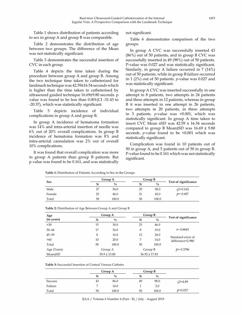
IJAA�/�Volume�6�Number�4�(Part�-�II)�/�July�-�August�2019
1453
Table�1�shows�distribution�of�patients�according�to�sex�in�group�A�and�group���was�comparable.
Table� 2� demonstrates� the� distribution� of� age�between� two� groups.� The� difference� of� the�Mean�was�not�statistically�signi��cant.
Table�3�demonstrates�the�successful�insertion�of�CVC�in�each�group.
Table� 4� depicts� the� time� taken� during� the�procedure�between�group�A�and�group��.�Among�the� two� techni�ue� time� taken� to� catheterized� for�landmark�techni�ue�was�42.59�16.54�seconds�which�is� higher� than� the� time� taken� to� catheterised� by�ultrasound�guided�techni�ue�16.69�9.80�seconds.�p�value�was�found�to�be�less� than�0.001(CI�-31.43�to�-20.37),�which�was�statistically�signi��cant.
Table� 5� depicts� incidence� of� individual�complications�in�group�A�and�group��.
In� group� A� incidence� of� hematoma� formation�was�14��and�intra-arterial�insertion�of�needle�was�6�� out� of� 20�� overall� complications.� In� group� ��incidence� of� hematoma� formation� was� 8�� and�intra-arterial� cannulation� was� 2�� out� of� overall�10��complications.�
It�was�found�that�overall�complication�was�more�in� group� A� patients� than� group� �� patients.� �ut�p�value�was�found�to�be�0.161,�and�was�statistically�
not�signi��cant.
Table� 6� demonstrates� comparison� of� the� two�groups.
In� group� A� CVC� was� successfully� inserted� 43�(86�)�out�of�50�patients,�and�in�group���CVC�was�successfully�inserted�in�49�(98�)�out�of�50�patients.�P-value�was�0.027�and�was�statistically�signi��cant.�Similarly,� in� group�A� failure� occurred� in� 7� (14�)�out�of�50�patients,�while�in�group���failure�occurred�in�1�(2�)�out�of�50�patients.�p-value�was�0.027�and�was�statistically�signi��cant.
In�group�A�CVC�was�inserted�successfully�in�one�attempt� in� 8�patients,� two� attempts� in� 24�patients�and�three�attempts�in�12�patients,�whereas�in�group��� it� was� inserted� in� one� attempt� in� 26� patients,�two� attempts� in� 20� patients,� in� three� attempts�in� 3� patients.� p-value� was� �0.001,� which� was�statistically� signi��cant.� In� group� A� time� taken� to�insert�CVC�Mean� �SD�was� 42.59� �� 16.54� seconds�compared� to� group� ��Mean�SD�was� 16.69� �� 9.80�seconds.� p-value� found� to� be� �0.001� which� was�statistically�signi��cant.
Complication� was� found� in� 10� patients� out� of�50�in�group�A,�and�5�patients�out�of�50�in�group��.�P-value�found�to�be�0.161�which�was�not�statistically�signi��cant.
Table��:�Distribution�of�Patients�According�to�Sex�in�the�Groups
Se��roup�A �roup�B
Test�of�significanceN � N �
Male 27 54.0 29 58.0 �2�0.162
p��0.687Female 23 46.0 21 42.0
Total 50 100.0 50 100.0
Table��:�Distribution�of�Age��etween�Group�A�and�Group��
Age�in�years�
�roup�A �roup�BTest�of�significance
N � N �
�30 15 30.0 23 46.0
t��0.8845
Standard�error�of�difference�2.980
p��0.3786
30–44 17 34.0 8 19.0
45–59 8 16.0 12 24.0
�60 10 20.0 7 14.0
Total 50 100.0 50 100.0
Age�(�ears) Group�A Group��
Mean�SD 39.9���15.80 36.92���17.83
Table��:�Successful�Insertion�of�Central�Venous�Cathrter
�roup�A �roup�B
N � N �
Success 43 86.0 49 98.0 �2�4.89
p�0.027Failure 7 14.0 1 2.0
Total 50 100.0 50 100.0
Real-time��ltrasound-Guided�Catheterization�of�the�Internal�Jugular�Vein:�A�Prospective�Comparison�with�the�Landmark�Techni�ue
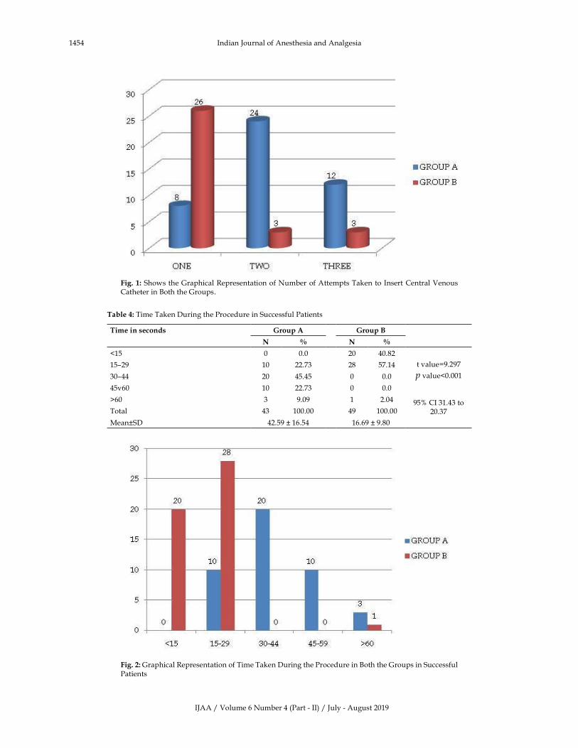
IJAA�/�Volume�6�Number�4�(Part�-�II)�/�July�-�August�2019
1454 Indian�Journal�of�Anesthesia�and�Analgesia
Fig���:�Shows�the�Graphical�Representation�of�Number�of�Attempts�Taken�to� Insert�Central�Venous�Catheter�in��oth�the�Groups.
Table��:�Time�Taken�During�the�Procedure�in�Successful�Patients
Time�in�seconds �roup�A �roup�B
N � N �
�15 0 0.0 20 40.82
t�value�9.297
p�value�0.001
95��CI�31.43�to�20.37
15–29 10 22.73 28 57.14
30–44 20 45.45 0 0.0
45v60 10 22.73 0 0.0
�60 3 9.09 1 2.04
Total 43 100.00 49 100.00
Mean�SD 42.59���16.54 16.69���9.80
Fig���:�Graphical�Representation�of�Time�Taken�During�the�Procedure�in��oth�the�Groups�in�Successful�Patients
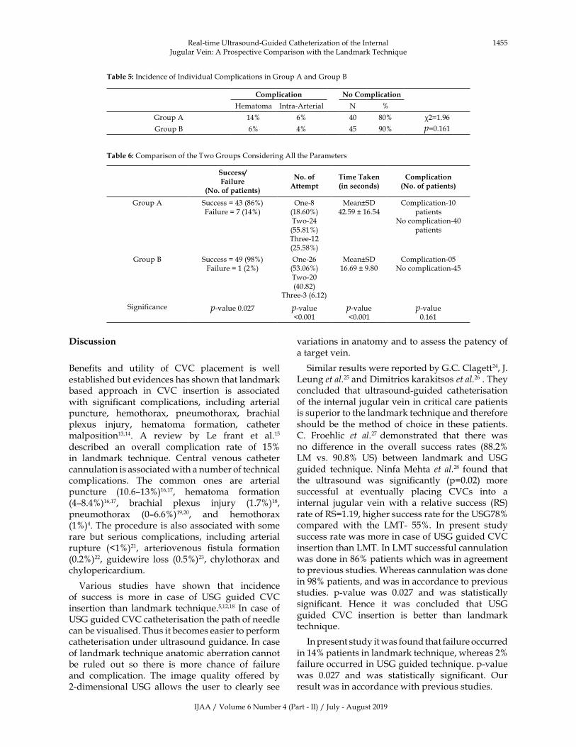
IJAA�/�Volume�6�Number�4�(Part�-�II)�/�July�-�August�2019
1455
�iscussion
�ene��ts� and� utility� of� CVC� placement� is� well�established�but�evidences�has�shown�that�landmark�based� approach� in� CVC� insertion� is� associated�with� signi��cant� complications,� including� arterial�puncture,� hemothorax,� pneumothorax,� brachial�plexus� injury,� hematoma� formation,� catheter�malposition13,14.� A� review� by� Le� frant� et� al.15�described� an� overall� complication� rate� of� 15��in� landmark� techni�ue.� Central� venous� catheter�cannulation�is�associated�with�a�number�of�technical�complications.� The� common� ones� are� arterial�puncture� (10.6–13�)16,17,� hematoma� formation�(4–8.4�)16,17,� brachial� plexus� injury� (1.7�)18,�pneumothorax� (0–6.6�)19,20,� and� hemothorax�(1�)4.�The�procedure� is�also�associated�with�some�rare� but� serious� complications,� including� arterial�rupture� (�1�)21,� arteriovenous� ��stula� formation�(0.2�)22,� guidewire� loss� (0.5�)23,� chylothorax� and�chylopericardium.
Various� studies� have� shown� that� incidence�of� success� is� more� in� case� of� �SG� guided� CVC�insertion� than� landmark� techni�ue.5,12,18� In� case� of��SG�guided�CVC�catheterisation�the�path�of�needle�can�be�visualised.�Thus�it�becomes�easier�to�perform�catheterisation�under�ultrasound�guidance.�In�case�of�landmark�techni�ue�anatomic�aberration�cannot�be� ruled� out� so� there� is� more� chance� of� failure�and� complication.� The� image� �uality� offered� by�2-dimensional��SG� allows� the� user� to� clearly� see�
variations�in�anatomy�and�to�assess�the�patency�of�a�target�vein.
Similar�results�were�reported�by�G.C.�Clagett24,�J.�Leung�et�al.25�and�Dimitrios�karakitsos�et�al.26�.�They�concluded� that� ultrasound-guided� catheterisation�of�the�internal�jugular�vein�in�critical�care�patients�is�superior�to�the�landmark�techni�ue�and�therefore�should�be� the�method�of� choice� in� these�patients.C.� Froehlic� et� al.27� demonstrated� that� there� was�no� difference� in� the� overall� success� rates� (88.2��LM� vs.� 90.8�� �S)� between� landmark� and� �SG�guided� techni�ue.�Ninfa�Mehta� et� al.28� found� that�the� ultrasound� was� signi��cantly� (p�0.02)� more�successful� at� eventually� placing� CVCs� into� a�internal� jugular� vein� with� a� relative� success� (RS)�rate�of�RS�1.19,�higher�success�rate�for�the��SG78��compared� with� the� LMT-� 55�.� In� present� study�success�rate�was�more�in�case�of��SG�guided�CVC�insertion�than�LMT.�In�LMT�successful�cannulation�was�done�in�86��patients�which�was�in�agreement�to�previous�studies.�Whereas�cannulation�was�done�in�98��patients,�and�was�in�accordance�to�previous�studies.� p-value� was� 0.027� and� was� statistically�signi��cant.� �ence� it� was� concluded� that� �SG�guided� CVC� insertion� is� better� than� landmark�techni�ue.
In�present�study�it�was�found�that�failure�occurred�in�14��patients�in�landmark�techni�ue,�whereas�2��failure�occurred�in��SG�guided�techni�ue.�p-value�was� 0.027� and� was� statistically� signi��cant.� Our�result�was�in�accordance�with�previous�studies.
Table��:�Incidence�of�Individual�Complications�in�Group�A�and�Group��
Complication No�Complication
�ematoma Intra-Arterial N �
Group�A 14� 6� 40 80� �2�1.96
p�0.161Group�� 6� 4� 45 90�
Table��:�Comparison�of�the�Two�Groups�Considering�All�the�Parameters
Success/Failure
�No��of�patients�
No��of�Attempt
Time�Taken�in�seconds�
Complication�No��of�patients�
Group�A Success���43�(86�)Failure���7�(14�)
One-8�(18.60�)Two-24�(55.81�)Three-12�(25.58�)
Mean�SD42.59���16.54
Complication-10�patients
No�complication-40�patients
Group�� Success���49�(98�)Failure���1�(2�)
One-26�(53.06�)Two-20�(40.82)
Three-3�(6.12)
Mean�SD16.69���9.80
Complication-05No�complication-45
Significance p-value�0.027 p-value��0.001
p-value��0.001
p-value�0.161
Real-time��ltrasound-Guided�Catheterization�of�the�Internal�Jugular�Vein:�A�Prospective�Comparison�with�the�Landmark�Techni�ue
IJAA�/�Volume�6�Number�4�(Part�-�II)�/�July�-�August�2019
1456 Indian�Journal�of�Anesthesia�and�Analgesia
It� has� been� shown� in�many� studies� that� use� of�ultrasound� decreases� number� of� attempts.25,29-31
Variations� in� external� landmarks� and� internal�anatomy� can�make� landmark-guided� cannulation�challenging.
E.�oski� et� al.31� compared� the� conventional�method�with� the�ultrasound-aided�techni�ue.�The�venous� lumen�was� reached�with� fewer�punctures�while� using� the� ultrasound-aided� techni�ue.�Similarly,�Randolph� et� al.30� evaluated� the� effect� of�real-time� ultrasound� guidance� using� a� regular� or�Doppler� ultrasound� techni�ue� for� placement� of�central� venous� catheters.� They� found� that� �SG�signi��cantly� decreases� the� need� for� multiple�catheter�placement�attempts�when�compared�with�the�standard�LMT.
C.�Froehlic�et�al.27�demonstrated�median�number�of� attempts� were� fewer� with� �SG� for� all� CVCs�attempted.� �ltrasound� identi��ed� the� vein� size�and�location,�anomalies,�and�vessels�patency,�thus�avoiding�futile�attempts�in�patients�with�absent�or�thrombosed� veins� and� congenital� anomalies� such�as� persistent� left� superior� vena� cava.M.� �ruzoni�et�al.32�also�compared�and�reported�success�at���rst�attempt�in�65��of�patients�in�the�ultrasound�group�vs� 45�� in� LMT� (p� �� 0.021).� �ltrasound� reduced�the�number�of�cannulation�attempts�necessary� for�venous�access.
In� the� present� study� out� of� 50� patient�s�cannulation�was� done� in� 1� attempt� in� 8� (18.60�)�patients,�two�attempts�in�24�(55.81�)�patients�and�three�attempts� in�11�(25.58�)�patients�by�LMT.� In��SG�guided�techni�ue�CVC�inserted�in�one�attempt�in� 26� (52�)� patients,� in� two� attempts� in� 20� (40�)�patients,�and�in�three�attempts�in�3�(6�)�patients�out�of�50�patients.�There�was�a�statistically�signi��cant�difference�between�the�two�groups.�It�was�found�in�previous�studies�that�use�of��SG�decreases�the�time�taken� to� puncture� IJV� as� compared� to� traditional�LMT.��.�Teichgraber�et�al33�showed�that�access�time�was� markedly� shorter� with� the� sonographically�guided�techni�ue�(mean,�15.2�sec:�range,�8-76�sec)�than�with�the�anatomic�LMT�(mean,�51.4�sec;�range,�3-820� sec)/p��� .001).� In�ultrasound� lumen�of� vein�could� be� visualised� which� probably� reduces� the�time� taken� from� skin� puncture� to� aspiration� of�blood.� Thus� time� taken� to� cannulate� the� vessel�decreases.�
C.� Froehlic� et� al.34� demonstrated� mean� time�with� �SG� (median� 919� seconds� vs.� 405� seconds,�p� �� 0.02)� and� �enjarappa� et� al.35� reported� the�mean� access� time� in� �SG� techni�ue� lesser� than�in� LMT.� Similarly� in� the� present� study� it� was�found�that�mean�time�taken�to�cannulate�IJV�was�
signi��cantly� lower� in� �SG� guided� techni�ue� as�compared� to� LMT.� In� group� A� time� range� was�18-96� seconds� while� in� group� �� it� was� between�8-76�seconds.�In�group�A�time�taken�to�catheterise�was� Mean�� SD�42.59� �� 16.54,� was� signi��cantly�higher� as� compared� to� group� �,� which� was�Mean��SD�16.69���9.80.
Randolph� et� al.12� evaluated� that� ultrasound�guidance� signi��cantly� decreases� complications�during� catheter� placement.� �.� Teichgraber� et�al.31� demonstrated� that� complications� were�fewer� with� �SG� (neck� hematoma,� 2�� versus�10�;� plexus� irritation,� 4�� versus� 6�;� carotid�artery� puncture,� 0�� versus� 12�).J.� Leung� et� al.26�observed� that� there� was� a� 10.8�� complication�rate,�with�16.9��complications� in�LMT�and�4.6��in� the� ultrasonographic� group,� a� difference� of�12.3�.� �owever� in� the� present� study� we� found�that�out�of�50�patient�s�complications�occurred�in�10�(20�)�patients�in�LMT�while�in�5�(10�)�patients�in��SG�guided�techni�ue.�Although�the�difference�in�incidence�of�complication�between�two�groups�was�not�statistically�signi��cant,�the�incidence�was�less�in��SG�guided�group.
�i�itations
As� with� most� of� the� studies� our� study� is� not�exceptional.�It�has�some�limitations�and�weaknesses�which� were� inevitable.� It� could� not� be� double�blinded,� this� limitations� has� an� inborn� chance� of�being� biased.� The� study� was� randomised� on� the�basis� of� techni�ue� of� insertion� of� central� venous�catheter.��owever,� the� strength� of� the� study�was�the� sample� size,� and� populations� with� various�disease�process.
Conclusion
Considering� the� ��ndings� of� the� study,� we�concluded� that� �SG� guided� CVC� insertion� has�a� better� success� rate,� less� failure,� re�uired� less�number� of� attempt� and� time� taken� to� insert� the�CVC� was� signi��cantly� less.� �owever,� there� was�less�complication�with��SG�guided�CVC�insertion�in�present� study,� but� it� did�not� achieve� statistical�signi��cance.
�ence,� we� suggest� that� as� per� the� NICE�guideline� Central� Venous� Cannulation� should�be� done� under� the� �SG� guidance,� if� available�especially� in� patients� who� are� critically� ill.� This�will�increase�the�success�rate;�decrease�the�number�of� attempts,� duration� of� insertion,� duration� of�insertion�and�complications.
IJAA�/�Volume�6�Number�4�(Part�-�II)�/�July�-�August�2019
1457
Source(s)�of�su��ort:�Nil.
Conflict�of�interest:�None
�eferences
1.� Civetta� JM,�Taylor�RW,��irby�RR.�Critical�Care.�3rd�ed.�Philadelphia:�Lippincott�Raven;�1997.
2.� Shoemaker�W,�Ayres�S,�Grenvik�A,�et�al.�Textbook�of� Critical� Care.� 4th� ed.� Philadelphia:� W��Saunders;�2000.
3.� �urki� A,� �han� A,� �utt� �.� Comparison� between�anatomical� versus� ultrasonographic� insertion� of�central�venous�line.�Pak�Armd�Force�Med�J.�2012�Dec;4:203–209.
4.� Ruesch�S,�Walder��,�Tramer�MR.�Complications�of�central� venous�catheters:� Internal� jugular�versus�subclavian�access:�A�systematic�review.�Crit�Care�Med.�2002;30:454–60.
5.� Merrer� Jj,� De� Jonghe� �� et� al.� Complications� of�femoral� and� subclavian� venous� catheterization�in� critically� ill�patients.:a� randomized�controlled�trial.�JAMA�2001;286(6):700–7.
6.� �ermosura� �,� Vanags� L,� Dickey� MW:�Measurement� of� pressure� during� intravenous�therapy.�JAMA.�1966;195:181.
7.� �ayashi��,�Ootaki�C,�Tsuzuku�M,�et�al.�Respiratory�jugular� vasodilation:� a� new� landmark� for� right�internal� jugular� vein� puncture� in� ventilated�patients.�J�Cardiothorac�Vasc�Anesth.�2000;14:40–44.
8.� Mateer� J,� Plummer� D,� �eller� M,� et� al.� Model�curriculum� for� physician� training� in� emergency�ultrasonography.� Ann� Emerg� Med.� 1994;23:95–102.
9.� llman� JI,� Stoelting� R�.� Internal� jugular� vein�location� with� the� ultrasound� �Doppler� blood�flow�detector.�Anesth�Analg.�1978;57:118.
10.� �arakitsos�D,�Labropoulos�N,�De�Groot�E,� et� al.Real-time� ultrasound-guided� catheterisation�of� the� internal� jugular� vein:� A� prospective�comparison� with� the� landmark� techni�ue� in�critical�care�patients.�Crit�Care.�2006;10:R162.
11.� Denys� �G,� �retsky� �F,� Reddy� S.� �ltrasound-assisted� cannulation� of� the� internal� jugular�vein:� A� prospective� comparison� to� the� external�Landmark-Guided� Techni�ue.� Circulation.�1993;87:1557–62.
12.� Randolph�G,�Cook�DJ,�Gonzales�C,�et�al.��ltrasound�guidance�for�placement�of�central�venous�catheters:�a� meta-analysis� of� the� literature.� Critical� Care�Medicine,�1996;24(12):2053–58.
13.� Sznajder� JI,� �veibil� FR,� et� al.� Central� vein�catheterization.�Failure�and�complication�rates�by�
three�percutaneous�approaches.�Arch�Intern�Med.1986;146(2):259–61.
14.� McGee�DC,�Gould�M�.�Preventing�complications�of�central�venous�catheterization.�N�Engl� J�Med.�2003;348(12):1123–33.
15.� Lefrant� J�,�Muller�L,�De�La�Coussaye�et�al.�Risk�factors� of� failure� and� immediate� complication�of� subclavian�vein�catheterization� in�critically� ill�patients.�Intensive�Care�Med.�2002;28:1036–41.
.�16 Martin� MJ,� �usain� FA,� Piesman� M,� ,� et� al.� Is�routine� ultrasound� guidance� for� central� line�placement� beneficial�� A� prospective� analysis.�Curr�Surg.�2004;61:71–4.
17.� Denys� �G,� �retsky� �F,� Reddy� S.� �ltrasound-assisted� cannulation� of� the� internal� jugular�vein:� A� prospective� comparison� to� the� external�Landmark-Guided� Techni�ue.� Circulation.�1993;87:1557–62.
18.� Takeyama��,�Taniguchi�M,�Sawai��,�et�al.�Limiting�vein�puncture�to�three�needle�passes�in�subclavian�vein� catheterization� by� the� infraclavicular�approach.�Surg�Today�2006;36:779–82.
19.� Plaus�WJ.�Delayed�pneumothorax�after�subclavian�vein� catheterization.� J� Parenter� Enteral� Nutr.�1990;14:414–5.
20.� Robinson� JF,� Robinson�WA,� Cohn� A,� et� al.� 2nd�Perforation� of� the� great� vessels� during� central�venous� line� placement.� Arch� Intern� Med.�1995;155:1225–8.
21.� Asteri� T,� Tsagaropoulou� I,� Vasiliadis� �,� et� al.��eware�Swan-Ganz�complications.�Perioperative�management.�J�Cardiovasc�Surg.�2002;43:467–70.
22.� Eisenhauer� E,� Derveloy� RJ,� �astings� PR.�Prospective�evaluation�of�central�venous�pressure�(CVP)� catheters� in� a� large� citycounty� hospital.�Ann�Surg.�1982;196:560–4.
23.� Jefferson� P,�Ogbue�MN,��amilton� �E,� �all� DR.�A� survey� of� the� use� of� portable� ultrasound� for�central� vein� cannulation�on� critical� care�units� in�the���.�Anesthesia.�2002;57:365–8.
24.� Fry� WR.,� Clagett� GC,� Rourke� PT.� �ltrasound-guided� central� venous� access.� Archives� of�Surgery.�1999;134(7):738–741.
25.� Leung� J,� Duffy� M,� Finckh� A.� Real-time�ultrasonographically-guided� internal� jugular�vein�catheterization�in�the�emergency�department�increases�success�rates�and�reduces�complications:�A� randomized,� prospective� study.� Ann� Emerg�Med.�2006;48:540–7.
26.� �arakitsos�D,�Labropoulos�N,�De�Groot�E,� et� al.�Real-time� ultrasound-guided� catheterisation�of� the� internal� jugular� vein:� A� prospective�comparison� with� the� landmark� techni�ue� in�critical�care�patients.�Crit�Care.�2006;10:R162.
27.� Froehlich� CD,� Rigby� MR,� Rosenberg� ES.,� et� al.��ltrasound-guided� central� venous� catheter�
Real-time��ltrasound-Guided�Catheterization�of�the�Internal�Jugular�Vein:�A�Prospective�Comparison�with�the�Landmark�Techni�ue
IJAA�/�Volume�6�Number�4�(Part�-�II)�/�July�-�August�2019
1458 Indian�Journal�of�Anesthesia�and�Analgesia
placement�decreases�complications�and�decreases�placement�attempts�compared�with�the�landmark�techni�ue�in�patients�in�a�pediatric�intensive�care�unit.�Critical�Care�Medicine.�2009;37(3):1090–6.
28.� Mehta� N� et� al.� Is� Real� time� �ltrasonic� guided�central� line� placement� by� approach:� Emergency�Medicine�J.�2013;30(5):355–359.
29.� Rothschild� JM.� �ltrasound� guidance� of�central� vein� catheterization.� Evidence� Report/Technology� Assessment� No� 43.� Making� �ealth�Care� Safer� A� critical� Analysis� of� Patient� Safety�Practices.�A�R�,�Publication�No�01-EO58.�2001.pp.245–253.
30.� Guidance� on� the� use� of� ultrasound� locating�devices� for� placing� central� venous� catheters.�London:� NICE;� 2002.� National� Institute� for�Clinical� Excellence.�NICE�Technology�Appraisal�No�49.
31.� �oski�EM,�Suhonen�M,�Mattila�MA.��ltrasound-facilitated� central� venous� catheterization.� Crit�CareMed.�1992;20:424–426.
32.� Gopal���Palepu,�Juneja�Deven,�M�Subrahmanyam,�et� al.� Impact� of� �SG� on� CVC� insertion� in�IC�.� Indian� J� Radiological� Imaging.� 2009�August;19(3):19(3):191–198.
33.� Teichgraber� ��M,� �enter� T,� Gebel� M,� et�al.� A� sonographically� guided� techni�ue� for�central� venous� access.� American� Journal� of�Roentgenology.�1997;169(3):731–733.
34.� Froehlich� CD,� Rigby� MR,� Rosenberg� ES.,� et� al.�ltrasound-guided� central� venous� catheter�placement�decreases�complications�and�decreases�placement�attempts�compared�with�the�landmark�techni�ue�in�patients�in�a�pediatric�intensive�care�unit.�Critical�Care�Medicine.�2009;37(3):1090–6.
35.� �enjarappa� �S,� Pavan� P�,� �ussain� SS.� A�comparative� study� brtween� ultrasound� guided�catheterisation� of� the� Internal� jugular� vein� and�classical� landmark� techni�ue.� Int� J� Anat� Res�2014;2(4):757–60.�
��Red�Flower�Publication�Pvt.�Ltd.�
�Original�Research�ArticleIndian�Journal�of�Anesthesia�and�Analgesia�
2019;6(4)�(Part�-�II):�1465-1473DOI:�http://dx.doi.org/10.21088/ijaa.2349.8471.6419.56
A�Comparati�e�Study�of�Intrathecal��e�obupi�acaine�and��e�obupi�acaine��ith�Mida�olam�in��o�er�Abdominal�and��o�er�limb�Surgeries
Siddesh�N��adur����a�i��umar�M���Abhishek�M�Patil�
1,2Assistant�Professor,�3Senior�Resident,�Dept.�of�Anaesthesiology,�SDM�Medical�College,�Dharwad,��arnataka�580009,�India.
Corresponding� Author:� Siddesh� N� �adur,� Assistant� Professor,� Dept.� of� Anaesthesiology,� SDM� Medical� College,� Dharwad,��arnataka�580009,�India.
E�mail:�[email protected]
�ecei�ed�on�03.07.2019,�Accepted�on�22.07.2019
Abstract
Conte�t:� Intrathecal�midazolam�as� an� adjuvant� to� levobupivacaine� provides� excellent� intraoperative�hemodynamic� stability�and�also�good�postoperative�analgesia.�Present� study�was�done� to�evaluate� the�efficacy,�duration�of�pain� relief,� the� incidence�of�adverse�effects�and�complications�when�midazolam� is�given�along�with�levobupivacaine�intrathecally.�Aims:�To�compare�between�intrathecal�levobupivacaine�(Group�L�)�and�intrathecal�levobupivacaine�with�adjuvant�midazolam�1�mg�(Group�L�M)�with�respect�to�onset�and�duration�of�analgesia,�motor�blockade,�intraoperative�discomfort�and�postoperative�analgesia�re�uirement� and� complications� like� nausea,� vomiting,� respiratory� depression� etc.�Methodolog�:� In� this�prospective,� randomized,� double� blind,� placebo� controlled� study� a� total� of� 100� patients� of� American�Society�of�Anaesthesiologists�(ASA)�grade�I�and�II,�undergoing�elective�lower�abdominal�and�lower�limb�orthopaedic� surgery� under� sub-arachnoid� block�were� randomized� into� two� groups.� Group� L�� (n�50)�received�3�ml�of�0.5��isobaric�levobupivacaine�with�0.2�ml�of�normal�saline�and�Group�L�M�(n�50)�received�3�ml� of� 0.5�� isobaric� levobupivacaine�with�0.2�ml�midazolam� (1�mg)� (preservative� free)� as� intrathecal�anesthesia.� Assessment� of� sensory� blockade,� motorblockade,� duration� of� analgesia,� intraoperative�hemodynamics,�discomfort�and�postoperative�analgesia�estimated.�After�surgery,�patients�were�asked�to�score�their�pain�at�2,�4,�6,�12,�18�and�24�hr�by�VAS�score.�The�presence�of�postoperative�nausea�vomiting�(PONV),� pruritus� and� respiratory� depression� were� recorded� and� compared� between� the� two� groups.�Statistical�anal�sis:�For�continuous�variables�the�summary�statistics�of�N,�mean,�standard�deviation�(SD)�were�used.�For�categorical�data,the�number�and�percentage�were�used�in�data�summaries.Chi-s�uare(�2)/Fisher�exact�test�was�employed�to�determine�the�significance�of�differences�between�groups�for�categorical�data.�The�difference�of�the�means�of�analysis�variables�was�tested�with�unpaired�t-test.�p-value��0.05�was�considered�significant.�Data�were�analysed�using�SPSS�software�v.23.0.�Results:�Group�L�M�had�superior��uality�of�analgesia,prolonged�duration�of�analgesia,� reduced�postoperative�analgesic� re�uirement�and�minimal�hemodynamic�changes�compared�to�Group�L�.�Conclusions:�Intrathecal�midazolam�potentiates�levobupivacaine�effect�leading�to�better��uality�and�longer�duration�of�analgesia,�better�sedation,�better�postoperative�outcome�with�minimum�side�effects.
�ey�ords:�Midazolam;�PONV;�Visual�Analogue�Pain�Scale;�Adjuvants,�Anesthesia;�Assessment,�Pain.
�o��to�cite�this�article:
Siddesh�N��adur,�Ravi�kumar�M,�Abhishek�M�Patil.�A�Comparative�Study�of�Intrathecal�Levobupivacaine�and�Levobupivacaine�with�Midazolam�in�Lower�Abdominal�and�Lower�limb�Surgeries.�Indian�J�Anesth�Analg.�2019;6(4):�1465-1467.
IJAA�/�Volume�6�Number�4�(Part�-�II)�/�July�-�August�2019
1460 Indian�Journal�of�Anesthesia�and�Analgesia
Introduction
Subarachnoid� block� is� one� of� the� most� versatile�regional� anesthesia� techni�ues� available� today.�Subarachnoid�block�provides� ade�uate�anesthesia�for�patients�undergoing�lower�abdominal�and�lower�limb� surgeries.� In� order� to� maximize� �uality� of�anesthesia�and�post�operative�analgesia,�a�number�of�adjuvants�have�been�added�to�local�anaesthetics.�The� bene��ts� of� intrathecal� opioids� and� non-opioids�as� adjuvants� in� spinal�anesthesia� are�well�documented.� The� addition� of� intrathecal� opioids�is� however� associated� with� dose� related� adverse�effect� such� as� respiratory� depression,� nausea,�vomiting,�urinary�retention,�pruritus�and�sedation.1�So,�use�of�non-opioids�such�as�ketamine,�clonidine,�neostigmine,� magnesium� sulphate,� midazolam�have� become� popular� adjuncts� for� postoperative�analgesia.2
�yperbaric� bupivacaine� is� one� of� the� common�local� anaesthetic� used� for� spinal� anesthesia� in�patients� undergoing� lower� abdominal� and� lower�limb� surgeries.� �owever,� it� has� considerable�adverse� effects� on� the� cardiovascular� and� central�nervous�system.�Enantiomers�of�bupivacaine�may�have�the�same�desired�pharmacological�properties,�but� fewer� side� effects.� Levobupivacaine,� the� S(-)-enantiomer� of� bupivacaine� has� been� shown� to�provide� a�more� selective� neuraxial� blockade� than�racemic� bupivacaine.3� Intrathecal� midazolam�abolishes�pain�of�somatic�origin,�produces�selective�sensory� block� and� depresses� somatosympathetic�re��exes�without�any�neurotoxicity.�It�potentiates�the�blocking�actions�of� local� anaesthetics.� It� improves�the� �uality� of� sensory� and� motor� block,� without�prolonging� the� recovery� and� also� provides� good�postoperative�analgesia.
The� subarachnoid� midazolam� was� originally�shown� to� have� anti-nociceptive� properties� in�studies� performed� in� animals� in� early� 1980�s.4�
The� subarachnoid� midazolam� has� been� used� in�humans� since� 1986� and� doses� up� to� 2� mg� have�been� described.5� There� are� many� clinical� studies�in� favour� of� intrathecal� midazolam� which� has�added� advantages� of� sedation,� amnesia� and� anti-nociceptive� effects� without� any� neurotoxicity� or�other�side�effects.�
�ence,� this�study�was�designed�to�evaluate� the�ef��cacy,�to�know�the�duration�of�pain�relief,�to�know�the�incidence�of�adverse�effects�and�complications�when�midazolam�(preservative�free)�is�given�along�with�levobupivacaine�intrathecally.
Materials�and�Methods
This� prospective,� randomized,� double-blind�study� was� conducted� at� Department� of�Anaesthesiology,� Shri��.M.�Patil�Medical�College,��ospital� and� Research� Centre,� Vijayapur� from�December� 2014� to� June� 2016.� The� study� included�100�patients�of�ASA�Grade�I�and�II,�age�20-45�years,�body� weight� 50-80� kg� who� underwent� elective�lower� abdominal� and� lower� limb� surgery.� The�study� was� approved� by� the� institutional� ethical�committee.
Patients� belonging� to� ASA� grade� III� and� IV,�pregnant�women,�patients�on� long� term�analgesic�therapy�and�chronic�alcoholics,�had�any�deformity�or�local�pathology�in�lumbar�spine�region,�history�of�convulsions,�allergy�to�the�drugs�used,�bleeding�disorders,� were� uncooperative� and� with� severe�neurological� de��cit� were� excluded� from� the�study.�Patients�with� severe�hypovolemia,� anemia,�receiving�steroid�medication�and�patients�in�whom�spinal�anesthesia�failed�and�general�anesthesia�was�re�uired�were�also�excluded�from�the�study.
Preanaesthetic� check-up� was� done� on� the� day�before� surgery,� and� included� a� complete� history�and�any�known�drug�allergy,�general�and�systemic�examination�and�local�examination�of�lumbar�spine�region.� Pulse� rate,� blood� pressure,� respiratory�rate,� and� weight� and� height� of� the� patient� were�noted.�Relevant�investigations�were�done�in�all�the�patients.
The�anaesthetic�procedure�was�brie��y�explained�to� the� patient.� The� patients�were� also� introduced�to�Visual�Analogue�Scale�(VAS)�and�taught�how�to�use� it.�An� informed�written�consent�was�obtained�from�the�patient�or�his/her�relatives.
The� patients� were� randomized� on� the� day� of�surgery�into�two�groups�of�50�each.��undred�pieces�of�paper,� 50�with� �saline��written�on� them�and�50�with��midazolam��written�were�put�in�a�box�and�the�patients�were�asked�to�pick�one�piece�of�paper.�This�piece�of�paper�was�handed�to�an�anaesthesiologist�unconnected� to� the� study� who� prepared� the�medications.� Group� L�� patients� received� 3� ml�of� 0.5�� isobaric� levobupivacaine� with� 0.2� ml� of�normal� saline� and� Group� L�M� patients� received�3�ml�of�0.5��isobaric�levobupivacaine�with�0.2�ml�of�midazolam�(1�mg)�(preservative�free)�intrathecally.�1� ml� midazolam� ampoule� (preservative� free)�consisted�5�mg,�of�which�0.2�ml���1�mg�was�given�intrathecally�.
On�arrival�in�the�operating�room�fasting�status�(at�least�for�8�hours),�and�written�consent�was�checked,�
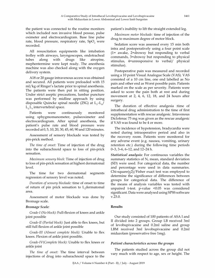
IJAA�/�Volume�6�Number�4�(Part�-�II)�/�July�-�August�2019
1461
the�patient�was�connected�to�the�routine�monitors�which� included� non� invasive� blood�pessue,� pulse�oximeter� and� electrocardiogram.� �ase� line� pulse�rate,� blood� pressure,� respiratory� rate,� SpO
2� were�
recorded.�
All� resuscitation� e�uipments� like� intubation�trolley�with� airways,� laryngoscopes,� endotracheal�tubes� along� with� drugs� like� atropine,�mephentermine� were� kept� ready.� The� anesthesia�machine�was�also�checked�along�with� the�oxygen�delivery�system.
A18�or�20�gauge�intravenous�access�was�obtained�and�secured.�All�patients�were�preloaded�with�15�ml/kg�of�Ringer�s�lactate�prior�to�spinal�anesthesia.The� patients� were� then� put� in� sitting� position.��nder�strict�aseptic�precautions,� lumbar�puncture�was� performed� by� midline� approach� by� using�disposable��uincke� spinal� needle� (25G)�at� L
3–L
4/
L2-L
3�intervertebral�space.
Patients� were� continuously� monitored�using� sphygmomanometer,� pulseoximeter� and�electrocardiogram.� After� spinal� anesthesia,� the�patient�s� pulse� rate� and� blood� pressure� were�recorded�at�0,�5,�10,�20,�30,�45,�60,�90�and�120�minutes.
Assessment� of� sensory� blockade�was� tested� by�pin-prick�method.�
The� time� of� onset:� Time� of� injection� of� the� drug�into� the� subarachnoid� space� to� loss� of� pin-prick�sensation.�
Ma�imum�sensor��block:�Time�of�injection�of�drug�to�loss�of�pin-prick�sensation�at�highest�dermatomal�level.�
The� time� for� two� dermatomal� segments�regression�of�sensory�level�was�noted.�
�uration�of�sensor��blockade:�time�of�onset�to�time�of� return� of� pin� prick� sensation� to� L
2dermatomal�
area.
Assessment� of� motor� blockade� was� done� by��romage�scale.
�ro�a�e�Scale:
�rade�I���o�block�:�Full-��exion�of�knees�and�ankle�joint�possible�
�rade�II���artial�block�:�Just�able�to���ex�knees,�but�still�full���exion�of�ankle�joint�possible
�rade�III� �Almost� complete� block�:� �nable� to� ��ex�knees.�Flexion�of�ankle�joint�possible.
�rade�I��Complete�block�:��nable�to���ex�knees�or�ankle�joint
The� time� of� onset:� The� time� interval� between�injections� of� drug� into� subarachnoid� space� to� the�
patient�s�inability�to�lift�the�straight�extended�leg.
Ma�imum�motor�blockade:�time�of�injection�of�the�drug�to�maximum�degree�of�motor�block.
Sedation�score�was� assessed�every�15�min�both�intra�and�postoperatively�using�a�four�point�scale�(1�� awake,� 2�drowsy� but� responding� to� verbal�commands,� 3�drowsy�but� responding� to�physical�stimulus,� 4�unresponsive� to� verbal/� physical�stimulus).
Postoperative�pain�was�measured�and�recorded�using�a�10�point�Visual�Analogue�Scale�(VAS).�VAS�consisted�of�a� 10� cm� line,� one� end� labelled� as�No�pain�and�other�end�as�Worst�possible�pain.�Patients�marked�on�the�scale�as�per�severity.�Patients�were�asked� to� score� the� pain� both� at� rest� and� during�movement� at� 2,� 4,� 6,� 12,� 18,� and� 24� hours� after�surgery.
The� duration� of� effective� analgesia� :time� of�intrathecal�drug�administration�to�the�time�of���rst�supplementation�with�rescue�analgesic.�Intavenous�Diclofenac�75�mg�was�given�as�the�rescue�analgesic�if�VAS�was�found�to�be�4�or�more.
The�incidence�of�hypotension,�bradycardia�were�noted� during� intraoperative� period� and� also� in�the� recovery� room.� Patients� were� monitored� for�any�adverse� event� (e.g.�nausea,�vomiting,�urinary�retention� etc.)� during� the� following� time� periods�0–3,�3–6,�6–12,�and�12–24�h.
Statistical� analysis:�For� continuous� variables� the�summary�statistics�of�N,�mean,�standard�deviation�(SD)�were�used.� For� categorical� data,� the�number�and� percentage� were� used� in� data� summaries.Chi-s�uare(�2)/Fisher�exact� test�was�employed� to�determine� the� signi��cance� of� differences� between�groups� for� categorical� data.� The� difference� of�the� means� of� analysis� variables� was� tested� with�unpaired� t-test.� p-value� �0.05� was� considered�signi��cant.�Data�were�analysed�using�SPSS�software�v.23.0.
�esults
Our�study�consisted�of�100�patients�of�ASA�I�and�II� divided� into� 2� groups.� Group� L�� received� 3ml�of� levobupivacaine� and� 0.2ml� saline� and� group�L�M� received� 3ml� levobupivacaine� and� 0.2ml�midazolam�(preservative�free�1mg).
Patient�characteristics�across�the��rou�s
The� patients� studied� across� the� group� did� not�vary�much�with�respect� to�age,�sex�or�height.�The�
A�Comparative�Study�of�Intrathecal�Levobupivacaine�and�Levobupivacaine�with�Midazolam�in�Lower�Abdominal�and�Lower�limb�Surgeries
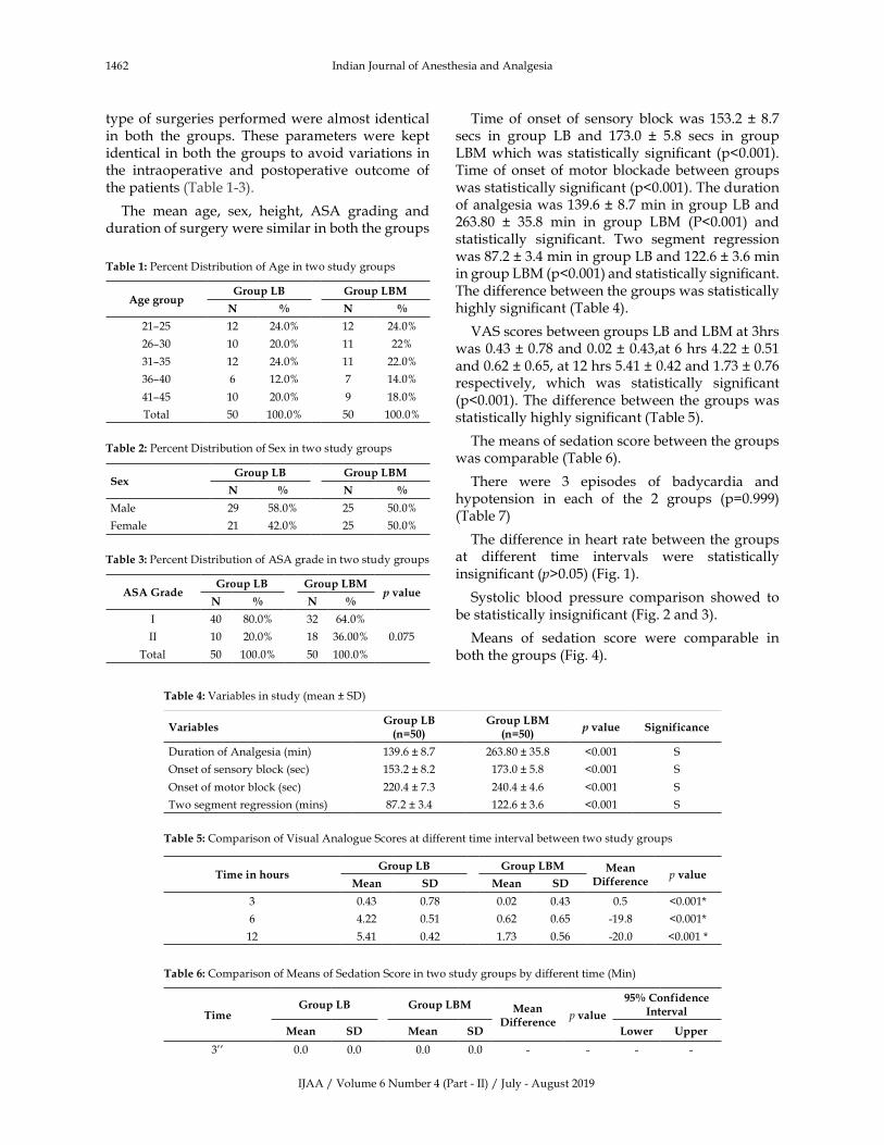
IJAA�/�Volume�6�Number�4�(Part�-�II)�/�July�-�August�2019
1462 Indian�Journal�of�Anesthesia�and�Analgesia
type�of�surgeries�performed�were�almost�identical�in� both� the� groups.� These� parameters� were� kept�identical� in�both�the�groups�to�avoid�variations�in�the� intraoperative� and� postoperative� outcome� of�the�patients�(Table�1-3).
The� mean� age,� sex,� height,� ASA� grading� and�duration�of�surgery�were�similar�in�both�the�groups�
Table��:�Percent�Distribution�of�Age�in�two�study�groups
Age�group�roup��B �roup��BM
N � N �
21–25 12 24.0� 12 24.0�
26–30 10 20.0� 11 22�
31–35 12 24.0� 11 22.0�
36–40 6 12.0� 7 14.0�
41–45 10 20.0� 9 18.0�
Total 50 100.0� 50 100.0�
Table��:�Percent�Distribution�of�Sex�in�two�study�groups
Se��roup��B �roup��BM
N � N �
Male 29 58.0� 25 50.0�
Female 21 42.0� 25 50.0�
Table��:�Percent�Distribution�of�ASA�grade�in�two�study�groups
ASA��rade�roup��B �roup��BM
���alueN � N �
I 40 80.0� 32 64.0�
0.075II 10 20.0� 18 36.00�
Total 50 100.0� 50 100.0�
Time� of� onset� of� sensory� block�was� 153.2� �� 8.7�secs� in� group� L�� and� 173.0� �� 5.8� secs� in� group�L�M�which�was� statistically� signi��cant� (p�0.001).�Time�of�onset� of�motor�blockade�between�groups�was�statistically�signi��cant�(p�0.001).�The�duration�of�analgesia�was�139.6���8.7�min� in�group�L��and�263.80� �� 35.8� min� in� group� L�M� (P�0.001)� and�statistically� signi��cant.� Two� segment� regression�was�87.2���3.4�min�in�group�L��and�122.6���3.6�min�in�group�L�M�(p�0.001)�and�statistically�signi��cant.�The�difference�between�the�groups�was�statistically�highly�signi��cant�(Table�4).
VAS�scores�between�groups�L��and�L�M�at�3hrs�was�0.43���0.78�and�0.02���0.43,at�6�hrs�4.22���0.51�and�0.62���0.65,�at�12�hrs�5.41���0.42�and�1.73���0.76�respectively,� which� was� statistically� signi��cant�(p�0.001).�The�difference�between�the�groups�was�statistically�highly�signi��cant�(Table�5).
The�means�of�sedation�score�between�the�groups�was�comparable�(Table�6).�
There� were� 3� episodes� of� badycardia� and�hypotension� in� each� of� the� 2� groups� (p�0.999)�(Table�7)
The�difference�in�heart�rate�between�the�groups�at� different� time� intervals� were� statistically�insigni��cant�(p�0.05)�(Fig.�1).
Systolic� blood� pressure� comparison� showed� to�be�statistically�insigni��cant�(Fig.�2�and�3).�
Means� of� sedation� score� were� comparable� in�both�the�groups�(Fig.�4).
Table��:�Variables�in�study�(mean���SD)
Variables�roup��B�n����
�roup��BM�n����
���alue Significance
Duration�of�Analgesia�(min) 139.6���8.7 263.80���35.8 �0.001 S
Onset�of�sensory�block�(sec) 153.2���8.2 173.0���5.8 �0.001 S
Onset�of�motor�block�(sec) 220.4���7.3 240.4���4.6 �0.001 S
Two�segment�regression�(mins) 87.2���3.4 122.6���3.6 �0.001 S
Table��:�Comparison�of�Visual�Analogue�Scores�at�different�time�interval�between�two�study�groups
Time�in�hours�roup��B �roup��BM Mean�
�ifferencep��alue
Mean S� Mean S�
3 0.43 0.78 0.02 0.43 0.5 �0.001�
6 4.22 0.51 0.62 0.65 -19.8 �0.001�
12 5.41 0.42 1.73 0.56 -20.0 �0.001��
Table��:�Comparison�of�Means�of�Sedation�Score�in�two�study�groups�by�different�time�(Min)
Time�roup��B �roup��BM Mean�
�ifferencep��alue
����Confidence�Inter�al
Mean S� Mean S� �o�er �pper
3�� 0.0 0.0 0.0 0.0 - - - -
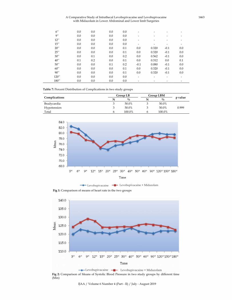
IJAA�/�Volume�6�Number�4�(Part�-�II)�/�July�-�August�2019
1463
Fig��:�Comparison�of�means�of�heart�rate�in�the�two�groups
Fig��:�Comparison�of�Means�of�Systolic��lood�Pressure�in�two�study�groups�by�different�time�(Min)
6�� 0.0 0.0 0.0 0.0 - - - -
9�� 0.0 0.0 0.0 0.0 - - - -
12�� 0.0 0.0 0.0 0.0 - - - -
15�� 0.0 0.0 0.0 0.0 - - - -
20�� 0.0 0.0 0.0 0.1 0.0 0.320 -0.1 0.0
25�� 0.0 0.0 0.0 0.1 0.0 0.320 -0.1 0.0
30�� 0.0 0.1 0.0 0.2 0.0 0.562 -0.1 0.0
40�� 0.1 0.2 0.0 0.1 0.0 0.312 0.0 0.1
50�� 0.0 0.0 0.1 0.2 -0.1 0.080 -0.1 0.0
60�� 0.0 0.0 0.0 0.1 0.0 0.320 -0.1 0.0
90�� 0.0 0.0 0.0 0.1 0.0 0.320 -0.1 0.0
120�� 0.0 0.0 0.0 0.0 - - - -
180�� 0.0 0.0 0.0 0.0 - - - -
Table��:�Percent�Distribution�of�Complications�in�two�study�groups
Complications�roup��B �roup��BM
p��alueN � N �
�radycardia 3 50.0� 3 50.0�
0.999�ypotension 3 50.0� 3 50.0�
Total 6 100.0� 6 100.0�
Levobupivacaine���MidazolamLevobupivacaine
Levobupivacaine���MidazolamLevobupivacaine
A�Comparative�Study�of�Intrathecal�Levobupivacaine�and�Levobupivacaine�with�Midazolam�in�Lower�Abdominal�and�Lower�limb�Surgeries

IJAA�/�Volume�6�Number�4�(Part�-�II)�/�July�-�August�2019
1464 Indian�Journal�of�Anesthesia�and�Analgesia
Levobupivacaine���MidazolamLevobupivacaine
Fig���:�Comparison�of�Means�of�Diastolic��lood�Pressure�in�two�study�groups�by�different�time�(Min)
Levobupivacaine���MidazolamLevobupivacaine
Fig���:�Comparison�of�Means�of�Sedation�Score�in�two�study�groups�by�different�time�(Min)
�iscussion
The� subarachnoid� blockade� is� the� common� form�of� centrineuraxial� blockade� performed� for� lower�abdomen� and� lower� limb� surgeries.Intrathecal�midazolam�has�been�used�as� an�adjuvant� to� local�anaesthetics� since� 1980s.� It� has� been� tried�widely�and�antinociceptive�effect�with�neurological�safety�has�been�well�established�in�animals�and�humans.
The� intrathecal� benzodiazepine� induced�analgesia� is� spinally� mediated.� The� binding� sites�of� midazolam� are� GA�A� receptors� which� are�abundant�in�dorsal�root�nerve�cells�of�spinal�cord.�The� maximum� concentration� of� GA�A� receptors�
are�found�within�lamina�II�of�dorsal�nerve�cells,�a�region� which� plays� prominent� role� in� processing�nociceptive�and�thermoceptive�stimulation.�Acting�over� the�GA�A�receptors�benzodiazepines� induce�changes� in� chloride� conductance� and� enhance�GA�A� induced� presynaptic� inhibition� of� primary�afferent�terminals.
Chan�es� in� the� �erio�erative� cardiovascular��ara�eters
In�the�present�study,�the�incidence�of�hypotension�was�e�ual,�with�3�patients�in�each�group�had�a�fall�in� blood�pressure.��ypotension�was� corrected� by�administration� of� injection� mephentermine� 6� mg�
IJAA�/�Volume�6�Number�4�(Part�-�II)�/�July�-�August�2019
1465
IV�in�incremental�doses�and�giving�IV���uids.��eart�rate,� systolic� and�diastolic� blood�pressure� in�both�the�groups�did�not�vary�signi��cantly.�Goodchild�CS,�Noble�J�in�19875,��ahar�M,�Cohen�ML�et�al.�in�19976,��atra����and�et�al.�in�19997�and��harti�N�and�et�al.�in�20038� found�no�difference� in� the�hemodynamic�responses�to�the�drugs�used.�Our�study�correlated�with.� Rosa� �errera� and� et� al.9� study� of� stable�hemodynamics�with�isobaric�levobupivacaine.
Chan�es�in�res�iratory��ara�eters
None� of� the� patients� in� the� present� study� had�respiratory�depression.��ahar�M�and�et�al.�in�19976�found� no� changes� in� the� arterial� blood� gases� or�respiratory�rate�when�given�intrathecal�midazolam�in�animal�model.� Sen�A�and� et� al.� in� 200110� found�that� intrathecal� midazolam� produces� better�tran�uility�of�patients�of�caesarian�section�delivery�without�much�sedation�and�respiratory�depression.�Apgar�score�of�baby�in�1st�and�5th�minute�of�delivery�was� found� to� be� normal.In� our� study�we� did�not�include� caesarian� section�deliveries.��harti�N�and�et�al.�in�20038�studied�the�effect�of�intrathecal�1�mg�of� midazolam� with� hyperbaric� bupivacaine� in�patients�undergoing�lower�abdominal�surgery�and�found�no�change�in�oxygen�saturation.
The�above�observations�were�similar�to�our�study�results.� We� conclude� that� intrathecal� midazolam�1� mg� is� safe� to� use� without� causing� respiratory�depression.
Chan�es�in�the�onset�of�sensory�and��otor��loc�ade
In�our�study�the�onset�of�sensory�blockade�in�group-��was�153.2���8.2�seconds�compared�to�173.0���5.8�seconds�in�group-II�which�was�statistically�highly�signi��cant�(p� �� 0.001).� It� shows� that� addition� of� midazolam�to� local� anaesthetic� delays� the� onset� of� analgesia.�Similarly�the�onset�of�motor�blockade�in�group-��was�220.4���7.3�compared�to�240.4���4.6�seconds�in�group-�M� which� was� also� statistically� highly� signi��cant�(p� �� 0.001)� i.e.,� the� addition� of�midazolam� to� local�anaesthetic�delays�the�onset�of�motor�blockade.��egin�A�and�et�al.�200411�did�not���nd�anydelay�in�onset�of�sensory�and�motor�blockade�with�addition�of�2�mg�of� midazolam� to� hyperbaric� bupivacaine� in� spinal�anesthesia� in�patients�undergoing�perianal�surgery.��ut� in�our�study�we�used� isobaric� levobupivacaine�instead�of�bupivacaine.
From�the�above�study�we�conclude�that�there�is�variation�in�the�onset�of�sensory�and�motor�blockade�in� different� studies.� Though� it� is� statistically�signi��cant�in�our�study�it�does�not�have�any�clinical�implications.
�i�e� for� t�o� der�ato�al� se��ents� re�ression� of�sensory�level
In�our�study,�the�two�segment�regression�of�sensory�level�in�group�L��was�87.50���4.4�minutes�compared�to�122.00���3.6�minutes� in�group�L�M�which�was�statistically� highly� signi��cant� (p� �� 0.001).� This�shows� that� addition� of� midazolam� increases� the�duration�of�sensory�blockade.
�harti�N�and�et�al.�in�2003�found�that�duration�of�sensory�block�(ie.,�time�to�regression�to�S
2�segment)�
was� signi��cantly� longer� in� the� midazolam� group�than� the� control� group� (218� min� vs� 165� min,�p���0.001).�Venkatesh�Selvaraj,�Tapan�Ray�in�201512�
had�similar���nding�with�midazolam�as�an�adjuvant�to� intrathecal� lignocaine.�So�we�can�conclude� that�intrathecal� midazolam� increases� the� duration� of�sensory�blockade.
�i�e�of�first�re�uest�of�anal�esics
In�our�study,�the�time�of���rst�re�uest�of�analgesics�in�group�L��was�139.00���8.77�minutes�compared�to�263.8���35.8�minutes� in�group�L�M�which�was�statistically� highly� signi��cant� (p� �� 0.001).� This�shows� that� there� was� signi��cantly� longer� period�of�analgesia�with�intrathecal�midazolam.��im�M��and�et�al.�in�200113�found�signi��cantly�greater�time�to���rst�analgesia�in�the�midazolam�group�in�patients�undergoing�haemorrhoidectomy.�Amr�M�and�et�al.in�200314�had�similar���nding�in�patients�undergoing�knee�arthroscopy.�Valentine�J.M.�J�and�et�al.�in�199615,�Shah�FR�et�al.� in�200316� found�prolonged�duration�of� postoperative� pain� relief� with� midazolam� as�adjuvant.� These� studies� validate� our� ��ndings� of�prolonged���rst�re�uest�of�supplemental�analgesics�in�the�postoperative�period.
�isual�Analo�ue�Score
In� our� study,� there� was� signi��cant� reduction� in�the�visual�analogue�score�of�the�patients� in�group�L�M�in�comparison�with�higher�VAS�in�group�L��recorded�at�3�hours,�6�hours�and�12�hours�of�spinal�anesthesia.�Shah�FR�and�et�al.�in�2003�showed�that�patients� treated� with� intrathecal� midazolam� had�better�pain� relief� judged�by�visual� analogue�scroe�on� coughing� (p� �� 0.0013)� and� a� nursing�mobility�score�(p���0.0001).��egin�A�and�et�al.�in�2004�found�signi��cantly� lower�visual� analogue�pain� scores� in�midazolam�group�at�the���rst�4�hours.
Above� studies� support� that� intrathecal�midazolam� potentiates� the� sensory� blockade�of� levobupivacaine,� there� by� reduce� the� visual�analogue�scores�in�the�early�post�operative�period�bringing�about�better�post�operative�outcome.
A�Comparative�Study�of�Intrathecal�Levobupivacaine�and�Levobupivacaine�with�Midazolam�in�Lower�Abdominal�and�Lower�limb�Surgeries
IJAA�/�Volume�6�Number�4�(Part�-�II)�/�July�-�August�2019
1466 Indian�Journal�of�Anesthesia�and�Analgesia
Sedation�Score
The�sedation�score�was�assessed�by�scoring�system�of�Chernic�et�al.17
In� our� study� the� sedation� score� ranged� from�0� to� 1� in�both� the�groups.�Most�of� the�patients� in�group� L�M�were� calm� and� sleeping� comfortably�were�as�most�of�the�patients�in�the�group�L��were�awake�and�alert.�Nishiyama�T�199518�studies�used�midazolam� for� pre� and� post� operative� sedation�which�showed� the�sedation�scores�were�higher� in�the�patients� receiving�midazolam�by� the� epidural�or�intrathecal�route.�Vaswani�et�al.19�found�sedation�scores� were� less� but� more� sustained� when� the�midazolam� is� administered� intrathecally.� Anjana�Sen�et� al.� also� reported� the�higher� sedation� scores�with�intrathecal�midazolam.
The� results� of� present� study� are� consistent�with� both� the�authors� though� the�duration� of� the�sedation� is� less.� This�may� be� because� of� different�doses�of�the�drug.
Adverse�Effects
In�the�present�study,�3�patients�had�hypotension�and�bradycardia,� 1� patient� had� shivering� and� nausea�vomiting�in�group�L�M�compared�to�3�patients�of�hypotension,�2�patients�of�shivering�and�1�patient�of� nausea��� vomiting� in� group�L�.� This� signi��es�that� adverse� effects� are� minimal� with� intrathecal�midazolam.
Studies� in� humans� by� Valentine� JMJ� et� al.� in�1999,�Sen�A�and� et� al.� in� 2001,��harti�N�and� et� al.�in�2003,�Shah�FR�and�et�al.�in�2003,�Amr�M�and�et�al.�in�2003,�Tucker�AP�and�et�al.�in�2004,�found�no�adverse� neurological� symptoms� in� those� received�intrathecal� midazolam.� They� also� found� that�intrathecal� midazolam� has� mild� sedative� and�antiemetic�effect.
Conclusion
With�all� the�above�observations�we� conclude� that�addition�of�midazolam�to�levobupivacaine�provides�prolonged�analgesia,�superior�pain�relief�and�better�sedation� with� minimal� side� effects� compared� to�levobupivacaine�alone�in�spinal�anesthesia�
�eferences
1.� Ramkumar� V,� Prasad� �N.� Management� of�postoperative�pain.� Indian�J.�Anaesth.�2006;50(5)�:345–354.
2.� Agrawal� N,� �smani� A,� Sehgal� R,� et� al.� Effect�of� intrathecal� midazolam� levobupivacaine�combination� on� post� operative� analgesia.� IJA.�2005;49(1):37.
3.� �rbanek��,��apral�S.�Levobupivacaine�for�regional�anesthesia.� A� systematic� review.� Anaesthesist.�2006;55:296–313.
4.� Whitwam� JG,� and� Loh� L,� Depression� of�nociceptive�sympathetic�reflexes�by�the�intrathecal�administration� of� midazolam.� �r� Journal� of�Anesthesia.�1983;55(6):p.541–7.
5.� Goodchild�CS,�Noble�J.�The�effects�of�intrathecal�midazolam� on� sympathetic� nervous� system�reflexes�in�man--a�pilot�study.��r�J�Clin�Pharmacol.1987;23(3):279–85.
6.� �ahar�M,�Cohen�ML,�Grinshpon��,� et� al.�Spinal�anesthesia� with� midazolam� in� the� rat.� Can� J.�Anesthesia.�1997;44(2):208–215.
7.� �atra� ��,� Jain� �,� Chari� P,� et� al.� Addition� of�intrathecal� midazolam� to� levobupivacaine�produces�better�post-operative�analgesia�without�prolonging� recovery.� Int� J�Clin�Pharmacol�Ther.�1999;37(10):519–23.
8.� �harti�N,�Madan�R,�Mohanty�PR,�et�al.�Intrathecal�midazolam� added� to� levobupivacaine� improves�the� duration� and� �uality� of� spinal� anesthesia.�Acta�Anaesthesiol�Scand.�2003�Oct;47(9):1101–5.
9.� Rosa��,� Jose�De�A,�Luis�E,� et� al.��emodynamic�impact� of� isobaric� levobupivacaine� versus�hyperbaric� bupivacaine� for� subarachnoid�anesthesia� in� patients� aged� 65� and� older�undergoing� hip� surgery.� �MC� Anesthesiol2014;14:97.
10.� Sen� A,� Rudra� A,� Sarkar� S�,� et� al.� Intrathecal�midazolam� for� postoperative� pain� relief� in�caesarean� section� delivery.� J� Indian�Med�Assoc.2001;99(12):683–4,�686.
11.� �egin�A,�Sanli�S,�Dosemeci�L,�et�al.�The�analgesic�and� sedative� effects� of� intrathecal� midazolam�in� perianal� surgery.� Eur� J� Anaesthesiol.�2004;21(8):658–62.
12.� Selvaraj�V,�Ray�T.�Midazolam�as�an�adjuvant� to�intrathecal�lignocaine:�A�prospective�randomized�control�study.�Saudi�J�Anaesth�2015;9:393-6.
13.� �im� M�,� Lee� �M.� Intrathecal� midazolam�increases�the�analgesic�effects�of�spinal�blockade�with� levobupivacaine� in� patients� undergoing�haemorrhoidectomy.� �r� J� Anaesth.� 2001Jan;86(1):77–9.
14.� Abdelfatah� AM,� Fawaz� A,� Al-Azazi� �M.� The�postoperative� analgesic� effect� of� intrathecal�fentanyl�versus�midazolam�in�knee�arthroscopy.�Egyptian� Journal� of� Anesthesia.� 2003;19(2):173–77.
15.� Valentine� JMJ,�Lyons�G,��ellamy�MC.�The�effect�
IJAA�/�Volume�6�Number�4�(Part�-�II)�/�July�-�August�2019
1467
of� intrathecal�midazolam�on�postoperative�pain.�European�Journal�of�Anaesthesiology.�1996�Nov;�13(6):589–593.
16.� Shah� FR,� �albe� AR,� Panchal� ID,� et� al.�Improvement� in� postoperative� pain� relief� by�the� addition� of� midazolam� to� an� intrathecal�injection�of�buprenorphine�and�levobupivacaine.Eur�J�Anaesthesiol.�2003�Nov;20(11):904-10.�
17.� Chernik�DA,�Gillings�D,�Laine��,� et� al.�Validity�and� reliability� of� the� Observer�s� Assessment�of� Alertness/Sedation� Scale:� study� with�
intravenous�midazolam.� J� ClinPsychopharmacol�1990;10:244–51.
18.� Nishiyama�T,�Matsukawa�T,��anaoka��.�Effects�of� adding� midazolam� on� the� postoperative�epidural� analgesia� with� two� different� doses�of� levobupivacaine.� J� Clin� Anesth.� 2002� Mar;�14(2):92–7.
19.� Vaswani�R�,�Raiger�L�,�Richa�P�et�al.�The�effect�of� Intrathecal�midazolam� on� postoperative� pain�relief� in� orthopaedic� surgery.� �ospitals� Today.�2002;4:217-20.
A�Comparative�Study�of�Intrathecal�Levobupivacaine�and�Levobupivacaine�with�Midazolam�in�Lower�Abdominal�and�Lower�limb�Surgeries
��Red�Flower�Publication�Pvt.�Ltd.�
�Original�Research�ArticleIndian�Journal�of�Anesthesia�and�Analgesia�
2019;6(4)�(Part�-�II):�1474-1482DOI:�http://dx.doi.org/10.21088/ijaa.2349.8471.6419.57
Attenuation�of��emodynamic��esponse�to��aryngoscopy�and�Endotracheal�Intubation�by�using�Oral�I�abradine
Vikas�Bedi����aura��Chopra���Ashutosh�Singh���Madhuri�Sharma�
1,3Assistant�Professor,�4Associate�Professor,�2Professor�and��ead,�Department�of�Anaesthesiology,�Shri�Guru�Ram�Rai�Institute�Of�Medical����ealth�Sciences,�Dehradun,��ttarakhand�248001,�India.
Corresponding�Author:��aura��Chopra,�Professor,��ead�of�the�Department,�Department�of�Anaesthesiology,�Shri�Guru�Ram�Rai�Institute�Of�Medical����ealth�Sciences,�Dehradun,��ttarakhand�248001,�India.
E�mail:�[email protected]
�ecei�ed�on�16.04.2019,�Accepted�on�14.05.2019
Abstract
Introduction:� Laryngoscopy� gives� rise� to� a� stress� response� that� is� seen� as� rise� in� heart� rate� and� blood�pressure�and�as�dysrhythmia.�Such�changes�are�at�peak�immediately�after�intubation�and�can�be�dangerous�for� those� having� already� limited� or� compromised� cardiovascular� reserve.� They� can� lead� to� increased� risk�of�myocardial� infarction� (MI),� stroke,� congestive� cardiac� failure� or� sudden� death.�Aims� and�Ob�ecti�es:� To�study� the� effect� of� oral� Ivabradine� in� attenuating� the� hemodynamic� stress� response� to� laryngoscopy;� and�to�endotracheal�intubation�and�extubation.�Materials�and�Methods:�A�randomized�double�blinded�study�was�done.�Total�number�of�140�patients�(adult�patients�undergoing�ENT�surgery)�were�randomly�taken�into�two�Groups.�The�first�group�was�labelled�as�Test�Gropu�(A)�and�the�second�group�was�called�Control�Group�(�).�Each�group�had�70�patients.�The�Test�group�(A)� received�5�mg�oral� Ivabradine�and�the�Control�group�(�)�received�placebo.�Patients�were�monitored�for�hemodynamic�changes�as�per�the�protocol.�Result:�Mean�heart�rate,�S�P,�D�P�and�MAP�reading�at�intubation,�post-intubation�period�till�10�mins,�at�extubation�and�till�10�minutes�post-extubation�were�significantly�less�in�test�group�i.e.�in�patients�who�received�oral�Ivabradine�as�compared�to�control�group�(p�0.05).�Conclusion:�We�conclude�that�Ivabradine�is�an�extremely�useful�drug�to�prevent�abnormal�tachycardias�during�the�procedure�of�laryngoscopy�and�intubation.�Its�stabilizing�effect�of�haemodynamics�also�extends�up�to�extubation�and�immediate�post-operative�period.
�ey�ords:� Ivabridine;� �eart� Rate;� Diastolic� �lood� Pressure;� Systolic� �lood� Pressure;� Mean� Arterial�Pressure.
�o��to�cite�this�article:
Vikas��edi,�Gaurav�Chopra,�Ashutosh�Singh�et�al.�Attenuation�of��emodynamic�Response�to�Laryngoscopy�and�Endotracheal�Intubation�by�using�Oral�Ivabradine.�Indian�J�Anesth�Analg.�2019;6(4):�1468-1476.
Introduction
Endotracheal� intubation� is� a� very� common�procedure,� where� the� anesthesiologists� have� an�important�role�to�play.
Stress� response� with� laryngoscopy�manifest� as�tachycardia,� hypertension� and� dysrhythmias� and�
may� have� deleterious� respiratory,� neurological�and� cardiovascular� effects1.� These� changes� are�
maximum� immediately� after� intubation� and� lasts�for�5-10�minutes.
These� effects� are� generally� well� tolerated� by�
overall�healthy�patients�but�can�be�lethal�to�patients�with� preexisting� conditions� such� as� coronary�

IJAA�/�Volume�6�Number�4�(Part�-�II)�/�July�-�August�2019
1469
artery� disease,� recent� myocardial� infarction,�hypertension,� geriatric� population,� pre-eclampsia,�and� cerebrovascular� pathology� such� as� tumours,�aneurysms� or� increased� intracranial� pressure� and�are�at�increased�risk�of�morbidity�and�mortality.2
Geriatric�and�elderly�patients�are�more�susceptible�to� coronary� artery� disease� and� cerebrovascular�disease� as� they� have� elevated� baseline� blood�pressure,� making� them� especially� susceptible� to�variations�in�blood�pressure,�and�heart�rate�during�the�procedures� of� laryngoscopy�and� endotracheal�intubation,�which� can�become� critical� for� patients�with�compromised�cardiovascular�reserve�with�the�resultant�risk�of�myocardial�infarction�(MI),�stroke,�congestive�heart�failure�or�sudden�death.3
Ivabradine�also�known�as�a�cardiotonic�agent�is�a� highly� selective� inhibitor� of� �If�� channels�which�causes� a� reduction� in� the� slope� of� spontaneous�depolarisation.� This� results� in� an� increase� in� the�time� interval�between�successive�action�potentials�in�the�SA�node,�which�in�turn�leads� to�slowing�of�heart�rate.
Ivabradine�slows�down�the�heart�rate�but�it�does�not� cause� sudden� fall� in� blood� pressure� and� for�this�reason�it�is�desirable�to�use�it�in�patients�with�pre-exsisting�heart�conditions�like�angina�pectoris,�coronary� artery� disease,� heart� failure,� obstructive�cardiomyopathies�and�other�conditions�where� the�myocardial�perfusion�is�reduced.4
The��-2�receptors�are�present�at�various�sites�in�peripheral�and�central�nervous�system,� in�visceral�organs�such�as�liver,�kidney,�pancreas,�in�eyes,�the�media�of�vessel�walls,�and�also�in�platelets.5
It� is�dif��cult�to�classify�the��-2�receptors�on�the�basis� of� anatomical� location� as� these� receptors�are� present� in� presynaptic,� postsynaptic� and�extrasynaptic�locations.6
These�receptors�are�divided�into� three�subtypes�and�each�type�has�uni�ue�actions�of��-2�receptors.�Within�the�CNS,�the�subtype�A�receptors�are�more�common� and� are� responsible� for� the� sedative,�analgesic�and�sympatholytic�effect.�The�peripheral�vessels� and� vasculature� house� the� subtype� ��receptors� that� are� responsible� for� the� short� term�hypertensive�response.�The�CNS�also�has�subtype�C�receptors�which�play�a�role�in�anxiolysis.7
The��-2�adrenergic�receptor�mediates�its�effect�by�activating� guanine-nucleotide� regulatory� protein�(G� proteins)� which� modulate� cellular� activity� by�signalling�a�second�messenger�system,�which�when�activated� leads� to� inhibition� of� adenylate� cyclase�which�in�turn,�results�in�decreased�formation�of�3,�5-�cyclic�adenosine�monophosphate�(c-AMP).
This� leads� to�hyperpolarization� of� the� excitable�cell� membranes� and� provides� effective� means� of�suppressing� neuronal� ��ring.� Stimulation� of� �-2�receptor� also� suppresses� calcium� entry� into� the�nerve�terminal,�which�may�be�responsible�for�this�inhibitory�effect�on�secretion�of�neurotransmitters.8
Ai�s�and����ectives
1.� To� study� the� effect� of� oral� Ivabradine� on�the� hemodynamics� (�eart� rate,� S�P,�D�P�and�MAP)�at�the�time�of�laryngoscopy�and�endotracheal� intubation� and� also� during�extubation,�in�patients�undergoing�surgical�procedures�under�general�anesthesia.
2.� To� study� the� incidence� of� side� effects�(bradycardia,� hypotention)� and�complications,�if�any,�due�to�study�drug.
�Material�and�Methods
Study�Area
Department� of� Anesthesia,� Shri� Mahant� Indresh��ospital,� the� associated� hospital� of� Shri� Guru�Ram�Rai�Institute�of�Medical�and��ealth�Sciences,�Dehradun�(�ttarakhand).
Study�Po�ulation
Adult� patients� undergoing� ENT� Surgery� at� our�hospital
Study��esi�n
A�randomized�double�blinded�study.
Sa��le�Si�e
A� total� of� 140� patients� were� studied.� They� were�randomly� put� in� two� groups� labelled� as� Test�group� and� Control� so� that� each� group� had�70� patients.� The� opa�ue,� se�uentially� numbered�sealed� envelopes�were� stored� in� the� preoperative�room.� Randomization� of� the� patients� to� one� of�the� 2� treatment� arms� was� done� using� computer�generated� random� numbers.� Each� patient� picked�up� an� envelope� which� contained� a� folded� card.�Those�marked�with��A��were�randomized�to�receive�5�mg�oral�ivabradine�and�those�marked�received�����placebo�(sugar�coated�tablet).
�roup�A�or�the�Test��roup:��ad�70�patients,�and�they�were�given�oral�Ivabradine,�5�mg�tab�one�hour�before�intubation.
Attenuation�of��emodynamic�Response�to�Laryngoscopy�and�Endotracheal�Intubation�by�using�Oral�Ivabradine
IJAA�/�Volume�6�Number�4�(Part�-�II)�/�July�-�August�2019
1470 Indian�Journal�of�Anesthesia�and�Analgesia
�roup�B� or� the� Control� �roup:��ad� 70� patients,�and�they�were�given�placebo�(sugar�coated�tablet)�
one�hour�before�intubation.
Study��uration
Dec�2016�-�May�2018
Inlusion�Criteria
Adult� patients� undergoing� ENT� Surgery,� ASA�Grade-1�and�Grade-�2�and�Age�group�of�20-50�years
Exclusion�Criteria
Patients� who� refused� to� participate� in� the� study,�
patients�with� low�base� line�heart� rate�of� less� than�60� beats� per�minute,� base� line� S�P� �100�mm��g,�
those� having� abnormal� ECG� readings,� patients�with� previous� history� of� angina� or� chest� pain,� or�palpitations� or� history� of� syncope� or� any� visual�disturbances.
Patients� already� taking� calcium� channel�blockers,� azole� antifungals,� antiretroviral� drugs,�
and�macrolide�antibiotics�were�excluded.
ASA�Grade�3�and�4�were�excluded.
Pregnant� and� breastfeeding� females� were�
excluded.
Patients�with�dif��cult�intubation�that�took�more�
than�20�seconds�to�intubate�were�also�excluded.
�ethodolo�y
The�premedication,�induction�agent�and�muscle�relaxant� to�facilitate�intubation�were�standardized�
for�both�the�groups.
Intravenous� cannulation� with� 18G� cannula�
was� done� once� the� patients�were� shifted� into� the�preoperative� room� and� a� drip� of� ringer� lactate�
solution�was�started.
Premedication�was� done�with�Midazolam� 1mg�and�Ondansetron�4�mg�slowly� intravenously,� just�
before�induction.
Injection� fentanyl� in� a� dose� of� 2�mcg/kg� body�
weight�was�given�as�an�analgesic.�
Patient� was� connected� to� non-� invasive� blood�
pressure� monitors,� pulse� oximeter� probe� and�electrocardiographic� leads.� All� patients� were� pre�oxygenated�with�100��oxygen�for�3�minutes.
Injection� Propofol� (2�mg/kg� body�weight)�was�used�for�induction�of�ptients.
Vecuronium� 0.1� mg/kg� intravenously� was�used� to� facilitate� the� intubation.� The� lungs� were�ventilated�with�100��oxygen�for�3�minutes.
For� both� the� groups,� intubation� was� timed�at� 60� minutes� after� Ivabradine� pre-treatment.�Intubation�was�performed�with�an�appropriate�size�oral�cuffed,�portex�endotracheal�tube�by�the�aid�of�Macintosh�laryngoscope�blade.
Care� was� taken� to� ascertain� that� the� time�taken� for� intubation� did� not� exceed� 20� seconds.�Anesthesia� was� maintained� with� Vecuronium�bromide� 0.02� mg/kg� top-up� doses;� inhalational�agent�Iso��urane�and�intermittent�positive�pressure�ventilation�with� nitrous� oxide� and� oxygen� in� the�ratio� of� 60�:� 40�� using� circle� absorber� system�connected� to� the�Anesthesia�work�station� (Drager�fabius�plus).
Recording� of� hemodynamic� parameters� were�done�as�the�patient�was�being�cleaned�and�draped�followed�by�commencement�of�surgery.
The�recording�of�hemodynamic�parameters�was�taken�throughout�the�intra�operative�period.
At�the�end�of�the�surgery,�neuromuscular�blockade�was� reversed�with�neostigmine� (0.05�mg/kg)�and�glycopyrrolate�(0.008�mg/kg).�All�the�patients�were�thoroughly�followed�in�the�post-operative�period.
In� the� postoperative� period,� both� the� groups�were�checked� for�any� incidence�of�adverse�effects�of�Ivabradine�for�4�hours.
Any� incidence� of� severe� bradycardia� (�eart�Rate��50�bpm)�was�treated�with�injection�Atropine�0.6� mg� intravenously� stat� and� hypotension�(�P� �� 90/60� mm� hg)� was� treated� with� injection�ephedrine�6�mg�intravenously�stat.�
�he��ara�eters�that��ere�recorded��ere
�eart� rate,� Systolic� �lood� Pressure,� Diastolic��lood�Pressure�and�Mean�Arterial�Pressure.
�he� a�ove� �ara�eters� �ere� recorded� at� s�ecific�intervals�as�follo�s
Pre-operatively� i.e.� after�premedication� (for� the�basal�line�value).
At� the� time� of� induction/intubation,� 1� minute�after�intubation�3,�5,�8�minutes�and�10�minutes�after�intubation.
At� the� time� of� extubation,� 1� minute� after�extubation,� 3,� 5,� 8� minutes� and� 10� minutes� after�extubation.
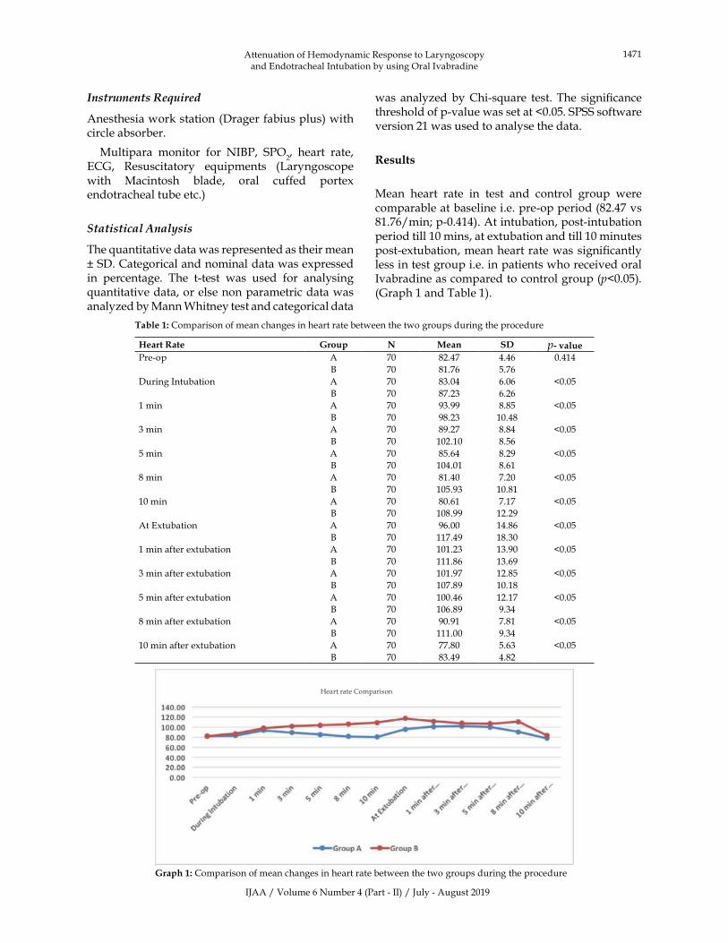
IJAA�/�Volume�6�Number�4�(Part�-�II)�/�July�-�August�2019
1471
Instru�ents�Re�uired
Anesthesia�work�station�(Drager�fabius�plus)�with�circle�absorber.
Multipara� monitor� for� NI�P,� SPO2,� heart� rate,�
ECG,� Resuscitatory� e�uipments� (Laryngoscope�with� Macintosh� blade,� oral� cuffed� portex�endotracheal�tube�etc.)�
Statistical�Analysis
The��uantitative�data�was�represented�as�their�mean���SD.�Categorical�and�nominal�data�was�expressed�in� percentage.� The� t-test� was� used� for� analysing��uantitative�data,�or�else�non�parametric�data�was�analyzed�by�Mann�Whitney�test�and�categorical�data�
was� analyzed�by�Chi-s�uare� test.�The� signi��cance�threshold�of�p-value�was�set�at��0.05.�SPSS�software�version�21�was�used�to�analyse�the�data.
�esults
Mean� heart� rate� in� test� and� control� group� were�comparable�at�baseline�i.e.�pre-op�period�(82.47�vs�81.76/min;�p-0.414).�At�intubation,�post-intubation�period�till�10�mins,�at�extubation�and�till�10�minutes�post-extubation,�mean�heart�rate�was�signi��cantly�less�in�test�group�i.e.�in�patients�who�received�oral�Ivabradine�as�compared�to�control�group�(p�0.05).�(Graph�1�and�Table�1).
Table��:�Comparison�of�mean�changes�in�heart�rate�between�the�two�groups�during�the�procedure
�eart��ate �roup N Mean S� p���aluePre-op A 70 82.47 4.46 0.414
� 70 81.76 5.76
During�Intubation A 70 83.04 6.06 �0.05
� 70 87.23 6.26
1�min A 70 93.99 8.85 �0.05
� 70 98.23 10.48
3�min A 70 89.27 8.84 �0.05
� 70 102.10 8.56
5�min A 70 85.64 8.29 �0.05
� 70 104.01 8.61
8�min A 70 81.40 7.20 �0.05
� 70 105.93 10.81
10�min A 70 80.61 7.17 �0.05
� 70 108.99 12.29
At�Extubation A 70 96.00 14.86 �0.05
� 70 117.49 18.30
1�min�after�extubation A 70 101.23 13.90 �0.05
� 70 111.86 13.69
3�min�after�extubation A 70 101.97 12.85 �0.05
� 70 107.89 10.18
5�min�after�extubation A 70 100.46 12.17 �0.05
� 70 106.89 9.34
8�min�after�extubation A 70 90.91 7.81 �0.05
� 70 111.00 9.34
10�min�after�extubation A 70 77.80 5.63 �0.05
� 70 83.49 4.82
�eart�rate�Comparison
� �raph��:�Comparison�of�mean�changes�in�heart�rate�between�the�two�groups�during�the�procedure
Attenuation�of��emodynamic�Response�to�Laryngoscopy�and�Endotracheal�Intubation�by�using�Oral�Ivabradine
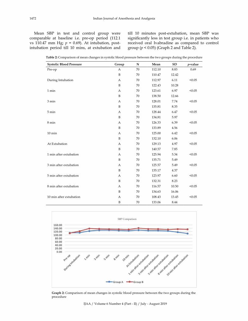
IJAA�/�Volume�6�Number�4�(Part�-�II)�/�July�-�August�2019
1472 Indian�Journal�of�Anesthesia�and�Analgesia
Mean� S�P� in� test� and� control� group� were�comparable� at� baseline� i.e.� pre-op� period� (112.1�vs� 110.47�mm��g;� p��� 0.69).� At� intubation,� post-intubation� period� till� 10� mins,� at� extubation� and�
till� 10� minutes� post-extubation,� mean� S�P� was�signi��cantly�less� in� test�group�i.e.� in�patients�who�received� oral� Ivabradine� as� compared� to� control�group�(p���0.05)�(Graph�2�and�Table�2).
Table��:�Comparison�of�mean�changes�in�systolic�blood�pressure�between�the�two�groups�during�the�procedure
Systolic�Blood�Pressure �roup N Mean S� ���alue
Pre-op A 70 112.10 8.83 0.69
� 70 110.47 12.42
During�Intubation A 70 112.97 6.11 �0.05
� 70 122.43 10.28
1�min A 70 123.61 6.97 �0.05
� 70 138.50 12.66
3�min A 70 128.01 7.74 �0.05
� 70 135.81 8.35
5�min A 70 128.44 6.47 �0.05
� 70 134.81 5.97
8�min A 70 126.33 6.39 �0.05
� 70 133.89 4.56
10�min A 70 125.00 6.42 �0.05
� 70 132.10 6.06
At�Extubation A 70 129.13 4.97 �0.05
� 70 140.57 7.85
1�min�after�extubation A 70 125.94 5.34 �0.05
� 70 135.71 5.49
3�min�after�extubation A 70 125.57 5.49 �0.05
� 70 135.17 4.37
5�min�after�extubation A 70 123.97 6.60 �0.05
� 70 132.31 8.23
8�min�after�extubation A 70 116.57 10.50 �0.05
� 70 134.63 16.06
10�min�after�extubation A 70 108.43 13.45 �0.05
� 70 133.06 8.44
S�P�Comparison
�raph��:�Comparison�of�mean�changes�in�systolic�blood�pressure�between�the�two�groups�during�the�procedure

IJAA�/�Volume�6�Number�4�(Part�-�II)�/�July�-�August�2019
1473
Table��:�Comparison�of�mean�changes�in�diastolic�blood�pressure�between�the�two�groups�during�the�procedure
�iastolic�Blood�Pressure �roup N Mean S� p���aluePre-op A 70 82.47 5.75 0.57
� 70 80.71 5.47
During�Intubation A 70 79.51 6.81 �0.05
� 70 84.27 7.01
1�min A 70 80.21 6.63 �0.05
� 70 91.50 6.36
3�min A 70 87.43 7.03 �0.05
� 70 97.21 8.92
5�min A 70 83.97 6.62 �0.05
� 70 101.10 10.63
8�min A 70 80.17 5.92 �0.05
� 70 104.74 12.94
10�min A 70 78.96 6.16 �0.05
� 70 106.91 14.84
At�Extubation A 70 95.17 16.65 �0.05
� 70 129.66 17.72
1�min�after�extubation A 70 99.54 16.23 �0.05
� 70 123.97 13.81
3�min�after�extubation A 70 103.57 15.99 �0.05
� 70 118.74 11.02
5�min�after�extubation A 70 105.14 16.13 �0.05
� 70 112.57 9.66
8�min�after�extubation A 70 97.13 10.98 �0.05
� 70 108.37 10.42
10�min�after�extubation A 70 95.66 7.33 0.22
� 70 104.26 6.07
D�P�Comparison
�raph��:�Comparison�of�mean�changes�in�diastolic�blood�pressure�between�the�two�groups�during�the�procedure
Mean� D�P� in� test� and� control� group� were�comparable� at� baseline� i.e.� pre-op� period� (82.47�vs� 80.71� mm� �g;� p� �� 0.21).� At� intubation,� post-intubation� period� till� 10� mins,� at� extubation� and�
till� 10� minutes� post-extubation,� mean� D�P� was�signi��cantly�less� in� test�group�i.e.� in�patients�who�received� oral� Ivabradine� as� compared� to� control�group�(p�0.05)�(Graph�3�and�Table�3).
Attenuation�of��emodynamic�Response�to�Laryngoscopy�and�Endotracheal�Intubation�by�using�Oral�Ivabradine
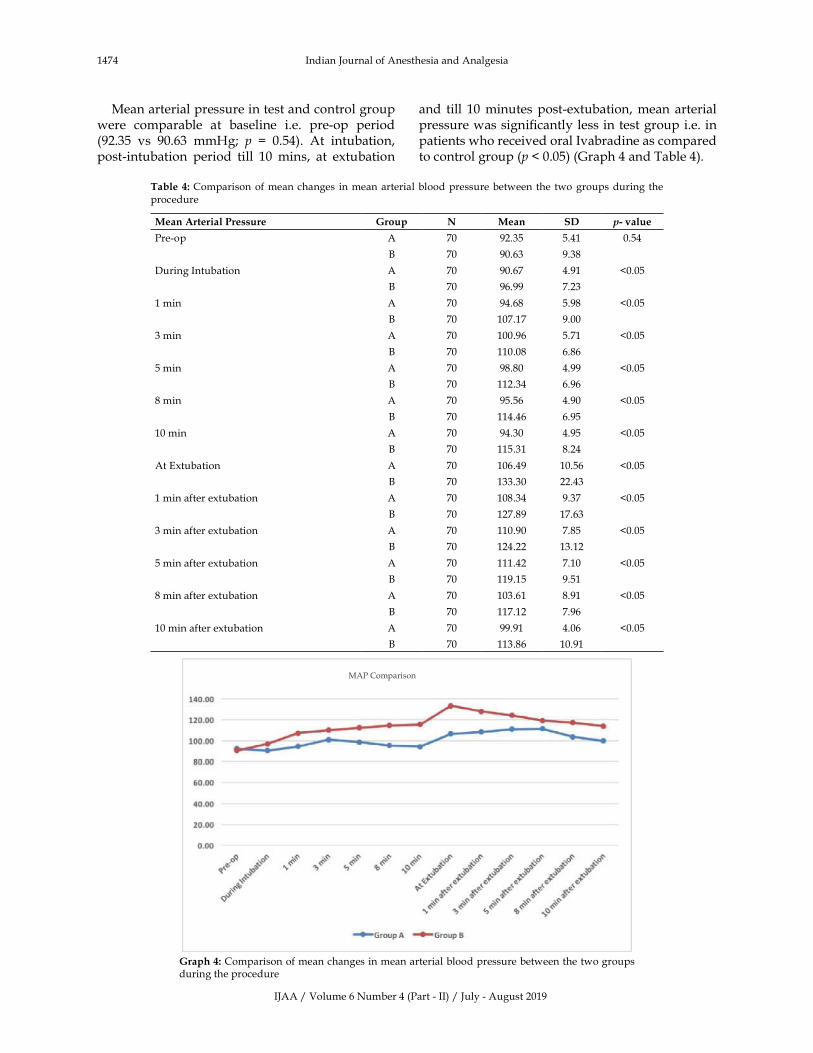
IJAA�/�Volume�6�Number�4�(Part�-�II)�/�July�-�August�2019
1474 Indian�Journal�of�Anesthesia�and�Analgesia
Mean�arterial�pressure�in�test�and�control�group�were� comparable� at� baseline� i.e.� pre-op� period�(92.35� vs� 90.63� mm�g;� p� �� 0.54).� At� intubation,�post-intubation� period� till� 10� mins,� at� extubation�
and� till� 10�minutes�post-extubation,�mean�arterial�pressure�was�signi��cantly�less�in�test�group�i.e.�in�patients�who�received�oral�Ivabradine�as�compared�to�control�group�(p���0.05)�(Graph�4�and�Table�4).
Table��:�Comparison�of�mean�changes� in�mean�arterial�blood�pressure�between�the�two�groups�during�the�procedure
Mean�Arterial�Pressure �roup N Mean S� ����alue
Pre-op A 70 92.35 5.41 0.54
� 70 90.63 9.38
During�Intubation A 70 90.67 4.91 �0.05
� 70 96.99 7.23
1�min A 70 94.68 5.98 �0.05
� 70 107.17 9.00
3�min A 70 100.96 5.71 �0.05
� 70 110.08 6.86
5�min A 70 98.80 4.99 �0.05
� 70 112.34 6.96
8�min A 70 95.56 4.90 �0.05
� 70 114.46 6.95
10�min A 70 94.30 4.95 �0.05
� 70 115.31 8.24
At�Extubation A 70 106.49 10.56 �0.05
� 70 133.30 22.43
1�min�after�extubation A 70 108.34 9.37 �0.05
� 70 127.89 17.63
3�min�after�extubation A 70 110.90 7.85 �0.05
� 70 124.22 13.12
5�min�after�extubation A 70 111.42 7.10 �0.05
� 70 119.15 9.51
8�min�after�extubation A 70 103.61 8.91 �0.05
� 70 117.12 7.96
10�min�after�extubation A 70 99.91 4.06 �0.05
� 70 113.86 10.91
MAP�Comparison
�raph��:�Comparison�of�mean�changes�in�mean�arterial�blood�pressure�between�the�two�groups�during�the�procedure
IJAA�/�Volume�6�Number�4�(Part�-�II)�/�July�-�August�2019
1475
��iscussion
The� parameters� of� Mean� heart� rate,� systolic�and� diastolic� blood� pressure� and� mean� arterial�pressure�in�test�and�control�group�were�similar�and�comparable� at�baseline� i.e.� pre-op�period� (p�0.05)�(Table���Graph�1,�2,�3,�4).
After� intubation,� the� placebo� group� showed� a�signi��cant� rise� in� heart� rate� and� blood� pressure�(p�0.05).� Whereas,� the� Ivabradine� group� showed�only� a� slight� increase� in� heart� rate� (82.47� to�83.04/�min)�and�S�P�(112.1�to�112.97�mm��g)�was�observed� post-intubation� while� a� decrease� was�observed�in�D�P�(82.47�to�79.51�mm��g)�and�MAP�(92.35�to�90.67�mm��g)�(Table���Graph�1,�2,�3,�4).
After�induction�and�intubation�a�gap�period�of�ten�minutes� (duration�of� observation)�was� taken�wherein� the� surgeon�was� asked� to�wait� and� not�start� the� surgery.� Ivabradine� has� no� analgesic�properties� and� immediate� skin� incision� if� given�would� raise� the� heart� rate� and� blood� pressure�giving�false�results.
In� the� post-intubation� observation� period� of�10� mins,� mean� heart� rate,� S�P,� D�P� and� MAP�readings�were�signi��cantly� less� in�oral�Ivabradine�group�as�compared� to� the�control�group� that�was�given�placebo�(p�value���0.05).� (�Table���Graph�1,�2,�3,�4).
Mean� heart� rate,� S�P,�D�P� and�MAP� readings�were�found�to�be�signi��cantly�less�in�oral�Ivabradine�group�when�compared�to�control�group�of�placebo�at� extubation� and� post-extubation� observation�period�of�10�mins�(p���0.05)�(Table���Graph�1,2,3,4).
In� a� similar� study� by� �unwer� R� et� al.9,� mean�heart� rate�which�was� 102.86� at� preoperative� time�was� gone� up� to� 120.88� during� intubation� and� it�was� still� at� 110.64/min.� after� 10� minutes,� while�in� test� group� it� was� 82.6/min.� which� rose� only�up� to� 85.43�during� intubation� and� came�down� to�73.46�at�10�minutes.�Similar�pattern�was�observed�with�mean�S�P,�D�P�and�MAP.�The�study�results�showed� that� haemodynamic� parameters� (during�intubation� and� after� 1,� 3,� 5,� 8� and� 10� min.)�were�found�to�be�signi��cantly�lower�in�cases�pre-treated�with�Ivabradine�as�compared�to�placebo.
Ibrahim� et� al.10� in� their� study� evaluated� oral�Ivabradine� and� oral� propranolol� in� achieving� a�hemodynamic� stability� in� microlaryngoscopic�surgeries.� Study� observed� that� the� changes� in�blood� pressure� and� heart� rate� in� both� groups�were�mild�after�intubation,�laryngoscope���xation�for� surgery� and� extubation� but� the� changes� in�Ivabradine� group� were� signi��cantly� less� than�
the�changes� in�propranolol�group� (p���0.05).�The�authors� concluded� that� premedication� with� oral�Ivabradine�in�a�dose�of�5�mg�or�oral�propranolol�in� a� diose� of� 10�mg� prior� to�microlaryngoscopic�surgeries� was� effective� in� conferring� good�hemodynamic�stability�but�that�between�the�two,�Ivabradine�was�more�effective.
The� advantage� of� Ivabradine� noted� in� present�study�were�–�it�gave�good�attenuation�of�heart�rate�response� and�blood�pressure� response�during� the�process�of� intubation,�the�cardiovascular�response�in� intraoperative� period� was� smooth� without�any� deviations,� and� also� it� was� effective� during�extubation�too.
No� observed� side� effects� with� the� dosage�of� Ivabradine� were� seen� during� the� study.� We�thus� conclude� that� Ivabradine� is� simple,� safe,�economical� and� easy� to� use� drug� that� gives�ade�uate� and� satisfactory� haemodynamic�stability� during� induction,� laryngoscopy� and�intubation.��aemodynamic�stability�was�also�well�maintained� during� intraoperative� and� immediate�postoperative�period.
Conclusion
We�conclude�that�Ivabradine�is�an�extremely�useful�drug�and�can�be�used�to�prevent�tachycardia�during�laryngoscopy� and� intubation.� Its� stabilizing� effect�of� haemodynamics� also� extends� up� to� extubation�and� immediate� postoperative� period.� There�were�no� side� effects� seen� with� the� dose� of� Ivabradine�given�in�our�study.
Reco��endations
We� recommend� the� routine� use� of� Ivabradine� to�prevent� the� abnormal� increase� in� heart� rate� and�blood� pressure� seen� during� laryngoscopy� and�endotracheal�intubation.
Ac�no�led�e�ent
I� am� extremely� thankful� to� my� wife� Dr� Srishti�Thakur� for� her� support.� I� even� want� to� extend�my� thanks� to� the� patients� and� hospital� for� their�cooperation.
�unding:�Self
Conflict�of�interest:�none
�thical�Appro�al:�granted
Attenuation�of��emodynamic�Response�to�Laryngoscopy�and�Endotracheal�Intubation�by�using�Oral�Ivabradine
IJAA�/�Volume�6�Number�4�(Part�-�II)�/�July�-�August�2019
1476 Indian�Journal�of�Anesthesia�and�Analgesia
�eferences
1.� Shribman�AJ,�Smith�G,�Achola��J.�Cardiovascular�and� catecholamine� responses� to� laryngoscopy�with� and� without� tracheal� intubation.� �r� J�Anaesth.�1987;59:295–9.
2.� Reid� LC,� �race� DE.� Irritation� of� the� respiratory�tract� and� its� reflex� upon� head.� Surg� Gynecol�Obstet.�1940;70:157–162.
3.� �agberg� CA.� Artime� CA.� Airway�Management�in�Adults:�Miller�s�Anesthesia.�Miller�RD,�Cohen�N�,�Erikson�LI,�Fleisher�LA,�Wiener-�ronish�JP,��oung�WL.�8th�Ed.;�2015;1:1647–80.
4.� Murat�SN,�Orcan�S,�Akdemir�R�et�al.�Arrhythmic�effects� of� ivabradine� in� patients� with� coronary�artery�disease.�Clin�Invest�Med.�2009;32:322–6.
5.� Maze� M,� Tran�uili� W.� Alpha-2� adrenoceptor�agonists;�defining�the�role�in�clinical�anaeathesia�.�Anaesthesiology.�1991;74:581–605.
6.� Drew�GM,�Whiting�S�.�Evidence�for�two�distinct�types� of� postsynaptic� alpha-� adrenoceptor� in�
vascular�smooth�muscle�in�vivo.��r�J�Pharmacol.�1991;67:207–15.
7.� �unter� JC,� Fontana�DJ,��edley� LR.�Assessment�of� the� role�of� alpha-2�adrenoceptors�subtypes� in�the� antinociceptive,� sedative� and� hypothermic�action�of�dexmedetomidine�in�transgenic�mice.��r�J�Pharmacol.�1997;122:1339–44.
8.� Ralph� Getler,� Clieghton� �� �rown,� Mitchel� �,�Silvius� N.� Dexmedetomidine:� a� novel� sedative�analgesic�agent.��aylor��niversity�Medical�Centre�Proceedings.�2001;14(1).
9.� �unwer� R,� Joshi� M,� Pathak� S.� Prevention� of�haemodynamic� changes� during� laryngoscopy�and� endotracheal� intubation-� a� clinical� study�of� oral� ivabradine.� International� Journal� of�Contemporary�Medical�Research.�2016;3(7):1995–98.
10.� Ibrahim� AN,� Atallah� R�.� Ivabradine� versus�propranolol� given� orally� in� microlaryngoscopic�surgeries� in� attenuating� stress� response:� A�comparative�prospective�double�blind�randomized�study.�Egyptian� Journal�of�Anesthesia.� 2016�Oct�1;32(4):503–11.
��Red�Flower�Publication�Pvt.�Ltd.�
�Original�Research�ArticleIndian�Journal�of�Anesthesia�and�Analgesia�
2019;6(4)�(Part�-�II):�1483-1486DOI:�http://dx.doi.org/10.21088/ijaa.2349.8471.6419.58
Comparison�of��aryngeal�Mask�Air�ay�and�Endotracheal� Intubation� in�Paediatric�Patients:�A�Comparati�e�Study
Viral�Prakashkumar�Patel����ekha�Bayer�
1Assistant�Professor,�2Senior�Resident,�Department�of�Anesthesia,�GMERS�Medical�College,��immatnagar,�Gujarat�383001,�India.
Corresponding�Author:��ekha�Bayer,�Senior�Resident,�Department�of�Anesthesia,�GMERS�Medical�College,��immatnagar,�Gujarat�383001,�India.
E�mail:�[email protected]
�ecei�ed�on�22.05.2019,�Accepted�on�19.06.2019
Abstract
Aim:�The�aim�of�the�present�study�was�to�compare�the�subse�uent�factors�that�take�place�during�laryngeal�mask� airway� insertion�and�endotracheal� intubation� for� surgical� procedures�under�general� anesthesia.�The�parameters� compared�were:�Ease� of� insertion� and�number� of� efforts� and�Postoperative� complications� like�laryngospasm,� bronchospasm� and� sore� throat.� Materials� and� Methods:� The� study� participants� included�80�paediatric�patients�between�the�ages�of�2–8�years.�All�the�participants�were�schedule�to�experience�optional�surgeries�under�general�anesthesia� in�the�medical�hospital.�Results:�LMA�had�advantages�over�the�tracheal�tube�in�the�form�of�lower�incidence�of�cough�during�appearance�and�lower�occurrence�of�postoperative�sore�throat,�though�offered�no�advantage�more�than�tracheal�tube�in�occurrence�of�bronchospasm�or�laryngospasm�during�appearance.�Conclusion:�The�LMA�provides�a�reliable�airway�in�children.�Incidence�of�postoperative�complications� is� also� less�with�LMA� than� endotracheal� tubes.� Therefore,� LMA� is� a� appropriate� option� to�endotracheal�intubation�for�possible�short�surgical�procedures�in�pediatric�patients.
�ey�ords:�Laryngeal�mask�airway;�Endotracheal�tube;�Pediatric.
�o��to�cite�this�article:
Viral� Prakashkumar� Patel,� Rekha� bayer.�Comparison� of� Laryngeal�Mask�Airway� and�Endotracheal� Intubation� in� Paediatric�Patients:�A�Comparative�Study.�Indian�J�Anesth�Analg.�2019;6(4):�1477-1480.
Introduction
The�laryngeal�mask�airway�(LMA)�is�wrought�like�
a�large�endotracheal�tube�on�the�proximal�end�that�
connects�to�an�elliptical�mask�on�the�distal�end.�It�
is�intended�to�sit�in�the�patient�s�hypopharynx�and�
cover� up� the� supraglottic� structures,� thus� allows�
comparative�isolation�of�the�trachea.1,2�A�laryngeal�
mask� airway� (LMA)��� also� known� as� laryngeal�
mask��is�a�medical�device�that�keeps�a�patient�s�
airway�open�during�anesthesia�or�unconsciousness.�
It� is� a� type� of� supraglottic� airway.3� A� laryngeal�
mask� is�composed�of�an�airway� tube�that�connect�to�an�elliptical�mask�with�a�cuff�which�is� inserted�through�the�patient�s�mouth,�down�the�windpipe,�and�once�deployed�forms�an�airtight�seal�on�top�the�
glottis�permits�a� secure� airway�to�be�managed�by�a� health� care� provider.4� Laryngeal� mask� airways�
come�in�several�types,�as�follows:�The�LMA�Classic�is�the�original�reusable�design,�The�LMA��ni�ue�is�a�disposable�version,�making�it�ideal�for�emergency�and� prehospital� settings� and� The� LMA� Fastrach,�
an�intubating�LMA�(ILMA),�is�designed�to�serve�as�
IJAA�/�Volume�6�Number�4�(Part�-�II)�/�July�-�August�2019
1478 Indian�Journal�of�Anesthesia�and�Analgesia
a�conduit�for�intubation.�They�are�largely�used�by�anaesthetists� to� channel� oxygen�or� anesthesia� gasto�a�patient�s�lungs�during�surgery�and�in�the�pre-hospital�setting�for�unconscious�patients.5
The�LAM,�with�its�advantages�over�both�the�face�mask/nasal� hood� and� endotracheal� intubation,�potentially� has� a� place� in� oral� and� maxillofacial�surgery� by� increasing� the� safety� and� ef��cacy� of�outpatient�general�anesthesia�in�speci��c�situations.�The�LAM�also�has�an�important�role�in�acute�airway�management� in� the� trauma� setting� or� during�anesthetic� emergencies.� Airway� control� may� be�established� with� the� LAM� when� the� patient� can�neither� be� intubated� nor� ventilated.� In�managing�the� dif��cult� airway,� the� laryngeal� mask� airway�can� be� considered� before� either� transtracheal� jet�ventilation�or�establishing�a�surgical�airway.
The� progress� of� small� sized� laryngeal� masks�authorized�its�utilize�in�anesthesia�for�the�paediatric�surgical�patients.�These�patients�have�diverse�airway�characteristics� such� as� high� larynx,� large� tongue,�funnel� shaped� laryngeal� cartilaginous� skeleton,�lack� of� teeth,� and� short� neck� which� makes� the�likelihood�of�tricky�intubation�higher�than�in�adult�patients.�Added� to� this� is� the� �uick� development�of�hypoxemia�during�trials�of�intubation�in�a�some�what�hard�airway�of�few�kilograms�infant.6
The� LMA� has� many� recompense� over� the�endotracheal� tube� with� more� hemodynamic�stability,� and� condensed� occurrence� of�perioperative� complications� such� as� coughing,�bucking,� laryngospasm,� soft-tissue� trauma,�laryngeal�edema,�and�sore� throat.7�Adding�up�the�occurrence�of�postoperative�sore� throat�connected�with�placement�of�LMA�is�fewer�than�that�connected�with�ETT.8�The�aim�of�present�study�is�to�evaluate�the� subse�uent� factors� that� take� place� during�laryngeal�mask�airway�insertion�and�endotracheal�intubation� for� surgical� procedures� under� general�anesthesia.�The�parameters�compared�are:�Ease�of�insertion�and�number�of�attempts�and�Postoperative�complications� like� laryngospasm,� bronchospasm�and�sore�throat.
Materials�and�Methods
The� study� participants� included� 80� paediatric�patients� between� the� age� of� 2� –� 8� years.� All� the�participants� were� schedule� to� undergo� elective�surgeries�under�general� anesthesia� in� the�medical�hospital.� The� institute� ethical� committee� were�informed�about�the�study�and�the�ethical�clearance�certi��cate�was�obtained�from�them.�All�the�surgeries�
were�short�procedures�of�less�than�40�mins.�All�the�study� paediatric� patients� were� alienated� into�two�groups�with� 40�patients� each.� Study�group� 1�(group� L):� LMA� of� appropriate� size�was� inserted�and� cuff� in��ated� with� appropriate� volume� of�air.� Study� group� 2� (group� E):� Laryngoscopy� and�endotracheal� intubation� with� appropriate� sized�endotracheal�tubes�was�done.
A�detailed�preanesthetic�assessment�was���nished�for�all�the�paediatric�patients�a�day�prior�the�surgery�time.� A� detailed� history,� airway� assessment� and�physical�examination�was�done�to�rule�out�for�the�exclusion�criteria.�Exclusion�criteria:�Patients�in�ASA�grade�III�and�IV,�emergency�surgeries,�Presence�of�cardiac�and�pulmonary�problems.�Inclusion�criteria:�Paediatric�patients�between�2�–�8�years,��elonging�to�ASA�I�and�II�grade,�Schedule� for�elective�short�surgeries.
Induction�agent�utilized�for�surgery�was�3�mg/kg�propofol�given�over�1�min.�For�all� the�patients�the�IV�line�is�secured,�all�the�children�premedicated�with� inj.� Glycopyrollate� and� inj.� Midazolam.�Analgesia� was� provided� with� Inj.� Fentanyl� 2�micrograms/kg�IV,�following�which�patients�were�calm� with� Inj.� Scoline� 2� mg/kg� IV� to� facilitate�insertion� of� LMA� or� endotracheal� tube.� The�appropriate�sized�LMA�was�chosen�based�upon�the�weight�of� the�children�as�follows:�size�1.5� for�5-10�kgs,� size� 2� for� 10-20� kgs,� size� 2.5� for� 20-30� kgs.� ��The�ease�of�insertion�of�LMA/ETT�was�graded�as�easy,�dif��cult,�impossible�and�number�of�attempts�of�insertion�of�LMA/ETT�was�noted.�With�the�help�of� bilateral� chest� lift� and� ausculatation� of� breath�sounds�the�position�of�ETT/LMA�was�con��rmed.�Monitoring�of�vital�signs�i.e.�heart�rate,�noninvasive�blood� pressure,� pulse� oximetry,� E�G� lead� II�was�completed�in�perioperative�period.��emodynamic�changes�in��R,��P,�MAP,�SpO
2�were�watched�just�
previous� to� induction� (baseline),� just� following�intubation/insertion�0�minute�and�then�at�1,�3,�5,�10,�15,�20�minutes�after�intubation�/�insertion�of�LMA.�Postoperative� complications� like� laryngospasm,�bronchospasm� and� sore� throat� was� recorded�postoperatively.
Statistical�analysis
The�recorded�data�was�compiled�and�entered�in�a�spreadsheet� computer� program� (Microsoft� Excel�2007)�and�then�exported�to�data�editor�page�of�SPSS�version�15�(SPSS�Inc.,�Chicago,�Illinois,��SA).
Descriptive� statistics� included� computation� of�percentages,�means� and� standard� deviations.� For�all� tests,� con��dence� level�and� level�of�signi��cance�were� set� at� 95�� and� 5�� respectively.� Statistical�
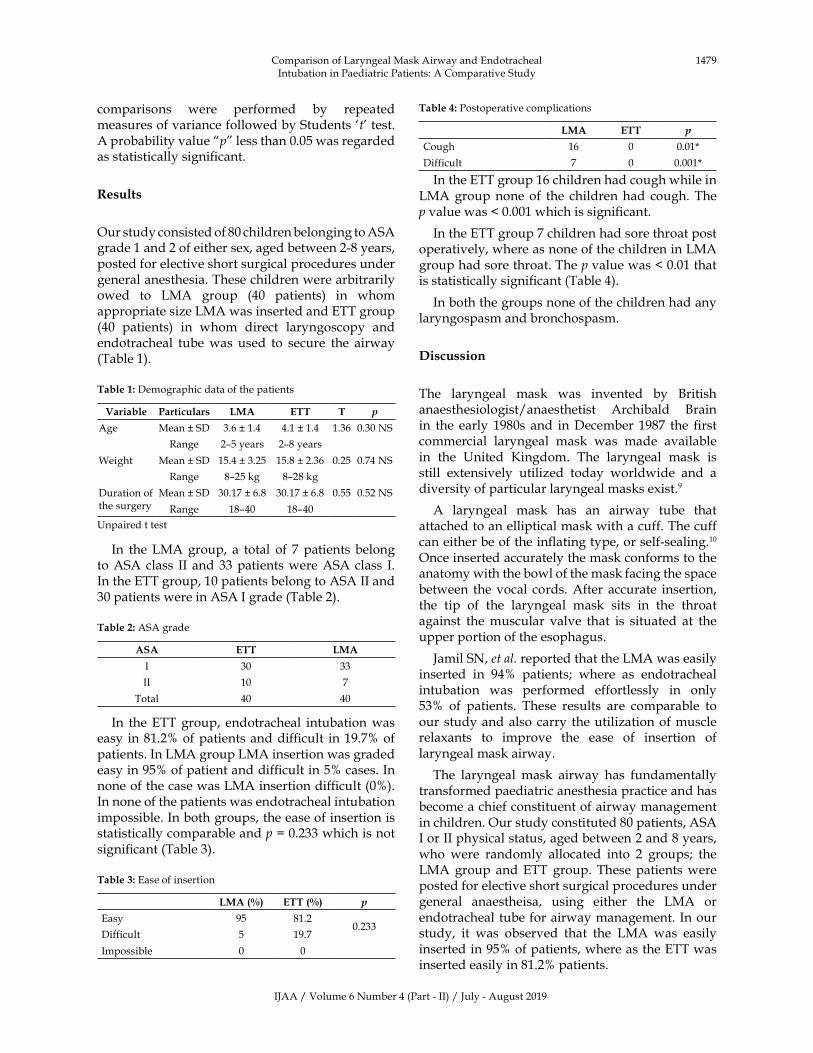
IJAA�/�Volume�6�Number�4�(Part�-�II)�/�July�-�August�2019
1479
comparisons� were� performed� by� repeated�measures�of�variance�followed�by�Students��t��test.�A�probability�value��p��less�than�0.05�was�regarded�as�statistically�signi��cant.
�esults
Our�study�consisted�of�80�children�belonging�to�ASA�grade�1�and�2�of�either�sex,�aged�between�2-8�years,�posted�for�elective�short�surgical�procedures�under�general�anesthesia.�These�children�were�arbitrarily�owed� to� LMA� group� (40� patients)� in� whom�appropriate�size�LMA�was�inserted�and�ETT�group�(40� patients)� in� whom� direct� laryngoscopy� and�endotracheal� tube�was� used� to� secure� the� airway�(Table�1).
Table��:�Demographic�data�of�the�patients
Variable Particulars �MA ETT T �
Age Mean���SD 3.6���1.4 4.1���1.4 1.36 0.30�NS
Range 2–5�years 2–8�years
Weight Mean���SD 15.4���3.25 15.8���2.36 0.25 0.74�NS
Range 8–25�kg 8–28�kg
Duration�of�the�surgery
Mean���SD 30.17���6.8 30.17���6.8 0.55 0.52�NS
Range 18–40 18–40
�npaired�t�test
In� the�LMA�group,� a� total� of� 7�patients�belong�to�ASA�class� II�and�33�patients�were�ASA�class� I.�In�the�ETT�group,�10�patients�belong�to�ASA�II�and�30�patients�were�in�ASA�I�grade�(Table�2).
Table��:�ASA�grade
ASA ETT �MA
I 30 33
II 10 7
Total 40 40
In� the�ETT�group,� endotracheal� intubation�was�easy� in�81.2��of�patients�and�dif��cult� in�19.7��of�patients.�In�LMA�group�LMA�insertion�was�graded�easy�in�95��of�patient�and�dif��cult�in�5��cases.�In�none�of�the�case�was�LMA�insertion�dif��cult�(0�).�In�none�of�the�patients�was�endotracheal�intubation�impossible.�In�both�groups,�the�ease�of�insertion�is�statistically�comparable�and�p���0.233�which�is�not�signi��cant�(Table�3).
Table��:�Ease�of�insertion
�MA���� ETT���� �
Easy 95 81.20.233
Difficult 5 19.7
Impossible 0 0
Table��:�Postoperative�complications
�MA� ETT� �
Cough 16 0 0.01�
Difficult 7 0 0.001�
In�the�ETT�group�16�children�had�cough�while�in�LMA�group�none�of� the� children�had� cough.�The�p�value�was���0.001�which�is�signi��cant.
In�the�ETT�group�7�children�had�sore�throat�post�operatively,�where�as�none�of�the�children�in�LMA�group�had�sore�throat.�The�p�value�was���0.01�that�is�statistically�signi��cant�(Table�4).
In�both�the�groups�none�of�the�children�had�any�laryngospasm�and�bronchospasm.
�iscussion
The� laryngeal� mask� was� invented� by� �ritish�anaesthesiologist/anaesthetist� Archibald� �rain�in� the� early� 1980s� and� in�December� 1987� the���rst�commercial� laryngeal� mask� was� made� available�in� the� �nited� �ingdom.� The� laryngeal� mask� is�still� extensively� utilized� today� worldwide� and� a�diversity�of�particular�laryngeal�masks�exist.9
A� laryngeal� mask� has� an� airway� tube� that�attached�to�an�elliptical�mask�with�a�cuff.�The�cuff�can�either�be�of�the�in��ating�type,�or�self-sealing.10�Once�inserted�accurately�the�mask�conforms�to�the�anatomy�with�the�bowl�of�the�mask�facing�the�space�between� the�vocal� cords.�After� accurate� insertion,�the� tip� of� the� laryngeal� mask� sits� in� the� throat�against� the�muscular� valve� that� is� situated� at� the�upper�portion�of�the�esophagus.
Jamil�SN,�et�al.�reported�that�the�LMA�was�easily�inserted� in� 94�� patients;� where� as� endotracheal�intubation� was� performed� effortlessly� in� only�53�� of� patients.� These� results� are� comparable� to�our�study�and�also�carry�the�utilization�of�muscle�relaxants� to� improve� the� ease� of� insertion� of�laryngeal�mask�airway.
The� laryngeal� mask� airway� has� fundamentally�transformed�paediatric�anesthesia�practice�and�has�become�a�chief�constituent�of�airway�management�in�children.�Our�study�constituted�80�patients,�ASA�I�or�II�physical�status,�aged�between�2�and�8�years,�who� were� randomly� allocated� into� 2� groups;� the�LMA�group� and�ETT� group.� These�patients�were�posted�for�elective�short�surgical�procedures�under�general� anaestheisa,� using� either� the� LMA� or�endotracheal� tube�for�airway�management.�In�our�study,� it� was� observed� that� the� LMA� was� easily�inserted�in�95��of�patients,�where�as�the�ETT�was�inserted�easily�in�81.2��patients.
Comparison�of�Laryngeal�Mask�Airway�and�Endotracheal�Intubation�in�Paediatric�Patients:�A�Comparative�Study
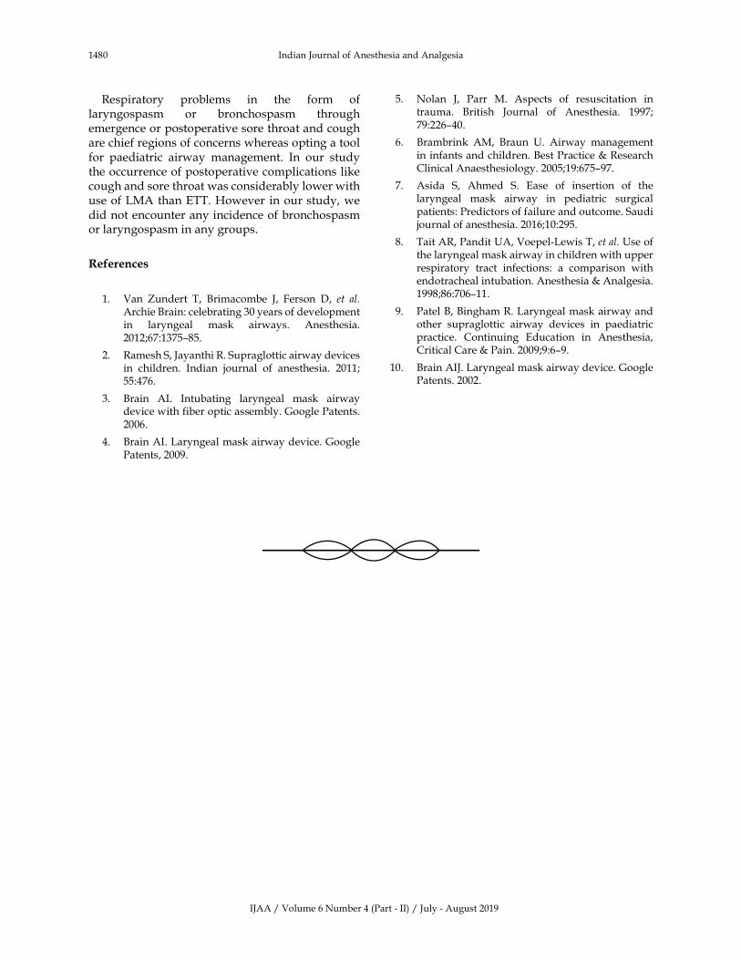
IJAA�/�Volume�6�Number�4�(Part�-�II)�/�July�-�August�2019
1480 Indian�Journal�of�Anesthesia�and�Analgesia
Respiratory� problems� in� the� form� of�laryngospasm� or� bronchospasm� through�emergence�or�postoperative�sore�throat�and�cough�are�chief�regions�of�concerns�whereas�opting�a�tool�for� paediatric� airway� management.� In� our� study�the�occurrence�of�postoperative�complications�like�cough�and�sore�throat�was�considerably�lower�with�use�of�LMA�than�ETT.��owever� in�our�study,�we�did�not�encounter�any�incidence�of�bronchospasm�or�laryngospasm�in�any�groups.
�eferences
1.� Van� �undert� T,� �rimacombe� J,� Ferson� D,� et� al.�Archie��rain:�celebrating�30�years�of�development�in� laryngeal� mask� airways.� Anesthesia.�2012;67:1375–85.
2.� Ramesh�S,�Jayanthi�R.�Supraglottic�airway�devices�in� children.� Indian� journal� of� anesthesia.� 2011;�55:476.
3.� �rain� AI.� Intubating� laryngeal� mask� airway�device�with�fiber�optic�assembly.�Google�Patents.�2006.
4.� �rain�AI.�Laryngeal�mask�airway�device.�Google�Patents,�2009.
5.� Nolan� J,� Parr� M.� Aspects� of� resuscitation� in�trauma.� �ritish� Journal� of� Anesthesia.� 1997;�79:226–40.
6.� �rambrink� AM,� �raun� �.� Airway� management�in�infants�and�children.��est�Practice���Research�Clinical�Anaesthesiology.�2005;19:675–97.
7.� Asida� S,� Ahmed� S.� Ease� of� insertion� of� the�laryngeal� mask� airway� in� pediatric� surgical�patients:�Predictors�of�failure�and�outcome.�Saudi�journal�of�anesthesia.�2016;10:295.
8.� Tait�AR,�Pandit��A,�Voepel-Lewis�T,�et�al.��se�of�the�laryngeal�mask�airway�in�children�with�upper�respiratory� tract� infections:� a� comparison� with�endotracheal�intubation.�Anesthesia���Analgesia.�1998;86:706–11.
9.� Patel��,��ingham�R.�Laryngeal�mask�airway�and�other� supraglottic� airway� devices� in� paediatric�practice.� Continuing� Education� in� Anesthesia,�Critical�Care���Pain.�2009;9:6–9.
10.� �rain�AIJ.�Laryngeal�mask�airway�device.�Google�Patents.�2002.
��Red�Flower�Publication�Pvt.�Ltd.�
�Case�ReportIndian�Journal�of�Anesthesia�and�Analgesia�
2019;6(4)�(Part�-�II):�1487-1489DOI:�http://dx.doi.org/10.21088/ijaa.2349.8471.6419.59
Pre�entable�Anesthesia�Mishaps:�An�O�er�ie�
�enuka�����Anshumali�
1Associate�Professor,�Department�of�Anaesthesiology,�Sapthagiri�Institute�of�Medical�Sciences���Research�Centre,�Chikkabanavara,��engaluru,��arnataka� 560090,� India.� 2Associate�Professor,�Department�of�Anaesthesiology,�North�DMC�Medical�College,�Malka�Ganj,�Delhi�110007,�India.
Corresponding�Author:��enuka��,�Associate�Professor,�Department�of�Anaesthesiology,�Sapthagiri�Institute�of�Medical�Sciences���Research�Centre,�Chikkabanavara,��engaluru,��arnataka�560090,�India.
E�mail:�[email protected]
�ecei�ed�on�10.05.2019,�Accepted�on�11.07.2019
Abstract
We�report�a�case�of�malposition�of�the�vaporizer�on�the�selectatec�manifold�of�the�anesthesia�workstation�leading�to�interruption�in�the�fresh�gas�flow�at�anesthesia�machine�outlet.�We�emphasize�the�importance�of�checking�the�anaesthetic�vaporizer�after�mounting�it�on�the�backbar�of�the�anesthesia�workstation.
�ey�ords:�Interruption�in�fresh�gas�flow;�General�anesthesia;�Tilted�vaporizer;�Selectatec�manifold.
�o��to�cite�this�article:
Renuka�R,�Anshumali.�Preventable�Anesthesia�Mishaps:�An�Overview.�Indian�J�Anesth�Analg.�2019;6(4):�1481-1483.
Introduction
Anesthesia�workstations�have�come�a� long�way�since�the�ancient��oyles�apparatus.�The�main�focus�of�research�in�these�machines�has�been�to�improve�the�safety�features,�in�order�to�prevent�delivery�of�hypoxic�gas�mixtures�and�alert�the�anesthesia�care�provider�of�any�other�problems.�Modern�anesthesia�machines�have� several� safety� features,� for�patient�as� well� as� user� safety.� �ltramodern� anaesthetic�machines� have� additional� safety� features� and�are� programmed�with� a� computerized� safety� self�checkout�feature�which�is�initiated�at�start�up�which�needs�to�be�repeated�before�every�case�and�ideally�not�to�be�bypassed.
Inspite� of� all� the� advances� and� safety� features,�the� occurrence� of� anaesthetic� misadventures� is�
still� a� problem� of� concern.� We� report� a� case� of�interruption� in� the� fresh� gas� ��ow� (FGF)� during�general�anesthesia�caused�by� incorrectly�mounted�vaporizer�on�the�Selectatec�manifold�of�the��lease�Sirius� Spacelabs� anesthesia� workstation� (�lease�Medical�E�uipment�Limited,�Washington,��SA).
Case��eport
A�42�year� old�male� patient,�ASA� grade� 1�with�diagnosis� of� subacute� intestinal� obstruction�was� scheduled� for� emergency� laparotomy.� An�automated� checkout� was� performed� for� �lease�Sirius� Spacelabs� anesthesia� workstation� (�lease�Medical� E�uipment� Limited,� Washington,� �SA)�before�the�case�and�it�passed�all�the�tests.�On�table,�standard�monitors�were�attached�to�the�patient�and�
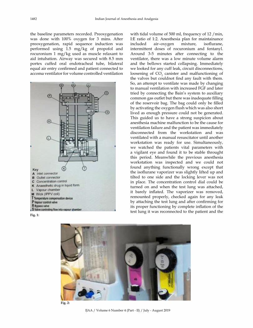
IJAA�/�Volume�6�Number�4�(Part�-�II)�/�July�-�August�2019
1482 Indian�Journal�of�Anesthesia�and�Analgesia
the�baseline�parameters�recorded.�Preoxygenation�was� done� with� 100�� oxygen� for� 3� mins.� After�preoxygenation,� rapid� se�uence� induction� was�performed� using� 1.5� mg/kg� of� propofol� and�rocuronium� 1�mg/kg� used� as� muscle� relaxant� to�aid� intubation.�Airway�was� secured�with� 8.5�mm�portex� cuffed� oral� endotracheal� tube,� bilateral�e�ual�air�entry�con��rmed�and�patient�connected�to�accoma�ventilator�for�volume�controlled�ventilation�
with�tidal�volume�of�500�ml,�fre�uency�of�12�/min,�I:E� ratio� of� 1:2.� Anesthesia� plan� for�maintainance�included� air–oxygen� mixture,� iso��urane,�intermittent� doses� of� rocuronium� and� fentanyl.Around� 3-5� minutes� after� connecting� to� the�ventilator,� there�was� a� low�minute� volume� alarm�and� the� bellows� started� collapsing.� Immediately�we�looked�for�any�cuff�leak,�circuit�disconnections,�loosening� of� CO
2� canister� and� malfunctioning� of�
the�valves�but�couldnot���nd�any�fault�with� them.�So,�an�attempt�to�ventilate�was�made�by�changing�to�manual�ventilation�with�increased�FGF�and�later�tried�by�connecting� the��ain�s� system�to�auxiliary�common�gas�outlet�but�there�was�inade�uate���lling�of� the� reservoir�bag.�The�bag� could� only�be���lled�by�activating�the�oxygen���ush�which�was�also�short�lived�as� enough�pressure� could�not�be�generated.�This� guided� us� to� have� a� strong� suspicion� about�anesthesia�machine�malfunction�to�be�the�cause�for�ventilation�failure�and�the�patient�was�immediately�disconnected� from� the� workstation� and� was�ventilated�with�a�manual�resuscitator�until�another�workstation� was� ready� for� use.� Simultaneously,�we� watched� the� patients� vital� parameters� with�a� vigilant� eye� and� found� it� to� be� stable� throught�this� period.� Meanwhile� the� previous� anesthesia�workstation� was� inspected� and� we� could� not�found� anything� functionally� wrong� except� that�the�iso��urane�vaporizer�was�slightly�lifted�up�and�tilted� to� one� side� and� the� locking� lever� was� not�in� place.� The� concentration� control� dial� could� be�turned� on� and� when� the� test� lung� was� attached,�it� barely� in��ated.� The� vaporizer� was� removed,�remounted� properly,� checked� again� for� any� leak�by�attaching�the�test�lung�and�after�con��rming�for�its�proper�functioning�by�complete�in��ation�of�the�test�lung�it�was�reconnected�to�the�patient�and�the�
Fig���:
Fig���:
IJAA�/�Volume�6�Number�4�(Part�-�II)�/�July�-�August�2019
1483
surgical� procedure� was� carried� out� successfully.�At�the�end�of�surgery,�patient�was�extubated�with�an�uneventful�recovery�and�he�did�not�experience�any�conse�uence�like�awareness�as�a�result�of�this�momentary�loss�of�FGF.
�iscussion
There�are�2�types�of�vaporizer�mounting�systems:�permanent� mounting� and� detachable� mounting.�Detachable� mounting� systems� are� the� standard�on� most� anesthesia� machines� as� they� allow� the�vaporizer�to�be�mounted�and�removed�without�the�use� of� tools.� The� Selectatec� system� and� a� Plug-in�system�from�Drager�Medical�are� the�most�widely-used�detachable�mounting�systems.�The�Selectatec�system� consists� of� a� pair� of� port� valves� for�positioning�of� each�vaporizer1.� The��lease�Datum�cage� mount� vaporizer� has� a� special� mounting�bracket� containing� two�plungers� (spindles)�which���ts� over� the�port� valves.�Weight� of� the� vaporizer�and�an�O-ring�around�each�port�valve�creates�a�seal�between� the�mounting� system�and� the�vaporizer.�The�interlocking�extension�rods�prevent�more�than�one�vaporizer�being�used� simultaneously� (Fig.� 1).�Swinging�the�locking�lever�clockwise�to�90�degrees�is�necessary�to�secure�the�vaporizer�on�the�manifold�and� the� concentration� control� dial� cannot� be�moved� unless� the� locking� lever� of� the� system� is�engaged.� The� FGF� through� the� vapour� chamber�to�produce�the�re�uired�concentration�is�regulated�by� the� concentration� control�C� (Fig.� 2).�When� the�concentration�dial�is�in�the�zero�position�the�bypass�valve�R� opens� isolating� the�FGF� from� the�vapour�chamber.�When�the�dial�is�turned�past�zero,�valve�S� opens� allowing� ��ow� into� the� vapour� chamber2.�In� the� modern� designs� the� vapour� concentration�supplied�by�the�vaporizer�is�virtually�independent�of�the�FGF�between�0.5�and�15�litres/minute.
There� are� three� reported� cases� of� anesthetic�vapor� leakage� from� unlocked� vaporizers.� In� two�of�them,�awareness�with�recall�was�identi��ed�after�surgery3,4�while�in�one�there�was�desaturation�and�hypercapnia5.�Accidental�removal�or�damage�of�the�O-ring�due�to�fre�uent�changing�of�the�vaporizers�in� the� Selectatec� system� increases� the� potential�for� leaks� leading� to� alteration� in� the� fresh� gas�composition� and� ��ow,� operating� room� pollution,�hypoventilation,� rebreathing,� desaturation� and�awareness�in�patients1�is�known.�In�our�case�and�the�case�reported�by��im�and��im,5�the�concentration�
dial�could�be�opened�even�when�there�was�a�faulty�mounting�of�the�vaporizer�on�the�manifold�leading�to� interruption�in�the�FGF�at�the�outlet.��owever,�because� of� �uick� detection� and� recti��cation� of�the� problem� there�was� no� signs� of� hypoxemia� or�hypercarbia� noticed� in� our� case.� This� case� report�highlights�about�the�fault�which�is�not�detectable�by�performing� the� routine� anesthesia�machine� check�as� recommended� by� the� manufacturers� and� also�the� importance� of� anaesthetic� agent� monitors� in�detection�of�such�faults.�It�also�provides�an�insight�into�the�lapses�and�lacunae�in�the�level�of�training,�alerts� us� about� e�uipment� malfunction,� human�errors�and�emphasizes�that�constant�vigilance�of�the�anesthetist�is�highly�essential�for�the�safe�conduct�of�anesthesia.
Conclusion
Vaporizer�malfunction�may�not�be�detected�during�conventional�anaesthetic�machine�check�and�human�errors�can�recur.�We�would�like�to�highlight�that�the�vaporizer�interlock�safety�mechanism�is�not�a�part�of�routine�safety�check�but�needs�incorporation�in�the� revised� safety� checklist� with� the� advent� of�newer�technology.
Consent:�Consent�was� obtained� directly� from� the�patient� to� allow� for�discussion�and� review� in� this�case�report.
�eclaration�of�interest:��one
�eferences
1.� Dorsch�J�A,�Dorsch�SE.��nderstanding�Anesthesia�E�uipment� 4th� Edition:� Williams� and� Wilkins:�1999.pp.167–73
2.� �lease�Datum�Operators�manual�guide:�18-19.
3.� Lee� S�,� Suh� S�,� Cho� J�,� et� al.� An� unexpected�vapor� leakage� from�unlocked�vaporizer� (Penlon�Sigma�Delta�Anesthetic�Vaporizer):�a�case�report.��orean�J�Anesthesiol.�2006;51:230–32.
4.� �im� ��,� Cho� S�,� �im� �N,� et� al.� Awareness�during� general� anesthesia� due� to� the� unlock�position�of� the�vaporizer:�a� report�on�two�cases.��orean�J�Anesthesiol.�2009;56:693–97.
5.� �im� �J,� �im� MW.� Interruption� in� the� supply�of� breathing� gas� during� general� anesthesia� due�to� malposition� of� the� vaporizer-� A� case� report.��orean�J�Anesthesiol.�2010;59(4):270–74.
Preventable�Anesthesia�Mishaps:�An�Overview
IJAA�/�Volume�6�Number�4�(Part�-�II)�/�July�-�August�2019
Manuscripts�must�be�prepared�in�accordance�with���niform� re�uirements� for� Manuscripts� submitted�
to� �iomedical� Journal�� developed� by� international�committee�of�medical�Journal�Editors
Types�of�Manuscripts�and��imits
Original� articles:� �p� to� 3000� words� excluding�
references�and�abstract�and�up�to�10�references.
Review� articles:� �p� to� 2500� words� excluding�
references�and�abstract�and�up�to�10�references.
Case� reports:� �p� to� 1000� words� excluding�references�and�abstract�and�up�to�10�references.
Online�Submission�of�the�Manuscripts
Articles�can�also�be�submitted�online�from�http://rfppl.co.in/customer_index.php.
I)�First�Page�File:�Prepare� the� title�page,�covering�
letter,�acknowledgement,�etc.�using�a�word�processor�
program.� All� information� which� can� reveal� your�
identity�should�be�here.�use� text/rtf/doc/PDF���les.�Do�not�zip�the���les.
2)�Article���le:�The�main�text�of�the�article,�beginning�from�Abstract�till�References�(including�tables)�should�
be�in�this���le.�Do�not�include�any�information�(such�as�acknowledgement,�your�name�in�page�headers,�etc.)�
in�this���le.��se�text/rtf/doc/PDF���les.�Do�not�zip�the���les.�Limit�the���le�size�to�400��b.�Do�not�incorporate�
images� in� the���le.� If���le�size�is� large,�graphs�can�be�submitted�as�images�separately�without�incorporating�
them�in�the�article���le�to�reduce�the�size�of�the���le.
3)�Images:�Submit�good��uality�color�images.�Each�image�should�be�less�than�100��b�in�size.�Size�of�the�
image�can�be�reduced�by�decreasing�the�actual�height�and�width�of�the�images�(keep�up�to�400�pixels�or�3�
inches).�All� image� formats� (jpeg,� tiff,� gif,�bmp,�png,�eps�etc.)�are�acceptable;�jpeg�is�most�suitable.
Legends:� Legends� for� the� ��gures/images� should�be�included�at�the�end�of�the�article���le.
If� the� manuscript� is� submitted� online,� the�contributors��form�and�copyright�transfer�form�has�to�
be�submitted�in�original�with�the�signatures�of�all�the�contributors�within�two�weeks�from�submission.��ard�
copies� of� the� images� (3� sets),� for� articles� submitted�online,�should�be�sent�to�the�journal�of��ce�at�the�time�
of�submission�of�a�revised�manuscript.�Editorial�of��ce:�Red�Flower�Publication�Pvt.�Ltd.,�48/41-42,�DSIDC,�
Pocket-II,�Mayur�Vihar�Phase-I,�Delhi�–�110�091,�India,�Phone:� 91-11-22754205,� 45796900,� 22756995.� E-mail:�
[email protected].� Submission� page:� http://rfppl.co.in/article_submission_system.php�mid�5.
Preparation�of�the�Manuscript
The� text� of� observational� and� experimental�
articles� should� be� divided� into� sections� with� the�
headings:�Introduction,�Methods,�Results,�Discussion,�
References,� Tables,� Figures,� Figure� legends,� and�
Acknowledgment.�Do�not�make�subheadings�in�these�sections.
Title�Page
� The�title�page�should�carry
1)� Type�of�manuscript�(e.g.�Original�article,�Review�
article,�Case�Report)
2)� The� title� of� the� article,� should� be� concise� and�
informative;
3)� Running� title� or� short� title� not� more� than� 50�
characters;
4)� The�name�by�which� each� contributor� is� known�
(Last� name,� First� name� and� initials� of� middle�
name),�with�his�or�her�highest�academic�degree(s)�and�institutional�af��liation;
5)� The�name�of�the�department(s)�and�institution(s)�
to�which�the�work�should�be�attributed;
6)� The� name,� address,� phone� numbers,� facsimile�
numbers� and� e-mail� address� of� the� contributor�
responsible� for� correspondence� about� the�
manuscript;�should�be�mentoined.
7)� The� total� number� of� pages,� total� number� of�
photographs� and� word� counts� separately� for�
abstract�and�for�the�text�(excluding�the�references�
and�abstract);
8)� Source(s)� of� support� in� the� form� of� grants,�e�uipment,�drugs,�or�all�of�these;
9)� Acknowledgement,�if�any;�and
l0)� If� the� manuscript� was� presented� as� part� at� a�meeting,� the�organization,�place,�and�exact�date�
on�which�it�was�read.
Abstract�Page
The�second�page�should�carry�the�full� title�of� the�
manuscript� and� an� abstract� (of� no� more� than� 150�
words� for�case�reports,�brief� reports�and�250�words�for�original�articles).�The�abstract�should�be�structured�
and� state� the�Context� (�ackground),�Aims,� Settings�
and� Design,� Methods� and� Materials,� Statistical�
analysis� used,� Results� and� Conclusions.� �elow� the�
abstract�should�provide�3�to�10�keywords.
�uidelines�for�Authors

IJAA�/�Volume�6�Number�4�(Part�-�II)�/�July�-�August�2019
1485
Introduction
State� the�background�of� the�study�and�purpose�of� the� study� and� summarize� the� rationale� for� the�study�or�observation.
Methods
The� methods� section� should� include� only�information� that� was� available� at� the� time� the�plan�or�protocol�for�the�study�was�written�such�as�study� approach,� design,� type� of� sample,� sample�size,� sampling� techni�ue,� setting� of� the� study,�description� of� data� collection� tools� and�methods;�all�information�obtained�during�the�conduct�of�the�study�belongs�in�the�Results�section.
Reports� of� randomized� clinical� trials� should�be� based� on� the� CONSORT� Statement� (http://www.� consort-statement.� org).� When� reporting�experiments�on�human� subjects,� indicate�whether�the�procedures� followed�were� in� accordance�with�the�ethical�standards�of�the�responsible�committee�on� human� experimentation� (institutional� or�regional)�and�with�the��elsinki�Declaration�of�1975,�as�revised�in�2000�(available�at�http://www.wma.net/e/policy/l�7-c_e.html).
�esults
Present�your�results�in�logical�se�uence�in�the�text,�tables,� and� illustrations,� giving� the�main� or�most�important���ndings���rst.�Do�not� repeat� in� the� text�all�the�data�in�the�tables�or�illustrations;�emphasize�or� summarize� only� important� observations.� Extra�or� supplementary� materials� and� technical� details�can� be� placed� in� an� appendix� where� it� will� be�accessible� but� will� not� interrupt� the� ��ow� of� the�text;� alternatively,� it� can�be�published�only� in� the�electronic�version�of�the�journal.
�iscussion
Include� summary� of� key� ��ndings� (primary�outcome�measures,� secondary�outcome�measures,�results� as� they� relate� to� a� prior� hypothesis);�Strengths� and� limitations� of� the� study� (study��uestion,� study� design,� data� collection,� analysis�and�interpretation);�Interpretation�and�implications�in�the�context�of�the�totality�of�evidence�(is�there�a�systematic� review� to� refer� to,� if�not,� could�one�be�reasonably�done�here� and�now�,�What� this� study�adds� to� the� available� evidence,� effects� on� patient�care� and� health� policy,� possible� mechanisms)��Controversies� raised� by� this� study;� and� Future�research� directions� (for� this� particular� research�collaboration,� underlying� mechanisms,� clinical�research).� Do� not� repeat� in� detail� data� or� other�
material� given� in� the� Introduction� or� the� Results�
section.
�eferences
List�references�in�alphabetical�order.�Each�listed�
reference�should�be�cited�in�text�(not�in�alphabetic�
order),� and� each� text� citation� should� be� listed� in�
the�References� section.� Identify� references� in� text,�
tables,�and� legends�by�Arabic�numerals� in�s�uare�
bracket�(e.g.��10�).�Please�refer�to�ICMJE�Guidelines�
(ht tp://www.nlm.n ih .gov/bsd/uniform_�
re�uirements.html)�for�more�examples.
Standard��ournal�article�
�1�� Flink��,� Tegelberg��,� Th�rn�M,� Lagerl�f�F.�
Effect�of�oral�iron�supplementation�on�unstimulated�
salivary� ��ow� rate:� A� randomized,� double-blind,�
placebo-controlled� trial.� J� Oral� Pathol� Med� 2006;�
35:�540-7.�
�2��Twetman�S,�Axelsson� S,�Dahlgren��,��olm�
A�,���llest�l�C,�Lagerl�f�F,�et�al.�Caries-preventive�
effect�of���uoride�toothpaste:�A�systematic�review.�
Acta�Odontol�Scand�2003;�61:�347-55.�
Article�in�supplement�or�special�issue�
�3�� Fleischer� W,� Reimer� �.� Povidone� iodine�
antisepsis.�State�of� the�art.�Dermatology�1997;�195�
Suppl�2:�3-9.�
Corporate��collecti�e��author�
�4��American�Academy�of�Periodontology.�Sonic�
and�ultrasonic�scalers�in�periodontics.�J�Periodontol�
2000;�71:�1792-801.�
�npublished�article�
�5�� Garoushi� S,� Lassila� LV,� Tezvergil� A,�
Vallittu� P�.� Static� and� fatigue� compression� test�
for� particulate� ��ller� composite� resin� with� ��ber-
reinforced� composite� substructure.� Dent� Mater�2006.�
Personal�author�s��
�6�� �osmer� D,� Lemeshow� S.� Applied� logistic�
regression,�2nd�edn.�New��ork:�Wiley-Interscience;�
2000.�
Chapter�in�book�
�7��Nauntofte��,�Tenovuo�J,�Lagerl�f�F.�Secretion�
and�composition�of�saliva.�In:�Fejerskov�O,�
IJAA�/�Volume�6�Number�4�(Part�-�II)�/�July�-�August�2019
1486 Indian�Journal�of�Anesthesia�and�Analgesia
�idd� EAM,� editors.� Dental� caries:� The� disease�
and� its� clinical� management.� Oxford:� �lackwell�
Munksgaard;�2003.�p.�7-27.�
No�author�gi�en�
�8�� World� �ealth� Organization.� Oral� health�surveys� -� basic�methods,� 4th� edn.�Geneva:�World�
�ealth�Organization;�1997.�
�eference�from�electronic�media
�9��National�Statistics�Online�Trends�in�suicide�
by�method�in�England�and�Wales,�1979-2001.�www.
statistics.gov.uk/downloads/theme_health/�S��
20.pdf� (accessed� Jan�24,� 2005):� 7-18.�Only� veri��ed�references� against� the� original� documents� should�
be�cited.�Authors�are� responsible� for� the�accuracy�and�completeness�of�their�references�and�for�correct�
text� citation.� The� number� of� reference� should� be�kept�limited�to�20�in�case�of�major�communications�
and�10�for�short�communications.
More� information� about� other� reference� types�
is� available� at� www.nlm.nih.gov/bsd/uniform_�
re�uirements.html,� but� observes� some� minor�deviations�(no�full�stop�after�journal�title,�no�issue�
or�date�after�volume,�etc).
Tables
Tables� should� be� self-explanatory� and� should�
not�duplicate�textual�material.
Tables�with�more�than�10�columns�and�25�rows�are�not�acceptable.
Table� numbers� should� be� in� Arabic� numerals,�consecutively� in� the�order�of� their���rst� citation� in�
the�text�and�supply�a�brief�title�for�each.
Explain� in� footnotes� all� non-standard�
abbreviations�that�are�used�in�each�table.�
For�footnotes�use�the�following�symbols,�in�this�se�uence:��,��,��,���,
�Illustrations��Figures�
Graphics� ��les� are� welcome� if� supplied� as� Tiff,�EPS,� or� PowerPoint� ��les� of� minimum� 1200x1600�
pixel�size.�The�minimum�line�weight�for�line�art�is�0.5�point�for�optimal�printing.
When� possible,� please� place� symbol� legends�
below�the���gure�instead�of�to�the�side.
Original�color���gures�can�be�printed�in�color�at�
the�editor�s�and�publisher�s�discretion�provided�the�author�agrees�to�pay.
Type� or� print� out� legends� (maximum� 40�
words,� excluding� the� credit� line)� for� illustrations�
using� double� spacing,� with� Arabic� numerals�
corresponding�to�the�illustrations.
Sending�a�re�ised�manuscript�
While� submitting� a� revised� manuscript,�
contributors� are� re�uested� to� include,� along�
with� single� copy� of� the� ��nal� revised�manuscript,�
a� photocopy� of� the� revised� manuscript� with�
the� changes� underlined� in� red� and� copy� of� the�
comments�with� the� point� to� point� clari��cation� to�
each� comment.� The� manuscript� number� should�
be� written� on� each� of� these� documents.� If� the�
manuscript� is� submitted� online,� the� contributors��
form� and� copyright� transfer� form� has� to� be�
submitted� in� original� with� the� signatures� of� all�
the� contributors�within� two�weeks�of� submission.�
�ard�copies�of�images�should�be�sent�to�the�of��ce�
of� the� journal.� There� is� no� need� to� send� printed�
manuscript�for�articles�submitted�online.
�eprints
Journal� provides� no� free� printed� reprints,�
however�a�author�copy�is�sent� to�the�main�author�
and� additional� copies� are� available� on� payment�
(ask�to�the�journal�of��ce).
Copyrights
The�whole�of�the�literary�matter�in�the�journal�is�
copyright� and� cannot� be� reproduced�without� the�
written�permission.
�eclaration
A�declaration� should� be� submitted� stating� that�
the� manuscript� represents� valid� work� and� that�
neither�this�manuscript�nor�one�with�substantially�
similar� content� under� the� present� authorship�
has� been� published� or� is� being� considered� for�
publication� elsewhere� and� the� authorship� of� this�
article� will� not� be� contested� by� any� one� whose�
name�(s)�is/are�not�listed�here,�and�that�the�order�of�
authorship�as�placed�in�the�manuscript�is���nal�and�
accepted�by�the�co-authors.�Declarations�should�be�
signed�by�all�the�authors�in�the�order�in�which�they�
are�mentioned�in�the�original�manuscript.�Matters�
appearing�in�the�Journal�are�covered�by�copyright�
but�no�objection�will�be�made�to�their�reproduction�
provided� permission� is� obtained� from� the� Editor�
prior� to� publication� and� due� acknowledgment� of�
the�source�is�made.
IJAA�/�Volume�6�Number�4�(Part�-�II)�/�July�-�August�2019
1487
Appro�al�of�Ethics�Committee
We� need� the� Ethics� committee� approval� letter�from� an� Institutional� ethical� committee� (IEC)� or�an� institutional� review� board� (IR�)� to� publish�your� Research� article� or� author� should� submit� a�statement� that� the� study� does� not� re�uire� ethics�
approval�along�with�evidence.�The�evidence�could�either� be� consent� from� patients� is� available� and�there� are�no� ethics� issues� in� the�paper�or� a� letter�from�an�IR��stating�that�the�study�in��uestion�does�not�re�uire�ethics�approval.
Abbre�iations
Standard� abbreviations� should�be� used� and�be�spelt�out�when���rst�used�in�the�text.�Abbreviations�should�not�be�used�in�the�title�or�abstract.
Checklist
�� Manuscript�Title
�� Covering�letter:�Signed�by�all�contributors
�� Previous� publication/� presentations�mentioned,�Source�of�funding�mentioned
�� Con��icts�of�interest�disclosed
Authors
�� Middle�name�initials�provided.
�� Author� for� correspondence,� with� e-mail�address�provided.
�� Number� of� contributors� restricted� as� per� the�instructions.
�� Identity�not�revealed�in�paper�except�title�page�(e.g.name� of� the� institute� in� Methods,� citing�previous�study�as��our�study�)
Presentation�and�Format
�� Double�spacing
�� Margins�2.5�cm�from�all�four�sides
�� Title�page�contains�all�the�desired�information.�Running� title� provided� (not� more� than� 50�characters)
�� Abstract� page� contains� the� full� title� of� the�manuscript
�� Abstract� provided:� Structured� abstract�provided�for�an�original�article.�
�� �ey�words�provided�(three�or�more)
�� Introduction�of�75-100�words
�� �eadings� in� title� case� (not� ALL� CAPITALS).�
References�cited�in�s�uare�brackets
�� References�according�to�the�journal�s�instructions
�anguage�and�grammar
�� �niformly�American�English
�� Abbreviations�spelt�out� in� full� for� the���rst� time.�
Numerals�from�1�to�l0�spelt�out
�� Numerals�at� the�beginning�of� the�sentence�spelt�
out
Tables�and���gures
�� No�repetition�of�data�in�tables�and�graphs�and�in�
text.
�� Actual� numbers� from� which� graphs� drawn,�
provided.
�� Figures�necessary�and�of�good��uality�(color)
�� Table� and���gure�numbers� in�Arabic� letters� (not�
Roman).�
�� Labels� pasted� on� back� of� the� photographs� (no�
names�written)�
�� Figure�legends�provided�(not�more�than�40�words)
�� Patients�� privacy�maintained,� (if� not�permission�
taken)�
�� Credit�note�for�borrowed���gures/tables�provided
�� Manuscript�provided�on�a�CDROM�(with�double�
spacing)
Submitting�the�Manuscript
�� Is�the�journal�editor�s�contact�information�current�
�� Is�the�cover�letter�included�with�the�manuscript��
Does�the�letter:
1.� Include� the� author�s� postal� address,� e-mail�
address,� telephone�number,�and�fax�number�for�
future�correspondence�
2.� State� that� the� manuscript� is� original,� not�
previously�published,�and�not�under�concurrent�
consideration�elsewhere�
3.� Inform�the� journal�editor�of�the�existence�of�any�
similar� published� manuscripts� written� by� the�
author�
4.� Mention� any� supplemental� material� you� are�
submitting� for� the� online� version� of� your�
article.�Contributors��Form� (to�be�modi��ed�as�
applicable�and�one�signed�copy�attached�with�
the�manuscript)

IJAA�/�Volume�6�Number�4�(Part�-�II)�/�July�-�August�2019
1488 Indian�Journal�of�Anesthesia�and�Analgesia
�ed�Flo�er�Publication��P���td��
�resents�its�Book��ublications�for�sale
��� Practical�Emergency�Trauma�To�icology�Cases�Workbook�� in�Simulation�Training�������� b���i�ekanshu��erma��Shi��Rattan��ochar�&��e�endra�Richhari�a� IN����/�S���
��� MC�s�in�Minimal�Access�&�Bariatric�Surgery�������� b��Anshuman��aushal�&��hru���undra� IN����/�S���
��� Biostatistics�Methods�for�Medical��esearch��������� b��San�ee��Sarmukaddam� � IN����/�S���
��� MC�s�in�Medical�Physiology��������b��Bharati�Mehta�&� Bharti�Bhandari�Rathore� � IN����/�S���
��� Synopsis�of�Anesthesia��������b���alit��upta�MBBS�&�� Bha�na��upta�MBBS� � IN�����/�S���
��� Shipping�Economics��������b���.�Amutha���h.�.� IN����/�S���
��� Breast�Cancer:�Biology��Pre�ention�and�Treatment��������
� b��Rana��.�Singh���h.�.�&�A.�Ramesh�Rao���h.�.������� IN����/�S����
��� Child�Intelligence��������b��Ra�esh�Shukla��M�.� IN����/�S���
��� Pediatric�Companion��������b��Ra�esh�Shukla��M�.� IN����/�S���
Order�from
�ed�Flo�er�Publication�P�t���td�
48/41-42,�DSIDC,�Pocket-II
Mayur�Vihar�Phase-I
Delhi�-�110�091(India)
Mobile:�8130750089,�Phone:�91-11-45796900,�22754205,�22756995
E-mail:�[email protected]
Special�Note!
Please�note�that�our�all�Customers,�Advertisers,�Authors,�Editorial��oard�Members�and�Editor-in-chief�are�advised�to�pay�any�type�of�charges�against�Article�Processing,�Editorial��oard�Membership�Fees,�Postage����andling�Charges�of�author�copy,�Purchase�of�Subscription,�Single�issue�Purchase�and�Advertisement�in�any�Journal�directly�to�Red�Flower�Publication�Pvt.�Ltd.
Nobody� is� authorized� to� collect� the� payment� on� behalf� of� Red� Flower� Publication�Pvt.� Ltd.� and�company�is�not�responsible�of�respective�services�ordered�for.