
Ca2+Activated K+Current Density Is Correlated With Soma Size in Rat Vestibular-Afferent Neurons in...
12
94:3751-3761, 2005. First published Aug 17, 2005; doi:10.1152/jn.00177.2005 JN Agenor Limón, Cristina Pérez, Rosario Vega and Enrique Soto in Culture With Soma Size in Rat Vestibular-Afferent Neurons -Current Density Is Correlated + -Activated K 2+ Ca You might find this additional information useful... 74 articles, 42 of which you can access free at: This article cites http://jn.physiology.org/cgi/content/full/94/6/3751#BIBL including high-resolution figures, can be found at: Updated information and services http://jn.physiology.org/cgi/content/full/94/6/3751 can be found at: Journal of Neurophysiology about Additional material and information http://www.the-aps.org/publications/jn This information is current as of November 21, 2005 . http://www.the-aps.org/. American Physiological Society. ISSN: 0022-3077, ESSN: 1522-1598. Visit our website at (monthly) by the American Physiological Society, 9650 Rockville Pike, Bethesda MD 20814-3991. Copyright © 2005 by the publishes original articles on the function of the nervous system. It is published 12 times a year Journal of Neurophysiology on November 21, 2005 jn.physiology.org Downloaded from
-
Upload
independent -
Category
Documents
-
view
4 -
download
0
Transcript of Ca2+Activated K+Current Density Is Correlated With Soma Size in Rat Vestibular-Afferent Neurons in...

94:3751-3761, 2005. First published Aug 17, 2005; doi:10.1152/jn.00177.2005 JNAgenor Limón, Cristina Pérez, Rosario Vega and Enrique Soto in Culture With Soma Size in Rat Vestibular-Afferent Neurons
-Current Density Is Correlated+-Activated K2+Ca
You might find this additional information useful...
74 articles, 42 of which you can access free at: This article cites http://jn.physiology.org/cgi/content/full/94/6/3751#BIBL
including high-resolution figures, can be found at: Updated information and services http://jn.physiology.org/cgi/content/full/94/6/3751
can be found at: Journal of Neurophysiologyabout Additional material and information http://www.the-aps.org/publications/jn
This information is current as of November 21, 2005 .
http://www.the-aps.org/.American Physiological Society. ISSN: 0022-3077, ESSN: 1522-1598. Visit our website at (monthly) by the American Physiological Society, 9650 Rockville Pike, Bethesda MD 20814-3991. Copyright © 2005 by the
publishes original articles on the function of the nervous system. It is published 12 times a yearJournal of Neurophysiology
on Novem
ber 21, 2005 jn.physiology.org
Dow
nloaded from

Ca2�-Activated K�-Current Density Is Correlated With Soma Sizein Rat Vestibular-Afferent Neurons in Culture
Agenor Limon,1 Cristina Perez,1,2 Rosario Vega,1 and Enrique Soto1
1Institute of Physiology, Autonomous University of Puebla, Puebla and 2Institute of Cellular Physiology,National Autonomous University of Mexico, Mexico City, Federal District, Mexico
Submitted 18 February 2005; accepted in final form 14 August 2005
Limon, Agenor, Cristina Perez, Rosario Vega, and Enrique Soto.Ca2�-activated K�-current density is correlated with soma size in ratvestibular- afferent neurons in culture. J Neurophysiol 94: 3751–3761,2005. First published August 17, 2005; doi:10.1152/jn.00177.2005.Vestibular-afferent neurons (VANs) transmit information about linearand angular accelerations during head movements from vestibular endorgans to vestibular nuclei. In situ, these neurons show heterogeneousdischarge patterns that may be produced by differences in theirintrinsic properties. However, little is known about the ionic currentsunderlying their different firing patterns. Using the whole cell patch-clamp technique, we analyzed the expression of Ca2� and Ca2�-activated K� currents (IKCa) in primary cultured neurons isolatedfrom young rats (p7–p10). We found two overlapping subpopulationsof VANs classified according to low-threshold Ca2�-current [low-voltage–activated (LVA)] expression; LVA (�) neurons, formed bysmall cells, and LVA (�) neurons composed of medium to large cells.The IKCa in both cell-groups was carried through channels of high(BK), intermediate (IK), and low conductance (SK), besides a resis-tant channel to classical blockers (IR). BK was expressed preferen-tially in LVA (�) cells, whereas IR expression was preferentially inLVA (�) cells. No correlation between SK and IK expression with thesoma size was found. Current-clamp experiments showed that BKparticipates in the adaptation of discharge and in the duration of theaction potential, whereas SK and IK did not show a significantcontribution to electrical discharge of cultured VANs. However,because of the low number of VANs in culture with repetitive firingit is difficult to interpret our results in terms of discharge patterns. Ourresults demonstrate that vestibular-afferent neurons possess differentCa2�-activated K� (KCa) channels and that their expression, hetero-geneous among the cells, would contribute to explain some of thedifferences in the electrical-firing properties of these neurons.
I N T R O D U C T I O N
The vestibular system transforms mechanical stimuli fromlinear and angular accelerations during head movements intospike trains that are transmitted to the vestibular nuclei throughbipolar-afferent neurons. Vestibular-afferent neurons (VANs)have a resting discharge in the absence of any stimuli thatdepend on the spontaneous release of a neurotransmitter fromthe sensory hair cells (Annoni et al. 1984; Soto and Vega 1988;Starr and Sewell 1991). Based on the coefficient of variation oftheir resting discharge, VANs have been classified into regularand irregular cells. However, there is no clear separation intotwo neuronal subgroups because the regularity of the dischargevaries from the most irregular to regular cells forming acontinuum among all cells (Goldberg and Fernandez 1971;Honrubia et al. 1989). VAN morphology has been correlated
with the regularity of the resting discharge. Calyx-endingneurons, with the largest somas and thick afferent dendritesthat innervate type I hair cells mainly located in the centralzones of the sensory neuroepithelia, have an irregular restingdischarge. The smallest neurons, with thin dendrites that es-tablish bouton synapses with type II hair cells in the peripheralzones of sensory epithelia, have a regular resting discharge.Dimorphic neurons, with medium soma size innervating bothtype I and type II hair cells, are distributed throughout thesensory epithelia and have intermediate electrical properties(Fernandez et al. 1988, 1995; Kevetter and Leonard 2002;Leonard and Kevetter 2002; Lysakowski et al. 1995; Si et al.2003).
Besides this, vestibular neurons exhibit differences in theirdynamic spike response to mechanical (Baird et al. 1988;Curthoys 1982; Goldberg and Fernandez 1971; Lysakowski etal. 1995) and electrical stimulation (Bronte-Stewart and Lis-berger 1994; Ezure et al. 1983; Goldberg et al. 1987) of themembranous labyrinth. Differences in the firing properties ofVANs cannot be explained solely by their synaptic input(Smith and Goldberg 1986) nor by the type of hair cells theyinnervate because vestibular neurons from animals lackingtype I hair cells can also be grouped into irregular and regulartypes with a similar regional distribution of their terminalswithin the neuroepithelia (Honrubia et al. 1989; Myers andLewis 1990). Thus as suggested (Goldberg 2000; Smith andGoldberg 1986), ionic conductance of vestibular neurons mayvary depending on the location of the cells within the neuro-epithelia that these neurons innervate. However, there is littleinformation about the ionic conductance expressed by vestib-ular neurons in mammals. Studies in embryonic and neonatalmice have shown that VANs express a tetrodotoxin (TTX)-sensitive Na� current (Chabbert et al. 1997), a hyperpolariza-tion-activated inward current (Chabbert et al. 2001b), threevoltage-dependent K� currents (Chabbert et al. 2001a), and avoltage-dependent Ca2� current composed of L-, N-, P/Q-, R-,and T-type channels (Chambard et al. 1999; Desmadryl et al.1997). Only the T-type current has been shown to have aheterogeneous distribution among afferent neurons (Chambardet al. 1999; Desmadryl et al. 1997), leaving unsolved thequestion of a putative differential expression of ionic conduc-tance that in turn could be correlated with the differences in thefiring pattern observed in VANs.
In mathematical models, interspike-interval statistics, sensi-tivity to galvanic currents, and the relation between discharge
Address for reprint requests and other correspondence: A. Limon, Univer-sidad Autonoma de Puebla, Apartado Postal 406, Puebla, Pue, C.P. 72000,Mexico (E-mail: [email protected]).
The costs of publication of this article were defrayed in part by the paymentof page charges. The article must therefore be hereby marked “advertisement”in accordance with 18 U.S.C. Section 1734 solely to indicate this fact.
J Neurophysiol 94: 3751–3761, 2005.First published August 17, 2005; doi:10.1152/jn.00177.2005.
37510022-3077/05 $8.00 Copyright © 2005 The American Physiological Societywww.jn.org
on Novem
ber 21, 2005 jn.physiology.org
Dow
nloaded from
VAN regularity and galvanic sensitivity can be accounted forby interactions between the synaptic noise and the slope of theafterhyperpolarization (AHP) (Smith and Goldberg 1986),which is in part dependent on the Ca2�-activated K� current(IKCa) characteristics (Cloues and Sather 2003; Sah 1996). Atpresent, there is only one report of big conductance current(BK) channels in the sacular nerve of goldfish, in which it wasshown that BK current is selectively expressed in a populationof cells innervating the caudal portion of the saccular macula(Davis 1996). In this work, we report for the first time that inthe primary afferent neurons of the vestibular system theCa2�-activated K� current (IKCa) is composed of four compo-nents: 1) big conductance current (BK), sensitive to iberiotoxin(IbTx); 2) small conductance current (SK), sensitive to apamin;3) intermediate conductance current (IK), sensitive to clotrim-azole (CLT) and charibdotoxin (ChTx); and 4) a resistantcurrent (IR) that is not sensitive to any of the drugs used in thiswork and that is activated by extracellular Ca2�. In addition wefound that the current density of total IKCa, the BK, and the IRwere correlated with soma size and with the expression of alow-voltage–activated (LVA) Ca2� current. Current-clamp ex-periments using the perforated-patch technique indicate thatthe BK current participates in the adaptation of discharge andin the repolarization of the action potential of cultured VANs.
Some of these results were previously presented in abstractform (Limon et al. 2003).
M E T H O D S
Young Wistar rats of either gender were used for the experiments.Animal care and procedures were in accordance with the NationalInstitutes of Health Guide for the Care and Use of Laboratory Animalsand the Reglamento de la Ley General de Salud en Materia deInvestigacion para la Salud of the Secretarıa de Salud de Mexico. Allexperimental procedures were done after approval by an appropriatecommittee within the institution. All efforts were made to minimizeanimal suffering and to reduce the number of animals used, asoutlined in the “Guide to the Care and Use of Laboratory Animals”issued by the National Academy of Sciences.
Cell somata of rat VANs initiate their myelinization around theeighth day after birth (p8) (Toesca 1997). To maintain accessibility toneuronal plasmalemma with patch pipettes, we decided to use rats atages between p7 and p10, a time at which we can obtain isolatedneurons without a myelin sheath. Some of the experiments were donein acutely dissociated vestibular neurons as indicated. However,because enzymatic dissociation could alter properties of KCa channels(Spreadbury et al. 2004) and produce soma size changes caused bycell swelling (Emgard et al. 2002), we decided to use primary cultured(18–24 h) VANs to allow cells to recover from possible alterations ofmembrane ionic channels. Also, some of the acutely isolated cells havea thin myelin sheet covering the cell body. Because of the culturingprocedure, this myelin sheet disappears, facilitating patch-clamping pro-cedures (Santos-Sacchi 1993). Because the culture procedure also re-moves the myelin in some adult neurons, a small experimental samplewas done in VANs from rats p23 to p26. Soma size of neurofilament(NF)-immunoreactive cultured VANs follows a Gaussian distribution[21.8 � 5 �m (mean � SD); Soto et al. 2002] similar to the distributionof NF-immunoreactive VANs in the rat vestibular ganglion in situ (19 �4 �m; Dememes et al. 1992), indicating that morphologically they arerepresentative of the in vivo conditions.
Cell culture
Young Wistar rats (p7–p10) were anesthetized with ether anddecapitated. The head was cleaned with 70% alcohol, the inferior
maxillary was removed, and the cranium immersed in L-15 medium(Gibco, Grand Island, NY). The upper part of the skull and the brainwere removed and both the otic capsule and the vestibular gangliawere identified by using a stereoscopic microscope (Nikon, Tokyo,Japan). The superior vestibular ganglion was dissected and placed infresh L-15 with added collagenase IA at 1.25 mg/mL (Sigma-Aldrich,St. Louis, MO) and porcine trypsin 1.25 mg/mL (USB, Cleveland,OH) for 30 min at 37°C. Tissue pieces were then washed three timesin L-15 medium. After each wash, tissue was centrifuged 5 min at4,000 rpm, after which the tissue was suspended in incubated feedingmedium. The feeding medium contained L-15, 10% fetal bovineserum, 100 IU/mL penicillin (Lakeside, Toluca, Mexico), 2.5 �L/mLFungizone (Gibco), 15.7 mM NaHCO3 (Merck, Naucalpan, Mexico),and 15.8 mM HEPES (Sigma-Aldrich), and was adjusted with NaOHto pH 7.7 (medium was incubated 30 min in a CO2 tissue-cultureincubator for it to reach pH 7.4). The cells were mechanicallydissociated from the tissue by gently sucking it back and forth with aflame-narrowed Pasteur pipette. The cell suspension was diluted withincubated feeding medium and seeded into 35-mm Nunclon tissue-culture petri dishes (Nunc Denmark, Roskilde, Denmark) previouslytreated with 100 �g/mL poly-D-lysine (Sigma-Aldrich). The petridishes were transferred to a 5% CO2 tissue-culture incubator (Nuaire,Plymouth, MN) and maintained there at 37°C until the electrophysi-ological recording (2 h for acutely dissociated neurons and 18 to 24 hfor cultured cells). Three vestibular ganglia per dish were used in eachexperiment.
Soma size measurements
Although the electrical capacitance of a membrane depends on themembrane area of the cells, and it is an indirect measurement of somasize, we decided to analyze the correlation between soma diameterand membrane capacitance to ensure that there were no differences incellular-membrane characteristics between small and large cells, suchas differences in the membrane folding (Garcıa-Perez et al. 2004) thatcould prevent the use of membrane capacitance as an indicator ofsoma size. For this, cell images were acquired with a CCD camera(TI-24A, NEC, Elk Grove Village, IL) mounted in a triocular micro-scope (Diaphot, Nikon) and digitized with a frame-grabber boardDT2867-LC (Data Translation, Marlboro, MA). The soma diameterwas calculated as the average of major and minor longitudinal axesusing the Global Lab Image (Data Translation) software tools.
Electrophysiological recording
The culture dish with attached neurons was mounted on the stage ofan inverted phase-contrast microscope (TMS, Nikon). Membraneionic currents and voltage changes in the cell membrane were studiedby standard protocols of whole cell voltage-clamp and current-clamptechniques at room temperature (23–25°C) using an Axopatch 200Bamplifier (Axon Instruments, Foster City, CA). Some of the current-clamp experiments, as indicated in RESULTS, were done at temperaturesof 35 to 37°C using the perforated-patch technique with 260 �Mamphotericin B (Sigma-Aldrich). Command-pulse generation anddata sampling were controlled by pClamp 7.0 software (Axon Instru-ments) using a 12-bit data-acquisition system (Digidata 1200, AxonInstruments). Signals were low-pass filtered at 5 or 2 kHz anddigitized at 20 or 10 kHz depending on the ionic current under study.Patch pipettes were pulled from borosilicate glass capillaries(TW120-3; WPI, Sarasota, FL) using a Flaming–Brown electrodepuller (80/PC; Sutter Instruments, San Rafael, CA); they typically hada resistance of 1 to 3 M� when filled with internal solutions. Cellswere bathed with different solutions depending on the experimentalprotocol (Table 1). To isolate the Ca2� current (ICa), Cs�, tetraeth-ylammonium (TEA), and 4-aminopyridine (4-AP) were used to elim-inate outward K� currents (Table 1, external and internal Cs� solu-tions) and TTX was used to block Na� currents. For the analyses of
3752 A. LIMON, C. PEREZ, R. VEGA, AND E. SOTO
J Neurophysiol • VOL 94 • DECEMBER 2005 • www.jn.org
on Novem
ber 21, 2005 jn.physiology.org
Dow
nloaded from

the Ca2�-activated K� current (IKCa), Na� and 4-AP–sensitive K�
currents were blocked. The outward current elicited using internal andexternal choline solutions consisted of the IKCa and a voltage-depen-dent, Ca2�-independent outward current resistant to 4-AP. To evalu-ate the total IKCa component, cells were perfused with a Ca2�-free, 5mM EGTA external choline solution (Table 1) and the IKCa wasobtained by subtraction. The free Ca2� concentration in the internalsolutions using 2 mM EGTA was about 4 nM estimated with theMaxc software (from Chris Patton, Hopkins Marine Station, StanfordUniversity). In current-clamp experiments, internal and external so-lutions were the same as those used to analyze IKCa except thatcholine-Cl was replaced by NaCl 18 (internal) and by 140 mM(external) and the CaCl2 in the external solution was set to 1.8 mM(Table 1, external and internal CC). All internal solutions also con-tained 2 mM ATP-Mg and 0.5 mM GTP-Na. The pH was adjusted forthe external solution to 7.4 and for the internal solution to 7.2.Osmolarity was monitored by a vapor pressure osmometer (Wescor,Logan, UT) and was around 300 mOsm for internal solutions andadjusted with dextrose to 310 mOsm for external solutions.
The analysis of the pharmacological properties of KCa channelsunderlying the IKCa was made with 100 nM IbTx, 100 nM ChTx, 100nM apamin, and 1 �M CLT (Sigma-Aldrich) added to the correspond-ing external solution. A gravity-driven perfusion system maintainedthe external solution flowing into the culture dish at a rate of around2 mL/min. Additionally a three-barrel array of square borosilicate-glass capillaries connected to a SF-B77 perfusion system (WarnerInstruments, Hamden, CT) was placed approximately 50 �m from theneuron under study. Each capillary was coupled to an independentsyringe driven by a Baby Bee pump (BAS, West Lafayette, IN).Through this system, the neuron was continuously microperfused (20�L/min) with external solution alone or with the selected drug added.Drugs were perfused until the current amplitude was nearly stable.The stability of membrane properties was evaluated periodicallyduring the course of the experiment and data were discarded wherethere was ostensible variation in the access or seal resistance. Steady-state properties of the membrane and series resistance were measuredon-line using pClamp 7.0 software (Axon Instruments) and calculatedoff-line by measuring the transient current induced by a �10 mV stepfrom a VH of �70 mV (Amstrong and Gilly 1992). Cells that showedinstability of ICa or IKCa were eliminated from the analysis. Forcurrent-clamp experiments, only cells with a membrane potential atrest above �35 mV were considered.
The residual series resistance after 80% of compensation in wholecell patch-clamp experiments was 2.6 � 0.3 M� (n � 107; range0.8–5.9 M�). Measured liquid junction potentials were 3 mV forsolutions used to study ICa and 2 mV for solutions for IKCa. Solutionswith Na� used to analyze membrane voltage responses in currentclamp had a liquid junction potential of 1 mV. To plot the currentdensity versus membrane capacitance, the inward Ca2� current wasmeasured at the peak of the current. The K� currents were measuredat 800 ms after the onset of depolarizing pulses. The maximum currentdensity of total IKCa and of its components was estimated from theirrespective I–V plots, which were obtained by subtraction of I–V
relationships (corrected for residual series resistance and liquid junc-tion potential) before and during perfusion of pharmacological agentsor external free Ca2� solution. This was done instead of constructingI–V curves from isolated currents to avoid possible errors that couldemerge because of an uncompensated series resistance when largecurrents with different kinetics are subtracted from one another(Marcotti et al. 2004).
Action potential analysis
Some of the current-clamp experiments as indicated in RESULTS
were done at temperatures of 35 to 37°C using the perforated-patchtechnique with 260 �M amphotericin B. In these experiments, action-potential variables were measured. For this, the voltage value of thethreshold level was used as a reference point. Threshold was definedin two forms: 1) as the point where the time course of a voltageresponse to a suprathreshold pulse diverts from a single exponential fitto the electrical charging of the membrane [the exponential fit was ofthe form V(t) � exp(�t/�) � C, where V is the voltage, t is time, and� is the time constant of the cell membrane]; 2) as the point where thefirst-order derivative dV/dt of membrane potential reached a valuehigher than the noise 5 ms before spike onset (Leger et al. 2005), therewere no significant differences using both methods. The amplitude ofthe action potential was defined as the peak action-potential valueminus the threshold voltage. The action-potential duration was mea-sured at 75% of the spike amplitude.
Data analysis
Recordings were analyzed off-line using Clampfit in the pClamp7.0 suite and Origin software (Microcal Software, Northampton, MA).Statistical differences of means were determined using a Student’st-test, considering significant those with a P � 0.05. Curve-fittingroutines were made by using a nonlinear least-squares method. Pooleddata are presented as means � SE unless otherwise stated.
R E S U L T S
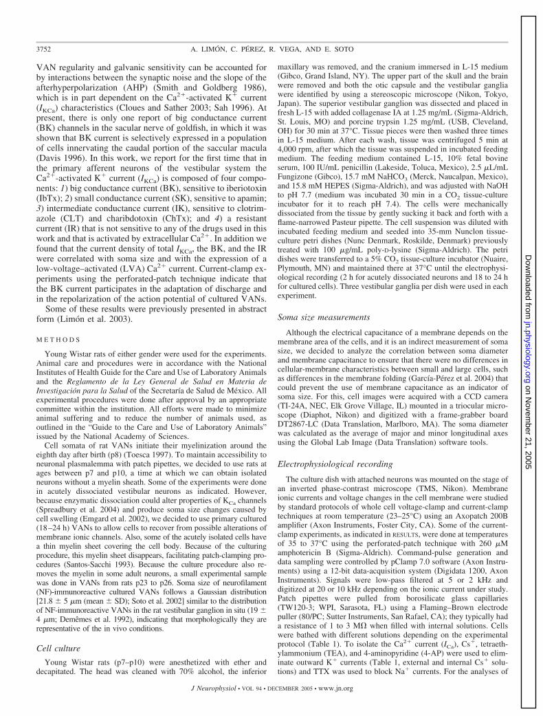
Vestibular-afferent neurons in culture were identified bytheir birefringent round or ovoid soma when viewed underphase-contrast optics (Fig. 1A). The relationship betweenmembrane capacitance and neuronal size showed a linearcorrelation (r � 0.92), indicating that membrane capacitancecan be used as an indirect measure of soma size (Fig. 1B).
Previous reports indicate that in acutely dissociated VANsfrom embryonic mice, the largest cells express a LVA-Ca2�
current, whereas the high-voltage–activated (HVA) Ca2� cur-rent is expressed homogeneously in all cells (Chambard et al.1999; Desmadryl et al. 1997). Thus we analyzed the Ca2�
current to define whether cultured rat VANs have the samerelationship between soma size and Ca2� current expression.
TABLE 1. Solutions
Sol NaCl Choline CI KCl CsCl CaCl2 MgCl2 TEA 4-AP EGTA HEPES
ICa External Cs� * 5 3.6 1.2 130 10 10ICa Internal Cs� 140 0.134 10 2 5IKCa External choline* 130 5.4 3.6 1.2 10 10IKCa Ca2� free choline solution* 130 5.4 0 1.2 10 5 10IKCa Internal choline 10 140 0.134 2 5External CC 140 5.4 1.8 1.2 10 10Internal CC 18 140 0.134 2 5
The leftmost column indicates the ionic current that each solution was used for. External and internal CC solutions were used for current-clamp experiments.*Added with TTX 200 nM.
3753Ca2�-ACTIVATED K� CURRENT IN PRIMARY VESTIBULAR NEURONS
J Neurophysiol • VOL 94 • DECEMBER 2005 • www.jn.org
on Novem
ber 21, 2005 jn.physiology.org
Dow
nloaded from
Voltage-dependent calcium current ICa
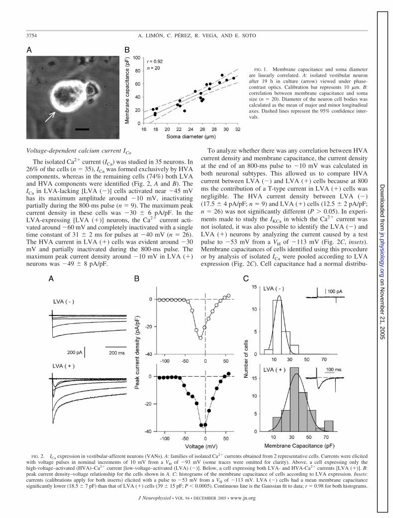
The isolated Ca2� current (ICa) was studied in 35 neurons. In26% of the cells (n � 35), ICa was formed exclusively by HVAcomponents, whereas in the remaining cells (74%) both LVAand HVA components were identified (Fig. 2, A and B). TheICa in LVA-lacking [LVA (�)] cells activated near �45 mVhas its maximum amplitude around �10 mV, inactivatingpartially during the 800-ms pulse (n � 9). The maximum peakcurrent density in these cells was �30 � 6 pA/pF. In theLVA-expressing [LVA (�)] neurons, the Ca2� current acti-vated around �60 mV and completely inactivated with a singletime constant of 31 � 2 ms for pulses at �40 mV (n � 26).The HVA current in LVA (�) cells was evident around �30mV and partially inactivated during the 800-ms pulse. Themaximum peak current density around �10 mV in LVA (�)neurons was �49 � 8 pA/pF.
To analyze whether there was any correlation between HVAcurrent density and membrane capacitance, the current densityat the end of an 800-ms pulse to �10 mV was calculated inboth neuronal subtypes. This allowed us to compare HVAcurrent between LVA (�) and LVA (�) cells because at 800ms the contribution of a T-type current in LVA (�) cells wasnegligible. The HVA current density between LVA (�)(17.5 � 4 pA/pF; n � 9) and LVA (�) cells (12.5 � 2 pA/pF;n � 26) was not significantly different (P � 0.05). In experi-ments made to study the IKCa in which the Ca2� current wasnot isolated, it was also possible to identify the LVA (�) andLVA (�) neurons by analyzing the current caused by a testpulse to �53 mV from a VH of �113 mV (Fig. 2C, insets).Membrane capacitances of cells identified using this procedureor by analysis of isolated ICa were pooled according to LVAexpression (Fig. 2C). Cell capacitance had a normal distribu-
FIG. 1. Membrane capacitance and soma diameterare linearly correlated. A: isolated vestibular neuronafter 19 h in culture (arrow) viewed under phase-contrast optics. Calibration bar represents 10 �m. B:correlation between membrane capacitance and somasize (n � 20). Diameter of the neuron cell bodies wascalculated as the mean of major and minor longitudinalaxes. Dashed lines represent the 95% confidence inter-vals.
FIG. 2. ICa expression in vestibular-afferent neurons (VANs). A: families of isolated Ca2� currents obtained from 2 representative cells. Currents were elicitedwith voltage pulses in nominal increments of 10 mV from a VH of �93 mV (some traces were omitted for clarity). Above, a cell expressing only thehigh-voltage–activated (HVA)–Ca2� current [low-voltage–activated (LVA) (�)]. Below, a cell expressing both LVA- and HVA-Ca2� currents [LVA (�)]. B:peak current density–voltage relationship for the cells shown in A. C: histograms of the membrane capacitance of cells according to LVA expression. Insets:currents (calibrations apply for both inserts) elicited with a pulse to �53 mV from a VH of �113 mV. LVA (�) cells had a mean membrane capacitancesignificantly lower (18.5 � 7 pF) than that of LVA (�) cells (39 � 15 pF; P � 0.0005). Continuous line is the Gaussian fit to data; r � 0.98 for both histograms.
3754 A. LIMON, C. PEREZ, R. VEGA, AND E. SOTO
J Neurophysiol • VOL 94 • DECEMBER 2005 • www.jn.org
on Novem
ber 21, 2005 jn.physiology.org
Dow
nloaded from

tion of 18.5 � 7 pF (mean � SD; n � 19/68) for LVA (�) and39 � 15 pF (n � 49/68) for LVA (�). The statistical differencein capacitance (soma size) between these subgroups was sig-nificant (P � 0.0005). Experiments made in VANs isolatedfrom p23 to p26 rats indicate that the separation of LVA (�)and LVA (�) cells is still present in the mature vestibularsystem. The mean � SD of membrane capacitance for LVA(�) cells was 12 � 2 pF (n � 5) and for LVA (�) it was 29 �1 pF (n � 10; P � 0.0005).
Calcium-activated potassium current IKCa
The expression of IKCa in VANs was analyzed by recordingoutward currents in external and internal choline solutions andduring perfusion with Ca2�-free external choline solution (Ta-ble 1). The control current was composed of ICa, IKCa, and avoltage-dependent, 4-AP–insensitive K� current. The perfu-sion with Ca2�-free external choline solution reversibly re-duced the outward current, leaving the Ca2�-independent com-ponents. The IKCa component of the outward current wasobtained by subtraction of the current remaining after perfusionwith the Ca2�-free external solution from the control current(Fig. 3A). The IKCa component of the outward currents wasobserved in 100% of acutely dissociated (n � 6) and culturedneurons (n � 47). The IKCa activated above �40 mV reachedits maximum around 10 mV, followed by a decline in thecurrent amplitude for more depolarized values (Fig. 3B). TotalIKCa current density was 87 � 7 pA/pF and it was notsignificantly different (P � 0.05) in cultured or acutely disso-ciated neurons.
The analysis of correlation between membrane capacitanceand IKCa shows that small LVA (�) cells express three times
higher IKCa current density (171 � 32 pA/pF; n � 8) thanlarger LVA (�) cells (54 � 5 pA/pF; n � 17: P � 0.01) (Fig.3C). Two cell groups are evident based on the expression of theLVA-Ca2� current. Without this association, the analysis ofcorrelation between the membrane capacitance and IKCa has acontinuous pseudoexponential form.
IKCa was also observed in the 100% of cells obtained fromadult p23 to p26 rats (n � 15). The total IKCa current densityin VANs from p23 to p26 rats was 318 � 75 (range 105 to 968pA/pF). The difference in IKCa current density between young andadult VANs was considerably significant (P � 0.0001), indicatingan increase in IKCa expression during the postnatal development.The IKCa current density of LVA (�) cells (681 � 99 pA/pF; n �5) was also significantly higher than that found in LVA (�) cells(137 � 10 pA/pF; n � 10) (P � 0.001) (Fig. 3D).
The correlation between current density and membrane ca-pacitance shows that IKCa decreased as the cell membranecapacitance increased (r � 0.92), indicating a qualitativelysimilar relationship between p7 to p10 and p23 to p26 VANs.The range and the mean of membrane capacitance of the p23to p26 cells were smaller because of a smaller culturing time (5h for p23–p26 compared with 18 h in p7–p10 neurons).
The outward current that remains during perfusion with Ca2�-free solution showed a significant difference (P � 0.05) betweenLVA (�) (58 � 7 pA/pF; n � 8) and LVA (�) (38 � 4 pA/pF;n � 17) cells. No efforts to characterize that current were made.
Calcium-activated potassium current components SK, BK,IK, and IR
To analyze IKCa components in cultured vestibular-afferentneurons from p7 to p10 rats, the pharmacological agents used
FIG. 3. Ca2�-dependent outward current expression in VANs. A: currents obtained from a representative neuron in external and internal choline solutions (left)and during application of Ca2�-free external choline solution (middle). At right, the IKCa obtained by digital subtraction. Ionic currents were elicited with nominalpulses of 10 mV from a VH of �61 mV. B: I–V curves from the outward current of the cell shown in A, before and after Ca2� removal. I–V relationship of IKCa
was obtained by subtraction of I–V curves before and after Ca2� removal. C: plot of maximum current density vs. membrane capacitance for LVA (�) (n �8) and LVA (�) (n � 17) cells from p7 to p10 rats. Also, neurons in which the expression of LVA-Ca2� current was not evaluated (n � 22) were included.D: plot of maximum current density vs. membrane capacitance for LVA (�) (n � 5) and LVA (�) (n � 10) from p23 to p26 rats. Continuous line is the linearfit to the whole data set (r � 0.92).
3755Ca2�-ACTIVATED K� CURRENT IN PRIMARY VESTIBULAR NEURONS
J Neurophysiol • VOL 94 • DECEMBER 2005 • www.jn.org
on Novem
ber 21, 2005 jn.physiology.org
Dow
nloaded from

were specific blockers for BK (100 nM IbTx) (Galvez et al.1990; Giangiacomo et al. 1992), SK (100 nM apamin) (Hugueset al. 1982), and IK (1 �M CLT) (Grissmer et al. 1993; Jensenet al. 2001; Kaczorowski and Garcia 1999). In some experi-ments, ChTx was also used to isolate the IK current. BecauseChTx blocks both BK and IK channels (Jensen et al. 2001;Kaczorowski and Garcia 1999), BK channels were firstblocked with IbTx and then cells were subsequently perfusedwith an external solution with ChTx � IbTx added. IK wasobtained by the subtraction of the current during IbTx perfu-sion minus the current elicited with IbTx � ChTx. We founda Ca2�-activated K� current that was resistant to classical IKCablockers (100 nM IbTx, 100 nM ChTx, 100 nM apamin, and 1�M CLT). Because no specific blockers for this current exist,we obtained the resistant current (IR) by removal of externalCa2� after IbTx � apamin � ChTx/CLT application.
The cells used to study IKCa components extend within allthe range of capacitances. Values (mean � SD) of the initialtotal outward current (Ca2�-independent K� current � IKCa) ofthe cells used to analyze each IKCa component before drugapplication were 115.4 � 83.8 pA/pF for SK (n � 35), 117.2 �86.8 pA/pF for IK (n � 15), 114.1 � 87.7 pA/pF for BK (n �30), and 148.7 � 130.3 for IR (n � 15). There were nostatistical differences between these data (P � 0.05), indicatingthat the cell sample used to make the analyses of IKCa compo-nents was not biased.
The perfusion of 100 nM apamin reduced the outwardcurrent in 86% of the cells (n � 30/35) (Fig. 4A). Subtractionof the current during apamin perfusion from control currentgave the SK current. The SK component activates for voltagespositive to �30 mV and reaches its maximum amplitudebetween 5 and 10 mV (data not shown). The SK current had aslow activation and did not decay during the 800-ms pulse,indicating a minimal, if any, inactivation. The maximum cur-rent density of SK was classified according to LVA expression.The SK current density was not different between LVA (�)and LVA (�) cells (8.3 � 3.8 vs. 13.6 � 3.5 pA/pF; P � 0.05).No significant correlation between SK expression and cellmembrane capacitance was found (r � 0.21), indicating thatthe SK current is expressed independently of VAN size (Fig. 5,A and B).
The perfusion of 100 nM IbTx reduced the outward currentin 73% of the cells (n � 22/30) (Fig. 4B). Digital subtractionof the outward current before and during IbTx perfusionyielded the BK current. The BK current activated at voltagespositive to �60 mV in 64% of the cells (n � 14/22). In theremaining cells BK activated around �40 mV. The maximumamplitude was observed around 20 mV (data not shown). Theplot of BK current density versus cell membrane capacitance(r � 0.66) indicates that small LVA (�) cells have a smallerBK than that of larger LVA (�) cells, which have higher BK
FIG. 4. Subtypes of Ca2�-activated K�
currents. Currents were recorded in externaland internal choline solutions. A and B: ef-fect of apamin and iberiotoxin (IbTx, 100nM each) on outward current elicited withpulses in nominal increments of 10 mV froma VH of �61 mV. Outward current in controlconditions (above), during toxin perfusion(middle), and toxin-sensitive current ob-tained by digital subtraction (below). C: ef-fect of 100 nM charibdotoxin (ChTx) onapamin- and IbTx-resistant current. ChTxperfusion after big conductance current (BK)and small conductance current (SK) channelblockade with IbTx and apamin allows iso-lation of ionic current through intermediateconductance current (IK) channels. Currentcalibration bars shown in B apply for thesame row in A and C. Timescale is for allrecording traces. D: time course of outwardcurrent in control conditions (elicited with apulse test to 25 mV from a VH of �61 mV)and during application of indicated toxins ata concentration of 100 nM each. E: resistantcurrent (IR) in a LVA (�) cell, obtained byexternal Ca2� removal and after apamin �IbTx � clotrimazole (CLT) perfusion. Ioniccurrents were generated with pulses in nom-inal increments of 10 mV from a VH of �61mV.
3756 A. LIMON, C. PEREZ, R. VEGA, AND E. SOTO
J Neurophysiol • VOL 94 • DECEMBER 2005 • www.jn.org
on Novem
ber 21, 2005 jn.physiology.org
Dow
nloaded from

current density (3 � 1.4 vs. 17 � 3.3 pA/pF; P � 0.01; Fig. 5,A and B).
Perfusion of 100 nM ChTx (after BK blockade with IbTx)reduced the outward current in 86% of the cells (n � 6/7; Fig.4C). The perfusion of 1 �M CLT (n � 8) allowed us to isolatean outward current whose characteristics were indistinguish-able from the ChTx-sensitive current. Therefore for IK analy-sis, data obtained from ChTx- and CLT-sensitive currents werepooled. The IK did not inactivate during 800-ms voltagepulses. The IK activated above �40 mV, reaching its maxi-mum amplitude at 10 mV (data not shown). The mean IKcurrent density was higher in LVA (�) than in LVA (�) cells,although because of its large variability this difference was notstatistically significant (26 � 9 vs. 11 � 3.8 pA/pF; P � 0.05).The analysis of correlation between IK current density andmembrane capacitance gave a weak correlation (r � 0.58) (Fig.5, A and B).
In 80% of the cells (n � 12/15), the application of IKCablockers (100 nM IbTx, 100 nM ChTx, 100 nM apamin, and 1�M CLT) did not remove a Ca2�-dependent outward current(Fig. 4D). This resistant current (IR) was evident by removal ofexternal Ca2� after IbTx � apamin � ChTx/CLT application(Fig. 4E). The IR current density in LVA (�) cells wassignificantly higher than that in LVA (�) cells (86 � 3 vs.6.2 � 1.8 pA/pF; P � 0.01). The plot of IR current densityversus membrane capacitance shows a significant correlation(r � 0.82) and confirms the existence of two separated cellgroups (Fig. 5, A and B). The first was composed of LVA (�)cells with higher IR current density dispersed in a wide rangefrom 36 to 165 pA/pF. The second was composed of LVA (�)cells that did not express a significant IR component, with arange of IR current density from 0 to 14 pA/pF. Note that theIR current density is always underestimated because it is
contaminated with ICa. This is because removal of Ca2� fromthe external solutions used to obtain IR simultaneously abol-ishes the Ca2� current. The range of ICa current density at 800ms (time at which IR was measured) was between �4 and �43pA/pF, so the IR could be underestimated within this range.Because HVA-Ca2� current density was not different betweenLVA (�) and LVA (�) subgroups, the difference observed inIR current density between LVA (�) and LVA (�) cells isproduced exclusively by differences in IR expression.
Effect of IKCa blockers on the voltage responseto current pulses
To analyze the influence of IKCa components on the dis-charge of the cultured vestibular neurons, the effect of IKCachannel blockers on the electrical response of VANs wasstudied. For this, the perforated patch-clamp technique wasused to avoid dialysis of internal constituents of VANs. Re-cordings were done at temperatures between 35 and 37°C toapproximate to more physiological conditions. Voltage re-sponses were recorded using external and internal CC solutions(Table 1), and with holding membrane potential of �60 mV(close to the zero current potential of VANs). In these condi-tions, 60% of VANs fired one or two action potentials (APs)during 200-ms and 2-s pulses of suprathreshold current injec-tion (n � 21). The remaining 40% of the cells showed a slowlyadapting response, firing no more than eight action potentialswith long-duration pulses (2 s). The threshold of the first AP incontrol conditions was �36 � 1 mV (n � 21). AP amplitudefrom the threshold to the maximum peak was 53 � 3 mV. TheAP duration (measured at 75%) was 4 � 0.4 ms (note thatexternal CC solution contains 10 mM 4-AP).
The perfusion of 100 nM apamin or 1 �M CLT did notsignificantly modify any of these variables (Fig. 6, A and B).
FIG. 5. Expressions of IKCa subtypes are correlated with membrane capacitance and with expression of LVA-Ca2� current. A: IKCa and IR maximum currentdensities were significantly higher in LVA (�) than in LVA (�) cells. On the contrary the expression of BK was significantly larger in LVA (�) when comparedwith LVA (�) cells (*P � 0.01). SK and IK did not show significant differences between subgroups. Difference of total IKCa current density between LVA (�)and LVA (�) cells is mainly caused by the IR current that is the principal component of IKCa in LVA (�) cells. B: plots of maximum current density for eachIKCa subtype vs. membrane capacitance. Cross symbols indicate cells in which the presence of LVA-Ca2� current was not evaluated. Continuous line is the linearfit to the whole data set. r is the correlation coefficient for each linear fit. Number of cells in SK, IK, BK, and IR plots were 35, 15, 30, and 15.
3757Ca2�-ACTIVATED K� CURRENT IN PRIMARY VESTIBULAR NEURONS
J Neurophysiol • VOL 94 • DECEMBER 2005 • www.jn.org
on Novem
ber 21, 2005 jn.physiology.org
Dow
nloaded from

However, in 38% of the cells, either apamin or CLT perfusionproduced a slight (5–10 mV) oscillatory behavior in the voltageresponse after the first action potential. This oscillatory re-sponse continuously decreased and vanished within 150 ms.
The effect of IbTx was variable among cells as expectedfrom correlation analysis between IKCa current density andmembrane capacitance. The perfusion of IbTx in small cellsdid not significantly modify AP waveform, whereas in largecells the AP duration increase was �770% (r � 0.78, n � 7)(Fig. 6C). In those cells with repetitive discharge, the perfusionof 100 nM IbTx removed the adaptation of the response during200-ms and 2-s current pulses (Fig. 6D).
These results indicate that the BK current contributes torepolarization of the action potential and to discharge adapta-tion, and this contribution is correlated with the membranecapacitance of VANs.
Voltage-clamp experiments indicate that the IR is the prin-cipal IKCa component expressed by small LVA (�) cells and itscontribution is correlated with the membrane capacitance.Because of the lack of a specific channel blocker for thiscurrent, there is no possibility of making a pharmacologicalanalysis of its contribution to the VANs’ discharge.
D I S C U S S I O N
Calcium-activated current expression
Rat VANs in primary culture expressed a Ca2� currentformed by LVA and HVA components. Whereas the HVA-Ca2� current was present in 100% of VANs, the expression ofLVA-Ca2� was heterogeneous among cells and preferentiallypolarized to medium and large neurons. Correlation betweenT-type Ca2�-current expression and diameter has been re-ported in vestibular-ganglion neurons of mice (Chambard et al.1999; Desmadryl et al. 1997) and in the dorsal root andnodosus-ganglion neurons of rat (Fedulova et al. 1985; Lam-bert et al. 1997). The percentage of cells expressing theLVA-Ca2� current in this study (74%) was greater than that
reported in newborn mice (20%). The differences are becausein mice only the largest cells express the LVA-Ca2� current,whereas in the rat, medium and large cells express this current.This may be an interspecies difference or caused by distinctculturing times of isolated neurons (2–8 h as compared with18–24 h in the present study). Another possible source for thedifferences could arise from the distinct age of the animalsused (p4–p8 compared with p7–p10 in the present study). Inmice, the separation of VANs into two groups on the basis ofT-type current starts around E17, and at p4 it is possible toobserve two completely separated groups (Chambard et al.1999). As shown in this work, in rats this separation is stillpresent in the nearly mature (p23 to p26) neurons. The expres-sion of the LVA-Ca2� current in medium to large cells sug-gests that the T-type current could contribute to dischargedifferences between small and large cells in situ. The T-typecurrent is preferentially expressed in dendrites of central neu-rons and participates in the threshold for action-potential gen-eration, in synaptic integration, and in the configuration ofspike discharge (Gauck et al. 2001; Perez-Reyes 2003; Pouilleet al. 2000). Therefore as proposed for mice (Desmadryl et al.1997), the presence of an LVA-Ca2� current in large cells maydecrease the threshold for spike generation, which could ex-plain the increased sensitivity of thick axons that innervatecentral zones of the neuroepithelium compared with thinneraxons that innervate peripheral zones.
For the HVA-Ca2� currents, although we did not perform apharmacological dissection, its characteristics were similar tothose reported in mice (Desmadryl et al. 1997), where it hasbeen found that the HVA-Ca2� current is composed of L-,P/Q-, N-, and R-type currents (Chambard et al. 1999; Des-madryl et al. 1997). The whole HVA-Ca2� current was foundin 100% of the rat VANs, and no correlation between itscurrent density and soma size (membrane capacitance) wasobserved. However, we cannot discard the possibility thatsome of the ionic channels that make up the HVA-Ca2� currenthave a polarized distribution or are coupled differently with
FIG. 6. Effect of KCa channels blockers on the electrical discharge of VANs. Records were made using a perforated patch in the current-clamp mode. A andB: current-clamp recordings showing the effects of 100 nM apamin and 1 �M CLT. Voltage responses were elicited with 0.6 and 0.4 nA current injections forA and B. B, inset: morphological variables of the action potentials (APs) that were measured: a, AP amplitude; b, threshold; and c, AP duration at 75%. Dottedlines indicate the zero voltage. Darker traces indicate the exponential fit to the electrical charge of the membrane. C: summary of drug effects on the AP durationas a function of cell capacitance. D: IbTx 100 �M removes the adaptation of the response to 0.4-nA current pulse in a reversible form.
3758 A. LIMON, C. PEREZ, R. VEGA, AND E. SOTO
J Neurophysiol • VOL 94 • DECEMBER 2005 • www.jn.org
on Novem
ber 21, 2005 jn.physiology.org
Dow
nloaded from

some of the KCa channels reported in this work. Thereforefurther studies focused on the characterization of channelsunderlying the HVA-Ca2� current and its functional couplingwith KCa channels are needed to determine its particular role inthe electrical firing of VANs.
Calcium-activated potassium current expression
The removal of Ca2� from the external solution decreasedthe outward current, indicating the presence of an outwardcurrent that is activated by the influx of extracellular Ca2�.
The IKCa current was found in 100% of the studied cells.Current-density analysis showed that small LVA (�) neuronshad up to four times higher current density than that of largerLVA (�) cells. Our results indicate that in both cell groupsfour different currents compose the IKCa—SK, IK, BK, andIR—although in different proportions. Of these, only BK andIR have a clear correlation with soma size (membrane capac-itance) and with the expression of the LVA-Ca2� current.
The BK current was preferentially expressed in LVA (�)cells, whereas the IR was strongly expressed in LVA (�) cells.In 60% of the cells, BK was activated at voltages below the APthreshold, indicating its physiological coupling with LVA-Ca2� channels, similar to data reported in central-vestibularneurons (Smith et al. 2002). If the correlation between somadiameter and BK expression and the functional coupling withT-type currents are present in VANs in situ, it might haveimportant functional consequences. The T-type current de-creases the AP threshold, but the major contribution of BKmay tend to repolarize the cells, thus producing failures in theAP generation and irregularity of discharge. Actually, theparticipation of BK in the adaptation of the response to currentpulses, revealed by IbTx effects, indicates that BK inhibitionconverts the firing pattern from phasic to tonic. Furthermore,the BK participates in the repolarization of the action potentialand contributes to the spike duration, supporting the hypothesisthat the expression of BK significantly contributes to determi-nation of the discharge properties of VANs in situ.
Our current-clamp experiments with the perforated-patchtechnique, which should maintain endogenous intracellularbuffers, reveal that SK and IK have an insignificant participa-tion in the discharge of VANs caused by constant-currentpulses, despite the fact that electrical firing was recorded usingexternal solutions with 4-AP, which enhances the excitabilityof sensory neurons (Sculptoreanu et al. 2004; Soto et al. 2002;Stansfeld et al. 1986). However, the possibility that SK and IKcurrents participate in the more dynamic and complex condi-tions in situ cannot be discarded.
SK as well as IK activity depends largely on the concentra-tion of intracellular Ca2�. However, the I–V relationships ofSK and IK did not follow the expected decrease for voltages�0 mV (at which the inward Ca2� current decreases). This canbe attributable to contamination with voltage-dependent chan-nels (such as BK currents), although this is unlikely becauseChTx and CLT inhibit similar currents and to date there are noreports about nonspecific effects of CLT and apamin on volt-age-dependent channels at the concentrations used in thiswork. Another possibility is that Ca2� may accumulate by anincomplete clearance by mechanisms of extrusion or seques-tration of Ca2� between voltage pulses. In dorsal root ganglionneurons (Thayer and Miller 1990) and in chromaffin cells of
the rat (Herrington et al. 1996) large Ca2� loads (1–2 �M)produce a rapid initial Ca2� uptake by mitochondria, butintracellular [Ca2�] decay to resting levels can last �2 min.The modulation of IKCa currents by Ca2� released from intra-cellular stores will also be taking place in our system and thatparticular issue should be addressed in future studies.
The identity of the KCa channels underlying the IR currentwill require further studies. However, the electrophysiologicalproperties of the IR current are similar to drug-resistant cur-rents from oocytes expressing BK channels coupled with the�4 subunit (Meera et al. 2000) and astrocytes expressing the�4 subunit (Gebremedhin et al. 2003). The �4 subunit has beenreported to confer resistance to IbTx to the BK channels(Gebremedhin et al. 2003; Meera et al. 2000). Unfortunately,the lack of selective blockers of the IR current deters anadequate analysis of its contribution to the firing discharge ofVANs. The IR has current densities �160 pA/pF, which areconsiderably greater than those of other currents reported inthis study. Thus the differences found in outward currentamong cells seems to be caused by the expression of IR.
In mathematical models of VAN, it has been proposed thatthe AHP slope accounts for differences in sensibility andregularity of discharge (Smith and Goldberg 1986). Accordingto this, the irregular discharge of large neurons innervatingtype I hair cells may be caused by a smaller slope of postspikevoltage trajectory compared with small regular neurons inner-vating type II hair cells. Although the AP duration did not havea significant linear correlation with total outward current, thereis a tendency of small cells to have shorter APs, as observed inVANs in situ innervating the peripheral zones of chick vestib-ular-neuroepithelium (Yamashita and Ohmori 1990).
It is worth noting that the electrophysiological properties ofVANs in situ are changing during the first weeks after the birthuntil reaching a stable phenotype around p23 (Curthoys 1979).Therefore because most of our results were obtained from ratsbetween p7 and p10, our conclusions are mainly circumscribedby this time window in the development of the vestibularsystem. However, at this age irregular neurons almost show amature phenotype except for the presence of firing, bursting,and silent neurons at rest (Curthoys 1979). The percentage ofregular neurons between p7 and p10 is in the range of 10 to15%, which is nearly half of those in the adult animal (32%).Regular neurons at p7 to p10 have an electrical discharge atrest of about 20 spikes/s, which is around one third of the ratein the adult rat (Curthoys 1979, 1982). The inability of ourcultured VANs to fire tonically during sustained pulses incurrent clamp suggests that they are in an intermediate stage ofmaturity or the spatial relationships of ionic channels is not thesame as that in the vestibular system in situ. That total IKCaincreases in adult p23 to p26 rats reinforces the idea of theimmaturity of VANs at p7 to p10 and is in agreement withstudies of vestibular-nucleus neurons, which show that electri-cal properties of vestibular-nucleus neurons change during theembryonic ages (Peusner and Giaume 1997) and beyond birth(Dutia and Johnston 1998). Despite the increase in IKCa currentdensity, the correlation between total IKCa and membranecapacitance in p23 to p26 adult VANs indicates that thedifferences in the expression of IKCa are maintained in themature vestibular system. However, the individual contributionof each IKCa subtype to total IKCa in p23 to p26 neurons was notdefined. It is also possible that some of the kinetic and elec-
3759Ca2�-ACTIVATED K� CURRENT IN PRIMARY VESTIBULAR NEURONS
J Neurophysiol • VOL 94 • DECEMBER 2005 • www.jn.org
on Novem
ber 21, 2005 jn.physiology.org
Dow
nloaded from

trophysiological properties of IKCa change during the devel-opment as previously reported in central neurons (Kang etal. 1996).
The IKCa has been shown to significantly contribute to theshaping of the action potential waveform and to the firing-discharge pattern of neurons (Dutia and Johnston 1998; Faberand Sah 2002; Johnston et al. 1994; Sah and Faber 2002; Smithet al. 2002). In lamprey motoneurons, blockade of the SKcurrent increased the variation coefficient of the spike dis-charge (El Manira et al. 1994). The heightened contribution ofthe IKCa in small LVA (�) cells could determine a faster AHPslope, thus significantly influencing discharge regularity ofVANs (Smith and Goldberg 1986).
The functional role of KCa channels is not restricted to themodulation of the electrical firing but also may participate atthe central synapse controlling neurotransmitter release (Hu etal. 2001; Robitaille et al. 1993). The particular role of KCachannels at central terminals should be analyzed in experimen-tal models keeping intact the synapses between primary affer-ents and second-order neurons. It is worth noting that ourresults are limited to analysis of the KCa currents expressed inspecific developmental stages and that there is no evidenceindicating whether the complement of these currents is alsoexpressed in afferent dendrites or central terminals of themature VAN. Our results indicate that cultured VANs expressan IKCa carried by several channel subtypes. As shown in othersystems IKCa may participate in determining the threshold,latency, repolarization rate, and afterhyperpolarization of theaction potential. Its differential expression among VANs mayhave important functional implications in the coding of ves-tibular sensory information. Future studies to analyze theexpression and distribution of IKCa subtypes within the primarysensory neuron in the mature vestibular system, includingrecordings in the vestibular nerve in situ, would contribute toour understanding of the role of the intrinsic properties ofVANs in the coding of the vestibular information.
A C K N O W L E D G M E N T S
The authors thank Dr. Ellis Glazier for editing the English-language text.
G R A N T S
This research was partially supported by Consejo Nacional de Ciencia yTecnologıa (CONACyT) Grant 35525-N to E. Soto and Grant VIEP-II84G01to R. Vega. During this work A. Limon and C. Perez were recipients ofCONACyT fellowships 124156 and 185855.
R E F E R E N C E S
Adamson CL, Reid MA, Mo ZL, Bowne-English J, and Davis RL. Firingfeatures and potassium channel content of murine spiral ganglion neuronsvary with cochlear location. J Comp Neurol 447: 331–350, 2002.
Akita T and Kuba K. Functional triads consisting of ryanodine receptors,Ca2� channels, and Ca2�-activated K� channels in bullfrog sympatheticneurons, plastic modulation of action potential. J Gen Physiol 116: 697–720,2000.
Amstrong CM and Gilly WF. Access resistance and space clamp problemsassociated with whole-cell patch clamping. Methods Enzymol 207: 100–122, 1992.
Annoni JM, Cochran SL, and Precht W. Pharmacology of the vestibular haircell afferent fiber synapse in frog. J Neurosci 4: 2106–2116, 1984.
Baird RA, Desmadryl G, Fernandez C, and Goldberg JM. The vestibularnerve of the chinchilla. II. Relation between afferent response properties andperipheral innervation patterns in the semicircular canals. J Neurophysiol60: 182–203, 1988.
Bronte-Stewart HM and Lisberger SG. Physiological properties of vestib-ular primary afferents that mediate motor learning and normal performanceof the vestibuloocular reflex in monkeys. J Neurosci 14: 1290–1308, 1994.
Chabbert C, Chambard JM, Sans A, and Desmadryl G. Three types ofdepolarization-activated potassium currents in acutely isolated mouse ves-tibular neurons. J Neurophysiol 85: 1017–1026, 2001a.
Chabbert C, Chambard JM, Sans A, and Desmadryl G. Voltage-activatedsodium currents in acutely isolated mouse vestibular neurones. Neuroreport8: 1253–1256, 1997.
Chabbert C, Chambard JM, Valmier J, Sans A, and Desmadryl G.Hyperpolarization-activated (Ih) current in mouse vestibular primary neu-rons. Neuroreport 12: 2701–2704, 2001b.
Chambard JM, Chabbert C, Sans A, and Desmadryl G. Developmentalchanges in low and high voltage-activated calcium currents in acutelyisolated mouse vestibular neurons. J Physiol 518: 141–149, 1999.
Cloues RK and Sather WA. Afterhyperpolarization regulates firing rate inneurons of the suprachiasmatic nucleus. J Neurosci 23: 1593–1604, 2003.
Curthoys IS. The development of function of horizontal semicircular canalprimary neurons in the rat. Brain Res 167: 41–52, 1979.
Curthoys IS. Postnatal developmental changes in the response of rat primaryhorizontal semicircular canal neurons to sinusoidal angular accelerations.Exp Brain Res 47: 295–300, 1982.
Davies PJ, Ireland DR, and Mc Lachlan EM. Sources of Ca2� for differentCa2�-activated K� conductances in neurones of the rat superior cervicalganglion. J Physiol 495: 353–366, 1996.
Davis RL. Differential distribution of potassium channels in acutely demyeli-nated primary-auditory neuron in vitro. J Neurophysiol 76: 438–447, 1996.
Dememes D, Raymond J, Atger P, Grill C, Winsky L, and Dechesne C.Identification of neuron subpopulations in the rat vestibular ganglion bycalbindin-D 28 K, calretinin and neurofilament proteins immunoreactivity.Brain Res 582: 168–172, 1992.
Desmadryl G, Chambard JM, Valmier J, and Sans A. Multiple voltage-dependent calcium currents in acutely isolated mouse vestibular neurons.Neuroscience 78: 511–522, 1997.
Dopico AM, Widmer H, Wang G, Lemos JR, and Treistman SN. Ratsupraoptic magnocellular neurones show distinct large conductance, Ca2�-activated K� channel subtypes in cell bodies versus nerve endings. J Physiol519: 101–114, 1999.
Duncan RK and Fuchs PA. Variation in large-conductance, calcium-acti-vated potassium channels from hair cells along the chicken basilar papilla.J Physiol 547: 357–371, 2003.
Dutia MB and Johnston AR. Development of action potentials and apaminsensitive after-potentials in mouse vestibular nucleus neurones. Exp BrainRes 118: 148–154, 1998.
El Manira A, Tegner J, and Grillner S. Calcium-dependent potassiumchannels play a critical role for burst termination in the locomotor networkin lamprey. J Neurophysiol 72: 1852–1861, 1994.
Emgard M, Blomgren K, and Brundin P. Characterization of cell damageand death in embryonic mesencephalic tissue: a study on ultrastructure, vitalstains and protease activity. Neuroscience 115: 1177–1187, 2002.
Ezure K, Cohen M, and Wilson V. Response of cat semicircular afferents tosinusoidal polarizing currents: implications for input–output properties ofsecond-order neurons. J Neurophysiol 49: 639–648, 1983.
Faber ES and Sah P. Physiological role of calcium-activated potassiumcurrents in the rat lateral amygdala. J Neurosci 22: 1618–1622, 2002.
Fedulova SA, Kostyuk PG, and Veselovsky NS. Two types of calciumchannels in the somatic membrane of new-born rat dorsal root ganglionneurones. J Physiol 359: 431–446, 1985.
Fernandez C, Baird RA, and Goldberg JM. The vestibular nerve of thechinchilla. I. Peripheral innervation patterns in the horizontal and superiorsemicircular canals. J Neurophysiol 60: 167–181, 1988.
Fernandez C, Lysakowski A, and Goldberg JM. Hair-cell counts andafferent innervation patterns in the cristae ampullares of the squirrel monkeywith comparison to the chinchilla. J Neurophysiol 73: 1253–1269, 1995.
Galvez A, Gimenez-Gallego G, Reuben JP, Roy-Contancin L, FeigenbaumP, Kaczorowski GJ, and Garcia ML. Purification and characterization ofa unique potent peptidyl probe for the high conductance calcium activatedpotassium channel from the venom of the scorpion Buthus talamus. J BiolChem 265: 11083–11090, 1990.
Garcıa-Perez E, Vargas-Caballero M, Velazquez-Ulloa N, Minzoni A, andDe-Miguel FF. Synaptic integration in electrically coupled neurons. Bio-phys J 86: 646–655, 2004.
3760 A. LIMON, C. PEREZ, R. VEGA, AND E. SOTO
J Neurophysiol • VOL 94 • DECEMBER 2005 • www.jn.org
on Novem
ber 21, 2005 jn.physiology.org
Dow
nloaded from
Gauck V, Thomann M, Jaeger D, and Borst A. Spatial distribution of low-and high-voltage-activated calcium currents in neurons of the deep cerebel-lar nuclei. J Neurosci 21: RC158, 2001.
Gebremedhin D, Yamaura K, Zhang C, Bylund J, Koehler RC, andHarder DR. Metabotropic glutamate receptor activation enhances the ac-tivities of two types of Ca2�-activated K� channels in rat hippocampalastrocytes. J Neurosci 23: 1678–1687, 2003.
Giangiacomo KM, Garcia ML, and McManus OB. Mechanism of iberio-toxin block of the large-conductance calcium-activated potassium channelfrom bovine aortic smooth muscle. Biochemistry 31: 6719–6727, 1992.
Goldberg JM. Afferent diversity and the organization of central vestibularpathways. Exp Brain Res 130: 277–297, 2000.
Goldberg JM and Fernandez C. Physiology of peripheral neurons innervat-ing semicircular canals of the squirrel monkey. 3. Variations among units intheir discharge properties. J Neurophysiol 34: 676–684, 1971.
Goldberg JM, Highstein SM, Moschovakis AK, and Fernandez C. Inputsfrom regularly and irregularly discharging vestibular nerve afferents tosecondary neurons in the vestibular nuclei of the squirrel monkey. I. Anelectrophysiological analysis. J Neurophysiol 58: 700–718, 1987.
Golding NL, Jung H, Mickus T, and Spruston N. Dendritic calcium spikeinitiation and repolarization are controlled by distinct potassium channelsubtypes in CA1 pyramidal neurons. J Neurosci 18: 8789–8798, 1999.
Grissmer S, Nguyen A, and Cahalan MD. Calcium-activated potassiumchannels in resting and activated human T lynfocytes. Expression levels,calcium dependence, ion selectivity and pharmacology. J Gen Physiol 102:601–630, 1993.
Herrington J, Park YB, Babcock DF, and Hille B. Dominant role ofmitochondria in clearance of large Ca2� loads from rat adrenal chromaffincells. Neuron 16: 219–228, 1996.
Honrubia V, Hoffman LF, Sitko S, and Schwartz IR. Anatomic andphysiological correlates in the bullfrog vestibular nerve. J Neurophysiol 61:688–701, 1989.
Hu H, Shao LR, Chavoshy S, Gu N, Trieb M, Behrens R, Laake P, PongsO, Knaus HG, Ottersen OP, and Storm JF. Presynaptic Ca2�-activatedK� channels in glutamatergic hippocampal terminals and their role in spikerepolarization and regulation of neurotransmitter release. J Neurosci 21:9585–9597, 2001.
Hugues M, Romey G, Duval D, Vincent JP, and Lazdunski M. Apamin asa selective blocker of the calcium-dependent potassium channel in neuro-blastoma cells: voltage-clamp and biochemical characterization of the toxinreceptor. Proc Natl Acad Sci USA 79: 1308–1312, 1982.
Jensen BS, Strøbæck D, Olesen SP, and Christophersen P. The Ca2�-activated K� channel of intermediate conductance: a molecular target fornovel treatments? Curr Drug Targets 2: 401–422, 2001.
Johnston AR, MacLeod NK, and Dutia MB. Ionic conductances contribut-ing to spike repolarization and afterpotentials in rat vestibular nucleusneurones. J Physiol 15: 61–77, 1994.
Kaczorowski GJ and Garcia ML. Pharmacology of voltage gated andcalcium-activated potassium channels. Curr Opin Chem Biol 3: 448–458,1999.
Kang J, Huguenard JR, and Prince DA. Development of BK channels inneocortical pyramidal neurons. J Neurophysiol 76: 188–198, 1996.
Kevetter GA and Leonard RB. Molecular probes of the vestibular nerve. II.Characterization of neurons in Scarpa’s ganglion to determine separatepopulations within the nerve. Brain Res 928: 18–29, 2002.
Koyama S, Kanemitsu Y, and Weight FF. Spontaneous activity and prop-erties of two types of principal neurons from the ventral tegmental area ofrat. J Neurophysiol 93: 3282–3293, 2005.
Lambert RC, Maulet Y, Mouton J, Beattie R, Volsen S, De Waard M, andFeltz A. T-type Ca2� current properties are not modified by Ca2� channel� subunit depletion in nodosus ganglion neurons. J Neurosci 17: 6621–6628, 1997.
Leger JF, Stern EA, Aertsen A, and Heck D. Synaptic integration in ratfrontal cortex shaped by network activity. J Neurophysiol 93: 281–293,2005.
Leonard RB and Kevetter GA. Molecular probes of the vestibular nerve. I.Peripheral termination patterns of calretinin, calbindin and peripherin con-taining fibers. Brain Res 928: 8–17, 2002.
Limon A, Perez C, Vega R, and Soto E. Intrinsic properties of vestibularprimary afferent neurons in culture. Soc Neurosci Abstr 702.7, 2003.
Lysakowski A, Minor LB, Fernandez C, and Goldberg JM. Physiologicalidentification of morphologically distinct afferent classes innervating thecrista ampullares of the squirrel monkey. J Neurophysiol 73: 1270–1281,1995.
Marcotti W, Johnson SL, and Kros CJ. Effects of intracellular stores andextracellular Ca2� on Ca2�-activated K� currents in mature mouse innerhair cells. J Physiol 557: 613–633, 2004.
Meera P, Wallner M, and Toro L. A neuronal � subunit (KCNMB4) makesthe large conductance, voltage- and Ca2�-activated K� channel resistant tocharybdotoxin and iberiotoxin. Proc Natl Acad Sci USA 97: 5562–5567,2000.
Myers SF and Lewis ER. Hair cell tufts and afferent innervation of thebullfrog crista ampullaris. Brain Res 534: 15–24, 1990.
Perez-Reyes E. Molecular physiology of low-voltage-activated T-type cal-cium channels. Physiol Rev 83: 117–161, 2003.
Peusner KD and Giaume C. Ontogeny of electrophysiological properties anddendritic pattern in second-order chick vestibular neurons. J Comp Neurol384: 621–633, 1997.
Pouille F, Cavelier P, Desplantez T, Beekenkamp H, Craig PJ, Beattie RE,Volsen SG, and Bossu JL. Dendro-somatic distribution of calcium-medi-ated electrogenesis in Purkinje cells from rat cerebellar slice cultures.J Physiol 527: 265–282, 2000.
Ricci AJ, Gray-Keller M, and Fettiplace R. Tonotopic variations of calciumsignaling in turtle auditory hair cells. J Physiol 524: 423–436, 2000.
Robitaille R, Garcia ML, Kaczorowski GJ, and Charlton MP. Functionalcolocalization of calcium and calcium-gated potassium channels in controlof transmitter release. Neuron 11: 645–655, 1993.
Sah P. Ca2�-activated K� currents in neurones: types, physiological roles andmodulation. Trends Neurosci 19: 150–154, 1996.
Sah P and Faber ES. Channels underlying neuronal calcium-activated potas-sium currents. Prog Neurobiol 66: 345–353, 2002.
Santos-Sacchi J. Voltage-dependent ionic conductances of type I spiralganglion cells from the guinea pig inner ear. J Neurosci 13: 3599–3611,1993.
Sculptoreanu A, Yoshimura N, and de Groat WC. KW-7158[(2s)-(�)-3,3,3-trifluoro-2-hydroxy-2-methyl-N-(5,5,10-trioxo-4,10-dihydrothie-nol[3,2-c][1]benzothiepin-9-yl) propanamide] enhances A-type K� currentsin neurons of the dorsal root ganglion of the adult rat. J Pharmacol Exp Ther310: 159–168, 2004.
Si X, Zakir M, and Dickman JD. Afferent innervation of the utricular maculain pigeons. J Neurophysiol 89: 1660–1677, 2003.
Smith CE and Goldberg JM. A stochastic afterhyperpolarization model ofrepetitive activity in vestibular afferents. Biol Cybern 54: 41–51, 1986.
Smith MR, Nelson AB, and DuLac S. Regulation of firing response gain bycalcium dependent mechanisms in vestibular nucleus neurones. J Neuro-physiol 87: 2031–2042, 2002.
Soto E, Limon A, Ortega A, and Vega R. Caracterısticas morfologicas yelectrofisiologicas de las neuronas del ganglio vestibular en cultivo. GacMed Mex 138: 1–13, 2002.
Soto E and Vega R. Actions of excitatory amino acids agonists and antago-nists on the primary afferents of the vestibular system of the axolotl(Ambystoma mexicanum). Brain Res 462: 104–111, 1988.
Spreadbury IC, Kros CJ, and Meech RW. Effects of trypsin on large-conductance Ca2�-activated K� channels of guinea-pig outer hair cells.Hear Res 190: 115–127, 2004.
Stansfeld CE, Marsh SJ, Halliwell JV, and Brown DA. 4-Aminopyridineand dendrotoxin induce repetitive firing in rat visceral sensory neurons byblocking a slowly inactivating outward current. Neurosci Lett 64: 299–304,1986.
Starr PA and Sewell WF. Neurotransmitter release from hair cells and itsblockade by glutamate-receptor antagonists. Hear Res 52: 23–41, 1991.
Thayer SA and Miller RJ. Regulation of the intracellular free calciumconcentration in single rat dorsal root ganglion neurones in vitro. J Physiol425: 85–115, 1990.
Toesca A. Central and peripheral myelin in the rat cochlear and vestibularnerves. Neurosci Lett 221: 21–24, 1997.
Yamashita M and Ohmori H. Synaptic responses to mechanical stimulationin calyceal and bouton type vestibular afferents studied in an isolatedpreparation of semicircular canal ampullae of chicken. Exp Brain Res 80:475–488, 1990.
3761Ca2�-ACTIVATED K� CURRENT IN PRIMARY VESTIBULAR NEURONS
J Neurophysiol • VOL 94 • DECEMBER 2005 • www.jn.org
on Novem
ber 21, 2005 jn.physiology.org
Dow
nloaded from