Coordinated Post-translational Responses of Aquaporins to ...

Brassinosteroid functions to protect the translationalmachinery and heat-shock protein synthesis followingthermal stress
Sangeeta Dhaubhadel1, Karen S. Browning2, Daniel R. Gallie3 and Priti Krishna1,*
1Department of Plant Sciences, University of Western Ontario, London, Ontario, Canada N6A 5B7,2Department of Chemistry and Biochemistry, University of Texas, Austin, TX 78712, USA, and3Department of Biochemistry, University of California, Riverside, CA 92521-0129, USA
Received 20 August 2001; revised 26 November 2001; accepted 17 December 2001.*For correspondence (fax +1 519 661 3935; e-mail [email protected]).
Summary
In addition to their essential role in plant development, brassinosteroids have the ability to protect plants
from various environmental stresses. Currently it is not understood how brassinosteroids control plant
stress responses at the molecular level. We have begun an investigation into the molecular mechanisms
underlying 24-epibrassinolide (EBR)-mediated stress resistance. Earlier we found that treatment of
Brassica napus seedlings with EBR leads to a signi®cant increase in their basic thermotolerance, and
results in higher accumulation of four major classes of heat-shock proteins (hsps) as compared to
untreated seedlings. Surprisingly, previous studies have shown that while hsp levels were signi®cantly
higher in treated seedlings during the recovery period, transcripts corresponding to these hsps were
present at higher levels in untreated seedlings. To understand mechanisms controlling hsp synthesis in
EBR-treated and untreated seedlings, we studied protein synthesis in vivo as well as in vitro, and assessed
the levels of components of the translational machinery in these seedlings. We report here that increased
accumulation of hsps in EBR-treated seedlings results from higher hsp synthesis, even when the mRNA
levels are lower than in untreated seedlings, and that several translation initiation and elongation factors
are present at signi®cantly higher levels in EBR-treated seedlings as compared to untreated seedlings.
These results suggest that EBR treatment limits the loss of some of the components of the translational
apparatus during prolonged heat stress, and increases the level of expression of some of the components
of the translational machinery during recovery, which correlates with a more rapid resumption of cellular
protein synthesis following heat stress and a higher survival rate.
Keywords: brassinosteroid, thermotolerance, protein synthesis, heat-shock proteins, eEFs, eIFs.
Introduction
Brassinosteroids (BRs) are natural plant steroidal com-
pounds that promote growth and affect a broad spectrum
of physiological responses at nanomolar to micromolar
concentrations (reviewed by Mandava, 1988; Mussig and
Altmann, 1999). These properties, together with the
®ndings that BRs have an essential role in plant develop-
ment, have given these compounds the status of plant
hormones. Rapid advance has been made through studies
of BR-de®cient and BR-insensitive mutants (reviewed by
Clouse and Sasse, 1998; Li and Chory, 1999). BR-de®cient
mutants have provided a better understanding of BR
biosynthesis, and BR-insensitive mutants are helping in
the dissection of BR signal-transduction pathways. The
cloning of the BRI1 gene identi®ed a leucine-rich repeat
(LRR) receptor-like kinase as a putative receptor of BR (Li
and Chory, 1997), and recently, direct binding studies of
BRI1 and brassinolide have provided convincing evidence
that BRI1 is at least one component of the BR receptor
(Wang et al., 2001).
In addition to their role in plant development, BRs have
the ability to protect plants from various environmental
stresses, including chilling, drought, salinity and herbicidal
injury (reviewed by Khripach et al., 2000), and heat stress
(Dhaubhadel et al., 1999; Kulaeva et al., 1991; Wilen et al.,
The Plant Journal (2002) 29(6), 681±691
ã 2002 Blackwell Science Ltd 681

1995). Currently the understanding of this aspect of BRs at
the molecular level is very limited. We have established a
system with which to study the anti-stress effect of 24-
epibrassinolide (EBR) in a reproducible and consistent
manner, and have begun an investigation into the
molecular mechanisms underlying EBR-mediated stress
resistance. We have found that treatment of Brassica
napus seedlings with 1 mM EBR leads to a signi®cant
increase in their basic thermotolerance (Dhaubhadel et al.,
1999). As a ®rst step towards understanding how treat-
ment with EBR induces heat tolerance in B. napus, we
analyzed the accumulation of four major classes of heat-
shock proteins (hsps) in untreated and treated seedlings
before, during and after heat stress. Our results demon-
strated that compared to untreated seedlings, EBR-treated
seedlings accumulated higher levels of all the hsps
examined, both during and after exposure to high tem-
perature stress. Surprisingly, while the hsp levels were
signi®cantly higher in treated versus untreated seedlings
during the recovery period, transcripts corresponding to
these hsps were found to be at higher levels in untreated
seedlings. This pattern of expression suggests that, in
addition to transcriptional regulation, hsp synthesis in
EBR-treated seedlings is also controlled at the post-
transcriptional level.
Most eukaryotes respond to heat shock by increasing the
rate of transcription of hsp genes to attain high levels of
hsps. However, the heat-shock response is not always
controlled at the level of transcription. In Xenopus oocytes
it is mediated at the translational level by activation of
preformed hsp mRNA (Bienz and Gurdon, 1982), whereas
in Leishmania infantum the accumulation of hsp70 in
response to heat shock occurs at the post-transcriptional
level by mechanisms involving speci®c sequences of the
3¢-untranslated regions (Quijada et al., 1997). Expression of
hsps in plants is regulated primarily at the transcriptional
level, but there is a growing appreciation of control
mechanisms that in¯uence translational ef®ciency (Gallie
et al., 1997). Translational selection of hsp mRNA does not
always correlate with rapid synthesis and accumulation of
hsp mRNA. For instance, in response to heat shock,
undifferentiated carrot callus cells synthesize higher levels
of hsp mRNAs than do globular embryos. However, both
cell types synthesize comparable levels of hsps (Apuya
and Zimmerman, 1992).
Changes in the translational machinery itself can also
affect protein synthesis following an environmental stress.
Following heat shock in wheat, the expression and phos-
phorylation status of speci®c initiation factors is altered,
which may be important for the selective translation of
certain mRNAs under this condition (Gallie et al., 1997,
1998b). Further, some mRNAs can be associated with
polysomes, but arrested in their translation. Translational
arrest of polysome-associated mRNAs has been observed
in plants following transition from light to dark (Berry et al.,
1988), or exposure to environmental stresses such as
onset of hypoxia (Crosby and Vayda, 1991), or wounding
(Davies, 1993). Thus it appears that regulation by transla-
tional control is common during plant responses to
various stresses (Kirk and Kirk, 1985; Taliercio and
Chourey, 1989).
To understand mechanisms controlling hsp synthesis in
EBR-treated and untreated seedlings, we have studied
protein synthesis in vivo as well as in vitro, and assessed
the levels of components of the translational machinery in
these seedlings. Here we report that increased accumula-
tion of hsps in EBR-treated compared to untreated seed-
lings results from higher hsp synthesis in EBR-treated
seedlings, even when the mRNA levels are lower than in
untreated seedlings. Signi®cant differences in the levels of
several translation initiation factors (eIFs) and elongation
factors (eEFs) were detected between EBR-treated and
untreated seedlings. These results suggest that differences
in the translational machinery of the two sets of seedlings
are responsible for differential hsp synthesis.
Results
Hsp synthesis in untreated and EBR-treated Brassica
napus seedlings
In our earlier study we had detected higher steady-state
levels of hsps and their corresponding mRNAs in EBR-
treated seedlings than in untreated seedlings during heat
stress. However, during recovery hsp mRNA levels in EBR-
treated seedlings were lower than in untreated seedlings,
although hsp levels remained higher in treated seedlings
(Dhaubhadel et al., 1999). To further investigate this para-
dox, we studied protein synthesis by labeling newly
synthesized polypeptides with [35S]methionine and
[35S]cysteine before, during and after heat stress.
Figure 1 shows that during heat stress, synthesis of most
proteins was shut off in untreated seedlings and that the
synthesis of hsps was induced, as would be expected. The
expression of hsp70 was maximally induced in response to
heat stress, followed by the induction of hsp90. The levels
of [35S]-labeled hsp70 and hsp90 during heat stress were
approximately 29- and 21-fold higher, respectively, in
EBR-treated than in untreated seedlings. Furthermore,
during this period the synthesis of several other proteins
appeared to be induced in EBR-treated seedlings.
Following exposure to 45°C, the seedlings were allowed
to recover at 20°C, and at selected time points proteins
were labeled with [35S]. Although, at 6 h of recovery,
synthesis of hsps had slowed relative to their synthesis
during heat stress, the substantial difference in hsp levels
between the two sets of seedlings was obvious (Figure 1,
compare lanes 2.5 h C, E with 6 h C, E). By 19 h, synthesis
682 Sangeeta Dhaubhadel et al.
ã Blackwell Science Ltd, The Plant Journal, (2002), 29, 681±691

of hsp70 and hsp90 in EBR-treated seedlings had returned
to control levels (20°C). By comparison, protein synthesis
was undetectable in untreated seedlings during the period
of recovery examined, despite the lack of apparent heat-
induced physical damage at these time points. These
results, and those of our earlier study (Dhaubhadel et al.,
1999), indicate that during recovery from heat stress, hsp
synthesis is not correlated with hsp mRNA levels, and
therefore it must be under post-transcriptional control.
Polysome-associated hsp mRNAs during heat stress and
recovery
The presence of a speci®c mRNA species in the cell does
not necessarily correlate with active translation or trans-
lation in accordance with its levels. Thus it is possible that
hsp mRNAs observed in untreated seedlings are not
translated ef®ciently during the recovery period. In order
to examine the pattern of recruitment of hsp mRNAs into
polysomes in treated and untreated seedlings, a protocol
developed by Bailey-Serres and Freeling (1990) was used
to isolate polysomes. The pellet fraction obtained by this
method represents polysome-bound mRNA, whereas the
supernatant fraction contains free RNA.
Examination of hsp transcripts demonstrated that a class
II shsp (small hsp) and hsp90 mRNAs were present in the
polysome fraction from seedlings exposed for 3 or 4 h to
45°C (Figure 2). The level of polysome-associated hsp90
mRNA was approximately twofold higher in EBR-treated
seedlings relative to untreated seedlings exposed to a 3 h
heat stress, but the difference was much less dramatic
than that observed for hsp synthesis (21-fold) following a
2.5 h heat shock (Figure 1). The level of polysome-associ-
ated and free hsp mRNAs decreased signi®cantly during
recovery at 20°C from the levels observed during stress.
However, in accordance with the higher steady-state
mRNA levels in untreated seedlings during recovery
(Dhaubhadel et al., 1999), more hsp mRNA was detected
in the polysome fraction of untreated seedlings during this
period. Hsp90 and class II shsp transcripts representing
free mRNA species, and possibly mRNAs bound to a
single ribosome (monosomes), were also detected in the
Figure 2. Analysis of hsp transcripts associated with polysomes inBrassica napus seedlings during heat stress and recovery.Seedlings grown in the absence (C) or presence (E) of 1 mM EBR wereexposed to 45°C for 3 or 4 h. Seedlings exposed to 45°C for 4 h wereallowed to recover at 20°C for 6 or 19 h. RNA was isolated fromresuspended polysome pellet (P) and supernatant (S). RNA (10 mg) wasseparated on a denaturing formaldehyde agarose gel, blotted onto amembrane, and hybridized separately with [32P]-labeled gene fragmentsencoding a class II shsp, hsp90 and small subunit of rubisco. Aphotograph of the ethidium bromide-stained gel is shown below theblot.
Figure 1. Protein synthesis in untreated and EBR-treated Brassica napusseedlings.A set of seedlings grown in the absence (C) or presence (E) of 1 mM EBRwere labeled for 1 h with [35S]methionine and [35S]cysteine at 20°C. Asecond set of seedlings were exposed to 45°C for 1.5 h then labeled at45°C for 1 h. A third set were exposed to 45°C for 4 h then allowed torecover at 20°C for 5 or 18 h followed by labeling for 1 h at 20°C. Totalproteins (30 mg) were separated on a 10% polyacrylamide gel and¯uorographed. Numbers on the left indicate molecular mass markers inkDa. Arrows indicate the positions of hsp70 and hsp90. A photograph ofthe dried Coomassie blue-stained gel is shown below.
Brassinosteroid and thermotolerance 683
ã Blackwell Science Ltd, The Plant Journal, (2002), 29, 681±691

supernatant fraction. If all factors were considered to be
constant in the two sets of seedlings, then the level of hsp
synthesis does not correlate with the level of hsp mRNAs
present in the polysomal fraction of these seedlings. Thus
other factors must exist that contribute to higher synthesis
of hsps in treated seedlings.
Actin and ribulose 1,5 bisphosphate carboxylase
(rubisco) small-subunit mRNAs were used as control
mRNAs that are not induced by heat stress. Actin mRNA
levels also served as a loading control. Actin transcripts
were detected at approximately equivalent levels in all
samples with the exception of recovery time points, where
the levels were higher for EBR-treated seedlings in the
supernatant fraction (data not shown). Rubisco mRNA
was undetectable in untreated seedlings following 19 h
recovery (Figure 2), presumably indicating the onset of
their death despite the lack of visible symptoms. By
contrast, in EBR-treated seedlings the level of rubisco
mRNA was highest following 19 h of recovery, indicating
good recovery of seedlings from heat stress.
Translational analysis of polysome-associated mRNAs
It is possible that hsp mRNAs recruited onto polysomes in
untreated seedlings were arrested in their translation, and/
or mRNAs recruited onto polysomes in EBR-treated seed-
lings were translated in a highly ef®cient manner. To
further assess translational activity under each set of
conditions, polysomes isolated for the analysis described
in Figure 2 were translated in vitro using wheatgerm
lysate. The ef®ciency of the in vitro translation system was
examined in several ways: (i) translation of brome mosaic
virus (BMV) RNA or total RNA isolated from EBR-treated
B. napus seedlings resulted in different translation pro-
ducts (Figure 3a, lanes 1 and 2); (ii) when RNA was
omitted, no translation products were obtained (lane 3).
Total RNA (deproteinized) isolated from untreated and
treated seedlings that had been exposed to 45°C for 3 h
was ef®ciently translated in vitro in the absence of 7-
methylguanosine monophosphate (m7GMP), a speci®c
inhibitor of translation initiation (Figure 3b). However, in
the presence of m7GMP protein synthesis was completely
inhibited, as expected.
The translation products of polysomes isolated from
EBR-treated and untreated seedlings are shown in Figure
3(c). No signi®cant difference in the levels of hsp90 and
hsp70 following 3 h heat stress was detected between the
two sets of seedlings, but higher levels of both hsps were
consistently observed in EBR-treated seedlings following
4 h heat stress. Addition of m7GMP to the wheatgerm
Figure 3. Translational analysis in vitro ofpolysome-associated mRNAs.(a) BMV RNA (lane 1), deproteinizedBrassica napus RNA (lane 2) and water (lane3) were incubated for translation in vitro inthe presence of [35S]methionine. Thetranslation products were separated on a10% SDS±polyacrylamide gel and detectedby ¯uorography.(b) Deproteinized total RNA from untreated(C) and EBR-treated (E) B. napus seedlingsthat had been exposed to 45°C for 3 h wereincubated for translation in vitro in thepresence of [35S]methionine, but in theabsence (±) or presence (+) of m7GMP. Thetranslation products were separated on a7.5% SDS±polyacrylmide gel and detectedby ¯uorography.(c) Polysomes were isolated from untreated(C) and EBR-treated (E) seedlings that hadbeen exposed to 45°C for 3 or 4 h, as wellas from seedlings exposed to 45°C for 4 hthat had recovered at 20°C for 6 or 19 h.Resuspended polysome pellets (8 mg) wereincubated for translation in vitro in thepresence of [35S]methionine, but in theabsence (±) or presence (+) of m7GMP. Thetranslation products were analyzed asdescribed in (a).Numbers on the left indicate molecularmass markers in kDa; arrows indicate thepositions of hsp70 and hsp90.
684 Sangeeta Dhaubhadel et al.
ã Blackwell Science Ltd, The Plant Journal, (2002), 29, 681±691

system greatly reduced polysome-directed protein synthe-
sis. Signi®cant differences in the translational competence
of polysomes were observed between the two sets of
seedlings during the recovery period, in both the presence
and absence of m7GMP (Figure 3c). The higher amounts,
as well as greater number, of products synthesized in EBR-
treated seedlings during the recovery period are in agree-
ment with the results of protein synthesis in vivo (Figure 1).
Although some in vivo events were not duplicated
in vitro (compare Figure 1, lanes 2.5 h HS, C, E with
Figure 3c, lanes 3 h HS, C, E), overall, the results of run-off
translation of polysomes in vitro appear to further con®rm
the hypothesis that hsp synthesis is more ef®cient in EBR-
treated seedlings.
Analysis of translation factors in EBR-treated and
untreated seedlings
We determined whether the levels of translation factors in
untreated and EBR-treated seedlings were different and, if
so, whether they could be correlated with the levels of hsp
synthesis in these seedlings. Previously, antibodies raised
against wheat eIFs and eEFs have been used in determin-
ing the levels of these factors by Western blotting
(Browning et al., 1990; Gallie et al., 1998a; Gallie et al.,
1998b). Antibodies to wheat proteins that cross-reacted
speci®cally with B. napus polypeptides of sizes consistent
with those of wheat factors were used. In each case,
puri®ed wheat protein was included as a reference for the
B. napus proteins, although signals representing the wheat
factor and B. napus factor were obtained from different
®lm exposures due to the less intense response of the
wheat antibodies to B. napus proteins (Figure 4). The anti-
eEF1Bg antibody detected two extra bands in B. napus, one
of which was also detected in the wheat sample. The
identity of the bands is not known, but is likely to represent
a degradation product. eEF1A, eEF2 and eEF1Bg accumu-
lated to approximately the same levels in untreated and
EBR-treated seedlings at the normal growth temperature
(20°C). Following 1 h temperature stress at 45°C, the level
of eEF1A was slightly higher in treated than in untreated
seedlings. The difference was more pronounced following
4 h heat stress (8-fold higher in EBR-treated seedlings), but
the most signi®cant difference in the level of eEF1A in the
two sets of seedlings was following 19 and 24 h of
recovery. During this time, the level of eEF1A was 16-
and 18-fold higher, respectively, in EBR-treated seedlings
than in untreated seedlings. Similarly, the levels of eEF2
and eEF1Bg were also much higher in treated seedlings
during recovery as compared to untreated seedlings.
Furthermore, following heat stress the levels of eEF1A
and eEF2 were signi®cantly higher than the levels present
under control conditions (20°C) in EBR-treated seedlings.
eEF1A is highly conserved in evolution (Pokalsky et al.,
1989). A cDNA encoding eEF1A was isolated from B. napus
by RT±PCR and its identity con®rmed by sequence analysis
(data not shown). The cDNA was used as a probe in
Northern blotting. An approximately 2-fold higher level of
eEF1A transcripts was detected in EBR-treated seedlings
relative to untreated seedlings at 20°C (Figure 5). eEF1A
mRNA levels declined in both sets of seedlings as a
function of the length of exposure to heat stress, but
were restored to higher levels during recovery only in EBR-
treated seedlings. The pattern of eEF1A transcript
expression did not precisely correlate with eEF1A protein
expression (Figure 4), but was consistent with it in that
there was higher expression in EBR-treated seedlings.
Among the initiation factors, only the antisera raised
against wheat eIFiso4E and eIF4A recognized similarly
sized proteins in B. napus. Expression of eIFiso4E was
higher in EBR-treated than in untreated seedlings at all
times, including at 20°C (Figure 6). The difference between
the two sets of seedlings was most pronounced during
recovery from heat stress. eIF4A also accumulated to
higher levels in EBR-treated seedlings, but only during the
recovery period. The levels of both proteins were much
higher in EBR-treated seedlings during recovery than
under non-stress condition (20°C). Because expression of
chaperonin 60-b (cpn60-b) does not change signi®cantly in
response to heat stress (Parsell and Lindquist, 1993), it was
used as a control. As expected, cpn60-b was present at
approximately the same level in both untreated and EBR-
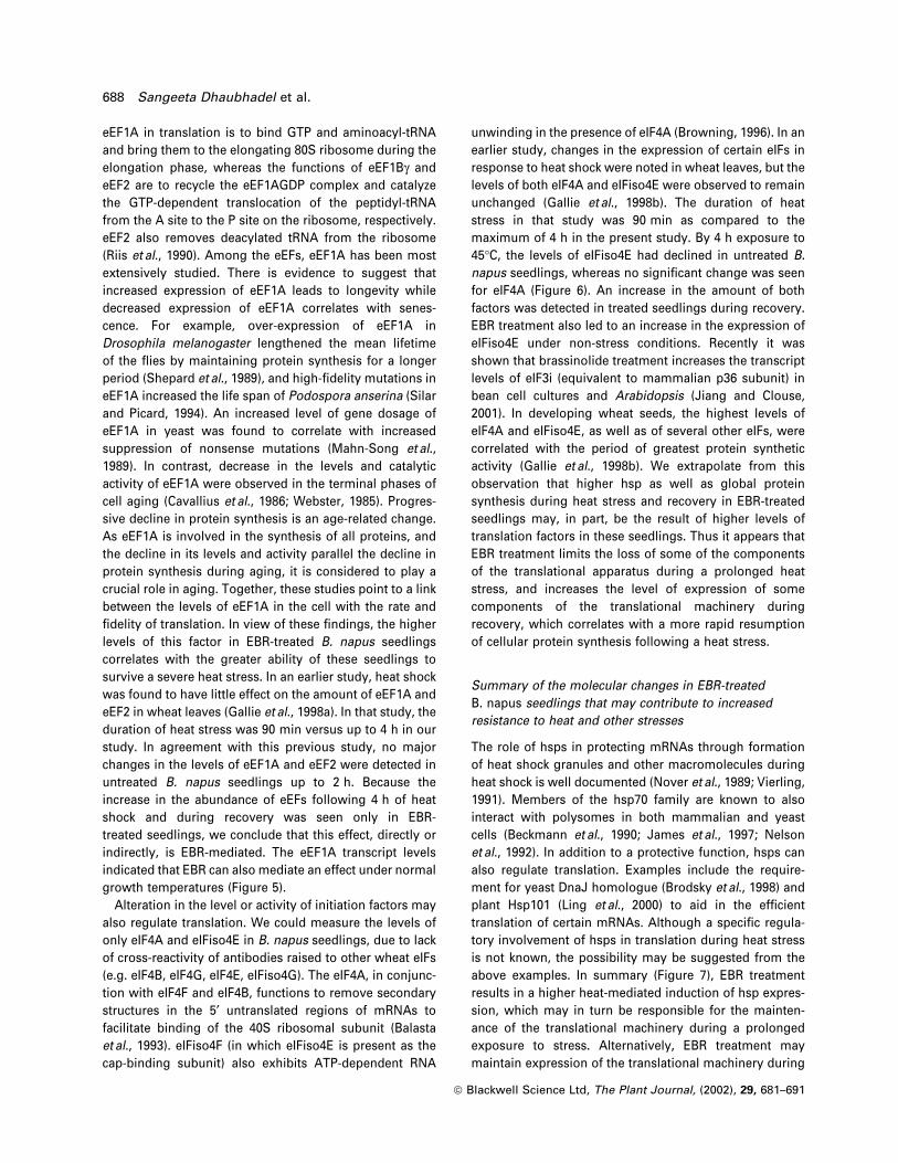
Figure 4. Accumulation of translation-elongation factors in untreated andEBR-treated Brassica napus seedlings during heat stress and recovery.Seedlings grown in the absence (C) or presence (E) of 1 mM EBR wereexposed to 45°C for 1, 2, 3 or 4 h. Seedlings exposed to 45°C for 4 hwere allowed to recover at 20°C for 6, 19 or 24 h. Total proteins (30 mg)were separated on a 10% SDS±polyacrylmide gel and transferred ontonitrocellulose membrane by electroblotting. The eEFs were detected bysequential incubation of the membrane with anti-eEF1A, eEF1Bg andeEF2 antibodies and peroxidase-conjugated anti-rabbit IgG followed bythe chemiluminescent reaction. Arrows on the right indicate puri®edwheat proteins (P) against which antibodies were raised. An anti-cpn60-bantibody was used to detect cpn60-b as a loading control.
Brassinosteroid and thermotolerance 685
ã Blackwell Science Ltd, The Plant Journal, (2002), 29, 681±691

treated seedlings. Thus the high levels of eIFs and eEFs in
EBR-treated seedlings are not a result of global changes to
cellular proteins in these seedlings, but rather are the
result of a speci®c event(s) occurring during the heat-
stress response in the presence of EBR.
Discussion
The results of this study demonstrate that, in comparison
to untreated seedlings, EBR-treated B. napus seedlings
have signi®cantly higher translational activity during and
following exposure to heat stress, and that this difference
in protein synthetic activity is accompanied by dramatic
differences in the expression level of several translation
factors in the two sets of seedlings. Earlier we had
observed that the transcript levels of hsp100, hsp90,
hsp70 and low molecular weight hsps are higher in heat-
stressed EBR-treated seedlings than in untreated seed-
lings, but during recovery the opposite is observed.
Nevertheless, hsps accumulate to higher levels in treated
than in untreated seedlings during and after exposure to
heat stress (Dhaubhadel et al., 1999). The higher level of
hsps in EBR-treated seedlings may be due to increased
synthesis and/or increased stability.
Hsp synthesis in vivo does not correlate with the level of
polysome-associated hsp mRNAs
To understand the regulation of hsp synthesis in EBR-
treated seedlings, newly synthesized proteins were labeled
with [35S]. Figure 1 shows that hsp synthesis in treated
seedlings was signi®cantly higher than in untreated seed-
lings, not only during stress but also during recovery. The
apparent difference in hsp synthesis between the two sets
of seedlings during the recovery period may have resulted
from either preferential recruitment of hsp mRNAs onto
polysomes in EBR-treated seedlings, the translational
arrest of polysome-associated hsp mRNAs in untreated
seedlings, or differences in the activity of the translational
machinery between the two sets of seedlings. To distin-
guish between these possibilities, the levels of polysome-
associated mRNAs were determined for a class II shsp and
hsp90 during and after heat stress. The overall pattern of
polysome-associated hsp mRNAs (Figure 2) correlated
with steady-state hsp mRNA levels observed in our
previous study (Dhaubhadel et al., 1999). For instance,
hsp mRNA levels were higher in untreated seedlings than
in EBR-treated seedlings during recovery (Dhaubhadel
et al., 1999), and so was their association with polysomes
(Figure 2). Surprisingly, the pattern of hsp synthesis did
not correlate with the level of RNA observed. A consider-
able amount of hsp mRNA was also detected in the
supernatant fraction, indicating that not all hsp mRNA was
loaded onto polysomes during heat stress. This is not
unusual ± it has been noted previously that carrot callus
cells accumulate hsp17.7 mRNA to a high level in response
to heat shock, but recruit only a small fraction of the mRNA
into polysomes (Apuya and Zimmerman, 1992). A charac-
teristic feature of the plant heat-stress response is the
translational competence of hsp mRNAs in a cellular
environment in which non-hsp mRNAs are subject to
translational repression. Non-hsp mRNAs may become
sequestered in heat-shock granules, degraded, or a pro-
portion may remain bound to polysomes (Apuya and
Zimmerman, 1992). The presence of rubisco mRNA in the
polysome fraction indicates that a portion of it remains
bound to ribosomes. The decline in the level of rubisco
mRNA during prolonged heat stress probably results from
its degradation, and its increase during the recovery period
may result from an increase in its stability and/or synthe-
sis. While understanding the mechanism governing
rubisco mRNA sequestration/availability during heat stress
is not the focus of this work, the pattern of rubisco mRNA
Figure 6. Accumulation of translational initiation factors in untreated andEBR-treated Brassica napus seedlings during heat stress and recovery.Seedlings grown in the absence (C) or presence (E) of 1 mM EBR wereexposed to 45°C for 1, 2, 3 or 4 h. Seedlings exposed to 45°C for 4 hwere allowed to recover at 20°C for 6, 19 or 24 h. Total proteins (30 mg)were separated by SDS±polyacrylamide gel and transferred tonitrocellulose membrane by electroblotting. The initiation factors weredetected by sequential incubation of the membrane with anti-eIF4A andeIFiso4E antibodies and peroxidase-conjugated anti-rabbit IgG followedby the chemiluminescent reaction. Arrows on the right indicate puri®edwheat proteins (P) against which antibodies were raised. Cpn60-b wasdetected as a loading control.
Figure 5. Accumulation of eEF1A transcripts in untreated and EBR-treated Brassica napus seedlings during heat stress and recovery.Seedlings grown in the absence (C) or presence (E) of 1 mM EBR wereexposed to 45°C for 1, 2, 3 or 4 h. Seedlings exposed to 45°C for 4 hwere allowed to recover at 20°C for 6, 19 or 24 h. Total RNA (15 mg) wasseparated on a denaturing formaldehyde agarose gel, blotted onto amembrane and hybridized with [32P]-labeled eEF1A gene fragment. Thesame blot, after removal of the probe, was hybridized with [32P]-labeled18S rRNA gene fragment to serve as loading control.
686 Sangeeta Dhaubhadel et al.
ã Blackwell Science Ltd, The Plant Journal, (2002), 29, 681±691

expression and recruitment onto polysomes con®rms
ef®cient recovery of EBR-treated seedlings from heat
stress and damaging effects of heat stress in untreated
seedlings.
Translation is inhibited in untreated seedlings following
heat stress
Translation of isolated polysomes was carried out in vitro
in order to gain further insight into the mechanism
governing hsp synthesis in EBR-treated seedlings.
Translation of intact polysomes isolated from treated and
untreated seedlings during heat shock demonstrated that
hsp mRNAs predominated in the polysomal fraction
(Figure 3c). The failure to observe signi®cant expression
of non-heat-shock protein synthesis during the in vitro
translation of polysomes isolated from heat-stressed
seedlings is consistent with the observation that heat
shock causes the disassembly of most, if not all, non-heat-
shock polysomes and the preferential recruitment of heat-
shock mRNAs to polysomes (Key et al., 1981).
In contrast to the substantially higher level of hsp
expression observed in vivo in EBR-treated seedlings
(Figure 1), the in vitro translation of polysomes isolated
from treated and untreated seedlings during a heat shock
yielded similar levels of hsps, particularly following a 3 h
exposure to the stress (Figure 3c). Translation of poly-
somes in the presence of m7GMP also revealed similar
levels of translation products in both sets of seedlings.
m7GMP inhibits new cap-dependent initiation, but allows
translational run-off from the polysomes using elongation
factors from the wheatgerm lysate. This accounts for the
difference in the level of translation in the absence (re-
initiation and elongation can take place) and presence
(attached ribosomes complete elongation but cannot
reinitiate translation) of m7GMP. Results presented in
Figure 3(c) indicate that polysomes isolated from the two
sets of seedlings during a heat stress were of similar size,
suggesting that the difference between in vivo and in vitro
hsp expression between the two sets of seedlings was not
a consequence of differential ribosome loading that would
occur as a result of differences in translational initiation.
The failure of untreated, heat-stressed seedlings to
synthesize hsps in vivo to the level observed for EBR-
treated, heat-stressed seedlings may be a result of inhibi-
tion of translation elongation. Such inhibition may result
from reduced activity of one or more of the eEFs during a
short exposure to a heat stress or a reduction in their level,
as was observed for a prolonged exposure to the stress
(Figure 4). Under such a scenario, inhibition of elongation
would be reversible, as polysomes isolated from untreated
seedlings yielded a similar level of protein products as
those isolated from EBR-treated seedlings when translated
in wheatgerm lysate (Figure 3c). Inhibition of translation
elongation might also have been achieved through the
association of a factor that reduced the rate of elongation
in the untreated seedlings. An alternative explanation for
the higher level of hsp expression in EBR-treated seedlings
in vivo (Figure 1) might be that hsp70 and hsp90 proteins,
in particular, are more stable in these seedlings during a
heat stress than in untreated seedlings. This possibility is
supported by the ratio of the abundance of these two
proteins relative to other proteins synthesized in vivo
(Figure 1) compared to the same ratio observed when the
polysomes were translated in vitro (Figure 3c).
During recovery, a higher level of protein synthesis was
observed in vivo in EBR-treated seedlings than in
untreated seedlings (Figure 1). When polysomes isolated
from both sets of seedlings during their recovery from a
heat shock were translated in vitro, those isolated from
EBR-treated seedlings yielded a higher level of protein
products than did those isolated from untreated seedlings
(Figure 3c). These observations suggest that polysomes
from treated seedlings were larger (more ribosomes per
polysome) than those of untreated seedlings, resulting in a
higher yield of translational run-off products during in vitro
translation. Larger polysomes may have resulted from a
higher rate of translation initiation in EBR-treated recover-
ing seedlings, consistent with the observation of higher
levels of eIFs in these seedlings (Figure 6). A difference in
polysome size may also have resulted from an acute
inhibition of elongation in untreated seedlings during
recovery, a possibility which is supported by the substan-
tial reduction in the level of elongation factors in the
untreated seedlings (Figure 4). Inhibition of elongation
following formation of the 80S ribosome at an initiation
codon would inhibit further ribosome loading and thus
reduce polysome size. An increase in translational initi-
ation in EBR-treated seedlings and an inhibition of elonga-
tion, as well as re-initiation in untreated seedlings during
recovery, are not mutually exclusive.
EBR treatment leads to an increase in the expression of
translation elongation and initiation factors
The translational machinery in plants is modi®ed in
response to thermal stress through changes in their
abundance and phosphorylation state (Gallie et al., 1997;
Gallie et al., 1998a; Gallie et al., 1998b). To identify factors
involved in translation that may be responsible for the
differential protein synthesis in untreated and EBR-treated
seedlings, we compared the levels of eIFs and eEFs in
untreated and EBR-treated seedlings before, during and
after heat stress. Dramatic differences in the levels of the
eEFs examined were observed between the two sets of
seedlings following exposure to heat stress, although
differences to a lesser extent were also observed before
and during heat stress (Figure 4). The main function of
Brassinosteroid and thermotolerance 687
ã Blackwell Science Ltd, The Plant Journal, (2002), 29, 681±691
eEF1A in translation is to bind GTP and aminoacyl-tRNA
and bring them to the elongating 80S ribosome during the
elongation phase, whereas the functions of eEF1Bg and
eEF2 are to recycle the eEF1AGDP complex and catalyze
the GTP-dependent translocation of the peptidyl-tRNA
from the A site to the P site on the ribosome, respectively.
eEF2 also removes deacylated tRNA from the ribosome
(Riis et al., 1990). Among the eEFs, eEF1A has been most
extensively studied. There is evidence to suggest that
increased expression of eEF1A leads to longevity while
decreased expression of eEF1A correlates with senes-
cence. For example, over-expression of eEF1A in
Drosophila melanogaster lengthened the mean lifetime
of the ¯ies by maintaining protein synthesis for a longer
period (Shepard et al., 1989), and high-®delity mutations in
eEF1A increased the life span of Podospora anserina (Silar
and Picard, 1994). An increased level of gene dosage of
eEF1A in yeast was found to correlate with increased
suppression of nonsense mutations (Mahn-Song et al.,
1989). In contrast, decrease in the levels and catalytic
activity of eEF1A were observed in the terminal phases of
cell aging (Cavallius et al., 1986; Webster, 1985). Progres-
sive decline in protein synthesis is an age-related change.
As eEF1A is involved in the synthesis of all proteins, and
the decline in its levels and activity parallel the decline in
protein synthesis during aging, it is considered to play a
crucial role in aging. Together, these studies point to a link
between the levels of eEF1A in the cell with the rate and
®delity of translation. In view of these ®ndings, the higher
levels of this factor in EBR-treated B. napus seedlings
correlates with the greater ability of these seedlings to
survive a severe heat stress. In an earlier study, heat shock
was found to have little effect on the amount of eEF1A and
eEF2 in wheat leaves (Gallie et al., 1998a). In that study, the
duration of heat stress was 90 min versus up to 4 h in our
study. In agreement with this previous study, no major
changes in the levels of eEF1A and eEF2 were detected in
untreated B. napus seedlings up to 2 h. Because the
increase in the abundance of eEFs following 4 h of heat
shock and during recovery was seen only in EBR-
treated seedlings, we conclude that this effect, directly or
indirectly, is EBR-mediated. The eEF1A transcript levels
indicated that EBR can also mediate an effect under normal
growth temperatures (Figure 5).
Alteration in the level or activity of initiation factors may
also regulate translation. We could measure the levels of
only eIF4A and eIFiso4E in B. napus seedlings, due to lack
of cross-reactivity of antibodies raised to other wheat eIFs
(e.g. eIF4B, eIF4G, eIF4E, eIFiso4G). The eIF4A, in conjunc-
tion with eIF4F and eIF4B, functions to remove secondary
structures in the 5¢ untranslated regions of mRNAs to
facilitate binding of the 40S ribosomal subunit (Balasta
et al., 1993). eIFiso4F (in which eIFiso4E is present as the
cap-binding subunit) also exhibits ATP-dependent RNA
unwinding in the presence of eIF4A (Browning, 1996). In an
earlier study, changes in the expression of certain eIFs in
response to heat shock were noted in wheat leaves, but the
levels of both eIF4A and eIFiso4E were observed to remain
unchanged (Gallie et al., 1998b). The duration of heat
stress in that study was 90 min as compared to the
maximum of 4 h in the present study. By 4 h exposure to
45°C, the levels of eIFiso4E had declined in untreated B.
napus seedlings, whereas no signi®cant change was seen
for eIF4A (Figure 6). An increase in the amount of both
factors was detected in treated seedlings during recovery.
EBR treatment also led to an increase in the expression of
eIFiso4E under non-stress conditions. Recently it was
shown that brassinolide treatment increases the transcript
levels of eIF3i (equivalent to mammalian p36 subunit) in
bean cell cultures and Arabidopsis (Jiang and Clouse,
2001). In developing wheat seeds, the highest levels of
eIF4A and eIFiso4E, as well as of several other eIFs, were
correlated with the period of greatest protein synthetic
activity (Gallie et al., 1998b). We extrapolate from this
observation that higher hsp as well as global protein
synthesis during heat stress and recovery in EBR-treated
seedlings may, in part, be the result of higher levels of
translation factors in these seedlings. Thus it appears that
EBR treatment limits the loss of some of the components
of the translational apparatus during a prolonged heat
stress, and increases the level of expression of some
components of the translational machinery during
recovery, which correlates with a more rapid resumption
of cellular protein synthesis following a heat stress.
Summary of the molecular changes in EBR-treated
B. napus seedlings that may contribute to increased
resistance to heat and other stresses
The role of hsps in protecting mRNAs through formation
of heat shock granules and other macromolecules during
heat shock is well documented (Nover et al., 1989; Vierling,
1991). Members of the hsp70 family are known to also
interact with polysomes in both mammalian and yeast
cells (Beckmann et al., 1990; James et al., 1997; Nelson
et al., 1992). In addition to a protective function, hsps can
also regulate translation. Examples include the require-
ment for yeast DnaJ homologue (Brodsky et al., 1998) and
plant Hsp101 (Ling et al., 2000) to aid in the ef®cient
translation of certain mRNAs. Although a speci®c regula-
tory involvement of hsps in translation during heat stress
is not known, the possibility may be suggested from the
above examples. In summary (Figure 7), EBR treatment
results in a higher heat-mediated induction of hsp expres-
sion, which may in turn be responsible for the mainten-
ance of the translational machinery during a prolonged
exposure to stress. Alternatively, EBR treatment may
maintain expression of the translational machinery during
688 Sangeeta Dhaubhadel et al.
ã Blackwell Science Ltd, The Plant Journal, (2002), 29, 681±691

a heat stress, which might result in higher hsp expression,
two possibilities that are not mutually exclusive. Although
higher levels of hsps must contribute to increased
thermotolerance in EBR-treated seedlings, using differen-
tial display we sought to ®nd factors other than hsps that
may directly or indirectly contribute to EBR-mediated
increase in stress tolerance. Four cDNAs characterized
thus far that were upregulated in treated seedlings encode
3-ketoacyl CoA thiolase, myrosinase, glycine-rich protein
22, and a hypothetical protein (S. Dhaubhadel & P.
Krishna, unpublished results). The thiolase transcript
levels were higher in treated seedlings as compared to
untreated seedlings during heat stress, but transcripts of
the other three cDNAs were present at higher levels in
treated seedlings prior to any stress. Higher expression of
3-ketoacyl thiolase, myrosinase and glycine-rich protein 22
can be linked, at least hypothetically, to an increase in the
general stress resistance of plants. The results of the
present study, coupled with increased expression of genes
involved in a variety of physiological responses (S.
Dhaubhadel & P. Krishna, unpublished results) and other
as-yet unidenti®ed factors in treated seedlings, may con-
tribute to increased overall stress tolerance in these
seedlings (Figure 7).
Experimental procedures
Plant material and growth conditions
Plant material, growth conditions and heat stress were asdescribed previously (Dhaubhadel et al., 1999).
In vivo protein labeling
Brassica napus seedlings were grown for 14 days in liquidmedium supplemented with either 1 mM EBR or 0.01% ethanol(control). At appropriate times, Trans [35S] Label (ICN Biomedical,
Aurora, OH, USA) was added to the medium at a concentration of40 mCi ml±1. The labeling was continued for 1 h, following whichplant tissue above the medium was collected and quick-frozen inliquid nitrogen. Protein extraction and quanti®cation were carriedout as described previously (Dhaubhadel et al., 1999). Totalsoluble proteins of each sample were separated on SDS±polyacrylamide gels which were stained with Coomassie blueR-250, destained and ¯uorographed.
Polysome isolation
Polysomes were isolated according to the procedure of Bailey-Serres and Freeling (1990) with some modi®cations. Frozen planttissue was ground to powder under liquid nitrogen using a mortarand pestle. Powdered tissues were immediately suspended inpolysome extraction buffer (0.2 M Tris±HCl pH 9.0, 0.4 M KCl,25 mM EGTA, 35 mM MgCl2, 0.2 M sucrose, 15 mM b-mercapto-ethanol). All manipulations were carried out on ice or at 4°C. Theextract was homogenized for 30 sec using a polytron homo-genizer following the addition of Triton X-100 to a ®nal concen-tration of 1%. The extract was incubated on ice for 15 min withoccasional mixing, then centrifuged at 30 000 g for 20 min. Thesupernatant was layered over a 3 ml sucrose cushion (1.75 M
sucrose, 40 mM Tris±HCl pH 9.0, 0.2 M KCl, 5 mM EGTA,30 mM MgCl2, 7 mM b-mercaptoethanol), and centrifuged at173 000 3 g in a Type 90 Ti rotor (Beckman, Coulter,Mississauga, ON, USA) at 4°C for 4.5 h in an ultracentrifuge.The polysome pellet was resuspended in resuspension buffer(40 mM Tris±HCl pH 8.5) and incubated on ice for 30 min. Theresuspended polysome preparations were clari®ed by centrifuga-tion at 3500 3 g in a microcentrifuge for 1 min at 4°C to discardany undissolved material. The polysomal RNA was quanti®ed bymeasuring the absorbance at 260 nm.
RNA isolation and Northern blotting
The resuspended polysomes were extracted twice with 0.5 volphenol : chloroform, then precipitated with 3 M sodium acetate(pH 5.2) and absolute ethanol. The RNA pellet was recovered bycentrifugation at 16 000 3 g for 20 min at 4°C in a micro-centrifuge. To isolate RNA from supernatant, the supernatantwas diluted 2 : 1 in DEPC-treated water and processed in thesame way as polysomal pellet RNA. Northern blotting (Figure 2)was carried out as described previously (Dhaubhadel et al., 1999).
Total RNA was isolated from frozen plant tissue according toGlisin et al. (1974) and analyzed by Northern blotting (Figure 5).Following hybridization with radiolabeled eEF1A gene fragment,the membrane was washed twice at room temperature for 15 minwith 1 3 SSC, 0.1% SDS, then once with 0.25 3 SSC, 0.1% SDS at55°C for 5 min, followed by autoradiography. The same blot afterstripping was hybridized with an 18S ribosomal DNA fragment toserve as loading control. For generation of eEF1A gene fragment,RT±PCR was carried out using RNA isolated from EBR-treatedseedlings grown at 20°C for 14 days. Reverse transcription wascarried out using random hexamers. Conserved sequences, F (5¢-GCTGAGATGAACAAGAGGTC-3¢) and R (5¢-CAACAGTCTGCC-TCATGTC-3¢), which were identi®ed by alignment of eEF1Agenes from wheat (accession no. M90077), tomato (accessionno. X14449), barley (accession no. Z23130) and Arabidopsis(accession no. X16430), were used as primers for PCR. The PCRconditions were as follows: 95°C for 1 min, 52°C for 30 sec and72°C for 2 min (35 cycles). The PCR product was cloned into theTA cloning vector and sequenced at the Robart SequencingFacility, London, Ontario.
Figure 7. Summary of the molecular changes in EBR-treated Brassicanapus seedlings that may contribute to increased resistance to heat andother stresses.Photograph of the seedlings at 7 days' recovery at 20°C after exposure to45°C for 4 h, reproduced from Dhaubhadel et al. (1999).
Brassinosteroid and thermotolerance 689
ã Blackwell Science Ltd, The Plant Journal, (2002), 29, 681±691

In vitro translation of polysomes
For translation of polysomes in vitro, 8 mg resuspended polysomeswere incubated with 12.5 ml wheatgerm extract (Promega,Madison, WI, USA) supplemented with 80 mM amino acid mixtureminus methionine, 67 mM potassium acetate, 40 units RNAsinRibonuclease inhibitor and 1.25 ml [35S]methionine (1000 Cimmol±1, Amersham Biosciences, Baie d'Urfe , QC). The ®nalvolume was made up to 25 ml with sterile distilled water. Whererequired, the reaction mixtures were supplemented either with200 mM m7GMP to block translational initiation, or with an equiva-lent volume of water (controls). The translation reactions wereincubated at 26°C for 60 min, and the results were analyzed byelectrophoresis on a 10% SDS±polyacrylamide gel followed bystaining, destaining and ¯uorography. Control reactions usingBMV RNA, deproteinized B. napus RNA, or no RNA were alsocarried out.
Analysis of translational initiation and elongation factors
by Western blot analysis
Total soluble proteins of each sample were separated on SDS±polyacrylamide gels and transferred to nitrocellulose membranesas described previously (Dhaubhadel et al., 1999). The blots wereprobed separately with anti-eEF1A, eEF1Bg, eEF2, eIF4A, eIFiso4Eand cpn60-b antibodies, each at a dilution of 1 : 1000, followed byincubation with peroxidase-conjugated anti-rabbit IgG (1 : 5000)and chemiluminescent reaction (ECL System, AmershamBiosciences, Baie d'Urfe , QC).
Acknowledgements
This research was supported by research grants to P.K. from DowElanco and the Natural Sciences and Engineering ResearchCouncil of Canada. S.D. was a recipient of the Ontario GraduateScholarship.
References
Apuya, N.R. and Zimmerman, J.L. (1992) Heat shock geneexpression is controlled primarily at the translational level incarrot cells and somatic embryos. Plant Cell. 4, 657±665.
Bailey-Serres, J. and Freeling, M. (1990) Hypoxic stress-inducedchanges in ribosomes of maize seedling roots. Plant Physiol.94, 1237±1243.
Balasta, M.L., Carberry, S.E., Friedland, D.E., Perez, R.A. and Goss,D.J. (1993) Characterization of the ATP-dependent binding ofwheat germ protein synthesis initiation factors eIF-(iso) 4F andeIF4A to mRNA. J. Biol. Chem. 268, 18599±18603.
Beckmann, R.P., Mizzen, L.E. and Welch, W.J. (1990) Interaction ofhsp70 with newly synthesized proteins: implications for proteinfolding and assembly. Science, 248, 850±854.
Berry, J.O., Carr, J.P. and Klessig, D.F. (1988) mRNAs encodingriboluse 1,5-bisphosphate carboxylase remain bound topolysomes but are not translated in amaranth seedlingstransferred to darkness. Proc. Natl Acad. Sci. USA, 85, 4190±4194.
Bienz, M. and Gurdon, J.B. (1982) The heat-shock response inXenopus oocytes is controlled at the translational level. Cell, 29,811±819.
Brodsky, J.L., Lawrence, J.G. and Caplan, A.J. (1998) Mutations inthe cytosolic DnaJ homologue, YDJ1, delay and compromise
the ef®cient translation of heterologous proteins in yeast.Biochemistry, 37, 18045±18055.
Browning, K.S. (1996) The plant translational apparatus. PlantMol. Biol. 32, 107±144.
Browning, K.S., Humphreys, J., Hobbs, W., Smith, G.B. and Ravel,J.M. (1990) Determination of the amounts of the proteinsynthesis initiation and elongation factors in wheat germ. J.Biol. Chem. 265, 17967±17973.
Cavallius, J., Rattan, S.I.S. and Clark, B.F.K. (1986) Changes inactivity and amount of active elongation factor 1a in aging andimmortal human ®broblast cultures. Exp. Gerentol. 21, 149±157.
Clouse, S.D. and Sasse, J.M. (1998) Brassinosteroids: essentialregulators of plant growth and development. Annu. Rev. PlantPhysiol. Plant Mol. Biol. 49, 427±451.
Crosby, J.S. and Vayda, M.E. (1991) Stress-induced translationalcontrol in potato tubers may be mediated by polysome-associated proteins. Plant Cell, 3, 1013±1023.
Davies, E. (1993) Intercellular and intracellular signals and theirtransduction via the plasma membrane cytoskeleton interface.Seminars Cell Biol. 4, 139±147.
Dhaubhadel, S., Chaudhary, S., Dobinson, K.F. and Krishna, P.(1999) Treatment with 24-epibrassinolide, a brassinosteroid,increases the basic thermotolerance of Brassica napus andtomato seedlings. Plant Mol. Biol. 40, 333±342.
Gallie, D.R., Le, H., Caldwell, C., Tanguay, R.L., Hoang, N.X. andBrowing, K.S. (1997) The phosphorylation state of translationinitiation factors is regulated developmentally and followingheat shock in wheat. J. Biol. Chem. 272, 1046±1053.
Gallie, D.R., Le, H., Caldwell, C. and Browing, K.S. (1998a)Analysis of translation elongation factors from wheat duringdevelopment and following heat shock. Biochem. Biophys. Res.Commu. 245, 295±300.
Gallie, D.R., Le, H., Tanguay, R.L. and Browning, K.S. (1998b)Translation initiation factors are differentially regulated incereals during development and following heat shock. PlantJ. 14, 715±722.
Glisin, V., Crkvenjakov, R. and Byus, C. (1974) Ribonucleic acidisolated by cesium chloride centrifugation. Biochemistry, 13,2633±2637.
James, P., Pfund, C. and Criag, E.A. (1997) Functional speci®cityamong hsp70 molecular chaperones. Science, 275, 387±389.
Jiang, J. and Clouse, S.D. (2001) Expression of a plant gene withsequence similarity to animal TGF-b receptor interactingprotein is regulated by brassinosteroids and required fornormal plant development. Plant J. 26, 35±45.
Key, J.L., Lin, C.Y. and Chen, Y.M. (1981) Heat shock proteins ofhigher plants. Proc. Natl Acad. Sci. USA, 78, 3526±3530.
Khripach, V., Zhabinskii, V. and de Groot, A.D. (2000) Twentyyears of brassinosteroids: steroidal plant hormones warrantbetter crops for the XXI century. Ann. Bot. 86, 441±447.
Kirk, M.M. and Kirk, D.L. (1985) Translational regulation of proteinsynthesis in response to light at a critical stage of Volvoxdevelopment. Cell, 41, 419±428.
Kulaeva, O.N., Burkhanova, E.A., Fedina, A.B., Khokhlova, V.A.,Bokebayeva, G.A., Vorbrobt, H.M. and Adam, G. (1991) Effect ofbrassinosteroids on protein synthesis and plant cellultrastructure under stress conditions. In: Brassinosteroids(Culter, H.G., Yokota, T. and Adam, G., eds). Washington, DC:American Chemical Society, pp. 141±155.
Li, J. and Chory, J. (1997) A putative leucine-rich repeat receptorkinase involved in brassinosteroid signal transduction. Cell, 90,929±938.
Li, J. and Chory, J. (1999) Brassinosteroid actions in plants. J. Exp.Bot. 50, 275±282.
690 Sangeeta Dhaubhadel et al.
ã Blackwell Science Ltd, The Plant Journal, (2002), 29, 681±691

Ling, J., Wells, D.R., Tanguay, R.L., Dickey, L.F., Thompson, W.F.and Gallie, D.R. (2000) Heat shock protein Hsp101 binds to theFed-1 internal light regulatory element and mediates its hightranslational activity. Plant Cell. 12, 1213±1228.
Mandava, N.B. (1988) Plant growth-promoting brassinosteroids.Ann. Rev. Plant Physiol. Plant Mol. Biol. 39, 23±52.
Mussig, C. and Altmann, T. (1999) Physiology and molecularmode of action of brassinosteroids. Plant Physiol. Biochem. 37,363±372.
Nelson, R.J., Ziegelhoffer, T., Nicolet, C., Werner-Washburne, M.and Criag, E.A. (1992) The translation machinery and 70 kDheat shock protein cooperate in protein synthesis. Cell, 71, 97±105.
Nover, L., Scharf, K.D. and Neumann, D. (1989) Cytoplasmic heatshock granules are formed from precursor particles and areassociated with a speci®c set of mRNAs. Mol. Cell Biol. 9, 1298±1308.
Parsell, D.A. and Lindquist, S. (1993) The functions of heat-shockproteins in stress tolerance: degradation and reactivation ofdamaged proteins. Annu. Rev. Genet. 27, 437±496.
Polasky, A.R., Hiatt, W.R., Ridge, N., Rasmussen, R., Houck, C.M.and Shewmaker, C.K. (1989) Structure and expression ofelongation factor 1a in tomato. Nucl. Acids Res. 17, 4661±4673.
Quijada, L., Soto, M., Alonso, C. and Requena, J.M. (1997)Analysis of post-transcriptional regulation operating ontranscription products of the tandemly linked Leishmaniainfantum hsp70 genes. J. Biol. Chem. 272, 4493±4499.
Riis, B., Rattan, S.I., Clark, B.F. and Merrick, W.C. (1990)
Eukaryotic protein elongation factors. Trends Biochem. Sci.15, 420±424.
Shepherd, J.C.W., Walldorf, U., Hug, P. and Gehring, W.J. (1989)Fruit ¯ies with additional expression of the elongation factorEF-1a live longer. Proc. Natl Acad. Sci. USA, 86, 7520±7521.
Silar, P. and Picard, M. (1994) Increased longevity of EF-1a high-®delity mutants in Podospora anserina. J. Mol. Biol. 235, 231±236.
Song, J.M., Picologlou, S., Grant, C.M., Firoozan, M., Tuite, M.F.and Liebman, S. (1989) Elongation factor EF-1a gene dosagealters translational ®delity in Saccharomyces cerevisiae. Mol.Cell Biol. 9, 4571±4575.
Taliercio, E.W. and Chourey, P.S. (1989) Post-translational controlof sucrose synthase expression in anaerobic seedlings ofmaize. Plant Physiol. 90, 1359±1364.
Vierling, E. (1991) The roles of heat shock proteins in plants.Annu. Rev. Plant Physiol. Plant Mol. Biol. 42, 579±620.
Wang, Z.Y., Seto, H., Fujioka, S., Yoshida, S. and Chory, J. (2001)BRI1 is a critical component of a plasma-membrane receptor forplant steroids. Nature, 410, 380±383.
Webster, G.C. (1985) Protein synthesis in aging organisms. In:Molecular Biology of Aging: Gene Stability and GeneExpression (Sohal, R.S., ed.). New York, NY: Raven Press, pp.263±289.
Wilen, R.W., Sacco, M., Gusta, L.V. and Krishna, P. (1995) Effectsof 24-epibrassinolide on freezing and thermotolerance ofbromegrass (Bromus inermis) cell cultures. Plysiol. Plant. 95,195±202.
Brassinosteroid and thermotolerance 691
ã Blackwell Science Ltd, The Plant Journal, (2002), 29, 681±691
Copyright © 2022 FDOKUMEN