Book: Evolutionary Biology of Primtes
516
EVOLUTIONARY BIOLOGY OF THE PRIMATES: An Introductory Reader Daryl G. Frazetti University of Massachusetts Boston - May 2002 (2004) © Daryl G. Frazetti, 2002 (2004) All Rights Reserved “There is something fascinating about science. One gets such wholesale returns of conjecture out of such a trifling investment of fact”. - Mark Twain, Life on the Mississippi, 1874
-
Upload
independent -
Category
Documents
-
view
2 -
download
0
Transcript of Book: Evolutionary Biology of Primtes

EVOLUTIONARY BIOLOGY OF THE PRIMATES: AnIntroductory Reader
Daryl G. FrazettiUniversity of Massachusetts Boston - May 2002 (2004)
© Daryl G. Frazetti, 2002 (2004) All Rights Reserved
“There is something fascinating about science. One gets such wholesale returns of conjecture out of such a trifling investment of fact”. - Mark Twain, Life on the Mississippi, 1874
Description:
Evolution of the Order Primates can be traced back to the early Tertiary period about 65 million years ago. The primates are considered to have developed as an offshoot of a group of small, nocturnal, insectivorous mammals known as tree shrews. A tremendous amount of work has been done on primate evolution, anatomy and physiology, contributing greatly to the overall understanding of primate origins, taxonomy, and phylogeny. Many factors, such as climate, environment, competition, diet, and geographical/behavioral isolation factors, have played significant roles in the origins, biological and physiological evolution of primates. Such factors also account for adaptation,s which can be traced in the fossil record, the loss or retention of ancestral (primitive) traits in some primate species, and in particular for speciation events which led to the divergences of lineages from the earliest protoprimates to anatomically modern humans.
Special thanks and appreciation to the following individuals and departments who assisted in guiding me through the original paper as well as the challenging task of creating this work:
Dr. Michael F. Gibbons, Jr of the University of Massachusetts at Boston for his advice, guidance exceptional sense of humor, and phenomenal patience.Ellen Royalty , the most creative, perspicacious and patient reference librarian..Brian Butler, Dave Ford and the Northern Illinois University graduate computerlab for so much time in guiding and assisting with the layout, software, and final printing. And as always….. my “boys” Jasper and Junior.
"The more complex the mind, the greater the need for the simplicity of play." –Captain Kirk, Shore Leave

Table of Contents
1. Introduction
2. Evolution and Origin of the Primates
Models of the Evolutionary Process Arborealism Continental Drift Rooneyia Primate Origins Tree Shrews Early Primates Parapathecidae Catarrhines Origins of Platyrrhines 3. Hominoids
Miocene Radiation of the Apes Miocene Paleoecology Lothagam Mandible Proconsul Sivapithecus Dryopithecus Ramapithecus

Aegyptopithecus Gigantopithecus Oreopithecus Afropithecus Morotopithecus Early Bipedalism Early Speech ( Vocalization)
4. Hominids
Single Species : Evolutionary Scenarios Pylogenetics and Cladistics Anatomical Variance Taxonomy and Variation Sexual Dimorphism Ontogenetic variation Racial Variation Polymorphic Variation Pathologic Variation Temporal Variation Taphonomy
5. The Australopithecines
The Pliocene Apes and Humans Australopithecus africanus The africanus pattern Robust Australopithecines The Robust Pattern Controversies: Homo erectus and How many Genera Homo erectus Homo sapiens Phylogeny

6. The Neandertals
The Climatic Setting of the Pleistocene Variation and Adaptation Theoretical Models for the Evolution of the Genus Homo Dehydroepiandrosterone and Hominid Evolution Final Note on Neandertals
7. Comparative Anatomy and Osteology of Extant Primates
Overview of Primate Anatomy Dentition Skeletal Elements The Jaw Vertebral Column Thorax Pectoral Girdle Arms Hands Pelvic Girdle Legs Foot Skeletal Maturation Locomotor Anatomy Primate Vision Overview of Primates and Primate Identification
8. Applicable Identification Techniques
Estimating the Age of an Individual Estimating the Gender of an Individual Estimating the Ancestry of an Individual

9. Conclusion
10. Appendices 1. Taxonomy of the Extant Primates and Geographic Ranges 2. Tree Shew Taxonomy 3. Shared characteristics between tree-shrews and primates 4. Primate Range Map ( extants) 5. Geological time chart 7. Identification Based upon Dental Eruption and Developmental Information
8. Classification of Primates 9. Primate Phylogenies 10. Locomotor Anatomy 11. History of the Order: Primates
Introduction
The history of primates is primarily centralized within the
Tertiary period, beginning at about 65 million years ago when

mammalian species began to emerge and diversify. The suborder (or
infraorder) Plesiadapiformes is the first group of primate-like
mammals known in the fossil record. Plesiadapiformes first appear
in the early Paleocene, some 65 million years ago in sediments of
the Western Interior of North America. The relationship of
Paleocene plesiadapiforms to Eocene primates of modern aspect and
relationships among various families, genera, and species of
plesiadapiforms are uncertain. In particular, the relationship of
Microsyopoidea to plesiadapiforms has been questioned. However,
it still stands that plesiadapiformes are the earliest group to
emerge from mammals and begin to diverge into other primate
lineages, ( Herskovitz, 1977; Fleagle et al 1988; Groves, 1989).
The Tertiary period begins with the Paleocene epoch at 65
million years ago and ends at the Pleistocene-Holocene boundary
at about 0.010 million years ago,(Berggren et al, 1995). The
primates are a diverse eutherian ( placental) group with an
extensive life history throughout geological time. Many major
mammal groups evolved in relative isolation on separate
continental landmasses following the separations of the various
continents at various points in time. As continents continued to
move, as ocean levels shifted or as glaciers formed ice-type land
bridges, such formations as isthmus or island (land ) chain
bridges allowed for some interchange, resulting in extinction of
many groups and the divergence of new ones, (Gibbons
1972,1981;Rosen, 1974;Conroy, 1990;Rowe, 1996).
Transitional primate-like creatures, the shrews, first
appeared by the end of the Mesozoic Era ( 65 million years ago)
at the K/T boundary. At that time, the world was very different
from today. The continents were in other locations and they had
somewhat different shapes, there were also fewer of them at that
time. North America was still connected to Europe but not to
South America. India was not yet part of Asia but heading
towards it. Australia was close to Antarctica. Most land masses
had tropical or subtropical climates. The flora and fauna at the
end of the Mesozoic Era would have seemed alien since most of
the plants and animals that are familiar to us had not yet
evolved. Large reptiles were beginning to be replaced by mammals
as the dominant large land animals. Among the mammals, there
were a few archaic egg-layers (monotremes) like the ancestors of
the platypus and echidna. The oldest fossil monotremes come from

the Lightning Ridge opal fields of New South Wales, Australia. An
opalized lower jaw fragment of Steropodon galmani was found to be
more than 100 million years old (middle Albian, Cretaceous),
(Archer et al, 1992). The first marsupials date from about 100
million years ago (Cretaceous), but they are almost certainly a
bit older than that, possibly dating back as far as the Jurassic
(160 million years ago). Whatever the case, there was
diversification underway by the Cretaceous which set the scene
for an explosive radiation in the Tertiary, following the demise
of the dinosaurs. Telling marsupial mammals from the placentals
in the fossil record is difficult. There are lots of soft tissue
and physiological differences but these do not fossilize
especially well. There are dental and jaw differences which are
useful and a specialized epipubic bone which is sometimes found.
Though there were larger numbers of pouched opossum-like mammals
(marsupials) in existence, the first placental mammals did not
appear until around 70 million years ago, (Rosen, 1974; Gibbons,
1972,1981;Quirk et al, 1983; Szalay et al, 1987; Walker et al,
1989; Griffiths et al, 1991;Pasqual et al, 1992). Those early
placental mammals mainly consisted of the transitional primate-

like creatures ( shrews) and their insectivore ancestors. At thi
spoint, the great proliferation of flowering plants had not even
taken place as yet,(Archer et al, 1985; Obradovich, 1993;
Graddstein et al, 1995).
The first primate-like mammals, or proto-primates, were
roughly similar to squirrels and tree shrews in size and
appearance. The existing, fragmentary fossil evidence (mostly
from North Africa) suggests that they were adapted to an arboreal
way of life in warm, moist climates. They probably were equipped
with relatively good eyesight ( early stereoscopic vision
development) as well as hands and feet with pads and claws for
climbing, (Birdsell, 1972;Gibbons, 1972; Simons, 1972; Fleagle,
1999). Morphological and functional studies of dental, cranial,
and postcranial remains of plesiadapiforms suggest retention of
plesiadapiform traits within the order Primates. Four distinct
superfamilies are recognized within Plesiadapiformes.
Microsyopoidea are characterized by the retention of generalized
primitive (relative to Purgatorius) dental and cranial
adaptations. Microsyopoids are represented by two families:
Paleocene Palaechthonidae and Eocene Microsyopidae. Available

evidence indicates that microsyopids are most closely related to
palaechthonids and can best be viewed as having descended from
that Paleocene family, not from the families Mixodectidae or
Leptictidae as previously suggested, (Rosen, 1974;Szalay et al,
1987, Walker et al, 1989;)
The other three superfamilies of Plesiadapiformes are
Plesiadapoidea, Mixodectoidea, and Apatemyoidea. Of these,
plesiadapoids are characterized by specialized dental and cranial
adaptations. Mixodectoids and apatemyoids are distinctly more
primitive than either of the other two superfamilies, remaining
more insectivore-like.Two families, Microsyopidae and
Paromomyidae, survived well into the Eocene, while other
plesiadapiform families disappeared by the earliest Eocene.
Dental characteristics suggest that these two families
specialized on diets different from those of adapids and omomyids
and thus avoided direct competition with primates of modern
aspect. Geographic distributions and climatic reconstructions
indicate that warming temperatures into and through the Eocene
contributed to the extinction of most plesiadapiforms, (Simpson,
1953; Rosen, 1974; Eldredge, 1980; Szalay, 1981; Kimball, 1993;

Amundson, 1996; Fleagle, 1999).
The first true primates were early prosimians that evolved
by the middle of the Paleocene Epoch. Their bones have been
found in 60,000,000 year old geological deposits in North Africa.
However, they are physically different from today’s primate
species. Prosimians were still somewhat squirrel-like in size and
appearance, but apparently they had grasping hands and feet that
were increasingly more efficient in manipulating objects and
climbing trees. It is likely that they were also developing
effective stereoscopic vision. The beginning of the Eocene Epoch
coincides with the appearance of primate species that somewhat
resemble modern prosimians such as lemurs, lorises, and possibly
tarsiers. This was the epoch of maximum prosimian adaptive
radiation. There were at least 60 genera of them in two families
(Adapidae and Omomyidae). This is nearly four times greater than
the prosimian diversity which exists today. Eocene prosimians
lived in North America, Europe, Africa, and Asia. It was during
this epoch that they reached the island of Madagascar. The great
diversity of Eocene prosimians was probably a consequence of the
fact that they did not have competition from monkeys and apes, as

such lineages had not yet begun to diverge. Major evolutionary
changes were beginning in some of the Eocene prosimians that
foreshadow species yet to come. Their brains and eyes were
becoming larger, while their snouts were getting smaller. At the
base of a skull lies the foramen magnum. The position of this
cranial opening, which allows the passage of the spinal column
into the cranium, is a strong indicator of the angle of the
spinal column to the head and subsequently whether the body is
habitually horizontal or vertical . During the Eocene, the
foramen magnum in some primate species was beginning to move from
the back of the skull towards the center. This suggests that they
were beginning to hold their bodies erect while hopping and
sitting, like modern lemurs, galagos, and tarsiers. By the end of
the Eocene Epoch, many of the prosimian species had become
extinct. This may be connected with the appearance of the first
monkeys during the transition to the next geologic epoch, the
Oligocene (35.4 million years ago), (Simpson, 1953; Rosen, 1974;
Groves, 1977; Eldredge, 1980; Szalay, 1981; Bearder et al, 1987;
Kimball, 1993; Amundson, 1996; Anderson, 1998; Fleagle, 1999).
The Oligocene Epoch was largely a gap in the primate fossil
record in most parts of the world. This is especially true for
prosimian fossils. Most of what is known about them came from
the Fayum deposits in Western Egypt. While this area is a desert
today, 36-31 million years ago (during the early and mid
Oligocene) it was a tropical rainforest. Other Oligocene
deposits containing some fossil primate bones have been found in
North and West Africa, the southern Arabian Peninsula, China,
Southeast Asia, as well as North and South America. Monkeys
evolved from prosimians sometime during the early part of the
Oligocene. They were the first species of the suborder
Anthropoidea. Two genera of these early monkeys have been
identified, Apidium and Aegyptopithecus. The former was about
the size of a large squirrel (2-3 pounds), while the latter was
the size of a large domestic cat (13-20 pounds). Both were
probably fruit and seed eating forest tree-dwellers. Compared to
the prosimians, these early monkeys had fewer teeth, less
pronounced muzzles, larger brains, and increasingly more forward-
looking eyes. Due to the comparative scarcity of Oligocene Epoch
prosimians, it is generally believed that the monkeys out-
competed and replaced them in most environments at that time.

Supporting this hypothesis is the fact that modern prosimians
either live in locations where monkeys and apes are absent or
they are normally active only at nighttime when most of the
larger, primates are sleeping, (Rosen, 1974;Szalay et al, 1987,
Walker et al, 1989).
The Oligocene was an epoch of major geological change with
resulting regional climate shifts that likely affected the
direction of evolution and altered fossil preservation
conditions. It was during that time period that North America
and Europe drifted apart and became distinct continents. The
Great Rift Valley system of East Africa also was formed along a
1200 mile long volcanically active fault zone between large
tectonic plates. Approximately 55,000,000 years ago, India
finally came into contact with Asia and began forcing up the
Himalayan chain of mountains and the Tibetan Plateau beyond.
Progressively, growth of this immense barrier altered continental
weather patterns by blocking the summer monsoonal rains. These
and other major geological events during the Oligocene triggered
global climatic changes. There began a cooling trend, especially
in the Northern Hemisphere. A result was the general

disappearance of primates from these northern areas. By the
middle of the Miocene the continued movement of tectonic plates
caused Africa and Asia to reconnect. New mountain chains were
forced up and major climatic changes occurred. Much of the East
African and South Asian tropical forests began to be replaced by
sparse dry woodlands and grasslands. As a result, there were new
selective pressures affecting primate evolution, (Herskovitz,
1977; Fleagle et al, 1988, 1999; Conroy, 1992).
Primate fossils are most common from the Miocene. Apes
diverged from monkeys early on during this time. Fossil monkeys
and prosimians are comparatively rare from the Miocene, but apes
are common. Apparently, apes at that time occupied some
ecological niches that would later be filled by monkeys. Among
the Miocene primates most likely were the ancestors of the modern
species of apes and humans. The group of apes that included a
more direct lineage leading to anatomically modern humans were
apparently in the process of adapting to life on the edges of the
expanding savannas in East and South Africa. By late Miocene
times, the line leading to humans probably diverged from that of
the apes. It is not yet possible to say which of several genera

of Miocene apes led to humans or to specific living ape species,
(Bown , 1976, et al , 1984 and 1991; Fleagle et al, 1988, et al,
1986; Conroy, 1992).
While there are many physical traits that characterize most
living primates, there are very few traits which characterize all
primates to the exclusion of all other mammals. Overall, primates
can be defined from a purely descriptive sense: Unguiculate,
(possessing claws or nails), claviculate (possessing a clavicle),
placental mammals, with orbits encircled by bone; three kinds of
teeth, at least at one time of life; brain always with a
posterior lobe and calcarine fissure; the innermost digit of at
least one pair of extremities opposable; hallux with a flat nail
or none; a well developed caecum; penis pendulous; testes
scrotal; always two pectoral mammae, (Mivart 1873, Le Gross
Clark, 1959, Vaughn, 1986). There are also trends in the primate
lineage towards shorter snouts, convergence of axes of vision,
enlargement of brain, lessening of olfactory ability and
prolongation of postnatal growth period (Conroy, 1990). Today
there are approximately 233 living species today which have been
placed into 13 families. The smallest of the primates is the

pygmy lemur, which weighs about 30 grams, the largest is the
gorilla, weighing up to 175 kilograms, all also displaying a wide
variation of life histories. The fossil record clearly indicates
that distinctively primate, and subsequently human, traits
appeared neither recently nor all at once. Rather, they evolved
piecemeal over a period of roughly 5 million years, with the
earliest record of the primates appearance coming in the
beginning of the Cenozoic. In terms of primate evolution overall,
and the concept of species for the purposes of taxonomic
placement, it is important to bear in mind that many species,
primates being no exception, existed as ring species at times.
Meaning, there were populations that were larger at times during
good environmental conditions and all came together and could
breed. During harsh and scarce environmental times, populations
would break apart and small pockets would exist in whatever small
niches they could. This led to some degree of differentiation,
some isolation, and therefore some speciation, while
simultaneously creating enough variation within one species yet
not creating an entirely new one, (Hill, 1972; Rosen
1974;Fleagle et al, 1978;Szalay et al, 1987, Walker et al, 1989).

Evolution and Origin of the Primates
Models of the Evolutionary Process
Punctuated equilibrium is a model of speciation, which
discounts selection. Speciation is initiated by a radical
mutation, which is said to affect an organism’s early
development. If the so-called “mutants” can survive and
reproduce long enough to adapt to their environment, then it is
believed that they are likely to be reproductively isolated from
the parent species, and thought to eventually become able to
replace the parent species. The result is that punctuated
equilibrium states there is a pattern observable in the fossil
record which demonstrates and supports the sudden appearances of
new species, along with the idea that each species changes very
little (an extended stasis period) until another radical mutation
event occurs, (Gould and Eldredge, 1972). Phyletic gradualism
proposes that beneficial mutations occur once in a while and
spread through the population, gradually increasing the
variations between it and the population of origin. It further
states that the environment is also gradually changing and the
population accumulates mutations, which aid in dealing with such
events. Therefore, a fossil pattern is created which is similar
to that of the one described by punctuated equilibrium (still
demonstrating an extended stasis period for a species).
According to this model, however, divergent selection and drift
quickly act to morphologically and genetically isolate a
population from the population of origin, and most evidence
collected via lab and nature studies tend to support a more
gradual evolutionary pattern, (Moller et al, 1993; Rice et al,
1993). There should be no reason, however, why both selective and
non-selective forms of speciation should not occur.
Overall, Eldredge and Gould (1972) claim that changes within
a species are neither insignificant nor so great that they
warrant the definition of a whole new species. Taxonomy, they
claim, has no real application to evolutionary pattern. The
reason that few fossil types are found that are transitional
between related species, they argue, is that all the changes
occur within one or a few generations. Their perception of the
taxonomic hierarchy as discontinuous is the basis for their

model, as it distinguishes between populations and species. The
difference then being that species are separated by full or
partial reproductive isolation. Selective changes, they agree, do
occur within a species, which results in adaptation, but the
changes are not governed by selection (selection merely
determines which species survive).
Phyletic gradualism relies upon selection and drift,
continuous processes, which include genetic changes that then
result in the development of subspecies as well as entirely new
species. It predicts that the genes which are being selected upon
and which cause such differentiation (divergences) are mostly
what are known as additive genes which directly affect the
phenotype. Such changes are small, but are numerous. The result
is that change will be gradual over the time scale of many
generations (geological time) and intermediate fossils will be
found in the fossil record, (Moller et al, 1993; Rice et al,
1993; Weiner, 1995).
Charlesworth (1990) described a combined mechanism for
speciation, which was a weak attempt to combine the two models.
His error was that he also failed to account for small

populations or organisms existing and perhaps being left out of
the fossil record altogether, or the fact that minor changes
occurred both within the environment and the populations,
oftentimes in response to one another. Therefore, this also led
to a similarly distorted view of evolution’s mechanisms. Species
tend to become separated from one another for a variety of
environmental and adaptational reasons, and each is held together
then via gene flow, selection, and their developmental processes.
There is a constant fluctuation within a species, meaning that
there is constant action by selection pressures in response to
continuous changes in the environment. So, evolution in actuality
is a continuous process, as these fluctuations are minor. Allele
frequencies for various traits are under constant selection
pressures to either increase or decrease in response to the
environment. Minor mutations occur which are acted on in a
similar manner as well, which can be either beneficial or harmful
to the population. Also, major geological events can occur which
can leave a small population or a smaller organism completely out
of the fossil record, particularly if either enough offspring
were not produced or preservation of remains was poor. In which

case, only macromutations would become more apparent in the
fossil record, supporting the punctualist’s position, yet upon
close examination of marine microorganisms a more gradual pattern
can be detected. Thus leading to the belief that perhaps a
combination of models is a more accurate representation of the
true mechanisms of evolution, (Simpson, 1953; Hoffman, 1989;
Charlesworth, 1990). Neither punctuated equilibrium nor phyletic
gradualism is well supported, and it may be impossible without
combining the two to ever be able to estimate rates of speciation
and discover what actually may occur between speciation events.
Arborealism
Primates are primarily arboreal mammals. The earlieset
primates evolved most likely from an insectivorous
mammalduringthe arly part of the Paleocene.The fossil record
makes it difficult to determine precise realtionships between
primates and other orders of mammls, however, it is the same
fossil record which makes use of comparative anatomy , along with
paleontological and molecular studies, that can provide
information as to the degree of relatedness and the shared

evolutionary histories between primates and non-primate mammals,
(Fleagle 1988; Begun et al, 1997). The few primates that live
terrestrially still display an arboreal
ancestry. Primate ancestors were generalized arboreal mammals
with longer muzzles and laterally placed eye orbits. They were
structured much like the tree shrews of Southeast Asia,with short
limbs and bushy tails as well. Since primates have not been the
only mammals to have lived in the trees, yet display such
adaptations, it is likely thattheir speciizations are not merely
adaptations to arboreality, but to a specific means for being
able to conduct life in the trees overall. Cartmill, (1974), has
shown that a likely explanation for the specialized features of
primates has been due to what is known as visual predation, which
has been correlated with insect hunting while living in the
trees. Therefore, to a tree-swelling predatory species certain
specializations would be advantageous. Such adaptations would
include: in terms of locomotion, the ability to grab and hold
branches in addition to prey, thus accounting for the development
of the prehensile tail, and to provide a strong hold on
branches, the big toe is separated from the other toes in all

species except humans, and the thumb is always separated from
the fingers, although it is fully opposable only in apes and in
some Old World monkeys, and the arm and wrist bones are not
fused, which increases dexterity. In those with prehensile tails,
the tail acts as a fifth limb for the purposes of grasping as
well as balance during locomotion. Since primates climb with
their hands and feet as opposed to claws such as the case with
mammals like the squirrel, the thumb and big toe close in
opposition to the other digits. Primates also have flexible
shoulders for the accomodation of locomotive behaviors such as
swinging, hanging, or brachiating. In trees the predation
hypothesis prevails since the sense of sight is the most depended
on in arboreal habitats. Therefore, the eye orbits evolved with a
trend toward more forward directed eyes ( orbital convergence).
This provides overlapping three dimensional vision. Unlike other
mammals, this visual adaptation led to protection for the eyes in
the development of the postorbital ring which surrounds the eye.
The mammalian longer snout then gave way to the smaller primate
ones due to an increased dependence on sight rather than smell.
Primates also developed specialized structures, Meissner's

corpuscles, in order to reduce slippage and increase sensitivity
to arboreal support. Since these arboreal environments encompass
their own varying degrees of environmental diversity, so do thier
primate and non-primate mammalian inhabitants. It was over the
course of varying geological events over time which drove the
development of such adaptive diversity, (Gregory, 1951;Biegert,
1961;Hennig, 1965;Cartmill, 1974; Fleagle et al, 1980, 1988;
Conroy, 1990). The Cenozoic era ( 65mya -10,000ya), (Park ,
1992), is considered to be the "age of mammals" due to the fact
that prior to this time none existed in the fossil record. It
was just prior to this, at the tail end of the Mezozoic, that
therapsids began to diverge and give rise to early mammals.
During the Cenozoic's first period, the Tertiary ( 65mya -
5.2mya), (Park, 1992), proto-prosimiams , true primates, and
finally hominoids began to appear. During it's second, the
Quarternary (1.6mya-10,000ya), (Fleagle, 1988; Park, 1992),
hominids, more modern mammals appeared. These protoprosimians, or
pleisiadapiformes , emerged during the Paleocene epoch , just
following the massive extinction of the dinosaurs, with the true
prosimians showing up by the Eocene, about 56.5mya. By the

Oligocene, (35.4mya), the anthropoids begin to emerge.
Anthropoids being the lineages which later diversified and led to
apes, gorillas and humans, (Fleagle, 1988; Park, 1992).
What accounts for both such radical and subtle changes in
the climates over time , a factor in extinctions as well as in
diversification of species, is the dynamics of the earth's
internal and external sturctures. There are what's know as plates
which comprise both the terrestrial continents and oceanic
floors. It is the constant motion of these plates which brings
about environmental changes and affects migration as well.
Continental Drift
The separation of the continents of the Paleozoic (570-438
million years ago), (Laing, 1991), after having drifted apart
through the fragmentation of the supercontinent Rodinia,
(Windley, 1984), drifted together again During the Paleozoic,
colliding to form the supercontinent, Pangea During the Devonian
(408-360 million years ago), (Laing, 1991), and Carboniferous
(360-286 million years ago),(Laing, 1991). More specifically,

Pangea was assembled by the collisions of three main blocks,
Gondwananland, Laurussia, and Siberia, During the Permo-
Carbiniferous time of about 350-260 million years ago, (Irving,
1977;Windley, 1984;Laing, 1991). Various smaller blocks of land
also contributed to its overall formation. During the Ordovician
period of around 500 million years ago,( Irving , 1977), the
continents of Laurasia and Siberia collided. The next impact
occurred during the lower Devonian. This first collision is known
as the Acadian orogney (as it encompassed what is now the
Maritime Provinces of Canada) and it continued throughout the
Devonian and into the Mississippian (360-320 million years ago),
(Laing, 1991). Most of the ocean that had once separated them had
vanished as mountain ranges formed. Simultaneously, Gondwanaland
was moving across the South Pole and northward towards what is
now the South Atlantic. Gondwanaland included all of Africa,
South American, Antarctica, Australia and New Guinea, (Du Toit,
1937; Creer, 1970; Burrett, 1974). The second collision, the
Appalachian orogney, involved Gondwanaland was moving north, and
Laurassia was moving as well, and was in its path, (Hume, 1948;
Morel et al, 1948). Laurassia’s contact with Gondwanaland is

thought to have occurred at some point During the Late
Mississippian in the area where Oklahoma now sits, and also in
the Early Pennsylvanian ( 320-286 million years ago), (Laing,
1991) in the present area of the Appalachians. Other timings
throughout such areas as Europe are not well known, but the
collision ultimately resulted in mountain ranges extending from
most of North America through France and into Eastern Europe,
(Churkin, 1973). While this collision was occurring, smaller
continents such as Angaria and Baltica were uplifiting the Urals
and the mountains of Nova Zemlya, (Du Toit, 1937; Churkin, 1973;
Bridges, 1990). All of these collisions combined, formed the one
continent hypothesized by Wegener to represent Pangea. Wegener
compiled evidence which included such things as ancient
glaciations, the distribution of fossils, the grooves and cuts
found along coastal edges of the then existing continents, along
with the fact that preservation was key to fossil evidence. It
was Rodinia that is believed to have separated and became
Lauassia and Gondwanaland. Laurassia is the proDuct of North
America and Eurasia coming together, (Tarling et al, 1975; Raymo,
1983; Windley, 1984). Gonwanaland included all of Africa, India,

South America, Antarctica, Australia, New Guinea, and New
Zealand, (Tarling et al, 1975: Raymo, 1983; Windley, 1984).
Evidence that Pangea existed can be found when discussing land
animals, vegetation, mountains and climate. Early observations
uncovered two very different floras which flourished about 350-
220 million years ago, one in Laurassia and one in Gonwanaland.
The Laurassian flora was dominated by large scale trees related
to ground pines, found to be more tropical. Examination of annual
growth rings revealed that the equator was at one time in
Laurassia during the late Paleozoic. In contrast, the flora of
Gonwanaland was dominated by large seed fern trees which had stem
wood displaying well developed growth rings indicating a more
temperate climate. Given the unequal distribution of heating
throughout the continental and oceanic crusts of the earth, this
could be explained as different regional climatic environments
locally responding to the internal planetary forces which in tern
also affected the weather conditions regionally. Since vast
amounts of land comprised this one continent, it is reasonable to
believe that local conditions would vary resulting in such
differences in flora and perhaps even to some extent in the land

animals found throughout it. The other possible secondary
explanation would lie in the abilities of the seeds to be
transported throughout the continents. This would be dependent
upon weather patterns and the types of land animals in existence
at the time. Seed dispersals in localized areas would be more
possible than dispersal over long ranges. Two reptiles also
provide more convincing evidence for the existence of Pangea.
Mesosaurus lived During the Permian ( 286-245 million years ago),
(Laing, 1991), was unsuited for marine environments Due to its
body conformation, and perhaps even most likely unsuited for
lengthy transcontinental treks, yet evidence suggests that over
time it was able to accomplish them. Fossils of Mesosaurus have
been found in both Argentina and South Africa. This is also true
of Lystrosaurus , a wholly terrestrial animal with a similar build,
and whose fossils appear in disjunct locations separated by
thousands of miles of ocean. One of the greatest challenges of
the continental drift hypothesis was to see if Lystrosaurus fossils
could be found on Antarctica, as it had already been found on
other Gonwanaland continents. In December of 1969, such fossils
were unearthed, despite the fact that the search was

unintentional. They were found 650km from the South Pole in the
Transantarctic Mountains, (Du Toit, 1937; Churkin, 1973; Windley,
1984; Bridges, 1990;de Blij,et al,1996). Other evidence points to
the formation of mountain ranges mentioned earlier, ranges which
are now separated by large bodies of water, such as the
Appalachians which are found in North America as well as the
British Isle and Scandanavia. The mountains have been found to be
of the same age and structure, and if fit together would form a
continuous belt. In terms of climatic evidence, the Southern
Hemisphere was once by Antarctica and the Norhtern by the
equator. There is evidence that ice sheets once covered the
Southern Hemisphere, which is now an equatorial region, and the
large tropical swamps of the Northern only exists as faunal
remains, (Tarling et al, 1975;Raymo, 1983; Windley, 1984).
The Pangean supercontinent lead to many changes in the shape
of the land, glaciation patterns and climate, which in turn
altered sea levels and increased ocean salinity. The formation of
Pangea led to the initiation of some extreme environments, and
along with volcanic activities related to the impact this
formation is often seen as the cause of many Permian mass

extinctions. During Pangea’s formation, there was massive cooling
and an increase in glaciation. There was also a drop in sea
levels and a loss of shallow bodies of warm water, as well as
lost continental shelf habitats. Fossil, faunal and geological
evidence supports not only such a formation, but the
interrelatedness of a mass extinction, which also left behind
much of the support evidence of the formation. It is this example
of continental drift that explains major climatic changes as well
as accounting for a variety of seemingly Old World or Prosimian
fossils found in various parts of North America. This supports
continental drift as a major factor of climatic change leading to
alterations in migratory patterns of mammals as well as early
primates, (Raymo, 1983; Windley, 1984; Fleagle 1988, 1999).
Rooneyia
The Rooneyia omomyid skull is a fossil tarisform primate of
Europe and North America (Texas)of the Oligocene, (Wilson,
1966),( though some literature lists it as Eocene). The first
unequivocal primates occur about 50 mya. There are two main
groups identified: Adapiformes which are usually considered to be

ancestral to modern Strepsirhines; Tarsiiformes which are
(mostly) considered to be early Haplorhines. Omomyids are the
best examples of early Tarsiiformes. For example Rooneyia or
Necrolemur. These early Tarsiiformes have some features to
associate them with later anthropoids, including short face, big
eyes, narrow gap between eyes, large brain, and tubular
ectotympanic bone. The finding of Rooneyia’s skull provided
evidence that the continents were most likely one when early
forms began to migrate on a global scale, (Bown, 1976;Fleagle,
1988;Rose et al, 1991; Gunnell, 1995).
Primate Origins
Early primates evolved from insectivorous mammal ancestry at
some point during the late Cretaceous to early Paleocene (about
73-65 million years ago) (Fleagle, 1999). Through the use of
comparative morphological studies based upon fossil evidence,
molecular data, and morphological data from living primates,
inferences can be made about the relationships between primates
and other mammals. The superorder Archonta includes Primates,

Scandentia (tree shrews), Dermoptera (flying lemurs) and
Chiroptera (bats). These suboders are felt to be the most closely
related with respect to primates, (Wible et al, 1987;
MacPhee,1993; Buckley, 1997;Fleagle 1999). The Plesiadapiforms
(primate-like mammals) have been thought to have played a role in
primate origins, yet have now been removed from Primates to their
own order, Plesiadapiformes. They, as are the other orders, very
close to the divergence times when primates began to emerge.
However, they may still be the best known mammals of the early
Cenozoic and are still an important part of primate origins. Much
of what is known of primates origins stems from dental
comparisons between both fossil and extant species of primates
and other mammals, and most group early primates into adapids
( lemur-like) and omomyids (tarsier-like). Plesiadapiformes gave
rise, however, to both adapids and omomyids, and therefore are
considered early primate forms or a type of primate-like mammal.
Despite phylogenetic problems in the tracing of primate origins,
it is generally felt that ecological factors were the most
important driving force behind their emergence, ( Szalay,1972;
Cartmill, 1974;Sussman et al, 1978).

Very late in the Cretaceous period the first placental
mammals and early mammal-like primates appear, at about 73
million years ago (Fleagle 1999). The Paleocene contains the best
documented evidence of these first primates, the
Plesiadapiformes. Ecologically, this was a time of warmer
climates and the development of both tropical and semi-tropical
forests. The majority of the mammals at the time were
insectivorous in nature, and at their peak during the Eocene,
were globally widespread, more so than the extant species today,
(Szalay, 1972;Gingerich, 1976; Sussman et al, 1978). From these
insectivores, some feel early primates emerged. These early
primates adapted to the now more arboreal environments in several
ways. Their orbits converged more forward and towards the center
of their skulls leading to an increased reliance on visual
senses. This in turn led to the facial and jaw reduction
associated with tooth reduction in primates. Also, grasping hands
and feet with nails rather than claws developed, (Cartmill,1972,
1974; Conroy, 1990).
The fossil mammals that are most likely the common ancestors
of the prosimians are the plesiadapines, which originated about
the middle of the Paleocene (roughly 80 million years ago)
(MacPhee 1993;Fleagle 1999). Their cranial capacity was small,
and they possessed elongated muzzles with a short-tailed,
elongated body comparable in size to a squirrel. They had
specialized anterior teeth which were rodent-like. Their
incisors were elongated and chisel shaped, with molars that were
wider than their length with pointed cusps for shearing. Further
evidence indicates that they also possessed claws rather than
nails on their digits. Today it is not felt that these primate-
like mammals led to the prosimians of today, but that they led to
a similar intermediate of more direct ancestry, (Van Valen et al,
1965; Wible et al, 1987; MacPhee,1993; Fleagle 1999 ). One of
the earliest plesiadapids was Purgatorius, from the earliest part of
the Paleocene. It has a dental formula of 3-1-4-3 with molars
that are more like those of Ptilocercus (a member of the Tupaiidae,
tree shrews). Purgatorius has been thought to have diverged from
this group of insectivores, leading to primate development. There
are other intermediate forms linking Purgatorius with both the
insectivores and other plesiadapiformes. Such identifying common
features for this basis include relatively lower molar trigonids,

broad second lower molars, and elongated third molars, (Clark,
1934; Van Valen et al, 1965;Beard, 1990; Buckley, 1997).
The Eocene brought about the expansion of rodents into the
niches of early primates and the disappearance of land bridges
between continents. It became more evident that early primates of
this time did begin to adapt to a more arboreal life, altering
them morphologically as previously discussed. Some important
adaptations to emerge as a result included larger brains, orbits
shifting more forward in the skull, and a more forwardly placed
foramen magnum
(demonstrating a shift to a more upright position), (Van Valen et
al, 1965; Cartmill, 1974; Gingerich, 1986;Wible et al, 1987;
MacPhee,1993; Fleagle 1999 ). One fossil primate group to have
been derived from the plesiadapiformes were the Adapines
(Adapidae). Like the plesiadapines, they were also globally
spread and were represented by a number of lemur-like primates,
the best known being Adapis, whose body proportions and size were
similar to those of the extant lemurs (30g - 2kg)(Sussman et al,
1978). Adapis lacked the prosimian toothcomb
(procumbent lower incisors), yet possessed the first known

opposable digits, and were ancestral to the extant lemurs and
lorises, (Gingerich, 1986;Wible et al, 1987; MacPhee,1993;
Fleagle 1999 ).
Fossil tarsiers from the Eocene found in both Europe and
North America showed considerable amount of variation in that
they tended to have larger forebrains and shorter faces than the
lemurs, and evolved into vertical climbers and leapers. Though it
is not known as yet if these Eocene tarsiers evolved from the
lemuroid mammals or directly from tree shrews, (Van Valen et al,
1965;Sussman et al 1978; Gingerich, 1986). The Eocene was likely
the time when the ancestors of both New World and Old World
monkeys were undergoing parallel evolution. These ancestors are
collectively known as the Omomyids, and inhabited North America,
Europe and part of Asia. They have been thought to also be
ancestral to the anthropoid apes ( Fleagle 1999). The earliest of
the apes were a fossil species from Burma, Amphipithecus, who had
three premolar teeth yet displayed some advanced morphology, and
the less well known Pondaungia, also from Burma, (Bown, 1976;
Bown et al, 1987;Wible et al, 1987; MacPhee,1993; Fleagle 1999 ).
Primates are usually grouped with Plesiadapiformes,

Scandentia, Dermoptera and Chiroptera in the superorder Archonta.
This grouping was based upon many aspects of postcranial anatomy
and a few features of the skull and dentition. The
phylogenetics have as yet to be ironed out, since some feel the
basis of relatedness lies in skeletal features, while others feel
the basis lies in behavioral traits ( such as dietary and habitat
choices). However, fossil record of early primates still leans
toward the indication that primates were derived from placental
mammals which arose in the Cretaceous, and which gave rise to the
orders discussed, particularly the tree shrews and their
subfamilies, (Bown, 1976; Fleagle, 1988; Rose, 1995).
Tree Shrews
The eighteen species of tree shrews, Family Tupaiidae, are
scattered throughout southeastern Asia and extending into
Malasia and Indonesia. Their body size and habits are much like
squirrels. Little is known of their behavior or reproduction.
It was long thought the group was closely related to Primates and

Insectivora, perhaps forming a link between them, and various
literature has at various times have placed the tree shrews as
a family within each of these orders. They have also been linked
by various others to bats, elephant shrews, and dermopterans
(flying lemurs).It is also quite possible that tree shrews are an
example of a mammal in transition which may have not only given
rise to a number of taxonomic orders, but retained many primitive
traits which has led to such confusion over its own taxonomic
placement. Both the dentition and ovarian bursa features of these
animals act as indications that this is most likely the case, and
that the tree shrew is indeed a primitive primate, (Clark, 1926,
1934;1959; Gibbons, 1981; Fleagle, 1988; Wibble et al, 1994).
The fossil record of tree shrews extends only into the
Pliocene, and the modern species are closely similar to
earliest fossils. Early on, this scanty record presented real
problems with linkages, as noted above. The best position at
present is to consider this a very conservative group in its
evolution, (meaning they have retained primitive traits).
Arboreal species generally present a problem with preservation of
fossils, so early relationships based on anatomical evidence

from modern species may remain enigmatic. It is possible that
such a group gave rise to at least three major orders, Primates,
Insectivora and Tupaiid, (Clark, 1926, 1934;1959; Gibbons, 1981;
Fleagle, 1988; Wibble et al, 1994).
The Tupaiiformes are not in actuality shrews or arboreal
mammals. They live in bushes and lower branches of trees in
tropical rain forests. One species, Tupaia glis, may be a ground
dweller in certain regions. The biggest problem surrounding
these creatures however, is what has already been mentioned
above, are they primates, insevtivores, or something in between,
(Huxley , 1872; Clark, 1926, 1934;1959; Gibbons, 1981; Fleagle,
1988; Wibble et al, 1994).
There is a definitive tree shrew pattern, and they have a
wide distribution range throughout Southeast Aasia and some parts
of Asia, India, Ceylon, Vietnam, Cambodia, and the Phillipines.
They live in primarily the tropical rainforests which border
mountainous terrains. Generally, the smaller species of tree
shrew, the closer it lives to the ground, and at best can only be
considered partly arboreal. The tree shrews at first have a
rodent-like or squirrel-like appearance. In fact, the Malay word

“tupai” means squirrel, (Dolhinow, 1972). They have long bodies
and tails with dull coats, and little sexual dimorphism is
evident. The muzzle is elongated and expands into an extensive
rhinarium (or the naked area like a wet nose ). The upper lip is
attached to the maxilla and therefore is said to be teethed. This
limits facial expression. Unlike true primates, all digits are
clawed. They can not pick up objects or climb. The hands are not
truly prehensile, although a convergent grip of both hands is
utilized., and there can be at times a tendency towards the
independent movement of digits. The thumb is divergent from the
other digits, but is not opposable to them. The pen-tailed
treeshrew is the one exception, as it is known to be somewhat
aberrant in that its thumb and big toe are opposable, thus making
its hands and feet prehensile,
(Huxley , 1872; Clark, 1926, 1934;1959; Dolhinow, 1972).
Tree shrews are quadrupeds and their extremities are rather
short with respect to their bodies, with the hindlimbs somewhat
longer than the forelimbs. They are diurnal, again, with the
exception of the pen-tailed shrew. The tree shrews incisors are
primate-like in being not chisel shaped, but generalized. The

lower incisors tend to be procumbent (directed horizontally) and
are used to comb their body fur ( ie: tooth comb teeth). Like
many prosimians, tree shrews also posses an additional sublingual
organ (an extra tongue) that is thought to act as a type of
toothbrush for the cleaning of the lower incisors, (Schultz,
1969). They are omnivores, consuming mainly soft foods such as
fruits, vegetables, insects, and other various smaller animals.
Compared to other mammals of similar size, the tree shrew
possesses a relatively large brain. It is a brain which displays
some degree of reduction in the olfactory area, and in the visual
area some degree of expansion is evident. However, the tree shrew
brain is still considered more similar to the brain of the
insectivore. The visual expansion though is further seen in the
relatively large eyes placed in the “primitive” lateral position,
with the exception yet again being the pen-tailed shrew who’s
eyes are placed somewhat more forwardly. Unlike the generally
accepted primates, tree shrews are not thought to have
stereoscopic vision, but they do possess color vision ability.
Again, unlike most primates, they also tend to have multiple
births, with as many as three or four young at a time. Several

species have primate -like placenta and fetal membranes, and the
retained (primitive) trait of the presence of an ovarian bursa.
Multiple pairs of breasts are also found, with some species
having as many as three pairs. Another interesting feature many
species posses is a throat glad, which has been thought to be for
the purposes of territory marking. These traits comprise what is
considered the tree shrew pattern, (Huxley , 1872; Clark, 1926,
1934;1959; (Schultz, 1969;Dolhinow, 1972; Gibbons, 1981;
Fleagle, 1988; Wibble et al, 1994).
When G.G. Simpson (1945) produced his classification of
mammals, he placed tree shrews in the order Primates. Since this
time, a continuing debate has been waged as to whether or not
this is so. Are tree shrews primates, insectivores, or something
in between? Did they perhaps actually give rise to multiple
orders, such as Primates, Insectivora, or Tupaiid and Scandentia?
It is most likely so given the retention of traits and the bits
and pieces of scattered commonalities with such multiple orders.
Simpson was not the first however to suggest that tree shrews had
primate affinities. In 1872 T. H. Huxley had pointed out certain
primate-like features. In 1910 W. Kaudern did as well, as did A.
Carlsson in 1922 and Le Gros Clark in 1934. William Gregory of
the American Museum of natural History also favored such a
placement, (Dolhinow, 1972). It was during the latter half of the
twentieth century that the debate over tree shrew taxonomy
blossomed, and subsequently rekindled interest in the primates
overall.
Some have placed the treeshrews , along with a group known
as the elephant shrews, in the separate order , Menotyphla. This
has not proven satisfactory because such animals are not all that
closely related. The tree shrews are a mixture of primitive
mammalian and incipient prosiniam characteristics, and most
taxononomies place them in either Primates or Insectivora. It was
William Straus Jr. who proposed they be placed in a separate
order of their own, Tupaioidea, ( Dolhinow, 1972). It was W.C.
Osman Hill (1953) who felt that if tree shrews were to be
included with Primates, then the entire order would be left vague
and undefinable. Not only anatomically, ( as was the case in the
retention of the ovarian bursa, the lacking of the os penis and
clitoris bones, vision and digit differences), but also
behaviorally, the tree shrew pattern is ambiguous. Shortly after

giving birth, the mother abandons her young and only returns
about once every two days for brief feeding encounters. None of
the bonding habits of primates
(grooming and nuturing) take place. However, it is in this sea of
ambiguity that evidence is found for the divergence of tree
shrews from early mammalian ancaestors into several orders,
Tupaioidea, Primates, Insectivora, and Scandentia, ( Huxley ,
1872; Clark, 1926, 1934;1959; Gibbons, 1981,1999; Fleagle, 1988;
Wibble et al, 1994).
Regardless of the arguments, there tends to be agreement on
at least the one point that tree shrews were probably very close
anatomically to the extant tree shrews. These first primates
(about 70 million years ago) ( Dolhinow, 1972) were most likely
ground dwelling quadrupeds who became partially arboreal and even
at that some to the extent of fully arboreal with expansion of
such populations as the rodents who were forcing tree shrews from
their terrestrial econiches. It is quite possible that rodetns
separately diverged from the same common insectivore ancestor of
todays’ tree shrew. This makes the tree shrews an excellent
living fossil , or rather more precisely, a good structural

ancestor for the primates. By this it is meant that such an
animal need only posses a total morphological pattern which would
have likely been posses by the true ancestor. Therefore, a
structural ancestor can be a fossilized animal or an extant
species. The tree shrew is an ideal candidate for such a role.
Early Primates
As previously stated, the history of the primates is primarily
featured in the Tertiary period beginning about 65 million years
ago with the massive radiation of the mammals following the
extinction of the dinosaurs, (Fleagle et al 1988; Groves, 1989).
The Paleocene (first epoch of the Tertiary, 65-55mya) primates
are referred to as Plesiadapiformes. At least one Paleocene
family, Plesiadapidae, possess molar teeth and a bony ear region
similar to today’s extant primates, establishing a base for a
lineage. The lineage problem is an interesting one, as the
tendency is to attempt to try to directly link extinct primates
directly to known living primates. The problem here is that many
fossil groups , including those currently under study, are likely
to have become extinct without having left direct descendants.
That in mind, then, what is known today of primates is only a
small sampling of a larger adaptive radiation of animals, and
that any fossil group seen may not itself have been directly
ancestral to anything now living. There are always many more
species that were not ancestors than those which were, and it
should be noted that such differences are appreciably subtle
since they were all part of the same radiation of closely related
animals, (McHenry, 1975; Delson1981; Andrews et al, 1987; Beard et
al, 1991).
It was during the Paleocene when many of the anatomically
modern mammals began to develop. These early primates had very
few , if any, recognizable features which primates are known for
today, hence the tree shrew difficulties previously discusses, as
many looked superficially like rodents more than like any known
primates. These primates are considered “archaic” or “primitive”.
They contrast with those who appear more anatomically modern, the
euprimates, greatly. Their eye sockets were not completely
encircled in bone ( postorbital enclosures), their digits were
clawed and lacked nails, their faces much more prognathic, and
incisors were enlarged and elongated much like those of rodents.
It is important to note that these do not constitute synamorphies

(shared , derived traits) , but are primitive mammalian traits
with the exception of the incisors, which most likely were an
adaptive feature of rodents. It is this group, these archaics ,
which are the base of the primates, (Le Gros Clark, 1934, 1959;
McHenry, 1975; Herskovitz, 1977; Delson1981; Andrews et al, 1987;
Beard et al, 1991).
From the Euocene then (55-38 mya), (Park, 1999), that the
euprimates first make their appearance. They are the group most
phenotypically primate, as they possessed the postorbital bar,
nails rather than claws, shorter faces, forward directed orbits,
and a grasping big toe. The Eocene primates are known from most
of the Northern Hemisphere and fall into two groups, adapids
( Adapidae) and omomyids ( Omomyidae), (Herskovitz, 1977; Beard
et al, 1991).
Adapids and extant lemurs and lorises appear to be linked in
terms of the anatomical aspects of their ankle and wrist joints.
Due to the fact that the links demonstrate a closer tie to the
European adapids rather than those found from North America, it
appears that the origin and divergence stems from Europe from the
ancestor of extant prosimians prior to the formation of a tooth

comb. One of the best known fossils is Notharctus, an adapid
from North America, whose limbs were indicative of the ability to
grasp. It also had ridges on its teeth which are markers of a
folivorous diet, along with diurnal eye orbits directed more
forward. It had longer hindlimbs thought to be evidence of
leaping and quadrupedal running. It further appears to have had a
small opening for a maxillary nerve suggesting a reduction in the
emphasis on the use of tactile whiskers of prosimians. Some
adapids also appear to have had a fusion of the mandibular
symphysis, similar to anthropoids. Others have the stapedial
artery as the main blood source to the brain , as lemurs today
do. On the other hand, some still utilized the promontory artery
like the anthropoids. Therefore, adding up to the possibility
that the adapids are similar to members of a radiation that
ultimately diverged into quite different primates, (Le Gros
Clark, 1934, 1959; McHenry, 1975; Herskovitz, 1977; Delson1981;
Andrews et al, 1987; Beard et al, 1991).
Omomyids provide a deeper mystique. While the adapids had
the ring shaped tympanic bone as the lemurs and lorises do, the
omomyids had a tube shaped tympanic bone, similar to most

anthropoids. The Necrolemur has long been thought to be an Euocene
relative of the tarsier. The basis for this lies in the
interpretation of three synamorphic features; elongated ankle
bones, fused lower leg bones ( tibia and fibula) in some , and
enlarged orbitals. Still, when examining the skull base, it
almost appears that the differences are so vast that the omomyids
are not at all closely related to extant primates. It is
therefore crucial to bear in mind the fact that over geological
time there have been numerous radiations and adapations to
specific environments have occurred as a result. The skull of
Shoshonius ( an omomyid genus) has reinforced a link to tarsiers.
The Euocene then of Southeast Asia may have been the home to the
stem lineage of anthropoids, as both teeth and jaw fragments of
two genera, Amphipithecus and Pondaugnia have suggested strong
anthropoid relations despite the fact that little is known of
them still, (Le Gros Clark, 1934, 1959; McHenry, 1975;
Herskovitz, 1977; Delson1981; Andrews et al, 1987; Beard et al,
1991).
Parapithecidae

During the Oligocene, (38-23mya), (Park, 1999), the primary
radiations of both the anthropoids and catarrhines took place.
The majority of the information pertaining to this time period
comes from a single site, the Jeble Qatrani Formation of the
Fayum region in Egypt. Studies have shown that 30-40 million
years ago, this presently arid region was a swamp area bordered
by forests. Plants similar to present day Southeast Asian plant
life could be found along with fossil water birds ( mainly
jacanas and shoefilled storks) , which is suggestive of an
environment conducive to the niche needs of modern primates,
(Howell, 1977; Kay et al, 1980 Simons, et al, 1987,1989; Bown et al,
1995).
Despite the fact that it is possible the first
anthropoids appeared in the late Eocene, they have been
identified in the earliest parts of the Oligocene as well. The
Parapathecidae comprise several genera, of which Qatrania is the
earliest and smallest, particularly in comparison to any extand
Old World Monkey. Its relative, Parapithecus, was about ten times
larger, howeverm, which indicates that then , as opposed to now,
primates tended to diversify in appearance. Parapithecus had lost

its lower incisors completely, which is an exception among
primates and to some excludes Parapithecus from the direct ancestry
of anthropoids, ((Howell, 1977; Kay et al, 1980 Simons, et al,
1987).
The most well known of these parapithecids is Apidium, which
displays several derived features for anthropoids: a fused left
and right side of the mandible, fused left and right frontal
bones of the skull, and an eye orbit entirely sealed off from the
rear. What is most interesting is its lower leg and how the lower
leg bones were pressed tightly together for just about their full
length but not fused into a single bone, (Harrison, 1987). This
suggests a very limited range of mobility to the ankle joint, and
therefore indicates the likelihood that it made its way via
leaping through branches. Its smaller eye orbits suggest a
diurnal lifestyle, while the thicker tooth enamel suggests hard
foods in the diet, (Olson, 1981, Harrison, 1987;Tuttle, 1988).
All the parapithecids had three premolars per jaw quadrant,
which makes them likely ancestors of both catarrhines and
platyrrhines (loss of a premolar is a derived feature in
catarrhines). They do not seem to show derived features that
would place them specificaly in the catarrhine lineage though,
despite the fact they are obviously found in the Old World. In
nearly all ways in which catarrhine primates have a derived
anatomical feature and platyrrhines havea primitive one, Apidium
has the primitive, platyrrhine feature. This is particularly so
for the inner ear details, which are often diagnostic of primate
taxa. The ischial tuberosities of the pelvis or catarrhines are
also absent from the pelvis of Apidium. Some dental details such
as a new cusp , the hyperconulid , and a particular pattern of
wear on the teeth unique to catarrhines have been taken to
indicate that parapathecids may have been on the catarrhine
lineage after the divergence of the Platyrrhini. The majority of
the anatomy however, indicates that these were primitive
anthropoids , and that catarrhines and platyrrhines were each
other’s closest relatives with the parapathecieds representing an
early and primitive out-group, (Howell, 1977; Kay et al, 1980
Simons, et al, 1987,1989; Bown et al, 1995).
Catarrhines
Another type of primate coexisted in the Fayum at the same
time as the known parapathecids. This kind of primate, known

collectively as the Family Propliopithecidae, or
propliopithecids, is best represented by Aegytopithecus zeuxis, (Kay et
al, 1981;Simons, 1995; Fleagle et al , 1999). Aegyptopithecus had
lost its anterior premolar, so it had a dental formula of
2.1.2.3, which is considered synapomorphic with other
catarrhines, placing this specimen clearly on the catarrhine
lineage. Another possibly synapomorphic feature indicating a
relationship specifically to the catarrhines involves the
arrangement of skull bones at the side of the skull. In most
other repsects though, the skull is still considered primitive,
along with the limb bones, more in accordance with New World
monkeys and the parapathecids. In the ear region, this specimen
had a ring bone similar to modern platyrrhines as opposed to the
tube which is characteristic of living catarrhines. Also, just
above the elbow, Aegytptopithecus had a small hole referred to as
the entepicondylar foramen, which is primitive and not present in
extant catarrhines, (Szalay et al, 1979; Fleagle et al, 1980,1982,
1999; Simons 1995).
Sexual dimorphism also could be seen in Aegyptopithecus.
This implies a type of polygynous social structure analogous to
most modern sexually dimorphic Old World primates, (Andrews ,
1985). Old World primates, in addition to several skulls,
postcranial remains include bones of arm , foot, and tail.
Overall, morphology is most consistent with an arboreal,
quadrupedal, diurnal animal who’s diet consisted of primarily
fruits. The Oligocene primate fauna has been lacking though in
some of the characcteristic features of catarrhines. There is ,
for example, no evidence that any of these species was
particularly large, despite known variations in size. There is
also little evidence thus far of adaptations for leaf eating,
seed eating, or terrestrial habits. All features which are common
in their descendents, yet Aegyptopithecus is still considered
one of he closest relatives to extant catarrhines,(Schwartz et al,
1978; Szalay et al 1979; Kay et al, 1981; Simons, 1995).
Origins of Platyrrhines
During the late Eocene (30-38 million years ago) the higher
primates (anthropoids) began to develop from their prosimian
ancestors and, with the aid of continental drift, diverged into

the
platyrrhines ( New World monkeys). These platyrrhines of
South and Central America are now a diverse radiation of these
higher primates which has evolved in the New World over
approximately the past 28-25 million years (beginning during the
late Oligocene/early Miocene). However, the
fossil record remains quite poor and their phylogeny unresolved.
What does exist in the fossil record however is suggestive of
an African origin, and in turn, is also supportive of
continental drift, leading to the origins of platyrrhines and the
eventual dispersment and diversification seen
today, (Ciochon et al, 1980; Fleagle, 1999;Houle, 1999).
The somewhat ambiguous ancestry of the platyrrhines derives
from a low abundance of fossil material, as well as limitations
based upon the knowledge of continental drift. Similar
biological diversity and geology suggest that the continent of
South America was once a part of Africa. It eventually broke away
from the mainland, migrating to it’s present position. This took
place over 100 million years ago, far earlier than the first
primate fossils on either continent on either continent. This has

led to two main hypotheses for the ancestry of platyrrhines. One,
that they evolved from similar ancestors as the Old World
monkeys, a “proto-primate” of sorts which evolved in North
America. However, due to the severe lack of fossil evidence, and
given the wind and water current data of the time, this
hypothesis has been virtually discarded. The second, and more
popularly accepted and more plausible hypothesis, is that of an
African origin. The splitting of the continents left
approximately 600 miles between them, and groups of primates have
been believed to have “rafted” across on large clumps of
vegetation and pieces of land. Given the data on the wind and
water current at the time, this is indeed a possibility. Also,
there is fossil evidence supportive of morphological similarities
between African anthropods and none of Old World monkeys in South
or Central America, suggesting an African origin, isolation, and
eventual geographic adaptations, (Rosenberger et al, 1991;
Rosenberger 1992;Ciochon et al, 1980; Fleagle, 1999;Houle, 1999).
The geographic questions appear to be the main ones surrounding
platyrrhine origins. Such issues require the examination of both
ecological information as well as what little fossil information
exists.
South America was an island continent throughout most of the
Cenozoic, separated from Africa and North America. Most studies
indicate that positions of North and South America and Africa
were much as they are now during the Eocene and Oligocene in
terms of position, this is due to the fact that rifting had taken
place much earlier during the Mesozoic. During the Cenozoic
there were thought to be some large areas of shallow waters
resulting from crustal uplifts and the possible existence of
several islands throughout the southern Atlantic. In other words,
much dry land was available along the continents. Most evidence
tends to favor a crossing of the waters via “rafts” of vegetative
or land matter from Africa to South America. Wind and water
currents at the time favored “floating” from Africa. Most likely,
in order to have survived such a journey, these primates had to
have been pre-adapted to strong seasonal variations in water
availability. This is quite possible, as in Africa many climate
changes ( warm to cold and back to warmer ) affected vegetation
and water, particularly during transformations from more forested
regions to the expansion of the savannahs, (Simpson

1980;Rosenberger et al, 1991; Rosenberger 1992; Fleagle, 1999).
This would have been taking place during the times of the early
evolutionary stages of primate evolution making it necessary to
be able to adapt to ever changing conditions over time. So, it
is likely that the migratory primates were able to recognize dry
seasons and were able to utilize alternate food sources until
reaching land. This in turn could have provided them with the
ability to adapt to a variety of dietary niches upon their
arrival as well, leading to today’s diversification and modern
forms. Africa, therefore, has turned out to be the most likely
source of early platyrrhines, as the only true anthropoids have
balso been placed in Africa during the Oligocene, and there have
been found to be a number of similarities between those fossils
found in Fayem Valley Egypt and both fossil and extant
platyrrines. Parapathecids have been found to be the most likely
predecessors to the divergence of both catarrhines and
platyrrhines based upon cranium and dentition similarities. The
similarities between South American rodents has also been noted
as providing additional faunal support for the “rafting”
hypothesis, (Simpson 1980; Rosenberger 1992; Fleagle, 1999). One
or more species of Branisella most likely arrived and adaptive
radiation occurred leading to diversification and continual
adaptations to given environments and dietary niches throughout
the continent.
Branisella dates to about 26 million years ago and is known (as
is much of the fossil material) from only dental and jaw
fragments, yet it is the most likely candidate to serve as the
link between African origins and the appearance of platyrrhines
in South America. As a result of continental drift, prosimians
or additional anthropoids then were mot likely prevented from
reaching either South America or Australia, leaving the
platyrrhines to develop an independent and distinct pattern. The
adaptive radiation of the platyrrhines began to unfold as major
lineages became diversified within specific ecological/dietary
niches, (Simpson 1980; Rosenberger 1992; Fleagle, 1999). The
available fossil evidence tends to lend support to the idea of
the African origin and the adaptation to specific niches.
The earliest connections platyrrhines have to Africa lie in
the fossils of Branisella, found in Bolivia along with Szalatavus.
These both have been determined to be dentally similar to modern

day tamarins and marmosets, while also demonstrating a
relationship to the African fossils from the El Fayum Valley in
Egypt. During the early to mid 1990’s more Branisella fossils
surfaced in Bolivia, again dental. The new premolars found were
found to be related to Callitrichines along with the Szalatavus
specimens. In 1995 a complete platyrrhine skull was found in the
Andeas of Central Chile, which due to its pereservation serves as
the best indicator thus far of an African origin. A primate
scapula and ulna fragments recovered in Argentina in 1991 were
found to not only resemble living platyrrhines such as Cebus, but
given the arm length and breadth, it was also determined this
species may have led to branching of what are now the arboreal
Alouatta and Lagothrix genera. One indication of expansion and the
adaptive diversity of the platyrrhines showing they clearly
underwent body size expansion during the Pleistocene was the find
of a nearly complete skeleton of a robust body type resembling
living spider monkeys. It was found in Pleistocene deposits in
Brazil in 1993. The skeleton showed the highly specialized post
cranial pattern shared by spider and wolly monkeys exclusively.
The specimen suggests that they were once twice the size they are
today. In 1986 anatomical evidence from additional skulls and
dentition from Colombia showed yet another clear transitional
link between Neogene fossils and the owl monkey of today. All
fossil evidence has tended to show extensive similarities with
modern genera, (Simons, 1976;Ciochon et al, 1980; Fleagle,
1999;Houle, 1999).
All of the fossil species of platyrrines have thus far been
shown to be potentially good candidates as ancestors of their
extant relatives. The La Ventan Aotus is further support for the
idea that there indeed was a modern platyrrhine radiation which
includes many species traceable to the early Miocene, while also
providing data in support of an African origin. Both fossil data,
though rare, along with data pertaining to continental drift and
the available wind and water current data at the time are highly
supportive of not only an African origin of today’s platyrrhines,
but also of their divergences from earlier forms showing
environmental/dietary niche adaptation along the way to modern
forms.
Hominoids

Miocene Radiation of the Apes
During the Miocene, about 23-5.5 million years ago, (Rosen,
1974), there was an extensive diversification of ape-lie
creatures. Miocene apes exhibited a wide range of diversity with
respect to shape, size and distribution. They ahve been found
throughout Europe, Africa, and Asia. The earliest ones , however,
originate from Africa. During the Oligocene, Africa was a
lowland tropical rainforest, but that all changed come the
Miocene. The early Miocene opened the way for a more diversified
environment to flourish. This challeneged the stem catarrhines ,
and forced them to adapt to new habitats. Partly, greater
fluctuations in climates during the seasons led to greater
variation in conditions even in any given single locale. More
importantly, though, areas of grassland and savanna expanded at
the expense of the rainforests. The climate was changing, in
part, due to the fact the continent of Africa was drifting and
moving northward. At the begining of the Miocene, Africa and
Arabia formed a southern island continent which was isolated from
Eurasia by the Tethys Sea. Thus, the Atlantic and the Indian
Oceas were connected. The Mediterranean is now currently what is

left of what was once the Tethys. The geological action,
continental drift, brought about by plate tectonics as previously
discussed , caused a collision between the southern and northern
continents approximately 17 million years ago, which resulted in
new geological features and therefore new climatic patterns. For
the apes, it had important effects in their overall adaptations
and colonization patterns, and in areas which previously did not
have any such animals residing in it. The result then of this
environmental diversity was the emergence and diversification
biologically among what are known as the Miocene hominoids,
(McHenry, 1975;Howell, 1977;Pilbeam, 1986; Skelton et al, 1986;
Simons, 1989).
Early Miocene hominoids are found only in Africa, later
Miocene hominoids are found throughout the Old World. Of the
latest Miocene hominoid species, which appear to have been fewer
in number than their predecessors, one group became adapted for
bipedal locomotion and terrestrial life habitually. Some have
attempted in the past to group the Miocene hominoids into two
groups, those ancestral to humans and those ancestral to the
other extant apes. However, it is now known that few fall into
such catagories, and there are also so few moderns among them.
Those which became extinct also did not leave living descendents
for the most part and it is difficult to decipher which are the
few who did. What will follow will proceed to discuss some of the
primary Miocene hominoids and their relationships to the overall
process of human ( primate) evolution , while proceeding to
uncover the difficulties in understanding the roots of the Family
Hominidae, (1975;Howell, 1977;Pilbeam, 1986; Skelton et al,
1986).
Miocene Paleoecology
The Miocene epoch began approximately 23.7 million years ago
and lasted approximately 18.4 million years (Harland et al, 1990;
Park, 1999). It was originally dated to approximately 12 million
years ago until the development and use of argon/argon dating
( K-40/K-39 ratio) proved to be reliable enough to determine the
detailed thermal history of rock (Harland et al, 1990; Dodd et
al, 1990). This is considered an evolutionary phase in which
mammals began to increase in number and spread throughout the Old

World (Staski et al, 1992; Park 1999). There have been
significant geological events ( biotic events) which reflect the
changes in both the paleogeography and climate that are the
responsible factors for the development of the evolutionary
pattern followed by both plants and animals, and their ecosystems
( Foster et al, 1970; Aubry et al, 1988).
The Miocene is considered to be one of the largest
transitional times, a time when more modern-type ecosystems
began to emerge. The overall pattern of biological change is one
of expanding open vegetation systems ( deserts, tundras, and
grasslands) and a decrease in forests. This led to a
rediversification of more temperate ecosystems and numerous
morphological changes in animals, ( developing herbivores,
predators, rodents), and an increase in more modern vegetation (
as evident by the many pollen and spore studies conducted). A
mid-Miocene warming , followed by a cooling, is considered
responsible for the retreat of tropical ecosystems, the
expansion of northern coniferous forest and increased
seasonality. Kelp forests also appeared for the first time
(Hayes et al, 1976; Aubrey , 1988; Pole, 1993).

During the late Miocene the island continent of India
slammed into Asia, thereby pushing up the Himalayas and
triggering a global cooling that would last into the Pleistocene,
the Rockies and Andes also rose in similar fashions. Throughout
the early and mid- Miocene however, the climate was generally
warmer globally. It was during this time that increasingly more
modern patterns of atmospheric and ocean circulation formed.
Antarctica became isolated from Australia and South America
establishing the polar ocean circulation, eventually leading to
the ice cap build up. The most significant events of the
Miocene were the formation of the East Antarctic ice sheet,
closure of the equatorial Indo-Pacific passage, and the
development of specific air and ocean circulation patterns
throughout the Mediterranean. It was such cooling and drying
during the mid-Miocene and late Miocene which was responsible for
the expansion of the grasslands, the rise of mammals, especially
anthropoid apes, and the start of massive migrations on a global
scale (Hayes et al, 1976; Aubrey, 1988; Pole, 1993; Park, 1999).
Perhaps in terms of the evolutionary and taxonomic status
of both plants and animals however was the fact that 40AR/39Ar

dating of strata extended the Miocene by nearly 7 million years.
This is a radioisotopic dating method that entails measuring
the ratio of two argon isotopes, argon-39 and argon-40.
Measurements of both isotopes are simultaneously made at the same
location in the crystal lattice where the argon is trapped
(Alvarez et al, 1997; Staski et al, 1992). It has been proven to
be a reliable dating method, which in turn has made the study of
the Miocene far more interesting in terms of the many avenues
open to reexamination and reinterpretation overall.
Lothagam Mandible
Though most of the literature places the Lothagam site in
Northern Kenya, just west of Lake Turkana (Rudolfensis), it would
appear that Lothagam itself actually is found to be located in
Uganda, just over the Kenyan border. So, most likely this is due
to the possibility that the site has extended over the border.
The Lothagam area itself has been described as being a dry,
desert-like plain region in the north (at an altitude of 1,329
meters) comprised of a slight westerly slopped sequence of

volcanic and sedimentary rocks. Its coordinates, 3, 1’0N by 34,
28’ 0E, place it just over the Northern Kenyan border in Uganda.
The lower part of the rock sequence, lavas, and the more course
volcanic sediments of what is know as the Nabwal Arangan beds, is
said to have been deposited between 14-12 million years ago,
while the upper sections are dated to about 9 million years ago.
What are believed to be two hominoid teeth found in the higher
formation has been dated to between 6.5-5 million years ago, and
the Lothagam mandible still in question today is said to be more
than 4.2 million, but younger than 5 million years ago, (Hill et
al, 1992; McDougal et al, 1999; calle.com, map 2000).
Since the Patterson 1967 team discovery of the Lothagam
mandible, hundreds of bone fragments and teeth were uncovered
between 1990 and 1993from primarily the upper portion of the
lower section of the sediment strata ( Miocene-Pliocene region).
Most have been dated to be slightly younger than 4 million years
ago. The Lothagam mandible has been presumed to be the earliest
hominid fragment dated to about 5.7 million years ago. This site
has been a major contributor to the revisions continually being
made in both stratigraphic frameworks and interpretations of

paleoenvironmental settings. The sediment layers suggest a mid to
late major Miocene environmental change, (Hill et al, 1992;
Leakey et al, 1996; Stewart, 1997; McDougal et al, 1999).
The 5.7 million year mandible is also part of the reason,
as it has sparked debate over the origins of hominid species, as
well as over it’s own identification and classification since it
has been argued to belong to either Ardipithecus ramidus (dating to
about 4.4mya) or Australopithecus afarensis (dating to about 4.2mya).
Since the Lothagam fossil dates further back, and appears to have
some hominid characteristics, it has been tossed between these
two. However, it has yet to be determined if it is hominine or
panine, making it a fossil which would actually resemble hominoid
dentition instead.
Ardipithecus ramidus though is the oldest recognized hominid
species thus far, (dated to just after the hominine/panine
phyletic split), and since the status of the Lothagam fossil is
still questionable yet dates to before this and appears quite
similar morphologically, many have chosen to place it with A.
ramidus. This was due to the fact that this particular species
is thought to be an ideal intermediate between A. anamensis and

afarensis. Dentally, compared to later hominids, it has a larger
canine in comparison to the post-canine, smaller first premolars,
and a thinner enamel. When compared to African apes the canines
are smaller, and the enamel is thicker, with more molar-like
premolars. It is also non-sectorial as opposed to sectorial as
found in the pongids. Therefore, since the tiny mandibular
segment found at Lotahgam appears more hominid-like and less
pongid-like, it has been tossed between ramidus and anamensis,
while seemingly better suited to ramidus, (White, 1980; Hill et
al, 1992; Leakey et al, 1996; Stewart, 1997; McDougal et al,
1999).
The paleoenvironment of the time has been fairly well
preserved and has been interpreted as being indicative of a
forested setting, as supported by macrofaunal remains. Some
Colobines and baboons were known to be present, (White, 1980;
Hill et al, 1992; Leakey et al, 1996). The approximately 5.7
million year old mandible from Lothagam, similar morphologically
to Ardipithecus ramidus at 4.4 million years ago, in conjunction with
the environmental data suggests that neither may have differed
all that much from modern African apes, and that Lothagam may

very well have been an early point on the hominid lineage.
Proconsul
Proconsul is one of the best represented Miocene hominoids in
the fossil record, dating anywhere from approximately 23 to 14
million years ago. From numerous examinations of the remains, it
appears that there is considerable variation within the genus.
Now one of the best known of the ape ancestors, and considered
the last common ancestor of both humans and apes, the fossilized
remains were first unearthed in 1927 in Western Kenya by a
settler named H.L. Gordon, and first examined and described by
Tindell Hopwood ( 1933). Though the relationship between many of
the Miocene apes and anatomically modern hominoids is still not
well understood, a good many of the Miocene specimens have been
placed within the family Proconsulidae. Proconsul appears to be
close morphologically, particularly on the basis of dentition ,
to lines which lead to all later hominoids. It also may be
that later, established, genera such as Drypithecus and Sivapithecus
more appropriately belong re-classified into Proconsulidae as

opposed to Pongidae as species within Proconsul. Proconsul apes,
africanus in particular , have been perceived of as being “dentally
specialized” ( highly adapative dentition for the varied
environments of the early to mid Miocene ). It is this
particular feature which raises questions about the role of
Proconsul in hominoid evolution and it’s relationship to other
Miocene hominoids (Simmons, 1972; Schwartz et al, 1978; Szalay et
al , 1979; Fleagle, 1988).
Early on, during the rise of the hominoids, there is a split
which begins between cercopithecoids and hominoids, brining
about the appearance of Proconsul about 23 million years ago.
This split is characterized by the following features: Y-5
molar pattern ( the cusp pattern found among humans and is a
characteristic found among early hominoids ), loss of tail,
larger brain size, and a more mobile shoulder and elbow joint for
suspension. Granted Proconsul has a more monkey – like posture as
opposed to the trunks of more anatomically modern apes, but this
is due to the belief that since great variation existed, many
inhabited a wide range of environmental niches and adapted
accordingly. Body sizes also ranged from about 10 to 150

pounds. The dental formula was similar to that of the Old World
anthropoids, 2-1-2-3, and their enamel thin suggesting their diet
consisted of fruits and vegetation primarily. Given the
similarities (primarily dental) among Proconsul and others such as
Dryopithecus, Sivapithecus, and the dentition of anatomically modern
humans, it is likely that this genus was not only the result of
the split between hominoids and cercopithecoids, but was also
responsible for the Dryopithecine lineage which led later to other
apes such as the Gibbons and Siamangs (Hylobatidae), and larger
bodied Sivapithecines which led later to Orangutans (Pongidae) and
the smaller Sivapithecines then leading to Autralopithecines,
(Hominidae), (Simmons, 1972; Scott et al, 1974; Schwartz et al,
1978; Szalay et al , 1979; Fleagle, 1988).
Since most of the Miocene Hominoids belong to Proconsul, it
could perhaps be argued based upon morphological comparisons
( primarily dental) that the Miocene hominoids appear actually to
belong to one genus, Proconsul, with many species given the
variation within the genus and the variation in environmental
niches. Given the dental similarities alone, it could also be
argued that both Sivapithecus and Dryopithecus would best be suited to

placements as species within this genus. The common dental
formula has changed very little, as well as cusp pattern and
enamel. The most likely conclusion would be that this one genus
displayed such variety that it not only was comprised of numerous
species, but also did give rise to three other main family
lineages, (Simmons, 1972; Scott et al, 1974; Schwartz et al,
1978; Szalay et al , 1979; Fleagle, 1988).
Sivapithecus
In terms of an approach to take when studying such Miocene
Hominoids as Sivapithecus,
there are actually two ways in which to view it. One would be
from a dental perspective, the other from the paleoenvironment.
In looking back at where such a species perhaps originated, it
appears as though there are perhaps two possibilities. In
looking at the remains of Proconsul (Proconsulidae), it is believed
that this is the origin point for other hominoids of the mid to
late Miocene. When comparing the remains of this genus to those
of Dryopithecus and Sivapithecus, craniofacial traits which are found
in all, including later apes and Australopithecines can be seen.
In terms of taxonomic status, it is possible that Sivapithecus then

was either a derivative of Proconsul, or perhaps even a variation
of one of the Dryopithecines. It is well documented that a great
deal of variation existed within both the genus Proconsul, and one
of it’s species ( most likely, based upon fossil evidence),
Dryopithecus. All of these hominoids also are known for having
varied environmental habitats, wide-ranges, and therefore the
probabilities of seeing the development of local adaptations
would be increased. This would result in the high degree of
intra-species variation being reported in the literature. It
would also perhaps offer an explanation for the divergence
patterns of Miocene hominoids overall. In terms of Sivapithecus, it
too displays the Y-5 molar cusp pattern found in anatomically
modern humans. It is possible to think that both Sivapithecus and
Dryopithecus are best suited to placement as species within the
genus Proconsul. From dental and cranial images, it appears that
Sivapithecus gave rise to not only present day Orangutans, but also
to the Australopithecine lineages, while Dryopithecus possibly is
responsible for present day gorillas and chimps, (Simons et al,
1965; Martin, 1985; Pilbeam et al 1990; Beyon et al, 1991; Grine,
1991; Benefit et al, 1995; Scott et al, 1999).
Martin (1985) reviewed the evolutionary significance in
hominoid evolution by studying cut faces of hominoid teeth using
various methods of analysis in order to relate enamel prism
patterns and thickness. He looked at formation rates, and found
that thick
(fast rate) enamel was found to be ancestral to humans and great
apes, while thin ( slow rate ) enamel was common to African apes.
This data could be interpreted as support for the variability in
Sivapithecus, ( as well as other Miocene hominoids), and could also
explain how the wide range of environmental habitats led to
localized adaptations, again to the variability, and the
possibility that this variability led to a divergence of other
lineages off of one species. In the case of Sivapithecus this would
mean it could have been possible that it led to both Orangutans
and Australopithecines. Studies have also linked various
morphological traits such as size, femoral characteristics, and
craniofacial features to the environments. The thickness of the
enamel and rates of formation, size of the individual, and limb
type adaptations would therefore also be localized adaptations in
terms not only of the environment, but dietary factors as well

( which are also somewhat rooted in the specific environments).
So, depending upon the degree of vegetation present in an
environment, evolutionary patterns would be affected in both
teeth and bone in general. This would account for the high
degree of intra-species variation once again.
Sivapithecus also displays the 2-1-2-3 dental formula, along with
sexual dimorphism in size of teeth (some literature still places
Ramapithecus within the same genus as a female version, despite
some morphological differences in both the teeth and cranium).
Sivapithecus displays more Orang-like features, though depending
upon the specimen being examined, could also lead to thinking it
also displays early hominine features. The conclusions drawn
would be that this species most likely evolved from Proconsul, more
specifically , from the Dryopithecines it would seem, and due to the
intra-species variations in both morphology and environments it
most likely led to a divergence within the species bringing about
both Orangutans and Australopithecines, (Simons et al, 1965;
Martin, 1985; Pilbeam et al 1990; Beyon et al, 1991; Grine, 1991;
Benefit et al, 1995; Scott et al, 1999).
Dryopithecus
The Dryopithecines are a small group of generalized apes which
can be thought of as a group which led to both anatomically
modern apes and humans. Although Dryopithecus has been known by a
variety of names based upon fragmentary material found over a
widespread area (Europe, Asia, Africa), it appears probable that
despite it’s classification in by Lartet in 1856 as a genus
within the family Pongidae, that it perhaps is better suited to the
status of species within the family Proconsulidae. It is quite
possible that Proconsul not only gave rise to Pongidae lineages,
but even more possible that due to many dental similarities and
given that the literature regarding both Proconsul and Dryopithecus
clearly indicate that there was considerable variability based
upon the fossil evidence of each, that Dryopithecus indeed could
be a species of Proconsul. The Dryopithecines are remarkably similar
in terms of skull and dental comparisons to Pongids, ( gorillas in
particular), leading to the opinion that they did in fact develop
within Proconsulidae and during the course of future divergences
and adaptive processes, ( as they too occupied a variety of

ecological niches), led to the emergence of the Pongids.
Sivapithecines also perhaps deserve similar taxonomic
reconsideration, but yet seem to resemble more the Orangutans
within Pongidae, yet also displays some morphological similarities
to early Australopithecines ( particularly from the perspective of
dentition). This leads to the opinion that despite it’s original
classification in 1910 by Pilgrim, that it too is perhaps best
placed as a species under Proconsul, and was also part of a
divergence that perhaps occurred with the Dryopithecines ( it
leading to Orangutans and Austalopithecines, and the Dryopiths
leading to other apes). All three tend to be depicted in the
literature as having displayed a great deal of variation within
their respective groups, as well as having inhabited a wide
variety of niches. Dryopithecus therefore most likely gave rise to
a derivative that eventually led to the earliest of hominine
forms, and given the remarkable resemblance even to a close
branch such as Ramapithecus ( thought to be an early hominid by
some), the possibilities may also exist that they also gave rise
to Sivapith lineages as well. So, despite the literature and some
issues surrounding dates of naming of these specimens, it is

still felt that there is enough evidence to support consideration
of Dryopithecus as a species within the genus Proconsul and within the
family Proconsulidae, (Holloway, 1967; Simmons, 1972; Pilbeam,
1978, 1986; Szalay et al, 1979; Beynon et al, 1988; Grine, 1987;
Anemone et al, 1990; Begun, 1994; Andrews et al, 1996).
Both environmental and cultural (localized) adaptations are
other factors which must be considered when examining the
placement of not only Dryopithecus, but all Miocene hominoids in
general. The dental formula of Dryopitjhecus is still in line with
other hominoids, 2-1-2-3, with sexual dimorphism expressed in
tooth size ( canines in particular) and the P3 sectorial
presence. The shape of the maxilla or mandibular pieces is
suggestive of perhaps an intermediate form, as it is not really a
parabola as found in hominids, but yet not quite as squared off
as found in Pongids. This point may be key to understanding the
relationship Dryoptihecus actually holds with other Miocene
hominoids overall then. Zygomatic characteristics and
phylogenetic evidence also would tend to suggest that there is
more of a relationship with gorillas than with orangutans.
Therefore, the conclusions drawn must lead to pointing

towards Dryopithecus as a species within Proconsul ( Proconsulidae), and
a predecessor even perhaps to Sivapithecus lineages, other Pongids,
and eventually early hominines. Given again the high degree of
variability and the numerous ecological niches inhabited, there
is perhaps some reason to believe this to be possible, (Holloway,
1967; Simmons, 1972; Pilbeam, 1978, 1986; Szalay et al, 1979;
Beynon et al, 1988; Grine, 1987; Anemone et al, 1990; Begun,
1994; Andrews et al, 1996).
Ramapithecus
During the 1960’s Elwyn Simons and David Pilbeam, (then at
Yale University) began a campaign to bring life back to a primate
of the Miocene, Ramapithecus. Named in 1932 by G. E. Lewis,
Ramapithecus brevirostris was an ape-like primate having a short face
and fairly small canines. It was a species primarily only known
from tooth fragments, and yet it appeared at first to be more
like humans and less like chimpanzees in a number of significant
ways, ( Andrews et al, 1982; Lewin 1988; Staski et al 1992).
Not only were the canines small and the face short,
Ramapithecus was found to have thick enamel on its molar teeth,

again more human-like than chimp-like. Furtehrmore, it also
seemed to have a tooth-row which diverged at the rear, (the more
parabola shape common to apes and humans), not parallel ( u-
shaped as in chimps). Simons and Pilbeam felt that Miocene
primates were taxonomically “oversplit”, meaning too many
names/species for what could be actually justified by the fossil
record, and therefore combined the fossils of both Kenyapithecus in
with those of Ramapithecus, justifying a new human ancestor,
(Andrews et al, 1982; Lewin 1988).
Due to this now more specific alliance with humans, and
also due to the inclusion of the fossils of Kenyapithecus, the age
of Ramapithecus was forced to be dated then to about 14mya, the
known age of Kenyapithecus. It was on this basis that Simon and
Pilbeam argued for the split of humans and apes to be pushed back
to prior to 14mya, when they claimed now that the first primate
unlike a chimp, and more like a human, based on their dentition
analysis, could be detected, (Andrews et al, 1982; Lewin 1988).
There were several problems with their thinking however.
First, they assumed that similarity was an indicator of the
proximity of descent. This, however, tends to lead to a degree of

morphological confusion regarding perceived derived
characteristics possessed by several species which would indicate
inheritance from a recent common ancestor. Second, the then
assumed that the diversity of the apes in the Miocene was
comparable to the diversity of apes alive during recent times,
not taking into account changes in DNA over time ( phylogenetic
changes). Third, and perhaps the most significant of the
problems, was the fact that they implicitly divided the fossils
into two catagories which they referred to as “ us/not us” or
simply “human/ape”. This tended to imply that the apes were all
more closely related to one another than to humans, and that all
the evolutionary changes occurred in the human lineage alone.
Therefore, it was stated that humans had to be full of derived
features, and any similarities between a human and a Miocene ape
were considered to be a synapomorphy (derived characters
possessed by several species indicating inheritance from a common
ancestor). Their claim was that these characteristics could be
detected in the fossil record as early as 14 million years ago,
(Lewin 1984, 1988; Thorne et al 1992).
This interpretation was challenged by studies of molecular

evolutionary rated during the late 1960’s. If molecular data were
being interpreted properly, then the 14 million year date for the
divergence of humans and apes might very well have come out to be
a serious overestimation. As it would turn out, humans, chimps
and gorillas were so genetically similar that it was considered
nearly impossible for them to have been separate lineages for
that length of time. Subsequent discoveries in Turkey Pakistan
showed that Ramapithecus was not all that similar after all to
either humans or chimps, but rather to the orangutan. The
thicker molar enamel was also found to exist in orangutans. The
evolutionary event in question, it would seem, was the issue of
the thinning of enamel, since it occurred in all the common
ancestry of chimps and gorillas. Other characteristics, such as
the parabolic dental form were also reconsidered. These new
finds suggested that Simons and Pilbeam had misinterpreted the
old fossils (Andrews et al 1982; Lewin 1984, 1988; Thorne et al
1992).
Furthermore, the Miocene apes have come to be recognized as
having been exceedingly diverse, moreso even than contemporary
apes. Only one species at any time has been thought to be part of

human ancestry, and only a few species are even in the ancestry
of all living apes; most have become extinct without ever having
left any evidence even of their existence. This means basically
that it is nearly impossible to actually isolate the specific one
which gave rise to humans, and it may never be found. Grouping
all apes that were not human-like into a single category created
a phylogenetic heterogeneous group. By assuming that anything
like humans is therefore a close relation and therefore anything
not like humans is closely related to anything not at all like
humans they failed to differentiate between similarity and
synapomorphy. Simons and Pilbeam were making much weaker
phylogenetic inferences than they thought. Ramapithecus is now
widely accepted to the be female Sivapithecus, and both are
closely related to the orangs. Kenyapithecus is thought to be a
genus all its own again, and it may or may not ever emerge as
ancestral to some or all of the living apes and humans, (Andrews
et al 1982; Lewin 1984, 1988; Thorne et al 1992).
The Ramapithecus discovery demostrated three important issues.
First., it highlighted the difficulty in inferring
ancestral/descendent relationships from the fossil record.

Second, it illustrates the incompleteness and interpretive
difficulties of the fossil evidence. Finally, it shows how
genetic data can be used to test hypotheses of the evolutionary
relationships of organisms, yet how such evidence is intricately
tied to morphological interpretation; one can not be utilized to
justify relatedness independent of the other.
Aegyptopithecus
Perhaps the best candidate for an early ancestor of the
great apes is a primate from the Fayum Depression of Egypt. Found
by Simons (1972), Agyptopithecus zeuxis is known from one skull
(without the mandible) and some jaw fragments. It dates from the
late Oligocene (28-30mya), predating Dryopithecus of the Miocene.
What is known of its total morphological pattern turns out to be
significant among the hominoids, (Simons 1970, 1972;Olson, 1981).
Aegyptopithecus had the Y-5 molar cusp pattern, and is the
oldest known fossil primate to posses it. The Y-5 cusp pattern is
characteristic, as previously noted in the dryopiths, of
hominoids which later gave rise to the hominid lineages. It also
had large, projecting conical canine teeth, yet still retained a
prosimian-like muzzle. The remainder of the skull is reminiscent

of both the monkeys and lemurs. A small and weakly developed
sagital crest was found as well. The supraorbital region was
moderately developed into supraorbital ridges, while the back of
the skull had a well -developed occipital crest. What is known of
the post cranial anatomy indicates that Aegyptopithecus was a monkey
sized primate who possessed a tail and was most likely a
generalized quadruped, given the fact that the Fayum at the time
was highly tropical. It makes for an excellent structural
ancestor to Dryopithecus, and may be better placed taxonomically
within the dryopith genus as Dryopithecus zeuxis, (Day, 1970;Simons
1970, 1972;Olson, 1981).
Gigantopithecus
Over the course of many centuries oriental pharmacologists
have marketed ground up fossil bone and teeth as something known
as “dragon bones”, and promoted these as cures for a variety of
ailments. In 1935, G.H. Ralph von Keonigswald found one such
tooth, a molar belonging to some type of fossil hominoid. He
believed it to be a pongid , and named it Gigantopithecus. By 1939
he had found other similar molars , but no jaws or other skeletal

remains to coincide with his other teeth. These teeth are
believed to be of the middle Pleistocene and were found in fossil
beds in Southern China, making Gigantopithecus a leader as a
contemporary of the true fossil hominids to come, (LeGros Clark
1954, 1959, 1965;Howell, 1967; Day, 1970; Simons,1972).
Over the course of the 1950's the first discoveries of what
were Gigantopithecus’s mandibles were recovered in China, known as
the Kwangsi mandibles. In 1968 another mandible was found in
India which dated to around the middle Pleistocene, indicative of
populations which probably existed from about 5 million years ago
to possibly 250,000 years ago ( Birdsell, 1972). The early part
of the Pleistocene is known for megafauna (large animals which
are today much smaller). The gigantopithecines of the Pleistocene
likely represent one such group with respect to extant gorillas.
There were also some known “giant” baboons in east Africa at
around 2 million years ago ( Birdsell, 1972) existing at about
the same time as the australopithecines. The Pliocene
Gigantopithecus jaw is not all that large, and may be a female,
indicating the presence of sexual dimorphism within the
populations, and is confirmed in the Chinese specimens,

(1965;Howell, 1967; Day, 1970; Simons,1972).
The largest Gigantopithecus mandible actually is smaller than
that of extant gorillas, so caution is best when assessing total
body size on the basis of teeth and jaws alone. For it is also
known that the australopithecines possessed larger teeth and jaws
than anatomically modern humans, yet were quite smaller in terms
of total body size. Mosaic evolution , therefore, seems to have
always been ongoing with respect to the higher primates ,
(Simpson, 1949). It has been suggested by Simons, ( 1970), that
Gigantopithecus weighed nearly 600 pounds and was perhaps over nine
feet tall, though this has yet to be verifiable. It is highly
likely though that these large simian shelved mandibles did
indeed belong to gorilla-like primates who’ s evolutionary
lineage has yet to be determined, though they may well be “dead
ends”, in addition to being descendants of the dryopithecines.
Gigantopithecus canines were not excessively projecting or large.
Its molar teeth were in some ways quite hominid. It was such
features which led Franz weidenreich to deduce that this primate
was in fact some type of giant hominid, which he named
“Giganthropus”. He believed that man’s early ancestors were

giants. This idea had been well refuted by fossil evidence
though. At present, Gigantopithecus is considered a giant gorilla-
like pongid who was terrestrial and herbivorous, which possessed
some early hominid traits. It could very well have been one
lineage which arose from the dryopiths , as it is evident in the
fossil record that they were a highly generalized group of
hominoids which followed a number of evolutionary pathways,
(LeGros Clark 1954, 1959, 1965;Howell, 1967; Day, 1970;
Simons,1972).
Oreopithecus
Many have considered it another evolutionary dead end, while
others place it within the Cercopithecidae, and still others who
believe this was one of the earliest hominid forms capable of
habitual bipedal locomotion, (Simons, 1972; Delson, 1981;
Harrison, 1986; Fleagle, 1999). It is quite possible though that
Oreopithecus bamboli , first found in Tuscany, Italy in 1872, in what
was once swamp and now a coal mining area, is an example of one
of the first true hominids. Though also known as the “swamp ape”,
few consider it an ape, (Simons, 1964).
Oreopithecus is known from some fifty individual specimens
found from 1954 on by Johannes Hurzeler, and dates from the late
Miocene, about 10 million years ago. It presents the most unusual
mosaic of primate traits to date. Its brain was about the size of
a chimp’s, but the skull has a non pongid -like appearance ( well
rounded cranial vault with large supraorbital ridges) ( Simons,
1964). The cranial vault is well rounded and smooth, similar to
monkeys. The heavy supraorbital torus overhangs the quite
vertical and shortened face. There is also a well developed
hominid nasal bridge. The canine teeth project, but are not
considered large in comparison to those of apes and monkeys. The
incisors are small and more vertical, like hominids. The entire
dentition in fact is balanced ( hominid trait) in which the
anterior and posterior teeth are proportionate in size to one
another. Diastamata do not regularly occur, nor does the lower
jaw possess a simian shelf. The upper molar teeth show some
similarities with the cercopiths, indicating some ancestral
affinities perhaps, and lineage divergences, but a more hominid
like creature than any cercopith. However, more evidence to
support such a divergence can be found in the 1920's work of

William Gregory that noted the cheek teeth of the lower jaw and
their strong resemblance of Apidium’s, but no direct ancestry is
supported or sufficient enough to place Oreopithcus with the
cercoptihs due to the fact that a direct relationship is ruled
out on the basis of Apidium’s lacking of one pair of incisors still
present in Oreopithecus, (Simons,1970). The skull base is more
crushed and may be indicative of a more forwardly placed foramen
magnum, as well as more forwardly placed occipital condyles. The
upper limb of Oreopithecus is quite long, similar to a brachiator,
yet the iliac blade portion of the pelvis is horizontally
expanded in a hominid manner. This tends to indicate an erect
posture and bipedal locomotion, yet still maintaining a partial
brachiating habit while in the forest swamp regions. Some foot
bones also exhibit hominid traits, yet it still had an opposable
large toe, (Simons, 1970; Pilbeam, 1972; Simons, 1972; Harrison,
1986: Delson, 2000).
Few still classify Oreopithecus as cercopithecoid, pongid or
hominid though. The majority tend to favor placing this primate
in the superfamily Hominoidea. While Hurzeler and William Straus,
the two most noted for study of this particular primate, tend

towards placing it as an aberrant hominid, while others still
tend to want to give it its’ own family, Oreopithecidae. If
Oreopithecus is so unusual, what then makes it that important? It
is because this primate establishes and makes evident that
hominid, pongid and cercopithecoid traits were probably acquired
( as well as lost) independently a number of times in the course
of primate evolution. Oreopithecus further clarifies the principle
that no one animal can be defined or classified by isolated
single traits , only by total morphological patterns, ( LeGros
Clark, 1959). It is evident that during the Miocene and into the
early part of the Pliocen, higher primate populations were
evolving along a number of adaptive pathways with some remaining
generalized arboreal quadrupeds and others becoming true
brachiators. Finally, some were also evolving along the hominid
pathway, while a few like Oreopithecus, were adapting into multi
niches, (Huthcinson,1965). Such adaptation would likely have
required a more complex neurological organization of the brain
than ever before in any other fossil primates of the time. It is
such potential ancestors as this then that would continually make
each group of fossil primates appear more aberrant when compared

to more recent evolving populations and /or extants despite the
fact they could not be defined clearly as ape or monkey in terms
of ancestry in any clear sense due to niche adaptations over time
, thereby explaining the more generalized anatomies found in the
fossil record of what are considered intermediate species,
driving home again the significance of LeGros Clark’s emphasis
( 1959) of the importance of total morphological patterns and
Huchinson’ s ( 1965) emphasis on niche adaptation over time as a
continual and ever changing dynamic process in response to
fluctuating environments, (Simons, 1970; Pilbeam, 1972; Simons,
1972; Harrison, 1986: Delson, 2000).
Afropithecus
There are several wide ranging perspectives pertaining to
the ancestral ties of Afropithecus. The longer snout , straighter
craniofacial profile, and smaller frontal bone have suggested to
some ( Simons, 1987) that this is a genus more closely related to
early cattarrhines such as Aegyptopithecus. Yet it’s specimens
posses premolars which are indicative of some more advanced forms
of other proconsulids , making it a genus closer in relation to
the origin lines of apes and humans, ( Andres et al, 1987, 1992).
Morotopithecus
This newest species, Morotopithecus bishopi, (Gebo et al, 1997),
dates to the early Miocene, roughly 20.6 million years , though
some evidence has been put forth stating that the beds
surrounding the fossils were contaminated to some extent and were
therefore not well enough preserved, making the date roughly 15-
17 million years, as Andrews (1992, 1996) had previously dated
the fauna in the Moroto regions to 17 million years, (Delson,
2000), and Martin Pickford (1999) has also suggested an age of
the Middle Miocene of 15-17 million years.
In 1961, W.W. Bishop and David Allbrook took a team into
Uganda and collected the craniofacial fragments which were later
used to reconstruct the skull of what was to become taxonomically
shifted to Dryopithecus major,moved by Simons and Pilbeam ( 1965)
from Proconsul major on the basis of primarily dental remains, ie:
the Moroto palate and the loose teeth fit into it over time. It
was this palate , located in a site known as Moroto II in Uganda

that is seemingly taxonomically mysterious with respect to bishopi.
Going back to 1969,David Pilbeam’s dissertation, he was
hesitant it appears, to split the dryopiths much at all, or not
at all, stating that the material is highly unlikely to be that
of more than one species. This is most likely the case, leaving
the taxonomic creation of bishopi somewhat questionable with
respect to the moving of what were once considered D. major pieces
forward in time grouped now with new finds of a species most
likely the same, as also noted by Pilbeam in 1969 with respect to
the then issues surrounding Proconsul major and the move he and
Simons made in 1965 of the palate and craniofacial fragments to
D. major. The latest Moroto pieces from 1997 ( Gebo et al) and 2000
( McClatchy et al) do indicate that this species, being given the
new name of bishopi, morpholigcally is the same as D. major, which
was presumably moved forward to become part of bishopi, creating a
taxonomic puzzle.
In 1959, there was also a Moroto I site where Wilson
found some of the first fossils of a larger ape, much more robust
than any others known, and seemingly more hominid dentally, while
also being somewhat upright in posture. The Bishop site is known

as Moroto II, and in 1962, Bishop states that some of the fossils
his team had uncovered did appear to represent Proconsul. It is
important to note here that Wilson did not classify the fossils
he came across, yet noted they were quite large and different
from anything else known. There was the left maxilla found by
the first 1961 Bishop team, and the right found by the next
Allbrook team in 1962. According to the 1963 Bishop/Allbrook
paper, the various loose teeth found ( by Leakey 1958 and 1961)
and placed into the then brought together palate, along with the
mandibular and facial fragments, were also examined by LeGros
Clark and Louise Leakey, and it was noted the two also concurred
with Allbrook and Bishop that these were indeed Proconsul. However,
confusion still existed, and this placement was noted in
publication as being a provisional one. What was done by Gebo,
Pilbeam and McClatchy was a bringing together, as they called
it, of both the Moroto I and Moroto II pieces into one new
species based upon their new finds of various robust, upright,
postcranial remains, (1997,2000). However, according to taxonomic
standards, sinking can only be done in the reverse, to the
earlier , and first named genus and species, though it is felt

that one piece of a given collection can be moved forward if it
indeed is that unfitting, thereby leaving the remainder of the
species intact. From skull reconstructions though, it appears
that even the craniofacial fragments of D. major were also moved
forward with the palate, effectively and presumably dissolving
the entire species. Given the fact that originally the pieces
were placed provisionally into Proconsul, it was easy to understand
how Simons and Pilbeam, (1965) were able to taxonomically take
them and bring them forward to Dryopithecus. This move seemingly
dissolved the provisional status, and is listed as a solid genus
and species in both Pilbeam’s 1969 dissertation, as well as the
two book series he and Simons both put out in 1972.
In any case, the dryopiths certainly represent a
transitional ( intermediate ) species. In which case, issues of
again the idea of ring species existence, environmental factors,
along with sexual dimorphisms must also be taken into account.
The taxonomic side of this also should be concerned with how the
pieces were seemingly shifted forward into a brand new species,
when taxonomy only allows for a shift back to the first named
genus and species. It is most likely that Gebo and his team
( 1997) did find some new pieces, but perhaps those which are
dryopiths and bring new light to their inferred postition as an
intermmediate to hominids. This status is based upon their molar
( Y-5) cusp patters indicative of hominids, a thicker and more
robust dentition overall with some similarities to Proconsul and
early cercopiths, such as an incisive fossa which opened directly
onto the floor of the nose, something also seen in hylobates.
Morotopithecus bishopi , with all its similarities to such a species,
and in conjunction with the rules of taxonomy, perhaps should
have best been sunk back to D. major, since it’s postcranial
anatomy unearthed to date also indicates the same intermediate,
robust, and somewhat upright hominoid along the hominid path.
Early Bipedalism
Many of the Miocene ape locales have brought a diverse
return of fossil information to the surface. Fossil monkeys such
as Victoriapithecs and fossil prosimians such as Komba are known
from the Miocene, (Delson, 2000). The tendency to focus on these
apes is most likely due to the fact that they are the most

directly related and most relevant to the history of anatomically
modern Homo sapiens. There are some hominoid fossils from the late
Miocene-early Pliocene when human lineages began to emerge, and
Andrew Hill along with Steven Ward (1988)concluded that only
eleven African fossil specimens could that clearly be identified
as being hominoid and between 14-4 million years. However, some
pieces, such as teeth and mandibular fragments found in Tabarin
in Kenya , are dated to 5-6 million years and are hominid in
nature, though teeth are not the distinguishing feature alone
between hominoids and hominids, (Delson, 2000).
During the latter part of the Miocene , a
population of hominoids began habitually walking in a bipedal
manner as opposed to tree suspension or quadrupedal locomotion.
These were early intermmediates which became the first of the
true hominids. Unfortunately the fossil record of this period i s
exceedingly sparse until about 4.5-3.7 million years ago when
hominoids are found already to be in possession of many of the
main features of bipedalism, (Reader, 1988, Simons, 1989).
There is a definite anatomical basis for bipedal locomotion.
It is also the type of locomotion pattern in the morphologies

which is used to distinguish humans from apes. The first
anatomically modern human ancestors were the hominids, whose
adaptations for bipedalism were selected for and carried on
throughout the human branch. Therefore, the hominids, ( in the
human family, a subcatagory of the human and ape superfamily), of
roughly 3.7 million years ago possessed ape-like brains and
teeth, but had the more human-like trait of walking on two legs,
(Childe, 1939;Binford, 1968; Aiello et al, 1990).
When comparing the locomotor anatomy of apes to taht of
humans, there are a number of significant differences seen, all
of which are structural modifications of the skeleton. On the
skull, there is the occipital condyles, which are found directly
beneath, and not toward the rear, in the vertically held head of
a human. Likewise, the foramen magnum ( the hole which the spinal
column emerges from ) is directly beneath the skull in bipedal
skeletons. The human vertebral column has more marked curves than
that of apes, and the lumbar curve in the lower back only appears
in human spines. It’s purpose is to offer stronger lower back
support for the overall weight of the body in an upright
position. The pelvis also takes on new meaning, that of bearing

the brunt of the upper body’s weight, and in a bipedal specimen,
the pelvis is located directly beneath the trunk. In quadrupeds
it is located behind the trunk. The iliac blades have also become
shortened and rotated, forming a bowl like structure. The lower
limbs have also become longer and more muscular with respect to
the upper limbs., and it is the lower limbs of a human which
comprise more than 30 percent of the weight of a human, but less
than 20 percent or so the total weight of a chimpanzee, (Olson,
1981;Aielo, et al, 1990). The hip joint ( formed by the head of
the femur inserting into the acetabulum of the pelvis) has also
been strengthened and reoriented. Part of its new roles has been
taken over by an expanded gluteus maximus in the human. Other
muscles rotate the hips in humans, keeping the center of gravity
stable during upright locomotion , as opposed to the side -to -
side motion of the swaying apes. The knees , which are normally
always bent to some degree in the apes are fully straightened in
humans. To become this way, it involves the inward , or medial,
orientation of the patella as well. This is like a torsion or
twisting of the femur and tibia which reorients the knee while
keeping the body facing forward, resulting in a hominid knee, or

valgus knee ( the knees are brought together inward under the
body’s center of gravity), (Olson, 1981;Aielo, et al, 1990). This
valgus knee is the key to balance, since the bipedal gait
requires the balancing on one leg while the other moves forward,
the knee provides stability during motion. For this same reason,
the inner condyle of the femur is larger in humans, whereas in
apes , the medial, and lateral are about the same size. The
human foot bears weight in three locations during a stride: the
heel , ball and large toe. All three areas are greatly expanded
for this reason, providing flexible structure and stronger
ligaments, and the larger big toe brings into alignment then all
the other toes and is now no longer opposable and capable of
acting like a thumb as it does in apes, (Childe, 1939;Binford,
1968;Olson,1981 al, 1990).
The most important reason for such detailed study of
these differences of the muscular skeletal system is for the
detection of relatedness, divergence and identification of
species in the fossil record. Bipedal gait and when it began to
emerge can be inferred from such evidence. Habitual bipedalism
has evolved several times along the way to humans, dinosaurs and

kangaroos for example), (Aiello et al , 1990). Howver, these are
only examples of convergence since the locomotor mechanics in
such groups differ a great deal from one another. Bipedalims
therefore is said to be a synamorphy between any extinct species
and extant humans. In other words, it is thought that bipedalism
has arisen only once among hominoids, and any other species
possessing the trait would be considered closely related. This
trait has become a defining marker within the family Hominidae,
(hilde, 1939;Binford, 1968;Olson,1981 al, 1990).
The earliest evidence for bipedalism in the hominid
fossil record is actually indirect evidence. It was actually
discovered when Andrew Hill was working in conjunction with Mary
Leakey at Laetoli in Tanzania. A dried riverbed layered with
volcanic ash was found with several foot depressions in it. The
depressions were of a variety of animals, but also ones belonging
to early hominids.The volcanic ash made dating possible, since it
can be dated utilizing potassium-argon dating ( technique used
for rock material). They were dated to 3.7 million years, (Leakey
et al, 1979).
The feet responsible for the prints were short and

wide, with a large toe aligned with the others for the
transference of weight associated with a bipedal gait. The foot
proportions were found to be similar to those of the earliest
know hominids. Other hominid fossils from Laetoli have come to be
known as Australopithecus afarensis, known from jaws and teeth, and have
been found to be slightly older than similar fossils from Hadar
in Ethiopia. These populations formed the roots it is felt of
the hominid phyletic tree. Since the earliest evidence is out of
Africa, it is believed that Africa is the continent within
hominoids evolved into the hominids of the Miocene. Pliocene
fossil remains of australopithecines, early hominids, are found
in both southern and eastern Africa and range in age from roughly
5-6 million years ago, with the mandibular fragment and teeth
from Tabarin identified as Australopithecus afarensis dating to about 1
million years ago , falling within the Pleistocene,(Leakey et al,
1979; Andrews, 1987; Reader, 1989; Simons, 1989).
It should be noted that when dealing with primate
fossils in general,and the hominoids or hominids in particular,
given the age and the issues surrounding both contamination and
preeservation, it is helpful to refer to keep in mind that the

taxonomic relationships of these specimens is not always clear.
Certain fossils, as the result of the often gradual or
transitional nature of evolution with particular respect to the
environment and surrounding populations tend to span the
morphological gaps that many often attempt to try to make real
whenever something new turns up in order to bring species
together in geological time and establish links in relatedness.
Such fossils take on specific significance though nonetheless in
terms of what they tell us of the total morphological patterns
evolving through time while providing the best insight still into
realtedness and adaptiveness between species, and between species
and their environments, (Bates, 1954;LeGros Clark, 1959;
Pilbeam , 1972; Simons, 1972;Gibbons 1981;1999)
Early Speech ( vocalization)
Throughout the history of mankind there have been
questions as to the causes and the events that led to the
evolution of speech in humans. Unfortunately, researchers have
not been able to pinpoint a specific time frame or place where

speech first occurred. This is because sound is not a resource
that leaves physical record or evidence of existence. It is also
important to note that there is a difference between sounds being
vocalized or speech of some sort and language. Speech and or
vocalization is the production of linguistic sounds by the vocal
apparatus, while language is a symbolic form of communication.
The earliest evidence of written language provides an indication
that some form of spoken communication was in use. However,
early written records cannot be cited as proof that speech
existed. And these documents cannot indicate how long speech may
have existed prior to the written language itself. Walker and
Shipman, (1996) express it more eloquently when defining
language: "As Petitto observed, the fact that deaf infants babble
shows that language is an innate capacity in humans; it is the
mode of expression, not the ability itself, that is learned. Or,
to use the felicitous phrase of Steven Pinker, a linguist at MIT,
there is a 'language instinct' hard-wired into the human brain”,
(Zuckerman, 1932;Lieberman, 1968;Linden, 1974;Johansen et al,
1996).
In terms of speech itself, speech is produced

through coordinated movement of the organs of the human vocal
tract, (vocal chords of the larynx, trachea ( airway). To
understand the evolution of speech, it is therefore necessary to
understand the emergence and development of the
neurophysiological structures that are involved incontrolling and
perceiving these movements. Recent research has tended to focus
either on the development of cognitive capacities associated with
changes in the structure of the central nervous system, or on the
morphology of the vocal tract, which imposes certain physical
constraints. Less attention has been paid to the channelling of
information in the peripheral nervous system, which
is determined by the structure of the effectors and receptors
connected to the brainstem nuclei via the cranial nerves.
Peripheral feedback loops are known to be important in
coordinating respiration, phonation, and oral movement to produce
speech. Tongue,jaw, lip, and larynx movements are monitored by
muscle spindles located within the different muscle groups, which
provide information on muscle contraction, and by
mechanoreceptors in the oral mucosa,which provide information on
patterns of contact. The type and spatialdistribution of these

receptors can be expected to play some role in determining the
sensory motor representations that can be exploited at higher
levels. The evolution of appropriate patterns of motor and
sensory motor responses within the vocal tract may therefore
constitute a probable important pre-adaptation for the control
of oral movement required for the production of speech,
(Lieberman et al 1971,1972;Gibbons, 1977, 1981; Johanson et al,
1996; Walker et al, 1996).
The basic mechanism of speech contains three main
components: respiratory, phonatory, and articulatory. The role of
the respiratory component in the production of sound is to
produce an outward flow of air under pressure. Phonation
(voicing) is produced by vocal folds that are pulled together so
that they vibrate when the air flows across them (Negus 1949;
Lieberman et al , 1972;Gibbons, 1981). The articulatory component,
which is essentially the mouth, is usually opened for some time
during the voicing process, and the sh ape of the cavity between
the lips and vocal tract (larynx) modulates the tone and shapes
the quality of the noise. In hominids, there is a two-tubed vocal
tract (Lieberman 1984), which considerably increase the ability

to make different sounds, an essential aspect of speech. However,
of all the primates with this vocal system, only humans evolved
the organization and orchestration of the various components that
allowed them to be used carefully and productively for speech.
Furthermore, this was perfected to allow for syllabic chunks of
sound. Except for humans, mammals typically have a small range of
vocal noises (calls) because of their relative disorganization of
the larynx and the lack of
development around Broca’s area. However, it is interesting to
note that the more advanced primates have a greater number of
calls, and their characters are more complex (Dunbar 1975). This
again lends evidence that the evolution of speech was a slow
gradual process, not a case of punctuated equilibrium.
In most of the mammalia, present evidence suggests that
sensory terminations appear to be concentrated along a series of
transversal ridges (rugae palatinae) distributed along the length
of the palate, presumed to be linked to the manipulation of food
within the oral cavity. In man, however, the palatal region has
undergone a number of morphological modifications, and the rugae
have shifted towards the incisors. This is partly due to changes
in bone structure, but may
also be linked to changes in diet. It is not yet known if the
spatial distribution of sensory receptors in the palate has
undergone similar changes, distorting the representation of
linguo-palatal contact patterns, and possibly favouring the
appearance of coronals, for
example, by focusing attention on the alveolar region, (Negus
1949; Lieberman et al , 1972; Dunbar, 1975; Gibbons, 1981).
The main physical component that separates human
speech from other mammalian vocalizations is the articulatory
component. Other components are identical in all mammals: the
respiratory cycle of inhalation/exhalation is modified slightly
to produce the desired length of the call, while the phonentic
component is the vocal cords opening and closing, with different
tension and pressure levels to create variations in perceived
pitch. In humans, however, there is coordination between the
articulatory component and the phonetic component that can
produce consonants, vowels, and more elaborately, syllables. The
repeated cycle of opening and closing the vocal tract is both
unique to humans, and the crucial behavior that provides us with

speech capabilities. In fact,virtually every utterance of every
speaker of every one of the world’s languages involves this
process,( Lieberman et al , 1972; Dunbar, 1975; Gibbons, 1981;
MacNeilage, 1991).
However, although this is the process that
provides the critical bridge between simple primate calls and the
complex nature of speech, none of it is possible without the
fundamental structures shared by all primates and to a lesser
extent by other mammals. These structures were not created for
the purpose of speech, but rather were adopted by the speech
mechanisms. It is possible to see how the evolution of the
various structures and components might have been useful for
other reasons, and how the y could be combined over time by
looking to the primates, and it also acts as an insightful method
for exploration of the vocal/speech and language abilities
developed by both human and non-human primates, (Jacob 1977).
Movements of the mouth in the normal mammalian activities of
chewing, licking, and sucking are easily seen to be similar to
common motions in speech. They also all involve "successive
cycles of mandibular oscillation," which is also crucial to
speech. The respiratory cycle is necessary for gas exchange, and
provides "free" airflow for speech. It is thought that vocal
folds were initially used to control airflow though the
esophagus. The controlled, cyclic movements of mastication use
the same muscles in the same manner as is necessary for speech,
which lends more credence to the hypothesis that speech is
closely linked to the digestive system. The proximity of the
brain center for speech to Broca’s area also supports this
theory. Although physiologically and genetically humans are not
dissimilar from other primates, psychologically as well as
anatomically with respect to the hyoid bone and vocal chord
funtions, they are just different enough to be able to utilize a
particular range of vowel pronunciation abilites in order to form
the communications which evolved along with the lineage over
time, and in response to bipedal locomotion, given the
morphological changes and their correlations to the eventual
placement of the hyoid and larynx abilities. All the evidence
points to a slow,gradual adaptation from pre-existing structures
relating to respiratory and digestive systems, as well as
skeletal adaptations responding to bipedalism habitually,and the

transition from hominoids to hominids, ( Lieberman et al ,1971,
1972; Dunbar, 1975; Gibbons,1977,1981; MacNeilage, 1991).
Hominids
Single Species Evolutionary Scenarios :Phylogenies and Cladistics
Phylogentic trees, which represent branching of
species in terms of their ancestral divergences and therefore
relatedness, are filled out (drawn) by making inferences about
the selective pressures involved in adaptations and therefore
speciation events, along with inferring causes of evolutionary
divergences which have been traced. This information comes from
such things as studies on paleoclimatology, paleoecology, or
taphonomy (soil analysis). For example, one example previously
discussed in this paper was the emergence of habitual bipedalism.
There are a variety of explanations for its development which is
the result of inferences made from available data. Not only is it
recognized as a derived character, but that it was also inherited
from a common ancestor (Australopithecus afarensis), and then attempts
are made to offer explanation. Just as many different trees are
compatible with a single cladogram (cladistic methodology), many

different scenarios are compatible with a single tree. An
evolutionary scenario will provide the maximum range of data, but
it will also contain the most assumptions. A cladogram on the
other hand, will have the least amount of data and the fewest
assumptions. That is why it is the first and most fundamental
aspect of historical biological records. The cladogram is a
branching diagram without a time dimension as in the case of
phylogenies. It, too, shows the closest relatives among a group
of taxa from the distribution of synapomorphies. Cladograms are
constructed based also on the traits in common of species, and it
recognizes that there are also two sets of traits to an organism:
the original condition , primitive, and the altered one, or
derived, as in the case of the tree shrew earlier in this paper.
Its taxonomic status has been in question due to the conflict and
comprehension of what are its primitive characteristics and its
derived charaacteristics and where those put it in relation to
not only other primates, but other mammals as well. Every
species is a mosaic of both primitive and derived traits.
Therefore, no extant species is more advanced than another, yet
all have individual derived traits in order to distinguish it

from others. In adding time to a cladogram , the phylogenetic
tree is constructed. Descents are represented in such trees.The
phylogeneitc tree therefore is a representation indicating when
certain species lived and diverged from ancestral species. It
contains what a cladogram contains, with the only difference
being the addition of temporal data , and a good many more
inferences, (Deetz, 1968;Hill et al, 1972;Tatterasall et al, 1977;
Landau, 1984; Gibbons,1999).
Anatomical Variance
Briefly, it is important to put forth an example of
interpretation. If there are two skulls found as fossils, and
they are different from one another in many ways, then
interpretation can be done based upon morphological traits.
Morphological interpretation is one of the leading difficulties
of the taxonomic process, though work being conducted today in
the area of DNA interpretations is beginning to aid in such
analyses.
Taxonomy and Variation
Much of the variation in the fossil record is interpreted

as representing taxonomic diversity. Not only do different
species look different from one another, but there are also a
large number of extant species alone. So, two fossils which look
different could be from different species. However, there is a
tendency in the fossil record to interpret too much of the
observable differences, leading to splitting, or the creation of
fossils being divided up into far too many taxa. On the other
hand, a tendency to interpret too little of the observable
diversity results in just the opposite, lumping, or placing what
may have been different species into the same taxa, (Andrews,
1978;Fleagle, et al, 1980;Mayr, 1988).
Sexual Dimorphism
Males and females of most of the primate species look
different from one another, particularly skeletally. Males tend
to be larger , have larger canine teeth, and larger sagital
crests along the skull for larger muscles to attach. Most
splitting is the result of a failure to recognize such diversity.
The next issue then is how to distinguish between a male and
female from the same species. Fleagle, Kay, and Simons, (1980),

found that in their sample of Aegyptopithecus zeuxis, molar teeth were
all about the same size, while canine teeth were either large or
small. This suggested that , based on the general pattern in
catarrhine primates, the ones with larger canines were the males
and the ones with the smaller were females. During the 1960's
some views held that large, “robust” australopithecines and the
small, gracile australopithecines, who lived in Africa 1-3
million years ago might simply be males and females. Through
further finds of female “robust fossils, it was suggested that
lumping them would underestimate their taxonomic value, going
against the single species hypothesis then by keeping them split
so vastly. So, whether or not the fossils from Hadar, Ethiopia,
represent a single highly dimorphic species ( Australopithecus
afarensis) or a mixture of two or more related species, large and
small, has been argued over, though most settle for considering
it all one species, (McHenry, 1975;Delson, 1981; Andrews, 1987).
Onotgenetic Variation
The end of the preformism-epigenesis debate in the
eighteenth century (Stocking, 1987) resolved once and for all
that at all stages of life, organisms do more than just grow,
they also change in the process. Animals are therefore dynamic
entities over time and space and possess an ontogeny. This leads
to a problem though, if most of the species which have ever lived
throughout time are extinct, which they are, then how is it then
possible to link a juvenile form of any extinct species with an
adult form? The fossil record can not account for all
possibilities, and often only one species will be found from a
particular group in a given place at a given time. Therefore, if
an immature specimen is found ( ie: the erectus skull of a child
from Modjokerto , Java) they most likely belong to that species.
However, if a child’s skull in a context for which there are more
than one hominid species , as in the case of the Plio-Pleistocene
hominids of soutehrn Africa from 2-3 million years ago, how is it
determined as to which species it belongs to? The criteria
relied upon then becomes more subjective. In fact, the Taung
skull has often been referred to as a juvenile “robust”
australopithecine as opposed to where it normally is placed,
among the graciles. Since it is difficult to know what these
immature individuals or species looked like, then it is also just

as difficult to allocate any found isolated into one species or
another, (Johanson et al 1976, et al, 1979;Skelton, et al, 1986).
Ancestral Variation
Variation at the subspecies level is certainly common in
animals. There are several subspecies of chimps for example, each
very subtly yet consistently different from one another. The
subspecies (Gorilla gorilla gorilla - the lowland gorilla, and Gorilla
gorilla beringei - the mountain gorilla) differ in their general
length of hair, size of nostrils, chest area, skull form, and
various hand and feet aspects. The subspecies of orangutans (
Pongo pygmaeus pygmaeus , the Borneo orang, and Pongo pygmaeus abelii,
the Sumatra orang) have consistent differences in the sturcture
of their faces and in their facial hair. Now then, the question
is how would it be known to make them different subspecies as
opposed to different species or the same subspecies if found as
fossils. Again, much of this would be subjective, based upon the
amount of morphological differences taken to be appropriate at
the subspecies level. In one example of two subspecies of Homo
sapiens: Homo sapiens sapiens and Homo sapiens neandertalensis, many have

divided these into two separate species, while others support the
subspecies decision, (Cuppy, 1931; Tinkaus, 1979; Smith et al,
1984;Bray, 1986).
Polymorphic Variation
In all populations , a high degree of variation is noticable
among all of the individuals who comprise them. This means that
from the exact same genetic background any growing organism can
develop in different ways due to given environmental pressures
affecting the underlying genes. For example, two genetically
identical plants can be grown at different altitudes and will
most likely grown to different sizes, ( Hill, 1968). Humans
living in higher altitudes tend to develop certain anatomical and
physiological responses to the long term stress of breathing
thinner air or being in colder or warmer climates.
These are not all genetically dependent, but the traits will
appear in a population only as long as the environmental stimulus
is present and acting upon them. This is known as the
development of ecotypes. Ecotypes are stable developmental
responses by whole populations to a varied environment and its

pressures when significant genetic differences are absent within
the population. So, if two fossils are found which appear
similar but somewhat different, it could be that these
differences are due to environmental stress and developmental
plasticity. Briefly, the opposite fo ecotypic variation would
then be polymorphic variation. This would be the hereditary
differences atrributable to shuffling of genes which occurs as a
natural result of reproduction. In inter[reting these differences
then between any two fossils, what then needs to be considered as
well is the possibility that these differences are simply a
sample of the normal genetic diversity which exists in
populations, ( Hill, 1968, Fleagle et al, 1980;Landau, 1984).
Pathological Variation
Not all fossils represent totally healthy specimens of any
species, but do represent a variety of species in a variety of
conditions and at a variety of ages. Some diseases in human
populations such as syphylis, tuberculosis, and various vitamin
deficiencies leave marks on the bones and can be studied along
with traumatic injuries or birth defects. All of these create a

variety of skeletal differences among individuals. This is true
of any species, and among humans, cultural practices also can
result in dramatic bodily changes to certain areas, especially
the skull. Though not every change is a pathological variation,
the results are the same, a change in morphology is seen. These
sorts of variants can be detected even in the smallest sampling
of skeletal material. If there are normal individuals, then the
pathologies will be more spread out. The problem in this is that
some specimens may be the first ones found of a given species ,
therefore complicating what might be considered normal as opposed
to what is considered a pathological condition, (Deetz et al,
1968; Hill, 1968; Tattersall et al, 1977).
In 1913, Marcellin Boule, of France conpletely
reconstructed a skeleton ( Neandertal specimen) as permitting
only a bent-over stooped kind of gait. This became the image of
how such individuals moved on the ground. Others eventually came
forth with challenges. When reanalysed by W.L. Straus (1957)
showed that Boule’s reconstruction was essentially correct, but
that the specimen actually belonged to an older and more
arthritic male, holding up that generalizations can not be made

on the basis of one specimen, (Deetz et al, 1968; Hill, 1968;
Tattersall et al, 1977).
Temporal Variation
Due to the fact that evolution is change through time, or
successive generations, it is often found that populations of
organisms living at any one time are slightly different from
populations having lived at other times. Therefore, the fossils
of say Homo erectus from Ngandon and Sangiran , both in Java, span
roughly about 500,000 years. Despite the fact they are from the
same locations and are similar, they differ in terms of skull
size and shape. So, if two groups of remains from the same
species at the same place , but from differing time periods, and
show consitent differences, then extreme caution must be
exercised when interpreting biological variation so as not to
overly split a given speicies, (Deetz et al, 1968; Hill, 1968;
Tattersall et al, 1977).

Taphonomy
Fossils are not necessarily the keys to the organism from
which it came. Fossils lie in the ground for much longer than the
organism ever lived, and are therefore subjected to many
chemical, biological and mechanical forces: the stresses of
destruction, deterioration, and deformation. Taphonomy studies
what happens to the bones between the time of death of an
organism and the time the remains are found. Oftentimes, fossils
will be found as part of the remains of an old den where some
carnivorous organism lived. In which case, not only do the
fossils need to be interpreted from the perspective of the
chemical alteration of bone into fossil and the geological
stresses of being buried and embedded in rock for millions of
years, but also in light of the fact is was subjected to the
stresses of something else chewing on it with its teeth, or by
tools they may have used at the time. Bones are also continually
walked over by wild animals, and subjected to the movements of
water which will determine how they wear , decay or are preserved
as well. Taphonomy there fore acts asa bridge between
paleontology and archeology, ( Deetz, 1968; Hill et al, 1972;

Tattersall et al, 1977).
The Australopithecines
The Pliocene
The Pliocene epoch was a rather short period, yet a
time of much change in terms of climate and fauna. Tectonic
events led to the formation of such land bridges as the Panama
one joining both North and South America as well, allowing for
additional faunal changes, and variations of migration patterns
between species of both continents. Temperatures also began to
warm and sea levels rose in comparison to Miocene conditions.
Cercopiths began to populate the Old World and hominids began to
evolve and migrate. 10-5 million years ago though, during the
Miocene, is when the divergence between the humans and apes began
to take place. Since many pieces of the few fossils recovered
though are mere fragments, then the determinations at times
between humans and apes has been difficult, ( Hill, 1994.
Fleagle, 1988,1999). The first known hominids though are thought
to be from the early Pliocene at about 4.4-4million years ago,

(Hill, 1994). The first really recognizable hominid fossils
however have been found to be from the late Pleistocene, dating
to about 2-3 million years ago, (Fleagle 1988). The Pleistocene
itself was a time of great climatic changes yet again with
respect to a series of glacial and interglacial periods causing
greater regional adaptations to occur throughout hominid
populations on a global scale, (Dart, 1925; Grine, 1988, 1993;
Fleagle 1999).
Apes and Humans
The finding of the australopithecines has been one of
the most stimulating and controversial continuing series of
events throughout the twentieth century. They have been referred
to as ape-men or man-apes , and yet seem to clearly be more
closely tied to early man than to apes. The early fossil primate
record, as well as almost the entire history of mammalian
evolution, has indicated an emerging and distinctive pattern. The
earliest groups of ancestors to mammals are anatomically quite
generalized. Some of the descendents remain so, while others
develop specializations. This specialization can bring about
extinctions due to the fact it causes the organisms’ niches to be

quite narrow, and more vulnerable to change. Howver, continued
generations are able to adapt over time, and therefore survive,
which means they become other forms, either of the same species,
or become other species eventually altogether. The other issue to
consider, again, in terms of all species, and in particular
respect to the australopithecines, is the idea of groups of ring
species. That is , in times of favorable environmental
conditions the populations are larger and breed more regularly
with one another. In times of poor environmental conditions, the
populations split off into smaller pockets and smaller niches.
They survive, but breed only within these pockets, causing more
variation to occur between them and the others. When conditions
become good again, they may regroup and continue to breed, or
some smaller pockets may become environmentally isolated , thus
leading to so many changes over time that they develop into
either a subspecies or new species altogether. This pattern is
fairly evident in terms of the australopithecines, although the
generalized ancestors have not positively been found as yet. The
best known common ancestor to date is Australopithecus africanus,
considered to be a small, gracile, australopithecine. Though some

consider the more robust version of this to be a more
climatically specialized hominid, Australopithecus robustus , some
refer to it as Paranthropus robustus. Here it is now important to
note though that in terms of meaningful taxonomic levels
concerned with primates and primate paleontology, the meaningful
level is the genus as it was evident in the consideration of the
earliest fossil primates ( the species is to be sunk back to the
earliest known genus of its kind), (Simons et al, 1965, Simons,
1972, Delson, 1985; Fleagle, 1988, Grine, 1988;White et al, 1991;
Wolpoff, 1996).
Australopithecus africanus
In 1924 raymond Dart, then an anatomist as the University
of Witwatersrand in Sout Africa found the first fossils which
were more man-like than ape-like. In 1925 Dart presented a
partial skull and brain case of this first australopithecine, or
southern ape from which the name derives. Since no extant apes
live in this part of Africa, it was significant even if it had
been more ape-like. The skull was found at a limestone quarry
near the Taung railway station in Botswana and is thought to be
of a child, hence the name “Taung Child”, ( Dart, 1925). The

first premolar teeth had been erupted placing the age of it at
roughly 6years old in terms of what was known of dates of dental
eruption at the time, though it is hard to know precisely what
the rates of growth and maturation were during the time this
specimen lived. Also, though it is not common to define a fossil
on the basis of only one immature specimen, Dart and those who
followed in reanalysis, found that this Taung specimen exhibited
many important diagnostic traits found among the
australopithecines overall, particularly in terms of the hominid
traits of the skull. Though the australopithecines were for the
most part originally viewed as some type of transitional ape,
especially since upon first glance the skulls of both the
perceived graciles and robusts appeared more pongid than hominid,
but the overall pattern has been found to clearly indicate
hominid, (Dart, 1925;McHenry, 1975;Howell, 1978; Fleagle, 1988,
1999; Grine, 1986; Wood, 1991).
Dart’s adversary was Robert Broom, a Scottish paleontologist
and physician. Between 1936 and 1948, Broom collected adult
specimens of both the gracile and robust specimens from limestone
caves. The most important thing to turn up in his discoveries was

an adult australopithecine believed to be female, found at the
site known as Sterkfontein. It was a complete skull, with only a
lower jaw missing, and it confirmed the hominid pattern found in
the Taung Child. However, being adversairous in paleoanthropology
has always seemed to mean that each new find should be given a
new designation in with respect to genus and species. So, Broom
designated his specimen Plesianthropus transvallensis. It has since been
reclassified as A. africanus. Additional australopithecine material
was found at Makapansgat, about 150 miles north of Johannesburg.
Broom found two more sites then in the Transvaal , here
considered to be the first of the robust material. Due to Broom’s
work, in conjunction with J.T. Robinson, there emerged samples of
both males and females as well as general age groups of the
southern African australopithecines, (McHenry, 1975;Howell, 1978;
Fleagle, 1988).
Dating had been difficult throughout time, given that
chronometric dating could not be applied to the formations, and
dating by faunal remains has only made it more complex. It is
believed that the graciles appeared earlier on than the robusts,
and dating at sites such as Sdterkfontein and Makapansgat which

are gracile sites are older , dating some two to three million
years, making the australopithecines individuals of the late
Pliocene and early Pleistocene, (1925;McHenry, 1975;Howell, 1978;
Fleagle, 1988).
The africanus Pattern
The gracile australopithecines possess a total
morphological pattern that is clearly hominid. This is based upon
a number of cranial features primarily. The skulls of
Australopithecus shows a well developed forehead. Some frontal lobe
brain expansion was therefore likely to have occurred. The
supraorbital region is not well developed however, as there are
no supraorbital ridges or a barlike type torus. There is also a
more elevated and more developed cranial vault (braincase), and
the main areas of the brain were placed higher than the level of
the face, not below it and more towards the rear as in the
pongids. The cranial capacity range falls in at about 450-600cc,
which is considered more of an ape-size though. However, the
brain size is not a clear cut indicator of overall development.
With respect to overall body size, these individuals were roughly

four feet tall and their maximum weights probably were about 80
pounds. Though the australopithecines had a larger brain than a
great ape, it was more of a pongid size. The nuchal muscles wer
not all that heavy as they are in pongids, as is evident by the
lower placed occipital torus on the rear of the skull. This bone
is a bar which runs across the back of the skull and marks the
height , and to some extent , the nature of the neck muscles. In
the case of africanus, the torus is not all that robust. Given this
morphological data, it is evident that these individuals
possessed and erect posture, since heavy neck muscles are needed
in order to support a larger skull, and particularly one which
was not all that well balanced as yet on top of a spine.
Additionally, an erect posture is also supported by the presence
of forwardly placed occipital condyles and foramen magnum.
Australopithecines had large jaws, giving the skull a somewhat
pongid appearance, in addition to the extreme prognathisism which
also was present. This protusion of the jaw was most likely due
to the fact that these gracile individuals had larger incisor and
canine teeth which requires a good amount of butressing by bone.
These jaws supported a definitve hominid dentition. The teeth

were arranged in a parabolic arcade coming closer to the
appearance of the anatomically modern human dental arcade, and in
terms of the size of the anterior teeth, they have been found to
be proportionate to the posterior teeth (premolars and molars)
and this is said to represent what is referred to as a balanced
dentition. While the dentition was large, the muscles for
mastication that supported it were not all that large themselves.
There was no sagittal crest needed for anchoring of the temporal
muscles. The masseter, another prime muscle of mastication, was
also not all that large, therefore, the zygomatic region ( cheek)
was not that robust as it did not need to absorb a great deal of
chewing force, (Dart, 1925;McHenry, 1975;Howell, 1978; Fleagle,
1988, 1999; Grine, 1986; Wood, 1991).
So, given what is known of both the cranial and
postcranial remains of these australopithecines, they were indeed
erect and bipedal hominids. The iliac portion of the pelvis
presents as more broad and short, another key feature in a
bipedal hominid. Therefore, the total morphological pattern is
one which supports Dart’s ( 1925) conclusions early on , as well
as the knowledge of what an early anatomically modern human

ancestor looked like.
Robust
Australopithecines
Although not all that much more different in terms of
appearances or in comparison say to the more robust appearance of
later classic Neandertals, Robert Broom also had found that there
seemed to be enough anatomical differences to decide to place
these particular australopithecines as a more robust version of
the graciles. Broom originally named then in their own genus,
Paranthropus, though they are acknowledged to be
australopithecines and are more appropriately placed back with
Australopithecus. These individuals also lived in South Africa
around the middle of the Pleistocene, about a million or so more
years later than africanus did. They are known from the Transvall
site, and in 1938 were found at Kromdraii and again in 1948 at
Swartkrans. What is striking to note about these so-called more
robust australopithecines, is that the Mary Leakey find in 1959

of a skull, then named by her as a new genus and species
(Zinjanthropus boisei) is now known to be merely a variety of the
already known East African robustus. What is important about this
find though is that it is evidence of the fact that this
australopithecine did in fact inhabit other regions of Africa and
was in fact variable due to regional adaptations, therefore
explaining the slight differences between these robusts and
graciles. Most of what the Leakey’s found again even in 1968 and
1969 is also similar material , supporting the wide range these
individuals inhabited along with the existence of both sexual
dimorphisms and regional adaptations in morphologies. Some
robust material is also known from Java in China, again
supporting a wide range of distribution and therefore variation
within the genus,(Howell, 1978; Fleagle, 1988, 1999; Grine,
1986; Wood, 1991).
The Robust Pattern
The total morphological pattern of these said to be
more robust australopithecines is best described as that of an
aberrant hominid, and as is the case with the more gracile
australopithecines, these also have pongid features though they
are quite hominid. This particular group happened to adapt to an
econiche that was either similar to a gorilla’s or at least
required some gorilla-like features for survival. Though these
particular australopithecines are considered to have been a
lineage which veered off the main hominid evolutionary path
leading to a dead end, there are some indications that they may
have been slightly more robust due only to regional variations
which developed , and that their traits could have been found in
other populations to the extent that they contributed to future
gene pools which possessed genes capable of being expressed under
certain environmental conditions, for example as a case of
preexisting variation which could have been expressed in the much
more robust features of the classic Neandertals, though this is
only one thought. These individuals were also about four feet in
height , but their weight ranged up to roughly 120 pounds, though
they had similar (450-600cc) cranial capacities as the others,
all adding up to the fact that these were also australopithecines

which did not differ much from what was thought to be the more
gracile version, other than the fact they displayed some regional
variation, (Howell, 1978; White, 1981; Lovejoy, 1981; Fleagle,
1988, 1999; Grine, 1986,1988; Wood, 1991).
The frontal region of the skull had a slightly larger
supraorbital ridge in comparison back to the graciles, though the
forehead is barely present, and the cranial vault was not found
to be as rounded or as expanded. A slight sagittal crest was also
evident, at least in the males, along with a larger zygomatic
region and jaw. The occipital torus was slightly more raised,
indicating that the neck muscles were slightly more massive than
those of the graciles. The foramen magnum and occipital condyles
were also slightly more forward in robustus, and probably related
to the jaws not being all that prognathic. This decrease in
prognathicism is due to the fact that the anterior teeth were
smaller and the molars larger, an unbalanced dentition, yet the
parabolic arcade remained. The larger molars, and decreased
prognathicism were most likely adaptations to a more crushing and
grinding type diet, (Fleagle, 1988, 1999; Grine, 1986,1988; Wood,
1991).

What is known of the postcranial anatomy seems to indicate
a mixture of pongid and hominid traits yet again. The hand bones
indicate a good possibility that these individuals may have also
been knucklewalkers to some extent, along with some features of
the pelvis. The pelvic remains from Swartkrans for example, show
differences of a more elongated pelvis with some curvature,
supporting the idea that they may not have been completely adept
at bipedalism just yet, though a better biped than either chimps
or gorillas,(Fleagle, 1988, 1999; Grine, 1986,1988; Wood, 1991).
Controversies:
1. Homo habilis
From 1925 to the present, the australopithecines have
been the primary center of continued debate and speculation. In
terms of What 1964 brought in the redefining of the Homo genus
in order for Leakey, Tobias and Napier to pull off a naming of an
entirely new species based on juvenille remains found in Olduvai
Gorge, claiming it to be a more advanced australopithecine,
though it more closely resembles africanus morphologically, and
most feel it is an Eastern African version of this

australopithecine,(Tobias 1967;Johanson et al, 1979; Fleagle,
1999).
2. How Many Genera:
Paranthropus robustus V. Australopithecus robusts and A. africanus
One of the major australopithecine debates has been over
the issue of whether the gracile and robust australopithecines
are two biologically distinct genera or if they are merely two
species within the same genus. According to J.T. Robinson,
(1972), is the prime adherent to the two genera school of
thought. During the 1960's he devised what has become known now
as the dietary hypothesis. He claimed that the differences in the
dentitions of the two astralopithecine types represented major
dietary differences. The gracile form was more of an omnivore,
and thus a hunter-gatherer. The larger canine teeth he said were
a carnivorous adaptation. In the case of the robust types, he
claimed an all vegetarian diet which included seeds, roots, and
bulbs. This type of vegetation has sand or silica woven into it

resulting in excessive wear patterns and scarring seen on the
molars. Robinson based his hypothesis partly on evidence that the
gracile australopithecines lived in rather arid times when
vegetation would have been less abundant and animal protein a
more reliable source of food, while the robusts lived in wetter
times when vegetation was more abundant. The only thing this
hypothesis really supports is that these were contemporary
species who’s diverse econiches accounted for specific regional
adaptations leading to their minor morphological differences,
(Robinson, 1972; Grine, 1981, 1986, 1988, 1993; Fleagle 1999).
One argument favoring Robinson’s position though has been
the Competitive exclusion Principle. This hypothesis holds that
two species of the same genus could not both survive for long in
an environment in which they were competing for the same
econiche. One would either become extinct or evolve into another
niche, likely to become a new species and or genus. Similar
creatures can live in the same environment and seem to occupy the
same econiche but actually be adapted to less obvious microniches
that would not be apparent in the fossil record, and example
being diurnal and nocturnal niches. Therefore, it is more

appropriate to classify these two forms as two species within one
genus, Australopithecus, as both regional adaptations in conjunction
with sexual dimorphisms are the most likely cause of
morphological variance, (Hutchinson, 1965; Gibbons, 1981; Grine,
1988; Fleagle, 1999).
Homo erectus
A more advanced hominid had begun to emerge during the
middle part of the Pleistocene, roughly 2.5 million years ago,
which was the first true representative of the genus Homo,
(Rightmire, 1990). This Asian species is similar to the African
version , or ergaster by some, giving it a separate species status,
(Wood, 1992; Fleagle, 1999). Wood , (1992) has felt ergaster
displays distinct cranial proportions in comparison. However,
other analyses between the African and Asian specimens have yet
to find consistent and considerable differences, leading them to
both fall under one species, erectus, (Gibbons, 1981;Rightmire,
1992; Walker et al, 1993; Brauer, 1994).
Traditionally, it has been felt that the African remains
were older than the Asian ones, with dates having ben reported

early Pleistocene dates for the appearance of early Homo in both
China and Indonesia, (Swisher et al, 1994). If so, then Homo erectus
was established on both continents from the early Pleistocene
right up through the middle Pleistocene, (Ciochon, 1996). To
date, no fossils exist which can as yet place the genus and
species in Europe. However, the geographic distribution of this
genus, and of erectus in particular, is far wider than of any other
species prior to this time, ( going back to it’s initial
appearance of roughly 2.5 million years ago). This temporal and
spacial span continue to be issues of debate. In reexaminations
conducted during the late 1990's of fossils from the Solo River
in Java have indicated though that the erectus species survived at
least until about 27,000 years ago in Southeast Asia, (Swisher et
al, 1996).
In comparison to the australopithecines, and even H. habilis (
going back to the issue of how Leakey, Tobias and Napier
redefined for themselves the genus in order to place their find
within it due to its tool traditions as previously discussed
above) , H. erectus has smaller cheek teeth and a thinner
mandibule, supporting a reduction in dentition within the genus.

It’s brain size is larger than in the earlier hominids, though it
is important to note that considerable amounts of variation have
been seen throughout the species, ( Brown et al, 1985; Fleagle ,
1999). The cranium is characterized by thicker bones, a longer
and lower placed vault with some sagittal cresting , projecting
brow ridges and a more prominent occipital torus. The face is
more broad and the nasal passages are larger, with an overall
body size a bit larger than the australopithecines but with less
sexual dimorphism present, (Rightmire, 1992; Walker et al, 1993;
Brauer, 1994).
The most complete specimen thus far known is from the lake
Turkana in northern Kenya and dated on the basis of deposits in
the area to about 1.6 million years ago, ( Brown et al, 1985;
Walker et al, 1994). It is of an approximately 12 year old male
who stood an estimated six feet in height, with similar limb
proportions to those of the later H. sapiens, but with a more
robust appearance and a chest which was found to be more conical
in shape, or more ape-like still, along with a femoral neck which
was longer as is found among the australopithecines, and yet a
larger femoral head characteristic of anatomically modern H.
sapiens. Overall, it was seen as being similar to specimens found
in northern latitude populations, most likely this specimen
represents mosaic evolution and a transitional phase in hominid
evolution,( Fleagle, 1988, 1999; Ruff, 1993).
Homo sapiens
Antomically modern H. sapiens, similar to living populations
of our species, begin to emerge in the fossil record from Africa
as well as the Middle East about 100,000 years ago and about
40,000 years ago in Europe, with what are intermediate forms
showing up even prior to this, (Brauer et al, 1997). This species
is best distinguished from previous ones by its smaller teeth,
vertical mandibular ramus, chin formation , much more shortened
face overall, very little pronouncement of brow ridges, and a
higher vertical forehead. Even in a comparison to Neandertal
specimens the limbs are thinner and the trunk longer and more
narrow, a definite indication of climate adaptive strategies
affecting both types of populations of individuals, as the
markedly lower robustness of the overall skeleton of H. sapiens,

and subsequently of the early H. sapiens sapiens ( anatomically
moderns), during the later Pleistocene. This species far
surpassed the geographic ranges of hominids coming before them,
and successfully colonized many varied climatic regions of the
North and South America, Asia, and Europe, (Gibbons,
1981;Rightmire, 1992; Walker et al, 1993; Brauer, 1994).
Phylogeny
The main issues are concerned with whether the various
populations and species of fossil humans ( hominids) are part of
a single lineage ( single species hypothesis) or whether there
have been multiple lineages at any one time with some giving rise
to others while some became extinct. This has been an issue for
every transitional ( intermediate) specimen found of hominids
dating back to roughly the past 2 million years. The fossil
record has been limited though, by issues of poor preservation,
small pockets of populations leaving no evidence of having lived,
issues of geographic adaptations and sexual dimorphisms.
Therefore, phyletic details cannot be clearly resolved either
purely morphologically or purely through DNA analysis, but is

best done when such techniques are combined making inferences
more reliable. On this basis then, there were most likely only
three species of australopithecines ( afarensis, africanus, robustus) and
at least two species of Homo (erectus and sapiens) , as well as two
subspecies (sapiens sapiens and neandertalensis), living in and
subsequently arising from Africa. Though questions still remain
in terms of seperating the australopithecines as previously
mentioned into two genera, as well as splitting the erectus
material from Africa and the ergaster material from Asia, and the
ongoing issues over the placement of Neandertals. The simplest
and most likely scenario is the single species hypothesis, three
species of australopithecines, two species of Homo and it’s two
subspecies. In terms of a single species, though not all would
describe hominid evolution from this perspective as an adaptive
radiation, this is a lineage which shows close relatedness yet
still developed remarkable adaptive abilites in order to inhabit
such a wide geographic range,
( Gibbons, 1981; Thorne et al, 1981;Wolpoff, 1994). Further down a
more detailed discussion regarding the various models of hominid
emergence and relatedness follows, as well as analysis pertaining
to the issues of morphology and molecular techniques.
The Neandertals
It was during the Pleistocene epoch (2.5mya - 11,000years
ago),(Binford 1985; Melentes et al 1989), that the genus Homo
began to emerge, and by approximately 130,000-13,000ya, Homo
sapiens began to appear in the fossil record. It was during this
period, the Upper Pleistocene, that early Homo sapiens began to be
supplemented by anatomically modern Homo sapiens, (Trinkaus et al
1992; Treisman 1995). It is at this point that the controversy
in the fossil record of modern man is at its most dense,
particularly in Western Europe. Alongside anatomically modern
Homo sapiens in Europe were the Neandertals, found in the range
from the Atlantic seaboard to Uzbekistan and south to the Middle
East and possibly parts of northern Africa. They are known in
this region from 120,000-35,000ya, and characteristic Neandertal

features have been seen in earlier African H. sapiens specimens.
The seemingly rapid disappearance of Neandertals is based upon
fossil and geographical evidence, (Butzer et al 1975; Treisman
1995). They disappeared quickly from Europe, giving rise to the
popular myths that they were somehow exterminated by, or perhaps
even interbred with, Homo sapiens,(Cavalli-Sforza et al 1988;
Treisman 1995; Smith et al 1997).
The modern human origins debate within paleoanthropology has
become polarized between two dominant models, Recent African
Origins (RAO) and Multiregional Evolution (MRE). The debate has
persisted and shows no real sign of resolution despite the
incorporation of new data over the past decade. The RAO model
holds that moderns emerged out of Africa relatively recently,
probably over the last two or three hundred thousand years,
(Wilson and Caan 1992; Frayer et al 1993). The opposing view
(MRE) is that modern man has resulted from parallel evolution in
different regions, producing convergent modernization of local
populations over the last million years or so (Frayer et al 1993;
Roberts 1999). Proponents of both views believe that their
interpretations are irreconcilable despite the introduction in

recent years of mitochondrial DNA (mtDNA) evidence, which offers
a basis for yet two additional models of human evolution that are
compatible with the evidence of all views. These models would
propose mechanisms by which complete replacement of archaic mtDNA
may have occurred in a population by recent admixture of archaic
and modern types, supporting a hypothesis that Neandertals and H.
sapiens did in fact interbreed, yet referring to Neandertal
populations as hybrids, with one model accounting for migration
of African populations and one not, (Wallace 1992;Wilson and Caan
1992;Templeton 1993;Treisman 1995).
Anatomical differences found to exist between the
various specimens of hominids collected has added fuel to the
debate, however, since genetic evidence tends to lend support to
the hypothesis that Neandertals and H. sapiens interbred, then the
noted anatomical variety may be attributed to geographic
adaptation by given populations. Most likely such evidence will
eventually demonstrate that only one species of hominid existed.
It is likely that adaptation of various populations occurred,
leaving Neandertals to be classified as a subspecies of Homo
sapiens. The cold periods of the Pleistocene epoch directly caused

changes in hominid evolution by forcing populations to adapt to
extreme cycles of cold, as this period is the best-known glacial
period of the earth’s history. Ice sheets at one time covered
large parts of Europe, North America and South America, as well
as small areas in Asia. The ice sheets of Europe radiated from
Scandinavia, Finland and NW Russia to Northern Germany and the
British Isles. The glaciations of the Pleistocene were not
continuous, but consisted of a number of glacial advances
interrupted by interglacial stages, during which the ice
retreated and a comparatively mild climate prevailed, (Jorde
1991; Chang et al 1993; Roberts 1997). Both Bergmann’s and
Allen’s rules support climatic adaptation by a population, as
both pertain to body type and limb size being a direct result of
environmental factors. Bergmann’s states that the mean body
weight (increased mass) of a population of a species increases
with a decrease in average environmental temperatures, with the
reverse also holding true. Allen’s rule is related to limb size,
stating that as temperatures decrease shorter and thicker limbs
will result in relation to the increase in body mass. Also,
Beal’s hypothesis, (Wolpoff et al, 1997), pertaining to head form

and climatic stress can also be seen as support for population
adaptation and variances appearing in fossil evidence between
various geographic locations of discovery. This states that in
colder environments that a rounder head has less surface area to
body mass and is adaptive to cold. Golger’s rule, (Wolpoff et al,
1997), brings this into focus by stating that the phenotypic
differences seen among populations of the same species are merely
the result of clinal variation, or adaptation to a specific
geographic region (Leibermen 1982; Kollett et al 1993; Harold et al
1997; Park 1998).
Since human evolution consists of chronological changes in
gene regulation, a process activated by hormones, then
consideration must be given to a variety of both exogenous and
endogenous factors intermittently affecting this process. Also,
since hormonal levels would regulate many physical adaptations,
then it may be that periodic variations in gonadal androgen,
testosterone, and dehydroepiandrosterone (DHEA) played a
significant, yet overlooked, role in hominid evolution. The cold
periods of the Pleistocene directly caused changes in hominid
evolution by causing selection for individuals that produced more

DHEA (increase provides an advantage during the cold), thereby
increasing body weight, bone density, and impairing fat synthesis
to ensure more efficient thermogenesis. Individuals producing
more DHEA were able to produce more heat from the same
nutrition. As cold decreased available sustinance, individuals
could derive more benefit from sparse nutrition thus giving them
a survival advantage (Tagliaferro et al 1986; Bobyleva et al 1993;
Baulieu 1999;Howard 1999, 2000).
The first significant cold of the Pleistocene could have
resulted in increased amounts of DHEA being selected for that
simultaneously produced a more robust body confirmation and the
larger cranial capacity found among the remains of Neandertal
populations which occupied territories with subarctic climates.
DHEA also affects total serum alkaline phosphatase, the enzymatic
marker of bone formation, and possibly a key to explaining
anatomical differences and/or similarities found among the fossil
remains of both Neandertals and H. sapiens in the region. It may
also provide an explanation for the nature of the fossil evidence
in terms of supporting the hypothesis that the anatomical
differences are the result of biological adaptations to a given

environment, thus offering clarity on the place of Neandertals
in hominid evolution. It may also help explain what became of
their populations. The climatic cycles of the Pleistocene could
have had a major impact on the course of hominid evolution that
has yet to be fully explored at the physiological level.
Pleistocene’s Climatic Setting and Homo sapiens
neandertalensis
Those populations most likely ancestral to H.s. neandertalensis
are documented in the Middle Pleistocene, that period post-dating
the Brunhes-Matuyama paleomagnetic boundary at about 0.7mya.
(Butzer et al 1975; Rightmire1990;Graves1991). Stringer et al
(1979) classified several later European Homo sapiens as
intermediate specimens dating between the Middle to early Late
Pleistocene, intermediate between what is known as erectus and
what is known as modern. This was due to a commonality of
anatomical characteristics. Many of these European specimens are
thought by some to display similarities in anatomical features to
many Asian specimens as well, and are sometimes considered to be
“pre-neandertals”, (Stringer et al 1979; Fleming 1995;Rogers

1998). Such observations would serve to imply that there were
most likely several smaller populations in Africa of one species,
all displaying degrees of regional, or local, variability
phenotypically. It would also lend support to the idea of a
series of migrations at various points in time of such
populations, as well as to the hypothesis that these intermediate
populations did in fact have genetic contact which not only
affected allele frequencies, but overall geographic adaptation.
The above observations also imply that perhaps at some point, due
to geographic or cultural factors, some intermediate populations
became isolated.
Major fluctuations in global climate which occurred over
the past three million years have been associated with a periodic
build-up of the ice cap over the northern hemisphere, (Shakelton
et al 1977; Edlund et al 1995; Roberts 1999). A continuous record
of these climatic fluctuations has been preserved in marine
sediments, where it is manifested as variations in microfaunal
species. However, it is the oxygen isotope record from deep-sea
sediment cores, which is considered to be the best indicator of
the climate of the Pleistocene, as it can be an indication of the
volume of ice deposited on the land, (Shakelton 1977, Bloom et al
1979; Coop 1989; Hayes et al 1991).
The ocean water vapor is enriched in the lighter (16) O
isotope. During the glacial periods (16) O isotope would be
concentrated in the ice on the land by precipitation of the
atmospheric water. The ocean water, depleted of (16 )O, would
therefore be rich in (18) O, the heavier, isotope. The fossil
record of (18) O/(16) O in the seawater during the glacial and
interglacial periods is preserved in the marine sediments. Such
a ratio provides an indication of increased ice volume, the
stages of ice increase, minimum ice periods, and information for
comparison to present day conditions. The isotopic ratio
accounts, though, for only about 10 percent of the time over the
past one million years, (Bloom et al 1979; Coop 1989; Hayes et al
1991;Bonatti et al 1994).
At the height of the Weichselian glaciation, mean annual
temperatures in Europe were 12-14 C lower than present
temperatures, and at approximately one million years ago Western
Europe was cold and dry. Cycles of colder or more humid periods
correlate directly related to the isotopic ratio. Also, Sarnthein
(1978) once demonstrated a correlation between the marked
increase in area of sand dunes and deserts in Africa during peak
glaciation periods demonstrating fairly diverse geographic
conditions existed in areas inhabited by hominid populations,
(Sarenthein 1978; Coop 1989; Hayes et al 1991; Wiley 1998). Most
Neandertal remains have been found throughout parts of Europe
where the conditions have been recorded to be cold and dry, and
eventually somewhat isolating. However, it is also important to
note that remains of erecti have also been discovered in parts of
Europe, as well as in other parts of the tropical old world.
They have been dated from about 1.6mya to 250,000ya, (Trinkaus
1986; Wolpoff et al 1986;Edlund et al 1995; Fleming 1995). This
information supports the hypothesis of interbreeding among the
various populations of what has been known as erectus and what has
been known as Neandertal. This rules out the idea that Neandertal
populations were completely isolated. A non-glaciated and non-
forested area along the Mediterranean littoral probably accounts
for Neandertals populating the Iberian Peninsula past the
glaciated Pyrenees, and for other populations, which were found
to have inhabited the Middle East and southern areas of Russia.

The corridor from Europe to Asia was also more widely opened by
retreat of the ice sheet during warming periods, thereby leading
to limited gene flow between the established Neandertal
populations and a variety of other intermediate Homo populations
(early archaics), (Hayes et al 1991; Wiley 1998). This offers
explanation to the scattered classification of several “types” of
Neandertals as well, while simultaneously supporting the blending
seen between Neandertals and the genus Homo specimens.
Basically, this supports, again, the hypothesis of the existence
of one species with a variety of regional (clinal) intermediate
forms. It also demonstrates variety in allele frequencies for
similar adaptations to a given geographic region. Another factor
to consider concerning biological adaptation would be the
cultural changes that may have brought them on.
Population movements out of Africa were probably driven by
the onset of dry, arid conditions in local regions leading to the
necessity of seeking new food sources. Another possible
contributing factor may have been an increase in population
density leading to increased competition, and greater pressure to
search for new food sources. All things considered, the changing

climate of the now desert regions led to marked floral and faunal
changes in these areas thus affecting both food supplies and food
choices. The adverse effects of these changes on the existing
subsistence strategies, access to more and more limited food
sources, and population pressure of hunting and gathering
hominids in North Africa and Southwest Asia played a role in
changing the migratory patterns of individual populations of
hominids. Harsh, arid conditions of South Africa may have been
the most significant of the climatic driving forces, leading to
population movement into the cold, but more productive regions of
Europe in terms of subsistence. This migratory pattern led to the
need to develop new tool traditions in order to develop new
subsistence systems, leading to the biological adaptations
necessary to manage a new diet, climate, and social structure,
(Day 1969; Smith 1976;Shakelton 1977;Stringer et al 1979;Wolpoff et
al 1986;Trinkaus et al 1989;Edlund et al 1995; Fleming 1995; Wily
1998).
During the early part of the Middle Pleistocene, allele
frequencies changed as a result of selection pressures in various
geographic regions, leading to the continued adaptation of

intermediate members of the genus Homo. The hominids, which
became differentiated and noted for inhabiting much of Europe
eventually came to be what is now known as Neandertal. Dates for
many of the remains of specimens are approximate, as even the
oldest (Mauer) may be less than 450,000BP. Although dating has
been imprecise, it has been established that the fossils cover a
substantial span of the Middle Pleistocene. Coon (1991) and
Wolpoff (1993) have argued that this change was continuous in
different geographical regions. They have assumed that the change
was gradual and those populations of late erectus were succeeded
by populations of sapiens in unbroken progression. Neandertals
then constitute intermediate populations within the same species.
Few groups can be considered to be extinct under these
circumstances,(Tattersall 1968; Trinkaus 1986;Wolpoff et al 1986;
Rightmire 1990,1995; Fleming 1995; Holliday 1997; Park 1999).
Then, since no other fossil candidate exists, anatomically modern
humans can be said to have evolved from such archaic populations.
Since archaic Homo sapiens have been found in Africa, Europe
and Asia, the point of origin must be one of these locations.
Most argue that diffusion occurred out of Africa. Cavalli-Sforza

et al (1988) have indicated that moderns diffused out of Africa at
about 90,000BP, using the discoveries at Border Cave and the
Klassies River Mouth Cave to support these findings. Border Cave
fossils have been controversial, however. The findings from the
Klassies River site had been shown to be conclusive in dating
anatomically modern humans there to about 100,000BP (Fleming
1995; Vigilant et al 1997). Since it has become generally accepted
that the point of origin was Africa, and that moderns did evolve
from archaic humans, then this would intimately bind archaics,
Neandertals and moderns into one species. There have been a
number of questions surrounding the classification of
Neandertals. Should they be a species or a subspecies, (included
in Homo sapiens as a geographic variant or intermediate form)?
Another point of consideration would be the date controversy
between erectus and modern. It is believed by some that erectus is
as recent as 250,000ya, with some early modern fossil evidence
emerging as early as 300,000ya. If so, then this also lends some
support to the idea of one species diverging into two
populations, adapting to specific geographical conditions,
because Neandertals appear about the time of migration of erectus,

(Gould 1992; Brauer et al 1997; Holliday 1997;Park 1999).
It is important to consider Neandertal morphology in terms
of its apparent theromoregulation of body heat in relation to
colder environments. Trinkaus (1989) mentions: low crural and
brachial indices, heavy muscle attachment sites as reflected in
large epiphyses, robusticity of diaphyses of limb bones, enormous
chest cavities, and the elongation of the pubic bone in the
pelvis. These traits, the result of adaptation to colder
environments, have been found to be present in neonates, and
young individuals both at Amud Cave and Regourdou 1 Neandertal
sites (Trinkaus 1989; Rak et al 1994; Vandermeersch et al 1995).
Neandertal remains have been found in Europe, Asia and Africa
dating between 125,000ya to about 35,000ya, a time span that is
comparable to that of the migration dates out of Africa (92,000ya
-125,000ya). So, with this in mind, it is likely that several
populations of erectus did exist at one time becoming
differentiated both anatomically as well as behaviorally and due
to adaptations necessary for survival in their respective
geographic regions. There are ongoing debates regarding the
origins and relations of Neandertals to moderns, and conflict

exists even within the genetic realm. However, the areas of
variation and adaptation by populations may offer clarification,
as periods of isolation and migration of old world hominids may
have been related to cycles of cold and warmth every 100,000
years or so during the last one million years, (Wolpoff et al
1986; Rak et al 1994; Park 1999).
Variation and Adaptation
Anthropology has long been concerned with human social
evolution. Some have viewed the process as entirely constrained
by biology, others as independent of it. Studies over the past
two decades began the use of social evolution as a means for
solving biological problems, particularly the transition to
anatomically modern Homo sapiens, (Binford 1981; Gargett 1989;
Gilman 1983; Halverson 1987; Mellars 1988; Johnson 1989; Trinkaus
et al 1989). Evidence for the hypothesis that Neandertals were
not related to moderns has been mounting and now also consists of
studies by Lieberman (1984,1989), Lieberman and Crelin (1971),
and Lieberman, Crelin and Klatt (1972) of the Neandertal vocal
tract which suggests that Neandertals were incapable of fully

human linguistic communication, studies of mitochondrial DNA
(mtDNA) that identify what is referred to as a post-Neandertal
common ancestor in South Africa some 200,000ya, (Gould 1987;
Mellars et al 1987; Stringer et al,1988) along with supporting
geological and archeological evidence, (Gould 1987; Mellars
1988). The basic question must be why migrate in the first
place? Migration is normally seen as a response to an increase in
population size resulting in stress on resources and increased
competition, becoming therefore a social response, (Johnson
1989). It could also be a response to climate change. This
supports the idea of erectus becoming several populations with one
or more migrating early on, at about 1.5-1.2 million years ago,
(Wolpoff et al 1986; Rak et al 1994; Park 1999).
Climatic evidence, as previously stated, supports the
existence of the coldest temperatures of the Pleistocene
correlating with the appearance of the “classic” or European
neandertal populations around 150,000ya. This cold period also
could have been brought about in part by the eruption of Toba,
Sumatra, the largest known volcanic eruption of the Quarternary.
The resulting volcanic winter could have eradicated some

populations while others migrated in order to survive, eventually
adapting locally. This volcanic winter could have reduced
populations to levels low enough for local adaptations and rapid
population differentiation to become part of the gene pool.
During this time, it is possible that some interbreeding did
occur, perhaps selectively and out of necessity, but then also
isolation and geographic differentiation increased. This could
have led to the emergence of Neandertals, and during the warming
trends to follow to moderns as well, (Ambrose 1998;Hawks et al
2000). It is apparent that crucial aspects of hominid evolution
and dispersal relate to global climatic trends, particularly with
respect to cooling and drying associated with the northern
hemisphere. These climatic trends are what prompted the existing
hominids to adapt behaviorally and biologically. However, it
would not seem as though both the genetic and fossil evidence has
accounted completely for this adaptation, let alone degrees of
variation.
Published literature now includes data from several thousand
samples of mitochondrial DNA, all so similar that they might have
been from a population of only a few thousand individuals.

Perhaps best explained as a speciation event-giving rise to not
only hominids, but also specifically to the genus Homo, thereby
establishing one species comprised of many populations.
Eventually some would choose, or be forced, to migrate thereby
establishing residency, starting over the course of one million
years ago throughout the Old World. Over time, selection
pressures dictated allele frequencies towards local adaptive
needs thereby accounting for fluctuation in variation in both the
fossil and genetic records, or depending upon perspective, lack
of high rates of genetic diversity, (Brown 1980;Frayer et al 1993;
Wolpoff 1989; Wolpoff et al 1988; Caan et al 1997; Balliard et al
1998).
It is important to note the degree of genetic
differentiation among populations, particularly among lineages
within the same genus. Populations belonging to a species are
capable of change independent of other populations within the
same species. More than a few studies have documented isolation,
reproductive or other, limiting or precluding gene flow among
different populations that could justifiably be called a species,
but that show very little or no observable genetic

differentiation based upon a variety of DNA data, (Smith et al
1990;Ruvolo et al 1993; Balliard 1997).
Anatomical evidence supports the idea that adaptation to a
colder environment occurred. Simply put, pre-existing allelic
variation could have been enough to propel population adaptations
as a result of certain selection pressures. It can be seen in
such traits as brow ridges, occipital buns, and pelvic
morphologies that similar traits already may have existed to some
extent at the point of origin population, thereby also providing
for an explanation for the so-called non-European Neandertal. The
potential for change existed, and given the climatic conditions,
allelic frequencies enhancing or favoring certain adaptive traits
were increased over time, and continued over successive
generations altering in frequency in response to both
environmental and social conditions. Populations of Homo erectus
spread out of Africa shortly after their emergence, as fossil
remains of both erectus and neandertalensis are known from Indonesia
(Java), China, and the Middle East, as well as Africa, (Wolpoff
1986, et al 1989; Trinkaus 1986;Aiello 1993; Escalante et al
1994;Battisti et al 1996).

Trinkaus (1989) considers Neandertal morphologies in terms
of their apparent thermoregulation of body heat in relation to
cold environments. Low crural and brachial indices, heavy muscle
attachment sites as reflected by large epiphyses, robusticity of
diaphyses of limb bones, enormous chest cavities, and elongation
of the pubic bone of the pelvis are seen as functionally suited
to cold. These traits are considered heritable and adaptable as
they are also found to be evident in neonates, and young
individuals both at Amud cave and the Regourdou 1 Neandertal
sites (Rak, Kimbel and Hovers 1994; Vandermeersch and Trinkaus
1995). Also, the cranium displays a large nasal cavity that
projects anteriorly, the presence of extreme bite force is
evident, with a large brain and no true chin. All of this is
reflective of a response to cold and chewing stress, and the use
of the teeth as tools indicated by the forward placement of the
jaws and the large size of the incisors. The Neandertal nose,
located far from the brains, could have functioned to warm cold
air before it reached the lungs, and could possibly have served
to dissipate body heat during frequent periods of heightened
activity. Post cranial features are also indicative of adaptation

to the cold in terms of the short and stalky body conformation
and short limbs with a barrel chest leading to a low surface area
to body mass ratio thereby providing for more effective
insulation and thermoregulation, (Cavalli-Sforza et al
1986;Trinkaus 1986; Wolpoff et al 1989;Frayer 1992;Rak et al 1994;
Vandermeersch et al 1995). Diet may have been a factor as well.
It is made clear by fossil evidence that early populations
of the genus Homo incorporated large amounts of animal material
into their diet, (Wolpoff 1980;Bunn 1981;Leonard 1994). Some have
linked metabolic rates to brain size, and it has been argued that
this not only correlates to, but also is what initiated, other
important anatomical and behavioral adaptations, (Bunn
1981;Milton 1987;Potts 1988; Leonard 1994). Population size
would also affect overall physical changes.
It was perhaps out of necessity that some interbreeding did
occur among various populations of intermediates, as small
populations migrating out were susceptible to the rise of
deleterious mutations at greater rates due to bottlenecking. It
is likely that some periods of varying degrees of isolation
occurred either due to environmental or cultural barriers giving
rise to an increase in inbreeding. This may have lead to some
populations suffering from a high percentage of deleterious
mutations resulting in a population decrease. Breeding with
neighboring intermediates, when contact was available, would have
been advantageous. Culture, meaning tool use and subsistence
systems, also mediated terms between individual populations and
the environment. Since behavior is generally considered
genetically oriented, and passed on in a way such that is
considered heritable, then this too altered the evolutionary
process by altering certain behavioral allele frequencies within
the population, thereby increasing or decreasing overall
fitness, (Bunn 1981, Potts 1988;Leonard 1994; Felsenstein 1999;
Howard 1999,2000).
Gene expression is known to be subject to the actions of
hormones, the production which is affected by a variety of
external as well as internal factors. Variations, which arose in
the actions of various androgens, impacted on hominid evolution
perhaps to a greater degree than current data would indicate.
The first cold of the Pleistocene could have resulted in greater
selection for an increase in hormones affecting bone density,

overall body mass, and brain development. This may have resulted
in the more robust morphologies seen in many Neandertal
populations. Subsequent Pleistocene climatic changes and
differential reproduction changes affecting the
dehydroepiandrosterone (DHEA, a testosterone precursor) and
testosterone ratio may have also played a role in the extinction
of a variety of such populations. Changes in these hormonal
substances may produce allometric and behavioral changes that
have been identified in modern populations (Nadler et al
1980;Winter et al 1980; Davis 1996; Howard 1999,2000).
The return of the cold in the later Pleistocene could have
returned selection for increased DHEA. Neandertal habitat was
characterized by relative containment. The cold and containment
increased DHEA and testosterone in Neandertals. These hominids
also continued to increase in brain size. The teeth and facial
structures and the brain development of Neandertal are
exaggerated because of the increase in testosterone and DHEA.
The large teeth and brains are indicative of the fact that there
was sufficient DHEA to affect a variety of tissues. This large
brain, however, was increased in posterior regions, not in

anterior regions, a development consistent with the onset of
early puberty. High blood levels of DHEA and testosterone could
lead to an early puberty. Increased androgen receptors in the
brains of individuals with higher testosterone also increases use
of DHEA for brain growth during childhood, thus accelerating the
onset of puberty. The brain structures that control puberty
mature early. This shortens the time for hypothalamic stimulation
of testosterone production by the gonads. As testosterone-target-
tissues grow and begin to increase in their use of DHEA, intra
tissue competition for available DHEA increases. Therefore, early
puberty reduces the amount of available DHEA for growth of
anterior parts of the cerebrum. Large bodies, (increased muscle
mass and bone density), and early puberty combine to reduce
anterior brain development. The diaphyses of the radius, cervical
vertebrate and the femur have all been shown to correlate
dimensionally to higher or lower levels of DHEA. High levels of
DHEA have been shown to increase bone size and prognathisism.
Continued cycling of cold in the upper Pleistocene and changes in
containment areas for Neandertals selected for hominids with
ratios of DHEA and testosterone so different from other hominid

populations, as this was necessary to produce the physiological
changes needed for cold adaptation. High levels of testosterone
have also been associated with a decrease in immune capabilities,
an effect, which is exacerbated when trauma is involved, since
the testosterone levels interfere with the release of
corticosterone (Frayer 1994;Wiehmann et al 1996 Offner et al
1999;Ishikama et al 1999; Howard 1999; 2000). This may have played
a role in the extinction of Neandertal populations, but also
perhaps in the extinction of a variety of neighboring
populations. This may have led to the eventual emergence of
populations becoming adapted to warming climates and the
periodic reduction of the percentage of individuals with higher
testosterone possible in later populations. While isolation
limited population interaction, or perhaps prevented some, a
degree of variation existed and persisted. Selection pressures
would have acted on these, thereby causing the development of
local populations. Evolution, along with some degree of gene
flow, could have promoted the spread of morphological and
behavioral changes across a single species, evidence of which is
present in populations existing today.

The Neandetals most likely interbred here and there with
neighboring populations of other intermediate forms as
migrations continued, and as geography permitted. Did the
apparent disappearance of these populations result from a
cultural and or biological absorption? It is also possible that
there was not only gene exchange, but cultural exchange in terms
of fire and tool use as well. However, a genetic divergence seems
to have possibly occurred about 500-600,000ya, most likely due to
isolation and inbreeding and/or this idea of absorption, perhaps
only of individuals. Neandertals shared similarities with other
intermediate populations in terms of basic anatomy, social
organization, and perhaps similar subsistence strategies in terms
of subsistence patterns, thereby allowing for the classification
of Neandertals as a subspecies of Homo sapiens. Another possible
explanation for the extinction of Neandertal populations would be
that the same increased competition that may have led to
populations migrating out of Africa may also have led to ongoing
and rapid adaptations in populations inhabiting the same ranges
as other populations of intermediates, leading to a state of
maladpativeness and an overall decrease in overall population
fitness for the adaptively lagging population, (Webster 1987;
Wolpoff et al 1989; Dykes 1995;Vandermeersch et al 1995;Goodson et al
1996;Balliard 1997; Holliday 1997). When talking about the
evolutionary life history of the genus Homo, it is important to
identify that material ( fossil, molecular and cultural) which is
important to include within the genus. There are many who
question the taxonomic placement, the inclusion of Neandertals
within this genus, while others are advocates for it. Various
theoretical models concerning the evolution of anatomically
modern humans have been developed as a result of this.
Theoretical Models for the Evolution of
the genus Homo
The oldest of the seemingly anatomically modern Homo sapiens
fossils are found in Africa ( Jebel Irhoud), and are dated to
about,150-250,000ya, (Brauer et al 1989;Stringer et al 1993). They
most likely evolved from one localized population of origin for
the species. They demonstrate clear evidence of having evolved
from local African populations of the time, and most researchers

agree that this local evolution had to occur in at least one
place. The argument about the origins of modern groups of humans
though centers on what happened next, because the next step
either involved: local change as separate groups in each part of
the world, or that the so-called moderns evolved in Africa and
migrated out replacing existing, less -evolved populations.
These two theories dominate the argument. However, there have
been a total of four variations taking form. These models will be
presented in overview with the emphasis being placed on the two
dominant ones along with the supporting fossil and genetic
evidence that supports them.
The African Replacement Model or (RAO) argues that modern
humans first arose in Africa about 10,000ya and spread from that
continent throughout the world. This model has been rigorously
defended by Christopher Stringer, and subsequently supported by
genetic (mtDNA) evidence presented by Cann and Stoneking
(Stoneking and Cann 1989;Cann1992; Cann et al 1994). The African
Hybridization and Replacement Model is basically the same RAO,
although it allows for a greater or lesser extent of
hybridization between the migrating population and the indigenous

premodern populations of Europe (Aiello 1993). This position has
been supported by Gunter Brauer. Next is the Assimilation Model.
It differs from the previous models in denying replacement, or
population migration, as a major factor in the appearance of
modern humans (Aiello 1993). This model emphasizes the importance
of gene flow, admixture, and changing selection pressures
resulting in directional morphological change. Thus, local
speciation is possible in the rise of moderns. This is most
notably supported by Smith and Trinkaus, both of whom continue to
question cladistic methodology, (Aiello 1993). Finally, The
MultiRegional Evolution Model contrasts the African origin model
for moderns. Instead, it emphasizes the role of both genetic
continuity overtime and gene flow between contemporary
populations in arguing that moderns arose not only in Africa, but
also in Europe and Asia as well during the Middle Pleistocene,
(Aiello 1993). Milford Wolpoff (et al, 1988, 1989) has been an
adamant supporter of this particular model.
The theoretical problems begin with defining exactly which
genera are valid, and then deciding who belongs where based on
the parameters for defining the genera in the first place. The
problem is that when material is discovered and described, it is
sometimes overzealously given a new taxonomic designation, often
based on extremely fragmentary material. These generic names
sometimes go unresolved and fuel the debates for long periods of
time. As a result, loss in the essence of human evolution is
inevitable, as long lists of genera are then compiled and
presented in various forms. Solutions to taxonomic questions
based on current theoretical and cladistic models therefore are
difficult and ambiguous, (Aiello 1993, Rak 1993, Park 1998).
The patterns of regional genetic variation predicted by the
two main models are in themselves distinct. The MRE predicts that
the similar, yet locally distinct in terms of phenotypic
expression of traits, evolutionary transformations occurred
across Africa, Europe and Asia, with local populations of
intermediates gradually becoming more “modern” at a rate varying
with locale. Local differences in drift, selection, and access to
gene flow accounted for the variations found between populations.
Some restriction in gene flow allowed for, or accounted for,
differentiation of gene pools, leaving some populations to
exhibit a greater degree of phenotypic variation than others.

The RAO, on the other hand predicts different patterns in
variation in African populations compared with those found
elsewhere. Variation should be greatest within African
populations, based on their supposed earlier divergence, and
assuming predominantly neutral genetic change. They should be
quite distinctive in terms of allele frequencies from the non-
African populations and transitional fossils would not have been
found to occur outside the African area of origin. This would
justify the population replacement argument to some extent.
Transitional, or blending, shown in the fossil record in Europe,
Asia, and Africa dispute this model, as the human species
demonstrates a great deal of morphological variation, with
genetic variation being minimal, and there has been little
protein variation found between human populations. As much as 84
percent of protein polymorphism in human populations results from
variation among individuals within a population, so variations
between populations is often small in comparison to variations
within them, (Trinkaus 1982, 1986; Cavalli-Sforza et al 1986; Cann
et al 1987;Stringer et al 1988; Wolpoff 1985,1986, et al 1989, Aiello
1993; Rak 1993;Park 1999). Genetic data has been used to explain

many questions about the history of H. sapiens, as it has been used
to look at the relatedness between living as well as non-living
human specimens.
The technique of G banding on chromosomes, a process of
labeling them so that any chromosome can be recognized by the
pattern of stain has allowed for comparisons to be made when
working with genetic samples between and among populations.
Chromosomes change with time due to various types of mutations,
which, unlike genetic mutations, can be detected by this method
at such a level. Conclusions about relatedness can be inferred
from this, but problems do exist. Karyotypes of very closely
related species are difficult to separate out, and these
conclusions also rely upon the idea that change is equal to time.
What isn’t known is the rate of chromosomal change and whether or
not it is constant. Various methodologies for genetic analysis
have emerged to address such problems,(Caan et al 1987; Jarnick et
al 1998;Stoneking et al 1998). Sequencing of amino acids provides
direct information on which amino acids are changing between
species. Myoglobins and hemoglobins are used specifically for
such studies, as they can be quite similar between related

species, and therefore act as indicators of evolutionary
similarities. Again rates of change for amino acid substitutions
caused by mutations can be a problem, and loss of conserved
information ( conserved regions) can occur as well. Conserved
regions, or regions of high variability, are valuable landmarks
in gaining both structural and functional roles of DNA regions
( it makes a difference what genes are being observed at the time
). The conserved regions dictate the identity of a structure, as
they must contain a certain amino acid structure. The variations
tell a different story, as mutations can survive in areas where
they will either not cause impairment to function or enhance
function. Then the degree of divergence between one species and
another can be inferred to some degree, therefore the story of
biological function is written in the degree to which the
sequences of a particular functional molecule ( such as
myoglobin) are similar. Since identification of conserved regions
is critical to such analyses, it is important to note how easily
they can be lost in the extraction or handling of aDNA from
fossils, (Cann and Stoneking 1987;Brauer 1992;Jarnick et al
1998;Stoneking et al 1998).

DNA has conserved regions, which do code for such things as
calmodium (regulator of calcium levels in cells) and hemoglobin,
along with many segments, which do not code for anything, and are
most likely not conserved. DNA hybridization is a technique,
which boils DNA to split it into strands, lets it cool and then
it reassociates into a double helix. If it is mixed with the
DNA of another species hybrids will form if at least 80 percent
or so of the sequences match. In employing radioiodine, a measure
of the degree of hybridization can be made. The melting curve of
this will indicate the degree of hybridization and therefore the
degree of similarity between the two species. By melting the DNA
( separating the double strand) and then measuring the rate or
renaturation, it is possible to estimate the complexity of an
organism’s genome. Altering temperature, pH, or ionic strength
of the DNA will cause denaturing ( or melting it from a double
strand to single strands). The strands of two species are brought
together in an assay analysis which measures the renaturation (
hybridization) rate through the UV resorption process. The
combination of the assay curve plotted over time in relation to
the temperature changes is the melting curve. Low changes in

temperature during hybridization indicate the hybrid DNA to be
very similar to ordinary DNA and therefore indicative of the
degree of relatedness between species. Overall mutation rates as
well as estimated divergence dates between species are
determined through such a comparison. The dates provided using
these mutation rates seem to bring the genetic data more in line
with fossil evidence, (Can et al 1987; Bauer 1992; Jarnick et al
1998; Stone king et al 1998). The one problem with this is that
the process requires a good amount of DNA, about 500 nucleotides
at once, and making use of random bits doesn’t work, so
restricting enzymes are used which are cut out of parts of
functional DNA for the analysis. This then is really the use
again of protein sequences, and another problem is the fact that
there is such high intrapopulation variability in comparison to
interpopulation variability, that the use of nuclear DNA is
rendered ineffective as a diagnostic tool. Mitochonrial DNA was
turned to as an alternative. Present and heritable through the
female, it made tracing maternal lineages a possibility. The
idea was to collect samples from populations as widespread as
possible and fairly isolated in order that the present would bear

some resemblance to the past, (Cann and Stoneking 1987). It was
this work which led to the sequencing of mtDNA from 147
individuals from seven major subcontinental areas (Sub-Saharan
Africa, Asia, Australia, Papua and New Guinea, Europe, North
Africa, and the Middle East). They found 133 different types,
only seven of which represented more than one individual, and of
these groups, Africans were found to have the highest degree of
variability. They then constructed a phylogenetic tree of all
types. Starting from their one ancestor, there is an early split
into a lineage which is exclusively African, but doesn’t contain
all Africans, and another which gradually splits to include all
other Africans, and then progressively the other five groups,
(Cann and Stoneking 1987). Basically this material offers
support for the RAO model, as it states that there was an
ancestral African origin, more than one migration out of Africa,
and based upon mutation rate calculations the first ancestor
lived at about 140-290,000ya with the first migration occurring
at about 23,000-180,000ya. This is perhaps overstated and overly
accepted. This “Eve” DNA type from a single population of
perhaps several hundreds of thousands of females with most having

left perhaps no descendents that lived long enough to breed, and
given the small size of most populations and the fact that males
hardly ever transmit mtDNA, then it is probable that these five
types could actually be reexamined and reduced to a single type.
This supports the single species hypothesis and makes sense of
the little genetic diversity seen between all human populations,
(Cann and Stoneking 1987; Lum 1989; Vigilant et al 1991; Jin et al
1995;Burrows et al 1997; Stoneking 1993; Harding 1997;Parsons
1997;Wolpoff et al, 1989; Jorde et al 1998).
The work done in 1997 by Krings et al, utilized extracted
mtDNA from small pieces of the humerus of a Neandertal discovered
near Dusseldorf in 1856. Its DNA sequence was more similar to
that of moderns than either was to chimp mtDNA, indicating
probable genetic contact between varieties of intermediate
populations, and demonstrating support for the MRE model. Still,
the mtDNA sequencing is out of the established normal parameter
for human variability by about an 8 base-pair difference in the
particular piece of mtDNA studied. This supports the ROA model,
or does it? The problem with the use of some of this DNA is that
when only segments, and smaller ones at that, are being utilized

the problem exists of merely being a matter of protein
sequencing, and not accounting for the probable missing pieces of
functional DNA. What is being used for sequencing must be
accounted for. Protein sequencing presented the problem of again
knowing rates of mutational changes, (Brauer et al 1992; Kriggs et
al 1997;Zietkiewicz et al 1998;Quintana-Murci et al 1999). In terms
of the two main models (RAO and MRE) a closer look at both
genetic as well as fossil evidence is in order. Evidence for the
RAO model comes from two sources, the fossils and the tools left
behind some 100-300,000ya.
Fossil evidence shows no indication that hominids older than
approximately 2mya existed at any other location outside of
Africa , so it is fairly certain to say that the point of origin
of the species was indeed Africa and spread from there. Early
populations of the genus Homo in Africa spread to Asia by 1.6mya
and to Europe by 0.8mya. They were classified as erectus material,
yet most likely were only a sampling of the intermediates of the
original population, as were later Neandertals. The erectus
fossils found in Africa have been determined to be similar to
those of specimens of the same species found in Java and China.

The African remains have been found to be less robust, as would
be expected as a result of variability and environmental
adaptations by local populations of intermediates. It is
uncertain just how much gene flow occurred between the African,
Asian and European populations, yet these were all groups of
people living at times when climates were in flux. In all
populations of a species there are traits which differed between
its members and which are heritable. Due to environmental changes
over time, these traits change due to selection pressures leaving
the population with a different genetic composition and
phenotypic appearance. There can be a change in allele
frequencies which show up in the physical evidence of fossils of
the same species. Microevolution has very little to do with
speciation, but is much more important in the need for a
population developing local adaptations. Macroevolution on the
other hand involves several mechanisms. Take a single population,
and if its given geographical environment separates this
population into two or more others, they will each diverge
genetically if the local environments call for differing
adaptations. After the populations have been separated for a

period of time their genes may become so differentiated such that
if reunited breeding may no longer be possible. The above
mentioned fossil record however would contradict the idea that
Neandertal total isolation occurred at all or for a long enough
period of time to cause them to speciate from H. sapiens. Another
mechanism would be the difference between two sets of nuclear
genes, the structural genes that encode the adult’s enzymes and
structural proteins such as muscles; and the regulatory genes
that operate only during early development to effect the general
body plans of the embryo.
One regulatory gene affects the action of many structural
genes by determining whether the structural genes make their
proteins or not. It is possible to see that a single mutation in
a regulatory gene can have far more important consequences for an
organism’s structure than a single mutation in a structural gene,
because one regulatory gene mutation can completely alter the
types of proteins produced and the timing of production of each
protein. In addition to being able to make large structural
alterations, mutations in regulatory genes can also produce
changes rapidly, over say only a few generations, (Bauer

1992;Smith 1994; Jurmain et al 1995; Krings et al 1997; Linfield et al
1999; Quintana-Murci et al 1999). Thus regulatory genes are now
recognized as a major mechanism for macroevolutionary change with
little doubt that they have operated along with geographical
separation to generate the rapid and extensive changes in human
lineage over the past four million years. It is also important to
keep in mind the previous mention of how these regulatory genes
can also be subject to hormonal influences.
Though there is not a complete fossil record for the
important period of 500-200,000 ya, the fossil evidence suggests
that the lineages leading to moderns as well as Neandertals have
been separate for a longer time than would be allowed by the MRE
model. Dates of origin from the fossils for Neandertals is more
than 200,000ya and for moderns about 100,000ya, and the
morphological characters of these groups has held relatively
constant throughout that period. Though it is believed from the
fossil evidence that they each did in fact evolve from the
original population in Africa, this technically lends support to
a single species hypothesis with the resulting variety due to
local adaptations. The fossil record also suggests that these two

groups were together in two locations for perhaps several
thousand years, in Israel 100,000ya and in France 35,000ya. It
is not certain whether the two were actually in the same exact
location at the same time, but it is possible because of the
nature of the Chatelperronian industry, which may have been a
Neandertal tool assemblage with Cro Magnon influence. It is not
known, for certain, if interbreeding occurred, but this suggests
at least some cultural contact between groups. Gene flow would
certainly support and explain the idea of a parallel evolutionary
path, or continuity among intermediate populations evolving in
different locations at different rates. Both DNA from the
nucleus and the mitochondria, as previously noted, have been used
to either refute or support the fossil record.
Genetic analysis indicates that Africa is clearly the source
of all hominid DNA and that other groups split off from this root
at some point, or points, in the past. The DNA evidence shows
some important features: a single origin, intrapopulation
variation higher that interpopulation variation suggesting that a
greater gene pool was available to migratory populations, and
finally, that the split from the original population occurred

about 120,000ya with perhaps another European /Asian split at
about 60,000ya. Mitochondrial DNA puts a different spin on this.
Over a long period of time random mutations crop up on mtDNA
alleles, leading to the current variation seen among populations,
basically the farther back in time, the greater the variety seen.
Critics of Cann et al (1987) have maintained that the variability
in mutation rates in mtDNA was underestimated, and that the model
of RAO implied in some way that the erectus specimens suddenly
became extinct after only one of its populations evolved into
moderns in Africa. It doesn’t account for the possible
interbreeding between any of the intermediate populations which
is suggested by the fossil data. The problem of only utilizing a
small segment of the DNA has also been pointed out by critics of
Cann for the reasons previously stated, that mutation rates are
not known, and that the use of such segments (restriction)
results in the use of the protein sequencing. Considering both
the fossil and genetic data, it would seem that a more critical
analysis of the genetic information can be made than would be the
case for the fossil evidence.
In summation of the two models ( RAO and MRE), the

Neandertals in Europe and the Near East were most likely
migratory populations of the original African one. Moderns, or
so-called moderns, are evident as far back in Africa as 250-
300,000ya, suggesting that differences did occur and at various
times and places based on local cultural and biological needs.
Neandertals perhaps suffered from a degree of isolation leading
to far too many deleterious mutations to maintain a fit
population thereby causing immune deficiencies and possibly a
maladaptive state. Also, competition may have led to the
isolation. Previously it had been noted that a degree of sharing
of tool technology could be found among Neandertals and Cro
Magnon populations. So did Neandertals become absorbed or die
out? Neandertal populations probably had many individuals die
off; entire local populations may have become extinct, while many
others could have become absorbed by other intermediates again
accounting for evidence of blending in the fossil record. Genetic
data does suggest that all humans today are closely related, and
that genes have all been derived from single source rather than
parallel sources. Discoveries in Portugal in November of 1998 of
a juvenile Neandertal-like skeleton and modern -like

characteristics is just one of the fossil examples that exists to
demonstrate gene flow between intermediate populations within the
genus Homo, therefore supporting the classification of
Neandertals as a subspecies of Homo sapiens, (Trinkaus et al 1986,
1989; Wolpoff et al 1986,1989;Cann et all 1987;Wood et al 1991; Brauer
1992; Stein et al 1993; Stringer et al 1993;Smith 1994; Jurmain et al
1995; Treisman 1995;Krings et al 1997; Linfield et al 1999; Park
1999;Quintana-Murci et al 1999).
Growth and development as well as gene regulation in
evolutionary time has become a focus of the appearance of
anatomical patterns in hominid evolution. Morphological
differentiation and climatic adaptation could be attributed to
the function of the previously mentioned regulatory genes,
providing a more in-depth explanation for the variation seen
anatomically, as well as culturally, among various Neandertal and
other intermediate hominid populations during the Pleistocene,
while, at the same time, lending support to the MRE model.
Dehydroepiandrosterone and Hominid Evolution
The human hormone dehydroepiandrosterone (DHEA), a
testosterone precursor, may enhance the effects of the hormone

estrogen as well producing a more robust body confirmation and
increased bone density in females and in males. It is produced in
the adrenal gland, and though the most abundant of the steroid
hormones. Its physiological implications are unclear. It has been
suggested that DHEA can have dramatic effects on bone growth, as
well as impacting negatively on the immune system. DHEA has also
been shown to increase in an individual in response to cold
environments. In hominids, the effects of testosterone on brain
size, teeth, and other tissues are due to changes in the body’s
production of DHEA, and it has been suggested that DHEA is
involved in the growth and maintenance of all tissues,
particularly the brain, and is the leading cause in the formation
of the more robust body confirmation seen in Neandertal
populations,
(Perel et al 1981; Tagliaferro et al 1986;Verdonck et al 2000; Howard
1999,2000).
Dehydroepiandrosterone and its sulfate ester are neuroactive
and both are imported into the brain from the circulatory system,
and are readily absorbed by the nervous system. Cold weather
selects for a greater increase in both production and absorption

of DHEA in order to bring about the necessary physical adaptive
changes. The enlargement of neural tissue during primate
evolution, and subsequently hominid evolution, may have been due
to increased absorption and production of DHEA. Since DHEA is a
precursor to both androstenedione and testosterone, growth prior
to greater testosterone production is reliant upon DHEA levels.
It is believed that DHEA leading to increased testosterone was
advantageous due to its ability to optimize transcription and
replication of DNA, (Perel et al 1981; Tagliaferro et al
1986;Baulieu et al 1999; Wickings et al 1999;Verdonck et al 2000;
Howard 1999,2000). Therefore it may be that testosterone’s
advantage is that is directs DHEA use for genes that are targets
(including bone and muscle tissue) of testosterone action, as it
increases DHEA use. When testosterone increased use of DHEA for
the brain, the available DHEA for growth and development of
canine teeth was reduced, leading to the belief that the brain
may be the primary tissue affecting hominid evolution due to its
ability to capture DHEA to a greater extent than other tissues.
When the brain increases in size, the canines tend to decrease,
as brain tissue simply takes more DHEA up for use therefore the

canines do not grow as large in response, (Perel et al 1981;
Tagliaferro et al 1986;Baulieu et al 1999; Wickings et al 1999).
The cold periods of the Pleistocene directly caused changes
in hominid evolution, which resulted in the development of the
robust body conformations seen most notably in Neandertal
populations. These cold periods may have selected for
individuals that produced more DHEA. Rats injected with DHEA have
shown increases in body weight, develop stronger body
composition, show a more efficient utilization of dietary energy
through the subsequent impairment of fat synthesis along with the
promotion of fat-free tissue deposition and resting heat
production (Tagliaferro et al 1986). This effect of DHEA is due to
increased thermogenesis (Bobyleva et al 1993). Individuals who
produce more DHEA derive more heat from the same nutrition. As
cold decreased availablenutrition, those individuals who could
derive more benefit from sparse nutrition had a survival
advantage.
During the first cold period of the upper Pleistocene this
more robust body type has been documented, to some extent, in
some australopithecine material as well, explaining the

noticeable variation in hominids existing prior to the Neandertal
populations migrating out and adapting to various other
environments. It also offers some explanation for the existence
of what were perceived as the non-European Neandertals. Teeth and
facial structures are also seen as representatives of
testosterone-target-tissues. The available DHEA, not utilized in
thermogenesis, caused an increase in the size of both teeth and
facial structures. As the production of DHEA and testosterone
increased during this time period, increases in growth continued
in the brain and began to affect growth of the body overall.
Unfortunately, the effects of these hormones increased, and
continued even as the climate grew warm. There are no selection
pressures known to reduce these levels in response to increased
environmental warming. Reduced use of DHEA for themogenesis,
however, increased the availability for body and brain growth.
This applies to both males and females, and it is possible that
what has been perceived as a decrease in sexual dimorphism in
some specimens of the genus Homo could have actually been the
signs of an increase in testosterone in both sexes. Although,
there may be other interpretations with respect to the

australopithecines and the existence of preexisting variation
needed for the selection process overall. Bone mineral density
has been shown to increase in response to increases in
testosterone levels, and a direct correlation in bone formation
and increased levels has been noted,(Nadler et al 1980;Ozasa et al
1982;Perry et al 1996; Sarrel et al 1998;Offner et al 1999; Howard
1999,2000).
The return of the cold later in the Pleistocene may have
brought about selection for increased DHEA. Neandertal
populations in Europe experienced some degree of isolation, based
upon climatic (geographical) and/or cultural differences, perhaps
experienced the development of increased levels of testosterone
due to increased DHEA, again effectively increasing brain size.
The teeth, facial structures, and brain development of
Neandertals are exaggerated due to increased levels of these
hormones indicating ample amounts were available to share with
other target tissues. Since the brain was seen only to increase
in posterior regions and not anterior regions, an early puberty
occurred in these populations, again as a result of increased
hormonal levels. Increased androgen receptors in the brains of

individuals with higher levels of testosterone would increase use
of DHEA for brain growth during childhood. This accelerates the
onset of puberty because the brains structures controlling
puberty mature early. This shortens the time before hypothalamic
stimulation of testosterone production to the gonads. As
testosterone-target-tissues grow and begin to increase their use
of DHEA, competition for available DHEA increases. Therefore,
early puberty reduces available DHEA for growth of anterior parts
of the cerebrum. Large bodies and early puberty reduce final
anterior brain development. That is, early puberty reduces the
time of basic growth and development of the brain that occurs
under the influence of DHEA. Assuming then that sufficient DHEA
is available, too much testosterone increases bone size and
prognathism and too little has the opposite effect, (Ozasa et al
1982; Perry et al 1996; Sarrel et al 1998;Offner et al 1999; Ishikama
et al 1999;Verdonck et al 1999;Howard 1999,2000). Continued
cycling of the cold during the upper Pleistocene and changes in
containment areas selected for hominids with different ratios of
DHEA and testosterone which would coincide with these changes.
Reduced nutritional availability or quality will slow the pace

of puberty. Increased nutritional supplies and quality should
favor those with early puberty. The percentage of individuals
with high testosterone levels would decrease, and average size of
the forebrain would increase, this most likely began with the
emergence of the genus Homo, (Offner et al 1999; Ishikama et al
1999;Verdonck et al 1999;Howard 1999,2000). High levels of
testosterone have also been shown to compromise the immune
system. The effects are especially dangerous when trauma is
involved, and males have been shown to be associated with a
dramatically increased risk of major infections following trauma,
(Offner et al 1999). The maintenance of immune function by
androgen deficiency does not seem to be related to changes in the
release of corticosterone. The negative effects of testosterone
on immunity could increase the probability of infectious
epidemics that could radically change the percentage of
individuals with high testosterone levels in a population. In
terms of the Neandertals, this could have meant that not only
were they more susceptible to infections perhaps introduced by
neighboring populations, or by the altering environmental effects
on microbial life, but also more at risk for infection due to

traumas suffered for whatever reason. Neandertals would then
loose many of the better-adapted individuals in colder settings
thereby effectively decreasing overall fitness. During warmer
periods, this increase may also have acted as a selective
disadvantage, and further was possibly a mechanism in decreasing
overall fitness leading to the extinction of certain select
populations. Only if enough individuals with higher levels
remaining would then be able to aid in the recovery of the select
allele frequencies over time, therefore not only were climatic
cycles possible, but perhaps testosterone cycles as well. This
would offer an explanation for the eventual decline of body size
in both males and females by the end of the upper Paleolithic
lasting through the Neolithic. This might explain much of the
variation in anatomical features found in hominids of the
Pleistocene, and again support the taxonomic placement of
Neandertals as a subspecies of Homo sapiens, (Frayer 1984;
Wiehmann et al 1996;Bachrach et al 1999;Baulieu 1999; Howard
1999,2000).
Final Note on Neandertals

Neandertals were first discovered in 1856, and
clarification has still not been established concerning the
relationship between their populations and the genus Homo. Some
feel they were a subspecies demonstrating local adaptations to
cold environments, while other still classify them as separate
species which became either hybridized with moderns and/or was
forced into extinction due to the overpowering by more
intelligent moderns. Even genetic evidence has yet to show any
conclusive results, which might shed light on this ongoing
debate. By the early 1900’s a large number of Neandertal remains
had been uncovered, primarily in France. Rather than assisting in
finding a resolution, the debate heated up. Anatomical
morphologies and tool archeological remains only add to the
classification and origin conflict. To this day some classify
Neandertals as a subspecies within Homo sapiens, while others list
it as a separate species. Genetically, some feel that there are
remnants of Neandertal DNA still found in modern DNA, while
others dispute this saying that there is too vast a difference in
genetic sequencing. Milford Wolpoff is perhaps the leading
advocate for what is known as the Multiregional Evolutionary

Model. This model explains Nenadertals as one of many
intermediate populations of Homo that migrated form Africa
breaking away from the main population of a single species in
search of food, better climatic conditions, and perhaps even less
resource and habitat competition. The variability is explained in
terms of merely a case of regional adaptation with distinct
alleles for particular traits were selected for in order to
assist in drawing the population into a state of optimum fitness.
This is also given as the explanation for all intermediate
populations, culture and biology evolved based on the needs of
the given environments of each population. After all, some have
found that anatomically modern humans existed as early as 300-
250,000ya, and many have found similar phenotypic traits in
Neandertals found in Europe in non-European specimens, meaning
variability within the species made it possible for selection
pressures to act on preexisting alleles which were advantageous
later in colder environments. Another factor accounting for
similarities would be that some believe that interbreeding took
place between neighboring populations of intermediates, and that
the various minor genetic differences are a result of some

populations or even perhaps individuals, becoming isolated either
from their own population or from neighboring populations at
various points in time based upon climatic and environmental
events.
Other than Wolpoff, Ofer Bar-Yosef also has felt that
interbreeding occurred between Neandertals and other
intermediates at the time the advances of the glaciers took place
forcing Neandertals to move further south into areas inhabited by
other intermediate populations. This too would also offer an
explanation for the findings for the non-Europeans again as well.
The similarities found showing a blending between traits of
various intermediates, including Neandertals. Retreating glaciers
allowed then for Homo sapiens to also follow Neandertals back into
northern regions. Still there are others, citing anatomical
changes in the more recent Neandertals, think they evolved
independently into early Europeans. Wolpoff suggests that the
more appropriate approach would be to investigate the possibility
that Neandertals are ancestors of some Europeans. There is a good
possibility that Neandertals evolved form a population of early
Homo sapiens in Africa that may have experienced some periods of

population or individual isolation. Evidence shows Neandertals
displayed body types and cultures that adapted them to the cold.
Recent evidence has pointed to evolution as a consequence of
differential gene regulation, in continuous and relatively
comparable genomes, resulting mainly form chronological
differences in production of the androgenic hormones,
testosterone and dehydroepiandrosterone (DHEA). The cold periods
of the Pleistocene selected for individuals with more DHEA, which
interacted with subsequently higher levels of testosterone that
varied according to behavioral advantages. The two principle
events of hominid evolution would then be increased testosterone
in populations affecting body size, bone density, tooth and
facial feature development, brain size and regional development,
as well as having a negative effect on immune system responses by
causing an increased risk for the development of infections.
Neandertal fossil finds were considered to be in areas of prime
climatic advantages during the Pleistocene in the middle of
Europe, therefore supporting the idea that other neighboring
populations could have been nearby and interbred and exchanged
cultural ideas. Smith, Trinkaus and Wolpoff argue for the

possibility the populations interbred, that they originated in
Africa, and over time there were various migrations out by small
populations establishing a subspecies based on having to adapt to
local geographic conditions, as did all intermediate populations
of the species of origin.
Many fossil finds, of juveniles in particular, ( Stringer
et al 1985,;Tattersall 1986;Trinkaus 1986, et al 1989),demonstrate
anatomical as well as some genetic similarities between
Neandertals and other specimens of Homo, and their mtDNA is not
completely outside of the modern human range. It would appear
that the differences between Neandertals and other Homo specimens
may not be as significant as it is believed to be by those
attempting to still find support for the replacement model. It
would appear that as more fossil and genetic evidence surfaces,
support is gained for the regional continuity model and the idea
that Neandertals indeed ought to be classified as a subspecies of
the genus Homo. Increasing support for this model also supports
the idea that extinction was perhaps a result of maladaptation
over time due to increased competition with neighboring
populations leading to an eventual increase in deleterious

mutations and eventual maladaptation to their environments on
both a cultural and biological level.
Increasing evidence continues to support the idea that
there was an original population of one species with small groups
that broke off at various times adapting to local geographies and
experiencing genetic and cultural contact with neighboring
populations. As this evidence grows, it may very well cause a
sweeping reassessment of current theories on the history of
Neandertals.
Comparative Anatomy and Osteology of Extant
Primates
Overview of Primate Anatomy
The primates are a diverse eutherian (placental) group

with an extensive life history throughout geological time.
Transitional primate-like mammals, such as tree shrews and
Purgatorius, first appeared by the end of the Mesozoic Era
( 65million years ago), (Cartmill, 1975;Ciochon et al, 1987). The
proliferation of mammalian forms, along with the emergence of
these primate-like mammals, from this point on coincided with the
extinction of many other life forms, particularly the dinosaurs,
(Cartmill, 1975). There are approximately 193 living primate
species placed into 13 families. The smallest of the primates
living in the wild is the pygmy lemur, which weighs about 30
grams, the largest is the gorilla, weighing up to 175 kilograms,
(Napier et al, 1967;Kavanaugh, 1983).
Primates, or their tree shrew-like predecessors, (Cartmill,
1975), were an opportunistic niche filling mammal who radiated
once the populations of dinosaurs virtually disappeared. The
depopulation of the planet likely opened up many new niches,
which accounts for the rapid increase in mammalian species
following the Cretaceous-Tertiary boundary. Primates , or their
tree shrew-like progenitors were one of these opportunistic,
niche-filling mammals. An early theory of primate evolution,

proposed by F.W. Jones, (1916), relates primate characteristics
of grasping hands and feet, orbital frontation (increased
binocularity of vision) and enhanced cognitive processing
capacity to the challenges of arboreal life,( the arboreal theory
of primate evolution). Primates radiated in arboreal habitats,
and many of the characteristics by which they are recognized
today (shortened rostrum, and forwardly directed orbits
associated with stereoscopic vision, relatively large braincases
in comparison to other mammalian groups, opposable thumbs,
unfused and mobile radius, and ulna in the forelimb and tibia and
fibula in the hindlimb) arose as adaptations for life in the
trees or are primitive traits (from those of the last known
common ancestor). Such traits were retained for the same reason,
they served as adaptations to the new lineages. Several species
began to leave the trees early on as environmental changes
occurred and adapted to a terrestrial life, and yet still
retained many of these features. Some primate evolutionary trends
have included a shift towards stereoscopic vision, shortening of
the muzzle, a decreased dependency on olfactory senses, nails
rather than claws, sensitive pads for gripping, and an increase

in brain size. Primates are recognized based on sets of
characteristics of the skull, teeth and limbs, making
identification and distinction of remains ( fossil or otherwise)
possible,(LeGros Clark, 1961;Napier et al, 1967, Cartmill,
1975;Kavanaugh, 1983, Fleagle, 1988).
Dentition
Primates have four types of teeth, incisors, canines,
premolars, and molars. Incisors are like shovels that cut and
lift food, canines are pointed and not only cut, but can also act
as weapons in most primates. Premolars and molars have large
crown surfaces that shear and grind food during mastication,
(Herzkovitz, 1977; Ankel-Simons, 1999). The number of teeth and
types are summarized by listing only the elements on one side
from incisor, canine, premolar to molar ( mesial to distal) for
each jaw. Therefore, the primitive mammalian formula of
3.1.4.3/3.1.4.3 represents a probable mammalian ancestor with a
total of 44 teeth. However, no living primate has more than three
incisors or three premolars on each side in either the maxilla or
mandible. Prosimians and Platyrrhines have three premolars, while
Catarrhines have only two. Third molars are often absent in some

primate genera. Once a tooth is lost (due to the adaptation
process) in a species it is usually not reproduced again, meaning
that descendants tend to possess fewer teeth than their ancestors
did, (Savage et al, 1986;Fleagle, 1988;Ankel-Simons, 1999).
Primate dentitions vary widely both between genera and between
species within a genus. In most primates the canine teeth are
much longer than the other teeth. In humans the size of the
canines are reduced and the ends are blunt. Short canines allow
for increased side to side movement of the jaw. The short canines
in humans are a functional consequence of the enlarged cranium
and associated reduction of the size of the jaws. The canines
also became reduced in size in response to the increase in
vegetation in the diet. In primates, canines function as both
defense weapons and visual threat devices. Interestingly, the
primates with the larger canines with respect to body size
(gorillas and gelada baboons) both have basically vegetarian
diet, demonstrating that the larger canines are primarily a
defensive tool, (Savage et al, 1986; Kay et al, 1994).
The teeth of primates vary considerably. The dental formula
for the order is 0-2/1-2, 0-1/0-1, 2-4/2-4, 2-3/2-3 = 18-36. The

incisors are especially variable. In some forms, most incisors
have been lost, although all retain at least 1 lower incisor. In
others, the incisors are intermediate in size and appear to
function as pincers or nippers, as they commonly do in other
groups of mammals. In some, including most strepsirhines (see
next paragraph), the lower incisors form a toothcomb used in
grooming and perhaps foraging. In the aye-aye (Daubentoniidae ),
the incisors are reduced to 1 in each jaw and are rodent-like in
form and function. Canines are usually (but not always) present;
they vary in size, including within species between males and
females. Premolars are usually bicuspid (bilophodont), but
sometimes canine-like or molar-like. Molars have 3-5 cusps,
commonly 4. A hypocone (4th cusp) was added early in primate
history, and the paraconid (part of the triangular cusp pattern
of early mammal-like reptiles) was lost, leaving both upper and
lower teeth with a basically quadrate pattern. Primitively,
primate molars were brachydont and tuberculosectorial , but they
have become bunodont and quadrate in a number of modern forms.
Living primates are divided into two great groups, the
Strepsirhini and the Haplorhini. Strepsirhines have naked noses,

(wet rhinarium) with separated nostrils, lower incisors forming a
toothcomb for grooming and no plate separating orbit from
temporal fossa. The Haplorhini ( higher primates) do not posses a
toothcomb, and do have a plate which separates the orbit from the
temporal fossa, (Savage et al, 1986; Kay et al, 1994; Fleagle et al,
1995).
The tooth itself consists of a crown, the portion covered
with enamel, and a root of dentine covered with cementum. The
interior of the tooth is the pulp chamber that consists of soft
cellular tissue. The primary mineral in both bone and dental
structures is crystals of apatite (a form of calcium phosphate).
Dentine is a bone-like substance (about 75% mineral) but enamel
is much more heavily mineralized ( 96% mineral by weight),
(Ankel-Simons, 1999). Cementum, whose composition differs only
slightly from that of dentine, attaches the tooth to the
periodontal ligament and provides an interface between tooth and
surrounding bone, while enamel forms a crystalline cap over the
actual working surface of the crown, (Ankel-Simons, 1999).
Primates have two sets of teeth during their lives, a
deciduous set that is replaces by permanent teeth during

childhood and adolescence. The Old World higher primate dental
formulas are:
Deciduous: incisors canines Premolar/
molar
Maxilla 2 1 2
Mandible 2 1 2
Permanent incisors canines Premolar Molar
Maxilla 2 1 2 3
Mandible 2 1 2 3
Note that in humans there are no deciduous premolars. Substantial
individual variation occurs in tooth number, but most often the
variant is the loss of a tooth at the boundary between tooth kind
( incisor, canine, premolar, molar). Humans are also missing
fourth premolars, leaving them with three or four. The greatest
difference between humans and most other primates is in the
canine teeth. Small , incisoral human canines do not project from
the tooth row. Diastemas, gaps in the tooth row of the maxilla

allow projecting mandibular canines to pass the opposing canine
and incisor during occlusion. The maxillary canine passes the
buccal side of its opposing third premolar, allowing the lingual
surface of the canine to make contact with a blade-like sectorial
surface of the premolar. Humans lack this large diastema and the
human third premolar is non-sectorial. Human anterior teeth
( canines and incisors) are greatly reduced in size compared to
their early hominoid ( ape-like ) ancestors, and the incisors are
positioned close to a transverse plane that passes through the
canine teeth. Other primates, the chimpanzee in particular, have
incisors which are positioned well forward of this plane. As a
result, the parabolic shape of the hominoid dental area contrasts
with the more sharply defined U shaped area of the others, such
as the chimp. Also, human molars tend to be more rounded due to a
softer diet, along with an increase overall in foodstuffs and
foodstuff variety ,which requires more of a grinding action as
opposed to cutting or shearing. Human molars are also more
compact in comparison to other primates, and the occlusal molar
surfaces of human teeth are relatively flat and quickly become
even flatter over time, (Napier et al, 1967;Kavanaugh, 1983;

Savage et al, 1986;Fleagle, 1988).
Skeletal Elements
The skull consists of 28 bones that are described as the
bones of the calvarium (supporting and surrounding the brain) and
those of the face. Tiny bones of the middle ear, the conchae and
vomer in the nose, and the ethmoid of the orbital cavity are not
easily seen. Although usually not considered part of the face,
the hyoid bone form a skeletal element for the larynx. In
addition to the openings that represent the eyes, nose, mouth,
and ears, the skull has numerous foramina for the passage of
nerves and vessels. The bones of the cranium ( the skull minus
the mandible) are joined by irregular sutures that fuse after
growth ceases, (Hartman, et al, 1933; Kavanaugh, 1983;Ankel-
Simons, 1999).
There are several major differences between human skulls
and those of other primates. Within those of the non-human
primates however, there are even more degrees of variety to be
found. The brain volume of the human is about three times greater
in terms of the endocranial volume than that of non-human

primates. This is to accommodate the larger brain in humans,
which ranges in anatomically modern humans from roughly 1200cc -
1400cc. The size of the face relative to the braincase provides
and indirect feel for relative brain size. In humans, there is a
negative correlation between face size and the neurocranium. The
face is relatively small in comparison to the neurocranium,
meaning that humans have a large brain even when taking into
consideration their body size, though they do not possess the
largest brains overall. The largest brain is possessed by the
mammal, the sperm whale (Physeter catadon), which is about seven
times the size of a human’s, (Norwak, 1991). Though the human’s
is the largest among primates. Distinguishing differences between
apes and monkeys can only be accomplished when precise data is
available pertaining to brain volume and body size. However, apes
have the relatively larger between the two, (Ciochon et al, 1987;
Aiello et al, 1995;Ankel-Simons 1999).
The larger human brain volume also means that the
calvarium is large relative to the size of the face. Though not
yet well understood, it is believed that as the brain expands
during the early phases of embryonic development leading to
vesicle formation, it appears to flex on an axis around the
pituitary fossa. This produces flexion of the cranial base, a
downward shift in the posterior part of the cranium, and a
forward rotation of the foramen magnum. (Swindler et al, 1973;
Hill, 1974; Ankel-Simons, 1983; Marieb, 1997). The larger range
of the human calvarium allows for sufficient surface area for the
attachment of the larger and stronger temporalis. In mammals,
this muscle evolved to become advantageous in allowing the jaws
to capture and subdue prey, (Swindler et al, 1973; Hill, 1974;
Ankel-Simons, 1983).
There is an expansion of the occipital lobes, and the
cerebellum swells. This leads to the correlation with the
formation of an asymmetrical cranial venous sinus system. In the
human, a superior sagittal sinus drains venous blood in a
transverse sigmoid route to the internal jugular veins. Enlarged
occipitomarginal sinus systems typical of humans are infrequent
in other primates. The human middle cranial fossa expands with
the enlargement of the temporal lobes of the brain expanding the
midsection of the calvarium outward above the petrous portion of
the temporal bone and the glenoid fossa. In others, such as

anthropoids and hominoids, the calvarium tends to be narrower
than the cranial base, but the anatomically modern human
calvarium has its maximum width high on the parietal eminences
rather than low toward the cranial base. The anterior cranial
fossa expands with the human frontal lobes. Therefore,
anatomically modern humans lack the postorbital constriction that
occurs in primates such as the chimpanzees, and modern humans
also exhibit frontal eminence (forehead) above a much less
obvious supraorbital torus ( eyebrow ridge). Olfactory nerve
tracts pass through the cribriform plate, a structure in the
middle of the ethmoid bone. In humans , the median plane of this
plate is the site of a process, the crista galli, which is absent
in some other primates, (Hartman et al, 1933; Swindler et al, 1973;
Kavanaugh, 1983; Ankel-Simons, 1983,1999).
The ethmoid bone itself developed in response to the
overall evolution of the vertebrate skull. The ethmoid bone is
exceedingly light and spongy, and cubical in shape. It is
situated at the anterior part of the base of the cranium, between
the two orbits, at the roof of the nose, and contributes to each
of these cavities. It consists of four parts: a horizontal or

cribriform plate (previously mentioned) forming part of the base
of the cranium; a perpendicular plate, constituting part of the
nasal septum; and two lateral masses or labyrinths. During
development, the cribiform plate is received into the ethmoidal
notch of the frontal bone and roofs in the nasal cavities.
Projecting upward from the middle line of this plate is a thick,
smooth, triangular process, the crista galli, so called from its
resemblance to a cock’s comb. The long thin posterior border of
the crista galli serves for the attachment of the falx cerebri.
Its anterior border, short and thick, articulates with the
frontal bone, and presents two small projecting alæ, which are
received into corresponding depressions in the frontal bone and
complete the foramen cecum. Its sides are smooth, and sometimes
bulging from the presence of a small air sinus in the interior.
On either side of the crista galli, the cribriform plate is
narrow and deeply grooved; it supports the olfactory bulb and is
perforated by foramina for the passage of the olfactory nerves.
The foramina in the middle of the groove are small and transmit
the nerves to the roof of the nasal cavity; those at the medial
and lateral parts of the groove are larger, and the former

transmit the nerves to the upper part of the nasal septum, the
latter ones transmit the nerves to the superior nasal concha. At
the anterior part of the cribriform plate, on either side of the
crista galli, is a small fissure which is occupied by a process
of dura mater. Lateral to this fissure is a notch or foramen
which transmits the nasociliary nerve; from this notch a groove
extends backward to the anterior ethmoidal foramen. The
perpendicular plate is a thin, flattened lamina, polygonal in
form, which descends from the under surface of the cribriform
plate, and assists in forming the septum of the nose. It is
generally deflected a little to one or other side. The anterior
border articulates with the spine of the frontal bone and the
crest of the nasal bones. The posterior border articulates by its
upper half with the sphenoidal crest, by its lower with the
vomer. The inferior border is thicker than the posterior, and
serves for the attachment of the septal cartilage of the nose.
The surfaces of the plate are smooth, except above, where
numerous grooves and canals are seen; these lead from the medial
foramina on the cribriform plate and lodge filaments of the
olfactory nerves. The Labyrinth or Lateral Mass (labyrinthus

ethmoidalis) consists of a number of thin-walled cellular
cavities, the ethmoidal cells, arranged in three groups,
anterior, middle, and posterior, and interposed between two
vertical plates of bone. The lateral plate forms part of the
orbit, the medial, part of the corresponding nasal cavity. In the
disarticulated bone many of these cells are opened into, but when
the bones are articulated, they are closed in at every part,
except where they open into the nasal cavity. The upper surface
of the labyrinth presents a number of half-broken cells, the
walls of which are completed, in the articulated skull, by the
edges of the ethmoidal notch of the frontal bone. Crossing this
surface are two grooves, converted into canals by articulation
with the frontal; they are the anterior and posterior ethmoidal
canals, and open on the inner wall of the orbit. The posterior
surface presents large irregular cellular cavities, which are
closed in by articulation with the sphenoidal concha and orbital
process of the palatine. The lateral surface is formed of a thin,
smooth, oblong plate, the lamina papyracea (os planum), which
covers in the middle and posterior ethmoidal cells and forms a
large part of the medial wall of the orbit; it articulates above

with the orbital plate of the frontal bone, below with the
maxilla and orbital process of the palatine, in front with the
lacrimal, and behind with the sphenoid. In front of the lamina
papyracea are some broken air cells which are overlapped and
completed by the lacrimal bone and the frontal process of the
maxilla. A curved lamina, the uncinate process, projects downward
and backward from this part of the labyrinth, where it forms a
small part of the medial wall of the maxillary sinus, and
articulates with the ethmoidal process of the inferior nasal
concha. The medial surface of the labyrinth forms part of the
lateral wall of the corresponding nasal cavity. It consists of a
thin lamella, which descends from the under surface of the
cribriform plate, and ends below in a free, convoluted margin,
the middle nasal concha. It is rough, and marked above by
numerous grooves, directed nearly vertically downward from the
cribriform plate where they house branches of the olfactory
nerves. These nerves are then distributed to the mucous membrane
covering the superior nasal concha. The back part of the surface
is subdivided by a narrow oblique fissure, the superior meatus of
the nose, bounded above by a thin, curved plate, the superior
nasal concha; the posterior ethmoidal cells open into this
meatus. Below, and in front of the superior meatus, is the convex
surface of the middle nasal concha; it extends along the whole
length of the medial surface of the labyrinth, and its lower
margin is free and thick. The lateral surface of the middle
concha is concave, and assists in forming the middle meatus of
the nose. The middle ethmoidal cells open into the central part
of this meatus, and a sinuous passage, termed the infundibulum,
extends upward and forward through the labyrinth and communicates
with the anterior ethmoidal cells, and in about 50 per cent of
all skulls ( Warwick et al, 1973;Picq et al, 1989 , 1994) it is
continued upward as the frontonasal duct into the frontal sinus.
The ethmoid is ossified in the cartilage of the nasal capsule by
three centers: one for the perpendicular plate, and one for each
labyrinth. The labyrinths are first developed, ossific granules
making their appearance in the region of the lamina papyracea
between the fourth and fifth months of fetal life, and extending
into the conchæ. At birth, the bone consists of the two
labyrinths, which are small and ill-developed. During the first
year after birth, the perpendicular plate and crista galli begin

to ossify from a single center, and are joined to the labyrinths
about the beginning of the second year. The cribriform plate is
ossified partly from the perpendicular plate and partly from the
labyrinths. The development of the ethmoidal cells begins during
fetal life. The ethmoid articulates with fifteen bones: four of
the cranium : the frontal, the sphenoid, and the two sphenoidal
conchæ; and eleven of the face: the two nasals, two maxillæ, two
lacrimals, two palatines, two inferior nasal conchæ, and the
vomer. In addition to the usual centers of ossification of the
cranium, others may occur in the course of the sutures, giving
rise to irregular, isolated bones, termed sutural or Wormian
bones. They occur most frequently in the course of the lambdoidal
suture, but are occasionally seen at the fontanelles, especially
the posterior. One, the pterion ossicle, sometimes exists between
the sphenoidal angle of the parietal and the great wing of the
sphenoid. They have a tendency to be more or less symmetrical on
the two sides of the skull, and vary much in size. Their number
is generally limited to two or three; but more than a hundred
have been found in the skull of an adult hydrocephalic subject,
(LeGros Clark,1961; Warwick et al, 1973;Picq et al, 1989, 1994;

White, 1991;Ankel-Simons, 1983,1999; Lieberman, 2000; Marieb,
2000).
The swelling out of the calvaria in the temporal and occipital
area associated with the expansion of brain volume produces an
apparent forward shift in the relative position of the foramen
magnum. The human foramen magnum lies on a line, the bitympanic
line that connects the most inferior points on the lateral end of
the right and left tympanic plates. Most often with other
primates, such as the chimpanzees, pygmy chimps and bonobos, the
foramen magnum lies behind this line, (Hartman, et al, 1933;
Swindler et al, 1973). Muscles that attach to the cranial base
serve to position, move, and stabilize the head and cervical
vertebrae. From a functional perspective, the skull is also the
most superior point of origin for muscles ( such as the M.
trapezius) that provide movement and stability to the back and
shoulder. As a result, some of the powerful muscles that attach
to the skull have large attachement surfaces. In non -human
primates the smaller skulls , combined with more robusticity and
muscle tend to produce a stronger sagittal crest for attachement.
The human occipital is a cup-like bone with a noticeable

eminence, the external occipital protuberance. It is marked by
nuchal lines and markings that represent attachment surfaces. The
nuchal crests of other primates tend to be more extreme due to
their quadrupedal posture. The purpose of nuchal crests is for
attachment of neck muscles,. In non-human primates, especially
with respect to the gorilla, these crests need to be larger and
thicker than those of other primates due to a greater need to
hold the head upright in the maintenance of a quadrupedal type of
habitual locomotion pattern. In humans, such crests along the
occipital region are present, but are slightly different in that
they do not need to hold up the cranium as completely as they do
in other primates. In humans such crests are smaller and act more
as origin sites for less robust muscles while still aiding in
stabilizing the head and cervical vertebrae. This is due to an
upright, bipedal type of locomotion and posture which reduces the
stress and robusticity of this region. There is also a good
amount of sexual dimorphism found in the occipital region. In
males an external occipital protuberance is present, while it is
generally absent in females. The occipital bone itself also
presents much rougher in texture in males than in females. Due to

the flexion of the cranial base with expanded endocranial
volume , the superior nuchal line is lower on the human skull.
Also, the human mastoid process, the insertion surface of the
sternocleidomastoid muscles which originate at the
sternum/clavicle , is more distinct and separated from the
outline of the occipital by a digastric fossa. These processes
(one on each side) are inferiorly-projecting nubs of bone, placed
laterally on the basicranium, just posterior to the coronal
plane, which serve as attachment points for the major muscles of
lateral head rotation (looking from side to side) and abduction
(tilting head forward after looking upwards). (Hartman, et al,
1933; Swindler et al , 1973; Kavanaugh, 1983;Ankel-Simons,1983,
1999;Picq et al, 1989, 1994;White, 1991).
The smaller human incisivum, a homologue of the chimpanzee
premaxilla, joins with the maxilla near the time of birth. In
most primates, the premaxilla is epanded to support the large and
forward projecting incisors. It joins with the maxilla
postnatally and the suture between it and the maxilla become
fused. The projection of the face beyond the calvaria is greater
in most other primates ( prognathic) than in humans

( orthognathic). The craniofacial angle, the angle between the
most anterior point on the maxilla, the most anterior point of
the sphenoid bone and the most anterior point of the foramen
magnum, is used to determine the extent to which the face
projects beyond the neurocranuim, because this angle can not
easily be measured on intact skulls, (Hill, 1974). Due to this
problem, the angle that a sellion-prosthion line makes with a
Frankfort plane (an imaginary line from the lower margin of the
eye socket to the notch above the tragus of the ear) serves as a
way of making measurement of facial projection easier. This plane
passes through porion, right and left orbitales. The porion is
the most lateral and superior point of the external auditory
meatus. The orbitale is the lowest point of the infraorbital
margin. The sellion is the deepest point in the hollow beneath
the glabella in the median plane. The glabella is the most
anterior point in the median plane on the supraorbital torus. The
prosthion is the most anterior point on the maxilla in the median
plane,(Swindler et al, 1973;Hill, 1974;Ankel-Simons, 1983). The
human mandible is reinforced by what is known as a bar of bone,
the mental protuberance, that strengthens the symphysis ( joining

of the right and left halves of the bone). The lingual , or
posterior, surface of the symphysis bears a pair of tubercles
that represent muscle attachment sites for the M. genioglossus
and M. geniohyoid. The ape mandible lacks a mental protuberance
and is reinforced by an inferior transverse torus (or simian
shelf). Viewed from above, the contrast is shape of the dental
arcade becomes distinct, (Swindler et al, 1973;Hill, 1974;Ankel-
Simons, 1983; White 1991).
The human hyoid is a U-shaped bone just above the larynx.
The stylohyoid ligament attaches the lesser horn of the hyoid to
the styloid process of the temporal bone. Generally, these
styloid processes point to the lesser horns of the hyoid bone. In
the chimpanzee, the hyoid is expanded anteriorly to make room for
a larygeal air sack, and is located higher in the neck, (Hill,
1974;Ankel-Simons, 1983,1999). The human hyoid is a U-shaped bone
just above the larynx. The stylohyoid ligament attaches the
lesser horn of the hyoid to the styloid process of the temporal
bone. The thyrohyoid membrane is not part of the larynx but
connects the cartilaginous skeleton of the larynx to the hyoid
bone. This membrane connects the body and greater wing of the

hyoid bone to the upper border of the lamina and superior horn of
the thyroid cartilage. The posterior free border of this membrane
is thickened to form lateral thyrohyoid ligaments and the midline
part of this membrane is thickened to form the median thyrohyoid
ligament. The internal laryngeal nerve and superior laryngeal
vessels perforate this membrane. The thyrohyoid membrane forms
the lateral boundary of the piriform recess. Generally, these
styloid processes point to the lesser horns of the hyoid bone. In
the chimpanzee, the hyoid is expanded anteriorly to make room for
a larygeal air sack, and is located higher in the neck, (Hill,
1974;Ankel-Simons, 1983,1999; Marieb, 2000).
In primates, most possess a sagittal crest to anchor the
temporalis, as it acts to pull the lower jaw towards the skull.
In humans, the crest is absent due to both dietary changes,
(cooked foods, softer diets of both plants and animals) and an
increase in brain size, so the temporalis ( which is located
within the temporal fossa) is attached to the top process
(coronoid ) of the mandible to the side of the braincase just
below the zygomatic process. Unlike the masseter, whose
attachment allows for a broad mastication application, the

temporalis' focused attachment (form) allows for a focused
function, that is to close the jaw until the teeth come into
contact. The temporalis' evolutionary role has been to aid in
grabbing prey with the canine teeth, which is why the temporalis'
strength is greatest when the mandible is open about 20 mm. Once
the teeth come together, the temporalis has built in resistance
(the maxilla), and can contract at many times its necessary
intensity. In carnivores (those animals with prominent canine
teeth) and both human and non-human primates then, the temporalis
is the strongest and most efficient muscle of mastication, as
some of the fibers of the lateral part of the temporalis muscle
originate from the temporal fascia.The temporal fascia attaches
to the zygomatic arch and braces the arch against the pull of the
masseter. ( Swindler et al, 1973; Ankel-Simons, 1983; Aeillo et al,
1995; Marieb, 1997).
The Jaw
The four primary muscles of mastication are the
temporalis, masseter, ( both synergists, as both are involved in

closing the jaw), medial pterygoid, and lateral pterygoid. The
masseter and the pterygoids form a u-shaped sling for support,and
act synergistically in order to provide power for the crushing
and shearing . Other smaller muscles in the neck, including the
digastric, attach to the hyoid and assist in stabilizing or
lowering the jaw. The temporalis arises again , from the side of
the braincase and from the overlying temporal fascia and inserts
on the coronoid process of the mandible. On the cranium the
muscle leaves scars, the temporal lines, indicating the extent of
its attachment. The temporal fascia, which is anchored to the
zygomatic arch, is suggestive of the membrane that closed over
the fenestra in the synapsid skull. The convergence of fibers
from the two origins gives the muscle a bipennate arrangement
that is appropriate for a muscle with great strength and limited
distance of contraction. The synapsid condition is characterized
by the presence of a single temporal fenestra bordered minimally
by the jugal, postorbital, and squamosal. The quadratojugal and
the parietal occasionally contribute to the edge of this
fenestra. By comparison with diapsids, this fenestra can be
called a lower temporal fenestra. All early members of Synapsida,

the taxon that includes mammals and their extinct relatives, had
a synapsid skull, but the temporal fenestra has been drastically
modified in mammals by ventral processes of the frontal and the
parietal that occlude the temporal fenestra. The location of the
old fenestra is still visible between the zygomatic arch, the
orbit, and the dorsal part of the skull, but it is no longer a
hole in the skull, (Osborn, 1903; Heaton, 1979; Basmajian , 1985,
et al, 1985; Hylander, 1985 ).
The primary action of temporalis is elevation of the mandible in
the act of biting or chewing. From a lateral view, the fibers
from the muscle appear to be fan-shaped and are different
lengths, indicating that they have multiple functions. The
posterior fibers are important in retraction of the mandible. It
has been suggested that the more horizontal posterior fibers,
which are aligned with the anterior dentition, are more important
in incisive biting, while the more vertical anterior fibers allow
a greater biting force for the molars in the back of the mouth,
(Basmajian , 1985, et al, 1985; Hylander, 1985; Aiello, 1990 ).The
mass and strength of the temporalis, in general, is dependent on
the surface area from which it can arise on the side of the

cranium. In most primates, including the great apes and
australopithecines, the strong development of the muscle in adult
males leads to an expansion of the origin as far as the sagittal
suture. Additional surface area on the bone is provided by a
sagittal crest rising vertically on the skull and a nuchal crest
posteriorly on the occipital bone. Such crests are absent from
the genus Homo in part because of the reduced size of the
musculature and in part because the extreme expansion of the
brain and braincase provide abundant surface area for muscular
attachment. The massive masseter, arising from the zygomatic arch
is functionally simpler. Attaching to the mandible below and
posterior to the molars, the masseter, assisted by medial
pterygoid applies its forces primarily in molar chewing,
(Basmajian, 1985, et al, 1985; Hylander, 1985 ).
During the course of hominid evolution, one of the most
striking series of changes involved the development and loss of a
powerful chewing complex . This pattern contradicted the long
held notion that evolution was linear, from an apelike ancestor
to anatomically modern humans, and that any so - called “missing
link” would be something of an average between chimps and humans.

Instead, it is now known that the evolutionary pathway can
wander, following adaptative strategies unique to a particular
epoch or species. Primitively for mammals the mandible is
composed of right and left dentary bones joined at the midline
symphysis by ligaments. Such a joint can provide some movement,
if desired, but it can also weaken the jaw under heavy chewing
stresses. Thus in anthropoids (and many other mammalian groups)
the symphysis is fully fused and immobile. This appears to relate
to a larger body size and tougher diet, (Du Brul et al, 1954;
Biggerstaff , 1977; Basmajjan, et al, 1985; Demes et al, 1988).
Modern apes and monkeys typically reinforce the symphysis with
ridges of bone on the inner curvature of the chin called a
mandibular torus or simian shelf. This reinforcement behind the
joint resists the actions of the pterygoid muscles in squeezing
the condyles together (Daegling 1993). Various primates have also
strengthened that area by making the jaw deeper near the midline
(Hylander, 1985 ; Ravosa 1991).Modern humans have replaced the
shelf with an external protuberance, the chin. The prominent
human chin distinguishes the species from other living primates
and from all earlier hominids. It has been argued that the

development of a chin is the only character that consistently
distinguishes anatomically modern humans from nearly all archaic
populations of hominids, ( Neandertals perhaps an exception),
(Pope 1992). Several explanations have been put forth regarding
the evolution of the primate chin, not all of them conflicting
(DuBrul et al, 1954). As the jaw shortened, it potentially
"crowded" the tongue and floor of the mouth. The lower part of
the mandible was therefore reduced less than the tooth-bearing
region, leaving a jutting chin. Developmentally, these two parts
of the mandible appear to be under independent controls
(Biggerstaff , 1977). Developmental models may tell how the chin
developed, but not why. Mechanical models of chewing stresses can
answer that (DuBrul et al, 1954; Scott 1963). Human chewing
produces vertical bending stress and also twisting and shearing
stresses near the symphysis (Hylander, 1985). It is reasonable to
conclude that the chin represents a reinforcement of the bone to
resist fracturing, although it may not be possible to identify
any one type of stress over others as responsible for its unique
shape, (DuBrul et al, 1954; Scott 1963).
The human mandible is reinforced by what is known as a bar

of bone, the mental protuberance, that strengthens the symphysis
( joining of the right and left halves of the bone). The
lingual , or posterior, surface of the symphysis bears a pair of
tubercles that represent muscle attachment sites for the M.
genioglossus and M. geniohyoid. The ape mandible lacks a mental
protuberance and is reinforced by an inferior transverse torus or
simian shelf.. This is due to the human chin. Human chins have a
forward projection , the mental protuberance and mental tubercles
( knobs), which often all extend farther forward than the lower
teeth. Some living apes and early human ancestors have poorly
developed mental knobs. Viewed from above, the contrast in the
dental arcade shape becomes that much more distinct. The pongids
have a rectangular shaped palate with very large canine teeth
(left picture). In order to be able to close their mouth, there
must be a gap (diastema) for the opposing tooth between the
incisors and the canine. The fully modern human palate is
parabolic in shape, rather than rectangular (right picture). The
canines are relatively smaller so that the teeth can occlude
without a gap for their placement. The lower jaw (mandible) must
have the same general shape as the palate in order for the teeth

to occlude. The rectangular shape of the pongid palate is
represented in the mandible as well, including the diastema (left
picture). Once again, the pongids have a rectangular shaped tooth
row, whereas modern humans (right picture) have a parabolic
mandible shape. The pongid mandible has a bony extension lingual
to the incisors, called the ? simian shelf. ? Fully modern
humans lack this bony extension, (Swindler et al, 1973;Hill,
1974;Ankel-Simons, 1983; Aiello et al, 1990; White , 1991).
Vertebral Column
The human vertebral column consists of 33 vertebrae divided
into five functional regions, (Marieb, 1997). Seven cervical
vertebrae are easily identified by their transverse foramina, and
form the skeletal region of the neck. The articular surfaces
between each are flexible in depending upon degrees of mobility
in each region. The twelve thoracic vertebrae are only mobile
within the coronal plane (frontal or horizontal plane) and
function to support the ribs. Five more robust vertebrae are
located in the lumbar region in the lower back and are tightly
articulated in order to be able to withstand the weight of the

torso. Five sacral vertebrae fuse to form the sacrum, the
component of the axial skeleton in the pelvis. The sacrum is at
the bottom of the spine and lies between the fifth lumbar segment
and the coccyx (tailbone). The sacrum is a triangular-shaped bone
and consists of five segments (S1 - S5) that are fused together
and connect to the pelvis (ilium) and form joints called the
sacroiliac joints. Finally, four caudal vertebrae extend downward
from the sacrum. In adults these caudal vertebrae join to form
the coccyx, a hidden human tail that partially blocks the
inferior pelvic aperature, (Swindler et al, 1973; Marieb, 1997).
If viewed from the side, humans have a series of four curvatures.
The dorsal outline cervical and lumbar regions are concave while
the thoracic and sacral regions are convex. The forward curve of
the lumbar region is called lordosis and the backward like
curvature of the thoracic region is called kyphosis. Most other
non-human primates tend to have one more thoracic , one less
lumbar, and one less caudal vertebrae than do humans due to being
quadrupedal as opposed to habitually bipedal. The brachiators
also would have the additional thoracic vertebrate as well due to
the need for greater upper body support. Both the chimpanzee and

humans normally have seven cervical vertebrae and normally the
combined thoracic, lumbar, and sacral regions consists of 22
vertebrae. Chimpanzees though lack the extreme curves of the
human column, and the angle between the lumbar and sacral region
is more acute, (Hill, 1974;Marieb,1997;Ankel-Simons, 1983,1999).
Thorax
The human chest consists of 12 paired ribs that articulate
with the vertebral column. Ribs I through VII attach ventrally to
the sternum. Ribs VIII through X terminate in cartilage
extensions that eventually fasten to the sternum. Ribs XI and XII
have free distal ends. The sternum is composed of six flat bones
that fuse in adults to three units, the manubrium body and
xiphoid process. The individual bones that comprise the sternum
are known as sternebra. These are connected to the ribs by the
costal cartilage. A spear shaped manubrium is at the anterior end
and the xiphoid process is at the posterior end of the sternum.
The manubrim, also called the "handle", is located at the top of
the sternum and moves slightly. It is connected to the first two
ribs. The body, also called the "blade" or the "gladiolus", is
located in the middle of the sternum and connects the third to
seventh ribs directly and the eighth through tenth ribs
indirectly. The xiphoid process, also called the "tip", is
located on the bottom of the sternum. It is often cartilaginous
(cartilage), but does become bony in later years. In some humans,
especially older ones, the three units of the sternum may fuse to
one another , (Swindler , 1973; Hill, 1974; Marieb 1997,2001).
Many non-human primates have an added rib, or 13 pairs, this
again being related to posture and locomotive patterns. The human
rib cage is slightly broader for its depth, and the human thorax
is less funnel-shaped. Many non-human primates have thoraxes more
rounded when viewed cross sectionally since they are habitually
quadrupedal and this provides for more effective muscle
attachement, along with the differences in respiration due to
posture, (Swindler , 1973; Hill, 1974; Ankel -Simons, 1983;
Marieb 1997,2001).
Pectoral Girdle
The shoulder is formed by the humerus, clavicle and scapula.
The clavicle attaches firmly to the manubrium and acts as a strut

to hold the shoulder joint away from the chest. Distally, the
clavicle articulates with the acromion process of the scapula, a
large triangular flat bone at the back of the shoulder. The
scapula actually forms the posterior part of the shoulder girdle,
and its primary purpose is that of muscle attachment. The glenoid
cavity of the scapula articulates with the head of the humerus.
The most noticeable difference between humans and non-human
primates is in the proportions of the scapula. Human arm
strength, much less powerful in movements when in a raised
position, is reflected in the shape of the scapula which provides
attachment surfaces and lever arms for muscles. In the upright or
erect posture, the scapula is somewhat enlarged and more
vertical. The more enlarged/elongated the scapula, the greater
the associated muscle strength. Maximized shoulder strength is a
direct result of posture and type of locomotion, and therefore
overall positioning of component muscles. The scapula acts to
facilitate this positioning. In brachiators, the scapula spine is
more oblique, larger and more dorsally placed, while in
quadrupeds it is still somewhat larger due to posture and means
of locomotion, but it is more laterally placed. (Swindler et al,

1973, Ankel-Simmons, 1983; Fleagle, 1985; Aiello,1990; Marieb,
1997).
Arms
The upper arm is a single bone, the humerus. While in the
anatomical postion, ( the most ubiquitous starting point from
which to begin to describe anatomical features and positions),
the arms are held rotated externally at the shoulders and the
forearms fully supinated and therefore facing forwards. The body
is not in the usual position adopted at rest. In terms of
articulations then, the forearm is formed medially by the ulna,
which articulates firmly by a hinge joint to the humerus, and
laterally by the radius which is firmly attached to the hand. The
radius pivots on the humerus and ulna to supply pronation ( hand
rotation) and is attached to the ulna by powerful interossesous
muscles and ligaments. The head of the humerus ( as well as the
femur) is useful in identification of gender of a mature unknown
cadaver. If the maximum diameter of the head is greater than

45mm, the individual is probably male. If the head is less than
42mm it is probably female, (Preuschoft et al, 1993;Swindler et
al,1973, Marieb, 1997).
Other non-human primates contrast with the human in this
area as well. The human lacks the robust lateral supracondyle
ridge, a high and robust lateral epicondyle, and the steep,
sharp, lateral margin of the olecranon fossa. Brachiating and
knuckle walking primates tend to have a relatively longer
forearm. They also have a radius and ulna which are more curved,
and the distal radius had a radiocarpal joint surface which
diverges medially. The major differences between human and non-
human limbs are contrasts in relative proportion. The non-humans
tend to have larger , longer and more powereful arms, and shorter
legs. The reasons for similarities between the anatomical
structure of the arms between both brachiators and knucklewalkers
is due to the fact that this portion of the body is used in each
case for weight support, both differing from humans since their
arms are utilized for manipulation. (Preuschoft et al, 1973,
Swindler et al, 1973, Ankel-Simons, 1983; Fleagle, 1985;
Aiello,1990).

Arm or forelimb similarities between apes and humans reflect
common origins. The humerus has a wide trochlea in apes and
humans compared to monkeys. The large trochlear ridge contributes
to stability between humerus and ulna, and frees the radius for
pronation and supination in a variety of elbow postures. The
radial head is round in humans and apes, while in monkeys it is
more elliptical. There are , however, a greater number of
differences anatomically among the primates, particularly between
apes and humans. In the distal humerus, the capitulum extends
onto the posterior surface in apes, and gives the radius greater
arc of movement. The lateral trochlea ridge is well developed and
stabilizes the elbow joint in hyperextension when knuckle
walking, and also prevents lateral dislocation. The olecranon
fossa is found deeper in apes , with a steep margin and greater
hyperextension. The lateral epicondyle and ridge is also more
developed in apes and provides greater lever advantage of long
extensors of wrist and fingers. The medial epicondyle and ridge
are more developed in apes also, and serve to increase lever
advantage of flexors of wrist and fingers, and provide an
insertion area for extra pongid muscle (dorsoepitrochlearis - a

tensor of arm fascia). The radius has a lateral curvature of
shaft which is greater in apes, which allows for longer pronator
muscles, as well as increased power of forearm and hand in
pronation (in knuckle walking) in apes. Radial tuberosity is
medial in apes and anterior in humans, and apes posses the
greater mechanical advantage of biceps over other primates. The
ulna’s coronoid process and larger trochlea notch are more robust
in apes due to the transmission of weight during knuckle walking
locomotion, ( Swindler et al, 1973, Andrews et al, 1976;Ankel-
Simons, 1983; Fleagle, 1985; Aiello,1990).
Hands
The hand has three skeletal regions, the carpus, a series of
eight carpals, form the wrist. The hand consists of five
metacarpal bones. Phalanges form the skeleton of the fingers. The
thumb, digit one has two phalanges ( proximal and distal), while
digits II through V have three phalanges ( proximal, middle and
distal). Most non-human primates differ in that their hands
differ from the human’s in terms of relative length of the
digits. The thumbs are usually shorter, and phalanges tend to

exhibit a more robust appearance, and are areas of insertion for
flexor tendons, while metacarpals have transverse ridges across
their heads that will limit dorsiflexion in order to reduce joint
strain due to locomotor patterns, (Preuschoft et al, 1973 Andrews
et al , 1976; Ankel-Simons, 1983;Marieb, 1997).
Hand morphologies and grips also exhibit a wide range of
variation despite the fact that most species retain the primitive
condition of five manual digits. These morphologies are related
to postural and locomotor adaptations. However, feeding and
grooming adaptations are important in the evolution of hand
morphology. Both Strepsirhines (lemurs and lorises) and
Haplorhines (tarsiers and anthropoids) show diversity in hand
morphologies. The hands of anthropoid primates show less
diversity than the hands of strepsirhine primates. Nevertheless,
considerable diversity is evident in these forms. The main
feature that distinguishes these forms is the size and shape of
the thumb. Note that the human thumb is long compared to that of
the chimpanzee . The human thumb is fully opposable ( able to
touch all the other digits tip-to-tip). This is not possible for
chimpanzees, gorillas, and orangutans (although it is possible

for some monkeys), (Biegert, 1963; Jungers, 1985; Aiello, 1990).
Pelvic Girdle
The pelvic girdle is formed by the sacrum, coccyx, and the
two coxae. Each coxae is attached by ligaments to the sacrum and
to each other at the pubic symphysis. A coxa is formed by the
fusion of three bones, the ilium, the ischium, and pubis, which
meet in the acetabulum ( hip socket), (Marieb, 1997). The pelvic
girdle supports the trunk on the legs and encloses and protects
the pelvic viscera. Each half of the pelvic girdle is called the
os coxae; where these two join in the front is the symphysis
pubis. The pelvis is bowl-shaped, with greater and lesser pelves,
separated by the pelvic brim. The os coxae have three distinctive
features: iliac crest, acetabulum, and obturator foramen. The
adult os coxae forms by the fusion of the ilium, ischium, and
pubis. The ischial tuberosity is the knob upon which a body rests
(sits). Other features include the greater sciatic notch, iliac
fossa, and ischial spine. In humans the pelvis has a basin-like
shape, while in apes it is longer and more narrow, also the
females have larger birth canals. Therefore, there is a set of

characteristics which can be used to identify both the gender and
species of an unknown pelvis, (Andrews et al , 1976; Ankel-
Simons, 1983;Aiello, 1990; Marieb, 1997,2001).
The pelvic inlet of the female is larger and has a greater
absolute circumference. The superior ramus of the pubic bone is
longer, increasing the pubic/ischium ration. The greater sciatic
notch is wider and forms a longer angle. The increased pubic
length and laterally displaced ischia result in a wider subpubic
angle. The growth and remodeling of the pubis produces extra bone
at the symphysis, leaving a concave inferior ramus, a ventral arc
that represents a previous border of the symphysis, and a narrow
inferior pubic ramus. The female pubic symphysis is likely to be
longer in its superior-inferior diameter and smaller in its
dorsal-ventral diameters than in the male. Females are more
likely to have a well-developed preauricular sulcus, and those
who have borne offspring may have pits along the dorsal border of
the pubic symphysis. Since they have smaller femurs, females also
have smaller acetabula, (Swindler et al, 1973; Roche et
al,1996;Marieb, 1997). Such sexual dimorphisms in the pelvic
region have an affect upon the carrying angle. The carrying angle
is the angle at which the femur comes down and meets the knee.
This does not occur in apes. In hominids a carrying angle is
evidence for bipedalism. This angle is the amount of deviation
from the perpendicular of the femur; 11 degrees in humans. The
carrying angle provides better support and leverage for bipedal
locomotion. Bipedalism is basically a problem of balancing the
body while moving on one foot at a time. To maintain balance, the
foot must be moved toward the midline of the body; the carrying
angle allows this to happen. The angle itself also provides a
clue as to gender, as the wider pelvis leads to an increased
inward slant of the thigh and, therefore, an increased angle at
the knee. Females also tend to have shorter limbs, especially the
upper arms, and thus have less lever action with a resultant loss
of power. Therefore, females also have an increased carrying
angle at the elbow, (Andrews et al, 1976; Ankel-Simons,
1983;Aiello, 1990; Roche et al, 1996). The growth and remodeling
of the pubis produces extra bone at the symphysis, leaving a
concave inferior ramus, a ventral arc that represents a previous
border of the symphysis, and a narrow inferior pubic ramus. The
female pubic symphysis is likely to be longer in its superior-

inferior diameter and smaller in its dorsal-ventral diameters
than in the male. Females are more likely to have a well-
developed preauricular sulcus, and those who have borne offspring
may have pits along the dorsal border of the pubic symphysis.
Since they have smaller femora, females also have smaller
acetabula,(Swindler et al, 1973; Roche et al,1996;Marieb, 1997).
Differences in the pelvis reflect the differences between
the habitual locomotion of humans and the quadrupedal movements
of non-human primates. The pelvises of a non-human and human
primate have radically different form and locomotor function. The
human pelvis (bipedal) consists of two coxal bones plus the
sacrum. Each of these elements has become stronger by the fusion
of smaller elements. The sacrum represents the bodies of five
different vertebrae whose movement has been eliminated. The coxal
bone is produced by the union of three bones, the ilium, pubic,
and ischium. The two coxal bones join on the midline in front at
the pubic symphysis. The three parts of the coxal bone meet and
fuse at the acetabulum, the deep socket for the hip joint. Above
and below the acetabulum, the coxal bone extends as a flat
surface for the attachment of broad muscles. The bone can be

allowed to thin in the center because stresses occur primarily
around the edges. The iliac blade above is so thin it can
transmit light on a dry bone. Below the hip joint, the bone can
be replaced by the obturator membrane, which serves the same
function of muscular attachment. Along the superior edge, the
iliac crest anchors muscles of the abdominal wall. Inferiorly,
the ischio-pubic ramus anchors important muscles of the thigh. In
anatomical position, the pelvis is oriented so that the two tips
of the iliac crests and the pubis symphysis lie approximately in
the same coronal plane. This directs the birth canal somewhat
posteriorly and permits the pubic bones to give direct support to
some of the viscera. The skeleton and musculature of the human
hip and thigh show clear adaptations for bipedalism. A
considerable proportion of the effort for balancing and shifting
weight comes from the hip. Except for climbing behaviors, the
most important hip movement occurs near full extension. In
quadrupeds, full extension is impossible and gait movements
center about a highly flexed position of the joint. Motion of the
hip in abduction and adduction, although small in range, becomes
far more crucial in a biped for balance than in a quadruped. The

human pelvis is overall shorter, broader, and deeper than that of
other primates and of mammals in general. Quadrupedal locomotion
required hip flexors attaching more cranially on the pelvis and
powerful extensors more caudally. An elongated pelvis provides a
greater lever arm and thus more power for those muscles. In a
human, cranial-caudal length is unimportant, while locomotion
depends on ventral-dorsal attachments of the muscles. For this
reason the ilium and ischium are both shortened. To gain some
mechanical advantage from the shape of the coxal bone, however,
it is not entirely aligned with the spine, but is tilted
significantly in a sagittal plane. The short, tilted ilium also
brings the sacroiliac and acetabular joints closer together to
reduce shear stresses between these joints, (Haxton, 1947; Ankel-
Simons, 1983;Campbell, 1985; Aiello et al, 1990).
As a result of brachiation, the pelvis has moved forwards at
the top and back at the bottom. This is pelvic tilt. Both
locomotor and postural behaviors determine the morphology of the
pelvic girdle. The pelvis of hylobates is somewhat long or
elongated and more narrow and broad, though not as broad as in
pongids who only engage in modified brachiation. However, in both

instances the ilia form flat blades, unlike the shorter , more
broad and ventrally bent ilia of humans. The pelvis and sacrum
artiuclation is narrower in brachiators, in non-human primates
overall, so the space between the hip and the sacro-iliac
articulation is also more narrow. The femur articulates at the
proximal end of the hindlimb. The neck length and angle at which
it articulates varies with each species’ pelvic morphology and
locomotor behaviors. The femoral head is more globular in most
primates with the exception of the specialized leaping of
prosimians, galagidae and tarsiidae. In these species the femoral
head is tends to be more cylindrical, while in the terrestrial
primates the femur is bent craniocaudally, (Stern et al,
1973;Jenkins, 1974;Ankel-Simons 1983). In quadrupeds the condyle
articulation is more equal in terms of size, as with humans the
two sides of the pelvic and femoral articulations tend to
converge downward while they converge in an upward manner in
pongids. As a result, the inner trochlear condyle of the lower
femoral articulation is larger in humans and smaller in pongids,
while the shaft of the femur is more narrowed and straight in
lesser apes, yet more robust and forwardly tilted in pongids. In

leapers such as tarsiers, the femoral head which articulates with
the hip is more of a cylinder than of a globular shape, the long
axis of the head is basically perpendicular to the long axis of
the femur itself. The opposite is true of gibbons, since they are
brachiators and movement is performed primarily with the arms,
(Stern et al, 1973;Jenkins, 1974;Ankel-Simons 1983).
The relative width of the iliac blade is much larger in
humans in comparison to non-human primates. As previously
mentioned, in humans the ilium is broad and low while in other
primates it is higher and more narrow. The humans have a slightly
noticeable iliac pillar (thickening which extends from the iliac
tubercle to the acetabulum). The humans also have an anterior
inferior iliac spine. The human acetabulum is larger, again as a
result of the larger femoral head, and the superior border of the
acetabulum is reinforced so as to be able to sustain the stress
of bipedal locomotion. The inferior border of the illium near the
auricular surface forms the greater sciatic notch, where the
articular surface is again larger in humans. The innominate bones
of Papio again are elongated in a craniocaudal orientation,
leaving the transverse diameter of the pelvis more narrow in

quadrupeds. Their pubic symphysis includes both the pubis and
ischium, and a sacrum composed of only two or three vertebrate.
In brachiators, such as Pan, as in humans, the diameter is
greater and there are five sacral vertebrate. Ischial
tuberosities are more well developed and defined in
cercopithecoids, this being linked directly to posture rather
than locomotor behavior. In baboons there is a larger triangular
development of the processes, which have rougher and more of a
broad surface with an everted laterl margin. This forms the
surface for the ischial callosities of Old World Monkeys. Such
pads are utilized for sitting or sleeping in a seated position,
(Haxton, 1947; Swindler et al, 1973; Ankel-Simons, 1983; Aiello
et al, 1990) There has long since been an established
relationship between anatomy and locomotor behaviors, but it is
also such sitting postures as these which have contributed to the
alteration of a species’ morphology as well, and perhaps moreso
in the case of the pelvic region since most primates engage in
posture related behaviors more often than locomotor behaviors,
(Morbeck et al, 1979).
Legs

The femur articulates ball and socket fashion in the
acetabulum of the coax. The lower leg contains a large medial
bone, the tibia, which articulates with the femoral condyles to
form the knee. Lateral to the tibia, the fibula is a small ,
irregular, weight-bearing bone that provides attachment surfaces
for muscles. Projections on the distal ends of the tibia and
fibula, the medial and lateral malleolus, form a secure slotted
proximal structure for the ankle joint. As the head in the
humerus, a maximum diameter of 45mm or greater for the femoral
head indicates a male gender. The human femur is longer than in
most other primates, and usually has an elevated pilaster that
supports the linea aspera down the shaft. The angles of the head,
shaft, and condyles contrast greatly with those of the apes. Out
of the three long bones of the hind leg, (femur, tibia and
fibula) the femur is the most proximal and largest, and has three
bony protrusions on the end: the caput femoris (head), which
articulates with the hip bone., at the lateral side of the upper
end is the trochanter major ( greater trochanter) , where the
gluteus muscles insert, and finally just below the femoral head
is the trochanter minor ( lesser trochanter). These trochanters
are connected by a crest (intertrichanteric line). The backside
also has a crest , the linea aspera, a line of intersection for
several muscles. At the patella end there are two somewhat
rounded inner and outer condyles. It is the axis between these
which forms a right angle to the long axis of the femur, an axis
which varies among primate species. The long axis never
intersects the shaft in an ape femur. The femoral condyles of the
human are larger and more elliptical than those of apes. The
human femur has a large articular surface and mid-shaft
circumference when compared to the arm than do apes. Apes have a
similar ratio between femur length and tibia length but the ape
leg is much shorter overall relative to the arm. The ape proximal
tibia is smaller, less well supported by the shaft , and has
condyles that are more convex than is usual in humans, ( Swindler
et al, 1973; Kavanaugh, 1983; Roche et al,1996;Marieb, 1997,
Ankel-Simons, 1983, 1999).
Among New World Monkeys the one feature that tends to have
the most influence over the morphology of long bones of the
hindlimbs is body size rather than locomotion, (Ankel-Simons,
1983). Therefore, two types of hindlimbs can be determined among

these primates. The first is what Halaczek, (1972: in Ankel-
Simons , 1983) calls the Aotus group, containing the
Callitrichidae and cebids, all of small body size along with
Pithecia. The second, or Ateles group, includes: Ateles,
Brachyteles, Lagothrix and Alouatta . Cebus tends to demonstrate
a morphological intermediate type. In the group of the smaller
boy types, the skeleton of the hind leg is smaller, and shorter
overall than the skeletal trunk. In the larger body type group,
with the exception of Alouatta , the skeleton of the hind leg is
longer than the skeletal trunk. In the smaller body type group
again, the femur is also shorter than the tibia, and just the
opposite in the larger body type group. The angle between the
distal condyles of the femur is open laterally in the smaller
monkeys and open medially in the large body type group, ( Ankel-
Simons, 1983).
Old World Monkeys exhibit a particular hindlimb morphology.
Among Cercopithecoidea the length of the femur and tibia is
shorter than the length of the skeletal trunk, Nasalis ( the
proboscis monkey) being the exception. The Femur and tibia are
about a quarter longer in Hylobatidae, and among the pongids the

hindlimbs of the orangutan are about equal to the skeletal trunk.
Whereas leg length exceeds the trunk among both chimps and
gorillas, this being attributed to their
modified( knucklewalking) style of quadrupedal locomotion. In
terms of robusticity, these long bones of the hind legs are about
equal and somewhat robust in Cercopithecidae and Pongidae, while
among the gibbons they are more slender, again due to differences
in locomotor and posutural behaviors, ( Swindler et al, 1973;
Ankel-Simons, 1983; Kavanaugh et al, 1983; Roche et al , 1996).
Foot
Just like the hand, the foot has three anatomical regions.
The seven bones of the tarsus form the ankle and proximal half of
the foot. The middle portion of the foot consists of five
metatarsals. Phalanges, the skeletal elements of the toes, have
the same number and arrangements as in the fingers. The primary
differences then between the human and non-human foot then are
seen in the contrast between prehensile functions of the ape foot
and the anatomy of bipedal striding in the human. The ape foot
has an opposable hallux and long phalanges. The human foot has
greatly reduced digits, with all metatarsals parallel and an

increase in the lever arm of the tarsus for striding. In the
human foot, a longitudinal arch provides a shock absorbing and
weight distribution system. The orientation of the ankle joint
allows the tibia to take a straighter path over the foot during
walking, (Hill, 1972; Swindler et al, 1973; Kavanaugh, 1983;Roche
et al,1996;Marieb, 1997, Ankel-Simons, 1983, 1999).
The tarsal bones are grouped as follows: A proximal row with
calcaneus and talus, a distal row with meidl , intermediate, and
lateral cuneiform bones, and the cuboid. Positioned between these
two rows on the medial side is the navicular. The tarsal bones
are larger and show greater variation in size. This is due to
their direct correlation to the function of weight bearing,
particularly in humans. Tarsal bone variation among primates
shows that in both Papio and Pan the axis of the neck of the
talus forms a 20 degree or greater angle with the articular
surface of the tibia, whereas in humans it is far less
pronounced, ( Swindler et al , 1973). This difference is linked
to the positioning of the hallux, abducted in brachiators and
quadrupeds and adducted in humans. In addition, the articular
surface of the tibia extends onto the dorsal surface of the neck

in baboons, (Hill, 1970) but not in other primates. The calcaneus
extends posteriorly into the tuberosity, and in humans it is
positioned more inferiorly and posteriorly to form a heel. This
posterior extension is more constricted in both Pan and Papio
genera. The rest of the tarsal bones are morphologically similar
with the exception of some minor size and proportional
differences. In terms of proportions, there is an obvious
shortening anteroposteriorly of the proximal and intermediate
tarsal bones which LeGros Clark( 1960) states is due to the
forward displacement of the center of gravity in the great apes.
The metatarsals are long and cylindrical with enlarged proximal
and distal ends. In chimps the third metatarsal is the longest
due to the functional axis of the foot, but in humans and baboons
this axis is located between the first and second digits, making
the second the longest, (Swindler et al, 1973). In terms of the
hallux, it is only found in relation to the others in humans,
where it is long, robust, and oftentimes the longest of the
digits. According to Schultz (1936) this length of the hallux is
the result of the reduction of the other digits in the genus
Homo, ( Swindler et al , 1973; Ankel-Simons, 1983).
The talus articulates in the trochlear joint with the tibia
and with the two malleolae. The malleolar facets are nearly
vertical to restrain the talus within the joint. The calcaneus
underlies the talus and supports body weight on the ground. The
calcaneal tuberosity is the heel process and acts as a lever arm
for plantar flexors of the ankle. This tuberosity developed an
inferior process among hominoids and other climbing primates to
enhance the action of short flexors of the toes. Humans have a
unique second inferior process, more lateral, that increases the
area of bone bearing body weight. One of the striking
characteristics of the human foot skeleton compared with that of
other mammals is the relatively large size of individual bones.
The tarsals of a chimpanzee, for example, appear compact and
strengthened with a thick layer of cortical bone. Human tarsals
appear puffed up. This contrast can be understood as two
alternative strategies for dealing with stresses of
weightbearing. The thick cortical bone of the chimpanzee is
strong and resists the pressures that might fracture it. The
increased volume of human cancellous bone, otherwise filled with
fat, is better able to absorb and dissipate the greater
compressive stresses delivered during normal bipedal gait. The
thin cortex permits some deformation that is padded by the fat-
filled interior. This same pattern is also found in many other
parts of the human skeleton, (Swindler et al, 1973; Kavanaugh,
1983;Roche et al,1996;Marieb, 1997, Ankel-Simons, 1983, 1999).
The longitudinal arch of the foot is a uniquely human
construction. It plays a critical role in normal gait. The
skeleton of the arch is the row of bones along the medial side of
the foot: the calcaneus, talus, navicular, first cuneiform, and
first metatarsal. Only the calcaneus and the head of the
metatarsal normally bear weight, although the height of the
middle of the arch is, and should be, variable. The longitudinal
arch is a dynamic structure, increasing and decreasing in its
height and strength during gait. Although the foot should be
pliable and supple during passive weight-bearing and weight-
acceptance phases, the arch must be rigid and strong in push-off.
The ability of the foot to alternate between these two functional
states is due to the conversion mechanism built into the major
joints, (Cahill, 1965; Bruckner, 1992).
There are many articulations of the tarsal and metatarsal
bones, but only a few have significant movements. Most are
irregular gliding joints, permitted enough wiggle by their
ligaments to relieve stresses and absorb shock, but not
possessing identifiable axes of movement. Two important
exceptions are the subtalar joint and the midtarsal joint. On the
other hand, the tarsometatarsal joint at the base of the hallux
is notable for its lack of movement. The subtalar joint (also
called the talocalcaneal or inferior ankle joint) exists between
the talus and the calcaneus. The midtarsal joint (also called the
transverse tarsal joint) divides the foot into anterior and
posterior portions. On the proximal side of the joint are the
head of the talus and the end of the calcaneus. Distally are the
navicular and the cuboid. All of these joints are linked together
so that when one moves, both are affected. The talonavicular
articulation has the form of a ball-and-socket joint with a good
degree of freedon,. The calcaneocuboid joint is also somewhat
free. However, these two parts of the midtarsal joint constrain
one another. When both articulations are aligned with one
another, a small degree of flexion/extension is permitted in the
midfoot enough to flatten the arch. When the calcaneus is
inverted under the talus, the former axes are brought out of
alignment. No movement may occur and the foot is rigid. In
addition, inversion of the calcaneus twists and tightens
ligaments crossing the midtarsal joint, further stabilizing it.
The foot is therefore able to shift back and forth between mobile
and immobile positions. This design of the joints, plus the
muscles and ligaments that prevent the bones from separating may
be referred to as the conversion mechanism of the foot (Mann et
al, 1964). The joints around the talus, subtalar and transverse
tarsal, are critical in establishing the arch. The interactions
of these creating the conversion mechanism are described above.
The rigid supinated position of the joints corresponds to the
highest position of the arch. Pronation loosens and lowers the
arch. Normal variation in the design of these joints can affect
the performance and health of the arch. The arch is strengthened
by long tendons of muscles active during push-off in gait, the
tibialis posterior, flexor hallucis longus, and flexor digitorum
longus in the posterior compartment, and many of the smaller
plantar intrinsic muscles. As these contract, they pull the bones
more tightly together, especially across the medial side of the

transverse tarsal joint; and they resist passive dislocation of
the joints, (Morton, 1924; Mann et al , 1964; Ankel-Simons,
1983).
Skeletal Maturation
The process of bone formation (osteogenesis) involves three
main steps: production of the extracellular organic matrix
(osteoid); mineralization of the matrix to form bone; and bone
remodeling by resorption and reformation. The cellular activities
of osteoblasts, osteocytes, and osteoclasts are essential to the
process. Osteoblasts synthesize the collagenous precursors of
bone matrix and also regulate its mineralization. As the process
of bone formation progresses, the osteoblasts come to lie in tiny
spaces (lacunae) within the surrounding mineralized matrix and
are then called osteocytes. The cell processes of osteocytes
occupy minute canals (canaliculi) which permit the circulation of
tissue fluids. To meet the requirements of skeletal growth and
mechanical function, bone undergoes dynamic remodeling by a
coupled process of bone resorption by osteoclasts and reformation
by osteoblasts. Osteoblasts are derived from mesenchymal stem
cells of the bone marrow stroma. They possess a single nucleus,

have a shape that varies from flat to plump, reflecting their
level of cellular activity, and in later stages of maturity line
up along bone-forming surfaces. Osteoblasts synthesize and lay
down precursors of collagen 1, which comprises 90-95% of the
organic matrix of bone. Osteoblasts also produce osteocalcin
( the most abundant non-collagenous protein of bone matrix) and
the proteoglycans of ground substance and are rich in alkaline
phosphatase, an organic phosphate-splitting enzyme.Osteoblasts
have receptors for parathyroid hormone and apparently for
estrogen.Hormones, growth factors, physical activity, and other
stimuli act mainly through oteoblasts to bring about their
effects on bone. The collagen 1 formed by osteoblasts is
typically deposited in parallel or concentric layers to produce
mature (lamellar) bone. But when bone is rapidly formed, as in
the fetus or certain pathological conditions (fracture callus,
fibrous dysplasia, hyperparathyroidism), the collagen is not
deposited in a parallel array but in a basket-like weave and is
called woven, immature, or primitive bone. In fully decalcified
bone sections, the extracellular matrix stains pink with H+E,
similar to collagen elsewhere but with a more homogeneous than

fibrillar structure which latter is easily observed by polarizing
microscopy. The main mineral component of bone is an imperfectly
crystalline hydroxyapatite [Ca10(PO4)6(OH)2] which comprises
about 1/4 the volume and 1/2 the mass of normal adult bone. The
mineral crystals, as shown by electron microscopy, are deposited
along, and in close relation to, the bone collagen fibrils.
Calcium and phosphorus (Pi, inorganic phosphate) are, of course,
derived from the blood plasma and ultimately from nutritional
sources. Vitamin D metabolites and parathormone (PTH) are
important mediators of calcium regulation, and lack of the former
or excess of the latter leads to bone mineral depletion. The
extracellular matrix of bone is mineralized soon after its
deposition, but a very thin layer of unmineralized matrix is seen
on the bone surface, and this is called the osteoid layer or
osteoid seam. In some pathological conditions, the thickness and
extent of the osteoid layer may be increased (hyperosteoidosis)
or decreased. Hyperosteoidosis may be caused by conditions of
delayed bone mineralization (as in osteomalacia/rickets resulting
from vitamin D deficiency) or of increased bone formation .
Osteoclasts are derived from hematopoietic stem cells that also
give rise to monocytes and macrophages.Typically multinucleated,
osteoclasts adhere to the surface of bone undergoing resorption
and lie in depressions termed Howship's lacunae or resorption
bays. The boundary between the old and new bone is distinguished
in an H+E section by a blue (basophilic) line called a cement
line or reversal line. Osteoclasts are apparently activated by
"signals" from osteoblasts. For example, osteoblasts have
receptors for PTH whereas osteoclasts do not, and PTH-induced
osteoclastic bone resorption is said not to occur in the absence
of osteoblasts, (Roche et al,1996; Ankel-Simons, 1983, 1999;
Marieb, 1997,2000).
At an early stage of embryonic development, a cartilage
model of much of the skeleton of extremities, trunk, and base of
the skull) is formed from the mesenchyme, ( each pathway
differing per species). In the further fetal development of long
bones, a rim of primitive bone is first laid down in layers over
the middle of the shaft by osteoblasts arising from the overlying
periosteum, and subperiosteal bone formed in this way soon
extends up and down the shaft (diaphysis). The process by which
bone tissue replaces membranous fibrous tissue is called

intramembranous ossification.The bones of the limbs and vertebral
column are endochondral ( they are first formed as cartilage
which is gradually replaced by bone). Centers of ossification in
the cartilage gradually enlarge to form the bone. The shaft of
long bones is referred to as the diaphysis, or primary center of
ossification. The ends of the long bones have secondary centers
of ossification, the epiphysis, that are separated from the
diaphysis by cartilage plates. Eventually, the cartilage plates,
the epiphysial cartilage, are replaced by bone and the epiphyses
unite with the diaphysis to form a single bone. The flat bones of
the skull, mandible, and clavicles are intramembranous bones,
formed from membranes, and generally do not have epiphyses. The
skull itself has a separate growth center, the neurocranium. This
is the braincase itself, composed of cartilage and /or
endochondral bone, ( Ankel-Simons, 1983; Marieb, 1997;2000).
Locomotor Anatomy
Much of primate anatomy reflects habits of movement and
commonly utilized substrate. Since the powerful legs of most
primates are slightly longer than their arms, the pelvis is
normally higher than the head when standing quadrupedally.

However, a few species exhibit extreme locomotor specialization,
emphasizing arms for arm-swinging, legs for leaping, or arms and
legs comparable in length for quadrupedal climbing or walking on
the ground, behaviors which are directly correlated both with
searching and obtaining food and the postures assumed while doing
so. Therefore, limb length can serve as an index of the relative
emphasis upon arm versus leg for movement, (Swindler et al,
1973).
Short limbs with legs and arms of comparable size are found
in those who are habitually quadrupedal and arboreal. Most of the
time there is a good amount of sexual dimorphism among primate
species, particularly in those who are arboreal. Females have an
easier time maintaining movment in the trees due to the fact the
limbs are shorter and they are lower to the ground with a lower
center of gravity making balancing easier than for males. This is
also true when comparing larger and smaller arboreal primates
overall. Therefore, having shorter limbs tends to be an advantage
to arboreal life, making it quicker and easier to move and
balance in order to obtain food or escape predation. These
animals walk on longer horizontal tree branches and they are

normally shorter and more robust than other primates. Other
characteristics include moderately sized fingers and toes,
prehensile hands and feet, and somewhat mobile shoulder joints
located and directed sternally on the thorax. Some species,
especially those who can leap, have flexible and elongated backs
with extra vertebrae and powerful muscles associated with both
the back and hind limb. Some prehensile tails also serve to
anchor the animals to branches, (Swindler et al, 1973;
Herskovitz, 1977;Fleagle et al, 1999).
Longer limbs with legs and arms of equal length are found in
those quadrapeds which are terrestrial. These primates tend to
have shortened digits and elongated, robust tarsal and metatarsal
elements. The shoulder joint, lying alongside the narrow and
laterally flattened thorax, is oriented toward the ground. The
weak clavicular-sternal joint is easily dislocated from the
segmented sternum. Arms and legs, modified for powerful sagittal
motions, have a relatively small range of motions. The humerus
has a prominent deltoid process, the insertion point of the M.
triceps brachii, a powerful forearm extensor. The hands are
pronated when in contact with the ground. One species, the patas

monkey, is digitigrade ( like a cat) , with specialized anatomy
for terrestrial running in which only the fingers and toes make
contact with the ground, (Kavanaugh, 1983, Ankel-Simons,
1983,1999). The vertebral column is the axis of the body's
support for all parts of the skeletal system. All are protected
by segments of bone and cartilage, and areas of specialization
are grouped under the names cervical, thoracic, lumbar, sacral,
and coccygeal, according to the regions they are located in or
associated with. The total number of vertebrate a particular
species possesses depends upon the type of locomotor behaviors it
practices.So, the number is sometimes increased by an additional
vertebra in one region, (particualrly with respect to the
presence of a tail) or it may be diminished in one region, the
deficiency often being supplied by an additional vertebra in
another.The vertebra in the upper three regions of the column
remain distinct throughout life, and are known as true or
movable vertebra, while those of the sacral and coccygeal regions
are referred toas false or fixed vertebra, because they are
united with one another in the adult to form two bones, which
are found forming the upper bone or sacrum, and the terminal bone

or coccyx, (Ankel-Simons, 1983; Aiello, 1990; Marieb,
1997;2000).
In suspensory primates, the bones are under tensile rather
than compressive forces. So, the bones can be thinner due to
reduced bending stress. Their trunks are Relatively short and
broad. Short lumbar region - decreases flexion and extension
during hanging and reaching. The scapula is located dorsally
rather than laterally , which increases range of movement,
thereby also decreasing tensile stress. Arboreal, as well as
terrestrial, quadrupeds have extremely well developed clavicles
which act as struts to aid in the resistance of compressive
forces. Leapers have longer lumbar regions for both increased
flexion and extension, making movement faster while decreasing
overall stress. In bipeds, there is dual curvature in the spine,
which is designed with vertebrate supported by cartilgeneous
joint articulations for absorption of compressive stress. The
curvature design moves centre of mass forward and closer to hip
joint to aid balance as well as to decrease stress, since the
vertebrae increase in size from the cervical region to the lumbar
increasing weight-bearing capability, (Kavanaugh, 1983, Aiello,

1990;Ankel-Simons, 1983,1999). Very long limbs with legs and arms
of comparable length on quadrapedes who are arboreal with an
emphasis on quadrupedal climbing and suspension. A few primate
species in the Family Lorisidae combine quadrupedal suspensory
climbing with quadrupedal arborealism, requiring great joint
mobility and a wide range of movements. Their hands and feet are
particularly prehensile. (Napier et al, 1967). Arms
longer than legs occur in brachiators who are arboreal.
Brachiation is a special form of locomotion in which the body is
suspended below the branches. It allows utilization of small
branches near the fringes of a tress canopy since the brachiator
is suspended beneath them. In contrast, large bodied quadrupeds
that try to walk on small branches have difficulty in balancing
as the supporting tree limbs bend. A brachiator can easily
exploit such areas of tree canopies by dispersing its weight to
the ends of several branches. New World brachiators use their
prehensile tails as fifth limbs to further disperse weight. Most
rapid brachiators use gravity to convert vertical height to
speed., (Swindler et al, 1973;Herskovitz, 1977). Brachiation is
generally associated with major alterations in the arm, head,

hand and thorax. The shoulder joint is positioned laterally and
cranially on a barrel shaped thorax. Robust muscles attach to the
sternum, vertebral column ,head, and rib cage, stabilizing the
shoulder. The more powerful the arm movements, the more robust
the muscles are. The clavicle acts as a strut to stabilize the
shoulder joint against a sternum whose segments join to form a
single bone. This clavicular-sternal joint is strong and is not
easily dislocated. A relatively round head of the humerus
indicates a very wide range of motion. Additional elbow strength
results from a more distinct seperation of the radius and ulna on
the articular surface of the distal humerus. The olecranon
process of the ulna is small, allowing full extension of robust
forearms. Brachiators tend to have reduced thumbs, and if a thumb
is even present, it is folded out of the way against the palm
where it does not interfere with elongated fingers that hook to
branches. The lumbar region of the vertebral column is shortened
and stabilized, and a very mobile hip joint allows the foot to
grasp on and anchor in a range of positions, (Hill, 1974;
Kavanaugh, 1983, Ankel-Simons, 1983,1999).
In terms of locomotor types , Napier and Davis (1959) were

the first people to actually define the word ‘brachiator’ as an
arm-swinging primate. However, the earliest use of the word can
be seen in Owens work back in 1859 when he described it as
‘locomotion by means of the arms in the manner of gibbons’.
Brachiation is now thought of as a highly specialised form of
suspensory locomotion which can be broken down into five
different types: True brachiation as demonstrated by gibbons
involves the use of the pectoral limbs to support the body in
suspension beneath a superstratum as the animal moves in a
swinging. Modified brachiation as seen with the Great Apes. Great
Apes are not thought to brachiate but they seem to have a
morphology that suggests they evolved as brachiators. Pro-
brachiation as seen in fossil pongids such as Proconsul (a
Miocene African Hominoid). New World semi-brachiation, which is
seen in the prehensile-tailed cebids or Ateles (Spider monkey).
These animals are more or less brachiators today but have
achieved this position independently of gibbons. Old World semi-
brachiation as in Presbytis (Asian Leaf monkey, langur).The only
true brachiators are gibbons but even they do not totally rely
on brachiation to get them around their territory. Fox (1972) and

Elfson (1967) both broke down gibbon locomotion into five main
types: Brachiation, which involves travel along a branch via a
series of swings below alternating handholds. Climbing, which
involves any continuous progression using three or more limbs.
Bipedalism, which involves walking on the two hind legs with the
forelimbs abducted atthe shoulder and used for balance.Leaping or
diving , which involves the gibbon jumping or dropping from a
higher to a lower level. Finally, there is swinging, or arm
swinging, which involves the gibbon swinging from one branch or
bough with one or both hands in contact with the handhold.
Chimpanzees, gorillas and humans are capable of this type of
brachiation, but do not practice it habitually as a means of
locomotion. The orangutan combines quadrapedal climbing with
brachiation, but like chimps and gorillas, it habitually a
terrestrial quadruped, (Hill, 1974; Herzkovitz, 1977; Kavanaugh,
1983, Aiello, 1990; Ankel-Simons, 1983,1999).
A higher forelimb/hindlimb ratio is seen in habitual
quadrupeds that knuckle-walk or fist walk. This is quadrupedal
locomotion in which the hands are pronated and fingers flexed
resulting in dorsal surfaces of the middle phalanges contacting

the ground, supporting the weight on the knuckles. Gorillas and
chimpanzees are habitual knuckle -walkers, whereas orangutans
usually move quadrupedally with the hand made into a fist,
( Herskovitz, 1977). On the other hand, a higher
hindlimb/forelimb ratio is found in those primates who are
leapers and are arboreal. A special class of leaping locomotor
behavior , in which the body is positioned vertically at rest, is
called vertical clinging and leaping. It requires powerful hind
limbs to propel the leap as well as to break the impact of
landing. Most, but not all, vertical clinging and leaping species
have a tail that is used to maintain attitude control during
leaps. Rapid movements are well coordinated during the leap that
the animals transits the crown of a tree without appearing to
ever have made contact with the branches, (Herskovitz, 1977;
Ankel-Simons, 1983). There is a tendency towards elongation of
tarsal elements, especially calcaneus and navicular. Posterior
elongation of tuberosity of the calcaneus serves as a robust
lever arm for M. gastrocnemius and M. soleus muscles, powerful
flexors of the foot. The tendency for fusion of the tibia and
fibula is fully expressed only in the Tarsiers, (Napier et al,

1967;Herskovitz, 1977, Ankel-Simons, 1983).
Legs longer than arms are found in those primates who are
bipedal. This is only found among humans, though many other
primates are capable of facultative bipedalism. Foot
specializations for bipedalism include an enlarged and robust
tarsal region, greatly reduced phalanges, and strong ligaments
that bind tarsals and metatarsals into shock -absorbing
longitudinal and transverse plantar arches. A large calcaneus
tuberosity acts as a lever arm for plantar flexion. The most
unique characteristic of the long, robust legs is the placement
of the knees close to the median sagittal plane, functionally
beneath the body’s center of gravity. The knee itself is adapted
to locking in full extension with deep grooves to stabilize the
patella, a bone that forms in tendons of the quadriceps muscle.
The broadened hip becomes a weight-bearing joint, characterized
by an enlarged femur head as a weight -bearing bone surface.
Pelvic anatomy is dramatically rearranged. A relatively broad
sacrum positioned above the hip joint transfers weight to the
femur head through the wide and more robust illium. A shortened
ischium places the ischial tuberosity relatively close the the

acetabulum. The vertebrae, increasing in size progressively from
skull to sacrum , are arranged in a ventral-dorsal s-shaped curve
above the pelvis. Though free of locomotor function, the arm
retains the range of motion seen in brachiators. The small,
light, yet highly specialized dentition found among vertical
clingers and leapers tends to aid in the attitude control of the
body. Since the tooth comb is designed for highly specialized
grooming, it acts to keep the tail clean and free of debris that
could weigh it down and cause the fur to matte up, thereby
decreasing attitude efficiency, since their tails act as rudders
while they are in motion. (Hill, 1972; Swindler et al, 1973;
Kavanaugh, 1983;Roche et al,1996;Marieb, 1997, Ankel-Simons,
1983, 1999).
There is also one additional method of primate locomotion,
climbing by nails. Elongated and laterally compressed nails of
callithricines have the functional attributes of claws. Although
they climb by grasping small branches, they are able to use these
specialized nails to cling to relatively flat ,
vertical ,surfaces of larger trees, ( Herskovitz, 1977).

Primate Vision
Color vision is a primate characteristic that presumably
reflects an arboreal ancestry. It is evident in those displaying
diurnal lifestyles, and it is thought to have evolved as a result
of the greater reliance on sight and the need for distinction of
objects. Though all primates are visually oriented, not all
posses color distinction. The eyes are structured in order to
point reflect the areas of light perception, or their retinas.
The innermost layer of the eye is the retina, centrally placed.
This is the area where optical impulses are received, and a
direct connection exists with the optic nerve. The retina is
composed of an outer pigmented region with an inner layer of
specialized nerve cells. These cells are receptors, they are
cylindrical rods arranged perpendicualr to the retinal surface.
On the outer rims of these rods there is rhodopsin ( visually,
purple in color). This is a specialized pigment which facilitates
the absorption of low intensity light waves, making the rods
sensitive to the differences between black and white. Rods are
predominantly invovled in night vision, therefore, nocturnal
primates primarily have rods associated with their vision

abilities. The second type of specialized cells are the cones,
which are just that, conical in shape. The broader end is facing
toward the center of the eye, and they contain the pigment
iodopsin. The cones act to provide visual color activity
abilities, abilities which are common among diurnal primates. It
is among the higher primates that the retina is found to be
composed of both types of light receptors. Though it has been
found that prosimians also posess color vision abilities, yet to
a lesser extent than anthropoids. Meaning, color vision cones are
more developed in the higher rather than lowe primates, and
although the eyes of lower primates are quite forward-facing, the
left and right visual fields do not overlap as much as they do in
higher primates. This arrangement limits depth perception to the
central part of the field of view. And whereas it is natural that
the nocturnal lower primates lack color-sensitive cone cells in
their retinas, what little is known about visual discrimination
in the diurnal lemurs suggests that their color vision is at best
limited, (Ankel-Simons, 1983;Schnapf et al, 1999; Schneeweis et
al, 1999).
The above mentioned photoreceptors, the rods and cones

within the retina function at varied intensities of light. The
rods function better at low light levels ( scoptic vision) ,
while the cones respond to much higher light intensities
( photopic vision). The eyes of most diurnal mammals have cones
more numerous toward the center of the retina ( the region of
sharp focus) and more rods toward the periphery. Nocturnal
primates have only rod photoreceptors in the retina, though some
have very few cones as well. The retina of higher primates has a
macula lutea ( yellow spot) of cones. The focea, a small
depression in the center of the macula in which there is only a
single layer of cones, is the area of keenest vision and the
target of focusing by the lens. The color perception allowed by
the cones is dependent upon the relative degree to which the
rhodposin and iodopsin are stimulated by varied light intensities
which act to breakdown these receptors inorder to generate an
action which generates nerve impulses to the brain in order to
create vision. Primates have thress different pigments, producing
trichromatic vision, (Hill, 1972; Savage, 1972, et al,
1986;Marieb, 1997;Fleagle 1988;Schnapf et al, 1999).
Anthropoid vision is stereoscopic. Their eyes are positioned

forward, allowing and overlap of most of the fields of vision
with the optic axes parallel. An object is focused on both
retinas simultaneously. The optic nerve tracts that pass
information from the retina to the brain meet at the optic
chiasma. In most vertebrates the fibers of the optic nerves cross
at the chiasma and pass to the opposite of the brain. However, in
mammals, some of the fibers do not cross over, so information
from each eye is processed in both hemispheres of the brain,
(Hill, 1972; Savage et al, 1986;Marieb, 1997;Flea gle 1988).
Overview of Primates and Primate Identification
There are approximately 680 primate species overall, 310
extant, (Napier et al, 1967;Kavanaugh, 1983). Primates range in
size from the 160 kg male mountain gorilla to the less than 100
gram pygmy marmoset. Primates evolved from insectivores (tree
shrews, or tupaias, were once classified as the most primitive
primates. Now they are considered a separate order by some, while
others see them as divergent lineages). The general evolutionary
trend within the order is towards stereoscopic color vision
(shortening of the muzzle, flattening of the face, decline in the

importance of smell) and refinement of hands and feet as grasping
organs (flat nails instead of claws, sensitive pads for
gripping). This evolution has culminated in the versatile human
hand with its completely opposable thumb. Other trends are an
increase in brain size; a reduced reproductive rate (many
primates give birth to single offspring), and an increase in
social complexity. There are specialized limbs, eyes, noses,
brains, teeth , all identifiable and distinguishable based upon
analysis of skeletal features and skeletal analysis. Primates can
be distinguished not only between one another, but also in terms
of their ancestral affinities, divergence times , and in the case
of humans, racial categories ( or geographic regions of origin).
Each has demonstrated adaptive features to individual
environments, which has been the primary determinant in altering
skeletal features, (LeGros Clark, 1961; Ankel-Simons, 1983, Rowe,
1996; Fleagle 1988). gle 1988).
Applicable Identification Techniques
Estimating the Age of the Individual
During the period of growth and development, age estimation

is usually quite precise du to evidence available through growth
plates and centers throughout the body. However, with the
cessation of growth and eruption of the permanent dentition,
assessment of age becomes more difficult, relying on evidence of
degenerative processes. Age estimations in adults are therefore
made from such markers as dental eruption, dental attrition and
dental loss , which are indicators of older age categories.
Severe tooth wear is common in prehistoric individuals, and can
sometimes prove useful if there is some indication as to the
types of diets they had (this could be indicated in the types of
teeth and both their cusp and overall functional structures as
well though). Skeletal maturation of epiphyses to diaphyses is a
fairly accurate guide to age. Though cranial suture closure is
more variable, and it is useful for older age ranges. Many areas
of the older adult skeleton undergo progressive deterioration
that serves as useful indicators of age. The pubis symphysis and
sternal ends of ribs are particularly useful. As living bones
age, there is a slight but cumulative loss of osteons and a
greater degree of ossification ( the circular structures of bone
surrounding the haversian canal), (Bennett, 1993; Reichs,1998).

In terms of the pubic symphysis, Morphological changes of
the articular surfaces of the innominate provided the best
postcranial age indicators. Variation in the face of the pubic
symphysis, the anterior-most point of articulation between the
two innominates in the pelvic girdle, is a common region analyzed
for age determination.Changes in the symphyseal surface over time
proceed in a predictable pattern from a heavily contoured face,
to one delimited by a rim in the mid 30s, to a surface marked by
increasing porosity after 40 years, (Brothwell, 1981; Bennet,
1993; Reichs, 1998).
The degree of tooth wear (attrition), when seriated within a
population, is a useful indicator of an individual's age at
death. Again, dental attrition varies with types of food
consumed. So, again, it is important to compare teeth within the
group of interest, and understand their mode of subsistence.
Prior to any estimations based upon toothwear, it is important to
have an inventory of the teeth present and their general degree
of preservation. Primates display reduced complexity in the
heterodont dentition, a characteristic most notable in the
omnivorous mouths of humans. Food is taken into the mouth with

the anterior dentition, the incisors and canines, and processed
by the premolars and molars. These posterior teeth grind, pulp,
slice and dice the foods consumed, which increases the surface
area for the digestive enzymes in the mouth to act upon to begin
the digestion process, (Brothwell, 1981; Hilson, 1996; Miles,
1962; White, 1999).
The normal aging process can also adversely affect tissue
properties leading to degenerative conditions which affect
tendons, ligaments, articular cartilage and bone. Articular
cartilage is a tissue which allows near frictionless movement
between bones yet transmits high loads across the joint. As a
result, it thins over time, and is evidnet in conditions such as
osteoarthritis, which occurs naturally over time as an idividual
ages due to this thinning in conjuntion with subchondral bone
density changes, and mechanical property changes along with
ossification within the tendons and cartilage as well,(Brothwell,
1981; Bennet, 1993; Reichs, 1998).
Estimating gender

Gender can be distinguished on the basis of size and
robusticity. Males are 5-10 % larger and heavier ( in humans,
though non-human primate males also tend to be larger than
females overall). Females as a group tend to be smaller and have
finer muscle markings. However, when dealing with a specimen of
an unknown background it is difficult to determine just what is
large or robust. It is also difficult to make accurate gender
estimates on immature specimens since many distinguishing
characteristics are evident only following adolescence, (Rowe,
1996;Reichs, 1998).
The most dramatic differences between males and females are
in the pelvis where sexual dimorphism reflects the compromise
between childbirth and locomotion. Since the heads of the humerus
and femur reflect body size, the diameter of these two features
provides a guide: less than 41mm for females, and greater than
45mm for males. The gender of the pelvis is recognizable in most
individuals by the ration of the length of the ischium divided by
the length of the pelvis, and a value then of greater than .9
indicates female, ( again, the values represent markers for
humans and not non-human primates),(Lohman et al, 1988).

A similar relationship works also for the foot. If the
maximum length of the os talus is greater than 52mm , the
individual is likely to be male. The skull is often used as well
as a guide to gender. Males tend to have larger and more robust
faces, especially the supraorbital torus. If the upper border of
the orbit has a knife-like margin, the individual is likely to be
female. A mastoid process that is large is also a male indicator.
The male mandible tends to be larger, with an outward flaring
gonial angle ( approximately 90 degrees) and a square chin. A
female chin is more pointed, (Rowe,1996;Reichs, 1998).
Estimating Ancestry
A useful technique in estimating the gender or biological
ancestry has been to examine cadavers of known age and background
to develop measurements and computations that would correctly
distinguish gender and population ( unclaimed bodies from morgues
or bodies donated for medical dissections ). Since it is
questionable that most such samples are comparable to present
populations, such statistics are used only as a supplement or aid
in the estimation, ( Kennedy et al, 1992;Saur, 1992 Reichs,

1998).
There are certain features which are more common in one
group than in other groups. These features are mostly present in
the facial region. A point to be made, however, is that there is
more individual variation within races than variation between
races. There are certain traits that tend to be more common in
people of specific ancestry. One such trait is the shape of the
incisors; people of African ancestry more commonly have blade
shaped incisors, Australian ancestry tend toward trace shape,
East Asian ancestry often have shoveled incisors and European
ancestry rarely have shoveled incisors, (Groves, 1989). Skulls
tend to be the most reliable bones of evidence, particularly
since they exhibit racial differences in higher or lower
zygomatic bones/arches, rounded or more oval orbits, wider versus
more narrow nasal passages, and smoother versus a more rough
contour to the chin. Though Asian and European populations vary
little between one another, such features can be distinguished
enough from a skull to at least indicate such geographic regions
as points of origin or ancestry, ( Andrews et al, 1976; Molnar,
1998).

Discriminate function analysis is used to determine which
variables discriminate between two or more naturally occurring
groups, or in the case of remains identification, between two or
more features in order to establish a better idea of race, though
it also has been used to aid in identifying gender or age. It
involves the taking of various measurements to the nearest
millimeter. Each measurement is then multiplied by a coefficient
specified in the functional formula, and the products are added
or subtracted. If the sum is above the value specified as a
sectioning point, the estimate then indicates membership in the
group specified by that equation, ( Kennedy et al, 1992;Saur,
1992 Reichs, 1998). Also, racial differences in bone mineral
density, as well as overall bone density measurements have
allowed for the distinction of placement of individual specimens
into specific geographic areas of origin. This, along with
identification of distinguishing craniofacial features, allows
race, or ancestral affinities to be estimated, (Saur, 1992 ;
Reichs, 1998; Nelson, 2001).
Conclusion

The fossil record of the primates is exceedingly diverse.
It depicts isolations, geographic adaptations, radiations and
emergences, lineage/phyletic histories, and brings together a
living picture of the relationship between the many species of
extinct primates, the earth and geological time, and the extant
species of the present. Regardless of what the fossil record may
be lacking, it still provides the best information pertaining to
diversification and adaptation from the past into the present,
and allows a glimpse into how primates can be identified and
related in terms of their evolutionary trends based upon
morphological, geological and molecular data, leading to an
understanding of such relationships and identification markers
known throughout extant primates today, both human and non-human.
The fossil record is still fairly restrictive in terms of its own
geographical range however, and parts of Africa, South and
Central America, along with Southeast Asia still await further
exploration.
Data today is still collected and interpretations
( inferences) are still made on the basis of comparative
morphologies and the reconstruction of ecological diversification

utilizing faunal remains of any given location in conjunction
with what molecular data may be available from both extant and
extinct populations. This offers some reconstructive potential
then of the types of relationships which occurred and the types
of adaptations which took place among the varied genera and
species giving some indication of how they were either similar or
different from those same things used to characterize present day
radiations. Body size and dentition are known to be directly
correlated to such things as the econiche a particular species
inhabited, diet and locomotion habits. Each feature affects the
other. Faunas provide then some indication of diversity and
geographic distribution, and some insight into why and how some
particular adaptations occurred as well as the reasons for varied
degrees of sexual dimorphisms and the retention or not of
ancestral ( primitive) traits in some species.
Evolutionary patterns can be seen in total skeletal
morphologies in several areas. The first being size. There has
been a good amount of diversity in terms of size given the range
of climates and conditions the varied species of primates
inhabited over time and space. Primates, as a whole, have

increased in size over the past 65 million years when they first
began to appear and diversify. Many of the known features were
early adaptations which occurred more rapidly during the
Paleocene and Eocene when primates were first on the scene, and
they were slower to occur over the Oligocene through the present
time ( Holocene), (Fleagle 1978; Gibbons, 1981; Covert, 1986).
Many of the newer species, human lineages and apes in particular,
did not begin to appear until the Miocene and are not found in
earlier times, but can be traced on the basis of total
morphological features, molecular evidence, behavioral patterns
along with evidence of dietary diversity, back to the first
primates of 65 million years ago, ( Le Gros Clark, 1959; Fleagle
et al, 1985).
Dietary adaptations are directly tied to ecological
diversity as well, in that over the course of primate evolution,
there was considerable variation depending upon a particular
species’ niche, and at that, even a frugivorous plesiadapid was
somewhat different anatomically and behaviorally from a
frugivorous adapid , particularly with respect to dentition,
(Covert, 1986; Fleagle, 1988). Foraging strategies would differ

as well. Old World anthropoids have changed a good deal in terms
of their own diversity, as have the Early Oligocene higher
primates which are clearly shown throughout the Fayum deposits of
Egypt showing they were primarily frugivorous anthropoids all
based upon changes in dentition over time, ( Kay, 1997). Later,
during the Miocne, higher primates with teeth showed more of a
folivorous habit and began to appear more like anatomically
modern apes and humans in terms of dentition and adaptive
morphologies with respect to such genera as Dryopithecus,
Oreopithecus, and Ramapithecus, leading later to the pongids and
hominids, (Brown et al, 1985; Fleagle , 1999).
Again , the lack of adequacy in the fossil record an make it
difficult to also note all temporal changes in locomotor habits,
but overall patterns can be detected nonetheless. In the
plesiadapiformes the ankle and claws alone are indicative of an
arboreal lifestyle, though they lack the opposable thumb and
longer and more curved claws associated with arborealism of the
Eocene through the present ( Holocene). So, this shows they were
arboreal , but most likely did not leap the way some extant
species ( tariers for example) today do. For comparison and to

demonstrate changes, it is interesting to note that the Eocene
fossil prosimian species are similar to the extants in that they
still have maintained a more generalized anatomy. For the most
part, they have remained arboreal quadrupeds and quadrupedal
leapers ( cheirogaleids for example) , while in some, a more
specilaized vertical clinging habit is seen as is the case among
extant indriids and tarsiers, ( Covert, 1995; Fleagle et al,
1997).
Phylogenies , again, are depicted through the fossil record
and inferred on the basis of morphological and molecular data,
(Delson, 1984; Howells, 1997). In the case when there are few
fossils and they are known from a wide range of time, then it is
thought that they are representative then of a series of
intermediate or transitional lineage forms stemming from one
species. These relationships become clearer, or can change, as
more fossils become known and more genera, species , etc are
defined or even redefined on the basis of analyis of new
materials. Though in general terms, the initial appearance of the
majority of the taxa is known by the rapid proliferation of
forms, which brings about an evolutionary tree looking like a

branching bush ( ie: new buds and lineages of branches stemming
from the same base), and it is through the finding and
identification of new fossils and new faunal evidence that the
tree is formed and or altered, affecting how speciation and
relatedness are perceived, (Simpson, 1945, 1949; LeGros Clark,
1959; Gibbons, 1981; Carroll, 1997).
New species, afterall, are only modifications of the
previously known forms which have adapted to new econiches
throughout time. What emerges are overlapping traits and total
morphological patterns consistent with the ideas of intermediate
forms displaying mosaic evoltutionary trends. Mosaic evolution is
considered a pattern of the adaptiveness of certain anatomical
traits throughout the lineages of the many species which existed
in comparison to what is then seen in the anatomical features of
extant species over the course of the entire history of their
taxonomy ( genera, families, etc). What is seen in the extant
species is the result of competitive econiche and climatic
changes in response to challenges presented by food supplies,
types of food stuffs, competition with other species,predation,
geological and climatic changes, and overall habitat changes

leading to adaptations which then become evident in the fossil
record as the previous species become extinct and new
intermediates form continually becoming those which are
considered the living species at any given time, (Darwin,
1859;McHenry, 1975; Gibbons, 1981;Wolpoff, 1984;Bown et al,
1987;Carroll, 1997).
Certainly, the origin of the earth is important, as is the
first life appearing on it. Following that , there is the origin
of complex single celled organisms (protozoa), then multi-celled
organisms (metazoa), animals with backbones (vertebrates), first
land vertebrates, and even mammals. Traditionally, however,
there is a limit to the study of human evolution
(paleoanthropology), which is the study of just one mammalian
order: that of the primates (Primates).The aim of this paper has
been to discuss what a primate is, and provide an introduction
to the range of primates present in the world today, their
histories, and how both non-human and subsequently human primates
evolved through time for the purposes of establishing an
appreciation of the wide variation in these animals which is
essential for any discussions pertaining to how humans evolved.

Like many definitions, the definition of what makes a primate (as
opposed to a rodent, or a carnivore etc.) is complex. There is
little argument as to the core groups of animals today that are
primates asas has been illustrated throughout this paper, but as
one goes back in the fossil record, there is more dissension.
LeGros Clark ( 1959) pointed out continually the need to observe
the total morphological patterns , and Simpson ( 1945, 1949)
stressed the need to combine morphological data with molecular
( or protein data at the time) in order to establish phylogenies.
Mirvart (1873), of course , provided a much needed and still
highly applicable and purely descriptive definition as a
starting point in the process of understanding primate evolution,
phylogeny and taxonomy: “Unguiculate, claviculate, placental
mammals, with orbits encircled by bone; three kinds of teeth, at
least at one time of life; brain always with a posterior lobe
and calcarine fissure; the innermost digit of at least one pair
of extremities opposable; hallux with a flat nail or none; a
well developed caecum; penis pendulous; testes scrotal; always
two pectoral mammae”. Though there is no characteristic feature
of the order primates that every primate shares or that does not

exist in other orders of animal. This makes recognition of
fossils and determination of evolution difficult. For example,
the question of when and why primates evolved is impossible to
answer if the exact characters that constitute a primate cannot
be defined. The definition of a primate is therefore, of key
importance in the study of primate origins and primate taxonomy
in conjunction with morphological and molecular remains.
The primates are divided into a basic two groups, moderns
( extants) and fossil. The best way to look at such a diverse
and complex group throughout geological time would be through the
utilization of the pattern concepts, or examining the total
morphological patterns occurring over time as well as the
evolutionary patterns ( or trends) found throughout the order. No
one species is defined or classified on the basis of one or even
a few select characteristics, but in the general patterns
displayed in both human and non -human primates. Taxonomy was
originally designed to make it easier to understand these
evolutionary trends and relationships between lineages leading to
extant primates as well as humans, but it has become one of the
most complex games of paleoanthropology with oversplitting,
overlumping, due to the issues of gaps in the fossil record.
Though, it is still based on total morphological patterns, and
despite its complexities and controversies , it is still one of
the most interesting components of primate evolutionary study.
Many controversies have been around for centuries, some resolved,
others not, and new ones emerge continually. Nonetheless, new
molecular and morphological evidence continues to surface which
will ensure a never ending quest for piecing together primate
evolutionary history , primate phylogeny, and the particularly
interesting aspects as to where human primates fit in the total
evolutionary scheme. Integration is the key to this, as many
factors have had profound affects up the appearance and adaptive
features of all primates, as well as being the root of confusion
and causes of overlumping or splitting throughout all of primate
( human in partiuclar) evolution. Such factors which must be
taken into consideration at all times are: climatic events,
faunal remains, geological events and time, econiche alterations
resulting in ring speices formations, biological and
physiological responses to environmental stressors, and
behavioral adaptations which also occur in relationship to all

environmental changes. If such factors are not integrated into
the evolutionary scheme overall, the beauty of the process is not
only lost, but skewed for all of evolutionary history, (Darwin,
1859;Huxley, 1872;Clark, 1926,1934, 1959; McHenry, 1975; Gibbons,
1981;Wolpoff, 1984;Bown, 1976, et al, 1987;Carroll, 1997).
Marston Bates perhaps summarizes this paper best with the
following analogy: “ I object to dividing the study of living
processes into botany, zoology, and microbiology because by any
such arrangement, the interrelations within the biological
community get lost. Corals cannot be studied without reference
to the algae that live with them; flowering plants without the
insects that pollinate them; grasslands without the grazing
animals”. Marston Bates- The Forest and The Sea, 1964.
Appendices
1. Taxonomy of the Extant Primates and
Geographic Ranges
From : Daryl G. Frazetti Spring 2001 Based on MF
Gibbons, Jr

Kingdom: Animalia Phylum: Chordata Class: Mammalia Order:
Primates
1.FAMILY: Cheirogaleidae
GENUS: Microcebus : (mouse lemurs ) ¼range: w. and
s. coast of
Madagascar SPECIES:
Microcebus murinus : lesser mouse lemur
Microcebus rufus : russet mouse lemur
GENUS: Mirza ( Microcebus) ¼.range: N-NW /SW coasts
of
Madagascar
SPECIES: Mirza coquerelli: Coquerel’s dwarf lemur
GENUS: Chelrogaleus ( dwarf lemurs)¼ range: west
coast of
Madagascar
SPECIES: Cheirogaleus major: greater dwarf lemur

Cheirogaleus medus: fat-tailed dwarf lemur
GENUS: Allocebus: ¼ range: highland rainforests of
Madagascar
SPECIES: Allocebus trichotis : hairy-eared lemur
GENUS: Phaner:¼ range: western coastal forests of
Madagascar
SPECIES: Phaner furcifer: fork-marked lemur
2.FAMILY: Lemuridae
GENUS: Lemur:¼range: tropical rainforests and dry
thornbush of
Madagascar and Comoro Islands
SPECIES: Lemur catta: ring-tailed lemur
GENUS: Petterus:¼ range: west and east
Madagascar
SPECIES: Petterus coronatus: crowned lemur
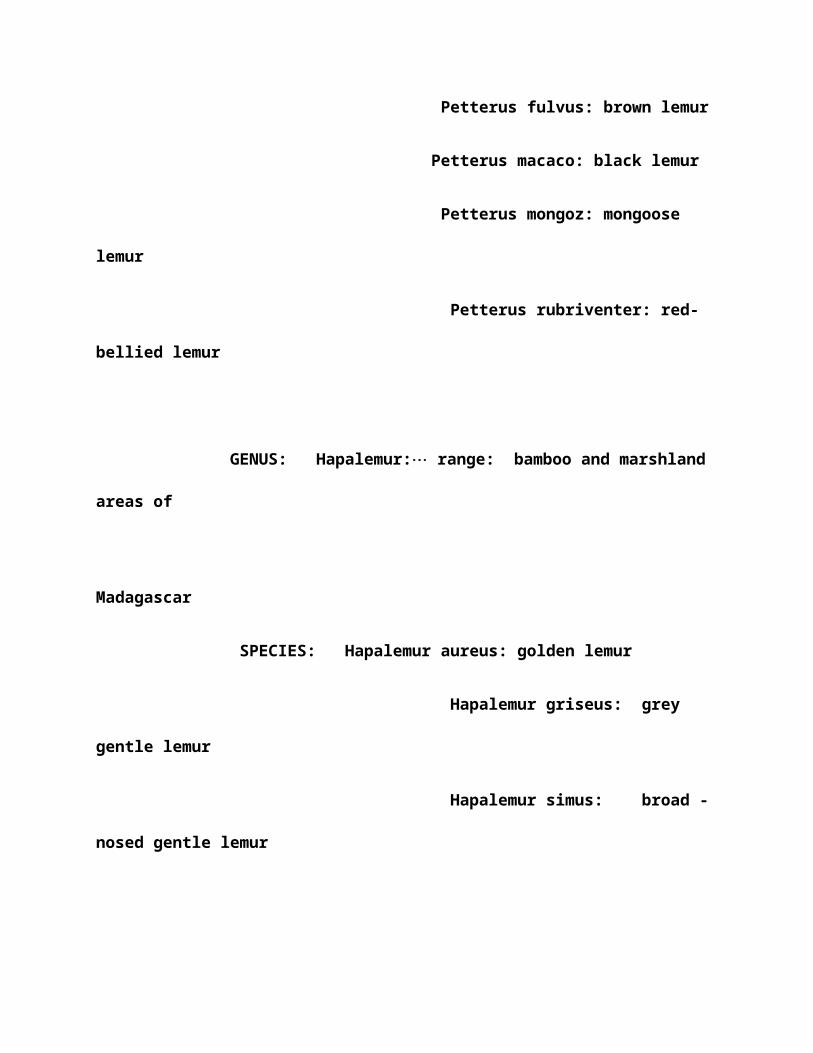
Petterus fulvus: brown lemur
Petterus macaco: black lemur
Petterus mongoz: mongoose
lemur
Petterus rubriventer: red-
bellied lemur
GENUS: Hapalemur:¼ range: bamboo and marshland
areas of
Madagascar
SPECIES: Hapalemur aureus: golden lemur
Hapalemur griseus: grey
gentle lemur
Hapalemur simus: broad -
nosed gentle lemur
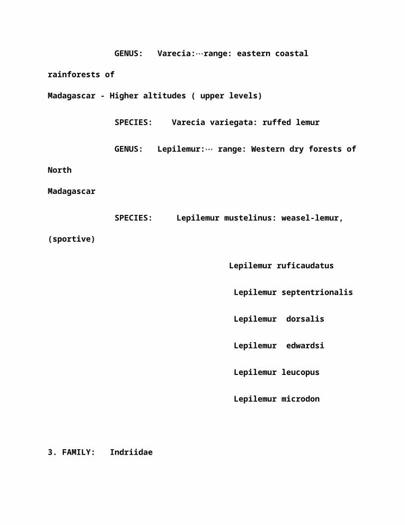
GENUS: Varecia:¼range: eastern coastal
rainforests of
Madagascar - Higher altitudes ( upper levels)
SPECIES: Varecia variegata: ruffed lemur
GENUS: Lepilemur:¼ range: Western dry forests of
North
Madagascar
SPECIES: Lepilemur mustelinus: weasel-lemur,
(sportive)
Lepilemur ruficaudatus
Lepilemur septentrionalis
Lepilemur dorsalis
Lepilemur edwardsi
Lepilemur leucopus
Lepilemur microdon
3. FAMILY: Indriidae

GENUS: Avahi:¼range: coastal rainforests of
Madagascar
SPECIES: Avahi laniger: wolly lemur
GENUS: Propithecus:¼range: mixed deciduous and
evergreen
forests of NW Madagascar
SPECIES: Propithecus diadema: Diadem sifaka
Propithecus tattersalli
Propithecus verreauxi:
Verreaux’s sifika
GENUS: Indri:¼range: eastern coastal
rainforests Madagascar
SPECIES: Indri indri: Indri
4.FAMILY: Daubentoniidae
GENUS: Daubentonia:¼ ( Aye-Ayes)¼ range: east
coast and NW
forests Madagascar
SPECIES: Daubentonia madagascariensis: Aye-Aye

5.FAMILY: Lorisidae
GENUS: Loris:¼range: India and Sri Lanka
( trop.rain and
dry/semidry Deciduous forests)
SPECIES: Loris tardugradus : slender loris:
range : swamps
India and Sri Lanka
GENUS: Nycticebus:¼range: rainforests of SE
Asia, Assam,
Burma,
Thailand, Indochina,
Parts of Malaysia and East Indian Islands
SPECIES: Nycticebus coucang: slow loris
Nycticebus pygmaeus: pygmy
low loris
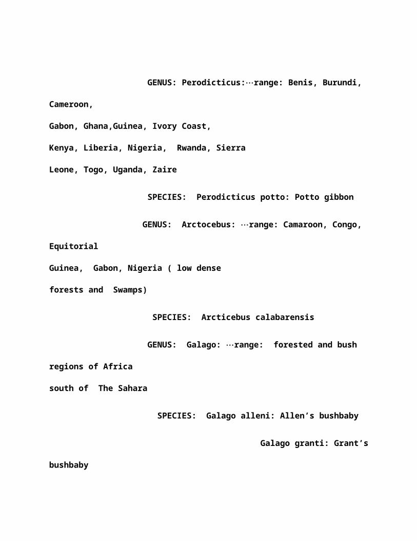
GENUS: Perodicticus:¼range: Benis, Burundi,
Cameroon,
Gabon, Ghana,Guinea, Ivory Coast,
Kenya, Liberia, Nigeria, Rwanda, Sierra
Leone, Togo, Uganda, Zaire
SPECIES: Perodicticus potto: Potto gibbon
GENUS: Arctocebus: ¼range: Camaroon, Congo,
Equitorial
Guinea, Gabon, Nigeria ( low dense
forests and Swamps)
SPECIES: Arcticebus calabarensis
GENUS: Galago: ¼range: forested and bush
regions of Africa
south of The Sahara
SPECIES: Galago alleni: Allen’s bushbaby
Galago granti: Grant’s
bushbaby

Galago moholi:
Southern lesser bushbaby
Galago senegalensis:
Northern lesser bushbaby
GENUS: Otolemur:¼range: Ethiopia
SPECIES: Otolemur crassucaudatus
Otolemur garnettii
GENUS: Euoticus:¼range: Camaroon,
Congo, Equatorial
Guinea, Gabon, Nigeria
SPECIES: Euoticus elecantus:
western needle-clawed
Bushbaby
Euoticus
inustus: eastern needle-clawed
Bushbaby
GENUS: Galagoides:¼range:
African rainforests

SPECIES: Galgoides
demidoff: demidoff’s galago
Galagoides zanzibaricus: zanibar bushbaby
6. FAMILY: Tarsiidae
GENUS: Tarsius:¼range: East
Indies rainforests (
common on Samar,
Leyte, Bohor and Minanao)
SPECIES: Tarsius bancanus:
western tarsier
Tarsius
pumilus: pygmy tarsier
Tarsius
syrichta: phillipine tarsier
7.FAMILY: Callithricidae
GENUS: Callithrix:¼range:

Upper Amazon of S.
America, W. Brazil, SE
Colombia, E. Ecaudor, Peru and N. Bolivia
SPECIES: Callithrix
argentata: silvery marmoset
Callithrix humeralifer: santarem
marmoset
Callithrix chrysoleuca
Callithrix jacchus: common marmoset
Callithrix flaviceps: white-eared marmoset
Callithrix penicillata: white-headed
marmoset
GENUS: Cebuella: (pygmy
marmoset): range:
Rainforests of Brazil, Ecuador, Colombia,

Peru
SPECIES: Cebuella pygmaea:
pygmy marmoset
GENUS: Saguinus: (tamarins):
range: Lower rainforests
of South America.
SPECIES: Saguinus
bicolor: bare-faced tamarin
Saguinus martinsi: Martin’s tamarin
Saguinus fuscicollis: saddle-backed
tamarin
Saguinus imperator: emperor tamarin
Saguinus inustus: mottle-faced tamarin

Saguinus labiatus: white-lipped tamarin
Saguinus leucopus: white-footed tamarin
Saguinus midas: red-handed tamarin
Saguinus tamarin: Negro tamarin
Saguinus mystax: moustached tamarin
Saguinus nigricollis: black and red tamarin
Saguinus graellsi
Saguinus geoffroyi: Geoffroy’s tamarin
Saguinus tripartitus

Saguinus fuscicollis
GENUS: Leontopithecus:
( Goldens)¼ range:
Evergreen and Broadleaf
forests. Brazil.
SPECIES:
Leontopithecus chysomelas: golden-
headed Tamarin
Leontropithecus chrysopygus: golden-
rumped Tamarin
Leontropithecus rosali: golden lion
tamarin
8. FAMILY: Cebidae: ( capuchins)
GENUS: Cebus:

range: deep rainforests of South
America
SPECIES: Cebus
albinfrons: brown pale-fronted
capuchin
Cebus apella: black-capped capuchin
Cebus capucinus: white-throated
capuchin
Cebus olivaceus:weeper capuchin
Cebus nigrivittatus
GENUS: Aotus:
( night monkeys)¼range: Central
And South America

SPECIES: Aotus
azarae: Southern night monkey
Aotus infulatus
Aotus miconax
Aotus nancymai
Aotus nigriceps
Aotus trivirgatus: Northern night monkey
Aotus brumbacki
Aotus lemurinus

Aotus vociferans
GENUS:
Callicebus:¼range: Bolivia and Brazil
SPECIES:
Callicebus brunneus
Callicebus calligatus
Callicebus cinerascens
Callicebus cupreus
Callicebus donacophilus
Callicebus dubius
Callicebus hoffmannsi

Callicebus modestus
Callicebus moloch: dusty titi
Callicebus oenanthe
Callicebus olallae
GENUS: Samiri: ( squirrel
monkeys)¼range: South America
SPECIES: Saimiri boliveiensis
Saimiri oerstedii: red-backed squirrel
monkey
Saimiri sciureus: common squirrel
monkey

Saimiri ustus
Saimiri vanzolinii
GENUS: Pithecia:
(saki)¼range: NW Brazil,
Colombia, Ecuador, Peru
SPECIES:
Pithecia aequatorialis: Equitorial
saki
Pithecia albicans: white saki
SPECIES:
Pithecia irrorata: bald-faced saki
Pithecia monachus: monk saki
Pithecia pithecia: white-faced saki

GENUS: Cacajao
( uakari)¼range: Brazil, E. Peru
SPECIES: Cacajao calvus: white uakari
Cacajao melanocephalus: black-
headed uakari
Cacajao rubicundus: red uakari
Cacajao calvus
GENUS:
Chiropotes: (saki)¼range: Brazil
SPECIES:
Chiropotes albinasus: white-bearded
saki
Chiropotes satanas: black-bearded
GENUS: Alouatta:

(howlers): ¼range: S. Mexico,
Central and South America
SPECIES: Alouatta belzebul: black and red
howler
Alouatta caraya: black howler
Alouatta fusca: brown howler
Alouatta palliata: mantled howler
Alouatta seniculus: red howler
Alouatta villosa: Guatamalan howler
Alouatta pigra
GENUS: Ateles: ( spider monkey)¼range:

Tropical South and
Central America
SPECIES: Ateles belzebuth: longhaired
spider monkey
Ateles
fusciceps:brownhaired spider monkey
Ateles geoffroyi:
black-handed spider monkey
Ateles paniscus:
black spider monkey
GENUS: Brachyteles: ( Wolley spider
monkey)¼range: Brazil
SPECIES: Brachyteles arachnoids:
Wolley spider monkey
GENUS: Lagothrix: ( Wolley
monkeys)..range: Andes North of
Peru

SPECIES: Lagothrix flavicauda :
yellow-tailed wolley
Lagothrix
lagothricha: common wolley
9. FAMILY: Cercopithecidae
a. Sub-Family: Cercopithecinae
GENUS: Macaca: ( macaques)¼range: SE Asia
SPECIES: Macaca arctoides: bear macaque
Macaca speciosa: stump-
tailed macaque
Macaca assamensis : Assam
macaque
Macaca fascicularis:
crab-eating macaque
Macaca maurus: Moor
macaque
Macaca
mulatta: Rhesus macaque

Macaca
nemestrina: pigtail macaque
Macaca nigra:
Celebes ape
Macaca
nigrescens : black ape
Macaca
radiata: bonnet macaque
Macaca
silenus: liontail macaque
Macaca
sinica: torque macaque
Macaca
sylvanus: barbary ape
Macaca
thibetana: Tibetan stump-tailed
Macaque
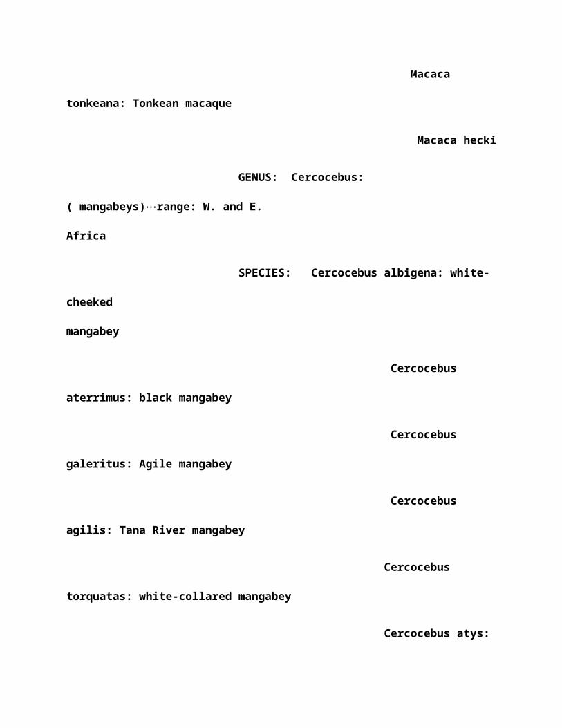
Macaca
tonkeana: Tonkean macaque
Macaca hecki
GENUS: Cercocebus:
( mangabeys)¼range: W. and E.
Africa
SPECIES: Cercocebus albigena: white-
cheeked
mangabey
Cercocebus
aterrimus: black mangabey
Cercocebus
galeritus: Agile mangabey
Cercocebus
agilis: Tana River mangabey
Cercocebus
torquatas: white-collared mangabey
Cercocebus atys:

sooty mangabey
GENUS: Papio: ( baboons)¼range: Sub-Saharan East
Africa,
Arabian arid zones
SPECIES: Papio anubis: Olive baboon ( sub-
saharan E.
Africa)
Papio cynocephalus: yellow
baboon
Papio hamadryas: Hamadryas
baboon
Papio papio: Guinea baboon
(Ethiopian savannas)
Papio ursinus: Chacma
baboon (S. Africa)
GENUS: Mandrillus:
(mandrill/drill)¼range: forests of W.

Africa
SPECIES: Mandrillus leucophaeus:
Drill
Mandrillus sphinx
: Mandrill
GENUS: Theropithecus: ( gelada)¼range:
Ethiopian
Highland Mtns.
( Gich and Sankaber areas of
Semien Mtns Nat’l Park)
SPECIES: Theropithecus gelada:
Gelada
GENUS: Cercopithecus¼range: East Africa,
Mtns of W. Uganda,
W. C.
Africa, Medium altitude forests

SPECIES: Cercopithecus aethiops: savanna
monkey
Cercopithecus pygerythrus:
green monkey
Cercopithecus tantalus:
vervet
Cercopithecus sabaeus: grivet
Cercopithecus ascanius:
Schmidt’s guenon
Cercopithecus campbelli:
Campbell’s monkey
Cercopithecus cephus:
Moustached monkey
Cercopithecus denti: Dents
monkey
Cercopithecus wolfi
Cercopithecus diana: Diana
monkey

Cercopithecus dryas:Dryas
monkey
Cercopithecus erythrogaster:
red-bellied monkey
Cercopithecus erythrotis:
red-eared monkey
Cercopithecus hamlyni: owl-
faced monkey
Cercopithecus hamlyni:
L’Hoest’s monkey
Cercopithecus mitis: Diademed
monkey
Cercopithecus
albogularis:Sykes’ monkey
Cercopithecus mona: mona
monkey
Cercopithecus neglectus: De
Brazza’s monkey

Cercopithecus nictitans:
greater white-nosed Monkey
Cercopithecus petaurista:
lesser white-nosed Monkey
Cercopithecus pogonias:
crowned monkey
Cercopithecus preussi:
Preuss’ monkey
Cercopithecus
lhoesti
Cercopithecus
salongo: Zaire Diana monkey
Cercopithecus
solatus: sun-tailed monkey
Cercopithecus wolfi:
Wolf’s monkey
GENUS: Miopithecus: ( talapoi)¼range: West Central
Africa

SPECIES: Miopithecus talpoin: Talapoi
GENUS: Allenopithecus: ( Allen’s swamp monkey)¼range:
C.
Africa
SPECIES: Allenopithecus nigroviridis: Allen’s swamp
Monkey
GENUS: Erythrocebus: (patas monkey)¼range: West,
Central and
parts Of east Africa
SPECIES: Erythrocebus patas : patas monkey
b. Sub-Family: Colobinae
GENUS: Colobus: ( colobus monkeys)¼range:
West , W. Central
and East Africa. SPECIES:
Colobus angolensis: Angolan colobus
Colobus badius: red
colobus ( pilocolobus)
Colobus rufomitratus: Bay

colobus
Colobus guereza: Guereza
( Eastern black and White colobus)
Colobus kirkii: Kirk’s
colobus
Colobus polykomos: King’s
colobus ( Western
Black and white colobus)
Colobus satanas: black
colobus
GENUS: Procolobus:(olive)¼range: West Africa
only
SPECIES: Procolobus verus: olive colobus
GENUS: Pygathrix:¼range: all are limited to
forests of Indochina
SPECIES: Pygathrix avunculus: Tokin snub-
nosed monkey

Pygathrix brelichi:
Brelichi’s snub-nosed monkey
Pygathrix nigripes
Pygathrix roxellana:
Chinese snub-nosed monkey
GENUS: Simias ( pig-tailed langur)¼range:
limited to Mentawi
Islands west of Sumatra
SPECIES: Simias concolor: pig-tailed
langur
GENUS: Nasalis: ( proboscis
monkey)¼range: Coastal areas of Borneo
SPECIES: Nasalis larvatus: proboscis
monkey
GENUS: Prebytis ( leaf/langur
monkeys)¼range: S/SW India
and Sri Lanka.

SPECIES: Prebytis aurata
Prebytis comata :
sundra leaf monkey
Prebytis hosei:
grizzled leaf monkey
Prebytis thomasi:
ebony leaf monkey
Prebytis cristata:
silvered leaf monkey
Prebytis
entellus: Hanuman langur
Prebytis
francoisi: Francois monkey
Prebytis
delacouri
Prebytis
frontata: white-fronted leaf monkey
Prebytis

geei: golden leaf monkey
Prebytis
johnii: Nilgiri monkey
Prebytis
melalophos: banded leaf monkey
Prebytis
femoralis: mitred leaf monkey
Prebytis
obscura: dusky leaf monkey
Prebytis
phayrei: Phayre’s leaf monkey
Prebytis
pileata: capped leaf monkey
Prebytis
potenziani: Mentawai leaf monkey
Prebytis
rubicunda: maroon leaf monkey
Prebytis

vetulus: purple-faced leaf monkey
Prebytis
senex
10. FAMILY: Hylobatidae
GENUS: Hylobates: ( gibbons
and siamang)¼range:
Rainforests of SE Asia
SPECIES: Hylobates agilis: agile gibbon
Hylobates
concolor: crested gibbon
Hylobates
hoolock: hoolock gibbon
Hylobates
klossii: Kloss’ gibbon
Hylobates
lar: common gibbon
Hylobates
moloch: Javan gibbon

Hylobates
muelleri: Muller’s gibbon
Hylobates
pileatus: Pileated gibbon
Hylobates
syndactylus: Siamang
11. FAMILY: Pongidae
GENUS: Pongo: (orangutans)¼range: Only Borneo
and Sumatra
Islands
SPECIES: Pongo pygmaeus: orangutans
GENUS: Pan: (bonobos and chimpanzees)¼range:
Bonobo:
Congo River region
Chimps:
Ethiopian: west and

Central equitorial Africa
SPECIES: Pan paniscus: bonobos
Pan
troglodytes: chimpanzees
12. FAMILY: Hominidea
GENUS: Gorilla: (gorillas)¼range:
Western lowland: Ethiopian tropical
rainforests,
S.
Nigeria to Congo
Mountain: Ethiopian Virunga Mtns
separating Zaire From Rwanda and
Uganda.
GENUS: Homo
SPECIES: Sapiens: humans

Tree Shew Taxonomy
This list shows all of the extant tree-shrews
(Lyon,1913; Napier and Napier, 1967; cited in Martin, 1990).
Subfamily Ptilocercinae
Ptilocercus lowii
Pen-tailed tree-shrew
Subfamily Tupaiinae
Tupaia belangeri
Belanger's tree-shrew
Tupaia glis
Common tree-shrew
Tupaia longipes
Long-footed tree-shrew

Tupaia montana
Montane tree-shrew
Tupaia nicobarica
Nicobar tree-shrew
Tupaia picta
Painted tree-shrew
Tupaia palawanensis
Palawan tree-shrew
Tupaia splendidula
Rufous-tailed tree-shrew
Tupaia minor
Pygmy tree-shrew
Tupaia javanica
Indonesian tree-shrew
Tupaia gracilis
Slender tree-shrew

Anathana ellioti
Indian tree-shrew
Lyonogale tana
Terrestrial tree-shrew
Lyonogale dorsalis
Striped tree-shrew
Urogale everetti
Philippine tree-shrew
Dendrogale melanura
Southern smooth-tailed tree-shrew
Dendrogale murina
Northern smooth-tailed tree-shrew
This table shows supposed shared characteristics between
tree-shrews and primates, from Martin (1990). Results of
Analysis of Characters Context
Shared

Similarities
Skull
1. Snout relatively short 1. Snout in fact secondarily
elongated in tree-shrews
2. Simplified set of turbinal bones 2. Set of six turbinal
bones probably
primitive for placental mammals
3. Enlarged, forward-facing orbits 3. Orbits relatively small
and laterally facing
4. Postorbital bar present 4. Postorbital bar present as a
convergent
development
in various mammals
5. Pattern of bones in medial orbital wall 5.
Palatine/lacrimal contact in medial
orbital wall probably primitive
6. Well-developed jugal bone with foramen

6. Well-developed jugal bone with foramen probably primitive
7. Enlarged braincase 7. Braincase has become enlarged
convergently in
various mammalian groups
8. Inflated auditory bulla containing 'free' ectotympanic ring
8. Auditory bulla formed from entotympanic, not from petrosal;
ectotympanic
ring is primitively ring-shaped
in placental
mammals
9. Internal carotid pattern (bony tubes) 9. Enclosure of
internal carotid in bony
tubes probably primitive
10. 'Advanced' form of auditory ossicles
10. Auditory ossicles do not clearly share derived features with
primates

Dentition
1. Tooth-comb present at front of lower
jaw, linked with a specialized, serrated
sublingua
1. Tooth-comb formed exclusively from
incisors as a convergent feature;
sublingua
present in common ancestor of marsupials
and placentals
2. Reduced dental formula
2. Convergent reduction of dental formula in
many mammalian groups
3. Similarities in cheek teeth between
tree-shrews and certain primates with
relatively primitive cheek teeth (e.g.
Tarsius)
3. Limited similarities in cheek teeth

between tree-shrews and certain primates
undoubtedly due to primitive retention
Postcranial Morphology
1. Limbs and digits highly mobile
1. Limbs and digits probably highly mobile in
ancestral placental mammals
2. Numerous details of limb musculature
2. Limb musculature shares primitive retentions
with prosimians
3 Osteological similarities in both forelimbs and
hindlimbs
3 Osteological similarities in forelimbs and
hindlimbs attributable to primitive
retention from ancestral placental mammals

4. Ridged skin on palms and soles
4. Ridged skin on palms and soles possibly a primitive feature
for placental mammals; tree-shrews lack the characteristic
Meissner's corpuscles of primates
Brain and Sense
Organs
1. Olfactory apparatus reduced
1. Olfactory apparatus not reduced relative
to body size in tree-shrews
2. Visual apparatus enhanced
2. Visual apparatus mildly enhanced;numerous
primate features lacking
3. Central, avascular area of retina
3. Unusual, spoke-like radiation of retinal
vessels; unusual innervation
4. Neocortex expanded; brain size increased

4. Expansion of neocortex and brain size found
in many mammals
5. Calcarine sulcus present
5. Calcarine sulcus not present in the brain
of tree-shrews
Reproductive Biology
1. Penis pendulous; testes scrotal
1. Pendulous penis and scrotal testes
present in many mammals
2. Discoidal placenta, as in tarsiers and
simians
2. Discoidal placenta common in mammals;
endotheliochorial in tree-shrews
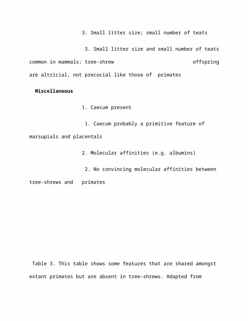
3. Small litter size; small number of teats
3. Small litter size and small number of teats
common in mammals; tree-shrew offspring
are altricial, not precocial like those of primates
Miscellaneous
1. Caecum present
1. Caecum probably a primitive feature of
marsupials and placentals
2. Molecular affinities (e.g. albumins)
2. No convincing molecular affinities between
tree-shrews and primates
Table 3. This table shows some features that are shared amongst
extant primates but are absent in tree-shrews. Adapted from

Martin (1990).
Primate Condition
Tree-shrew Condition
hands and feet prehensile
not prehensile
brain lateral and calcarine sulcus present
lateral and calcarine sulcus absent
scrotum postpenial
prepenial
lower incisors two or less on each side of the lower jaw
three on each side of the
lower jaw
upper incisors
arranged transversely *
arranged longitudinally
offspring at birth
precocial
altricial

gestation period
relatively long compared to body size relatively short
compared to body size
·1 many archaic primates have longitudinally arranged upper
incisors (e.g. Plesiadapis tricuspidens)
Primate Ranges (Adapted From martin, 1990)

Geological Time
(From Aiello 1993)

Age Based upon Dental Eruption and Developmental Information
(Park, 1999)

Classification of the Primates
This is the taxonomy Szalay and Delson (1979) proposed for the primates. Thetaxonomy only goes to the level of genus in the Linnean hierarchy.

PRIMATA
SUBORDER: Plesiadapiformes
SUPERFAMILY: Paromomyoidea
FAMILY: Paromomyidae
TRIBE: Purgatoriini
Purgatorius
TRIBE: Paromomyini
SUBTRIBE: Palaechthonina
Palaechthon
Plesiolestes
Palenochtha
SUBTRIBE: Paromomyina
Paromomys
Ignacius
Phenacolemur
TRIBE: Micromomyini

Micromomyini
Tinimomys
TRIBE: Navajoviini
Navajovius
Berruvius
FAMILY: Picrodontidae
Picrodus
Zanycteris
SUPERFAMILY: Plesiadapoidea
FAMILY: Plesiadapidae
Pronothodectes
Plesiadapis
Chiromyoides
Platychoerops
FAMILY: Saxonellidae

Saxonella
FAMILY: Carpolestidae
Elphidotarsius
Carpodaptes
SUBORDER: Strepsirhini
INFRAORDER: Adapiformes
FAMILY: Adapidae
SUBFAMILY: Notharctinae
Pelycodus
Notharctus
Smilodectes
Copelemur
SUBFAMILY: Adapinae
TRIBE: Protoadapini
Protoadapis
Agerinia
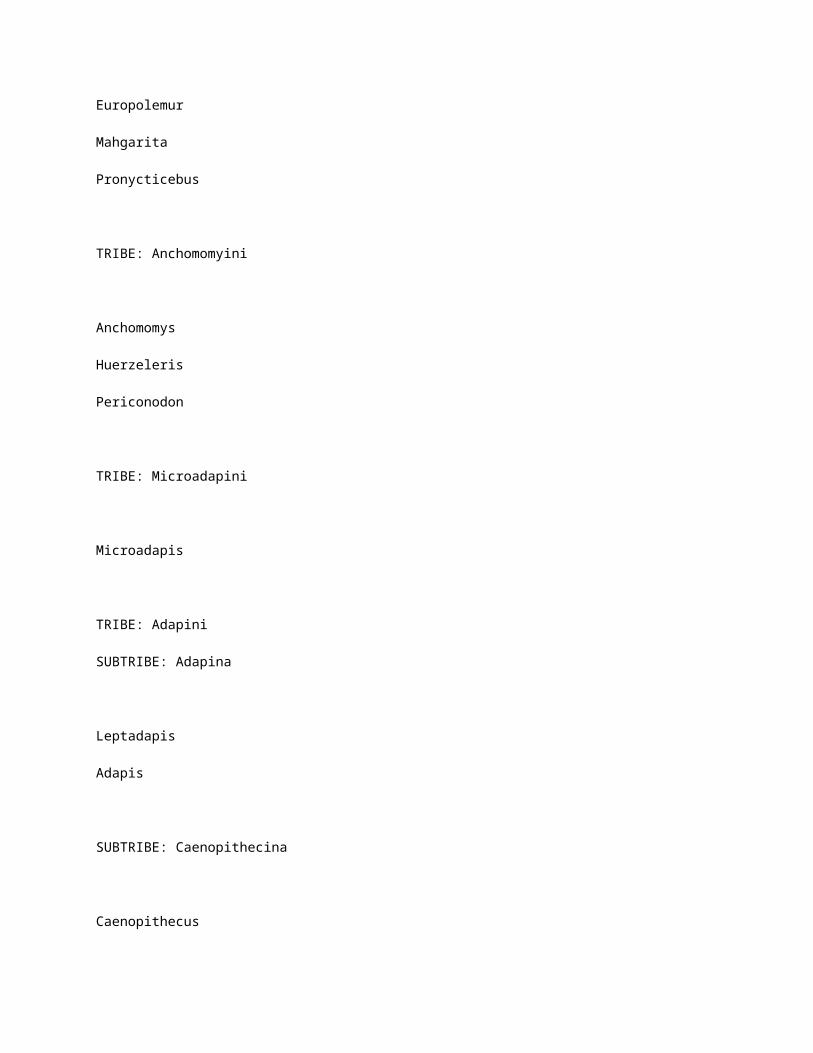
Europolemur
Mahgarita
Pronycticebus
TRIBE: Anchomomyini
Anchomomys
Huerzeleris
Periconodon
TRIBE: Microadapini
Microadapis
TRIBE: Adapini
SUBTRIBE: Adapina
Leptadapis
Adapis
SUBTRIBE: Caenopithecina
Caenopithecus

TRIBE: Indralorisini
Indraloris
FAMILY: incertae sedis
Amphipithecus
Lushius
INFRAORDER: Lemuriformes
SUPERFAMILY: Lemuroidea
FAMILY: Lemuridae
Lemur
Lepilemur
Hapalemur
Varecia
FAMILY: Megaladapidae
Megaladapis
SUPERFAMILY: Indrioidea

FAMILY: Indriidae
Indri
Propithecus
Avahi
Mesopropithecus
FAMILY: Daubentoniidae
Daubentonia
FAMILY: Archaeolemuridae
Archaeolemur
Hadropithecus
FAMILY: Palaeopropithecidae
Palaeopropithecus
Archaeoindris
SUPERFAMILY: Lorisoidea
FAMILY: Cheirogaleidae

Cheirogaleus
Phaner
Allocebus
Microcebus
FAMILY: Lorisidae
SUBFAMILY: Galaginae
Galago
Galagoides
Euoticus
Progalago
Komba
SUBFAMILY: Lorisinae
Loris
Nycticebus
Arctocebus
Perodicticus
Mioeuoticus

SUBORDER: Haplorhini
INFRAORDER: Tarsiiformes
FAMILY: Omomyidae
SUBFAMILY: Anaptomorphinae
TRIBE: Anaptomorphini
SUBTRIBE: Teilhardinina
Teilhardina
Chlorohysis
SUBTRIBE: Anaptomorphina
Anaptomorphus
SUBTRIBE: Tetoniina
Tetonius
Absarokius
Anemorhysis
Altanius
Mckennamorphus
TRIBE: Trogolemurini

Trogolemur
SUBFAMILY: Omomyinae
TRIBE: Omomyini
SUBTRIBE: Omomyina
Omomys
Chumashius
SUBTRIBE: Mytoniina
Ourayia
Macrotarsius
TRIBE: Washakiini
Loveina
Shoshonius
Washakius
Dyseolemur
Hemiacodon

TRIBE: Uintaniini
Uintanius
TRIBE: Utahiini
Utahia
Stockia
TRIBE: Rooneyiini
Rooneyia
SUBFAMILY: Ekgmowechashalinae
Ekgmowechashala
SUBFAMILY: Microchoerinae
Nannopithex
Necrolemur
Microchoerus
Pseudoloris
FAMILY: incertae sedis

Donrussellia
Hoanghonius
FAMILY: Tarsiidae
Tarsius
INFRAORDER: Platyrrhini
FAMILY: Cebidae
SUBFAMILY: Cebinae
Cebus
Saimiri
Neosaimiri
Dolichocebus
SUBFAMILY: Braniselllinae
Branisella
SUBFAMILY: Callitrichinae
TRIBE: Callitrichini

Callithrix
Cebuella
Saguinus
Leontopitheucs
TRIBE: Callimiconini
Callimico
FAMILY: Atelidae
SUBFAMILY: Atelinae
TRIBE: Atelini
Ateles
Lagothrix
Brachyteles
TRIBE: Alouattini
Alouatta
Stirtonia
SUBFAMILY: Pitheciinae
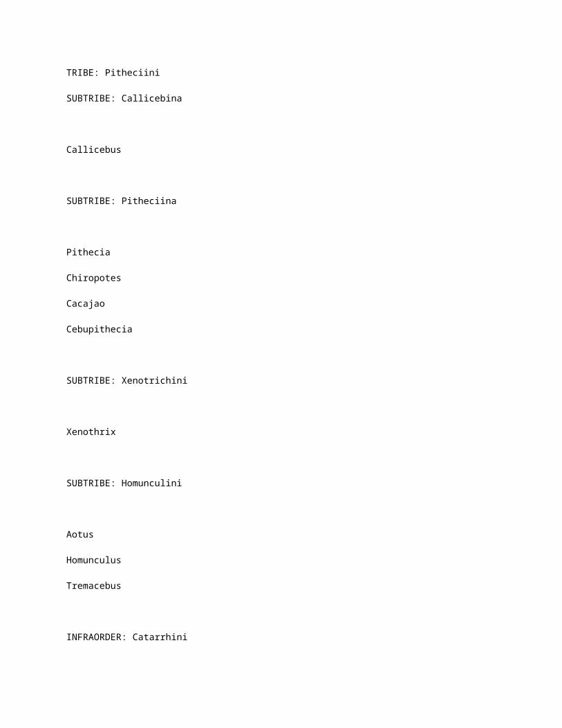
TRIBE: Pitheciini
SUBTRIBE: Callicebina
Callicebus
SUBTRIBE: Pitheciina
Pithecia
Chiropotes
Cacajao
Cebupithecia
SUBTRIBE: Xenotrichini
Xenothrix
SUBTRIBE: Homunculini
Aotus
Homunculus
Tremacebus
INFRAORDER: Catarrhini

SUPERFAMILY: Parapithecoidea
FAMILY: Parapithecidae
Parapithecus
Apidium
SUPERFAMILY: Cercopithecoidea
FAMILY: Cercopithecidae
SUBFAMILY: Cercopithecinae
TRIBE: Cercopithecini
Cercopithecus
Erythrocebus
Allenopithecus
TRIBE: Papionini
SUBTRIBE: Papionina
Papio
Cercocebus
Parapapio
Dinopithecus
Gorgopithecus

SUBTRIBE: Macacina
Macaca
Procynocephalus
Paradolichopithecus
SUBTRIBE: Theropithecina
Theropithecus
SUBFAMILY: Colobinae
SUBTRIBE: Colobina
Colobus
Libypithecus
Cercopithecoides
Paracolobus
Colobina, new genus
Colobina, gen. Indet.
SUBTRIBE: Semnopithecina

Presbyits
Pygathrix
Nasalis
SUBFAMILY: incertae sedis
Mesopithecus
Dolichopithecus
FAMILY: incertae sedis
Prohylobates
Victoriapithecus
FAMILY: Oreopithecidae
Oreopithecus
SUPERFAMILY: Hominoidea
FAMILY: Pliopithecidae
Propliopithecus
Pliopithecus

Dendropithecus
FAMILY: incertae sedis
FAMILY: Hominidae
SUBFAMILY: Hylobatinae
Hylobates
SUBFAMILY: Ponginae
TRIBE: Pongini
Pongo
Pan
TRIBE: Dryopithecini
Dryopithecus
TRIBE: Sugrivapithecini
Sivapithecus
Gigantopithecus

SUBFAMILY: Homininae
Ramapithecus
Australopithecus
Homo
Homo or Australopithecus sp(p). indet.
Homininae, gen. Et sp(p). indet.
INFRAORDER: incertae sedis
Pondaungia
Oligopithecus
REFERENCES:
Szalay, F. and Delson, E. 1979. Evolutionary History of the Primates. Academic
Press: New York.
This is the taxonomy Fleagle (1999) proposed for the primates. This is a
revision of the Fleagle (1988)taxonomy. The taxonomy only goes to the level of
genus in the Linnean hierarchy.
ORDER: incertae sedis
Purgatorius
ORDER: Plesiadapiformes

SUPERFAMILY: Plesiadapoidea
FAMILY: Plesiadapidae
Pandemonium
Pronothodectes
Nannodectes
Plesiadapis
Chiromyoides
Platychoerops
FAMILY: Carpolestidae
Chronolestes
Elphidotarsius
Carpodaptes
Carpolestes
Carpocristes
FAMILY: Saxonellidae

Saxonella
FAMILY: Paromomyidae
Paromomys
Ignacius
Phenacolemur
Elwynella
Simpsonlemur
Dillerlemur
Pulverflumen
Arcius
FAMILY: Micromomyidae
Micromomys
Tinimomys
Chalicomomys

Myrmekomomys
SUPERFAMILY: Microsyopoidea
FAMILY: Palaechthonidae
Palaechthon
Plesiolestes
Talpohenach
Torrejonia
Palenochtha
Premnoides
FAMILY: Microsyopidae
Navajovius
Berruvius
Niptomomys
Uintasorex
Avenius

Microsyops
Arctodontomys
Craseops
Megadelphus
SUPERFAMILY: incertae sedis
FAMILY: Picrodontidae
Picrodus
Zanycteris
Draconodus
FAMILY: Picromomyidae
Picromomys
Alveojunctus
ORDER: Primata

SUBORDER: Prosimii
FAMILY: incertae sedis
Altanius
Altiatlasius
INFRAORDER: Lemuriformes
SUPERFAMILY: Adapoidea
FAMILY: Notharctidae
SUBFAMILY: Notharctinae
Cantius
Copelemur
Notharctus
Smilodectes
Pelycodus
Hesperolemur
SUBFAMILY: Cercamoniinae

Donrussellia
Protoadapis
Europolemur
Periconodon
Caenopithecus
Pronycticebus
Cercamonius
Anchomomys
Huerzeleria
Buxella
Agerinia
Panobius
Mahgarita
Djebelemur
Aframonius
Omanodon
Shizarodon
Wadilemur

FAMILY: incertae sedis
Azibius
Hoanghonius
Lushius
Rencunius
Wailekia
FAMILY: Adapidae
SUBFAMILY: Adapinae
Adapis
Cryptadapis
Microadapis
Leptadapis
Adapoides
FAMILY: Sivaladapidae
SUBFAMILY: Sivaladapinae

Indraloris
Sivaladapis
Sinoadapis
SUPERFAMILY: Lemuroidea
FAMILY: Cheirogaleidae
Microcebus
Mirza
Cheirogaleus
Phaner
Allocebus
FAMILY: Lemuridae
Lemur
Eulemur
Varecia

Pachylemur
Hapalemur
FAMILY: Lepilemuridae
SUBFAMILY: Lepilemurinae
Lepilemur
SUBFAMILY: Megaladapinae
Megaladapis
FAMILY: Indriidae
SUBFAMILY: Indriinae
Avahi
Propithecus
Indri

SUBFAMILY: Archaeolemurinae
Archaeolemur
Hadropithecus
SUBFAMILY: Palaeopropithecinae
Mesopropithecus
Babakotia
Palaeopropithecus
Archaeoindris
FAMILY: Daubentoniidae
Daubentonia
SUPERFAMILY: Lorisoidea
FAMILY: Galagidae

Progalago
Komba
Otolemur
Galago
Euoticus
Galagoides
FAMILY: Lorisidae
Perodicticus
Pseudopotto
Arctocebus
Loris
Nycticebus
Mioeuoticus
FAMILY: Plesiopithecidae

Plesiopithecus
INFRAORDER: Tarsiiformes
SUPERFAMILY: Omomyoidea
FAMILY: Omomyoidae
SUBFAMILY: Anaptomorphinae
TRIBE: Anaptomorphini
Teilhardina
Anaptomorphus
Gazinius
Tetonius
Pseudotetonius
Absarokius
Tatmanius
Strigorhysis
Acrossia
TRIBE: Trogolemurini

Trogolemur
Sphacorhysis
Anemorhysis
Tetonoides
Arapahovius
Chlororhysis
TRIBE: Washakiini
Washakius
Shoshonius
Dyseolemur
Loveina
SUBFAMILY: Omomyinae
TRIBE: Omomyini
Omomys
Chumashius

Steinius
TRIBE: Uintaniini
Uintanius
Jemezius
TRIBE: Macrotarsiini
Macrotarsius
Hemiacodon
Yaquius
TRIBE: Ourayini
Ourayia
Wyomomys
Ageitodendron
Utahia

Stockia
Chipetaia
Asiomomys
TRIBE: incertae sedis
Ekgmowechashala
FAMILY: Microchoeridae
Nannopithex
Pseudoloris
Necrolemur
Microchoerus
FAMILY: incertae sedis
Rooneyia
Kohatius

SUPERFAMILY: Tarsioidea
FAMILY: Tarsiidae
Afrotarsius
Tarsius
Xanthorhysis
SUBORDER: Anthropoidea
INFRAORDER: incertae sedis
SUPERFAMILY: incertae sedis
FAMILY: Eosimiidae
Eosmias
FAMILY: incertae sedis
Amphipithecus
Pondaungia

Siamopithecus
SUPERFAMILY: Parapithecoidea
FAMILY: Parapithecidae
Serapia
Qatrania
Apidium
Parapithecus
Biretia
SUPERFAMILY: incertae sedis
FAMILY: incertae sedis
Proteopithecus
Arsinoea
Algeripithecus
Tabelia

INFRAORDER: Platyrrhini
SUPERFAMILY: Ceboidea
FAMILY: Atelidae
SUBFAMILY: Pitheciinae
Soriacebus
Carlocebus
Homunculus
Cebupithecia
Nuciruptor
Propithecia
Pithecia
Chiropotes
Cacajao
SUBFAMILY: Callicebinae
Callicebus

SUBFAMILY: Atelinae
Stirtonia
Protopithecus
Caipora
Alouatta
Lagothrix
Brachyteles
Ateles
FAMILY: Cebidae
SUBFAMILY: Aotinae
Tremacebus
Aotus
SUBFAMILY: Cebinae
Dolichocebus

Chilecebus
Neosaimiri
Laventiana
Cebus
Saimiri
SUBFAMILY: Callitrichinae
Micodon
Patasola
Lagonimico
Callimico
Saguinus
Leontopithecus
Callithrix
Cebuella
SUBFAMILY: incertae sedis

Branisella
Szalatavus
Mohanamico
Paralouatta
Xenothrix
Antillothrix
INFRAORDER: Catarrhini
SUPERFAMILY: Propliopithecoidea
FAMILY: Propliopithecidae
Propliopithecus
Aegyptopithecus
FAMILY: Oligopithecidae
Oligopithecus
Catopithecus
SUPERFAMILY: Hominoidea

FAMILY: Proconsulidae
Proconsul
Rangwapithecus
Limnopithecus
Dendropithecus
Simiolus
Micropithecus
Kalepithecus
Kamoyapithecus
Dionysopithecus
Platydontopithecus
FAMILY: Oreopithecidae
Mabokopithecus
Nyanzapithecus
Oreopithecus

FAMILY: incertae sedis
Afropithecus
Morontopithecus
Turkanapithecus
Kenyapithecus
Otavipithecus
Samburupithecus
FAMILY: Pliopithecidae
Pliopithecus
Plesiopliopithecus
Anapithecus
Laccopithecus
FAMILY: Hylobatidae
Hylobates

FAMILY: Pongidae
Dryopithecus
Lufengpithecus
Griphopithecus
Sivapithecus
Ankarapithecus
Gigantopithecus
Graecopithecus
Ouranopithecus
Pongo
Gorilla
Pan
FAMILY: Hominidae
SUBFAMILY: Australopithecinae
Ardipithecus

Australopithecus
Paranthropus
SUBFAMILY: Homininae
Homo
SUPERFAMILY: Cercopithecoidea
FAMILY: Victoriapithecidae
SUBFAMILY: Victoriapithecinae
Prohylobates
Victoriapithecus
FAMILY: Cercopithecidae
SUBFAMILY: Cercopithecinae
Macaca
Procynocephalus

Paradolichopithecus
Papio
Dinopithecus
Cercocebus
Lophocebus
Parapapio
Gorgopithecus
Theropithecus
Cercopithecus
Allenopithecus
Chlorocebus
Erythrocebus
Mandrillus
Miopithecus
SUBFAMILY: Colobinae
Mesopithecus
Dolichopithecus

Semnopithecus
Presbytis
Kasi
Trachypithecus
Nasalis
Simias
Pygathrix
Rhinopithecus
Colobus
Piliocolobus
Procolobus
Libypithecus
Microcolobus
Cercopithecoides
Paracolobus
Rhinocolobus
REFERENCES:
Fleagle, J.G. 1999. Primate Adaptation and Evolution. Academic
Press: New York.

Hominid Phylogeny
( based upon Gibbons, 1981)
Australopithecus anamensis ( 4.4mya)
Australopithecus africanus (4.2mya)
Homo erectus (2.5mya)
Australopithecus robustus (3.9mya)
Homo sapiens (1.5mya)
Homo sapiens neandertalensis (300,000ya) Homo sapiens sapiens
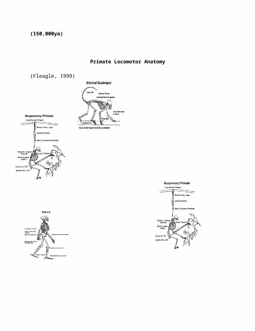
(150,000ya)
Primate Locomotor Anatomy
(Fleagle, 1999)
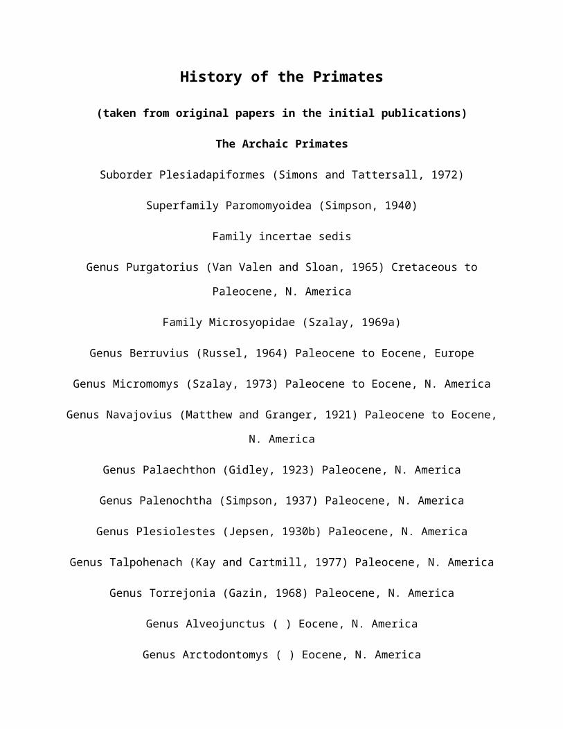
History of the Primates
(taken from original papers in the initial publications)
The Archaic Primates
Suborder Plesiadapiformes (Simons and Tattersall, 1972)
Superfamily Paromomyoidea (Simpson, 1940)
Family incertae sedis
Genus Purgatorius (Van Valen and Sloan, 1965) Cretaceous to
Paleocene, N. America
Family Microsyopidae (Szalay, 1969a)
Genus Berruvius (Russel, 1964) Paleocene to Eocene, Europe
Genus Micromomys (Szalay, 1973) Paleocene to Eocene, N. America
Genus Navajovius (Matthew and Granger, 1921) Paleocene to Eocene,
N. America
Genus Palaechthon (Gidley, 1923) Paleocene, N. America
Genus Palenochtha (Simpson, 1937) Paleocene, N. America
Genus Plesiolestes (Jepsen, 1930b) Paleocene, N. America
Genus Talpohenach (Kay and Cartmill, 1977) Paleocene, N. America
Genus Torrejonia (Gazin, 1968) Paleocene, N. America
Genus Alveojunctus ( ) Eocene, N. America
Genus Arctodontomys ( ) Eocene, N. America

Genus Craseops ( ) Eocene, North America
Genus Microsyops (Leidy, 1972) Eocene, N. America
Genus Niptomomys ( ) Eocene, N. America
Genus Tinimomys (Szalay, 1974b) Eocene, North America
Genus Uintasorex (Brown and Rose, 1976) Eocene, N. America
Family Paromoyidae (Simpson, 1940)
Genus Paromomys (Gidley, 1923) Paleocene, N. America
Genus Ignacius (Matthew and Granger, 1921) Paleocene to Eocene,
N. America
Genus Phenacolemur (Matthew, 1915) Paleocene to Eocene, N.
America, Europe
Genus Arcius () Eocene, Europe
Genus Elwynella () Eocene, N. America
Family Picrodontidae Simpson, 1937)
Genus Draconodus (Tjomida, 1982) Paleocene, N. America
Genus Picrodus (Douglass, 1908) Paleocene, . America
Genus Zanycteris (Matthew, 1917a) Paleocene, N. America
Superfamily Plesiadapoidea (Trouessart, 1897)
Family Plesiadapidae (Trouessart, 1897)
Genus Chiromyoides (Stehlin, 1916) Paleocene to Eocene, N.

America, Europe
Genus Nannodectes Gingerich, 1974) Paleocene, N. America
Genus Platychoerops (Charlesworth, 1854) Eocene, Europe
Genus Plesiadapis (Gervais, 1877) Paleocene to Eocene, N.
America, Europe
Genus Pronothodectes (Gidley, 1923) Paleocene, N. America
Family Saxonellidae (Russel, 1964)
Genus Saxonella (Russell, 1964) Paleocene, N. America, Europe
Family Carpolestidae (Simpson, 1935b )
Genus Elphidotarsius (Gidley, 1923) Paleocene, N. America
Genus Carpodaptes (Matthew and Granger, 1921) Paleocene, N.
America
Genus Carpolestes (Simpson, 1928) Paleocene to Eocene, N. America
Eocene Primates
Order Primates (Linnaeus, 1758)
Suborder Prosimii (Illiger, 1811)
Infraorder Tarsiiformes (Gregory, 1915b)
Family Omomyidae (Trouessart, 1879)

Subfamily Anaptomorphinae (Cope, 1883)
Genus Absarokius (Matthew, 1915) Eocene, N. America
Genus Aycrossia () Eocene, N. America
Genus Altanius (Dashzeveg and McKenna, 1977) Eocene, N. America
Genus Anaptomorphus (Cope, 1872) Eocene, N. America
Genus Anemorhysis (Gazin, 1958) Eocene, N. America
Genus Chlororhysis (Gazin, 1958) Eocene, N. America
Genus Gazinius () Eocene, N. America
Genus Lovenia (Simpson, 1940) Eocene, N. America
Genus Mckennamorphus (Szalay, 1976) Eocene, N. America
Genus Steinius () Eocene, N. America
Genus Strigorhysis () Eocene, N. America
Genus Teilhardina (Simpson, 1940) Eocene, France
Genus Tetonius (Matthew, 1915) Eocene, N. America
Genus Trogolemus (Matthew, 1909) Eocene, N. America
Subfamily Omomyinae (Trouessart, 1879)
Genus Arapahovius () Eocene, N. America
Genus Chumashius (Stock, 1933) Eocene, N. America
Genus Dyseolemur (Stock, 1934) Eocene, N. America

Genus Hemiacodon (Marsh, 1872a) Eocene, N. America
Genus Jemezius () Eocene, N. America
Genus Macrotarsius (Clark, 1941) Eocene to Oligocene, N. America
Genus Omomys (Leidy, 1869) Eocene, N. America
Genus Ourayia (Gazin, 1958) Eocene, N. America
Genus Shoshonius (Granger, 1910) Eocene, N. America
Genus Stockia (Gazin, 1958) Eocene, N. America
Genus Uintanius (Matthew, 1915) Eocene, N. America
Genus Utahia (Gazin, 1958) Eocene, N. America
Genus Washakius (Leidy, 1873) Eocene, N. America
Subfamily Microchoerinae (Lydekker, 1887)
Genus Microchoerus (Wood, 1846) Eocene to Oligocene, England,
France, Germany
Genus Nannopithex (Stehlin, 1916) Eocene, France, Germany
Genus Necrolemur (Fihol, 1873) Eocene, France
Genus Psleudoloris (Stehlin, 1916) Eocene, France, Spain
Family Omomyidae, incertae sedis
Genus Hoanghonius (Zdansky, 1930) Eocene, China
Infraorder Adapiformes (Szalay and Delson, 1979)
Family Adapidae (Trouessart, 1879)

Subfamily Notharctinae (Trouessart, 1879)
Genus Cantius (Simons, 1962b) Eocene, N. America
Genus Copelemur (Gingerich and Simons, 1977) Eocene, Rocky Mt.
Genus Notharctus (Leidy, 1870) Eocene, Rocky Mt.
Genus Pelycodus Cope, 1875) Eocene, N. Americas and Europe
Genus Smilodectes (Wortman, 1903) Eocene, Rocky Mt.
Subfamily Adapinae (Trouessart, 1879)
Genus Adapis (Cuvier, 1821) Eocene to Oligocene, Europe
Genus Agerinaia (Crusafont-Pairo and Golpe-Posse, 1973) Eocene,
Spain
Genus Anchomomys (Stehlin, 1916) Eocene, Europe
Genus Caenopithecus (Rütimeyer, 1862) Eocene, Europe
Genus Cercamonius (Gingerich, 1975c) Eocene, Europe
Genus Cryptadapis () Eocene, Europe
Genus Donrussellia (Szalay, 1976) Eocene, Europe
Genus Europolemur (Weigelt, 1933) Eocene, German Democratic
Republic
Genus Huerzeleris (Szalay, 1974a) Eocene, Europe
Genus Indraloris (Lewis, 1933) Miocene, Northern India
Genus Leptadapis (Gervais, 1876) Eocene, Europe

Genus Mahgarita (Wilson and Szalay, 1976) Eocene, Texas
Genus Microadapis (Szalay, 1974a) Eocene, Switzerland
Genus Periconodon (Stehlin, 1916) Eocene, Europe
Genus Pronycticebus (Grandidier, 1904) Eocene, France
Genus Protoadapis (Lemoine, 1878) Eocene, Western Europe
Family Adapidae, incertae sedis
Genus Lushius (Chow, 1961) Eocene, China
Suborder incertae sedis
Genus Amphipithecus (Colbert, 1937) Eocene, Burma
Oligocene Primates
Order Primates (Linnaeus, 1758)
Suborder Prosimii (Illiger, 1811)
Infraorder Tarsiiformes (Gregory, 1915b)
Family Omomyidae (Trouessart, 1879)
Subfamily Omomyinae (Trouessart, 1879)
Genus Rooneyia (Wilson, 1966) Oligocene, N. America
Subfamily Ekgmowechashalinae (Szalay, 1976)
Genus Edgmowechashala (Macdonald, 1963) Oligocene, N. America
Family Tarsiidae (Gray, 1825)

Genus Afrotarsius (Simons and Brown, 1985) Oligocene, Africa
Suborder Anthropoidea (Mivart, 1864)
Infraorder Platyrrhini (E. Geoffroy, 1812)
Superfamily Ceboidea (Simpson, 1931)
Family Cebidae (Bonaparte, 1831)
Subfamily Cebinae (Bonaparte, 1831)
Genus Dolichocebus (Kraglievich, 1951) Oligocene, Argentina
Subfamily Branisellinae (Hershkovitz, 1977)
Genus Branisella (Hoffstetter, 1969) Oligocene, Bolivia
Family Atelidae (Gray, 1825)
Subfamily Pitheciinae (Mivart, 1865)
Genus Tremacebus (Hershkovitz, 1974a) Oligocene, Argentina
Infraorder Catarrhini E. Geoffroy, 1812)
Superfamily Parapithecoidea (Schlosser, 1911)
Family Parapithecidae (Schlosser, 1911)
Genus Parapithecus (Schlosser, 1910) Oligocene, Egypt
Genus Apidium (Osborn, 1908) Oligocene, Egypt
Genus Qatrania (Simons and Kay, 1983) Oligocene, Egypt
Genus Catopithecus (), Oligocene, Egypt

Superfamily Hominoidea (Gray, 1825)
Family Propliopithecidae (Straus, 1961)
Subfamily Propliopithecinae (Delson and Andrews, 1975)
Genus Propliopithecus (Schlosser, 1916) Oligocene, Egypt
Genus Aegyptopithecus (Simons, 1965) Oligocene, Egypt
Subfamily Oligopithecinae
Genus Catopithecus (Simons, 1989) Oligocene, Egypt
Genus Proteopithecus (Simons, 1989) Oligocene, Egypt
Genus Oligopithecus (Simons, 1962a) Oligocene, Egypt
Miocene Primates
Order Primates (Linnaeus, 1758)
Suborder Prosimii (Illiger, 1811)
Infraorder Adapiformes (Szalay and Delson, 1979)
Family Adapidae (Trousessart, 1879)
Subfamily Sivaladapinae
Genus Indraloris (Lewis, 1933) Miocene, Asia
Genus Sivaladapis () Miocene, Asia
Genus Sinoadapis () Miocene, Asia
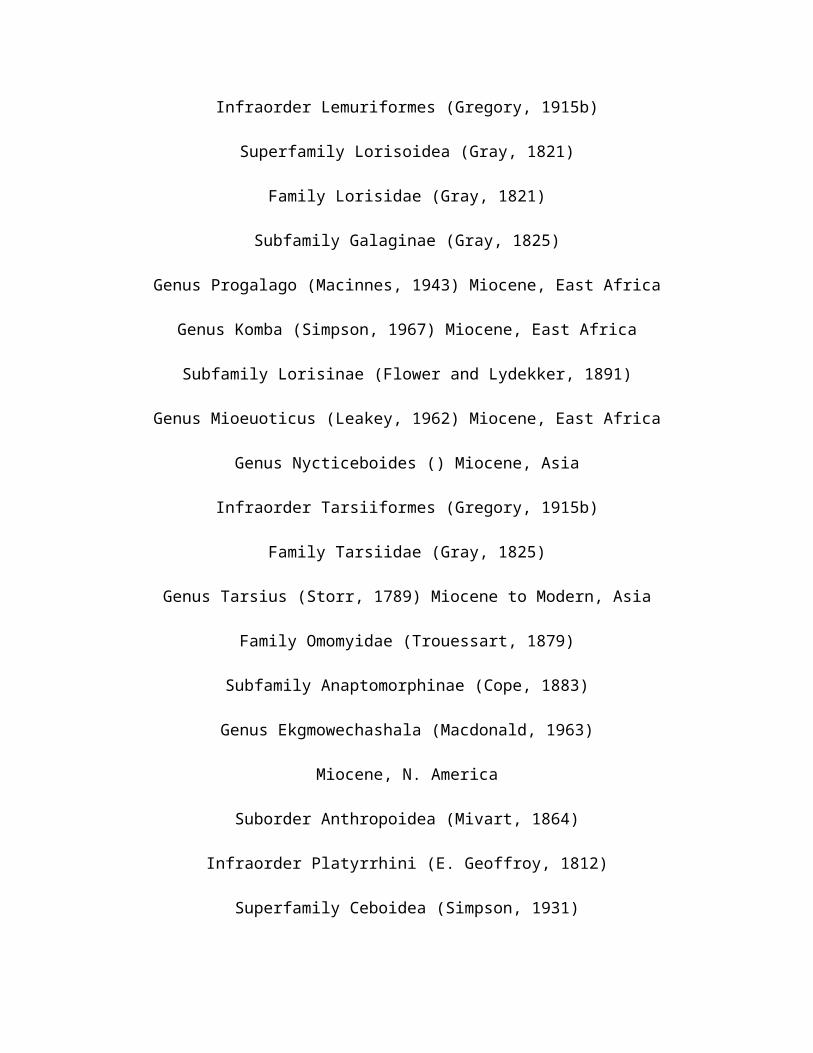
Infraorder Lemuriformes (Gregory, 1915b)
Superfamily Lorisoidea (Gray, 1821)
Family Lorisidae (Gray, 1821)
Subfamily Galaginae (Gray, 1825)
Genus Progalago (Macinnes, 1943) Miocene, East Africa
Genus Komba (Simpson, 1967) Miocene, East Africa
Subfamily Lorisinae (Flower and Lydekker, 1891)
Genus Mioeuoticus (Leakey, 1962) Miocene, East Africa
Genus Nycticeboides () Miocene, Asia
Infraorder Tarsiiformes (Gregory, 1915b)
Family Tarsiidae (Gray, 1825)
Genus Tarsius (Storr, 1789) Miocene to Modern, Asia
Family Omomyidae (Trouessart, 1879)
Subfamily Anaptomorphinae (Cope, 1883)
Genus Ekgmowechashala (Macdonald, 1963)
Miocene, N. America
Suborder Anthropoidea (Mivart, 1864)
Infraorder Platyrrhini (E. Geoffroy, 1812)
Superfamily Ceboidea (Simpson, 1931)

Family Cebidae (Bonaparte, 1831)
Subfamily Cebinae (Bonaparte, 1831)
Genus Neosaimiri (Stirton, 1951) Miocene, Colombia
Subfamily Callitrichinae
Genus Micodon () Miocene, Colombia
Subfamily Aotinae ()
Genus Tremacebus (Hershkovitz, 1974a) Miocene, Argentina
Genus Aotus (Illiger, 1811) Miocene, Colombia
Genus Homunculus (Ameghino, 1891) Miocene, Argentina
Family Atelidae (Gray, 1825)
Subfamily Atelinae (Gray, 1825)
Genus Stirtonia (Hershkovitz, 1970a) Miocene, Colombia
Subfamily Pitheciinae (Mivart, 1865) Miocene, South America
Genus Cebupithecia (Stirton and Savage, 1951) Miocene, Colombia
Genus Mohanamico () Miocene, Colombia
Infraorder Catarrhini E. Geoffroy, 1812)
Superfamily Cercopithecoidea (Gray, 1821)
Family Cercopithecidae (Gray, 1821

Subfamily Cercopithecinae (Gray, 1821)
Genus Parapapio (Jones, 1937) Miocene to Pleistocene, Africa
Genus Macaca (Lacépéde, 1799) Miocene to modern, Europe, N.
Africa, Asia
Subfamily Colobinae (Blyth, 1875)
Genus Colobus (Illiger, 1811) Miocene to modern. Africa
Genus Libypithecus (Stromer, 1913) Miocene, Egypt
Genus Microcolobus () Miocene, East Africa
Genus Presbytis (Eschscholtz, 1821) Miocene to modern, Asia
Genus Mesopithecus (Wagner, 1839) Miocene to Pliocene, Europe
Family Victoriapithecidae
Genus Prohylobates (Fourtau, 1918) Miocene, Egypt, Libya
Genus Victoriapithecus (von Koenigswald, 1969) Miocene, Kenya,
Uganda
Superfamily Hominoidea (Gray, 1825)
Family Oreopithecidae (Schwalbe, 1915)
Genus Nyanzapithecus (Harrison, 1987) Miocene, East Africa
Genus Oreopithecus (Gervais, 1872) Miocene, Italy
Family Pliopithecidae (Zapfe, 1961a)
Genus Pliopithecus (Gervais, 1849) Miocene, Europe

Genus Laccopithecus (Wu and Pan, 1984) Miocene, China
Genus Dendropithecus (Andrews and Simons, 1977) Miocene, Kenya
Genus Crouzelia (Ginsburg, 1975) Miocene, Europe
Family Proconsulidae ()
Genus Proconsul (Hopwood, 1933a) Miocene, East Africa
Genus Limnopithecus (Hopwood, 1933a) Miocene, Africa
Genus Dendropithecus (Andrews, Pilbeam and Simons, 1976) Miocene,
Africa
Genus Simiolus (R.E.F. Leakey and Leakey, 1987) Miocene, Africa
Genus Rangwapithecus (Pilbeam et al., 1977) Miocene, Africa
Genus Micropithecus (Fleagle & Simons, 1978) Miocene, Africa
Genus Dionysopithecus () Miocene, Asia
Genus Platydontopithecus () Miocene, Asia
Family Hylobatidae (Gray, 1870)
Genus Hylobates (Illiger, 1811)
Pleistocene to modern, South East Asia, Southern China
Family Pongidae (Elliot, 1913)
Genus Dryopithecus (Lartet, 1856) Miocene, Africa and Europe
Genus Lufengpithecus () Miocene, Asia

Genus Ouranopithecus () Miocene, Europe
Genus Sivapithecus (Pilgrim, 1910) Miocene, Africa, Europe, Asia
Genus Ramapithecus (Lewis, 1934) Miocene, Africa, Europe, Asia
Genus Gigantopithecus (von Koenigswald, 1935) Miocene to
Pleistocene, Asia
Genus Graecopithecus (von Koenigswald, 1972) Miocene, Europe
Family incertae sedis
Genus Turkanapithecus (Leakey and Leakey, 1986a) Miocene, East
Africa
Genus Afropithecus (Leakey and Leakey, 1986b) Miocene, East
Africa Genus Kenyapithecus (L. Leakey, 1962a)
Miocene, East Africa
Pliocene Primates
Order Primates (Linnaeus, 1758)
Suborder Prosimii (Illiger, 1811)
Suborder Anthropoidea (Mivart, 1864)
Infraorder Catarrhini E. Geoffroy, 1812)
Superfamily Cercopithecoidea (Gray, 1821)
Family Cercopithecidae (Gray, 1821

Subfamily Cercopithecinae (Gray, 1821)
Genus Cercopithecus (Linnaeus, 1758) Pliocene to modern, Africa
Genus Papio (Müller, 1773) Pliocene to modern, Africa
Genus Cercocebus (E. Geoffroy, 1812) Pliocene to modern, Africa
Genus Dinopithecus (Broom, 1937) Pliocene, Africa
Genus Gorgopithecus (Broom and Robinson, 1949a) Pliocene, South
Africa
Genus Procynocephalus (Schlosser, 1924) Pliocene, Central China,
N. India
Genus Paradolichopithecus (Necrasov, Samson, and Radules, 1976)
Pliocene, Europe
Genus Theropithecus (I. Geoffroy, 1843)Pliocene to modern, Africa
Subfamily Colobinae (Blyth, 1875)
Genus Cercopithecoides (Mollett, 1947) Pliocene to Pleistocene,
Africa
Genus Paracolobus (R. Leakey, 1969)Pliocene, East Africa
Genus Rhinocolobus() Pliocene to Pleistocene, Africa
Genus Dolichopithecus (Deperet, 1889) Pliocene to Miocene, Europe
Superfamily Hominoidea (Gray, 1825)
Family Hominidae (Gray, 1825)

Genus Australopithecus (Dart, 1925) Pliocene to Pleistocene,
Africa
Genus Homo (Linnaeus, 1758) Pliocene to modern, Worldwide
Note: Species that continue into the Pleistocene are not repeated
in the Pleistocene table.
Pleistocene and subfossil Primates
Order Primates (Linnaeus, 1758)
Suborder Prosimii (Illiger, 1811)
Infraorder Lemuriformes (Gregory, 1915b)
Superfamily Lemuroidea (Gill, 1872)
Family Lemuridae (Gray, 1821)
Genus Varecia (Gray, 1863)
Living and subfossil , Madagascar
Family Megaladapidae (Flower and Lydekker, 1891)
Genus Megaladapis (Major, 1894)
Subfossil , Madagascar
Superfamily Indriodea (Burnett, 1828)
Family Indriidae (Burnett, 1828)

Genus Mesopropithecus (Standing, 1905)
Subfossil , Madagascar
Family Daubentoniidae (Gray, 1863)
Genus Daubentonia (E. Geoffroy, 1795)
Modern and subfossil , Madagascar
Family Archaeolemuridae (Major, 1896)
Genus Archaeolemur (Filhol, 1895)
Subfossil , Madagascar
Genus Hadropithecus (Lorenz, 1899)
Subfossil , Madagascar
Family Palaeopropithecidae (Tattersall, 1973)
Genus Palaeopropithecus (G. Grandidler, 1899)
Subfossil , Madagascar
Genus Archaeoindris (Standing, 1908)
Subfossil , Madagascar
Suborder Anthropoidea (Mivart, 1864)
Infraorder Platyrrhini (E. Geoffroy, 1812)
Superfamily Ceboidea (Simpson, 1931)
Family Atelidae (Gray, 1825)

Subfamily Pitheciinae (Mivart, 1865)
Genus Xenothrix (Williams and Koopman, 1952)
Subfossil, Jamaica
`Infraorder Catarrhini E. Geoffroy, 1812)
Superfamily Cercopithecoidea (Gray, 1821)
Family Cercopithecidae (Gray, 1821
Subfamily Cercopithecinae (Gray, 1821)
Subfamily Colobinae (Blyth, 1875)
Genus Rhinopithecus
Pleistocene to modern, Asia
Superfamily Hominoidea (Gray, 1825)
Family Hylobatidae (Gray, 1870)
Genus Hylobates (Illiger, 1811)
Pleistocene to modern, South East Asia, Southern China
Family Pongidae (Elliot, 1913)
Genus Pongo (Lacepede, 1799)
Pleistocene to modern, South China to Borneo and Sumatra
PLIOCENE AND EARLY PLEISTOCENE HUMANS

Ardipithecus ramidus. White, Suwa & Asfaw, 1994
Date: 4.4 MYBP Distribution: Africa Sites: Aramis
Synonyms: Australopithecus ramidus
Australopithecus (Dart, 1925)
Australopithecus anamensis
Date: 3.9 to 4.2 MYBP
Distribution: Africa (Figure 13-5 map)
Sites: Kanapoi
Australopithecus afarensis (Johanson, White, and Coppens, 1978)
Date: 5 to 3 MYBP
Distribution: Africa
Australopithecus africanus (Dart, 1925)
Date: 3 to 2 MYBP
Distribution: Africa
Australopithecus robustus (Broom, 1938)
Date: 2 to 1.5 MYBP
Distribution: Africa
Sites: Kromdraai, Swartkrans (SK 46, SK 48,)
Synonyms: Paranthropus robustus (Broom, 1938); Paranthropus
crassidens (Broom, 1949)

Australopithecus boisei (L. Leakey, Tobias, and Napier, 1964)
Date: 2.8 to 1.4 MYBP
Distribution: Africa
Sites: Olduvai Gorge (OH 5), Peninj, Lake Turkana (KNM-ER 406,
KNM-ER 732, KNM-WT
17,000)
Synonyms: Paraustralopithecus aethiopicus (Arambourg and
Coppens), 1968; Homo
aethiopicus (Olson, 1985)
Homo (Linnaeus, 1758)
Homo habilis (L. Leakey, Tobias, and Napier, 1964)
Date: 2.4 to 1.5 MYBP
PLEISTOCENE HUMANS
Homo erectus (Dubois, 1892)
Date: 1.8 to 0.2 MYBP
synonyms: Pithecanthropus erectus (Dubois 1893); Hylobates
giganteus (Bumuller,
1899); Sinanthropus pekinensis (Black and Zdansky, 1927); Homo
modjokertensis (von
Koenigswald, 1936); Atlanthropus mauritanicus (Arambourg, 1954);

Homo leakeyi
(Heberer, 1963); Tchadanthropus uxoris (Coppens, 1965); Homo
ergaster (Groves and
Mazak, 1975)
Homo sapiens (Linnaeus, 1758)
Homo sapiens soloensis (Oppenoorth, 1932)
Date: ca 400,000 to 100,000 YBP
Distribution: Africa, Asia, Europe
omo sapiens rhodesiensis (Woodward, 1921)
Date: ca 0.4 to 0.1 MYBP
Distribution: Africa, Europe
Sites: Bodo, Elandsfontien; Kabwe; Petralona; ?Singa
Synonyms: Cyphanthropus rhodesiensis (Pycraft, 1928), Homo
kanamensis (L. Leakey,
1935), Palaeoanthropus njarensis (Reck and Kohl-Larsen, 1936),
Africanthropus
njarensis (Weinert, 1938)
omo sapiens neanderthalensis (King, 1864)
Date: 135,000 to 29,000 YBP
Distribution: Circum-Mediterranean

Sites: Amud, Combe-Grenal, Devil's Tower, Ehringsdorf, Forbes'
Quarry, Jebel Irhoud,
Krapina, La Chapelle-aux-Saints, La Ferrassie, Le Moustier, La
Quina, Monte Circeo,
Neanderthal, Saccopastore, Shanidar, Tabun, Teshik-Tash
Synonyms: Protanthropus atavus (Haeckel, 1895); Homo europaeus
primigenius (Wisler,
1898); Paleanthropus krapiniensis (Sergi, 1911); Homo primigenius
(Schwalbe, 1903);
Homo antiquus (Adloff, 1908);Homo transprimigenius mousteriensis
(Forrer, 1908),
Homo neanderthalensis (Bonarelli, 1909);Homo priscus (Krause,
1909); Palaeanthropus
europaeus (Sergi, 1910); Homo calpicus (Keith,
1911);Archanthropus (Arldt, 1915);
Anthropus neanderthalensis (Boyd-Dawkins, 1926);Metanthropus
(Sollas, 1933);
Pithecanthropus neanderthalensis (Sklerj, 1937)
Homo sapiens sapiens (Linnaeus, 1758)
Date: ca. 100,000 YBP to Holocene
Distribution: Africa, Asia, Europe

Sites: ?Border Cave, Combe-Capelle, Cro-Magnon, Fish Hoek,
Grimaldi Jebel Qafzeh
[#6], Mugharet es Skhul [Skhul 5], Omo (Omo 1), Predmosti;
Zhoukoutian Upper Cave,
Synonyms: Homo sapiens fissilis (Gorjanovic-Kramberger, 1905);
Homo grimaldii
(Lapouge, 1906); Notanthropus eurafricanus recens (Sergi, 1911);
Notanthropus
eurafricanus archaius (Sergi, 1911); Homo mediterraneus fossilis
(Behm, 1915); Homo
capensis (Broom, 1917); Homo sapiens cromagnonensis (Gregory,
1921); Homo larterti
(Pycraft, 1935); Palaeanthropus palestinus (McCown and Keith,
1932 in Weidenreich 1932b).

Bibliography
Introduction
Archer, J.C., and Cracraft, J., 1992. “Phylogenetic Concepts”. Genetics. 134:1299-1303.
Anderson, R.C., 1996. “Historical Development of the Concept of Adaptation.” In: Rose, M.R., and Lauder, G.V., (eds). Adaptation. Academy Press. San Diego, CA.

Anderson, R.C. 1998. Toward a Sustainable Enterprise: The Interface Model. Chelsea Green Publishing, VT.
Bearder, S.K., 1997. “Bushbabies and Tarsiers:Diverse Societies in Solitary Foragers.”In: Primate Societies. Smuts, B.B., Cheney, D.L., Seyfart, R.M.,Wrangham, R.W., and Struhsaker, T.T., (eds). University of Chicago Press.
Berggren, W.A., Kent, C.C., Swisher, M., and Aubry, P., 1995. A Revised Conozoic Geochronology and Chronostratigraphy. SEPM ( Society for Sedimentary Geology) Special Publication. 54:129-212.
Birdsell, J.B., 1972. Human Evolution. Rand McNally and Company, Chicago, IL.
Bown, T.M., 1976. “Affinites of Teilhardina (Primates, Omomyidae)with Description of a New Species from North America.”Folia Primatologica. 25:62-72.
Bown, T.M., and Rose, K.D., 1984. Folia Primatologica 43:97-112.
Bown, T.M., and Rose, K.D., 1991. Journal of Human Evolution. 20:465-480.
Conroy, G.C., 1990. Primate Evolution. Norton. London, NY.
Conroy, G.C., 1992. Primate Evolution. Norton. London, NY.
Eldredge, N., 1980. “Newly Described Taxa in Micronesica.” HomolaDickinsoni 16(2) 74-277.
Eldredge, N., 1989. Microevolutionary Dynamics: Species, Niches, and Adaptive Peaks.

McGraw Hill. NY.
Fleagle, J.G., Bown, T.M., Obradovich, J.D., and Simons, E.L., 1986. “How Old are the Fayum Primates?” In: Else, J.G., and Lee, P.C. (eds). Primate Evolution. Cambridge University Press. NY.
Fleagle, J.G., 1988. Primate Adaptation and Evolution. Academic Press. San Diego, CA.
Fleagle, J.G., 1999. Primate Adaptation and Evolution. Academic Press. San Diego, CA.
Gibbons, M.F., Jr., 1972. “What is a Primate?: A Study in CurrentTaxonomic Thinking.” Discovery. 8:1
Gibbons, M.F., Jr., 1981. “Marsupials –Their Place in the NaturalHistory of Mammals.” Biology Digest. 8:2.
Gibbons, M.F., Jr., 1999. “Biological Classification / Taxonomy /Systematics / Biosystematics : A Penultimate Draft. ( non published).”
Gradstein, F.M., Agherberg, F.P., Ogg, J.G., Hardenbol, J., Van Veen, P., Thierry, J., and Huang Zehui, 1995. “A Triassic and Cretaceous Time Scale.” In: Berggren, W.A., Kent, D.V., Aubrey, M.P., and Hardenbol, J., (eds). Geochronology, Time Scales and Global Stratigraphic Correlation. SEPM ( Society for Sedimentary Geology) Special Publication. 54:95-126.
Griffiths, M., 1978. The Biology of Monotremes. Academic Press. NY.

Groves, C.P., 1977. A Theory of Human Evolution and Primate Evolution. Clarendon. Oxford.
Groves, C.P., 1989. The Biology of Race. University of CaliforniaPress. Berkely.
Hershkovitz, P., 1977. Living New World Monkeys ( Platyrrhini) with and Introduction to the Primates. Volume one. University of Chicago Press, IL.
Hill, W.G., 1972. “Effective Size of Populations with OverlappingGenerations.” Theoretical Population Biology. 3:279-289.
Kimball, W.J., 1993.”Functional Adaptations.” Journal of Physiology. 741-765.
Mirvart, G.J., 1873. Man and Apes. London.
Obradovich, J.D., 1993. “A Cretaceous Time Scale.” In: Evolution of the Western Basin. Caldwell, W.G.E., (ed). Geological Association of Canada. Special Paper. 39:379- 396.
Park, M.A., 1999. Biological Anthropology, 2 nd edition . Mayfield.CA.
Pasqual, F., and Murphy, R., 1992. “Prehistoric Change”. Journal of Human Evolution. 4:335-355.
Quirk, J.A. , and Donohue, M., 1983. Evolutionary Advantages. Columbia University Press.

Rosen, S.I., 1974. Introduction to the Primates:Living and Fossil. Prentice Hall. NJ.
Rowe, N., 1996. The Pictorial Guide to the Living Primates. Pognonias Press.NY.
Simons, E., 1972. Primate Evolution. MacMillan. NY.
Simpson, G.G., 1945. The Principles of Classification and a Classification of Mammals. Bulletin of the American Museum of Natural History. 85(ixvi).
Simpson, G.G., 1949. The Meaning of Evolution: A Study of the History of Life and of its Significance for man. Yale University Press. CT.
Simpson, G.G., 1953. The Main Features of Evolution. Columbia
University Press. NY.
Szalay, F. and Delson, E., 1979. Evolutionary History of the
Primates. Academic Press. NY.
Szalay, F., Rosenberger, and Dagosto, M., 1987. “Diagnosis and
Differentiation of the Order Primates.” Yearbook of Physical
Anthropology. 30:75-105.
Vaughan, T.A., 1986. Mammology, 3 rd edition. Saunders College
Publishing. PA.
Walker, A., and Teaford, M., 1989. “The Hunt for Proconsul.”

Scientific American. 261:76-82.
Models of the Evolutionary Process
Charlesworth, B., 1990. “Mutation-Selection Balance and the
Evolutionary Advantage of Sex and Recombination.”
Genetic Research. 55:199-221.
Eldredge, N., and Gould, S.J., 1972. “Punctuated Equilibria: An
Alternative to Phyletic Gradualism.” In: Models in
Paleobiology. Schoff, T.J.M. , (ed). Freeman
Cooper.San Francisco, CA.
Hoffman, A., 1989. Arguments on Evolution: A Paleontologist’s
Perspective. Oxford University Press.
Moller, A.P. and Pomiankowski, A., 1993. “Why Have Birds Got
Multiple Sexual
Ornaments?” Behavioral Ecological Sociobiology. 32:167-176.
Rice, C.M., Fuchs, R., Higgins, D.G., Stoehr, P.J., and Cameron,
G.N., 1993. “The EMBL Data Library.” Nucleic Acid

Research. 21:2967-2971.
Simpson, G.G., 1953. The Main Features of Evolution. Columbia
University Press. NY.
Weiner, J., 1995. The Beak of the Finch. Knopf Publishing.
Arborealism
Begun, D.R., Ward, C.V., and Rose, M.D., (eds), 1997. Function,
Phylogeny, and Fossils: Miocene Hominoid Evolution and
Adaptations. Plenum Press. NY.
Biegert, J., 1963. “The Evaluation of Characteristics of the
Skull, Hands, and Feet for Primate Taxonomy.” In: Classification
and Human Evolution. Washburn, S.L., (ed). Aldine Press.
Chicago, IL.
Cartmill, M., 1974. “Rethinking Primate Origins.” Science.
184:436-443.
Conroy, G.C., 1990. Primate Evolution. Norton. NY.

Fleagle, J.G., 1988. Primate Adaptation and Evolution. Academic
Press. NY.
Gregory, W.K., 1951. Evolution Emerging: A Survey of Changing
patterns From
Primeval Life to Man. MacMillan. NY.
Hennig, W., 1965. “Phylogenetic Systematics.” Annual Review of
Entomology.
10: 97-116.
Park, M.A., 1999. Biological Anthropology, 2 nd edition . Mayfield.
CA.
Continental Drift
Bridges, T., 1990. “Rare Earth Tagging Methods for the Study of the Larval Dispersal by Marine Invertebrates.” Limology and Oceonography. 38: 346-360.
Burrett, C.F., 1974. “Plate Tectonics and the Hercynian Orogeny.”Nature. 239:155-157.
Churkin, M., Jr., 1973. “Paleozoic and Precacamitrian Rocks of Alaska and Their Role in the Structural Evolution of the U.S.” Ecological Survey Paper. 7(10):6.
Creer, K.M., 1970. “A Review of Paleomagnetism.” Earth Science Reviews. 6.DeBlij, 1996a. Physical Geography of the Global Environment, 2 nd edition. Wiley. NY.
DuToit, A.L., 1937. Our Wandering Continents: A Hypothesis of Continental Drift. Hafner Publishing. NY.
Fleagle, J.G., 1988. Primate Adaptation and Evolution. Academic Press. San Diego, CA.
Fleagle, J.G., 1999. Primate Adaptation and Evolution. Academic
Press. San Diego, CA.
Hume, W.F., 1867. “Terrestrial Theorie: A Digest of Various Views
as to the Origin and Development of the Earth and Their Bearing
on the Geology of Egypt.” Cairo Gov’t Press.
Irving, E., and Irving, G.A., 1977. “Apparent polar Wander
Paths:Carbiniferous through Cenozoic and the Assembly of
Gondwana.” Geophysical Surveys. 5:141-188.
Laing, D., 1991. The Earth System: An Introduction to Earth
Science. WC Brown. Iowa.
Morel, H.J.,Wilcox, B.P., and Wood, M.K., 1948. “Factors
Influencing Infiltration and Erosion in the Gaudalupe Mountains
of New Mexico.” In: Proceedings AGU Front Range Branch Hydrology
Days. Morel-Seytoux, H.J., and Warner, J.W., (eds). 6(11):122.

Raymo, C.,1983. The Crust of the Earth. Prentice Hall. NJ.
Tarling, D. , and Tarling, M., 1975. Continental Drift: A Study
of the Earth’s Moving Surface. Anchor Books.
Windley, 1984. The Evolving Continents. Wiley, NY.
Rooneyia
Bown, T.M., 1976. “Affinites of Teilhardina (Primates, Omomyidae) with Description of a New Species from North America.”Folia Primatologica. 25:62- 72.
Fleagle, J.G., 1988. Primate Adaptation and Evolution. Academic
Press. NY.
Gunnell, G.F., 1995. “New Notharctine (Primates, Adapiformes)
Skull from the Uintan (Middle Eocene) of San Diego County,
California.” American Journal of Physical Anthropology. 98:447-
470.
Rose, K.D., and Bown, T.M., 1991. “Additional Fossil Information
on the Differentiation Of the Oldest Euprimates.” Proceedings of

the National Academy of Science. 88:98-101.
Wilson, 1996. Measuring and Monitoring Diversity:Mammals.
Primate Origins
Beard, K.C., 1990. “Gliding Behavior and Paleoecology of the
Alleged Primate Family Paromomyidae (Mamalis, Dermoptera).”
Nature. 345:340-341.
Bown, T.M., 1976. “Affinities of Teilhardina (Primates, Omomyidae) with Description of a New Species from North America.”Folia Primatologica. 25:62-72.
Bown, T.M., and Rose, K.D., 1987. “Patterns of Dental Evolution
in Early Euocene Anaptomorphine Primates ( Omomyidae) From the
Bighorn Basin, Wyoming.” Paleontological Society. 23:1-162.
Buckely, G.A., 1997. “A New Species of Purgatorius
( Mammalia ;Primatophora)
From the Lower Paleocene Bear Formation. Crazy Mountain
Basis, South Central Montana.” Journal of Paleontology. 71:149-
155.

Cartmill, M., 1972. “Arboreal Adaptations and the Origin of the
Order Primates.” In: The Functional and Evolutionary Biology of
Primates. Tuttle, R., (ed). Aldine. IL.
Cartmill, M., 1974. “Rethinking Primate Origins”.
Science.184:436-443.
Conroy, G.C., 1990. Primate Evolution. WW Norton. NY.
Dolhinow, P., 1972. Primate Patterns, 1 st edition. Holt, Rhinehart and Winston. NY.
Fleagle, J.G., 1988. Primate Adaptation and Evolution. Academic Press. San Diego, CA.
Fleagle, J.G., 1999. Primate Adaptation and Evolution. Academic
Press. San Diego, CA
Gingerich, P.D., 1976. “Cranial Anatomy and Evolution of Early
Tertiary Plesiadapidae (Mammalisa, Primates).” University of
Michigan, Museum of Paleontology, Papers on Paleontology no. 15.
Gingerich, P.D., 1986. “Plesiadapids and the Deliniation of the
Order Primates.”In:Major Topics in Primate and Human Evolution.
Wood, B., Martinand, L., and Andrews, P., (eds). Cambridge
University Press.
LeGros Clark, W., 1934. Early Forunners of Man. London.

MacPhee, R.D.E., 1993. Primates and Their Relatives in
Phylogenetic Perspectives(Advances in Primatology). Plenium
Press.NY.
Rose, K.D., 1995. “Anterior Dentition and Relationships of the
Early Euocen Omomyids.Arapahovias Advena and Teilhadina Demissa.”
Journal of Human Evolution. 28:231- 244.
Sussman, R.W., and raven, P.H., 1978. “Pollination by Lemurs and
Marsupials. An Archaic Coevolutionary System.” Science. 200:731-
736.
Szalay, F.S., 1972. “Paleobiology of the Earliest Primates.” In:
The Functional and Evolutionary biology of Primates. Tuttle, R.,
(ed). Aldine. IL.
Van Valen, J.R., and Sloan, R.E., 1965. “The Earliest Primates.”
Science. 150:743-745.
Wible, J.R., and Covert, H.H., 1987. “Primates: Cladistic
Diagnosis and
Relationship.” Journal of Human Evolution. 16:1-20.
Tree Shrews

Carlsson, A., 1992. “Uber Die tupaiidae und l HRE Bexiehungen zu
Den l insectivora und Den Prosimiae. (translated).” Acta Zoology.
Stockholm 3:227-270.
Fleagle, J.G., 1988. Primate Adaptation and Evolution. Academic
Press. NY.
Gibbons, M.F., Jr., 1981. “The Taxonomic Significance of the
Ovarian Bursa in the Tree Shrew ( Mammalia : Tupaiidae).”
Endicott Research Paper no. 1. Hanley, W, and Stratton, J.
(eds).
Gibbons, M.F., Jr., 1999. “Biological Classification / Taxonomy /
Systematics /
Biosystematics: A Penultimate Draft.” ( non published).
Hill, W.C.O., 1953. “The Female Reproductive Organs or Tarsius
with Observations on the Physiological Changes Herein.”
Proceedings of the Zoological Society of London. 123:589-598.
Huxley, T.H., 1872. The Manual of the Anatomy of Vertebrated
Animals. Appleton.NY.
LeGros Clark, W., 1926. “On The Anatomy of the Pentailed Shrew

Ptilocercus lowii.” Proceedings of the Zoological Society of London.
46:1179-11809.
LeGros Clark, W., 1934. Early Forunners of Man. London.
LeGros Clark, W., 1960 ( 1959). The Antecedents of Man. Edinburgh
University Press. Edinburgh.
Schultz, A.H., 1969. The Life of Primates. Universe Books. NY.
Simpson, G.G., 1945. “The Principles of Classification and a
Classification of
Mammals.” Bulletin of the American Museum of Natural
History. 85(ixvi).
Wbblew, J.R., and Zeller, U., 1994. “Cranial Circulation of the
Pentailed Tree Shrew Ptilocercus lowii and Realtionships of
Scandentia.” Journal of Mammalian Evolution. 2(4):209-230.
Early Primates
Andrews, P, and Martin, M., 1987. “Cladistic Relationships of

Extant Fossil Hominoids.” Journal of Human Evolution. 16:101-
116.
Beard, K.C., Krishtalka, L., and Stucky, R.K., 1991. “First
Skulls of the early Eocene Primates Shoshonius cooperi and the
Anthropoid Tarsier Dichotomy.” Nature. 349: 64-66.
Delson, E., 1981. “Paleoanthropology:Pliocene and Pleistocene
Human Evolution.” Paleobiology. 7:298-305.
Fleagle, J.G., 1988. Primate Adaptation and Evolution. Academic
Press. NY.
Groves, C.P., 1989. The Biology of Race. University of California
Press. Berkeley.
Herskovitz, P., 1977. Living New World Monkeys ( Platyrrhini)
Volume one. University
of Chicago Press.
McHenry, H., 1975. “Fossils and the Mosaic Nature of Human
Evolution.” Science. 190:425-431.
Park, M.A., 1999. Biological Anthropology, 2 nd edition .
Mayfield. CA.

LeGros Clark, W., 1934. Early Forunners of Man. London.
LeGros Clark., W., 1960 (1959). The Antecedents of Man. Edinburgh
University Press. Edinburgh.
Parapathecidae
Bown, T.M., and Harrell, J.A., 1995. “The Oldest Paved Road,
Fayum Depression, Egypt.” The Ostracon. 6(3):1-4.
Harrison, T., 1997. “The Phylogenetic Relationships of Early
Catarrhine Primates: A Review of the Current Evidence.” Journal
of Human Evolution. 16:41-80.
Howell, F.C., 1977. “Hominidae.” In: Evolution of African
Mammals. Magio, V.,
And Cooke, H.B.S., (eds). Cambridge and Harvard University
Press.
Kay, R., and Simons, E., 1980. “The Ecology of Oligocene African
Anthropoidea.” International Journal of Primatology. 1:21-37.

Olson, T., 1981. “Basicranial Morphology of the Extant Hominoids
and Pliocene
Hominids: The New Material from the Hadar Formation,
Ethiopia, and its
Significance in Early Human Evolution and Taxonomy.” In
Aspects of Human
Evolution. Stringer, C, (ed). Taylor and Francis. London.
Park, M.A., 1999. Biological Anthropology, 2 nd edition .
Mayfield. CA.
Tuttle, R., 1988. “What’s New in African Paleoanthropology?”
Annual Review of
Anthropology. 17:391-426.
Catarrhines
Andrews, P., 1985. “Hybrid Frequencies”. Physical review Letters,no 54, 2022.
Fleagle, J.G., and Jungers, W., 1982. Fifty Years of Higher Primate Phylogeny. A History of American Physical Anthropology, 1930-1980. Spencer. NY.
Fleagle, J.G., 1988. Primate Adaptation and Evolution. Academic Press. San Diego, CA.
Fleagle, J.G., 1999. Primate Adaptation and Evolution. Academic Press. San Diego, CA.
Kay, R.F., Plavcan, J.M., Glander, K.E., and Wright, P.C., 1981. “Behavioral and Size Correlates of Canine Dimorphism in Platyrrhine Primates.” American Journal of Physical Anthropology. 77(3):385-397.
Schwartz, J., Tattersall, I, and Eldredge, N., 1978. “Phylogeny and Classification of the Primates Revisited.” Yearbook of Physical Anthropology. 21:95-133.
Simons, E., 1995. “Fossil Skulls , Jaws Shed New Light on Originsof Anthropoids.” Science. June 20, 1995.
Szalay, F., and Delson, E., 1979. Evolutionary History of the Primates. Academic Press. NY.
Platyrrhines
Ciochon, R.L and Chiarelli, A.B., 1980. Evolutionary Biology of the New World Monkeys and Continental Drift. Perseus, Cambridge, MA.
Fleagle, J.G., 1999. Primate Adaptation and Evolution. Academic Press, San Diego, CA.

Houle, A., 1999. “The Origin of Platyrrhines: An Evolution of theArctic Scenario and the Floating Island Model.” American Journal of PhysicalAnthropology. 109(4):541-549.
Rosenberger, A,L., Setoguchi, T., and Hartwig, W.C., 1991. “Laventiana annectens, new genus and species: fossil evidence for the origin of callitrichine New World monkeys.” Proceedings of the National Academy of Sciences. March 15;88(6):2137- 2140.
Rosenberger, A.L., 1992. “Evolution of Feeding Niches in New World Monkeys.” American Journal of Physical Anthropology. 88:525-562.
Simons, E., 1976. “The Fossil Record of Primate Phylogeny.” In: Molecular Anthropology. Goodman, M, Tashian, R.E. and Tashian, J.H. editors. Plentium Press, New York
Simpson, G.G., 1980. Splendid Isolation. Yale University Press, New Haven , CT.
Miocene Radiation of the Apes
Howell, F.C., 1977. “Hominidae”. In: Evolution of African
Mammals. Magio, V.,
And Cooke, H.B.S., (eds). Cambridge and Harvard University
Press.

McHenry, H., 1975. “Fossils and the Mosaic Nature of Human
Evolution.” Science. 190:425-431.
Pilbeam, D., 1988. Human Origins and Evolution. Cambridge
University Press.
Rosen, S.I., 1974. Introduction to the Primates, Living and
Fossil. Prentice Hall. NJ.
Simons, E., 1989. “Human Origins.” Science. 245:1343-1350.
Skelton, R., McHenry, H., and Drawhorn, G. 1986. “Phylogenetic
Analysis of Early Hominids.” Current Anthropology. 27:21-43.
Miocene Paleoecology
Alvarez, W., 1997. T. rex and the Crater of Doom. Princeton
University Press.
Alvarez, H., Aparici, J., May, J., and Olmos, F., 1997.
“Emplacement of Cretaceous-
Tertiary Boundry: Shocked Quartz from Chicxulub Crater.”
Science. 269:930-935.

Aubrey, M.P., Berggren, D.V., kent, J.J., Flynn, K.D., Klitgord,
J.D., Obradovich, J.D., and Prothero, D.R., 1988. “Paleogene
Geochronology: An Integrated Approach.” Paleooceanography.
3:707-742.
Dodd, J.R., and Stanton, R.J., 1990. Paleoecology, Concepts and
Applications, 2 nd
edition. Wiley-Intersciences. NY.
Foster, J.H., and Opdyke, N.D., 1970. “Upper Miocene to Recent
Magnetic Stratigraphy in Deep Sea Sediments.” Journal of
Geophysical Research. 75:4465-4473.
Harland, W.B., Armstrong, R.L., Cox, A.V., Craig, L.E., Smith,
A.G., and Smith, D.G., 1990. A Geological Time Scale, 1989
edition. Cambridge University Press.
Hayes, D.E., 1972. Antarctic Oceanology II. The Australian-New
Zealand Sector.
American Geophysical Union 1972, Antarctic Research Series
no. 19.
Park, M.A., 1999. Biological Anthropology, 2 nd edition . Mayfield.
CA.

Staski, E., and Marks, J., 1992. Evolutionary Anthropology: An
Introduction to PhysicalAnthropology. Harcourt Brace. TX.
Lothagam Mandible
Hill, A., Ward, S., and Brown, B., 1992. “Anatomy and Age of the Lothagam Mandible.” Journal of Human Evolution. 22:439-451.
Leakey, M.G., Feibel, C.S., Bernor, R.L., Harris, J.M., Cerling,
T.E., Stewart, K.M.,
Storrs, G.W., Walker, A., Werdelin, L., and Winkler, A.J.,
1996. “Lothagam: A
Record of Faunal Change in the Late Miocene of East Africa.”
Journal of
Vertebrate Paleontology. 16(3): 556-570.
McDougall, I., and Fiebel, C.S., 1999. “Numerical Age Control for
the Miocene-
Pliocene Succession at Lothagam, a Hominoid-Bearing
Sequence in the
Northern Kenya Shift.” Journal of the Geological Society.
July. 156(4):731.
Stewart, K.M., 1997. “A New Species of Sindacharax
(Teleostei:Characidae) From

Lothagam, Kenya, and Some Implications for the Genus.”
Journal of Vertebrate
Paleontology. 17(1):34-38.
White,T.D., 1980. “Additional Fossil Hominids From Laetoli,
Tanzania:1976-1979
Specimens.” American Journal of Physical Anthropology.
53:487-504.
Available HTTP: http://www.calle.com/world/uganda/Lo.html
(Lothagam altitude
and coordinate information site).
Proconsul
Fleagle,J.G., 1988. Primate Adaptation and Evolution. Academic
Press. New York, N.Y.
Schwartz, J., Tattersall, I., and Eldredge, N., 1978. “Phylogeny and Classification of the
Primates Revisited.” Yearbook of Physical Anthropology. 21:95-133.

Scott, J., and Symons, N., 1974. Introduction to Dental
Anatomy. Edinburgh. Churchill Livingston.
Simmons, E., 1972. Primate Evolution. Macmillan. New York, N.Y.
Szalay, F., and Delson, E., 1979. Evolutionary History of thePrimates. Academic Press.
New York, N.Y.
Sivapithecus
Benefit, B.R., and McCrossin, M.L., 1995. “Miocene Hominoids andHominid Origins.”
Annual Review of Anthropology. Annual 1995. 24:237-257.
Beynon, A.D., Dean, M.C., and Reid, D.J., 1991. “On Thick and Thin Enamel in Hominoids ( Primate tooth formation:A Symposium).” American Journal of Physical Anthropology. 86(2):295-310.
Grine, F.E., and Ungar, P.S., 1991. “Incisor Size and Wear inAustralopithecus africanus
and Paranthropus robustus.” Journal of Human Evolution. 20:313-340.
Martin, L., 1985. “Significance of Enamel Thickness in Hominoid Evolution.” Nature. 314:260-263.

Pilbeam, D. R., Rose, M.D., Barry, J.C., and Ibrahim Shah, S.M., 1990. “New Sivapithecus Humeri from Pakistan and the Relationship of Sivapithecus and Pongo (comparing humerous bones from a fossil ape and the orangutan).” Nature. 348(6298):237-239.
Scott, R.S., Kappelman, J., and Kelley, J., 1999. “The Paleoenvironment of Sivapithecus parvada.” Journal of Human Evolution. 36:245-274.
Simons, E.L., and Pilbeam, D.R., 1965. “Some Problems of Hominid Classification. (a broad overview from the mid 1960’s. Origin and diversification
of hominids).” American Scientist. 53:237-246.
Dryopithecus
Andrews, P., and Pilbeam, D., 1996. “The Nature of the Evidence.”
Nature. 379:123-124.
Anemone, R.L., and Watts, E.S., 1992. “Dental Development in Apesand Humans: a comment on Simpson, Lovejoy and Meindl (1990).” Journal
of Human Evolution. 22:149-153.
Beynon, A.D., and Dean, M.C., 1988. “Distinct Dental Development
Patterns in Early

Fossil Hominids.” Nature. 335:509-514.
Grine, F., 1987. “On the Eruption Pattern of the Permanent
Incisors and First Permanent
Molars in Paranthropus.” American Journal of Physical
Anthropology. 72:353-359.
Holloway, R.L., 1967. “Tools and Teeth: Some Speculations
Regarding Canine
Reduction.” American Anthropologist. 69:63-67.
Pilbeam, D., 1978. “Rearranging Our Family Tree.” Human Nature.
1(6):38-45.
Pilbeam, D., 1986. “Hominoid Evolution and Hominoid Origins.”
American
Anthropologist. 88:295-312.
Simmons, E. L., 1972. Primate Evolution. Macmillan. New York, N.Y.
Szalay, F., and Delson, E., 1979. Evolutionary History of the Primates. Academic Press. New York, N.Y.
Ramapithecus
Andrews, P., and Cronin, J., (1982). “The Relationships of Sivapithecus and

Ramapithecus and the Evolution of the Orangutan.” Nature. 297:541-546.
Lewin, R., (1984). “The Dethroned Ape: Divergence of Man and Ape Rules Out Ape as Ancestor of Man.” Science. 226 :1182-1183.
Lewin, R. (1988). Bones of Contention. New York, Simon and Schuster.Pilbeam, D., (1966). “Notes on Ramapithecus, The Earliest Known Hominid, and Dryopithecus” American Journal of Physical Anthropology. July, 25(1):1-5.
Pilbeam,D., (1969). “Newly Recognized Mandible of Ramapithecus.” Nature. June, 222(198):1093.
Pilbeam, D., (1978). “Rearranging Our Family Tree.” Human Nature. June. Sarich, V.M., and Wilson, A.C., (1967). “Immunological Time Scale For Hominid Evolution.” Science. 128:1200-1203.
Staski, E., and Marks, J., (1992). Evolutionary Anthropology: AnIntroduction to Physical Anthropology and Archaeology. Harcourt Brace.TX.
Thorne, A., and Wolpoff M.H., (1992). “The Multiregional Evolution of Humans.” Scientific American.266 (4): 76-82.
Wu, R.K., and Oxnard, C.E., (1983). “Ramapithecus From China: Evidence From Tooth Dimensions.” Nature. 306(5940):258-260.

Agyptopithecus
Day, M.H., 1970. All Color Guide to Fossil Man. Grosset and Dunlap. NY.
Olson, T., 1981. “Basicranial Morphology of the Extant Hominoids
and Pliocene
Hominids: The New Material from the Hadar Formation,
Ethiopia, and its
Significance in Early Human Evolution and Taxonomy.” In
Aspects of Human
Evolution. Stringer, C, (ed). Taylor and Francis. London.
Simons, E., 1970. “The Origin and Radiation of the Primates” Annals of New York Academy of Sciences. 167. 319.
Simons, E., 1972. Primate Evolution. MacMillan. NY.
Gigantopithecus
Birdsell, J.B., 1972. Human Evolution:An Introduction to the New Physical Anthropology. Rand McNally. Chicago, IL.
Day, M.H., 1970. All Color Guide to Fossil Man. Grossett and Dunlap, NY.
LeGros Clark, W., 1955 (1954). The Fossil Evidence for Human

Evolution. University of Chicago Press.
LeGros Clark, W., 1960 ( 1959). The Antecedents of Man. EdinburghUniversity Press. Ediburgh.
LeGros Clark, W., 1965. History of the Primates. Chicago University Press.
Simons, E., 1972. Primate Evolution: An Introduction to Man’s Place in Nature. MacMillan. NY.
Simpson, G.G., 1949. The Meaning of Evolution: A Study of the History of Life and of its Significance for Man. Yale University Press. CT.
Von Koenigswald,G.H.R., 1952. Gigantopithecus blacki, a Giant Fossil Hominoid from the Pleistocene of Southern China.” Anthropological Paper of the American Museum of Natural History. 43: 293-325.
Oreopithecus
LeGros Clark, W., 1960 (1959). The Antecedents of Man. Edinburgh
University Press Ediburgh.
Delson, E., 1979. "Oreopithecus is a Cercopithecoid After All." American Journal of Physical Anthropology 50(3):431-432.

Delson, E., 1981. “Paleoanthropology:Pliocene and Pleistocene Human Evolution.” Paleobiology. 7:298-305.
Fleagle, J.G., 1999. Primate Adaptation and Evolution. Academic Press. CA.
Harrison, T., 1986. “New Fossil Anthropoids from the Middle Miocene of East Africa and Their Bearing on the Origin of the Oreopithecidae.” American Journal of Physical Anthropology. 71:265-284.
Hutchinson, G.E., 1965.The Evolutionary Theatre and the Evolutionary Play. Yale University Press.
Hurzeler, J. 1954. Zur Systemtischen Stellung Von
OreopithecusVerhandlungen Der Natureforschenden Gesellschaft in
Basel. Naturforschenden Gesellschaft in Basel, Basel
(translated ).
Pilbeam, D., 1972. The Ascent of Man: An Introduction to Human
Evolution. The
Macmillan Series in Physical Anthropology. New York:
Macmillan Publishing Co., Inc.
Simons, E., 1964. “On the Mandible of Ramapithecus.” Proceedings
of the NationalAcademy of Sciences. 51:528-535.
Simons, E., 1970. “The Origin and Radiation of the Primates” Annals of New York
Academy of Sciences. 167. 319.
Simons, E., 1972. Primate Evolution: An Introduction to Man’s Place in Nature. MacMillan. CA.
Afropithecus
Fleale, J.G., 1999. Primate Adaptation and Evolution. Academic
Press.CA.
Leakey, R.E., and Leakey, M.G., 1986. “A New Miocene Hominoid
from Kenya.”
Nature. 324:143-146.
Leakey, M.G., and Walker, A., 1997. “Afropithecus: Function and
Phylogeny.” In:
Function, Phylogeny and Fossils: Miocene Hominoid Evolution
and Adaptation.Begun, D.R., Ward, C.V., and Rose, M.D., (eds).
Plenum.NY.
Rose, M.D., 1993. “Functional Anatomy of the Primate Elbow and
Forearm.” In:
Postcranial Adaptation in Nonhuman Primates. Gebo, D.(ed).
Northern Illinois

University Press.
Simons, E. 1989. “Human Origins.” Science. 245:1343-1350.
Morotopithecus
Andrews, P., 1992. “Evolution and Environment in the Hominoidea.”
Nature. 360:641- 646.
Andrews, P., and Pilbeam, D., 1996. “The Nature of the
Evidence.”. Nature. 379:123-4.
Bishop, W., 1963. “The Late Tertiary and Pleistocene in Eastern
Equitorial Africa.” In: African Ecology and Human Evolution.
Howell, F., and bourliere, F., (eds). Aldine. IL.
Bishop, W., and Allbrook, D.,1963. “New Fossil Hominoid
Material From Uganda.” Nature. 197:1187-1190.
Delson, E., 2000. “Morotopithecus.” Encylopedia of Human
Evolution and Prehistory, 2nd edition. Delson, E., Tattersall,
I., Van Couvering, J.A., and Brooks, A.S., (eds). Garland, NY.
Gebo, D.G., MacLatchy, L., Kityo, R., Deino, A., Kingston, J.,
and Pilbeam, D., 1997. “A New Hominoid Genus from the Uganda

Early Miocene.” Science. 276. 401-404.
LeGros Clark, W., and Leakey, L., 1951. Fossil Mammals of Africa
No. 1. “The
Miocene Hominoidea of East Africa.” British Museaum of
Natural History. London.
Maclatchy, L., Gebo, D.G., Kityo, R., and Pilbeam, D., 2000.
“Postcranial Functional Morphology of Morotopithecus bishopi,
with Implications for the Evolution of Modern Ape Locomotion.”
Journal of Human Evolution.39(2):159-83.
Pickford M., Senut B. and Gommery D., 1999. “Sexual dimorphism
in Morotopithecus bishopi, an early Middle Miocene hominoid from
Uganda and a reassessment of its geological and biological
contexts.” In : Late Cenozoic Environments and Hominid
Evolution : a tribute to Bill Bishop. Andrews, P., and Banham,
P., (eds). Geological Society, Londres.
Pilbeam, D., and Simons, E. , “Some Problems of Hominid
Classification,” American Scientist 53:237.
Pilbeam, D., 1969. “Tertiary Pongidae of East Africa:
Evolutionary Relationships and Taxonomy.” Bulliten 31. Peabody
Museum of Natural History. Yale Univeristy. CT.

Simons, E. , Pilbeam, D., 1965. “A Preliminary Revision of the
Dryopithecinae
(Pongidae, Anthropoidea).” Folia Primatologica. 3:81-152.
Early Bipedalism
Aiello, L., and Dean, C., 1990. An Introduction to Human
Evolutionary Anatomy.
Academic Press. CA.
Andrews, P, and Martin, L., 1987. “Cladistic Relationships of
Extant Primates and Fossil Hominoids.” Journal of Human
Evolution. 16:101-118.
Bates, M., 1964. Man in Nature, 2 nd edition. Prentice Hall. NJ.
Binford, L., 1968. “Post-Pleistocene Adaptations.” In: New
Perspectives in Archeology. Binford, S.R., and Binford, L.R.,
(eds). Aldine. IL.
Childe, V.G.,1939. Man Makes Himself. Oxford University Press.
Gibbons, M.F., Jr., 1981. “The Human Ancestry.” Biology Digest.

Gibbons, M.F., Jr., 1999. “Biological Classification / Taxonomy /Systematics / Biosystematics : A Penultimate Draft. ( non published).”
Leakey, M, and Hay, R., 1979. “Pliocene Footprints in the Laetolil Beds at Laetoli, Northern Tanzania.” Nature. 278:317-323.
LeGros Clark, W., 1960 ( 1959). The Antecedents of Man. Edinburgh
University Press. Edinburgh.
Lovejoy,C.O., 1988. “Evolution of Human Walking.” Scientific
American. 259:118-125.
Olson, T., 1981. “Basicranial Morphology of the Extant Hominoids
and Pliocene
Hominids: The New Material from the Hadar Formation,
Ethiopia, and its
Significance in Early Human Evolution and Taxonomy.” In
Aspects of Human
Evolution. Stringer, C, (ed). Taylor and Francis. London.
Pilbeam, D., 1972. The Ascent of Man: An Introduction to Human
Evolution. The
Macmillan Series in Physical Anthropology. New York:
Macmillan Publishing Co., Inc.
Reader, J., 1988 (1989). Missing Links, 2 nd edition. Viking. NY.
Simons, E., 1972. Primate Evolution. MacMillan. NY.
Simons, E., 1989. “Human Origins.” Science. 245:1343-1350
Wolpoff, M.H., 1996. Human Evolution: 1996-1997 edition. McGraw Hill. NY.
Early Speech
Dunbar,R., 1993. “Co-Evolution of Neocortex Size, Group Size, andLanguage in Humans.”Behavioral and Brain Sciences. May 2nd 1993.
Gibbons, M.F., Jr., 1977. “The Anatomy of Speech.” Science. 195:774-775.
Gibbons, M.F., Jr., 1981. “An Evolution of Speech.”. Biology Digest.
Lieberman, P., 1975. On the Origins of Language. MacMillan. NY.
Lieberman, P., The Evolution of Speech. MacMillan. NY.

Lieberman, P., Crelin, E., and Klatt., 1972. “Phoenetic Ability, and Related Anatomy of the Newborn and Adult Human, Neanderthal Man and the Chimpanzee.” American Anthropologist. 74:287-307.
Linden, E., 1974. Apes, Men, and language. Penguin. NY.
MacNeilage, P.,1983. The Production of Speech. Springer. NY.
Negus, V.E., 1949. The Comparative Anatomy and Physiology of the Larynx. London: Heinemann.
Walker, S.F. 1988. “Language, Handedness, and the Larynx.” Behavioural and Brain Sciences, 11, 731-2.
Zuckerman, S., 1932. The Social Life of Monkeys and Apes. Routeledge and Kegan Paul. London.
Phylogenetics and Cladistics
Deetz, J., 1968. “The Inference of Residence and Descent Rules from Archeological Data.” In:New Perspectives in Archeology. Binford, S.R., andBinford, L.R., (eds). Aldine. NY.
Gibbons, M.F., Jr., 1999. “Biological Classification / Taxonomy /Systematics / Biosystematics : A Penultimate Draft. ( non published).”

Hill, J.N.,and Evans, R.K., 1972. “A Model for Classification andTypology.” In: Models in Archeology. Clarke, D.L., (ed). Methuen. London.
Landau, M., 1984. “Human Evolution as Narrative.” American Scientist. 72:262-267.
Tattersall, I., and Eldredge, N., 1977. “Fact, Theory, and Fantasy in Human Paleontology.” American Scientist. 65:204-211.
Taxonomy and Variation
Andrews, 1978. “A Revision of the Hominoidea of East Africa.” Bulletin of the British Museum of Natural History (Geology). 30(2):85-224.
Fleagle, J. G., and Mittermeier, R.A., . 1980. “Locomotor Behavior, Body Size, and Comparative Ecology of Seven Surinam Monkeys.” American Journal of Physical Anthropology. 52: 301-314.
Mayr, E. 1988. Toward a New Philosophy of Biology: Observations of an Evolutionist Belknap Press/Harvard University Press.
Sexual Dimorphism
Andrews, P., and Martin, L., 1987. “Cladistic Relationships of Extant and Fossil Hominoids.” Journal of Human Evolution. 16:101-118.

Delson, E., 1981. “Paleoanthropology:Pliocene and Pleistocene Human Evolution.” Paleobiology. 7:298-305.
Fleagle, J. G., and Mittermeier, R.A., . 1980. “Locomotor Behavior, Body Size, and Comparative Ecology of Seven Surinam Monkeys.” American Journal of Physical Anthropology. 52: 301-314.
Kay, R., and Simons, E., 1980. “Sexual Dimorphism in Early Australopithecines.” Nature. 287:328-330.
Ontogenic Variation
Howell, F.C., 1977. “Hominoidea.”. In: Evolution of African Mammals. Maglio, V., and Cooke, H.B.S., (eds). Harvard University Press.
Johanson, D, and Taieb, M., 1976. “Plio-Pleistocene hominid Discoveries in Hadar, Ethiopia.” Nature. 260:293-297.
Johanson, D., and White, T., 1979. “A Systematic Assesment of Early African Hominoids.” Science. 203:321-330.
McHenry, H., 1975. “Fossils and the Mosaic Nature of Human
Evolution.” Science.190:425-431.
Skelton, R., McHenry, H., and Drawhorn, G., 1986. “Phylogenetic Analysis of Early Hominids.” Current Anthropology. 27:21-43.

Racial Variation
Bray, W., 1986. “The Paleoindian Debate.” Nature. 332:107.
Cuppy, W., 1931. How to Tell Your Friends From the Apes. Horace Liveright. NY.
Smith, F., and Spencer, F., (eds)., 1984. The Origins of Modern Humans: A World Survey of the Fossil Evidence. Alan Liss. NY.
Trinkaus, E., and Howells, W., 1979. “The Neanderthals.” Scientific American. 24:118-134.
Polymorphic Variation
Fleagle, J. G., and Mittermeier, R.A., . 1980. “Locomotor Behavior, Body Size, and Comparative Ecology of Seven Surinam Monkeys.” American Journal of Physical Anthropology. 52: 301-314.
Hill, J.N., 1968. “Broken K Pueblo:Patterns of Form and Function.” IN: New Perspectives in Archeology. Binford, S.R., and Binford, L.R., (eds). Aldine. NY.
Landau, M., 1984. “Human Evolution as Narrative.” American Scientist. 72: 262-267.
Pathological Variation

Deetz, J., 1968. “The Inference of Residence and Descent Rules from Archeological Data.” In: New Perspectives in Archeology. Binford, S.R., and Binford, L.R., (eds). Aldine. NY.
Hill, J.N., 1968. “Broken K Pueblo:Patterns of Form and Function.” IN: New Perspectives in Archeology. Binford, S.R., and Binford, L.R., (eds). Aldine. NY.
Tattersall, I., and Eldredge, N., 1977. “Fact, Theory, and Fantasy in Human Paleontology.” American Scientist. 65:204-211.
Trinkaus, E., and Howells, W., 1979. “The Neanderthals.” Scientific American. 24:118- 134.
Straus, W., Jr., and Cave, A., 1957. “Pathology and the Posture of Neanderthal Man.” Quarterly Review of Biology. 32:348-363.
Temporal Variation
Deetz, J., 1968. “The Inference of Residence and Descent Rules from Archeological Data.” In: New Perspectives in Archeology. Binford, S.R., and Binford, L.R., (eds). Aldine. NY.
Hill, J.N., 1968. “Broken K Pueblo:Patterns of Form and Function.” IN: New Perspectives in Archeology. Binford, S.R., and Binford, L.R., (eds). Aldine. NY.

Tattersall, I., and Eldredge, N., 1977. “Fact, Theory, and Fantasy in Human Paleontology.” American Scientist. 65:204-211.
Taphonomy
Deetz, J., 1968. “The Inference of Residence and Descent Rules from Archeological Data.” In: New Perspectives in Archeology. Binford, S.R., and Binford, L.R., (eds). Aldine. NY.
Hill, J.N., 1968. “Broken K Pueblo:Patterns of Form and Function.” IN: New Perspectives in Archeology. Binford, S.R., and Binford, L.R., (eds). Aldine. NY.
Tattersall, I., and Eldredge, N., 1977. “Fact, Theory, and Fantasy in Human Paleontology.” American Scientist. 65:204-211.
Pliocene
Dart, R.A., 1925. “Australopithecus africanus: The Man-Ape of South Africa.” Nature. 115:195-199.
Fleagle, J.G., 1988. Primate Adaptation and Evolution. Academic Press. San Diego, CA.
Fleagle, J.G., 1999. Primate Adaptation and Evolution. Academic Press. San Diego, CA.
Grine, F., 1988. Evolutionary History of the “Robust”

Australopithecines. Gruyter: New York
Apes and Humans
Delson, E., 1985. Ancestors: The Hard Evidence. Alan Liss. NY.
Fleagle, J.G., 1988. Primate Adaptation and Evolution. Academic Press. San Diego, CA.
Grine, F., 1988. Evolutionary History of the “Robust” Australopithecines. Gruyter: New York
Simons, E.L., and Pilbeam, D.R., 1965. “Some Problems of Hominid Classification. (a broad overview from the mid 1960’s. Origin and diversification
of hominids).” American Scientist. 53:237-246.
Simons, E., 1972. Primate Evolution. MacMillan. NY.
White, T.D. and Folkens, P.A., 1991. Human Osteology. Academic Press.CA.
Wolpoff, M., 1996. Human Evolution: 1996-1997 edition. McGraw-Hill. NY.
Australopithecus Africanus
Broom, R., 1936. “The Pleistocene Anthropoid Apes of South Africa.” Nature. 142:377- 379.
Broom, R., 1947. “Further Remains of the Sterkfontein Ape-Man, Plesianthropus.”
Nature. 160:430-431.
Dart, R.A., 1925. “Australopithecus africanus: The Man-Ape of South Africa.” Nature. 115:195-199.
Fleagle, J.G., 1988. Primate Adaptation and Evolution. Academic Press. San Diego, CA.
Fleagle, J.G., 1999. Primate Adaptation and Evolution. Academic Press. San Diego, CA.
Grine, F., 1988. Evolutionary History of the “Robust” Australopithecines. Gruyter: New York
Howell, F.C., 1977. “Hominidae.” In: Evolution of African
Mammals. Magio, V.,
And Cooke, H.B.S., (eds). Cambridge and Harvard University
Press.
McHenry, H., 1975. “Fossils and the Mosaic Nature of Human
Evolution.” Science.
190:425-431.
Beynon, A. D. and Wood, B. A., 1986. “Variations in Enamel Thickness and Structure in East African Hominids.” American Journal of Physical Anthropology. 70 (2): 177-193.
The Africanus Pattern

Dart, R.A., 1925. “Australopithecus africanus: The Man-Ape of South Africa.” Nature. 115:195-199.
Fleagle, J.G., 1988. Primate Adaptation and Evolution. Academic Press. San Diego, CA.
Fleagle, J.G., 1999. Primate Adaptation and Evolution. Academic Press. San Diego, CA.
Grine, F., 1988. Evolutionary History of the “Robust” Australopithecines. Gruyter: New York
Howell, F.C., 1977. “Hominidae.” In: Evolution of African
Mammals. Magio, V.,
And Cooke, H.B.S., (eds). Cambridge and Harvard University
Press.
Beynon, A. D. and Wood, B. A., 1986. “Variations in Enamel Thickness and Structure in East African Hominids.” American Journal of Physical Anthropology. 70 (2): 177-193.
The robustus Pattern
Beynon, A. D. and Wood, B. A., 1986. “Variations in Enamel Thickness and Structure in East African Hominids.” American Journal of Physical Anthropology. 70 (2): 177-193.
Fleagle, J.G., 1988. Primate Adaptation and Evolution. Academic

Press. San Diego, CA.
Fleagle, J.G., 1999. Primate Adaptation and Evolution. Academic Press. San Diego, CA.
Grine, F., 1988. Evolutionary History of the “Robust” Australopithecines. Gruyter: New York
Howell, F.C., 1977. “Hominidae.” In: Evolution of African
Mammals. Magio, V.,
And Cooke, H.B.S., (eds). Cambridge and Harvard University
Press.
Lovejoy, C.O. 1981 “The Origin of Man.” Science. 211(4479):341-350.
Lovejoy, C.O. 1981 “Models of Human Evolution”. Science. 217:304-306.
White, T. D. 1988. “The Comparative Biology of ‘Robust’ Australopithecus: Clues from Context.” Evolutionary History of the “Robust” Australopithecines.. Grine, F.E., (ed). Gruyter. NY
Homo Habilis
Fleagle, J.G., 1988. Primate Adaptation and Evolution. Academic

Press. San Diego, CA.
Fleagle, J.G., 1999. Primate Adaptation and Evolution. Academic
Press. San Diego, CA
Johansen, D., Masao, F., Eck, G., White, T., Kimbel, W., Asfaw, B., Manega, P., Ndessokia, P., and Suwa, G., 1987. “New Partial Skeleton of Homo habilis from Olduvai Gorge, Tanzania.” Nature. 327: 205-209.
Leakey, L.S.B., Tobias,P.V., and Napier, J.R. 1964. "A new species of genus Homo from Olduvai Gorge." Nature. 202: 7-9.
Tobias, P.V. 1967. Olduvai Gorge, Volume 2. The Cranium of Australopithecus (Zinjanthropus) boisei. Cambridge University Press.
How Many Genera
Fleagle, J.G., 1999. Primate Adaptation and Evolution. Academic
Press. San Diego, CA
Gibbons, M.F., Jr., 1981. “The Human Ancestry.” Biology Digest.
Grine, F.E., Jungers, W., and Schulz, J., 1996. "Phenetic affinities among early Homo crania from East and South Africa." Journal of Human Evolution. 30(3):189-225.
Hutchinson, G.E., 1965. The Ecological Theatre and the Evolutionary Play. Yale

University Press.
Robinson, J.T. 1972. Early Hominid Posture and Locomotion. University of Chicago Press.
Homo erectus
Brauer, G, and Smith , F., 1992.Continuity or Replacement: Controversies in Homo sapien Evolution. Balkema.
Brown, F., Harris, J., Leakey, R., and Walker, A., 1985. “Early Homo Erectus Skeleton From West Lake Turkana, Kenya.” Nature. 316:788-792
Ciochon, R.L., and larick, R., 1996. “The African Emergence and Early Asian Dispersals of the Genus Homo: Early Hominids Began Leaving Africa Almost One Million Years Before Previously Thought.” American Scientist. 84:6.
Fleagle, J.G., 1988. Primate Adaptation and Evolution. Academic Press. San Diego, CA.
Fleagle, J.G., 1999. Primate Adaptation and Evolution. Academic Press. San Diego, CA.
Gibbons, M.F., Jr., 1981. “The Human Ancestry.” Biology Digest.
Rightmire, G.P., 1990. The Evolution of Homo erectus: Comparative
Anatomical Studies of an Extinct Human Species. Cambridge University Press.
Rightmire, G.P., 1992. “Homo erectus: Ancestor or Evolutionary Side Branch?” Evolutionary Anthropology 1:43-49.
Ruff, C., 1993. “ Climatic Adaptation and Hominid Evolution:The Thermoregulatory Imperative.” Evolutionary Anthropology. 2:53-60.
Swisher, C.C., III., Curtis, G.H., and Jacob, T., 1994. “Age of Earliest Known Hominids in Java, Indonesia.” Science. 263:1118-1121.
Walker, A., 1993. “The Origin of the Genus Homo.” In: The Origin and Evolution of Humans and Humanness. Rasmussen, D.T., (ed). Jones and Bartlett Publisher. MA.
Wood, B., 1992 “Origin and Evolution of the Genus Homo.” Nature.355:783 - 790.
Homo sapiens
Brauer, G, and Smith , F., 1992.Continuity or Replacement: Controversies in Homo sapien Evolution. Balkema.
Gibbons, M.F., Jr., 1981. “The Human Ancestry.” Biology Digest.
Walker, A., 1993. “The Origin of the Genus Homo.” In: The Origin and Evolution of Humans and Humanness. Rasmussen, D.T., (ed). Jones and Bartlett Publisher. MA.
Phylogeny
Gibbons, M.F., Jr., 1981. “The Human Ancestry.” Biology Digest.
Frayer, D.W., Wolpoff, A.G., Smith, F.H., and Pope, G.G., 1993. “Theories of Modern Human Origins: The Paleontological Ttest.” American Anthropologist. 95(1):14 - 50.
Wolpoff, M., 1995. Human Evolution. 1996 edition. McGraw-Hill. NY.
The Neandertals
Aiello, L.C.1993. “The Fossil Evidence for Modern Human Origins
in
Africa: A revised review.” American Anthropologist. 95:73-
99.
Ambrose, S.1998. “Late Pleistocene Human Population Bottlenecks,
Volcanic Winter,
the Differentiation of Modern Humans.” Journal of Human
Evolution. 34:623-51.

Bachrach, L.K., Hastie, T., Wang, M.C., Narasimhan, B., and
Marcos, R. 1984.
“Bone mineral acquisition in healthy Asian, Hispanic, Black,
and Caucasian Youth: A longitudinal study.”
Journal of Clinical Endocrine Metabolism.
84:4702-4712.
Balliard, T.1997. “Modern human origins: The faunal remains.”
Bioessays. 25(11):50-54.
Balliard, T., Sizer, F.S., and Lim, S. 1998. “Neandertals and
other archaic Homo
sapiens.” American Journal of Physical Anthropology. 70:241-250.
Battisti, M., Chung, K.F., and Donnelly, L.E. 1996. “In Support
of the Out-of-Africa
Theory of Human Evolution.” American Journal of Physical
Anthropology.
65:243-252.
Battisti, M., Donnelly, L.E., and Hamilton, M.N. 1991.
“Reconstruction of African
Human Populations.” Paleogeography, Paleoclimatology,
Paleoecology. 623-651.
Balieu, L. 1999. “Neuroactive Neurosteroids:
Dehydroepiandrosterone (DHEA) and
DHEA sulphate.” Acta Pediatricics, supplement. 88:78-80.
Balieu,L., Howard, J.M., and Dutz, G. 1999.

“Dehydroepiandrosterone: Metabolic and
Developmental Effects.” Clinical Endocrine Metabolism.
77:1123-1125.
Binford, L.R. 1985. “Human ancestors: Changing Views of Their
Behavior.” Journal of
Physical Anthropological Archeology. 4:292-327.
Binford,L.R. 1981. “Food Production in the Stone Age.” Journal
of Physical
Anthropological Archeology. 11:221-237.
Bloom, J., Bronage,T., and Schrenk, F. 1979. “Lower Pleistocene
Hominids.” Journal of
Human Evolution. 28:109-114.
Bobyleva, T. R., Cassoli,P.F., Mallengini, F., Piperno,M., and Solano, A. 1993. “The Neandertal Debate.” Current Anthropology. 25:301-330.
Bonatti, R.J., Chase, P.G. 1994. “Glacial Puzzles.” American Journal of Physical Anthropology. 88(2):525-562.
Brauer, G. 1992. “Modern Human Dispersal Patterns.” American Journal of Physical Anthropology. 83(1):249-253.
Brauer, G., and Stringer, C. 1989. “Models of Human Evolution: The Current Debate.” Journal of Human Evolution. 17:744-747.
Brauer, G., and Stringer, C. 1997. “Models, Polarization, and Perspectives on Modern Human Origins.” In: Conceptual Issues in Modern Human Origins Research.

Clark, G.A., and Willermet., C.M.,(eds). Aldine de Gruyter.NY.
Brown, F. 1980. “Early Homo erectus from West Lake Turkana, Kenya.” Nature. 316:788-792.
Bunn, H.T. 1981. “Archeological Evidence for Meat-Eating Plio-
Pleistocene
Hominids from Koobi Fora and Olduvai Gorge.” Nature.
291:574-577.
Burrows, J., Alfonzo, D., Maslov, A., and Meyeres, A. 1997.
“Dietary Reconstruction
in the Upper Pleistocene.” Antiquity. 70(279):552-563.
Butzer, E., Johnson, E., and Klein, R.G. 1975. “Behavior and
Modern Human Origins.”
Journal of World Prehistory. 9:167-198.
Cann,R.L.1992. “African Origins of Modern Humans.” Scientific
American. 266(4):66-
69.
Cann, R.L., and Lum, K.L. 1991 “Human Evolution.” Endevour.
21(2):91-93.
Cann, R.L., Ching, C., Lum, K.L., and Richards, O. 1994.
“Polynesian Mitochondrial
DNAs Reveal Three Deep Maternal Lineage Clusters.” Human
Biology.
66(4):567-574.
Cann, R.L., and Jorde,L.B. 1997. “Mitochondrial DNA Variation.”

Human Biology.
68(1):1-28.
Cavalli-Sforza, L.L., Prazza, A., Menozzi, P., and Mountain, J.
1988. “Historical
Genetic Patterns.” Proceedings of the National Academy of
Sciences of
the USA. 2:6002-6006.
Cavalli-Sforza, L.L., Prazza, A., Menozzi, P. 1998. “The Genetics of Archeology.” Proceedings of the National Academy of Sciences of the USA. 4:1420-1427.Coon, C. 1962. “The Origin of Races.” Knopf. NY.
Coop, T. 1989. “Late Pleistocene Paleoecology and Archeology.”
Journal of
Archeological Sciences. 6:235-243.
Chang, N., Li, H.W., and Sadler, L.A. 1993. “Low Nucleotide
Diversity in Man.”
Genetics. 129:513-523.
Davis, K. 1977. “The Ungulate Remains from Kebrara Cave.” Journal
of Anthropological
Research. 47:239-248.
Day, H. 1969. “Stratographical Records of Environmental Change in
the Pleistocene.”
Paleogeography, Paleoclimatology, Paleoecology. 80:25-34.

Dykes, A.H. 1995. “Genetic Affinities.” Evolutionary
Anthropology. 2:60-67.
Edlund, A., Coop, T., and Sarenthein, R.T. 1995. “The Evolution
of Modern Humans
During the Upper Pleistocene.” Current Anthropology.
26(4):413-419.
Escalante, N., Valladas, H., Joron, J., and Vandermeersch,
B.1994. “Out of Africa: New
Perspectives on “Eve”.” Current Anthropology. 40(3):341-
344.
Fellostein, S.1999. “Inference of Human Evolution Through
Cladistic Analysis.”
American Journal of Physical Anthropology. 81:113-124.
Fleming, R. H. 1995. “The Role of Evolutionary Ideas.” Choice.
35(6):919-923.
Foley, K. 1994. “Long River Profiles, Tectonism, and Eustosy.”
Journal of Geogphysical
Research. 99:14031-14037.
Foley, K., and Harpending, H.C. 1994. “Quarternary
Climatology.” Journal of
Geophysical Research. 92:1398-1403.
Frayer, R.T.1982. “Demographic Structure of Early Hominid
Populations.” Journal of
Variability and Evolution. 7:3-9.
Frayer, R.T.1984. “Inference in Human Evolution.” Journal of

Variability and
Evolution. 3:32-37.
Frayer, D. 1991. “Mousterian Variability.” Journal of Anthropological Research. 47(2):39-44.
Frayer, D., Wolpoff, M., and Thorne, A.G.1992. “Hominids of the Pleistocene.” Journal of Variability and Evolution. 11(3):47-53.
Frayer, D., and Thorne, A.G., and Pope,G.1993. “Pleistocene Habitats.” Journal of Anthropological Research. 17(1):21-23.
Frayer, D., Thorne, A.G. and Smith,F.H. 1994. “Theories of Modern Human Origins: the Paleontological Test.” Journal of Human Variability. 7(9):76-82.Gargett, S.1989. “The Evolution of Modern Humans:Recent Evidence from Southwest Asia.” Journal of Human Evolution. 12:731-742.
Gilman, M.1983. “Continuity in Human Evolution: Bodies, Brains, and the Role of Variability.” American Journal of Physical Anthropology. 40:329-341.
Goodson, N. Nei, M, and Graur, D.1991. “How We Got Here.” Theoretical Population Biology. 21:132-134.
Goodson, N. Nei, M, and Dangas, W.R. 1996. “Divergence Time and Population Size in the Lineage Leading to Modern Humans.” Theoretical Population Biology. 48:198-221. Gould, S.J. 1987. The Flamingo’s Smile: Reflections in Natural

History. W.W. Norton and Company. NY.
Gould, S.T.1992. “What is a Species?” Discover. December:40-45.
Graves, P. 1991. “New Models and Metaphors for the Neandertal
Debate.” Current
Anthropology. 32(5):737-739.
Halverson, J.S.1987. “Pleistocene Gravels in the Lower Loye.”
Journal of Geographic
Research. 99:14031-14050.
Harding, H.M.1997. “The Lower Paleolithic of the Neareast.”
Journal of World
Prehistory. 8:211-265.
Harold, C.L., Kollett, R., and Chang, N. 1997. “Lineage
Hypotheses in Hominid
Evolution.” Evolutionary Theory. 11:31-36.
Hawkes, M.T., and Walker, A. 2000. “Foraging for Faunal
Resources.” British
Archeological Reports. International Series. 163:51-60.
Hayes, D.J., Molnar, P., and Gardner, T. 1991. “Stratigraphy and
Ecology in the Middle
Pleistocene.” Journal of Quarternary Science. 12:539-545.
Holliday, T.W. 1997. “Evidence of Cold Adaptation in European Neandertals.” Journal of Variability and Evolution. 22(7):245-259.
Howard, J.M. 1999. “Dehyroepiandrosterone, Melatonin, and Testosterone in

Human Evolution.” Steroids. 37:345-352.
Howard, J.M. 2000. “Androgens and Evolution.” Steroids. 9:78-80.
Howell, F.C. 1951. “The Place of Neanderthal Man in Human Evolution.” American Journal of Physical Anthropology. 9:379-416.
Howell, F.C. 1952. “Pleistocene Glacial Ecology and the Evolutionof “Classic” Neanderthal Man.” Southwestern Journal of Anthropology. 8:337-410.
Howell, F.C. 1957. “The Evolutionary Significance of Variation and Varieties of “Neanderthal” Man.” Quarterly Review of Biology. 32:330-347.
Ishikawa, S., Ishikawa, A., Yoh, K., Tanaka, H., and Fujiwara, M.1999. “ Osteoporosis in Male and Female Leprosy Patients.” Calcification Tissues International. 64:144-147.
Jarnick, M., Labudda, D., Stoneking, M., Korab-Laskowska, M., Tishkoff, S., Batzer, M., Modiano, D., and Scozzari, R. 1998. “Genetic Structure of the Ancestral Populations of Modern Humans.” Journal of Molecular Evolution. 47(2):146-155.
Jin, T., Bromage, T., and Schrenk, F. 1995. “Biogeographical and Climatic Basis for a Narrative of Hominid Evolution.” Journal of Human Evolution. 28:109-114.
Johnson, T.C. 1989. “The Neanderthals and Population as a Prime
Mover.” Current Anthropology. 30(4):534-536.
Jorde, R. 1991. “Genetic Structure of Early Hominids.” Journal ofMolecular Evolution. 276(2):129-131.
Jorde, R., Thorne, A.G., and Smith, F.H. 1998. “Neanderthals Debated.” Journal of Variability and Evolution. 65:109-113.
Jurmain, R., Nelson, H., and Kilgore, L. 1995. Essentials of Physical Anthropology, 2nd edition. West publishing Co. St Paul, MN.
Klein, R.G. 1989. The Human career: Human Biological and Cultural
Origins, 2nd ed.
University of Chicago Press. Chicago and London.
Kollett, R., Wang, H., and Raheel, M. 1993. “Evidence,
Mechanisms, and Controversy.”
American Journal of Physical Anthropology. 75:193-202.
Krings, M.A., Stone, A., Schmitz, W., and Kraninitzki, H.,
Stoneking, M., and Paabo, S.
1997. “Neandertal DNA Sequences and the Origin of Modern
Humans.” Cell.
90(1):19-30.
Kuzawa, P. 1998. “Plesistocene Land-Sea Correlations.” Earth Science Reviews. 13:307- 311.
Lahr, M.M. 1994. “Gondal Hormone Influences on the Emergence of Cortical Function

in Non-Human Primates.” Behavioral Neuroscience. 103:1287-1295.
Lahr, M.M., and Foley, R.A. 1998. “Towards a Theory of Modern Human Origins: Geography, Demography, and Diversity in Recent Human Evolution.” Yearbook of Physical Anthropology. 41:137-145.
Leonard, W.J. 1994. “No Severe Bottleneck During Human Evolution.” American Journal of Human Genetics. 48:383-389.
Lieberman, P., and Crelin, E., 1971. “On the Speech of Neanderthal.” Linguistic Inquiry. 2:203-333.
Lieberman, P., Crelin, E., and Klatt, D.H., 1972. “Phonetic Ability and Related Anatomy of the Newborn and Adult Human, Neanderthal Man and the Chimpanzee.” American Anthropologist. 74:287-307.
Lieberman, L.S. 1982. “Normal and Abnormal Sexual Dimorphic Patterns of Growth and Development.” In: Sexual Dimorphism in Homo sapiens , a Question of size. Hall, R.L. (ed). Praeger. NY
Linfield, L., Stute, M., and Simpson, H.J. 1999. “Evolution of
Variation.” Journal of
Variability and Evolution. 22(7):245-251.
Lum, Y.M. 1989. “The Late Quarternary Fauna.” Quarternary
Research. 27:239-243.
Marieb, E.N. 1996. Human Anatomy and Physiology, 2nd edition.
Addison Welsey

Longman, Incorporated.
Martel, T.E., Bunn, H.T., and Kroll, E.M. 1998. “Ancestrial
Behavior.” Journal of
Anthropological Research. 33:493-502.
Melentes, F.T., and Roe, D. 1979. “Human Evolution.” Bioessays.
18:945-954.
Mellars, P. , Kunzig, R., and Lewontin, R. 1987. “Evolution
Revisited.” Journal of
Human Evolution. 32:43-46.
Mellars, P. 1988. “The Neandetal Problem Continued.” Current
Anthropology.
40(3):341.
Milton, E. 1987. “The Myth of “Eve.”” Evolutionary Anthropology.
4:53-63.
Milton, E. 1999. “The Fate of the Neanderthals: Between about
30,000 and 40,000 years
ago, the Neanderthals in Europe were replaced by populations
of behaviorally
and biologically modern humans. What happened during that
period?” Journal
of Human Evolution. 39:179-181.
Nadler, W.H., Burrows, W., and Ryder, O.A. 1980. “Populations of the Pleistocene.” Human Molecular Genetics. 4:1485-1487.
Offner, P.J., Moore, E.E., and Biffele, W.L. 1999. “Male Gender is a Risk Factor for

Major Infections After Surgery.” Archives of Surgery. 134:935-938.
Ozasa, H., and Gould, K.G. 1982. “Demonstration and Characterization of Estrogen Receptor Chimpanzee Sex Skin: Correlation Between Nuclear Receptor Levels and Degree of Swelling.” Endocrinology. 111:125-131.
Park, M.A., 1999. Biological Anthropology, 2nd edition. Mayfield Publishing Company. Mountain View, CA.
Parsons, H. 1997. “Earliest Human Occupation in Central Asia.”
Current Anthropology.
36:337-346.
Payne, A.K., Binford, L.R., Zilhao, J., and Marean, C. 1983.
“Paleoclimatic Changes and
Faunal Remains.” Journal of Archeological Sciences. 22:495-
503.
Perel, E., Davis, S., and Killinger, D.W. 1981. “Androgen
Metabolism in Male and
Female Breast Tissue.” Steroids. 37:345-352.
Perry, H.M. 3rd , Horowitz, M., Morley, J.E., Fleming, S.,
Jensen, J., Caccione, P.,
Miller, D.K., Kaiser, F.E., and Sundarum, M. 1996. “Aging
and Bone Metabolism in
African American and Caucasian Women.” Journal of Clinical
Endocrine
Metabolism. 81:1108-1117.
Potts, R.B. 1988. Early Hominid Activities at Olduvai. Aldinede
Gruyter. NY.
Potts, R.B. 1996. “Evolution and Climate Variability.” Science.
August 16:922-23.
Quintana-Murci, L., Semino, O., Bandelt, H., Paparino, G.,
McElreavey, K., and
Santachiara-Benerecetti, A.S. 1999. “Genetic Evidence of and
Early Exit of Homo sapiens from Africa
Through Eastern Africa.” Nature Genetics. 23:437-441.
Rak, Y. 1993. “Remains from Amud Cave.” American Scientist.
58(2):60-69.
Rak, Y., Kimbel, W.H., and Hovers, E. 1994. “A Neandertal Infant
from Amud Cave
Israel. Journal of Human Evolution. 26:313-324.
Relethford, J.H., and Harpending, H.C. 1994. “Ancient Differences
in Population Size
Can Mimic a Recent African Origin of Modern Humans.”
Current Anthropology.
36(4):667-674.
Rightmire,G.P. 1990. The Evolution of Homo erectus , Comparative
Anatomical Studies
of an Extinct Species. Cambridge University Press.

Rightmire, G.P.1995. “Putting Neandertals Back in Our Family
Tree.” Science. 252.
Roberts, M.A. 1997. “Population Growth.” Molecular Biological
Evolution. 9:552-569.
Roberts, M.A. 1999. “Human Biological Origins.” Nature Genetics.
23:437-441.
Rogers, R. 1998. “Phylogeny in Evolution.” New Scientist.
128(1743):33-35.
Ruvolo,M.E., Carretero, J.M., and Arsuaga, J.L. 1993. “Late
Pleistocene Archeology.”
Current Anthropology. 3(1):347-353.
Sarenthein, L. 1978. “The Faunal Remains.” American
Anthropologist. 94:306-307.
Sarrel, P., Dobay, B.J., and Willta, B. 1998. “Estrogen and
Estrogen-Androgen
Replacement in Post-Menopausal Women Dissatisfied with
Estrogen
Only Therapy: Sexual Behavior and Neuroendocrine
Responses.”
Reproductive Medicine. 43:847-856.
Shakelton, M., Kroll, E.M., and Klein, R.G. 1977. “Re-Analysis
of Faunal
Assemblages.” Journal of Archeological Science. 13:515-518.
Simpson,G.G. 1945. The Principles of Classification and a
Classification of Mammals.

Bulletin of the American Museum of Natural
History. 85(ixvi).
Simpson, G.G. 1949. The Meaning of Evolution: A Study of the
History of Life and
of its Significance for Man. Yale University
Press. CT.
Smith, J.M. 1976. “Behavior and Evolution.” Scientific American.
239(3):176-181.
Smith, S.L. and Harold, F.B. 1990. “Evolution Rates in Plants.”
Bioessays. 11:32-35.
Smith, F.H. “Bodies and Brains in Hominid Evolution.” Journal of
Variability and
Evolution.12:13-17.
Smith, F.H., Steudel, K., and Norgan, N.G. 1997. “Early Hominid
Body Size.” American
Journal of Physical Anthropology. 52:63-70.
Stein, P., and Rowe,B. Physical Anthropology, 5th edition. McGraw
Hill, New York.
Stoneking, M, and Cann, R. 1989. “African Origins of Human
Mitochondrial DNA.” In:
The Human Revolution. Mellars, A., and Stringer, C.
editors. 17-30. Zdinburg
University Press.
Stoneking, M. 1993. “A Case for “Eve.”” Scientific American.

232(6)71-72.
Stoneking, M. , and Cann, R. 1998. “Mitochondrial DNA and Human
Evolution.”
Nature. 325:31-36.
Stringer,C.B., Aldhous, S., and Pettitt, P. 1979. “European
Neanderthals.” Natural
History. 92:6-9.
Stringer, C.B., Grun, R., and Frayer, D. 1985. “Hominids of the Pleistocene.” Scientific American. 64(3):44-45.
Stringer, C.B., and Andrews, P. 1988. “Genetic and Fossil Evidence for the Origin of Modern Humans.” Science. 239(1743):33-38.
Stringer, C.B., and Brauer, G. 1993. “Early Asian Hominid Populations.” Antiquity. 57(247):943-946.
Tagliaferro, A.R., Davis, J., Truchon, S. and Van Hamilton, N. 1986. “Effects of Dehydroepiansrosterone Acetate on Metabolism, Body Weight and Composition of Male and Female Rats.” Journal of Nutrition. 116:1977-1183.
Tattersall, I.1986. “Species Recognition in Human Paleontology.” Journal of Human Evolution. 15:165-175.
Tchernov, P. 1994. “Biological Change in the Late Pleistocene.” Proceedings of The National Academy of Sciences, USA. 88(1):3942-3946.
Templeton,A.R. 1993. “Perspectives in Human Biology.”

Evolutionary Anthropology. 2(2):53-56.
Tortura, G.J., Funke, B.R., and Case, C.L. 1996. Microbiology, anIntroduction, 5th edition. 425-441. Benjamin Cummings. Redwood City, CA.Treisman, R. 1995. “The Multiregional and Single Origin
Hypotheses of the
Evolution of Modern Man: A reconciliation.” Journal of
Theoretical
Biology. 173:23-29.
Trinkaus, E. 1986. “The Neanderthals and Modern Human Origins.”
Annual Review of
Anthropology. 15:193-218.
Trinkaus, E. 1989. “Documenting the Origin of Modern Humans.” In:
The Emergence of Modern Humans: Biocultural Evolution
in the Late Pleistocene. Trinkaus, E. (ed).
Cambridge University Press.
Vigilant, L., Stoneking, M., Harpending, H., Hawkes, K., and
Wilson, A.C. 1991.
“African Poulations and the Evolution of Human
Mitochondrial DNA.”
Science. 153:1503-1507.
Vandermeersch, B.1985. “The Origin of Neanderthals.” In: The
Ancestors: The hard
Evidence. 306-309. Delson, E. (ed). A.R. Liss. New York.

Verdonck, A., Gaethofs, M., Carels, C., and deZegher, F.1999.
“Effect of Low-Dose
Testosterone Treatment on Craniofacial Growth in Boys with
Delayed Puberty.”
European Journal of Orthodontics. 21:137-142.
Wallace,A.R.1992. “Anthropology and Race.” American Journal of Physical Anthropology. 73:33-40.
Webster, D.B.1987. “Rates of Evolution.” American Journal of Physical Anthropology. 32:99-109.
Wickings, E.J., and Nieschlag, E., 1999. “Seasonality in Endocrine and Exocrine Testicular Function of the Adult Rhesus Monkey (Macaca mulatta).” International Journal of Andrology. 3:87-104.
Wiehmann, M.W., Zellweger, R., DeMaso, C.M., Ayala, A. and
Chaudry, I.H. 1996.
“Mechanisms of Immunosuppression in Males Following
Trauma-Hemorrage:
Critical role of Testosterone.” Archives of Surgery.
131. 1186-1191.
Wiley, E. 1998. “Calibration of Upper Pliocene-Lower Pleistocene
Fossil Events with
Oxygen Isotope Stratigraphy.” Paleooceanography. 8:88-
100.
Wilson, A., and Cann, R. 1992. “The Recent African Genesis of

Humans.” Scientific
American. 266(4):68-73.
Wolpoff, M., Thorne, A., and Lawn, R. 1991. “The Case Against
Eve. ( theory that
humans originate from a single African woman refuted by
new evidence
fossils).” New Scientist. 130(1774):37-41.
Wolpoff, M., and Thorne, A.G. 1992. “The Multiregional Evolution
of Humans.”
American Scientist. 266(4):76-81.
Wolpoff, M., Frayer, D.W., Thorne, A.G., Geoffrey, F.H., and Pop,
G. 1994. “Getting it
Straight. (response to article by Chris Stringer and
Gunter Brauer).”American
Anthropologist. 96(2):424-439.
Wolpoff, M., and Caspari, R., 1997. Race and Human Evolution: A
fatal attraction.
Simon and Schuster.
Wood, B.A., and Straits, D.S. 1991. “Early Hominid Biogeography.”
Proceedings of The
National Academy of Sciences USA. 9:9196-9200.
Zietkiewicz, B., Turner, A., and Suwa, G. 1998. “Human Migration
in the Pleistocene.”

Indo-Pacific Prehistory Association Bulletin. 14:22-26.
Comparative Anatomy and Ostoeology of Extant Primates: Additional
Identification Techniques
Aiello, L., and Dean, C., 1990. An Introduction to Human Evolutionary Anaotomy. Academic Press. London.
Aiello, L., and Wheeler, P., 1995. “The Expensive Tissue
Hypothesis: the brain and
Digestive system in human and primate evolution”. Current
Anthropology.
36:199-221.
Andrews, P, and Groves, C.P., 1976. “Gibbons and Brachiation.” Gibbons and Siamang. Rumbaugh, D. M., ed., IV, S. Karger, Basel.
Ankel-Simons, F., 1983. A Survey of Living Primates and Their
Anatomy. Macmillan.
NY.
Ankel-Simons, F., 1999. Primate Anatomy, 2nd edition. Academic
Press. NY.
Basmajian, J.V., 1985. Biofeedback: Principles and Practice.
Williams and Wilkins. NY.
Basmajian, J. V., and De Luca, C., 1985. Muscles Alive: Their

Functions Revealed by
Electromyography. 5th ed.Williams and Wilkins. Baltimore,
MD.
Bennet, K.A., 1993. A Field Guide for Human Skeletal
Identification, 2nd edition. C.C.
Thomas. Springfield, IL.
Biegert, J. , 1963. “The Evaluation of the Skull, Hands , and
Feet for Primate
Taxonomy.” In: Classification and Human Evolution. Washburn, S.L., editor. Aldine Atherton. Chicago, IL.
Biggerstaff, R.H., 1977. “The Biology of the Human Chin”. In:
Orofacial Growth and
Development. Albert, T., Dahlberg, A., and Graber, T.M.,
(eds.), Hague- Mouton.
Brothwell, Brothwell, D. 1981. Digging Up Bones. Oxford
University Press. London.
Bruckner, J.S., 1992. “The Human Subtalar Joint: A theme on
Variations”. PhD
Dissertation. Indiana University.
Cahill, D.R., 1965. “The Anatomy and Function of the Contents of
the Human Tarsal
Sinus and Canal.” Anatomical Record. 153:1-18.
Campbell, B., 1985. Human Evolution, 3rd edition. Aldine
Publishing. New York, NY.

Cartmill, M., 1975. Primate Origins. Burgess Press. Minneapolis,
MN.
Ciochon, R. L., and Fleagle, J.G., 1987. Primate Evolution and
Human Origins.
Aldine De Grutyner. New York, NY.
Demes, B, and Creel, M, 1988. “Bite Force, Diet, and Cranial
Morphology of Fossil
Hominids”. Journal of Human Evolution 17:657-670.
DuBrul. E.L., and Sicher, H., 1954. The Adaptive Chin. Charles
Thomas. Springfield.
Elefson, J.O., 1967. “A Natural History of Gibbons in the Malay
Penunsula.” Ph D
Thesis. University of California at Berkeley.
Fleagle, J.G., 1985. “Size and Adaptations in Primates” In: Size
and Scaling in Primate Biology. Jungers, W.L.,
editor. Plenum Press, London.
Fleagle, J.G. 1988. Primate Adaptation and Evolution. Academic
Press. San Diego, CA.
Fleagle, J.G., Janson, G.C., and Reeds, K., 1999. Primate
Communities. Cambridge
University Press. New York, NY.
Fox, G.J., 1972. “Some Comparisons Between Siamang and Gibbon
Behavior”.
Folia Primatologica. 18:122-139.
Groves, C.P., 1989. The Biology of Race. University of California
Press. Berkeley.
Hartman, C.G., and Straus, Jr., W.L., 1933. The Anatomy of the
Rhesus Monkey.
Williams and Wilkins. Baltimore, MD.
Haxton, H.A., 1947. “Muscles of the Pelvic Limb”. Anatomical
Record. 98:337-346.
Heaton M. J. 1979. “Cranial Anatomy of Primitive Captorhinid
Reptiles from the Late
Pennsylvanian and Early Permian Oklahoma and Texas.”
Bulletin of the
Oklahoma Geological Survey , 127: 1-84.
Hershkovitz, P., 1977. Living New World Monkeys ( Platyrrhini)
with an Introduction to
the Primates. Volume One. University of Chicago Press.
Chicago, IL.
Hill, W.C.O., 1972. Evolutionary Biology of the Primates.
Academic Press. New
York, NY.
Hill, W.C.O., 1974, (1953-1974). Primates: Comparative Anatomy
and Taxonomy.
Edinburgh University Press. Edinburgh, and Wiley-
Interscience, New York,NY.
Hillson, S W, 1996. Dental Anthropology. Cambridge University
Press. Cambridge.
Hylander, W.L. 1985, “Mandibular Function and Bimechanical Stress

and
Scaling.” American Zoologist. 25: 315-330.
Jenkins, F.A., Jr., 1974. Primate Locomotion. Academic Press. New
York, London. Jungers, W.L., 1985. “Body Size and Scaling in Limb
Proportions in Primates”. In:
Size and Scaling in Primate Biology. Jungers, W.L.,
editor. Plenum Press.
London.
Jones, F.W.,1916. Arboreal Man. Edward Arnold , London.
Kavanaugh, M., 1983. A Complete Guide to Monkeys, Apes, and Other
Primates.
Viking Press. New York, NY.
Kay, R.F, and Fleagle, J.G., 1994. Anthropoid Origins. Advances
in Primatology.
Plenum Publishing. New York, NY.
Kennedy, K.A.R., and Chiment, J., 1992. “Racial Identification in
the Context of
Prehistoric-Historic Biological Continua: Examples
from South Asia.”
Social Science and Medicine. 34(2):119-123.
LeGros Clark, W., 1960, ( 1959). The Antecedents of Man.
Edinburgh University
Press. Edinburgh.
LeGros Clark, W., 1961. The History of the Primates. University

of Chicago Press.
Chicago, IL.
Lieberman,D.E.,2000. “Ontogeny, Homology, and Phylogeny in the
Hominid
Craniofacial Skeleton: The problem of the browridge.” In:
Development, Growth and Evolution: Implications for
the study of hominid skeletal evolution.
O'Higgins,P., and Cohn, M.,(eds.) Academic Press. London.
Lohman, T.G., Roche, A.F., and Martorell, R., 1988.
Anthropometric Standardization
Reference Manual. Human Kinetics Books. Champaign, IL.
Mann, R., and Imman, V.T., 1964. “Phasic Axtivity of Intrinsic
Muscles of the Foot.”
Journal of Bone and Joint Surgery. 46A(3):469-481.
Marieb, E.N., 1997. Human Anatomy and Physiology, 4th edition.
Benjamin Cummings.
San Francisco, CA.
Marieb, E.N., 2000. Human Anatomy and Physiology, 5th edition.
Benjamin Cummings.
San Francisco, CA.
Miles, A.E.W., 1962. “Assessment of Age from the Dentition.”
Proceedings of the
Royal Society of Medicine. 51:1057-1050.
Molnar, S., 1998. Human Variation: Races, Types, and Ethnic
Groups. Prentice Hall.
Morbeck, Preuschoft, H., and Gomberg, N., 1979. Environment,
Behavior, and
Morphology: Dynamic interactions in primates. Fischer.
New York, NY.
Morton, D.J., 1924. “Evolution of the Longitudinal Arch of the
human Foot.” Journal
of Bone and Joint Surgery. 6:56-90.
Napier, J.R., and Davis, P.R., 1959. “The Forelimb Skeleton and
Associated Remains of
Pro-Consul africanus.” Fossil Mammals of Africa. 16.
Napier, J.R., and Napier, P.H., 1967. A Handbook of Living
Primates. Academic Press.
London.
Nelson, D.A., 2001. “Ethnic Differences in Bone Mass and
Architecture.” American
Journal of Physical Anthropology. Annual. 112.
Nowak, R.M., 1991. Walker's Mammals of the World. John Hopkins
University Press
Baltimore, MD.
Osborn H. F. 1903. “On the Primary Divison of the Reptilia into
Two Sub- Classes,
Synapsida and Diapsida.” Science 17: 275-276.
Owen, R., 1959. On the Classification and Geographic Distribution
of the Mammalia.
Parker. London.
Picq,P.G., 1994. “Craniofacial Size and Proportions and the
Functional
Significance of the Supraorbital Region in Primates.”
Zeitschrift für Morphologie
und Anthropologie 80(1):51-63.
Picq, P.G., and Hylander, 1985, W., 1989. “Endo's Stress Analysis
of the Primate Skull
and the Functional Significance of the Supraorbital
Region”. American Journal of
Physical Anthropology. 79:393-398.
Pope, G.G., 1992. “Taxonomic Diagnosis and Morphological
Definition of Anatomically
Modern Homo Sapiens.” American Journal of Physical
Anthropology.
(Supp. 14):133.
Preuschoft, H., and Chivers, D.J., editors, 1993. Hands of
Primates. Springer-Verlag.
New York, NY.
Ravosa, M.J., 1991. “Structural Allometry of the Prosimian
Mandibular Corpus and
Symphysis.” Journal of Human Evolution 20:3-20.
Reichs,K.J., 1998. Forensic Osteology: Advances in the
Identification of Human
Remains. C.C. Thomas. Springfield, IL.
Roche, A.F., Heymsfield, S.B., and Lohman, T.G., editors, 1996.

Human Body
Composition. Human Kinetics. Champaign, IL.
Rowe, N., 1996. The Pictorial Guide to the Living Primates.
Pognonias Press.
East Hampton, NY.
Sauer, N.J., 1992. “Forensic Anthropology and the Concept of
Race: If Races Don’t
Exist, Why are Forensic Anthropologists so Good at
Identifying Them?”
Social Science and Medicine. 34(2):107.
Savage, R. J. G., 1972. “Review of the Fossil Mammals of Libya”.
Symposium on the
Geology of Libya.
Savage, R.J.G., and Long, M.R., 1986. Mammal Evolution, an
Illustrated Guide. Facts of
File Publications. New York, NY.
Schnapf, J.L. , and Schneeweis, D.M., 1999. Electrophysiology of
Primate Cone
Photoreceptors. in Color Vision: From Genes to
Perception.
Cambridge University Press. Cambridge.
Schneeweis, D.M., and Schnapf, J.L. 1999. “Photovoltage of
Macaque Cone

Photoreceptors: Adaptation, noise and kinetics.”Journal of
Neuroscience
19:1203-1216.
Schultz A. H., 1936. “Characters Common to Higher Primates and
Characters Specific
for Man.” Quarternary Review of Biology. 11: 259-283 and
425-455.
Scott, J., 1963. “Factors Determining Skull Form.”Symposia of the
Zoological Society
of London. 10:127-134.
Stern, J.T., and Susman, R.L., 1973. “Primate Locomotion: some
links with evolution
and morphology.” Primatlogia. IV:11
Swindler, D.R., and Wood, C.D., 1973. An Atlas of Primate Gross
Anatomy: Baboon,
Chimpanzee and Man. University of Washington Press.
Seattle, WA.
Warwick, R., and Williams, P.L., 1973. Gray’s Anatomy, 35th
Brittish Edition. WB
Saunders. Philadelphia, PA.
White, T.D., 1991. Human Osteology. Academic Press. San Diego,
CA.
White, T.D., 1999. Human Osteology, 2nd edition. Academic
Press. San Diego, CA.

Conclusion
Bown, T.M., 1976. “Affinities of Teilhardina (Primates, Omomyidae) with Description of a New Species from North America.”Folia Primatologica. 25:62-72.
Bown, T.M., and Rose, K.D., 1987. “Patterns of Dental Evolution
in Early Euocene
Anaptomorphine Primates ( Omomyidae) From the Bighorn
Basin, Wyoming.”
Paleontological Society. 23:1-162.
Brown, F., Harris, J., Leakey, R., and Walker, A., 1985. “Early Homo Erectus Skeleton From West Lake Turkana, Kenya.” Nature. 316:788-792
Carroll, R.L., 1997. Patterns and Processes of Vertebrate
Evolution. Cambridge
University Press.
Covert, H.H., 1995. “Locomotor Adaptations of Eocene Primates:
Adaptive Diversity
Among the Earliest Prosimians.” In: Creatures of the Dark:

The Nocturnal -
Prosimians. Alterman, L., Doyle, G.A., and Izard, M.K.,
(eds.). Plenum Press.
Covert, H.H., 1997. “The Earliest Primate Adaptive Radiations and
New Evidence about
Anthropoid Origins.” In: Biological Anthropology: The State
of the Science. Boaz,
N.T., and Wolfe, L.D., (eds.). Oregon State University
Press.
Darwin, C., 1859. Origin of the Species. ( a facsimile of the
first edition). Harvard
University Press.
Delson, E., 1984. “Cercopithecid Biochronology of the African
Plio-Pleistocene:
Correlation Among Eastern and Southern Hominid-Bearing
Localities.” Cour.
Forsch.-Inst. Senckenberg. 69:199-218.
Fleagle, J.G., 1988. Primate Adaptation and Evolution. Academic Press. San Diego, CA.
Fleagle, J.G., 1999. Primate Adaptation and Evolution. Academic Press. San Diego, CA.
Fleagle, J.G., and Kay, R.F., 1994. Anthropoid Origins ( Advancesin Primatology).

Plenum, N.Y.
Gibbons, M.F., Jr., 1981. “Marsupials –Their Place in the NaturalHistory of Mammals.” Biology Digest. 8:2.
Gibbons, M.F., Jr., 1981. “The Human Ancestry.” Biology Digest.
Howells, W., 1997. Getting Here: The Story of Human Evolution. Compass Press. Washington, D.C.
Huxley, T.H., 1872. Lay Sermons, Addresses, and Reviews. Appleton.NY.
Huxley, T.H., 1872. Lessons in Elementary Physiology. MacMillan. London.
Huxley, T.H., 1872. A Manual of the Anatomy of Vertebrated Animals. Appleton. NY.
Kay, R.F., Ross, C., and williams, B.A., 1997. “Anthropoid Origins.” Science. 275:797- 804.
LeGros Clark, W., 1926. “On The Anatomy of the Pentailed Shrew
Ptilocercus lowii.” Proceedings of the Zoological Society of London.
46:1179-11809.
LeGros Clark, W., 1934. Early Forunners of Man. London.
LeGros Clark, W., 1960 ( 1959). The Antecedents of Man. Edinburgh
University Press. Edinburgh.
McHenry, H., 1975. “Fossils and the Mosaic Nature of Human
Evolution.” Science.
190:425-431.
Mirvart, G.J., 1873. Man and Apes. London.
Wolpoff, M., 1984. “Evolution in Homo erectus: The Question of
Stasis.” Paleobiology.
10(4):389-406.