Bone regeneration: stem cell therapies and clinical studies in orthopaedics and traumatology
21
Bone regeneration: stem cell therapies and clinical studies in orthopaedics and traumatology Enrique Gómez-Barrena a, * , Philippe Rosset b , Ingo Müller c , Rosaria Giordano d , Carmen Bunu e , Pierre Layrolle f , Yrjö T. Konttinen g, h, i , Frank P. Luyten j a Orthopaedic Surgery Service, Hospital Universitario ‘La Paz’, Autónoma University of Madrid, Madrid, Spain b Service of Orthopaedic Surgery, Centre Hospitalier Universitaire de Tours, University of Tours, Tours, France c Clinic of Pediatric Hematology and Oncology, University Medical Center Hamburg-Eppendorf, Hamburg, Germany d Cell Factory ‘Franco Calori’, Center of Transfusion Medicine, Cellular Therapy and Cryobiology, Fondazione IRCCS ‘Ca’ Granda’ Ospedale Maggiore Policlinico, Milan, Italy e ‘Victor Babes’ University of Medicine and Pharmacy Timisoara, Timisoara, Romania f Inserm U957, LPRO, Faculty of Medicine, University of Nantes, Nantes, France g Department of Medicine, Helsinki University Central Hospital, Helsinki, Finland h ORTON Orthopaedic Hospital, ORTON Foundation, Helsinki, Finland i COXA Hospital for Joint Replacement, Tampere, Finland j Department of Musculoskeletal Sciences, Prometheus Division, KULeuven, Belgium Received: July 21, 2010; Accepted: January 7, 2011 Abstract Regenerative medicine seeks to repair or replace damaged tissues or organs, with the goal to fully restore structure and function with- out the formation of scar tissue. Cell based therapies are promising new therapeutic approaches in regenerative medicine. By using mes- enchymal stem cells, good results have been reported for bone engineering in a number of clinical studies, most of them investigator initiated trials with limited scope with respect to controls and outcome. With the implementation of a new regulatory framework for advanced therapeutic medicinal products, the stage is set to improve both the characterization of the cells and combination products, and pave the way for improved controlled and well-designed clinical trials. The incorporation of more personalized medicine approaches, including the use of biomarkers to identify the proper patients and the responders to treatment, will be contributing to progress in the field. Both translational and clinical research will move the boundaries in the field of regenerative medicine, and a coordinated effort will provide the clinical breakthroughs, particularly in the many applications of bone engineering. Keywords: regenerative medicine • stem cell therapy • bone regeneration • clinical studies • ethics J. Cell. Mol. Med. Vol 15, No 6, 2011 pp. 1266-1286 © 2011 The Authors Journal of Cellular and Molecular Medicine © 2011 Foundation for Cellular and Molecular Medicine/Blackwell Publishing Ltd doi: 10.1111/j.1582-4934.2011.01265.x *Correspondence to: Prof. Dr. Enrique GÓMEZ-BARRENA, Orthopaedic Surgery Service ‘A’, Autónoma University of Madrid, Avda Arzobispo Morcillo 2, 28029 Madrid, Spain. Tel.: 34914975300 Fax: 34914975353 E-mail: [email protected] • Introduction • Characterization of cells for bone regeneration in human beings • State of the art in bone tissue engineering - Biomaterials for scaffolding mesenchymal stem cells - Future trends in bone tissue engineering • Clinical targets for cell therapy in orthopaedics - Current clinical problems and therapeutic approaches - Potential clinical applications of cell based therapies for bone repair - Data reported in clinical studies • Selected paediatric bone disorders and cellular therapies - Inborn errors of bone metabolism and cellular therapy - Degenerative bone disorders in childhood • Ethical aspects of EU clinical trials - Ethics related to information and consent - Approval of the studies - Ethics related to privacy/data protection - Ethics related to the risk-benefit assessment - Ethics related to protection of the health of persons involved in clinical trials - Ethics related to transparency regarding research results • Future directions and remarks
Transcript of Bone regeneration: stem cell therapies and clinical studies in orthopaedics and traumatology

Bone regeneration: stem cell therapies and clinical studies
in orthopaedics and traumatology
Enrique Gómez-Barrena a, *, Philippe Rosset b, Ingo Müller c, Rosaria Giordano d, Carmen Bunu e, Pierre Layrolle f, Yrjö T. Konttinen g, h, i, Frank P. Luyten j
a Orthopaedic Surgery Service, Hospital Universitario ‘La Paz’, Autónoma University of Madrid, Madrid, Spainb Service of Orthopaedic Surgery, Centre Hospitalier Universitaire de Tours, University of Tours, Tours, France
c Clinic of Pediatric Hematology and Oncology, University Medical Center Hamburg-Eppendorf, Hamburg, Germanyd Cell Factory ‘Franco Calori’, Center of Transfusion Medicine, Cellular Therapy and Cryobiology, Fondazione IRCCS ‘Ca’ Granda’
Ospedale Maggiore Policlinico, Milan, Italye ‘Victor Babes’ University of Medicine and Pharmacy Timisoara, Timisoara, Romania
f Inserm U957, LPRO, Faculty of Medicine, University of Nantes, Nantes, Franceg Department of Medicine, Helsinki University Central Hospital, Helsinki, Finland
h ORTON Orthopaedic Hospital, ORTON Foundation, Helsinki, Finlandi COXA Hospital for Joint Replacement, Tampere, Finland
j Department of Musculoskeletal Sciences, Prometheus Division, KULeuven, Belgium
Received: July 21, 2010; Accepted: January 7, 2011
Abstract
Regenerative medicine seeks to repair or replace damaged tissues or organs, with the goal to fully restore structure and function with-out the formation of scar tissue. Cell based therapies are promising new therapeutic approaches in regenerative medicine. By using mes-enchymal stem cells, good results have been reported for bone engineering in a number of clinical studies, most of them investigatorinitiated trials with limited scope with respect to controls and outcome. With the implementation of a new regulatory framework foradvanced therapeutic medicinal products, the stage is set to improve both the characterization of the cells and combination products,and pave the way for improved controlled and well-designed clinical trials. The incorporation of more personalized medicine approaches,including the use of biomarkers to identify the proper patients and the responders to treatment, will be contributing to progress in thefield. Both translational and clinical research will move the boundaries in the field of regenerative medicine, and a coordinated effort willprovide the clinical breakthroughs, particularly in the many applications of bone engineering.
Keywords: regenerative medicine • stem cell therapy • bone regeneration • clinical studies • ethics
J. Cell. Mol. Med. Vol 15, No 6, 2011 pp. 1266-1286
© 2011 The AuthorsJournal of Cellular and Molecular Medicine © 2011 Foundation for Cellular and Molecular Medicine/Blackwell Publishing Ltd
doi:10.1111/j.1582-4934.2011.01265.x
*Correspondence to: Prof. Dr. Enrique GÓMEZ-BARRENA, Orthopaedic Surgery Service ‘A’, Autónoma University of Madrid,Avda Arzobispo Morcillo 2, 28029 Madrid, Spain.
Tel.: �34914975300Fax: �34914975353E-mail: [email protected]
• Introduction• Characterization of cells for bone regeneration in human beings• State of the art in bone tissue engineering
- Biomaterials for scaffolding mesenchymal stem cells- Future trends in bone tissue engineering
• Clinical targets for cell therapy in orthopaedics- Current clinical problems and therapeutic approaches- Potential clinical applications of cell based therapies
for bone repair- Data reported in clinical studies
• Selected paediatric bone disorders and cellular therapies
- Inborn errors of bone metabolism and cellular therapy- Degenerative bone disorders in childhood
• Ethical aspects of EU clinical trials- Ethics related to information and consent- Approval of the studies- Ethics related to privacy/data protection- Ethics related to the risk-benefit assessment- Ethics related to protection of the health of persons involved
in clinical trials- Ethics related to transparency regarding research results
• Future directions and remarks

J. Cell. Mol. Med. Vol 15, No 6, 2011
1267© 2011 The AuthorsJournal of Cellular and Molecular Medicine © 2011 Foundation for Cellular and Molecular Medicine/Blackwell Publishing Ltd
Introduction
Bone grafting is widely used in hospitals to repair injured, aged ordiseased skeletal tissue. In Europe, about one million patientsundergo a surgical bone reconstruction annually and this numberis increasing, also due to our ageing population. Bone autograft isthe safest and most effective grafting procedure, because it usespatient’s own autologous and thus ‘safe’ bone, and provides a nat-ural substrate for new cells to grow into the graft, to be replacedby remodelling new bone. However, it adds another surgical‘donor’ site (typically the iliac crest), with often additional morbid-ity including pain and infections and is particularly limited in vol-ume (about 20 cm3). Allograft bone coming from tissue banksmay transfer disease or lead to immunological rejection [1].
Because both autograft and allograft have drawbacks, scien-tists have long been searching for materials that could be used toreplace the transplanted bone [2–4]. Although most syntheticbone substitutes available possess some of the beneficial proper-ties of autograft, none yet has all the benefits of autologous bone.For instance, calcium phosphate (CaP) bioceramics do not pos-sess sufficient osteoinductive properties to allow reconstructionof large bone defects [5]. In view of these limitations and due tothe increasing number of bone grafting procedures, surgeons arelooking for more advanced therapies [6–9]. The use of recombi-nant bone morphogenetic proteins (BMPs) has added anotherpromising dimension to bone healing. Underpinned by solid sci-entific research, BMPs induce and promote critical steps of mostlyendochondral bone formation. BMP devices have now beenapproved for clinical use in spinal fusion, non-unions and severelycompromised long bone fractures. However, BMP technology hasits limitations in particular when the microenvironment is compro-mised with poor or no vascularization [10, 11]. Therefore, currentresearch focuses on tissue engineering incorporating also (stem)cell therapy into the strategy. Although significant basic researchhas been generated about this issue, the practical transfer of thisresearch into the clinical field deserves major attention from theEuropean and world health authorities and the general public.
In this review, we explore the present status of (stem) cellbased therapies with specific focus on human bone regeneration,and subsequent progress towards bone tissue engineering.Present clinical targets under research in adults and children willbe discussed. Finally, ethical issues and European regulationsabout the transfer of cell therapy research into patients with bonedefects will be reviewed, with closing remarks about the presentand future status of this remarkable resource.
Characterization of cells for boneregeneration in human beings
The novel field of regenerative medicine has recently expandedwith the use of cells as effective therapeutic tools also for bone
repair. These innovative medicinal products including mesenchy-mal stem cells (MSCs), require an adequate regulatory framework,with the dual aim to promote development of new therapies to sig-nificant health problems and to protect the safety of the individu-als to whom these products are administered. Besides safety,attention must be given to several scientific and technical aspectsto set up standards for the final quality, and efficacy of these novelproducts. In particular, the possibility to produce a high number ofMSCs from a single donor [12] and their potential lower immuno-genicity [13] make them particularly attractive as ‘off-the shelf’products, to be used in an autologous or in an allogeneic setting.In this context, there is a need to guarantee the traceability of theproduct, from the donation of the starting material to the use ofthe final product perhaps in several different recipients.
For these reasons, the regulatory agencies in different countrieshave chosen to adapt two consolidated sets of already existingrules to cellular products: the rules governing the pharmaceuticalproduction and the regulation on the donation, manipulation anddistribution of cells, tissues and organs. Thus, a new regulatorycategory of ‘advanced therapy medicinal products’ (ATMP) wasproposed, finally going into effect by the end of 2007. In Europe,MSCs or combination products are thus considered ATMP, asdefined by the European Regulation (European Commission [EC])No. 1394/2007. ATMP include: (i) gene therapy medicinal productsas defined in part IV of Annex I to Directive 2001/83/EC; (ii) somaticcell therapy medicinal products as defined in Part IV to Directive2001/83/EC and (iii) tissue-engineered products, intended as prod-ucts that contain or consist of engineered cells or tissues, and pre-sented as having properties for, or administered to human beingsin order to regenerate, repair or replace human tissues. MSCs mayfall under EC No. 1394/2007 as somatic cell therapy products ortissue-engineered products depending on the source, manufactur-ing process and proposed indication for use. Due to the wide useof osteofixation biomaterials used for fracture and bone tissuerepair, often in long-term contact with living bone tissue and cells,one clear-cut and specific issue related to the use of MSCs for bonerepair is the possibility to use them in combination with biomateri-als, thus identifying them as a typical tissue-engineered product.On the other hand, the EU Regulation is also in compliance with the2004/23/EC directive on donation, procurement and testing ofhuman cells and tissues and with directive 2002/98/EC on humanblood and blood components.
Besides the need to guarantee the safety of the donor, the trace-ability from the donor to the recipient and the quality of the produc-tion process at any step, the main objective of these regulations isto obtain products with standards of pre-defined quality (qualitycontrol), depending on the identity of the product (or in otherwords, demands for a certain quality standard in an intended use).Identity is generally defined as the collective aspects of those char-acteristics that make one specific cellular product recognizable.These characteristics can be defined by the production process andthe immunophenotype of the final product. On this basis, Dominici

1268 © 2011 The AuthorsJournal of Cellular and Molecular Medicine © 2011 Foundation for Cellular and Molecular Medicine/Blackwell Publishing Ltd
et al. [14] proposed the minimal criteria to define a cell as an MSC.These parameters included the property to adhere to the plastic(that is still the most common aspect exploited for MSC productionfrom different sources) and the classic membrane marker (CD)phenotype (absence of e.g. haematopoietic or immunogenic mark-ers, such as CD45, CD34, CD14 or CD11b, CD79� or CD19 andHLA-DR – negative criteria – and presence of the typical MSC mark-ers CD90, CD105, CD73 – positive criteria). In addition, stem cellsneed to be characterized by multipotency and stemness, i.e. theyneed to be able to differentiate along several different stem cell spe-cific lineages, and at the same time, when based on asymmetriccellular divisions, they are able to retain their stemness. Theseaspects, even thought important to define the identity of MSCs, areat our present level of knowledge not sufficient to explain differ-ences in behaviour, both in vitro and in vivo, of MSCs obtainedfrom different sources or individuals and require further investiga-tion. For practical reasons, the current identity definition of MSCsis mostly based on plastic adherence, on the immunophenotypic(CD markers) pattern and the differentiation potential in vitro. Thisdefinition is also used for the scope of this review.
The production of MSCs for clinical applications requiresadhering to good manufacturing practices (GMP) to ensure theproduct safety and efficacy. This is a complex and often expensiveprocess that starts from the qualification of the starting material,the definition of the culture process and of the quality controls tobe applied as ‘in process’ controls and on the final product.Regarding the potential source of MSCs, it has been demonstratedthat their precursors are typically associated with the blood ves-sels, and found in most of the human tissues [15], thus making ittheoretically possible to obtain MSCs from an unlimited number oforgans and tissues. In spite of this, only few tissues are currentlyconsidered as a source material for the clinical grade productionof MSCs, due to their ease of collection, their wide availability andthe safety of the donor. These widely used sources include bonemarrow (BM), fat (adipose) tissue and cord blood. To be compli-ant to the GMP and the rules governing the safety of donation, thecollection of the starting material should follow a donor-validationstep and procedures addressed to guarantee the compliance toseveral pre-defined quality standards. This aspect is importantbecause its consequences can dramatically interfere with the out-come of MSC production. As an example, in case of cord blood, ithas been demonstrated that several parameters of the startingmaterial, such as the time from collection to processing, the vol-ume and the cell content, can affect the probability to obtain MSCs[16]. For BM, the age of the donor has been found to be inverselycorrelated to the yield of MSCs that can be obtained [17], whereascontroversial evidence has been reported for adipose tissuederived MSCs with regard to the age of the donor [18, 19] and tothe site and the procedure used for harvesting [20]. Because thestandard culture media for obtaining MSCs have been historicallyrepresented by DMEM or �-Minimum Essential Medium (MEM)medium supplemented with foetal bovine serum, besides themany general aspects of MSC production, an important issue isrepresented by the possibility to use alternatives to animal prod-ucts in the culture medium, such as human platelet lysate or
plasma. In particular, platelet lysate contains a number of growthfactors related to osteoblastic differentiation such as BMP2–4-6,transforming growth factor-�1, insulin-like growth factors, fibrob-last growth factor-�, platelet-derived epidermal growth factor,platelet factor-4, interleukin-1 and osteonectin [21]. This couldexplain why the use of such animal-free culture systems couldresult in some limited induction of differentiation with up-regula-tion of several late osteoblastic genes such as alkaline phos-phatase, bone sialoprotein and osteopontin when compared toMSCs grown and expanded in the presence of foetal calf serumbased media [22].
The manufacturing of MSCs combined with biomaterials is gen-erally achieved by expanding MSCs on plastic, seeding them on theselected scaffolds and culturing the seeded cells inside the scaf-fold, with the aim of obtaining a homogeneous cell distribution ofviable cells that are metabolically active, and this aspect representsan important challenge for the GMP-compliant manufacturingprocess. It is therefore critical not only to understand and controlparameters that influence the behaviour of the seeded cells, butalso to set up validated methods for the quality control of thesecombination products. Several parameters that are usually part ofthe quality controls of cellular products should be adapted to thecombination of cells with biomaterial and this issue poses impor-tant technical challenges. As an example, some currently used via-bility assays such as chromogenic methods [e.g. 3-(4,5-dimethylthiazol-2-yl)-2,5-diphenyl-tetrazolium bromide (MTT)] orother viable-dye based methods (e.g. neutral red staining), couldlead to false positive results, due to the presence of a ‘backgroundnoise’ of the dye adsorbed by the biomaterial [23, 24]. For that rea-son, the validation of these assays should include the calculation ofthis background noise from a blank control that must be subtractedfrom the results obtained with the whole hybrid sample. Definitionof lineage potency assays should be chosen with attention. In fact,the evaluation of the osteogenic differentiation inside the scaffoldthat can be made indirectly, by measuring osteo-specific secretedproteins, such as osteocalcin, or bone-specific m-RNA by PCR, isof great importance to understand and exploit the biomaterial-spe-cific effect on osteogenesis [25, 26].
In conclusion, the characterization of cell populations includingMSCs alone or in combination with biomaterials for bone repair isa new challenging field of regenerative medicine in the wider con-text of biotechnology and innovation, thus opening new perspec-tives to cellular therapy. A multidisciplinary approach is thereforeneeded to make cell-based bone regeneration available to patients.
State of the art in bone tissueengineering
Tissue engineering combines MSCs, synthetic scaffolds and molec-ular signals (growth or differentiating factors) in order to formhybrid constructs. In a classical approach, bone tissue engineeringconsists of harvesting BM from a patient, culturing those cells

J. Cell. Mol. Med. Vol 15, No 6, 2011
1269© 2011 The AuthorsJournal of Cellular and Molecular Medicine © 2011 Foundation for Cellular and Molecular Medicine/Blackwell Publishing Ltd
in vitro to a sufficient number (amplification) and then seeding ontoa suitable scaffold prior to implantation into the same patient fordifferentiation and/or tissue regeneration [7, 8]. The sketch in Figure 1 illustrates the initial concept of bone tissue engineering.
MSCs, originally also called BM stromal cells, were for the firsttime isolated from BM by Friedenstein et al. in 1976 and have beenconsidered since then progenitor cells for skeletal tissues [27].MSCs are clonogenic and multipotent cells that are capable of dif-ferentiating into several mesodermal cell lineages includingosteoblasts, chondrocytes, adipocytes, tenocytes and myoblasts.From a small volume of BM, MSCs can be isolated and expandedinto large numbers due to their high proliferative capacity.Although moving towards senescence, they maintain their func-tionality after culture expansion and cryopreservation [28]. Whensubcutaneously implanted in immunocompromised mice, MSCscultured on a three-dimensional bioceramic scaffold can formbone tissue and haematopoiesis-supportive stroma (niche) [29].BM is considered a readily available and abundant source of MSCsfor bone tissue engineering applications.
Recent studies have shown that liposuction aspirates containpluripotent adipose tissue-derived stem cells that can differentiateinto various mesodermal cell types, including osteoblasts,myoblasts, chondroblasts and preadipocytes [30, 31]. Apart theeasiness of harvesting these cells for clinical applications, thereare limited data regarding the osteogenic potential of adipose tis-sue-derived stem cells in vivo.
Human MSCs from BM or adipose tissue have a great potentialfor bone regeneration. Other tissue sources such as periost havealso been reported as containing progenitors for bone engineering[32, 33]. Significant growth opportunities exist for MSCs on syn-thetic or natural biomaterials as bone tissue-engineered substitutes.
Reconstruction of segmental large bone defect still representsa challenge in orthopaedic and oral oncology situations. CaPceramics alone have failed to provide enough capacity for induc-tion of new bone formation and/or as bone substitutes for bridg-ing large or critical size bone defects. Hybrid materials made ofautologous MSCs and synthetic bone substitutes have beenscarcely used in clinical situations. The reasons of a limited clini-cal success may be related to several bottlenecks in the multidis-ciplinary field of bone tissue engineering:
(1) Biomaterials used as bone void fillers are inspired by the boneextracellular matrix (hydroxyapatite [HA], collagen I) but theyneed to be actively or passively colonized by cells and vascu-larized in order to promote real bone tissue formation andhealing. The regenerative capabilities of current biomaterialsare still limited to small bone defects.
(2) The autologous approach for isolation and osteogenic differ-entiation of MSCs is highly demanding in terms of logistics,production and safety of culture conditions leading to a costlytherapeutic procedure.
(3) The selection of a restricted and well-defined population ofcells from different donors with age and genetic diversityremains a challenge for regenerative medicine at this earlystage of research.
(4) The association of biomaterials and osteoprogenitor cellsraises technical challenges (i.e. cell sources, types, doses,timing) and regulatory issues (devices with medicinal drugs)for the implementation in clinical trials.
(5) Bone formation requires different cell populations that coop-erate to set up a complex 3D tissue under the guidance of bio-mechanical cues and vascularization plays a major role insuch complete tissue healing. Bone integration and remodel-ling, critical for functional recovery, requires additional celltypes, including osteoclasts.
Biomaterials for scaffolding MSCs
Depending on the clinical targets, different biomaterials such asCaP ceramics, functionalized hydrogels or advanced compositesof CaP and bioresorbable polymers could be used to maintain cellsand to allow regeneration of bone tissue.
Scientists have focused for many years in the development ofmaterials mimicking the mature bone tissue. They have preparedporous materials that resemble both to their composition and 3Dstructure the trabecular bone. Macroporous CaP ceramics, partic-ularly HA, �-tricalcium phosphate (�-TCP) or biphasic mixtures(BCP), have been widely used for scaffolding cells. Others haveused collagen or polymeric biodegradable sponges. For minimalinvasive surgery, injectable formulations of CaP particles sus-pended in hydrogels would be useful. Examples of such biomate-rials are shown in Figure 2.
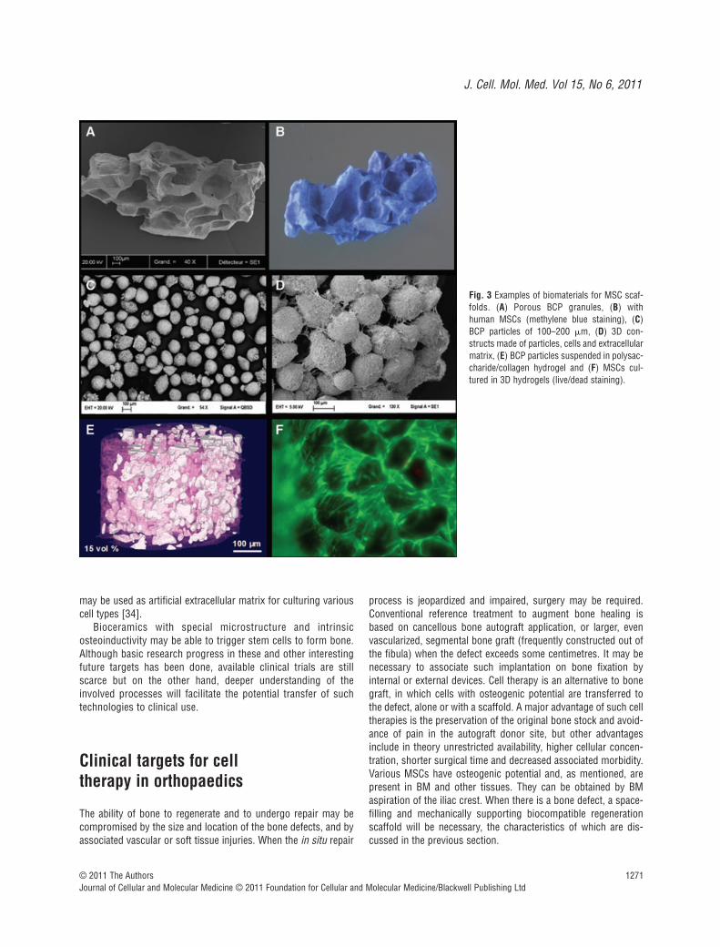
The adhesion, proliferation and osteogenic differentiation ofMSCs on scaffolds are expected to be critical steps in the devel-opment of bone tissue engineering. The scaffold should supportMSC adhesion with a relatively high efficiency. As shown in Figure 3, MSCs attached, proliferated and produced an abundantextracellular matrix on CaP ceramics. However, the cell/biomater-ial volume ratio is highly important for osteoinduction in hybridconstructs. High porosity and permeability of scaffolds areextremely important for a uniform cell invasion, in vivo and in locotissue ingrowth, vascularization and osteogenesis. Intergranularspaces between particulate scaffolds can provide an intercon-nected porosity for cell seeding and a high degree of freedom for
Fig. 1 Principle of bone tissue engineering.

1270 © 2011 The AuthorsJournal of Cellular and Molecular Medicine © 2011 Foundation for Cellular and Molecular Medicine/Blackwell Publishing Ltd
tissue ingrowth. Intergranular spaces may be further increased bysuspending CaP particles in hydrogels. Another advantage ofhydrogels is that they allow cell injection to complex shaped bonedefects or minimal invasive surgery without damaging cell viability.
Hydrogels are promising materials for tissue engineering as they contain a relatively low amount of dry mass (1–20%)causing little inflammation and foreign body reaction duringdegradation. They are appealing materials for tissue engineeringbecause they can homogeneously suspend cells while allowingrapid diffusion of nutrients and metabolites [34]. Ideally, thehydrogel should form a temporary mechanical support for thecells and degradation should keep pace with new tissue formationand the reciprocal extracellular matrix production. Hydrogelsbased on extracellular matrix components (collagen, fibrin,polypeptides) provide an adhesive surface for the cells, but theircomposition, mechanical properties and degradation rates aredifficult to control. Polysaccharides such as agarose, alginate,hyaluronic acid, chitosan and hydroxylpropylmethyl cellulose, areinteresting candidates for cell encapsulation but their chemicalmodifications are difficult due to their insolubility in most oforganic solvents.
Future trends in bone tissue engineering
For several years, the research has attempted to mimic both thecomposition and structure of trabecular bone. Considering thevolume, scaffolds occupy a large part whereas only a layer ofcells is present on the surface of biomaterial. This poor scaffold-to-cell volume ratio may be one of the reasons that only a lim-ited amount of bone tissue is formed in vivo by such hybrid con-structs. Indeed, mimicking ‘the end product’ and not the naturalprocess leading to its formation or bone tissue healing may notbe the most appropriate approach because nature proceeds inthe reverse way, starting with cells which increasingly formmatrix.
During osteogenesis, a relatively high cellular content pre-vails with numerous osteoprogenitors producing extracellularmatrix whereas a limited number of cells can be found in ready-made mature bone tissue. Furthermore, bone tissue is able toheal and is constantly remodelled by cellular activity originatingfrom the periost and BM reservoir. In this process, multiple celltypes cooperate to fabricate this complex 3D tissue whereas theextracellular matrix is degraded as part of the renewal (remod-elling) process by multinucleated osteoclast cells. The balancebetween the osteoblast-mediated formation and osteoclast-mediated resorption is under biochemical and biomechanicalcontrol. Bone tissue is also a highly vascularized tissue withblood capillaries supplying oxygen and nutrients to the cells. Itis therefore a complex 3D tissue containing different types andhigh numbers of cells at the early stage of its formation, butonly a few cells in an abundant extracellular matrix at its maturestate.
The research in tissue engineering has recently moved to thefabrication of artificial extracellular matrices resembling thoseencountered at the early stage of tissue development. In thisrespect, a developmental engineering approach has been pro-posed and described in detail [35, 36]. Recent experimental datahave supported this new conceptual framework [37].
To support this new developmental engineering approach, newbiologically relevant biomaterials, better responding to therequirements of the developmental processes and their modulardesign, need to be developed. The group of Stupp has designedbiomimetic peptides that self-assemble into supramolecular struc-tures under physiological conditions [38]. Hydrogels formed bycrosslinking hydrophilic polymer chains have also been developedas three-dimensional cell and tissue culture environments. Thesehydrogels cross-link into 3D network in response to changes intemperature, pH, ionic environment or via chemical moieties,enzymatic reaction or UV light. The ability to control the synthesisof hydrogels is attractive for building complex 3D structures thatmimic those of tissues. Ladet et al. have recently reported thedesign and fabrication of multilayered polysaccharide-basedhydrogels with highly controlled physical properties [39]. Ourgroup has developed injectable bone fillers using silanized-cellu-lose hydrogels combined with solid CaP particles that gel in situ atphysiological pH [40]. These pH sensitive cross-linking hydrogels
Fig. 2 Examples of biomaterials for scaffolding of human MSCs. (A) PorousBCP ceramics and (B) injectable paste made of CaP particles suspended inhydrogel for minimal invasive surgery.
J. Cell. Mol. Med. Vol 15, No 6, 2011
1271© 2011 The AuthorsJournal of Cellular and Molecular Medicine © 2011 Foundation for Cellular and Molecular Medicine/Blackwell Publishing Ltd
may be used as artificial extracellular matrix for culturing variouscell types [34].
Bioceramics with special microstructure and intrinsicosteoinductivity may be able to trigger stem cells to form bone.Although basic research progress in these and other interestingfuture targets has been done, available clinical trials are stillscarce but on the other hand, deeper understanding of theinvolved processes will facilitate the potential transfer of suchtechnologies to clinical use.
Clinical targets for cell therapy in orthopaedics
The ability of bone to regenerate and to undergo repair may becompromised by the size and location of the bone defects, and byassociated vascular or soft tissue injuries. When the in situ repair
process is jeopardized and impaired, surgery may be required.Conventional reference treatment to augment bone healing isbased on cancellous bone autograft application, or larger, evenvascularized, segmental bone graft (frequently constructed out ofthe fibula) when the defect exceeds some centimetres. It may benecessary to associate such implantation on bone fixation byinternal or external devices. Cell therapy is an alternative to bonegraft, in which cells with osteogenic potential are transferred tothe defect, alone or with a scaffold. A major advantage of such celltherapies is the preservation of the original bone stock and avoid-ance of pain in the autograft donor site, but other advantagesinclude in theory unrestricted availability, higher cellular concen-tration, shorter surgical time and decreased associated morbidity.Various MSCs have osteogenic potential and, as mentioned, arepresent in BM and other tissues. They can be obtained by BMaspiration of the iliac crest. When there is a bone defect, a space-filling and mechanically supporting biocompatible regenerationscaffold will be necessary, the characteristics of which are dis-cussed in the previous section.
Fig. 3 Examples of biomaterials for MSC scaf-folds. (A) Porous BCP granules, (B) withhuman MSCs (methylene blue staining), (C)BCP particles of 100–200 �m, (D) 3D con-structs made of particles, cells and extracellularmatrix, (E) BCP particles suspended in polysac-charide/collagen hydrogel and (F) MSCs cul-tured in 3D hydrogels (live/dead staining).

1272 © 2011 The AuthorsJournal of Cellular and Molecular Medicine © 2011 Foundation for Cellular and Molecular Medicine/Blackwell Publishing Ltd
The clinical indications for the cell or a hybrid cell and scaffoldgraft have to be precise and well defined. In this part, we discusspotential clinical indications for stem cell therapy, and the choiceof type of cell therapy approach that may be appropriate for whichclinical indication (Fig. 4).
Current clinical problems and therapeutic approaches
Local bone defects, whatever the cause of the defects, are at thispoint the main indications for cell/combination therapy in adults.Osteoporosis, a systemic metabolic bone disease, may be a targetin the future due to the risk for associated fractures and relatedlocal problems. The main orthopaedic problems in adults are non-union or delayed consolidation following fracture (as an example,Fig. 5), osteotomy, arthrodesis or limb lengthening; bone defectssubsequent to trauma, infection, prosthesis loosening or tumourresection and finally, bone necrosis (particularly, avascular necro-sis [AVN] of the femoral head, as shown in Fig. 6).
Non-union or delayed consolidation is a potential complicationof a long bone fracture, even if there is no bone loss. Apart fromstability, that may require intra- or extramedullary fixation, autolo-gous bone graft is the treatment of preference. For non-union afterarthrodesis (mainly spine fusion) or lengthening, a new bone graft
may be necessary. Alternative non-surgical treatments with pulsedelectromagnetic fields or low-intensity ultrasound are currently inthe clinical armamentarium.
Bone defects pose an important clinical problem. Surgeonsattempt to fill bone gaps with autograft promoting the formation ofnew bone and allowing integration with the host bone at the margins(graft-host bone interface). Current treatment depends on the lengthor the volume of the defect and there are no clear treatment algo-rithms in effect. Autogenous bone grafting, harvested from iliac crest,is still considered the gold standard, especially in small defects (2–3cm) where the success rate of bone grafting is high. For large bonedefects, the success is limited also due to the amount of graft mate-rial that can be collected. For larger defects (3–10 cm), the Masquelettechnique evolves as being quite useful; the defect is first filled withacrylic cement, which promotes the formation of a pseudo-mem-brane and, 6 weeks later, the cement is replaced with autologous
Fig. 4 Relationship between bone diseases, cell therapy and biomaterials.
Fig. 5 Computerized (CT) reconstruction of a non-union in a tibial diaph-ysis fracture.
Fig. 6 (A) Early AVN of the hip, radiologicalimages; (B) Magnetic resonance imaging ofhips in the frontal plane, the same patient as in(A) showing characteristic images of AVN ofboth femoral heads, Ficat and Arlet stage II.

J. Cell. Mol. Med. Vol 15, No 6, 2011
1273© 2011 The AuthorsJournal of Cellular and Molecular Medicine © 2011 Foundation for Cellular and Molecular Medicine/Blackwell Publishing Ltd
bone graft in which situation the pseudo-membrane appears toincrease the implantation yield, possibly also due to the local expres-sion of cytokines and growth factors [41]. The Masquelet techniqueindirectly demonstrates the importance of the control of the microen-vironment or implantation site for the successful outcome of bonegrafting. For defects larger than 6–8 cm, particularly in long bones,vascularized bone grafts may be necessary. An alternative to thosebone grafting techniques is the Ilizarov distraction technique, knownas bone transport, where the bone defect is filled utilizing the dis-placement of a contiguous diaphyseal segment with the help of anexternal fixation device. This procedure takes several months, usageof an external fixator is necessary and often a bone graft is required
at the end of procedure due to non-union at the junction with themobilized diaphyseal segment. Large allogenic bone grafts may beused to fill large defects; however, they are only very partially recolo-nized by host cells and may be resorbed, as well as potential risk ofpathogen transmission. Bone cement may be used as permanentfiller but this non-biological solution does not regenerate bone.
In bone necrosis, a significant bone defect may not be theproblem, because the bone scaffold is still present. Surgical con-servative treatments to enhance bone repair have been proposed,from the least invasive core decompression to interventions withpossible morbidity such as bone grafting, osteotomy or freevascularized bone graft.
Fig. 7 Different possibilities for cellular thera-pies for bone regeneration (A and B).

1274 © 2011 The AuthorsJournal of Cellular and Molecular Medicine © 2011 Foundation for Cellular and Molecular Medicine/Blackwell Publishing Ltd
Potential clinical applications of cell based therapies for bone repair
The use of stem cells, alone or associated with biomaterials, maybe an alternative to the limited amount of bone autografts availableand the morbidity induced at the harvest site. Autologous BM isrich in osteoprogenitors and growth factors, as well as cell popu-lations such as stromal cells present in the mononuclear cellularfraction of the BM. BM-derived MSCs can be easily obtained bybone aspiration at the iliac crest, prepared and used in differentways (Fig. 7):(1) Mononuclear cells with MSCs may be used directly. The aspi-
rated BM, without other manipulation, can be percutaneouslyinjected at the affected site. To increase the number ofmononuclear cells and thus of MSCs in the volume to beinjected, it is possible to separate by centrifugation themononuclear cells and concentrate them 8- to 10-fold. Afteraspiration with or without concentration, mononuclear cellscan be mixed in the operating room with an osteoconductivescaffold and implanted at the defect site.
(2) Mononuclear cells may be cultured in vitro to allow selectionand expansion of an adherent fraction corresponding toMSCs. This increases the number of MSCs to millions of cells.MSCs can be percutaneously injected alone or extemporane-ously mixed with scaffolds during surgery, so that this com-posite material is used in the same way as bone grafts. Priorto implantation, cultured MSCs may be expanded in vitro onscaffolds during several days/weeks, allowing for scaffold col-onization and for cell differentiation, before grafting of thisprocessed composite material at the affected site.
(3) An alternative to the two previous ways is to implant the com-posite material (cell � scaffold) in a heterotopic site, e.g. in arichly vascularized muscle, to promote angiogenesis andblood vessel growth into the construct during some weeksand then to transfer it to the affected site with vascular anato-moses for the transferred muscle flap containing the implant.
Data reported in clinical studies
Clinical trials, clinical series and case reports have been publishedon the above listed possible uses of MSCs and are summarized inTables 1, 2 and 3.
Regarding therapies for bone healing problems with no signif-icant bone defect (Table 1, reporting on non-union or delayedunion in long bone fractures or osteotomies, lengthening orarthrodesis), Connolly et al. [42] demonstrated back in 1989 apositive correlation between the osteogenic capacity and BM cellconcentration, and published in 1991 on 20 cases of tibial non-union treated by percutaneous injection of BM, with consolidationin 8/10 if immobilized with cast, and 10/10 with intramedullarynailing [43]. Garg et al. [44] reported on 20 cases of long bonenon-unions treated with two percutaneous injections of BM with
an interval of 3 weeks : 17/20 (85%) healed in 5 months. Toimprove the effectiveness of injections, Hernigou [45] used injec-tions of concentrated BM which allowed an enrichment of about 5-fold of nucleated cells in the same 50 ml volume. With this tech-nique, bone union was obtained in 53 of 60 tibia non-unions(88%) and a significantly positive correlation was concludedbetween bone union and the MSC concentration in the injectedgraft [45]. Before concentration, the aspirate contained an averageof 612 � 134 progenitors/cm3 but after concentration, the aver-age was 2579 � 1121/cm3. For the patients with bone union,more than 1500 progenitors/cm3 were injected, with an averagetotal of 54,962 � 17,431 progenitors. A mean of 20 cm3 wasinjected in this study.
Non-concentrated BM combined with a scaffold during surgeryis probably a more frequently used technique in clinical practicethan has been published. Neen et al. [46], in a prospective study,compared two groups of 25 patients for lumbar spinal fusion witheither iliac crest autograft or type I collagen HA matrix soaked withBM aspirate. The fusion rate was equivalent for post-erolateralfusion, but the group with biomaterial had no donor site morbid-ity. Ateschrang et al. [47] reported on 15 infected tibia non-unions,after establishing an infection-free environment, using allogeniccancellous bone graft ‘vitalized’ through the injection of autolo-gous BM. Infection control was obtained in 14/15 (93.3%) andconsolidation in 11/15 (73.3%).
Concentrated BM may be combined with a scaffold during sur-gery. Dallari et al. [48], in a prospective study on high tibialosteotomy, compared three methods to fill the defect : (i)lyophilized bone chips with platelet gel; (ii) lyophilized bone chipswith platelet gel and concentrated BM stromal cells and (iii)lyophilized bone chips alone as a control. Consolidation was bestin group (ii) and in (i) better than in (iii). Gan et al. [49] used con-centrated BM combined with porous �-TCP for posterior instru-mented spinal fusion in 41 patients. After 34.5 months, 95.1%cases had good spinal fusion. This was as effective as autologousbone graft without the morbidity associated with the harvesting ofgraft from the iliac crest.
MSCs can be culture expanded ex vivo to allow injection of agreater number of cells than after concentration. A particularapplication is delayed union/consolidation on which differentstudies are available. Kitoh et al. [50] demonstrated, in a retro-spective study of 56 bones in 20 patients on delayed complica-tion after limb lengthening, that the group with injection of exvivo expanded autologous MSCs (differentiated in osteoblasts)mixed with platelet-rich plasma had a significantly shorter con-solidation period than the control (average index of healing 27.1� 6.89 days/cm versus control 36.2 � 10.4 days/cm). The aver-age number of transplanted cells was 3.2 � 1.37 � 107. In 2009,Kitoh et al. [51] analysed the difference between the femur andthe tibia, and the healing index was lower for the femur. Theirresults suggested that regionally varying bone-formingprocesses by cell transplantation might be related to local bloodsupply and soft tissue covering. Kim et al. [52] performed aprospective study on 64 cases of long bone with closed andsimple fractures with delayed union randomized in two groups:

J. Cell. Mol. Med. Vol 15, No 6, 2011
1275© 2011 The AuthorsJournal of Cellular and Molecular Medicine © 2011 Foundation for Cellular and Molecular Medicine/Blackwell Publishing Ltd
standard treatment and injection of ex vivo cultured MSCs with amedium facilitating osteoblast differentiation. The group withinjection showed at 2 months a significantly better callus forma-tion. Bajada et al. [53] reported one case of tibia pseudoarthrosistreated with the same procedure but using expanded cells mixedwith CaSO4 pellets.
The filling of a (larger) bone defect is a more challenging situ-ation, as cell suspensions alone cannot be used and it is necessaryto deliver them locally and have some containment on a scaffold.The available clinical studies are summarized in Table 2. Wright et al. [54] randomized 90 patients with simple bone cyst in twogroups of treatment, comparing methylprednisolone acetatesteroid injection and injection of non-concentrated BM alone. Thesteroid provided better healing rate (42%) than the BM (23%),suggesting that BM cells in a cavity without matrix cannot con-tribute significantly to healing. Zamzan et al. [55] obtained healingin 82% of 28 cysts treated by aspiration and one to three percuta-neous injections of autogenous BM. Treatment of a bone defect by
a composite implant of non-concentrated, non-expanded BM withscaffold or demineralized bone matrix (DBM) has also beenreported. Tiedeman et al. [56] used BM and DBM to treat osseousdefects in 39 patients with comparable results to iliac crest bonegraft (61% to 77% success rate). Park et al. [57] compared thetreatment of 23 unicameral bone cysts of the calcaneus associat-ing autologous BM with an open chip allogenic bone graft depositor with a closed percutaneous injection of demineralized bonepowder. Results were similar with the advantage of the low mor-bidity associated with a percutaneous treatment. Ochs et al. [58]compared two matched cohorts of patients with deficient acetab-ular bone stock (type III according to the American Academy ofOrthopaedic Surgeons classification) at revision hip replacement,treated by impaction bone grafting and a reinforcement ring. Acontrol group with standard frozen non-irradiated bone bank allo-graft was compared to a group with freeze-dried irradiated boneallograft vitalized with BM aspirated from iliac crest or tibial meta-physis. The results on allograft incorporation were not different,
Table 1 Clinical studies on non-union or delayed union treated by cell therapy
References Cases Treatment Outcome and conclusions
[43] 20 cases, tibia non-union Percutaneous injection of BMConsolidation in 8/10 immobilized with cast and 10/10 with nail
[44]20 cases, long bone non-union
2 percutaneous injection of BM with an interval of 3 weeks
Consolidation of 17/20 (85%) in 5 months
[45] 60 tibia non-union Injection (mean 20 cm3) of concentrated BM (5 to 6 times more nucleated cell in 50 ml).Injected
Bone union in 53/60 (88%) in 4 months. Positive correlation between bone union and concentration of MSCs
[46]2 groups of 25 patients,prospective study in lumbar spinal fusion
One group of Iliac crest autograft and one of type I collagen HA matrix soaked with BM aspirate
Equivalent fusion rate for post-erolateral fusion. Biomaterial group with no complicationson donor site
[47] 15 infected tibia non-unionInfection-free environment, allogenic cancellous bone graft ‘vitalized’ with autologous BM to perform fibula and tibia fusion
Infection control in 14/15 (93.3%). Consolidationin 11/15 (73.3%)
[48]33 patients HTO, prospective in 3 groups
A: lyophilized bone chips with platelet gel wereimplanted (11); B: lyophilized bone chips withplatelet gel and BM stromal cells (12); C:lyophilized bone chips without gel (10),controls
Increased osteoblasts, bone apposition andosseointegration from 6 weeks in groups A and B. Adding platelet gel or platelet gel combined with BM stromal cells to lyophilizedbone chips increases osteogenic potential
[49]41 patients, posterior instrumented spinal fusion
Concentrated BM combined with porous �-TCP
Good spinal fusion 95.1% after 34.5 months
[50]Lengthening of 56 bones in 20 patients
Injection of ex vivo expanded autologous MSCsmixed with platelet-rich plasma (PRP) in 24bones Controls in 32 bones
Significantly shorter consolidation period withMSC-PRP. Average index of healing 27.1 � 6.89days/cm. Control healing: 36.2 � 10.4 days/cm
[52]RCT 64 long bone closedsimple fractures delayedunion in 2 groups
One group of standard treatment. One group ofinjection, ex vivo cultured MSCs with medium forosteoblast differentiation
The group with injection showed at 2 months significant better callus formation
[53]1 case of tibia pseudoarthro-sis resistant to six previoussurgical procedures
Autologous BM stromal cells expanded to 5 � 10(6) cells after three weeks. Combined in sur-gery with calcium sulphate (CaSO4) in pellet form
Clinically and radiologically healed 2 months after implantation

1276 © 2011 The AuthorsJournal of Cellular and Molecular Medicine © 2011 Foundation for Cellular and Molecular Medicine/Blackwell Publishing Ltd
Table 2 Clinical studies on bone defect treatment with cells and a scaffold
References Cases Treatment Outcome and conclusions
[54]
90 patients with simple bonecysts BM (39, 2 year FU)Methylprednisolone acetate(38, 2 year FU)
BM or methylprednisolone acetate injection, at random.
9/39 (23%) with BM healed 16/38 (42%) withmethylprednisolone healed. Superior healing ratewith esteroid injection. The cells of BM in a cavitywithout matrix may not induce healing.
[55]28 patients with simple bonecyst
Aspiration and percutaneous autogenous BM injection (single injection in 16, 2 or 3 injections in 12)
Healing in 23/28 cysts (82%), mean FU 34.7 � 6.87 months. Autogenous BM injection,safe and effective treatment method for simplebone cysts, but sometimes repeated injections arenecessary
[56]48 patients (39 with 19 months av. FU) withdifferent bone defects
DBM used alone and with BM (DBM-BM)
30/39 patients, osseous union (77%). Fracture non-union, most recalcitrant group (unionachieved in only 61%). Comparable to iliac crestautograft
[57]23 calcaneal unicameralcysts in 20 patients (av. FU of 49.4 months)
Lyophilized irradiated chip allogeneic bone andautogenous BM (13 cysts in 11 patients).Percutaneous injection of irradiated allogeneicdemineralized bone powder and autogenousBM (10 cysts in 9 patients).
Comparable results, advantage of safety for the percutaneous treatment.
[58]
78 patients (79 hips) withacetabular defects at revi-sion THR. 87% (69 hips),type III AAOS defects
Standard frozen non-irradiated bone bank allo-graft (group A). Freeze-dried irradiated boneallograft, vitalized with autologous marrow(group B).
Results on incorporation of the allograft were not different, with the advantage of microbiological safety for the irradiated allograft
[59]
10 patients with volumetricbone defects (curettage of 7 benign tumours, 2 pseudoarthrosis, 1 aseptic loosening)
Concentrated BM in association with a collagenmatrix
Bone healing in 7 of 10 patients
[60]Bone defects 4–7 cm in 3 patients (tibia, humerusand ulna)
Osteoprogenitor cells from BM and expandedex vivo. Placed during the operation on macro-porous HA scaffold
Success confirmed at 6.5 years FU [61], but scaffold remained without resorption
[62]One case of avulsed distalphalange of the thumb
Ex vivo expanded cells shed from periosteum,injected in a porous coral block inserted withno contact with bone
Good functional result Biopsy: lamellar bone and ossified endochondral tissue
[63]3 cases of defect after curettage for benigntumours
Composite of ex vivo expanded MSCs on scaffold during several days before operation
Satisfactory osseointegration at 29 months FU
[64] 6 cases of mandible defectComposite of ex vivo expanded MSCs from BMfor 7 days on a bone substitute in osteogenicculture medium
Biopsies at 4 months: bone formation in 3 patients (in 2, unrelated to the tissue-engineered construct)
[65] One patient with subtotalmandilectomy of 7 cm fortumour 8 years before
Titanium mesh cage filled with bone mineralblocks infiltrated with 7 mg rh BMP7 +20 mlautologous BM. Transplant implanted into latis-simus dorsi 7 weeks. Transplanted as a free bone-muscle flap to repair themandibular defect.
Heterotopic bone induction to form a mandibularreplacement inside the latissimus dorsi muscle in ahuman being (patient = bioreactor).
FU: follow-up; AAOS: American Academy of Orthopaedic Surgeons.

J. Cell. Mol. Med. Vol 15, No 6, 2011
1277© 2011 The AuthorsJournal of Cellular and Molecular Medicine © 2011 Foundation for Cellular and Molecular Medicine/Blackwell Publishing Ltd
but with the advantage of microbiological safety for the irradiatedallograft. Jäger et al. [59] used concentrated BM in associationwith a collagen matrix in 10 patients (including filling after curet-tage of 7 benign tumours, 2 pseudoarthrosis and one aseptic loos-ening), and obtained bone healing in 7/10. Quarto et al. [60] firstreported the use of osteoprogenitor cells isolated from BM,expanded ex vivo and placed on a macroporous HA scaffold dur-ing surgery to fill bone defects (4 to 7 cm) in 3 patients (tibia,humerus and ulna). The success of these three cases was con-firmed with a follow-up of 6.5 years by Marcacci et al. [61], but thescaffold, essentially unresorbable, remained unchanged. Thesame year, Vacanti et al. [62] reported the replacement of anavulsed distal phalanx of the thumb by cells harvested from theperiostum and expanded ex vivo, injected in a porous coral(porous HA) block inserted in a pocket beneath a flap at theextremity of the thumb, with no contact with bone tissue. Thefunctional result was good and a biopsy revealed lamellar boneand ossified endochondral tissue.
The next step in therapeutic approach would be to expandMSCs on a scaffold ex vivo before surgery during severaldays/weeks, and then implant this composite in the defect.Morishita et al. [63] reported three cases of defects treated withthis technique after curettage of benign tumours. The BM MSCsafter 2 weeks proliferation were culture expanded on HA blocks orgranules during 2 more weeks with osteoblastic differentiationmedium, before implantation. At a minimum follow up of 29 months,the osseointegration was satisfactory without radiolucent zones.In maxillofacial surgery, Meijer et al. [64] reported six cases of amandible defects filled using this principle and biopsied 4 monthslater. Biopsies showed bone formation in only three out of sixpatients, and in only one out of six patients bone formation wasinduced by the tissue-engineered construct. He concluded that it is important to differentiate between bone formation induced by the cells from the border of the osseous defect and byimplanted cells.
Another approach does not use ex vivo expanded cells but thepatient acts as his own bioreactor. Warnke et al. [65, 66] reportedthe case of a patient with subtotal mandibulectomy of 7 cm, due
to a tumour treated 8 years before, by reconstruction with a bonemuscle flap prefabricated in vivo. A titanium mesh, computer-designed to fit within the defect region, was loaded with HA blockscoated with rhBMP-7 and aspirated BM. This composite wasimplanted in his latissimus dorsi muscle to allow for heterotopicbone growth and vessels ingrowth from the muscle vessels. After7 weeks, the composite was transplanted into the mandible defectand vascular pedicles were anastomosed onto the external carotidvessels. Bone density and mineralization improved with time andbone formation was detected in all parts of the replaced mandible.Unfortunately the initial good results were not durable with frac-ture and infection. The patient died from cardiac arrest 15 monthsafter implantation.
Regarding femoral head avascular osteonecrosis, many tech-niques of core decompression with bone graft have beendescribed for its treatment. Hernigou et al. [67] found a decreasein the number of MSCs in the upper end of the femur in patientswith corticosteroid-induced osteonecrosis. Because osteonecro-sis may thus be a ‘stromal disease’, the possibility ofinjecting/implanting MSCs or BM in the femoral head may be apotential treatment for this condition. Hernigou et al. [68], afterdecompression, used grafting with concentrated BM and reportedresults in 189 hips of 116 patients. Patients with higher numbersof progenitor cells transplanted in their hips had better outcome.In 2009, Hernigou et al. [69] reported satisfactory results on 534hips with avascular osteonecrosis at early stages (stages I and II)treated by this technique, with only 94 total hip replacements at afollow up of 8–18 years. Gangji et al. [70] studied 18 hips (13patients) with stage I or II osteonecrosis of the femoral head. Hipswere randomized in two groups, core decompression alone orcore decompression with implantation of concentrated BM. After24 months, there were significantly better clinical and radiologicalresults with the BM graft. Yamasaki et al. [71] studied 30 hips (22patients) using concentrated BM seeded into a porous HA cylinderto fill the drilled hole after core decompression in osteonecrosis offemoral head. These were compared to a control group of ninehips (eight patients) with the HA cylinder without cells. At themean follow-up of 31 months, a reduction in the osteonecrotic
Table 3 Clinical studies on AVN treated by cell therapy
Reference Cases Treatment Outcome and conclusions
[68]189 hips (116 patients)with AVN of femoral head
Concentrated BM after forage decompression Higher number of progenitor cells transplanted in their hips had better outcome
[70]18 hips (13 patients) stageI/II femoral head AVN
Core decompression alone or core decompressionwith concentrated BM (randomized)
At 24 months, significantly better clinical and radiological with BM
[69]534 hips (342 patients)with AVN (stages I and II)
Core decompression and autologous BM grafting obtained from the iliac crest of patients
Severe collapse and total hip replacement in 94/534 at FU 8 to 18 years after treatment
[71]30 hips (22 patients) and 9 hips (8 patients) in 2 groups
Concentrated BM seeded into porous HA cylinder after core decompression (30 hips). HA cylinder without cells (9 hips).
At a mean 29 months FU, severe collapse in 3/22patients with BM and in 6/8 patients without BM.
FU: follow-up; AAOS: American Academy of Orthopaedic Surgeons.

1278 © 2011 The AuthorsJournal of Cellular and Molecular Medicine © 2011 Foundation for Cellular and Molecular Medicine/Blackwell Publishing Ltd
lesion was observed in the group with cells, where only three pro-gressed to collapse. In the control, a majority of patients had asevere collapse of the femoral head (see Table 3).
The reported trials and studies have established the feasibilityand reasonable safety of cell therapy approaches, and some meas-ures of efficacy in obtaining bone healing. In all these cases, noautologous bone grafts were harvested. However, only small num-bers of patients have so far been included in such studies. Largertrials should be implemented to better define/characterize theimplant (e.g. the optimal cell numbers, parameters for the cell andscaffold combinations) and the long-term efficacy.
Selected paediatric bone disorders and cellular therapies
Two major groups of paediatric bone disorders have been focusedin paediatric patients treated using cellular therapies. These areinborn errors of bone metabolism and degenerative bone disor-ders in childhood.
Inborn errors of bone metabolism and cellular therapy
Plasticity, mineralization and bone remodelling are particularlyimportant during childhood. The critical role of the balancebetween bone formation and bone resorption becomes evident ina number of inborn errors of bone metabolism [72]. Osteopetrosiswas one of the first paediatric bone disorders in which molecularanalysis identified the heterogeneous genetic background of a sin-gle clinical disorder [73]. This helped significantly to identifypatients who will benefit from BM transplantation (BMT) [74, 75].Although the hallmark of osteopetrosis is excessive bone mass,osteogenesis imperfecta is characterized by greatly reduced boneformation and severe fragility of these bones [76]. Interestingly,treatment by BMT in this disease was attempted much later [77].However, in this disease, which is mainly caused by a collagentype I synthesis defect, not only haematopoietic stem cellsbecame of interest, but also stromal cell populations [78, 79]. Infact, the first systemic application of isolated human MSCs wasperformed in osteogenesis imperfecta. Although the growth ratesignificantly improved during the first months and years, it sloweddown subsequently [80]. This correlated with in vitro findingssuggesting that systemic osteopoiesis is more difficult to achieveby current BMT strategies than haematopoiesis [81]. These limi-tations become evident, when children undergo BMT for inbornerrors of metabolism, which affect several tissues, e.g.mucopolysaccharidosis [82, 83]. If the toxicity of BMT can bedecreased in non-malignant diseases, this technique becomes the
most promising alternative for novel cell therapy approaches [84].Novel cell types and targeted conditioning regimens of reducedintensity will allow for systemic cellular therapies of inborn errorsof bone metabolism [85, 86].
Degenerative bone disorders in childhood
Osteonecrosis in paediatric patients is frequently caused bysteroid-based therapies of haematological malignancies [87].Furthermore, alterations of blood rheology, e.g. in sickle cellanaemia, render children susceptible to AVN of the bone [88].Predominantly, femoral bones are affected with distal epiphy-seal lesions being more frequent than proximal lesions. Theincidence of AVN in children under the age of 10 years is below0.2% after treatment in the acute lymphoblastic leukaemia –Berlin–Frankfurt–Münster (ALL-BFM) protocol 95 (trial ALL-BFM 95). However, 16% of the teenagers older than 15 yearstreated using this protocol are affected by AVN. Thus, AVN is afrequent complication of steroid-based therapies for haemato-logical malignancies. Similarly, juvenile idiopathic arthritis orother autoimmune diseases in children may lead to AVN, whenglucocorticosteroids are added to the treatment regimen [89].Current treatment strategies aim primarily at elimination ofpain, restoration of function and prevention of disease progres-sion. Conservative measures are limited to immobilization andphysiological rehabilitation, which is often complicated in chil-dren due to compliance problems [90]. Surgical measuresinclude core decompression and ultimately prosthetic replace-ment. The outcome after core decompression is very variableand often dependent on risk factors, localization and surgicalaccessibility [91–93]. In order to improve the efficacy of coredecompression, healthy BM or BM mononuclear cells from anaspirate at a distant site have been instilled in the interventions[68, 70, 94]. The most potent osteogenic cells in the BM knownto date are MSCs [95–97]. Recent analyses of this population inthe marginal zone of steroid-induced AVN showed a significantreduction in their numbers, viability and plasticity [67].However, these cells are of critical importance for regeneratingbony tissue via secretion of factors modulating the hypoxic andinflammatory environment as well as stimulating angiogenesis,which seems equally important for bone regeneration as thetransdifferentiation of MSCs [98]. It is a matter of debate,whether MSCs may differentiate into tissues of interest,although this proof of principle has been verified in various ani-mal model experiments [99–101]. In some studies, the benefi-cial effect of MSCs was rather attributed to the secretion ofcytokines and growth factors at the site of injury [102, 103].Obviously, for the use of MSCs in regenerative medicine a sus-tained engraftment is desirable. In fact, in the osteonecrosismodel experiments advantage is taken of both mechanisms,secretion of tissue-repair modulating factors as well as cellengraftment with osteogenic differentiation.

J. Cell. Mol. Med. Vol 15, No 6, 2011
1279© 2011 The AuthorsJournal of Cellular and Molecular Medicine © 2011 Foundation for Cellular and Molecular Medicine/Blackwell Publishing Ltd
Osteonecrotic lesions are bradytrophic areas with low concen-trations of oxygen and nutrients. Interestingly, MSCs produce vas-cular endothelial growth factors and insulin-like growth factorbinding proteins which are anti-apoptotic and involved in neovas-cularization and osteogenesis [104]. It is noteworthy that MSCsgrow in culture media with low glucose content and thus areadapted to limited nutrient supply at the time when they areinjected into the necrotic lesion.
Taken together, cellular therapy approaches are quitepromising for several inherited as well as acquired bone diseasesduring childhood, because a durable cure of the musculoskeletalsystem for a lifetime is of crucial importance for development andquality of life.
Ethical aspects of EU clinical trials
Continuous development of the new fields in medicine led to aboost of studies involving both human samples and animal exper-iments used for clinical and preclinical trials. The European Unionseeks to unify all the ethical and legal aspects regarding such con-cerns under a common framework, so that the design of the stud-ies, procedures for their approval and other related issues wouldbe similar throughout the Union. Current research activities in thefield of human health raise various ethical concerns. Worldwidethere is a continuing interest in ethical aspects, including researchintervention and clinical trials conducted in human beings, the useof human adult/embryonic stem cell (ESC) and/or foetal cells andthe experiments on non-human primates and other animals.
The main ethical issues related to the scientific research is theneed to carry out all types of research activities using such cells,because there is few other feasible alternative and the need toevaluate the benefit/burden balance, including the scientificaspects and the social and cultural gains. Induced pluripotentstem cells and somatic cell nuclear transfer (therapeutic andreproductive cloning) are examples of innovations, which can per-haps, to an extent, solve some of the important immunologicaland ethical issues, but will also create new ones [105].
The development of scientific research based on experimentson human beings (the human model) raised a social problem –patients’ desire to have access to the latest discoveries in the med-ical field and researchers’ desire to discover new therapies, inves-tigative methods and drugs. The risks of research areacceptable/accepted, as it is believed that the outcome contributeto the public good. The purpose of research is therefore theimprovement of healthcare [106]. Whereas scientists claim aca-demic freedom, self-direction and self-regulation, the publicresponses ranged between wonder and awe and fear and anger.As science is publicly funded and performed for the benefit of thesociety, it is expected to act responsibly in exchange of fulfilling itsdemands for resources and autonomy, and that the researcherswill abide to the highest ethical standards [107].
The ethical framework presumes two essential elements: pro-vision of information regarding the ethics of research on humanbeings and the fundamental principles (rules and ideas) that willinfluence the conduct of research and the establishment of proce-dures which have been designed to facilitate the application of theethical principles. Research should respect human life and dignity,and the integrity of scientists is the basis of the privilege ofresearch freedom granted by society to such undertakings [107].
A special ethical concern is raised by ESC research. Manyauthors argued that ESC research is opposed to human dignity, asit requires the destruction of human embryos, considered to behuman beings at a very early stage of their development. Instead,it promotes research with adult stem cells, as this does not involvethe destruction of human embryos, and that adult stem cellsappear equally promising when compared to ESC in the context ofspecific clinical applications such as bone regeneration[108–111]. This position considers that moral status of an embryois absolute at all stages of its development [109], and thereforeembryos should be considered persons, involving the respect oftheir rights which are the same as those of all human beings.Another position deems the moral status to gradually increase,therefore ESC research needing careful consideration [112, 113].Finally, some authors acknowledge no moral status of embryos,so the research that uses ESC would be moral for them, and eventhey claim not conducting ESC research would be immoral [114, 115]. Moreover, different countries hold different opinionson the subject, not always translated in their laws. In a global sur-vey performed in 2006 involving 50 countries, 23 allow researchon human embryos under strict conditions, out of which 16 havelaws in force, 7 conduct ESC research by guidelines. Somecountries, such as Austria, Ireland, Cyprus, Costa Rica and Italyexplicitly prohibit ESC research. US law allows the procurement ofhuman ESC lines and research on supernumerary embryos byguidelines. Japan, as well as Belgium, Singapore, South Korea,Sweden and UK have adopted national laws allowing embryocloning for therapeutic or research purposes. The remaining coun-tries have no explicit policy on the topic [111].
Nationally and internationally, there has been a proliferationof laws, regulations and guidelines, generated by different reg-ulatory bodies, which has resulted in disharmony in national,regional and international recommendations for research. Amultitude of different organizations provide multinational guide-lines, standards, regulations and opinions including the WorldHealth Organization, World Medical Association (WMA), EC,European Medicines Agency (EMEA), European ScienceFoundation with a recent Science Policy Briefing [116],International Conference on Harmonization (ICH), Council forInternational Organizations of Medical Sciences, United NationsEducational, Scientific and Cultural Organization, EuropeanGroup on Ethics in Science and New Technologies (EGE),International Society for SC Research, Human GenomeOrganization, cleric organizations, etc.
Among these documents, only the most significant ones are mentioned in this review. The Code of Nuremberg [117],
1280 © 2011 The AuthorsJournal of Cellular and Molecular Medicine © 2011 Foundation for Cellular and Molecular Medicine/Blackwell Publishing Ltd
published in 1947 as a result of the medical experimentation dur-ing World War II, states the voluntary participation based oninformed consent [118]. The Declaration of Helsinki [119] wasdeveloped by the WMA in 1964, in order to provide a set of prin-ciples to physicians and other participants in medical researchinvolving human beings. The Declaration focused on theresearcher’s responsibility to protect the individuals of research[118]. The Belmont Report, published in 1979, provides an ethicalfoundation on the protection of human beings and it became asymbol of a fight against racism and abuse of vulnerable individ-uals of medical research. Written by the National Commission forthe Protection of Human Subjects of Biomedical and BehaviouralResearch, drafted first in the Belmont Conference Centre, theReport discusses six key ethical principles and their application inresearch: informed consent, beneficence, justice, fidelity, non-malfeasance and veracity [118]. In 1990, the ICH of TechnicalRequirements for Registration of Pharmaceuticals for Human Usewas founded by the FDA (Food and Drug Administration), the EC(European Commission) and the MHLW (Japanese Ministry ofHealth, Labor and Social Affairs) in alliance with the pharmaceuti-cal industry. As a result, the EMEA was established by theEuropean Commission [110]. ICH deals with quality, safety, effi-cacy and multidisciplinary and other relevant aspects of perform-ing clinical trials are approached. The council of EuropeConvention on Human Rights and Biomedicine [120] signed on 4 April, 1997 in Oviedo that ‘an intervention in the health field mayonly be carried out after the person concerned has given free andinformed consent to it’. The scientific research in the field of biol-ogy and medicine shall be carried out freely and by ensuring theprotection of the individual. The Directive 2004/23/EC defines thestandards of quality and safety for the donation, procurement,testing, processing, preservation, storage and distribution ofhuman tissues and cells [121].
The fundamental ethical principles applicable to human stemcell research, stipulated in the Opinion #15 of EGE in Sciencesand New Technologies regarding ‘Ethical aspects of human stemcell research and use’ [122] involve the principles of respect forhuman dignity, individual autonomy, justice and beneficence(with regard to the improvement and protection of health), free-dom of research and proportionality. It also recommends takinginto account, based on a precautionary approach, the potentiallong-term consequences of stem cell research and use for indi-viduals and the society. The basic ethical issues connected withclinical trials are: (1) the procedure for obtaining informed con-sent of the individuals of the clinical trial; (2) approval of thestudies; (3) data protection, confidentiality and anonymization;(4) risk-benefit assessment; (5) protection of the health of per-sons involved in clinical trials and (6) transparency regardingresearch results.
Ethics related to information and consent
Persons who take part in the research are entitled to be informedabout and to consent or not to clinical research [123]. EGE
Opinion #15 [122] states the ethical aspects of clinical trialsregarding the protection of the recipients of transplantation. Itargues ‘the potential long-term consequences of stem cellresearch and use for individuals and the society’ must be takeninto account. Information should be provided as clear and preciseas possible by the doctor supervising the procurement: arrange-ments, in particular on the ‘free nature of the donation’, and itsanonymity; possible tissue storage time and conditions; registra-tion of data in databases, in conformity with requirements ofprivate life protection and medical confidentiality; foreseeable useof the tissues (diagnostic, allograft or autograft, pharmaceuticalproducts, research, cell lines production for various uses, etc.);the donor may at any time withdraw her/his consent, without anynegative consequences for the person. There are two key issuesthat must be included in the informed consent forms: who benefits and what happens to data, samples and animals at theend. Only persons able to freely understand and question the pro-tocols may/must provide consent. Vulnerable individual categoriesare considered to be pregnant women, human foetuses, neonates,children, prisoners, persons who are physically handicapped,mentally disabled, economically or educationally disadvantaged,racial minorities, the very sick and the institutionalized. Thesepopulations are generally excluded, but there are possibilities ofincluding them in order to avoid loss of opportunity.
Informed consent is a requirement for ethical conduct of clin-ical trials, but it is not sufficient [118]. In order to obtain aninformed consent, the investigators must determine the cultureand literacy of the individuals. The notion of individuality is lack-ing in some cultures. The individuals must be adults who showliteracy and responsibility. Written documents are not always pro-vided. The World Health Organization provides templates for different informed consent forms. All of these consist of two mainsections: an information sheet, and a certificate of consent (or certificate of assent for children).
Approval of the studies
All clinical trial must be approved by the Research EthicsCommittee. In this respect the requirement of scientific journals tomention the ethical approval and the name of the approving com-mittee has improved the situation although in one recent study31% of manuscripts published in high impact journals lacked themention of the ethical approval [124]. More demanding studies,such as multicenter, multinational or ethically complicated studies,may require approval by national competent authority. Similarly,for the commercially orientated parties and sponsors, the require-ment of FDA- and EMEA-like agencies for ethical approval beforeregistration of new drugs and ATMPs provides a good incentive.
Ethics related to privacy/data protection
The Charter of Fundamental Rights refers to protection ofpersonal data and non-discrimination, which also included

J. Cell. Mol. Med. Vol 15, No 6, 2011
1281© 2011 The AuthorsJournal of Cellular and Molecular Medicine © 2011 Foundation for Cellular and Molecular Medicine/Blackwell Publishing Ltd
genetic status. EGE Opinion #11, point 2.4, states: ‘In order toreconcile the traceability requirement and the need to protectthe donor’s rights (medical confidentiality and privacy), tissuebanks must take the necessary steps to protect confidential-ity of the data by developing appropriate coding systems’[125].
EGE Opinion #13 considers that personal health data formpart of the personality of the individual, and must not be treatedas mere objects of commercial transaction [126]. The humanright to respect for private life requires that confidentiality ofpersonal health data is guaranteed at all times and that informedconsent of the individual is required for the collection andrelease of such data. The principles of self-determination,legitimate purpose, security and the right to participate in themedical decision-making process are addressed. At the time,EGE considered a directive on medical data protection wasneeded within the existing Data Protection Directive to addressthe issues related to use of medical data through informationand communications technologies means [126]. This led to theestablishment of Directive 2002/58/EC of the EuropeanParliament and of the Council of 12 July 2002 concerning theprocessing of personal data and the protection of privacy in theelectronic communications sector (directive on privacy and elec-tronic communications). Personal data, such as health informa-tion, criminal justice, financial, genetic and location informationmust be protected.
The investigator’s challenge is to process (obtain, hold, dis-close) data while protecting identity. This is achieved by fair andlawful processing, and only for limited purposes. The process-ing must be adequate, relevant and not excessive. The datamust be recorded accurately and securely, not kept longer thannecessary and processed in accordance with the individual’srights. It is not to be transferred to countries without adequatedata protection.
Ethics related to the risk-benefit assessment
This is critical in stem cell research, as in any research, but ismore difficult as the uncertainties are considerable given thegaps in our knowledge. Attempts to minimize the risks andincrease the benefits have been made, and strategies for safetyhave been elaborated. These have utmost importance in thetransplantation of genetically modified cells and when stem cellsare derived from somatic cells (the risks that transplanted stemcells cause abnormalities or induce tumours or cancer). Thepotential benefits for the patients should be taken into account,but not exaggerated. The risk-benefit assessment for each indi-vidual is an independent ethical requirement for a trial to beacceptable [127]. Clinical research represents a context with dis-tinctive risk and benefit characteristics, which requires applica-tion of patient standard rather than professional standardregarding the uncertainty of the risks and the limited benefits tothe patient [123].
Ethics related to protection of the health of persons involved in clinical trials
This must be made in order to minimize the possibility that irre-versible and potentially harmful changes are introduced in clini-cal applications of stem cell research. Techniques enhancing thepossibilities of reversibility should be used whenever possible.Usually some groups, such as prisoners, mentally impaired per-sons, severely injured patients and very young children areexcluded from clinical trials. ‘The general social goal should be toensure that all decisions about biomedical research are consis-tent with standards for research integrity and public health protection’ [128].
Ethics related to transparency regarding research results
EGE recommended in the context of funding stem cell researchwithin the EU Framework Programs for Research that ‘the EUshould insist that the results of such research be widely dissemi-nated and not hidden for reasons of commercial interest’. There isalso a need to avoid unnecessary replication of research. Whenresearchers conceal the presence of selected trials, these studiescannot influence the thinking of patients, clinicians, otherresearchers and experts who write practice guidelines.Pharmaceutical companies will have to reveal some information,as descriptions of studies on drugs [128]. If every trial’s existenceis part of the public record the stakeholders in clinical researchcan explore the full range of clinical evidence. The clinical trial reg-istries were introduced to address this issue. Research sponsorshave the opportunity to show the public they carefully consider theethical issues related to research. They may submit information onclinical trials they initiate and report the outcome in the registries[128, 129].
Future directions and remarks
There is a high interest for biomedical research on innovative(stem) cell therapeutic approaches. New cell based technologiesand stem cell research arouse fears and worries in many peopleincluding the issues of embryo research and cell nuclear replace-ment. The development of a policy for research and therapy in thisfield within the EU should be broadly permissive, but with rigor-ous ethical and legal oversight, enabling the policy to evolve in arational way and with public support.
Among the potential cell therapy approaches that can promotebone healing in a clinical scenario, the easiest and current tech-nique is the use of aspirated BM. Hernigou et al. [45], using BM
1282 © 2011 The AuthorsJournal of Cellular and Molecular Medicine © 2011 Foundation for Cellular and Molecular Medicine/Blackwell Publishing Ltd
concentrates, demonstrated positive correlation between success-ful bone union and the number of the injected progenitor cells. Theconcentration of BM aspirates is a rather simple procedure, but toassess its efficacy, it is necessary to know the quantity of injectedmononuclear cells and of the MSCs. Even after concentration, weneed more information on the purity i.e. not only nucleated cellsare present, but also other cell types, platelets and an array ofsecreted factors are also included and may have a role in the effec-tiveness of the graft. It is a simple procedure, during surgery, toco-implant native BM aspirates with a scaffold, which are particu-larly needed in bone defect surgery. However, it is not presentlypossible to ascertain the optimum number of cells and the volumeto be used, and how many and which cells are really sufficient andrequired for an improved clinical outcome.
The culture expansion of cells ex vivo is at first a more com-plex technique, but easier for subsequent characterization andquantification, and the number of cells obtained for transplanta-tion is more than a 1000 times greater than in concentrates.Correlation with clinical outcome therefore should be moreachievable. The exact quantification also allow for an improvedseeding on a wider scaffold surface. The currently available datafavour the opinion that to make this all more predictable in clinicaloutcome, expanded, well-characterized MSC populations androbustly manufactured combination products are the best long-term solution. However, this requires some serious additionalresearch and preclinical work, and the implementation of theseprocesses along GMP guidelines.
In addition, quite some steps have to be taken with regard tothe clinical studies and their outcome. For bone healing studiesmore in particular, the evolution of the graft is often controlledonly by imaging, because biopsies may be hazardous on ahealed bone union. There is little histological information on theosseointegration. Probably, the main part of the graft undergoesnecrosis, similarly to the autologous cancellous bone graft. Thedevelopment of new imaging modalities will certainly be instru-mental to better understand the processes. To improve the vas-cularization of the forming bone, the currently available options
include the use of vascularized bone grafts (fibula) and the twosteps procedure as proposed by Warnke et al. [66], technicallydemanding and not possible in all localizations. In view of allthis, adaptive clinical trial design and evaluation with combinedoutcome are aspects that will need to be integrated with thesenew cell based therapeutics. Identifying the proper indication,patients at risk and responders to treatment are part of the clin-ical challenges.
In future, using more advanced developmental engineeringapproaches will lead to the manufacturing of robust tissue inter-mediates enhancing many critical aspects of bone healing includ-ing vascularization, tissue integration and remodelling. Finally,because MSCs appear to have some immune modulatory/sup-pressive effects, an allogenic approach may become feasible. Theuse of progenitor cell population must always take safety intoaccount including caution when this therapeutic approach is usedin bone defects after tumour surgery.
Acknowledgements
The authors are grateful to the European Science Foundation for theResearch Network Program (RNP) funding the Regenerative MedicineNetwork (REMEDIC), which took the initiative to this review. The EuropeanUnion support by means of the 7th Frame Program is appreciated with thefunding of the REBORNE project (‘Regenerating Bone defects using Newbiomedical Engineering approaches’, EC-GA n�241879, HEALTH-2009–1.4.2); the Danish Council for Strategic Research of the‘Individualized musculoskeletal regeneration and reconstruction’ and EUMATERA projects/TEKES for stem cell research and new generation titania.
Conflict of interest
The authors confirm that there are no conflicts of interest.
References
1. Burchardt H. Biology of bone transplanta-tion. Orthop Clin North Am. 1987; 18:187–96.
2. De Groot K. Ceramics of calcium phos-phates: preparation and properties.Bioceramics of calcium phosphate BocaRaton FL: CRC Press; 1983.
3. Daculsi G. Biphasic calcium phosphateconcept applied to artificial bone, implantcoating and injectable bone substitute.Biomaterials. 1998; 19: 1473–8.
4. Laurencin C, Khan Y, El-Amin SF. Bonegraft substitutes. Expert Rev Med Devices.2006; 3: 49–57.
5. Fellah BH, Gauthier O, Weiss P, et al.Osteogenicity of biphasic calcium phos-phate ceramics and bone autograft in a goatmodel. Biomaterials. 2008; 29: 1177–88.
6. Langer R, Vacanti JP. Tissue engineering.Science. 1993; 260: 920–6.
7. Petite H, Viateau V, Bensaid W, et al.Tissue-engineered bone regeneration. NatBiotechnol. 2000; 18: 959–63.
8. Derubeis AR, Cancedda R. Bone marrow stromal cells (BMSCs) in boneengineering: limitations and recentadvances. Ann Biomed Eng. 2004; 32:160–5.
9. Khan Y, Yaszemski MJ, Mikos AG, et al.Tissue engineering of bone: material andmatrix considerations. J Bone Joint SurgAm. 2008; 90: 36–42.
10. Giannoudis P, Einhorn T. Bone morpho-genetic proteins – applications inorthopaedic and trauma surgery. London:Elsevier; 2010.
11. Garrison KR, Shemilt I, Donell S, et al.Bone morphogenetic protein (BMP) forfracture healing in adults. CochraneDatabase Syst Rev. 2010; 6: CD006950.
12. Schallmoser K, Rohde E, Reinisch A, et al. Rapid large-scale expansion of

J. Cell. Mol. Med. Vol 15, No 6, 2011
1283© 2011 The AuthorsJournal of Cellular and Molecular Medicine © 2011 Foundation for Cellular and Molecular Medicine/Blackwell Publishing Ltd
functional mesenchymal stem cells fromunmanipulated bone marrow without ani-mal serum. Tissue Eng Part C Methods.2008; 14: 185–96.
13. Di Nicola M, Carlo-Stella C, Magni M, et al. Human bone marrow stromal cellssuppress T-lymphocyte proliferationinduced by cellular or nonspecific mito-genic stimuli. Blood. 2002; 99: 3838–43.
14. Dominici M, Le Blanc K, Mueller I, et al.Minimal criteria for defining multipotentmesenchymal stromal cells. TheInternational Society for Cellular Therapyposition statement. Cytotherapy. 2006; 8:315–7.
15. Crisan M, Chen CW, Corselli M, et al.Perivascular multipotent progenitor cellsin human organs. Ann N Y Acad Sci. 2009;1176: 118–23.
16. Bieback K, Kern S, Kluter H, et al. Criticalparameters for the isolation of mesenchy-mal stem cells from umbilical cord blood.Stem Cells. 2004; 22: 625–34.
17. Xin Y, Wang YM, Zhang H, et al. Agingadversely impacts biological properties ofhuman bone marrow-derived mesenchy-mal stem cells: implications for tissueengineering heart valve construction. ArtifOrgans. 2010; 34: 215–22.
18. Shi YY, Nacamuli RP, Salim A, et al. Theosteogenic potential of adipose-derivedmesenchymal cells is maintained withaging. Plast Reconstr Surg. 2005; 116:1686–96.
19. van Harmelen V, Skurk T, Rohrig K, et al.Effect of BMI and age on adipose tissuecellularity and differentiation capacity inwomen. Int J Obes Relat Metab Disord.2003; 27: 889–95.
20. Jurgens WJ, Oedayrajsingh-Varma MJ,Helder MN, et al. Effect of tissue-harvest-ing site on yield of stem cells derived fromadipose tissue: implications for cell-basedtherapies. Cell Tissue Res. 2008; 332:415–26.
21. Marx RE, Carlson ER, Eichstaedt RM, et al. Platelet-rich plasma: growth factorenhancement for bone grafts. Oral SurgOral Med Oral Pathol Oral Radiol Endod.1998; 85: 638–46.
22. Chevallier N, Anagnostou F, Zilber S, et al. Osteoblastic differentiation of humanmesenchymal stem cells with plateletlysate. Biomaterials. 2010; 31: 270–8.
23. Anselme K, Broux O, Noel B, et al. Invitro control of human bone marrow stro-mal cells for bone tissue engineering.Tissue Eng. 2002; 8: 941–53.
24. Materna T, Rolf HJ, Napp J, et al. In vitrocharacterization of three-dimensional scaf-
folds seeded with human bone marrowstromal cells for tissue engineered growthof bone: mission impossible? A method-ological approach. Clin Oral Implants Res.2008; 19: 379–86.
25. Giannoni P, Muraglia A, Giordano C, et al. Osteogenic differentiation of humanmesenchymal stromal cells on surface-modified titanium alloys for orthopedicand dental implants. Int J Artif Organs.2009; 32: 811–20.
26. Mastrogiacomo M, Scaglione S,Martinetti R, et al. Role of scaffold inter-nal structure on in vivo bone formation inmacroporous calcium phosphate bioce-ramics. Biomaterials. 2006; 27: 3230–7.
27. Caplan AI. Mesenchymal stem cells. J Orthop Res. 1991; 9: 641–50.
28. Bruder SP, Jaiswal N, Haynesworth SE.Growth kinetics, self-renewal, and theosteogenic potential of purified humanmesenchymal stem cells during exten-sive subcultivation and following cryop-reservation. J Cell Biochem. 1997; 64:278–94.
29. Sacchetti B, Funari A, Michienzi S, et al.Self-renewing osteoprogenitors in bonemarrow sinusoids can organize ahematopoietic microenvironment. Cell.2007; 131: 324–36.
30. de Girolamo L, Sartori MF, Albisetti W, et al. Osteogenic differentiation of humanadipose-derived stem cells: comparison oftwo different inductive media. J TissueEng Regen Med. 2007; 1: 154–7.
31. Lee JH, Rhie JW, Oh DY, et al.Osteogenic differentiation of human adi-pose tissue-derived stromal cells (hASCs)in a porous three-dimensional scaffold.Biochem Biophys Res Commun. 2008;370: 456–60.
32. De Bari C, Dell’Accio F, Vanlauwe J, et al. Mesenchymal multipotency of adulthuman periosteal cells demonstrated bysingle-cell lineage analysis. ArthritisRheum. 2006; 54: 1209–21.
33. Eyckmans J, Luyten FP. Species speci-ficity of ectopic bone formation usingperiosteum-derived mesenchymal pro-genitor cells. Tissue Eng. 2006; 12:2203–13.
34. Vinatier C, Guicheux J, Daculsi G, et al.Cartilage and bone tissue engineeringusing hydrogels. Biomed Mater Eng. 2006;16: S107–13.
35. Lenas P, Moos M, Luyten FP.Developmental engineering: a new para-digm for the design and manufacturing ofcell-based products. Part I: from three-dimensional cell growth to biomimetics of
in vivo development. Tissue Eng B Rev.2009; 15: 381–94.
36. Lenas P, Moos M, Luyten FP.Developmental engineering: a new para-digm for the design and manufacturing ofcell-based products. Part II: from genes tonetworks: tissue engineering from theviewpoint of systems biology and networkscience. Tissue Eng B Rev. 2009; 15:395–422.
37. Scotti C, Tonnarelli B, PapadimitropoulosA, et al. Recapitulation of endochondralbone formation using human adult mes-enchymal stem cells as a paradigm fordevelopmental engineering. Proc Natl AcadSci USA. 2010; 107: 7251–6.
38. Zhang S, Greenfield MA, Mata A, et al. Aself-assembly pathway to aligned mon-odomain gels. Nat Mater. 2010; 9:594–601.
39. Ladet S, David L, Domard A. Multi-membrane hydrogels. Nature. 2008; 452:76–9.
40. Fellah BH, Weiss P, Gauthier O, et al.Bone repair using a new injectable self-crosslinkable bone substitute. J OrthopRes. 2006; 24: 628–35.
41. Masquelet AC, Begue T. The concept ofinduced membrane for reconstruction oflong bone defects. Orthop Clin North Am.2010; 41: 27–37.
42. Connolly JF, Guse R, Tiedeman J, et al.Autologous marrow injection for delayedunions of the tibia: a preliminary report. JOrthop Trauma. 1989; 3: 276–82.
43. Connolly JF, Guse R, Tiedeman J, et al.Autologous marrow injection as a substi-tute for operative grafting of tibialnonunions. Clin Orthop Relat Res. 1991;259–70.
44. Garg NK, Gaur S, Sharma S.Percutaneous autogenous bone marrowgrafting in 20 cases of ununited fracture. Acta Orthop Scand. 1993; 64:671–2.
45. Hernigou P, Poignard A, Beaujean F, et al. Percutaneous autologous bone-marrow grafting for nonunions. Influenceof the number and concentration of pro-genitor cells. J Bone Joint Surg Am. 2005;87: 1430–7.
46. Neen D, Noyes D, Shaw M, et al.Healos and bone marrow aspirate usedfor lumbar spine fusion: a case con-trolled study comparing healos withautograft. Spine. 2006; 31: E636–40.
47. Ateschrang A, Ochs BG, Ludemann M, etal. Fibula and tibia fusion with cancellousallograft vitalised with autologous bonemarrow: first results for infected tibial

1284 © 2011 The AuthorsJournal of Cellular and Molecular Medicine © 2011 Foundation for Cellular and Molecular Medicine/Blackwell Publishing Ltd
non-union. Arch Orthop Trauma Surg.2009; 129: 97–104.
48. Dallari D, Savarino L, Stagni C, et al.Enhanced tibial osteotomy healing withuse of bone grafts supplemented withplatelet gel or platelet gel and bone marrowstromal cells. J Bone Joint Surg Am. 2007;89: 2413–20.
49. Gan Y, Dai K, Zhang P, et al. The clinicaluse of enriched bone marrow stem cellscombined with porous beta-tricalciumphosphate in posterior spinal fusion.Biomaterials. 2008; 29: 3973–82.
50. Kitoh H, Kitakoji T, Tsuchiya H, et al.Distraction osteogenesis of the lowerextremity in patients with achondropla-sia/hypochondroplasia treated with trans-plantation of culture-expanded bone marrow cells and platelet-rich plasma. J Pediatr Orthop. 2007; 27: 629–34.
51. Kitoh H, Kawasumi M, Kaneko H, et al.Differential effects of culture-expandedbone marrow cells on the regeneration ofbone between the femoral and the tibiallengthenings. J Pediatr Orthop. 2009; 29:643–9.
52. Kim SJ, Shin YW, Yang KH, et al. A multi-center, randomized, clinical study to com-pare the effect and safety of autologouscultured osteoblast(Ossron) injection totreat fractures. BMC MusculoskeletDisord. 2009; 10: 20–9.
53. Bajada S, Harrison PE, Ashton BA, et al.Successful treatment of refractory tibialnonunion using calcium sulphate and bonemarrow stromal cell implantation. J BoneJoint Surg Br. 2007; 89: 1382–6.
54. Wright JG, Yandow S, Donaldson S, etal. A randomized clinical trial comparingintralesional bone marrow and steroidinjections for simple bone cysts. J BoneJoint Surg Am. 2008; 90: 722–30.
55. Zamzam MM, Abak AA, Bakarman KA, etal. Efficacy of aspiration and autogenousbone marrow injection in the treatment ofsimple bone cysts. Int Orthop. 2009; 33:1353–8.
56. Tiedeman JJ, Garvin KL, Kile TA, et al.The role of a composite, demineralizedbone matrix and bone marrow in the treat-ment of osseous defects. Orthopedics.1995; 18: 1153–8.
57. Park IH, Micic ID, Jeon IH. A study of 23unicameral bone cysts of the calcaneus:open chip allogeneic bone graft versuspercutaneous injection of bone powderwith autogenous bone marrow. Foot AnkleInt. 2008; 29: 164–70.
58. Ochs BG, Schmid U, Rieth J, et al.Acetabular bone reconstruction in revi-
sion arthroplasty: a comparison of freeze-dried, irradiated and chemically-treatedallograft vitalised with autologous marrow versus frozen non-irradiated allograft. J Bone Joint Surg Br. 2008; 90:1164–71.
59. Jäger M, Jelinek EM, Wess KM, et al.Bone marrow concentrate: a novel strategyfor bone defect treatment. Curr Stem CellRes Ther. 2009; 4: 34–43.
60. Quarto R, Mastrogiacomo M, CanceddaR, et al. Repair of large bone defects withthe use of autologous bone marrow stro-mal cells. N Engl J Med. 2001; 344: 385–6.
61. Marcacci M, Kon E, Moukhachev V, et al.Stem cells associated with macroporousbioceramics for long bone repair: 6- to 7-year outcome of a pilot clinical study.Tissue Eng. 2007; 13: 947–55.
62. Vacanti CA, Bonassar LJ, Vacanti MP, etal. Replacement of an avulsed phalanxwith tissue-engineered bone. N Engl JMed. 2001; 344: 1511–4.
63. Morishita T, Honoki K, Ohgushi H, et al.Tissue engineering approach to the treat-ment of bone tumors: three cases of cul-tured bone grafts derived from patients’mesenchymal stem cells. Artif Organs.2006; 30: 115–8.
64. Meijer GJ, de Bruijn JD, Koole R, et al.Cell based bone tissue engineering in jawdefects. Biomaterials. 2008; 29:3053–61.
65. Warnke PH, Springer IN, Wiltfang J, et al. Growth and transplantation of a cus-tom vascularised bone graft in a man.Lancet. 2004; 364: 766–70.
66. Warnke PH, Wiltfang J, Springer I, et al.Man as living bioreactor: fate of an exoge-nously prepared customized tissue-engi-neered mandible. Biomaterials. 2006; 27:3163–7.
67. Hernigou P, Beaujean F, Lambotte JC.Decrease in the mesenchymal stem-cellpool in the proximal femur in corticos-teroid-induced osteonecrosis. JBone JointSurgBr. 1999; 81: 349–55.
68. Hernigou P, Beaujean F. Treatment ofosteonecrosis with autologous bone mar-row grafting. ClinOrthopRelat Res. 2002;14–23.
69. Hernigou P, Poignard A, Zilber S, et al.Cell therapy of hip osteonecrosis withautologous bone marrow grafting. IndianJ Orthop. 2009; 43: 40–5.
70. Gangji V, Hauzeur JP, Matos C, et al.Treatment of osteonecrosis of the femoralhead with implantation of autologousbone-marrow cells. A pilot study. J BoneJoint Surg Am. 2004; 86-A: 1153–60.
71. Yamasaki T, Yasunaga Y, Ishikawa M, et al. Bone-marrow-derived mononuclearcells with a porous hydroxyapatite scaffoldfor the treatment of osteonecrosis of thefemoral head: a preliminary study. J BoneJoint Surg Br. 2010; 92: 337–41.
72. Cohen MM Jr. The new bone biology:pathologic, molecular, and clinical corre-lates. Am J Med Genet A. 2006; 140:2646–706.
73. Stark Z, Savarirayan R. Osteopetrosis.Orphanet J Rare Dis. 2009; 4: 5–17.
74. Steward CG. Hematopoietic stem celltransplantation for osteopetrosis. PediatrClin North Am. 2010; 57: 171–80.
75. Driessen GJ, Gerritsen EJ, Fischer A, et al. Long-term outcome of haematopoi-etic stem cell transplantation in autosomalrecessive osteopetrosis: an EBMT report.Bone Marrow Transplant. 2003; 32:657–63.
76. Shapiro JR, Sponsellor PD. Osteogenesisimperfecta: questions and answers. CurrOpin Pediatr. 2009; 21: 709–16.
77. Horwitz EM, Prockop DJ, Gordon PL, et al. Clinical responses to bone marrowtransplantation in children with severeosteogenesis imperfecta. Blood. 2001; 97:1227–31.
78. Le Blanc K, Gotherstrom C, Ringden O,et al. Fetal mesenchymal stem-cellengraftment in bone after in utero trans-plantation in a patient with severe osteoge-nesis imperfecta. Transplantation. 2005;79: 1607–14.
79. Horwitz EM, Prockop DJ, Fitzpatrick LA,et al. Transplantability and therapeuticeffects of bone marrow-derived mes-enchymal cells in children with osteogen-esis imperfecta. Nat Med. 1999; 5:309–13.
80. Horwitz EM, Gordon PL, Koo WK, et al.Isolated allogeneic bone marrow-derivedmesenchymal cells engraft and stimulategrowth in children with osteogenesisimperfecta: implications for cell therapy ofbone. Proc Natl Acad Sci USA. 2002; 99:8932–7.
81. Dominici M, Marino R, Rasini V, et al.Donor cell-derived osteopoiesis originatesfrom a self-renewing stem cell with alimited regenerative contribution aftertransplantation. Blood. 2008; 111:4386–91.
82. Aldenhoven M, Sakkers RJ, Boelens J, et al. Musculoskeletal manifestations of lysosomal storage disorders.AnnRheumDis. 2009; 68: 1659–65.
83. Sauer M, Meissner B, Fuchs D, et al.Allogeneic blood SCT for children with

J. Cell. Mol. Med. Vol 15, No 6, 2011
1285© 2011 The AuthorsJournal of Cellular and Molecular Medicine © 2011 Foundation for Cellular and Molecular Medicine/Blackwell Publishing Ltd
Hurler’s syndrome: results from theGerman multicenter approach MPS-HCT2005. Bone Marrow Transplant. 2009; 43:375–81.
84. Willasch A, Hoelle W, Kreyenberg H, et al. Outcome of allogeneic stem celltransplantation in children with non-malig-nant diseases. Haematologica. 2006; 91:788–94.
85. Kwan MD, Slater BJ, Wan DC, et al. Cell-based therapies for skeletal regenerativemedicine. Hum Mol Genet. 2008; 17:R93–8.
86. Jethva R, Otsuru S, Dominici M, et al.Cell therapy for disorders of bone.Cytotherapy. 2009; 11: 3–17.
87. Burger B, Beier R, Zimmermann M, et al.Osteonecrosis: a treatment related toxicityin childhood acute lymphoblastic leukemia(ALL) – experiences from trial ALL-BFM95. Pediatr Blood Cancer. 2005; 44:220–5.
88. Adekile AD, Gupta R, Yacoub F, et al.Avascular necrosis of the hip in childrenwith sickle cell disease and high Hb F:magnetic resonance imaging findings andinfluence of alpha-thalassemia trait. ActaHaematol. 2001; 105: 27–31.
89. Kobayakawa M, Rydholm U, WingstrandH, et al. Femoral head necrosis in juvenilechronic arthritis. Acta Orthop Scand. 1989;60: 164–9.
90. Castro FP Jr, Barrack RL. Core decom-pression and conservative treatment foravascular necrosis of the femoral head: ameta-analysis. Am J Orthop. 2000; 29:187–94.
91. Beltran J, Knight CT, Zuelzer WA, et al.Core decompression for avascular necro-sis of the femoral head: correlationbetween long-term results and preopera-tive MR staging. Radiology. 1990; 175:533–6.
92. Low K, Mont MA, Hungerford DS.Steroid-associated osteonecrosis of theknee: a comprehensive review. InstrCourse Lect. 2001; 50: 489–93.
93. Simank HG, Brocai DR, Strauch K, et al. Core decompression in osteonecro-sis of the femoral head: risk-factor-dependent outcome evaluation using sur-vivorship analysis. Int Orthop. 1999; 23:154–9.
94. Yan ZQ, Chen YS, Li WJ, et al.Treatment of osteonecrosis of thefemoral head by percutaneous decom-pression and autologous bone marrowmononuclear cell infusion. Chin JTraumatol. 2006; 9: 3–7.
95. Datta N, Holtorf HL, Sikavitsas VI, et al.Effect of bone extracellular matrix synthe-sized in vitro on the osteoblastic differentia-tion of marrow stromal cells. Biomaterials.2005; 26: 971–7.
96. Kotobuki N, Hirose M, Machida H, et al.Viability and osteogenic potential of cryop-reserved human bone marrow-derivedmesenchymal cells. Tissue Eng. 2005; 11:663–73.
97. Kraus KH, Kirker-Head C. Mesenchymalstem cells and bone regeneration. VetSurg. 2006; 35: 232–42.
98. Phinney DG, Prockop DJ. Concise review:mesenchymal stem/multipotent stromalcells: the state of transdifferentiation andmodes of tissue repair–current views.Stem Cells. 2007; 25: 2896–902.
99. Liechty KW, MacKenzie TC, Shaaban AF,et al. Human mesenchymal stem cellsengraft and demonstrate site-specific dif-ferentiation after in utero transplantation insheep. Nat Med. 2000; 6: 1282–6.
100. Meyerrose TE, De Ugarte DA, Hofling AA,et al. In vivo distribution of human adi-pose-derived mesenchymal stem cells innovel xenotransplantation models. StemCells. 2007; 25: 220–7.
101. Wang Z, Goh J, Das DS, et al. Efficacy ofbone marrow-derived stem cells instrengthening osteoporotic bone in a rab-bit model. Tissue Eng. 2006; 12:1753–61.
102. Caplan AI, Dennis JE. Mesenchymal stemcells as trophic mediators. JCell Biochem.2006; 98: 1076–84.
103. Noiseux N, Gnecchi M, Lopez-IlasacaM, et al. Mesenchymal stem cells over-expressing Akt dramatically repairinfarcted myocardium and improve car-diac function despite infrequent cellularfusion or differentiation. Mol Ther. 2006;14: 840–50.
104. Müller I, Vaegler M, Holzwarth C, et al.Secretion of angiogenic proteins by humanmultipotent mesenchymal stromal cellsand their clinical potential in the treatmentof avascular osteonecrosis. Leukemia.2008; 22: 2054–61.
105. Takahashi K, Yamanaka S. Induction ofpluripotent stem cells from mouse embry-onic and adult fibroblast cultures bydefined factors. Cell. 2006; 126: 663–76.
106. Cave E, Nichols C. Clinical audit andreform of the UK research ethics reviewsystem. Theor Med Bioeth. 2007; 28:181–203.
107. Jones NL. A code of ethics for the life sci-ences. Sci Eng Ethics. 2007; 13: 25–43.
108. Oduncu FS. Stem cell research inGermany: ethics of healing versus humandignity. Med Health Care Philos. 2003; 6:5–16.
109. Doerflinger R. Destructive stem-cellresearch on human embryos. Origins.1999; 28: 769, 71–73.
110. Lo B, Parham L. Ethical issues in stemcell research. Endocr Rev. 2009; 30:204–13.
111. Isasi RM, Knoppers BM. Beyond the per-missibility of embryonic and stem cellresearch: substantive requirements andprocedural safeguards. Hum Reprod.2006; 21: 2474–81.
112. Mertes H, Pennings G, Van SteirteghemA. An ethical analysis of alternative meth-ods to obtain pluripotent stem cells with-out destroying embryos. Hum Reprod.2006; 21: 2749–55.
113. Holm S. The ethical case against stem cellresearch. Camb Q Healthc Ethics. 2003;12: 372–83.
114. Cregan K. Ethical and social issues ofembryonic stem cell technology. InternMed J. 2005; 35: 126–7.
115. McCullough LB, Chervenak FA. Ethics inobstetrics and gynecology. Oxford, UK:Oxford University Press; 1994.
116. Human Stem Cell Research andRegenerative Medicine. A EuropeanPerspective on Scientific, Ethical and LegalIssues. Available at http://www.esf.org/publications/science-policy-briefings.(Accessed 2 Feb 2011).
117. Code of Nuremberg. Available at http://ohsr.od.nih.gov/guidelines/nuremberg.html.(Accessed 2 Feb 2011).
118. Slavicek G, Forsdahl G. Ethics andregulatory aspects in medical research. Int J Stomat Occ Med. 2009; 2: 45–9.
119. Declaration of Helsinki. Available athttp://www.wma.net/en/20activities/10ethics/10helsinki/index.html. (Accessed 2 Feb2011).
120. Convention of the Council of Europe forthe Protection of Human Rights andDignity of the Human Being regarding theApplication of Biology and Medicine.Convention on Human Rights andBiomedicine signed in Oviedo on 4th April1997, and the additional protocols.Available at http://conventions.coe.int/Trea ty /EN/Trea t i es /H tml /164 .h tm.(Accessed 2 Feb 2011).
121. Directive 2004/23/EC. Available athttp://www.eortc.be/Services/Doc/clinical-EU-directive-04-April-01.pdf. (Accessed 2Feb 2011).
1286 © 2011 The AuthorsJournal of Cellular and Molecular Medicine © 2011 Foundation for Cellular and Molecular Medicine/Blackwell Publishing Ltd
122. Opinion of EGE No. 15/2000. Ethicalaspects of human stem cell research and use. Available at http://ec.europa.eu/european_group_ethics/docs/avis15_en.pdf.(Accessed 2 Feb 2011).
123. Berry RM. Informed consent law, ethics,and practice: from infancy to reflectiveadolescence. HEC Forum. 2005; 17:64–81.
124. Schroter S, Plowman R, Hutchings A, et al. Reporting ethics committee approvaland patient consent by study design in five
general medical journals. J Med Ethics.2006; 32: 718–23.
125. Opinion of EGE No. 11/1998. Ethicalaspects of human tissue banking. Availableat http://ec.europa.eu/european_group_ethics/docs/avis11_en.pdf. (Accessed 2 Feb2011).
126. Opinion of EGE No. 13/1999. Ethical issueson healthcare in the information society.Available at http://ec.europa.eu/euro-pean_group_ethics/docs/avis13_en.pdf.(Accessed 2 Feb 2011).
127. Hansson SO. Uncertainty and the ethics ofclinical trials. Theor Med Bioeth. 2006; 27:149–67.
128. Dresser R. Private-sector research ethics:marketing or good conflicts management?The 2005 John J. Conley Lecture on MedicalEthics. Theor Med Bioeth. 2006; 27: 115–39.
129. De Angelis C, Drazen JM, Frizelle FA, et al. Clinical trial registration: a statementfrom the International Committee of MedicalJournal Editors. Circulation. 2005; 111:1337–8.

![Tachdjian's Pediatric Orthopaedics [Index 01] - Global HELP](https://static.fdokumen.com/doc/165x107/63237037f021b67e740817f7/tachdjians-pediatric-orthopaedics-index-01-global-help.jpg)