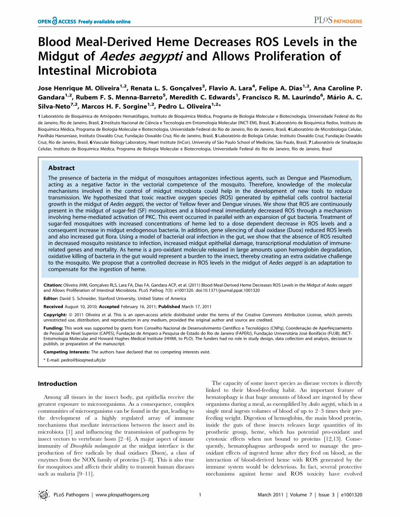
Blood Meal-Derived Heme Decreases ROS Levels in the Midgut of Aedes aegypti and Allows Proliferation...
14
Blood Meal-Derived Heme Decreases ROS Levels in the Midgut of Aedes aegypti and Allows Proliferation of Intestinal Microbiota Jose Henrique M. Oliveira 1,2 , Renata L. S. Gonc ¸alves 3 , Flavio A. Lara 4 , Felipe A. Dias 1,2 , Ana Caroline P. Gandara 1,2 , Rubem F. S. Menna-Barreto 5 , Meredith C. Edwards 1 , Francisco R. M. Laurindo 6 , Ma ´rio A. C. Silva-Neto 7,2 , Marcos H. F. Sorgine 1,2 , Pedro L. Oliveira 1,2 * 1 Laborato ´ rio de Bioquı ´mica de Artro ´ podes Hemato ´ fagos, Instituto de Bioquı ´mica Me ´ dica, Programa de Biologia Molecular e Biotecnologia, Universidade Federal do Rio de Janeiro, Rio de Janeiro, Brasil, 2 Instituto Nacional de Cie ˆ ncia e Tecnologia em Entomologia Molecular (INCT-EM), Brasil, 3 Laborato ´ rio de Bioquı ´mica Redox, Instituto de Bioquı ´mica Me ´ dica, Programa de Biologia Molecular e Biotecnologia, Universidade Federal do Rio de Janeiro, Rio de Janeiro, Brasil, 4 Laborato ´ rio de Microbiologia Celular, Pavilha ˜o Hansenı ´ase, Instituto Oswaldo Cruz, Fundac ¸a ˜o Oswaldo Cruz, Rio de Janeiro, Brasil, 5 Laborato ´ rio de Biologia Celular, Instituto Oswaldo Cruz, Fundac ¸a ˜o Oswaldo Cruz, Rio de Janeiro, Brasil, 6 Vascular Biology Laboratory, Heart Institute (InCor), University of Sa ˜o Paulo School of Medicine, Sa ˜o Paulo, Brasil, 7 Laborato ´ rio de Sinalizac ¸a ˜o Celular, Instituto de Bioquı ´mica Me ´ dica, Programa de Biologia Molecular e Biotecnologia, Universidade Federal do Rio de Janeiro, Rio de Janeiro, Brasil Abstract The presence of bacteria in the midgut of mosquitoes antagonizes infectious agents, such as Dengue and Plasmodium, acting as a negative factor in the vectorial competence of the mosquito. Therefore, knowledge of the molecular mechanisms involved in the control of midgut microbiota could help in the development of new tools to reduce transmission. We hypothesized that toxic reactive oxygen species (ROS) generated by epithelial cells control bacterial growth in the midgut of Aedes aegypti, the vector of Yellow fever and Dengue viruses. We show that ROS are continuously present in the midgut of sugar-fed (SF) mosquitoes and a blood-meal immediately decreased ROS through a mechanism involving heme-mediated activation of PKC. This event occurred in parallel with an expansion of gut bacteria. Treatment of sugar-fed mosquitoes with increased concentrations of heme led to a dose dependent decrease in ROS levels and a consequent increase in midgut endogenous bacteria. In addition, gene silencing of dual oxidase (Duox) reduced ROS levels and also increased gut flora. Using a model of bacterial oral infection in the gut, we show that the absence of ROS resulted in decreased mosquito resistance to infection, increased midgut epithelial damage, transcriptional modulation of immune- related genes and mortality. As heme is a pro-oxidant molecule released in large amounts upon hemoglobin degradation, oxidative killing of bacteria in the gut would represent a burden to the insect, thereby creating an extra oxidative challenge to the mosquito. We propose that a controlled decrease in ROS levels in the midgut of Aedes aegypti is an adaptation to compensate for the ingestion of heme. Citation: Oliveira JHM, Gonc ¸alves RLS, Lara FA, Dias FA, Gandara ACP, et al. (2011) Blood Meal-Derived Heme Decreases ROS Levels in the Midgut of Aedes aegypti and Allows Proliferation of Intestinal Microbiota. PLoS Pathog 7(3): e1001320. doi:10.1371/journal.ppat.1001320 Editor: David S. Schneider, Stanford University, United States of America Received August 10, 2010; Accepted February 16, 2011; Published March 17, 2011 Copyright: ß 2011 Oliveira et al. This is an open-access article distributed under the terms of the Creative Commons Attribution License, which permits unrestricted use, distribution, and reproduction in any medium, provided the original author and source are credited. Funding: This work was supported by grants from Conselho Nacional de Desenvolvimento Cientı ´fico e Tecnolo ´ gico (CNPq), Coordenac ¸a ˜o de Aperfeic ¸oamento de Pessoal de Nı ´vel Superior (CAPES), Fundac ¸a ˜o de Amparo a Pesquisa de Estado do Rio de Janeiro (FAPERJ), Fundac ¸a ˜o Universita ´ria Jose ´ Bonifa ´cio (FUJB), INCT- Entomologia Molecular and Howard Hughes Medical Institute (HHMI, to PLO). The funders had no role in study design, data collection and analysis, decision to publish, or preparation of the manuscript. Competing Interests: The authors have declared that no competing interests exist. * E-mail: [email protected] Introduction Among all tissues in the insect body, gut epithelia receive the greatest exposure to microorganisms. As a consequence, complex communities of microorganisms can be found in the gut, leading to the development of a highly regulated array of immune mechanisms that mediate interactions between the insect and its microbiota [1] and influencing the transmission of pathogens by insect vectors to vertebrate hosts [2–4]. A major aspect of innate immunity of Drosophila melanogaster at the midgut interface is the production of free radicals by dual oxidases (Duox), a class of enzymes from the NOX family of proteins [5–8]. This is also true for mosquitoes and affects their ability to transmit human diseases such as malaria [9–11]. The capacity of some insect species as disease vectors is directly linked to their blood-feeding habit. An important feature of hematophagy is that huge amounts of blood are ingested by these organisms during a meal, as exemplified by Aedes aegypti, which in a single meal ingests volumes of blood of up to 2–3 times their pre- feeding weight. Digestion of hemoglobin, the main blood protein, inside the guts of these insects releases large quantities of its prosthetic group, heme, which has potential pro-oxidant and cytotoxic effects when not bound to proteins [12,13]. Conse- quently, hematophagous arthropods need to manage the pro- oxidant effects of ingested heme after they feed on blood, as the interaction of blood-derived heme with ROS generated by the immune system would be deleterious. In fact, several protective mechanisms against heme and ROS toxicity have evolved PLoS Pathogens | www.plospathogens.org 1 March 2011 | Volume 7 | Issue 3 | e1001320
Transcript of Blood Meal-Derived Heme Decreases ROS Levels in the Midgut of Aedes aegypti and Allows Proliferation...
Blood Meal-Derived Heme Decreases ROS Levels in theMidgut of Aedes aegypti and Allows Proliferation ofIntestinal MicrobiotaJose Henrique M. Oliveira1,2, Renata L. S. Goncalves3, Flavio A. Lara4, Felipe A. Dias1,2, Ana Caroline P.
Gandara1,2, Rubem F. S. Menna-Barreto5, Meredith C. Edwards1, Francisco R. M. Laurindo6, Mario A. C.
Silva-Neto7,2, Marcos H. F. Sorgine1,2, Pedro L. Oliveira1,2*
1 Laboratorio de Bioquımica de Artropodes Hematofagos, Instituto de Bioquımica Medica, Programa de Biologia Molecular e Biotecnologia, Universidade Federal do Rio
de Janeiro, Rio de Janeiro, Brasil, 2 Instituto Nacional de Ciencia e Tecnologia em Entomologia Molecular (INCT-EM), Brasil, 3 Laboratorio de Bioquımica Redox, Instituto de
Bioquımica Medica, Programa de Biologia Molecular e Biotecnologia, Universidade Federal do Rio de Janeiro, Rio de Janeiro, Brasil, 4 Laboratorio de Microbiologia Celular,
Pavilhao Hansenıase, Instituto Oswaldo Cruz, Fundacao Oswaldo Cruz, Rio de Janeiro, Brasil, 5 Laboratorio de Biologia Celular, Instituto Oswaldo Cruz, Fundacao Oswaldo
Cruz, Rio de Janeiro, Brasil, 6 Vascular Biology Laboratory, Heart Institute (InCor), University of Sao Paulo School of Medicine, Sao Paulo, Brasil, 7 Laboratorio de Sinalizacao
Celular, Instituto de Bioquımica Medica, Programa de Biologia Molecular e Biotecnologia, Universidade Federal do Rio de Janeiro, Rio de Janeiro, Brasil
Abstract
The presence of bacteria in the midgut of mosquitoes antagonizes infectious agents, such as Dengue and Plasmodium,acting as a negative factor in the vectorial competence of the mosquito. Therefore, knowledge of the molecularmechanisms involved in the control of midgut microbiota could help in the development of new tools to reducetransmission. We hypothesized that toxic reactive oxygen species (ROS) generated by epithelial cells control bacterialgrowth in the midgut of Aedes aegypti, the vector of Yellow fever and Dengue viruses. We show that ROS are continuouslypresent in the midgut of sugar-fed (SF) mosquitoes and a blood-meal immediately decreased ROS through a mechanisminvolving heme-mediated activation of PKC. This event occurred in parallel with an expansion of gut bacteria. Treatment ofsugar-fed mosquitoes with increased concentrations of heme led to a dose dependent decrease in ROS levels and aconsequent increase in midgut endogenous bacteria. In addition, gene silencing of dual oxidase (Duox) reduced ROS levelsand also increased gut flora. Using a model of bacterial oral infection in the gut, we show that the absence of ROS resultedin decreased mosquito resistance to infection, increased midgut epithelial damage, transcriptional modulation of immune-related genes and mortality. As heme is a pro-oxidant molecule released in large amounts upon hemoglobin degradation,oxidative killing of bacteria in the gut would represent a burden to the insect, thereby creating an extra oxidative challengeto the mosquito. We propose that a controlled decrease in ROS levels in the midgut of Aedes aegypti is an adaptation tocompensate for the ingestion of heme.
Citation: Oliveira JHM, Goncalves RLS, Lara FA, Dias FA, Gandara ACP, et al. (2011) Blood Meal-Derived Heme Decreases ROS Levels in the Midgut of Aedes aegyptiand Allows Proliferation of Intestinal Microbiota. PLoS Pathog 7(3): e1001320. doi:10.1371/journal.ppat.1001320
Editor: David S. Schneider, Stanford University, United States of America
Received August 10, 2010; Accepted February 16, 2011; Published March 17, 2011
Copyright: � 2011 Oliveira et al. This is an open-access article distributed under the terms of the Creative Commons Attribution License, which permitsunrestricted use, distribution, and reproduction in any medium, provided the original author and source are credited.
Funding: This work was supported by grants from Conselho Nacional de Desenvolvimento Cientıfico e Tecnologico (CNPq), Coordenacao de Aperfeicoamentode Pessoal de Nıvel Superior (CAPES), Fundacao de Amparo a Pesquisa de Estado do Rio de Janeiro (FAPERJ), Fundacao Universitaria Jose Bonifacio (FUJB), INCT-Entomologia Molecular and Howard Hughes Medical Institute (HHMI, to PLO). The funders had no role in study design, data collection and analysis, decision topublish, or preparation of the manuscript.
Competing Interests: The authors have declared that no competing interests exist.
* E-mail: [email protected]
Introduction
Among all tissues in the insect body, gut epithelia receive the
greatest exposure to microorganisms. As a consequence, complex
communities of microorganisms can be found in the gut, leading to
the development of a highly regulated array of immune
mechanisms that mediate interactions between the insect and its
microbiota [1] and influencing the transmission of pathogens by
insect vectors to vertebrate hosts [2–4]. A major aspect of innate
immunity of Drosophila melanogaster at the midgut interface is the
production of free radicals by dual oxidases (Duox), a class of
enzymes from the NOX family of proteins [5–8]. This is also true
for mosquitoes and affects their ability to transmit human diseases
such as malaria [9–11].
The capacity of some insect species as disease vectors is directly
linked to their blood-feeding habit. An important feature of
hematophagy is that huge amounts of blood are ingested by these
organisms during a meal, as exemplified by Aedes aegypti, which in a
single meal ingests volumes of blood of up to 2–3 times their pre-
feeding weight. Digestion of hemoglobin, the main blood protein,
inside the guts of these insects releases large quantities of its
prosthetic group, heme, which has potential pro-oxidant and
cytotoxic effects when not bound to proteins [12,13]. Conse-
quently, hematophagous arthropods need to manage the pro-
oxidant effects of ingested heme after they feed on blood, as the
interaction of blood-derived heme with ROS generated by the
immune system would be deleterious. In fact, several protective
mechanisms against heme and ROS toxicity have evolved
PLoS Pathogens | www.plospathogens.org 1 March 2011 | Volume 7 | Issue 3 | e1001320
independently in different species of blood-feeding organisms,
including heme aggregation [14–16], heme degradation [17,18],
the expression of antioxidant enzymes [19,20] and heme-binding
proteins [21]. As a consequence, the oxidative challenge imposed
by blood feeding is well circumvented by hematophagous insects,
as evidenced by their extraordinary adaptive success. However,
one overlooked aspect of this problem is the impact of these large
amounts of heme on the redox metabolism of the midgut and,
particularly on the operation of gut immune pathways connected
to the production of reactive oxygen species (ROS). The concept
of oxidative stress, originally defined as the imbalance between
pro-oxidant compounds and antioxidant defenses, has recently
been re-described as the ‘‘disruption of redox signaling and
control’’ [22]. Related to this subject, heme notably interferes with
several signaling pathways, including modulation of gene expres-
sion, protein synthesis and phosphorylation connected with
cellular responses to stress [23,24]. We have studied the effect of
a blood meal on ROS levels and immune function in the midgut of
Aedes aegypti, the vector of yellow fever and dengue virus. ROS
levels in the midgut epithelia will be shown to play an important
role in controlling bacteria in the midgut and is dramatically
reduced soon after the ingestion of blood through a mechanism
that involves PKC-dependent heme signaling.
Materials and Methods
Ethics statementAll animal care and experimental protocols were conducted
following the guidelines of the institutional care and use committee
(Committee for Evaluation of Animal Use for Research from the
Federal University of Rio de Janeiro, CAUAP-UFRJ) and the
NIH Guide for the Care and Use of Laboratory Animals (ISBN 0-
309-05377-3). The protocols were approved by CAUAP-UFRJ
under registry #IBQM001. Technicians dedicated to the animal
facility at the Institute of Medical Biochemistry (UFRJ) carried out
all aspects related to rabbit husbandry under strict guidelines to
insure careful and consistent handling of the animals.
MosquitoesAedes aegypti (Red Eye strain) were raised in an insectary at the
Federal University of Rio de Janeiro, Brazil, under a 12 h light/
dark cycle at 28 uC and 70–80% relative humidity. Larvae were
fed with dog chow, and adults were maintained in a cage and
given a solution of 10% sucrose ad libitum. Two to ten day-old
females were used in the experiments.
Mosquito mealsMosquitoes were artificially fed with different diets: (1) 10%
sucrose (ad libitum), (2) heparinized rabbit blood or (3) ‘‘bicarbon-
ate-buffered saline-agarose’’ (BBSA) supplemented with diverse
chemicals, as indicated in the figure legends. The BBSA solution
was composed of glucose (10 mg), 500 mM freshly made
bicarbonate buffer pH 7.4 (10 mL), 0.5 mg low melting-point
agarose and 100 mM ATP, pH 7.4 (5 mL). The final volume was
set to 500 mL with 150 mM NaCl. Feeding was performed using
water-jacketed artificial feeders maintained at 37 uC sealed with
parafilm membranes.
Midgut dissection and cultureDissection was carried out in a drop of PBS at room
temperature. Ten to fifteen midguts were transferred to a 24-
well tissue culture flask containing 1 mL of L-15 medium
supplemented with 5% fetal bovine serum without antibiotics.
Midgut cultures were maintained at room temperature and were
viable for at least 2 h, as assessed by the MTT reduction assay (data
not shown) [25].
Determination of reactive oxygen species (ROS) in themidgut
To assess ROS levels, midguts were incubated with a 2 mM
solution of the oxidant-sensitive fluorophores, CM-H2DCFDA(5-
(and-6)-chloromethyl-29,79-dichloro-dihydrofluorescein diacetate,
acetyl ester) or dihydroethidium (hydroethidine) (DHE) (Invitrogen).
After a 20-min incubation at room temperature in the dark, the
midguts were washed in dye-free medium, and the tissue
transferred to a glass slide in a drop of PBS for epifluorescence
or confocal microscopic examination. Midguts were examined
with a Zeiss Axioskop 40 with an Axiocam MRC5 using a Zeiss-09
filter set (excitation BP 450–490; beam splitter FT 510; emission
LP 515, for CM-H2DCFDA) or a Zeiss-15 filter set (excitation BP
546/12; beam splitter FT 580; emission LP 590, for DHE).
Differential interference contrast (DIC) images were acquired with
a Zeiss AxioObserver, which was also used for some fluorescence
images, with two filter sets, Zeiss-15 and Zeiss-10 (excitation BP
450–490; beam splitter FT 510; emission BP 515–565) for CM-
H2DCFDA. Comparison of fluorescence levels among distinct
images was performed under identical conditions, using the same
objectives, microscopes and similar exposure times in each
experiment. Confocal images were acquired with a Zeiss LSM
510 META (Excitation at 488 nm). For hydrogen peroxide
quantification, the midgut epithelia were dissected in PBS at 4
uC, the gut contents were washed out, and the tissues (pools of 10
organs) were incubated in PBS under dim light and at room
temperature in the presence of 100 mM Amplex Red reagent
(Invitrogen) and 2 units horseradish peroxidase (HRP). After 30-min
incubation, the epithelia were spun, and the supernatant collected.
Fluorescence (Ex: 530 nn; Em: 590 nn) was measured with a Cary
Eclipse spectrofluorimeter (Varian, Palo Alto, CA, USA) and
compared to a hydrogen peroxide standard curve. The total H2O2
release was corrected for non-specific oxidation of Amplex Red
measured in the absence of HRP.
Author Summary
Mosquitoes are vectors of human pathogens, such asDengue virus and Malaria parasites, which profoundlyaffect health worldwide, killing millions of people annually.Recent studies have demonstrated that the presence ofbacteria in the gut of mosquitoes is able to antagonize theestablishment of pathogens. Therefore, mechanisms withthe potential to regulate bacterial growth in the digestivetract of mosquitoes may hamper disease transmission.Here, we show that reactive oxygen species (ROS) arepresent in the gut epithelia of sugar-fed mosquitoes andare drastically reduced after blood feeding through amechanism that involves activation of protein kinase C byheme. ROS levels are inversely correlated with thepresence of bacteria in the midgut and therefore weinvestigated if ROS are involved in fighting bacterialinfections in the gut. We discovered that mosquitoesproducing low levels of ROS challenged with an oralbacterial infection exhibited increased mortality due tointense bacterial proliferation and epithelial cell damage.We propose that reduction in ROS levels after bloodfeeding is an adaptation to compensate for the ingestionof blood, a pro-oxidant meal. This finding has conse-quences for the understanding of disease transmission dueto the ability of ROS to modulate the gut bacterial levels.
ROS in the Midgut of Aedes aegypti
PLoS Pathogens | www.plospathogens.org 2 March 2011 | Volume 7 | Issue 3 | e1001320
HPLC analysis of superoxide in the midgut epitheliumTo provide a more accurate assessment of superoxide levels,
HPLC fractionation of dihydroethidium (DHE) oxidation prod-
ucts was used, as previously described [26]. The midgut epithelia
of 20 female mosquitoes fed on sugar or 24 hours after blood meal
were dissected in PBS at room temperature and incubated in L-15
medium + 5% FBS in 1.5-ml polypropylene tubes. Immediately
after dissection, the midguts were spun, the supernatant removed
and PBS supplemented with 150 mM dyhydroethidium (DHE) was
added for 30 min at ambient temperature under dim light. In
some experiments, the epithelium of sugar fed females was treated
with 25 mM diphenylene iodonium (DPI) or 100 U/mL of PEG-
SOD 30 min before DHE incubation. After DHE incubation, the
midguts were washed twice with PBS, frozen in liquid N2 and
homogenized. The resulting material was resuspended in aceto-
nitrile (500 mL), sonicated (3 cycles of 8W for 5 s) and centrifuged
at 2000 g for 1 min). The supernatant was dried under vacuum
(SpeedVac SVC 100 – Savant), and the resulting pellet stored at
220 uC until use. Three to six pools (20 midgut epithelia/pool)
were prepared, depending on the conditions. Samples were
resuspended in PBS supplemented with 100 mM diethylenetria-
mine pentaacetic acid (DTPA) and injected into an HPLC system
(Waters) equipped with a photodiode array (W2996) and
fluorescence detectors (W2475). Chromatographic separation of
DHE oxidation products was carried out using a NovaPak C18
column (3.96150 mm, 5 mm particle size) equilibrated in solution
A (water/10% acetonitrile/0.1% trifluoracetic acid) with a flow
rate of 0.4 mL/min. After injection of the samples, a 0–40% linear
gradient of solution B (100% acetonitrile) was applied for 10 min,
followed by 10 min of 40% solution B, 5 min of 100% solution B
and 10 min of 100% solution A. The amount of DHE was
measured by light absorption at 245 nm, and DHE oxidation
products, Hydroxyethidium (EOH) and Ethidium (E), were
monitored by fluorescence detection with excitation at 510 nm
and emission at 595 nm.
RNA extraction and qPCR analysisThe protocol used was identical to Goncalves et al. [27].
Midguts were dissected in PBS and RNA extracted using TRIzol
(Invitrogen) according to the manufacturer protocol. RNA was
subjected to DNAse I treatment and cDNA synthesized using
High-Capacity cDNA Reverse transcription kit (Applied Biosys-
tems). qPCR was perfomed with in a StepOnePlus Real Time
PCR System (Applied Biosystems) using Power SYBR-green PCR
master MIX (Applied Biosystems). The Comparative Ct Method
[28] was used to compare changes in gene expression levels. A.
aegypti ribosomal protein 49 gene (RP-49) was used as
endogenous control. Primer sequences are given in supplementary
table 1.
dsRNA synthesis and RNAi experimentsA 964-base pair fragment from Duox gene (AAEL007563-RA)
was amplified from Aedes aegypti with the following primers: F -
GCGATCGATACATTCCGTTT and R - TTCAACAGTTCT-
GGCTGTCG. The amplicon was subjected to nested PCR with
an additional set of primers for Duox that included T7 promoters
(F - TAATACGACTCACTATAGGGATAATGTGGTCGC-
CAA GAGG and R - TAATACGACTCACTATAGGGTG GG-
ACCGAACAGTTTATCC), generating a 450-base pair fragment
that was used to synthesize double-stranded RNA (dsRNA) with
MEGAscript RNAi kit (Ambion, Austin, TX, USA) according to
the manufacturer protocol and standard mosquito RNAi settings
[29]. Gene silencing experiments were performed injecting 69 nL
of a 3 mg /mL solution of dsRNA into the thorax of cold-
anesthetized 2 day-old female mosquitoes. Two days after
injection the mosquitoes were used for experiments.
Midgut bacterial culture and sequencingTo follow the growth profile of cultivable bacteria from the
midgut of sugar or blood-fed (BF) mosquitoes, insects were surface-
sterilized with 70% ethanol and dissected under aseptic conditions.
Pools of 5 guts were homogenized in Luria-Bertani (LB) medium,
serially diluted, plated on LB agar, allowed to grow overnight at 37
uC, and the number colony forming units (CFU) counted. One
bacterial colony presenting low catalase activity (data not shown)
was selected from the midgut of blood-fed mosquitoes (6 h after
the meal) and grown in LB medium for subsequent experiments.
Bacterial DNA was extracted with the DNeasy Blood & Tissue Kit
from Qiagen according the manufacturer’s instructions. DNA was
subjected to PCR amplification using primers designed to amplify
the 16S rDNA (Forward: 5-CCAGACTCCTACGGGAGG-
CAGC-3 and Reverse: 5-CTTGTGCGGGCCCCCGTCAAT-
TC-3) (kindly provided by Dr. Carolina Barillas-Mury). The
resulting product was purified, sequenced and identified using
BLAST against the nucleotide collection database (nr/nt).
Aedes aegypti feeding with bacteria and theinvestigation of ROS impact on the outcome of infection
Females were fed with BBSA supplemented with bacteria
previously isolated from the midguts of blood-fed mosquitoes
(Enterobacter asburiae), as described above, in the presence or
absence of 50 mM ascorbic acid (ASC) (neutralized to pH 7 with
NaOH) or 10 mM DPI (diphenylene iodonium). After growing
overnight in liquid LB medium, the appropriate amount of
bacteria was pelleted, washed, re-suspended in 150 mM NaCl and
mixed with the above components to a final volume of 500 mL.
Fully engorged mosquitoes taken immediately after being fed with
bacteria were transferred to new cages and scored for survival and
bacterial loads at different time-points.
Transmission electron microscopy analysisMidguts were dissected 24 h after feeding and fixed with 2.5%
glutaraldehyde in 0.1 M Na-cacodylate buffer (pH 7.2) at room
temperature for 1 h before being post-fixed in 1% OsO4, 0.8%
potassium ferricyanide and 2.5 mM CaCl2 in the same buffer for
1 h at 25 uC. The cells were dehydrated in an ascending acetone
series and embedded in PolyBed 812 resin. Ultrathin sections were
stained with uranyl acetate and lead citrate and examined in a Jeol
JEM1011 transmission electron microscope (Tokyo, Japan).
Results
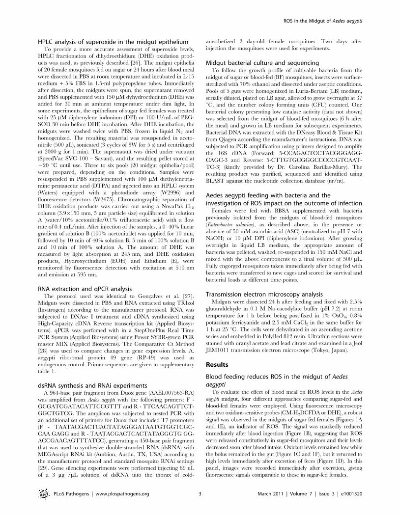
Blood feeding reduces ROS in the midgut of Aedesaegypti
To evaluate the effect of blood meal on ROS levels in the Aedes
aegypti midgut, four different approaches comparing sugar-fed and
blood-fed females were employed. Using fluorescence microscopy
and two oxidant-sensitive probes (CM-H2DCFDA or DHE), a robust
signal was observed in the midguts of sugar-fed females (Figures 1A
and 1E), an indicator of ROS. The signal was markedly reduced
immediately after blood ingestion (Figure 1B), suggesting that ROS
were released constitutively in sugar-fed mosquitoes and their levels
decreased soon after blood intake. Oxidant levels remained low while
the bolus remained in the gut (Figure 1C and 1F), but it returned to
high levels immediately after excretion of feces (Figure 1D). In this
panel, images were recorded immediately after excretion, giving
fluorescence signals comparable to those in sugar-fed females.
ROS in the Midgut of Aedes aegypti
PLoS Pathogens | www.plospathogens.org 3 March 2011 | Volume 7 | Issue 3 | e1001320
Although redox-sensitive dyes have been extensively used in
fluorescence microscopy to study biological roles of ROS, their ability
to identify the nature of the oxidizing species has been questioned due
to the lack of selectivity of most probes in vivo [30,31], including DCF-
based probes [32]. With DHE, its oxidation by different reactive
species results in the formation of distinct products that can be
separated by HPLC. Two main compounds are formed: ethidium
(E), which is most likely formed by reaction with more than one
oxidant species, and 2-hydroxyethidium (EOH), shown to be a
reliable indicator of the presence of superoxide [32–34]. The amount
of ethidium (E) was relatively constant throughout all conditions
tested (Figure 1G), while the levels of 2-hydroxyethidium were higher
in sugar-fed mosquitoes, corroborating the results obtained by
microscopy and demonstrating that superoxide is one of the major
reactive species found in gut epithelia from sugar-fed insects. This
experiment was performed with midgut epithelia (free of gut content),
supporting the conclusion that ROS are generated by epithelial cells.
In addition, EOH signal was specifically reduced in sugar-fed midguts
both by incubation with DPI, an inhibitor of the flavin-containing
NADPH oxidase, and PEG-SOD (supplementary figure S1). We also
investigated alternative sources of ROS/RNS through the incubation
of midguts from sugar-fed mosquitoes with inhibitors of nitric oxide
synthase and xanthine oxidase, respectively L-NAME and allopuri-
nol, but none of these reagents were able to decrease ROS signal in
the midgut (supplementary figure S2).
It is well known that superoxide spontaneously or enzymatically,
through the action of SOD, forms hydrogen peroxide (H2O2), a
diffusible ROS. We measured H2O2 release in the midgut and
detected higher levels in the epithelium of sugar-fed mosquitoes
when compared to blood-fed insects (Figure 1H). Altogether, the
data in Figure 1 strongly support the conclusion that at least
superoxide and hydrogen peroxide are present in the midgut of
Aedes aegypti females fed on sugar and that a blood meal results in
reduced ROS levels.
ROS is released by midgut epithelial cells at the luminalsurface in sugar-fed mosquitoes
CM-H2DCFDA fluorescence was not uniformly distributed
throughout the gut, being strongly concentrated in the lumen
(Figure 2A–C), further evidenced by confocal microscopy, which
showed an intense ROS signal at the lumenal surface in a
longitudinal optical section of the midgut (supplementary figure
S3). Thus, most of the ROS was generated by epithelial cells and
released into the lumen. Transverse optical slices taken at the
apical region of the midgut epithelium of sugar-fed mosquitoes
showed a honeycomb-like appearance, indicating that ROS were
located mostly at the periphery of epithelial cells (Figure 2D – red
arrows). After a blood meal, overall CM-H2DCFDA oxidation was
strongly reduced (Figure 1), cells were flattened due to midgut
distension, and fluorescence associated with intracellular organ-
Figure 1. Blood meal decreases ROS levels in the midgut. Female mosquitoes were fed with sugar or blood, and midguts were dissected atdifferent times after the meal and incubated with CM-H2DCFDA (2 mM) (A–D) or DHE (2 mM) (E–F) for 20 min: (A) sugar; (B) blood (0 hrs after bloodingestion); (C) blood (48 h – before excretion); (D) blood (48 h – after excretion). (E) sugar; (F) blood (24 h). The same camera exposure time was usedto allow side-by-side comparison of fluorescence intensity. Differential interference contrast (DIC) images are shown as insets. Scale bar – 100 mm. (G)Superoxide radical production measured by HPLC-separation of DHE oxidation products in midgut epithelia from sugar or blood-fed mosquitoes(24 h). Asterisk indicates P = 0.0239 for the comparison sugar-EOH vs blood-EOH (T-test). EOH – 2-hydroxyethidium; E - Ethidium. (H) Hydrogenperoxide release from midgut. Asterisk indicates statistically different values (P,0.05, T-test) between sugar (n = 9) and blood-fed (n = 6) pools.doi:10.1371/journal.ppat.1001320.g001
ROS in the Midgut of Aedes aegypti
PLoS Pathogens | www.plospathogens.org 4 March 2011 | Volume 7 | Issue 3 | e1001320

elles was observed (Figure 2E – yellow arrows). It is important to
emphasize that the overall fluorescence intensity in Figure 2D was
much greater than that in 2E, as shown in the previous results in
which sugar-fed and blood-fed CM-H2DCFDA fluorescence had
been compared (Figure 1). Panels 2D and 2E have a similar
intensity because the microscope setup was adjusted to acquire the
best image under each condition to allow for the best localization
of ROS. A careful examination of Figure 2D shows that
intracellular fluorescence, probably associated with organelles, is
also seen in the epithelia of sugar-fed mosquitoes (Figure 2D –
yellow arrow).
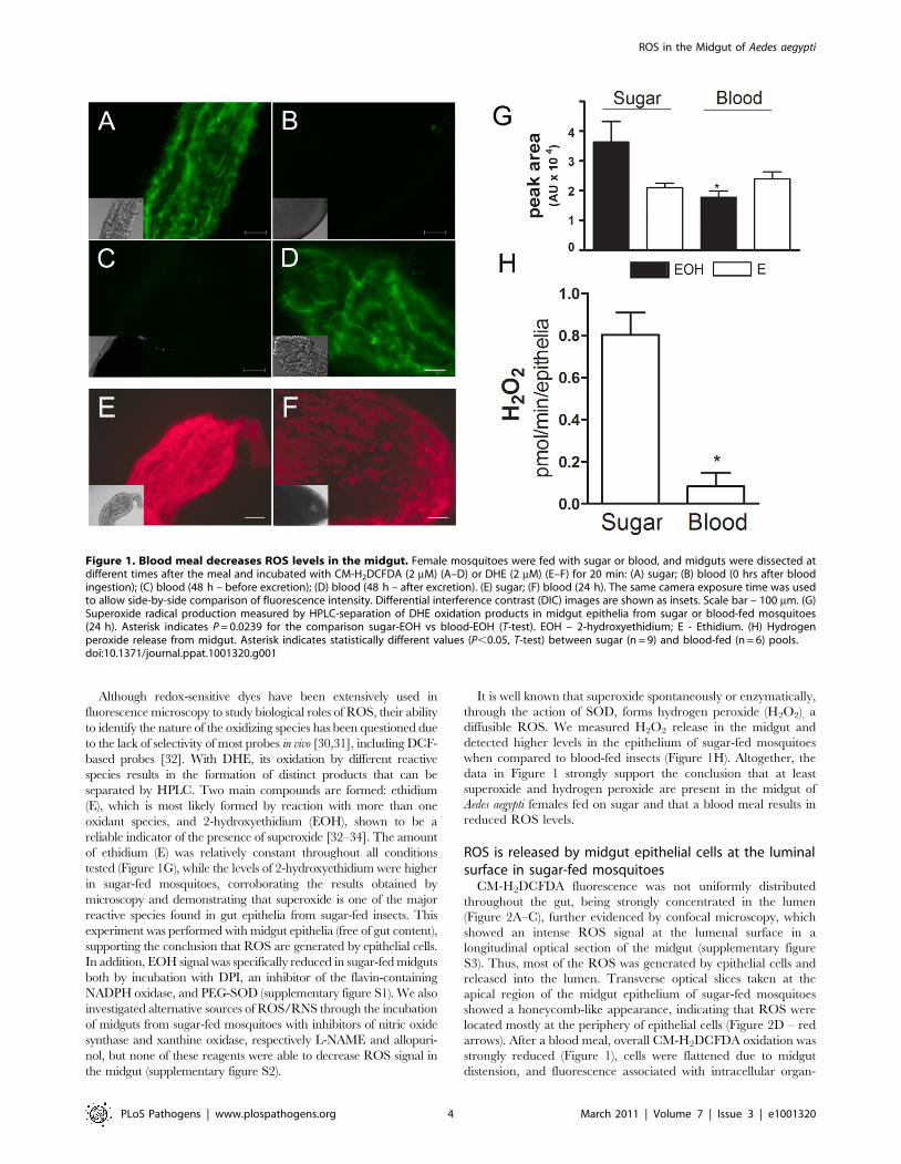
Decrease in ROS signal after a blood meal is triggered byboth hemoglobin and heme, which is mediated by PKC
An incoming blood meal decreases ROS levels, as shown above
in Figure 1 and 3A–B. Feeding insects on a solution containing
salts and low melting-point agarose, referred hereafter as ‘‘BBSA’’
(see Materials and Methods) resulted in CM-H2DCFDA fluores-
cence that was comparable (Figure 3C) or even higher than sugar-
fed midguts (Figure 3A), suggesting that midgut distention is not
responsible for decreasing ROS levels after feeding. Interestingly,
the addition of hemoglobin to BBSA decreased ROS in a dose-
dependent manner, with almost complete suppression of oxidation
of the probe at 10 mg/mL, equivalent to 7% of the total blood
hemoglobin concentration after a full meal (Figures 3E–G).
Feeding mosquitoes with BBSA enriched with albumin, the main
plasma protein, did not reduce ROS levels, showing that this effect
was specific to hemoglobin (Figure 3D). Most important, a low
concentration of heme alone added to the BBSA solution reduced
ROS to levels similar to those observed after a complete blood
meal (Figure 3H and 4H). Further confirmation that the
fluorescence under this experimental condition was due to the
presence of ROS came from suppression of fluorescence signals
after feeding females with BBSA supplemented with the
antioxidants N-acetyl-cysteine (NAC) or uric acid (supplementary
figure S4).
To gain insight into the mechanism that mediates heme-
induced decrease of ROS, mosquitoes fed with BBSA supple-
mented with heme (Figure 3H and 4D) showed lowered ROS
levels similar to that observed after a regular blood meal
(Figure 3B). Interestingly, sugar-fed midguts incubated with heme
added in the culture medium showed no reduction of fluorescence
signal (Figure 4B), with their ROS levels remaining the same as in
sugar-fed midguts alone (Figure 4A). As heme has previously
shown to activate PKC both in vertebrates and invertebrate
models (42, 43), as well as modulate MAP kinase activity (44), we
have explored a possible involvement of PKC in the pathway that
decreases ROS levels after a blood meal. Feeding insects with
heme together with bisindolylmaleimide (BIS), an inhibitor of
protein kinase C (PKC) [35], prevented inhibition of ROS by
heme (BIS in Figure 4E). However, decrease of ROS by heme was
not reversed by feeding with PD98059 (Figure 4F), a MAPK
inhibitor [36], demonstrating the specific involvement of PKC in
this pathway. Furthermore, PKC activation through PMA
supplementation of BBSA lowered ROS levels, even in the
Figure 2. Sites of ROS location in the midgut epithelia. Midguts were incubated with CM-H2DCFDA (2 mM) for 20 min, washed and visualizedunder an epifluorescence (A–C) or confocal microscope (D–E). (A) DIC image of sugar-fed midgut. Scale bar– 100 mm. (B) ROS staining of midgut in(A). (C) Merge of (A) and (B). (D) Confocal image of epithelia from sugar-fed or (E) blood-fed (24 hrs) females showing different patterns of ROSproduction. Scale bar – 20 mm. The fluorescence intensity is not directly comparable in the images shown in panels D and E because the microscopewas set to acquire the best image under each condition to allow optimal visualization of cellular ROS production sites. Red arrows indicatefluorescence associated with the cell periphery. Yellow arrows indicate fluorescence associated with intracellular organelles.doi:10.1371/journal.ppat.1001320.g002
ROS in the Midgut of Aedes aegypti
PLoS Pathogens | www.plospathogens.org 5 March 2011 | Volume 7 | Issue 3 | e1001320
absence of heme (Figure 4G). In contrast, feeding with dibutyryl-
cAMP (Dib in Figure 4H), an agonist of PKA (42), did not change
ROS levels, demonstrating the specific involvement of PKC
activation, but not MAPK and PKA, in heme-mediated down-
regulation of ROS. DIC images from the midguts used in Figure 4
are also shown (supplementary figure S5).
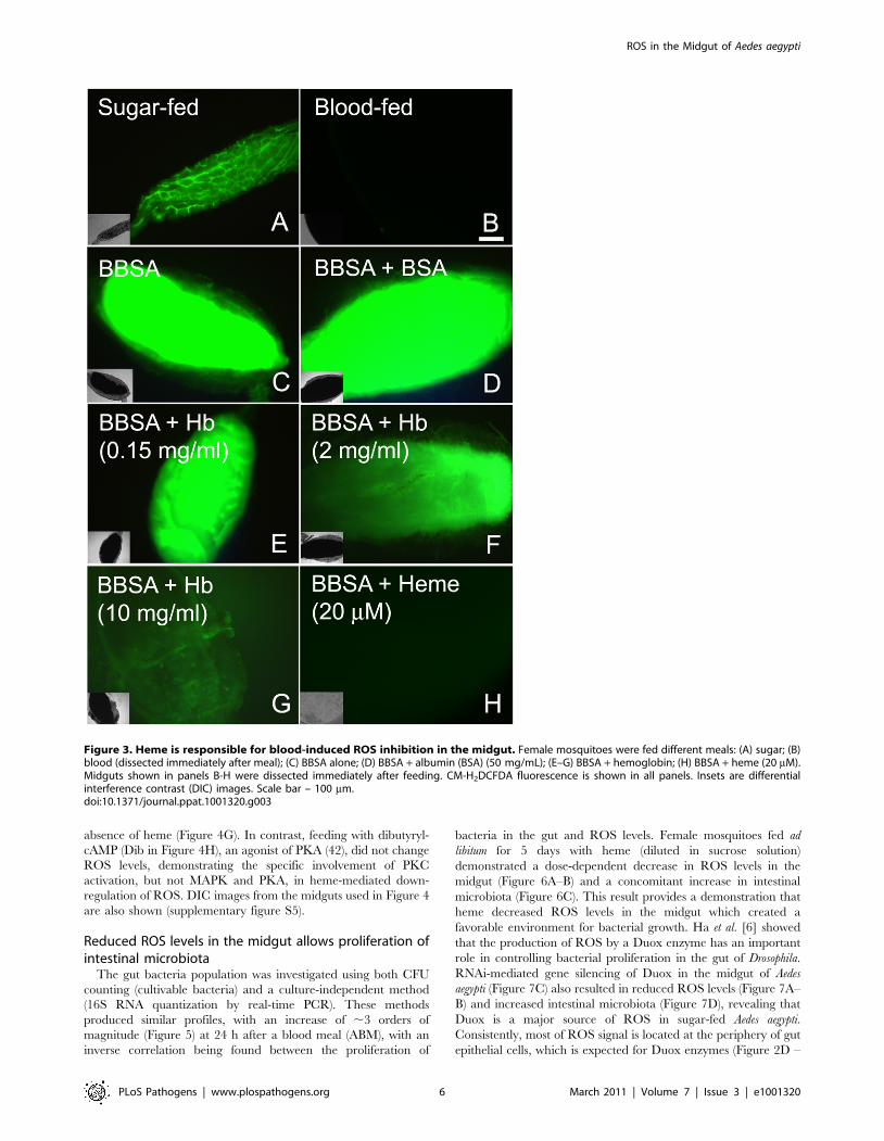
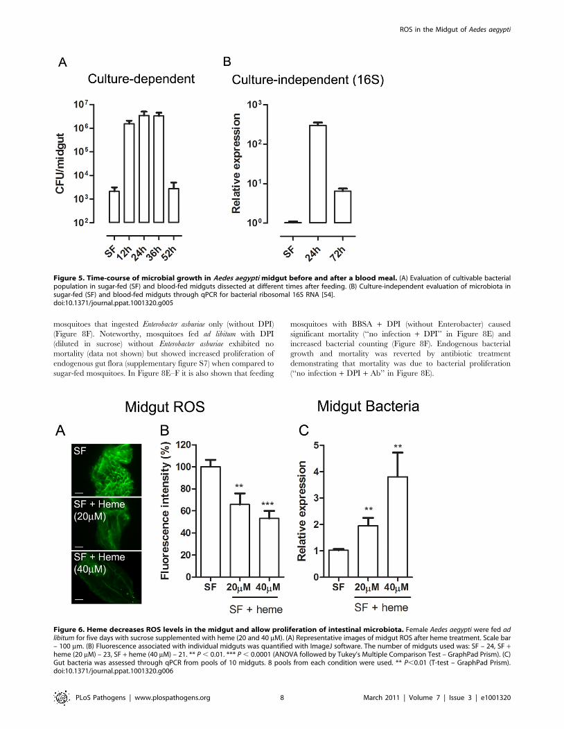
Reduced ROS levels in the midgut allows proliferation ofintestinal microbiota
The gut bacteria population was investigated using both CFU
counting (cultivable bacteria) and a culture-independent method
(16S RNA quantization by real-time PCR). These methods
produced similar profiles, with an increase of ,3 orders of
magnitude (Figure 5) at 24 h after a blood meal (ABM), with an
inverse correlation being found between the proliferation of
bacteria in the gut and ROS levels. Female mosquitoes fed ad
libitum for 5 days with heme (diluted in sucrose solution)
demonstrated a dose-dependent decrease in ROS levels in the
midgut (Figure 6A–B) and a concomitant increase in intestinal
microbiota (Figure 6C). This result provides a demonstration that
heme decreased ROS levels in the midgut which created a
favorable environment for bacterial growth. Ha et al. [6] showed
that the production of ROS by a Duox enzyme has an important
role in controlling bacterial proliferation in the gut of Drosophila.
RNAi-mediated gene silencing of Duox in the midgut of Aedes
aegypti (Figure 7C) also resulted in reduced ROS levels (Figure 7A–
B) and increased intestinal microbiota (Figure 7D), revealing that
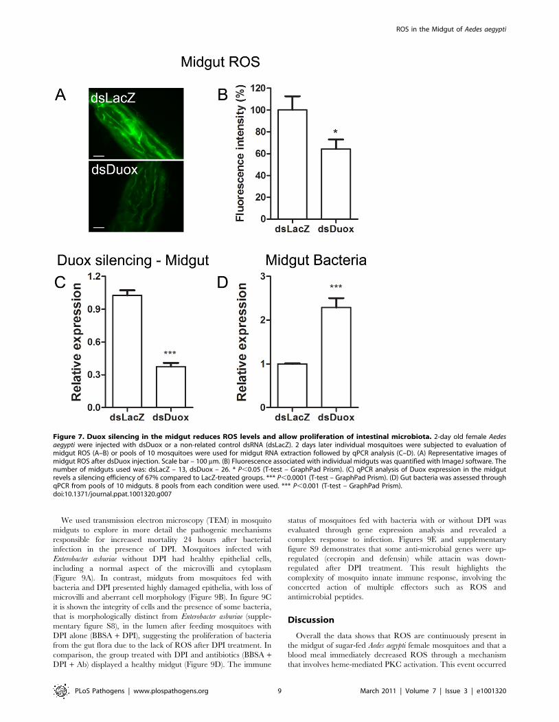
Duox is a major source of ROS in sugar-fed Aedes aegypti.
Consistently, most of ROS signal is located at the periphery of gut
epithelial cells, which is expected for Duox enzymes (Figure 2D –
Figure 3. Heme is responsible for blood-induced ROS inhibition in the midgut. Female mosquitoes were fed different meals: (A) sugar; (B)blood (dissected immediately after meal); (C) BBSA alone; (D) BBSA + albumin (BSA) (50 mg/mL); (E–G) BBSA + hemoglobin; (H) BBSA + heme (20 mM).Midguts shown in panels B-H were dissected immediately after feeding. CM-H2DCFDA fluorescence is shown in all panels. Insets are differentialinterference contrast (DIC) images. Scale bar – 100 mm.doi:10.1371/journal.ppat.1001320.g003
ROS in the Midgut of Aedes aegypti
PLoS Pathogens | www.plospathogens.org 6 March 2011 | Volume 7 | Issue 3 | e1001320

red arrow), and is distinct from localization of ROS in blood-fed
epithelia, which is basically associated to intracellular organelles
(Figure 2E – yellow arrows). Mosquitoes treated with antibiotics
still exhibited an intense ROS signal associated to the midgut,
despite having their gut flora reduced by .90% (data not shown).
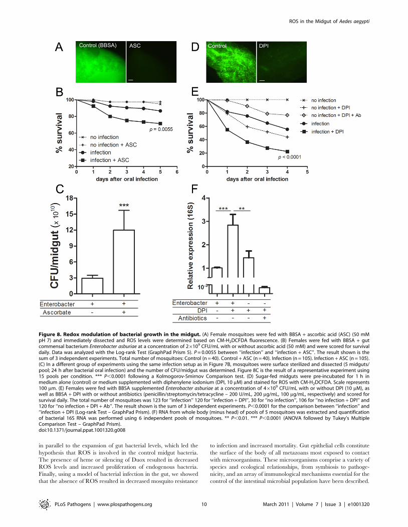
Redox modulation of pathogenesis after bacterialinfection in Aedes aegypti
We decided to investigate whether ROS modulates the ability of
Aedes aegypti to fight a bacterial oral infection. Females were fed a
sub-lethal dose of a bacteria, identified as Enterobacter asburiae based
on 16S rDNA sequencing (supplementary figure S6), isolated from
the midgut of blood-fed (6 h ABM) insects from our colony.
Figure 8B shows that mosquitoes orally infected with this bacteria
in the presence of ascorbic acid (BBSA + Enterobacter + ASC), a
condition that reduces ROS levels (Figure 8A), had a significantly
decreased life span compared to the group infected in the absence
of the antioxidant. Concomitantly, there was a 4-fold increase in
the amount of bacteria in the midgut 24 h after the bacteria-
containing meal (Figure 8C), suggesting that increased mortality
might be attributable to increased proliferation of bacteria in the
midgut. In vitro growth of Enterobacter asburiae in the presence of
ascorbic acid did not result in increased proliferation of bacteria
after 24 h of culture in LB media (data not shown), demonstrating
that the increased bacterial growth in mosquitoes infected in the
presence of ascorbate (Figure 8C) was due to the absence of ROS.
This conclusion was supported by feeding mosquitoes with
Enterobacter asburiae together with DPI (BBSA + Enterobacter +DPI), which also inhibited ROS production (Figure 8D and
supplementary figure S1), causing a marked increase in mortality
(Figure 8E), accompanied by a 3-fold increment in the
proliferation of bacteria 24 h after challenge when compared to
Figure 4. Heme-induced inhibition of ROS is mediated by PKC. ROS signal from the midgut under different conditions was evaluated. (A)Sugar-fed mosquitoes (B) Sugar-fed midguts incubated with heme (20 mM) added in the culture medium. In Figures C–H, female mosquitoes were fedwith BBSA supplemented with different chemicals as indicated and dissected immediately after feeding. (C) BBSA alone; (D) BBSA + heme (20 mM); (E)BBSA + heme (20 mM) + BIS (10 nM) (PKC inhibitor). (F) BBSA + heme (20 mM) + PD98059 (50 mM) (MAPK inihibitor). (G) BBSA + PMA (100 ng/ ml) (PKCagonist). (H) BBSA + dibutyryl-cAMP (50 mM) (PKA agonist). These fluorescent images were acquired using the same microscope setup to allow directcomparison of signal intensities. Representative images are shown.doi:10.1371/journal.ppat.1001320.g004
ROS in the Midgut of Aedes aegypti
PLoS Pathogens | www.plospathogens.org 7 March 2011 | Volume 7 | Issue 3 | e1001320
mosquitoes that ingested Enterobacter asburiae only (without DPI)
(Figure 8F). Noteworthy, mosquitoes fed ad libitum with DPI
(diluted in sucrose) without Enterobacter asburiae exhibited no
mortality (data not shown) but showed increased proliferation of
endogenous gut flora (supplementary figure S7) when compared to
sugar-fed mosquitoes. In Figure 8E–F it is also shown that feeding
mosquitoes with BBSA + DPI (without Enterobacter) caused
significant mortality (‘‘no infection + DPI’’ in Figure 8E) and
increased bacterial counting (Figure 8F). Endogenous bacterial
growth and mortality was reverted by antibiotic treatment
demonstrating that mortality was due to bacterial proliferation
(‘‘no infection + DPI + Ab’’ in Figure 8E).
Figure 5. Time-course of microbial growth in Aedes aegypti midgut before and after a blood meal. (A) Evaluation of cultivable bacterialpopulation in sugar-fed (SF) and blood-fed midguts dissected at different times after feeding. (B) Culture-independent evaluation of microbiota insugar-fed (SF) and blood-fed midguts through qPCR for bacterial ribosomal 16S RNA [54].doi:10.1371/journal.ppat.1001320.g005
Figure 6. Heme decreases ROS levels in the midgut and allow proliferation of intestinal microbiota. Female Aedes aegypti were fed adlibitum for five days with sucrose supplemented with heme (20 and 40 mM). (A) Representative images of midgut ROS after heme treatment. Scale bar– 100 mm. (B) Fluorescence associated with individual midguts was quantified with ImageJ software. The number of midguts used was: SF – 24, SF +heme (20 mM) – 23, SF + heme (40 mM) – 21. ** P , 0.01. *** P , 0.0001 (ANOVA followed by Tukey’s Multiple Comparison Test – GraphPad Prism). (C)Gut bacteria was assessed through qPCR from pools of 10 midguts. 8 pools from each condition were used. ** P,0.01 (T-test – GraphPad Prism).doi:10.1371/journal.ppat.1001320.g006
ROS in the Midgut of Aedes aegypti
PLoS Pathogens | www.plospathogens.org 8 March 2011 | Volume 7 | Issue 3 | e1001320
We used transmission electron microscopy (TEM) in mosquito
midguts to explore in more detail the pathogenic mechanisms
responsible for increased mortality 24 hours after bacterial
infection in the presence of DPI. Mosquitoes infected with
Enterobacter asburiae without DPI had healthy epithelial cells,
including a normal aspect of the microvilli and cytoplasm
(Figure 9A). In contrast, midguts from mosquitoes fed with
bacteria and DPI presented highly damaged epithelia, with loss of
microvilli and aberrant cell morphology (Figure 9B). In figure 9C
it is shown the integrity of cells and the presence of some bacteria,
that is morphologically distinct from Enterobacter asburiae (supple-
mentary figure S8), in the lumen after feeding mosquitoes with
DPI alone (BBSA + DPI), suggesting the proliferation of bacteria
from the gut flora due to the lack of ROS after DPI treatment. In
comparison, the group treated with DPI and antibiotics (BBSA +DPI + Ab) displayed a healthy midgut (Figure 9D). The immune
status of mosquitoes fed with bacteria with or without DPI was
evaluated through gene expression analysis and revealed a
complex response to infection. Figures 9E and supplementary
figure S9 demonstrates that some anti-microbial genes were up-
regulated (cecropin and defensin) while attacin was down-
regulated after DPI treatment. This result highlights the
complexity of mosquito innate immune response, involving the
concerted action of multiple effectors such as ROS and
antimicrobial peptides.
Discussion
Overall the data shows that ROS are continuously present in
the midgut of sugar-fed Aedes aegypti female mosquitoes and that a
blood meal immediately decreased ROS through a mechanism
that involves heme-mediated PKC activation. This event occurred
Figure 7. Duox silencing in the midgut reduces ROS levels and allow proliferation of intestinal microbiota. 2-day old female Aedesaegypti were injected with dsDuox or a non-related control dsRNA (dsLacZ). 2 days later individual mosquitoes were subjected to evaluation ofmidgut ROS (A–B) or pools of 10 mosquitoes were used for midgut RNA extraction followed by qPCR analysis (C–D). (A) Representative images ofmidgut ROS after dsDuox injection. Scale bar – 100 mm. (B) Fluorescence associated with individual midguts was quantified with ImageJ software. Thenumber of midguts used was: dsLacZ – 13, dsDuox – 26. * P,0.05 (T-test – GraphPad Prism). (C) qPCR analysis of Duox expression in the midgutrevels a silencing efficiency of 67% compared to LacZ-treated groups. *** P,0.0001 (T-test – GraphPad Prism). (D) Gut bacteria was assessed throughqPCR from pools of 10 midguts. 8 pools from each condition were used. *** P,0.001 (T-test – GraphPad Prism).doi:10.1371/journal.ppat.1001320.g007
ROS in the Midgut of Aedes aegypti
PLoS Pathogens | www.plospathogens.org 9 March 2011 | Volume 7 | Issue 3 | e1001320
in parallel to the expansion of gut bacterial levels, which led the
hypothesis that ROS is involved in the control midgut bacteria.
The presence of heme or silencing of Duox resulted in decreased
ROS levels and increased proliferation of endogenous bacteria.
Finally, using a model of bacterial infection in the gut, we showed
that the absence of ROS resulted in decreased mosquito resistance
to infection and increased mortality. Gut epithelial cells constitute
the surface of the body of all metazoans most exposed to contact
with microorganisms. These microorganisms comprise a variety of
species and ecological relationships, from symbiosis to pathoge-
nicity, and an array of immunological mechanisms essential for the
control of the intestinal microbial population have been described.
Figure 8. Redox modulation of bacterial growth in the midgut. (A) Female mosquitoes were fed with BBSA + ascorbic acid (ASC) (50 mMpH 7) and immediately dissected and ROS levels were determined based on CM-H2DCFDA fluorescence. (B) Females were fed with BBSA + gutcommensal bacterium Enterobacter asburiae at a concentration of 26109 CFU/mL with or without ascorbic acid (50 mM) and were scored for survivaldaily. Data was analyzed with the Log-rank Test (GraphPad Prism 5). P = 0.0055 between ‘‘infection’’ and ‘‘infection + ASC’’. The result shown is thesum of 3 independent experiments. Total number of mosquitoes: Control (n = 40). Control + ASC (n = 40). Infection (n = 105). Infection + ASC (n = 105).(C) In a different group of experiments using the same infection setup as in Figure 7B, mosquitoes were surface sterilized and dissected (5 midguts/pool; 24 h after bacterial oral infection) and the number of CFU/midgut was determined. Figure 8C is the result of a representative experiment using15 pools per condition. *** P,0.0001 following a Kolmogorov-Smirnov Comparison test. (D) Sugar-fed midguts were pre-incubated for 1 h inmedium alone (control) or medium supplemented with diphenylene iodonium (DPI, 10 mM) and stained for ROS with CM-H2DCFDA. Scale represents100 mm. (E) Females were fed with BBSA supplemented Enterobacter asburiae at a concentration of 46109 CFU/mL with or without DPI (10 mM), aswell as BBSA + DPI with or without antibiotics (penicillin/streptomycin/tetracycline – 200 U/mL, 200 mg/mL, 100 mg/mL, respectively) and scored forsurvival daily. The total number of mosquitoes was 123 for ‘‘infection’’ 120 for ‘‘infection + DPI’’, 30 for ‘‘no infection’’, 106 for ‘‘no infection + DPI’’ and120 for ‘‘no infection + DPI + Ab’’. The result shown is the sum of 3 independent experiments. P,0.0001 for the comparison between ‘‘infection’’ and‘‘infection + DPI (Log-rank Test – GraphPad Prism). (F) RNA from whole body (minus head) of pools of 5 mosquitoes was extracted and quantificationof bacterial 16S RNA was performed using 6 independent pools of mosquitoes. ** P,0.01. *** P,0.0001 (ANOVA followed by Tukey’s MultipleComparison Test – GraphPad Prism).doi:10.1371/journal.ppat.1001320.g008
ROS in the Midgut of Aedes aegypti
PLoS Pathogens | www.plospathogens.org 10 March 2011 | Volume 7 | Issue 3 | e1001320

The production of reactive species is one of the key players in gut
immunity. Ha et al. [5,6] showed that the redox balance in the
gastrointestinal tract of Drosophila melanogaster is a major microbial
control system, determining whether a fly lives or dies after oral
infection with bacteria. Two components have been identified, a
Duox enzyme that generates ROS to oxidize and kill microbes,
and an immune-regulated extracellular catalase that removes any
excess of luminal ROS that might harm the gut epithelia of the fly.
In the malaria vector, the mosquito Anopheles gambiae, it was
recently described that after a blood meal the concerted action of
Duox and a peroxidase is required to form a dityrosine barrier that
decreases midgut permeability to bacterial elicitors, preventing
immune activation and creating a favorable environment for
plasmodium development [11]. In addition, control of levels of
hydrogen peroxide seems to directly modulate the immune
responses against both bacteria and Plasmodium [10]. Similar
ROS-mediated immune responses have been described in
Caenorhabditis elegans [37] and Manduca sexta [38].
Here, we provide for the first time direct evidence that in Aedes
aegypti superoxide anion – and hence hydrogen peroxide – is
produced by epithelial cells and secreted into the lumen of the
midgut (Figures 1 and 2). ROS levels were inversely correlated
with the occurrence of bacteria in the midgut (Figure 1, 5, 6 and
7), and the presence of ROS increased mosquito survival after an
oral challenge with bacteria (Figure 8). However, a unique feature
of the mosquito midgut is that a dramatic decrease in ROS levels
occurs after a blood meal (Figures 1). If sugar-fed mosquitoes
adopt the same pattern of intestinal immunity as in other insects, it
is not clear why they should behave differently after a blood meal,
renouncing the use of ROS as a major weapon to regulate the
growth of gut bacteria. The explanation probably resides in the
fact that the pro-oxidant activity of heme released in the gut upon
digestion of hemoglobin interacts and converts lipid hydroperox-
ides (ROOH), which exhibit quite low reactivity, into the highly
reactive peroxyl (ROO2N) and alcoxyl (RO2N) radicals that have
very pronounced cytotoxicity [39–41]. Lipid hydroperoxides are
normally produced due to abstraction of electrons from lipids by
reactive species produced by metabolic pathways, such as
respiration in mitochondria, or as a consequence of immune-
related oxidase action. Therefore, heme alone does not generate
ROS; it only converts pre-formed oxidized molecules back into
highly reactive intermediates in the lipid peroxidation chain, thus
acting as a catalyst for the formation of potentially toxic radicals.
Thus, we propose that after blood feeding, Aedes aegypti shuts down
ROS generation to avoid heme-mediated oxidative stress.
Consequently, ROS-based immunity is greatly reduced after a
blood meal, contributing to bacterial proliferation.
Recognizing this phenomenon as an important adaptation that
attenuates heme toxicity led us to investigate the signaling
mechanism triggering the down-regulation of ROS after a blood
meal. Midgut distention can be excluded as a potential mechanism
because fully engorged insects fed with BBSA showed intense CM-
H2DCFDA fluorescence (Figure 3C). Although hemoglobin is able
to decrease ROS, heme alone can account for down-regulation of
ROS levels (Figure 3, panel H). The fact that this effect is observed
upon early exposure to the incoming diet (,20 min) excluded
mechanisms based on modification of gene expression and led us
to search for the involvement of protein kinases, a hypothesis that
was confirmed by preventing the heme-mediated suppression of
ROS with a PKC inhibitor (Figure 4E) and by mimicking the
effect of heme using a PKC activator (Figure 4G). In this regard,
heme-induced reduction in ROS levels was only found when heme
was located in the apical (Figure 4D) but not the basal side of the
midgut epithelial cells (Figure 4B), revealing that this signaling
pathway was triggered specifically through a mechanism that
activated PKC after sensing heme in the lumen, which was
achieved by feeding, but not by incubating heme in the culture
medium. Alternatively, we cannot exclude the possibility that the
result obtained in Figure 4B may reveal that the gut does not
respond to heme in vitro. It was already shown that the synthesis of
uric acid by Rhodnius prolixus fat body could be triggered by heme
through activation of PKC [42], suggesting conservation of this
signaling pathway. Activation of PKC by heme modulates ROS
production in human neutrophils [43,44]. Curiously, in these cells
heme was a positive effector of ROS production, suggesting that,
although the heme capacity to activate PKC is probably conserved
in this signaling pathway, a modification downstream of this
protein kinase leads to suppression of ROS in the mosquito midgut
instead of activation. This hypothesis is a major target for future
research.
Gut bacteria experience an explosion in growth after ingestion
of a blood meal by a mosquito (Figure 5). A simple explanation
Figure 9. Bacterial infection in the presence of DPI causes celldamage and immune activation in mosquito midgut. Mosqui-toes were fed with BBSA with Enterobacter asburiae at a concentrationof 46109 CFU/mL with or without DPI (10 mM). 24 h later midguts weredissected and processed for transmission electron microscopy. (A)Mosquitoes infected with bacteria only. (B) Mosquitoes infected withbacteria + DPI (10 mM) to reduce ROS. (C) Mosquitoes fed with BBSA +DPI. (D) Mosquitoes fed with BBSA + DPI + antibiotics (penicillin/streptomycin/tetracycline – 200 U/mL, 200 mg/mL, 100 mg/mL,respectively). All the scale bars represent 2 mm. (E) Gene expressionanalysis of whole body (minus head) mosquitoes 24 h after feedingwith BBSA + bacteria + DPI. Dashed line indicates gene expression ofmosquitoes fed with BBSA + bacteria (without DPI). Different classes ofimmune genes are indicated with colors.doi:10.1371/journal.ppat.1001320.g009
ROS in the Midgut of Aedes aegypti
PLoS Pathogens | www.plospathogens.org 11 March 2011 | Volume 7 | Issue 3 | e1001320

would be that the proliferation of bacteria after blood feeding is
favored by the increase in availability of nutrients compared to
sugar-fed mosquitoes (data not shown). Our data suggest that
bacterial proliferation is also stimulated by the down-regulation of
ROS levels. In spite of the fact that the reduction in ROS levels
was sufficient to increase the gut flora, none of these treatments
was able to allow the growth of endogenous bacteria to levels
found after a blood-meal (100–1000 times more bacteria),
probably due to the lack of nutrient supply to support microbial
growth. This conclusion is further supported by the data in
Figure 6 and 7, where reduced ROS levels due to the presence of
heme or RNAi-mediated silencing of Duox resulted in prolifer-
ation of endogenous bacteria. ROS reduction occurring in the
presence of ascorbate or DPI, 2 unrelated antioxidants that
decrease ROS levels through different modes of action (Figure 8),
also resulted in increased bacterial proliferation, leading to tissue
damage and increased mortality in insects given a sub-lethal dose
of a bacterial species naturally found in the gut. A large amount of
work has been done on invertebrate immunity, especially in
Drosophila melanogaster and mosquitoes, since the discovery that Toll
and IMD pathways play a paramount role in the defense against
invading microorganisms [45,46]. However, knockout of the IMD
pathway alone in Drosophila leads only to modest alterations in
survival when infected orally with ROS-susceptible bacteria [5],
but this NF-kB pathway was essential in insects challenged with
ROS-resistant microbes [47]. In this regard, it is interesting that
the bacteria we used, an Enterobacter (Gram-negative) isolated from
the midgut, whose growth was favored by reduced ROS levels
(Figure 8), had low levels of catalase (data not shown) similar to
most species of this genus [48], and was also found in the gut of
Anopheles gambiae (3) In a similar way to that described for Drosophila
[47], the immune response triggered in our infection system was
not entirely based on production of ROS, but included the up-
regulation of the antimicrobial peptides, cecropin and defensin
(Figure 9E), known to be part of the IMD pathway [49], and are
responsive to Gram-negative (G-) bacterial infection [50].
However, not all genes related to immune response behave in
the same way and several genes did not show significant activation.
Reduced expression of attacin, which is involved in the defense
against gram-negative bacteria [51,52], prompts us to speculate
that attacin down-regulation may be part of the pathophysiological
mechanism(s) involved in increased mosquito mortality. When
ROS production was blocked by DPI, there was bacterial
proliferation in the gut and several antimicrobial genes were up-
regulated (Figure 9E and S9), in a possible attempt to reduce tissue
damage induced by the bacteria. Immune genes are overexpressed
after a blood meal (data not shown) and this could compensate for
reduction of ROS levels reported here, explaining why mosquitoes
do not die after blood intake, in spite of having increased bacteria
proliferation together with the lack of a major antibacterial
mechanism. Taken together, these results highlight a complex
effect of the blood meal on the immune regulation network.
This work has several consequences for the biology of insects
that are vectors of disease. One is that a similar phenomenon may
operate in the guts of other blood-feeding insects, a possibility
currently being studied in our laboratory. The other is that it has
the potential to influence infection rates of pathogens transmitted
by insect vectors. In fact, a strain of Anopheles gambiae that is
refractory to Plasmodium infection lives in a chronic state of
oxidative stress [9]. At first glance, one might expect that the
decrease in midgut ROS levels after blood meal would be
beneficial for the establishment of viral or protozoan infections.
However, this situation allows bacterial growth a condition that
antagonizes Dengue and Plasmodium infections [2,3].
Our results are also in line with the hypothesis we proposed a
few years ago, namely, that while degrading hemoglobin, some
hematophagous organisms such as the blood fluke, Schistosoma
mansoni, and Plasmodium parasites decrease ROS generation by
shifting energy metabolism to a glycolysis-based anaerobic mode
in order to avoid heme-induced oxidative stress [53]. Interestingly,
this effect seems to not only affect the midgut but also may
constitute a systemic trend, because respiration and H2O2
generation in Aedes flight muscle mitochondria are also reduced
following a blood meal [27].
Our data provide a novel view of ROS production in the
midgut of a disease vector, highlighting the complexity of the
mosquito immune response, where the decrease in ROS
generation that comes with hematophagy creates a favorable
environment for bacterial proliferation, with possible implications
for a better understanding of molecular mechanisms that influence
vector competence.
Supporting Information
Figure S1 Modulation of superoxide radical by DPI and PEG-
SOD. Sugar-fed midguts were pre-incubated in the presence of
either PEG-SOD (100 U/mL) (Sigma) or DPI (25 mM) for 30 min
and transferred to medium with DHE for 20 min; the DHE
oxidation products were measured by HPLC. * P,0.0001 for the
comparison between sugar and sugar + PEG-SOD or Sugar + DPI
(ANOVA, followed by Dunnetts multiple comparison test).
Found at: doi:10.1371/journal.ppat.1001320.s001 (0.49 MB TIF)
Figure S2 Nitric oxide synthase and xanthine oxidase inhibitors
do not decrease ROS in sugar-fed midguts. (A) L-NAME (1 mg/
mL) or (B) allopurinol (500 mM) was added to sugar-fed midgut
cultures for 1 h at room temperature, and ROS levels were
evaluated under the microscope using CM-H2DCFDA.
Found at: doi:10.1371/journal.ppat.1001320.s002 (1.27 MB TIF)
Figure S3 ROS produced by midgut epithelial cells is released into
the lumen in sugar-fed mosquitoes. (A) ROS staining with CM-
H2DCFDA in the midgut of sugar-fed mosquitoes. The image shows a
longitudinal optical section of the midgut. Scale bar- 50 mm. Black
asterisk indicates an air bubble in the gut lumen. (B) The same
experimental setup as in ‘‘A’’ showing the gut at a lower magnification.
Blue represents DAPI (nuclear stain). Scale bar - 20 mm
Found at: doi:10.1371/journal.ppat.1001320.s003 (1.18 MB TIF)
Figure S4 ROS modulation by different antioxidants. Female
mosquitoes were fed with BBSA alone (A) or BBSA supplemented
with 20 mM N-acetyl-cysteine (NAC) (B) (solubilized in 200 mM
Tris-buffer) or 500 mM urate (C) and immediately dissected. ROS
levels were determined based on CM-H2DCFDA fluorescence.
Scale bar represents 100 mm.
Found at: doi:10.1371/journal.ppat.1001320.s004 (2.03 MB TIF)
Figure S5 Differential interference contrast images of midguts
from experiment shown in Figure 4.
Found at: doi:10.1371/journal.ppat.1001320.s005 (1.46 MB TIF)
Figure S6 16S DNA gene sequence from Enterobacter asburiae
isolated from the midgut of Aedes aegypti. Females had their midguts
dissected 6 h after a blood meal before being plated on LB agar.
One colony with low catalase activity was isolated; PCR of the 16S
gene was performed after DNA extraction and the sequencing
data are shown. BLAST analysis of the 1026-bp fragment allowed
identification of the bacterial colony as Enterobacter asburiae
(accession number AJ506159), a gram-negative bacteria known
to be weakly reactive to the catalase test [48].
Found at: doi:10.1371/journal.ppat.1001320.s006 (2.06 MB TIF)
ROS in the Midgut of Aedes aegypti
PLoS Pathogens | www.plospathogens.org 12 March 2011 | Volume 7 | Issue 3 | e1001320

Figure S7 Mosquitoes were fed ad libitum with sucrose 5%
supplemented 10 mM DPI for 5 days and RNA was extracted from
the midgut and processed for 16S quantification through qPCR.
Found at: doi:10.1371/journal.ppat.1001320.s007 (0.27 MB TIF)
Figure S8 Transmission electron microscopy from the bacterial
population typically found in the gut of Aedes aegypti 24 hours after
feeding mosquitoes with BBSA + Enterobacter asburiae + DPI (left) or
BBSA + DPI (right).
Found at: doi:10.1371/journal.ppat.1001320.s008 (0.67 MB TIF)
Figure S9 Mosquitoes were fed with BBSA + DPI (10 mM) or
BBSA + DPI + antibiotics (penicillin/streptomycin/tetracycline).
24 h later RNA from whole body (minus head) was extracted and
gene expression was performed by qPCR. Dashed line indicates
gene expression of mosquitoes fed with BBSA + bacteria (without
DPI), similar to Figure 9E. Different classes of immune genes are
indicated with colors. * P,0.05, ** P,0.01. *** P,0.0001 after t-
test comparing each condition with mosquitoes fed with BBSA +bacteria (without DPI).
Found at: doi:10.1371/journal.ppat.1001320.s009 (1.31 MB TIF)
Acknowledgments
We thank all of the members of the Laboratory of Biochemistry of
Hematophagous Arthropods, especially Katia Anastacio Laia for rearing
insects and S.R. Cassia for technical assistance. Dr. Marlene Benchimol
and M.S. Lorian Cobra Striker for assistance in TEM experiments. We
also thank Dr. Carolina Barillas-Mury and Dr. Alvaro Molina-Cruz
(NIAID-NIH) for helpful discussions. Dr. Marcelo Felippe Santiago for the
use of confocal microscope.
Author Contributions
Conceived and designed the experiments: JHMO RLSG FAL MHFS
PLO. Performed the experiments: JHMO RLSG FAL FAD ACPG
RFSMB MCE. Analyzed the data: JHMO RLSG FAL FAD ACPG
RFSMB FRML MACSN MHFS PLO. Contributed reagents/materials/
analysis tools: FRML MACSN MHFS PLO. Wrote the paper: JHMO
RLSG PLO.
References
1. Ryu JH, Kim SH, Lee HY, Bai JY, Nam YD, et al. (2008) Innate immune
homeostasis by the homeobox gene caudal and commensal-gut mutualism in
Drosophila. Science 319: 777–782.
2. Xi Z, Ramirez JL, Dimopoulos G (2008) The Aedes aegypti toll pathway
controls dengue virus infection. PLoS Pathog 4: e1000098.
3. Dong Y, Manfredini F, Dimopoulos G (2009) Implication of the mosquito
midgut microbiota in the defense against malaria parasites. PLoS Pathog 5:
e1000423.
4. Meister S, Agianian B, Turlure F, Relogio A, Morlais I, et al. (2009) Anopheles
gambiae PGRPLC-mediated defense against bacteria modulates infections with
malaria parasites. PLoS Pathog 5: e1000542.
5. Ha EM, Oh CT, Ryu JH, Bae YS, Kang SW, et al. (2005) An antioxidant
system required for host protection against gut infection in Drosophila. Dev Cell
8: 125–132.
6. Ha EM, Oh CT, Bae YS, Lee WJ (2005) A direct role for dual oxidase in
Drosophila gut immunity. Science 310: 847–850.
7. Ha EM, Lee KA, Park SH, Kim SH, Nam HJ, et al. (2009) Regulation of
DUOX by the Galphaq-phospholipase Cbeta-Ca2+ pathway in Drosophila gut
immunity. Dev Cell 16: 386–397.
8. Ha EM, Lee KA, Seo YY, Kim SH, Lim JH, et al. (2009) Coordination of
multiple dual oxidase-regulatory pathways in responses to commensal and
infectious microbes in drosophila gut. Nat Immunol 10: 949–957.
9. Kumar S, Christophides GK, Cantera R, Charles B, Han YS, et al. (2003) The
role of reactive oxygen species on Plasmodium melanotic encapsulation in
Anopheles gambiae. Proc Natl Acad Sci U S A 100: 14139–14144.
10. Molina-Cruz A, DeJong RJ, Charles B, Gupta L, Kumar S, et al. (2008)
Reactive oxygen species modulate Anopheles gambiae immunity against
bacteria and Plasmodium. J Biol Chem 283: 3217–3223.
11. Kumar S, Molina-Cruz A, Gupta L, Rodrigues J, Barillas-Mury C (2010) A
peroxidase/dual oxidase system modulates midgut epithelial immunity in
Anopheles gambiae. Science 327: 1644–1648.
12. Ryter SW, Tyrrell RM (2000) The heme synthesis and degradation pathways:
role in oxidant sensitivity. Heme oxygenase has both pro- and antioxidant
properties. Free Radic Biol Med 28: 289–309.
13. Jeney V, Balla J, Yachie A, Varga Z, Vercellotti GM, et al. (2002) Pro-oxidant
and cytotoxic effects of circulating heme. Blood 100: 879–887.
14. Graca-Souza AV, Maya-Monteiro C, Paiva-Silva GO, Braz GR, Paes MC, et al.
(2006) Adaptations against heme toxicity in blood-feeding arthropods. Insect
Biochem Mol Biol 36: 322–335.
15. Oliveira MF, Silva JR, Dansa-Petretski M, de Souza W, Lins U, et al. (1999)
Haem detoxification by an insect. Nature 400: 517–518.
16. Devenport M, Alvarenga PH, Shao L, Fujioka H, Bianconi ML, et al. (2006)
Identification of the Aedes aegypti peritrophic matrix protein AeIMUCI as a
heme-binding protein. Biochemistry 45: 9540–9549.
17. Paiva-Silva GO, Cruz-Oliveira C, Nakayasu ES, Maya-Monteiro CM,
Dunkov BC, et al. (2006) A heme-degradation pathway in a blood-sucking
insect. Proc Natl Acad Sci U S A 103: 8030–8035.
18. Pereira LO, Oliveira PL, Almeida IC, Paiva-Silva GO (2007) Biglutaminyl-
biliverdin IX alpha as a heme degradation product in the dengue fever insect-
vector Aedes aegypti. Biochemistry 46: 6822–6829.
19. Paes MC, Oliveira MB, Oliveira PL (2001) Hydrogen peroxide detoxification in
the midgut of the blood-sucking insect, Rhodnius prolixus. Arch Insect Biochem
Physiol 48: 63–71.
20. Citelli M, Lara FA, da Silva VI, Jr., Oliveira PL (2007) Oxidative stress impairs
heme detoxification in the midgut of the cattle tick, Rhipicephalus (Boophilus)
microplus. Mol Biochem Parasitol 151: 81–88.
21. Oliveira PL, Kawooya JK, Ribeiro JM, Meyer T, Poorman R, et al. (1995) A
heme-binding protein from hemolymph and oocytes of the blood-sucking insect,
Rhodnius prolixus. Isolation and characterization. J Biol Chem 270:
10897–10901.
22. Jones DP (2006) Redefining oxidative stress. Antioxid Redox Signal 8:
1865–1879.
23. Igarashi K, Sun J (2006) The heme-Bach1 pathway in the regulation of oxidative
stress response and erythroid differentiation. Antioxid Redox Signal 8: 107–
118.
24. Chen JJ (2007) Regulation of protein synthesis by the heme-regulated eIF2alpha
kinase: relevance to anemias. Blood 109: 2693–2699.
25. Liu Y, Peterson DA, Kimura H, Schubert D (1997) Mechanism of cellular 3-
(4,5-dimethylthiazol-2-yl)-2,5-diphenyltetrazolium bromide (MTT) reduction.
J Neurochem 69: 581–593.
26. Fernandes DC, Wosniak J, Pescatore LA, Bertoline MA, Liberman M, et al.
(2007) Analysis of DHE-derived oxidation products by HPLC in the assessment
of superoxide production and NADPH oxidase activity in vascular systems.
Am J Physiol Cell Physiol 292: C413–C422.
27. Goncalves RL, Machado AC, Paiva-Silva GO, Sorgine MH, Momoli MM, et al.
(2009) Blood-feeding induces reversible functional changes in flight muscle
mitochondria of Aedes aegypti mosquito. PLoS One 4: e7854.
28. Livak KJ, Schmittgen TD (2001) Analysis of relative gene expression data using
real-time quantitative PCR and the 2(-Delta Delta C(T)) Method. Methods 25:
402–408.
29. Gupta L, Molina-Cruz A, Kumar S, Rodrigues J, Dixit R, et al. (2009) The
STAT pathway mediates late-phase immunity against Plasmodium in the
mosquito Anopheles gambiae. Cell Host Microbe 5: 498–507.
30. Benov L, Sztejnberg L, Fridovich I (1998) Critical evaluation of the use of
hydroethidine as a measure of superoxide anion radical. Free Radic Biol Med
25: 826–831.
31. Hempel SL, Buettner GR, O’Malley YQ, Wessels DA, Flaherty DM (1999)
Dihydrofluorescein diacetate is superior for detecting intracellular oxidants:
comparison with 29,79-dichlorodihydrofluorescein diacetate, 5(and 6)-carboxy-
29,79-dichlorodihydrofluorescein diacetate, and dihydrorhodamine 123. Free
Radic Biol Med 27: 146–159.
32. Winterbourn CC, Hampton MB (2008) Thiol chemistry and specificity in redox
signaling. Free Radic Biol Med 45: 549–561.
33. Fridovich I (2003) Editorial commentary on ‘‘Superoxide reacts with hydro-
ethidine but forms a fluorescent product that is distinctly different from
ethidium: potential implications in intracellular fluorescence detection of
superoxide’’ by H. Zhao et al. Free Radic Biol Med 34: 1357–1358.
34. Zielonka J, Vasquez-Vivar J, Kalyanaraman B (2008) Detection of 2-
hydroxyethidium in cellular systems: a unique marker product of superoxide
and hydroethidine. Nat Protoc 3: 8–21.
35. Davies SP, Reddy H, Caivano M, Cohen P (2000) Specificity and mechanism of
action of some commonly used protein kinase inhibitors. Biochem J 351:
95–105.
36. Alessi DR, Cuenda A, Cohen P, Dudley DT, Saltiel AR (1995) PD 098059 is a
specific inhibitor of the activation of mitogen-activated protein kinase kinase in
vitro and in vivo. J Biol Chem 270: 27489–27494.
ROS in the Midgut of Aedes aegypti
PLoS Pathogens | www.plospathogens.org 13 March 2011 | Volume 7 | Issue 3 | e1001320
37. Chavez V, Mohri-Shiomi A, Maadani A, Vega LA, Garsin DA (2007) Oxidative
stress enzymes are required for DAF-16-mediated immunity due to generation ofreactive oxygen species by Caenorhabditis elegans. Genetics 176: 1567–1577.
38. Eleftherianos I, Felfoldi G, ffrench-Constant RH, Reynolds SE (2009) Induced
nitric oxide synthesis in the gut of Manduca sexta protects against oral infectionby the bacterial pathogen Photorhabdus luminescens. Insect Mol Biol 18:
507–516.39. Balla J, Vercellotti GM, Jeney V, Yachie A, Varga Z, et al. (2007) Heme, heme
oxygenase, and ferritin: how the vascular endothelium survives (and dies) in an
iron-rich environment. Antioxid Redox Signal 9: 2119–2137.40. Kalyanaraman B, Mottley C, Mason RP (1983) A direct electron spin resonance
and spin-trapping investigation of peroxyl free radical formation by hematin/hydroperoxide systems. J Biol Chem 258: 3855–3858.
41. van der Zee J, Barr DP, Mason RP (1996) ESR spin trapping investigation ofradical formation from the reaction between hematin and tert-Butyl hydroper-
oxide. Free Radic Biol Med 20: 199–206.
42. Graca-Souza AV, Silva-Neto MA, Oliveira PL (1999) Urate synthesis in theblood-sucking insect rhodnius prolixus. Stimulation by hemin is mediated by
protein kinase C. J Biol Chem 274: 9673–9676.43. Graca-Souza AV, Arruda MA, de Freitas MS, Barja-Fidalgo C, Oliveira PL
(2002) Neutrophil activation by heme: implications for inflammatory processes.
Blood 99: 4160–4165.44. Figueiredo RT, Fernandez PL, Mourao-Sa DS, Porto BN, Dutra FF, et al.
(2007) Characterization of heme as activator of Toll-like receptor 4. J Biol Chem282: 20221–20229.
45. Lemaitre B, Nicolas E, Michaut L, Reichhart JM, Hoffmann JA (1996) Thedorsoventral regulatory gene cassette spatzle/Toll/cactus controls the potent
antifungal response in Drosophila adults. Cell 86: 973–983.
46. Lemaitre B, Hoffmann J (2007) The host defense of Drosophila melanogaster.
Annu Rev Immunol 25: 697–743.
47. Ryu JH, Ha EM, Oh CT, Seol JH, Brey PT, et al. (2006) An essential
complementary role of NF-kappaB pathway to microbicidal oxidants in
Drosophila gut immunity. EMBO J 25: 3693–3701.
48. Taylor WI, Achanzar D (1972) Catalase test as an aid to the identification of
Enterobacteriaceae. Appl Microbiol 24: 58–61.
49. Antonova Y, Alvarez KS, Kim YJ, Kokoza V, Raikhel AS (2009) The role of
NF-kappaB factor REL2 in the Aedes aegypti immune response. Insect Biochem
Mol Biol 39: 303–314.
50. Lowenberger C, Bulet P, Charlet M, Hetru C, Hodgeman B, et al. (1995) Insect
immunity: isolation of three novel inducible antibacterial defensins from the
vector mosquito, Aedes aegypti. Insect Biochem Mol Biol 25: 867–873.
51. Vizioli J, Bulet P, Hoffmann JA, Kafatos FC, Muller HM, et al. (2001)
Gambicin: a novel immune responsive antimicrobial peptide from the malaria
vector Anopheles gambiae. Proc Natl Acad Sci U S A 98: 12630–12635.
52. Asling B, Dushay MS, Hultmark D (1995) Identification of early genes in the
Drosophila immune response by PCR-based differential display: the Attacin A
gene and the evolution of attacin-like proteins. Insect Biochem Mol Biol 25:
511–518.
53. Oliveira PL, Oliveira MF (2002) Vampires, Pasteur and reactive oxygen species.
Is the switch from aerobic to anaerobic metabolism a preventive antioxidant
defence in blood-feeding parasites? FEBS Lett 525: 3–6.
54. Nadkarni MA, Martin FE, Jacques NA, Hunter N (2002) Determination of
bacterial load by real-time PCR using a broad-range (universal) probe and
primers set. Microbiology 148: 257–266.
ROS in the Midgut of Aedes aegypti
PLoS Pathogens | www.plospathogens.org 14 March 2011 | Volume 7 | Issue 3 | e1001320